Note: Descriptions are shown in the official language in which they were submitted.
CA 03098015 2020-10-21
WO 2019/210282
PCT/US2019/029505
MYCOBACTERIAL ANTIGEN COMPOSITIONS AND METHODS OF USE
CROSS REFERENCE TO RELATED APPLICATIONS
[0001] This application claims the benefit of provisional U.S. Application
No. 62/663,239,
filed, April 26, 2018, the entirety of which is hereby incorporated by
reference for all purposes.
FIELD OF INVENTION
[00217] The present invention relates generally to novel immunogenic
combinations
comprising mycobacterial polynucleotides and polypeptides, to fragments or
variants
thereof, and to cells comprising such combined antigens, where the antigens
are from a
Mycobacterium species.
BACKGROUND
[00218] Mycobacteria are ubiquitous pathogens which are a cause of
potentially serious
opportunistic infections in immunocompromised patients. Treatment of
mycobacterial infections
is complicated by broad antimicrobial resistance, which often requires
antibiotic courses with
multiple agents.
[00219] There is evidence that T-cell immunity to mycobacteria is critical
in controlling and
preventing mycobacterial infections, as T-cell deficiency imparts high risk of
invasive
mycobacterial infection.
[00220] In view of the increasing threat and global prevalence of
mycobacterial infection,
new strategies are required for more effective prevention, treatment, and
diagnosis of
mycobacterial infection.
SUMMARY OF THE DISCLOSURE
[00221] The disclosure relates, at least in part, to compositions
comprising mycobacterial
polynucleotides and polypeptides, or fragments or variants thereof, and
exposing such
compositions to cells, such as T cells. The disclosure also relates, at least
in part, to compositions
comprising polynucleotides expressing mycobacterial polypeptides and/or
polypeptides
mycobacterial, or fragments or variants thereof, and exposing such
compositions to cells, such as
T cells. The disclosure also relates to cells comprising polynucleotides
expressing mycobacterial
polypeptides and/or mycobacterial polypeptides, or fragments or variants
thereof, and methods of
priming cells comprising exposing the polynucleotides and/or polypeptides to
the cells for a time
1
CA 03098015 2020-10-21
WO 2019/210282
PCT/US2019/029505
period sufficient for: (i) the polynucleotides to express the polypeptides and
stimulate an antigen-
specific immune response against the polypeptides encoded by the
polynucleotides within the cell
or cells; and/or (ii) stimulate an antigen-specific immune response against
the polypeptide. In
some embodiments, the disclosure relates to expanding T-cells ex vivo, and to
methods of use
thereof In some embodiments, the T cells are naïve T cells. In some
embodiments, the T cells are
naïve T cells from a subject that has not been exposed to mycobacteria. In
some embodiments, T
cells are naïve T cells from a subject that has not contracted a mycobacterial
infection. In some
embodiments, T cells are naïve T cells from a subject that has not contracted
a mycobacterial
infection from one or a plurality of Mycobacterium species disclosed herein.
In some
embodiments, T cells are naïve T cells from a subject that has not contracted
a mycobacterial
infection. In some embodiments, the T cells are naïve T cells isolated from a
subject. The
disclosure is also based, in part, on the surprising finding that human T-
cells from healthy donors
may be expanded using a rapid ex vivo expansion protocol using overlapping
synthetic peptide
pools encompassing various Mycobacterial antigens, and in particular
embodiments, antigens
Ag85B, PPe68, P9WNK7, ESXA, ESXB and ADK.
[00222] In one aspect, the disclosure features a composition comprising a
nucleic acid
sequence encoding an Ag85B antigen, or a functional fragment thereof, from a
Mycobacterium
species, a nucleic acid encoding a PPE68 antigen, or a functional fragment
thereof, from a
Mycobacterium species, a nucleic acid encoding a ESXA antigen, or a functional
fragment
thereof, from a Mycobacterium species, a nucleic acid encoding an ESXB
antigen, or a functional
fragment thereof, from a Mycobacterium species, a nucleic acid encoding an ADK
antigen, or a
functional fragment thereof, from a Mycobacterium species, or a combination
thereof In some
embodiments, the nucleic acid sequence encoding an Ag85B antigen, or a
functional fragment
thereof, is at least 50% identical to SEQ ID NO. 1. In some embodiments, the
nucleic acid
sequence encoding an Ag85B antigen, or a functional fragment thereof, is at
least about 50%,
55%, 60%, 65%, 70%, 75%, 80%, 85%, 90%, 95%, 96%, 97%, 98%, 99% identical to
SEQ ID
NO. 1. In some embodiments, the nucleic acid sequence encoding a PPE68
antigen, or a
functional fragment thereof, is at least 50% identical to SEQ ID NO. 2. In
some embodiments,
the nucleic acid sequence encoding a PPE68 antigen, or a functional fragment
thereof, is at least
about 50%, 55%, 60%, 65%, 70%, 75%, 80%, 85%, 90%, 95%, 96%, 97%, 98%, 99%
identical to
SEQ ID NO. 2. In some embodiments, the nucleic acid sequence encoding an ESXA
antigen, or a
functional fragment thereof, is at least 50% identical to SEQ ID NO. 3. In
some embodiments,
the nucleic acid sequence encoding a ESXA antigen, or a functional fragment
thereof, is at least
about 50%, 55%, 60%, 65%, 70%, 75%, 80%, 85%, 90%, 95%, 96%, 97%, 98%, 99%
identical to
2
CA 03098015 2020-10-21
WO 2019/210282
PCT/US2019/029505
SEQ ID NO. 3. In some embodiments, the nucleic acid sequence encoding an ESXB
antigen, or a
functional fragment thereof, is at least 50% identical to SEQ ID NO. 4. In
some embodiments,
the nucleic acid sequence encoding an ESXB antigen, or a functional fragment
thereof, is at least
about 50%, 55%, 60%, 65%, 70%, 75%, 80%, 85%, 90%, 95%, 96%, 97%, 98%, 99%
identical to
SEQ ID NO. 4. In some embodiments, the nucleic acid sequence encoding an ADK
antigen, or a
functional fragment thereof, is at least 50% identical to SEQ ID NO. 5. In
some embodiments,
the nucleic acid sequence encoding an ADK antigen, or a functional fragment
thereof, is at least
about 50%, 55%, 60%, 65%, 70%, 75%, 80%, 85%, 90%, 95%, 96%, 97%, 98%, 99%
identical to
SEQ ID NO. 5.
[00223] In other aspects, the disclosure features a composition comprising
a polypeptide
comprising an amino acid sequence, or fragment thereof, coding for an Ag85B
antigen from a
Mycobacterium species, a polypeptide comprising an amino acid sequence, or
fragment thereof,
coding for a PPE68 antigen from a Mycobacterium species, a polypeptide
comprising an amino
acid sequence, or fragment thereof, coding for a ESXA antigen from a
Mycobacterium species, a
polypeptide comprising an amino acid sequence, or fragment thereof, coding for
a ESXB antigen
from a Mycobacterium species, a polypeptide comprising an amino acid sequence,
or fragment
thereof, coding for an ADK antigen from a Mycobacterium species, or a
combination thereof. In
some embodiments, the polypeptide comprising an amino acid sequence, or
fragment thereof,
coding for an Ag85B antigen is about 50% identical to SEQ ID NO. 6. In some
embodiments,
the polypeptide comprising an amino acid sequence, or fragment thereof, coding
for an Ag85B
antigen is about 50%, 55%, 60%, 65%, 70%, 75%, 80%, 85%, 90%, 95%, 96%, 97%,
98%, 99%
identical to SEQ ID NO. 6. In some embodiments, the polypeptide comprising an
amino acid
sequence, or fragment thereof, coding for an PPE68 antigen is 50% identical to
SEQ IS NO. 7. In
some embodiments, the polypeptide comprising an amino acid sequence, or
fragment thereof,
coding for an PPE68 antigen is about 50%, 55%, 60%, 65%, 70%, 75%, 80%, 85%,
90%, 95%,
96%, 97%, 98%, 99% identical to SEQ IS NO. 7. In some embodiments, the
polypeptide
comprising an amino acid sequence, or fragment thereof, coding for an ESXA
antigen is 50%
identical to SEQ ID NO. 8. In some embodiments, the polypeptide comprising an
amino acid
sequence, or fragment thereof, coding for an ESXA antigen is about 50%, 55%,
60%, 65%, 70%,
75%, 80%, 85%, 90%, 95%, 96%, 97%, 98%, 99% identical to SEQ ID NO. 8. In some
embodiments, the polypeptide comprising an amino acid sequence, or fragment
thereof, coding
for an ESXB antigen is 50% identical to SEQ ID NO. 9. In some embodiments, the
polypeptide
comprising an amino acid sequence, or fragment thereof, coding for an ESXB
antigen is about
50%, 55%, 60%, 65%, 70%, 75%, 80%, 85%, 90%, 95%, 96%, 97%, 98%, 99% identical
to SEQ
3
CA 03098015 2020-10-21
WO 2019/210282
PCT/US2019/029505
ID NO. 9. In some embodiments, the polypeptide comprising an amino acid
sequence, or
fragment thereof, coding for an ADK antigen is about 50% identical to SEQ ID
NO. 10. In some
embodiments, the polypeptide comprising an amino acid sequence, or fragment
thereof, coding
for an ADK antigen is about 50%, 55%, 60%, 65%, 70%, 75%, 80%, 85%, 90%, 95%,
96%, 97%,
98%, 99% identical to SEQ ID NO. 10. In another embodiment, one or more amino
acid
sequences overlap in sequence to span part or all of the Ag85B, PPE68, ESXA,
ESXB and ADK
antigens. In some embodiments of any of the above aspects or embodiments, the
Mycobacterium
species is selected from the group consisting ofM tuberculosis, M bovis, M
bovis BCG, M
avium, M abscessus, M chelonae, M kansasii, M africanum, M canetti,M caprae,M
microt,
M mungi, M orygis, M avium, M avium paratuberculosis, M avium silvaticum, M
columbiense, M intracellulare, M gordonae, M ulcerans, M genavense, M
scrofulaceum, M
intermedium, M fortuitum, and M mucogenicum. In another embodiment of any of
the above
aspects or embodiments, the composition is used to stimulate an immune cell.
In some
embodiments, stimulating the immune cell comprises activating the immune cell.
In some
embodiments, stimulating the immune cell comprises expanding the immune cell.
In some
embodiments, the immune cell is a CD8+ T cell. In some embodiments, the immune
cell is a NK
cell. In some embodiments, the immune cell is a CD4+ T cell.
[00224] In other aspects, the disclosure features a cell or plurality of
cells comprising one or
a combination of (i) a nucleic acid sequence encoding an Ag85B antigen at
least about 50%
identical to SEQ ID NO. 1, a nucleic acid sequence encoding a PPE68 antigen at
least about 50%
identical to SEQ ID NO. 2, a nucleic acid sequence encoding a ESXA antigen at
least about 50%
identical to SEQ ID NO. 3, a nucleic acid sequence encoding an ESXB antigen at
least about 50%
identical to SEQ ID NO. 4, a nucleic acid sequence encoding an ADK antigen at
least about 50%
identical to SEQ ID NO. 5, or a combination thereof; (ii) a polypeptide
comprising an amino acid
sequence coding for an Ag85B antigen at least about 50% identical to SEQ ID
NO. 6, a
polypeptide comprising an amino acid sequence coding for an PPE68 antigen at
least about 50%
identical to SEQ ID NO. 7, a polypeptide comprising an amino acid sequence
coding for an
ESXA antigen at least about 50% identical to SEQ ID NO. 8, a polypeptide
comprising an amino
acid sequence coding for an ESXB antigen at least about 50% identical to SEQ
ID NO. 9, a
polypeptide comprising an amino acid sequence coding for an ADK antigen at
least about 50%
identical to SEQ ID NO. 10, or a combination thereof; (iii) a nucleic acid of
(i), encoding a
functional fragment of a nucleic acid sequence of SEQ ID NO. 1, SEQ ID NO. 2,
SEQ ID NO. 3,
SEQ ID NO. 4 or SEQ ID NO. 5; and (iv) an amino acid sequence of (ii),
encoding a functional
fragment of an amino acid sequence of SEQ ID NO. 6, SEQ ID NO. 7, SEQ ID NO.
8, SEQ ID
4
CA 03098015 2020-10-21
WO 2019/210282 PCT/US2019/029505
NO. 9 or SEQ ID NO. 10. In some embodiments, the cell is a helper (CD4+) T-
cell. In some
embodiments, the cell is a cytotoxic (CD8+) T-cell. In some embodiments, the
cell is a
Gamma/Delta T-cell. In some embodiments, the cell is a central memory T-cell.
In some
embodiments, the cell is an effector memory T-cell. In some embodiments, the
CD4+ T cell
comprises about 60% to about 90% of the total T-cell population. In some
embodiments, the
CD8+ T-cell comprises about 0% to about 40% of the total T-cell population. In
some
embodiments, the Gamma/Delta T-cell comprises about 0.5% to about 10% of the
total T-cell
population. In some embodiments, the central memory T-cell comprises about
0.5% to about
15% of the total T-cell population. In some embodiments, the central memory T-
cell comprises
about 20% to about 60% of the total T-cell population. In some embodiments,
the plurality of
cells comprise CD4+ T-cells and CD8+ T-cells, wherein the number of CD8+T-
cells is greater
than the number of CD4+ T-cells. In another embodiment, the cell is from a
human subject. In a
further embodiment, the human subject is immunocompromised. In another further
embodiment,
the human subject has been diagnosed or is suspected of having a Mycobacterial
infection. In
some embodiments, the cell or plurality of cells are expanded in cell culture.
In some
embodiments, the cell or plurality of cells comprises at least one primary T-
cell. In some
embodiments, the cell is an antigen presenting cell (APC). In another
embodiment, the APC cell
is an artificial antigen presenting cell. In some embodiments, the cell is a
macrophage. In some
embodiments, the cell is a dendritic cell. In some embodiments of any of the
above aspects or
embodiments, the composition further comprises one or more antigens from a
Mycobacterial
species, wherein the one or more antigens are provided in Table 1.
po,w, 281..*6 81.8.8,1818 8.12,8,21:P*1 880.1248P
980.88.4:4 i{38,..484$ bt9:4:06:82t1ix8 MTh:88M
giZ.X* , "
= NV: MO X=tO,
rn
1 7 : 1 =:. = ,,, : : Otrtt "
OkW
.,,$..*0:0P IA* fti* .8,08081\NY,
= ::: :888. ' <4co 0)08
8 : : :8810 8,80,
0:.s8 . = .. = .. .. 0:08 8,;1 *4,
. ....................... : :(,1,:,4=03 = !M ($7$,=. =
',=,,41 4.Z =
gµIM . .. " ' = =
.................. , = ;: <.µ *141 0*Z
jlta *kW,
[00225] In another
embodiment of any of the above aspects or embodiments, the cell is
CA 03098015 2020-10-21
WO 2019/210282
PCT/US2019/029505
capable of expressing a polypeptide comprising an amino acid sequence, or
fragment thereof,
coding for an Ag85B antigen from a Mycobacterium species, a polypeptide
comprising an amino
acid sequence, or fragment thereof, coding for a PPE68 antigen from
aMycobacterium species, a
polypeptide comprising an amino acid sequence, or fragment thereof, coding for
a ESXA antigen
from a Mycobacterium species, a polypeptide comprising an amino acid sequence,
or fragment
thereof, coding for a ESXB antigen from a Mycobacterium species, a polypeptide
comprising an
amino acid sequence, or fragment thereof, coding for an ADK antigen from a
Mycobacterium
species, or a combination thereof.
[00226] .. In other aspects, the disclosure features a cell engineered to
expand T-cells ex vivo,
wherein the cell comprises at least 5 antigens selected from Ag85B, PPE68,
ESXA, ESXB and
ADK, wherein the cell is produced by a process comprising introducing one or
more nucleic
acids, each encoding one or more of the at least 5 antigens, into the cell;
and culturing the cell
under conditions suitable for production of one or more of the antigens. In
some embodiments,
the cell is an antigen-presenting cell, including T-cell, B-cells, monocytes,
dendritic cells,
Phytohemagglutinin blasts, or artificial antigen presenting cells based on
immortalized cells such
as K562 or other cell lines. In some embodiments, the nucleic acid comprises
DNA or RNA. In
some embodiments, the introducing step comprises viral transduction. In some
embodiments, the
introducing step comprises electroporation. In another embodiment, the
disclosure features a
composition comprising one or a plurality of cells of any of the aspects or
embodiments herein.
[00227] In other aspects, the disclosure features a pharmaceutical
composition comprising
(i) a pharmaceutically effective amount of the composition of any of the
aspects and embodiments
herein; and (ii) a pharmaceutically acceptable carrier. In other aspects, the
disclosure features a
pharmaceutical composition comprising (i) a pharmaceutically effective amount
of the
composition of any of the aspects and embodiments herein; and (ii) a
pharmaceutically acceptable
carrier for treatment of mycobacterial infection in a subject in need thereof.
In other aspects, the
disclosure features a pharmaceutical composition comprising (i) a
pharmaceutically effective
amount of the composition of any of the aspects and embodiments herein; and
(ii) a
pharmaceutically acceptable carrier, for treatment or prevention of a
mycobacterial infection in a
subject. In some embodiments, the subject is immunocompromised. In some
embodiments, the
subject has or is identified as having an organ transplant. In some
embodiments, the subject is
immunocompromised. In some embodiments, the subject has or is identified as
having a cancer.
In some embodiments, the subject is immunocompromised. In some embodiments,
the subject has
or is identified as having a cancer of the blood.
[00228] In other aspects, the disclosure features a pharmaceutical
composition comprising
6
CA 03098015 2020-10-21
WO 2019/210282
PCT/US2019/029505
(i) a pharmaceutically effective amount of one or a plurality of cells of any
of the aspects and
embodiments here; and (ii) a pharmaceutically acceptable carrier.
[00229] In other aspects, the disclosure features a method of expanding T
cells ex vivo, the
method comprising (a) culturing one or a plurality of T-cells; (b) contacting
the plurality of T-
cells with a polypeptide comprising an amino acid sequence, or fragment
thereof, coding for an
Ag85B antigen from a Mycobacterium species, a polypeptide comprising an amino
acid sequence,
or fragment thereof, coding for a PPE68 antigen from a Mycobacterium species,
a polypeptide
comprising an amino acid sequence, or fragment thereof, coding for a ESXA
antigen from a
Mycobacterium species, a polypeptide comprising an amino acid sequence, or
fragment thereof,
coding for a ESXB antigen from a Mycobacterium species, a polypeptide
comprising an amino
acid sequence, or fragment thereof, coding for an ADK antigen from a
Mycobacterium species, or
a combination thereof; or contacting the plurality of T-cells with a
composition of any of the
aspects or embodiments herein or the pharmaceutical composition of any of the
aspects or
embodiments herein.
[00230] In other aspects, the disclosure features a method for expanding T-
cells ex vivo, the
method comprising (a) culturing one or a plurality of isolated T-cells; (b)
contacting the plurality
of T-cells with an antigen presenting cell, wherein the antigen presenting
cell presents expressing
an amino acid sequence, or fragment thereof, coding for an Ag85B antigen from
a Mycobacterium
species, a polypeptide comprising an amino acid sequence, or fragment thereof,
coding for a
PPE68 antigen from a Mycobacterium species, a polypeptide comprising an amino
acid sequence,
or fragment thereof, coding for a ESXA antigen from a Mycobacterium species, a
polypeptide
comprising an amino acid sequence, or fragment thereof, coding for a ESXB
antigen from a
Mycobacterium species, a polypeptide comprising an amino acid sequence, or
fragment thereof,
coding for an ADK antigen from a Mycobacterium species, or a combination
thereof. In some
embodiments, the method further comprises stimulating the one or plurality of
T-cells with one or
more cytokines. In some embodiments, the cytokine is selected from the group
consisting of IL-4,
IL-7, IL-15, IL-21, TNFI3, and IFNa. In some embodiments, the method further
comprises
isolating a sample from a subject prior to step (a) and isolating T-cell from
the samples.
[00231] In other aspects, the disclosure features a method of treating
Mycobacterium
infection in a subject in need thereof, the method comprising administering to
the subject a
therapeutically effective amount of the composition of any of the aspects or
embodiments herein,
or the pharmaceutical composition of any of the aspects or embodiments herein.
In some
embodiments, the subject is an immunocompromised host. In some embodiments,
the subject has
been diagnosed as having, or suspected of having, infection with a
Mycobacterium species. In
7
CA 03098015 2020-10-21
WO 2019/210282
PCT/US2019/029505
some embodiments, the infection is an active infection.
[00232] In other aspects, the disclosure features a method of preventing or
delaying
infection with a Mycobacterium in a subject in need thereof, the method
comprising administering
to the subject a therapeutically effective amount of the composition of any of
the aspects or
embodiments herein, or the pharmaceutical composition of any of the aspects or
embodiments
herein. In some embodiments of any of the above aspects or embodiments, the
Mycobacterium
species is selected from the group consisting ofM tuberculosis, M bovis, M
bovis BCG, M
avium, M abscessus, M chelonae, M kansasii, M africanum, M canetti,M caprae,M
microt,
M mungi, M orygis, M avium, M avium paratuberculosis, M avium silvaticum, M
columbiense, M intracellulare, M gordonae, M ulcerans, M genavense, M
scrofulaceum, M
intermedium, M fortuitum, and M mucogenicum. In some embodiments of any of the
above
aspects or embodiments, the subject is immunocompromised. In some embodiments
of any of the
above aspects or embodiments, the subject is a child under the age of 21. In
some embodiments
of any of the above aspects or embodiments, the subject is a child under the
age of 12.
BRIEF DESCRIPTION OF DRAWINGS
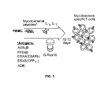
[0018] Figure 1 depicts a manufacturing schema of ex vivo expansion of
mycobacteria-
specific T cells. Peripheral blood mononuclear cells (PBMCs) are stimulated
with overlapping
peptide pools encompassing listed mycobacterial antigens and cultured in a G-
Rex-10 bioreactor
with cytokines for 10-12 days.
[0019] Figure 2A depicts IFN-y ELISpot of ex vivo expanded MSTs at day 10
showed
specificity to multiple mycobacterial antigens in both BCG immunized donors
and non-BCG
vaccinated donors.
[0020] Figure 2B depicts significant differences between groups was noted
in the
responses against PPE68 (*p = 0.028) and ADK (**p = 0.015). SFC, Spot forming
colonies.
[0021] Figure 3A depicts Mycobacterial-specific T cells expanded during
culture with a
mean fold-expansion of 4.4 BCG- = BCG non-immunized; BCG + = BCG immunized.
[0022] Figure 3B surface phenotyping of MSTs following expansion showed a
predominance of CD4+ T cells with large effector memory population and smaller
central
memory population. Lines, median value.
[0023] Figure 3C depicts example plots from MTSs expanded from Donor 9 show
a large
CD4+ effector memory (TEm) population and smaller effector (Teff) and central
memory (Tcm)
population with minimal naïve T cells (T.).
8
CA 03098015 2020-10-21
WO 2019/210282
PCT/US2019/029505
[0024] Figure 4 depicts MSTs expanded from healthy donors are
polyfunctional.
Intracellular flow cytometry demonstrated production of IFN-y and TNF in
response to
mycobacterial pepmix restimulation exclusively in CD4+ T cells from MSTs
expanded from
healthy donors, with no responses seen in CD8+ T cells.
[0025] Figure 5A depicts IFN-y ELISpot of T cells expanded from patients
with primary
immunodeficiency disorders (PID) showed decreased to absent responses to
mycobacterial
antigens, with exception of a patient with NFKB1 hapioinsufficiency. Two
patients with IFN-y
autoantibodies had detectable responses. SEB, staphylococcal enterotoxin B;
CID, combined
immunodeficiency.
[0026] Figure 5B depicts Ex vivo culture of T cells from patients with PID
yielded no
expansion in all but two patients.
[0027] Figure 6 depicts MST responses are comparable using peptide
stimulation vs.
lysate or sensitin. IFN-y ELISpot from MSTs expanded using TB lysate or M
avium sensitin,
showed specificity to multiple mycobactieral pepmixes, which were comparable
in magnitude to
the response to restimulation with lysate or sensitin. Differences in
responses were only
significant for PPE68 (*p= 0.032). SFC, spot forming colonies; SEB,
staphylococcal enterotoxin
B.
[0028] Figure 7A depicts epitope mapping of AG85B and ESXB
[0029] Figure 7B depicts epitope mapping of AG85B and ESXB via IFN-y
ELISpot
showed eight peptides from AG85B and three from ESXB recognized by MSTs from
multiple
healthy donors. SFC, spot forming colonies; SEB, staphylococcal enterotoxin B.
[0030] Figure 8 depicts a gating strategy for surface staining flow
cytometry.
CD14/CD19 are combined for exclusion gating. LD = live/dead.
[0031] Figure 9 depicts a gating strategy for intracellular flow cytometry.
LD = live/dead.
[0032] Figure 10 depicts surface immunophenotyping of MSTs produced using
pepmix,
sensitin, and lysate showed minimal differences in T cell subsets for the
different growth
conditions. The expanded cells were predominantly CD4+ effector memory T cells
(CD4+/CD45R0+/CCR7-), with smaller central memory population
(CD4/CD45R0+/CCR7P/CD62L+).
[0033] Figure 11 provides an analysis of identified T cell epitopes from
AG85B showed
moderate to high conservation across mycobacterial species. Peptide 7 table
begins from top to
bottom SEQ ID NO: 17, 18, 19, 20, 21 and 22. Peptide 14 table from top to
bottom is SEQ ID
NO: 23, 24, 25, 26, 27, 28. Peptide 15 Table begins from top to bottom as SEQ
ID NO:29, 30, 31,
32, 33, 34. Peptide 19 table begins from top to bottom SEQ ID NO:35, 36, 37,
38, 39, 40.
9
CA 03098015 2020-10-21
WO 2019/210282
PCT/US2019/029505
[0034] Figure 12 provides an analysis of identified T cell epitopes from
ESXB showed
low to moderate conservation across mycobacterial species. Peptide 8 table
begins from top to
bottom SEQ ID NO: 41, 42, 43, 44, 45, 46. Peptide 9 table from top to bottom
is SEQ ID NO: 47,
48, 49, 50, 51, 52. Peptide 10 Table begins from top to bottom as SEQ ID
NO:53, 54, 55, 56, 57,
58.
DETAILED DESCRIPTION OF EMBODIMENTS
[0035] Before the present compositions and methods are described, it is to
be understood
that this disclosure is not limited to the particular molecules, compositions,
methodologies or
protocols described, as these may vary. It is also to be understood that the
terminology used in the
description is for the purpose of describing the particular versions or
embodiments only, and is not
intended to limit the scope of the present disclosure which will be limited
only by the appended
claims. It is understood that these embodiments are not limited to the
particular methodology,
protocols, cell lines, vectors, and reagents described, as these may vary. It
also is to be understood
that the terminology used herein is for the purpose of describing particular
embodiments only, and
is not intended to limit the scope of the present embodiments or claims.
Furthermore, the terms
first, second, third and the like in the description and in the claims, are
used for distinguishing
between similar elements and not necessarily for describing a sequential or
chronological order. It
is to be understood that the terms so used are interchangeable under
appropriate circumstances
and that the embodiments of the disclosure described herein are capable of
operation in other
sequences than described or illustrated herein.
Definitions
[0036] Unless defined otherwise, all technical and scientific terms used
herein have the
same meanings as commonly understood by one of ordinary skill in the art.
Although any
methods and materials similar or equivalent to those described herein can be
used in the practice
or testing of embodiments of the present disclosure, the preferred methods,
devices, and materials
are now described. All publications mentioned herein are incorporated by
reference. Nothing
herein is to be construed as an admission that the disclosure is not entitled
to antedate such
disclosure by virtue of prior disclosure.
[0037] As used in the specification and the appended claims, the singular
forms "a," "an"
and "the" include plural referents unless the context clearly dictates
otherwise.
[0038] The term "about" as used herein when referring to a measurable value
such as an
CA 03098015 2020-10-21
WO 2019/210282
PCT/US2019/029505
amount, a temporal duration, and the like, is meant to encompass variations of
20%, 10%,
5%, 1%, 0.9%, 0.8%, 0.7%, 0.6%, 0.5%, 0.4%, 0.3%, 0.2% or 0.1% from
the
specified value, as such variations are appropriate to perform the disclosed
methods.
[0039] The phrase "and/or," as used herein in the specification and in the
claims, should
be understood to mean "either or both" of the elements so conjoined, i.e.,
elements that are
conjunctively present in some cases and disjunctively present in other cases.
Other elements may
optionally be present other than the elements specifically identified by the
"and/or" clause,
whether related or unrelated to those elements specifically identified unless
clearly indicated to
the contrary. Thus, as a non-limiting example, a reference to "A and/or B,"
when used in
conjunction with open-ended language such as "comprising" can refer, In some
embodiments, to
A without B (optionally including elements other than B); in another
embodiment, to B without A
(optionally including elements other than A); in yet another embodiment, to
both A and B
(optionally including other elements); etc.
[0040] As used herein in the specification and in the claims, "or" should
be understood to
have the same meaning as "and/or" as defined above. For example, when
separating items in a
list, "or" or "and/or" shall be interpreted as being inclusive, i.e., the
inclusion of at least one, but
also including more than one, of a number or list of elements, and,
optionally, additional unlisted
items. Only terms clearly indicated to the contrary, such as only one of' or
"exactly one of," or,
when used in the claims, "consisting of," will refer to the inclusion of
exactly one element of a
number or list of elements. In general, the term "or" as used herein shall
only be interpreted as
indicating exclusive alternatives (i.e. "one or the other but not both") when
preceded by terms of
exclusivity, "either," "one of," "only one of," or "exactly one of'
"Consisting essentially of," when
used in the claims, shall have its ordinary meaning as used in the field of
patent law.
[0041] As used herein, the phrase "integer from X to Y" means any integer
that includes
the endpoints. That is, where a range is disclosed, each integer in the range
including the
endpoints is disclosed. For example, the phrase "integer from X to Y"
discloses 1, 2, 3, 4, or 5 as
well as the range 1 to 5.
[0042] As used herein, when used to define products, compositions and
methods, the term
"comprising" (and any form of comprising, such as "comprise" and "comprises"),
"having" (and
any form of having, such as "have" and "has"), "including" (and any form of
including, such as
"includes" and "include") or "containing" (and any form of containing, such as
"contains" and
"contain") are open-ended and do not exclude additional, unrecited elements or
method steps.
Thus, a polypeptide "comprises" an amino acid sequence when the amino acid
sequence might be
part of the final amino acid sequence of the polypeptide. Such a polypeptide
can have up to
11
CA 03098015 2020-10-21
WO 2019/210282
PCT/US2019/029505
several hundred additional amino acids residues (e.g. tag and targeting
peptides as mentioned
herein). "Consisting essentially of' means excluding other components or steps
of any essential
significance. Thus, a composition consisting essentially of the recited
components would not
exclude trace contaminants and pharmaceutically acceptable carriers. A
polypeptide "consists
essentially of an amino acid sequence when such an amino acid sequence is
present with
eventually only a few additional amino acid residues. "Consisting of means
excluding more than
trace elements of other components or steps. For example, a polypeptide
"consists of an amino
acid sequence when the polypeptide does not contain any amino acids but the
recited amino acid
sequence.
[0043] As used herein, "substantially equal" means within a range known to
be correlated
to an abnormal or normal range at a given measured metric. For example, if a
control sample is
from a diseased patient, substantially equal is within an abnormal range. If a
control sample is
from a patient known not to have the condition being tested, substantially
equal is within a normal
range for that given metric.
[0044] As used herein, the term "subject," "individual" or "patient," used
interchangeably,
means any animal, including mammals, such as mice, rats, other rodents,
rabbits, dogs, cats,
swine, cattle, sheep, horses, or primates, such as humans. In some
embodiments, the subject or
patient is a human child of no more than about 20 years of age. In some
embodiments, the subject
or patient is a human child of no more than about 18, 17, 16, 15, 14, 13, 12,
11, 10, 9, 8, 7, 6, 5,
4, 3, 2, or 1 year of age. In some embodiments, the subject has been diagnosed
with or has a
cancer. In some embodiments, the subject has undergone an organ transplant,
such as a bone
marrow transplant. In some embodiments, the subject is a T cell donor if the
embodiment relates
to a method of isolating one or a plurality of cells from a donor for
stimulation or priming of the T
cell.
[0045] The term "subject" is used throughout the specification to describe
an animal from
which a cell sample is taken. In some embodiments, the subject is a human. For
diagnosis of those
conditions which are specific for a specific subject, such as a human being,
the term "patient" may
be interchangeably used. In some instances in the description of the present
invention, the term
"patient" will refer to human patients suffering from a particular disease or
disorder. In some
embodiments, the subject may be a human suspected of having or being
identified as at risk to
develop an infection with a Mycobacterium. In some embodiments, the subject
may be diagnosed
as having an infection with a Mycobacterium and of having or being identified
as at risk to
develop an infection with a Mycobacterium.
[0046] As used herein, an "immunocompromised subject" is meant to refer to
a subject
12
CA 03098015 2020-10-21
WO 2019/210282
PCT/US2019/029505
with a congenital or acquired defect in adaptive or innate immunity, including
but not limited to
primary immunodeficiency disorders, patients undergoing chemotherapy or
immunosuppressive
therapy, or patients undergoing hematopoietic stem cell transplantation. In
some embodiments,
the subject is an immunocompromised adult or child.
[0047] As used herein, the term "animal" includes, but is not limited to,
humans and non-
human vertebrates such as wild animals, rodents, such as rats, ferrets, and
domesticated animals,
and farm animals, such as dogs, cats, horses, pigs, cows, sheep, and goats. In
some embodiments,
the animal is a mammal. In some embodiments, the animal is a human. In some
embodiments,
the animal is a non-human mammal.
[0048] As used herein, the term "mammal" means any animal in the class
Mammalia such
as rodent (i.e., a mouse, a rat, or a guinea pig), a monkey, a cat, a dog, a
cow, a horse, a pig, or a
human. In some embodiments, the mammal is a human. In some embodiments, the
mammal refers
to any non-human mammal. The present disclosure relates to any of the methods
or compositions
of matter disclosed herein wherein the sample is taken from a mammal or non-
human mammal.
The present disclosure relates to any of the methods or compositions of matter
disclosed herein
wherein the sample is taken from a human or non-human primate.
[0049] As used herein, the phrase "in need thereof' means that the animal
or mammal has
been identified or suspected as having a need for the particular method or
treatment. In some
embodiments, the identification can be by any means of diagnosis or
observation. In any of the
methods and treatments described herein, the animal or mammal can be in need
thereof. In some
embodiments, the animal or mammal is in an environment or will be traveling to
an environment
in which a particular disorder or condition is prevalent or more likely to
occur.
[0050] As used herein, "Mycobacterium infection" refers to the exposure of
a subject to a
Mycobacterium species followed by a colonization of the subject or the
subject's tissue(s) by the
bacterium. The colonization can cause serious diseases (e.g. tuberculosis,
leprosy, Bureli ulcer etc,
depending on the Mycobacterium), or can result in no adverse signs
(asymptomatic or latent
infection).
[0051] As used herein, "cell culture" means growth, maintenance,
transfection,
transduction and/or propagation of cells, tissues, or their products. As used
herein, "culture
medium" refers to any solution capable of sustaining the growth of the
targeted cells either in
vitro or in vivo, or any solution with which targeted cells or exogenous
nucleic acids are mixed
before being applied to cells in vitro or to a patient in vivo.
[0052] As used herein, the terms "heterologous" and "foreign" with
reference to nucleic
acids, such as DNA and RNA, are used interchangeably and refer to nucleic acid
that does not
13
CA 03098015 2020-10-21
WO 2019/210282
PCT/US2019/029505
occur naturally as part of a genome or cell in which it is present or which is
found in a location(s)
and/or in amounts in a genome or cell that differ from the location(s) and/or
amounts in which it
occurs in nature, i.e., nucleic acid that is not endogenous to the cell and
has been exogenously
introduced into the cell. Examples of heterologous DNA include, but are not
limited to, DNA that
encodes a gene product or gene product(s) of interest introduced into cells,
for example, for
production of an encoded protein.
[0053] As used herein, "delivery" refers to the process by which exogenous
nucleic acid
molecules are transferred into a cell such that they are located inside the
cell. Delivery of nucleic
acids is a distinct process from expression of nucleic acids. Nucleic acid
material can be
introduced into the cell ex vivo or in vivo by genetic transfer methods, such
as transfection or transduction, to provide a genetically modified cell.
Various expression vectors
(i.e., vehicles for facilitating delivery of exogenous nucleic acid into a
target cell) are known to
one of ordinary skill in the art.
[0054] As used herein, "expression" refers to the process by which nucleic
acid is
translated into peptides or is transcribed into mRNA and translated into
peptides, polypeptides or
proteins. If the nucleic acid is derived from genomic DNA, expression may, if
an appropriate
eukaryotic host cell or organism is selected, include splicing of the mRNA.
For heterologous
nucleic acid to be expressed in a host cell, it must initially be delivered
into the cell and then, once
in the cell, ultimately reside in the nucleus.
[0055] As used herein, "transfection of cells" refers to the acquisition by
a cell of new
nucleic acid material by incorporation of added DNA. Thus, transfection refers
to the insertion of
nucleic acid into a cell using physical or chemical methods. Several
transfection techniques are
known to those of ordinary skill in the art including: calcium phosphate DNA
co-precipitation
(Methods in Molecular Biology (1991)); DEAE-dextran (supra); electroporation
(supra); cationic
liposome-mediated transfection (supra); and tungsten particle-facilitated
microparticle
bombardment (Johnston (1990)). Strontium phosphate DNA co-precipitation (Brash
et al. (1987))
is also a transfection method.
[0056] In contrast, "transduction of cells" refers to the process of
transferring nucleic acid
into a cell using a DNA or RNA virus. A RNA virus (i.e., a retrovirus) for
transferring a nucleic
acid into a cell is also referred to herein as a transducing retrovirus.
Exogenous nucleic acid
material contained within the retrovirus is incorporated into the genome of
the transduced cell. A
cell that has been transduced with a DNA virus (e.g., an adenovirus carrying a
cDNA encoding a
therapeutic agent), will not have the exogenous nucleic acid material
incorporated into its genome
but will be capable of expressing the exogenous nucleic acid material that is
retained
14
CA 03098015 2020-10-21
WO 2019/210282
PCT/US2019/029505
extrachromosomally within the cell.
[0057] The exogenous nucleic acid material can include a nucleic acid
encoding an antigen
from a Mycobacterium species together with a promoter to control
transcription. The promoter
characteristically has a specific nucleotide sequence necessary to initiate
transcription. The
exogenous nucleic acid material may further include additional sequences
(i.e., enhancers)
required to obtain the desired gene transcription activity. For the purpose of
this discussion an
"enhancer" is simply any non-translated DNA sequence that works with the
coding sequence (in
cis) to change the basal transcription level dictated by the promoter. The
exogenous nucleic acid
material may be introduced into the cell genome immediately downstream from
the promoter so
that the promoter and coding sequence are operatively linked so as to permit
transcription of the
coding sequence. An expression vector can include an exogenous promoter
element to control
transcription of the inserted exogenous gene. Such exogenous promoters include
both constitutive
and regulateable promoters.
[0058] The term "domain" as used herein applies to a portion or subsequence
of amino
acids within a peptide or nucleic acids within a nucleotide sequence. In some
embodiments, a
domain provides a functionality, activity, or benefit. In some embodiments for
example a domain
may be a receptor or a signaling portion of a receptor. In another embodiment
a domain may have
a linker function between two other domains. In another embodiment a domain
may serve to bind
a specific target analyte (target domain), such as an antigen or chemokine.
[0059] As used herein, the term "combination" refers to any arrangement
possible of
various components (e.g. mycobacterial antigens and/or encoding nucleic acid
molecules). Such
an arrangement includes mixture of mycobacterial antigens (e.g. mixture of
individual antigens
and/or fusion of antigens) or mixture of nucleic acid molecules (e.g. carried
by one or more
vector) as well as mixture of polypeptide(s) and nucleic acid molecule(s). The
present invention
encompasses combinations comprising equal molar concentrations of each
component as well as
combinations with very different concentrations. It is appreciated that
optimal concentration of
each Mycobacterium component can be determined by the artisan skilled in the
art.
[0060] As used herein, the term "immunogenic" refers to the ability to
induce or stimulate
a measurable T and/or B cell-mediated immune response in a subject into which
the component
qualified as immunogenic has been introduced. For example, the antigenic
combination of the
invention is immunogenic in the sense as it is capable of inducing or
stimulating an immune
response in a subject which can be innate and/or specific (i.e. against at
least one mycobacterial
antigen/epitope comprised in or expressed by said immunogenic combination),
humoral and/or
cellular (e.g. production of antibodies and/or cytokines and/or the activation
of cytotoxic T cells,
CA 03098015 2020-10-21
WO 2019/210282
PCT/US2019/029505
B, T lymphocytes, antigen presenting cells, helper T cells, dendritic cells,
NK cells, etc) and
usually results in a protective response in the administered subject. A vast
variety of direct or
indirect biological assays are available in the art to evaluate the
immunogenic nature of a
component either in vivo (animal or human being), or in vitro (e.g. in a
biological sample) as
described herein.
[0061] As used herein, the term "mycobacterial antigen" refers to a
polypeptide present in
or obtained from a Mycobacterium species or fragment thereof (e.g. an epitope)
capable of being
bound by an antibody or a T cell receptor. Typically, such an antigen contains
one or more B
and/or T epitope(s), in particular CTL or TH epitope(s) or both, involved in
recognition by a
particular antibody or T-cell receptor in the context of the Major
Histocompatibility Complex
(MHC). In the context of the invention, this term encompasses native
mycobacterial polypeptide
as well as fragment and modified version thereof (i.e. variant) as described
hereinafter.
[0062] An "epitope" corresponds to a minimal peptide motif (usually a set
of 8-25 amino
acid residues) that forms a site recognized by an antibody, a T-cell receptor
or a HLA molecule.
Those residues can be consecutive (linear epitope) or not (conformational
epitope that includes
residues that are not immediately adjacent to one another).
[0063] As used herein, the term "variants" is intended to mean
substantially similar
sequences. For nucleic acid molecules, a variant comprises a nucleic acid
molecule having
deletions (i.e., truncations) at the 5' and/or 3' end; deletion and/or
addition of one or more
nucleotides at one or more internal sites in the native polynucleotide; and/or
substitution of one or
more nucleotides at one or more sites in the native polynucleotide. As used
herein, a "native"
nucleic acid molecule or polypeptide comprises a naturally occurring
nucleotide sequence or
amino acid sequence, respectively. For nucleic acid molecules, conservative
variants include those
sequences that, because of the degeneracy of the genetic code, encode the
amino acid sequence of
one of the polypeptides of the disclosure. Variant nucleic acid molecules also
include
synthetically derived nucleic acid molecules, such as those generated, for
example, by using site-
directed mutagenesis but which still encode a protein of the disclosure.
Generally, variants of a
particular nucleic acid molecule of the disclosure will have at least about
70%, 75%, 80%, 85%,
90%, 91%, 92%, 93%, 94%, 95%, 96%, 97%, 98%, 99% or more sequence identity to
that
particular polynucleotide as determined by sequence alignment programs and
parameters as
described elsewhere herein.
[0064] As used herein, "conservative" amino acid substitutions may be
defined as set out
in Tables A, B, or C below. The polypeptides of the disclosure include those
wherein conservative
substitutions (from either nucleic acid or amino acid sequences) have been
introduced by
16
CA 03098015 2020-10-21
WO 2019/210282
PCT/US2019/029505
modification of polynucleotides encoding antigen(s) from aMycobacterium
species. In some
embodiments, these polypeptides comprise CDRs or functional fragments thereof.
Amino acids
can be classified according to physical properties and contribution to
secondary and tertiary
protein structure. A conservative substitution is recognized in the art as a
substitution of one
amino acid for another amino acid that has similar properties. In some
embodiments, the
conservative substitution is recognized in the art as a substitution of one
nucleic acid for another
nucleic acid that has similar properties, or, when encoded, has a binding
affinity to a target or
binding partner similar to the binding affinity of the sequence upon which the
conservative
substitution is based. Exemplary conservative substitutions are set out in
Table A.
Table A -- Conservative Substitutions I
Side Chain Characteristics Amino Acid
Aliphatic
Non-polar GAPILVF
Polar - uncharged CSTMNQ
Polar-charged DEKR
Aromatic HFWY
Other NQDE
[0065] Alternately, conservative amino acids can be grouped as described in
Lehninger,
(Biochemistry, Second Edition; Worth Publishers, Inc. NY, N.Y. (1975), pp. 71-
77) as set forth in
Table B.
Table B -- Conservative Substitutions II
Side Chain Characteristic Amino Acid
Non-polar (hydrophobic)
Aliphatic: ALIVP
Aromatic: F W Y
Sulfur-containing:
Borderline: G Y
Uncharged-polar
Hydroxyl: STY
Amides: NQ
Sulfhydryl:
Borderline: G Y
Positively Charged (Basic): K R H
17
CA 03098015 2020-10-21
WO 2019/210282
PCT/US2019/029505
Negatively Charged (Acidic): D E
[0066] Alternately, exemplary conservative substitutions are set out in
Table C.
Table C -- Conservative Substitutions III
Original Residue Exemplary Substitution
Ala (A) Val Leu Ile Met
Arg (R) Lys His
Asn (N) Gln
Asp (D) Glu
Cy s (C) Ser Thr
Gln (Q) Asn
Glu (E) Asp
Gly (G) Ala Val Leu Pro
His (H) Lys Arg
Ile (I) Leu Val Met Ala Phe
Leu (L) Ile Val Met Ala Phe
Lys (K) Arg His
Met (M) Leu Ile Val Ala
Phe (F) Trp Tyr Ile
Pro (P) Gly Ala Val Leu Ile
Ser (S) Thr
Thr (T) Ser
Trp (W) Tyr Phe Ile
Tyr (Y) Trp Phe Thr Ser
Val (V) Ile Leu Met Ala
[0067] It should be understood that the antigen(s) from aMycobacterium
species, or any
fragments thereof described herein are intended to include amino acid
sequences comprising
polypeptides bearing one or more insertions, deletions, or substitutions, or
any combination
thereof, of amino acid residues as well as modifications other than
insertions, deletions, or
substitutions of amino acid residues, such as but not limited to conservative
amino acid
substitutions.
[0068] As used herein, the term "fragment" or "functional fragment" means
any portion of
a polypeptide that is of a sufficient length to retain at least partial
biological function that is
18
CA 03098015 2020-10-21
WO 2019/210282
PCT/US2019/029505
similar to or substantially similar to the wild-type polypeptide upon which
the fragment is based.
In some embodiments, a fragment of a polypeptide associated with an antigen
from a
Mycobacterium species is a polypeptide that comprises 50, 55, 60, 65, 70, 75,
80, 85, 90, 95, 96,
97, 98, or 99% sequence identity of any polypeptide disclosed herein, and in
particular any
polypeptide comprising an amino acid sequence selected from SEQ ID NOs 6-10.
In some
embodiments, the fragment is a fragment of any polypeptide disclosed herein,
and in particular
any polypeptide comprising an amino acid sequence selected from SEQ ID NOs 6-
10, and has a
length of at least about 10, about 20, about 30, about 40, about 50 , about
60, about 70, about 80,
about 90, or about 100 contiguous amino acids. In some embodiments, the
fragment is a fragment
of any polypeptide disclosed herein, and in particular any polypeptide
comprising an amino acid
sequence selected from SEQ ID NOs 6-10 and has a length of at least about 50
amino acids. In
some embodiments, the fragment is a fragment of any polypeptide disclosed
herein, and in
particular any polypeptide comprising an amino acid sequence selected from SEQ
ID NOs 6-10,
and has a length of at least about 100 amino acids. In some embodiments, the
fragment is a
fragment of any polypeptide disclosed herein, and in particular any
polypeptide comprising an
amino acid sequence selected from SEQ ID NOs 6-10, and has a length of at
least about 150
amino acids. In some embodiments, the fragment is a fragment of any
polypeptide disclosed
herein, and in particular any polypeptide comprising an amino acid sequence
selected from SEQ
ID NOs 6-10 and has a length of at least about 200 amino acids. In some
embodiments, the
fragment is a fragment of any polypeptide disclosed in Table 1 and has a
length of at least about
250 amino acids. In some embodiments, the fragment is a fragment of any
polypeptide disclosed
herein, and in particular any polypeptide comprising an amino acid sequence
selected from SEQ
ID NOs 6-10, and has a length of at least about 300 amino acids. In some
embodiments, the
fragment is a fragment of any polypeptide disclosed herein, and in particular
any polypeptide
comprising an amino acid sequence selected from SEQ ID NOs 6-10, and has a
length of at least
about 350 amino acids. In some embodiments, the fragment is a fragment of any
polypeptide
disclosed herein, and in particular any polypeptide comprising an amino acid
sequence selected
from SEQ ID NOs 6 through 10, and has a length of at least about 400 amino
acids.
[0069] As used herein, "more than one" or "two or more" 2, 3, 4, 5, 6, 7,
8, 9, or 10 or
more. In some embodiments, "more than one" means 2, 3, 4, or 5 of the amino
acids or nucleic
acids or mutations described herein. In some embodiments, "more than one"
means 2, 3, or 4 of
the amino acids or nucleic acids or mutations described herein. In some
embodiments, "more than
one" means 2 or 3 of the amino acids or nucleic acids or mutations described
herein. In some
embodiments, "more than one" means 2 of the amino acids or nucleic acids or
mutations
19
CA 03098015 2020-10-21
WO 2019/210282
PCT/US2019/029505
described herein.
[0070] "Sequence homology" or "sequence identity" or "homologous to" are
used herein
interchangeably for nucleotides and amino acids sequences determined using
FASTA, BLAST
and Gapped BLAST (Altschul et al., Nuc. Acids Res., 1997, 25, 3389, which is
incorporated
herein by reference in its entirety) and PAUP* 4.0b10 software (D. L.
Swofford, Sinauer
Associates, Massachusetts). Briefly, the BLAST algorithm, which stands for
Basic Local
Alignment Search Tool is suitable for determining sequence similarity
(Altschul et al., J. MoI.
Biol, 1990, 215, 403-410, which is incorporated herein by reference in its
entirety). Software for
performing BLAST analyses is publicly available through the National Center
for Biotechnology
Information (http://www.ncbi.nlm.nih.gov). One measure of similarity provided
by the BLAST
algorithm is the smallest sum probability (P(N)), which provides an indication
of the probability
by which a match between two nucleotide sequences would occur by chance. For
example, a
nucleic acid is considered similar to another if the smallest sum probability
in comparison of the
test nucleic acid to the other nucleic acid is less than about 1, preferably
less than about 0.1, more
preferably less than about 0.01, and most preferably less than about 0.001.
"Percentage of
similarity" or percentage of sequence identity" can be calculated using PAUP*
4.0bIO software
(D. L. Swofford, Sinauer Associates, Massachusetts). The average similarity of
the consensus
sequence is calculated compared to all sequences in the phylogenic tree. In
some embodiments,
the compositions disclosed herein comprise nucleic acid sequences that are at
least 50%, 55%,
60%, 65%, 70%, 75%, 80%, 85%, 90%, 95%, 96%, 97%, 98%, 99% homologous to any
of SEQ
ID NOS: 1-5, or amino acid sequences that are at least 50%, 55%, 60%, 65%,
70%, 75%, 80%,
85%, 90%, 95%, 96%, 97%, 98%, 99% homologous to any of SEQ ID NOS: 6-10.
[0071] The "percent identity" or "percent homology" of two polynucleotide
or two
polypeptide sequences may be determined by comparing the sequences using the
GAP computer
program (a part of the GCG Wisconsin Package, version 10.3 (Accelrys, San
Diego, Calif.)) using
its default parameters. "Identical" or "identity" as used herein in the
context of two or more
nucleic acids or amino acid sequences, may mean that the sequences have a
specified percentage
of residues that are the same over a specified region. The percentage may be
calculated by
optimally aligning the two sequences, comparing the two sequences over the
specified region,
determining the number of positions at which the identical residue occurs in
both sequences to
yield the number of matched positions, dividing the number of matched
positions by the total
number of positions in the specified region, and multiplying the result by 100
to yield the
percentage of sequence identity. In cases where the two sequences are of
different lengths or the
alignment produces one or more staggered ends and the specified region of
comparison includes
CA 03098015 2020-10-21
WO 2019/210282
PCT/US2019/029505
only a single sequence, the residues of single sequence are included in the
denominator but not the
numerator of the calculation. When comparing DNA and RNA, thymine (T) and
uracil (U) may
be considered equivalent. Identity may be performed manually or by using a
computer sequence
algorithm such as BLAST or BLAST 2Ø
[0072] The terms "polynucleotide," "oligonucleotide" and "nucleic acid" are
used
interchangeably throughout and include DNA molecules (e.g., cDNA or genomic
DNA), RNA
molecules (e.g., mRNA), analogs of the DNA or RNA generated using nucleotide
analogs (e.g.,
peptide nucleic acids and non-naturally occurring nucleotide analogs), and
hybrids thereof The
nucleic acid molecule can be single-stranded or double-stranded. In some
embodiments, the
nucleic acid molecules of the disclosure comprise a contiguous open reading
frame encoding an
antigen, or a fragment thereof, as described herein. "Nucleic acid" or
"oligonucleotide" or
"polynucleotide" as used herein may mean at least two nucleotides covalently
linked together. The
depiction of a single strand also defines the sequence of the complementary
strand. Thus, a
nucleic acid also encompasses the complementary strand of a depicted single
strand. Many
variants of a nucleic acid may be used for the same purpose as a given nucleic
acid. Thus, a
nucleic acid also encompasses substantially identical nucleic acids and
complements thereof. A
single strand provides a probe that may hybridize to a target sequence under
stringent
hybridization conditions. Thus, a nucleic acid also encompasses a probe that
hybridizes under
stringent hybridization conditions. Nucleic acids may be single stranded or
double stranded, or
may contain portions of both double stranded and single stranded sequence. The
nucleic acid may
be DNA, both genomic and cDNA, RNA, or a hybrid, where the nucleic acid may
contain
combinations of deoxyribo- and ribo-nucleotides, and combinations of bases
including uracil,
adenine, thymine, cytosine, guanine, inosine, xanthine hypoxanthine,
isocytosine and isoguanine
Nucleic acids may be obtained by chemical synthesis methods or by recombinant
methods.
A nucleic acid will generally contain phosphodiester bonds, although nucleic
acid analogs may be
included that may have at least one different linkage, e.g., phosphoramidate,
phosphorothioate,
phosphorodithioate, or 0-methylphosphoroamidite linkages and peptide nucleic
acid backbones
and linkages. Other analog nucleic acids include those with positive
backbones; non-ionic
backbones, and non-ribose backbones, including those described in U.S. Pat.
Nos. 5,235,033 and
5,034,506, which are incorporated by reference in their entireties. Nucleic
acids containing one or
more non-naturally occurring or modified nucleotides are also included within
one definition of
nucleic acids. The modified nucleotide analog may be located for example at
the 5'-end and/or the
3'-end of the nucleic acid molecule. Representative examples of nucleotide
analogs may be
selected from sugar- or backbone-modified ribonucleotides. It should be noted,
however, that also
21
CA 03098015 2020-10-21
WO 2019/210282
PCT/US2019/029505
nucleobase-modified ribonucleotides, i.e. ribonucleotides, containing a non-
naturally occurring
nucleobase instead of a naturally occurring nucleobase such as uridines or
cytidines modified at
the 5-position, e.g. 5-(2-amino)propyl uridine, 5-bromo uridine; adenosines
and guanosines
modified at the 8-position, e.g. 8-bromo guanosine; deaza nucleotides, e.g. 7-
deaza-adenosine; 0-
and N-alkylated nucleotides, e.g. N6-methyl adenosine are suitable. The 21-OH-
group may be
replaced by a group selected from H, OR, R, halo, SH, SR, NH<sub>2</sub>, NHR,
N<sub>2</sub> or CN,
wherein R is C<sub>1-C</sub><sub>6</sub> alkyl, alkenyl or alkynyl and halo is F, Cl, Br
or I. Modified
nucleotides also include nucleotides conjugated with cholesterol through,
e.g., a hydroxyprolinol
linkage as described in Krutzfeldt et al., Nature (Oct. 30, 2005), Soutschek
et al., Nature 432:173-
178 (2004), and U.S. Patent Publication No. 20050107325, which are
incorporated herein by
reference in their entireties. Modified nucleotides and nucleic acids may also
include locked
nucleic acids (LNA), as described in U.S. Patent No. 20020115080, which is
incorporated herein
by reference. Additional modified nucleotides and nucleic acids are described
in U.S. Patent
Publication No. 20050182005, which is incorporated herein by reference in its
entirety.
Modifications of the ribose-phosphate backbone may be done for a variety of
reasons, e.g., to
increase the stability and half-life of such molecules in physiological
environments, to enhance
diffusion across cell membranes, or as probes on a biochip. Mixtures of
naturally occurring
nucleic acids and analogs may be made; alternatively, mixtures of different
nucleic acid analogs,
and mixtures of naturally occurring nucleic acids and analogs may be made.
[0073] The terms "polypeptide", "peptide" and "protein" are used
interchangeably herein
to refer to polymers of amino acids of any length. The polymer may be linear
or branched, it may
comprise modified amino acids, and it may be interrupted by non-natural amino
acids or chemical
groups that are not amino acids. The terms also encompass an amino acid
polymer that has been
modified; for example, disulfide bond formation, glycosylation, lipidation,
acetylation,
phosphorylation, or any other manipulation, such as conjugation with a
labeling component. As
used herein the term "amino acid" includes natural and/or unnatural or
synthetic amino acids,
including glycine and both the D or L optical isomers, and amino acid analogs
and
peptidomimetics.
[0074] The term "polypeptide", as used herein, generally has its art-
recognized meaning of
a polymer of at least three amino acids. Those of ordinary skill in the art
will appreciate that the
term "polypeptide" is intended to be sufficiently general as to encompass not
only polypeptides
having the complete sequence recited herein, but also to encompass
polypeptides that represent
functional fragments (i.e., fragments retaining at least one activity) of such
complete polypeptides.
Moreover, those of ordinary skill in the art understand that protein sequences
generally tolerate
22
CA 03098015 2020-10-21
WO 2019/210282
PCT/US2019/029505
some substitution without destroying activity. Thus, any polypeptide that
retains activity and
shares at least about 30-40% overall sequence identity, often greater than
about 50%, 60%, 70%,
or 80%, and further usually including at least one region of much higher
identity, often greater
than 90% or even 95%, 96%, 97%, 98%, or 99% in one or more highly conserved
regions, usually
encompassing at least 3-4 and often up to 20 or more amino acids, with another
polypeptide of the
same class, is encompassed within the relevant term "polypeptide" as used
herein.
[0075] Two single-stranded polynucleotides are "the complement" of each
other if their
sequences can be aligned in an anti-parallel orientation such that every
nucleotide in one
polynucleotide is opposite its complementary nucleotide in the other
polynucleotide, without the
introduction of gaps, and without unpaired nucleotides at the 5' or the 3' end
of either sequence. A
polynucleotide is "complementary" to another polynucleotide if the two
polynucleotides can
hybridize to one another under moderately stringent conditions. Thus, a
polynucleotide can be
complementary to another polynucleotide without being its complement.
[0076] As used herein, the terms "treat," "treated," or "treating" can
refer to therapeutic
treatment and/or prophylactic or preventative measures wherein the object is
to prevent or slow
down (lessen) an undesired physiological condition, disorder or disease, or
obtain beneficial or
desired clinical results. For purposes of the embodiments described herein,
beneficial or desired
clinical results include, but are not limited to, alleviation of symptoms;
diminishment of extent of
condition, disorder or disease; stabilized (i.e., not worsening) state of
condition, disorder or
disease; delay in onset or slowing of condition, disorder or disease
progression; amelioration of
the condition, disorder or disease state or remission (whether partial or
total), whether detectable
or undetectable; an amelioration of at least one measurable physical
parameter, not necessarily
discernible by the patient; or enhancement or improvement of condition,
disorder or disease.
Treatment can also include eliciting a clinically significant response without
excessive levels of
side effects. Treatment also includes prolonging survival as compared to
expected survival if not
receiving treatment.
[0077] As used herein, the terms "diagnose," "diagnosing," or variants
thereof refer to
identifying the nature of a physiological condition, disorder or disease. In
some embodiments,
diagnosing a subject refers to identifying whether a patient has Mycobacterial
infection.
[0078] The term "parenteral" administration of an immunogenic composition
includes,
e.g., subcutaneous (s.c), intravenous (iv.), intramuscular (i.m.), or
intracisternal injection,
intratumoral, or infusion techniques.
[0079] \The present disclosure also provides prophylactic methods. In some
embodiments,
a method of preventing a Mycobacterial infection by administering a cell or a
polypeptide, as
23
CA 03098015 2020-10-21
WO 2019/210282
PCT/US2019/029505
disclosed herein to a subject who is not, at the time, infected with a
Mycobacterial infection. For
instance, in certain aspects, the present disclosure provides a method of
reducing a patient's risk of
a Mycobacterial infection, comprising administering to a subject in need
thereof a composition or
pharmaceutical composition, as described herein, in an amount effective to
reduce the risk of a
Mycobacterial infection. For example the risk may be reduced by, e.g., at
least 25%, 50%, 75%,
80%, 85%, 90%, 95%, 97%, 98%, 99%, or more. In some embodiments, the
compositions or
pharmaceutical compositions described herein are provided to a patient who
does not have a
Mycobacterial infection, with the result that if a Mycobacterial infection
occurs, the course of the
disease is likely to be milder than the course of disease in a similar patient
who has not received
the composition or pharmaceutical composition. Such risk may be reduced, e.g.,
by at least 25%,
50%, 75%, 80%, 85%, 90%, 95%, 97%, 98%, 99%, or more compared to a patient
that did not
receive the composition or pharmaceutical composition.
[0080] The term "therapeutically effective amount" means a quantity
sufficient to achieve
a desired therapeutic or prophylactic effect, for example, an amount which
results in the
prevention or amelioration of or a decrease in the symptoms associated with a
disease that is being
treated, e.g., a Mycobacterial infection. The amount of compound administered
to the subject will
depend on the type and severity of the disease and on the characteristics of
the individual, such as
general health, age, sex, body weight and tolerance to drugs. It will also
depend on the degree,
severity and type of disease. The skilled artisan will be able to determine
appropriate dosages
depending on these and other factors. The regimen of administration can affect
what constitutes
an effective amount. The compound of the invention can be administered to the
subject either
prior to or after the onset of aMycobacterial infection. Further, several
divided dosages, as well
as staggered dosages, can be administered daily or sequentially, or the dose
can be continuously
infused, or can be a bolus injection. Further, the dosages of the compound(s)
of the invention can
be proportionally increased or decreased as indicated by the exigencies of the
therapeutic or
prophylactic situation. Typically, an effective amount of the compounds of the
present invention,
sufficient for achieving a therapeutic or prophylactic effect, range from
about 0.000001 mg per
kilogram body weight per day to about 10,000 mg per kilogram body weight per
day. Preferably,
the dosage ranges are from about 0.0001 mg per kilogram body weight per day to
about 100 mg
per kilogram body weight per day. The compounds of the present invention can
also be
administered in combination with each other, or with one or more additional
therapeutic
compounds. Generally, therapeutically effective amount refers to an amount of
a composition or
pharmaceutical composition that ameliorates symptoms, or reverses, prevents or
reduces the rate
of progress of disease, or extends life span of an individual when
administered alone or in
24
CA 03098015 2020-10-21
WO 2019/210282
PCT/US2019/029505
combination with other therapeutic agents or treatments as compared to the
symptoms, rate of
progress of disease, or life span of an individual not receiving a
therapeutically effective amount
an inhibitor disclosed herein.
[0081] The phrase "pharmaceutically acceptable" refers to molecular
entities and
compositions that are physiologically tolerable and do not typically produce
an allergic or similar
untoward reaction, such as gastric upset, dizziness and the like, when
administered to a human.
Preferably, as used herein, the term "pharmaceutically acceptable" means
approved by a
regulatory agency of the Federal or a state government or listed in the U.S.
Pharmacopeia or other
generally recognized pharmacopeia for use in animals, and more particularly in
humans.
[0082] The phrase "pharmaceutically acceptable carrier" is art recognized
and includes a
pharmaceutically acceptable material, composition or vehicle, suitable for
administering
compounds of the present invention to mammals. The carriers include liquid or
solid filler,
diluent, excipient, solvent or encapsulating material, involved in carrying or
transporting the
subject agent from one organ, or portion of the body, to another organ, or
portion of the body.
Each carrier must be "acceptable" in the sense of being compatible with the
other ingredients of
the formulation and not injurious to the patient. Some examples of materials
which can serve as
pharmaceutically acceptable carriers include: sugars, such as lactose, glucose
and sucrose;
starches, such as corn starch and potato starch; cellulose, and its
derivatives, such as sodium
carboxymethyl cellulose, ethyl cellulose and cellulose acetate; powdered
tragacanth; malt; gelatin;
talc; excipients, such as cocoa butter and suppository waxes; oils, such as
peanut oil, cottonseed
oil, safflower oil, sesame oil, olive oil, corn oil and soybean oil; glycols,
such as propylene glycol;
polyols, such as glycerin, sorbitol, mannitol and polyethylene glycol; esters,
such as ethyl oleate
and ethyl laurate; agar; buffering agents, such as magnesium hydroxide and
aluminum hydroxide;
alginic acid; pyrogen-free water; isotonic saline; Ringer's solution; ethyl
alcohol; phosphate buffer
solutions; and other non-toxic compatible substances employed in
pharmaceutical formulations.
Suitable pharmaceutical carriers are described in "Remington's Pharmaceutical
Sciences" by E.
W. Martin, which is incorporated herein by reference in its entirety.
[0083] The term "salt" refers to acidic salts formed with inorganic and/or
organic acids, as
well as basic salts formed with inorganic and/or organic bases. Examples of
these acids and bases
are well known to those of ordinary skill in the art. Such acid addition salts
will normally be
pharmaceutically acceptable although salts of non-pharmaceutically acceptable
acids may be of
utility in the preparation and purification of the compound in question. Acid
addition salts of the
compounds of the invention are most suitably formed from pharmaceutically
acceptable acids,
and include for example those formed with inorganic acids e.g. hydrochloric,
hydrobromic,
CA 03098015 2020-10-21
WO 2019/210282
PCT/US2019/029505
sulphuric or phosphoric acids and organic acids e.g. succinic, malaeic, acetic
or fumaric acid.
Other non-pharmaceutically acceptable salts e.g. oxalates can be used for
example in the isolation
of the compounds of the invention, for laboratory use, or for subsequent
conversion to a
pharmaceutically acceptable acid addition salt. Also included within the scope
of the invention are
solvates and hydrates of the invention.
[0084] The conversion of a given compound salt to a desired compound salt
is achieved by
applying standard techniques, in which an aqueous solution of the given salt
is treated with a
solution of base e.g. sodium carbonate or potassium hydroxide, to liberate the
free base which is
then extracted into an appropriate solvent, such as ether. The free base is
then separated from the
aqueous portion, dried, and treated with the requisite acid to give the
desired salt.
In vivo hydrolyzable esters or amides of certain compounds of the invention
can be formed by
treating those compounds having a free hydroxy or amino functionality with the
acid chloride of
the desired ester in the presence of a base in an inert solvent such as
methylene chloride or
chloroform. Suitable bases include triethylamine or pyridine. Conversely,
compounds of the
invention having a free carboxy group can be esterified using standard
conditions which can
include activation followed by treatment with the desired alcohol in the
presence of a suitable
base. In some embodiments, the disclosure relates to a composition comprising
consisting
essentially of or consisting of polynucleotides expressing one or a plurality
of polynucleotides that
encode one or a combination of the amino acid sequences that are mycobacterial
antigens
disclosed herein. Methods of exposing isolated T cells are disclosed herein
wherein the methods
comprise exposing the compositions to one or more isolated T cells.
[0085] Examples of pharmaceutically acceptable addition salts include,
without limitation,
the non-toxic inorganic and organic acid addition salts such as the
hydrochloride derived from
hydrochloric acid, the hydrobromide derived from hydrobromic acid, the nitrate
derived from
nitric acid, the perchlorate derived from perchloric acid, the phosphate
derived from phosphoric
acid, the sulphate derived from sulphuric acid, the formate derived from
formic acid, the acetate
derived from acetic acid, the aconate derived from aconitic acid, the
ascorbate derived from
ascorbic acid, the benzenesulphonate derived from benzensulphonic acid, the
benzoate derived
from benzoic acid, the cinnamate derived from cinnamic acid, the citrate
derived from citric acid,
the embonate derived from embonic acid, the enantate derived from enanthic
acid, the fumarate
derived from fumaric acid, the glutamate derived from glutamic acid, the
glycolate derived from
glycolic acid, the lactate derived from lactic acid, the maleate derived from
maleic acid, the
malonate derived from malonic acid, the mandelate derived from mandelic acid,
the
methanesulphonate derived from methane sulphonic acid, the naphthalene-2-
sulphonate derived
26
CA 03098015 2020-10-21
WO 2019/210282
PCT/US2019/029505
from naphtalene-2-sulphonic acid, the phthalate derived from phthalic acid,
the salicylate derived
from salicylic acid, the sorbate derived from sorbic acid, the stearate
derived from stearic acid, the
succinate derived from succinic acid, the tartrate derived from tartaric acid,
the toluene-p-
sulphonate derived from p-toluene sulphonic acid, and the like. Particularly
preferred salts are
sodium, lysine and arginine salts of the compounds of the invention. Such
salts can be formed by
procedures well known and described in the art.
[0086] Other acids such as oxalic acid, which cannot be considered
pharmaceutically
acceptable, can be useful in the preparation of salts useful as intermediates
in obtaining a chemical
compound of the invention and its pharmaceutically acceptable acid addition
salt. Metal salts of a
chemical compound of the invention include alkali metal salts, such as the
sodium salt of a
chemical compound of the invention containing a carboxy group. Mixtures of
isomers obtainable
according to the invention can be separated in a manner known per se into the
individual isomers;
diastereoisomers can be separated, for example, by partitioning between
polyphasic solvent
mixtures, recrystallization and/or chromatographic separation, for example
over silica gel or by,
e.g., medium pressure liquid chromatography over a reversed phase column, and
racemates can be
separated, for example, by the formation of salts with optically pure salt-
forming reagents and
separation of the mixture of diastereoisomers so obtainable, for example by
means of fractional
crystallization, or by chromatography over optically active column materials.
[0087] As used herein, the term "sample" refers to a biological sample
obtained or derived
from a source of interest, as described herein. In some embodiments, a source
of interest
comprises an organism, such as an animal or human. In some embodiments, a
biological sample
comprises biological tissue or fluid. In some embodiments, a biological sample
may be or
comprise bone marrow; blood; blood cells; ascites; tissue or fine needle
biopsy samples; cell-
containing body fluids; free floating nucleic acids; sputum; saliva; urine;
cerebrospinal fluid,
peritoneal fluid; pleural fluid; feces; lymph; gynecological fluids; skin
swabs; vaginal swabs; oral
swabs; nasal swabs; washings or lavages such as a ductal lavages or
broncheoalveolar lavages;
aspirates; scrapings; bone marrow specimens; tissue biopsy specimens; surgical
specimens; feces,
other body fluids, secretions, and/or excretions; and/or cells therefrom, etc.
In some embodiments,
a biological sample is or comprises cells obtained from an individual. In some
embodiments, a
sample is a "primary sample" obtained directly from a source of interest by
any appropriate
means. For example, in some embodiments, a primary biological sample is
obtained by methods
selected from the group consisting of biopsy (e.g., fine needle aspiration or
tissue biopsy),
surgery, collection of body fluid (e.g., blood, lymph, feces etc.), etc. In
some embodiments, as
will be clear from context, the term "sample" refers to a preparation that is
obtained by processing
27
CA 03098015 2020-10-21
WO 2019/210282
PCT/US2019/029505
(e.g., by removing one or more components of and/or by adding one or more
agents to) a primary
sample. For example, filtering using a semi-permeable membrane. Such a
"processed sample"
may comprise, for example nucleic acids or proteins extracted from a sample or
obtained by
subjecting a primary sample to techniques such as amplification or reverse
transcription of
mRNA, isolation and/or purification of certain components, etc.
[0088] As used herein, "control sample" or "reference sample" refer to
samples with a
known presence, absence, or quantity of substance being measured, that is used
for comparison
against an experimental sample.
[0089] The term "Chimeric Antigen Receptor" or alternatively a "CAR" refers
to a set of
polypeptides, typically two in the simplest embodiments, which when in an
immune effector cell
or plurality of cells disclosed herein, provides the cell or cells with
specificity for a target cell and
with intracellular signal generation. In some embodiments, a CAR comprises at
least an
extracellular antigen binding domain, a transmembrane domain and a cytoplasmic
signaling
domain (also referred to herein as "an intracellular signaling domain")
comprising a functional
signaling domain derived from a stimulatory molecule and/or costimulatory
molecule as defined
below. In some aspects, the set of polypeptides are contiguous with each
other. In some
embodiments, the set of polypeptides include a dimerization switch that, upon
the presence of a
dimerization molecule, can couple the polypeptides to one another, e.g., can
couple an antigen
binding domain to an intracellular signaling domain. In one aspect, the
stimulatory molecule is the
zeta chain associated with the T cell receptor complex. In one aspect, the
cytoplasmic signaling
domain further comprises one or more functional signaling domains derived from
at least one
costimulatory molecule as defined below. In one aspect, the costimulatory
molecule is chosen
from the costimulatory molecules described herein, e.g., 4-1BB (i.e., CD137),
CD27 and/or
CD28. In some embodiments, the CAR comprises a chimeric fusion protein
comprising an
extracellular antigen binding domain, a transmembrane domain and an
intracellular signaling
domain comprising a functional signaling domain derived from a stimulatory
molecule. In one
aspect, the CAR comprises a chimeric fusion protein comprising an
extracellular antigen binding
domain, a transmembrane domain and an intracellular signaling domain
comprising a functional
signaling domain derived from a costimulatory molecule and a functional
signaling domain
derived from a stimulatory molecule. In one aspect, the CAR comprises a
chimeric fusion protein
comprising an extracellular antigen binding domain, a transmembrane domain and
an intracellular
signaling domain comprising two functional signaling domains derived from one
or more
costimulatory molecule(s) and a functional signaling domain derived from a
stimulatory molecule.
In one aspect, the CAR comprises a chimeric fusion protein comprising an
extracellular antigen
28
CA 03098015 2020-10-21
WO 2019/210282
PCT/US2019/029505
binding domain, a transmembrane domain and an intracellular signaling domain
comprising at
least two functional signaling domains derived from one or more costimulatory
molecule(s) and a
functional signaling domain derived from a stimulatory molecule. In one
aspect, the CAR
comprises an optional leader sequence at the amino-terminus (N-ter) of the CAR
fusion protein.
In one aspect, the CAR further comprises a leader sequence at the N-terminus
of the extracellular
antigen binding domain, wherein the leader sequence is optionally cleaved from
the antigen
binding domain (e.g., a scFv) during cellular processing and localization of
the CAR to the
cellular membrane.
[0090] A CAR that comprises an antigen binding domain (e.g., a scFv, or
TCR) that targets
a specific mycobacterial antigen X, such as those described herein, is also
referred to as XCAR.
For example, a CAR that comprises an antigen binding domain that targets ESXA
is referred to as
ESXA-CAR.
[0091] The term "signaling domain" refers to the functional portion of a
protein which acts
by transmitting information within the cell to regulate cellular activity via
defined signaling
pathways by generating second messengers or functioning as effectors by
responding to such
messengers.
[0092] As used herein, the term "binding domain" or "antibody molecule"
refers to a
protein, e.g., an immunoglobulin chain or fragment thereof, comprising at
least one
immunoglobulin variable domain sequence. The term "binding domain" or
"antibody molecule"
encompasses antibodies and antibody fragments. In an embodiment, an antibody
molecule is a
multispecific antibody molecule, e.g., it comprises a plurality of
immunoglobulin variable domain
sequences, wherein a first immunoglobulin variable domain sequence of the
plurality has binding
specificity for a first epitope and a second immunoglobulin variable domain
sequence of the
plurality has binding specificity for a second epitope. In an embodiment, a
multispecific antibody
molecule is a bispecific antibody molecule. A bispecific antibody has
specificity for no more than
two antigens. A bispecific antibody molecule is characterized by a first
immunoglobulin variable
domain sequence which has binding specificity for a first epitope and a second
immunoglobulin
variable domain sequence that has binding specificity for a second epitope.
The portion of the
CAR of the invention comprising an antibody or antibody fragment thereof may
exist in a variety
of forms where the antigen binding domain is expressed as part of a contiguous
polypeptide chain
including, for example, a single domain antibody fragment (sdAb), a single
chain antibody (scFv),
a humanized antibody, or bispecific antibody (Harlow et al., 1999, In: Using
Antibodies: A
Laboratory Manual, Cold Spring Harbor Laboratory Press, NY; Harlow et al.,
1989, In:
Antibodies: A Laboratory Manual, Cold Spring Harbor, New York; Houston et al.,
1988, Proc.
29
CA 03098015 2020-10-21
WO 2019/210282
PCT/US2019/029505
Natl. Acad. Sci. USA 85:5879-5883; Bird etal., 1988, Science 242:423-426). In
one aspect, the
antigen binding domain of a CAR composition of the invention comprises an
antibody fragment.
In a further aspect, the CAR comprises an antibody fragment that comprises a
scFv.
[0093] The term a "costimulatory molecule" refers to a cognate binding
partner on a T cell
that specifically binds with a costimulatory ligand, thereby mediating a
costimulatory response by
the T cell, such as, but not limited to, proliferation. Costimulatory
molecules are cell surface
molecules other than antigen receptors or their ligands that are contribute to
an efficient immune
response. Costimulatory molecules include, but are not limited to an MHC class
I molecule,
BTLA and a Toll ligand receptor, as well as 0X40, CD27, CD28, CDS, ICAM-1, LFA-
1 (CD!
la/CD18), ICOS (CD278), and 4-1BB (CD137). Further examples of such
costimulatory
molecules include CDS, ICAM-1, GITR, BAFFR, HVEM (LIGHTR), SLAMF7, NKp80
(KLRF1), NKp44, NKp30, NKp46, CD 160, CD 19, CD4, CD8alpha, CD8beta, IL2R
beta, IL2R
gamma, IL7R alpha, ITGA4, VLA1, CD49a, ITGA4, IA4, CD49D, ITGA6, VLA-6, CD49f,
ITGAD, CD! ld, ITGAE, CD103, ITGAL, CD! la, LFA-1, ITGAM, CD! lb, ITGAX, CD!
lc,
ITGB 1, CD29, ITGB2, CD18, LFA-1, ITGB7, NKG2D, NKG2C, TNFR2, TRANCE/RANKL,
DNAM1 (CD226), SLAMF4 (CD244, 2B4), CD84, CD96 (Tactile), CEACAM1, CRT AM, Ly9
(CD229), CD160 (BY55), PSGL1, CD100 (SEMA4D), CD69, SLAMF6 (NTB-A, Ly108),
SLAM
(SLAMF1, CD150, IP0-3), BLAME (SLAMF8), SELPLG (CD162), LTBR, LAT, GADS, SLP-
76, PAG/Cbp, CD 19a, and a ligand that specifically binds with CD83.
[0094] "Immune effector cell," as that term is used herein, refers to a
cell that is involved
in an immune response, e.g., in the promotion of an immune effector response.
Examples of
immune effector cells include T cells, e.g., alpha/beta T cells and
gamma/delta T cells, B cells,
natural killer (NK) cells, natural killer T (NKT) cells, mast cells, and
myeloic-derived phagocytes.
"Immune effector function or immune effector response," as that term is used
herein, refers to
function or response, e.g., of an immune effector cell, that enhances or
promotes an immune
attack of a target cell. E.g., an immune effector function or response refers
a property of a T or
NK cell that promotes killing or the inhibition of growth or proliferation, of
a target cell.
[0095] In the case of a T cell, primary stimulation and co- stimulation are
examples of
immune effector function or response.
Mycobacterium species
[0096] As described herein, the mycobacterial antigens comprised by the
nucleic acid
sequences, amino acid sequences, or fragments thereof of the invention can
independently be
obtained from any member of a Mycobacterium (M.) species. A vast number of
Mycobacteria for
CA 03098015 2020-10-21
WO 2019/210282 PCT/US2019/029505
use in the context of the invention are described in the art, and are intended
to be included in the
present disclosure. Exemplary Mycobacterium species include without limitation
M tuberculosis,
M bovis, M bovis BCG,M avium, M abscessus, M chelonae, M kansasii, M
africanum, M
canetti,M caprae,M microt, M mungi, M orygis, M avium, M avium
paratuberculosis, M
avium silvaticum, M columbiense, M intracellulare, M gordonae, M ulcerans, M
genavense, M
scrofulaceum, M intermedium, M fortuitum, and M mucogenicum.
[0097] In some embodiments, the mycobacterial antigens are from a
Mycobacterium
species of the tuberculosis complex which includes those species traditionally
considered as
causing the disease tuberculosis, as well as Mycobacterium environmental and
opportunistic
species that cause tuberculosis and pulmonary disease in immune compromised
subjects (e.g.
HIV-infected patients). Exemplary Mycobacterium tuberculosis antigens are
shown below in
Table 1.
Table 1
'cpics Antigcn Protcin Refseti ' ' ' ' ri.(1; &ler
' M111)1
Mycobacterium tuberculosis AG85B NP 216402. It 885785
;train ATCC 25618 / H37Rv) PPE68 YP_178022.1 886201
ESXA YP_178023.1 886209
ESXB NP 218391.1 886194
ADK NP 215247.1 888567
MCE I A YP_177701.1 886823
LPDC NP 214976.1 886300
Rv2251 NP 216767.1 888706
ESXC NP 218407.1 886222
ESXD NP 218408.1 886218
ESXE NP 218421.1 886237
ESXF NP 218422.1 886239
ESXG NP 214801.1 886604
ESXH NP 214802.1 886603
PE-PGRS YP_177846.1 885551
Ag85 a NP 218321.1 886132
Ag85 c YP 177694.1 886885
APA YP_177849.1 885896
31
CA 03098015 2020-10-21
WO 2019/210282 PCT/US2019/029505
TRXA NP 218431.1 886241
MPT51 YP_178017.1 886121
MPT53 NP 217394.1 887184
MPT63 NP 216442.1 885334
MPT64 NP 216496.1 885925
GROEL2 NP 214954.1 886354
[0098] Amino acid sequences of the suitable mycobacterial antigens and the
encoding
nucleotide sequences are readily available in publically available data banks
and in the literature.
For example, protein RefSeq identifiers and NCBI gene reference numbers for
exemplary
Mycobacterium tuberculosis antigens are shown in Table 1, above. However, it
is to be
understood that the present invention is not limited to these exemplary
Mycobacterium species
and antigens. Indeed the nucleotide and amino acid sequences can vary between
different isolates
and strains and this natural genetic variation is included within the scope of
the invention as well
as non-natural modification(s) such as those described below.
Compositions
[0099] The present disclosure relates to compositions comprising one
or more
nucleic acid sequences encoding one or more antigens, or functional fragments
thereof, from a
Mycobacterium species. In certain embodiments, antigens, or functional
fragments thereof, are
shown in Table 1. The present disclosure also relates to compositions
comprising one or more
polypeptides, comprising an amino acid sequence, or fragment thereof, coding
for one or more
antigens, from aMycobacterium species.
[00100] In some embodiments, the present
disclosure relates to compositions comprising at
least two nucleic acid sequences encoding at least two antigens, or functional
fragments thereof,
from a Mycobacterium species. In some embodiments, at least two is a number
comprised within
a range from 2 to 10 (i.e. 2, 3, 4, 5, 6, 7, 8, 9, 10, etc.). In some
embodiments, the present
disclosure relates to compositions comprising at least three nucleic acid
sequences encoding at
least three antigens, or functional fragments thereof, from a Mycobacterium
species. In some
embodiments, at least three is a number comprised within a range from 3 to 10
(i.e. 3, 4, 5, 6, 7, 8,
9, 10, etc.). In some embodiments, the present disclosure relates to
compositions comprising at
least four nucleic acid sequences encoding at least four antigens, or
functional fragments thereof,
from a Mycobacterium species. In some embodiments, at least four is a number
comprised within
a range from 4 to 10 (i.e. 4, 5, 6, 7, 8, 9, 10, etc.). In some embodiments,
the present disclosure
32
CA 03098015 2020-10-21
WO 2019/210282
PCT/US2019/029505
relates to compositions comprising at least five nucleic acid sequences
encoding at least three
antigens, or functional fragments thereof, from a Mycobacterium species. In
some embodiments,
at least five is a number comprised within a range from about 5 to about 10
(i.e. 5, 6, 7, 8, 9, 10,
etc.). In certain embodiments, antigens, or functional fragments thereof, are
shown in Table 1.
[00101] In another embodiment, the present disclosure relates to
compositions comprising at
least two polypeptides comprising at least two amino acid sequences, or
fragments thereof,
coding for at least two antigens, from a Mycobacterium species. In some
embodiments, at least
two is a number comprised within a range from 2 to 10 (i.e. 2, 3, 4, 5, 6, 7,
8, 9, 10, etc.). In some
embodiments, the present disclosure relates to compositions comprising at
least three polypeptides
comprising at least three amino acid sequences, or fragments thereof, coding
for at least three
antigens, from aMycobacterium species. In some embodiments, at least three is
a number
comprised within a range from 3 to 10 (i.e. 3, 4, 5, 6, 7, 8, 9, 10, etc.). In
some embodiments,
the present disclosure relates to compositions comprising at least four
polypeptides comprising at
least two amino acid sequences, or fragments thereof, coding for at least four
antigens, from a
Mycobacterium species. In some embodiments, at least four is a number
comprised within a
range from 4 to 10 (i.e. 4, 5, 6, 7, 8, 9, 10, etc.). In some embodiments, the
present disclosure
relates to compositions comprising at least five polypeptides comprising at
least five amino acid
sequences, or fragments thereof, coding for at least two antigens, from a
Mycobacterium species.
In some embodiments, at least five is a number comprised within a range from 5
to 10 (i.e. 5, 6, 7,
8, 9, 10, etc.). In certain embodiments, antigens, or functional fragments
thereof, are shown in
Table 1.
[00102] In the context of the present invention the at least two antigens
from a
Mycobacterium species, the at least three antigens from a Mycobacterium
species, the at least four
antigens from a Mycobacterium species, the at least five antigens from a
Mycobacterium species,
are different from each other (e.g. multiple copies of the same mycobacterial
antigen can be used
provided that the combination comprises/encodes at least 5 different
mycobacterial antigens). In
certain embodiments, antigens, or functional fragments thereof, are shown in
Table 1.
[00103] In some embodiments, the antigen, or functional fragment thereof,
from a
Mycobacterium species is an Ag85B antigen. In some embodiments, the antigen,
or functional
fragment thereof, from a Mycobacterium species is a PPE68 antigen. In some
embodiments, the
antigen, or functional fragment thereof, from a Mycobacterium species is an
ESXA antigen. In
some embodiments, the antigen, or functional fragment thereof, from a
Mycobacterium species is
an ESXB antigen. In some embodiments, the antigen, or functional fragment
thereof, from a
Mycobacterium species is an ADK antigen.
33
CA 03098015 2020-10-21
WO 2019/210282
PCT/US2019/029505
[00104] In some embodiments, the compositions of the present invention
comprise or
encode at least two mycobacterial antigens selected from the group consisting
of: Ag85B, PPE68,
ESXA, ESXB and ADK. In some embodiments, the compositions of the present
invention
comprise or encode at least three mycobacterial antigens selected from the
group consisting of
Ag85B, PPE68, ESXA, ESXB and ADK. In some embodiments, the compositions of the
present
invention comprise or encode at least four mycobacterial antigens selected
from the group
consisting of Ag85B, PPE68, ESXA, ESXB and ADK. In some embodiments, the
compositions
of the present invention comprise or encode at least five mycobacterial
antigens selected from the
group consisting of Ag85B, PPE68, ESXA, ESXB and ADK.
[00105] In one aspect, the present disclosure features a composition
comprising a nucleic
acid sequence encoding an Ag85B antigen, or a functional fragment thereof,
from a
Mycobacterium species, a nucleic acid encoding a PPE68 antigen, or a
functional fragment
thereof, from a Mycobacterium species, a nucleic acid encoding a ESXA antigen,
or a functional
fragment thereof, from a Mycobacterium species, a nucleic acid encoding an
ESXB antigen, or a
functional fragment thereof, from a Mycobacterium species, a nucleic acid
encoding an ADK
antigen, or a functional fragment thereof, from a Mycobacterium species, or a
combination
thereof.
[00106] In some embodiments, the Ag85B antigen corresponds to NCBI Gene
Reference
#885785 (diacylglycerol acyltransferase/mycolyltransferase Ag85B). In some
embodiments, the
nucleic acid sequence encoding an Ag85B antigen, or a functional fragment
thereof, is at least
50% identical to NCBI Gene Reference #885785. In some embodiments, the nucleic
acid
sequence encoding an Ag85B antigen, or a functional fragment thereof, is at
least 50%, at least
55%, at least 60%, at least 65%, at least 70%, at least 75%, at least 80%, at
least 85%, at least
90%, at least 95% identical to NCBI Gene Reference #885785. In some
embodiments, the nucleic
acid sequence encoding an Ag85B antigen, or a functional fragment thereof, is
at least 85%, at
least 86%, at least 87%, at least 88%, at least 89%, at least 90%, at least
91%, at least 92%, at
least 93%, at least 94%, at least 95%, at least 96%, at least 97%, at least
98%, at least 99%,
identical to NCBI Gene Reference #885785. NCBI Gene Reference #885785 nucleic
acid
sequence is shown below as SEQ ID NO. 1.
SEQ ID NO. 1
ATGACAGACGTGAGCCGAAAGATTCGAGCTTGGGGACGCCGATTGATGATCGGCACG
GCAGCGGCTGTAGTCCTTCCGGGCCTGGTGGGGCTTGCCGGCGGAGCGGCAACCGCG
GGCGCGTTCTCCCGGCCGGGGCTGCCGGTCGAGTACCTGCAGGTGCCGTCGCCGTCG
34
CA 03098015 2020-10-21
WO 2019/210282
PCT/US2019/029505
ATGGGCCGC GACATCAAGGTTCAGTTCCAGAGCGGTGGGAACAACTCACCTGCGGTT
TATCTGCTCGACGGCCTGCGCGCCCAAGACGACTACAACGGCTGGGATATCAACACC
CC GGC GTTC GAGTGGTACTACCAGTC GGGACTGTCGATAGTCATGCCGGTCGGCGGG
CAGTCCAGCTTCTACAGCGACTGGTACAGCCCGGCCTGCGGTAAGGCTGGCTGCCAG
ACTTACAAGTGGGAAACCTTCCTGACCAGCGAGCTGCCGCAATGGTTGTCCGCCAAC
AGGGCCGTGAAGCCCACCGGCAGCGCTGCAATCGGCTTGTCGATGGCCGGCTCGTCG
GCAATGATCTTGGCCGCCTACCACCCCCAGCAGTTCATCTACGCCGGCTCGCTGTCGG
CCCTGCTGGACCCCTCTCAGGGGATGGGGCCTAGCCTGATCGGCCTCGCGATGGGTG
ACGCCGGCGGTTACAAGGCCGCAGACATGTGGGGTCCCTCGAGTGACCCGGCATGGG
AGCGCAACGACCCTACGCAGCAGATCCCCAAGCTGGTCGCAAACAACACCCGGCTAT
GGGTTTATTGCGGGAACGGCACCCC GAACGAGTTGGGCGGTGCCAACATACCCGCCG
AGTTCTTGGAGAACTTCGTTCGTAGCAGCAACCTGAAGTTCCAGGATGCGTACAACG
CCGCGGGCGGGCACAACGCCGTGTTCAACTTCCCGCCCAACGGCACGCACAGCTGGG
AGTACTGGGGCGCTCAGCTCAACGCCATGAAGGGTGACCTGCAGAGTTCGTTAGGCG
CCGGCTGA
[00107] In some embodiments, the PPE68 antigen corresponds to NCBI Gene
Reference #886201 (PPE family protein PPE68). In some embodiments, the nucleic
acid
sequence encoding a PPE68 antigen, or a functional fragment thereof, is at
least 50% identical to
NCBI Gene Reference #886201. In some embodiments, the nucleic acid sequence
encoding a
PPE68 antigen, or a functional fragment thereof, is at least 50%, at least
55%, at least 60%, at
least 65%, at least 70%, at least 75%, at least 80%, at least 85%, at least
90%, at least 95%
identical to NCBI Gene Reference #886201. In some embodiments, the nucleic
acid sequence
encoding a PPE68 antigen, or a functional fragment thereof, is at least 85%,
at least 86%, at least
87%, at least 88%, at least 89%, at least 90%, at least 91%, at least 92%, at
least 93%, at least
94%, at least 95%, at least 96%, at least 97%, at least 98%, at least 99%,
identical to NCBI Gene
Reference #886201. NCBI Gene Reference #886201 nucleic acid sequence is shown
below as
SEQ ID NO. 2.
SEQ ID NO. 2
ATGCTGTGGCACGCAATGCCACCGGAGCTAAATACCGCACGGCTGATGGCCGGCGC G
GGTCCGGCTCCAATGCTTGCGGCGGCCGCGGGATGGCAGACGCTTTCGGCGGCTCTG
GACGCTCAGGCCGTCGAGTTGACCGCGCGCCTGAACTCTCTGGGAGAAGCCTGGACT
GGAGGTGGCAGCGACAAGGCGCTTGCGGCTGCAACGCCGATGGTGGTCTGGCTACAA
CA 03098015 2020-10-21
WO 2019/210282
PCT/US2019/029505
ACCGCGTCAACACAGGCCAAGACCCGTGCGATGCAGGCGACGGCGCAAGCCGCGGC
ATACACCCAGGCCATGGCCACGACGCCGTCGCTGCCGGAGATCGCCGCCAACCACAT
CACCCAGGCCGTCCTTACGGCCACCAACTTCTTCGGTATCAACACGATCCCGATCGCG
TTGACCGAGATGGATTATTTCATCCGTATGTGGAACCAGGCAGCCCTGGCAATGGAG
GTCTACCAGGCCGAGACCGCGGTTAACACGCTTTTCGAGAAGCTCGAGCCGATGGCG
TCGATCCTTGATCCCGGCGCGAGCCAGAGCACGACGAACCCGATCTTCGGAATGCCC
TCCCCTGGCAGCTCAACACCGGTTGGCCAGTTGCCGCCGGCGGCTACCCAGACCCTC
GGCCAACTGGGTGAGATGAGCGGCCCGATGCAGCAGCTGACCCAGCCGCTGCAGCA
GGTGACGTCGTTGTTCAGCCAGGTGGGCGGCACCGGCGGCGGCAACCCAGCCGACGA
GGAAGCCGCGCAGATGGGCCTGCTCGGCACCAGTCCGCTGTCGAACCATCCGCTGGC
TGGTGGATCAGGCCCCAGCGCGGGCGCGGGCCTGCTGCGCGCGGAGTCGCTACCTGG
CGCAGGTGGGTCGTTGACCCGCACGCCGCTGATGTCTCAGCTGATCGAAAAGCCGGT
TGCCCCCTCGGTGATGCCGGCGGCTGCTGCCGGATCGTCGGCGACGGGTGGCGCCGC
TCCGGTGGGTGCGGGAGCGATGGGCCAGGGTGCGCAATCCGGCGGCTCCACCAGGCC
GGGTCTGGTCGCGCCGGCACCGCTCGCGCAGGAGCGTGAAGAAGACGACGAGGACG
ACTGGGACGAAGAGGACGACTGGTGA
[00108] In some embodiments, the ESXA antigen corresponds to NCBI Gene
Reference
#886209 (ESAT-6 protein EsxA). In some embodiments, the nucleic acid sequence
encoding an
ESXA antigen, or a functional fragment thereof, is at least 50% identical to
NCBI Gene Reference
#886209. In some embodiments, the nucleic acid sequence encoding an ESXA
antigen, or a
functional fragment thereof, is at least 50%, at least 55%, at least 60%, at
least 65%, at least 70%,
at least 75%, at least 80%, at least 85%, at least 90%, at least 95% identical
to NCBI Gene
Reference #886209. In some embodiments, the nucleic acid sequence encoding an
ESXA
antigen, or a functional fragment thereof, is at least 85%, at least 86%, at
least 87%, at least 88%,
at least 89%, at least 90%, at least 91%, at least 92%, at least 93%, at least
94%, at least 95%, at
least 96%, at least 97%, at least 98%, at least 99%, identical to NCBI Gene
Reference #886209.
NCBI Gene Reference #886209 nucleic acid sequence is shown below as SEQ ID NO.
3.
SEQ ID NO. 3
ATGACAGAGCAGCAGTGGAATTTCGCGGGTATCGAGGCCGCGGCAAGCGCAATCCA
GGGAAATGTCACGTCCATTCATTCCCTCCTTGACGAGGGGAAGCAGTCCCTGACCAA
GCTCGCAGCGGCCTGGGGCGGTAGCGGTTCGGAGGCGTACCAGGGTGTCCAGCAAA
AATGGGACGCCACGGCTACCGAGCTGAACAACGCGCTGCAGAACCTGGCGCGGACG
36
CA 03098015 2020-10-21
WO 2019/210282
PCT/US2019/029505
ATCAGCGAAGCCGGTCAGGCAATGGCTTCGACCGAAGGCAACGTCACTGGGATGTTC
GCATAG
[00109] In some embodiments, the ESXB antigen corresponds to NCBI Gene
Reference
#886194 (ESAT-6-like protein EsxB). In some embodiments, the nucleic acid
sequence encoding
an ESXB antigen, or a functional fragment thereof, is at least 50% identical
to NCBI Gene
Reference #886194. In some embodiments, the nucleic acid sequence encoding an
ESXB antigen,
or a functional fragment thereof, is at least 50%, at least 55%, at least 60%,
at least 65%, at least
70%, at least 75%, at least 80%, at least 85%, at least 90%, at least 95%
identical to NCBI Gene
Reference #886194. In some embodiments, the nucleic acid sequence encoding an
ESXB antigen,
or a functional fragment thereof, is at least 85%, at least 86%, at least 87%,
at least 88%, at least
89%, at least 90%, at least 91%, at least 92%, at least 93%, at least 94%, at
least 95%, at least
96%, at least 97%, at least 98%, at least 99%, identical to NCBI Gene
Reference #886194. NCBI
Gene Reference #886194 nucleic acid sequence is shown below as SEQ ID NO. 4.
SEQ ID NO. 4
ATGGCAGAGATGAAGACCGATGCCGCTACCCTCGCGCAGGAGGCAGGTAATTTCGAG
CGGATCTCCGGCGACCTGAAAACCCAGATCGACCAGGTGGAGTCGACGGCAGGTTCG
TTGCAGGGCCAGTGGCGCGGCGCGGCGGGGACGGCCGCCCAGGCCGCGGTGGTGCG
CTTCCAAGAAGCAGCCAATAAGCAGAAGCAGGAACTCGACGAGATCTCGACGAATA
TTCGTCAGGCCGGCGTCCAATACTCGAGGGCCGACGAGGAGCAGCAGCAGGCGCTGT
CCTCGCAAATGGGCTTCTGA
[00110] In some embodiments, the ADK antigen corresponds to NCBI Gene
Reference
#888567 (adenylate kinase). In some embodiments, the nucleic acid sequence
encoding an ADK
antigen, or a functional fragment thereof, is at least 50% identical to NCBI
Gene Reference
#888567. In some embodiments, the nucleic acid sequence encoding an ADK
antigen, or a
functional fragment thereof, is at least 50%, at least 55%, at least 60%, at
least 65%, at least 70%,
at least 75%, at least 80%, at least 85%, at least 90%, at least 95% identical
to NCBI Gene
Reference #888567. In some embodiments, the nucleic acid sequence encoding an
ADK antigen,
or a functional fragment thereof, is at least 85%, at least 86%, at least 87%,
at least 88%, at least
89%, at least 90%, at least 91%, at least 92%, at least 93%, at least 94%, at
least 95%, at least
96%, at least 97%, at least 98%, at least 99%, identical to NCBI Gene
Reference #888567. NCBI
37
CA 03098015 2020-10-21
WO 2019/210282
PCT/US2019/029505
Gene Reference #888567 nucleic acid sequence is shown below as SEQ ID NO. 5.
SEQ ID NO. 5
GTGAGAGTTTTGTTGCTGGGACCGCCCGGGGCGGGCAAGGGGACGCAGGCGGTGAA
GCTGGCCGAGAAGCTCGGGATCCCGCAGATCTCCACCGGCGAACTCTTCCGGCGCAA
CATCGAAGAGGGCACCAAGCTCGGCGTGGAAGCCAAACGCTACTTGGATGCCGGTG
ACTTGGTGCCGTCCGACTTGACCAATGAACTCGTCGACGACCGGCTGAACAATCCGG
ACGCGGCCAACGGATTCATCTTGGATGGCTATCCACGCTCGGTCGAGCAGGCCAAGG
CGCTTCACGAGATGCTCGAACGCCGGGGGACCGACATCGACGCGGTGCTGGAGTTTC
GTGTGTCCGAGGAGGTGTTGTTGGAGCGACTCAAGGGGCGTGGCCGCGCCGACGACA
CCGACGACGTCATCCTCAACCGGATGAAGGTCTACCGCGACGAGACCGCGCCGCTGC
TGGAGTACTACCGCGACCAATTGAAGACCGTCGACGCCGTCGGCACCATGGACGAGG
TGTTCGCCCGTGCGTTGCGGGCTCTGGGAAAGTAG
[00111] The nucleic molecules of the invention may be native nucleic acids
(e.g. isolated
from a genome or genomic fragment of a Mycobacterium) or may be modified to
include
substitution, deletion, addition and/or insertion of one or more
nucleotide(s). The present
invention encompasses any modifications aimed to improve cloning, expression,
stability (e.g.
introduction of appropriate restriction sites, degeneration and/or
optimization of nucleotide
sequence to optimize translation in a given host cell and/or suppression of
potentially negative
elements that may destabilize the nucleic acid molecule or its transcript).
When several
modifications are contemplated, they can concern consecutive and/or non-
consecutive nucleotide
residues. The modification(s) contemplated by the present invention encompass
silent
modifications that do not change the amino acid sequence of the encoded
mycobacterial antigens
and fusion polypeptides, as well as modifications that are translated into the
encoded
mycobacterial polypeptide. Preferably the modifications do not decrease the
immunogenic
potential of encoded mycobacterial antigens and fusion polypeptides with
respect to the non-
modified ones.
[00112] Alternatively or in addition, the nucleic acid molecule of the
invention can be
optimized for providing high level expression in a particular host cell or
subject, e.g. avian (e.g.
chicken embryonic fibroblast, Cairina moschata cell lines described in
W02010/130756 and
W02012/001075), mammalian, yeast (e.g. Saccharomyces cerevisiae, Saccharomyces
pombe or
Pichia pastoris) or bacteria (e.g. E. coli, BCG or Listeria). It has been
indeed observed that, when
more than one codon is available to code for a given amino acid, the codon
usage patterns of
38
CA 03098015 2020-10-21
WO 2019/210282
PCT/US2019/029505
organisms are highly non-random and the utilisation of codons may be markedly
different
between different hosts. As the nucleotide sequences used in the invention are
mostly of bacterial
origin, they may have an inappropriate codon usage pattern for efficient
expression in host cells
such as higher eukaryotic cells. Typically, codon optimization is performed by
replacing one or
more "native" (mycobacterial) codon corresponding to a codon infrequently used
in the host cell
of interest by one or more codon encoding the same amino acid which is more
frequently used. It
is not necessary to replace all native codons corresponding to infrequently
used codons since
increased expression can be achieved even with partial replacement. Moreover,
some deviations
from strict adherence to optimized codon usage may be made to accommodate the
introduction of
restriction site(s) into the resulting nucleic acid molecule.
[00113] Further to optimization of the codon usage, expression in the host
cell or subject
can further be improved through additional modifications of the nucleotide
sequence. For
example, the nucleic acid molecule of the invention can be modified so as to
prevent clustering of
rare, non-optimal codons being present in concentrated areas and/or to
suppress or modify
"negative" sequence elements which are expected to negatively influence
expression levels. Such
negative sequence elements include without limitation the regions having very
high (>80%) or
very low (<30%) GC content; AT -rich or GC-rich sequence stretches; unstable
direct or inverted
repeat sequences; R A secondary structures; and/or internal cryptic regulatory
elements such as
internal TATA-boxes, chi-sites, ribosome entry sites, and/or splicing
donor/acceptor sites.
[00114] The present invention encompasses a nucleic acid molecule encoding
any
mycobacterial antigen selected from the group of polypeptides set forth in any
of SEQ ID NO: 6-
or any variant and fragment thereof
[00115] The nucleic acid molecules of the present invention can be
generated using
sequence data accessible in the art and the sequence information provided
herein. For example,
they may be isolated using routine techniques well known in the art, e.g. by
PCR isolation and/or
cloning by conventional molecular biology from a Mycobacterium genome of a
particular species
or genomic fragment thereof, cDNA and genomic libraries or any prior art
vector known to
include it. Alternatively, the nucleic acid molecules of the invention can
also be generated by
chemical synthesis in automatized process (e.g. assembled from overlapping
synthetic
oligonucleotides).
[00116] Another embodiment of the invention pertains to fragments of the
nucleic acid
molecules of the invention, e.g. restriction endonuclease and PCR-generated
fragments. Such
fragments can be used as probes, primers or fragments encoding relevant
immunogenic portion(s).
[00117] In other aspects, the disclosure features a composition comprising
a polypeptide
39
CA 03098015 2020-10-21
WO 2019/210282
PCT/US2019/029505
comprising an amino acid sequence, or fragment thereof, coding for an Ag85B
antigen from a
Mycobacterium species, a polypeptide comprising an amino acid sequence, or
fragment thereof,
coding for a PPE68 antigen from a Mycobacterium species, a polypeptide
comprising an amino
acid sequence, or fragment thereof, coding for a ESXA antigen from a
Mycobacterium species, a
polypeptide comprising an amino acid sequence, or fragment thereof, coding for
a ESXB antigen
from a Mycobacterium species, a polypeptide comprising an amino acid sequence,
or fragment
thereof, coding for an ADK antigen from a Mycobacterium species, or a
combination thereof.
[00118] In some embodiments, the Ag85B antigen corresponds to the amino
acid sequence
shown as NCBI Reference Sequence NP_216402.1
(diacylglycerol
acyltransferase/mycolyltransferase Ag85B). In some embodiments, the
polypeptide comprising
an amino acid sequence, or fragment thereof, coding for an Ag85B antigen is
50% identical to
NP_216402.1. In some embodiments, the polypeptide comprising an amino acid
sequence, or
fragment thereof, coding for an Ag85B antigen is at least 50%, at least 55%,
at least 60%, at least
65%, at least 70%, at least 75%, at least 80%, at least 85%, at least 90%, at
least 95% identical to
NCBI Reference Sequence NP_216402.1. In some embodiments, the polypeptide
comprising an
amino acid sequence, or fragment thereof, coding for an Ag85B antigen is at
least 85%, at least
86%, at least 87%, at least 88%, at least 89%, at least 90%, at least 91%, at
least 92%, at least
93%, at least 94%, at least 95%, at least 96%, at least 97%, at least 98%, at
least 99%, identical to
NCBI Reference Sequence NP_216402.1. NCBI Reference Sequence NP_216402.1 amino
acid
sequence is shown below as SEQ ID NO. 6.
SEQ ID NO. 6
1 mtdvsrkira wgrrlmigta aawlpglvg laggaataga fsrpglpvey lqvpspsmgr
61 dikvqfqsgg nnspavylld glraqddyng wdintpafew yyqsglsivm pvggqssfys
121 dwyspacgka gcqtykwetf ltselpqwls anravkptgs aaiglsmags samilaayhp
181 qqfiyagsls alldpsqgmg psliglamgd aggykaadmw gpssdpawer ndptqqipkl
241 vanntrlwvy cgngtpnelg ganipaefle nfvrssnlkf qdaynaaggh navfnfppng
301 thsweywgaq lnamkgdlqs slgag
[00119] In some embodiments, the PPE68 antigen corresponds to the amino
acid sequence
shown as NCBI Reference Sequence YP_178022.1 (PPE family protein PPE68). In
some
embodiments, the polypeptide comprising an amino acid sequence, or fragment
thereof, coding
for a PPE68 antigen is 50% identical to YP_178022.1. In some embodiments, the
polypeptide
CA 03098015 2020-10-21
WO 2019/210282
PCT/US2019/029505
comprising an amino acid sequence, or fragment thereof, coding for a PPE68
antigen is at least
50%, at least 55%, at least 60%, at least 65%, at least 70%, at least 75%, at
least 80%, at least
85%, at least 90%, at least 95% identical to NCBI Reference Sequence
YP_178022.1. In some
embodiments, the polypeptide comprising an amino acid sequence, or fragment
thereof, coding
for a PPE68 antigen is at least 85%, at least 86%, at least 87%, at least 88%,
at least 89%, at least
90%, at least 91%, at least 92%, at least 93%, at least 94%, at least 95%, at
least 96%, at least
97%, at least 98%, at least 99%, identical to NCBI Reference Sequence
YP_178022.1. NCBI
Reference Sequence YP_178022.1 amino acid sequence is shown below as SEQ ID
NO. 7.
SEQ ID NO. 7
1 mlwhamppel ntarlmagag papmlaaaag wqtlsaalda qaveltarin slgeawtggg
61 sdkalaaatp mwwlqtast qaktramqat aqaaaytqam attpslpeia anhitqavlt
121 atnffginti pialtemdyf irmwnqaala mevyqaetav ntlfeklepm asildpgasq
181 sttnpifgmp spgsstpvgq 1ppaatqtlg qlgemsgpmq qltqplqqvt slfsqvggtg
241 ggnpadeeaa qmgllgtspl snhplaggsg psagagllra eslpgaggsl trtplmsqli
301 ekpvapsvmp aaaagssatg gaapvgagam gqgaqsggst rpglvapapl aqereedded
361 dwdeeddw
[00120] In some embodiments, the ESXA antigen corresponds to the amino acid
sequence
shown as NCBI Reference Sequence YP_178023.1 (ESAT-6 protein EsxA). In some
embodiments, the polypeptide comprising an amino acid sequence, or fragment
thereof, coding
for a ESXA antigen is 50% identical to YP_178023.1. In some embodiments, the
polypeptide
comprising an amino acid sequence, or fragment thereof, coding for a ESXA
antigen is at least
50%, at least 55%, at least 60%, at least 65%, at least 70%, at least 75%, at
least 80%, at least
85%, at least 90%, at least 95% identical to NCBI Reference Sequence
YP_178023.1. In some
embodiments, the polypeptide comprising an amino acid sequence, or fragment
thereof, coding
for a ESXA antigen is at least 85%, at least 86%, at least 87%, at least 88%,
at least 89%, at least
90%, at least 91%, at least 92%, at least 93%, at least 94%, at least 95%, at
least 96%, at least
97%, at least 98%, at least 99%, identical to NCBI Reference Sequence
YP_178023.1. NCBI
Reference Sequence YP_178023.1 amino acid sequence is shown below as SEQ ID
NO. 8.
SEQ ID NO. 8
1 mteqqwnfag ieaaasaiqg nvtsihslld egkqsltkla aawggsgsea yqgvqqkwda
61 tatelnnalq nlartiseag qamastegnv tgmfa
41
CA 03098015 2020-10-21
WO 2019/210282
PCT/US2019/029505
[00121] In some embodiments, the ESXB antigen corresponds to the amino acid
sequence
shown as NCBI Reference Sequence NP_218391.1 (ESAT-6 protein EsxB). In some
embodiments, the polypeptide comprising an amino acid sequence, or fragment
thereof, coding
for a ESXB antigen is 50% identical to NP_218391.1. In some embodiments, the
polypeptide
comprising an amino acid sequence, or fragment thereof, coding for a ESXB
antigen is at least
50%, at least 55%, at least 60%, at least 65%, at least 70%, at least 75%, at
least 80%, at least
85%, at least 90%, at least 95% identical to NCBI Reference Sequence
NP_218391.1. In some
embodiments, the polypeptide comprising an amino acid sequence, or fragment
thereof, coding
for a ESXB antigen is at least 85%, at least 86%, at least 87%, at least 88%,
at least 89%, at least
90%, at least 91%, at least 92%, at least 93%, at least 94%, at least 95%, at
least 96%, at least
97%, at least 98%, at least 99%, identical to NCBI Reference Sequence
NP_218391.1. NCBI
Reference Sequence NP_218391.1 amino acid sequence is shown below as SEQ ID
NO. 9.
SEQ ID NO. 9
1 maemktdaat laqeagnfer isgdlktqid qvestagslq gqwrgaagta aqaavvrfqe
61 aankqkqeld eistnirqag vqysradeeq qqalssqmgf
[00122] In some embodiments, the ADK antigen corresponds to the amino acid
sequence
shown as NCBI Reference Sequence NP_215247.1 (adenylate kinase). In some
embodiments, the
polypeptide comprising an amino acid sequence, or fragment thereof, coding for
a ADK antigen is
50% identical to NP_215247.1. In some embodiments, the polypeptide comprising
an amino acid
sequence, or fragment thereof, coding for a ADK antigen is at least 50%, at
least 55%, at least
60%, at least 65%, at least 70%, at least 75%, at least 80%, at least 85%, at
least 90%, at least
95% identical to NCBI Reference Sequence NP_215247.1. In some embodiments, the
polypeptide comprising an amino acid sequence, or fragment thereof, coding for
a ADK antigen is
at least 85%, at least 86%, at least 87%, at least 88%, at least 89%, at least
90%, at least 91%, at
least 92%, at least 93%, at least 94%, at least 95%, at least 96%, at least
97%, at least 98%, at
least 99%, identical to NCBI Reference Sequence NP 215247.1. NCBI Reference
Sequence
NP 215247.1 amino acid sequence is shown below as SEQ ID NO. 10.
SEQ ID NO. 10
1 mrvillgppg agkgtqavkl aeklgipqis tgelfrrnie egtklgveak ryldagdlvp
61 sdltnelvdd finnpdaang fildgyprsv eqakalheml errgtdidav lefrvseevl
42
CA 03098015 2020-10-21
WO 2019/210282
PCT/US2019/029505
121 lerlkgrgra ddtddvilnr mkvyrdetap lleyyrdqlk tvdavgtmde vfaralralg
181 k
[00123] In another embodiment, composition of the invention comprises or
encodes at least
mycobacterial antigens showing at least 50% identity (e.g. 80%, 85%, 90%, 92%,
94%, 95%,
96%, 97%, 98%, 99% or 100% identity) with an amino acid sequence set forth
herein, in over the
full length polypeptide or a fragment thereof (e.g. a fragment of 30
consecutive amino acid
residues or more such as 30, 40, 50, 60, 70, 75, 80 or 90 amino acid
residues).
[00124] Alternatively or in addition, each of the at least five antigens
from a Mycobacterium
species may be a native mycobacterial antigen (e.g. a full length antigen) or
a modified version
(fragment or variant) thereof A "native" mycobacterial antigen can be found,
isolated, obtained
from a source of Mycobacterium in nature. Such sources include biological
samples (e.g. blood,
plasma, sera, saliva, sputum, tissue sections, biopsy specimen etc.) collected
from a subject
infected or that has been exposed to a Mycobacterium, cultured cells as well
as recombinant
materials available in depositary institutions (e.g. ATCC or TB institutions),
libraries or described
in the literature (e.g. Mycobacterium isolates, Mycobacterium genomes, genomic
fragments,
genomic RNA or cDNA as well as any plasmid and vector known in the art to
include such
elements).
[00125] A modified mycobacterial antigen (e.g. a variant) typically differs
from a
polypeptide specifically disclosed herein or a native one in one or more
position(s). Any
modification(s) can be envisaged, including substitution, insertion, addition
and/or deletion of one
or more amino acid residue(s), non-natural arrangements and any combination of
these
possibilities. Amino acid substitution can be conservative or not. When
several modifications are
contemplated, they can concern consecutive residues and/or non-consecutive
residues.
Modification(s) can be generated by a number of ways known to those skilled in
the art, such as
site-directed mutagenesis, PCR mutagenesis, DNA shuffling and by synthetic
techniques (e.g.
resulting in a synthetic nucleic acid molecule encoding the desired
polypeptide variant). Whatever
their origin (native or modified), according to embodiments of the present
invention, each of the
mycobacterial antigens comprised in or encoded by the compositions of the
invention retains one
or more immunogenic portions of the corresponding native antigen including B
and/or T cell
epitope(s). Methods to identify such relevant immunogenic portions are well
known in the art.
For example, T cell epitopes can be identified by implementing biological
assays (e.g. IFNg
assays using libraries of synthetic overlapping oligopeptides) or available
prediction programs.
[00126] Each modified mycobacterial antigen that can be envisaged in the
context of the
43
CA 03098015 2020-10-21
WO 2019/210282
PCT/US2019/029505
invention comprises one or more modifications with respect to the native
counterpart, and
especially one or more modifications which are beneficial to the synthesis,
processing, stability
and/or solubility of the resulting polypeptide and/or to its immunogenicity.
Representative
examples of suitable modifications include without limitation (a) the deletion
of internal highly
hydrophobic region(s), (b) the deletion of N-terminal signal peptide
(replacement with
heterologous ones if needed) and/or (c) the deletion of unfolded region that
may interfere
negatively with stability, immunogenicity and recombinant expression and/or
(d) the deletion or
mutation of a catalytic domain to abolish biological activity.
Vectors
[00127] The present invention also concerns vectors comprising one or more
nucleic acid
molecule(s) of the present invention as well as compositions comprising such
vector(s).
[00128] The term "vector" as used herein refers to a vehicle, preferably a
nucleic acid
molecule or a viral particle that contains the elements necessary to allow
delivery, propagation
and/or expression of any of the nucleic acid molecule(s) described herein
within a host cell or
subject. One type of vector is a "plasmid," which refers to a linear or
circular double stranded
DNA molecule into which additional nucleic acid segments can be ligated.
Another type of
vector is a viral vector (e.g., replication defective retroviruses,
adenoviruses and adeno-associated
viruses), wherein additional DNA segments can be introduced into the viral
genome. Usually
plasmid vectors contain selectable marker genes that allow host cells carrying
the plasmid vector
to be selected for or against in the presence of a corresponding selective
drug. A variety of
positive and negative selectable marker genes are known in the art. By way of
illustration, an
antibiotic resistance gene can be used as a positive selectable marker gene
that allows a host cell
to be selected in the presence of the corresponding antibiotic.
[00129] Certain vectors are capable of autonomous replication in a host
cell into which
they are introduced (e.g., bacterial vectors comprising a bacterial origin of
replication and
episomal mammalian vectors). Other vectors (e.g., non-episomal mammalian
vectors) are
integrated into the genome of a host cell upon introduction into the host
cell, and thereby are
replicated along with the host genome. An "expression vector" is a type of
vector that can direct
the expression of a chosen polynucleotide. The disclosure relates to any one
or plurality of vectors
that comprise nucleic acid sequences encoding any one or plurality of amino
acid sequence
disclosed herein. For the purpose of the present disclosure, the vectors may
be of naturally
occurring genetic sources, synthetic or artificial, or some combination of
natural and artificial
genetic elements.
44
CA 03098015 2020-10-21
WO 2019/210282
PCT/US2019/029505
[00130] Vectors which are appropriate in the context of the present
disclosure, include,
without limitation, bacteriophage, plasmid or cosmid vectors for expression in
prokaryotic host
cells such as bacteria (e.g. E. coli, BCG or Listeria); vectors for expression
in yeast (e.g.
Saccharomyces cerevisiae, Schizosaccharomyces pombe, Pichia pastoris); baculo
virus vectors for
expression in insect cell systems (e.g. Sf 9 cells); viral and plasmid vectors
for expression in plant
cell systems (e.g. Ti plasmid, cauliflower mosaic virus CaMV; tobacco mosaic
virus TMV); as
well as plasmid and viral vectors for expression in higher eukaryotic cells or
subjects. Typically,
such vectors are commercially available (e.g. in Invitrogen, Stratagene,
Amersham Biosciences,
Promega, etc.) or available from depositary institutions such as the American
Type Culture
Collection (ATCC, Rockville, Md.) or have been the subject of numerous
publications describing
their sequence, organization and methods of producing, allowing the artisan to
apply them.
Representative examples of suitable plasmid vectors include, without
limitation, pREP4, pCEP4
(Invitrogen), pCI (Promega), pVAX (Invitrogen) and pGWiz (Gene Therapy System
Inc).
Representative examples of suitable viral vectors are generated from a variety
of different viruses
(e.g. retrovirus, adenovirus, adenovirus-associated virus (AAV), poxvirus,
herpes virus, measles
virus, foamy virus, alphavirus, vesicular stomatis virus, etc). As described
above, the term "viral
vector" encompasses vector DNA, genomic DNA as well as viral particles
generated thereof, and
especially infectious viral particles.
[00131] In some embodiments, the viral vector employed in this disclosure
is replication-
defective or replication-impaired which means that it cannot replicate to any
significant extent in
normal cells (eg. normal human cells) or in the subject to whom it is
administered (the impairment
or defectiveness of replication functions can be evaluated by conventional
means - eg. via
measuring DNA synthesis and/ or viral titre in non-permissive cells). Such
replication-defective
or impaired vectors typically require for propagation, permissive cell lines
which bring up or
complement the missing/impaired functions.
[00132] Examples of viral vectors that are useful in the context of the
disclosure include
adenoviral vectors which have a number of well-documented advantages for
vaccination,
immunotherapy, gene transfer or for recombinant production (for a review, see
"Adenoviral
vectors for gene therapy", 2002, Ed D. Curiel and J. Douglas, Academic Press).
The adenoviral
vectors of the present invention can be derived from a variety of human or
animal sources (e.g.
canine, ovine, simian adenovirus, etc). Any serotype can be employed with a
special preference
for human adenoviruses and a specific preference for subgenus C such as Ad2,
Ad5, Ad6, and
subgenus B such as Adl 1, Ad34 and Ad35. It may also be advantageous to use
animal Ad with a
special preference for chimp Ad, such as chimp Ad3 and Ad63. The cited
adenovirus are available
CA 03098015 2020-10-21
WO 2019/210282
PCT/US2019/029505
from ATCC or have been the subject of numerous publications describing their
sequence,
organization and methods of producing, allowing the artisan to apply them (see
for example US
6,136,594; US 6,133,028; W000/50573; W000/70071; W02004/083418; W02004/097016
and
W02005/071093).
[00133] Replication-defective adenoviral vectors are El -defective with an
El deletion
extending from approximately positions 459 to 3328 or from approximately
positions 459 to 3510
(by reference to the sequence of Ad5 disclosed in the GeneBank under the
accession number M
73260). The cloning capacity can further be improved by deleting additional
portion(s) of the
adenoviral genome (all or part of the non-essential E3 region (e.g. deletion
from approximately
positions 27867 to 30743) or of other essential E2 and/or E4 regions as
described in W094/28152
and Lusky et al, 1998, J. Virol 72: 2022).
[00134] The nucleic acid molecules of the present disclosure can be
independently
inserted in any location of the adenoviral genome, with a specific preference
for insertion in
replacement of the El and/or E3 region. They may be positioned in sense or
antisense orientation
relative to the natural transcriptional direction of the region in question.
[00135] Other examples of viral vectors include poxvirus vectors such as
fowlpox
vectors (e.g. FP9), canarypox vectors (e.g. ALVAC) and vaccinia virus vectors,
the latter being
preferred. Suitable vaccinia viruses include without limitation the Copenhagen
strain, the Wyeth
strain, NYVAC (US 5,494,807) and the modified Ankara (MVA) strain (Antoine et
al, 1998,
Virol. 244: 365; W002/42480). The general conditions for constructing and
producing
recombinant poxvirus are well known in the art (see for example W02010/130753;
W003/008533; US 6,998,252; US 5,972,597 and US 6,440,422). The nucleic acid
molecules of
the present invention are preferably inserted within the poxviral genome in a
non-essential locus.
Thymidine kinase gene is particularly appropriate for insertion in Copenhagen
vaccinia vectors
and deletion II or III for insertion in MVA vector (W097/02355).
[00136] Other viral vectors are morbillivirus which can be obtained
from the
paramyxoviridae family, with a specific preference for measles virus. Various
attenuated strains
are available in the art (Brandler et al, 2008, CIMID, 31: 271; Singh et al,
1999, J. virol. 73(6):
4823), such as and without limitation, the Edmonston A and B strains (Griffin
et al, 2001, Field's
in Virology, 1401-1441), the Schwarz strain (Schwarz A, 1962, Am J Dis Child,
103 : 216), the 5-
191 or C-47 strains (Zhang et al, 2009, J Med Virol. 81(8): 1477). Insertion
between P and M
genes or between H and L genes is particularly appropriate.
[00137] Suitable vectors for use in the present disclosure also include
bacterium cell
which can be wild-type or mutant (e.g. avirulent). Well-known examples of such
bacterium cells
46
CA 03098015 2020-10-21
WO 2019/210282
PCT/US2019/029505
include without limitation avirulent Mycobacterium (e.g. Mycobacterium bovis
BCG),
Lactobacillus (e.g. Lactococcus lactis), Listeria (e.g. Listeria
monocytogenes) and other
microorganisms such as Salmonella and Pseudomona. A preferred embodiment is
directed to a
BCG vector into the genome of which has been incorporated nucleic acid
molecule(s) encoding
one or more mycobacterial antigen(s) or fusion polypeptide (s) as defined
above in a manner
allowing the BCG vector to express such element(s).
[00138] The present disclosure also encompasses vectors (e.g. plasmid DNA)
complexed
to lipids or polymers to form particulate structures such as liposomes,
lipoplexes or nanoparticles.
[00139] In accordance with the present disclosure, the nucleic acid
molecules comprised
in the vector of the invention are in a form suitable for expression in a host
cell or subject, which
means that each of the nucleic acid molecules set forth herein is operably
linked to appropriate
regulatory sequences. As used herein, the term "regulatory elements" or
"regulatory sequence"
refers to any element that allows, contributes or modulates the expression of
nucleic acid
molecule(s) in a given host cell or subject, including replication,
duplication, transcription,
splicing, translation, stability and/or transport of the nucleic acid(s) or
its derivative (i.e. m NA).
[00140] It will be appreciated by those skilled in the art that the choice
of the regulatory
sequences can depend on such factors as the vector itself, the host cell or
subject, the level of
expression desired, etc. The promoter is of special importance. In the context
of the invention, it
can be constitutive directing expression of the nucleic acid molecule in many
types of host cells or
specific to certain host cells (e.g. lung-specific regulatory sequences) or
regulated in response to
specific events or exogenous factors (e.g. by temperature, nutrient additive,
hormone, etc) or
according to the phase of a viral cycle (e.g. late or early). One may also use
promoters that are
repressed during the production step in response to specific events or
exogenous factors, in order
to optimize vector production and circumvent potential toxicity of the
expressed polypeptide(s).
[00141] Promoters suitable for constitutive expression in mammalian
cells include
but are not limited to the cytomegalovirus (CMV) immediate early promoter (US
5,168,062), the
RSV promoter, the adenovirus major late promoter, the phosphoglycero kinase
(PGK) promoter,
the thymidine kinase (TK) promoter of herpes simplex virus (HSV)-1 and the T7
polymerase
promoter. Promoters such as the trp, lac, phage promoters, tR A promoters and
glycolytic enzyme
promoters may be used in prokaryotic hosts. Useful yeast promoters include the
promoter regions
for metallothionein, 3-phosphoglycerate kinase or other glycolytic enzymes
such as enolase or
glyceraldehyde-3 -phosphate dehydrogenase, enzymes responsible for maltose and
galactose
utilization. Vaccinia virus promoters are particularly adapted for expression
in poxviral vectors.
Representative example include without limitation the vaccinia 7.5K, H5R,
11K7.5 (Erbs et al.,
47
CA 03098015 2020-10-21
WO 2019/210282
PCT/US2019/029505
2008, Cancer Gene Ther. 15: 18), TK, p28, p11 and KlL promoter, as well as
synthetic promoters
such as those described in Chakrabarti et al. (1997, Biotechniques 23: 1094-7;
Hammond et al,
1997, J. Virol Methods 66: 135-8; and Kumar and Boyle, 1990, Virology 179: 151-
8) as well as
early/late chimeric promoters. Promoters suitable for measles-mediated
expression include
without limitation any promoter directing expression of measles transcription
units (Brandler and
Tangy, 2008, CIMID 31: 271).
[00142] Those skilled in the art will appreciate that the regulatory
elements controlling
the expression of the nucleic acid molecule(s) of the disclosure may further
comprise additional
elements for proper initiation, regulation and/or termination of transcription
(e.g. polyA
transcription termination sequences), mRNA transport (e.g. nuclear
localization signal sequences),
processing (e.g. splicing signals), and stability (e.g. introns and non-coding
5' and 3' sequences),
translation (e.g. an initiator Met, tripartite leader sequences, IRES ribosome
binding sites, Shine-
Dalgarno sequences, etc.) into the host cell or subject and purification steps
(e.g. a tag as
described herein).
[00143] In some embodiments, the nucleic acid molecules encoding the
mycobacterial antigens are carried by a single vector, optionally comprising a
regulatory sequence
operably linked to the one or more mycobacterial antigens.
[00144] In alternative embodiments, the nucleic acid molecules encoding
the
mycobacterial antigens are carried out by two or more vectors. Each vector
encodes one or more
mycobacterial antigens among those described herein. The two or more vectors
can be
administered to the subject substantially simultaneously, or sequentially. In
some embodiments, T
cells that are isolated ex vivo are exposed to two, three, four, five, six or
more plasmids
sequences, wherein the each of the one, two, three, four, five, six or more
plasmids comprises at
least one nucleic acid sequence that encodes a mycobacterial antigen. The
compositions are
exposed to isolated T cells (cells from a healthy subject using the steps
outlined in Example) and
left for a time period to stimulate an immune response against one or a
plurality of antigens. In
some embodiments, the T cells are CD3+/CD4+ but CD8-. In some embodiments, the
T cells are
CD3+/CD4+ but CD4-.
[00145] If needed, the vector of the disclosure can further comprise
additional
polypeptides. Exemplary additional polypeptides include without limitation
immunomodulators
such as cytokines and any other antigen originating from a potentially co-
infecting organism.
[00146] According to one embodiment, the vector of the disclosure can be
encapsulated
into a viral particle or liposome or other particle (such as a pseudo-
particle). In some
embodiments, the viral particle is in the form of infectious, attenuated
and/or non-pathogenic viral
48
CA 03098015 2020-10-21
WO 2019/210282
PCT/US2019/029505
particle. Typically, such viral particles are produced by a process comprising
the steps of (i)
introducing the viral vector of the invention into a suitable cell line, (ii)
culturing said cell line
under suitable conditions so as to allow the production of said infectious
viral particle, (iii)
recovering the produced viral particle from the culture of said cell line, and
(iv) optionally
purifying said recovered viral particle. In some embodiments, the step of
exposing the one or
plurality of polynucleotides and/or polypeptides disclosed herein to the one
or more T cells is a
step free of using a viral particle.
[00147] When the viral vector is replication-defective or replication-
impaired, the
particles are usually produced in a permissive cell line or via the use of a
helper virus, which
supplies in trans the missing/impaired functions. For example, suitable cell
lines for
complementing El-deleted adenoviral vectors include the 293 cells (Graham et
al, 1997, J. Gen.
Virol. 36: 59-72) as well as the HER-96 and PER-C6 cells (e.g. Fallaux et al,
1998, Human Gene
Ther. 9: 1909-17; W097/00326) or any derivative of these cell lines. Avian
cells are particularly
suitable for propagating poxvirus vectors including without limitation primary
chicken embryo
fibroblasts (CEF) prepared from chicken embryos obtained from fertilized eggs,
and duck cell
lines (e.g. as described in W003/076601, W02009/004016, W02010/130756 and
US2011-
008872).
[00148] The infectious viral particles may be recovered from the culture
supernatant
and/or from the cells after lysis. They can be further purified according to
standard techniques
(chromatography, ultracentrifugation techniques, etc).
Host cells and production methods
[00149] In other aspects, the disclosure also relates to host cells
which comprise the
nucleic acid molecules or vectors as described herein, as well as compositions
comprising such a
host cell. In some embodiments, the host cell is a T cell disclosed herein. In
some embodiments,
the T cell is isolated from the body of a subject. In some embodiments, the
disclosure relates to a
tissue culture system comprising a vessel with one or a series of walls and a
surface onto which
the host cells grow. In some embodiments, the tissue culture system comprises
one or a plurality
of host cells; and any one or plurality of polynucleotides and/or polypeptides
disclosed herein. In
some embodiments, the tissue culture system further comprises a heating
element that encloses
the vessel and air regulation device that regulates the amount of air,
nitrogen, carbon dioxide
and/or other gas that is exposed to the cells. In some embodiments, the tissue
culture system is a
closed, sterile system.
[00150] As used herein, the term "host cell" should be understood
broadly without
49
CA 03098015 2020-10-21
WO 2019/210282
PCT/US2019/029505
any limitation concerning particular organization in tissue, organ, or
isolated cells. A "host cell" is
a cell that can be used to express a nucleic acid, e.g., a nucleic acid of the
disclosure, can be, but is
not limited to, a eukaryotic cell, a bacterial cell, an insect cell, or a
human cell. Suitable
eukaryotic cells include, but are not limited to, a cell from a primary sample
of PBMC, T cells, B
cells, Vero cells, HeLa cells, COS cells, CHO cells, HEK293 cells, BHK cells
and MDCKII cells.
Suitable insect cells include, but are not limited to, 519 cells. The phrase
"recombinant host cell"
can be used to denote a host cell that has been transformed or transfected
with a nucleic acid to be
expressed. A host cell also can be a cell that comprises the nucleic acid but
does not express it at
a desired level, such as a therapeutically effective amount, unless a
regulatory sequence is
introduced into the host cell such that it becomes operably linked with the
nucleic acid. It is
understood that the term host cell refers not only to the particular subject
cell but also to the
progeny or potential progeny of such a cell. Because certain modifications may
occur in
succeeding generations due to, e.g., mutation or environmental influence, such
progeny may not,
in fact, be identical to the parent cell, but are still included within the
scope of the term as used
herein. A host cell also includes cells which can be or has been the recipient
of the vector
described herein as well as progeny of such cells.
[00151] Still a further aspect of the present invention is a method for
recombinant
production of the mycobacterial antigens encoded by the nucleic acids of the
invention,
employing the vectors (or viral particles) and/or host cells of the invention.
Typically, the method
comprises the steps of (i) introducing a vector into a suitable host cell to
produce a transfected or
infected host cell, (ii) culturing in-vitro said transfected or infected host
cell under conditions
suitable for growth of the host cell, (iii) recovering the cell culture, and
(iv) optionally, purifying
the mycobacterial antigen(s) or the fusion polypeptide from the recovered cell
and/or culture
supernatant.
[00152] It is expected that those skilled in the art are knowledgeable
in the numerous
expression systems available in the art for expressing polypeptides and of the
methods for
introducing a vector into a host cell. Such methods include, but are not
limited to microinjection,
CaPO4- mediated transfection, DEAE-dextran-mediated transfection,
electroporation,
lipofection/liposome fusion, gene guns, transduction, viral infection as well
as direct
administration into a host organism via various means. The method may also be
used in
association with conventional transfection reagents that facilitate
introduction of nucleic acids in
host cells, such as polycationic polymers (e.g. chitosan, polymethacrylate,
PEI, etc) and cationic
lipids (e.g.DC-Chol/DOPE, transfectam, lipofectin, etc).
[00153] Host cells can be cultured in conventional fermentation
bioreactors, flasks,
CA 03098015 2020-10-21
WO 2019/210282
PCT/US2019/029505
and petri plates. Culturing can be carried out at a temperature, pH and oxygen
content appropriate
for a given host cell. The recovered mycobacterial antigens can optionally be
purified by well-
known purification methods including ammonium sulfate precipitation, acid
extraction, gel
electrophoresis; filtration and chromatographic methods (e.g. reverse phase,
size exclusion, ion
exchange, affinity, hydrophobic-interaction, hydroxyapatite, high performance
liquid
chromatography, etc). The conditions and techniques to be used depend on
factors such as net
charge, molecular weight, hydrophobicity, hydrophilicity and will be apparent
to those having
skill in the art. Moreover, the level of purification will depend on the
intended use. For example
protein concentration can be evaluated by Bransdford assay (Biorad), endotoxin
levels can be
evaluated by techniques such as the Portable Test System (Charles River
Laboratories) and the
mass of the purified polypeptides can be measured using MALDI (Matrix-
Assisted Laser
Desorption/Ionisation) or electrospray methods.
Tiling
[00154] According to certain embodiments, the one or more amino acid
sequences
disclosed herein overlap in sequence to span part or all of the Ag85B, PPE68,
ESXA, ESXB and
ADK antigens.
[00155] According to one embodiment, a polypeptide comprising an amino
acid
sequence, or fragment thereof, coding for a first antigen from a Mycobacterium
species and a
polypeptide comprising an amino acid sequence, or fragment thereof, coding for
a first antigen
from a Mycobacterium species have amino acid sequences which overlap. In
certain
embodiments, a polypeptide comprising an amino acid sequence, or fragment
thereof, coding for a
first antigen from a Mycobacterium species and a polypeptide comprising an
amino acid
sequence, or fragment thereof, coding for a first antigen from a Mycobacterium
species have
amino acid sequences which overlap by at least 2 amino acids. According to
certain
embodiments, the overlap is between 2 amino acids and 23 amino acids, for
example the overlap
is 2, 3, 4, 5, 6, 7, 8, 9, 10, 11, 12, 13, 14, 15, 16, 17, 18, 19, 20, 21, 22
or 23 amino acids. Tiling
polypeptides in this way provides broader recognition of antigen or
presentation with a set of
highly immunogenic peptides that span a length of one or more of the disclosed
polypeptides in
this disclosure.
[00156] Methods for tiling polypeptides are known in the art, and are
described, for
example in Harding et al., which describes the development and testing of 15
mer polypeptides,
overlapping by 12 amino acids, that were tested in a human CD4+ T-cell¨based
proliferative
assay (Molecular Cancer Therapeutics, November 2005, Volume 4, Issue 11,
incorporated by
51
CA 03098015 2020-10-21
WO 2019/210282
PCT/US2019/029505
reference in its entirety herein). Sticker, et al. describes a human cell-
based method to identify
functional CD4(+) T-cell epitopes in any protein (J Immunol Methods. 2003 Oct
1;281(1-2):95-
108, incorporated by reference in its entirety herein).
[00157] Pepmixes utilized in the invention may be from commercially
available
peptide libraries made up of peptides that are about 6, 7, 8, 9, 10, 11, 12,
13, 14, or 15 amino acids
long and overlapping one another by at least about 1, 2, 3, 4, 5, 6, 7, 8, 9,
10, 11 or more amino
acids, in certain aspects. Examples include those from JPT technologies or
Miltenyi. In particular
embodiments, the peptides are 7, 8, 9, 10, 11, 12, 13, 14, 15, 16, 17, 18, 19,
20, 21, 22, 23, 24, 25,
26, 27, 28, 29, 30, 31, 32, 33, 34, or 35 or more amino acids in length, for
example, and there is
overlap of 5, 6, 7, 8,9, 10, 11, 12, 13, 14, 15, 16, 17, 18, 19, 20, 21, 22,
23, 24, 25, 26, 27, 28, 29,
30, 31, 32, 33, or 34 amino acids in length. In some embodiments, the pepmixes
comprises any
one or combination of: SEQ ID NO:11 through 51. Any permutation of any of
those sequences is
considered such that any one or plurality of such sequence identifiers may be
used to elicit or
stimulate an antigen-specific immune response in a cell or plurality of cells
against the one or
plurality of antigen fragment from which the amino acid sequences SEQ ID NO:17
-59 are
based.
CAR-T Compositions
[00158] In some embodiments, the invention provides an immune effector
cell (e.g.,
T cell, NK cell) engineered to express a chimeric antigen receptor (CAR),
wherein the engineered
immune effector cell exhibits an anti-bacterial antigen property. A preferred
antigen is a
mycobacterial antigen described herein or a combination of any plurality of
mycobacterial
antigens disclosed herein. In one aspect, the antigen binding domain of the
CAR comprises a
partially humanized antibody fragment. In one aspect, the antigen binding
domain of the CAR
comprises a partially humanized scFv. Accordingly, the invention provides CARs
that comprises
a humanized antigen binding domain and is engineered into a cell, e.g., a T
cell or a NK cell, and
methods of their use for adoptive therapy. In some embodiments, the CARs of
the invention
comprise at least one intracellular domain selected from the group of a CD137
(4-1BB) signaling
domain, a CD28 signaling domain, a CD27 signal domain, a CD3zeta signal
domain, and any
combination thereof In one aspect, the CARs of the invention comprise at least
one intracellular
signaling domain is from one or more costimulatory molecule(s) other than a
CD137 (4-1BB) or
CD28.
Chimeric Antigen Receptor (CAR)
52
CA 03098015 2020-10-21
WO 2019/210282
PCT/US2019/029505
[00159] The present disclosure encompasses a recombinant DNA construct
comprising sequences encoding a CAR, wherein the CAR comprises an antigen
binding domain
(e.g., antibody or antibody fragment, TCR or TCR fragment) that binds
specifically to a cancer
associated antigen described herein, wherein the sequence of the antigen
binding domain is
contiguous with and in the same reading frame as a nucleic acid sequence
encoding an
intracellular signaling domain. The intracellular signaling domain can
comprise a costimulatory
signaling domain and/or a primary signaling domain, e.g., a zeta chain. The
costimulatory
signaling domain refers to a portion of the CAR comprising at least a portion
of the intracellular
domain of a costimulatory molecule. The disclosure relates to compositions
comprising one or a
plurality of effector cells expressing a CAR specific against a mycobacterial
antigen.
[00160] In one aspect, an exemplary CAR constructs comprise an optional
leader
sequence (e.g., a leader sequence described herein), an extracellular antigen
binding domain (e.g.,
an antigen binding domain described herein), a hinge (e.g., a hinge region
described herein), a
transmembrane domain (e.g., a transmembrane domain described herein), and an
intracellular
stimulatory domain (e.g., an intracellular stimulatory domain decribed
herein). In one aspect, an
exemplary CAR construct comprises an optional leader sequence (e.g., a leader
sequence
described herein), an extracellular antigen binding domain (e.g., an antigen
binding domain
described herein), a hinge (e.g., a hinge region described herein), a
transmembrane domain (e.g., a
transmembrane domain described herein), an intracellular costimulatory
signaling domain (e.g., a
costimulatory signaling domain described herein) and/or an intracellular
primary signaling
domain (e.g., a primary signaling domain described herein).
[00161] In one aspect, the present invention encompasses a recombinant
nucleic
acid construct comprising a nucleic acid molecule encoding a CAR, wherein the
nucleic acid
molecule comprises the nucleic acid sequence encoding an antigen binding
domain, e.g.,
described herein, that is contiguous with and in the same reading frame as a
nucleic acid sequence
encoding an intracellular signaling domain.
[00162] In one aspect, the present invention encompasses a recombinant
nucleic acid
construct comprising a nucleic acid molecule encoding a CAR, wherein the
nucleic acid molecule
comprises a nucleic acid sequence encoding an antigen binding domain, wherein
the sequence is
contiguous with and in the same reading frame as the nucleic acid sequence
encoding an
intracellular signaling domain. An exemplary intracellular signaling domain
that can be used in
the CAR includes, but is not limited to, one or more intracellular signaling
domains of, e.g., CD3-
zeta, CD28, CD27, 4- IBB, and the like. In some instances, the CAR can
comprise any
combination of CD3-zeta, CD28, 4- IBB, and the like. The nucleic acid
sequences coding for the
53
CA 03098015 2020-10-21
WO 2019/210282
PCT/US2019/029505
desired molecules can be obtained using recombinant methods known in the art,
such as, for
example by screening libraries from cells expressing the nucleic acid
molecule, by deriving the
nucleic acid molecule from a vector known to include the same, or by isolating
directly from cells
and tissues containing the same, using standard techniques. Alternatively, the
nucleic acid of
interest can be produced synthetically, rather than cloned.
[001631] The present disclosure includes retroviral and lentiviral vector
constructs expressing
a CAR that can be directly transduced into a cell.
[00164] The present disclosure also includes an RNA construct that can be
directly
transfected into a cell. A method for generating mRNA for use in transfection
involves in vitro
transcription (IVT) of a template with specially designed primers, followed by
polyA addition, to
produce a construct containing 3' and 5' untranslated sequence ("UTR") (e.g.,
a 3' and/or 5' UTR
described herein), a 5' cap (e.g., a 5' cap described herein) and/or Internal
Ribosome Entry Site
(IRES) (e.g., an IRES described herein), the nucleic acid to be expressed, and
a polyA tail,
typically 50-2000 bases in length. RNA so produced can efficiently transfect
different kinds of
cells. In some embodiments, the template includes sequences for the CAR. In an
embodiment, an
RNA CAR vector is transduced into a cell, e.g., a T cell or a NK cell, by
electroporation.
Antigen binding domain
[00165] In some embodiments, the CAR of the invention comprises a target-
specific
binding element otherwise referred to as an antigen binding domain. The choice
of moiety
depends upon the type and number of ligands that define the surface of a
target cell. For example,
the antigen binding domain may be chosen to recognize a ligand that acts as a
cell surface marker
on target cells associated with a particular disease state. Thus, examples of
cell surface markers
that may act as ligands for the antigen binding domain in a CAR of the
invention include those
associated with Mycobacterium infection. In one aspect, the CAR-mediated T-
cell response can
be directed to an antigen of interest by way of engineering an antigen binding
domain that
specifically binds a desired antigen into the CAR. In one aspect, the portion
of the CAR
comprising the antigen binding domain comprises an antigen binding domain that
targets a
mycobacterial antigen, e.g., a mycobacterial antigen described herein. The
antigen binding
domain can be any domain that binds to the antigen including but not limited
to a monoclonal
antibody, a polyclonal antibody, a recombinant antibody, a human antibody, a
humanized
antibody, and a functional fragment thereof, including but not limited to a
single-domain antibody
such as a heavy chain variable domain (VH), a light chain variable domain (VL)
and a variable
domain (VHH) of camelid derived nanobody, and to an alternative scaffold known
in the art to
function as antigen binding domain, such as a recombinant fibronectin domain,
a T cell receptor
54
CA 03098015 2020-10-21
WO 2019/210282
PCT/US2019/029505
(TCR), or a fragment there of, e.g., single chain TCR, and the like. In some
instances, it is
beneficial for the antigen binding domain to be derived from the same species
in which the CAR
will ultimately be used in. For example, for use in humans, it may be
beneficial for the antigen
binding domain of the CAR to comprise human or humanized residues for the
antigen binding
domain of an antibody or antibody fragment.
[00166] In other aspects, the antigen binding domain comprises a
humanized
antibody or an antibody fragment. In some aspects, a non-human antibody is
humanized, where
specific sequences or regions of the antibody are modified to increase
similarity to an antibody
naturally produced in a human or fragment thereof. In one aspect, the antigen
binding domain is
humanized.
[00167] A humanized antibody can be produced using a variety of
techniques
known in the art, including but not limited to, CDR-grafting (see, e.g.,
European Patent No. EP
239,400; International Publication No. WO 91/09967; and U.S. Pat. Nos.
5,225,539, 5,530,101,
and 5,585,089, each of which is incorporated herein in its entirety by
reference), veneering or
resurfacing (see, e.g., European Patent Nos. EP 592,106 and EP 519,596;
Padlan, 1991, Molecular
Immunology, 28(4/5):489-498; Studnicka et al., 1994, Protein Engineering,
7(6):805-814; and
Roguska et al., 1994, PNAS, 91:969-973, each of which is incorporated herein
by its entirety by
reference), chain shuffling (see, e.g., U.S. Pat. No. 5,565,332, which is
incorporated herein in its
entirety by reference), and techniques disclosed in, e.g., U.S. Patent
Application Publication No.
U52005/0042664, U.S. Patent Application Publication No. U52005/0048617, U.S.
Pat. No.
6,407,213, U.S. Pat. No. 5,766,886, International Publication No. WO 9317105,
Tan et al., J.
Immunol., 169: 1119-25 (2002), Caldas et al., Protein Eng., 13(5):353-60
(2000), Morea et al.,
Methods, 20(3):267-79 (2000), Baca et al., J. Biol. Chem., 272(16): 10678-84
(1997), Roguska et
al., Protein Eng., 9(10):895-904 (1996), Couto et al., Cancer Res., 55 (23
Supp):59735-59775
(1995), Couto et al., Cancer Res., 55(8): 1717-22 (1995), Sandhu J S, Gene,
150(2):409-10
(1994), and Pedersen et al., J. Mol. Biol., 235(3):959-73 (1994), each of
which is incorporated
herein in its entirety by reference. Often, framework residues in the
framework regions will be
substituted with the corresponding residue from the CDR donor antibody to
alter, for example
improve, antigen binding. These framework substitutions are identified by
methods well-known in
the art, e.g., by modeling of the interactions of the CDR and framework
residues to identify
framework residues important for antigen binding and sequence comparison to
identify unusual
framework residues at particular positions. (See, e.g., Queen et al., U.S.
Pat. No. 5,585,089; and
Riechmann et al., 1988, Nature, 332:323, which are incorporated herein by
reference in their
entireties.)
CA 03098015 2020-10-21
WO 2019/210282
PCT/US2019/029505
[00168] In one aspect, the invention provides methods for treating a
disease
associated with expression of a mycobacterial associated antigen described
herein. In one aspect,
the present invention provides methods of treating a latent or active
Mycobacterial infection (i.e.
an infection of any of the disclosed Mycobacterial species herein) by
providing to the subject in
need thereof immune effector cells (e.g., T cells, NK cells) that are
engineered to express an
XCAR, wherein X represents a mycobacterial antigen or functional fragment
thereof as described
herein, and wherein the infected cells of the subject express said X tumor
antigen or functional
fragment.
[00169] In one aspect, the present invention provides methods of
treating a latent or
active Mycobacterial infection by providing to the subject in need thereof
immune effector cells
(e.g., T cells, NK cells) that are engineered to express a XCAR described
herein, wherein the
infected cells express X. In some embodiments, X is expressed on both normal
cells and
bacterially infected cells, but is expressed at lower levels on normal cells.
In some embodiments,
the method further comprises selecting a CAR that binds X with an affinity
that allows the XCAR
to bind and kill the infected cells expressing X but less than 30%, 25%, 20%,
15%, 10%, 5% or
less of the normal cells expressing X are killed. In some embodiments, the
selected CAR has an
antigen binding domain that has a binding affinity KD of 10 24 M to 10 28 M,
e.g., 105 M to 10"
M, e.g., 106 M or 10" M, for the target antigen. In some embodiments, the
selected antigen
binding domain has a binding affinity that is at least five-fold, 10-fold, 20-
fold, 30-fold, 50-fold,
100-fold or 1,000-fold less than a reference antibody, e.g., an antibody
described herein.
[00170] The disclosure includes a type of cellular therapy where immune
effector
cells (e.g., T cells, NK cells) are genetically modified to express a chimeric
antigen receptor
(CAR) and the CAR-expressing T cell or NK cell is infused to a recipient in
need thereof. The
infused cell is able to kill cells infected by one or more Mycobacterium in
the recipient. Unlike
antibody therapies, CAR-modified immune effector cells (e.g., T cells, NK
cells) are able to
replicate in vivo resulting in long-term persistence that can lead to
sustained bacterial infection
control. In various aspects, the immune effector cells (e.g., T cells, NK
cells) administered to the
patient, or their progeny, persist in the patient for at least four months,
five months, six months,
seven months, eight months, nine months, ten months, eleven months, twelve
months, thirteen
months, fourteen month, fifteen months, sixteen months, seventeen months,
eighteen months,
nineteen months, twenty months, twenty-one months, twenty-two months, twenty-
three months,
two years, three years, four years, or five years after administration of the
T cell or NK cell to the
patient. The invention also includes a type of cellular therapy where immune
effector cells
(e.g.,T cells, NK cells) are modified, e.g., by in vitro transcribed RNA, to
transiently express a
56
CA 03098015 2020-10-21
WO 2019/210282
PCT/US2019/029505
chimeric antigen receptor (CAR) and the CAR T cell or NK cell is infused to a
recipient in need
thereof. The infused cell is able to kill cells infected by mycobacterium in
the recipient. Thus, in
various aspects, the immune effector cells (e.g., T cells, NK cells)
administered to the patient, is
present for less than one month, e.g., three weeks, two weeks, one week, after
administration of
the T cell or NK cell to the patient.
[00171] Methods of creating and making CAR effector cells are disclosed in
PCT
application No. PCT/US2016/052260, which is incorporated by reference in its
entirety. The
disclosure relates to a pharmaceutical composition comprising one or a
plurality of effector cells
comprising an XCAR, wherein X represents a mycobacterial antigen or functional
fragment
thereof as described herein, and wherein the infected cells of the subject
express said X tumor
antigen or functional fragment.
CD8+ T Cell Activation and Expansion
[00172] As described herein, the compositions of the present disclosure
(e.g.
compositions comprising a nucleic acid sequence encoding an antigen, or a
functional fragment
thereof, from a Mycobacterium species, compositions comprising a polypeptide
comprising an
amino acid sequence, or fragment thereof, coding for an antigen from a
Mycobacterium species)
and cells comprising the compositions of the present disclosure, are used or
are capable of
stimulating immune cells, including for example, cytolytic T cells (CD8+
cells), memory CD8+ T
cells, T helper cells (CD4+ cells) and NK cells. In preferred embodiments, the
compositions of
the present disclosure are capable of stimulating CD8+ T-cells. In some
embodiments, the
compositions described herein are capable of stimulating more than one type of
immune cell at
the same time, for example, more than one of cytolytic T cells (CD8+ cells),
memory CD8+ T
cells and/or NK cells.
[00173] The stimulation of the immune cells may enhance normal cellular
functions,
or initiate normal cell functions in an abnormal cell. Accordingly, the
present disclosure also
provides populations of cells resulting from stimulation with the compositions
described herein.
[00174] In some embodiments, stimulating the immune cells refers to
expansion of
the immune cells. In some embodiments, stimulating the immune cells refers to
activation of the
immune cells. In some embodiments, stimulating the immune cells refers to an
increase in
cytoxicity of the immune cells.
[00175] In certain embodiments, the compositions of the present disclosure
(e.g.
compositions comprising a nucleic acid sequence encoding an antigen, or a
functional fragment
thereof, from a Mycobacterium species, compositions comprising a polypeptide
comprising an
57
CA 03098015 2020-10-21
WO 2019/210282
PCT/US2019/029505
amino acid sequence, or fragment thereof, coding for an antigen from a
Mycobacterium species)
and cells comprising the compositions of the present disclosure, are
sufficient to stimulate an
immune killer cell ex vivo. In other embodiments, the compositions and cells
as described herein
are sufficient to stimulate an immune killer cell in vivo.
[00176] In some embodiments, the compositions and cells described
herein are
capable of activating CD8+ T-cells. In some embodiments, the compositions and
cells described
herein are capable of expanding CD8+ T-cells. In some embodiments, the
compositions and cells
described herein are capable of activating and expanding CD8+ T-cells.
[00177] T cell activation and expansion can be measured by various
assays as
described herein. For example, T cell activities that may be measured include
the induction of
proliferation of T cells, the induction of signaling in T cells, the induction
of expression of
activation markers in T cells, the induction of cytokine secretion by T cells,
and the cytotoxic
activity of T cells. For example, in certain embodiments, CD8+ T cell
activation is measured by a
proliferation assay.
Cytokine Secretion
[00178] The activation of CD8+ T-cells by compositions or cells of the
disclosure
may be assessed or measured by determining secretion of cytokines, such as
gamma interferon
(IFN-y), tumor necrosis factor alpha (TNFa), interleukin-12 (IL-12) or
interleukin 2 (IL-2). In
some embodiments, ELISA is used to determine cytokine secretion, for example
secretion of
gamma interferon (IFN-y), tumor necrosis factor alpha (TNFa), interleukin-12
(IL-12) or
interleukin 2 (IL-2). The ELISPOT (enzyme-linked immunospot) technique may be
used to
detect T cells that secrete a given cytokine (e.g., gamma interferon (IFN-y))
in response to
stimulation with the compositions described herein. T cells are cultured with
the compositions or
cells comprising the compositions described herein in wells which have been
coated with anti-
IFN-y antibodies. The secreted IFN-y is captured by the coated antibody and
then revealed with a
second antibody coupled to a chromogenic substrate. Thus, locally secreted
cytokine molecules
form spots, with each spot corresponding to one IFN-y-secreting cell. The
number of spots allows
one to determine the frequency of IFN-y-secreting cells in the analyzed
sample. The ELISPOT
assay has also been described for the detection of tumor necrosis factor
alpha, interleukin-4 (IL-
4), IL-5, IL-6, IL-10, IL-12, granulocyte-macrophage colony-stimulating factor
, and granzyme B-
secreting lymphocytes (Klinman D, Nutman T. Current protocols in immunology.
New York,
N.Y: John Wiley & Sons, Inc.; 1994. pp. 6.19.1-6.19.8, incorporated by
reference in its entirety
herein).
58
CA 03098015 2020-10-21
WO 2019/210282
PCT/US2019/029505
[00179] Flow cytometric analyses of intracellular cytokines may be used
to measure
the cytokine content in culture supernatants, but provides no information on
the number of T cells
that actually secrete the cytokine. When T cells are treated with inhibitors
of secretion such as
monensin or brefeldin A, they accumulate cytokines within their cytoplasm upon
activation (e.g.
with composition of the present invention). After fixation and
permeabilization of the
lymphocytes, intracellular cytokines can be quantified by cytometry. This
technique allows the
determination of the cytokines produced, the type of cells that produce these
cytokines, and the
quantity of cytokine produced per cell.
Cytotoxicity
[00180] The activation of CD8+ T-cells by compositions or cells
comprising the
compositions as described herein may be assessed by assaying the cytotoxic
activity of the CD8+
T-cells.
[00181] The cytotoxic activity of T cells may be assessed by any
suitable technique
known to those of skill in the art. For example, a sample comprising T cells
that have been
exposed to the compositions or cells comprising the compositions as described
herein can be
assayed for cytotoxic activity after an appropriate period of time, in a
standard cytotoxic assay.
Such assays may include, but are not limited to, the chromium release CTL
assay and the Alamar
BlueTM fluorescence assay known in the art.
Proliferation/ Expansion
[00182] The ability of the compositions or cells as described herein to
expand T cells
can be evaluated by using CFSE staining. Compositions or cells as described
herein are mixed
with CD8+ T cells (e.g. from a subject suffering from a disease or disorder,
such as a
Mycobacterial infection). To compare the initial rate of cell expansion, the
cells are subject to
CFSE staining to determine how well the compositions or cells comprising the
compositions as
described herein induced the proliferation of T cells. CFSE staining provides
a much more
quantitative endpoint and allows simultaneous phenotyping of the expanded
cells. Every day after
stimulation, an aliquot of cells is removed from each culture and analyzed by
flow cytometry.
CFSE staining makes cells highly fluorescent. Upon cell division, the
fluorescence is halved and
thus the more times a cell divides the less fluorescent it becomes. The
ability of the compositions
or cells comprising the compositions as described herein to induce T cell
proliferation is
quantitated by measuring the number of cells that divided once, twice, three
times and so on. The
compositions or cells comprising the compositions as described herein that
induce the greatest
number of cell divisions at a particular time point is deemed as the most
potent expander.
59
CA 03098015 2020-10-21
WO 2019/210282
PCT/US2019/029505
[00183] To determine how well these compositions or cells as described
herein
promote long-term growth of T cells, cell growth curves can be generated.
These experiments are
set up as the foregoing CFSE experiments, but no CFSE is used. Every 2-3 days
of culture, T cells
are removed from the respective cultures and counted using a Coulter counter
which measures
how many cells are present and the mean volume of the cells. The mean cell
volume is the best
predicator of when to restimulate the cells. In general, when T cells are
properly stimulated they
triple their cell volume. When this volume is reduced to more than about half
of the initial blast, it
may be necessary to restimulate the T cells to maintain a log linear expansion
(Levine et al., 1996,
Science 272:1939-1943; Levine etal., 1997, J. Immunol. 159:5921-5930). The
time it takes the
compositions or cells comprising the compositions as described herein to
induce 20 population
doublings is calculated. The relative differences of various compositions or
cells comprising the
compositions as described herein to induce this level of T cell expansion is
an important criteria
on which a composition or cell comprising the composition is assessed.
[00184] In addition, the phenotypes of the cells expanded by the
compositions or
cells comprising the compositions as described herein can be characterized to
determine whether a
particular subset is preferentially expanded. Prior to each restimulation, a
phenotype analysis of
the expanding T cell populations is performed to define the differentiation
state of the expanded T
cells using the CD27 and CD28 definitions proposed by Appay et al. (2002,
Nature Med. 8, 379-
385, incorporated by reference in its entirety herein) and CCR7 definitions
proposed by Sallusto
etal. (1999, Nature 401:708-712, incorporated by reference in its entirety
herein). Perforin and
Granzyme B intracellular staining can be used to perform a gross measure to
estimate cytolytic
potential.
Apoptosis Markers
[00185] In certain embodiments of the present invention, stimulation,
activation, and
expansion of T cells using the compositions or cells as described herein
enhances expression of
certain key molecules in T cells that protect again apoptosis or otherwise
prolong survival in vivo
or in vitro. Apoptosis usually results from induction of a specific signal in
the T cell. Thus, the
compositions or cells comprising the compositions as described herein may
provide for protecting
a T cell from cell death resulting from stimulation of the T cell. Therefore,
also included in the
present invention is the enhanced T cell growth by protection from premature
death or from
absence or depletion of recognized T cell growth markers, such as Bc1-xL,
growth factors,
cytokines, or lymphokines normally necessary for T cell survival, as well as
from Fas or Tumor
Necrosis Factor Receptor (TNFR) cross-linking or by exposure to certain
hormones or stress.
CA 03098015 2020-10-21
WO 2019/210282
PCT/US2019/029505
Cells Expressing an Antigen from a Mycobacterium species
[00186] In one aspect the disclosure provides a cell or plurality of cells
comprising the
compositions disclosed herein (e.g. compositions comprising a nucleic acid
sequence encoding an
antigen, or a functional fragment thereof, from a Mycobacterium species,
compositions
comprising a polypeptide comprising an amino acid sequence, or fragment
thereof, coding for an
antigen from aMycobacterium species).
[00187] In some embodiments, the compositions of the present disclosure
comprise one or
more cells, wherein said cells comprise at least one of the mycobacterial
antigens described
herein. In some embodiments, said one or more cells comprise at least two
antigens from a
Mycobacterium species. In some embodiments, said one or more cells comprise at
least three
antigens from a Mycobacterium species. In some embodiments, said one or more
cells comprise at
least four antigens from a Mycobacterium species. In some embodiments, said
one or more cells
comprise at least five antigens from a Mycobacterium species. In some
embodiments, the cell
comprise at least two, three, four or five antigens from a Mycobacterium
species, that are different
from each other (e.g. multiple copies of the same mycobacterial antigen can be
used provided that
the combination comprises/encodes at least 5 different mycobacterial
antigens).
[00188] In one aspect, the disclosure features a cell or plurality of
cells comprising
one or a combination of (i) a nucleic acid sequence encoding an Ag85B antigen
at least 50%
identical to SEQ ID NO. 1, a nucleic acid sequence encoding a PPE68 antigen at
least 50%
identical to SEQ ID NO. 2, a nucleic acid sequence encoding a ESXA antigen at
least 50%
identical to SEQ ID NO. 3, a nucleic acid sequence encoding an ESXB antigen at
least 50%
identical to SEQ ID NO. 4, a nucleic acid sequence encoding an ADK antigen at
least 50%
identical to SEQ ID NO. 5, or a combination thereof; (ii) a polypeptide
comprising an amino acid
sequence coding for an Ag85B antigen at least 50% identical to SEQ ID NO. 6, a
polypeptide
comprising an amino acid sequence coding for an PPE68 antigen at least 70%
identical to SEQ ID
NO. 7, a polypeptide comprising an amino acid sequence coding for an ESXA
antigen at least
50% identical to SEQ ID NO. 8, a polypeptide comprising an amino acid sequence
coding for an
ESXB antigen at least 50% identical to SEQ ID NO. 9, a polypeptide comprising
an amino acid
sequence coding for an ADK antigen at least 50% identical to SEQ ID NO. 10, or
a combination
thereof; (iii) a nucleic acid of (i), encoding a functional fragment of a
nucleic acid sequence of
SEQ ID NO. 1, SEQ ID NO. 2, SEQ ID NO. 3, SEQ ID NO. 4 or SEQ ID NO. 5; and
(iv) an
amino acid sequence of (ii), encoding a functional fragment of an amino acid
sequence of SEQ ID
NO. 6, SEQ ID NO. 7, SEQ ID NO. 8, SEQ ID NO. 9 or SEQ ID NO. 10.
61
CA 03098015 2020-10-21
WO 2019/210282
PCT/US2019/029505
[00189] In some embodiments, the cell is a helper (CD4+) T-cell.
[00190] In some embodiments, the cell is a cytotoxic (CD8+) T-cell.
[00191] In some embodiments, the cell is a Gamma/Delta T-cell.
[00192] In some embodiments, the cell is a central memory T-cell.
[00193] In some embodiments, the cell is an effector memory T-cell.
[00194] In some embodiments, helper T-cells comprise between about 60%
to about
90% of the total T-cell population, about 60% to about 80%, about 60% to about
70%, about 70%
to about 80%, about 70% to about 90%, about 80% to about 90% of the total T-
cell population. In
a further embodiment, helper T-cells may comprise about 60%, about 65%, about
70%, about
75%, about 80%, about 85% or about 90%, of the total T-cell population. Helper
T-cells can be
identified as CD45+CD3+CD4+ cells. Identification can be carried out, for
example, by flow
cytometry.
[00195] In some embodiments, cytotoxic T-cells comprise between about
0% to
about 40% of the total T-cell population, about 0% to about 30%, about 0% to
about 20%, about
0% to about 10%, about 10% to about 20%, about 10% to about 30%, about 10% to
about 40%,
about 20% to about 40%, about 30% to about 40% of the total T-cell population.
In a further
embodiment, cytotoxic T-cells may comprise about 0%, about 5%, about 10%,
about 15%, about
20%, about 25%, about 30%, about 35%, or about 40%, of the total T-cell
population. Cytotoxic
T-cells can be identified as CD45+CD3+CD8+ cells. Identification can be
carried out, for
example, by flow cytometry.
[00196] In some embodiments, Gamma/Delta T-cells comprise between about
0.5%
to about 10% of the total T-cell population, about 0.5% to about 5%, about
0.5% to about 1%,
about 1% to about 5%, about 1.5% to about 5%, about 2% to about 5%, about 3%
to about 5%,
about 4% to about 5% of the total T-cell population. In a further embodiment,
Gamma/Delta T-
cells may comprise about 0.5%, about 1%, about 1.5%, about 2%, about 2.5%,
about 3%, about
3.5%, about 4%, about 4.5%, about 5%, about 6%, about 7%, about 8%, about 9%
or about 10%
of the total T-cell population. Gamma/Delta T-cells can be identified as
CD45+CD3+CD4-CD8-
TCRgd+ cells. Identification can be carried out, for example, by flow
cytometry.
[00197] In some embodiments, central memory T-cells comprise between
about
0.5% to about 15% of the total T-cell population, about 0.5% to about 10%,
about 0.5% to about
5%, about 0.5% to about 1%, about 1% to about 15%, about 1% to about 10%,
about 1% to about
5%, about 5% to about 15%, about 5% to about 10%, about 10% to about 15% of
the total T-cell
population. In a further embodiment, central memory T-cells may comprise about
0.5%, about
1%, about 1.5%, about 2%, about 2.5%, about 3%, about 3.5%, about 4%, about
4.5%, about 5%,
62
CA 03098015 2020-10-21
WO 2019/210282
PCT/US2019/029505
about 6%, about 7%, about 8%, about 9%, about 10%, about 11%, about 12%, about
13%, about
14% or about 15% of the total T-cell population. Central memory T-cells can be
identified as
CD45+CD3+CD45RA-CCR7+ cells. Identification can be carried out, for example,
by flow
cytometry.
[00198] In some embodiments, effector memory T-cells comprise between
about
20% to about 60% of the total T-cell population, about 20% to about 50%, about
20% to about
40%, about 20% to about 30%, about 30% to about 60%, about 30% to about 50%,
about 30% to
about 40%, about 40% to about 60%, about 40% to about 50%, about 50% to about
60% of the
total T-cell population. In a further embodiment, effector memory T-cells may
comprise about
20%, about 25%, about 30%, about 35%, about 40%, about 45%, about 50%, about
55%, or about
60%, of the total T-cell population. Effector memory T-cells can be identified
as
CD45+CD3+CD45RA-CCR7- cells. Identification can be carried out, for example,
by flow
cytometry.
[00199] In some embodiments, the plurality of cells comprise CD4+ T-
cells and
CD8+ T-cells, wherein the number of CD8+T-cells is greater than the number of
CD4+ T-cells.
For example, In some embodiments, the number of CD8+T-cells is 1-fold greater
than the number
of CD4+ T-cells; In some embodiments, the number of CD8+T-cells is 2-fold
greater than the
number of CD4+ T-cells; In some embodiments, the number of CD8+T-cells is 3-
fold greater than
the number of CD4+ T-cells; In some embodiments, the number of CD8+T-cells is
4-fold greater
than the number of CD4+ T-cells; In some embodiments, the number of CD8+T-
cells is 5-fold
greater than the number of CD4+ T-cells; In some embodiments, the number of
CD8+T-cells is 6-
fold greater than the number of CD4+ T-cells; In some embodiments, the number
of CD8+T-cells
is 7-fold greater than the number of CD4+ T-cells; In some embodiments, the
number of CD8+T-
cells is 8-fold greater than the number of CD4+ T-cells; In some embodiments,
the number of
CD8+T-cells is 9-fold greater than the number of CD4+ T-cells; In some
embodiments, the
number of CD8+T-cells is 10-fold greater than the number of CD4+ T-cells.
[00200] In some embodiments, the cell is from a human subject. In a
further
embodiment, the human subject is immunocompromised. In another embodiment, the
human
subject has been diagnosed or is suspected of having a Mycobacterial
infection.
[00201] In some embodiments, the cell or plurality of cells are
expanded in cell
culture. In some embodiments, the cell culture comprises at least one primary
T-cell.
[00202] In some embodiments, the cell or plurality of cells is an
antigen-presenting
cell, a peripheral blood mononuclear cell, a cord blood cell, a purified
population of T cells, and a
T cell line, a B-cell, a monocytes, a dendritic cell, a Phytohemagglutinin
blast, or an artificial
63
CA 03098015 2020-10-21
WO 2019/210282
PCT/US2019/029505
antigen presenting cell based on immortalized cells such as K562 or other cell
lines.
[00203] In some embodiments, the cell is an antigen presenting cell
(APC). In a
further embodiments, the APC is an artificial antigen presenting cell.
[00204] In some embodiments, the cell is a macrophage.
[00205] In some embodiments, the cell is a dendritic cell.
[00206] In certain embodiments, the cell is capable of expressing a
polypeptide
comprising an amino acid sequence, or fragment thereof, coding for an Ag85B
antigen from a
Mycobacterium species, a polypeptide comprising an amino acid sequence, or
fragment thereof,
coding for a PPE68 antigen from a Mycobacterium species, a polypeptide
comprising an amino
acid sequence, or fragment thereof, coding for a ESXA antigen from a
Mycobacterium species, a
polypeptide comprising an amino acid sequence, or fragment thereof, coding for
a ESXB antigen
from a Mycobacterium species, a polypeptide comprising an amino acid sequence,
or fragment
thereof, coding for an ADK antigen from a Mycobacterium species, or a
combination thereof.
In some embodiments, the disclosure relates to a cell or plurality of cells
comprising one or a
combination of: (i) a nucleic acid sequence encoding an Ag85B antigen at least
50% identical to
SEQ ID NO. 1, a nucleic acid sequence encoding a PPE68 antigen at least 50%
identical to SEQ
ID NO. 2, a nucleic acid sequence encoding a ESXA antigen at least 50%
identical to SEQ ID
NO. 3, a nucleic acid sequence encoding an ESXB antigen at least 50% identical
to SEQ ID NO.
4, a nucleic acid sequence encoding an ADK antigen at least 50% identical to
SEQ ID NO. 5,
and/or a combination thereof;
(ii) a polypeptide comprising an amino acid sequence coding for an Ag85B
antigen
at least 50% identical to SEQ ID NO. 6, a polypeptide comprising an amino acid
sequence coding
for an PPE68 antigen at least 50% identical to SEQ ID NO. 7, a polypeptide
comprising an amino
acid sequence coding for an ESXA antigen at least 50% identical to SEQ ID NO.
8, a polypeptide
comprising an amino acid sequence coding for an ESXB antigen at least 50%
identical to SEQ ID
NO. 9, a polypeptide comprising an amino acid sequence coding for an ADK
antigen at least 50%
identical to SEQ ID NO. 10, and/or a combination thereof;
(iii) a nucleic acid of (i), encoding a functional fragment of a nucleic acid
sequence
of SEQ ID NO. 1, SEQ ID NO. 2, SEQ ID NO. 3, SEQ ID NO. 4 or SEQ ID NO. 5;
and/or
(iv) an amino acid sequence of (ii), encoding a functional fragment of an
amino
acid sequence of SEQ ID NO. 6, SEQ ID NO. 7, SEQ ID NO. 8, SEQ ID NO. 9 or SEQ
ID NO.
10.
In some embodiments, the disclosure relates to a cell or plurality of cells
comprising one or a combination of: (i) a nucleic acid sequence encoding an
Ag85B antigen at
64
CA 03098015 2020-10-21
WO 2019/210282
PCT/US2019/029505
least 50% identical to SEQ ID NO. 17 -59, a nucleic acid sequence encoding a
PPE68 antigen at
least 50% identical to SEQ ID NO. 17 - 59, a nucleic acid sequence encoding a
ESXA antigen at
least 50% identical to SEQ ID NO. 17 - 59, a nucleic acid sequence encoding an
ESXB antigen at
least 50% identical to SEQ ID NO. 17-59, a nucleic acid sequence encoding an
ADK antigen at
least 50% identical to SEQ ID NO. 17 - 59, and/or a combination thereof;
In some embodiments, the disclosure relates to an isolated cell or plurality
of cells
exposed to or stimulated by one or a combination of: (i) a nucleic acid
sequence encoding an
Ag85B antigen at least 50% identical to SEQ ID NO. 17 -59, a nucleic acid
sequence encoding a
PPE68 antigen at least 50% identical to SEQ ID NO. 17 - 59, a nucleic acid
sequence encoding a
ESXA antigen at least 50% identical to SEQ ID NO. 17 - 59, a nucleic acid
sequence encoding an
ESXB antigen at least 50% identical to SEQ ID NO. 17-59, a nucleic acid
sequence encoding an
ADK antigen at least 50% identical to SEQ ID NO. 17 - 59, and/or a combination
thereof; and/or
(ii) a nucleic acid sequence encoding an Ag85B antigen at least 50% identical
to SEQ ID NO. 1, a
nucleic acid sequence encoding a PPE68 antigen at least 50% identical to SEQ
ID NO. 2, a
nucleic acid sequence encoding a ESXA antigen at least 50% identical to SEQ ID
NO. 3, a
nucleic acid sequence encoding an ESXB antigen at least 50% identical to SEQ
ID NO. 4, a
nucleic acid sequence encoding an ADK antigen at least 50% identical to SEQ ID
NO. 5, and/or a
combination thereof; and/or
(iii) a polypeptide comprising an amino acid sequence coding for an Ag85B
antigen
at least 50% identical to SEQ ID NO. 6, a polypeptide comprising an amino acid
sequence coding
for an PPE68 antigen at least 50% identical to SEQ ID NO. 7, a polypeptide
comprising an amino
acid sequence coding for an ESXA antigen at least 50% identical to SEQ ID NO.
8, a polypeptide
comprising an amino acid sequence coding for an ESXB antigen at least 50%
identical to SEQ ID
NO. 9, a polypeptide comprising an amino acid sequence coding for an ADK
antigen at least 50%
identical to SEQ ID NO. 10, and/or a combination thereof; and/or
(iv) a nucleic acid of (ii), encoding a functional fragment of a nucleic acid
sequence
of SEQ ID NO. 1, SEQ ID NO. 2, SEQ ID NO. 3, SEQ ID NO. 4 or SEQ ID NO. 5;
and/or
(v) an amino acid sequence of (iii), encoding a functional fragment of an
amino
acid sequence of SEQ ID NO. 6, SEQ ID NO. 7, SEQ ID NO. 8, SEQ ID NO. 9 or SEQ
ID NO.
10.
Cells Engineered to Expand T-cells
[00207] In one aspect, the present disclosure provides a cell
engineered to expand T-
cells ex vivo, wherein the cell comprises at least two antigens selected from
Ag85B, PPE68,
CA 03098015 2020-10-21
WO 2019/210282
PCT/US2019/029505
ESXA, ESXB and ADK, wherein the cell is produced by a process comprising
introducing one or
more nucleic acids, each encoding one or more of the at least two antigens,
into the cell; and
culturing the cell under conditions suitable for production of one or more of
the antigens. In some
embodiments, one or a plurality of host cells are free of any exogenous DNA
and/or RNA and/or
polypeptides except for the those polypeptides or nucleic acid sequences
introduced for eliciting
or stimulating an immune response or clonal expansion of the cells.
[00208] In one aspect, the present disclosure provides a cell
engineered to expand T-
cells ex vivo, wherein the cell comprises at least three antigens selected
from Ag85B, PPE68,
ESXA, ESXB and ADK, wherein the cell is produced by a process comprising
introducing one or
more nucleic acids, each encoding one or more of the at least three antigens,
into the cell; and
culturing the cell under conditions suitable for production of one or more of
the antigens.
[00209] In one aspect, the present disclosure provides a cell
engineered to expand T-
cells ex vivo, wherein the cell comprises at least four antigens selected from
Ag85B, PPE68,
ESXA, ESXB and ADK, wherein the cell is produced by a process comprising
introducing one or
more nucleic acids, each encoding one or more of the at least four antigens,
into the cell; and
culturing the cell under conditions suitable for production of one or more of
the antigens.
[00210] In one aspect, the present disclosure provides a cell
engineered to expand T-
cells ex vivo, wherein the cell comprises at least five antigens selected from
Ag85B, PPE68,
ESXA, ESXB and ADK, wherein the cell is produced by a process comprising
introducing one or
more nucleic acids, each encoding one or more of the at least 5 antigens, into
the cell; and
culturing the cell under conditions suitable for production of one or more of
the antigens.
[00211] In some embodiments, the cell is an antigen-presenting cell, a
peripheral
blood mononuclear cell, a cord blood cell, a purified population of T cells,
and a T cell line, a B-
cell, a monocytes, a dendritic cell, a Phytohemagglutinin blast, or an
artificial antigen presenting
cell based on immortalized cells such as K562 or other cell lines.
In some embodiments, the nucleic acid nucleic acid comprises DNA or RNA. In
some
embodiments, the introducing step comprises viral transduction. In another
embodiment, the
introducing step comprises electroporation.
Sources of T Cells
[00212] In certain embodiments, prior to expansion, a source of T cells
is obtained
from a subject. Non-limiting examples of subjects include humans, dogs, cats,
mice, rats, and
transgenic species thereof In some embodiments, the subject is a human. T
cells can be obtained
66
CA 03098015 2020-10-21
WO 2019/210282
PCT/US2019/029505
from a number of sources, including peripheral blood mononuclear cells, bone
marrow, lymph
node tissue, spleen tissue, umbilical cord, and tumors. In certain
embodiments, any number of T
cell lines available in the art, may be used. In certain embodiments, T cells
can be obtained from a
unit of blood collected from a subject using any number of techniques known to
the skilled
artisan, such as Ficoll separation. In some embodiments, cells from the
circulating blood of an
individual are obtained by apheresis or leukapheresis. The apheresis product
typically contains
lymphocytes, including T cells, monocytes, granulocytes, B cells, other
nucleated white blood
cells, red blood cells, and platelets. The cells collected by apheresis may be
washed to remove the
plasma fraction and to place the cells in an appropriate buffer or media, such
as phosphate
buffered saline (PBS) or wash solution lacks calcium and may lack magnesium or
may lack many
if not all divalent cations, for subsequent processing steps. After washing,
the cells may be
resuspended in a variety of biocompatible buffers, such as, for example, Ca-
free, Mg-free PBS.
Alternatively, the undesirable components of the apheresis sample may be
removed and the cells
directly resuspended in culture media.
[00213] In another embodiment, T cells are isolated from peripheral
blood by lysing
the red blood cells and depleting the monocytes, for example, by
centrifugation through a
PERCOLLTM gradient. Alternatively, T cells can be isolated from umbilical
cord. In any event, a
specific subpopulation of T cells can be further isolated by positive or
negative selection
techniques.
[00214] The cord blood mononuclear cells so isolated can be depleted of
cells
expressing certain antigens, including, but not limited to, CD34, CD8, CD14,
CD19 and CD56.
Depletion of these cells can be accomplished using an isolated antibody, a
biological sample
comprising an antibody, such as ascites, an antibody bound to a physical
support, and a cell bound
antibody.
[00215] Enrichment of a T cell population by negative selection can be
accomplished using a combination of antibodies directed to surface markers
unique to the
negatively selected cells. A preferred method is cell sorting and/or selection
via negative magnetic
immunoadherence or flow cytometry that uses a cocktail of monoclonal
antibodies directed to cell
surface markers present on the cells negatively selected. For example, to
enrich for CD4+ cells by
negative selection, a monoclonal antibody cocktail typically includes
antibodies to CD14, CD20,
CD11b, CD16, HLA-DR, and CD8.
[00216] For isolation of a desired population of cells by positive or
negative
selection, the concentration of cells and surface (e.g., particles such as
beads) can be varied. In
certain embodiments, it may be desirable to significantly decrease the volume
in which beads and
67
CA 03098015 2020-10-21
WO 2019/210282
PCT/US2019/029505
cells are mixed together (i.e., increase the concentration of cells), to
ensure maximum contact of
cells and beads. For example, In some embodiments, a concentration of 2
billion cells/ml is used.
In some embodiments, a concentration of 1 billion cells/ml is used. In a
further embodiment,
greater than 100 million cells/ml is used. In a further embodiment, a
concentration of cells of 10,
15, 20, 25, 30, 35, 40, 45, or 50 million cells/ml is used. In yet another
embodiment, a
concentration of cells from 75, 80, 85, 90, 95, or 100 million cells/ml is
used. In further
embodiments, concentrations of 125 or 150 million cells/ml can be used. Using
high
concentrations can result in increased cell yield, cell activation, and cell
expansion.
[00217] T cells can also be frozen after the washing step, which does not
require the
monocyte-removal step. While not wishing to be bound by theory, the freeze and
subsequent thaw
step provides a more uniform product by removing granulocytes and to some
extent monocytes in
the cell population. After the washing step that removes plasma and platelets,
the cells may be
suspended in a freezing solution. While many freezing solutions and parameters
are known in the
art and will be useful in this context, in a non-limiting example, one method
involves using PBS
containing 20% DMSO and 8% human serum albumin, or other suitable cell
freezing media. The
cells are then frozen to ¨80 C. at a rate of 1 per minute and stored in the
vapor phase of a liquid
nitrogen storage tank. Other methods of controlled freezing may be used as
well as uncontrolled
freezing immediately at ¨20 C. or in liquid nitrogen.
[00218] In some embodiments, the population of cells may include peripheral
blood
mononuclear cells, cord blood cells, a purified population of T cells, and a T
cell line. In another
embodiment, the population of cells to be electroporated comprises peripheral
blood mononuclear
cells. In yet another embodiment, the population of cells to be electroporated
comprises purified T
cells.
Expansion of T Cells
[00219] In some embodiments, the disclosure relates to a composition
comprising a
T cell comprising one or more nucleic acids, each encoding one or more of the
at least 2, 3, 4, or 5
antigens. The invention also includes a population of expanded T cells
comprising one or more
nucleic acids, each encoding one or more of the at least 2, 3, 4, or 5
antigens. In some
embodiments, the one or more nucleic acids are introduced into at least one of
the population of
cells and expressed on the surface of the cells. The invention also includes a
population of
expanded T cells comprising one or more nucleic acids, each encoding one or
more of the at least
1, 2, 3, 4, or 5 antigens chosen from any one of combination of: SED ID NO:17
through SEQ ID
NO:59. In any set of embodiments, the methods or compositions disclosed herein
comprise
68
CA 03098015 2020-10-21
WO 2019/210282
PCT/US2019/029505
exposure or stimulation of isolated T cells to any any one, individually of
combination of two or
more: SED ID NO:17 through SEQ ID NO:59.
[00220] In some embodiments, the source of the T cells to be expanded
is peripheral
blood mononuclear cells (PMBCs).
[00221] In some embodiments, the T cells are expanded by contact with a
polypeptide comprising an amino acid sequence, or fragment thereof, coding for
an Ag85B
antigen from aMycobacterium species, a polypeptide comprising an amino acid
sequence, or
fragment thereof, coding for a PPE68 antigen from aMycobacterium species, a
polypeptide
comprising an amino acid sequence, or fragment thereof, coding for a ESXA
antigen from a
Mycobacterium species, a polypeptide comprising an amino acid sequence, or
fragment thereof,
coding for a ESXB antigen from a Mycobacterium species, a polypeptide
comprising an amino
acid sequence, or fragment thereof, coding for an ADK antigen from a
Mycobacterium species, or
a combination thereof.
[00222] In some embodiments, the T cells are expanded by contact with
an antigen
presenting cell, wherein the antigen presenting cell presents expressing an
amino acid sequence,
or fragment thereof, coding for an Ag85B antigen from a Mycobacterium species,
a polypeptide
comprising an amino acid sequence, or fragment thereof, coding for a PPE68
antigen from a
Mycobacterium species, a polypeptide comprising an amino acid sequence, or
fragment thereof,
coding for a ESXA antigen from a Mycobacterium species, a polypeptide
comprising an amino
acid sequence, or fragment thereof, coding for a ESXB antigen from a
Mycobacterium species, a
polypeptide comprising an amino acid sequence, or fragment thereof, coding for
an ADK antigen
from a Mycobacterium species, or a combination thereof.
[00223] In some embodiments, the present disclosure comprises a method
of
expanding a population of T cells (e.g. one or a plurality of isolated T-
cells) comprising culturing
the population, wherein the T cells contained within the population expand at
least about 10 fold.
In some embodiments, the T cells contained within the population expand at
least about 5 fold, 10
fold, 20 fold, 30 fold, 40 fold, 50 fold, 60 fold, 70 fold, 80 fold, 90 fold,
100 fold, 200 fold, 300
fold, 400 fold, 500 fold, 600 fold, 700 fold, 800 fold, 900 fold, 1000 fold,
2000 fold, 3000 fold,
4000 fold, 5000 fold, 6000 fold, 7000 fold, 8000 fold, 9000 fold, 10,000 fold,
100,000 fold,
1,000,000 fold, 10,000,000 fold, or greater. In some embodiments, the T cells
expand in the range
of from about 5 fold to about 100 fold.
[00224] Following culturing, the T cells can be incubated in cell
medium in a culture
apparatus for a period of time sufficient to reach or until the cells reach
confluency or high cell
density for optimal passage before passing the cells to another culture
apparatus. The culturing
69
CA 03098015 2020-10-21
WO 2019/210282
PCT/US2019/029505
apparatus can be any culture apparatus commonly used for culturing cells in
vitro. A period of
time can be any time suitable for the culture of cells in vitro. The T cell
medium may be replaced
during the culture of the T cells at any time. Preferably, the T cell medium
is replaced about every
2 to 3 days. The T cells are then harvested from the culture apparatus
whereupon the T cells can
be used immediately or cryopreserved to be stored for use at a later time. In
some embodiments,
the method further comprises cryopreserving the cultured T cells.
[00225] The cells can be further expanded using a method described in
U.S. Pat. No.
5,199,942 (incorporated herein by reference). Expansion, such as described in
U.S. Pat. No.
5,199,942 can be in addition to other methods of expansion described herein.
Briefly, ex vivo
culture and expansion of T cells comprises the addition to the cellular growth
factors, such as
those described in U.S. Pat. No. 5,199,942, or other factors, such as flt3-L,
IL-1, IL-2, IL-3 and c-
kit ligand, for example as those described in Dudley et al., J. Immunol.,
26(4):332-342, 2003, for
a Rapid Expansion Protocol (REP).
[00226] In some embodiments, the methods for expanding T-cells ex vivo
comprise
stimulating the one or plurality of T-cells with one or more cytokines before
after or
contemporaneously with contact to the nucleic acid encoding and/or the amino
acids coding for
the mycobacterial antigens or functional fragments disclosed herein. Exemplary
cytokines
include, but are not limited to, IL-4, IL-7, IL-21, IFNa, IL-15 and TGFI3. In
some embodiments,
each of the cytokines is a human variant or human sequence of the encoded
aforementioned
polypeptides.
[00227] In some embodiments, IL-4 corresponds to GenBank Accession No.
AAH70123.1, shown below as SEQ ID NO. 11.
SEQ ID NO. 11
1 mgltsqllpp lffllacagn fvhghkcdit lqeiiktlns lteqkticte ltvtdifaas
61 kntteketfc raatvlrqfy shhekdtrcl gataqqfhrh kqlirflkrl drnlwglagl
121 nscpvkeanq stlenflerl ktimrekysk css
[00228] In some embodiments, IL-7 corresponds to GenBank Accession No.
AAH47698.1, shown below as SEQ ID NO. 12.
SEQ ID NO. 12
1 mfhvsfryif glpplilvllpvassdcdie gkdgkqyesv lmvsidqlld smkeigsncl
61 nnefnffkrh icdankegmf lfraarklrq flkmnstgdf dlhllkvseg ttillnctgq
CA 03098015 2020-10-21
WO 2019/210282
PCT/US2019/029505
121 vkgrkpaalg eaqptkslee nkslkeqkkl ndlcflkr11 qeiktcwnki lmgtkeh
[00229] In some embodiments, IL-21 corresponds to GenBank Accession No.
BBA22643.1,
shown below as SEQ ID NO. 13.
SEQ ID NO. 13
1 mrsspgnmer iviclmvifl gtivhksssq gqdrhmirmr qlidivdqlk nyvndlvpef
61 1papedvetn cewsafscfq kaqlksantg nneriinvsi kklkrkppst nagrrqkhrl
121 tcpscdsyek kppkeflerf ksllqkmihq hlssrthgse ds
[00230] In some embodiments, IL-15 corresponds to GenBank Accession No.
AAI00964.1, shown below as SEQ ID NO. 14.
SEQ ID NO. 14
1 mriskphlrs isiqcylcll lnshflteag ihvfilgcfs aglpkteanw vnvisdlkki
61 edliqsmhid atlytesdvh psckvtamkc fllelqvisl esgdasihdt venliilann
121 slssngnvte sgckeceele eknikeflqs fvhivqmfin ts
[00231] In some embodiments, IFNa corresponds to GenBank Accession No.
AAA52724.1, shown below as SEQ ID NO. 15.
SEQ ID NO. 15
1 mallfpllaa lvmtsyspvg slgccllpqnh gllsrntivl lhqmrrispf lclkdrrdfr
61 fpqemvkgsq lqkahvmsvl hemlqqifsl fhterssaaw nmtlldqlht elhqqlqhle
121 tcllqwgeg esagaisspa ltlrryfqgi rvylkekkys dcawevvrme imkslflstn
181 mcierkskdr dlgss
[00232] In some embodiments, TGFI3 corresponds to GenBank Accession No.
AAA36738.1, shown below as SEQ ID NO. 16.
SEQ ID NO. 16
1 mlwrskaaa phsfvalwap lfllrsalad fsldnevhss fihrrlrsqe rremqreils
61 ilglphrprp hlqgkhnsap mfmldlynam aveegggpgg qgfsypykav fstqgpplas
121 lqdshfltda dmvmsfvnlv ehdkeffhpr yhhrefrfdl skipegeavt aaefriykdy
71
CA 03098015 2020-10-21
WO 2019/210282
PCT/US2019/029505
181 irerfdnetf risvyqvlqe hlgresdlfl ldsrtlwase egwlvfdita tsnhwvvnpr
241 hnlglqlsve tldgqsinpk lagligrhgp qnkqpfmvaf fkatevhfrs irstgskqrs
301 qnrsktpknq ealrmanvae nsssdqrqac kkhelyvsfr dlgwqdwiia pegyaayyce
361 gecafpinsy mnatnhaivq tivhfinpet vpkpccaptq lnaisvlyfd dssnvilkky
421 rnmwracgc h
[00233] In one aspect, the method of expanding the T cells can further
comprise a
culturing step. The culturing step can be very short, for example less than 24
hours such as 1, 2,
3, 4, 5, 6, 7, 8, 9, 10, 11, 12, 13, 14, 15, 16, 17, 18, 19, 20, 21, 22, or 23
hours. The culturing step
can be longer, for example 1, 2, 3, 4, 5, 6, 7, 8, 9, 10, 11, 12, 13, 14, or
more days.
[00234] Various terms are used to describe cells in culture. Cell
culture refers
generally to cells taken from a living organism and grown under controlled
condition or
conditions sufficient for the establishing a culture of the cells of interest -
to sustain the viability
of one or a plurality of the cells and proliferate or grow number of cells
outside of the subject. A
primary cell culture is a culture of cells, tissues or organs taken directly
from an organism and
before the first subculture. Cells are expanded in culture when they are
placed in a growth
medium under conditions that facilitate cell growth and/or division, resulting
in a larger
population of the cells. When cells are expanded in culture, the rate of cell
proliferation is
typically measured by the amount of time required for the cells to double in
number, otherwise
known as the doubling time.
[00235] Each round of subculturing is referred to as a passage. When
cells are
subcultured, they are referred to as having been passaged. A specific
population of cells, or a cell
line, is sometimes referred to or characterized by the number of times it has
been passaged. For
example, a cultured cell population that has been passaged ten times may be
referred to as a P10
culture. The primary culture, i.e., the first culture following the isolation
of cells from tissue, is
designated PO. Following the first subculture, the cells are described as a
secondary culture (P1 or
passage 1). After the second subculture, the cells become a tertiary culture
(P2 or passage 2), and
so on. It will be understood by those of skill in the art that there may be
many population
doublings during the period of passaging; therefore the number of population
doublings of a
culture is greater than the passage number. The expansion of cells (i.e., the
number of population
doublings) during the period between passaging depends on many factors,
including but is not
limited to the seeding density, substrate, medium, and time between passaging.
[00236] In some embodiments, the cells may be cultured for several
hours (about 3
hours) to about 14 days or any hourly integer value in between. Conditions
appropriate for T cell
culture include an appropriate media (e.g., Minimal Essential Media or RPMI
Media 1640 or, X-
72
CA 03098015 2020-10-21
WO 2019/210282
PCT/US2019/029505
vivo 15, (Lonza)) that may contain factors necessary for proliferation and
viability, including
serum (e.g., fetal bovine or human serum), interleukin-2 (IL-2), insulin, IFN-
y, IL-4, IL-7, GM-
CSF, IL-10, IL-12, IL-15, TGFI3, and TNF-a. or any other additives for the
growth of cells known
to the skilled artisan. Other additives for the growth of cells include, but
are not limited to,
surfactant, plasmanate, and reducing agents such as N-acetyl-cysteine and 2-
mercaptoethanol.
Media can include RPMI 1640, AIM-V, DMEM, MEM, a-MEM, F-12, X-Vivo 15, and X-
Vivo
20, Optimizer, with added amino acids, sodium pyruvate, and vitamins, either
serum-free or
supplemented with an appropriate amount of serum (or plasma) or a defined set
of hormones,
and/or an amount of cytokine(s) sufficient for the growth and expansion of T
cells. Antibiotics,
e.g., penicillin and streptomycin, are included only in experimental cultures,
not in cultures of
cells that are to be infused into a subject. In some embodiments, the target
cells are maintained
under conditions necessary to support growth, for example, an appropriate
temperature (e.g., 37
C.) and atmosphere (e.g., air plus 5% CO2).
[00237] The medium used to culture the T cells may include an agent
that can co-
stimulate the T cells. For example, an agent that can stimulate CD3 is an
antibody to CD3, and an
agent that can stimulate CD28 is an antibody to CD28.
[00238] In certain embodiments, particular populations of T-cells are
isolated. For
example, In some embodiments, the T-cell populations are selected from helper
T-cells, cytotoxic
T-cells, gamma/delta T-cells, central memory T-cells and/or effector memory T-
cells.
[00239] In some embodiments, helper T-cells can be identified as
CD45+CD3+CD4+ cells. Identification can be carried out, for example, by flow
cytometry. In
some embodiments, cytotoxic T-cells can be identified as CD45+CD3+CD8+ cells.
Identification
can be carried out, for example, by flow cytometry. In some embodiments,
Gamma/Delta T-cells
can be identified as CD45+CD3+CD4-CD8-TCRgd+ cells. Identification can be
carried out, for
example, by flow cytometry. In some embodiments, central memory T-cells can be
identified as
CD45+CD3+CD45RA-CCR7+ cells. Identification can be carried out, for example,
by flow
cytometry. In some embodiments, effector memory T-cells can be identified as
CD45+CD3+CD45RA-CCR7- cells. Identification can be carried out, for example,
by flow
cytometry. The disclosure relates to compositions, including pharmaceutical
compositions
comprising any one or plurality of cells disclosed herein in a
pharmaceutically effective amount
necessary to treat or prevent Mycobacterium infection in a subject. In some
embodiments, the
pharmaceutically effective amount
73
CA 03098015 2020-10-21
WO 2019/210282
PCT/US2019/029505
Therapy
[00240] The compositions of the present disclosure (e.g. compositions
comprising a
nucleic acid sequence encoding an antigen, or a functional fragment thereof,
from a
Mycobacterium species, compositions comprising a polypeptide comprising an
amino acid
sequence, or fragment thereof, coding for an antigen from a Mycobacterium
species) or
pharmaceutical compositions described herein may be used for various
therapies.
[00241] In certain embodiments, the compositions of the present
disclosure (e.g.
compositions comprising a nucleic acid sequence encoding an antigen, or a
functional fragment
thereof, from a Mycobacterium species, compositions comprising a polypeptide
comprising an
amino acid sequence, or fragment thereof, coding for an antigen from a
Mycobacterium species)
or pharmaceutical compositions described herein are used in treating a
Mycobacterium infection
or any disease and pathologic condition caused by or associated with it, in a
subject in need
thereof. In certain embodiments, the compositions of the present disclosure
(e.g. compositions
comprising a nucleic acid sequence encoding an antigen, or a functional
fragment thereof, from a
Mycobacterium species, compositions comprising a polypeptide comprising an
amino acid
sequence, or fragment thereof, coding for an antigen from a Mycobacterium
species) or
pharmaceutical compositions described herein mare used in preventing a
Mycobacterium infection
or any disease and pathologic condition caused by or associated with it, in a
subject tin need
thereof. Such uses aim at inducing or stimulating protective immune responses
against a
mycobacterial antigen/epitope. The disclosure relates to methods of treating
and/or preventing a
Mycobacterium infection comprising administering to a subject one or a
plurality of host cells
stimulated by one or a plurality of compositions disclosed herein. The
disclosure relates to
methods of treating and/or preventing a Mycobacterium infection comprising
administering to a
subject at therapeutically effective amount of cells stimulated by one or a
plurality of
compositions disclosed herein.
[00242] In some embodiments, the Mycobacterium infection is an active
infection.
An active infection refers to a Mycobacterium infection with manifested
serious disease
symptoms. For example, in a human subject, TB is characterized by general
clinical signs (such as
weight loss, asthenia, fever, night sweats), clinical signs and/or symptoms
(such as cough,
hemoptysis, thoracic pain in case of pulmonary TB), and/or in some cases
extrapulmonary signs
according to the sites of infection (such as lymph nodes, bone forms,
meningitis, urologenital
forms).
[00243] In some embodiments, the subject is an immunocompromised host.
In
embodiments of the present disclosure, an immunocompromised host refers to a
subject with
74
CA 03098015 2020-10-21
WO 2019/210282
PCT/US2019/029505
either a congenital or acquired defect in adaptive or innate immunity
(including but not limited to
primary immunodeficiency disorders, patients undergoing chemotherapy or
immunosuppressive
therapy, or patients undergoing hematopoietic stem cell transplantation). In
further embodiments,
the immunocompromised host has been diagnosed as having, or suspected of
having, infection
with a Mycobacterium species.
[00244] In another embodiment, the subject to be treated may be a
newborn, an
infant, a young adult or an adult. The subject may have been previously
treated for a
Mycobacterium infection before being treated with the compositions or cells
described herein.
The subject may or may not be co-infected with another pathogenic organism
(e.g. the human
immunodeficiency virus HIV). In some embodiments, the method of treatment may
reduce the
overall infection of within the subject by about 10% of the detectable levels
of mycobacterial
DNA within the subject. In some embodiments, the treatment reduce the overall
infection within
the subject to subclinical levels.
[00245] In some embodiments, the method or methods are used to prevent
or delay
infection in a subject who has been in close contact with an infected
individual having developed
an active disease and thus at risk of developing a Mycobacterium infection
(e.g. transmission by
inhalation of bacilli in moist droplets coughed out by the individual with
TB).
[00246] In some embodiments, methods of preventing or delaying
infection are
carried out in a latently infected subject. By a latently infected subject is
meant an individual,
who is already infected with a virulent Mycobacterium species (e.g. Mtb), but
shows no
manifested disease symptoms or clinical signs. Typically, the latently-
infected subject retains the
Mycobacterium within his bodies, is not clinically ill but retains a risk of
subsequent progression
to clinical disease (reactivation), particularly in the context of
immunosuppression (e.g. co-
infection with another pathogen such as HIV or under immunosuppressive
treatment such as
TNFa inhibitors). A Mtb latently- infected subject will be expected to be
positive if tested by any
test permitting the diagnosis of a Mtb infection (e.g. tuberculin test,
Mantoux test for PPD
reactivity, and/or IFNg release assays).
[00247] In some embodiments, the disclosure features a method for
stimulating a T
cell-mediated immune response in a subject in need thereof, the method
comprising administering
to the subject a therapeutically effective amount of the compositions of the
present disclosure (e.g.
compositions comprising a nucleic acid sequence encoding an antigen, or a
functional fragment
thereof, from a Mycobacterium species, compositions comprising a polypeptide
comprising an
amino acid sequence, or fragment thereof, coding for an antigen from a
Mycobacterium species)
or pharmaceutical compositions described herein.
CA 03098015 2020-10-21
WO 2019/210282
PCT/US2019/029505
[00248] In some embodiments, the disclosure includes a method for
adoptive cell
transfer therapy. The method comprises administering an expanded population of
cells comprising
T cells to a subject in need thereof to prevent or treat a Mycobacterial
infection. In this
embodiment, the expanded population of cells is expanded as described herein.
The expanded T
cells generated as described herein are uniform and possess T cell function.
[00249] In some embodiments, the induced or stimulated immune response
is
specific (i.e. directed to a mycobacterial epitopes/antigen). In the context
of the disclosure, the
immune response is preferably a CD4+ or CD8+-mediated T-cell response, or
both, directed to a
mycobacterial antigen/epitope.
[00250] In certain embodiments, the T-cell response is characterized by
activation of
a combination (one or more) of particular T-cell populations. For example, the
T-cell response
may be characterized by activation of helper T-cells, cytotoxic T-cells,
gamma/delta T-cells,
central memory T-cells and/or effector memory T-cells, wherein each particular
T-cell population
comprises a percent of the total T-cell population.
[00251] In some embodiments, helper T-cells comprise between about 60%
to about
90% of the total T-cell population, about 60% to about 80%, about 60% to about
70%, about 70%
to about 80%, about 70% to about 90%, about 80% to about 90% of the total T-
cell population. In
a further embodiment, helper T-cells may comprise about 60%, about 65%, about
70%, about
75%, about 80%, about 85% or about 90%, of the total T-cell population. Helper
T-cells can be
identified as CD45+CD3+CD4+ cells. Identification can be carried out, for
example, by flow
cytometry.
[00252] In some embodiments, cytotoxic T-cells comprise between about
0% to
about 40% of the total T-cell population, about 0% to about 30%, about 0% to
about 20%, about
0% to about 10%, about 10% to about 20%, about 10% to about 30%, about 10% to
about 40%,
about 20% to about 40%, about 30% to about 40% of the total T-cell population.
In a further
embodiment, cytotoxic T-cells may comprise about 0%, about 5%, about 10%,
about 15%, about
20%, about 25%, about 30%, about 35%, or about 40%, of the total T-cell
population. Cytotoxic
T-cells can be identified as CD45+CD3+CD8+ cells. Identification can be
carried out, for
example, by flow cytometry.
[00253] In some embodiments, Gamma/Delta T-cells comprise from about
0.5% to
about 10% of the total T-cell population, about 0.5% to about 5%, about 0.5%
to about 1%, about
1% to about 5%, about 1.5% to about 5%, about 2% to about 5%, about 3% to
about 5%, about
4% to about 5% of the total T-cell population. In a further embodiment,
Gamma/Delta T-cells
may comprise about 0.5%, about 1%, about 1.5%, about 2%, about 2.5%, about 3%,
about 3.5%,
76
CA 03098015 2020-10-21
WO 2019/210282
PCT/US2019/029505
about 4%, about 4.5%, about 5%, about 6%, about 7%, about 8%, about 9% or
about 10% of the
total T-cell population. Gamma/Delta T-cells can be identified as CD45+CD3+CD4-
CD8-
TCRgd+ cells. Identification can be carried out, for example, by flow
cytometry.
[00254] In some embodiments, central memory T-cells comprise between
about
0.5% to about 15% of the total T-cell population, about 0.5% to about 10%,
about 0.5% to about
5%, about 0.5% to about 1%, about 1% to about 15%, about 1% to about 10%,
about 1% to about
5%, about 5% to about 15%, about 5% to about 10%, about 10% to about 15% of
the total T-cell
population. In a further embodiment, central memory T-cells may comprise about
0.5%, about
1%, about 1.5%, about 2%, about 2.5%, about 3%, about 3.5%, about 4%, about
4.5%, about 5%,
about 6%, about 7%, about 8%, about 9%, about 10%, about 11%, about 12%, about
13%, about
14% or about 15% of the total T-cell population. Central memory T-cells can be
identified as
CD45+CD3+CD45RA-CCR7+ cells. Identification can be carried out, for example,
by flow
cytometry.
[00255] In some embodiments, eefector memory T-cells comprise between
about
20% to about 60% of the total T-cell population, about 20% to about 50%, about
20% to about
40%, about 20% to about 30%, about 30% to about 60%, about 30% to about 50%,
about 30% to
about 40%, about 40% to about 60%, about 40% to about 50%, about 50% to about
60% of the
total T-cell population. In a further embodiment, effector memory T-cells may
comprise about
20%, about 25%, about 30%, about 35%, about 40%, about 45%, about 50%, about
55%, or about
60%, of the total T-cell population. Effector memory T-cells can be identified
as
CD45+CD3+CD45RA-CCR7- cells. Identification can be carried out, for example,
by flow
cytometry.
[00256] The ability of the compositions and cells described herein to
induce or
stimulate an immune response can be evaluated either in vitro or in vivo using
a variety of direct
or indirect assays which are standard in the art.
[00257] Evaluation of cellular immunity can be estimated for example by
an
increased frequency in immune cells such as T lymphocytes specific for at
least one of the
mycobacterial antigens comprised in or encoded by the immunogenic combination
and fusion
polypeptide described herein. One may also monitor cell proliferation upon
radioactive labelling
(e.g. T cell proliferation assays by [3H] thymidine incorporation assay).
Another and sensitive
method for detecting the immune response is ELISpot in which the frequency of
IFNg-producing
cells is determined. Cytotoxic capacity for antigen- specific T lymphocytes
can also be evaluated
in a sensitized subject or by immunization of appropriate animal models. It is
also possible to
proceed by quantification of the release of relevant Thl and/or Th2
cytokine(s) produced by
77
CA 03098015 2020-10-21
WO 2019/210282
PCT/US2019/029505
activated T cells using routine bioassays (e.g. by multiparameters flow
cytometry (ICS), by
cytokine profile analysis using multiplex technologies or ELISA, etc.). PCR
techniques can also
be used to determine the presence of mRNA coding for the relevant cytokines.
It will be
appreciated by a skilled person that a significant increase or decrease in the
amount of such
relevant cytokines can be used to assess the immunogenic activity of one or
more of the active
agent(s) described herein.
[00258] The protective immune response can be evaluated in vivo in
appropriate
experimental animal, e.g. a mouse, a rat or a guinea pig (see Ashwin et al,
2008, Am J Resp, 39:
503-8; Acosta et al, 2011, Malays J Med, 18: 5-12), e.g. by measuring a
reduction in
mycobacterial colony- forming unit (cfu) from the spleen, lung or other tissue
homogenates
isolated from the animals which have received a challenge infection with a
virulent strain of a
Mycobacterium species (e.g. Mtb) after previously having been immunized with
one or more of
the compositions described herein, as compared to the mycobacterial cfu in a
control group of
experimental animals infected with the same virulent strain of Mycobacterium,
but which have
not previously been immunized. The comparison between treated and non-treated
groups can also
be assessed on animal survival (an increased survival in the treated group
will correlate with a
protective immune response).
[00259] Such immunological read outs are good correlates of protective
immune
response against a Mycobacterium infection provided by the active agent(s)
described herein.
[00260] In other aspects, the T cells described herein may be included
in a
composition for therapy. The composition may include a pharmaceutical
composition and further
include a pharmaceutically acceptable carrier. A therapeutically effective
amount of the
pharmaceutical composition comprising the T cells may be administered.
[00261] In another embodiment, the T cells described herein may be used
for the
manufacture of a medicament for the treatment of an immune response in a
subject in need
thereof.
[00262] The cells of the present disclosure can be administered to an
animal,
preferably a mammal, even more preferably a human, to treat a mycobacterial
infection.
[00263] Cells of the disclosure can be administered in dosages and
routes and at
times to be determined in appropriate pre-clinical and clinical
experimentation and trials. Cell
compositions may be administered multiple times at dosages within these
ranges. Administration
of the cells of the disclosure may be combined with other methods useful to
treat the desired
disease or condition as determined by those of skill in the art.
[00264] The cells of the disclosure to be administered may be
autologous, allogeniec
78
CA 03098015 2020-10-21
WO 2019/210282
PCT/US2019/029505
or xenogenic with respect to the subject undergoing therapy.
[00265] The administration of the cells of the disclosure may be
carried out in any
convenient manner known to those of skill in the art. The cells of the present
disclosure may be
administered to a subject by aerosol inhalation, injection, ingestion,
transfusion, implantation or
transplantation. The compositions described herein may be administered to a
patient
transarterially, subcutaneously, intradermally, intratumorally, intranodally,
intramedullary,
intramuscularly, by intravenous (i.v.) injection, or intraperitoneally. In
other instances, the cells of
the disclosure are injected directly into a site of inflammation in the
subject, a local disease site in
the subject, a lymph node, an organ, a tumor, and the like.
[00266] The cells described herein can also be administered using any
number of
matrices. The present disclosure utilizes such matrices within the novel
context of acting as an
artificial lymphoid organ to support, maintain, or modulate the immune system,
typically through
modulation of T cells. Accordingly, the present disclosure can utilize those
matrix compositions
and formulations which have demonstrated utility in tissue engineering.
Accordingly, the type of
matrix that may be used in the compositions, devices and methods of the
disclosure is virtually
limitless and may include both biological and synthetic matrices. In one
particular example, the
compositions and devices set forth by U.S. Pat. Nos. 5,980,889; 5,913,998;
5,902,745; 5,843,069;
5,787,900; or 5,626,561 are utilized, as such these patents are incorporated
herein by reference in
their entirety. Matrices comprise features commonly associated with being
biocompatible when
administered to a mammalian host. Matrices may be formed from natural and/or
synthetic
materials. The matrices may be non-biodegradable in instances where it is
desirable to leave
permanent structures or removable structures in the body of an animal, such as
an implant; or
biodegradable. The matrices may take the form of sponges, implants, tubes,
telfa pads, fibers,
hollow fibers, lyophilized components, gels, powders, porous compositions, or
nanoparticles. In
addition, matrices can be designed to allow for sustained release of seeded
cells or produced
cytokine or other active agent. In certain embodiments, the matrix of the
present disclosure is
flexible and elastic, and may be described as a semisolid scaffold that is
permeable to substances
such as inorganic salts, aqueous fluids and dissolved gaseous agents including
oxygen.
[00267] A matrix is used herein as an example of a biocompatible
substance.
However, the current disclosure is not limited to matrices and thus, wherever
the term matrix or
matrices appears these terms should be read to include devices and other
substances which allow
for cellular retention or cellular traversal, are biocompatible, and are
capable of allowing traversal
of macromolecules either directly through the substance such that the
substance itself is a semi-
permeable membrane or used in conjunction with a particular semi-permeable
substance.
79
CA 03098015 2020-10-21
WO 2019/210282
PCT/US2019/029505
Pharmaceutical Compositions
[00268] The disclosure relates to pharmaceutical compositions
comprising (i) a
pharmaceutically effective amount of the compositions described herein (e.g.
compositions
comprising a nucleic acid sequence encoding an antigen, or a functional
fragment thereof, from a
Mycobacterium species, compositions comprising a polypeptide comprising an
amino acid
sequence, or fragment thereof, coding for an antigen from a Mycobacterium
species ¨ or
combinations of both); and (ii) a pharmaceutically acceptable carrier. The
disclosure also relates
to a pharmaceutical composition comprising (i) a pharmaceutically effective
amount of one or a
plurality of cells as described herein; and (ii) a pharmaceutically acceptable
carrier.
Pharmaceutical compositions of the present disclosure may comprise an expanded
T cell
population as described herein, in combination with one or more
pharmaceutically or
physiologically acceptable carriers, diluents or excipients. Such compositions
may comprise
buffers such as neutral buffered saline, phosphate buffered saline and the
like; carbohydrates such
as glucose, mannose, sucrose or dextrans, mannitol; proteins; polypeptides or
amino acids such as
glycine; antioxidants; chelating agents such as EDTA or glutathione; adjuvants
(e.g., aluminum
hydroxide); and preservatives. Compositions of the present disclosure are
preferably formulated
for intravenous administration.
[00269] Pharmaceutical compositions of the present disclosure may be
administered
in a manner appropriate to the disease to be treated (or prevented). The
quantity and frequency of
administration will be determined by such factors as the condition of the
patient, and the type and
severity of the patient's disease, although appropriate dosages may be
determined by clinical
trials.
[00270] The composition of the disclosure is suitably buffered in order
to be
appropriate for human or animal use at a physiological or slightly basic pH
(e.g. from
approximately pH 7 to approximately pH 9). Suitable buffers include without
limitation phosphate
buffer (e.g. PBS), bicarbonate buffer and/or Tris buffer.
[00271] The composition of the disclosure can further comprise a
diluent appropriate
for human or animal use. It is preferably isotonic, hypotonic or weakly
hypertonic and has a
relatively low ionic strength. Representative examples include sterile water,
physiological saline
(e.g. sodium chloride), Ringer's solution, glucose, trehalose or saccharose
solutions, Hank's
solution, and other aqueous physiologically balanced salt solutions (see for
example the most
current edition of Remington: The Science and Practice of Pharmacy, A.
Gennaro, Lippincott,
Williams&Wilkins).
CA 03098015 2020-10-21
WO 2019/210282
PCT/US2019/029505
[00272] Additional pharmaceutically acceptable excipients may be used
for
providing desirable pharmaceutical or pharmacodynamic properties, including
for example
modifying or maintaining the pH, osmolarity, viscosity, clarity, colour,
sterility, stability, rate of
dissolution of the formulation, modifying or maintaining release or absorption
into an the human
or animal organism, promoting transport across the blood barrier or
penetration in a particular
organ (e.g. lung).
[00273] In addition, the composition of the disclosure may comprise one
or more
adjuvant(s) suitable for systemic or mucosal application in humans.
Preferably, the adjuvant is
capable of stimulating immunity to the composition of the disclosure,
especially a T cell-mediated
immunity e.g. through the toll-like receptors (TLR), such as TLR-7, TLR-8 and
TLR-9.
Representative examples of useful adjuvants include without limitation alum,
mineral oil
emulsion such as Freunds complete and incomplete (IF A), lipopolysaccharide or
a derivative
thereof (Ribi et al, 1986, Immunology and Immunopharmacology of Bacterial
Endotoxins,
Plenum Publ. Corp., NY, p407-419), saponins such as QS21 (WO 98/56415),
imidazo-quinoline
compounds such as Imiquimod (W02007/147529), cytosine phosphate guanosine
oligodeoxynucleotides such as CpG and cationic peptides such as IC-31 (Kritsch
et al, 2005, J.
Chromatogr Anal. Technol Biomed Life Sci 822: 263) or any derivative thereof.
[00274] The pharmaceutically acceptable vehicles included in the
composition of the
disclosure must also permit to preserve its stability under the conditions of
manufacture and long-
term storage (i.e. at least one month with a preference for at least one year)
at freezing (e.g. -70 C,
-20 C), refrigerated (e.g. 4 C), ambient temperatures. Such "long term"
formulations are known
in the art (e.g. W098/02522; W003/053463). One may cite (a) 1M saccharose, 150
mM NaCl,
ImM MgCl2, 54 mg/1 Tween 80, 10 mM Tris pH 8.5, (b) 10 mg/ml mannitol, 1 mg/ml
HSA, 20
mM Tris, pH 7.2, and 150 mM NaCl and (c) physiological saline which are
particularly adapted to
the composition of the disclosure.
[00275] The composition of the disclosure can be in various forms, e.g.
solid, liquid
or frozen. Solid (e.g. dry powdered or lyophilized) compositions can be
obtained by a process
involving vacuum drying and freeze-drying. In a specific embodiment, the
composition of the
disclosure is formulated for delivery in the respiratory tract (e.g. by
inhalation, intranasal or
intrapulmonary route) in a spray-dried (see e.g. W02010/135495) or droplet
form (with a specific
preference for droplets having an average diameter of 100-5000 Km).
[00276] Any of the conventional administration routes are applicable in
the context
of the disclosure including systemic, topical or mucosal routes. In some
embodiments, the
pharmaceutical composition is administered intravenously in a bag in fluid
communication with
81
CA 03098015 2020-10-21
WO 2019/210282
PCT/US2019/029505
the subject. In some embodiments, a purified population of one or more of
activated T cells are
[00277] Systemic administration includes for example subcutaneous,
intradermal,
intramuscular, intravenous, intraperitoneal, intravascular, intraarterial
injection as well as
scarification. Injections can be made with conventional syringes and needles,
or any other
appropriate devices available in the art (e.g. electroporation). Mucosal
administration includes
without limitation oral/alimentary, intranasal, intratracheal, intrapulmonary,
intravaginal or intra-
rectal route. Administration in the respiratory tract can be performed through
nebulisation or
aerosolization of droplet, spray, or dry powdered compositions using
appropriate dispenser.
Topical administration can also be performed using transdermal means (e.g.
patch and the like).
Intramuscular, intradermal and subcutaneous routes are particularly preferred
in the context of the
disclosure as well as intranasal intratracheal and intrapulmonary
administrations.
[00278] The appropriate dosage can be adapted as a function of various
parameters,
in particular the active agent(s) comprised in the composition, the mode of
administration; the
age, health, and weight of the subject; the nature and extent of symptoms;
kind of concurrent
treatment; the frequency of treatment; and/or the need for prevention or
therapy. Further
refinement of the calculations necessary to determine the appropriate dosage
for treatment is
routinely made by a practitioner, in the light of the relevant circumstances.
[00279] In certain embodiments, a pharmaceutical composition comprising
the
expanded T cells described herein may be administered at a dosage of 104 to
109 cells/kg body
weight, preferably 105 to 106 cells/kg body weight, including all integer
values within those
ranges. T cell compositions may also be administered multiple times at these
dosages. The cells
can be administered by using infusion techniques that are commonly known in
immunotherapy
(see, e.g., Rosenberg et al., New Eng. J. of Med. 319:1676, 1988). The optimal
dosage and
treatment regime for a particular patient can readily be determined by one
skilled in the art of
medicine by monitoring the patient for signs of disease and adjusting the
treatment accordingly.
[00280] In certain embodiments, it may be desired to administer
activated T cells to
a subject and then subsequently redraw blood (or have an apheresis performed),
activate T cells
therefrom according to the present disclosure, and reinfuse the patient with
these activated and
expanded T cells. This process can be carried out multiple times every few
weeks. In certain
embodiments, T cells can be activated from blood draws of from 10 ml to 400
ml. In certain
embodiments, T cells are activated from blood draws of 20 ml, 30 ml, 40 ml, 50
ml, 60 ml, 70 ml,
80 ml, 90 ml, or 100 ml. Not to be bound by theory, using this multiple blood
draw/multiple
reinfusion protocol, may select out certain populations of T cells.
[00281] In certain embodiments of the present disclosure, cells
activated and
82
CA 03098015 2020-10-21
WO 2019/210282
PCT/US2019/029505
expanded using the methods described herein, or other methods known in the art
where T cells are
expanded to therapeutic levels, are administered to a patient in conjunction
with (e.g., before,
simultaneously or following) any number of relevant treatment modalities. In
further
embodiments, the T cells of the disclosure may be used in combination with
chemotherapy,
radiation, immunosuppressive agents, such as cyclosporin, azathioprine,
methotrexate,
mycophenolate, and FK506, antibodies, or other immunoablative agents such as
CAM PATH,
anti-CD3 antibodies or other antibody therapies, cytoxin, fludaribine,
cyclosporin, FK506,
rapamycin, mycophenolic acid, steroids, FR901228, cytokines, and irradiation.
The dosage of the
above treatments to be administered to a patient will vary with the precise
nature of the condition
being treated and the recipient of the treatment. The scaling of dosages for
human administration
can be performed according to art-accepted practices.
[00282] The practice of the present disclosure employs, unless
otherwise indicated,
conventional techniques of molecular biology (including recombinant
techniques), microbiology,
cell biology, biochemistry and immunology, which are well within the purview
of the skilled
artisan. Such techniques are explained fully in the literature, such as,
"Molecular Cloning: A
Laboratory Manual", fourth edition (Sambrook, 2012); "Oligonucleotide
Synthesis" (Gait, 1984);
"Culture of Animal Cells" (Freshney, 2010); "Methods in Enzymology" "Handbook
of
Experimental Immunology" (Weir, 1997); "Gene Transfer Vectors for Mammalian
Cells" (Miller
and Cabs, 1987); "Short Protocols in Molecular Biology" (Ausubel, 2002);
"Polymerase Chain
Reaction: Principles, Applications and Troubleshooting", (Babar, 2011);
"Current Protocols in
Immunology" (Coligan, 2002). These techniques are applicable to the production
of the
polynucleotides and polypeptides of the disclosure, and, as such, may be
considered in making
and practicing the invention. Particularly useful techniques for particular
embodiments will be
discussed in the sections that follow.
[00283] All of the references, patent applications, or other documents
listed in this
application and the Examples section are herein incorporated by reference in
their entireties.
EXAMPLES
EXAMPLE 1
GENERATION AND TRANSFECTION OF DENDRITIC CELLS FOR CTL
STIMULATION (PROPHETIC)
[00282] Stimulation of peripheral blood (PB) T cells with mature dendritic
cells (DC)
expressing mycobacterial antigen can lead to reactivation of antigen-specific
cytotoxic T
lymphocytes (CTL). DC can be differentiated from adherent PB mononuclear cells
(PBMC) by
83
CA 03098015 2020-10-21
WO 2019/210282
PCT/US2019/029505
culture in GM-CSF and IL-4. Antigen(s) will be introduced into DCs by
transfection with DNA
plasmids encoding the disclosed antigens and the DCs can then be matured by
culture in a
cytokine cocktail containing IL-1, IL-6, TNFa, and PGE-2. In this example, an
exemplary
procedure is provided for preparing and transfecting dendritic cells as a
component required for
the generation of therapeutic T cells. The specimen in certain embodiments may
comprise
heparinized peripheral blood from subject or donor (or previously frozen
PBMC). Infectious
disease testing may have been performed within 7 days of blood collection.
[00283] Preparation
of PBMCs from fresh blood will be performed (if using cryopreserved
blood, proceed as described below). Dilute heparinized peripheral blood
(ideally 60m1) in an
equal volume of D-PBS or RPMI 1640 at ambient (room) temperature. In a 50 ml
centrifuge tube,
carefully overlay approximately 10m1 Lymphoprep with approximately 20 ml of
diluted blood.
Adjust as necessary to utilize all the available cells. Centrifuge at 400 x G
for 40 minutes at
ambient temperature. Save 3 x 1 ml plasma aliquots and store at -80 C. Harvest
PBMC interface
into an equal volume of D-PBS or RPMI 1640. Centrifuge at 450 x G for 10
minutes at ambient
temperature. Aspirate supernatant. Loosen pellet by "finger-flicking" and
resuspend in 20mL of
D-PBS or RPMI 1640. Remove 20 1 of cells. Add 200 of 50% red cell lysis buffer
and count
using a hemacytometer.
[00284] For
preparation of previously frozen PBMCs, cells will be thawed at 37 C,
diluted in 10 mL of warm CellGenix DC medium per 1 mL of frozen cells, and
counted. For DC
initiation, we will proceed as follows: calculate the number of 35 mm wells of
a 6 well plate(s)
seeded at -10 x 10" PBMC per plate (range 7 to 14 x 106) to use all available
PBMC. Centrifuge at
400 x G for 5 minutes at ambient temperature. Resuspend cells in 2 mLs per
plate to be seeded.
Cells will be transferred to 37 C/5% CO2 incubator for two hours to adhere DC
precursors. Wells
will be rinsed three times with 10 mLs of D-PBS or RPMI, combining the
supernatants containing
the PBMC non-adherent fraction. To the remaining adherent cells, add 2 mLs of
DC culture
medium containing 1000 units per ml of IL-4 and 800 units per ml of GM-CSF per
well. Return
flasks/ plate(s) to 37 C, 5% CO2 incubator. If not previously cryopreserved,
non-adherent cells
may be cryopreserved for future use responder T cells.
[00285] Immature DC
will be fed as follows. On day 3 or 4, replenish IL-4 to 1000
units per mL and GM-CSF to 800 units per ml. Make up CellGenix medium
containing 20X GM-
CSF and IL-4 and add 100 I. per well. At maturation of DC, harvest immature DC
on day 5 or 6
by gentle resuspension (by now there should only be a few cells adhering to
the flask). To remove
remaining adherent cells, add 5 mLs of cold D-PBS for approximately 1 minute
and gently
84
CA 03098015 2020-10-21
WO 2019/210282
PCT/US2019/029505
resuspend and combine with immature DC. Cell count will be performed using a
hemacytometer.
using only large dendritic cells. Resuspended DCs at 2 x10⁶ per mL in
CellGenix medium and
aliquot 1 mL per well of a 24 well plate. Sterile water will be added to
unused wells. 1 mL of DC
culture medium containing the cytokine maturation cocktail will be added to
the well to yield a
cytokine final concentration of: GM-CSF 800U/m1; IL-4 1000 U/ml; TNF-cc 10
ng/ml; PGE-1 1
gg/mL; IL-II3 10 ng/ml; IL-6 100 ng/ml
[00286] DC Cells will be incubated for 20-28 h in 37 C, 5% CO2
incubator. Harvest
24h-mature DC after 20-28 h by gentle resuspension with a 3 mL transfer
pipette. Cells will be
counted using hemacytometer. For transfection of dendritic cells, 4 ml of Cell
Genix media will
be pre-warmed in a 6 well plate in a 37 C/5% CO2 incubator. Harvested
dendritic cells will be
divided in 3 (Tube 1-3) 15 mL centrifugation tubes. DC cell number/tube will
not be lower than
0.5 x 10⁶ and higher than 2 x10⁶. DCs will be centrifuged for 10 min at 200g.
We will aspirate
supernatant and add DNA plasmids to DC pellet in a final concentration of 5 g
per plasmid per
tube each plasmid corresponding to and encoding one, two, three, four or more
mycobacterial
antigens disclosed herein. Resuspended DCs and DNA with 100 gl of transfection
reagent, mix
well and transfer to the nucleofection cuvettes. Cuvettes will be placed into
the Nucleofector,
choose program U2 and the nucleofection will be started by pressing the start
button. After
Nucleofection immediately add 500 gl of the pre-warmed Media to the cuvette
and the cells will
be transfered to a 37 C/5% CO2 incubator. After 10 minutes in the incubator
the cells will be
transfered to a 12-well tissue culture treated plate and add 1.5 ml of DC
culture medium
containing the cytokine maturation cocktail with Cytokine Final Concentration
of: GM-CSF at
800U/m1; IL-4 at 1000 U/ml; TNF-a at 10 ng/ml; PGE-1 at 1 m/m1; IL-1I3 at 10
ng/ml; and IL-6
at 100 ng/ml
[00287] The DCs will then be incubated for 12- 18 h at 37 C in 5% CO2
incubator.
DCs will be harvested and irradiated for use as APCs with T cell populations.
After harvesting
and counting, the DCs will be irradiated for use as APCs with 30 Gy. Then they
will be washed
once with 10 mL of medium, resuspended at 2 or lx 105 per mL with CTL culture
medium.
EXAMPLE 2
GENERATION OF ANTIGEN-SPECIFIC CYTOTOXIC T- LYMPHOCYTES (CTLS)
USING PLASMID NUCLEOFECTED DENDRITIC CELLS
[00288] The present example concerns exemplary manufacturing of antigen
specific
cytotoxic lymphocytes. This procedure may be used to prepare cells for
protocols that use plasmid
CA 03098015 2020-10-21
WO 2019/210282
PCT/US2019/029505
nucleofected DCs as antigen-presenting cells. In certain cases, DCs are
nucleofected with
plasmids encoding the mycobacterial antigens or functional fragments provided
herein.
[00289] Antigen-specific cytotoxic T cell lines (CTLs) will be
generated by
stimulation of peripheral blood mononuclear cells (PBMC) with autologous
antigen-presenting
cells (APC) expressing the antigens from a DNA plasmid encoding the
mycobacterial antigens.
The APC are the dendritic cells (DC), in certain embodiments of the invention.
Dendritic cells are
potent APCs that can be efficiently nucleofected and are used to generate
virus- specific T cells
from patients. In certain embodiments, plasmid-nucleofected dendritic cells
can be used for
second or subsequent stimulations. Under the culture conditions employed,
outgrowing T cell
lines should contain T cells specific for the antigens of interest (CMV-IE1
and pp65, Adenovirus
antigens and EBV antigens). Cytokines (IL-4 and IL-7) are added at the first
stimulation.
Heparinized peripheral blood will be used from the patient, or previously
frozen patient PBMCs.
Infectious disease testing may be performed within 7 days (depending on
specific protocol) of
blood collection. Plasmid nucleofected dendritic cells (DC) are prepared from
the patient or
donor.
[00290] One may calculate the final expanded T cell numbers as
required. Sufficient
cells are required for patient doses and QC testing according to the patient's
body surface area,
predicted dose levels and whether additional doses are allowed. In certain
embodiments, one
allows for the chance that the patient may be enrolled on a higher dose level
than expected. DCs
should be prepared in CellGenix medium to ensure that they are free of fetal
calf serum antigens,
in particular cases. CTL initiation will be performed in the presence of FCS.
[00291] Preparation of mononuclear "responder" cells from fresh blood
will be
performed by diluting heparinized peripheral blood (for example, 60m1) in an
equal volume of D-
PBS or RPMI 1640 at ambient (room) temperature. In a 50 ml centrifuge tube,
carefully overlay
approximately 10m1 Lymphoprep with approximately 20 ml of diluted blood.
Adjust as necessary
to utilize all the available cells. Centrifuge at 400 x G for 40 minutes at
ambient temperature. Save
3 x 1 ml plasma aliquots and store at -80 C. Harvest PBMC interface into an
equal volume of D-
PBS or RPMI 1640. Sample will be centrifuged at 450 x G for 10 minutes at room
temperature.
Supernatant will then be aspirated. Cell pellet will be loosened by "finger-
flicking" and
resuspended in 20 mLof D-PBS or RPMI 1640. Remove 20 1 of cells. Add 20 1 of
50% red cell
lysis buffer and count using a hemacytometer. If appropriate, one can then
proceed to PBMC
stimulation by dendritic cells in plates or bioreactors.
86
CA 03098015 2020-10-21
WO 2019/210282
PCT/US2019/029505
[00292] Preparation of CTLs from previously frozen PBMCs or nonadherent
mononuclear cells may be performed, if necessary. Cells will be thawed at 37
C, dilute in 10 mL
of warm medium per 1 mL of frozen cells and then counted.
[00293] In appropriate situations, one can proceed to PBMC stimulation
by
dendritic cells, for example in plates or in bioreactors as mentioned in
Example 1. PBMCs will be
centrifuged at 400 x G for 5 minutes at room temperature and then supernatant
will be removed.
Cells will be resuspended at 2 x 10⁶ cells per mL in CTL media + IL4 (1000
U/ml - final
concentration) and IL-7 (long/ml - final concentration). 1 mL aliquots of
cells per well will be
transferred to a 24 well and return plate to incubator or aliquoted from about
5 to about 7.5 mL
(10 to 15 x 106) PBMCs in a GP40 bioreactor and returned to the incubator. We
will obtain
prepared plasmid nucleofected DCs, irradiated with 30Gy and washed 4 times.
Antigen presenting
cells will be resuspended at 2x10⁵ to 1 x 10⁵ (DC) cells/ml for a 10:1 or 20:1
ratio of PBMC to
DC. lml aliquots of DCs will be placed into PBMC wells, or 7.5 mL of DCs into
Bioreactor and
add medium for a total of 30 mL of medium and we will culture the cells at 37
C in 5% CO2 in air
for 7 days. On Day 7: If there are <3x10⁶ cells/well perform a one-half media
change. Remove ¨ 1
mL of medium per well and replace with ¨ 1 mL of fresh CTL medium + cytokines.
On Day 7: If
there are >3x10⁶ cells/well split and feed CTL; transfer ¨ 1 mL of CTL to new
well; feed with ¨ 1
mL of fresh CTL medium + cytokines. On Day 7: If there are <50x106 cells in
bioreactor remove
10m1 media and replenish with fresh media + cytokines. On Day 7: If there are
>50x106 cells in
bioreactor transfer 15m1 CTL to new bioreactor and replenish both with fresh
media + cytokines.
Culture for additional 4-6 days. For clinical cryopreservation, when
sufficient cells have been
obtained, cryopreserve and characterize cells. Within one week of freezing
CTLs, cytotoxicity
assays should be set up with 5 x10⁶ - 1x10⁷ cells and phenotyping should be
done with an
additional 2 x 10⁶ cells according to specific protocol requirements.
[00294] In certain embodiments, there will be therapeutically effective
amount of
CTLs for infusion of the cells into the patient at the appropriate dose level
(determined at that
time) and for all QC requirements; < 10% killing of recipient PHA blasts at
20:1 (if allogeneic);
and <2% CD 83+/CD3 cells (exclusion of DC).
EXAMPLE 3
SOP: MANUFACTURING MYCOBACTERIA-SPECIFIC CTL
1. Purpose
1.1 Patients with T-cell immunodeficiency often have suspectibility to
invasive mycobacterial
infections.
87
CA 03098015 2020-10-21
WO 2019/210282
PCT/US2019/029505
1.2 Mycobacteria-specific T cells can be expanded from healthy donors.
1.3 The purpose of this procedure is to provide a means of manufacturing
mycobacteria-specific
T cells
targeting a range of antigens: AG85B, PPE68, ESXB, P9WNK7, and ADK.
1.4 Overlapping peptides (Pepmixes) of mycobacterial antigens will be used
to stimulate T
cells.
1.5.1 Pepmixes are comprised of peptides with 15 amino acids overlapping by 11
amino acids
covering the entire length of the proteins of interest.
2. Policies
1 This procedure is to be followed by trained GMP and QA/QC staff
2.2 This procedure may be used to prepare cells for protocols that use
Pepmix-pulsed PBMC as
antigen-presenting cells (APCs).
2.3 Dates are approximate and are +/- 3 days to account for weekends and
holidays unless
specified.
2.4 Cell counts and viability must be performed using validated and
established assays such as
Trypan Blue. However, equivalent assays or equipment may be used once
qualified.
3. Abbreviations and Definitions
3.1 MST Mycobacteria-specific T Lymphocytes
3.2 Pepmix Overlapping 15mer peptide library
3.3 QC Quality Control
3.4 GMP Good Manufacturing Practices
3.5 CETI Program for Cell Enhancement and Technologies for Immunotherapy
3.6 FDA Food and Drug Administration
3.7 PBMC Peripheral Blood Mononuclear Cells
3.8 IND Investigational New Drug
3.9 DC Dendritic Cell
3.10 SOP Standard Operating Procedure
3.11 PI Principal Investigator
3.12 QA Quality Assurance
3.13 PBS Phosphate Buffered Saline
3.14 CTL Medium 45% EHAA Click's, 45% advanced RPMI, 10% HI FBS or HS, 2mM
GlutaMAX
88
CA 03098015 2020-10-21
WO 2019/210282
PCT/US2019/029505
3.15 CoA Certificate of Analysis
3.16 HS Human Serum
3.17 HI Heat Inactivated
3.18 IL Interleukin
3.19 CNMC Children's National Medical Center
3.20 APC Antigen-presenting Cell
3.21 g Gravity
3.22 C Celsius
3.23 CO2 Carbon Dioxide
3.24 BSC Biological Safety Cabinet
3.25 HSA Human Serum Albumin (Flexbumin)
4. Specimens
4.1 Heparanized peripheral blood from the patient/donor, previously frozen
patient PBMC.
4.1.1 Infectious disease testing must be performed according to federal
regulations.
5. Materials and Equipment
5.1 Materials
5.1.1 RPMI 1640 Invitrogen
5.1.2 Advanced RPMI 1640 Invitrogen
5.1.3 EHAA Click's Irvine Scientific
5.1.4 HI Fetal Calf Serum HyClone
5.1.5 Human serum Gemini Bio Products
5.1.6 GlutaMAX (200 mM) Invitrogen
5.1.7 Tissue Culture Plates Corning
4.2.8 Gas permeable cultureware Wilson Wolf
4.2.9 Interleukin-4 R&D systems
4.2.10 Interleukin-7 R&D systems
4.2.11 Centrifuge tubes Corning
4.2.12 Serological pipets Falcon
4.2.13 Lymphoprep Nycomed
4.2.14 PBS Invitrogen
4.2.15 Red cell lysis buffer Becton Dickinson
4.2.16 Dendritic cell medium CellGenix
89
CA 03098015 2020-10-21
WO 2019/210282
PCT/US2019/029505
4.2.17 Pipette tips ART
4.2.18 Plasma transfer set Charter Medical
4.2.19 Syringes Becton Dickinson
4.2.20 Pepmixes JPT
5.2 Equipment
[0295] NOTE: All materials in contact with cells must be sterile,
pyrogen-free, and
stored and used according to the manufacturer's directions unless stated
otherwise. Equivalent
materials and equipment may be used with approval of QA but all changes must
be recorded on
the appropriate worksheets.
5.2.1 Biological Safety Cabinet (certified)
5.2.2 Microscope
5.2.3 Centrifuge
5.2.4 Incubator
5.2.5 Irradiator
5.2.6 Hemacytometer
5.2.7 Water Bath
5.2.8 Pipet Aid 5.2.9 LS selection magnet
6. Procedure
6.1 Production of Mycobacteria-specific T-cells from healthy donors:
6.1.1 Preparation of PBMC from fresh blood (for frozen PBMCs, proceed to
frozen
PBMC step below)
6.1.1.1 Dilute heparinized peripheral blood in an equal volume of PBS or RPMI
1640 at ambient temperature.
6.1.1.2 In a 50 mL centrifuge tube, carefully overlay approximately 10-15 mL
Lymphoprep
with approximately 30 mL of diluted blood.
6.1.1.3 Centrifuge at 800 x g for 20 minutes or 400 x g for 40 minutes at
ambient
temperature with acceleration and brake at level 1.
6.1.1.4 Save 1 mL plasma aliquot(s) (if applicable) and store at -80 C.
6.1.1.5 Harvest PBMC interface into an equal volume of PBS or RPMI 1640.
6.1.1.6 Centrifuge at 450 x g for 10 minutes at ambient temperature. Aspirate
supernatant.
6.1.1.7 Loosen pellet and resuspend cells in a volume of PBS or RPMI 1640 that
will yield
an estimated 1x10⁶ cells/mL.
CA 03098015 2020-10-21
WO 2019/210282
PCT/US2019/029505
6.1.1.8 Remove sample of cells for counting. Dilute sample with trypan blue or
red cell
lysis buffer and count using a hemacytometer according to SOP. A qualified
cell counter may also
be used.
6.1.1.9 Proceed to activation step below
6.1.2 Preparation of responder cells from previously frozen PBMCs
6.1.2.1 Thaw cells at 37 C, dilute in ¨10 mL of warm medium per 1 mL of frozen
cells according to SOP M02.
6.1.2.2 Count according to SOP.
6.1.2.3 Proceed to activation step below for CTL initiation in bioreactor
6.1.3 Fresh/previously frozen PBMC activation in bioreactors using viral
pepmixes.
6.1.3.1 Centrifuge ¨1.5x10⁷ PBMC/tube at 400 x g for 5 minutes at ambient
temperature.
6.1.3.2 Take 1 vial of Pepmix mastermix (5 ul at 40ng/uL, containing all
targeted
mycobacterial pepmixes equally mixed by volume and concentration) and dilute
in 200 uL
CTL medium making 1 ng/uL.
6.1.3.3 Remove supernatant from PBMCs and pulse with 100 uL of the diluted
Pepmix
Mastermix (10Ong/peptide) for approximately 30-60 minutes at 37 C, 5% CO2.
6.1.3.4 Resuspend Pepmix-pulsed PBMC in 30 mL of CTL media containing 6uL IL-4
(400U/m1 final concentration) and 30uL IL-7 (long/mL final concentration).
6.1.3.5 Transfer to one G-Rex bioreactor per 1.5x10⁷ cells and return to
incubator.
6.1.4 Day 3-4: Optional feed
6.1.4.1 Add 6 uL IL-4 (stock = 2000 U/ml, final 400U/m1) and 30 uL IL-7 (stock
=
lOng/ul, final concentration 10 ng/ml) to each Grex-10.
6.1.4.2 Return Grex bioreactors to incubator.
6.1.5 Day 6-8: T-cell feed
6.1.5.1 Remove 15 mL of media from each Grex-10 without disturbing cells.
6.2.5.2 Resuspend cells and perform a cell count per SOP.
6.1.5.3 If there are >5.0x10⁷ cells in GRex, transfer 7.5 mL CTL to new GRex
and
replenish both with fresh media + cytokines to a final volume of 30 mL.
6.1.5.4 Culture for additional 4-6 days.
6.1.6 Day 10-12: Proceed to SOP M03 (Freezing Cells for Clinical Use).
7. Notes and limitations
7.1 Cells (PBMC and CTL) should be frozen as back up when possible.
91
CA 03098015 2020-10-21
WO 2019/210282
PCT/US2019/029505
7.2 Since all cells prepared are intended for infusion to patients, it is
essential to adhere to
proper
procedures to prevent misidentification or contamination of patient samples.
7.3 ALL culture vessels and tubes must be labeled with product identifiers
including patient
name and/or donor name, component number, date of manufacture, and
patient/donor ID (P
number and/or MRN). Unlabeled material will be discarded.
7.4 Never work with more than one patient product at any one time.
7.5 Always use medium prepared and labeled specifically for each patient's
cells. Never use
medium to feed more than one patient's cells.
7.6 Perform all steps in a certified biological safety cabinet using
aseptic technique and
following universal precautions.
7.7 The principle investigator or a designee must calculate the final
expanded T cell numbers
required. Sufficient cells are required for patient doses as well as QC
testing according to the
patient's body surface area or weight, predicted dose levels, and whether
additional doses are
allowed. If possible allow for the chance that the patient may be enrolled on
a higher dose level
than expected.
8. Expected Results and Interpretation
8.1 The cells must meet release criteria for the given IND such as (but not
limited to):
8.1.1 Sufficient CTL numbers for infusion of the patient at the appropriate
dose level
(determined at that time) and for all QC requirements.
8.1.2 <10% killing of recipient PHA blasts at 20:1 (allogeneic)
8.1.3 <2% CD14+ cells (monocytes)
EXAMPLE 4.
METHODS OF TREATING MYCOBACTERIAL INFECTION IN A SUBJECT
[00296] Therapeutically effective amounts of effector cells will be
administered to
subjects over time and efficacy will be determined by monitoring symptoms of
bacterial infection
in the subject over time after administration.
92
CA 03098015 2020-10-21
WO 2019/210282
PCT/US2019/029505
EXAMPLE 5.
METHODS OF EXPANDING T CELLS FROM HEALTHY DONORS
Subjects and Patients
[00297] Healthy donors and patients were consented on research
protocols for blood
donation at Children's National Medical Center, the National Institutes of
Health, and All
Children's Hospital. Donors were evaluated for prior history of BCG
vaccination, and those who
were vaccinated were evaluated for recent histories of positivity on
tuberculin or Quantiferon
testing. Patient samples were obtained from individuals with primary
immunodeficiency disorders
and the presence of an active or recent invasive infection with M avium
complex or M abscessus
(Table 2).
Table 2: Diagnoses of Primary Immunodeficiency Patients and Infection Details
Subject # Disorder cobacterial species Sites of infection
1 CID/NOS M avium complex Mediastinal
lymphadenitis
2 Kabuki syndrome M. abscessus
Pulmonary infection
3 1FNGR1 deficiency M avium
Bacteremia, GI tract, mesenteric nodes
4 GATA2 haploinsufficiency M avium
Pulmonary infection
1L 12RB 1 deficiency M avium Bacteremia, GI tract,
pulmonary infection
6 NEMO M avium Bacteremia,
colitis
7 1FN- Autoantibody M. abscessus
Blood, soft tissue, bone
8 1FN- Autoantibody M avium complex
Soft tissue, bone
All research protocols were approved by the Institutional Review Boards at the
host institutions.
Isolation of Peripheral Blood Mononuclear cells
[00298] Peripheral blood mononuclear cells (PBMCs) were isolated via
Ficoll
density centrifugation. Blood was diluted 1:1 in phosphate buffered saline,
layered on top of 10-
mL of Lymphocyte Separation Medium (MP Biomedicals, CA), and spun for 40 min
at 400 G
at room temperature. PBMCs were harvested from the lymphocyte layer and washed
twice with
1X PBS prior to counting and generation of MST lines.
93
CA 03098015 2020-10-21
WO 2019/210282
PCT/US2019/029505
Rapid Generation of Mycobacteria-Specific T Cells From Healthy Donors and
Patients
[00299] On Day 1, PBMCs (10-15 x 106) were pelleted in a 50 ml conical
tube.
Overlapping 15-mer peptide pools encompassing antigens from M tuberculosis
(pepmixes) were
pooled, with 2 id of each TB pepmix (five 15-mer pepmix libraries, each
reconstituted at a
concentration of 0.5 nmol/ L) added to 200 id CTL medium (45% RPMI, 45%
Click's medium,
10% fetal bovine serum with 2 mmol L-glutamine), with a final peptide
concentration of 25
nmol/ml. TB pepmixes included peptides from AG85B, PPE68 (Rv3873), ESXA (ESAT-
6),
ESXB (CFP-10), and ADK. Protein consensus sequences were obtained from NCBI
RefSeq
(Table 3) for pepmix generation (JPT, Berlin, Germany).
Table 3: Mycobacterial Antigen Refseq numbers
Antigen RefSeq
AG85B NP 216402.1
PPE68 WP_003399879.1
ESXA WP_00033999963.1
ESXB NP 218391.1
ADK NP 215247.1
[00300] PBMC pellets were resuspended in 200 I of the CTL
medium/pepmix and
incubated at 37 C for 30-60 min (Figure 1). After incubation, PBMCs were
resuspended in CTL
medium/10% FBS with IL-7 (long/ml) and IL-4 (400 U/ml) at a final
concentration of 1 x 10⁶
cells/ml (R&D Systems, MN). Pepmix-pulsed PBMCs were plated in 24-well plates
at 2 ml/well.
On Days 3-5, culture medium was monitored for color and cell confluence. For
confluent cultures,
half-medium change (with IL-7 and IL-4) was performed. On Day 7, culture
medium was
monitored again and cells were split 1:1 if confluent with a half-medium
change. On Days 10-12,
cells were harvested and evaluated for antigen specificity and functionality.
MST Generation From Healthy Donors With M. Avium Sensitin or TB Lysate
[00301] M tuberculosis lysate (Strain CDC1551, BET Resources, Manassas,
VA)
was reconstituted in 10 mM ammonium bicarbonate at 10 mg/ml. M avium Sensitin
(Statens
Serum Institut, Denmark, provided courtesy of Dr. Ford von Reyn, Dartmouth
University) protein
94
CA 03098015 2020-10-21
WO 2019/210282
PCT/US2019/029505
was reconstituted at 1 ug/ml in 1.5 ml of saline. On Day 1, PBMCs (10-15 x
106) were co-
incubated with lysates at the following conditions: M avium Sensitin (50 ng)
or M tuberculosis
lysate (100 gg). PBMCs + lysates were resuspended in CTL medium/10% FBS with
IL-7
(long/ml) and IL-4 (400 U/ml) at a final concentration of 1 x 10⁶cells/m1 and
plated in 24-well
plates at 2 ml/well. On Days 3-7, culture medium was monitored as before and
changed as
appropriate. On Days 10-12, cells were harvested and evaluated for TB-
specificity and
functionality.
IFN-y ELISPOT Assay and Epitope Mapping
[00302] Antigen specificity of T cells was measured with IFN-y ELISPOT
(Millipore, Burlington, MA). T cells were plated at 1 x 10⁵/well with no
peptide or actin (negative
controls), Staphylococcus enterotoxin B (SEB) (positive control), or TB pepmix
and lysate as
stimulants. Specificity was defined as a minimum of 20 spot forming cells
(SFC)/1 x 105
cells/well with statistical significance of the result over the negative
controls by two-tailed
Student's T-Test (p < 0.05). For epitope mapping, 15 mer peptides were
synthesized (GenScript,
Piscataway Township, NJ, USA) which spanned the entire AG85B and ESXB
proteins, with
overlaps of five amino acids between each peptide. ELISPOT plates were sent
for IFN-y SFC
counting and confluence determination (Zellnet Consulting, Fort Lee, NJ, USA).
Immunophenotyping of MSTs
[00303] Phenotyping of MST cell cultures was performed by flow
cytometry with
antibodies against CD3, CD8, CD4, CD25, CD14, CD16, CD19, CD27, CD28, CD45RA,
CD45RO, CD56, CD57, CD62L, CD127, CCR7, IFN-y, TNF, CD223 (LAG3), CD95,
Perforin,
PD-1, TCRy6, CTLA4, and TIM3 (Milenyi Biotec, Bergisch Gadbach, Germany;
Biolegend, San
Diego, CA, USA; BD Bioscience, San Jose, CA, USA; Invitrogen, Carlsbad, CA,
USA; and
Ebioscience, San Diego, CA, USA) (Table 4).
Table 4: Conjugated Antibody Fluorophores and Clones
Company Marker Fluorophore Clone
BioLegend CD16 FITC 3G8
BioLegend CD127 PE A019D5
Miltenyi CD14 PerCP TUK4
CA 03098015 2020-10-21
WO 2019/210282
PCT/US2019/029505
BioLegend CD19 PerCP Cy5.5 HIB19
MIltenyi CD57 PE-Vio770 REA769
BioLegend TCRgd APC B1
BioLegend CD25 Ax700 BC96
BioLegend CD3 APC-Fire750 SK7
BioLegend CD8 BV421 RPA-T8
BioLegend CD4 BV605 DREG-56
BioLegend CD27 FITC 0323
BioLegend CD95 PE-Daz594 0KT4
BD CD28 PECy5 CD28.2 (RUO)
Miltenyi CD45R0 PE-Vio770 REA611
BioLegend CCR7 Ax700 G043H7
BioLegend CD62L BV650 DREG-56
eBioscience CTLA4 FITC 14D3
BioLegend TNFa PE MAb 11
BioLegend Perforin PerCP Cy5.5 B-D48
BioLegend CD223 (LAG3) PE-Cy7 11C3C65
BD PD-1 APC MIH4
Invitrogen IFNy Ax700 4S.B3
BioLegend CD366 (TIM3) BV650 F38-2E2
[00304] On Day 1, MSTs from healthy and BCG-vaccinated donors were
rested
overnight with low dose IL-2 (50 U/mL). On Day 2, T cells were washed and
plated at 1 x 10⁶
cells/well with corresponding pepmix, aCD28/CD49 co-stimulator, and Brefeldin
A and
incubated for 6 h. Conditions were as follows: no pepmix, actin pepmix, SEB,
or a mix of
mycobacterial peptides (equal concentrations of PPE68, ESXA, ESXB, AG85B, and
ADK
pepmixes) at 2.5 ug/well. After 6 h incubation in the above conditions, cells
were washed, stained
96
CA 03098015 2020-10-21
WO 2019/210282
PCT/US2019/029505
for surface markers, washed, and fixed with 4% paraformaldehyde. Cells were
then permeabilized
with saponin (Perm Wash Buffer, BD Biosciences, San Diego, CA), stained with
intracellular
antibodies, and washed. T cells transduced with a chimeric antigen receptor
specific for GD2 were
utilized as a control for presence of co-inhibitory receptors (courtesy of Dr.
Crystal Mackall,
Stanford University) (33). Samples were acquired on a CytoFlex S Flow
Cytometer (Beckman
Coulter, Indianapolis, IN, USA), and analyzed in FlowJo VX (FlowJo LLC,
Ashland, OR, USA).
Standardized gating strategies were utilized for surface staining (Figure 8)
and intracellular
staining (Figure 9).
Multiplex Cytokine Assay
[00305] MST product functionality was measured with the Bioplex Pro
Human 17-
plex Cytokine Assay kit (Biorad, Hercules, CA, USA). On Day 1, MSTs from
healthy donors
were rested overnight with low dose IL-2 (50 U/mL). On Day 2, T cells were
washed and plated
at 1 x 10' cells/well with 1 gl of corresponding pepmix. Conditions were as
follows: No pepmix
(control), actin only (control), SEB (positive control), AG85B, PPE68, ESXA,
ESXB, or ADK at
1 ug/well. On Day 3, supernatants were harvested from the wells, spun down to
remove debris,
and plated on the multiplex plate. For immunodeficient patients, supernatants
were collected from
ELISPOT plates to be run on 17-plex, due to limited cell numbers. The Biorad
17-plex multiplex
manufacturer's protocol was followed and read on a MAGPIX System (Luminex,
Austin, TX).
HLA Typing
[00306] Selected donor samples were sent for high resolution SSO HLA
typing
(Kashi Clinical Laboratories, Portland, OR).
Data Analysis
[00307] Data analysis was performed in Graphpad Prism (GraphPad
Software, La
Jolla, CA) and SAS 9.3 (SAS Institute, Cary, NC). The Kruskal-Wallis test with
two-tail
significance level a of 0.05 was used to test for differences between multiple
data groups, and
two-tailed T-tests were used for pairwise data analysis.
97
CA 03098015 2020-10-21
WO 2019/210282
PCT/US2019/029505
RESULTS
Mycobacteria-Specific T Cells Can Be Expanded From Healthy Donors
[00308] Ten healthy donors were evaluated for T cell responses to
mycobacterial
antigens. Six donors had prior histories of BCG vaccination, of whom three had
known histories
of positivity on delayed type hypersensitivity testing but negative chest
radiographs. One had a
previously negative Quantiferon test.
[00309] Following 10-days ex vivo expansion of MSTs, IFN-y ELISpot
demonstrated
reactivity against a median of 3 of 5 antigens per subject (range 1-5, Table
5).
Table 5: Statistical Analysis of Donor Antigen Specificity
P value
Donor Condition IFN-E SPW Mean (vs Actin)
_____________________________________________________
....:.:.:.:.:.:.:.:.:.:.:.:.:.:.:.:.:.:.:.:.:.:.:.:.:.:.:.:.::::
Donor 1 CTL only 5 0 1 2.00
...
Actin 1 2 2 1.67 ii = ..
:
..
:
...
:
SEB 483 455 456 464.67
= ..
:
..
:
"
AG85A 11 14 17 14.00 . 0.013596339 .
PPE68 6 2 2 3.33 0.422649731
ESAT6 0 1 27 9.33 0.469685578
ESXB 53 48 42 47.67 0.005623287
ADK 1 0 0 0.33 0.183503419
Donor 2 CTL only 1 2 1 1.33
i.............................................................:.:
Actin 4 0 4 2.67 il :
:.:
...
= :
:
..
:
SEB 408 409 444 420.33
:
...
= ..
:
:
..
AG85A 114 173 133 140.00 0.017961432'
PPE68 46 33 28 35.67 0.02390784
ESAT6 23 32 34 29.67 0.021679195
ESXB 25 29 28 27.33 0.008829797
98
CA 03098015 2020-10-21
WO 2019/210282
PCT/US2019/029505
ADK 0 0 0 0.00 0.183503419
Donor 3 CTL only 0 0 0 0.00
i.............................................................õ:
...
Actin 0 1 1 0.67 :
= ..
:
:
...
:
SEB 517 500 523 513.33
= ..
:
:
AG85A 51 49 62 54.00 Ø005385862 .
PPE68 1 2 2 1.67 0.101191507
ESAT6 12 11 14 12.33 0.005665768
ESXB 608 584 601 597.67 0.000152412
ADK 0 0 1 0.33 0.422649731
,.............................................................:.:
Donor 4 CTL only 1 0 1 0.67
..
:
Actin 0 2 0 0.67
:
= ..
:
..
:
:
SEB 452 358 381 397.00
...
:
..
:
:
AG85A 18 29 18 21.67 Ø019803941 .
PPE68 2 5 6 4.33 0.092735291
ESAT6 0 1 0 0.33 0.422649731
ESXB 1 1 0 0.67 1
ADK 1 0 0 0.33 0.74180111
,.............................................................:.:
Donor 5 CTL only 6 0 1 2.33
Actin 7 7 4 6.00
:
...
..
.==
:
:
..
:
SEB 596 585 519 566.67 li .
..
:
..
:
:
AG85A 501 581 529 537.00 Ø001917902 .
PPE68 107 80 102 96.33 0.009119242
ESAT6 29 29 38 32.00 0.022860164
ESXB 569 428 547 514.67 0.007457789
ADK 13 10 9 10.67 0.033908217
99
CA 03098015 2020-10-21
WO 2019/210282
PCT/US2019/029505
..
Donor 6 CTL only 0 1 1 0.67
...
..
:
:
:.:
= Actin 0 1 0 0.33
..
:
.:.
:
SEB 478 497 456 477.00
:
= ..
:
.:.
-
AG85A 7 6 5 6.00 0.01355995
PPE68 15 29 19 21.00 0.032901347
ESAT6 1 0 0 0.33 1
ESXB 1 0 0 0.33 1
ADK 0 1 2 1.00 0.422649731
Donor 7 CTL alone 8 5 6.50
Actin 2 5 ii 3.50 ii
..
:
:
:
..
:
SEB 604 576 ii 590.00
..
:
..
:
AG85A 385 400 ii 392.50 0.000386371
PPE68 386 374 ii 380.00 0.000269728
ESAT6 95 104 ii 99.50 0.002432502
ESXB 22 30 ii 26.00 0.034210227
ADK 31 32 ii 31.50 0.003173604
............................::
............................:.
:.............................................................:.:
Donor 8 CTL alone 0 1 0.50
Actin 0 0 ii 0.00j ..
:
...
.==
.: .==
:
:
SEB 561 895 ii 728.00
:.:
...
..
:
:
..
:
AG85A 269 227 ii 248.00 0.007094056
PPE68 174 190 ii 182.00 0.001926552
ESAT6 64 75 ii 69.50 0.006204393
ESXB 17 24 ii 20.50 0.02793371
ADK 35 36 ii 35.50 0.000198314
............................:.
:.............................................................:.:
Donor 9 CTL alone 3 2 2.50
..
100
CA 03098015 2020-10-21
WO 2019/210282
PCT/US2019/029505
Actin 9 6 ii 7.50
..
:
.:.
=
...
SEB 1957 1584 ii 1770.50
.==
...
= :
...
...
:
.. AG85A 466 419 ii 442.50 0.002917556
PPE68 135 127 li 131.00 0.001194401
ESAT6 97 71 ii 84.00 0.028037425
ESXB 37 47 il 42.00 0.022136969
ADK 27 35 ii 31.00 0.031493807
:::n:n:n:
................................:::
)onor 10 CTL alone 1 0 . :: 0.50
Actin 1 0 ii 0.50
= :
:
= :
SEB 906 810 ii 858.00
:
...
..
:
:
AG85A 111 109 ii 110.00 0.000104235
PPE68 318 283 li 300.50 0.003388257
ESAT6 4 1 il 2.50 0.333333333
ESXB 6 7 ii 6.50 0.013606076
ADK 1099 1042 li 1070.50 0.000708914
(bold: meets criteria for antigen specificity)
[00310] Comparison of BCG-immunized (Figure 2A) and non-immunized
donors
(Figure 2B) demonstrated a greater likelihood of response to PPE68 (p = 0.028)
and ADK (p =
0.015) in the BCG-non-immunized donors (Table 6).
Table 6: Statistical Analysis of MST Antigen Specificity of BCG Vaccinated
versus Naive
Healthy Donors
101
CA 03098015 2020-10-21
WO 2019/210282
PCT/US2019/029505
BCG
BCG Naive n-Parametric P-
Covariate Statistics
Vaccinated value
CTL_only N 5 5 0.834
Mean 1.27 2.13
Median 1.33 0.67
Min 0 0.5
Max 2.33 6.5
Std Dev 0.95 2.58
Actin N 5 5 0.463
Mean 2.34 2.37
Median 1.67 0.5
Min 0.67 0
Max 6 7.5
Std DeV 2.21 3.2
AG85B N 5 5 0.602
Mean 153.33 239.8
Median 54 248
Min 14 6
Max 537 442.5
Std Dev 220.22 184.38
PPE68 N 5 5 0.028
Mean 28.27 202.9
Median 4.33 182
Min 1.67 21
Max 96.33 380
Std Dev 40.59 141.05
102
CA 03098015 2020-10-21
WO 2019/210282
PCT/US2019/029505
BCG
BCG Naive n-Parametric P-
Covariate Statistics
Vaccinated value
ESXA N 5 5 0.402
Mean 16.73 51.17
Median 12.33 69.5
Min 0.33 0.33
Max 32 99.5
Std Dev 13.64 46.65
ESXB N 5 5 0.117
Mean 237.6 19.07
Median 47.67 20.5
Min 0.67 0.33
Max 597.67 42
Std DeV 292.76 16.47
ADK N 5 5 0.015
Mean 2.33 233.9
Median 0.33 31.5
Min 0 1
Max 10.67 1070.5
Std Dev 4.66 467.88
[00311] Cultures underwent a mean 4.4-fold expansion, with
recovery of 7-11 x 107
cells (Figure 3A).
Ex vivo Expanded MSTs Are Predominantly CD4+ T Cells
[00312] Flow cytometry of bulk MSTs following culture showed that the
majority
of cells were CD4+ T cells (median 63.7% CD3+/CD4+, range 47.5-77.7%, Figure
3B), with a
small minority of CD8+ T cells (median 6% CD3+/CD8+, range 1.1-23%). The
majority of CD4+
T cells were effectors (median 66.1%, range 60.8-68.9%) with a smaller central
memory
population (median 1.6%, range 1.4-4.4%) (Figure 3C). There was no outgrowth
of B cells or NK
cells.
103
CA 03098015 2020-10-21
WO 2019/210282
PCT/US2019/029505
Mycobacterial Responses Are Largely Absent in Patients With Primary
Immunodeficiency
[00313] Seven subjects with primary immunodeficiency disorders and
invasive
infections with M avium complex or M abscessus were tested for responses
against
mycobacterial antigens. Underlying diagnoses were IL12RB1 deficiency, NFKB1
haploinsufficiency, IFNGR1 deficiency, GATA2 haploinsufficiency, Kabuki
syndrome, NEMO
deficiency, and undefined combined immunodeficiency (CID). Two patients with
anti-IFN-y
autoantibodies and invasive infections with M avium and M abscessus were also
evaluated.
Following a 10-day expansion, evaluation of specificity via IFN-y ELISPOT
demonstrated
specificity to mycobacterial antigens in two of the seven patients with PID
(Figure 5A, Table 7).
Table 7: Statistical Analysis of PID Patient Antigen Specificity
P value
Donor Condition IFN- E SPW Mean (vs Actin)
IL12RB1 CTL only 0 0 0.00
... =
Actin 2 1 150 :
.== .== .
..
..
..
.== . .== .==
..
.==
.== .==
: = .= = :
SEB 324 354 ili 339.00
:.==
.:
= ..==
:
= =
...
AG85A 0 0 =
:.:
...
= 0.00 Ø095465966 . :
.:.
...
:
PPE68 0 0 :
= . 0.00 0.095465966
=
..
:
..
ESAT6 0 0 :
...
... 0.00 0.095465966
:
..
:
..
:
...
...
ESXB 0 1 :.:
...
= 0.50 0.292893219
..
:
= ADK 1 .. 0
...
= . 0.50 0.292893219
:
=
Kabuki CTL alone 0 1 1 0.7
Actin 16 11 9 12.0
..
= .:
,..:
= =
..==
SEB 0 0 0 0.0
:.==
..
=
:.
..
..
:
:.
AG85B 0 0 2 0.7 Ø048927794 .
PPE68 0 3 1 1.3 0.057190958
ESAT6 2 0 1 1.0 0.02390784
ESXB 2 2 2 2.0 0.040705781
104
CA 03098015 2020-10-21
WO 2019/210282
PCT/US2019/029505
ADK 3 0 5 2.7 0.075862293
__________________________________________________________
.,.............................................................:.
IFNGR1 CTL alone 3 1 3 2.3
Actin 3 0 1 1.3
..
..
:
.:
:
.:
:
.:
SEB 442 495 377 438.0
.:
..
..
AG85B 12 6 5 7.7 0.048810269
PPE68 14 9 22 15.0 0.066492647
ESAT6 2 4 3 3.0 0.370059212
ESXB 3 1 3 2.3 0.225403331
ADK 2 4 3 3.0 0.37005
NEMO Medium 4 3 8 5.00
Actin 7 10 8 8.33
= :.
.=
..
..
SEB 1068 2.66667 1092 1080.89
.:
..
..
AG85B 31 55 55 47.00 . 0.034338707 .
PPE68 18 9 21 16.00 0.22156794
ESAT6 8 10 20 12.67 0.376697708
ESXB 10 22 14 15.33 0.118082896
ADK 16 16 13 15.00 0.030996834
................................:.:
.,.............................................................:.
NFKB1 CTL only 6 9 7.5
..
. .
.. .
Actin 20 27 23.5 :
..
..
.==
..
:
:
. :
.. ::
:
SEB 456 428 iii 442.0
: .:
:
AG85A 396 381 I 388.5 0.000513772
PPE68 41 39 ...
40.0 0.045381191
..
.==
:
:
:
ESAT6 14 12 :
...
.. 13.0 0.102104838
:
:
..
:
:
ESXB 7 14
:::
= 10.5 0.1195289
:
:
:
:
ADK 10 9
:
:=.:
= 9.5 0.058258088
..
:
105
CA 03098015 2020-10-21
WO 2019/210282 PCT/US2019/029505
GATA2 CTL only 0 0 :
.:
.:
.
0.0 :
= .= :
:
Actin 0 0 0.0
..
= . :
..:
=..
.=
:
:
:
, :
.. :
.:
:
.== ::
:
SEB 42 21 ::
.= 315.
...
=
.: :
:.
:
.= .=
.. .:
:
:
AG85B 0 0 :
= = .:: 0.0 .. ND .
:
:
.:
PPE68 0 0 :
::
= 0.0 ND
:
.:
:
.:
ESAT6 0 0 :
= 0.0 ND
:
.:
:
.:
: ______
ESXB 0 0 ::
.. 0.0 ND
.:
:
.:
:
.:
:
ADK 0 0 = = :.:: 0.0 ND
.:
:
:
CID CTL only 0 0 0 0.00
:
.:
:
.:
:
Actin 0 0 0 0.00
.:
.:
.:
:
.:
SEB 366 450 356 390.67
..
.:
:
.:
:
AG85B 1 0 1 0.67 .. ND .
PPE68 0 0 0 0.00 ND
ESAT6 0 0 0 0.00 ND
ESXB 0 1 1 0.67 ND
ADK 1 0 0 0.33 ND
__________________________________________________________
.:.............................................................:.
Ng AutoAb CTL only 3 2 2.5
:
Actin 2 3 .: ..
= . 2.5
.== :
:.
:
SEB 3.142857 1157 ii 065.07143
.:
..
..
.:
:
.:
AG85B 142 128 ::
. 135 . 0.002793519 .
:
.:
:
.:
PPE68 3 4 :
= . 3.5
0.292893219
:
.:
:
ESAT6 16 23 .
:.
.=
. 19.5 0.040634498
..==
:
ESXB 6 3 4.5 0.333333333
ADK 23 33 ::
= 28 0.036706506
:
.:
:
................................õ
.:.............................................................:.
Ng AutoAb CTL only 20 2 11.00
106
CA 03098015 2020-10-21
WO 2019/210282
PCT/US2019/029505
Actin 2 2 2.00
=
SEB 51.05263 3.05263 1082.05 .==
.==
AG85B 12 22 17.00 0.095465966
PPE68 2 4 .==
3.00 0.422649731
.==
.== .==
=
ESAT6 26 22 24.00 0.008163402
ESXB 34 38 36.00 0.003442351
ADK 16 16 Hi 16.00 ND
[00314] Both of the subjects with anti-IFN-y autoantibodies had
detectable T cell
responses to mycobacterial antigens (AG85B and ADK in one subject, and ESXA
and ESXB in
the other). Cell expansion during the culture period was minimal or absent in
all patients (Figure
5B) with the exception of the subject with NFKB1 haploinsufficiency (3.2-fold
expansion).
MSTs Expanded Against M. tuberculosis Lysate or M. avium Sensitin Recognize
Immunodominant Antigens
[00315] Following 10 days of culture after stimulation with lysate from
M
tuberculosis or M avium sensitin, MSTs from all five tested donors showed
specificity for the
mycobacterial antigen pepmixes or against lysate or sensitin (Figure 6).
Analysis of MST
responses via IFN-y ELISPOT following expansion against the pepmixes, lysate,
or sensitin
showed significant differences in the response to PPE68 (p = 0.032), but not
to the other antigens
(Table 8).
Table 8: Statistical Analysis of MST Antigen Specificity by Culture Condition
Condition
Non-Parametric
Covariate Statistics 'epmix N=5 ensitin N=5 Lysate N=5
P-value
CTLonly N 5 5 5
107
CA 03098015 2020-10-21
WO 2019/210282
PCT/US2019/029505
Mean 2.13 6.93 4.87
Median 0.67 7.33 4
Min 0.5 0.33 1.67
Max 6.5 13.67 8
Std Dev 2.58 6.51 2.83
Actin N 5 5 5 0.504
Mean 2.37 3.33 4.67
Median 0.5 3.33 2.33
Min 0 0 1
Max 7.5 8.67 13
Std Dev 3.2 3.43 4.84
AG85B N 5 5 5 0.221
Mean 239.8 88.93 71.66
Median 248 97.67 25.33
Min 6 0 4
Max 442.5 169 236.33
Std Dev 184.38 79.14 96.92
PPE68 N 5 5 5 0.032
Mean 202.9 15.6 78.6
Median 182 15.67 26.33
Min 21 1 6.67
Max 380 37.67 271.67
Std Dev 141.05 14.11 109.76
ESXA N 5 5 5 0.431
Mean 51.17 9 10.07
108
CA 03098015 2020-10-21
WO 2019/210282
PCT/US2019/029505
Median 69.5 2 5
Min 0.33 1 2.67
Max 99.5 29.67 31.33
Std Dev 46.65 12.28 11.96
ESXB N 5 5 5 0.911
Mean 19.07 33.26 14.53
Median 20.5 7.33 6.33
Min 0.33 0 1
Max 42 122.33 48
Std Dev 16.47 51.98 19.34
ADK N 5 5 5 0.651
Mean 233.9 38.6 57.73
Median 31.5 5 3
Min 1 0 1.33
Max 1070.5 137.67 266
Std Dev 467.88 58.5 116.59
[00316] Pairwise analysis showed a statistically significant difference
in the
response to PPE68 of MSTs generated using pepmix vs. sensitin (p = 0.016), but
no difference
between MSTs generated using pepmix vs. lysate (p = 0.173) or lysate vs.
sensitin (p = 0.116).
Comparative surface flow cytometry of MSTs generated using pepmix, sensitin,
or lysate, all
showed a predominance of CD4+ effector memory cells, with no notable
differences between
subpopulations, and a minimal percentage of yI6 T-cells (Figure 10).
Epitopes in Mycobacterial AG85B and ESXB Are Variably Conserved Across Species
[00317] Mapping of epitope recognition within AG85B
and ESXB utilizing IFN-y
ELISPOT demonstrated several epitopes within each antigen that were recognized
by multiple
109
CA 03098015 2020-10-21
WO 2019/210282
PCT/US2019/029505
donors. Within AG85B, five donors recognized peptides #7 and 14, encompassing
amino acid
positions 61-75 and 131-145 (Figure 7). Peptide 15 (AA 141-155) elicited a
response in two
donors, and Peptide 19 (AA 181-195) elicited a response in three donors.
Within ESXB, peptides
8-10 at the C-terminus (AA 71-100) were recognized by three donors. Analysis
of shared donor
HLA alleles using predictive algorithms [NetMHC (http://www.cbs.
dtu.dk/services/NetMHCII/),
IEDB MHC Predictor (www.iedb.org)] (34, 35) suggested Class II MHC
restrictions of the
AG85B peptides through HLA DRB4 01:01, DPB1 04:01/02, DRB1 07:01, and DRB3
02:02, and
Class II restrictions of ESXB peptides through HLA DQB1 03:01/02 and DRB4
01:01 (Table 1).
Analysis of interspecies conservation of these epitopes showed a high degree
of conservation of
the AG85B epitopes (67-100%, Figure 11), and low to moderate conservation of
the ESXB
epitopes (40-93%, Figure 12).
DISCUSSION
[00318] Mycobacterial infections are common in immunocompromised hosts,
and
treatment can be exceedingly challenging. Even among immunocompetent
individuals, multi-drug
resistant tuberculosis is an emerging problem, with resistance to first line
antimycobacterial
agents reported in 4% of new cases and 21% of previously treated cases
worldwide (1). In infants
with SCID or similarly profound forms of PID, clearance of mycobacterial
infections is often
impossible without restoration of T cell immunity (2). The use of repeated
whole blood
transfusions from a BCG-immunized sibling was reported as adjunctive therapy
for an infant with
SCID with improvement in BCGosis (36). Accordingly, adoptive immunotherapy
targeting
mycobacteria could be a useful adjunctive therapy alongside antibiotics.
[00319] Our analysis of the functionality of MSTs derived from healthy
donors
demonstrated that responses to the selected mycobacterial antigens were CD4+
restricted and
polyfunctional. All donors (BCG vaccinated or otherwise) recognized at least
one antigen.
Analysis of responses between BCG-vaccinated and unvaccinated showed a
difference in
response magnitude on ELISPOT for PPE68, but not for the other four antigens.
ESXA and
ESXB were recognized by both donor groups, in spite of the fact that these
genes are deleted in
BCG. None of the donors had prior histories of tuberculosis infection. This
may suggest that the
reactivity to EXSA and ESXB (as well as the other antigens in the non-
vaccinated donors)
represents prior responses to other encountered mycobacterial species. If
true, this would support
the existence of cross-reactive epitopes shared amongst these species.
Multiplex cytokine analysis
110
CA 03098015 2020-10-21
WO 2019/210282
PCT/US2019/029505
showed consistent IFN-y, TNF, and GM-CSF production in response to antigen
restimulation, as
well as IL-13 and MIPla in a subset of donors. GM-CSF production has been
described in the
setting of experimental mycobacterial infection, though its role in human
infection is less clear
(37). IL-13 is a Th2 cytokine associated with fibrosis and mucus production,
and was only noted
in BCG-unvaccinated donors. It is possible that BCG vaccination maybe the
cause of the absence
of IL-13 in vaccinated donors, and may reinforce a Thl skewed cytokine
response to these
antigens in vaccinated individuals. Many studies have highlighted the
importance of Thl CD4+ T
cell responses in activating macrophages to control mycobacterial disease
(38). In experimental
models, Th2 cytokines have been associated with progression of mycobacterial
infections, though
in human tuberculosis, it is unclear if elevated Th2 cytokine profiles are a
cause or consequence
of mycobacterial infections.
[00320] In adoptive immunotherapy with partially HLA-matched virus-
specific T
cells, the HLA matching algorithm between the VST donor and recipient appears
to be one of the
key steps in improving the efficacy of this therapy, as identification of the
HLA restriction of one
or more immunodominant viral epitopes has correlated with antiviral activity
in vivo (39).
Mapping of mycobacterial epitopes and HLA restrictions would likely also be
essential for "off
the shelf' use of partially matched MSTs. Here, we describe several novel
epitopes within AG85B
and ESXB. Within AG85B, the recognized protein regions (AA 61-75, 131-145, 141-
155, 180-
195) were highly stable across species. Prior studies have shown that these
regions are involved in
secondary structure formation, which may explain their relative stability.
Amino acids 181-195
overlapped with a domain in AG85B that was previously predicted to contain T
cell epitopes and
elicited ex vivo CD4+ T cell proliferation (31). Recognized epitopes within
the C-terminus of
ESXB were more variable across species. This region of the protein has been
described to be
essential for monocyte binding of the ESXB complex, and accordingly may play
an important role
in mycobacterial pathogenesis (40). It has been postulated that ESXA/ESXB
deletion contributes
to the attenuation of BCG. Further testing of additional donors with a wide
breadth of HLA types
would be needed to better understand the breadth of HLA restrictions of these
antigens as well as
the stability of epitopes in clinically isolated mycobacterial species.
Comparison of published
protein sequences across different mycobacterial species shows differing
degrees of homology
(Table 9).
111
CA 03098015 2020-10-21
WO 2019/210282
PCT/US2019/029505
Table 9: Antigen Conservation across Mycobacterial Species
.....................,
AG85B M. bovis 100%
kansasaii 90%
avium 86%
M. intracellulare 87%
M. ulcerans 89%
marinum 89%
M. abscessus 64%
chelonae 65%
PPE68 M. bovis 100%
kansasaii 77%
avium 51%
M. intracellulare 50%
M. ulcerans 60%
marinum 74%
M. abscessus 50%
chelonae 49%
ESXA (ESAT-6*) M. bovis 100%
kansasaii 98%
avium NA
M. intracellulare 33%
M. ulcerans 92%
marinum 91%
112
CA 03098015 2020-10-21
WO 2019/210282
PCT/US2019/029505
M. abscessus 48%
M chelonae 33%
ESXB (CFP-10*) M. bovis 100%
M kansasaii 95%
M avium NA
M. intracellulare NA
M. ulcerans 95%
M marinum 97%
M. abscessus 40%
M chelonae NA
P9WKF5 (ADK) M. bovis 100%
M kansasaii 95%
M avium 88%
M. intracellulare 87%
M. ulcerans 87%
M marinum 86%
M. abscessus 71%
M chelonae 54%
*ES)A and ESXB are deleted in BCG. NA= not available
[00321] The genomes of mycobacterial species average 2 MB with >2,000
described genes in many species. Accordingly, there are likely a vast number
of immunogenic
proteins beyond the five antigens tested in this study. However, use ofM
tuberculosis lysate and
M avium Sensitin as non-biased antigen sources still yielded reactivity to the
selected proteins.
Though the breadth of antigen responses is likely much broader than the
selected proteins, they
were not overshadowed due to antigenic competition during expansion. Previous
studies have
similarly described cross reactivity between M tuberculosis and non-
tuberculous mycobacteria,
113
CA 03098015 2020-10-21
WO 2019/210282
PCT/US2019/029505
though the biologic importance of immunologic responses to these shared
antigens remains
unclear (41).
[00322] There is also evidence that y/8 T-cells are activated by
phosphate antigens
from mycobacteria, though their role in the control of mycobacterial
infections remains unclear
(42). However, we did not observe expansion of y/8 T-cells even when utilizing
whole cell lysates
from M. tuberculosis, which contains lipids and carbohydrates in addition to
proteins.
[00323] Though T cell immunity is clearly important for anti-
mycobacterial
defense, myeloid cells are also essential, as demonstrated by many forms of
primary
immunodeficiency such as GATA2 haploinsufficiency, IFNGR1/2 deficiency,
Chronic
granulomatous disease, and IRF8 deficiency. Of the tested patients with PID,
responses to
mycobacterial antigens were only found in two patients with NFKB1
haploinsufficiency (two of
five antigens) and NEMO (one of five antigens). Responses were detectable in
both tested patients
with anti-IFN-y autoantibodies, which was expected with ex vivo expansion of
these patient's cells
in the absence of patient serum. NFKB1 and related disorders have been well-
described to cause
impairment of T cell proliferation, and subtle T cell abnormalities have also
been described in
IFNGR1 deficiency (43, 44). T cell lymphopenia has been also described in
GATA2
haploinsufficiency (45).
[00324] In this study, we have shown that mycobacterial-specific T
cells can be
reliably expanded from healthy donors using a rapid expansion protocol that is
compatible with
good manufacturing practices. Though T cell therapy alone would likely not be
helpful for forms
of PID with predominantly myeloid defects, one could envision usage of MSTs
shortly after
myeloid engraftment post-transplant in order to hasten recovery of T cell
control of infection,
which would otherwise not be expected to occur until months later. Though
further work will be
necessary to better characterize ideal donors, antigens, and T cell
characteristics, T cell
immunotherapy targeting mycobacteria could be a useful future treatment for
patients with
invasive mycobacterial infections.
Example 6 - Evaluation of Mycobacteria-specific T-cell function In Vivo
[00325] To
evaluate biologic activity of Mycobacteria-specific T-cells in vivo,
immunodeficient humanized NSG mice (hu-NSG) would be utilized. This model
is
immunodeficient and enables rapid engraftment of T-cells and other human cell
lineages.
114
CA 03098015 2020-10-21
WO 2019/210282
PCT/US2019/029505
[00326] Mycobacterial species and methods of infection: NSG mice would be
exposed to
aerosolized mycobacteria (which could include M avium strain Chester, or M
tuberculosis strain
H37RV. Mycobacteria samples will be thawed and cultured for 4 weeks prior to
mouse infection.
Doses of 50-400 CFU would be utilized in lml saline for aerosolization.
[00327] MST infusion: All MST cell infusions will be performed using fresh
cells or thawed
cells from a cryopreserved batch. Mice are anesthetized using isoflurane.
Anesthesia is confirmed
by lack of response with toe pinch and deep breathing in animals (once every 2
seconds). Cells
will be slowly injected intravenously (2x10⁴ to 10x106 cells/50 uL of PBS)
using a gauge 27
needle. Control groups would be maintained who either receive MSTs alone
without
mycobacterial infection, or mycobacterial infection without MST infusion.
[00328] Clinical Observations. Following injection of tumors or immune
cells or both,
animals will be monitored daily for clinical signs and symptoms of the
mycobacterial infection,
with particular focus on respiratory symptoms. Mice would be followed for up
to 12 weeks
following infections. Survival time based on mycobacterial dosing and MST dose
would be
determined using a matrix grid.
[00329] The presence of one or more of the criteria below (from
http : //www. bu. edu/orccommittee s/iacuc/policie s -and- guideline s/tumor-
policy -for-mic e -and-rats/)
is indication for euthanasia: impaired mobility (the inability to reach food
and water), inability to
remain upright, tumor interference with a vital physiological function (
includes respiration,
mastication, swallowing, urination, defecation or locomotion), location of the
tumor on the
animal's belly or its inner leg causing the tumor to be abraded or interfering
with locomotion,
hunched abnormal posture for > 48 hours, labored breathing and cyanosis
[bluish pinnae (ears) or
feet or mucous membranes], clinical dehydration and/or prolonged decreased
food intake, muscle
atrophy and signs of lethargy and lack of physical activity, weight loss of
more than 20% of body
weight, chronic diarrhea for more than 48 hours, severe anemia (pale pinnae
(ears) or feet or
mucous membranes), bloodstained or mucopurulent discharge from any orifice,
self-mutilation,
lack of grooming behavior/rough/unkempt hair coat for >48 hours, bulging eye,
significant
abdominal distension, behavioral changes, restlessness, seizures, or
unconsciousness with no
response to external stimuli.
115
CA 03098015 2020-10-21
WO 2019/210282
PCT/US2019/029505
[00330] Immunologic assays: Animal blood and tissue would be analyzed to
presence of
mycobacterial infection (via acid fast stain of tissue) and for presence and
expansion the infused
MSTs. Retroorbital bleeding would be performed for blood collection
(http://web.jhu.edu/animalcare/procedures/retro-orbital.html). Standard
heparinized or non-
heparinized micro-hematocrit capillary tubes will be used for blood
collection. Animals will be
anesthetized with isoflurane prior to the procedure. MST analysis would be
performed by
Interferon-gamma ELISpot using mycobacterial pepmixes, and via flow cytometry
to identify
HLA proteins associated with the infused MST line. Intracellular cytokine
staining will be
performed to determine MST cytokine function at multiple time points in vivo.
116