Note: Descriptions are shown in the official language in which they were submitted.
CA 03098303 2020-10-23
WO 2019/210131 PCT/US2019/029286
CLOSED PROCESS FOR EXPANSION AND GENE EDITING OF TUMOR
INFILTRATING LYMPHOCYTES AND USES OF SAME IN IMMUNOTHERAPY
FIELD OF THE INVENTION
[0001] Methods for expanding tumor infiltrating lymphocytes (TILs) and
producing
therapeutic populations of TILs are described herein. In addition, methods for
gene-editing
TILs, and uses of gene-edited TILs in the treatment of diseases such as cancer
are disclosed
herein.
BACKGROUND OF THE INVENTION
[0002] Treatment of bulky, refractory cancers using adoptive transfer of tumor
infiltrating
lymphocytes (TILs) represents a powerful approach to therapy for patients with
poor
prognoses. Gattinoni, et al., Nat. Rev. Immunol. 2006, 6, 383-393. A large
number of TILs
are required for successful immunotherapy, and a robust and reliable process
is needed for
commercialization. This has been a challenge to achieve because of technical,
logistical, and
regulatory issues with cell expansion. IL-2-based TIL expansion followed by a
"rapid
expansion process" (REP) has become a preferred method for TIL expansion
because of its
speed and efficiency. Dudley, et at., Science 2002, 298, 850-54; Dudley, et
at., I Cl/n.
Oncol. 2005, 23, 2346-57; Dudley, et al., I Cl/n. Oncol. 2008, 26, 5233-39;
Riddell, et al.,
Science 1992, 257, 238-41; Dudley, et at., I Immunother. 2003, 26, 332-42. REP
can result
in a 1,000-fold expansion of TILs over a 14-day period, although it requires a
large excess
(e.g., 200-fold) of irradiated allogeneic peripheral blood mononuclear cells
(PBMCs, also
known as mononuclear cells (MNCs)), often from multiple donors, as feeder
cells, as well as
anti-CD3 antibody (OKT3) and high doses of IL-2. Dudley, et al., I Immunother.
2003, 26,
332-42. TILs that have undergone an REP procedure have produced successful
adoptive cell
therapy following host immunosuppression in patients with melanoma. Current
infusion
acceptance parameters rely on readouts of the composition of TILs (e.g., CD28,
CD8, or CD4
positivity) and on fold expansion and viability of the REP product.
[0003] Current TIL manufacturing processes are limited by length, cost,
sterility concerns,
and other factors described herein such that the potential to commercialize
such processes is
severely limited, and for these and other reasons, at the present time no
commercial process
has become available. There is an urgent need to provide TIL manufacturing
processes and
1
CA 03098303 2020-10-23
WO 2019/210131 PCT/US2019/029286
therapies based on such processes that are appropriate for commercial scale
manufacturing
and regulatory approval for use in human patients at multiple clinical
centers. Moreover,
there is a strong need for more effective TIL therapies that can increase a
patient's response
rate and response robustness.
BRIEF SUMMARY OF THE INVENTION
[0004] The present invention provides methods for expanding TILs and producing
therapeutic populations of TILs. According to exemplary embodiments, at least
a portion of
the therapeutic population of TILs are gene-edited to enhance their
therapeutic effect.
[0005] In an embodiment, the present invention provides a method for expanding
tumor
infiltrating lymphocytes (TILs) into a therapeutic population of TILs
comprising:
(a) obtaining a first population of TILs from a tumor resected from a patient
by
processing a tumor sample obtained from the patient into multiple tumor
fragments;
(b) adding the tumor fragments into a closed system;
(c) performing a first expansion by culturing the first population of TILs in
a cell
culture medium comprising IL-2, and optionally OKT-3, to produce a second
population of TILs, wherein the first expansion is performed in a closed
container
providing a first gas-permeable surface area, wherein the first expansion is
performed for about 3-14 days to obtain the second population of TILs, wherein
the second population of TILs is at least 50-fold greater in number than the
first
population of TILs, and wherein the transition from step (b) to step (c)
occurs
without opening the system;
(d) performing a second expansion by supplementing the cell culture medium of
the
second population of TILs with additional IL-2, optionally OKT-3, and antigen
presenting cells (APCs), to produce a third population of TILs, wherein the
second
expansion is performed for about 7-14 days to obtain the third population of
TILs, wherein the third population of TILs is a therapeutic population of
TILs,
wherein the second expansion is performed in a closed container providing a
second gas-permeable surface area, and wherein the transition from step (c) to
step (d) occurs without opening the system;
(e) harvesting the therapeutic population of TILs obtained from step (d),
wherein the
transition from step (d) to step (e) occurs without opening the system;
2
CA 03098303 2020-10-23
WO 2019/210131
PCT/US2019/029286
(f) transferring the harvested TIL population from step (e) to an infusion
bag,
wherein the transfer from step (e) to (f) occurs without opening the system;
and
(g) at any time during the method, gene-editing at least a portion of the
TILs.
[0006] In some embodiments, the method further comprises the step of
cryopreserving the
infusion bag comprising the harvested TIL population in step (f) using a
cryopreservation
process.
[0007] In some embodiments, the cryopreservation process is performed using a
1:1 ratio
of harvested TIL population to cryopreservation media.
[0008] In some embodiments, the antigen-presenting cells are peripheral blood
mononuclear cells (PBMCs). In some embodiments, the PBMCs are irradiated and
allogeneic. In some embodiments, the PBMCs are added to the cell culture on
any of days 9
through 14 in step (d). In some embodiments, the antigen-presenting cells are
artificial
antigen-presenting cells.
[0009] In some embodiments, the harvesting in step (e) is performed using a
membrane-
based cell processing system.
[0010] In some embodiments, the harvesting in step (e) is performed using a
LOVO cell
processing system.
[0011] In some embodiments, the multiple fragments comprise about 4 to about
50
fragments, wherein each fragment has a volume of about 27 mm3.
[0012] In some embodiments, the multiple fragments comprise about 30 to about
60
fragments with a total volume of about 1300 mm3 to about 1500 mm3.
[0013] In some embodiments, the multiple fragments comprise about 50 fragments
with a
total volume of about 1350 mm3.
[0014] In some embodiments, the multiple fragments comprise about 50 fragments
with a
total mass of about 1 gram to about 1.5 grams.
[0015] In some embodiments, the cell culture medium is provided in a container
selected
from the group consisting of a G-container and a Xuri cellbag.
[0016] In some embodiments, the cell culture medium in step (d) further
comprises IL-15
and/or IL-21.
3
CA 03098303 2020-10-23
WO 2019/210131 PCT/US2019/029286
[0017] In some embodiments, the IL-2 concentration is about 10,000 IU/mL to
about 5,000
IU/mL.
[0018] In some embodiments, the IL-15 concentration is about 500 IU/mL to
about 100
IU/mL.
[0019] In some embodiments, the IL-21 concentration is about 20 IU/mL to about
0.5
IU/mL.
[0020] In some embodiments, the infusion bag in step (f) is a HypoThermosol-
containing
infusion bag.
[0021] In some embodiments, the cryopreservation media comprises
dimethlysulfoxide
(DMSO). In some embodiments, the cryopreservation media comprises 7% to 10%
dimethlysulfoxide (DMSO).
[0022] In some embodiments, the first period in step (c) and the second period
in step (e)
are each individually performed within a period of 10 days, 11 days, or 12
days.
[0023] In some embodiments, the first period in step (c) and the second period
in step (e)
are each individually performed within a period of 11 days.
[0024] In some embodiments, steps (a) through (f) are performed within a
period of about
days to about 22 days.
[0025] In some embodiments, steps (a) through (f) are performed within a
period of about
days to about 22 days.
[0026] In some embodiments, steps (a) through (f) are performed within a
period of about
15 days to about 20 days.
[0027] In some embodiments, steps (a) through (f) are performed within a
period of about
10 days to about 20 days.
[0028] In some embodiments, steps (a) through (f) are performed within a
period of about
10 days to about 15 days.
[0029] In some embodiments, steps (a) through (f) are performed in 22 days or
less.
[0030] In some embodiments, steps (a) through (f) are performed in 20 days or
less.
[0031] In some embodiments, steps (a) through (f) are performed in 15 days or
less.
[0032] In some embodiments, steps (a) through (f) are performed in 10 days or
less.
4
CA 03098303 2020-10-23
WO 2019/210131 PCT/US2019/029286
[0033] In some embodiments, steps (a) through (f) and cryopreservation are
performed in
22 days or less.
[0034] In some embodiments, the therapeutic population of TILs harvested in
step (e)
comprises sufficient TILs for a therapeutically effective dosage of the TILs.
[0035] In some embodiments, the number of TILs sufficient for a
therapeutically effective
dosage is from about 2.3x10¹⁰ to about 13.7x10¹⁰.
[0036] In some embodiments, steps (b) through (e) are performed in a single
container,
wherein performing steps (b) through (e) in a single container results in an
increase in TIL
yield per resected tumor as compared to performing steps (b) through (e) in
more than one
container.
[0037] In some embodiments, the antigen-presenting cells are added to the TILs
during the
second period in step (d) without opening the system.
[0038] In some embodiments, the third population of TILs in step (d) provides
for
increased efficacy, increased interferon-gamma production, increased
polyclonality,
increased average IP-10, and/or increased average MCP-1 when administered to a
subject.
[0039] In some embodiments, the third population of TILs in step (d) provides
for at least a
five-fold or more interferon-gamma production when administered to a subject.
[0040] In some embodiments, the third population of TILs in step (d) is a
therapeutic
population of TILs which comprises an increased subpopulation of effector T
cells and/or
central memory T cells relative to the second population of TILs, wherein the
effector T cells
and/or central memory T cells in the therapeutic population of TILs exhibit
one or more
characteristics selected from the group consisting of expressing CD27+,
expressing CD28+,
longer telomeres, increased CD57 expression, and decreased CD56 expression
relative to
effector T cells, and/or central memory T cells obtained from the second
population of cells.
[0041] In some embodiments, the effector T cells and/or central memory T cells
obtained
from the third population of TILs exhibit increased CD57 expression and
decreased CD56
expression relative to effector T cells and/or central memory T cells obtained
from the second
population of cells.
[0042] In some embodiments, the risk of microbial contamination is reduced as
compared
to an open system.
[0043] In some embodiments, the TILs from step (g) are infused into a patient.
CA 03098303 2020-10-23
WO 2019/210131 PCT/US2019/029286
[0044] In some embodiments, the multiple fragments comprise about 4 fragments.
[0045] In some embodiments, the cell culture medium further comprises a 4-1BB
agonist
and/or an 0X40 agonist during the first expansion, the second expansion, or
both.
[0046] In some embodiments, the gene-editing is carried out after the 4-1BB
agonist and/or
the 0X40 agonist is introduced into the cell culture medium.
[0047] In some embodiments, the gene-editing is carried out before the 4-1BB
agonist
and/or the 0X40 agonist is introduced into the cell culture medium.
[0048] In some embodiments, the gene-editing is carried out on TILs from one
or more of
the first population, the second population, and the third population.
[0049] In some embodiments, the gene-editing is carried out on TILs from the
first
expansion, or TILs from the second expansion, or both.
[0050] In some embodiments, the gene-editing is carried out after the first
expansion and
before the second expansion.
[0051] In some embodiments, the gene-editing is carried out before step (c),
before step
(d), or before step (e).
[0052] In some embodiments, the cell culture medium comprises OKT-3 during the
first
expansion and/or during the second expansion, and the gene-editing is carried
out before the
OKT-3 is introduced into the cell culture medium.
[0053] In some embodiments, the cell culture medium comprises OKT-3 during the
first
expansion and/or during the second expansion, and the gene-editing is carried
out after the
OKT-3 is introduced into the cell culture medium.
[0054] In some embodiments, the cell culture medium comprises OKT-3 beginning
on the
start day of the first expansion, and the gene-editing is carried out after
the TILs have been
exposed to the OKT-3.
[0055] In some embodiments, the gene-editing causes expression of one or more
immune
checkpoint genes to be silenced or reduced in at least a portion of the
therapeutic population
of TILs.
[0056] In some embodiments, the one or more immune checkpoint genes is/are
selected
from the group comprising PD-1, CTLA-4, LAG-3, HAVCR2 (TIM-3), Cish, TGFO,
PKA,
CBL-B, PPP2CA, PPP2CB, PTPN6, PTPN22, PDCD1, BTLA, CD160, TIGIT, CD96,
6
CA 03098303 2020-10-23
WO 2019/210131 PCT/US2019/029286
CRTAM, LAIR1, SIGLEC7, SIGLEC9, CD244, TNFRSF10B, TNFRSF10A, CASP8,
CASP10, CASP3, CASP6, CASP7, FADD, FAS, SMAD2, SMAD3, SMAD4, SMAD10,
SKI, SKIL, TGIF1, ILlORA, ILlORB, HMOX2, IL6R, IL6ST, EIF2AK4, CSK, PAG1,
SIT1,
FOXP3, PRDM1, BATF, GUCY1A2, GUCY1A3, GUCY1B2, and GUCY1B3.
[0057] In some embodiments, the one or more immune checkpoint genes is/are
selected
from the group comprising PD-1, CTLA-4, LAG-3, HAVCR2 (TIM-3), Cish, TGFP, and
PKA.
[0058] In some embodiments, the gene-editing causes expression of one or more
immune
checkpoint genes to be enhanced in at least a portion of the therapeutic
population of TILs,
the immune checkpoint gene(s) being selected from the group comprising CCR2,
CCR4,
CCR5, CXCR2, CXCR3, CX3CR1, IL-2, IL-4, IL-7, IL-10, IL-15, IL-21, the NOTCH
1/2
intracellular domain (ICD), and/or the NOTCH ligand mDLL1.
[0059] In some embodiments, the gene-editing comprises the use of a
programmable
nuclease that mediates the generation of a double-strand or single-strand
break at said one or
more immune checkpoint genes.
[0060] In some embodiments, the gene-editing comprises one or more methods
selected
from a CRISPR method, a TALE method, a zinc finger method, and a combination
thereof
[0061] In some embodiments, the gene-editing comprises a CRISPR method.
[0062] In some embodiments, the CRISPR method is a CRISPR/Cas9 method.
[0063] In some embodiments, the gene-editing comprises a TALE method.
[0064] In some embodiments, the gene-editing comprises a zinc finger method.
[0065] In another embodiment, the present invention provides a method for
treating a
subject with cancer, the method comprising administering expanded tumor
infiltrating
lymphocytes (TILs) comprising:
(a) obtaining a first population of TILs from a tumor resected from a subject
by
processing a tumor sample obtained from the patient into multiple tumor
fragments;
(b) adding the tumor fragments into a closed system;
(c) performing a first expansion by culturing the first population of TILs in
a cell
culture medium comprising IL-2, and optionally OKT-3, to produce a second
population of TILs, wherein the first expansion is performed in a closed
container
7
CA 03098303 2020-10-23
WO 2019/210131 PCT/US2019/029286
providing a first gas-permeable surface area, wherein the first expansion is
performed for about 3-14 days to obtain the second population of TILs, wherein
the second population of TILs is at least 50-fold greater in number than the
first
population of TILs, and wherein the transition from step (b) to step (c)
occurs
without opening the system;
(d) performing a second expansion by supplementing the cell culture medium of
the
second population of TILs with additional IL-2, optionally OKT-3, and antigen
presenting cells (APCs), to produce a third population of TILs, wherein the
second
expansion is performed for about 7-14 days to obtain the third population of
TILs, wherein the third population of TILs is a therapeutic population of
TILs,
wherein the second expansion is performed in a closed container providing a
second gas-permeable surface area, and wherein the transition from step (c) to
step (d) occurs without opening the system;
(e) harvesting the therapeutic population of TILs obtained from step (d),
wherein the
transition from step (d) to step (e) occurs without opening the system; and
(f) transferring the harvested TIL population from step (e) to an infusion
bag,
wherein the transfer from step (e) to (f) occurs without opening the system;
(g) optionally cryopreserving the infusion bag comprising the harvested TIL
population from step (f) using a cryopreservation process;
(h) administering a therapeutically effective dosage of the third population
of TILs
from the infusion bag in step (g) to the patient; and
(i) at any time during the method steps (a)-(f), gene-editing at least a
portion of the
TILs.
[0066] In some embodiments, the therapeutic population of TILs harvested in
step (e)
comprises sufficient TILs for administering a therapeutically effective dosage
of the TILs in
step (h).
[0067] In some embodiments, the number of TILs sufficient for administering a
therapeutically effective dosage in step (h) is from about 2.3 x10¹⁰ to about
13.7x10¹⁰.
[0068] In some embodiments, the antigen presenting cells (APCs) are PBMCs.
[0069] In some embodiments, the PBMCs are added to the cell culture on any of
days 9
through 14 in step (d).
8
CA 03098303 2020-10-23
WO 2019/210131 PCT/US2019/029286
[0070] In some embodiments, prior to administering a therapeutically effective
dosage of
TIL cells in step (h), a non-myeloablative lymphodepletion regimen has been
administered to
the patient.
[0071] In some embodiments, the non-myeloablative lymphodepletion regimen
comprises
the steps of administration of cyclophosphamide at a dose of 60 mg/m2/day for
two days
followed by administration of fludarabine at a dose of 25 mg/m2/day for five
days.
[0072] In some embodiments, the method further comprises the step of treating
the patient
with a high-dose IL-2 regimen starting on the day after administration of the
TIL cells to the
patient in step (h).
[0073] In some embodiments, the high-dose IL-2 regimen comprises 600,000 or
720,000
IU/kg administered as a 15-minute bolus intravenous infusion every eight hours
until
tolerance.
[0074] In some embodiments, the third population of TILs in step (d) is a
therapeutic
population of TILs which comprises an increased subpopulation of effector T
cells and/or
central memory T cells relative to the second population of TILs, wherein the
effector T cells
and/or central memory T cells in the therapeutic population of TILs exhibit
one or more
characteristics selected from the group consisting of expressing CD27+,
expressing CD28+,
longer telomeres, increased CD57 expression, and decreased CD56 expression
relative to
effector T cells, and/or central memory T cells obtained from the second
population of cells.
[0075] In some embodiments, the effector T cells and/or central memory T cells
in the
therapeutic population of TILs exhibit increased CD57 expression and decreased
CD56
expression relative to effector T cells and/or central memory T cells obtained
from the second
population of cells.
[0076] In some embodiments, the cancer is selected from the group consisting
of
melanoma, ovarian cancer, cervical cancer, non-small-cell lung cancer (NSCLC),
lung
cancer, bladder cancer, breast cancer, cancer caused by human papilloma virus,
head and
neck cancer (including head and neck squamous cell carcinoma (HNSCC)), renal
cancer, and
renal cell carcinoma.
[0077] In some embodiments, the cancer is selected from the group consisting
of
melanoma, HNSCC, cervical cancers, and NSCLC.
[0078] In some embodiments, the cancer is melanoma.
9
CA 03098303 2020-10-23
WO 2019/210131 PCT/US2019/029286
[0079] In some embodiments, the cancer is HNSCC.
[0080] In some embodiments, the cancer is a cervical cancer.
[0081] In some embodiments, the cancer is NSCLC.
[0082] In some embodiments, the cell culture medium further comprises a 4-1BB
agonist
and/or an 0X40 agonist during the first expansion, the second expansion, or
both.
[0083] In some embodiments, the gene-editing is carried out after the 4-1BB
agonist and/or
the 0X40 agonist is introduced into the cell culture medium.
[0084] In some embodiments, the gene-editing is carried out before the 4-1BB
agonist
and/or the 0X40 agonist is introduced into the cell culture medium.
[0085] In some embodiments, the gene-editing is carried out on TILs from one
or more of
the first population, the second population, and the third population.
[0086] In some embodiments, the gene-editing is carried out on TILs from the
first
expansion, or TILs from the second expansion, or both.
[0087] In some embodiments, the gene-editing is carried out after the first
expansion and
before the second expansion.
[0088] In some embodiments, the gene-editing is carried out before step (c),
before step
(d), or before step (e).
[0089] In some embodiments, the cell culture medium comprises OKT-3 during the
first
expansion and/or during the second expansion, and the gene-editing is carried
out before the
OKT-3 is introduced into the cell culture medium.
[0090] In some embodiments, the cell culture medium comprises OKT-3 during the
first
expansion and/or during the second expansion, and the gene-editing is carried
out after the
OKT-3 is introduced into the cell culture medium.
[0091] In some embodiments, the cell culture medium comprises OKT-3 beginning
on the
start day of the first expansion, and the gene-editing is carried out after
the TILs have been
exposed to the OKT-3.
[0092] In some embodiments, the gene-editing causes expression of one or more
immune
checkpoint genes to be silenced or reduced in at least a portion of the
therapeutic population
of TILs.
CA 03098303 2020-10-23
WO 2019/210131 PCT/US2019/029286
[0093] In some embodiments, the one or more immune checkpoint genes is/are
selected
from the group comprising PD-1, CTLA-4, LAG-3, HAVCR2 (TIM-3), Cish, TGFO,
PKA,
CBL-B, PPP2CA, PPP2CB, PTPN6, PTPN22, PDCD1, BTLA, CD160, TIGIT, CD96,
CRTAM, LAIR1, SIGLEC7, SIGLEC9, CD244, TNFRSF10B, TNFRSF10A, CASP8,
CASP10, CASP3, CASP6, CASP7, FADD, FAS, SMAD2, SMAD3, SMAD4, SMAD10,
SKI, SKIL, TGIF1, ILlORA, ILlORB, HMOX2, IL6R, IL6ST, EIF2AK4, CSK, PAG1,
SIT1,
FOXP3, PRDM1, BATF, GUCY1A2, GUCY1A3, GUCY1B2, and GUCY1B3.
[0094] In some embodiments, the one or more immune checkpoint genes is/are
selected
from the group comprising PD-1, CTLA-4, LAG-3, HAVCR2 (TIM-3), Cish, TGFP, and
PKA.
[0095] In some embodiments, the gene-editing causes expression of one or more
immune
checkpoint genes to be enhanced in at least a portion of the therapeutic
population of TILs,
the immune checkpoint gene(s) being selected from the group comprising CCR2,
CCR4,
CCR5, CXCR2, CXCR3, CX3CR1, IL-2, IL-4, IL-7, IL-10, IL-15, IL-21, the NOTCH
1/2
intracellular domain (ICD), and/or the NOTCH ligand mDLL1.
[0096] In some embodiments, the gene-editing comprises the use of a
programmable
nuclease that mediates the generation of a double-strand or single-strand
break at said one or
more immune checkpoint genes.
[0097] In some embodiments, the gene-editing comprises one or more methods
selected
from a CRISPR method, a TALE method, a zinc finger method, and a combination
thereof
[0098] In some embodiments, the gene-editing comprises a CRISPR method.
[0099] In some embodiments, the CRISPR method is a CRISPR/Cas9 method.
[00100] In some embodiments, the gene-editing comprises a TALE method.
[00101] In some embodiments, the gene-editing comprises a zinc finger method.
[00102] In another embodiment, the present invention provides a method for
expanding
tumor infiltrating lymphocytes (TILs) into a therapeutic population of TILs
comprising:
(a) adding processed tumor fragments from a tumor resected from a patient into
a
closed system to obtain a first population of TILs;
(b) performing a first expansion by culturing the first population of TILs in
a cell
culture medium comprising IL-2, and optionally OKT-3, to produce a second
population of TILs, wherein the first expansion is performed in a closed
container
11
CA 03098303 2020-10-23
WO 2019/210131 PCT/US2019/029286
providing a first gas-permeable surface area, wherein the first expansion is
performed for about 3-14 days to obtain the second population of TILs, wherein
the second population of TILs is at least 50-fold greater in number than the
first
population of TILs, and wherein the transition from step (a) to step (b)
occurs
without opening the system;
(c) performing a second expansion by supplementing the cell culture medium of
the
second population of TILs with additional IL-2, optionally OKT-3, and antigen
presenting cells (APCs), to produce a third population of TILs, wherein the
second
expansion is performed for about 7-14 days to obtain the third population of
TILs, wherein the third population of TILs is a therapeutic population of
TILs,
wherein the second expansion is performed in a closed container providing a
second gas-permeable surface area, and wherein the transition from step (b) to
step (c) occurs without opening the system;
(d) harvesting the therapeutic population of TILs obtained from step (c),
wherein the
transition from step (c) to step (d) occurs without opening the system;
(e) transferring the harvested TIL population from step (d) to an infusion
bag,
wherein the transfer from step (d) to (e) occurs without opening the system;
and
(f) at any time during the method, gene-editing at least a portion of the
TILs.
[00103] In some embodiments, the therapeutic population of TILs harvested in
step (d)
comprises sufficient TILs for a therapeutically effective dosage of the TILs.
[00104] In some embodiments, the number of TILs sufficient for a
therapeutically effective
dosage is from about 2.3x10¹⁰ to about 13.7x10¹⁰.
[00105] In some embodiments, the method further comprises the step of
cryopreserving the
infusion bag comprising the harvested TIL population using a cryopreservation
process.
[00106] In some embodiments, the cryopreservation process is performed using a
1:1 ratio
of harvested TIL population to cryopreservation media.
[00107] In some embodiments, the antigen-presenting cells are peripheral blood
mononuclear cells (PBMCs).
[00108] In some embodiments, the PBMCs are irradiated and allogeneic.
[00109] The method according to claim 68, wherein the PBMCs are added to the
cell culture
on any of days 9 through 14 in step (c).
12
CA 03098303 2020-10-23
WO 2019/210131 PCT/US2019/029286
[00110] In some embodiments, the antigen-presenting cells are artificial
antigen-presenting
cells.
[00111] In some embodiments, the harvesting in step (d) is performed using a
LOVO cell
processing system.
[00112] In some embodiments, the multiple fragments comprise about 4 to about
50
fragments, wherein each fragment has a volume of about 27 mm3.
[00113] In some embodiments, the multiple fragments comprise about 30 to about
60
fragments with a total volume of about 1300 mm3 to about 1500 mm3.
[00114] In some embodiments, the multiple fragments comprise about 50
fragments with a
total volume of about 1350 mm3.
[00115] In some embodiments, the multiple fragments comprise about 50
fragments with a
total mass of about 1 gram to about 1.5 grams.
[00116] In some embodiments, the multiple fragments comprise about 4
fragments.
[00117] In some embodiments, the second cell culture medium is provided in a
container
selected from the group consisting of a G-container and a Xuri cellbag.
[00118] In some embodiments, the infusion bag in step (e) is a HypoThermosol-
containing
infusion bag.
[00119] In some embodiments, the first period in step (b) and the second
period in step (c)
are each individually performed within a period of 10 days, 11 days, or 12
days.
[00120] In some embodiments, the first period in step (b) and the second
period in step (c)
are each individually performed within a period of 11 days.
[00121] In some embodiments, steps (a) through (e) are performed within a
period of about
days to about 22 days.
[00122] In some embodiments, steps (a) through (e) are performed within a
period of about
10 days to about 20 days.
[00123] In some embodiments, steps (a) through (e) are performed within a
period of about
10 days to about 15 days.
[00124] In some embodiments, steps (a) through (e) are performed in 22 days or
less.
13
CA 03098303 2020-10-23
WO 2019/210131 PCT/US2019/029286
[00125] In some embodiments, steps (a) through (e) and cryopreservation are
performed in
22 days or less.
[00126] In some embodiments, steps (b) through (e) are performed in a single
container,
wherein performing steps (b) through (e) in a single container results in an
increase in TIL
yield per resected tumor as compared to performing steps (b) through (e) in
more than one
container.
[00127] In some embodiments, the antigen-presenting cells are added to the
TILs during the
second period in step (c) without opening the system.
[00128] In some embodiments, the third population of TILs in step (d) is a
therapeutic
population of TILs which comprises an increased subpopulation of effector T
cells and/or
central memory T cells relative to the second population of TILs, wherein the
effector T cells
and/or central memory T cells obtained in the therapeutic population of TILs
exhibit one or
more characteristics selected from the group consisting of expressing CD27+,
expressing
CD28+, longer telomeres, increased CD57 expression, and decreased CD56
expression
relative to effector T cells, and/or central memory T cells obtained from the
second
population of cells.
[00129] In some embodiments, the effector T cells and/or central memory T
cells obtained
in the therapeutic population of TILs exhibit increased CD57 expression and
decreased CD56
expression relative to effector T cells, and/or central memory T cells
obtained from the
second population of cells.
[00130] In some embodiments, the risk of microbial contamination is reduced as
compared
to an open system.
[00131] In some embodiments, the TILs from step (e) are infused into a
patient.
[00132] In some embodiments, the closed container comprises a single
bioreactor.
[00133] In some embodiments, the closed container comprises a G-REX-10.
[00134] In some embodiments, the closed container comprises a G-REX -100.
[00135] In some embodiments, at step (d) the antigen presenting cells (APCs)
are added to
the cell culture of the second population of TILs at a APC:TIL ratio of 25:1
to 100:1.
[00136] In some embodiments, the cell culture has a ratio of 2.5x10⁹ APCs to
100x106 TILs.
14
CA 03098303 2020-10-23
WO 2019/210131 PCT/US2019/029286
[00137] In some embodiments, at step (c) the antigen presenting cells (APCs)
are added to
the cell culture of the second population of TILs at a APC:TIL ratio of 25:1
to 100:1.
[00138] In some embodiments, the cell culture has ratio of 2.5x10⁹ APCs to
100x106 TILs.
[00139] In some embodiments, the cell culture medium further comprises a 4-1BB
agonist
and/or an 0X40 agonist during the first expansion, the second expansion, or
both.
[00140] In some embodiments, the gene-editing is carried out after the 4-1BB
agonist and/or
the 0X40 agonist is introduced into the cell culture medium.
[00141] In some embodiments, the gene-editing is carried out before the 4-1BB
agonist
and/or the 0X40 agonist is introduced into the cell culture medium.
[00142] In some embodiments, the gene-editing is carried out on TILs from one
or more of
the first population, the second population, and the third population.
[00143] In some embodiments, the gene-editing is carried out on TILs from the
first
expansion, or TILs from the second expansion, or both.
[00144] In some embodiments, the gene-editing is carried out after the first
expansion and
before the second expansion.
[00145] In some embodiments, the gene-editing is carried out before step (b),
before step
(c), or before step (d).
[00146] In some embodiments, the cell culture medium comprises OKT-3 during
the first
expansion and/or during the second expansion, and the gene-editing is carried
out before the
OKT-3 is introduced into the cell culture medium.
[00147] In some embodiments, the cell culture medium comprises OKT-3 during
the first
expansion and/or during the second expansion, and the gene-editing is carried
out after the
OKT-3 is introduced into the cell culture medium.
[00148] In some embodiments, the cell culture medium comprises OKT-3 beginning
on the
start day of the first expansion, and the gene-editing is carried out after
the TILs have been
exposed to the OKT-3.
[00149] In some embodiments, the gene-editing causes expression of one or more
immune
checkpoint genes to be silenced or reduced in at least a portion of the
therapeutic population
of TILs.
CA 03098303 2020-10-23
WO 2019/210131 PCT/US2019/029286
[00150] In some embodiments, the one or more immune checkpoint genes is/are
selected
from the group comprising PD-1, CTLA-4, LAG-3, HAVCR2 (TIM-3), Cish, TGFO,
PKA,
CBL-B, PPP2CA, PPP2CB, PTPN6, PTPN22, PDCD1, BTLA, CD160, TIGIT, CD96,
CRTAM, LAIR1, SIGLEC7, SIGLEC9, CD244, TNFRSF10B, TNFRSF10A, CASP8,
CASP10, CASP3, CASP6, CASP7, FADD, FAS, SMAD2, SMAD3, SMAD4, SMAD10,
SKI, SKIL, TGIF1, ILlORA, ILlORB, HMOX2, IL6R, IL6ST, EIF2AK4, CSK, PAG1,
SIT1,
FOXP3, PRDM1, BATF, GUCY1A2, GUCY1A3, GUCY1B2, and GUCY1B3.
[00151] In some embodiments, the one or more immune checkpoint genes is/are
selected
from the group comprising PD-1, CTLA-4, LAG-3, HAVCR2 (TIM-3), Cish, TGFP, and
PKA.
[00152] In some embodiments, the gene-editing causes expression of one or more
immune
checkpoint genes to be enhanced in at least a portion of the therapeutic
population of TILs,
the immune checkpoint gene(s) being selected from the group comprising CCR2,
CCR4,
CCR5, CXCR2, CXCR3, CX3CR1, IL-2, IL-4, IL-7, IL-10, IL-15, IL-21, the NOTCH
1/2
intracellular domain (ICD), and/or the NOTCH ligand mDLL1.
[00153] In some embodiments, the gene-editing comprises the use of a
programmable
nuclease that mediates the generation of a double-strand or single-strand
break at said one or
more immune checkpoint genes.
[00154] In some embodiments, the gene-editing comprises one or more methods
selected
from a CRISPR method, a TALE method, a zinc finger method, and a combination
thereof
[00155] In some embodiments, the gene-editing comprises a CRISPR method.
[00156] In some embodiments, the CRISPR method is a CRISPR/Cas9 method.
[00157] In some embodiments, the gene-editing comprises a TALE method.
[00158] In some embodiments, the gene-editing comprises a zinc finger method.
[00159] In another embodiment, the present invention provides a population of
therapeutic
TILs that have been expanded in accordance with any of the expansion methods
described
herein (e.g., for use in the treatment of a subject's cancer), wherein the
population of
therapeutic TILs has been permanently gene-edited.
[00160] In another embodiment, the present invention provides a population of
expanded
TILs for use in the treatment of a subject with cancer, wherein the population
of expanded
TILs is a third population of TILs obtainable by a method comprising:
16
CA 03098303 2020-10-23
WO 2019/210131 PCT/US2019/029286
(a) obtaining a first population of TILs from a tumor resected from a subject
by
processing a tumor sample obtained from the patient into multiple tumor
fragments;
(b) adding the tumor fragments into a closed system;
(c) performing a first expansion by culturing the first population of TILs in
a cell
culture medium comprising IL-2, and optionally OKT-3, to produce a second
population of TILs, wherein the first expansion is performed in a closed
container
providing a first gas-permeable surface area, wherein the first expansion is
performed for about 3-14 days to obtain the second population of TILs, wherein
the second population of TILs is at least 50-fold greater in number than the
first
population of TILs, and wherein the transition from step (b) to step (c)
occurs
without opening the system;
(d) performing a second expansion by supplementing the cell culture medium of
the
second population of TILs with additional IL-2, optionally OKT-3, and antigen
presenting cells (APCs), to produce a third population of TILs, wherein the
second
expansion is performed for about 7-14 days to obtain the third population of
TILs, wherein the third population of TILs is a therapeutic population of
TILs,
wherein the second expansion is performed in a closed container providing a
second gas-permeable surface area, and wherein the transition from step (c) to
step (d) occurs without opening the system;
(e) harvesting the therapeutic population of TILs obtained from step (d),
wherein the
transition from step (d) to step (e) occurs without opening the system;
(f) transferring the harvested TIL population from step (e) to an infusion
bag,
wherein the transfer from step (e) to (f) occurs without opening the system;
(g) optionally cryopreserving the infusion bag comprising the harvested TIL
population from step (f) using a cryopreservation process; and
(h) at any time during the method, gene-editing at least a portion of the
TILs.
[00161] In some embodiments, the above method further comprises one or more
features
recited in any of the methods and compositions described herein.
[00162] In some embodiments, the cell culture medium further comprises a 4-1BB
agonist
and/or an 0X40 agonist during the first expansion, the second expansion, or
both.
17
CA 03098303 2020-10-23
WO 2019/210131 PCT/US2019/029286
[00163] In some embodiments, the gene-editing is carried out after the 4-1BB
agonist and/or
the 0X40 agonist is introduced into the cell culture medium.
[00164] In some embodiments, the gene-editing is carried out before the 4-1BB
agonist
and/or the 0X40 agonist is introduced into the cell culture medium.
[00165] In some embodiments, the gene-editing is carried out on TILs from one
or more of
the first population, the second population, and the third population.
[00166] In some embodiments, the gene-editing is carried out on TILs from the
first
expansion, or TILs from the second expansion, or both.
[00167] In some embodiments, the gene-editing is carried out after the first
expansion and
before the second expansion.
[00168] In some embodiments, the gene-editing is carried out before step (c),
before step
(d), or before step (e).
[00169] In some embodiments, the cell culture medium comprises OKT-3 during
the first
expansion and/or during the second expansion, and the gene-editing is carried
out before the
OKT-3 is introduced into the cell culture medium.
[00170] In some embodiments, the cell culture medium comprises OKT-3 during
the first
expansion and/or during the second expansion, and the gene-editing is carried
out after the
OKT-3 is introduced into the cell culture medium.
[00171] In some embodiments, the cell culture medium comprises OKT-3 beginning
on the
start day of the first expansion, and the gene-editing is carried out after
the TILs have been
exposed to the OKT-3.
[00172] In some embodiments, the gene-editing causes expression of one or more
immune
checkpoint genes to be silenced or reduced in at least a portion of the
therapeutic population
of TILs.
[00173] In some embodiments, the one or more immune checkpoint genes is/are
selected
from the group comprising PD-1, CTLA-4, LAG-3, HAVCR2 (TIM-3), Cish, TGFO,
PKA,
CBL-B, PPP2CA, PPP2CB, PTPN6, PTPN22, PDCD1, BTLA, CD160, TIGIT, CD96,
CRTAM, LAIR1, SIGLEC7, SIGLEC9, CD244, TNFRSF10B, TNFRSF10A, CASP8,
CASP10, CASP3, CASP6, CASP7, FADD, FAS, SMAD2, SMAD3, SMAD4, SMAD10,
SKI, SKIL, TGIF1, ILlORA, ILlORB, HMOX2, IL6R, IL6ST, EIF2AK4, CSK, PAG1,
SIT1,
FOXP3, PRDM1, BATF, GUCY1A2, GUCY1A3, GUCY1B2, and GUCY1B3.
18
CA 03098303 2020-10-23
WO 2019/210131 PCT/US2019/029286
[00174] In some embodiments, the one or more immune checkpoint genes is/are
selected
from the group comprising PD-1, CTLA-4, LAG-3, HAVCR2 (TIM-3), Cish, TGFP, and
PKA.
[00175] In some embodiments, the gene-editing causes expression of one or more
immune
checkpoint genes to be enhanced in at least a portion of the therapeutic
population of TILs,
the immune checkpoint gene(s) being selected from the group comprising CCR2,
CCR4,
CCR5, CXCR2, CXCR3, CX3CR1, IL-2, IL-4, IL-7, IL-10, IL-15, IL-21, the NOTCH
1/2
intracellular domain (ICD), and/or the NOTCH ligand mDLL1.
[00176] In some embodiments, the gene-editing comprises the use of a
programmable
nuclease that mediates the generation of a double-strand or single-strand
break at said one or
more immune checkpoint genes.
[00177] In some embodiments, the gene-editing comprises one or more methods
selected
from a CRISPR method, a TALE method, a zinc finger method, and a combination
thereof
[00178] In some embodiments, the gene-editing comprises a CRISPR method.
[00179] In some embodiments, the CRISPR method is a CRISPR/Cas9 method.
[00180] In some embodiments, the gene-editing comprises a TALE method.
[00181] In some embodiments, the gene-editing comprises a zinc finger method.
[00182] In another embodiment, the present invention provides a method for
expanding
tumor infiltrating lymphocytes (TILs) into a therapeutic population of TILs
comprising:
(a) obtaining a first population of TILs from a tumor resected from a patient
by
processing a tumor sample obtained from the patient into multiple tumor
fragments;
(b) adding the tumor fragments into a closed system;
(c) performing a first expansion by culturing the first population of TILs in
a cell
culture medium comprising IL-2 and optionally comprising OKT-3 and/or a 4-
1BB agonist antibody for about 2 to 5 days;
(d) optionally adding OKT-3, to produce a second population of TILs, wherein
the
first expansion is performed in a closed container providing a first gas-
permeable
surface area, wherein the first expansion is performed for about 1 to 3 days
to
obtain the second population of TILs, wherein the second population of TILs is
at
least 50-fold greater in number than the first population of TILs, and wherein
the
19
CA 03098303 2020-10-23
WO 2019/210131 PCT/US2019/029286
transition from step (c) to step (d) occurs without opening the system;
(e) performing a sterile electroporation step on the second population of
TILs,
wherein the sterile electroporation step mediates the transfer of at least one
gene
editor;
(f) resting the second population of TILs for about 1 day;
(g) performing a second expansion by supplementing the cell culture medium of
the
second population of TILs with additional IL-2, optionally OKT-3 antibody,
optionally an 0X40 antibody, and antigen presenting cells (APCs), to produce a
third population of TILs, wherein the second expansion is performed for about
7
to 11 days to obtain the third population of TILs, wherein the second
expansion is
performed in a closed container providing a second gas-permeable surface area,
and wherein the transition from step (f) to step (g) occurs without opening
the
system;
(h) harvesting the therapeutic population of TILs obtained from step (g) to
provide a
harvested TIL population, wherein the transition from step (g) to step (h)
occurs
without opening the system, wherein the harvested population of TILs is a
therapeutic population of TILs;
(i) transferring the harvested TIL population to an infusion bag, wherein the
transfer
from step (h) to (i) occurs without opening the system; and
(j) cryopreserving the harvested TIL population using a dimethylsulfoxide-
based
cryopreservation medium,
wherein the electroporation step comprises the delivery of a Clustered
Regularly Interspersed
Short Palindromic Repeat (CRISPR) system, a Transcription Activator-Like
Effector (TALE)
system, or a zinc finger system for inhibiting the expression of a molecule
selected from the
group consisting of PD-1, LAG-3, TIM-3, CTLA-4, TIGIT, CISH, TGFOR2, PRA,
CBLB,
BAFF (BR3), and combinations thereof
[00183] In another embodiment, the present invention provides a method for
treating a
subject with cancer, the method comprising administering expanded tumor
infiltrating
lymphocytes (TILs) comprising:
(a) obtaining a first population of TILs from a tumor resected from a patient
by
processing a tumor sample obtained from the patient into multiple tumor
fragments;
(b) adding the tumor fragments into a closed system;
CA 03098303 2020-10-23
WO 2019/210131 PCT/US2019/029286
(c) performing a first expansion by culturing the first population of TILs in
a cell
culture medium comprising IL-2 and optionally comprising OKT-3 and/or a 4-
1BB agonist antibody for about 2 to 5 days;
(d) optionally adding OKT-3, to produce a second population of TILs, wherein
the
first expansion is performed in a closed container providing a first gas-
permeable
surface area, wherein the first expansion is performed for about 1 to 3 days
to
obtain the second population of TILs, wherein the second population of TILs is
at
least 50-fold greater in number than the first population of TILs, and wherein
the
transition from step (c) to step (d) occurs without opening the system;
(e) performing a sterile electroporation step on the second population of
TILs,
wherein the sterile electroporation step mediates the transfer of at least one
gene
editor;
(f) resting the second population of TILs for about 1 day;
(g) performing a second expansion by supplementing the cell culture medium of
the
second population of TILs with additional IL-2, optionally OKT-3 antibody,
optionally an 0X40 antibody, and antigen presenting cells (APCs), to produce a
third population of TILs, wherein the second expansion is performed for about
7
to 11 days to obtain the third population of TILs, wherein the second
expansion is
performed in a closed container providing a second gas-permeable surface area,
and wherein the transition from step (f) to step (g) occurs without opening
the
system;
(h) harvesting the therapeutic population of TILs obtained from step (g) to
provide a
harvested TIL population, wherein the transition from step (g) to step (h)
occurs
without opening the system, wherein the harvested population of TILs is a
therapeutic population of TILs;
(i) transferring the harvested TIL population to an infusion bag, wherein the
transfer
from step (h) to (i) occurs without opening the system;
(j) cryopreserving the harvested TIL population using a dimethylsulfoxide-
based
cryopreservation medium; and
(k) administering a therapeutically effective dosage of the harvested TIL
population
from the infusion bag to the patient;
wherein the electroporation step comprises the delivery of a Clustered
Regularly
Interspersed Short Palindromic Repeat (CRISPR) system, a Transcription
Activator-Like
21
CA 03098303 2020-10-23
WO 2019/210131 PCT/US2019/029286
Effector (TALE) system, or a zinc finger system for inhibiting the expression
of a molecule
selected from the group consisting of PD-1, LAG-3, TIM-3, CTLA-4, TIGIT, CISH,
TGFOR2, PRA, CBLB, BAFF (BR3), and combinations thereof.
[00184] In another embodiment, the present invention provides a population of
expanded
TILs for use in the treatment of a subject with cancer, wherein the population
of expanded
TILs is a harvested population of TILs obtainable by a method comprising:
(a) obtaining a first population of TILs from a tumor resected from a patient
by
processing a tumor sample obtained from the patient into multiple tumor
fragments;
(b) adding the tumor fragments into a closed system;
(c) performing a first expansion by culturing the first population of TILs in
a cell
culture medium comprising IL-2 and optionally comprising OKT-3 and/or a 4-
1BB agonist antibody for about 2 to 5 days;
(d) optionally adding OKT-3, to produce a second population of TILs, wherein
the
first expansion is performed in a closed container providing a first gas-
permeable
surface area, wherein the first expansion is performed for about 1 to 3 days
to
obtain the second population of TILs, wherein the second population of TILs is
at
least 50-fold greater in number than the first population of TILs, and wherein
the
transition from step (c) to step (d) occurs without opening the system;
(e) performing a sterile electroporation step on the second population of
TILs,
wherein the sterile electroporation step mediates the transfer of at least one
gene
editor;
(f) resting the second population of TILs for about 1 day;
(g) performing a second expansion by supplementing the cell culture medium of
the
second population of TILs with additional IL-2, optionally OKT-3 antibody,
optionally an 0X40 antibody, and antigen presenting cells (APCs), to produce a
third population of TILs, wherein the second expansion is performed for about
7
to 11 days to obtain the third population of TILs, wherein the second
expansion is
performed in a closed container providing a second gas-permeable surface area,
and wherein the transition from step (f) to step (g) occurs without opening
the
system;
(h) harvesting the therapeutic population of TILs obtained from step (g) to
provide a
harvested TIL population, wherein the transition from step (g) to step (h)
occurs
22
CA 03098303 2020-10-23
WO 2019/210131 PCT/US2019/029286
without opening the system, wherein the harvested population of TILs is a
therapeutic population of TILs;
(i) transferring the harvested TIL population to an infusion bag, wherein the
transfer
from step (h) to (i) occurs without opening the system; and
(j) cryopreserving the harvested TIL population using a dimethylsulfoxide-
based
cryopreservation medium,
wherein the electroporation step comprises the delivery of a Clustered
Regularly Interspersed
Short Palindromic Repeat (CRISPR) system, a Transcription Activator-Like
Effector (TALE)
system, or a zinc finger system for inhibiting the expression of a molecule
selected from the
group consisting of PD-1, LAG-3, TIM-3, CTLA-4, TIGIT, CISH, TGFOR2, PRA,
CBLB,
BAFF (BR3), and combinations thereof.
[00185] In some embodiments, the method comprises performing the first
expansion by
culturing the first population of TILs in a cell culture medium comprising IL-
2, OKT-3 and a
4-1BB agonist antibody, wherein the OKT-3 and the 4-1BB agonist antibody are
optionally
present in the cell culture medium beginning on Day 0 or Day 1.
[00186] In another embodiment, the present invention provides a
cryopreservation
composition comprising the population of TILs for use to treat a subject with
cancer, a
cryoprotectant medium comprising DMSO, and an electrolyte solution.
[00187] In some embodiments, the cryopreservation composition may further
comprise one
or more stabilizers (e.g., HSA) and one or more lymphocyte growth factors
(e.g., IL-2).
[00188] In some embodiments, the cryoprotectant medium comprising DMSO and the
electrolyte solution are present in a ratio of about 1.1:1 to about 1:1.1.
[00189] In some embodiments, the cryopreservation composition comprises the
cryoprotectant medium comprising DMSO in an amount of about 30 mL to about 70
mL, the
electrolyte solution in an amount of about 30 mL to about 70 mL, HSA in an
amount of about
0.1 g to about 1.0 g, and IL-2 in an amount of about 0.001 mg to about 0.005
mg.
BRIEF DESCRIPTION OF THE DRAWINGS
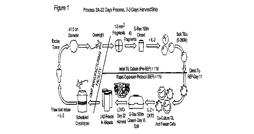
[00190] Figure 1: Shows a diagram of an embodiment of process 2A, a 22-day
process for
TIL manufacturing.
[00191] Figure 2: Shows a comparison between the 1C process and an embodiment
of the
2A process for TIL manufacturing.
23
CA 03098303 2020-10-23
WO 2019/210131 PCT/US2019/029286
[00192] Figure 3: Shows the 1C process timeline.
[00193] Figure 4: Shows the process of an embodiment of TIL therapy using
process 2A for
TIL manufacturing, including administration and co-therapy steps, for higher
cell counts.
[00194] Figure 5: Shows the process of an embodiment of TIL therapy using
process 2A for
TIL manufacturing, including administration and co-therapy steps, for lower
cell counts.
[00195] Figure 6: Shows a detailed schematic for an embodiment of the 2A
process.
[00196] Figures 7a, 7b and 7c: Depict the major steps of an embodiment of
process 2A
including the cryopreservation steps.
[00197] Figure 8: Depicts the clinical trial design including cohorts treated
with process 1C
and an embodiment of process 2A.
[00198] Figure 9: Exemplary Process 2A chart providing an overview of Steps A
through F.
[00199] Figure 10: Process Flow Chart on Process 2A Data Collection Plan
[00200] Figure 11: Scheme of on exemplary embodiment of the Rapid Expansion
Protocol
(REP). Upon arrival the tumor is fragmented, placed into G-Rex flasks with IL-
2 for TIL
expansion (pre-REP expansion), for 11 days. For the triple cocktail studies,
IL-2/1L-15/IL-21
is added at the initiation of the pre-REP. For the Rapid Expansion Protocol
(REP), TIL are
cultured with feeders and OKT3 for REP expansion for an additional 11 days.
[00201] Figure 12: Shows a diagram of an embodiment of process 2A, a 22-day
process for
TIL manufacturing.
[00202] Figure 13: Comparison table of Steps A through F from exemplary
embodiments of
process 1C and process 2A.
[00203] Figure 14: Detailed comparison of an embodiment of process 1C and an
embodiment of process 2A.
[00204] Figure 15: Detailed scheme of an embodiment of a TIL therapy process.
[00205] Figure 16: Depiction of an embodiment of a cryopreserved TIL
manufacturing
process (22 days).
[00206] Figure 17: Table of process improvements from Gen 1 to Gen 2.
[00207] Figure 18: An embodiment of a TIL manufacturing process of the present
invention.
24
CA 03098303 2020-10-23
WO 2019/210131 PCT/US2019/029286
[00208] Figure 19: Process Flow Chart of Process 2A.
[00209] Figure 20: Depiction of an embodiment of a TIL manufacturing process
including
electroporation step for use with gene-editing processes (including TALEN,
zinc finger
nuclease, and CRISPR methods as described herein).
[00210] Figure 21: Depiction of embodiments of TIL manufacturing processes
including
electroporation step for use with gene-editing processes (including TALEN,
zinc finger
nuclease, and CRISPR methods as described herein).
[00211] Figure 22: Depiction of the structures I-A and I-B, the cylinders
refer to individual
polypeptide binding domains. Structures I-A and I-B comprise three linearly-
linked TNFRSF
binding domains derived from e.g., 4-1BBL or an antibody that binds 4-1BB,
which fold to
form a trivalent protein, which is then linked to a second trivalent protein
through IgGl-Fc
(including CH3 and CH2 domains) is then used to link two of the trivalent
proteins together
through disulfide bonds (small elongated ovals), stabilizing the structure and
providing an
agonists capable of bringing together the intracellular signaling domains of
the six receptors
and signaling proteins to form a signaling complex. The TNFRSF binding domains
denoted
as cylinders may be scFv domains comprising, e.g., a VH and a VL chain
connected by a
linker that may comprise hydrophilic residues and Gly and Ser sequences for
flexibility, as
well as Glu and Lys for solubility.
[00212] Figure 23: Depiction of a TALEN construct that targets exon 2 of the
Pdcdl gene.
DETAILED DESCRIPTION OF THE INVENTION
I. Introduction
[00213] Adoptive cell therapy utilizing TILs cultured ex vivo by the Rapid
Expansion
Protocol (REP) has produced successful adoptive cell therapy following host
immunosuppression in patients with melanoma. Current infusion acceptance
parameters rely
on readouts of the composition of TILs (e.g., CD28, CD8, or CD4 positivity)
and on the
numerical folds of expansion and viability of the REP product.
[00214] Current REP protocols give little insight into the health of the TIL
that will be
infused into the patient. T cells undergo a profound metabolic shift during
the course of their
maturation from naive to effector T cells (see Chang, et al., Nat. Immunol.
2016, /7, 364,
hereby expressly incorporated in its entirety, and in particular for the
discussion and markers
of anaerobic and aerobic metabolism). For example, naive T cells rely on
mitochondrial
CA 03098303 2020-10-23
WO 2019/210131 PCT/US2019/029286
respiration to produce ATP, while mature, healthy effector T cells such as TIL
are highly
glycolytic, relying on aerobic glycolysis to provide the bioenergetics
substrates they require
for proliferation, migration, activation, and anti-tumor efficacy.
[00215] Previous papers report that limiting glycolysis and promoting
mitochondrial
metabolism in TILs prior to transfer is desirable as cells that are relying
heavily on glycolysis
will suffer nutrient deprivation upon adoptive transfer which results in a
majority of the
transferred cells dying. Thus, the art teaches that promoting mitochondrial
metabolism might
promote in vivo longevity and in fact suggests using inhibitors of glycolysis
before induction
of the immune response. See Chang, et al., Nat. Immunol. 2016, 17(364).
[00216] The present invention is further directed in some embodiments to
methods for
evaluating and quantifying this increase in metabolic health. Thus, the
present invention
provides methods of assaying the relative health of a TIL population using one
or more
general evaluations of metabolism, including, but not limited to, rates and
amounts of
glycolysis, oxidative phosphorylation, spare respiratory capacity (SRC), and
glycolytic
reserve.
[00217] Furthermore, the present invention is further directed in some
embodiments to
methods for evaluating and quantifying this increase in metabolic health.
Thus, the present
invention provides methods of assaying the relative health of a TIL population
using one or
more general evaluations of metabolism, including, but not limited to, rates
and amounts of
glycolysis, oxidative phosphorylation, spare respiratory capacity (SRC), and
glycolytic
reserve.
[00218] In addition, optional additional evaluations include, but are not
limited to, ATP
production, mitochondrial mass and glucose uptake.
[00219] The present invention is further directed in some embodiments to
enhancing the
therapeutic effect of TILs with the use of gene editing technology. While
adoptive transfer of
tumor infiltrating lymphocytes (TILs) offers a promising and effective
therapy, there is a
strong need for more effective TIL therapies that can increase a patient's
response rate and
response robustness. As described herein, embodiments of the present invention
provide
methods for expanding TILs into a therapeutic population that is gene-edited
to provide an
enhanced therapeutic effect.
Definitions
26
CA 03098303 2020-10-23
WO 2019/210131 PCT/US2019/029286
[00220] Unless defined otherwise, all technical and scientific terms used
herein have the
same meaning as is commonly understood by one of skill in the art to which
this invention
belongs. All patents and publications referred to herein are incorporated by
reference in their
entireties.
[00221] The term "in vivo" refers to an event that takes place in a subject's
body.
[00222] The term "in vitro" refers to an event that takes places outside of a
subject's body.
In vitro assays encompass cell-based assays in which cells alive or dead are
employed and
may also encompass a cell-free assay in which no intact cells are employed.
[00223] The term "ex vivo" refers to an event which involves treating or
performing a
procedure on a cell, tissue and/or organ which has been removed from a
subject's body.
Aptly, the cell, tissue and/or organ may be returned to the subject's body in
a method of
surgery or treatment.
[00224] The term "rapid expansion" means an increase in the number of antigen-
specific
TILs of at least about 3-fold (or 4-, 5-, 6-, 7-, 8-, or 9-fold) over a period
of a week, more
preferably at least about 10-fold (or 20-, 30-, 40-, 50-, 60-, 70-, 80-, or 90-
fold) over a period
of a week, or most preferably at least about 100-fold over a period of a week.
A number of
rapid expansion protocols are outlined below.
[00225] By "tumor infiltrating lymphocytes" or "TILs" herein is meant a
population of cells
originally obtained as white blood cells that have left the bloodstream of a
subject and
migrated into a tumor. TILs include, but are not limited to, CD8+ cytotoxic T
cells
(lymphocytes), Thl and Th17 CD4+ T cells, natural killer cells, dendritic
cells and M1
macrophages. TILs include both primary and secondary TILs. "Primary TILs" are
those that
are obtained from patient tissue samples as outlined herein (sometimes
referred to as "freshly
harvested"), and "secondary TILs" are any TIL cell populations that have been
expanded or
proliferated as discussed herein, including, but not limited to bulk TILs and
expanded TILs
("REP TILs" or "post-REP TILs"). TIL cell populations can include genetically
modified
TILs.
[00226] By "population of cells" (including TILs) herein is meant a number of
cells that
share common traits. In general, populations generally range from 1 X 10⁶ to 1
X 10' in
number, with different TIL populations comprising different numbers. For
example, initial
growth of primary TILs in the presence of IL-2 results in a population of bulk
TILs of
27
CA 03098303 2020-10-23
WO 2019/210131 PCT/US2019/029286
roughly 1 x 10⁸ cells. REP expansion is generally done to provide populations
of 1.5 x 10⁹ to
1.5 x 10¹⁰ cells for infusion.
[00227] By "cryopreserved TILs" herein is meant that TILs, either primary,
bulk, or
expanded (REP TILs), are treated and stored in the range of about -150 C to -
60 C. General
methods for cryopreservation are also described elsewhere herein, including in
the Examples.
For clarity, "cryopreserved TILs" are distinguishable from frozen tissue
samples which may
be used as a source of primary TILs.
[00228] By "thawed cryopreserved TILs" herein is meant a population of TILs
that was
previously cryopreserved and then treated to return to room temperature or
higher, including
but not limited to cell culture temperatures or temperatures wherein TILs may
be
administered to a patient.
[00229] TILs can generally be defined either biochemically, using cell surface
markers, or
functionally, by their ability to infiltrate tumors and effect treatment. TILs
can be generally
categorized by expressing one or more of the following biomarkers: CD4, CD8,
TCR c43,
CD27, CD28, CD56, CCR7, CD45Ra, CD95, PD-1, and CD25. Additionally and
alternatively, TILs can be functionally defined by their ability to infiltrate
solid tumors upon
reintroduction into a patient.
[00230] The term "cryopreservation media" or "cryopreservation medium" refers
to any
medium that can be used for cryopreservation of cells. Such media can include
media
comprising 7% to 10% DMSO. Exemplary media include CryoStor CS10,
Hyperthermasol,
as well as combinations thereof The term "CS10" refers to a cryopreservation
medium which
is obtained from Stemcell Technologies or from Biolife Solutions. The CS10
medium may
be referred to by the trade name "CryoStorg CS10". The CS10 medium is a serum-
free,
animal component-free medium which comprises DMSO.
[00231] The term "central memory T cell" refers to a subset of T cells that in
the human are
CD45R0+ and constitutively express CCR7 (CCR7h1) and CD62L (CD62h1). The
surface
phenotype of central memory T cells also includes TCR, CD3, CD127 (IL-7R), and
M-
ISR. Transcription factors for central memory T cells include BCL-6, BCL-6B,
MBD2, and
BMIl. Central memory T cells primarily secret IL-2 and CD4OL as effector
molecules after
TCR triggering. Central memory T cells are predominant in the CD4 compartment
in blood,
and in the human are proportionally enriched in lymph nodes and tonsils.
28
CA 03098303 2020-10-23
WO 2019/210131 PCT/US2019/029286
[00232] The term "effector memory T cell" refers to a subset of human or
mammalian T
cells that, like central memory T cells, are CD45R0+, but have lost the
constitutive
expression of CCR7 (CCR710) and are heterogeneous or low for CD62L expression
(CD62L10). The surface phenotype of central memory T cells also includes TCR,
CD3,
CD127 (IL-7R), and IL-15R. Transcription factors for central memory T cells
include
BLIMP 1. Effector memory T cells rapidly secret high levels of inflammatory
cytokines
following antigenic stimulation, including interferon-y, IL-4, and IL-5.
Effector memory T
cells are predominant in the CD8 compartment in blood, and in the human are
proportionally
enriched in the lung, liver, and gut. CD8+ effector memory T cells carry large
amounts of
perforin.
[00233] The term "closed system" refers to a system that is closed to the
outside
environment. Any closed system appropriate for cell culture methods can be
employed with
the methods of the present invention. Closed systems include, for example, but
are not
limited to closed G-containers. Once a tumor segment is added to the closed
system, the
system is no opened to the outside environment until the TILs are ready to be
administered to
the patient.
[00234] The terms "fragmenting," "fragment," and "fragmented," as used herein
to describe
processes for disrupting a tumor, includes mechanical fragmentation methods
such as
crushing, slicing, dividing, and morcellating tumor tissue as well as any
other method for
disrupting the physical structure of tumor tissue.
[00235] The terms "peripheral blood mononuclear cells" and "PBMCs" refers to a
peripheral
blood cell having a round nucleus, including lymphocytes (T cells, B cells, NK
cells) and
monocytes. Preferably, the peripheral blood mononuclear cells are irradiated
allogeneic
peripheral blood mononuclear cells. PBMCs are a type of antigen-presenting
cell.
[00236] The term "anti-CD3 antibody" refers to an antibody or variant thereof,
e.g., a
monoclonal antibody and including human, humanized, chimeric or murine
antibodies which
are directed against the CD3 receptor in the T cell antigen receptor of mature
T cells. Anti-
CD3 antibodies include OKT-3, also known as muromonab. Anti-CD3 antibodies
also
include the UHCT1 clone, also known as T3 and CD3E. Other anti-CD3 antibodies
include,
for example, otelixizumab, teplizumab, and visilizumab.
[00237] The term "OKT-3" (also referred to herein as "OKT3") refers to a
monoclonal
antibody or biosimilar or variant thereof, including human, humanized,
chimeric, or murine
29
CA 03098303 2020-10-23
WO 2019/210131 PCT/US2019/029286
antibodies, directed against the CD3 receptor in the T cell antigen receptor
of mature T cells,
and includes commercially-available forms such as OKT-3 (30 ng/mL, MACS GMP
CD3
pure, Miltenyi Biotech, Inc., San Diego, CA, USA) and muromonab or variants,
conservative
amino acid substitutions, glycoforms, or biosimilars thereof The amino acid
sequences of
the heavy and light chains of muromonab are given in Table 1 (SEQ ID NO:1 and
SEQ ID
NO:2). A hybridoma capable of producing OKT-3 is deposited with the American
Type
Culture Collection and assigned the ATCC accession number CRL 8001. A
hybridoma
capable of producing OKT-3 is also deposited with European Collection of
Authenticated Cell
Cultures (ECACC) and assigned Catalogue No. 86022706. Anti-CD3 antibodies also
include
the UHCT1 clone (commercially available from BioLegend, San Diego, CA, USA),
also
known as T3 and CD3c.
TABLE 1. Amino acid sequences of muromonab.
Identifier Sequence (One-Letter Amino Acid Symbols)
SEQ ID NO:1 QVQLQQSGAE LARPGASVKM SCKASGYTFT RYTMHWVKQR PGQGLEWIGY
INPSRGYTNY 60
Muromonab heavy NQKFKDKATL TTDKSSSTAY MQLSSLTSED SAVYYCARYY DDHYCLDYWG
QGTTLTVSSA 120
chain KTTAPSVYPL APVCGGTTGS SVTLGCLVKG YEPEPVTLTW NSGSLSSGVH
TFPAVLQSDL 180
YTLSSSVTVT SSTWPSQSIT CNVAHPASST KVDKKIEPRP KSCDKTHTCP PCPAPELLGG
240
PSVFLFPPKP KDTLMISRTP EVTCVVVDVS HEDPEVKFNW YVDGVEVHNA KTKPREEQYN
300
STYRVVSVLT VLHQDWLNGK EYKCKVSNKA LPAPIEKTIS KAKGQPREPQ VYTLPPSRDE
360
LTKNQVSLTC LVKGFYPSDI AVEWESNGQP ENNYKTTPPV LDSDGSFFLY SKLTVDKSRW
420
QQGNVFSCSV MHEALHNHYT QKSLSLSPGK
450
SEQ ID NO:2 QIVLTQSPAI MSASPGEKVT MTCSASSSVS YMNWYQQKSG TSPKRWIYDT
SKLASGVPAH 60
Muromonab light FRGSGSGTSY SLTISGMEAE DAATYYCQQW SSNPFTFGSG TKLEINRADT
APTVSIFPPS 120
chain SEQLTSGGAS VVCFLNNFYP KDINVYWKID GSERQNGVLN SWTDQDSKDS
TYSMSSTLTL 180
TKDEYERHNS YTCEATHKTS TSPIVKSFNR NEC
213
[00238] The term "IL-2" (also referred to herein as "IL2") refers to the T
cell growth factor
known as interleukin-2, and includes all forms of IL-2 including human and
mammalian
forms, conservative amino acid substitutions, glycoforms, biosimilars, and
variants thereof
IL-2 is described, e.g., in Nelson, I Immunol. 2004, 172, 3983-88 and Malek,
Annu. Rev.
Immunol. 2008, 26, 453-79, the disclosures of which are incorporated by
reference herein.
The amino acid sequence of recombinant human IL-2 suitable for use in the
invention is
given in Table 2 (SEQ ID NO:3). For example, the term IL-2 encompasses human,
recombinant forms of IL-2 such as aldesleukin (PROLEUKIN, available
commercially from
multiple suppliers in 22 million IU per single use vials), as well as the form
of recombinant
IL-2 commercially supplied by CellGenix, Inc., Portsmouth, NH, USA (CELLGRO
GMP) or
ProSpec-Tany TechnoGene Ltd., East Brunswick, NJ, USA (Cat. No. CYT-209-b) and
other
commercial equivalents from other vendors. Aldesleukin (des-alanyl-1, serine-
125 human
IL-2) is a nonglycosylated human recombinant form of IL-2 with a molecular
weight of
approximately 15 kDa. The amino acid sequence of aldesleukin suitable for use
in the
CA 03098303 2020-10-23
WO 2019/210131 PCT/US2019/029286
invention is given in Table 2 (SEQ ID NO:4). The term IL-2 also encompasses
pegylated
forms of IL-2, as described herein, including the pegylated IL2 prodrug NKTR-
214, available
from Nektar Therapeutics, South San Francisco, CA, USA. NKTR-214 and pegylated
IL-2
suitable for use in the invention is described in U.S. Patent Application
Publication No. US
2014/0328791 Al and International Patent Application Publication No. WO
2012/065086 Al,
the disclosures of which are incorporated by reference herein. Alternative
forms of
conjugated IL-2 suitable for use in the invention are described in U.S. Patent
Nos. 4,766,106,
5,206,344, 5,089,261 and 4902,502, the disclosures of which are incorporated
by reference
herein. Formulations of IL-2 suitable for use in the invention are described
in U.S. Patent
No. 6,706,289, the disclosure of which is incorporated by reference herein.
TABLE 2. Amino acid sequences of interleukins.
Identifier Sequence (One-Letter Amino Acid Symbols)
SEQ ID NO:3 MAPTSSSTKK TQLQLEHLLL DLQMILNGIN NYKNPELTRM LTFKFYMPKK
ATELKHLQCL 60
recombinant EEELKPLEEV LNLAQSKNFH LRPRDLISNI NVIVLELKGS ETTFMCEYAD
ETATIVEFLN 120
human IL-2 RWITFCQSII STLT
134
(rhIL-2)
SEQ ID NO:4 PTSSSTKKTQ LQLEHLLLDL QMILNGINNY KNPELTRMLT FKFYMPKKAT
ELKHLQCLEE 60
Aldesleukin ELKPLEEVLN LAQSKNFHLR PRDLISNINV IVLELKGSET TFMCEYADET
ATIVEFLNRW 120
ITFSQSIIST LT
132
SEQ ID NO:5 MHKCDITLQE IIKTLNSLTE QKTLCTELTV TDIFAASKNT TEKETFCRAA
TVLRQFYSHH 60
recombinant EKDTRCLGAT AQQFHRHKQL IRFLKRLDRN LWGLAGLNSC PVKEANQSTL
ENFLERLKTI 120
human IL-4 MREKYSKCSS
130
(rhIL-4)
SEQ ID NO:6 MDCDIEGKDG KQYESVLMVS IDQLLDSMKE IGSNCLNNEF NFFKRHICDA
NKEGMFLFRA 60
recombinant ARKLRQFLKM NSTGDFDLHL LKVSEGTTIL LNCTGQVKGR KPAALGEAQP
TKSLEENKSL 120
human IL-7 KEQKKLNDLC FLKRLLQEIK TCWNKILMGT KEH
153
(rhIL-7)
SEQ ID NO:7 MNWVNVISDL KKIEDLIQSM HIDATLYTES DVHPSCKVTA MKCFLLELQV
ISLESGDASI 60
recombinant HDTVENLIIL ANNSLSSNGN VTESGCKECE ELEEKNIKEF LQSFVHIVQM FINTS
115
human IL-15
(rhIL-15)
SEQ ID NO:8 MQDRHMIRMR QLIDIVDQLK NYVNDLVPEF LPAPEDVETN CEWSAFSCFQ
KAQLKSANTG 60
recombinant NNERIINVSI KKLEREPPST NAGRRQKHRL TCPSCDSYEK KPPKEFLERF
KSLLQKMIHQ 120
human IL-21 HLSSRTHGSE DS
132
(rhIL-21)
[00239] The term "IL-4" (also referred to herein as "IL4") refers to the
cytokine known as
interleukin 4, which is produced by Th2 T cells and by eosinophils, basophils,
and mast cells.
IL-4 regulates the differentiation of naive helper T cells (Th0 cells) to Th2
T cells. Steinke
and Borish, Respir. Res. 2001, 2, 66-70. Upon activation by IL-4, Th2 T cells
subsequently
produce additional IL-4 in a positive feedback loop. IL-4 also stimulates B
cell proliferation
and class II MHC expression, and induces class switching to IgE and IgGi
expression from B
cells. Recombinant human IL-4 suitable for use in the invention is
commercially available
from multiple suppliers, including ProSpec-Tany TechnoGene Ltd., East
Brunswick, NJ,
USA (Cat. No. CYT-211) and ThermoFisher Scientific, Inc., Waltham, MA, USA
(human
31
CA 03098303 2020-10-23
WO 2019/210131 PCT/US2019/029286
IL-15 recombinant protein, Cat. No. Gibco CTP0043). The amino acid sequence of
recombinant human IL-4 suitable for use in the invention is given in Table 2
(SEQ ID NO:5).
[00240] The term "IL-7" (also referred to herein as "IL7") refers to a
glycosylated tissue-
derived cytokine known as interleukin 7, which may be obtained from stromal
and epithelial
cells, as well as from dendritic cells. Fry and Mackall, Blood 2002, 99, 3892-
904. IL-7 can
stimulate the development of T cells. IL-7 binds to the IL-7 receptor, a
heterodimer
consisting of IL-7 receptor alpha and common gamma chain receptor, which in a
series of
signals important for T cell development within the thymus and survival within
the periphery.
Recombinant human IL-7 suitable for use in the invention is commercially
available from
multiple suppliers, including ProSpec-Tany TechnoGene Ltd., East Brunswick,
NJ, USA
(Cat. No. CYT-254) and ThermoFisher Scientific, Inc., Waltham, MA, USA (human
IL-15
recombinant protein, Cat. No. Gibco PHC0071). The amino acid sequence of
recombinant
human IL-7 suitable for use in the invention is given in Table 2 (SEQ ID
NO:6).
[00241] The term "IL-15" (also referred to herein as "IL15") refers to the T
cell growth
factor known as interleukin-15, and includes all forms of IL-2 including human
and
mammalian forms, conservative amino acid substitutions, glycoforms,
biosimilars, and
variants thereof IL-15 is described, e.g., in Fehniger and Caligiuri, Blood
2001, 97, 14-32,
the disclosure of which is incorporated by reference herein. IL-15 shares 0
and y signaling
receptor subunits with IL-2. Recombinant human IL-15 is a single, non-
glycosylated
polypeptide chain containing 114 amino acids (and an N-terminal methionine)
with a
molecular mass of 12.8 kDa. Recombinant human IL-15 is commercially available
from
multiple suppliers, including ProSpec-Tany TechnoGene Ltd., East Brunswick,
NJ, USA
(Cat. No. CYT-230-b) and ThermoFisher Scientific, Inc., Waltham, MA, USA
(human IL-15
recombinant protein, Cat. No. 34-8159-82). The amino acid sequence of
recombinant human
IL-15 suitable for use in the invention is given in Table 2 (SEQ ID NO:7).
[00242] The term "IL-21" (also referred to herein as "IL21") refers to the
pleiotropic
cytokine protein known as interleukin-21, and includes all forms of IL-21
including human
and mammalian forms, conservative amino acid substitutions, glycoforms,
biosimilars, and
variants thereof IL-21 is described, e.g., in Spolski and Leonard, Nat. Rev.
Drug. Disc.
2014, /3, 379-95, the disclosure of which is incorporated by reference herein.
IL-21 is
primarily produced by natural killer T cells and activated human CD4+ T cells.
Recombinant
human IL-21 is a single, non-glycosylated polypeptide chain containing 132
amino acids with
a molecular mass of 15.4 kDa. Recombinant human IL-21 is commercially
available from
32
CA 03098303 2020-10-23
WO 2019/210131
PCT/US2019/029286
multiple suppliers, including ProSpec-Tany TechnoGene Ltd., East Brunswick,
NJ, USA
(Cat. No. CYT-408-b) and ThermoFisher Scientific, Inc., Waltham, MA, USA
(human IL-21
recombinant protein, Cat. No. 14-8219-80). The amino acid sequence of
recombinant human
IL-21 suitable for use in the invention is given in Table 2 (SEQ ID NO:8).
[00243] When "an anti-tumor effective amount", "an tumor-inhibiting effective
amount", or
"therapeutic amount" is indicated, the precise amount of the compositions of
the present
invention to be administered can be determined by a physician with
consideration of
individual differences in age, weight, tumor size, extent of infection or
metastasis, and
condition of the patient (subject). It can generally be stated that a
pharmaceutical composition
comprising the tumor infiltrating lymphocytes (e.g. secondary TILs or
genetically modified
cytotoxic lymphocytes) described herein may be administered at a dosage of 104
to 1011
cells/kg body weight (e.g., 105 to 106, io5 to 1-1o,
u o5 to 1011, 106 to 1-1 , u 106
to 1011,107 to
ton, o7 to 1-1 , u 108 to 1011, 108 to 1-1 , u 109 to 1011,
or 109 to 1010 cells/kg body weight),
including all integer values within those ranges. Tumor infiltrating
lymphocytes (including in
some cases, genetically modified cytotoxic lymphocytes) compositions may also
be
administered multiple times at these dosages. The tumor infiltrating
lymphocytes (including
in some cases, genetically) can be administered by using infusion techniques
that are
commonly known in immunotherapy (see, e.g., Rosenberg et al., New Eng. I of
Med. 319:
1676, 1988). The optimal dosage and treatment regime for a particular patient
can readily be
determined by one skilled in the art of medicine by monitoring the patient for
signs of disease
and adjusting the treatment accordingly.
[00244] The term "hematological malignancy" refers to mammalian cancers and
tumors of
the hematopoietic and lymphoid tissues, including but not limited to tissues
of the blood,
bone marrow, lymph nodes, and lymphatic system. Hematological malignancies are
also
referred to as "liquid tumors." Hematological malignancies include, but are
not limited to,
acute lymphoblastic leukemia (ALL), chronic lymphocytic lymphoma (CLL), small
lymphocytic lymphoma (SLL), acute myelogenous leukemia (AML), chronic
myelogenous
leukemia (CML), acute monocytic leukemia (AMoL), Hodgkin's lymphoma, and non-
Hodgkin's lymphomas. The term "B cell hematological malignancy" refers to
hematological
malignancies that affect B cells.
[00245] The term "solid tumor" refers to an abnormal mass of tissue that
usually does not
contain cysts or liquid areas. Solid tumors may be benign or malignant. The
term "solid
tumor cancer refers to malignant, neoplastic, or cancerous solid tumors. Solid
tumor cancers
33
CA 03098303 2020-10-23
WO 2019/210131 PCT/US2019/029286
include, but are not limited to, sarcomas, carcinomas, and lymphomas, such as
cancers of the
lung, breast, prostate, colon, rectum, and bladder. The tissue structure of
solid tumors
includes interdependent tissue compartments including the parenchyma (cancer
cells) and the
supporting stromal cells in which the cancer cells are dispersed and which may
provide a
supporting microenvironment.
[00246] The term "liquid tumor" refers to an abnormal mass of cells that is
fluid in nature.
Liquid tumor cancers include, but are not limited to, leukemias, myelomas, and
lymphomas,
as well as other hematological malignancies. TILs obtained from liquid tumors
may also be
referred to herein as marrow infiltrating lymphocytes (MILs).
[00247] The term "microenvironment," as used herein, may refer to the solid or
hematological tumor microenvironment as a whole or to an individual subset of
cells within
the microenvironment. The tumor microenvironment, as used herein, refers to a
complex
mixture of "cells, soluble factors, signaling molecules, extracellular
matrices, and mechanical
cues that promote neoplastic transformation, support tumor growth and
invasion, protect the
tumor from host immunity, foster therapeutic resistance, and provide niches
for dominant
metastases to thrive," as described in Swartz, et al., Cancer Res., 2012, 72,
2473. Although
tumors express antigens that should be recognized by T cells, tumor clearance
by the immune
system is rare because of immune suppression by the microenvironment.
[00248] In an embodiment, the invention includes a method of treating a cancer
with a
population of TILs, wherein a patient is pre-treated with non-myeloablative
chemotherapy
prior to an infusion of TILs according to the invention. In some embodiments,
the population
of TILs may be provided wherein a patient is pre-treated with nonmyeloablative
chemotherapy prior to an infusion of TILs according to the present invention.
In an
embodiment, the non-myeloablative chemotherapy is cyclophosphamide 60 mg/kg/d
for 2
days (days 27 and 26 prior to TIL infusion) and fludarabine 25 mg/m2/d for 5
days (days 27
to 23 prior to TIL infusion). In an embodiment, after non-myeloablative
chemotherapy and
TIL infusion (at day 0) according to the invention, the patient receives an
intravenous
infusion of IL-2 intravenously at 720,000 IU/kg every 8 hours to physiologic
tolerance.
[00249] Experimental findings indicate that lymphodepletion prior to adoptive
transfer of
tumor-specific T lymphocytes plays a key role in enhancing treatment efficacy
by eliminating
regulatory T cells and competing elements of the immune system ("cytokine
sinks").
Accordingly, some embodiments of the invention utilize a lymphodepletion step
(sometimes
34
CA 03098303 2020-10-23
WO 2019/210131 PCT/US2019/029286
also referred to as "immunosuppressive conditioning") on the patient prior to
the introduction
of the rTILs of the invention.
[00250] The terms "co-administration," "co-administering," "administered in
combination
with," "administering in combination with," "simultaneous," and "concurrent,"
as used
herein, encompass administration of two or more active pharmaceutical
ingredients (in a
preferred embodiment of the present invention, for example, at least one
potassium channel
agonist in combination with a plurality of TILs) to a subject so that both
active
pharmaceutical ingredients and/or their metabolites are present in the subject
at the same
time. Co-administration includes simultaneous administration in separate
compositions,
administration at different times in separate compositions, or administration
in a composition
in which two or more active pharmaceutical ingredients are present.
Simultaneous
administration in separate compositions and administration in a composition in
which both
agents are present are preferred.
[00251] The term "effective amount" or "therapeutically effective amount"
refers to that
amount of a compound or combination of compounds as described herein that is
sufficient to
effect the intended application including, but not limited to, disease
treatment. A
therapeutically effective amount may vary depending upon the intended
application (in vitro
or in vivo), or the subject and disease condition being treated (e.g., the
weight, age and
gender of the subject), the severity of the disease condition, or the manner
of administration.
The term also applies to a dose that will induce a particular response in
target cells (e.g., the
reduction of platelet adhesion and/or cell migration). The specific dose will
vary depending
on the particular compounds chosen, the dosing regimen to be followed, whether
the
compound is administered in combination with other compounds, timing of
administration,
the tissue to which it is administered, and the physical delivery system in
which the
compound is carried.
[00252] The terms "treatment", "treating", "treat", and the like, refer to
obtaining a desired
pharmacologic and/or physiologic effect. The effect may be prophylactic in
terms of
completely or partially preventing a disease or symptom thereof and/or may be
therapeutic in
terms of a partial or complete cure for a disease and/or adverse effect
attributable to the
disease. "Treatment", as used herein, covers any treatment of a disease in a
mammal,
particularly in a human, and includes: (a) preventing the disease from
occurring in a subject
which may be predisposed to the disease but has not yet been diagnosed as
having it;
(b) inhibiting the disease, i.e., arresting its development or progression;
and (c) relieving the
CA 03098303 2020-10-23
WO 2019/210131 PCT/US2019/029286
disease, i.e., causing regression of the disease and/or relieving one or more
disease
symptoms. "Treatment" is also meant to encompass delivery of an agent in order
to provide
for a pharmacologic effect, even in the absence of a disease or condition. For
example,
"treatment" encompasses delivery of a composition that can elicit an immune
response or
confer immunity in the absence of a disease condition, e.g., in the case of a
vaccine.
[00253] The term "heterologous" when used with reference to portions of a
nucleic acid or
protein indicates that the nucleic acid or protein comprises two or more
subsequences that are
not found in the same relationship to each other in nature. For instance, the
nucleic acid is
typically recombinantly produced, having two or more sequences from unrelated
genes
arranged to make a new functional nucleic acid, e.g., a promoter from one
source and a
coding region from another source, or coding regions from different sources.
Similarly, a
heterologous protein indicates that the protein comprises two or more
subsequences that are
not found in the same relationship to each other in nature (e.g., a fusion
protein).
[00254] The terms "sequence identity," "percent identity," and "sequence
percent identity"
(or synonyms thereof, e.g., "99% identical") in the context of two or more
nucleic acids or
polypeptides, refer to two or more sequences or subsequences that are the same
or have a
specified percentage of nucleotides or amino acid residues that are the same,
when compared
and aligned (introducing gaps, if necessary) for maximum correspondence, not
considering
any conservative amino acid substitutions as part of the sequence identity.
The percent
identity can be measured using sequence comparison software or algorithms or
by visual
inspection. Various algorithms and software are known in the art that can be
used to obtain
alignments of amino acid or nucleotide sequences. Suitable programs to
determine percent
sequence identity include for example the BLAST suite of programs available
from the U.S.
Government's National Center for Biotechnology Information BLAST web site.
Comparisons between two sequences can be carried using either the BLASTN or
BLASTP
algorithm. BLASTN is used to compare nucleic acid sequences, while BLASTP is
used to
compare amino acid sequences. ALIGN, ALIGN-2 (Genentech, South San Francisco,
California) or MegAlign, available from DNASTAR, are additional publicly
available
software programs that can be used to align sequences. One skilled in the art
can determine
appropriate parameters for maximal alignment by particular alignment software.
In certain
embodiments, the default parameters of the alignment software are used.
[00255] As used herein, the term "variant" encompasses but is not limited to
antibodies or
fusion proteins which comprise an amino acid sequence which differs from the
amino acid
36
CA 03098303 2020-10-23
WO 2019/210131 PCT/US2019/029286
sequence of a reference antibody by way of one or more substitutions,
deletions and/or
additions at certain positions within or adjacent to the amino acid sequence
of the reference
antibody. The variant may comprise one or more conservative substitutions in
its amino acid
sequence as compared to the amino acid sequence of a reference antibody.
Conservative
substitutions may involve, e.g., the substitution of similarly charged or
uncharged amino
acids. The variant retains the ability to specifically bind to the antigen of
the reference
antibody. The term variant also includes pegylated antibodies or proteins.
[00256] By "tumor infiltrating lymphocytes" or "TILs" herein is meant a
population of cells
originally obtained as white blood cells that have left the bloodstream of a
subject and
migrated into a tumor. TILs include, but are not limited to, CD8+ cytotoxic T
cells
(lymphocytes), Thl and Th17 CD4+ T cells, natural killer cells, dendritic
cells and M1
macrophages. TILs include both primary and secondary TILs. "Primary TILs" are
those that
are obtained from patient tissue samples as outlined herein (sometimes
referred to as "freshly
harvested"), and "secondary TILs" are any TIL cell populations that have been
expanded or
proliferated as discussed herein, including, but not limited to bulk TILs,
expanded TILs
("REP TILs") as well as "reREP TILs" as discussed herein. reREP TILs can
include for
example second expansion TILs or second additional expansion TILs (such as,
for example,
those described in Step D of Figure 9, including TILs referred to as reREP
TILs).
[00257] TILs can generally be defined either biochemically, using cell surface
markers, or
functionally, by their ability to infiltrate tumors and effect treatment. TILs
can be generally
categorized by expressing one or more of the following biomarkers: CD4, CD8,
TCR c43,
CD27, CD28, CD56, CCR7, CD45Ra, CD95, PD-1, and CD25. Additionally, and
alternatively, TILs can be functionally defined by their ability to infiltrate
solid tumors upon
reintroduction into a patient. TILS may further be characterized by potency ¨
for example,
TILS may be considered potent if, for example, interferon (IFN) release is
greater than about
50 pg/mL, greater than about 100 pg/mL, greater than about 150 pg/mL, or
greater than about
200 pg/mL.
[00258] The terms "pharmaceutically acceptable carrier" or "pharmaceutically
acceptable
excipient" are intended to include any and all solvents, dispersion media,
coatings,
antibacterial and antifungal agents, isotonic and absorption delaying agents,
and inert
ingredients. The use of such pharmaceutically acceptable carriers or
pharmaceutically
acceptable excipients for active pharmaceutical ingredients is well known in
the art. Except
insofar as any conventional pharmaceutically acceptable carrier or
pharmaceutically
37
CA 03098303 2020-10-23
WO 2019/210131 PCT/US2019/029286
acceptable excipient is incompatible with the active pharmaceutical
ingredient, its use in the
therapeutic compositions of the invention is contemplated. Additional active
pharmaceutical
ingredients, such as other drugs, can also be incorporated into the described
compositions and
methods.
[00259] The terms "about" and "approximately" mean within a statistically
meaningful
range of a value. Such a range can be within an order of magnitude, preferably
within 50%,
more preferably within 20%, more preferably still within 10%, and even more
preferably
within 5% of a given value or range. The allowable variation encompassed by
the terms
"about" or "approximately" depends on the particular system under study, and
can be readily
appreciated by one of ordinary skill in the art. Moreover, as used herein, the
terms "about"
and "approximately" mean that dimensions, sizes, formulations, parameters,
shapes and other
quantities and characteristics are not and need not be exact, but may be
approximate and/or
larger or smaller, as desired, reflecting tolerances, conversion factors,
rounding off,
measurement error and the like, and other factors known to those of skill in
the art. In
general, a dimension, size, formulation, parameter, shape or other quantity or
characteristic is
"about" or "approximate" whether or not expressly stated to be such. It is
noted that
embodiments of very different sizes, shapes and dimensions may employ the
described
arrangements.
[00260] The transitional terms "comprising," "consisting essentially of," and
"consisting
of," when used in the appended claims, in original and amended form, define
the claim scope
with respect to what unrecited additional claim elements or steps, if any, are
excluded from
the scope of the claim(s). The term "comprising" is intended to be inclusive
or open-ended
and does not exclude any additional, unrecited element, method, step or
material. The term
"consisting of' excludes any element, step or material other than those
specified in the claim
and, in the latter instance, impurities ordinary associated with the specified
material(s). The
term "consisting essentially of' limits the scope of a claim to the specified
elements, steps or
material(s) and those that do not materially affect the basic and novel
characteristic(s) of the
claimed invention. All compositions, methods, and kits described herein that
embody the
present invention can, in alternate embodiments, be more specifically defined
by any of the
transitional terms "comprising," "consisting essentially of," and "consisting
of."
III. Gene-Editing Processes
A. Overview: TIL Expansion + Gene-Editing
38
CA 03098303 2020-10-23
WO 2019/210131 PCT/US2019/029286
[00261] Embodiments of the present invention are directed to methods for
expanding TIL
populations, the methods comprising one or more steps of gene-editing at least
a portion of
the TILs in order to enhance their therapeutic effect. As used herein, "gene-
editing," "gene
editing," and "genome editing" refer to a type of genetic modification in
which DNA is
permanently modified in the genome of a cell, e.g., DNA is inserted, deleted,
modified or
replaced within the cell's genome. In some embodiments, gene-editing causes
the expression
of a DNA sequence to be silenced (sometimes referred to as a gene knockout) or
inhibited/reduced (sometimes referred to as a gene knockdown). In other
embodiments,
gene-editing causes the expression of a DNA sequence to be enhanced (e.g., by
causing over-
expression). In accordance with embodiments of the present invention, gene-
editing
technology is used to enhance the effectiveness of a therapeutic population of
TILs.
[00262] A method for expanding tumor infiltrating lymphocytes (TILs) into a
therapeutic
population of TILs may be carried out in accordance with any embodiment of the
methods
described herein (e.g., an exemplary TIL expansion method known as process 2A
is
described below), wherein the method further comprises gene-editing at least a
portion of the
TILs. According to additional embodiments, a method for expanding TILs into a
therapeutic
population of TILs is carried out in accordance with any embodiment of the
methods
described in PCT/US2017/058610, PCT/US2018/012605, or PCT/US2018/012633, which
are
incorporated by reference herein in their entireties, wherein the method
further comprises
gene-editing at least a portion of the TILs. Thus, an embodiment of the
present invention
provides a therapeutic population of TILs that has been expanded in accordance
with any
embodiment described herein, wherein at least a portion of the therapeutic
population has
been gene-edited, e.g., at least a portion of the therapeutic population of
TILs that is
transferred to the infusion bag is permanently gene-edited.
B. Timing of Gene-Editing During TIL Expansion
[00263] According to one embodiment, a method for expanding tumor infiltrating
lymphocytes (TILs) into a therapeutic population of TILs comprises:
(a) obtaining a first population of TILs from a tumor resected from a patient
by
processing a tumor sample obtained from the patient into multiple tumor
fragments;
(b) adding the tumor fragments into a closed system;
(c) performing a first expansion by culturing the first population of TILs in
a cell
culture medium comprising IL-2, and optionally OKT-3 (e.g., OKT-3 may be
present in the
39
CA 03098303 2020-10-23
WO 2019/210131 PCT/US2019/029286
culture medium beginning on the start date of the expansion process), to
produce a second
population of TILs, wherein the first expansion is performed in a closed
container providing a
first gas-permeable surface area, wherein the first expansion is performed for
about 3-14 days
to obtain the second population of TILs, wherein the second population of TILs
is at least 50-
fold greater in number than the first population of TILs, and wherein the
transition from step
(b) to step (c) occurs without opening the system;
(d) performing a second expansion by supplementing the cell culture medium of
the
second population of TILs with additional IL-2, optionally OKT-3, and antigen
presenting
cells (APCs), to produce a third population of TILs, wherein the second
expansion is
performed for about 7-14 days to obtain the third population of TILs, wherein
the third
population of TILs is a therapeutic population of TILs, wherein the second
expansion is
performed in a closed container providing a second gas-permeable surface area,
and wherein
the transition from step (c) to step (d) occurs without opening the system;
(e) harvesting the therapeutic population of TILs obtained from step (d),
wherein the
transition from step (d) to step (e) occurs without opening the system;
(f) transferring the harvested TIL population from step (e) to an infusion
bag, wherein
the transfer from step (e) to (f) occurs without opening the system; and
(g) at any time during the method, gene-editing at least a portion of the
TILs.
[00264] As stated in step (g) of the embodiment described above, the gene-
editing process
may be carried out at any time during the TIL expansion method, which means
that the gene
editing may be carried out on TILs before, during, or after any of the steps
in the expansion
method; for example, during any of steps (a)-(f) outlined in the method above,
or before or
after any of steps (a)-(f) outlined in the method above. According to certain
embodiments,
TILs are collected during the expansion method (e.g., the expansion method is
"paused" for
at least a portion of the TILs), and the collected TILs are subjected to a
gene-editing process,
and, in some cases, subsequently reintroduced back into the expansion method
(e.g., back
into the culture medium) to continue the expansion process, so that at least a
portion of the
therapeutic population of TILs that are eventually transferred to the infusion
bag are
permanently gene-edited. In an embodiment, the gene-editing process may be
carried out
before expansion by activating TILs, performing a gene-editing step on the
activated TILs,
and expanding the gene-edited TILs according to the processes described
herein.
CA 03098303 2020-10-23
WO 2019/210131 PCT/US2019/029286
[00265] It should be noted that alternative embodiments of the expansion
process may differ
from the method shown above; e.g., alternative embodiments may not have the
same steps
(a)-(g), or may have a different number of steps. Regardless of the specific
embodiment, the
gene-editing process may be carried out at any time during the TIL expansion
method. For
example, alternative embodiments may include more than two expansions, and it
is possible
that gene-editing may be conducted on the TILs during a third or fourth
expansion, etc.
[00266] According to one embodiment, the gene-editing process is carried out
on TILs from
one or more of the first population, the second population, and the third
population. For
example, gene-editing may be carried out on the first population of TILs, or
on a portion of
TILs collected from the first population, and following the gene-editing
process those TILs
may subsequently be placed back into the expansion process (e.g., back into
the culture
medium). Alternatively, gene-editing may be carried out on TILs from the
second or third
population, or on a portion of TILs collected from the second or third
population,
respectively, and following the gene-editing process those TILs may
subsequently be placed
back into the expansion process (e.g., back into the culture medium).
According to another
embodiment, gene-editing is performed while the TILs are still in the culture
medium and
while the expansion is being carried out, i.e., they are not necessarily
"removed" from the
expansion in order to conduct gene-editing.
[00267] According to another embodiment, the gene-editing process is carried
out on TILs
from the first expansion, or TILs from the second expansion, or both. For
example, during
the first expansion or second expansion, gene-editing may be carried out on
TILs that are
collected from the culture medium, and following the gene-editing process
those TILs may
subsequently be placed back into the expansion method, e.g., by reintroducing
them back into
the culture medium.
[00268] According to another embodiment, the gene-editing process is carried
out on at least
a portion of the TILs after the first expansion and before the second
expansion. For example,
after the first expansion, gene-editing may be carried out on TILs that are
collected from the
culture medium, and following the gene-editing process those TILs may
subsequently be
placed back into the expansion method, e.g., by reintroducing them back into
the culture
medium for the second expansion.
[00269] According to alternative embodiments, the gene-editing process is
carried out before
step (c) (e.g., before, during, or after any of steps (a)-(b)), before step
(d) (e.g., before, during,
41
CA 03098303 2020-10-23
WO 2019/210131 PCT/US2019/029286
or after any of steps (a)-(c)), before step (e) (e.g., before, during, or
after any of steps (a)-(d)),
or before step (f) (e.g., before, during, or after any of steps (a)-(e)).
[00270] It should be noted with regard to OKT-3, according to certain
embodiments, that the
cell culture medium may comprise OKT-3 beginning on the start day (Day 0), or
on Day 1 of
the first expansion, such that the gene-editing is carried out on TILs after
they have been
exposed to OKT-3 in the cell culture medium on Day 0 and/or Day 1. According
to another
embodiment, the cell culture medium comprises OKT-3 during the first expansion
and/or
during the second expansion, and the gene-editing is carried out before the
OKT-3 is
introduced into the cell culture medium. Alternatively, the cell culture
medium may
comprise OKT-3 during the first expansion and/or during the second expansion,
and the
gene-editing is carried out after the OKT-3 is introduced into the cell
culture medium.
[00271] It should also be noted with regard to a 4-1BB agonist, according to
certain
embodiments, that the cell culture medium may comprise a 4-1BB agonist
beginning on the
start day (Day 0), or on Day 1 of the first expansion, such that the gene-
editing is carried out
on TILs after they have been exposed to a 4-1BB agonist in the cell culture
medium on Day 0
and/or Day 1. According to another embodiment, the cell culture medium
comprises a 4-1BB
agonist during the first expansion and/or during the second expansion, and the
gene-editing is
carried out before the 4-1BB agonist is introduced into the cell culture
medium.
Alternatively, the cell culture medium may comprise a 4-1BB agonist during the
first
expansion and/or during the second expansion, and the gene-editing is carried
out after the 4-
1BB agonist is introduced into the cell culture medium.
[00272] It should also be noted with regard to IL-2, according to certain
embodiments, that
the cell culture medium may comprise IL-2 beginning on the start day (Day 0),
or on Day 1
of the first expansion, such that the gene-editing is carried out on TILs
after they have been
exposed to IL-2 in the cell culture medium on Day 0 and/or Day 1. According to
another
embodiment, the cell culture medium comprises IL-2 during the first expansion
and/or during
the second expansion, and the gene-editing is carried out before the IL-2 is
introduced into
the cell culture medium. Alternatively, the cell culture medium may comprise
IL-2 during
the first expansion and/or during the second expansion, and the gene-editing
is carried out
after the IL-2 is introduced into the cell culture medium.
[00273] As discussed above, one or more of OKT-3, 4-1BB agonist and IL-2 may
be
included in the cell culture medium beginning on Day 0 or Day 1 of the first
expansion.
42
CA 03098303 2020-10-23
WO 2019/210131 PCT/US2019/029286
According to one embodiment, OKT-3 is included in the cell culture medium
beginning on
Day 0 or Day 1 of the first expansion, and/or a 4-1BB agonist is included in
the cell culture
medium beginning on Day 0 or Day 1 of the first expansion, and/or IL-2 is
included in the
cell culture medium beginning on Day 0 or Day 1 of the first expansion.
According to an
example, the cell culture medium comprises OKT-3 and a 4-1BB agonist beginning
on Day 0
or Day 1 of the first expansion. According to another example, the cell
culture medium
comprises OKT-3, a 4-1BB agonist and IL-2 beginning on Day 0 or Day 1 of the
first
expansion. Of course, one or more of OKT-3, 4-1BB agonist and IL-2 may be
added to the
cell culture medium at one or more additional time points during the expansion
process, as set
forth in various embodiments described herein.
[00274] According to one embodiment, a method for expanding tumor infiltrating
lymphocytes (TILs) into a therapeutic population of TILs comprises:
(a) obtaining a first population of TILs from a tumor resected from a patient
by
processing a tumor sample obtained from the patient into multiple tumor
fragments;
(b) adding the tumor fragments into a closed system;
(c) performing a first expansion by culturing the first population of TILs in
a cell
culture medium comprising IL-2 and optionally comprising a 4-1BB agonist
antibody for
about 2 to 5 days;
(d) adding OKT-3, to produce a second population of TILs, wherein the first
expansion is performed in a closed container providing a first gas-permeable
surface area,
wherein the first expansion is performed for about 1 to 3 days to obtain the
second population
of TILs, wherein the second population of TILs is at least 50-fold greater in
number than the
first population of TILs, and wherein the transition from step (c) to step (d)
occurs without
opening the system;
(e) performing a sterile electroporation step on the second population of
TILs,
wherein the sterile electroporation step mediates the transfer of at least one
gene editor;
(f) resting the second population of TILs for about 1 day;
(g) performing a second expansion by supplementing the cell culture medium of
the
second population of TILs with additional IL-2, optionally OKT-3 antibody,
optionally an
0X40 antibody, and antigen presenting cells (APCs), to produce a third
population of TILs,
wherein the second expansion is performed for about 7 to 11 days to obtain the
third
43
CA 03098303 2020-10-23
WO 2019/210131 PCT/US2019/029286
population of TILs, wherein the second expansion is performed in a closed
container
providing a second gas-permeable surface area, and wherein the transition from
step (f) to
step (g) occurs without opening the system;
(h) harvesting the therapeutic population of TILs obtained from step (g) to
provide a
harvested TIL population, wherein the transition from step (g) to step (h)
occurs without
opening the system, wherein the harvested population of TILs is a therapeutic
population of
TILs;
(i) transferring the harvested TIL population to an infusion bag, wherein the
transfer
from step (h) to (i) occurs without opening the system; and
(j) cryopreserving the harvested TIL population using a dimethylsulfoxide-
based
cryopreservation medium.
[00275] According to one embodiment, the foregoing method may be used to
provide an
autologous harvested TIL population for the treatment of a human subject with
cancer.
C. Immune Checkpoints
[00276] According to particular embodiments of the present invention, a TIL
population is
gene-edited by genetically modifying one or more immune checkpoint genes in
the TIL
population. Stated another way, a DNA sequence within the TIL that encodes one
or more of
the TIL' s immune checkpoints is permanently modified, e.g., inserted, deleted
or replaced, in
the TIL' s genome. Immune checkpoints are molecules expressed by lymphocytes
that
regulate an immune response via inhibitory or stimulatory pathways. In the
case of cancer,
immune checkpoint pathways are often activated to inhibit the anti-tumor
response, i.e., the
expression of certain immune checkpoints by malignant cells inhibits the anti-
tumor
immunity and favors the growth of cancer cells. See, e.g., Marin-Acevedo et
at., Journal of
Hematology & Oncology (2018) 11:39. Thus, certain inhibitory checkpoint
molecules serve
as targets for immunotherapies of the present invention. According to
particular
embodiments, TILs are gene-edited to block or stimulate certain immune
checkpoint
pathways and thereby enhance the body's immunological activity against tumors.
[00277] As used herein, an immune checkpoint gene comprises a DNA sequence
encoding
an immune checkpoint molecule. According to particular embodiments of the
present
invention, gene-editing TILs during the TIL expansion method causes expression
of one or
more immune checkpoint genes to be silenced or reduced in at least a portion
of the
44
CA 03098303 2020-10-23
WO 2019/210131 PCT/US2019/029286
therapeutic population of TILs. For example, gene-editing may cause the
expression of an
inhibitory receptor, such as PD-1 or CTLA-4, to be silenced or reduced in
order to enhance
an immune reaction.
[00278] The most broadly studied checkpoints include programmed cell death
receptor-1
(PD-1) and cytotoxic T lymphocyte-associated molecule-4 (CTLA-4), which are
inhibitory
receptors on immune cells that inhibit key effector functions (e.g.,
activation, proliferation,
cytokine release, cytoxicity, etc.) when they interact with an inhibitory
ligand. Numerous
checkpoint molecules, in addition to PD-1 and CTLA-4, have emerged as
potential targets for
immunotherapy, as discussed in more detail below.
[00279] Non-limiting examples of immune checkpoint genes that may be silenced
or
inhibited by permanently gene-editing TILs of the present invention include PD-
1, CTLA-4,
LAG-3, HAVCR2 (TIM-3), Cish, TGFP, PKA, CBL-B, PPP2CA, PPP2CB, PTPN6,
PTPN22, PDCD1, BTLA, CD160, TIGIT, BAFF (BR3), CD96, CRTAM, LAIR1, SIGLEC7,
SIGLEC9, CD244, TNFRSF10B, TNFRSF10A, CASP8, CASP10, CASP3, CASP6, CASP7,
FADD, FAS, SMAD2, SMAD3, SMAD4, SMAD10, SKI, SKIL, TGIF1, ILlORA, ILlORB,
HMOX2, IL6R, IL6ST, EIF2AK4, CSK, PAG1, SIT1, FOXP3, PRDM1, BATF, GUCY1A2,
GUCY1A3, GUCY1B2, and GUCY1B3. For example, immune checkpoint genes that may
be silenced or inhibited in TILs of the present invention may be selected from
the group
comprising PD-1, CTLA-4, LAG-3, TIM-3, Cish, TGFO, and PKA. BAFF (BR3) is
described in Bloom, et at., I Immunother., 2018, in press. According to
another example,
immune checkpoint genes that may be silenced or inhibited in TILs of the
present invention
may be selected from the group comprising PD-1, LAG-3, TIM-3, CTLA-4, TIGIT,
CISH,
TGFOR2, PRA, CBLB, BAFF (BR3), and combinations thereof.
[00280] According to one embodiment, a method for expanding tumor infiltrating
lymphocytes (TILs) into a therapeutic population of TILs comprises:
(a) obtaining a first population of TILs from a tumor resected from a patient
by
processing a tumor sample obtained from the patient into multiple tumor
fragments;
(b) adding the tumor fragments into a closed system;
(c) performing a first expansion by culturing the first population of TILs in
a cell
culture medium comprising IL-2 and optionally comprising a 4-1BB agonist
antibody for
about 2 to 5 days;
CA 03098303 2020-10-23
WO 2019/210131 PCT/US2019/029286
(d) adding OKT-3, to produce a second population of TILs, wherein the first
expansion is performed in a closed container providing a first gas-permeable
surface area,
wherein the first expansion is performed for about 1 to 3 days to obtain the
second population
of TILs, wherein the second population of TILs is at least 50-fold greater in
number than the
first population of TILs, and wherein the transition from step (c) to step (d)
occurs without
opening the system;
(e) performing a sterile electroporation step on the second population of
TILs,
wherein the sterile electroporation step mediates the transfer of at least one
gene editor;
(f) resting the second population of TILs for about 1 day;
(g) performing a second expansion by supplementing the cell culture medium of
the
second population of TILs with additional IL-2, optionally OKT-3 antibody,
optionally an
0X40 antibody, and antigen presenting cells (APCs), to produce a third
population of TILs,
wherein the second expansion is performed for about 7 to 11 days to obtain the
third
population of TILs, wherein the second expansion is performed in a closed
container
providing a second gas-permeable surface area, and wherein the transition from
step (f) to
step (g) occurs without opening the system;
(h) harvesting the therapeutic population of TILs obtained from step (g) to
provide a
harvested TIL population, wherein the transition from step (g) to step (h)
occurs without
opening the system, wherein the harvested population of TILs is a therapeutic
population of
TILs;
(i) transferring the harvested TIL population to an infusion bag, wherein the
transfer
from step (h) to (i) occurs without opening the system; and
(j) cryopreserving the harvested TIL population using a dimethylsulfoxide-
based
cryopreservation medium,
wherein the electroporation step comprises the delivery of a Clustered
Regularly Interspersed
Short Palindromic Repeat (CRISPR) system, a Transcription Activator-Like
Effector (TALE)
system, or a zinc finger system for inhibiting the expression of a molecule
selected from the
group consisting of PD-1, LAG-3, TIM-3, CTLA-4, TIGIT, CISH, TGFOR2, PRA,
CBLB,
BAFF (BR3), and combinations thereof
1. PD-1
46
CA 03098303 2020-10-23
WO 2019/210131 PCT/US2019/029286
[00281] One of the most studied targets for the induction of checkpoint
blockade is the
programmed death receptor (PD1 or PD-1, also known as PDCD1), a member of the
CD28
super family of T-cell regulators. Its ligands, PD-Li and PD-L2, are expressed
on a variety
of tumor cells, including melanoma. The interaction of PD-1 with PD-Li
inhibits T-cell
effector function, results in T-cell exhaustion in the setting of chronic
stimulation, and
induces T-cell apoptosis in the tumor microenvironment. PD1 may also play a
role in tumor-
specific escape from immune surveillance.
[00282] According to particular embodiments, expression of PD1 in TILs is
silenced or
reduced in accordance with compositions and methods of the present invention.
For
example, a method for expanding tumor infiltrating lymphocytes (TILs) into a
therapeutic
population of TILs may be carried out in accordance with any embodiment of the
methods
described herein (e.g., process 2A or the methods shown in Figures 20 and 21),
wherein the
method comprises gene-editing at least a portion of the TILs by silencing or
repressing the
expression of PD 1. As described in more detail below, the gene-editing
process may involve
the use of a programmable nuclease that mediates the generation of a double-
strand or single-
strand break at an immune checkpoint gene, such as PD 1. For example, a CRISPR
method, a
TALE method, or a zinc finger method may be used to silence or reduce the
expression of
PD1 in the TILs.
2. CTLA-4
[00283] CTLA-4 expression is induced upon T-cell activation on activated T-
cells, and
competes for binding with the antigen presenting cell activating antigens CD80
and CD86.
Interaction of CTLA-4 with CD80 or CD86 causes T-cell inhibition and serves to
maintain
balance of the immune response. However, inhibition of the CTLA-4 interaction
with CD80
or CD86 may prolong T-cell activation and thus increase the level of immune
response to a
cancer antigen.
[00284] According to particular embodiments, expression of CTLA-4 in TILs is
silenced or
reduced in accordance with compositions and methods of the present invention.
For
example, a method for expanding tumor infiltrating lymphocytes (TILs) into a
therapeutic
population of TILs may be carried out in accordance with any embodiment of the
methods
described herein (e.g., process 2A or the methods shown in Figures 20 and 21),
wherein the
method comprises gene-editing at least a portion of the TILs by silencing or
repressing the
expression of CTLA-4. As described in more detail below, the gene-editing
process may
47
CA 03098303 2020-10-23
WO 2019/210131 PCT/US2019/029286
comprise the use of a programmable nuclease that mediates the generation of a
double-strand
or single-strand break at an immune checkpoint gene, such as CTLA-4. For
example, a
CRISPR method, a TALE method, or a zinc finger method may be used to silence
or repress
the expression of CTLA-4 in the TILs.
3. LAG-3
[00285] Lymphocyte activation gene-3 (LAG-3, CD223) is expressed by T cells
and natural
killer (NK) cells after major histocompatibility complex (MHC) class II
ligation. Although its
mechanism remains unclear, its modulation causes a negative regulatory effect
over T cell
function, preventing tissue damage and autoimmunity. LAG-3 and PD-1 are
frequently co-
expressed and upregulated on TILs, leading to immune exhaustion and tumor
growth. Thus,
LAG-3 blockade improves anti-tumor responses. See, e.g., Marin-Acevedo et al.,
Journal of
Hematology & Oncology (2018) 11:39.
[00286] According to particular embodiments, expression of LAG-3 in TILs is
silenced or
reduced in accordance with compositions and methods of the present invention.
For
example, a method for expanding tumor infiltrating lymphocytes (TILs) into a
therapeutic
population of TILs may be carried out in accordance with any embodiment of the
methods
described herein (e.g., process 2A or the methods shown in Figures 20 and 21),
wherein the
method comprises gene-editing at least a portion of the TILs by silencing or
repressing the
expression of LAG-3. As described in more detail below, the gene-editing
process may
comprise the use of a programmable nuclease that mediates the generation of a
double-strand
or single-strand break at an immune checkpoint gene, such as LAG-3. According
to
particular embodiments, a CRISPR method, a TALE method, or a zinc finger
method may be
used to silence or repress the expression of LAG-3 in the TILs.
4. TIM-3
[00287] T cell immunoglobulin-3 (TIM-3) is a direct negative regulator of T
cells and is
expressed on NK cells and macrophages. TIM-3 indirectly promotes
immunosuppression by
inducing expansion of myeloid-derived suppressor cells (MDSCs). Its levels
have been found
to be particularly elevated on dysfunctional and exhausted T-cells, suggesting
an important
role in malignancy.
[00288] According to particular embodiments, expression of TIM-3 in TILs is
silenced or
reduced in accordance with compositions and methods of the present invention.
For
48
CA 03098303 2020-10-23
WO 2019/210131 PCT/US2019/029286
example, a method for expanding tumor infiltrating lymphocytes (TILs) into a
therapeutic
population of TILs may be carried out in accordance with any embodiment of the
methods
described herein (e.g., process 2A or the methods shown in Figures 20 and 21),
wherein the
method comprises gene-editing at least a portion of the TILs by silencing or
repressing the
expression of TIM-3. As described in more detail below, the gene-editing
process may
comprise the use of a programmable nuclease that mediates the generation of a
double-strand
or single-strand break at an immune checkpoint gene, such as TIM-3. For
example, a
CRISPR method, a TALE method, or a zinc finger method may be used to silence
or repress
the expression of TIM-3 in the TILs.
5. Cish
[00289] Cish, a member of the suppressor of cytokine signaling (SOCS) family,
is induced
by TCR stimulation in CD8+ T cells and inhibits their functional avidity
against tumors.
Genetic deletion of Cish in CD8+ T cells may enhance their expansion,
functional avidity,
and cytokine polyfunctionality, resulting in pronounced and durable regression
of established
tumors. See, e.g., Palmer et al., Journal of Experimental Medicine, 212 (12):
2095 (2015).
[00290] According to particular embodiments, expression of Cish in TILs is
silenced or
reduced in accordance with compositions and methods of the present invention.
For
example, a method for expanding tumor infiltrating lymphocytes (TILs) into a
therapeutic
population of TILs may be carried out in accordance with any embodiment of the
methods
described herein (e.g., process 2A or the methods shown in Figures 20 and 21),
wherein the
method comprises gene-editing at least a portion of the TILs by silencing or
repressing the
expression of Cish. As described in more detail below, the gene-editing
process may
comprise the use of a programmable nuclease that mediates the generation of a
double-strand
or single-strand break at an immune checkpoint gene, such as Cish. For
example, a CRISPR
method, a TALE method, or a zinc finger method may be used to silence or
repress the
expression of Cish in the TILs.
6. TGFP
[00291] The TGFP signaling pathway has multiple functions in regulating cell
growth,
differentiation, apoptosis, motility and invasion, extracellular matrix
production,
angiogenesis, and immune response. TGFP signaling deregulation is frequent in
tumors and
has crucial roles in tumor initiation, development and metastasis. At the
microenvironment
level, the TGFP pathway contributes to generate a favorable microenvironment
for tumor
49
CA 03098303 2020-10-23
WO 2019/210131 PCT/US2019/029286
growth and metastasis throughout carcinogenesis. See, e.g., Neuzillet et at.,
Pharmacology &
Therapeutics, Vol. 147, pp. 22-31 (2015).
[00292] According to particular embodiments, expression of TGFP in TILs is
silenced or
reduced in accordance with compositions and methods of the present invention.
For
example, a method for expanding tumor infiltrating lymphocytes (TILs) into a
therapeutic
population of TILs may be carried out in accordance with any embodiment of the
methods
described herein (e.g., process 2A or the methods shown in Figures 20 and 21),
wherein the
method comprises gene-editing at least a portion of the TILs by silencing or
reducing the
expression of TGFP. As described in more detail below, the gene-editing
process may
comprise the use of a programmable nuclease that mediates the generation of a
double-strand
or single-strand break at an immune checkpoint gene, such as TGFP. For
example, a
CRISPR method, a TALE method, or a zinc finger method may be used to silence
or repress
the expression of TGFP in the TILs.
[00293] In some embodiments, TGFPR2 (TGF beta receptor 2) may be suppressed by
silencing TGFPR2 using a CRISPR/Cas9 system or by using a TGFPR2 dominant
negative
extracellular trap, using methods known in the art.
7. PKA
[00294] Protein Kinase A (PKA) is a well-known member of the serine-threonine
protein
kinase superfamily. PKA, also known as cAMP-dependent protein kinase, is a
multi-unit
protein kinase that mediates signal transduction of G-protein coupled
receptors through its
activation upon cAMP binding. It is involved in the control of a wide variety
of cellular
processes from metabolism to ion channel activation, cell growth and
differentiation, gene
expression and apoptosis. Importantly, PKA has been implicated in the
initiation and
progression of many tumors. See, e.g., Sapio et al., EXCLI Journal; 2014; 13:
843-855.
[00295] According to particular embodiments, expression of PKA in TILs is
silenced or
reduced in accordance with compositions and methods of the present invention.
For
example, a method for expanding tumor infiltrating lymphocytes (TILs) into a
therapeutic
population of TILs may be carried out in accordance with any embodiment of the
methods
described herein (e.g., process 2A or the methods shown in Figures 20 and 21),
wherein the
method comprises gene-editing at least a portion of the TILs by silencing or
repressing the
expression of PKA. As described in more detail below, the gene-editing process
may
comprise the use of a programmable nuclease that mediates the generation of a
double-strand
CA 03098303 2020-10-23
WO 2019/210131 PCT/US2019/029286
or single-strand break at an immune checkpoint gene, such as PKA. For example,
a CRISPR
method, a TALE method, or a zinc finger method may be used to silence or
repress the
expression of PKA in the TILs.
8. CBLB
[00296] CBLB (or CBL-B) is a E3 ubiquitin-protein ligase and is a negative
regulator of T
cell activation. Bachmaier, et at., Nature, 2000, 403, 211-216; Wallner, et
al., Cl/n. Dev.
Immunol. 2012, 692639.
[00297] According to particular embodiments, expression of CBLB in TILs is
silenced or
reduced in accordance with compositions and methods of the present invention.
For
example, a method for expanding tumor infiltrating lymphocytes (TILs) into a
therapeutic
population of TILs may be carried out in accordance with any embodiment of the
methods
described herein (e.g., process 2A or the methods shown in Figures 20 and 21),
wherein the
method comprises gene-editing at least a portion of the TILs by silencing or
repressing the
expression of CBLB. As described in more detail below, the gene-editing
process may
comprise the use of a programmable nuclease that mediates the generation of a
double-strand
or single-strand break at an immune checkpoint gene, such as CBLB. For
example, a
CRISPR method, a TALE method, or a zinc finger method may be used to silence
or repress
the expression of PKA in the TILs. In some embodiments, CBLB is silenced using
a
TALEN knockout. In some embodiments, CBLB is silenced using a TALE-KRAB
transcriptional inhibitor knock in. More details on these methods can be found
in Boettcher
and McManus, Mol. Cell Review, 2015, 58, 575-585.
9. TIGIT
[00298] T-cell immunoreceptor with Ig and ITIM (immunoreceptor tyrosine-based
inhibitory motif) domain or TIGIT is a transmembrane glycoprotein receptor
with an Ig-like
V-type domain and an ITIM in its cytoplasmic domain. Khalil, et at., Advances
in Cancer
Research, 2015, 128, 1-68; Yu, et at., Nature Immunology, 2009, Vol. 10, No.
1, 48-57.
TIGIT is expressed by some T cells and Natural Killer Cells. Additionally,
TIGIT has been
shown to be overexpressed on antigen-specific CD8+ T cells and CD8+ TILs,
particularly
from individuals with melanoma. Studies have shown that the TIGIT pathway
contributes to
tumor immune evasion and TIGIT inhibition has been shown to increase T-cell
activation and
proliferation in response to polyclonal and antigen-specific stimulation.
Khalil, et at.,
Advances in Cancer Research, 2015, 128, 1-68. Further, coblockade of TIGIT
with either
51
CA 03098303 2020-10-23
WO 2019/210131 PCT/US2019/029286
PD-1 or TIM3 has shown synergistic effects against solid tumors in mouse
models. Id.; see
also Kurtulus, et al., The Journal of Clinical Investigation, 2015, Vol. 125,
No. 11, 4053-
4062.
[00299] According to particular embodiments, expression of TIGIT in TILs is
silenced or
reduced in accordance with compositions and methods of the present invention.
For
example, a method for expanding tumor infiltrating lymphocytes (TILs) into a
therapeutic
population of TILs may be carried out in accordance with any embodiment of the
methods
described herein (e.g., process 2A or the methods shown in Figures 20 and 21),
wherein the
method comprises gene-editing at least a portion of the TILs by silencing or
repressing the
expression of TIGIT. As described in more detail below, the gene-editing
process may
comprise the use of a programmable nuclease that mediates the generation of a
double-strand
or single-strand break at an immune checkpoint gene, such as TIGIT. For
example, a
CRISPR method, a TALE method, or a zinc finger method may be used to silence
or repress
the expression of TIGIT in the TILs.
D. Overexpression of Co-Stimulatory Receptors or Adhesion Molecules
[00300] According to additional embodiments, gene-editing TILs during the TIL
expansion
method causes expression of one or more immune checkpoint genes to be enhanced
in at least
a portion of the therapeutic population of TILs. For example, gene-editing may
cause the
expression of a stimulatory receptor to be enhanced, which means that it is
overexpressed as
compared to the expression of a stimulatory receptor that has not been
genetically modified.
Non-limiting examples of immune checkpoint genes that may exhibit enhanced
expression by
permanently gene-editing TILs of the present invention include certain
chemokine receptors
and interleukins, such as CCR2, CCR4, CCR5, CXCR2, CXCR3, CX3CR1, IL-2, IL-4,
IL-7,
IL-10, IL-15, IL-21, the NOTCH 1/2 intracellular domain (ICD), and/or the
NOTCH ligand
mDLL1.
1. CCRs
[00301] For adoptive T cell immunotherapy to be effective, T cells need to be
trafficked
properly into tumors by chemokines. A match between chemokines secreted by
tumor cells,
chemokines present in the periphery, and chemokine receptors expressed by T
cells is
important for successful trafficking of T cells into a tumor bed.
52
CA 03098303 2020-10-23
WO 2019/210131 PCT/US2019/029286
[00302] According to particular embodiments, gene-editing methods of the
present invention
may be used to increase the expression of certain chemokine receptors in the
TILs, such as
one or more of CCR2, CCR4, CCR5, CXCR2, CXCR3 and CX3CR1. Over-expression of
CCRs may help promote effector function and proliferation of TILs following
adoptive
transfer.
[00303] According to particular embodiments, expression of one or more of
CCR2, CCR4,
CCR5, CXCR2, CXCR3 and CX3CR1 in TILs is enhanced in accordance with
compositions
and methods of the present invention. For example, a method for expanding
tumor
infiltrating lymphocytes (TILs) into a therapeutic population of TILs may be
carried out in
accordance with any embodiment of the methods described herein (e.g., process
2A or the
methods shown in Figures 20 and 21), wherein the method comprises gene-editing
at least a
portion of the TILs by enhancing the expression of one or more of CCR2, CCR4,
CCR5,
CXCR2, CXCR3 and CX3CR1. As described in more detail below, the gene-editing
process
may comprise the use of a programmable nuclease that mediates the generation
of a double-
strand or single-strand break at a chemokine receptor gene. For example, a
CRISPR method,
a TALE method, or a zinc finger method may be used to enhance the expression
of certain
chemokine receptors in the TILs.
[00304] In an embodiment, CCR4 and/or CCR5 adhesion molecules are inserted
into a TIL
population using a gamma-retroviral or lentiviral method as described herein.
In an
embodiment, CXCR2 adhesion molecule are inserted into a TIL population using a
gamma-
retroviral or lentiviral method as described in Forget, et at., Frontiers
Immunology 2017, 8,
908 or Peng, et al., Cl/n. Cancer Res. 2010, 16, 5458, the disclosures of
which are
incorporated by reference herein.
[00305] According to one embodiment, a method for expanding tumor infiltrating
lymphocytes (TILs) into a therapeutic population of TILs comprises:
(a) obtaining a first population of TILs from a tumor resected from a patient
by
processing a tumor sample obtained from the patient into multiple tumor
fragments;
(b) adding the tumor fragments into a closed system;
(c) performing a first expansion by culturing the first population of TILs in
a cell
culture medium comprising IL-2 and optionally comprising a 4-1BB agonist
antibody for
about 2 to 5 days;
53
CA 03098303 2020-10-23
WO 2019/210131 PCT/US2019/029286
(d) adding OKT-3, to produce a second population of TILs, wherein the first
expansion is performed in a closed container providing a first gas-permeable
surface area,
wherein the first expansion is performed for about 1 to 3 days to obtain the
second population
of TILs, wherein the second population of TILs is at least 50-fold greater in
number than the
first population of TILs, and wherein the transition from step (c) to step (d)
occurs without
opening the system;
(e) performing a sterile electroporation step on the second population of
TILs,
wherein the sterile electroporation step mediates the transfer of at least one
gene editor;
(f) resting the second population of TILs for about 1 day;
(g) performing a second expansion by supplementing the cell culture medium of
the
second population of TILs with additional IL-2, optionally OKT-3 antibody,
optionally an
0X40 antibody, and antigen presenting cells (APCs), to produce a third
population of TILs,
wherein the second expansion is performed for about 7 to 11 days to obtain the
third
population of TILs, wherein the second expansion is performed in a closed
container
providing a second gas-permeable surface area, and wherein the transition from
step (f) to
step (g) occurs without opening the system;
(h) harvesting the therapeutic population of TILs obtained from step (g) to
provide a
harvested TIL population, wherein the transition from step (g) to step (h)
occurs without
opening the system, wherein the harvested population of TILs is a therapeutic
population of
TILs;
(i) transferring the harvested TIL population to an infusion bag, wherein the
transfer
from step (h) to (i) occurs without opening the system; and
(j) cryopreserving the harvested TIL population using a dimethylsulfoxide-
based
cryopreservation medium,
wherein the electroporation step comprises the delivery of a Clustered
Regularly Interspersed
Short Palindromic Repeat (CRISPR) system, a Transcription Activator-Like
Effector (TALE)
system, or a zinc finger system for inhibiting the expression of PD-1 and,
optionally, LAG-3,
and further wherein a CXCR2 adhesion molecule is inserted by a gammaretroviral
or
lentiviral method into the first population of TILs, second population of
TILs, or harvested
population of TILs.
54
CA 03098303 2020-10-23
WO 2019/210131 PCT/US2019/029286
[00306] According to one embodiment, a method for expanding tumor infiltrating
lymphocytes (TILs) into a therapeutic population of TILs comprises:
(a) obtaining a first population of TILs from a tumor resected from a patient
by
processing a tumor sample obtained from the patient into multiple tumor
fragments;
(b) adding the tumor fragments into a closed system;
(c) performing a first expansion by culturing the first population of TILs in
a cell
culture medium comprising IL-2 and optionally comprising a 4-1BB agonist
antibody for
about 2 to 5 days;
(d) adding OKT-3, to produce a second population of TILs, wherein the first
expansion is performed in a closed container providing a first gas-permeable
surface area,
wherein the first expansion is performed for about 1 to 3 days to obtain the
second population
of TILs, wherein the second population of TILs is at least 50-fold greater in
number than the
first population of TILs, and wherein the transition from step (c) to step (d)
occurs without
opening the system;
(e) performing a sterile electroporation step on the second population of
TILs,
wherein the sterile electroporation step mediates the transfer of at least one
gene editor;
(f) resting the second population of TILs for about 1 day;
(g) performing a second expansion by supplementing the cell culture medium of
the
second population of TILs with additional IL-2, optionally OKT-3 antibody,
optionally an
0X40 antibody, and antigen presenting cells (APCs), to produce a third
population of TILs,
wherein the second expansion is performed for about 7 to 11 days to obtain the
third
population of TILs, wherein the second expansion is performed in a closed
container
providing a second gas-permeable surface area, and wherein the transition from
step (f) to
step (g) occurs without opening the system;
(h) harvesting the therapeutic population of TILs obtained from step (g) to
provide a
harvested TIL population, wherein the transition from step (g) to step (h)
occurs without
opening the system, wherein the harvested population of TILs is a therapeutic
population of
TILs;
(i) transferring the harvested TIL population to an infusion bag, wherein the
transfer
from step (h) to (i) occurs without opening the system; and
CA 03098303 2020-10-23
WO 2019/210131 PCT/US2019/029286
(j) cryopreserving the harvested TIL population using a dimethylsulfoxide-
based
cryopreservation medium,
wherein the electroporation step comprises the delivery of a Clustered
Regularly Interspersed
Short Palindromic Repeat (CRISPR) system, a Transcription Activator-Like
Effector (TALE)
system, or a zinc finger system for inhibiting the expression of PD-1 and,
optionally, LAG-3,
and further wherein a CCR4 and/or CCR5 adhesion molecule is inserted by a
gammaretroviral or lentiviral method into the first population of TILs, second
population of
TILs, or harvested population of TILs.
[00307] According to one embodiment, a method for expanding tumor infiltrating
lymphocytes (TILs) into a therapeutic population of TILs comprises:
(a) obtaining a first population of TILs from a tumor resected from a patient
by
processing a tumor sample obtained from the patient into multiple tumor
fragments;
(b) adding the tumor fragments into a closed system;
(c) performing a first expansion by culturing the first population of TILs in
a cell
culture medium comprising IL-2 and optionally comprising a 4-1BB agonist
antibody for
about 2 to 5 days;
(d) adding OKT-3, to produce a second population of TILs, wherein the first
expansion is performed in a closed container providing a first gas-permeable
surface area,
wherein the first expansion is performed for about 1 to 3 days to obtain the
second population
of TILs, wherein the second population of TILs is at least 50-fold greater in
number than the
first population of TILs, and wherein the transition from step (c) to step (d)
occurs without
opening the system;
(e) performing a sterile electroporation step on the second population of
TILs,
wherein the sterile electroporation step mediates the transfer of at least one
gene editor;
(f) resting the second population of TILs for about 1 day;
(g) performing a second expansion by supplementing the cell culture medium of
the
second population of TILs with additional IL-2, optionally OKT-3 antibody,
optionally an
0X40 antibody, and antigen presenting cells (APCs), to produce a third
population of TILs,
wherein the second expansion is performed for about 7 to 11 days to obtain the
third
population of TILs, wherein the second expansion is performed in a closed
container
56
CA 03098303 2020-10-23
WO 2019/210131 PCT/US2019/029286
providing a second gas-permeable surface area, and wherein the transition from
step (f) to
step (g) occurs without opening the system;
(h) harvesting the therapeutic population of TILs obtained from step (g) to
provide a
harvested TIL population, wherein the transition from step (g) to step (h)
occurs without
opening the system, wherein the harvested population of TILs is a therapeutic
population of
TILs;
(i) transferring the harvested TIL population to an infusion bag, wherein the
transfer
from step (h) to (i) occurs without opening the system; and
(j) cryopreserving the harvested TIL population using a dimethylsulfoxide-
based
cryopreservation medium,
wherein the electroporation step comprises the delivery of a Clustered
Regularly Interspersed
Short Palindromic Repeat (CRISPR) system, a Transcription Activator-Like
Effector (TALE)
system, or a zinc finger system for inhibiting the expression of PD-1 and,
optionally, LAG-3,
and further wherein an adhesion molecule selected from the group consisting of
CCR2,
CCR4, CCR5, CXCR2, CXCR3, CX3CR1, and combinations thereof is inserted by a
gammaretroviral or lentiviral method into the first population of TILs, second
population of
TILs, or harvested population of TILs.
2. Interleukins
[00308] According to additional embodiments, gene-editing methods of the
present
invention may be used to increase the expression of certain interleukins, such
as one or more
of IL-2, IL-4, IL-7, IL-10, IL-15, and IL-21. Certain interleukins have been
demonstrated to
augment effector functions of T cells and mediate tumor control.
[00309] According to particular embodiments, expression of one or more of IL-
2, IL-4, IL-7,
IL-10, IL-15, and IL-21 in TILs is enhanced in accordance with compositions
and methods of
the present invention. For example, a method for expanding tumor infiltrating
lymphocytes
(TILs) into a therapeutic population of TILs may be carried out in accordance
with any
embodiment of the methods described herein (e.g., process 2A or the methods
shown in
Figures 20 and 21), wherein the method comprises gene-editing at least a
portion of the TILs
by enhancing the expression of one or more of IL-2, IL-4, IL-7, IL-10, IL-15,
and IL-21. As
described in more detail below, the gene-editing process may comprise the use
of a
programmable nuclease that mediates the generation of a double-strand or
single-strand break
57
CA 03098303 2020-10-23
WO 2019/210131 PCT/US2019/029286
at an interleukin gene. For example, a CRISPR method, a TALE method, or a zinc
finger
method may be used to enhance the expression of certain interleukins in the
TILs.
[00310] According to one embodiment, a method for expanding tumor infiltrating
lymphocytes (TILs) into a therapeutic population of TILs comprises:
(a) obtaining a first population of TILs from a tumor resected from a patient
by
processing a tumor sample obtained from the patient into multiple tumor
fragments;
(b) adding the tumor fragments into a closed system;
(c) performing a first expansion by culturing the first population of TILs in
a cell
culture medium comprising IL-2 and optionally comprising a 4-1BB agonist
antibody for
about 2 to 5 days;
(d) adding OKT-3, to produce a second population of TILs, wherein the first
expansion is performed in a closed container providing a first gas-permeable
surface area,
wherein the first expansion is performed for about 1 to 3 days to obtain the
second population
of TILs, wherein the second population of TILs is at least 50-fold greater in
number than the
first population of TILs, and wherein the transition from step (c) to step (d)
occurs without
opening the system;
(e) performing a sterile electroporation step on the second population of
TILs,
wherein the sterile electroporation step mediates the transfer of at least one
gene editor;
(f) resting the second population of TILs for about 1 day;
(g) performing a second expansion by supplementing the cell culture medium of
the
second population of TILs with additional IL-2, optionally OKT-3 antibody,
optionally an
0X40 antibody, and antigen presenting cells (APCs), to produce a third
population of TILs,
wherein the second expansion is performed for about 7 to 11 days to obtain the
third
population of TILs, wherein the second expansion is performed in a closed
container
providing a second gas-permeable surface area, and wherein the transition from
step (f) to
step (g) occurs without opening the system;
(h) harvesting the therapeutic population of TILs obtained from step (g) to
provide a
harvested TIL population, wherein the transition from step (g) to step (h)
occurs without
opening the system, wherein the harvested population of TILs is a therapeutic
population of
TILs;
58
CA 03098303 2020-10-23
WO 2019/210131 PCT/US2019/029286
(i) transferring the harvested TIL population to an infusion bag, wherein the
transfer
from step (h) to (i) occurs without opening the system; and
(j) cryopreserving the harvested TIL population using a dimethylsulfoxide-
based
cryopreservation medium,
wherein the electroporation step comprises the delivery of a Clustered
Regularly Interspersed
Short Palindromic Repeat (CRISPR) system, a Transcription Activator-Like
Effector (TALE)
system, or a zinc finger system for inhibiting the expression of PD-1 and,
optionally, LAG-3,
and further wherein a interleukin selected from the group consisting of IL-2,
IL-4, IL-7, IL-
10, IL-15, IL-21, and combinations thereof is inserted by a gammaretroviral or
lentiviral
method into the first population of TILs, second population of TILs, or
harvested population
of TILs.
E. Gene Editing Methods
[00311] As discussed above, embodiments of the present invention provide tumor
infiltrating lymphocytes (TILs) that have been genetically modified via gene-
editing to
enhance their therapeutic effect. Embodiments of the present invention embrace
genetic
editing through nucleotide insertion (RNA or DNA) into a population of TILs
for both
promotion of the expression of one or more proteins and inhibition of the
expression of one
or more proteins, as well as combinations thereof. Embodiments of the present
invention also
provide methods for expanding TILs into a therapeutic population, wherein the
methods
comprise gene-editing the TILs. There are several gene-editing technologies
that may be
used to genetically modify a population of TILs, which are suitable for use in
accordance
with the present invention.
[00312] In some embodiments, a method of genetically modifying a population of
TILs
includes the step of stable incorporation of genes for production of one or
more proteins. In
an embodiment, a method of genetically modifying a population of TILs includes
the step of
retroviral transduction. In an embodiment, a method of genetically modifying a
population of
TILs includes the step of lentiviral transduction. Lentiviral transduction
systems are known
in the art and are described, e.g., in Levine, et al., Proc. Nat'l Acad. Sci.
2006, 103, 17372-
77; Zufferey, et al., Nat. Biotechnol. 1997, 15, 871-75; Dull, et al., I
Virology 1998, 72,
8463-71, and U.S. Patent No. 6,627,442, the disclosures of each of which are
incorporated by
reference herein. In an embodiment, a method of genetically modifying a
population of TILs
includes the step of gamma-retroviral transduction. Gamma-retroviral
transduction systems
59
CA 03098303 2020-10-23
WO 2019/210131 PCT/US2019/029286
are known in the art and are described, e.g., Cepko and Pear, Cur. Prot. Mol.
Biol. 1996,
9.9.1-9.9.16, the disclosure of which is incorporated by reference herein. In
an embodiment,
a method of genetically modifying a population of TILs includes the step of
transposon-
mediated gene transfer. Transposon-mediated gene transfer systems are known in
the art and
include systems wherein the transposase is provided as DNA expression vector
or as an
expressible RNA or a protein such that long-term expression of the transposase
does not
occur in the transgenic cells, for example, a transposase provided as an mRNA
(e.g., an
mRNA comprising a cap and poly-A tail). Suitable transposon-mediated gene
transfer
systems, including the salmonid-type Tel-like transposase (SB or Sleeping
Beauty
transposase), such as SB10, SB11, and SB100x, and engineered enzymes with
increased
enzymatic activity, are described in, e.g., Hackett, et at., Mol. Therapy
2010, 18, 674-83 and
U.S. Patent No. 6,489,458, the disclosures of each of which are incorporated
by reference
herein.
[00313] In an embodiment, a method of genetically modifying a population
of TILs
includes the step of stable incorporation of genes for production or
inhibition (e.g., silencing)
of one or more proteins. In an embodiment, a method of genetically modifying a
population
of TILs includes the step of electroporation. Electroporation methods are
known in the art
and are described, e.g., in Tsong, Biophys. 1 1991, 60, 297-306, and U.S.
Patent Application
Publication No. 2014/0227237 Al, the disclosures of each of which are
incorporated by
reference herein. Other electroporation methods known in the art, such as
those described in
U.S. Patent Nos. 5,019,034; 5,128,257; 5,137,817; 5,173,158; 5,232,856;
5,273,525;
5,304,120; 5,318,514; 6,010,613 and 6,078,490, the disclosures of which are
incorporated by
reference herein, may be used. In an embodiment, the electroporation method is
a sterile
electroporation method. In an embodiment, the electroporation method is a
pulsed
electroporation method. In an embodiment, the electroporation method is a
pulsed
electroporation method comprising the steps of treating TILs with pulsed
electrical fields to
alter, manipulate, or cause defined and controlled, permanent or temporary
changes in the
TILs, comprising the step of applying a sequence of at least three single,
operator-controlled,
independently programmed, DC electrical pulses, having field strengths equal
to or greater
than 100 V/cm, to the TILs, wherein the sequence of at least three DC
electrical pulses has
one, two, or three of the following characteristics: (1) at least two of the
at least three pulses
differ from each other in pulse amplitude; (2) at least two of the at least
three pulses differ
from each other in pulse width; and (3) a first pulse interval for a first set
of two of the at
CA 03098303 2020-10-23
WO 2019/210131 PCT/US2019/029286
least three pulses is different from a second pulse interval for a second set
of two of the at
least three pulses. In an embodiment, the electroporation method is a pulsed
electroporation
method comprising the steps of treating TILs with pulsed electrical fields to
alter, manipulate,
or cause defined and controlled, permanent or temporary changes in the TILs,
comprising the
step of applying a sequence of at least three single, operator-controlled,
independently
programmed, DC electrical pulses, having field strengths equal to or greater
than 100 V/cm,
to the TILs, wherein at least two of the at least three pulses differ from
each other in pulse
amplitude. In an embodiment, the electroporation method is a pulsed
electroporation method
comprising the steps of treating TILs with pulsed electrical fields to alter,
manipulate, or
cause defined and controlled, permanent or temporary changes in the TILs,
comprising the
step of applying a sequence of at least three single, operator-controlled,
independently
programmed, DC electrical pulses, having field strengths equal to or greater
than 100 V/cm,
to the TILs, wherein at least two of the at least three pulses differ from
each other in pulse
width. In an embodiment, the electroporation method is a pulsed
electroporation method
comprising the steps of treating TILs with pulsed electrical fields to alter,
manipulate, or
cause defined and controlled, permanent or temporary changes in the TILs,
comprising the
step of applying a sequence of at least three single, operator-controlled,
independently
programmed, DC electrical pulses, having field strengths equal to or greater
than 100 V/cm,
to the TILs, wherein a first pulse interval for a first set of two of the at
least three pulses is
different from a second pulse interval for a second set of two of the at least
three pulses. In
an embodiment, the electroporation method is a pulsed electroporation method
comprising
the steps of treating TILs with pulsed electrical fields to induce pore
formation in the TILs,
comprising the step of applying a sequence of at least three DC electrical
pulses, having field
strengths equal to or greater than 100 V/cm, to TILs, wherein the sequence of
at least three
DC electrical pulses has one, two, or three of the following characteristics:
(1) at least two of
the at least three pulses differ from each other in pulse amplitude; (2) at
least two of the at
least three pulses differ from each other in pulse width; and (3) a first
pulse interval for a first
set of two of the at least three pulses is different from a second pulse
interval for a second set
of two of the at least three pulses, such that induced pores are sustained for
a relatively long
period of time, and such that viability of the TILs is maintained. In an
embodiment, a method
of genetically modifying a population of TILs includes the step of calcium
phosphate
transfection. Calcium phosphate transfection methods (calcium phosphate DNA
precipitation, cell surface coating, and endocytosis) are known in the art and
are described in
Graham and van der Eb, Virology 1973, 52, 456-467; Wigler, et at., Proc. Natl.
Acad. Sci.
61
CA 03098303 2020-10-23
WO 2019/210131 PCT/US2019/029286
1979, 76, 1373-1376; and Chen and Okayarea, Mol. Cell. Biol. 1987, 7, 2745-
2752; and in
U.S. Patent No. 5,593,875, the disclosures of each of which are incorporated
by reference
herein. In an embodiment, a method of genetically modifying a population of
TILs includes
the step of liposomal transfection. Liposomal transfection methods, such as
methods that
employ a 1:1 (w/w) liposome formulation of the cationic lipid N-[1-(2,3-
dioleyloxy)propy1]-
n,n,n-trimethylammonium chloride (DOTMA) and dioleoyl phophotidylethanolamine
(DOPE) in filtered water, are known in the art and are described in Rose, et
al., Biotechniques
1991, 10, 520-525 and Felgner, et al., Proc. Natl. Acad. Sci. USA, 1987, 84,
7413-7417 and
in U.S. Patent Nos. 5,279,833; 5,908,635; 6,056,938; 6,110,490; 6,534,484; and
7,687,070,
the disclosures of each of which are incorporated by reference herein. In an
embodiment, a
method of genetically modifying a population of TILs includes the step of
transfection using
methods described in U.S. Patent Nos. 5,766,902; 6,025,337; 6,410,517;
6,475,994; and
7,189,705; the disclosures of each of which are incorporated by reference
herein.
[00314] According to an embodiment, the gene-editing process may comprise the
use of a
programmable nuclease that mediates the generation of a double-strand or
single-strand break
at one or more immune checkpoint genes. Such programmable nucleases enable
precise
genome editing by introducing breaks at specific genomic loci, i.e., they rely
on the
recognition of a specific DNA sequence within the genome to target a nuclease
domain to
this location and mediate the generation of a double-strand break at the
target sequence. A
double-strand break in the DNA subsequently recruits endogenous repair
machinery to the
break site to mediate genome editing by either non-homologous end-joining
(NHEJ) or
homology-directed repair (HDR). Thus, the repair of the break can result in
the introduction
of insertion/deletion mutations that disrupt (e.g., silence, repress, or
enhance) the target gene
product.
[00315] Major classes of nucleases that have been developed to enable site-
specific genomic
editing include zinc finger nucleases (ZFNs), transcription activator-like
nucleases
(TALENs), and CRISPR-associated nucleases (e.g., CRISPR/Cas9). These nuclease
systems
can be broadly classified into two categories based on their mode of DNA
recognition: ZFNs
and TALENs achieve specific DNA binding via protein-DNA interactions, whereas
CRISPR
systems, such as Cas9, are targeted to specific DNA sequences by a short RNA
guide
molecule that base-pairs directly with the target DNA and by protein-DNA
interactions. See,
e.g., Cox et al., Nature Medicine, 2015, Vol. 21, No. 2.
62
CA 03098303 2020-10-23
WO 2019/210131 PCT/US2019/029286
[00316] Non-limiting examples of gene-editing methods that may be used in
accordance
with TIL expansion methods of the present invention include CRISPR methods,
TALE
methods, and ZFN methods, embodiments of which are described in more detail
below.
According to an embodiment, a method for expanding TILs into a therapeutic
population may
be carried out in accordance with any embodiment of the methods described
herein (e.g.,
process 2A) or as described in PCT/US2017/058610, PCT/US2018/012605, or
PCT/US2018/012633, wherein the method further comprises gene-editing at least
a portion of
the TILs by one or more of a CRISPR method, a TALE method or a ZFN method, in
order to
generate TILs that can provide an enhanced therapeutic effect. According to an
embodiment,
gene-edited TILs can be evaluated for an improved therapeutic effect by
comparing them to
non-modified TILs in vitro, e.g., by evaluating in vitro effector function,
cytokine profiles,
etc. compared to unmodified TILs.
[00317] In some embodiments of the present invention, electroporation is used
for delivery
of a gene editing system, such as CRISPR, TALEN, and ZFN systems. In some
embodiments of the present invention, the electroporation system is a flow
electroporation
system. An example of a suitable flow electroporation system suitable for use
with some
embodiments of the present invention is the commercially-available MaxCyte STX
system.
There are several alternative commercially-available electroporation
instruments which may
be suitable for use with the present invention, such as the AgilePulse system
or ECM 830
available from BTX-Harvard Apparatus, Cellaxess Elektra (Cellectricon),
Nucleofector
(Lonza/Amaxa), GenePulser MXcell (BIORAD), iPorator-96 (Primax) or siPORTer96
(Ambion). In some embodiments of the present invention, the electroporation
system forms a
closed, sterile system with the remainder of the TIL expansion method. In some
embodiments of the present invention, the electroporation system is a pulsed
electroporation
system as described herein, and forms a closed, sterile system with the
remainder of the TIL
expansion method.
1. CRISPR Methods
[00318] A method for expanding TILs into a therapeutic population may be
carried out in
accordance with any embodiment of the methods described herein (e.g., process
2A) or as
described in PCT/US2017/058610, PCT/US2018/012605, or PCT/US2018/012633,
wherein
the method further comprises gene-editing at least a portion of the TILs by a
CRISPR method
(e.g., CRISPR/Cas9 or CRISPR/Cpfl). According to particular embodiments, the
use of a
63
CA 03098303 2020-10-23
WO 2019/210131 PCT/US2019/029286
CRISPR method during the TIL expansion process causes expression of one or
more immune
checkpoint genes to be silenced or reduced in at least a portion of the
therapeutic population
of TILs. Alternatively, the use of a CRISPR method during the TIL expansion
process
causes expression of one or more immune checkpoint genes to be enhanced in at
least a
portion of the therapeutic population of TILs.
[00319] CRISPR stands for "Clustered Regularly Interspaced Short Palindromic
Repeats."
A method of using a CRISPR system for gene editing is also referred to herein
as a CRISPR
method. CRISPR systems can be divided into two main classes, Class 1 and Class
2, which
are further classified into different types and sub-types. The classification
of the CRISPR
systems is based on the effector Cas proteins that are capable of cleaving
specific nucleic
acids. In Class 1 CRISPR systems the effector module consists of a multi-
protein complex,
whereas Class 2 systems only use one effector protein. Class 1 CRISPR includes
Types I, III,
and IV and Class 2 CRISPR includes Types II, V, and VI. While any of these
types of
CRISPR systems may be used in accordance with the present invention, there are
three types
of CRISPR systems which incorporate RNAs and Cas proteins that are preferred
for use in
accordance with the present invention: Types I (exemplified by Cas3), II
(exemplified by
Cas9), and III (exemplified by Cas10). The Type II CRISPR is one of the most
well-
characterized systems.
[00320] CRISPR technology was adapted from the natural defense mechanisms of
bacteria
and archaea (the domain of single-celled microorganisms). These organisms use
CRISPR-
derived RNA and various Cas proteins, including Cas9, to foil attacks by
viruses and other
foreign bodies by chopping up and destroying the DNA of a foreign invader. A
CRISPR is a
specialized region of DNA with two distinct characteristics: the presence of
nucleotide
repeats and spacers. Repeated sequences of nucleotides are distributed
throughout a CRISPR
region with short segments of foreign DNA (spacers) interspersed among the
repeated
sequences. In the type II CRISPR/Cas system, spacers are integrated within the
CRISPR
genomic loci and transcribed and processed into short CRISPR RNA (crRNA).
These
crRNAs anneal to trans-activating crRNAs (tracrRNAs) and direct sequence-
specific
cleavage and silencing of pathogenic DNA by Cas proteins. Target recognition
by the Cas9
protein requires a "seed" sequence within the crRNA and a conserved
dinucleotide-
containing protospacer adjacent motif (PAM) sequence upstream of the crRNA-
binding
region. The CRISPR/Cas system can thereby be retargeted to cleave virtually
any DNA
sequence by redesigning the crRNA. Thus, according to certain embodiments,
Cas9 serves as
64
CA 03098303 2020-10-23
WO 2019/210131 PCT/US2019/029286
an RNA-guided DNA endonuclease that cleaves DNA upon crRNA-tracrRNA
recognition.
The crRNA and tracrRNA in the native system can be simplified into a single
guide RNA
(sgRNA) of approximately 100 nucleotides for use in genetic engineering. The
sgRNA is a
synthetic RNA that includes a scaffold sequence necessary for Cas-binding and
a user-
defined approximately 17- to 20-nucleotide spacer that defines the genomic
target to be
modified. Thus, a user can change the genomic target of the Cas protein by
changing the
target sequence present in the sgRNA. The CRISPR/Cas system is directly
portable to human
cells by co-delivery of plasmids expressing the Cas9 endo-nuclease and the RNA
components
(e.g., sgRNA). Different variants of Cas proteins may be used to reduce
targeting limitations
(e.g., orthologs of Cas9, such as Cpfl).
[00321] According to one embodiment, an engineered, programmable, non-
naturally
occurring Type II CRISPR-Cas system comprises a Cas9 protein and at least one
guide RNA
that targets and hybridizes to a target sequence of a DNA molecule in a TIL,
wherein the
DNA molecule encodes and the TIL expresses at least one immune checkpoint
molecule and
the Cas9 protein cleaves the DNA molecules, whereby expression of the at least
one immune
checkpoint molecule is altered; and, wherein the Cas9 protein and the guide
RNA do not
naturally occur together. According to an embodiment, the expression of two or
more
immune checkpoint molecules is altered. According to an embodiment, the guide
RNA(s)
comprise a guide sequence fused to a tracr sequence. For example, the guide
RNA may
comprise crRNA-tracrRNA or sgRNA. According to aspects of the present
invention, the
terms "guide RNA", "single guide RNA" and "synthetic guide RNA" may be used
interchangeably and refer to the polynucleotide sequence comprising the guide
sequence,
which is the approximately 17-20 bp sequence within the guide RNA that
specifies the target
site.
[00322] Variants of Cas9 having improved on-target specificity compared to
Cas9 may also
be used in accordance with embodiments of the present invention. Such variants
may be
referred to as high-fidelity Cas-9s. According to an embodiment, a dual
nickase approach
may be utilized, wherein two nickases targeting opposite DNA strands generate
a DSB within
the target DNA (often referred to as a double nick or dual nickase CRISPR
system). For
example, this approach may involve the mutation of one of the two Cas9
nuclease domains,
turning Cas9 from a nuclease into a nickase. Non-limiting examples of high-
fidelity Cas9s
include eSpCas9, SpCas9-HF1 and HypaCas9. Such variants may reduce or
eliminate
unwanted changes at non-target DNA sites. See, e.g., Slaymaker IM, et al.
Science. 2015 Dec
CA 03098303 2020-10-23
WO 2019/210131 PCT/US2019/029286
1, Kleinstiver BP, etal. Nature. 2016 Jan 6, and Ran etal., Nat Protoc. 2013
Nov;
8(11):2281-2308, the disclosures of which are incorporated by reference
herein.
[00323] Additionally, according to particular embodiments, Cas9 scaffolds may
be used that
improve gene delivery of Cas9 into cells and improve on-target specificity,
such as those
disclosed in U.S. Patent Application Publication No. 2016/0102324, which is
incorporated by
reference herein. For example, Cas9 scaffolds may include a RuvC motif as
defined by (D-
[J/L]-G-X-X-S-X-G-W-A) and/or a HNH motif defined by (Y-X-X-D-H-X-X-P-X-S-X-X-
X-
D-X-S), where X represents any one of the 20 naturally occurring amino acids
and [FL]
represents isoleucine or leucine. The HNH domain is responsible for nicking
one strand of
the target dsDNA and the RuvC domain is involved in cleavage of the other
strand of the
dsDNA. Thus, each of these domains nick a strand of the target DNA within the
protospacer
in the immediate vicinity of PAM, resulting in blunt cleavage of the DNA.
These motifs may
be combined with each other to create more compact and/or more specific Cas9
scaffolds.
Further, the motifs may be used to create a split Cas9 protein (i.e., a
reduced or truncated
form of a Cas9 protein or Cas9 variant that comprises either a RuvC domain or
a HNH
domain) that is divided into two separate RuvC and HNH domains, which can
process the
target DNA together or separately.
[00324] According to particular embodiments, a CRISPR method comprises
silencing or
reducing the expression of one or more immune checkpoint genes in TILs by
introducing a
Cas9 nuclease and a guide RNA (e.g., crRNA-tracrRNA or sgRNA) containing a
sequence of
approximately 17-20 nucleotides specific to a target DNA sequence of the
immune
checkpoint gene(s). The guide RNA may be delivered as RNA or by transforming a
plasmid
with the guide RNA-coding sequence under a promoter. The CRISPR/Cas enzymes
introduce a double-strand break (DSB) at a specific location based on a sgRNA-
defined
target sequence. DSBs may be repaired in the cells by non-homologous end
joining (NHEJ),
a mechanism which frequently causes insertions or deletions (indels) in the
DNA. Indels
often lead to frameshifts, creating loss of function alleles; for example, by
causing premature
stop codons within the open reading frame (ORF) of the targeted gene.
According to certain
embodiments, the result is a loss-of-function mutation within the targeted
immune checkpoint
gene.
[00325] Alternatively, DSBs induced by CRISPR/Cas enzymes may be repaired by
homology-directed repair (HDR) instead of NHEJ. While NHEJ-mediated DSB repair
often
disrupts the open reading frame of the gene, homology directed repair (HDR)
can be used to
66
CA 03098303 2020-10-23
WO 2019/210131 PCT/US2019/029286
generate specific nucleotide changes ranging from a single nucleotide change
to large
insertions. According to an embodiment, HDR is used for gene editing immune
checkpoint
genes by delivering a DNA repair template containing the desired sequence into
the TILs
with the sgRNA(s) and Cas9 or Cas9 nickase. The repair template preferably
contains the
desired edit as well as additional homologous sequence immediately upstream
and
downstream of the target gene (often referred to as left and right homology
arms).
[00326] According to particular embodiments, an enzymatically inactive version
of Cas9
(deadCas9 or dCas9) may be targeted to transcription start sites in order to
repress
transcription by blocking initiation. Thus, targeted immune checkpoint genes
may be
repressed without the use of a DSB. A dCas9 molecule retains the ability to
bind to target
DNA based on the sgRNA targeting sequence. According to an embodiment of the
present
invention, a CRISPR method comprises silencing or reducing the expression of
one or more
immune checkpoint genes by inhibiting or preventing transcription of the
targeted gene(s).
For example, a CRISPR method may comprise fusing a transcriptional repressor
domain,
such as a Kruppel-associated box (KRAB) domain, to an enzymatically inactive
version of
Cas9, thereby forming, e.g., a dCas9-KRAB, that targets the immune checkpoint
gene's
transcription start site, leading to the inhibition or prevention of
transcription of the gene.
Preferably, the repressor domain is targeted to a window downstream from the
transcription
start site, e.g., about 500 bp downstream. This approach, which may be
referred to as
CRISPR interference (CRISPRi), leads to robust gene knockdown via
transcriptional
reduction of the target RNA.
[00327] According to particular embodiments, an enzymatically inactive version
of Cas9
(deadCas9 or dCas9) may be targeted to transcription start sites in order to
activate
transcription. This approach may be referred to as CRISPR activation
(CRISPRa).
According to an embodiment, a CRISPR method comprises increasing the
expression of one
or more immune checkpoint genes by activating transcription of the targeted
gene(s).
According to such embodiments, targeted immune checkpoint genes may be
activated
without the use of a DSB. A CRISPR method may comprise targeting
transcriptional
activation domains to the transcription start site; for example, by fusing a
transcriptional
activator, such as VP64, to dCas9, thereby forming, e.g., a dCas9-VP64, that
targets the
immune checkpoint gene's transcription start site, leading to activation of
transcription of the
gene. Preferably, the activator domain is targeted to a window upstream from
the
transcription start site, e.g., about 50-400 bp downstream
67
CA 03098303 2020-10-23
WO 2019/210131 PCT/US2019/029286
[00328] Additional embodiments of the present invention may utilize activation
strategies
that have been developed for potent activation of target genes in mammalian
cells. Non-
limiting examples include co-expression of epitope-tagged dCas9 and antibody-
activator
effector proteins (e.g., the SunTag system), dCas9 fused to a plurality of
different activation
domains in series (e.g., dCas9-VPR) or co-expression of dCas9-VP64 with a
modified
scaffold gRNA and additional RNA-binding helper activators (e.g., SAM
activators).
[00329] According to other embodiments, a CRISPR-mediated genome editing
method
referred to as CRISPR assisted rational protein engineering (CARPE) may be
used in
accordance with embodiments of the present invention, as disclosed in US
Patent No.
9,982,278, which is incorporated by reference herein. CARPE involves the
generation of
"donor" and "destination" libraries that incorporate directed mutations from
single-stranded
DNA (ssDNA) or double-stranded DNA (dsDNA) editing cassettes directly into the
genome.
Construction of the donor library involves cotransforming rationally designed
editing
oligonucleotides into cells with a guide RNA (gRNA) that hybridizes to a
target DNA
sequence. The editing oligonucleotides are designed to couple deletion or
mutation of a
PAM with the mutation of one or more desired codons in the adjacent gene. This
enables the
entire donor library to be generated in a single transformation. The donor
library is retrieved
by amplification of the recombinant chromosomes, such as by a PCR reaction,
using a
synthetic feature from the editing oligonucleotide, namely, a second PAM
deletion or
mutation that is simultaneously incorporated at the 3' terminus of the gene.
This covalently
couples the codon target mutations directed to a PAM deletion. The donor
libraries are then
co-transformed into cells with a destination gRNA vector to create a
population of cells that
express a rationally designed protein library.
[00330] According to other embodiments, methods for trackable, precision
genome editing
using a CRISPR-mediated system referred to as Genome Engineering by Trackable
CRISPR
Enriched Recombineering (GEn-TraCER) may be used in accordance with
embodiments of
the present invention, as disclosed in US Patent No. 9,982,278, which is
incorporated by
reference herein. The GEn-TraCER methods and vectors combine an editing
cassette with a
gene encoding gRNA on a single vector. The cassette contains a desired
mutation and a
PAM mutation. The vector, which may also encode Cas9, is the introduced into a
cell or
population of cells. This activates expression of the CRISPR system in the
cell or population
of cells, causing the gRNA to recruit Cas9 to the target region, where a dsDNA
break occurs,
allowing integration of the PAM mutation.
68
CA 03098303 2020-10-23
WO 2019/210131 PCT/US2019/029286
[00331] Non-limiting examples of genes that may be silenced or inhibited by
permanently
gene-editing TILs via a CRISPR method include PD-1, CTLA-4, LAG-3, HAVCR2 (TIM-
3),
Cish, TGFP, PKA, CBL-B, PPP2CA, PPP2CB, PTPN6, PTPN22, PDCD1, BTLA, CD160,
TIGIT, CD96, CRTAM, LAIR1, SIGLEC7, SIGLEC9, CD244, TNFRSF10B, TNFRSF10A,
CASP8, CASP10, CASP3, CASP6, CASP7, FADD, FAS, SMAD2, SMAD3, SMAD4,
SMAD10, SKI, SKIL, TGIF1, ILlORA, ILlORB, HMOX2, IL6R, IL6ST, EIF2AK4, CSK,
PAG1, SIT1, FOXP3, PRDM1, BATF, GUCY1A2, GUCY1A3, GUCY1B2, and GUCY1B3.
[00332] Non-limiting examples of genes that may be enhanced by permanently
gene-editing
TILs via a CRISPR method include CCR2, CCR4, CCR5, CXCR2, CXCR3, CX3CR1, IL-2,
IL-4, IL-7, IL-10, IL-15, IL-21, the NOTCH 1/2 intracellular domain (ICD),
and/or the
NOTCH ligand mDLL1.
[00333] Examples of systems, methods, and compositions for altering the
expression of a
target gene sequence by a CRISPR method, and which may be used in accordance
with
embodiments of the present invention, are described in U.S. Patent Nos.
8,697,359;
8,993,233; 8,795,965; 8,771,945; 8,889,356; 8,865,406; 8,999,641; 8,945,839;
8,932,814;
8,871,445; 8,906,616; and 8,895,308, which are incorporated by reference
herein. Resources
for carrying out CRISPR methods, such as plasmids for expressing CRISPR/Cas9
and
CRISPR/Cpfl, are commercially available from companies such as GenScript.
[00334] In an embodiment, genetic modifications of populations of TILs, as
described
herein, may be performed using the CRISPR/Cpfl system as described in U.S.
Patent No. US
9,790,490, the disclosure of which is incorporated by reference herein. The
CRISPR/Cpfl
system is functionally distinct from the CRISPR-Cas9 system in that Cpfl-
associated
CRISPR arrays are processed into mature crRNAs without the need for an
additional
tracrRNA. The crRNAs used in the CRISPR/Cpfl system have a spacer or guide
sequence
and a direct repeat sequence. The Cpflp-crRNA complex that is formed using
this method is
sufficient by itself to cleave the target DNA.
[00335] According to one embodiment, a method for expanding tumor infiltrating
lymphocytes (TILs) into a therapeutic population of TILs comprises:
(a) obtaining a first population of TILs from a tumor resected from a patient
by
processing a tumor sample obtained from the patient into multiple tumor
fragments;
(b) adding the tumor fragments into a closed system;
69
CA 03098303 2020-10-23
WO 2019/210131 PCT/US2019/029286
(c) performing a first expansion by culturing the first population of TILs in
a cell
culture medium comprising IL-2 and optionally comprising a 4-1BB agonist
antibody for
about 2 to 5 days;
(d) adding OKT-3, to produce a second population of TILs, wherein the first
expansion is performed in a closed container providing a first gas-permeable
surface area,
wherein the first expansion is performed for about 1 to 3 days to obtain the
second population
of TILs, wherein the second population of TILs is at least 50-fold greater in
number than the
first population of TILs, and wherein the transition from step (c) to step (d)
occurs without
opening the system;
(e) performing a sterile electroporation step on the second population of
TILs,
wherein the sterile electroporation step mediates the transfer of at least one
gene editor;
(f) resting the second population of TILs for about 1 day;
(g) performing a second expansion by supplementing the cell culture medium of
the
second population of TILs with additional IL-2, optionally OKT-3 antibody,
optionally an
0X40 antibody, and antigen presenting cells (APCs), to produce a third
population of TILs,
wherein the second expansion is performed for about 7 to 11 days to obtain the
third
population of TILs, wherein the second expansion is performed in a closed
container
providing a second gas-permeable surface area, and wherein the transition from
step (f) to
step (g) occurs without opening the system;
(h) harvesting the therapeutic population of TILs obtained from step (g) to
provide a
harvested TIL population, wherein the transition from step (g) to step (h)
occurs without
opening the system, wherein the harvested population of TILs is a therapeutic
population of
TILs;
(i) transferring the harvested TIL population to an infusion bag, wherein the
transfer
from step (h) to (i) occurs without opening the system; and
(j) cryopreserving the harvested TIL population using a dimethylsulfoxide-
based
cryopreservation medium,
wherein the electroporation step comprises the delivery of a Clustered
Regularly Interspersed
Short Palindromic Repeat (CRISPR)/Cas9 or CRISPR/Cpfl system for modulating
the
expression of at least one protein.
CA 03098303 2020-10-23
WO 2019/210131 PCT/US2019/029286
[00336] According to one embodiment, a method for expanding tumor infiltrating
lymphocytes (TILs) into a therapeutic population of TILs comprises:
(a) obtaining a first population of TILs from a tumor resected from a patient
by
processing a tumor sample obtained from the patient into multiple tumor
fragments;
(b) adding the tumor fragments into a closed system;
(c) performing a first expansion by culturing the first population of TILs in
a cell
culture medium comprising IL-2 and optionally comprising a 4-1BB agonist
antibody for
about 2 to 5 days;
(d) adding OKT-3, to produce a second population of TILs, wherein the first
expansion is performed in a closed container providing a first gas-permeable
surface area,
wherein the first expansion is performed for about 1 to 3 days to obtain the
second population
of TILs, wherein the second population of TILs is at least 50-fold greater in
number than the
first population of TILs, and wherein the transition from step (c) to step (d)
occurs without
opening the system;
(e) performing a sterile electroporation step on the second population of
TILs,
wherein the sterile electroporation step mediates the transfer of at least one
gene editor;
(f) resting the second population of TILs for about 1 day;
(g) performing a second expansion by supplementing the cell culture medium of
the
second population of TILs with additional IL-2, optionally OKT-3 antibody,
optionally an
0X40 antibody, and antigen presenting cells (APCs), to produce a third
population of TILs,
wherein the second expansion is performed for about 7 to 11 days to obtain the
third
population of TILs, wherein the second expansion is performed in a closed
container
providing a second gas-permeable surface area, and wherein the transition from
step (f) to
step (g) occurs without opening the system;
(h) harvesting the therapeutic population of TILs obtained from step (g) to
provide a
harvested TIL population, wherein the transition from step (g) to step (h)
occurs without
opening the system, wherein the harvested population of TILs is a therapeutic
population of
TILs;
(i) transferring the harvested TIL population to an infusion bag, wherein the
transfer
from step (h) to (i) occurs without opening the system; and
71
CA 03098303 2020-10-23
WO 2019/210131 PCT/US2019/029286
(j) cryopreserving the harvested TIL population using a dimethylsulfoxide-
based
cryopreservation medium,
wherein the electroporation step comprises the delivery of a Clustered
Regularly Interspersed
Short Palindromic Repeat (CRISPR)/Cas9 or CRISPR/Cpfl system for inhibiting
the
expression of PD-1 and LAG-3.
2. TALE Methods
[00337] A method for expanding TILs into a therapeutic population may be
carried out in
accordance with any embodiment of the methods described herein (e.g., process
2A) or as
described in PCT/US2017/058610, PCT/US2018/012605, or PCT/US2018/012633,
wherein
the method further comprises gene-editing at least a portion of the TILs by a
TALE method.
According to particular embodiments, the use of a TALE method during the TIL
expansion
process causes expression of one or more immune checkpoint genes to be
silenced or reduced
in at least a portion of the therapeutic population of TILs. Alternatively,
the use of a TALE
method during the TIL expansion process causes expression of one or more
immune
checkpoint genes to be enhanced in at least a portion of the therapeutic
population of TILs.
[00338] TALE stands for "Transcription Activator-Like Effector" proteins,
which include
TALENs ("Transcription Activator-Like Effector Nucleases"). A method of using
a TALE
system for gene editing may also be referred to herein as a TALE method. TALEs
are
naturally occurring proteins from the plant pathogenic bacteria genus
Xanthomonas, and
contain DNA-binding domains composed of a series of 33-35-amino-acid repeat
domains
that each recognizes a single base pair. TALE specificity is determined by two
hypervariable
amino acids that are known as the repeat-variable di-residues (RVDs). Modular
TALE
repeats are linked together to recognize contiguous DNA sequences. A specific
RVD in the
DNA-binding domain recognizes a base in the target locus, providing a
structural feature to
assemble predictable DNA-binding domains. The DNA binding domains of a TALE
are
fused to the catalytic domain of a type ITS FokI endonuclease to make a
targetable TALE
nuclease. To induce site-specific mutation, two individual TALEN arms,
separated by a 14-
20 base pair spacer region, bring FokI monomers in close proximity to dimerize
and produce
a targeted double-strand break.
[00339] Several large, systematic studies utilizing various assembly methods
have indicated
that TALE repeats can be combined to recognize virtually any user-defined
sequence.
Strategies that enable the rapid assembly of custom TALE arrays include Golden
Gate
72
CA 03098303 2020-10-23
WO 2019/210131 PCT/US2019/029286
molecular cloning, high-throughput solid-phase assembly, and ligation-
independent cloning
techniques. Custom-designed TALE arrays are also commercially available
through Cellectis
Bioresearch (Paris, France), Transposagen Biopharmaceuticals (Lexington, KY,
USA), and
Life Technologies (Grand Island, NY, USA). Additionally web-based tools, such
as TAL
Effector-Nucleotide Target 2.0, are available that enable the design of custom
TAL effector
repeat arrays for desired targets and also provides predicted TAL effector
binding sites. See
Doyle, et al., Nucleic Acids Research, 2012, Vol. 40, W117-W122. Examples of
TALE and
TALEN methods suitable for use in the present invention are described in U.S.
Patent
Application Publication Nos. US 2011/0201118 Al; US 2013/0117869 Al; US
2013/0315884 Al; US 2015/0203871 Al and US 2016/0120906 Al, the disclosures of
which
are incorporated by reference herein.
[00340] According to an embodiment of the present invention, a TALE method
comprises
silencing or reducing the expression of one or more immune checkpoint genes by
inhibiting
or preventing transcription of the targeted gene(s). For example, a TALE
method may
include utilizing KRAB-TALEs, wherein the method comprises fusing a
transcriptional
Kruppel-associated box (KRAB) domain to a DNA binding domain that targets the
gene's
transcription start site, leading to the inhibition or prevention of
transcription of the gene.
[00341] According to another embodiment, a TALE method comprises silencing or
reducing
the expression of one or more immune checkpoint genes by introducing mutations
in the
targeted gene(s). For example, a TALE method may include fusing a nuclease
effector
domain, such as Fokl, to the TALE DNA binding domain, resulting in a TALEN.
Fokl is
active as a dimer; hence, the method comprises constructing pairs of TALENs to
position the
FOKL nuclease domains to adjacent genomic target sites, where they introduce
DNA double
strand breaks. A double strand break may be completed following correct
positioning and
dimerization of Fokl. Once the double strand break is introduced, DNA repair
can be
achieved via two different mechanisms: the high-fidelity homologous
recombination pair
(HRR) (also known as homology-directed repair or HDR) or the error-prone non-
homologous
end joining (NHEJ). Repair of double strand breaks via NHEJ preferably results
in DNA
target site deletions, insertions or substitutions, i.e., NHEJ typically leads
to the introduction
of small insertions and deletions at the site of the break, often inducing
frameshifts that
knockout gene function. According to particular embodiments, the TALEN pairs
are targeted
to the most 5' exons of the genes, promoting early frame shift mutations or
premature stop
codons. The genetic mutation(s) introduced by TALEN are preferably permanent.
Thus,
73
CA 03098303 2020-10-23
WO 2019/210131 PCT/US2019/029286
according to one embodiment, the method comprises silencing or reducing
expression of an
immune checkpoint gene by utilizing dimerized TALENs to induce a site-specific
double
strand break that is repaired via error-prone NHEJ, leading to one or more
mutations in the
targeted immune checkpoint gene.
[00342] According to additional embodiments, TALENs are utilized to introduce
genetic
alterations via HRR, such as non-random point mutations, targeted deletion, or
addition of
DNA fragments. The introduction of DNA double strand breaks enables gene
editing via
homologous recombination in the presence of suitable donor DNA. According to
an
embodiment, the method comprises co-delivering dimerized TALENs and a donor
plasmid
bearing locus-specific homology arms to induce a site-specific double strand
break and
integrate one or more transgenes into the DNA.
[00343] According to another embodiment, a TALEN that is a hybrid protein
derived from
FokI and AvrXa7, as disclosed in U.S. Patent Publication No. 2011/0201118, may
be used in
accordance with embodiments of the present invention. This TALEN retains
recognition
specificity for target nucleotides of AvrXa7 and the double-stranded DNA
cleaving activity
of FokI. The same methods can be used to prepare other TALEN having different
recognition specificity. For example, compact TALENs may be generated by
engineering a
core TALE scaffold having different sets of RVDs to change the DNA binding
specificity
and target a specific single dsDNA target sequence. See U.S. Patent
Publication No.
2013/0117869. A selection of catalytic domains can be attached to the scaffold
to effect
DNA processing, which may be engineered to ensure that the catalytic domain is
capable of
processing DNA near the single dsDNA target sequence when fused to the core
TALE
scaffold. A peptide linker may also be engineered to fuse the catalytic domain
to the scaffold
to create a compact TALEN made of a single polypeptide chain that does not
require
dimerization to target a specific single dsDNA sequence. A core TALE scaffold
may also be
modified by fusing a catalytic domain, which may be a TAL monomer, to its N-
terminus,
allowing for the possibility that this catalytic domain might interact with
another catalytic
domain fused to another TAL monomer, thereby creating a catalytic entity
likely to process
DNA in the proximity of the target sequences. See U.S. Patent Publication No.
2015/0203871. This architecture allows only one DNA strand to be targeted,
which is not an
option for classical TALEN architectures.
[00344] According to an embodiment of the present invention, conventional RVDs
may be
used create TALENs that are capable of significantly reducing gene expression.
In an
74
CA 03098303 2020-10-23
WO 2019/210131 PCT/US2019/029286
embodiment, four RVDs, NI, HD, NN, and NG, are used to target adenine,
cytosine, guanine,
and thymine, respectively. These conventional RVDs can be used to, for
instance, create
TALENs targeting the the PD-1 gene. Examples of TALENs using conventional RVDs
include the T3v1 and Ti TALENs disclosed in Gautron et at., Molecular Therapy:
Nucleic
Acids Dec. 2017, Vol. 9:312-321 (Gautron), which is incorporated by reference
herein. The
T3v1 and Ti TALENs target the second exon of the PDCD1 locus where the PD-Li
binding
site is located and are able to considerably reduce PD-1 production. In an
embodiment, the
Ti TALEN does so by using target SEQ ID NO: i27 and the T3v1 TALEN does so by
using
target SEQ ID NO:128.
[00345] According to another embodiment, TALENs are modified using non-
conventional
RVDs to improve their activity and specificity for a target gene, such as
disclosed in Gautron.
Naturally occurring RVDs only cover a small fraction of the potential
diversity repertoire for
the hypervariable amino acid locations. Non-conventional RVDs provide an
alternative to
natural RVDs and have novel intrinsic targeting specificity features that can
be used to
exclude the targeting of off-site targets (sequences within the genome that
contain a few
mismatches relative to the targeted sequence) by TALEN. Non-conventional RVDs
may be
identified by generating and screening collections of TALEN containing
alternative
combinations of amino acids at the two hypervariable amino acid locations at
defined
positions of an array as disclosed in Juillerat, et at., Scientific Reports 5,
Article Number
8150 (2015), which is incorporated by reference herein. Next, non-conventional
RVDs may
be selected that discriminate between the nucleotides present at the position
of mismatches,
which can prevent TALEN activity at off-site sequences while still allowing
appropriate
processing of the target location. The selected non-conventional RVDs may then
be used to
replace the conventional RVDs in a TALEN. Examples of TALENs where
conventional
RVDs have been replaced by non-conventional RVDs include the T3v2 and T3v3 PD-
1
TALENs produced by Gautron. These TALENs had increased specificity when
compared to
TALENs using conventional RVDs.
[00346] According to additional embodiments, TALEN may be utilized to
introduce genetic
alterations to silence or reduce the expression of two genes. For instance,
two separate
TALEN may be generated to target two different genes and then used together.
The
molecular events generated by the two TALEN at their respective loci and
potential off-target
sites may be characterized by high-throughput DNA sequencing. This enables the
analysis of
off-target sites and identification of the sites that might result from the
use of both TALEN.
CA 03098303 2020-10-23
WO 2019/210131 PCT/US2019/029286
Based on this information, appropriate conventional and non-conventional RVDs
may be
selected to engineer TALEN that have increased specificity and activity even
when used
together. For example, Gautron discloses the combined use of T3v4 PD-1 and
TRAC
TALEN to produce double knockout CAR T cells, which maintained a potent in
vitro anti-
tumor function.
[00347] In an embodiment, the method of Gautron or other methods described
herein may
be employed to genetically-edit TILs, which may then be expanded by any of the
procedures
described herein. In an embodiment, a method for expanding tumor infiltrating
lymphocytes
(TILs) into a therapeutic population of TILs comprises the steps of:
(a) activating a first population of TILs obtained from a tumor resected from
a patient
using CD3 and CD28 activating beads or antibodies for 1 to 5 days;
(b) gene-editing at least a portion of the first population of TILs using
electroporation
of transcription activator-like effector nucleases to obtain a second
population of
TILs;
(c) optionally incubating the second population of TILs;
(d) performing a first expansion by culturing the second population of TILs in
a cell
culture medium comprising IL-2, and optionally OKT-3, to produce a third
population of TILs, wherein the first expansion is performed in a closed
container
providing a first gas-permeable surface area, wherein the first expansion is
performed for about 3 to 14 days to obtain the third population of TILs;
(e) performing a second expansion by supplementing the cell culture medium of
the
third population of TILs with additional IL-2, OKT-3, and antigen presenting
cells
(APCs), to produce a fourth population of TILs, wherein the second expansion
is
performed for about 7 to 14 days to obtain the fourth population of TILs,
wherein
the fourth population of TILs is a therapeutic population of TILs;
(f) harvesting the therapeutic population of TILs obtained from step (e);
(g) transferring the harvested TIL population from step (e) to an infusion
bag,
wherein the transfer from step (e) to (f) occurs without opening the system;
and
(h) wherein one or more of steps (a) to (g) are performed in a closed, sterile
system.
[00348] In an embodiment, a method for expanding tumor infiltrating
lymphocytes (TILs)
into a therapeutic population of TILs comprises the steps of:
(a) activating a first population of TILs obtained from a tumor resected from
a patient
using CD3 and CD28 activating beads or antibodies for 1 to 5 days;
76
CA 03098303 2020-10-23
WO 2019/210131 PCT/US2019/029286
(b) gene-editing at least a portion of the first population of TILs using
electroporation
of transcription activator-like effector nucleases in cytoporation medium to
obtain
a second population of TILs;
(c) optionally incubating the second population of TILs;
(d) performing a first expansion by culturing the second population of TILs in
a cell
culture medium comprising IL-2, and optionally OKT-3, to produce a third
population of TILs, wherein the first expansion is performed in a closed
container
providing a first gas-permeable surface area, wherein the first expansion is
performed for about 6 to 9 days to obtain the third population of TILs;
(e) performing a second expansion by supplementing the cell culture medium of
the
third population of TILs with additional IL-2, OKT-3, and antigen presenting
cells
(APCs), to produce a fourth population of TILs, wherein the second expansion
is
performed for about 9 to 11 days to obtain the fourth population of TILs,
wherein
the fourth population of TILs is a therapeutic population of TILs;
(f) harvesting the therapeutic population of TILs obtained from step (e);
(g) transferring the harvested TIL population from step (e) to an infusion
bag,
wherein the transfer from step (e) to (f) occurs without opening the system;
and
(h) wherein one or more of steps (a) to (g) are performed in a closed, sterile
system.
[00349] In an embodiment, a method for expanding tumor infiltrating
lymphocytes (TILs)
into a therapeutic population of TILs comprises the steps of:
(a) activating a first population of TILs obtained from a tumor resected from
a patient
using CD3 and CD28 activating beads or antibodies for 1 to 5 days;
(b) gene-editing at least a portion of the first population of TILs using
electroporation
of transcription activator-like effector nucleases in cytoporation medium to
obtain
a second population of TILs;
(c) optionally incubating the second population of TILs, wherein the
incubation is
performed at about 30-40 C with about 5% CO2;
(d) performing a first expansion by culturing the second population of TILs in
a cell
culture medium comprising IL-2, and optionally OKT-3, to produce a third
population of TILs, wherein the first expansion is performed in a closed
container
providing a first gas-permeable surface area, wherein the first expansion is
performed for about 6 to 9 days to obtain the third population of TILs;
(e) performing a second expansion by supplementing the cell culture medium of
the
77
CA 03098303 2020-10-23
WO 2019/210131 PCT/US2019/029286
third population of TILs with additional IL-2, OKT-3, and antigen presenting
cells
(APCs), to produce a fourth population of TILs, wherein the second expansion
is
performed for about 9 to 11 days to obtain the fourth population of TILs,
wherein
the fourth population of TILs is a therapeutic population of TILs;
(f) harvesting the therapeutic population of TILs obtained from step (e);
(g) transferring the harvested TIL population from step (e) to an infusion
bag,
wherein the transfer from step (e) to (f) occurs without opening the system;
and
(h) wherein one or more of steps (a) to (g) are performed in a closed, sterile
system.
[00350] According to another embodiment, TALENs may be specifically designed,
which
allows higher rates of DSB events within the target cell(s) that are able to
target a specific
selection of genes. See U.S. Patent Publication No. 2013/0315884. The use of
such rare
cutting endonucleases increases the chances of obtaining double inactivation
of target genes
in transfected cells, allowing for the production of engineered cells, such as
T-cells. Further,
additional catalytic domains can be introduced with the TALEN to increase
mutagenesis and
enhance target gene inactivation. The TALENs described in U.S. Patent
Publication No.
2013/0315884 were successfully used to engineer T-cells to make them suitable
for
immunotherapy. TALENs may also be used to inactivate various immune checkpoint
genes
in T-cells, including the inactivation of at least two genes in a single T-
cell. See U.S. Patent
Publication No. 2016/0120906. Additionally, TALENs may be used to inactivate
genes
encoding targets for immunosuppressive agents and T-cell receptors, as
disclosed in U.S.
Patent Publication No. 2018/0021379, which is incorporated by reference
herein. Further,
TALENs may be used to inhibit the expression of beta 2-microglobulin (B2M)
and/or class II
major histocompatibility complex transactivator (CIITA), as disclosed in U.S.
Patent
Publication No. 2019/0010514, which is incorporated by reference herein.
[00351] Non-limiting examples of genes that may be silenced or inhibited by
permanently
gene-editing TILs via a TALE method include PD-1, CTLA-4, LAG-3, HAVCR2 (TIM-
3),
Cish, TGFP, PKA, CBL-B, PPP2CA, PPP2CB, PTPN6, PTPN22, PDCD1, BTLA, CD160,
TIGIT, CD96, CRTAM, LAIR1, SIGLEC7, SIGLEC9, CD244, TNFRSF10B, TNFRSF10A,
CASP8, CASP10, CASP3, CASP6, CASP7, FADD, FAS, SMAD2, SMAD3, SMAD4,
SMAD10, SKI, SKIL, TGIF1, ILlORA, ILlORB, HMOX2, IL6R, IL6ST, EIF2AK4, CSK,
PAG1, SIT1, FOXP3, PRDM1, BATF, GUCY1A2, GUCY1A3, GUCY1B2, and GUCY1B3.
[00352] Non-limiting examples of TALE-nucleases targeting the PD-1 gene are
provided in
the following table. In these examples, the targeted genomic sequences contain
two 17-base
78
CA 03098303 2020-10-23
WO 2019/210131 PCT/US2019/029286
pair (bp) long sequences (referred to as half targets, shown in upper case
letters) separated by
a 15-bp spacer (shown in lower case letters). Each half target is recognized
by repeats of half
TALE-nucleases listed in the table. Thus, according to particular embodiments,
TALE-
nucleases according to the invention recognize and cleave the target sequence
selected from
the group consisting of: SEQ ID NO: 127 and SEQ ID NO: 128. TALEN sequences
and
gene-editing methods are also described in Gautron, discussed above.
No. Target PD-1 Sequence Repeat Sequence Half-TALE nuclease
1 TTCTCCCCAGCCCTGCT Repeat PD-1-left PD-1-1 eft TALEN
cgtggtgaccgaagg
GGACAACGCCACCTTCA (SEQ ID NO:129) (SEQ ID NO:133)
(SEQ ID NO: i27)
Repeat PD-1-right PD-1-right TALEN
(SEQ ID NO: 130) (SEQ ID NO:134)
2 TACCTCTGTGGGGCCAT Repeat PD-1-left PD-1-1 eft TALEN
ctccctggcccccaa
GGCGCAGATCAAAGAGA (SEQ ID NO:131) (SEQ ID NO:135)
(SEQ ID NO:128)
Repeat PD-1-right PD-1-right TALEN
(SEQ ID NO: 132) (SEQ ID NO:136)
[00353] In an embodiment, a method for expanding tumor infiltrating
lymphocytes (TILs)
into a therapeutic population of TILs comprises the steps of:
(a) activating a first population of TILs obtained from a tumor resected from
a patient
using CD3 and CD28 activating beads or antibodies for 1 to 5 days;
(b) gene-editing at least a portion of the first population of TILs using
electroporation
of transcription activator-like effector nucleases targeting PDCD1 in
cytoporation
medium to obtain a second population of TILs;
(c) optionally incubating the second population of TILs, wherein the
incubation is
performed at about 30-40 C with about 5% CO2;
(d) performing a first expansion by culturing the second population of TILs in
a cell
culture medium comprising IL-2, and optionally OKT-3, to produce a third
population of TILs, wherein the first expansion is performed in a closed
container
providing a first gas-permeable surface area, wherein the first expansion is
performed for about 6 to 9 days to obtain the third population of TILs;
(e) performing a second expansion by supplementing the cell culture medium of
the
third population of TILs with additional IL-2, OKT-3, and antigen presenting
cells
79
CA 03098303 2020-10-23
WO 2019/210131 PCT/US2019/029286
(APCs), to produce a fourth population of TILs, wherein the second expansion
is
performed for about 9 to 11 days to obtain the fourth population of TILs,
wherein
the fourth population of TILs is a therapeutic population of TILs;
(f) harvesting the therapeutic population of TILs obtained from step (e);
(g) transferring the harvested TIL population from step (e) to an infusion
bag,
wherein the transfer from step (e) to (f) occurs without opening the system;
and
(h) wherein one or more of steps (a) to (g) are performed in a closed, sterile
system.
[00354] In an embodiment, a method for expanding tumor infiltrating
lymphocytes (TILs)
into a therapeutic population of TILs comprises the steps of:
(a) activating a first population of TILs obtained from a tumor resected from
a patient
using CD3 and CD28 activating beads or antibodies for 1 to 5 days;
(b) gene-editing at least a portion of the first population of TILs using
electroporation
of transcription activator-like effector nucleases targeting SEQ ID NO:128 in
cytoporation medium to obtain a second population of TILs;
(c) optionally incubating the second population of TILs, wherein the
incubation is
performed at about 30-40 C with about 5% CO2;
(d) performing a first expansion by culturing the second population of TILs in
a cell
culture medium comprising IL-2, and optionally OKT-3, to produce a third
population of TILs, wherein the first expansion is performed in a closed
container
providing a first gas-permeable surface area, wherein the first expansion is
performed for about 6 to 9 days to obtain the third population of TILs;
(e) performing a second expansion by supplementing the cell culture medium of
the
third population of TILs with additional IL-2, OKT-3, and antigen presenting
cells
(APCs), to produce a fourth population of TILs, wherein the second expansion
is
performed for about 9 to 11 days to obtain the fourth population of TILs,
wherein
the fourth population of TILs is a therapeutic population of TILs;
(f) harvesting the therapeutic population of TILs obtained from step (e);
(g) transferring the harvested TIL population from step (e) to an infusion
bag,
wherein the transfer from step (e) to (f) occurs without opening the system;
and
(h) wherein one or more of steps (a) to (g) are performed in a closed, sterile
system.
[00355] In an embodiment, a method for expanding tumor infiltrating
lymphocytes (TILs)
into a therapeutic population of TILs comprises the steps of:
(a) activating a first population of TILs obtained from a tumor resected from
a patient
CA 03098303 2020-10-23
WO 2019/210131 PCT/US2019/029286
using CD3 and CD28 activating beads or antibodies for 1 to 5 days;
(b) gene-editing at least a portion of the first population of TILs using
electroporation
of transcription activator-like effector nuclease mRNA according to SEQ ID
NO:135 and SEQ ID NO:136 in cytoporation medium to obtain a second
population of TILs;
(c) optionally incubating the second population of TILs, wherein the
incubation is
performed at about 30-40 C with about 5% CO2;
(d) performing a first expansion by culturing the second population of TILs in
a cell
culture medium comprising IL-2, and optionally OKT-3, to produce a third
population of TILs, wherein the first expansion is performed in a closed
container
providing a first gas-permeable surface area, wherein the first expansion is
performed for about 6 to 9 days to obtain the third population of TILs;
(e) performing a second expansion by supplementing the cell culture medium of
the
third population of TILs with additional IL-2, OKT-3, and antigen presenting
cells
(APCs), to produce a fourth population of TILs, wherein the second expansion
is
performed for about 9 to 11 days to obtain the fourth population of TILs,
wherein
the fourth population of TILs is a therapeutic population of TILs;
(f) harvesting the therapeutic population of TILs obtained from step (e);
(g) transferring the harvested TIL population from step (e) to an infusion
bag,
wherein the transfer from step (e) to (f) occurs without opening the system;
and
(h) wherein one or more of steps (a) to (g) are performed in a closed, sterile
system.
[00356] Other non-limiting examples of genes that may be enhanced by
permanently gene-
editing TILs via a TALE method include CCR2, CCR4, CCR5, CXCR2, CXCR3, CX3CR1,
IL-2, IL-4, IL-7, IL-10, IL-15, IL-21, the NOTCH 1/2 intracellular domain
(ICD), and/or the
NOTCH ligand mDLL1.
[00357] Examples of systems, methods, and compositions for altering the
expression of a
target gene sequence by a TALE method, and which may be used in accordance
with
embodiments of the present invention, are described in U.S. Patent No.
8,586,526, which is
incorporated by reference herein. These disclosed examples include the use of
a non-
naturally occurring DNA-binding polypeptide that has two or more TALE-repeat
units
containing a repeat RVD, an N-cap polypeptide made of residues of a TALE
protein, and a
C-cap polypeptide made of a fragment of a full length C-terminus region of a
TALE protein.
81
CA 03098303 2020-10-23
WO 2019/210131 PCT/US2019/029286
[00358] Examples of TALEN designs and design strategies, activity assessments,
screening
strategies, and methods that can be used to efficiently perform TALEN-mediated
gene
integration and inactivation, and which may be used in accordance with
embodiments of the
present invention, are described in Valton, et at., Methods, 2014, 69, 151-
170, which is
incorporated by reference herein.
[00359] According to one embodiment, a method for expanding tumor infiltrating
lymphocytes (TILs) into a therapeutic population of TILs comprises:
(a) obtaining a first population of TILs from a tumor resected from a patient
by
processing a tumor sample obtained from the patient into multiple tumor
fragments;
(b) adding the tumor fragments into a closed system;
(c) performing a first expansion by culturing the first population of TILs in
a cell
culture medium comprising IL-2 and optionally comprising a 4-1BB agonist
antibody for
about 2 to 5 days;
(d) adding OKT-3, to produce a second population of TILs, wherein the first
expansion is performed in a closed container providing a first gas-permeable
surface area,
wherein the first expansion is performed for about 1 to 3 days to obtain the
second population
of TILs, wherein the second population of TILs is at least 50-fold greater in
number than the
first population of TILs, and wherein the transition from step (c) to step (d)
occurs without
opening the system;
(e) performing a sterile electroporation step on the second population of
TILs,
wherein the sterile electroporation step mediates the transfer of at least one
gene editor;
(f) resting the second population of TILs for about 1 day;
(g) performing a second expansion by supplementing the cell culture medium of
the
second population of TILs with additional IL-2, optionally OKT-3 antibody,
optionally an
0X40 antibody, and antigen presenting cells (APCs), to produce a third
population of TILs,
wherein the second expansion is performed for about 7 to 11 days to obtain the
third
population of TILs, wherein the second expansion is performed in a closed
container
providing a second gas-permeable surface area, and wherein the transition from
step (f) to
step (g) occurs without opening the system;
(h) harvesting the therapeutic population of TILs obtained from step (g) to
provide a
harvested TIL population, wherein the transition from step (g) to step (h)
occurs without
82
CA 03098303 2020-10-23
WO 2019/210131 PCT/US2019/029286
opening the system, wherein the harvested population of TILs is a therapeutic
population of
TILs;
(i) transferring the harvested TIL population to an infusion bag, wherein the
transfer
from step (h) to (i) occurs without opening the system; and
(j) cryopreserving the harvested TIL population using a dimethylsulfoxide-
based
cryopreservation medium,
wherein the electroporation step comprises the delivery of a TALE nuclease
system for
modulating the expression of at least one protein.
[00360] According to one embodiment, a method for expanding tumor infiltrating
lymphocytes (TILs) into a therapeutic population of TILs comprises:
(a) obtaining a first population of TILs from a tumor resected from a patient
by
processing a tumor sample obtained from the patient into multiple tumor
fragments;
(b) adding the tumor fragments into a closed system;
(c) performing a first expansion by culturing the first population of TILs in
a cell
culture medium comprising IL-2 and optionally comprising a 4-1BB agonist
antibody for
about 2 to 5 days;
(d) adding OKT-3, to produce a second population of TILs, wherein the first
expansion is performed in a closed container providing a first gas-permeable
surface area,
wherein the first expansion is performed for about 1 to 3 days to obtain the
second population
of TILs, wherein the second population of TILs is at least 50-fold greater in
number than the
first population of TILs, and wherein the transition from step (c) to step (d)
occurs without
opening the system;
(e) performing a sterile electroporation step on the second population of
TILs,
wherein the sterile electroporation step mediates the transfer of at least one
gene editor;
(f) resting the second population of TILs for about 1 day;
(g) performing a second expansion by supplementing the cell culture medium of
the
second population of TILs with additional IL-2, optionally OKT-3 antibody,
optionally an
0X40 antibody, and antigen presenting cells (APCs), to produce a third
population of TILs,
wherein the second expansion is performed for about 7 to 11 days to obtain the
third
population of TILs, wherein the second expansion is performed in a closed
container
83
CA 03098303 2020-10-23
WO 2019/210131 PCT/US2019/029286
providing a second gas-permeable surface area, and wherein the transition from
step (f) to
step (g) occurs without opening the system;
(h) harvesting the therapeutic population of TILs obtained from step (g) to
provide a
harvested TIL population, wherein the transition from step (g) to step (h)
occurs without
opening the system, wherein the harvested population of TILs is a therapeutic
population of
TILs;
(i) transferring the harvested TIL population to an infusion bag, wherein the
transfer
from step (h) to (i) occurs without opening the system; and
(j) cryopreserving the harvested TIL population using a dimethylsulfoxide-
based
cryopreservation medium,
wherein the electroporation step comprises the delivery of a TALE nuclease
system for
suppressing the expression of PD-1 and LAG-3.
3. Zinc Finger Methods
[00361] A method for expanding TILs into a therapeutic population may be
carried out in
accordance with any embodiment of the methods described herein (e.g., process
2A) or as
described in PCT/US2017/058610, PCT/US2018/012605, or PCT/US2018/012633,
wherein
the method further comprises gene-editing at least a portion of the TILs by a
zinc finger or
zinc finger nuclease method. According to particular embodiments, the use of a
zinc finger
method during the TIL expansion process causes expression of one or more
immune
checkpoint genes to be silenced or reduced in at least a portion of the
therapeutic population
of TILs. Alternatively, the use of a zinc finger method during the TIL
expansion process
causes expression of one or more immune checkpoint genes to be enhanced in at
least a
portion of the therapeutic population of TILs.
[00362] An individual zinc finger contains approximately 30 amino acids in a
conserved f3f3a
configuration. Several amino acids on the surface of the a-helix typically
contact 3 bp in the
major groove of DNA, with varying levels of selectivity. Zinc fingers have two
protein
domains. The first domain is the DNA binding domain, which includes eukaryotic
transcription factors and contain the zinc finger. The second domain is the
nuclease domain,
which includes the FokI restriction enzyme and is responsible for the
catalytic cleavage of
DNA.
84
CA 03098303 2020-10-23
WO 2019/210131 PCT/US2019/029286
[00363] The DNA-binding domains of individual ZFNs typically contain between
three and
six individual zinc finger repeats and can each recognize between 9 and 18
base pairs. If the
zinc finger domains are specific for their intended target site then even a
pair of 3-finger
ZFNs that recognize a total of 18 base pairs can, in theory, target a single
locus in a
mammalian genome. One method to generate new zinc-finger arrays is to combine
smaller
zinc-finger "modules" of known specificity. The most common modular assembly
process
involves combining three separate zinc fingers that can each recognize a 3
base pair DNA
sequence to generate a 3-finger array that can recognize a 9 base pair target
site.
Alternatively, selection-based approaches, such as oligomerized pool
engineering (OPEN)
can be used to select for new zinc-finger arrays from randomized libraries
that take into
consideration context-dependent interactions between neighboring fingers.
Engineered zinc
fingers are available commercially; Sangamo Biosciences (Richmond, CA, USA)
has
developed a propriety platform (CompoZrg) for zinc-finger construction in
partnership with
Sigma-Aldrich (St. Louis, MO, USA).
[00364] Non-limiting examples of genes that may be silenced or inhibited by
permanently
gene-editing TILs via a zinc finger method include PD-1, CTLA-4, LAG-3, HAVCR2
(TIM-
3), Cish, TGFP, PKA, CBL-B, PPP2CA, PPP2CB, PTPN6, PTPN22, PDCD1, BTLA,
CD160, TIGIT, CD96, CRTAM, LAIR1, SIGLEC7, SIGLEC9, CD244, TNFRSF10B,
TNFRSF10A, CASP8, CASP10, CASP3, CASP6, CASP7, FADD, FAS, SMAD2, SMAD3,
SMAD4, SMAD10, SKI, SKIL, TGIF1, ILlORA, ILlORB, HMOX2, IL6R, IL6ST,
EIF2AK4, CSK, PAG1, SIT1, FOXP3, PRDM1, BATF, GUCY1A2, GUCY1A3,
GUCY1B2, and GUCY1B3.
[00365] Non-limiting examples of genes that may be enhanced by permanently
gene-editing
TILs via a zinc finger method include CCR2, CCR4, CCR5, CXCR2, CXCR3, CX3CR1,
IL-
2, IL-4, IL-7, IL-10, IL-15, IL-21, the NOTCH 1/2 intracellular domain (ICD),
and/or the
NOTCH ligand mDLL1.
[00366] Examples of systems, methods, and compositions for altering the
expression of a
target gene sequence by a zinc finger method, which may be used in accordance
with
embodiments of the present invention, are described in U.S. Patent Nos.
6,534,261,
6,607,882, 6,746,838, 6,794,136, 6,824,978, 6,866,997, 6,933,113, 6,979,539,
7,013,219,
7,030,215, 7,220,719, 7,241,573, 7,241,574, 7,585,849, 7,595,376, 6,903,185,
and 6,479,626,
which are incorporated by reference herein.
CA 03098303 2020-10-23
WO 2019/210131 PCT/US2019/029286
[00367] Other examples of systems, methods, and compositions for altering the
expression
of a target gene sequence by a zinc finger method, which may be used in
accordance with
embodiments of the present invention, are described in Beane, et al.,Mol.
Therapy, 2015, 23
1380-1390, the disclosure of which is incorporated by reference herein.
[00368] According to one embodiment, a method for expanding tumor infiltrating
lymphocytes (TILs) into a therapeutic population of TILs comprises:
(a) obtaining a first population of TILs from a tumor resected from a patient
by
processing a tumor sample obtained from the patient into multiple tumor
fragments;
(b) adding the tumor fragments into a closed system;
(c) performing a first expansion by culturing the first population of TILs in
a cell
culture medium comprising IL-2 and optionally comprising a 4-1BB agonist
antibody for
about 2 to 5 days;
(d) adding OKT-3, to produce a second population of TILs, wherein the first
expansion is performed in a closed container providing a first gas-permeable
surface area,
wherein the first expansion is performed for about 1 to 3 days to obtain the
second population
of TILs, wherein the second population of TILs is at least 50-fold greater in
number than the
first population of TILs, and wherein the transition from step (c) to step (d)
occurs without
opening the system;
(e) performing a sterile electroporation step on the second population of
TILs,
wherein the sterile electroporation step mediates the transfer of at least one
gene editor;
(f) resting the second population of TILs for about 1 day;
(g) performing a second expansion by supplementing the cell culture medium of
the
second population of TILs with additional IL-2, optionally OKT-3 antibody,
optionally an
0X40 antibody, and antigen presenting cells (APCs), to produce a third
population of TILs,
wherein the second expansion is performed for about 7 to 11 days to obtain the
third
population of TILs, wherein the second expansion is performed in a closed
container
providing a second gas-permeable surface area, and wherein the transition from
step (f) to
step (g) occurs without opening the system;
(h) harvesting the therapeutic population of TILs obtained from step (g) to
provide a
harvested TIL population, wherein the transition from step (g) to step (h)
occurs without
86
CA 03098303 2020-10-23
WO 2019/210131 PCT/US2019/029286
opening the system, wherein the harvested population of TILs is a therapeutic
population of
TILs;
(i) transferring the harvested TIL population to an infusion bag, wherein the
transfer
from step (h) to (i) occurs without opening the system; and
(j) cryopreserving the harvested TIL population using a dimethylsulfoxide-
based
cryopreservation medium,
wherein the electroporation step comprises the delivery of a zinc finger
nuclease system for
modulating the expression of at least one protein.
[00369] According to one embodiment, a method for expanding tumor infiltrating
lymphocytes (TILs) into a therapeutic population of TILs comprises:
(a) obtaining a first population of TILs from a tumor resected from a patient
by
processing a tumor sample obtained from the patient into multiple tumor
fragments;
(b) adding the tumor fragments into a closed system;
(c) performing a first expansion by culturing the first population of TILs in
a cell
culture medium comprising IL-2 and optionally comprising a 4-1BB agonist
antibody for
about 2 to 5 days;
(d) adding OKT-3, to produce a second population of TILs, wherein the first
expansion is performed in a closed container providing a first gas-permeable
surface area,
wherein the first expansion is performed for about 1 to 3 days to obtain the
second population
of TILs, wherein the second population of TILs is at least 50-fold greater in
number than the
first population of TILs, and wherein the transition from step (c) to step (d)
occurs without
opening the system;
(e) performing a sterile electroporation step on the second population of
TILs,
wherein the sterile electroporation step mediates the transfer of at least one
gene editor;
(f) resting the second population of TILs for about 1 day;
(g) performing a second expansion by supplementing the cell culture medium of
the
second population of TILs with additional IL-2, optionally OKT-3 antibody,
optionally an
0X40 antibody, and antigen presenting cells (APCs), to produce a third
population of TILs,
wherein the second expansion is performed for about 7 to 11 days to obtain the
third
population of TILs, wherein the second expansion is performed in a closed
container
87
CA 03098303 2020-10-23
WO 2019/210131 PCT/US2019/029286
providing a second gas-permeable surface area, and wherein the transition from
step (f) to
step (g) occurs without opening the system;
(h) harvesting the therapeutic population of TILs obtained from step (g) to
provide a
harvested TIL population, wherein the transition from step (g) to step (h)
occurs without
opening the system, wherein the harvested population of TILs is a therapeutic
population of
TILs;
(i) transferring the harvested TIL population to an infusion bag, wherein the
transfer
from step (h) to (i) occurs without opening the system; and
(j) cryopreserving the harvested TIL population using a dimethylsulfoxide-
based
cryopreservation medium,
wherein the electroporation step comprises the delivery of a zinc finger
nuclease system for
suppressing the expression of PD-1 and LAG-3.
IV. TIL Manufacturing Processes
[00370] An exemplary TIL process known as process 2A containing some of these
features
is depicted in Figure 1, and some of the advantages of this embodiment of the
present
invention over process 1C are described in Figure 2, as does Figure 14.
Process 1C is shown
for comparison in Figure 3. Two alternative timelines for TIL therapy based on
process 2A
are shown in Figure 4 (higher cell counts) and Figure 5 (lower cell counts).
An embodiment
of process 2A is shown in Figure 6 as well as Figure 9. Figures 13 and 14
further provides an
exemplary 2A process compared to an exemplary 1C process.
[00371] As discussed herein, the present invention can include a step relating
to the
restimulation of cryopreserved TILs to increase their metabolic activity and
thus relative
health prior to transplant into a patient, and methods of testing said
metabolic health. As
generally outlined herein, TILs are generally taken from a patient sample and
manipulated to
expand their number prior to transplant into a patient. In some embodiments,
the TILs may
be optionally genetically manipulated as discussed below.
[00372] In some embodiments, the TILs may be cryopreserved. Once thawed, they
may
also be restimulated to increase their metabolism prior to infusion into a
patient.
[00373] In some embodiments, the first expansion (including processes referred
to as the
preREP as well as processes shown in Figure 9 as Step A) is shortened to 3 to
14 days and the
second expansion (including processes referred to as the REP as well as
processes shown in
88
CA 03098303 2020-10-23
WO 2019/210131 PCT/US2019/029286
Figure 9 as Step B) is shorted to 7 to 14 days, as discussed in detail below
as well as in the
examples and figures. In some embodiments, the first expansion (for example,
an expansion
described as Step B in Figure 9) is shortened to 11 days and the second
expansion (for
example, an expansion as described in Step D in Figure 9) is shortened to 11
days, as
discussed in the Examples and shown in Figures 4, 5 and 27. In some
embodiments, the
combination of the first expansion and second expansion (for example,
expansions described
as Step B and Step D in Figure 9) is shortened to 22 days, as discussed in
detail below and in
the examples and figures.
[00374] The "Step" Designations A, B, C, etc., below are in reference to
Figure 9 and in
reference to certain embodiments described herein. The ordering of the Steps
below and in
Figure 9 is exemplary and any combination or order of steps, as well as
additional steps,
repetition of steps, and/or omission of steps is contemplated by the present
application and
the methods disclosed herein.
A. STEP A: Obtain Patient tumor sample
[00375] In general, TILs are initially obtained from a patient tumor sample
("primary TILs")
and then expanded into a larger population for further manipulation as
described herein,
optionally cryopreserved, restimulated as outlined herein and optionally
evaluated for
phenotype and metabolic parameters as an indication of TIL health.
[00376] A patient tumor sample may be obtained using methods known in the art,
generally
via surgical resection, needle biopsy or other means for obtaining a sample
that contains a
mixture of tumor and TIL cells. In general, the tumor sample may be from any
solid tumor,
including primary tumors, invasive tumors or metastatic tumors. The tumor
sample may also
be a liquid tumor, such as a tumor obtained from a hematological malignancy.
The solid
tumor may be of any cancer type, including, but not limited to, breast,
pancreatic, prostate,
colorectal, lung, brain, renal, stomach, and skin (including but not limited
to squamous cell
carcinoma, basal cell carcinoma, and melanoma). In some embodiments, useful
TILs are
obtained from malignant melanoma tumors, as these have been reported to have
particularly
high levels of TILs.
[00377] The term "solid tumor" refers to an abnormal mass of tissue that
usually does not
contain cysts or liquid areas. Solid tumors may be benign or malignant. The
term "solid
tumor cancer" refers to malignant, neoplastic, or cancerous solid tumors.
Solid tumor cancers
89
CA 03098303 2020-10-23
WO 2019/210131 PCT/US2019/029286
include, but are not limited to, sarcomas, carcinomas, and lymphomas, such as
cancers of the
lung, breast, triple negative breast cancer, prostate, colon, rectum, and
bladder. In some
embodiments, the cancer is selected from cervical cancer, head and neck cancer
(including,
for example, head and neck squamous cell carcinoma (HNSCC)) glioblastoma,
ovarian
cancer, sarcoma, pancreatic cancer, bladder cancer, breast cancer, triple
negative breast
cancer, and non-small cell lung carcinoma. The tissue structure of solid
tumors includes
interdependent tissue compartments including the parenchyma (cancer cells) and
the
supporting stromal cells in which the cancer cells are dispersed and which may
provide a
supporting microenvironment.
[00378] The term "hematological malignancy" refers to mammalian cancers and
tumors of
the hematopoietic and lymphoid tissues, including but not limited to tissues
of the blood,
bone marrow, lymph nodes, and lymphatic system. Hematological malignancies are
also
referred to as "liquid tumors." Hematological malignancies include, but are
not limited to,
acute lymphoblastic leukemia (ALL), chronic lymphocytic lymphoma (CLL), small
lymphocytic lymphoma (SLL), acute myelogenous leukemia (AML), chronic
myelogenous
leukemia (CML), acute monocytic leukemia (AMoL), Hodgkin's lymphoma, and non-
Hodgkin's lymphomas. The term "B cell hematological malignancy" refers to
hematological
malignancies that affect B cells.
[00379] Once obtained, the tumor sample is generally fragmented using sharp
dissection into
small pieces of between 1 to about 8 mm3, with from about 2-3 mm3 being
particularly
useful. The TILs are cultured from these fragments using enzymatic tumor
digests. Such
tumor digests may be produced by incubation in enzymatic media (e.g., Roswell
Park
Memorial Institute (RPMI) 1640 buffer, 2 mM glutamate, 10 mcg/mL gentamicin,
30
units/mL of DNase and 1.0 mg/mL of collagenase) followed by mechanical
dissociation (e.g.,
using a tissue dissociator). Tumor digests may be produced by placing the
tumor in
enzymatic media and mechanically dissociating the tumor for approximately 1
minute,
followed by incubation for 30 minutes at 37 C in 5% CO2, followed by repeated
cycles of
mechanical dissociation and incubation under the foregoing conditions until
only small tissue
pieces are present. At the end of this process, if the cell suspension
contains a large number
of red blood cells or dead cells, a density gradient separation using FICOLL
branched
hydrophilic polysaccharide may be performed to remove these cells. Alternative
methods
known in the art may be used, such as those described in U.S. Patent
Application Publication
No. 2012/0244133 Al, the disclosure of which is incorporated by reference
herein. Any of
CA 03098303 2020-10-23
WO 2019/210131 PCT/US2019/029286
the foregoing methods may be used in any of the embodiments described herein
for methods
of expanding TILs or methods treating a cancer.
[00380] In general, the harvested cell suspension is called a "primary cell
population" or a
"freshly harvested" cell population.
[00381] In some embodiments, fragmentation includes physical fragmentation,
including for
example, dissection as well as digestion. In some embodiments, the
fragmentation is physical
fragmentation. In some embodiments, the fragmentation is dissection. In some
embodiments,
the fragmentation is by digestion. In some embodiments, TILs can be initially
cultured from
enzymatic tumor digests and tumor fragments obtained from patients. In an
embodiment,
TILs can be initially cultured from enzymatic tumor digests and tumor
fragments obtained
from patients.
[00382] In some embodiments, where the tumor is a solid tumor, the tumor
undergoes
physical fragmentation after the tumor sample is obtained in, for example,
Step A (as
provided in Figure 9). In some embodiments, the fragmentation occurs before
cryopreservation. In some embodiments, the fragmentation occurs after
cryopreservation. In
some embodiments, the fragmentation occurs after obtaining the tumor and in
the absence of
any cryopreservation. In some embodiments, the tumor is fragmented and 10, 20,
30, 40 or
more fragments or pieces are placed in each container for the first expansion.
In some
embodiments, the tumor is fragmented and 30 or 40 fragments or pieces are
placed in each
container for the first expansion. In some embodiments, the tumor is
fragmented and 40
fragments or pieces are placed in each container for the first expansion. In
some
embodiments, the multiple fragments comprise about 4 to about 50 fragments,
wherein each
fragment has a volume of about 27 mm3. In some embodiments, the multiple
fragments
comprise about 30 to about 60 fragments with a total volume of about 1300 mm3
to about
1500 mm3. In some embodiments, the multiple fragments comprise about 50
fragments with
a total volume of about 1350 mm3. In some embodiments, the multiple fragments
comprise
about 50 fragments with a total mass of about 1 gram to about 1.5 grams. In
some
embodiments, the multiple fragments comprise about 4 fragments.
[00383] In some embodiments, the TILs are obtained from tumor fragments. In
some
embodiments, the tumor fragment is obtained by sharp dissection. In some
embodiments, the
tumor fragment is between about 1 mm3 and 10 mm3. In some embodiments, the
tumor
fragment is between about 1 mm3 and 8 mm3. In some embodiments, the tumor
fragment is
91
CA 03098303 2020-10-23
WO 2019/210131 PCT/US2019/029286
about 1 mm3. In some embodiments, the tumor fragment is about 2 mm3. In some
embodiments, the tumor fragment is about 3 mm3. In some embodiments, the tumor
fragment is about 4 mm3. In some embodiments, the tumor fragment is about 5
mm3. In
some embodiments, the tumor fragment is about 6 mm3. In some embodiments, the
tumor
fragment is about 7 mm3. In some embodiments, the tumor fragment is about 8
mm3. In
some embodiments, the tumor fragment is about 9 mm3. In some embodiments, the
tumor
fragment is about 10 mm3.
[00384] In some embodiments, the TILs are obtained from tumor digests. In some
embodiments, tumor digests were generated by incubation in enzyme media, for
example but
not limited to RPMI 1640, 2 mM GlutaMAX, 10 mg/mL gentamicin, 30 U/mL DNase,
and
1.0 mg/mL collagenase, followed by mechanical dissociation (GentleMACS,
Miltenyi
Biotec, Auburn, CA). After placing the tumor in enzyme media, the tumor can be
mechanically dissociated for approximately 1 minute. The solution can then be
incubated
for 30 minutes at 37 C in 5% CO2 and it then mechanically disrupted again for
approximately 1 minute. After being incubated again for 30 minutes at 37 C in
5% CO2,
the tumor can be mechanically disrupted a third time for approximately 1
minute. In some
embodiments, after the third mechanical disruption if large pieces of tissue
were present, 1
or 2 additional mechanical dissociations were applied to the sample, with or
without 30
additional minutes of incubation at 37 C in 5% CO2. In some embodiments, at
the end of
the final incubation if the cell suspension contained a large number of red
blood cells or
dead cells, a density gradient separation using Ficoll can be performed to
remove these
cells.
[00385] In some embodiments, the harvested cell suspension prior to the first
expansion step
is called a "primary cell population" or a "freshly harvested" cell
population.
[00386] In some embodiments, cells can be optionally frozen after sample
harvest and stored
frozen prior to entry into the expansion described in Step B, which is
described in further
detail below, as well as exemplified in Figure 9.
B. STEP B: First Expansion
1. Young TILs
[00387] In some embodiments, the present methods provide for obtaining young
TILs,
which are capable of increased replication cycles upon administration to a
subject/patient and
as such may provide additional therapeutic benefits over older TILs (i.e.,
TILs which have
92
CA 03098303 2020-10-23
WO 2019/210131 PCT/US2019/029286
further undergone more rounds of replication prior to administration to a
subject/patient).
Features of young TILs have been described in the literature, for example
Donia, at al.,
Scandinavian Journal of Immunology, 75:157-167 (2012); Dudley et al., Clin
Cancer Res,
16:6122-6131 (2010); Huang et al., J Immunother, 28(3):258-267 (2005); Besser
et al., Clin
Cancer Res, 19(17):0F1-0F9 (2013); Besser et al., J Immunother 32:415-423
(2009);
Robbins, et al., J Immunol 2004; 173:7125-7130; Shen et al., J Immunother,
30:123-129
(2007); Zhou, et al., J Immunother, 28:53-62 (2005); and Tran, et al., J
Immunother, 31:742-
751 (2008), all of which are incorporated herein by reference in their
entireties.
[00388] The diverse antigen receptors of T and B lymphocytes are produced by
somatic
recombination of a limited, but large number of gene segments. These gene
segments: V
(variable), D (diversity), J (joining), and C (constant), determine the
binding specificity and
downstream applications of immunoglobulins and T-cell receptors (TCRs). The
present
invention provides a method for generating TILs which exhibit and increase the
T-cell
repertoire diversity. In some embodiments, the TILs obtained by the present
method exhibit
an increase in the T-cell repertoire diversity. In some embodiments, the TILs
obtained by the
present method exhibit an increase in the T-cell repertoire diversity as
compared to freshly
harvested TILs and/or TILs prepared using other methods than those provide
herein including
for example, methods other than those embodied in Figure 9. In some
embodiments, the
TILs obtained by the present method exhibit an increase in the T-cell
repertoire diversity as
compared to freshly harvested TILs and/or TILs prepared using methods referred
to as
process 1C, as exemplified in Figure 13. In some embodiments, the TILs
obtained in the first
expansion exhibit an increase in the T-cell repertoire diversity. In some
embodiments, the
increase in diversity is an increase in the immunoglobulin diversity and/or
the T-cell receptor
diversity. In some embodiments, the diversity is in the immunoglobulin is in
the
immunoglobulin heavy chain. In some embodiments, the diversity is in the
immunoglobulin
is in the immunoglobulin light chain. In some embodiments, the diversity is in
the T-cell
receptor. In some embodiments, the diversity is in one of the T-cell receptors
selected from
the group consisting of alpha, beta, gamma, and delta receptors. In some
embodiments, there
is an increase in the expression of T-cell receptor (TCR) alpha and/or beta.
In some
embodiments, there is an increase in the expression of T-cell receptor (TCR)
alpha. In some
embodiments, there is an increase in the expression of T-cell receptor (TCR)
beta. In some
embodiments, there is an increase in the expression of TCRab (i.e., TCRa/f3).
93
CA 03098303 2020-10-23
WO 2019/210131 PCT/US2019/029286
[00389] After dissection or digestion of tumor fragments, for example such as
described in
Step A of Figure 9, the resulting cells are cultured in serum containing IL-2
under conditions
that favor the growth of TILs over tumor and other cells. In some embodiments,
the tumor
digests are incubated in 2 mL wells in media comprising inactivated human AB
serum with
6000 IU/mL of IL-2. This primary cell population is cultured for a period of
days, generally
from 3 to 14 days, resulting in a bulk TIL population, generally about 1 x 10⁸
bulk TIL cells.
In some embodiments, this primary cell population is cultured for a period of
7 to 14 days,
resulting in a bulk TIL population, generally about 1 x 10⁸ bulk TIL cells. In
some
embodiments, this primary cell population is cultured for a period of 10 to 14
days, resulting
in a bulk TIL population, generally about 1 x 10⁸ bulk TIL cells. In some
embodiments, this
primary cell population is cultured for a period of about 11 days, resulting
in a bulk TIL
population, generally about 1 x 10⁸ bulk TIL cells.
[00390] In a preferred embodiment, expansion of TILs may be performed using an
initial
bulk TIL expansion step (for example such as those described in Step B of
Figure 9, which
can include processes referred to as pre-REP) as described below and herein,
followed by a
second expansion (Step D, including processes referred to as rapid expansion
protocol (REP)
steps) as described below under Step D and herein, followed by optional
cryopreservation,
and followed by a second Step D (including processes referred to as
restimulation REP steps)
as described below and herein. The TILs obtained from this process may be
optionally
characterized for phenotypic characteristics and metabolic parameters as
described herein.
[00391] In embodiments where TIL cultures are initiated in 24-well plates, for
example,
using Costar 24-well cell culture cluster, flat bottom (Corning Incorporated,
Corning, NY,
each well can be seeded with 1 x 10⁶ tumor digest cells or one tumor fragment
in 2 mL of
complete medium (CM) with IL-2 (6000 IU/mL; Chiron Corp., Emeryville, CA). In
some
embodiments, the tumor fragment is between about 1 mm3 and 10 mm3.
[00392] In some embodiments, the first expansion culture medium is referred to
as "CM", an
abbreviation for culture media. In some embodiments, CM for Step B consists of
RPMI 1640
with GlutaMAX, supplemented with 10% human AB serum, 25 mM Hepes, and 10 mg/mL
gentamicin. In embodiments where cultures are initiated in gas-permeable
flasks with a 40
mL capacity and a 10 cm2 gas-permeable silicon bottom (for example, G-Rex10;
Wilson
Wolf Manufacturing, New Brighton, MN) (Fig. 1), each flask was loaded with 10-
40 x 106
viable tumor digest cells or 5-30 tumor fragments in 10-40 mL of CM with IL-2.
Both the G-
Rex10 and 24-well plates were incubated in a humidified incubator at 37 C in
5% CO2 and 5
94
CA 03098303 2020-10-23
WO 2019/210131 PCT/US2019/029286
days after culture initiation, half the media was removed and replaced with
fresh CM and IL-
2 and after day 5, half the media was changed every 2-3 days.
[00393] After preparation of the tumor fragments, the resulting cells (i.e.,
fragments) are
cultured in serum containing IL-2 under conditions that favor the growth of
TILs over tumor
and other cells. In some embodiments, the tumor digests are incubated in 2 mL
wells in
media comprising inactivated human AB serum (or, in some cases, as outlined
herein, in the
presence of aAPC cell population) with 6000 IU/mL of IL-2. This primary cell
population is
cultured for a period of days, generally from 10 to 14 days, resulting in a
bulk TIL
population, generally about lx108 bulk TIL cells. In some embodiments, the
growth media
during the first expansion comprises IL-2 or a variant thereof. In some
embodiments, the IL
is recombinant human IL-2 (rhIL-2). In some embodiments the IL-2 stock
solution has a
specific activity of 20-30x106 IU/mg for a 1 mg vial. In some embodiments the
IL-2 stock
solution has a specific activity of 20x106 IU/mg for a 1 mg vial. In some
embodiments the
IL-2 stock solution has a specific activity of 25 x106 IU/mg for a 1 mg vial.
In some
embodiments the IL-2 stock solution has a specific activity of 30x106 IU/mg
for a 1 mg vial.
In some embodiments, the IL- 2 stock solution has a final concentration of 4-
8x10⁶ IU/mg of
IL-2. In some embodiments, the IL- 2 stock solution has a final concentration
of 5-7x10⁶
IU/mg of IL-2. In some embodiments, the IL- 2 stock solution has a final
concentration of
6x10⁶ IU/mg of IL-2. In some embodiments, the IL-2 stock solution is prepare
as described
in Example 4. In some embodiments, the first expansion culture media comprises
about
10,000 IU/mL of IL-2, about 9,000 IU/mL of IL-2, about 8,000 IU/mL of IL-2,
about 7,000
IU/mL of IL-2, about 6000 IU/mL of IL-2 or about 5,000 IU/mL of IL-2. In some
embodiments, the first expansion culture media comprises about 9,000 IU/mL of
IL-2 to
about 5,000 IU/mL of IL-2. In some embodiments, the first expansion culture
media
comprises about 8,000 IU/mL of IL-2 to about 6,000 IU/mL of IL-2. In some
embodiments,
the first expansion culture media comprises about 7,000 IU/mL of IL-2 to about
6,000 IU/mL
of IL-2. In some embodiments, the first expansion culture media comprises
about 6,000
IU/mL of IL-2. In an embodiment, the cell culture medium further comprises IL-
2. In some
embodiments, the cell culture medium comprises about 3000 IU/mL of IL-2. In an
embodiment, the cell culture medium further comprises IL-2. In a preferred
embodiment, the
cell culture medium comprises about 3000 IU/mL of IL-2. In an embodiment, the
cell culture
medium comprises about 1000 IU/mL, about 1500 IU/mL, about 2000 IU/mL, about
2500
IU/mL, about 3000 IU/mL, about 3500 IU/mL, about 4000 IU/mL, about 4500 IU/mL,
about
CA 03098303 2020-10-23
WO 2019/210131 PCT/US2019/029286
5000 IU/mL, about 5500 IU/mL, about 6000 IU/mL, about 6500 IU/mL, about 7000
IU/mL,
about 7500 IU/mL, or about 8000 IU/mL of IL-2. In an embodiment, the cell
culture medium
comprises between 1000 and 2000 IU/mL, between 2000 and 3000 IU/mL, between
3000 and
4000 IU/mL, between 4000 and 5000 IU/mL, between 5000 and 6000 IU/mL, between
6000
and 7000 IU/mL, between 7000 and 8000 IU/mL, or about 8000 IU/mL of IL-2.
[00394] In some embodiments, first expansion culture media comprises about 500
IU/mL of
IL-15, about 400 IU/mL of IL-15, about 300 IU/mL of IL-15, about 200 IU/mL of
IL-15,
about 180 IU/mL of IL-15, about 160 IU/mL of IL-15, about 140 IU/mL of IL-15,
about 120
IU/mL of IL-15, or about 100 IU/mL of IL-15. In some embodiments, the first
expansion
culture media comprises about 500 IU/mL of IL-15 to about 100 IU/mL of IL-15.
In some
embodiments, the first expansion culture media comprises about 400 IU/mL of IL-
15 to about
100 IU/mL of IL-15. In some embodiments, the first expansion culture media
comprises
about 300 IU/mL of IL-15 to about 100 IU/mL of IL-15. In some embodiments, the
first
expansion culture media comprises about 200 IU/mL of IL-15. In some
embodiments, the
cell culture medium comprises about 180 IU/mL of IL-15. In an embodiment, the
cell culture
medium further comprises IL-15. In a preferred embodiment, the cell culture
medium
comprises about 180 IU/mL of IL-15.
[00395] In some embodiments, first expansion culture media comprises about 20
IU/mL of
IL-21, about 15 IU/mL of IL-21, about 12 IU/mL of IL-21, about 10 IU/mL of IL-
21, about 5
IU/mL of IL-21, about 4 IU/mL of IL-21, about 3 IU/mL of IL-21, about 2 IU/mL
of IL-21,
about 1 IU/mL of IL-21, or about 0.5 IU/mL of IL-21. In some embodiments, the
first
expansion culture media comprises about 20 IU/mL of IL-21 to about 0.5 IU/mL
of IL-21. In
some embodiments, the first expansion culture media comprises about 15 IU/mL
of IL-21 to
about 0.5 IU/mL of IL-21. In some embodiments, the first expansion culture
media comprises
about 12 IU/mL of IL-21 to about 0.5 IU/mL of IL-21. In some embodiments, the
first
expansion culture media comprises about 10 IU/mL of IL-21 to about 0.5 IU/mL
of IL-21. In
some embodiments, the first expansion culture media comprises about 5 IU/mL of
IL-21 to
about 1 IU/mL of IL-21. In some embodiments, the first expansion culture media
comprises
about 2 IU/mL of IL-21. In some embodiments, the cell culture medium comprises
about 1
IU/mL of IL-21. In some embodiments, the cell culture medium comprises about
0.5 IU/mL
of IL-21. In an embodiment, the cell culture medium further comprises IL-21.
In a preferred
embodiment, the cell culture medium comprises about 1 IU/mL of IL-21.
96
CA 03098303 2020-10-23
WO 2019/210131 PCT/US2019/029286
[00396] In an embodiment, the cell culture medium comprises OKT-3 antibody.
The OKT-
3 antibody may be present in the cell culture medium beginning on day 0 of the
REP (i.e., the
start day of the REP) and/or day 0 of the second expansion (i.e., the start
day of the second
expansion). In some embodiments, the cell culture medium comprises about 30
ng/mL of
OKT-3 antibody. In an embodiment, the cell culture medium comprises about 0.1
ng/mL,
about 0.5 ng/mL, about 1 ng/mL, about 2.5 ng/mL, about 5 ng/mL, about 7.5
ng/mL, about
ng/mL, about 15 ng/mL, about 20 ng/mL, about 25 ng/mL, about 30 ng/mL, about
35
ng/mL, about 40 ng/mL, about 50 ng/mL, about 60 ng/mL, about 70 ng/mL, about
80 ng/mL,
about 90 ng/mL, about 100 ng/mL, about 200 ng/mL, about 500 ng/mL, and about 1
pg/mL
of OKT-3 antibody. In an embodiment, the cell culture medium comprises between
0.1
ng/mL and 1 ng/mL, between 1 ng/mL and 5 ng/mL, between 5 ng/mL and 10 ng/mL,
between 10 ng/mL and 20 ng/mL, between 20 ng/mL and 30 ng/mL, between 30 ng/mL
and
40 ng/mL, between 40 ng/mL and 50 ng/mL, and between 50 ng/mL and 100 ng/mL of
OKT-
3 antibody. In some embodiments, the cell culture medium does not comprise OKT-
3
antibody.
[00397] In some embodiments, the first expansion culture medium is referred to
as "CM", an
abbreviation for culture media. In some embodiments, it is referred to as CM1
(culture
medium 1). In some embodiments, CM consists of RPMI 1640 with GlutaMAX,
supplemented with 10% human AB serum, 25 mM Hepes, and 10 mg/mL gentamicin. In
embodiments where cultures are initiated in gas-permeable flasks with a 40 mL
capacity and
a 10cm2 gas-permeable silicon bottom (for example, G-Rex10; Wilson Wolf
Manufacturing,
New Brighton, MN) (Fig. 1), each flask was loaded with 10-40x 106 viable tumor
digest cells
or 5-30 tumor fragments in 10-40mL of CM with IL-2. Both the G-Rex10 and 24-
well plates
were incubated in a humidified incubator at 37 C in 5% CO2 and 5 days after
culture
initiation, half the media was removed and replaced with fresh CM and IL-2 and
after day 5,
half the media was changed every 2-3 days. In some embodiments, the CM is the
CM1
described in the Examples, see, Example 5. In some embodiments, the first
expansion occurs
in an initial cell culture medium or a first cell culture medium. In some
embodiments, the
initial cell culture medium or the first cell culture medium comprises IL-2.
[00398] In some embodiments, the first expansion (including processes such as
for example
those described in Step B of Figure 9, which can include those sometimes
referred to as the
pre-REP) process is shortened to 3-14 days, as discussed in the examples and
figures. In
some embodiments, the first expansion (including processes such as for example
those
97
CA 03098303 2020-10-23
WO 2019/210131 PCT/US2019/029286
described in Step B of Figure 9, which can include those sometimes referred to
as the pre-
REP) is shortened to 7 to 14 days, as discussed in the Examples and shown in
Figures 4 and
5, as well as including for example, an expansion as described in Step B of
Figure 9. In some
embodiments, the first expansion of Step B is shortened to 10-14 days, as
discussed in the
Examples and shown in Figures 4 and 5. In some embodiments, the first
expansion is
shortened to 11 days, as discussed in the Examples and shown in Figures 4 and
5, as well as
including for example, an expansion as described in Step B of Figure 9.
[00399] In some embodiments, the first TIL expansion can proceed for 1 day, 2
days, 3 days,
4 days, 5 days, 6 days, 7 days, 8 days, 9 days, 10 days, 11 days, 12 days, 13
days, or 14 days.
In some embodiments, the first TIL expansion can proceed for 1 day to 14 days.
In some
embodiments, the first TIL expansion can proceed for 2 days to 14 days. In
some
embodiments, the first TIL expansion can proceed for 3 days to 14 days. In
some
embodiments, the first TIL expansion can proceed for 4 days to 14 days. In
some
embodiments, the first TIL expansion can proceed for 5 days to 14 days. In
some
embodiments, the first TIL expansion can proceed for 6 days to 14 days. In
some
embodiments, the first TIL expansion can proceed for 7 days to 14 days. In
some
embodiments, the first TIL expansion can proceed for 8 days to 14 days. In
some
embodiments, the first TIL expansion can proceed for 9 days to 14 days. In
some
embodiments, the first TIL expansion can proceed for 10 days to 14 days. In
some
embodiments, the first TIL expansion can proceed for 11 days to 14 days. In
some
embodiments, the first TIL expansion can proceed for 12 days to 14 days. In
some
embodiments, the first TIL expansion can proceed for 13 days to 14 days. In
some
embodiments, the first TIL expansion can proceed for 14 days. In some
embodiments, the
first TIL expansion can proceed for 1 day to 11 days. In some embodiments, the
first TIL
expansion can proceed for 2 days to 11 days. In some embodiments, the first
TIL expansion
can proceed for 3 days to 11 days. In some embodiments, the first TIL
expansion can
proceed for 4 days to 11 days. In some embodiments, the first TIL expansion
can proceed for
days to 11 days. In some embodiments, the first TIL expansion can proceed for
6 days to 11
days. In some embodiments, the first TIL expansion can proceed for 7 days to
11 days. In
some embodiments, the first TIL expansion can proceed for 8 days to 11 days.
In some
embodiments, the first TIL expansion can proceed for 9 days to 11 days. In
some
embodiments, the first TIL expansion can proceed for 10 days to 11 days. In
some
embodiments, the first TIL expansion can proceed for 11 days.
98
CA 03098303 2020-10-23
WO 2019/210131 PCT/US2019/029286
[00400] In some embodiments, a combination of IL-2, IL-7, IL-15, and/or IL-21
are
employed as a combination during the first expansion. In some embodiments, IL-
2, IL-7, IL-
15, and/or IL-21 as well as any combinations thereof can be included during
the first
expansion, including for example during a Step B processes according to Figure
9, as well as
described herein. In some embodiments, a combination of IL-2, IL-15, and IL-21
are
employed as a combination during the first expansion. In some embodiments, IL-
2, IL-15,
and IL-21 as well as any combinations thereof can be included during Step B
processes
according to Figure 9 and as described herein.
[00401] In some embodiments, the first expansion (including processes referred
to as the
pre-REP; for example, Step B according to Figure 9) process is shortened to 3
to 14 days, as
discussed in the examples and figures. In some embodiments, the first
expansion of Step B is
shortened to 7 to14 days, as discussed in the Examples and shown in Figures 4
and 5. In
some embodiments, the first expansion of Step B is shortened to 10 to14 days,
as discussed in
the Examples and shown in Figures 4, 5, and 9. In some embodiments, the first
expansion is
shortened to 11 days, as discussed in the Examples and shown in Figures 4, 5,
and 9.
[00402] In some embodiments, the first expansion, for example, Step B
according to Figure
9, is performed in a closed system bioreactor. In some embodiments, a closed
system is
employed for the TIL expansion, as described herein. In some embodiments, a
single
bioreactor is employed. In some embodiments, the single bioreactor employed is
for example
a G-REX -10 or a G-REX -100. In some embodiments, the closed system bioreactor
is a
single bioreactor.
[00403] In some embodiments, the first expansion is performed using an
additional 4-1BB
agonist antibody added to the cell culture medium at the start of the
expansion, using any of
the 4-1BB agonist antibodies described hereinafter.
C. STEP C: First Expansion to Second Expansion Transition
[00404] In some cases, the bulk TIL population obtained from the first
expansion, including
for example the TIL population obtained from for example, Step B as indicated
in Figure 9,
can be cryopreserved immediately, using the protocols discussed herein below.
Alternatively, the TIL population obtained from the first expansion, referred
to as the second
TIL population, can be subjected to a second expansion (which can include
expansions
sometimes referred to as REP) and then cryopreserved as discussed below.
Similarly, in the
case where genetically modified TILs will be used in therapy, the first TIL
population
99
CA 03098303 2020-10-23
WO 2019/210131 PCT/US2019/029286
(sometimes referred to as the bulk TIL population) or the second TIL
population (which can
in some embodiments include populations referred to as the REP TIL
populations) can be
subjected to genetic modifications for suitable treatments prior to expansion
or after the first
expansion and prior to the second expansion.
[00405] In some embodiments, the TILs obtained from the first expansion (for
example,
from Step B as indicated in Figure 9) are stored until phenotyped for
selection. In some
embodiments, the TILs obtained from the first expansion (for example, from
Step B as
indicated in Figure 9) are not stored and proceed directly to the second
expansion. In some
embodiments, the TILs obtained from the first expansion are not cryopreserved
after the first
expansion and prior to the second expansion. In some embodiments, the
transition from the
first expansion to the second expansion occurs at about 3 days, 4, days, 5
days, 6 days, 7
days, 8 days, 9 days, 10 days, 11 days, 12 days, 13 days, or 14 days from when
fragmentation
occurs. In some embodiments, the transition from the first expansion to the
second expansion
occurs at about 3 days to 14 days from when fragmentation occurs. In some
embodiments,
the transition from the first expansion to the second expansion occurs at
about 4 days to 14
days from when fragmentation occurs. In some embodiments, the transition from
the first
expansion to the second expansion occurs at about 4 days to 10 days from when
fragmentation occurs. In some embodiments, the transition from the first
expansion to the
second expansion occurs at about 7 days to 14 days from when fragmentation
occurs. In some
embodiments, the transition from the first expansion to the second expansion
occurs at about
14 days from when fragmentation occurs.
[00406] In some embodiments, the transition from the first expansion to the
second
expansion occurs at 1 day, 2 days, 3 days, 4 days, 5 days, 6 days, 7 days, 8
days, 9 days, 10
days, 11 days, 12 days, 13 days, or 14 days from when fragmentation occurs. In
some
embodiments, the transition from the first expansion to the second expansion
occurs 1 day to
14 days from when fragmentation occurs. In some embodiments, the first TIL
expansion can
proceed for 2 days to 14 days. In some embodiments, the transition from the
first expansion
to the second expansion occurs 3 days to 14 days from when fragmentation
occurs. In some
embodiments, the transition from the first expansion to the second expansion
occurs 4 days to
14 days from when fragmentation occurs. In some embodiments, the transition
from the first
expansion to the second expansion occurs 5 days to 14 days from when
fragmentation occurs.
In some embodiments, the transition from the first expansion to the second
expansion occurs
6 days to 14 days from when fragmentation occurs. In some embodiments, the
transition from
100
CA 03098303 2020-10-23
WO 2019/210131 PCT/US2019/029286
the first expansion to the second expansion occurs 7 days to 14 days from when
fragmentation occurs. In some embodiments, the transition from the first
expansion to the
second expansion occurs 8 days to 14 days from when fragmentation occurs. In
some
embodiments, the transition from the first expansion to the second expansion
occurs 9 days to
14 days from when fragmentation occurs. In some embodiments, the transition
from the first
expansion to the second expansion occurs 10 days to 14 days from when
fragmentation
occurs. In some embodiments, the transition from the first expansion to the
second expansion
occurs 11 days to 14 days from when fragmentation occurs. In some embodiments,
the
transition from the first expansion to the second expansion occurs 12 days to
14 days from
when fragmentation occurs. In some embodiments, the transition from the first
expansion to
the second expansion occurs 13 days to 14 days from when fragmentation occurs.
In some
embodiments, the transition from the first expansion to the second expansion
occurs 14 days
from when fragmentation occurs. In some embodiments, the transition from the
first
expansion to the second expansion occurs 1 day to 11 days from when
fragmentation occurs.
In some embodiments, the transition from the first expansion to the second
expansion occurs
2 days to 11 days from when fragmentation occurs. In some embodiments, the
transition
from the first expansion to the second expansion occurs 3 days to 11 days from
when
fragmentation occurs. In some embodiments, the transition from the first
expansion to the
second expansion occurs 4 days to 11 days from when fragmentation occurs. In
some
embodiments, the transition from the first expansion to the second expansion
occurs 5 days to
11 days from when fragmentation occurs. In some embodiments, the transition
from the first
expansion to the second expansion occurs 6 days to 11 days from when
fragmentation occurs.
In some embodiments, the transition from the first expansion to the second
expansion occurs
7 days to 11 days from when fragmentation occurs. In some embodiments, the
transition from
the first expansion to the second expansion occurs 8 days to 11 days from when
fragmentation occurs. In some embodiments, the transition from the first
expansion to the
second expansion occurs 9 days to 11 days from when fragmentation occurs. In
some
embodiments, the transition from the first expansion to the second expansion
occurs 10 days
to 11 days from when fragmentation occurs. In some embodiments, the transition
from the
first expansion to the second expansion occurs 11 days from when fragmentation
occurs.
[00407] In some embodiments, the TILs are not stored after the first expansion
and prior to
the second expansion, and the TILs proceed directly to the second expansion
(for example, in
some embodiments, there is no storage during the transition from Step B to
Step D as shown
101
CA 03098303 2020-10-23
WO 2019/210131 PCT/US2019/029286
in Figure 9). In some embodiments, the transition occurs in closed system, as
described
herein. In some embodiments, the TILs from the first expansion, the second
population of
TILs, proceeds directly into the second expansion with no transition period.
[00408] In some embodiments, the transition from the first expansion to the
second
expansion, for example, Step C according to Figure 9, is performed in a closed
system
bioreactor. In some embodiments, a closed system is employed for the TIL
expansion, as
described herein. In some embodiments, a single bioreactor is employed. In
some
embodiments, the single bioreactor employed is for example a G-REX -10 or a G-
REX -100.
In some embodiments, the closed system bioreactor is a single bioreactor.
D. STEP D: Second Expansion
[00409] In some embodiments, the TIL cell population is expanded in number
after harvest
and initial bulk processing for example, after Step A and Step B, and the
transition referred to
as Step C, as indicated in Figure 9). This further expansion is referred to
herein as the
second expansion, which can include expansion processes generally referred to
in the art as a
rapid expansion process (REP; as well as processes as indicated in Step D of
Figure 9). The
second expansion is generally accomplished using a culture media comprising a
number of
components, including feeder cells, a cytokine source, and an anti-CD3
antibody, in a gas-
permeable container.
[00410] In some embodiments, the second expansion or second TIL expansion
(which can
include expansions sometimes referred to as REP; as well as processes as
indicated in Step D
of Figure 9) of TIL can be performed using any TIL flasks or containers known
by those of
skill in the art. In some embodiments, the second TIL expansion can proceed
for 7 days, 8
days, 9 days, 10 days, 11 days, 12 days, 13 days, or 14 days. In some
embodiments, the
second TIL expansion can proceed for about 7 days to about 14 days. In some
embodiments,
the second TIL expansion can proceed for about 8 days to about 14 days. In
some
embodiments, the second TIL expansion can proceed for about 9 days to about 14
days. In
some embodiments, the second TIL expansion can proceed for about 10 days to
about 14
days. In some embodiments, the second TIL expansion can proceed for about 11
days to
about 14 days. In some embodiments, the second TIL expansion can proceed for
about 12
days to about 14 days. In some embodiments, the second TIL expansion can
proceed for
about 13 days to about 14 days. In some embodiments, the second TIL expansion
can
proceed for about 14 days.
102
CA 03098303 2020-10-23
WO 2019/210131 PCT/US2019/029286
[00411] In an embodiment, the second expansion can be performed in a gas
permeable
container using the methods of the present disclosure (including for example,
expansions
referred to as REP; as well as processes as indicated in Step D of Figure 9).
For example,
TILs can be rapidly expanded using non-specific T-cell receptor stimulation in
the presence
of interleukin-2 (IL-2) or interleukin-15 (IL-15). The non-specific T-cell
receptor stimulus
can include, for example, an anti-CD3 antibody, such as about 30 ng/ml of
OKT3, a mouse
monoclonal anti-CD3 antibody (commercially available from Ortho-McNeil,
Raritan, NJ or
Miltenyi Biotech, Auburn, CA) or UHCT-1 (commercially available from
BioLegend, San
Diego, CA, USA). TILs can be expanded to induce further stimulation of the
TILs in vitro by
including one or more antigens during the second expansion, including
antigenic portions
thereof, such as epitope(s), of the cancer, which can be optionally expressed
from a vector,
such as a human leukocyte antigen A2 (HLA-A2) binding peptide, e.g., 0.3 11M
MART-1 :26-
35 (27 L) or gpl 00:209-217 (210M), optionally in the presence of a T-cell
growth factor,
such as 300 IU/mL IL-2 or IL-15. Other suitable antigens may include, e.g., NY-
ESO-1,
TRP-1, TRP-2, tyrosinase cancer antigen, MAGE-A3, SSX-2, and VEGFR2, or
antigenic
portions thereof TIL may also be rapidly expanded by re-stimulation with the
same
antigen(s) of the cancer pulsed onto HLA-A2-expressing antigen-presenting
cells.
Alternatively, the TILs can be further re-stimulated with, e.g., example,
irradiated, autologous
lymphocytes or with irradiated HLA-A2+ allogeneic lymphocytes and IL-2. In
some
embodiments, the re-stimulation occurs as part of the second expansion. In
some
embodiments, the second expansion occurs in the presence of irradiated,
autologous
lymphocytes or with irradiated HLA-A2+ allogeneic lymphocytes and IL-2.
[00412] In an embodiment, the cell culture medium further comprises IL-2. In a
some
embodiments, the cell culture medium comprises about 3000 IU/mL of IL-2. In an
embodiment, the cell culture medium comprises about 1000 IU/mL, about 1500
IU/mL,
about 2000 IU/mL, about 2500 IU/mL, about 3000 IU/mL, about 3500 IU/mL, about
4000
IU/mL, about 4500 IU/mL, about 5000 IU/mL, about 5500 IU/mL, about 6000 IU/mL,
about
6500 IU/mL, about 7000 IU/mL, about 7500 IU/mL, or about 8000 IU/mL of IL-2.
In an
embodiment, the cell culture medium comprises between 1000 and 2000 IU/mL,
between
2000 and 3000 IU/mL, between 3000 and 4000 IU/mL, between 4000 and 5000 IU/mL,
between 5000 and 6000 IU/mL, between 6000 and 7000 IU/mL, between 7000 and
8000
IU/mL, or between 8000 IU/mL of IL-2.
103
CA 03098303 2020-10-23
WO 2019/210131 PCT/US2019/029286
[00413] In an embodiment, the cell culture medium comprises OKT3 antibody. The
OKT-3
antibody may be present in the cell culture medium beginning on day 0 of the
REP (i.e., the
start day of the REP) and/or day 0 of the second expansion (i.e., the start
day of the second
expansion). In some embodiments, the cell culture medium comprises about 30
ng/mL of
OKT3 antibody. In an embodiment, the cell culture medium comprises about 0.1
ng/mL,
about 0.5 ng/mL, about 1 ng/mL, about 2.5 ng/mL, about 5 ng/mL, about 7.5
ng/mL, about
ng/mL, about 15 ng/mL, about 20 ng/mL, about 25 ng/mL, about 30 ng/mL, about
35
ng/mL, about 40 ng/mL, about 50 ng/mL, about 60 ng/mL, about 70 ng/mL, about
80 ng/mL,
about 90 ng/mL, about 100 ng/mL, about 200 ng/mL, about 500 ng/mL, and about 1
pg/mL
of OKT3 antibody. In an embodiment, the cell culture medium comprises between
0.1
ng/mL and 1 ng/mL, between 1 ng/mL and 5 ng/mL, between 5 ng/mL and 10 ng/mL,
between 10 ng/mL and 20 ng/mL, between 20 ng/mL and 30 ng/mL, between 30 ng/mL
and
40 ng/mL, between 40 ng/mL and 50 ng/mL, and between 50 ng/mL and 100 ng/mL of
OKT3 antibody. In some embodiments, the cell culture medium does not comprise
OKT-3
antibody.
[00414] In some embodiments, a combination of IL-2, IL-7, IL-15, and/or IL-21
are
employed as a combination during the second expansion. In some embodiments, IL-
2, IL-7,
IL-15, and/or IL-21 as well as any combinations thereof can be included during
the second
expansion, including for example during a Step D processes according to Figure
9, as well as
described herein. In some embodiments, a combination of IL-2, IL-15, and IL-21
are
employed as a combination during the second expansion. In some embodiments, IL-
2, IL-15,
and IL-21 as well as any combinations thereof can be included during Step D
processes
according to Figure 9 and as described herein.
[00415] In some embodiments, the second expansion can be conducted in a
supplemented
cell culture medium comprising IL-2, OKT-3, and antigen-presenting feeder
cells. In some
embodiments, the second expansion occurs in a supplemented cell culture
medium. In some
embodiments, the supplemented cell culture medium comprises IL-2, OKT-3, and
antigen-
presenting feeder cells. In some embodiments, the second cell culture medium
comprises IL-
2, OKT-3, and antigen-presenting cells (APCs; also referred to as antigen-
presenting feeder
cells). In some embodiments, the second expansion occurs in a cell culture
medium
comprising IL-2, OKT-3, and antigen-presenting feeder cells (i.e., antigen
presenting cells).
[00416] In some embodiments, the second expansion culture media comprises
about 500
IU/mL of IL-15, about 400 IU/mL of IL-15, about 300 IU/mL of IL-15, about 200
IU/mL of
104
CA 03098303 2020-10-23
WO 2019/210131 PCT/US2019/029286
IL-15, about 180 IU/mL of IL-15, about 160 IU/mL of IL-15, about 140 IU/mL of
IL-15,
about 120 IU/mL of IL-15, or about 100 IU/mL of IL-15. In some embodiments,
the second
expansion culture media comprises about 500 IU/mL of IL-15 to about 100 IU/mL
of IL-15.
In some embodiments, the second expansion culture media comprises about 400
IU/mL of
IL-15 to about 100 IU/mL of IL-15. In some embodiments, the second expansion
culture
media comprises about 300 IU/mL of IL-15 to about 100 IU/mL of IL-15. In some
embodiments, the second expansion culture media comprises about 200 IU/mL of
IL-15. In
some embodiments, the cell culture medium comprises about 180 IU/mL of IL-15.
In an
embodiment, the cell culture medium further comprises IL-15. In a preferred
embodiment,
the cell culture medium comprises about 180 IU/mL of IL-15.
[00417] In some embodiments, the second expansion culture media comprises
about 20
IU/mL of IL-21, about 15 IU/mL of IL-21, about 12 IU/mL of IL-21, about 10
IU/mL of IL-
21, about 5 IU/mL of IL-21, about 4 IU/mL of IL-21, about 3 IU/mL of IL-21,
about 2 IU/mL
of IL-21, about 1 IU/mL of IL-21, or about 0.5 IU/mL of IL-21. In some
embodiments, the
second expansion culture media comprises about 20 IU/mL of IL-21 to about 0.5
IU/mL of
IL-21. In some embodiments, the second expansion culture media comprises about
15 IU/mL
of IL-21 to about 0.5 IU/mL of IL-21. In some embodiments, the second
expansion culture
media comprises about 12 IU/mL of IL-21 to about 0.5 IU/mL of IL-21. In some
embodiments, the second expansion culture media comprises about 10 IU/mL of IL-
21 to
about 0.5 IU/mL of IL-21. In some embodiments, the second expansion culture
media
comprises about 5 IU/mL of IL-21 to about 1 IU/mL of IL-21. In some
embodiments, the
second expansion culture media comprises about 2 IU/mL of IL-21. In some
embodiments,
the cell culture medium comprises about 1 IU/mL of IL-21. In some embodiments,
the cell
culture medium comprises about 0.5 IU/mL of IL-21. In an embodiment, the cell
culture
medium further comprises IL-21. In a preferred embodiment, the cell culture
medium
comprises about 1 IU/mL of IL-21.
[00418] In some embodiments the antigen-presenting feeder cells (APCs) are
PBMCs. In an
embodiment, the ratio of TILs to PBMCs and/or antigen-presenting cells in the
rapid
expansion and/or the second expansion is about 1 to 25, about 1 to 50, about 1
to 100, about 1
to 125, about 1 to 150, about 1 to 175, about 1 to 200, about 1 to 225, about
1 to 250, about 1
to 275, about 1 to 300, about 1 to 325, about 1 to 350, about 1 to 375, about
1 to 400, or about
1 to 500. In an embodiment, the ratio of TILs to PBMCs in the rapid expansion
and/or the
105
CA 03098303 2020-10-23
WO 2019/210131 PCT/US2019/029286
second expansion is between 1 to 50 and 1 to 300. In an embodiment, the ratio
of TILs to
PBMCs in the rapid expansion and/or the second expansion is between 1 to 100
and 1 to 200.
[00419] In an embodiment, REP and/or the second expansion is performed in
flasks with the
bulk TILs being mixed with a 100- or 200-fold excess of inactivated feeder
cells, 30 mg/mL
OKT3 anti-CD3 antibody and 3000 IU/mL IL-2 in 150 ml media. Media replacement
is done
(generally 2/3 media replacement via respiration with fresh media) until the
cells are
transferred to an alternative growth chamber. Alternative growth chambers
include G-REX
flasks and gas permeable containers as more fully discussed below.
[00420] In some embodiments, the second expansion (which can include processes
referred
to as the REP process) is shortened to 7-14 days, as discussed in the examples
and figures. In
some embodiments, the second expansion is shortened to 11 days.
[00421] In an embodiment, REP and/or the second expansion may be performed
using T-
175 flasks and gas permeable bags as previously described (Tran, et al., I
Immunother. 2008,
3/, 742-51; Dudley, et al., I Immunother. 2003, 26, 332-42) or gas permeable
cultureware
(G-Rex flasks). In some embodiments, the second expansion (including
expansions referred
to as rapid expansions) is performed in T-175 flasks, and about 1 x 10⁶ TILs
suspended in
150 mL of media may be added to each T-175 flask. The TILs may be cultured in
a 1 to 1
mixture of CM and AIM-V medium, supplemented with 3000 IU per mL of IL-2 and
30 ng
per ml of anti-CD3. The T-175 flasks may be incubated at 37 C in 5% CO2. Half
the media
may be exchanged on day 5 using 50/50 medium with 3000 IU per mL of IL-2. In
some
embodiments, on day 7 cells from two T-175 flasks may be combined in a 3 L bag
and 300
mL of AIM V with 5% human AB serum and 3000 IU per mL of IL-2 was added to the
300
ml of TIL suspension. The number of cells in each bag was counted every day or
two and
fresh media was added to keep the cell count between 0.5 and 2.0 x 10⁶
cells/mL.
[00422] In an embodiment, the second expansion (which can include expansions
referred to
as REP, as well as those referred to in Step D of Figure 9) may be performed
in 500 mL
capacity gas permeable flasks with 100 cm gas-permeable silicon bottoms (G-Rex
100,
commercially available from Wilson Wolf Manufacturing Corporation, New
Brighton, MN,
USA), 5 x 10⁶ or 10 x 106 TIL may be cultured with PBMCs in 400 mL of 50/50
medium,
supplemented with 5% human AB serum, 3000 IU per mL of IL-2 and 30 ng per ml
of anti-
CD3 (OKT3). The G-Rex 100 flasks may be incubated at 37 C in 5% CO2. On day 5,
250
mL of supernatant may be removed and placed into centrifuge bottles and
centrifuged at 1500
106
CA 03098303 2020-10-23
WO 2019/210131 PCT/US2019/029286
rpm (491 x g) for 10 minutes. The TIL pellets may be re-suspended with 150 mL
of fresh
medium with 5% human AB serum, 3000 IU per mL of IL-2, and added back to the
original
G-Rex 100 flasks. When TIL are expanded serially in G-Rex 100 flasks, on day 7
the TIL in
each G-Rex 100 may be suspended in the 300 mL of media present in each flask
and the cell
suspension may be divided into 3 100 mL aliquots that may be used to seed 3 G-
Rex 100
flasks. Then 150 mL of AIM-V with 5% human AB serum and 3000 IU per mL of IL-2
may
be added to each flask. The G-Rex 100 flasks may be incubated at 37 C in 5%
CO2 and
after 4 days 150 mL of AIM-V with 3000 IU per mL of IL-2 may be added to each
G-REX
100 flask. The cells may be harvested on day 14 of culture.
[00423] In an embodiment, the second expansion (including expansions referred
to as REP)
is performed in flasks with the bulk TILs being mixed with a 100- or 200-fold
excess of
inactivated feeder cells, 30 mg/mL OKT3 anti-CD3 antibody and 3000 IU/mL IL-2
in 150 ml
media. In some embodiments, media replacement is done until the cells are
transferred to an
alternative growth chamber. In some embodiments, 2/3 of the media is replaced
by
respiration with fresh media. In some embodiments, alternative growth chambers
include G-
REX flasks and gas permeable containers as more fully discussed below.
[00424] In an embodiment, the second expansion (including expansions referred
to as REP)
is performed and further comprises a step wherein TILs are selected for
superior tumor
reactivity. Any selection method known in the art may be used. For example,
the methods
described in U.S. Patent Application Publication No. 2016/0010058 Al, the
disclosures of
which are incorporated herein by reference, may be used for selection of TILs
for superior
tumor reactivity.
[00425] Optionally, a cell viability assay can be performed after the second
expansion
(including expansions referred to as the REP expansion), using standard assays
known in the
art. For example, a trypan blue exclusion assay can be done on a sample of the
bulk TILs,
which selectively labels dead cells and allows a viability assessment. In some
embodiments,
TIL samples can be counted and viability determined using a Cellometer K2
automated cell
counter (Nexcelom Bioscience, Lawrence, MA). In some embodiments, viability is
determined according to the Cellometer K2 Image Cytometer Automatic Cell
Counter
protocol described, for example, in Example 15.
[00426] In some embodiments, the second expansion (including expansions
referred to as
REP) of TIL can be performed using T-175 flasks and gas-permeable bags as
previously
107
CA 03098303 2020-10-23
WO 2019/210131 PCT/US2019/029286
described (Tran KQ, Zhou J, Durflinger KH, etal., 2008, J Immunother , 31:742-
751, and
Dudley ME, Wunderlich JR, Shelton TE, et al. 2003, J Immunother. , 26:332-342)
or gas-per-
meable G-Rex flasks. In some embodiments, the second expansion is performed
using
flasks. In some embodiments, the second expansion is performed using gas-
permeable G-
Rex flasks. In some embodiments, the second expansion is performed in T-175
flasks, and
about 1 x 10⁶ TIL are suspended in about 150 mL of media and this is added to
each T-175
flask. The TIL are cultured with irradiated (50 Gy) allogeneic PBMC as
"feeder" cells at a
ratio of 1 to 100 and the cells were cultured in a 1 to 1 mixture of CM and
AIM-V medium
(50/50 medium), supplemented with 3000 IU/mL of IL-2 and 30 ng/mL of anti-CD3.
The
T-175 flasks are incubated at 37 C in 5% CO2. In some embodiments, half the
media is
changed on day 5 using 50/50 medium with 3000 IU/mL of IL-2. In some
embodiments,
on day 7, cells from 2 T-175 flasks are combined in a 3 L bag and 300 mL of
AIM-V with
5% human AB serum and 3000 IU/mL of IL-2 is added to the 300 mL of TIL
suspension.
The number of cells in each bag can be counted every day or two and fresh
media can be
added to keep the cell count between about 0.5 and about 2.0 x 10⁶ cells/mL.
[00427] In some embodiments, the second expansion (including expansions
referred to as
REP) are performed in 500 mL capacity flasks with 100 cm2 gas-permeable
silicon bottoms
(G-Rex 100, Wilson Wolf) (Fig. 1), about 5x10⁶ or 10x106 TIL are cultured with
irradiated
allogeneic PBMC at a ratio of 1 to 100 in 400 mL of 50/50 medium, supplemented
with 3000
IU/mL of IL-2 and 30 ng/ mL of anti-CD3. The G-Rex 100 flasks are incubated at
37 C in
5% CO2. In some embodiments, on day 5, 250mL of supernatant is removed and
placed into
centrifuge bottles and centrifuged at 1500 rpm (491g) for 10 minutes. The TIL
pellets can
then be resuspended with 150 mL of fresh 50/50 medium with 3000 IU/ mL of IL-2
and
added back to the original G-Rex 100 flasks. In embodiments where TILs are
expanded
serially in G-Rex 100 flasks, on day 7 the TIL in each G-Rex 100 are suspended
in the 300
mL of media present in each flask and the cell suspension was divided into
three 100 mL
aliquots that are used to seed 3 G-Rex 100 flasks. Then 150 mL of AIM-V with
5% human
AB serum and 3000 IU/mL of IL-2 is added to each flask. The G-Rex 100 flasks
are
incubated at 37 C in 5% CO2 and after 4 days 150 mL of AIM-V with 3000 IU/mL
of IL-2 is
added to each G-Rex 100 flask. The cells are harvested on day 14 of culture.
[00428] The diverse antigen receptors of T and B lymphocytes are produced by
somatic
recombination of a limited, but large number of gene segments. These gene
segments: V
(variable), D (diversity), J (joining), and C (constant), determine the
binding specificity and
108
CA 03098303 2020-10-23
WO 2019/210131 PCT/US2019/029286
downstream applications of immunoglobulins and T-cell receptors (TCRs). The
present
invention provides a method for generating TILs which exhibit and increase the
T-cell
repertoire diversity. In some embodiments, the TILs obtained by the present
method exhibit
an increase in the T-cell repertoire diversity. In some embodiments, the TILs
obtained in the
second expansion exhibit an increase in the T-cell repertoire diversity. In
some embodiments,
the increase in diversity is an increase in the immunoglobulin diversity
and/or the T-cell
receptor diversity. In some embodiments, the diversity is in the
immunoglobulin is in the
immunoglobulin heavy chain. In some embodiments, the diversity is in the
immunoglobulin
is in the immunoglobulin light chain. In some embodiments, the diversity is in
the T-cell
receptor. In some embodiments, the diversity is in one of the T-cell receptors
selected from
the group consisting of alpha, beta, gamma, and delta receptors. In some
embodiments, there
is an increase in the expression of T-cell receptor (TCR) alpha and/or beta.
In some
embodiments, there is an increase in the expression of T-cell receptor (TCR)
alpha. In some
embodiments, there is an increase in the expression of T-cell receptor (TCR)
beta. In some
embodiments, there is an increase in the expression of TCRab (i.e., TCRa/f3).
[00429] In some embodiments, the second expansion culture medium (e.g.,
sometimes
referred to as CM2 or the second cell culture medium), comprises IL-2, OKT-3,
as well as
the antigen-presenting feeder cells (APCs), as discussed in more detail below.
[00430] In some embodiments, the second expansion, for example, Step D
according to
Figure 9, is performed in a closed system bioreactor. In some embodiments, a
closed system
is employed for the TIL expansion, as described herein. In some embodiments, a
single
bioreactor is employed. In some embodiments, the single bioreactor employed is
for example
a G-REX -10 or a G-REX -100. In some embodiments, the closed system bioreactor
is a
single bioreactor.
1. Feeder Cells and Antigen Presenting Cells
[00431] In an embodiment, the second expansion procedures described herein
(for example
including expansion such as those described in Step D from Figure 9, as well
as those
referred to as REP) require an excess of feeder cells during REP TIL expansion
and/or during
the second expansion. In many embodiments, the feeder cells are peripheral
blood
mononuclear cells (PBMCs) obtained from standard whole blood units from
healthy blood
donors. The PBMCs are obtained using standard methods such as Ficoll-Paque
gradient
separation.
109
CA 03098303 2020-10-23
WO 2019/210131 PCT/US2019/029286
[00432] In general, the allogenic PBMCs are inactivated, either via
irradiation or heat
treatment, and used in the REP procedures, as described in the examples, in
particular
example 14, which provides an exemplary protocol for evaluating the
replication
incompetence of irradiate allogeneic PBMCs.
[00433] In some embodiments, PBMCs are considered replication incompetent and
accepted
for use in the TIL expansion procedures described herein if the total number
of viable cells on
day 14 is less than the initial viable cell number put into culture on day 0
of the REP and/or
day 0 of the second expansion (i.e., the start day of the second expansion).
See, for example,
Example 14.
[00434] In some embodiments, PBMCs are considered replication incompetent and
accepted
for use in the TIL expansion procedures described herein if the total number
of viable cells,
cultured in the presence of OKT3 and IL-2, on day 7 and day 14 has not
increased from the
initial viable cell number put into culture on day 0 of the REP and/or day 0
of the second
expansion (i.e., the start day of the second expansion). In some embodiments,
the PBMCs
are cultured in the presence of 30 ng/ml OKT3 antibody and 3000 IU/ml IL-2.
See, for
example, Example 13.
[00435] In some embodiments, PBMCs are considered replication incompetent and
accepted
for use in the TIL expansion procedures described herein if the total number
of viable cells,
cultured in the presence of OKT3 and IL-2, on day 7 and day 14 has not
increased from the
initial viable cell number put into culture on day 0 of the REP and/or day 0
of the second
expansion (i.e., the start day of the second expansion). In some embodiments,
the PBMCs
are cultured in the presence of 5-60 ng/ml OKT3 antibody and 1000-6000 IU/ml
IL-2. In
some embodiments, the PBMCs are cultured in the presence of 10-50 ng/ml OKT3
antibody
and 2000-5000 IU/ml IL-2. In some embodiments, the PBMCs are cultured in the
presence
of 20-40 ng/ml OKT3 antibody and 2000-4000 IU/ml IL-2. In some embodiments,
the
PBMCs are cultured in the presence of 25-35 ng/ml OKT3 antibody and 2500-3500
IU/ml
IL-2.
[00436] In some embodiments, the antigen-presenting feeder cells are PBMCs. In
some
embodiments, the antigen-presenting feeder cells are artificial antigen-
presenting feeder cells.
In an embodiment, the ratio of TILs to antigen-presenting feeder cells in the
second
expansion is about 1 to 25, about 1 to 50, about 1 to 100, about 1 to 125,
about 1 to 150,
about 1 to 175, about 1 to 200, about 1 to 225, about 1 to 250, about 1 to
275, about 1 to 300,
110
CA 03098303 2020-10-23
WO 2019/210131 PCT/US2019/029286
about 1 to 325, about 1 to 350, about 1 to 375, about 1 to 400, or about 1 to
500. In an
embodiment, the ratio of TILs to antigen-presenting feeder cells in the second
expansion is
between 1 to 50 and 1 to 300. In an embodiment, the ratio of TILs to antigen-
presenting
feeder cells in the second expansion is between 1 to 100 and 1 to 200.
[00437] In an embodiment, the second expansion procedures described herein
require a ratio
of about 2.5x10⁹ feeder cells to about 100x106 TILs. In another embodiment,
the second
expansion procedures described herein require a ratio of about 2.5x10⁹ feeder
cells to about
50x106 TILs. In yet another embodiment, the second expansion procedures
described herein
require about 2.5x10⁹ feeder cells to about 25x106 TILs.
[00438] In an embodiment, the second expansion procedures described herein
require an
excess of feeder cells during the second expansion. In many embodiments, the
feeder cells
are peripheral blood mononuclear cells (PBMCs) obtained from standard whole
blood units
from healthy blood donors. The PBMCs are obtained using standard methods such
as Ficoll-
Paque gradient separation. In an embodiment, artificial antigen-presenting
(aAPC) cells are
used in place of PBMCs.
[00439] In general, the allogenic PBMCs are inactivated, either via
irradiation or heat
treatment, and used in the TIL expansion procedures described herein,
including the
exemplary procedures described in Figures 4, 5, and 9.
[00440] In an embodiment, artificial antigen presenting cells are used in the
second
expansion as a replacement for, or in combination with, PBMCs.
2. Cytokines
[00441] The expansion methods described herein generally use culture media
with high
doses of a cytokine, in particular IL-2, as is known in the art.
[00442] Alternatively, using combinations of cytokines for the rapid expansion
and or
second expansion of TILS is additionally possible, with combinations of two or
more of IL-2,
IL-15 and IL-21 as is generally outlined in International Publication No. WO
2015/189356
and W International Publication No. WO 2015/189357, hereby expressly
incorporated by
reference in their entirety. Thus, possible combinations include IL-2 and IL-
15, IL-2 and IL-
21, IL-15 and IL-21 and IL-2, IL-15 and IL-21, with the latter finding
particular use in many
embodiments. The use of combinations of cytokines specifically favors the
generation of
lymphocytes, and in particular T-cells as described therein.
111
CA 03098303 2020-10-23
WO 2019/210131 PCT/US2019/029286
3. Anti-CD3 Antibodies
[00443] In some embodiments, the culture media used in expansion methods
described
herein (including those referred to as REP, see for example, Figure 9) also
includes an anti-
CD3 antibody. An anti-CD3 antibody in combination with IL-2 induces T cell
activation and
cell division in the TIL population. This effect can be seen with full length
antibodies as well
as Fab and F(ab')2 fragments, with the former being generally preferred; see,
e.g., Tsoukas et
at., I Immunol. 1985, 135, 1719, hereby incorporated by reference in its
entirety.
[00444] As will be appreciated by those in the art, there are a number of
suitable anti-human
CD3 antibodies that find use in the invention, including anti-human CD3
polyclonal and
monoclonal antibodies from various mammals, including, but not limited to,
murine, human,
primate, rat, and canine antibodies. In particular embodiments, the OKT3 anti-
CD3 antibody
is used (commercially available from Ortho-McNeil, Raritan, NJ or Miltenyi
Biotech,
Auburn, CA).
[00445] In some embodiments, the anti-CD3 antibody, such as OKT3, may be added
to TIL
culture 2 days, 3 days, 4 days, or 5 days prior to an electroporation step. In
some
embodiments, the anti-CD3 antibody, such as OKT3, may be added immediately
before an
electroporation step. In some embodiments, the anti-CD3 antibody, such as
OKT3, may be
added immediately after an electroporation step. In some embodiments, the anti-
CD3
antibody, such as OKT3, may be added 2 days, 3 days, 4 days, or 5 days after
an
electroporation step.
4. 4-1BB and 0X40 Agonists
[00446] According to an embodiment, the cell culture medium further comprises
a 4-1BB
(CD137) agonist and/or an 0X40 agonist during the first expansion, the second
expansion, or
both. The gene-editing may be carried out after the 4-1BB agonist and/or the
0X40 agonist
are introduced into the cell culture medium. Alternatively, the gene-editing
may be carried
out before the 4-1BB agonist and/or the 0X40 agonist are introduced into the
cell culture
medium.
[00447] The 4-1BB agonist may be any 4-1BB binding molecule known in the art.
The 4-
1BB binding molecule may be a monoclonal antibody or fusion protein capable of
binding to
human or mammalian 4-1BB. The 4-1BB agonists or 4-1BB binding molecules may
comprise an immunoglobulin heavy chain of any isotype (e.g., IgG, IgE, IgM,
IgD, IgA, and
IgY), class (e.g., IgGl, IgG2, IgG3, IgG4, IgAl and IgA2) or subclass of
immunoglobulin
112
CA 03098303 2020-10-23
WO 2019/210131 PCT/US2019/029286
molecule. The 4-1BB agonist or 4-1BB binding molecule may have both a heavy
and a light
chain. As used herein, the term binding molecule also includes antibodies
(including full
length antibodies), monoclonal antibodies (including full length monoclonal
antibodies),
polyclonal antibodies, multispecific antibodies (e.g., bispecific antibodies),
human,
humanized or chimeric antibodies, and antibody fragments, e.g., Fab fragments,
F(ab')
fragments, fragments produced by a Fab expression library, epitope-binding
fragments of any
of the above, and engineered forms of antibodies, e.g., scFv molecules, that
bind to 4-1BB.
In an embodiment, the 4-1BB agonist is an antigen binding protein that is a
fully human
antibody. In an embodiment, the 4-1BB agonist is an antigen binding protein
that is a
humanized antibody. In some embodiments, 4-1BB agonists for use in the
presently
disclosed methods and compositions include anti-4-1BB antibodies, human anti-4-
1BB
antibodies, mouse anti-4-1BB antibodies, mammalian anti-4-1BB antibodies,
monoclonal
anti-4-1BB antibodies, polyclonal anti-4-1BB antibodies, chimeric anti-4-1BB
antibodies,
anti-4-1BB adnectins, anti-4-1BB domain antibodies, single chain anti-4-1BB
fragments,
heavy chain anti-4-1BB fragments, light chain anti-4-1BB fragments, anti-4-1BB
fusion
proteins, and fragments, derivatives, conjugates, variants, or biosimilars
thereof. Agonistic
anti-4-1BB antibodies are known to induce strong immune responses. Lee, et
at., PLOS One
2013, 8, e69677. In a preferred embodiment, the 4-1BB agonist is an agonistic,
anti-4-1BB
humanized or fully human monoclonal antibody (i.e., an antibody derived from a
single cell
line). In an embodiment, the 4-1BB agonist is EU-101 (Eutilex Co. Ltd.),
utomilumab, or
urelumab, or a fragment, derivative, conjugate, variant, or biosimilar
thereof. In a preferred
embodiment, the 4-1BB agonist is utomilumab or urelumab, or a fragment,
derivative,
conjugate, variant, or biosimilar thereof.
[00448] In a preferred embodiment, the 4-1BB agonist or 4-1BB binding molecule
may also
be a fusion protein. In a preferred embodiment, a multimeric 4-1BB agonist,
such as a
trimeric or hexameric 4-1BB agonist (with three or six ligand binding
domains), may induce
superior receptor (4-1BBL) clustering and internal cellular signaling complex
formation
compared to an agonistic monoclonal antibody, which typically possesses two
ligand binding
domains. Trimeric (trivalent) or hexameric (or hexavalent) or greater fusion
proteins
comprising three TNFRSF binding domains and IgGl-Fc and optionally further
linking two
or more of these fusion proteins are described, e.g., in Gieffers, et at.,
Mol. Cancer
Therapeutics 2013, 12, 2735-47.
113
CA 03098303 2020-10-23
WO 2019/210131
PCT/US2019/029286
[00449] Agonistic 4-1BB antibodies and fusion proteins are known to induce
strong immune
responses. In a preferred embodiment, the 4-1BB agonist is a monoclonal
antibody or fusion
protein that binds specifically to 4-1BB antigen in a manner sufficient to
reduce toxicity. In
some embodiments, the 4-1BB agonist is an agonistic 4-1BB monoclonal antibody
or fusion
protein that abrogates antibody-dependent cellular toxicity (ADCC), for
example NK cell
cytotoxicity. In some embodiments, the 4-1BB agonist is an agonistic 4-1BB
monoclonal
antibody or fusion protein that abrogates antibody-dependent cell phagocytosis
(ADCP). In
some embodiments, the 4-1BB agonist is an agonistic 4-1BB monoclonal antibody
or fusion
protein that abrogates complement-dependent cytotoxicity (CDC). In some
embodiments, the
4-1BB agonist is an agonistic 4-1BB monoclonal antibody or fusion protein
which abrogates
Fc region functionality.
[00450] In some embodiments, the 4-1BB agonists are characterized by binding
to human 4-
1BB (SEQ ID NO:9) with high affinity and agonistic activity. In an embodiment,
the 4-1BB
agonist is a binding molecule that binds to human 4-1BB (SEQ ID NO:9). In an
embodiment, the 4-1BB agonist is a binding molecule that binds to murine 4-1BB
(SEQ ID
NO:10). The amino acid sequences of 4-1BB antigen to which a 4-1BB agonist or
binding
molecule binds are summarized in Table 3.
TABLE 3. Amino acid sequences of 4-1BB antigens.
Identifier Sequence (One-Letter Amino Acid Symbols)
SEQ ID NO:9 MGNSCYNIVA TLLLVLNFER TRSLQDPCSN CPAGTFCDNN RNQICSPCPP
NSFSSAGGQR 60
human 4-1BB, TCDICRQCKG VFRTRKECSS TSNAECDCTP GFHCLGAGCS MCEQDCKQGQ
ELTKKGCKDC 120
Tumor necrosis CFGTFNDQKR GICRPWTNCS LDGKSVLVNG TKERDVVCGP SPADLSPGAS
SVTPPAPARE 180
factor receptor PGHSPQIISF FLALTSTALL FLLFFLTLRF SVVKRGRKKL LYIFKQPFMR
PVQTTQEEDG 240
superfamily, CSCRFPEEEE GGCEL 255
member 9 (Homo
sapiens)
SEQ ID NO:10 MGNNCYNVVV IVLLLVGCEK VGAVQNSCDN CQPGTFCRKY NPVCKSCPPS
TFSSIGGQPN 60
murine 4-1BB, CNICRVCAGY FRFKKFCSST HNAECECIEG FHCLGPQCTR CEKDCRPGQE
LTKQGCKTCS 120
Tumor necrosis LGTFNDQNGT GVCRPWTNCS LDGRSVLKTG TTEKDVVCGP PVVSFSPSTT
ISVTPEGGPG 180
factor receptor GHSLQVLTLF LALTSALLLA LIFITLLFSV LKWIRKKFPH IFKQPFKKTT
GAAQEEDACS 240
superfamily, CRCPQEEEGG GGGYEL 256
member 9 (Mus
musculus)
[00451] In some embodiments, the compositions, processes and methods described
include a
4-1BB agonist that binds human or murine 4-1BB with a KD of about 100 pM or
lower, binds
human or murine 4-1BB with a KD of about 90 pM or lower, binds human or murine
4-1BB
with a KD of about 80 pM or lower, binds human or murine 4-1BB with a KD of
about 70 pM
or lower, binds human or murine 4-1BB with a KD of about 60 pM or lower, binds
human or
murine 4-1BB with a KD of about 50 pM or lower, binds human or murine 4-1BB
with a KD
114
CA 03098303 2020-10-23
WO 2019/210131 PCT/US2019/029286
of about 40 pM or lower, or binds human or murine 4-1BB with a KD of about 30
pM or
lower.
[00452] In some embodiments, the compositions, processes and methods described
include a
4-1BB agonist that binds to human or murine 4-1BB with a kassoc of about 7.5 x
105 1/Ms or
faster, binds to human or murine 4-1BB with a kassoc of about 7.5 x 10⁵ 1/Ms
or faster, binds
to human or murine 4-1BB with a kassoc of about 8 x 10⁵ 1/Ms or faster, binds
to human or
murine 4-1BB with a kassoc of about 8.5 x 10⁵ 1/Ms or faster, binds to human
or murine 4-
1BB with a kassoc of about 9 x 10⁵ 1/Ms or faster, binds to human or murine 4-
1BB with a
kassoc of about 9.5 x 10⁵ 1/Ms or faster, or binds to human or murine 4-1BB
with a kassoc of
about 1 x 10⁶ 1/Ms or faster.
[00453] In some embodiments, the compositions, processes and methods described
include a
4-1BB agonist that binds to human or murine 4-1BB with a kdissoc of about 2 x
10-5 1/s or
slower, binds to human or murine 4-1BB with a kdissoc of about 2.1 x 10⁻⁵ 1/s
or slower, binds
to human or murine 4-1BB with a kdissoc of about 2.2 x 10⁻⁵ 1/s or slower,
binds to human or
murine 4-1BB with a kdissoc of about 2.3 x 10⁻⁵ 1/s or slower, binds to human
or murine 4-
1BB with a kdissoc of about 2.4 x 10⁻⁵ 1/s or slower, binds to human or murine
4-1BB with a
kdissoc of about 2.5 x 10⁻⁵ 1/s or slower, binds to human or murine 4-1BB with
a kdissoc of
about 2.6 x 10⁻⁵ 1/s or slower or binds to human or murine 4-1BB with a
kdissoc of about 2.7 x
10-5 1/s or slower, binds to human or murine 4-1BB with a kdissoc of about 2.8
x 10-5 1/s or
slower, binds to human or murine 4-1BB with a kdissoc of about 2.9 x 10⁻⁵ 1/s
or slower, or
binds to human or murine 4-1BB with a kdissoc of about 3 x 10⁻⁵ 1/s or slower.
[00454] In some embodiments, the compositions, processes and methods described
include a
4-1BB agonist that binds to human or murine 4-1BB with an IC50 of about 10 nM
or lower,
binds to human or murine 4-1BB with an IC50 of about 9 nM or lower, binds to
human or
murine 4-1BB with an IC50 of about 8 nM or lower, binds to human or murine 4-
1BB with an
IC50 of about 7 nM or lower, binds to human or murine 4-1BB with an IC50 of
about 6 nM or
lower, binds to human or murine 4-1BB with an IC50 of about 5 nM or lower,
binds to human
or murine 4-1BB with an IC50 of about 4 nM or lower, binds to human or murine
4-1BB with
an IC50 of about 3 nM or lower, binds to human or murine 4-1BB with an IC50 of
about 2 nM
or lower, or binds to human or murine 4-1BB with an IC50 of about 1 nM or
lower.
[00455] In a preferred embodiment, the 4-1BB agonist is utomilumab, also known
as PF-
05082566 or MOR-7480, or a fragment, derivative, variant, or biosimilar
thereof.
115
CA 03098303 2020-10-23
WO 2019/210131 PCT/US2019/029286
Utomilumab is available from Pfizer, Inc. Utomilumab is an immunoglobulin G2-
lambda,
anti-[Homo sapiens TNFRSF9 (tumor necrosis factor receptor (TNFR) superfamily
member
9, 4-1BB, T cell antigen ILA, CD137)], Homo sapiens (fully human) monoclonal
antibody.
The amino acid sequences of utomilumab are set forth in Table 4. Utomilumab
comprises
glycosylation sites at Asn59 and Asn292; heavy chain intrachain disulfide
bridges at
positions 22-96 (VH-VL), 143-199 (CH1-CL), 256-316 (CH2) and 362-420 (CH3);
light chain
intrachain disulfide bridges at positions 22'-87' (VH-VL) and 136'-195' (CH1-
CL); interchain
heavy chain-heavy chain disulfide bridges at IgG2A isoform positions 218-218,
219-219,
222-222, and 225-225, at IgG2A/B isoform positions 218-130, 219-219, 222-222,
and 225-
225, and at IgG2B isoform positions 219-130 (2), 222-222, and 225-225; and
interchain
heavy chain-light chain disulfide bridges at IgG2A isoform positions 130-213'
(2), IgG2A/B
isoform positions 218-213' and 130-213', and at IgG2B isoform positions 218-
213' (2). The
preparation and properties of utomilumab and its variants and fragments are
described in U.S.
Patent Nos. 8,821,867; 8,337,850; and 9,468,678, and International Patent
Application
Publication No. WO 2012/032433 Al, the disclosures of each of which are
incorporated by
reference herein. Preclinical characteristics of utomilumab are described in
Fisher, et at.,
Cancer Immunolog. & Immunother. 2012, 61, 1721-33. Current clinical trials of
utomilumab
in a variety of hematological and solid tumor indications include U.S.
National Institutes of
Health clinicaltrials.gov identifiers NCT02444793, NCT01307267, NCT02315066,
and
NCT02554812.
[00456] In an embodiment, a 4-1BB agonist comprises a heavy chain given by SEQ
ID
NO:11 and a light chain given by SEQ ID NO:12. In an embodiment, a 4-1BB
agonist
comprises heavy and light chains having the sequences shown in SEQ ID NO:11
and SEQ ID
NO:12, respectively, or antigen binding fragments, Fab fragments, single-chain
variable
fragments (scFv), variants, or conjugates thereof. In an embodiment, a 4-1BB
agonist
comprises heavy and light chains that are each at least 99% identical to the
sequences shown
in SEQ ID NO:11 and SEQ ID NO:12, respectively. In an embodiment, a 4-1BB
agonist
comprises heavy and light chains that are each at least 98% identical to the
sequences shown
in SEQ ID NO:11 and SEQ ID NO:12, respectively. In an embodiment, a 4-1BB
agonist
comprises heavy and light chains that are each at least 97% identical to the
sequences shown
in SEQ ID NO:11 and SEQ ID NO:12, respectively. In an embodiment, a 4-1BB
agonist
comprises heavy and light chains that are each at least 96% identical to the
sequences shown
in SEQ ID NO:11 and SEQ ID NO:12, respectively. In an embodiment, a 4-1BB
agonist
116
CA 03098303 2020-10-23
WO 2019/210131 PCT/US2019/029286
comprises heavy and light chains that are each at least 95% identical to the
sequences shown
in SEQ ID NO:11 and SEQ ID NO:12, respectively.
[00457] In an embodiment, the 4-1BB agonist comprises the heavy and light
chain CDRs or
variable regions (VRs) of utomilumab. In an embodiment, the 4-1BB agonist
heavy chain
variable region (VH) comprises the sequence shown in SEQ ID NO:13, and the 4-
1BB agonist
light chain variable region (VL) comprises the sequence shown in SEQ ID NO:14,
and
conservative amino acid substitutions thereof. In an embodiment, a 4-1BB
agonist comprises
VH and VL regions that are each at least 99% identical to the sequences shown
in SEQ ID
NO:13 and SEQ ID NO:14, respectively. In an embodiment, a 4-1BB agonist
comprises Vu
and VL regions that are each at least 98% identical to the sequences shown in
SEQ ID NO:13
and SEQ ID NO:14, respectively. In an embodiment, a 4-1BB agonist comprises VH
and VL
regions that are each at least 97% identical to the sequences shown in SEQ ID
NO:13 and
SEQ ID NO:14, respectively. In an embodiment, a 4-1BB agonist comprises VH and
VL
regions that are each at least 96% identical to the sequences shown in SEQ ID
NO:13 and
SEQ ID NO:14, respectively. In an embodiment, a 4-1BB agonist comprises VH and
VL
regions that are each at least 95% identical to the sequences shown in SEQ ID
NO:13 and
SEQ ID NO:14, respectively. In an embodiment, a 4-1BB agonist comprises an
scFv
antibody comprising VH and VL regions that are each at least 99% identical to
the sequences
shown in SEQ ID NO:13 and SEQ ID NO:14.
[00458] In an embodiment, a 4-1BB agonist comprises heavy chain CDR1, CDR2 and
CDR3 domains having the sequences set forth in SEQ ID NO:15, SEQ ID NO:16, and
SEQ
ID NO:17, respectively, and conservative amino acid substitutions thereof, and
light chain
CDR1, CDR2 and CDR3 domains having the sequences set forth in SEQ ID NO:18,
SEQ ID
NO:19, and SEQ ID NO:20, respectively, and conservative amino acid
substitutions thereof
[00459] In an embodiment, the 4-1BB agonist is a 4-1BB agonist biosimilar
monoclonal
antibody approved by drug regulatory authorities with reference to utomilumab.
In an
embodiment, the biosimilar monoclonal antibody comprises an 4-1BB antibody
comprising
an amino acid sequence which has at least 97% sequence identity, e.g., 97%,
98%, 99% or
100% sequence identity, to the amino acid sequence of a reference medicinal
product or
reference biological product and which comprises one or more post-
translational
modifications as compared to the reference medicinal product or reference
biological product,
wherein the reference medicinal product or reference biological product is
utomilumab. In
some embodiments, the one or more post-translational modifications are
selected from one or
117
CA 03098303 2020-10-23
WO 2019/210131 PCT/US2019/029286
more of: glycosylation, oxidation, deamidation, and truncation. In some
embodiments, the
biosimilar is a 4-1BB agonist antibody authorized or submitted for
authorization, wherein the
4-1BB agonist antibody is provided in a formulation which differs from the
formulations of a
reference medicinal product or reference biological product, wherein the
reference medicinal
product or reference biological product is utomilumab. The 4-1BB agonist
antibody may be
authorized by a drug regulatory authority such as the U.S. FDA and/or the
European Union's
EMA. In some embodiments, the biosimilar is provided as a composition which
further
comprises one or more excipients, wherein the one or more excipients are the
same or
different to the excipients comprised in a reference medicinal product or
reference biological
product, wherein the reference medicinal product or reference biological
product is
utomilumab. In some embodiments, the biosimilar is provided as a composition
which
further comprises one or more excipients, wherein the one or more excipients
are the same or
different to the excipients comprised in a reference medicinal product or
reference biological
product, wherein the reference medicinal product or reference biological
product is
utomilumab.
TABLE 4. Amino acid sequences for 4-1BB agonist antibodies related to
utomilumab.
Identifier Sequence (One-Letter Amino Acid Symbols)
SEQ ID NO:11 EVQLVQSGAE VKKPGESLRI SCKGSGYSFS TYWISWVRQM PGKGLEWMGK
IYPGDSYTNY 60
heavy chain for SPSFQGQVTI SADKSISTAY LQWSSLKASD TAMYYCARGY GIFDYWGQGT
LVTVSSASTK 120
utomilumab GPSVFPLAPC SRSTSESTAA LGCLVKDYFP EPVTVSWNSG ALTSGVHTFP
AVIQSSGLYS 180
LSSVVTVPSS NFGTQTYTCN VDHKPSNTKV DKTVERKCCV ECPPCPAPPV AGPSVFLFPP
240
KPKDTLMISR TPEVTCVVVD VSHEDPEVQF NWYVDGVEVH NAKTKPREEQ FNSTFRVVSV
300
LTVVHQDWLN GKEYKCKVSN KGLPAPIEKT ISKTKGQPRE PQVYTLPPSR EEMTHNQVSL
360
TCLVKGFYPS DIAVEWESNG QPENNYKTTP PMLDSDGSFF LYSKLTVDKS RWQQGNVFSC
420
SVMHEALHNH YTQKSLSLSP G
441
SEQ ID NO:12 SYELTQPPSV SVSPGQTASI TCSGDNIGDQ YAHWYQQKPG QSPVLVIYQD
KNRPSGIPER 60
light chain for FSGSNSGNTA TLTISGTQAM DEADYYCATY TGFGSLAVFG GGTKLTVLGQ
PKAAPSVTLF 120
utomilumab PPSSEELQAN KATLVCLISD FYPGAVTVAW KADSSPVYAG VETTTPSKQS
NNKYAASSYL 180
SLTPEQWKSH RSYSCQVTHE GSTVEKTVAP TECS
214
SEQ ID NO:13 EVQLVQSGAE VKKPGESLRI SCKGSGYSFS TYWISWVRQM PGKGLEWMG
KIYPGDSYTN 60
heavy chain YSPSFQGQVT ISADKSISTA YLQWSSLKAS DTAMYYCARG YGIFDYWGQ GTLVTVSS
118
variable region
for utomilumab
SEQ ID NO:14 SYELTQPPSV SVSPGQTASI TCSGDNIGDQ YAHWYQQKPG QSPVLVIYQD
KNRPSGIPER 60
light chain FSGSNSGNTA TLTISGTQAM DEADYYCATY TGFGSLAVFG GGTKLTVL
108
variable region
for utomilumab
SEQ ID NO:15 STYWIS 6
heavy chain CDR1
for utomilumab
SEQ ID NO:16 KIYPGDSYTN YSPSFQG 17
heavy chain CDR2
for utomilumab
SEQ ID NO:17 RGYGIFDY 8
heavy chain CDR3
for utomilumab
SEQ ID NO:18 SGDNIGDQYA H 11
light chain CDR1
for utomilumab
SEQ ID NO:19 QDKNRPS 7
light chain CDR2
for utomilumab
118
CA 03098303 2020-10-23
WO 2019/210131 PCT/US2019/029286
SEQ ID NO:20 ATYTGFGSLA V 11
light chain CDR3
for utomilumab
[00460] In a preferred embodiment, the 4-1BB agonist is the monoclonal
antibody urelumab,
also known as BMS-663513 and 20H4.9.h4a, or a fragment, derivative, variant,
or biosimilar
thereof. Urelumab is available from Bristol-Myers Squibb, Inc., and Creative
Biolabs, Inc.
Urelumab is an immunoglobulin G4-kappa, anti-[Homo sapiens TNFRSF9 (tumor
necrosis
factor receptor superfamily member 9, 4-1BB, T cell antigen ILA, CD137)], Homo
sapiens
(fully human) monoclonal antibody. The amino acid sequences of urelumab are
set forth in
Table 5. Urelumab comprises N-glycosylation sites at positions 298 (and 298");
heavy
chain intrachain disulfide bridges at positions 22-95 (VH-VL), 148-204 (CH1-
CL), 262-322
(CH2) and 368-426 (CH3) (and at positions 22"-95", 148"-204", 262"-322", and
368"-
426"); light chain intrachain disulfide bridges at positions 23'-88' (VH-VL)
and 136'-196'
(CH1-CL) (and at positions 23'-88' and 136"-196"); interchain heavy chain-
heavy chain
disulfide bridges at positions 227-227" and 230-230"; and interchain heavy
chain-light chain
disulfide bridges at 135-216' and 135" -216' . The preparation and properties
of urelumab
and its variants and fragments are described in U.S. Patent Nos. 7,288,638 and
8,962,804, the
disclosures of which are incorporated by reference herein. The preclinical and
clinical
characteristics of urelumab are described in Segal, et at., Clin. Cancer Res.
2016, available at
http:/dx.doi.org/ 10.1158/1078-0432.CCR-16-1272. Current clinical trials of
urelumab in a
variety of hematological and solid tumor indications include U.S. National
Institutes of
Health clinicaltrials.gov identifiers NCT01775631, NCT02110082, NCT02253992,
and
NCT01471210.
[00461] In an embodiment, a 4-1BB agonist comprises a heavy chain given by SEQ
ID
NO:21 and a light chain given by SEQ ID NO:22. In an embodiment, a 4-1BB
agonist
comprises heavy and light chains having the sequences shown in SEQ ID NO:21
and SEQ ID
NO:22, respectively, or antigen binding fragments, Fab fragments, single-chain
variable
fragments (scFv), variants, or conjugates thereof. In an embodiment, a 4-1BB
agonist
comprises heavy and light chains that are each at least 99% identical to the
sequences shown
in SEQ ID NO:21 and SEQ ID NO:22, respectively. In an embodiment, a 4-1BB
agonist
comprises heavy and light chains that are each at least 98% identical to the
sequences shown
in SEQ ID NO:21 and SEQ ID NO:22, respectively. In an embodiment, a 4-1BB
agonist
comprises heavy and light chains that are each at least 97% identical to the
sequences shown
in SEQ ID NO:21 and SEQ ID NO:22, respectively. In an embodiment, a 4-1BB
agonist
comprises heavy and light chains that are each at least 96% identical to the
sequences shown
119
CA 03098303 2020-10-23
WO 2019/210131 PCT/US2019/029286
in SEQ ID NO:21 and SEQ ID NO:22, respectively. In an embodiment, a 4-1BB
agonist
comprises heavy and light chains that are each at least 95% identical to the
sequences shown
in SEQ ID NO:21 and SEQ ID NO:22, respectively.
[00462] In an embodiment, the 4-1BB agonist comprises the heavy and light
chain CDRs or
variable regions (VRs) of urelumab. In an embodiment, the 4-1BB agonist heavy
chain
variable region (VH) comprises the sequence shown in SEQ ID NO:23, and the 4-
1BB agonist
light chain variable region (VL) comprises the sequence shown in SEQ ID NO:24,
and
conservative amino acid substitutions thereof. In an embodiment, a 4-1BB
agonist comprises
VH and VL regions that are each at least 99% identical to the sequences shown
in SEQ ID
NO:23 and SEQ ID NO:24, respectively. In an embodiment, a 4-1BB agonist
comprises Vu
and VL regions that are each at least 98% identical to the sequences shown in
SEQ ID NO:23
and SEQ ID NO:24, respectively. In an embodiment, a 4-1BB agonist comprises VH
and VL
regions that are each at least 97% identical to the sequences shown in SEQ ID
NO:23 and
SEQ ID NO:24, respectively. In an embodiment, a 4-1BB agonist comprises VH and
VL
regions that are each at least 96% identical to the sequences shown in SEQ ID
NO:23 and
SEQ ID NO:24, respectively. In an embodiment, a 4-1BB agonist comprises VH and
VL
regions that are each at least 95% identical to the sequences shown in SEQ ID
NO:23 and
SEQ ID NO:24, respectively. In an embodiment, a 4-1BB agonist comprises an
scFv
antibody comprising VH and VL regions that are each at least 99% identical to
the sequences
shown in SEQ ID NO:23 and SEQ ID NO:24.
[00463] In an embodiment, a 4-1BB agonist comprises heavy chain CDR1, CDR2 and
CDR3 domains having the sequences set forth in SEQ ID NO:25, SEQ ID NO:26, and
SEQ
ID NO:27, respectively, and conservative amino acid substitutions thereof, and
light chain
CDR1, CDR2 and CDR3 domains having the sequences set forth in SEQ ID NO:28,
SEQ ID
NO:29, and SEQ ID NO:30, respectively, and conservative amino acid
substitutions thereof
[00464] In an embodiment, the 4-1BB agonist is a 4-1BB agonist biosimilar
monoclonal
antibody approved by drug regulatory authorities with reference to urelumab.
In an
embodiment, the biosimilar monoclonal antibody comprises an 4-1BB antibody
comprising
an amino acid sequence which has at least 97% sequence identity, e.g., 97%,
98%, 99% or
100% sequence identity, to the amino acid sequence of a reference medicinal
product or
reference biological product and which comprises one or more post-
translational
modifications as compared to the reference medicinal product or reference
biological product,
wherein the reference medicinal product or reference biological product is
urelumab. In
120
CA 03098303 2020-10-23
WO 2019/210131 PCT/US2019/029286
some embodiments, the one or more post-translational modifications are
selected from one or
more of: glycosylation, oxidation, deamidation, and truncation. In some
embodiments, the
biosimilar is a 4-1BB agonist antibody authorized or submitted for
authorization, wherein the
4-1BB agonist antibody is provided in a formulation which differs from the
formulations of a
reference medicinal product or reference biological product, wherein the
reference medicinal
product or reference biological product is urelumab. The 4-1BB agonist
antibody may be
authorized by a drug regulatory authority such as the U.S. FDA and/or the
European Union's
EMA. In some embodiments, the biosimilar is provided as a composition which
further
comprises one or more excipients, wherein the one or more excipients are the
same or
different to the excipients comprised in a reference medicinal product or
reference biological
product, wherein the reference medicinal product or reference biological
product is urelumab.
In some embodiments, the biosimilar is provided as a composition which further
comprises
one or more excipients, wherein the one or more excipients are the same or
different to the
excipients comprised in a reference medicinal product or reference biological
product,
wherein the reference medicinal product or reference biological product is
urelumab.
TABLE 5. Amino acid sequences for 4-1BB agonist antibodies related to
urelumab.
Identifier Sequence (One-Letter Amino Acid Symbols)
SEQ ID NO:21 QVQLQQWGAG LLKPSETLSL TCAVYGGSFS GYYWSWIRQS PEKGLEWIGE
INHGGYVTYN 60
heavy chain for PSLESRVTIS VDTSKNQFSL KLSSVTAADT AVYYCARDYG PGNYDWYFDL
WGRGTLVTVS 120
urelumab SASTKGPSVF PLAPCSRSTS ESTAALGCLV KDYFPEPVTV SWNSGALTSG
VHTFPAVLQS 180
SGLYSLSSVV TVPSSSLGTK TYTCNVDHKP SNTKVDKRVE SKYGPPCPPC PAPEFLGGPS
240
VFLFPPKPKD TLMISRTPEV TCVVVDVSQE DPEVQFNWYV DGVEVHNAKT KPREEQFNST
300
YRVVSVLTVL HQDWLNGKEY KCKVSNKGLP SSIEKTISKA KGQPREPQVY TLPPSQEEMT
360
KNQVSLTCLV KGFYPSDIAV EWESNGQPEN NYKTTPPVLD SDGSFFLYSR LTVDKSRWQE
420
GNVFSCSVMH EALHNHYTQK SLSLSLGK
448
SEQ ID NO:22 EIVLTQSPAT LSLSPGERAT LSCRASQSVS SYLAWYQQKP GQAPRLLIYD
ASNRATGIPA 60
light chain for RFSGSGSGTD FTLTISSLEP EDFAVYYCQQ RSNWPPALTF CGGTKVEIKR
TVAAPSVFIF 120
urelumab PPSDEQLKSG TASVVCLLNN FYPREAKVQW KVDNALQSGN SQESVTEQDS
KDSTYSLSST 180
LTLSKADYEK HKVYACEVTH QGLSSPVTKS FNRGEC
216
SEQ ID NO:23 MKHLWFFLLL VAAPRWVLSQ VQLQQWGAGL LKPSETLSLT CAVYGGSFSG
YYWSWIRQSP 60
variable heavy EKGLEWIGEI NHGGYVTYNP SLESRVTISV DTSKNQFSLK LSSVTAADTA
VYYCARDYGP 120
chain for
urelumab
SEQ ID NO:24 MEAPAQLLFL LLLWLPDTTG EIVLTQSPAT LSLSPGERAT LSCRASQSVS
SYLAWYQQKP 60
variable light GQAPRLLIYD ASNRATGIPA RFSGSGSGTD FTLTISSLEP EDFAVYYCQQ
110
chain for
urelumab
SEQ ID NO:25 GYYWS 5
heavy chain CDR1
for urelumab
SEQ ID NO:26 EINHGGYVTY NPSLES 16
heavy chain CDR2
for urelumab
SEQ ID NO:27 DYGPGNYDWY FDL 13
heavy chain CDR3
for urelumab
SEQ ID NO:28 RASQSVSSYL A 11
light chain CDR1
for urelumab
SEQ ID NO:29 DASNRAT 7
light chain CDR2
for urelumab
121
CA 03098303 2020-10-23
WO 2019/210131 PCT/US2019/029286
SEQ ID NO:30 QQRSDWPPAL T 11
light chain CDR3
for urelumab
[00465] In an embodiment, the 4-1BB agonist is selected from the group
consisting of 1D8,
3Elor, 4B4 (BioLegend 309809), H4-1BB-M127 (BD Pharmingen 552532), BBK2
(Thermo
Fisher MS621PABX), 145501 (Leinco Technologies B591), the antibody produced by
cell
line deposited as ATCC No. HB-11248 and disclosed in U.S. Patent No.
6,974,863, 5F4
(BioLegend 31 1503), C65-485 (BD Pharmingen 559446), antibodies disclosed in
U.S.
Patent Application Publication No. US 2005/0095244, antibodies disclosed in
U.S. Patent
No. 7,288,638 (such as 20H4.9-IgG1 (BMS-663031)), antibodies disclosed in U.S.
Patent No.
6,887,673 (such as 4E9 or BMS-554271), antibodies disclosed in U.S. Patent No.
7,214,493,
antibodies disclosed in U.S. Patent No. 6,303,121, antibodies disclosed in
U.S. Patent No.
6,569,997, antibodies disclosed in U.S. Patent No. 6,905,685 (such as 4E9 or
BMS-554271),
antibodies disclosed in U.S. Patent No. 6,362,325 (such as 1D8 or BMS-469492;
3H3 or
BMS-469497; or 3E1), antibodies disclosed in U.S. Patent No. 6,974,863 (such
as 53A2);
antibodies disclosed in U.S. Patent No. 6,210,669 (such as 1D8, 3B8, or 3E1),
antibodies
described in U.S. Patent No. 5,928,893, antibodies disclosed in U.S. Patent
No. 6,303,121,
antibodies disclosed in U.S. Patent No. 6,569,997, antibodies disclosed in
International Patent
Application Publication Nos. WO 2012/177788, WO 2015/119923, and WO
2010/042433,
and fragments, derivatives, conjugates, variants, or biosimilars thereof,
wherein the
disclosure of each of the foregoing patents or patent application publications
is incorporated
by reference here.
[00466] In an embodiment, the 4-1BB agonist is a 4-1BB agonistic fusion
protein described
in International Patent Application Publication Nos. WO 2008/025516 Al, WO
2009/007120
Al, WO 2010/003766 Al, WO 2010/010051 Al, and WO 2010/078966 Al; U.S. Patent
Application Publication Nos. US 2011/0027218 Al, US 2015/0126709 Al, US
2011/0111494 Al, US 2015/0110734 Al, and US 2015/0126710 Al; and U.S. Patent
Nos.
9,359,420, 9,340,599, 8,921,519, and 8,450,460, the disclosures of which are
incorporated by
reference herein.
[00467] In an embodiment, the 4-1BB agonist is a 4-1BB agonistic fusion
protein as
depicted in Structure I-A (C-terminal Fc-antibody fragment fusion protein) or
Structure I-B
(N-terminal Fc-antibody fragment fusion protein) of Figure 22, or a fragment,
derivative,
conjugate, variant, or biosimilar thereof.
122
CA 03098303 2020-10-23
WO 2019/210131 PCT/US2019/029286
[00468] In structures I-A and I-B of Figure 22, the cylinders refer to
individual
polypeptide binding domains. Structures I-A and I-B comprise three linearly-
linked TNFRSF
binding domains derived from e.g., 4-1BBL or an antibody that binds 4-1BB,
which fold to
form a trivalent protein, which is then linked to a second trivalent protein
through IgGl-Fc
(including CH3 and CH2 domains) is then used to link two of the trivalent
proteins together
through disulfide bonds (small elongated ovals), stabilizing the structure and
providing an
agonists capable of bringing together the intracellular signaling domains of
the six receptors
and signaling proteins to form a signaling complex. The TNFRSF binding domains
denoted
as cylinders may be scFv domains comprising, e.g., a VH and a VL chain
connected by a
linker that may comprise hydrophilic residues and Gly and Ser sequences for
flexibility, as
well as Glu and Lys for solubility. Any scFv domain design may be used, such
as those
described in de Marco, Microbial Cell Factories, 2011, /0, 44; Ahmad, et al.,
Clin. & Dev.
Immunol. 2012, 980250; Monnier, et al., Antibodies, 2013,2, 193-208; or in
references
incorporated elsewhere herein. Fusion protein structures of this form are
described in U.S.
Patent Nos. 9,359,420, 9,340,599, 8,921,519, and 8,450,460, the disclosures of
which are
incorporated by reference herein.
[00469] Amino acid sequences for the other polypeptide domains of structure I-
A are given
in Table 6. The Fc domain preferably comprises a complete constant domain
(amino acids
17-230 of SEQ ID NO:31) the complete hinge domain (amino acids 1-16 of SEQ ID
NO:31)
or a portion of the hinge domain (e.g., amino acids 4-16 of SEQ ID NO:31).
Preferred
linkers for connecting a C-terminal Fc-antibody may be selected from the
embodiments given
in SEQ ID NO:32 to SEQ ID NO:41, including linkers suitable for fusion of
additional
polypeptides.
TABLE 6. Amino acid sequences for TNFRSF fusion proteins, including 4-1BB
fusion
proteins, with C-terminal Fc-antibody fragment fusion protein design
(structure I-A).
Identifier Sequence (One-Letter Amino Acid Symbols)
SEQ ID NO:31 KSCDKTHTCP PCPAPELLGG PSVFLFPPKP KDTLMISRTP EVTCVVVDVS
HEDPEVKFNW 60
Fc domain YVDGVEVHNA KTKPREEQYN STYRVVSVLT VLHQDWLNGK EYKORVSNKA
LPAPIEKTIS 120
KAYGQPREPQ VYTLPPSREE MTKNQVSLTC LVKGFYPSDI AVEWESNGQP ENNYKTTPPV
180
LDSDGSFFLY SKLTVDKSRW QQGNVFSCSV MHEALHNHYT QKSLSLSPGK
230
SEQ ID NO:32 GGPGSSKSCD KTHTCPPCPA PE 22
linker
SEQ ID NO:33 GGSGSSKSCD KTHTCPPCPA PE 22
linker
SEQ ID NO:34 GGPGSSSSSS SKSCDKTHTC PPCPAPE 27
linker
SEQ ID NO:35 GGSGSSSSSS SKSCDKTHTC PPCPAPE 27
linker
SEQ ID NO:36 GGPGSSSSSS SSSKSCDKTH TCPPCPAPE 29
linker
123
CA 03098303 2020-10-23
WO 2019/210131 PCT/US2019/029286
SEQ ID NO:37 GGSGSSSSSS SSSKSCDKTH TCPPCPAPE 29
linker
SEQ ID NO:38 GGPGSSGSGS SDKTHTCPPC PAPE 24
linker
SEQ ID NO:39 GGPGSSGSGS DKTHTCPPCP APE 23
linker
SEQ ID NO:40 GGPSSSGSDK THTCPPCPAP E 21
linker
SEQ ID NO:41 GGSSSSSSSS GSDKTHTCPP CPAPE 25
linker
[00470] Amino acid sequences for the other polypeptide domains of structure I-
B are given
in Table 7. If an Fc antibody fragment is fused to the N-terminus of an TNRF
SF fusion
protein as in structure I-B, the sequence of the Fc module is preferably that
shown in SEQ ID
NO:42, and the linker sequences are preferably selected from those embodiments
set forth in
SEQ ID NO:43 to SEQ ID NO:45.
124
CA 03098303 2020-10-23
WO 2019/210131 PCT/US2019/029286
TABLE 7. Amino acid sequences for TNFRSF fusion proteins, including 4-1BB
fusion
proteins, with N-terminal Fc-antibody fragment fusion protein design
(structure I-B).
Identifier Sequence (One-Letter Amino Acid Symbols)
SEQ ID NO:42 METDTLLLWV LLLWVPAGNG DKTHTCPPCP APELLGGPSV FLFPPKPKDT
LMISRTPEVT 60
Fc domain CVVVDVSHED PEVKFNWYVD GVEVHNAKTK PREEQYNSTY RVVSVLTVLH
QDWLNGKEYK 120
CKVSNKALPA PIEKTISKAK GQPREPQVYT LPPSREEMTK NQVSLTCLVK GFYPSDIAVE
180
WESNGQPENN YKTTPPVLDS DGSFFLYSKL TVDKSRWQQG NVFSCSVMHE ALHNHYTQKS
240
LSLSPG
246
SEQ ID NO:43 SGSGSGSGSG S 11
linker
SEQ ID NO:44 SSSSSSGSGS GS 12
linker
SEQ ID NO:45 SSSSSSGSGS GSGSGS 16
linker
[00471] In an embodiment, a 4-1BB agonist fusion protein according to
structures I-A or I-B
of Figure 22 comprises one or more 4-1BB binding domains selected from the
group
consisting of a variable heavy chain and variable light chain of utomilumab, a
variable heavy
chain and variable light chain of urelumab, a variable heavy chain and
variable light chain of
utomilumab, a variable heavy chain and variable light chain selected from the
variable heavy
chains and variable light chains described in Table 8, any combination of a
variable heavy
chain and variable light chain of the foregoing, and fragments, derivatives,
conjugates,
variants, and biosimilars thereof.
[00472] In an embodiment, a 4-1BB agonist fusion protein according to
structures I-A or I-B
of Figure 22 comprises one or more 4-1BB binding domains comprising a 4-1BBL
sequence.
In an embodiment, a 4-1BB agonist fusion protein according to structures I-A
or I-B
comprises one or more 4-1BB binding domains comprising a sequence according to
SEQ ID
NO:46. In an embodiment, a 4-1BB agonist fusion protein according to
structures I-A or I-B
comprises one or more 4-1BB binding domains comprising a soluble 4-1BBL
sequence. In
an embodiment, a 4-1BB agonist fusion protein according to structures I-A or I-
B comprises
one or more 4-1BB binding domains comprising a sequence according to SEQ ID
NO:47.
[00473] In an embodiment, a 4-1BB agonist fusion protein according to
structures I-A or I-B
of Figure 22 comprises one or more 4-1BB binding domains that is a scFy domain
comprising VH and VL regions that are each at least 95% identical to the
sequences shown in
SEQ ID NO:13 and SEQ ID NO:14, respectively, wherein the VH and VL domains are
connected by a linker. In an embodiment, a 4-1BB agonist fusion protein
according to
structures I-A or I-B comprises one or more 4-1BB binding domains that is a
scFy domain
comprising VH and VL regions that are each at least 95% identical to the
sequences shown in
SEQ ID NO:23 and SEQ ID NO:24, respectively, wherein the VH and VL domains are
125
CA 03098303 2020-10-23
WO 2019/210131 PCT/US2019/029286
connected by a linker. In an embodiment, a 4-1BB agonist fusion protein
according to
structures I-A or I-B comprises one or more 4-1BB binding domains that is a
scFv domain
comprising VH and VL regions that are each at least 95% identical to the VH
and VL
sequences given in Table 8, wherein the VH and VL domains are connected by a
linker.
TABLE 8. Additional polypeptide domains useful as 4-1BB binding domains in
fusion
proteins or as scFv 4-1BB agonist antibodies.
Identifier Sequence (One-Letter Amino Acid Symbols)
SEQ ID NO:46 MEYASDASLD PEAPWPPAPR ARACRVLPWA LVAGLLLLLL LAAACAVFLA
CPWAVSGARA 60
4-1BBL SPGSAASPRL REGPELSPDD PAGLLDLRQG MFAQLVAQNV LLIDGPLSWY
SDPGLAGVSL 120
TGGLSYKEDT KELVVAKAGV YYVFFQLELR RVVAGEGSGS VSLALHLQPL RSAAGAAALA
180
LTVDLPPASS EARNSAFGFQ GRLLHLSAGQ RLGVHLHTEA RARHAWQLTQ GATVLGLFRV
240
TPEIPAGLPS PRSE
254
SEQ ID NO:47 LRQGMFAQLV AQNVLLIDGP LSWYSDPGLA GVSLTGGLSY KEDTKELVVA
KAGVYYVFFQ 60
4-1BBL soluble LELRRVVAGE GSGSVSLALH LQPLRSAAGA AALALTVDLP PASSEARNSA
FGFQGRLLHL 120
domain SAGQRLGVHL HTEARARHAW QLTQGATVLG LFRVTPEIPA GLPSPRSE
168
SEQ ID NO:48 QVQLQQPGAE LVKPGASVKL SCKASGYTFS SYWMHWVKQR PGQVLEWIGE
INPGNGHTNY 60
variable heavy NEKEKSKATL TVDKSSSTAY MQLSSLTSED SAVYYCARSF TTARGFAYWG
QGTLVTVS 118
chain for 4B4-1-
1 version 1
SEQ ID NO:49 DIVMTQSPAT QSVTPGDRVS LSCRASQTIS DYLHWYQQKS HESPRLLIKY
ASQSISGIPS 60
variable light RFSGSGSGSD FTLSINSVEP EDVGVYYCQD GHSFPPTFGG GTKLEIK
107
chain for 4B4-1-
1 version 1
SEQ ID NO:50 QVQLQQPGAE LVKPGASVKL SCKASGYTFS SYWMHWVKQR PGQVLEWIGE
INPGNGHTNY 60
variable heavy NEKFKSKATL TVDKSSSTAY MQLSSLTSED SAVYYCARSF TTARGFAYWG
QGTLVTVSA 119
chain for 4E4-1-
1 version 2
SEQ ID NO:51 DIVMTQSPAT QSVTPGDRVS LSCRASQTIS DYLHWYQQKS HESPRLLIKY
ASQSISGIPS 60
variable light RFSGSGSGSD FTLSINSVEP EDVGVYYCQD GHSFPPTFGG GTKLEIKR
108
chain for 4B4-1-
1 version 2
SEQ ID NO:52 MDWTWRILFL VAAATGAHSE VQLVESGGGL VQPGGSLRLS CAASGFTFSD
YWMSWVRQAP 60
variable heavy GKGLEWVADI KNDGSYTNYA PSLTNRFTIS RDNAKNSLYL QMNSLRAEDT
AVYYCARELT 120
chain for H39E3-
2
SEQ ID NO:53 MEAPAQLLFL LLLWLPDTTG DIVMTQSPDS LAVSLGERAT INCKSSQSLL
SSGNQKNYL 60
variable light WYQQKPGQPP KLLIYYASTR QSGVPDRFSG SGSGTDFTLT ISSLQAEDVA
110
chain for H39E3-
2
[00474] In an embodiment, the 4-1BB agonist is a 4-1BB agonistic single-chain
fusion
polypeptide comprising (i) a first soluble 4-1BB binding domain, (ii) a first
peptide linker,
(iii) a second soluble 4-1BB binding domain, (iv) a second peptide linker, and
(v) a third
soluble 4-1BB binding domain, further comprising an additional domain at the N-
terminal
and/or C-terminal end, and wherein the additional domain is a Fab or Fc
fragment domain. In
an embodiment, the 4-1BB agonist is a 4-1BB agonistic single-chain fusion
polypeptide
comprising (i) a first soluble 4-1BB binding domain, (ii) a first peptide
linker, (iii) a second
soluble 4-1BB binding domain, (iv) a second peptide linker, and (v) a third
soluble 4-1BB
binding domain, further comprising an additional domain at the N-terminal
and/or C-terminal
end, wherein the additional domain is a Fab or Fc fragment domain, wherein
each of the
soluble 4-1BB domains lacks a stalk region (which contributes to trimerisation
and provides a
126
CA 03098303 2020-10-23
WO 2019/210131 PCT/US2019/029286
certain distance to the cell membrane, but is not part of the 4-1BB binding
domain) and the
first and the second peptide linkers independently have a length of 3-8 amino
acids.
[00475] In an embodiment, the 4-1BB agonist is a 4-1BB agonistic single-chain
fusion
polypeptide comprising (i) a first soluble tumor necrosis factor (TNF)
superfamily cytokine
domain, (ii) a first peptide linker, (iii) a second soluble TNF superfamily
cytokine domain,
(iv) a second peptide linker, and (v) a third soluble TNF superfamily cytokine
domain,
wherein each of the soluble TNF superfamily cytokine domains lacks a stalk
region and the
first and the second peptide linkers independently have a length of 3-8 amino
acids, and
wherein each TNF superfamily cytokine domain is a 4-1BB binding domain.
[00476] In an embodiment, the 4-1BB agonist is a 4-1BB agonistic scFv antibody
comprising any of the foregoing VH domains linked to any of the foregoing VL
domains.
[00477] In an embodiment, the 4-1BB agonist is BPS Bioscience 4-1BB agonist
antibody
catalog no. 79097-2, commercially available from BPS Bioscience, San Diego,
CA, USA. In
an embodiment, the 4-1BB agonist is Creative Biolabs 4-1BB agonist antibody
catalog no.
MOM-18179, commercially available from Creative Biolabs, Shirley, NY, USA.
[00478] The 0X40 (CD134) agonist may be any 0X40 binding molecule known in the
art.
The 0X40 binding molecule may be a monoclonal antibody or fusion protein
capable of
binding to human or mammalian 0X40. The 0X40 agonists or 0X40 binding
molecules
may comprise an immunoglobulin heavy chain of any isotype (e.g., IgG, IgE,
IgM, IgD, IgA,
and IgY), class (e.g., IgGl, IgG2, IgG3, IgG4, IgAl and IgA2) or subclass of
immunoglobulin molecule. The 0X40 agonist or 0X40 binding molecule may have
both a
heavy and a light chain. As used herein, the term binding molecule also
includes antibodies
(including full length antibodies), monoclonal antibodies (including full
length monoclonal
antibodies), polyclonal antibodies, multi specific antibodies (e.g.,
bispecific antibodies),
human, humanized or chimeric antibodies, and antibody fragments, e.g., Fab
fragments,
F(ab') fragments, fragments produced by a Fab expression library, epitope-
binding fragments
of any of the above, and engineered forms of antibodies, e.g., scFv molecules,
that bind to
0X40. In an embodiment, the 0X40 agonist is an antigen binding protein that is
a fully
human antibody. In an embodiment, the 0X40 agonist is an antigen binding
protein that is a
humanized antibody. In some embodiments, 0X40 agonists for use in the
presently disclosed
methods and compositions include anti-0X40 antibodies, human anti-0X40
antibodies,
mouse anti-0X40 antibodies, mammalian anti-0X40 antibodies, monoclonal anti-
0X40
127
CA 03098303 2020-10-23
WO 2019/210131 PCT/US2019/029286
antibodies, polyclonal anti -0X40 antibodies, chimeric anti-0X40 antibodies,
anti -0X40
adnectins, anti-0X40 domain antibodies, single chain anti-0X40 fragments,
heavy chain
anti-0X40 fragments, light chain anti-0X40 fragments, anti-0X40 fusion
proteins, and
fragments, derivatives, conjugates, variants, or biosimilars thereof In a
preferred
embodiment, the 0X40 agonist is an agonistic, anti-0X40 humanized or fully
human
monoclonal antibody (i.e., an antibody derived from a single cell line).
[00479] In a preferred embodiment, the 0X40 agonist or 0X40 binding molecule
may also
be a fusion protein. 0X40 fusion proteins comprising an Fc domain fused to
OX4OL are
described, for example, in Sadun, et al., I Immunother. 2009, 182, 1481-89. In
a preferred
embodiment, a multimeric 0X40 agonist, such as a trimeric or hexameric 0X40
agonist
(with three or six ligand binding domains), may induce superior receptor
(0X4OL) clustering
and internal cellular signaling complex formation compared to an agonistic
monoclonal
antibody, which typically possesses two ligand binding domains. Trimeric
(trivalent) or
hexameric (or hexavalent) or greater fusion proteins comprising three TNFRSF
binding
domains and IgGl-Fc and optionally further linking two or more of these fusion
proteins are
described, e.g., in Gieffers, et al., Mol. Cancer Therapeutics 2013, 12, 2735-
47.
[00480] Agonistic 0X40 antibodies and fusion proteins are known to induce
strong immune
responses. Curti, et al., Cancer Res. 2013, 73, 7189-98. In a preferred
embodiment, the
0X40 agonist is a monoclonal antibody or fusion protein that binds
specifically to 0X40
antigen in a manner sufficient to reduce toxicity. In some embodiments, the
0X40 agonist is
an agonistic 0X40 monoclonal antibody or fusion protein that abrogates
antibody-dependent
cellular toxicity (ADCC), for example NK cell cytotoxicity. In some
embodiments, the
0X40 agonist is an agonistic 0X40 monoclonal antibody or fusion protein that
abrogates
antibody-dependent cell phagocytosis (ADCP). In some embodiments, the 0X40
agonist is
an agonistic 0X40 monoclonal antibody or fusion protein that abrogates
complement-
dependent cytotoxicity (CDC). In some embodiments, the 0X40 agonist is an
agonistic
0X40 monoclonal antibody or fusion protein which abrogates Fc region
functionality.
[00481] In some embodiments, the 0X40 agonists are characterized by binding to
human
0X40 (SEQ ID NO:54) with high affinity and agonistic activity. In an
embodiment, the
0X40 agonist is a binding molecule that binds to human 0X40 (SEQ ID NO:54). In
an
embodiment, the 0X40 agonist is a binding molecule that binds to murine 0X40
(SEQ ID
NO:55). The amino acid sequences of 0X40 antigen to which an 0X40 agonist or
binding
molecule binds are summarized in Table 9.
128
CA 03098303 2020-10-23
WO 2019/210131 PCT/US2019/029286
TABLE 9. Amino acid sequences of 0X40 antigens.
Identifier Sequence (One-Letter Amino Acid Symbols)
SEQ ID NO:54 MCVGARRLGR GPCAALLLLG LGLSTVTGLH CVGDTYPSND RCCHECRPGN
GMVSRCSRSQ 60
human OX40 NTVCRPCGPG FYNDVVSSKP CKPCTWCNLR SGSERKQLCT ATQDTVCRCR
AGTQPLDSYK 120
(Homo sapiens) PGVDCAPCPP GHFSPGDNQA CKPWTNCTLA GKHTLQPASN SSDAICEDRD
PPATQPQETQ 180
GPPARPITVQ PTEAWPRTSQ GPSTRPVEVP GGRAVAAILG LGLVLGLLGP LAILLALYLL
240
RRDQRLPPDA HKPPGGGSFR TPIQEEQADA HSTLAKI
277
SEQ ID NO:55 MYVWVQQPTA LLLLGLTLGV TARRLNCVKH TYPSGHKCCR ECQPGHGMVS
RCDHTRDTLC 60
murine OX40 HPCETGFYNE AVNYDTCKQC TQCNHRSGSE LKQNCTPTQD TVCRCRPGTQ
PRQDSGYKLG 120
(Mus musculus) VDCVPCPPGH FSPGNNQACK PWTNCTLSGK QTRHPASDSL DAVCEDRSLL
ATLLWETQRP 180
TFRPTTVQST TVWPRTSELP SPPTLVTPEG PAFAVLLGLG LGLLAPLTVL LALYLLRKAW
240
RLPNTPKPCW GNSFRTPIQE EHTDAHFTLA KI
272
[00482] In some embodiments, the compositions, processes and methods described
include a
0X40 agonist that binds human or murine 0X40 with a KD of about 100 pM or
lower, binds
human or murine 0X40 with a KD of about 90 pM or lower, binds human or murine
0X40
with a KD of about 80 pM or lower, binds human or murine 0X40 with a KD of
about 70 pM
or lower, binds human or murine 0X40 with a KD of about 60 pM or lower, binds
human or
murine 0X40 with a KD of about 50 pM or lower, binds human or murine 0X40 with
a KD of
about 40 pM or lower, or binds human or murine 0X40 with a KD of about 30 pM
or lower.
[00483] In some embodiments, the compositions, processes and methods described
include a
0X40 agonist that binds to human or murine 0X40 with a kassoc of about 7.5 x
105 1/M. s or
faster, binds to human or murine 0X40 with a kassoc of about 7.5 x 10⁵ 1/M. s
or faster, binds
to human or murine 0X40 with a kassoc of about 8 x 10⁵ 1/M. s or faster, binds
to human or
murine 0X40 with a kassoc of about 8.5 x 10⁵ 1/M. s or faster, binds to human
or murine 0X40
with a kassoc of about 9 x 10⁵ 1/M. s or faster, binds to human or murine 0X40
with a kassoc of
about 9.5 x 10⁵ 1/M. s or faster, or binds to human or murine 0X40 with a
kassoc of about 1 x
106 1/M. s or faster.
[00484] In some embodiments, the compositions, processes and methods described
include a
0X40 agonist that binds to human or murine 0X40 with a kdissoc of about 2 x 10-
5 1/s or
slower, binds to human or murine 0X40 with a kdissoc of about 2.1 x 10⁻⁵ 1/s
or slower , binds
to human or murine 0X40 with a kdissoc of about 2.2 x 10⁻⁵ 1/s or slower,
binds to human or
murine 0X40 with a kdissoc of about 2.3 x 10⁻⁵ 1/s or slower, binds to human
or murine 0X40
with a kdissoc of about 2.4 x 10⁻⁵ 1/s or slower, binds to human or murine
0X40 with a kdissoc
of about 2.5 x 10⁻⁵ 1/s or slower, binds to human or murine 0X40 with a
kdissoc of about 2.6 x
10-5 1/s or slower or binds to human or murine 0X40 with a kdissoc of about
2.7 x 10⁻⁵ 1/s or
slower, binds to human or murine 0X40 with a kdissoc of about 2.8 x 10⁻⁵ 1/s
or slower, binds
to human or murine 0X40 with a kdissoc of about 2.9 x 10⁻⁵ 1/s or slower, or
binds to human
or murine 0X40 with a kdissoc of about 3 x 10⁻⁵ 1/s or slower.
129
CA 03098303 2020-10-23
WO 2019/210131 PCT/US2019/029286
[00485] In some embodiments, the compositions, processes and methods described
include
0X40 agonist that binds to human or murine 0X40 with an IC50 of about 10 nM or
lower,
binds to human or murine 0X40 with an IC50 of about 9 nM or lower, binds to
human or
murine 0X40 with an IC50 of about 8 nM or lower, binds to human or murine 0X40
with an
IC50 of about 7 nM or lower, binds to human or murine 0X40 with an IC50 of
about 6 nM or
lower, binds to human or murine 0X40 with an IC50 of about 5 nM or lower,
binds to human
or murine 0X40 with an IC50 of about 4 nM or lower, binds to human or murine
0X40 with
an IC50 of about 3 nM or lower, binds to human or murine 0X40 with an IC50 of
about 2 nM
or lower, or binds to human or murine 0X40 with an IC50 of about 1 nM or
lower.
[00486] In some embodiments, the 0X40 agonist is tavolixizumab, also known as
MEDI0562 or MEDI-0562. Tavolixizumab is available from the MedImmune
subsidiary of
AstraZeneca, Inc. Tavolixizumab is immunoglobulin Gl-kappa, anti-[Homo sapiens
TNFRSF4 (tumor necrosis factor receptor (TNFR) superfamily member 4, 0X40,
CD134)],
humanized and chimeric monoclonal antibody. The amino acid sequences of
tavolixizumab
are set forth in Table 10. Tavolixizumab comprises N-glycosylation sites at
positions 301
and 301", with fucosylated complex bi-antennary CHO-type glycans; heavy chain
intrachain
disulfide bridges at positions 22-95 (VH-VL), 148-204 (CH1-CL), 265-325 (CH2)
and 371-429
(CH3) (and at positions 22"-95", 148"-204", 265"-325", and 371"-429"); light
chain
intrachain disulfide bridges at positions 23'-88' (VH-VL) and 134'-194' (CH1-
CL) (and at
positions 23'"-88" and 134"-194"); interchain heavy chain-heavy chain
disulfide bridges
at positions 230-230" and 233-233"; and interchain heavy chain-light chain
disulfide bridges
at 224-214' and 224"-214". Current clinical trials of tavolixizumab in a
variety of solid
tumor indications include U.S. National Institutes of Health
clinicaltrials.gov identifiers
NCT02318394 and NCT02705482.
[00487] In an embodiment, a 0X40 agonist comprises a heavy chain given by SEQ
ID
NO:56 and a light chain given by SEQ ID NO:57. In an embodiment, a 0X40
agonist
comprises heavy and light chains having the sequences shown in SEQ ID NO:56
and SEQ ID
NO:57, respectively, or antigen binding fragments, Fab fragments, single-chain
variable
fragments (scFv), variants, or conjugates thereof. In an embodiment, a 0X40
agonist
comprises heavy and light chains that are each at least 99% identical to the
sequences shown
in SEQ ID NO:56 and SEQ ID NO:57, respectively. In an embodiment, a 0X40
agonist
comprises heavy and light chains that are each at least 98% identical to the
sequences shown
in SEQ ID NO:56 and SEQ ID NO:57, respectively. In an embodiment, a 0X40
agonist
130
CA 03098303 2020-10-23
WO 2019/210131 PCT/US2019/029286
comprises heavy and light chains that are each at least 97% identical to the
sequences shown
in SEQ ID NO:56 and SEQ ID NO:57, respectively. In an embodiment, a 0X40
agonist
comprises heavy and light chains that are each at least 96% identical to the
sequences shown
in SEQ ID NO:56 and SEQ ID NO:57, respectively. In an embodiment, a 0X40
agonist
comprises heavy and light chains that are each at least 95% identical to the
sequences shown
in SEQ ID NO:56 and SEQ ID NO:57, respectively.
[00488] In an embodiment, the 0X40 agonist comprises the heavy and light chain
CDRs or
variable regions (VRs) of tavolixizumab. In an embodiment, the 0X40 agonist
heavy chain
variable region (VH) comprises the sequence shown in SEQ ID NO:58, and the
0X40 agonist
light chain variable region (VL) comprises the sequence shown in SEQ ID NO:59,
and
conservative amino acid substitutions thereof. In an embodiment, a 0X40
agonist comprises
VH and VL regions that are each at least 99% identical to the sequences shown
in SEQ ID
NO:58 and SEQ ID NO:59, respectively. In an embodiment, a 0X40 agonist
comprises Vu
and VL regions that are each at least 98% identical to the sequences shown in
SEQ ID NO:58
and SEQ ID NO:59, respectively. In an embodiment, a 0X40 agonist comprises VH
and VL
regions that are each at least 97% identical to the sequences shown in SEQ ID
NO:58 and
SEQ ID NO:59, respectively. In an embodiment, a 0X40 agonist comprises VH and
VL
regions that are each at least 96% identical to the sequences shown in SEQ ID
NO:58 and
SEQ ID NO:59, respectively. In an embodiment, a 0X40 agonist comprises VH and
VL
regions that are each at least 95% identical to the sequences shown in SEQ ID
NO:58 and
SEQ ID NO:59, respectively. In an embodiment, an 0X40 agonist comprises an
scFv
antibody comprising VH and VL regions that are each at least 99% identical to
the sequences
shown in SEQ ID NO:58 and SEQ ID NO:59.
[00489] In an embodiment, a 0X40 agonist comprises heavy chain CDR1, CDR2 and
CDR3
domains having the sequences set forth in SEQ ID NO:60, SEQ ID NO:61, and SEQ
ID
NO:62, respectively, and conservative amino acid substitutions thereof, and
light chain
CDR1, CDR2 and CDR3 domains having the sequences set forth in SEQ ID NO:63,
SEQ ID
NO:64, and SEQ ID NO:65, respectively, and conservative amino acid
substitutions thereof
[00490] In an embodiment, the 0X40 agonist is a 0X40 agonist biosimilar
monoclonal
antibody approved by drug regulatory authorities with reference to
tavolixizumab. In an
embodiment, the biosimilar monoclonal antibody comprises an 0X40 antibody
comprising
an amino acid sequence which has at least 97% sequence identity, e.g., 97%,
98%, 99% or
100% sequence identity, to the amino acid sequence of a reference medicinal
product or
131
CA 03098303 2020-10-23
WO 2019/210131 PCT/US2019/029286
reference biological product and which comprises one or more post-
translational
modifications as compared to the reference medicinal product or reference
biological product,
wherein the reference medicinal product or reference biological product is
tavolixizumab. In
some embodiments, the one or more post-translational modifications are
selected from one or
more of: glycosylation, oxidation, deamidation, and truncation. In some
embodiments, the
biosimilar is a 0X40 agonist antibody authorized or submitted for
authorization, wherein the
0X40 agonist antibody is provided in a formulation which differs from the
formulations of a
reference medicinal product or reference biological product, wherein the
reference medicinal
product or reference biological product is tavolixizumab. The 0X40 agonist
antibody may be
authorized by a drug regulatory authority such as the U.S. FDA and/or the
European Union's
EMA. In some embodiments, the biosimilar is provided as a composition which
further
comprises one or more excipients, wherein the one or more excipients are the
same or
different to the excipients comprised in a reference medicinal product or
reference biological
product, wherein the reference medicinal product or reference biological
product is
tavolixizumab. In some embodiments, the biosimilar is provided as a
composition which
further comprises one or more excipients, wherein the one or more excipients
are the same or
different to the excipients comprised in a reference medicinal product or
reference biological
product, wherein the reference medicinal product or reference biological
product is
tavolixizumab.
TABLE 10. Amino acid sequences for 0X40 agonist antibodies related to
tavolixizumab.
Identifier Sequence (One-Letter Amino Acid Symbols)
SEQ ID NO:56 QVQLQESGPG LVKPSQTLSL TCAVYGGSFS SGYWNWIRKH PGKGLEYIGY
ISYNGITYHN 60
heavy chain for PSLKSRITIN RDTSKNQYSL QLNSVTPEDT AVYYCARYKY LYDGGHAMDY
WGQGTLVTVS 120
tavolixizumab SASTKGPSVF PLAPSSKSTS GGTAALGCLV KLYFPEPVTV SWNSGALTSG
VHTFPAVLQS 180
SGLYSLSSVV TVPSSSLGTQ TYICNVNHKP SNTKVDKRVE PKSCEKTHTC PPCPAPELLG
240
GPSVFLFPPK PKDTLMISRT PEVTCVVVDV SHEDPEVKFN WYVDGVEVHN AKTKPREEQY
300
NSTYRVVSVL TVLHQDWLNG KEYKCKVSNK ALPAPIEKTI SKAKGQPREP QVYTLPPSRE
360
EMTKNQVSLT CLVKGFYPSD IAVEWESNGQ PENNYKTTPP VLDSDGSFFL YSKLTVDKSR
420
WQQGNVFSCS VMHEALHNHY TQKSLSLSPG K
451
SEQ ID NO:57 DIQMTQSPSS LSASVGDRVT ITCRASQDIS NYLNWYQQKP GKAPKLLIYY
TSKLHSGVPS 60
light chain for RFSGSGSGTD YTLTISSLQP EDFATYYCQQ GSALPWTFGQ GTKVEIKRTV
AAPSVFIFPP 120
tavolixizumab SDEQLKSGTA SVVCLLNNFY PREAKVQWKV DNALQSGNSQ ESVTEQDSKD
STYSLSSTLT 180
LSKADYEKHK VYACEVTHQG LSSPVTKSFN RGEC
214
SEQ ID NO:58 QVQLQESGPG LVKPSQTLSL TCAVYGGSFS SGYWNWIRKH PGKGLEYIGY
ISYNGITYHN 60
heavy chain PSLKSRITIN RDTSKNQYSL QLNSVTPEDT AVYYCARYKY DYDGGHAMDY
WGQGTLVT 118
variable region
for
tavolixizumab
SEQ ID NO:59 DIQMTQSPSS LSASVGDRVT ITCRASQDIS NYLNWYQQKP GKAPKLLIYY
TSKLHSGVPS 60
light chain RFSGSGSGTD YTLTISSLQP EDFATYYCQQ GSALPWTFGQ GTKVEIKR
108
variable region
for
tavolixizumab
SEQ ID NO:60 GSFSSGYWN 9
heavy chain CDR1
for
tavolixizumab
132
CA 03098303 2020-10-23
WO 2019/210131 PCT/US2019/029286
SEQ ID NO:61 YIGYISYNGI TYH 13
heavy chain CDR2
for
tavolixizumab
SEQ ID NO:62 RYKYDYDGGH AMDY 14
heavy chain CDR3
for
tavolixizumab
SEQ ID NO:63 QDISNYLN 8
light chain CDR1
for
tavolixizumab
SEQ ID NO:64 LLIYYTSKLH S 11
light chain CDR2
for
tavolixizumab
SEQ ID NO:65 QQGSALPW 8
light chain CDR3
for
tavolixizumab
[00491] In some embodiments, the 0X40 agonist is 11D4, which is a fully human
antibody
available from Pfizer, Inc. The preparation and properties of 11D4 are
described in U.S.
Patent Nos. 7,960,515; 8,236,930; and 9,028,824, the disclosures of which are
incorporated
by reference herein. The amino acid sequences of 11D4 are set forth in Table
11.
[00492] In an embodiment, a 0X40 agonist comprises a heavy chain given by SEQ
ID
NO:66 and a light chain given by SEQ ID NO:67. In an embodiment, a 0X40
agonist
comprises heavy and light chains having the sequences shown in SEQ ID NO:66
and SEQ ID
NO:67, respectively, or antigen binding fragments, Fab fragments, single-chain
variable
fragments (scFv), variants, or conjugates thereof. In an embodiment, a 0X40
agonist
comprises heavy and light chains that are each at least 99% identical to the
sequences shown
in SEQ ID NO:66 and SEQ ID NO:67, respectively. In an embodiment, a 0X40
agonist
comprises heavy and light chains that are each at least 98% identical to the
sequences shown
in SEQ ID NO:66 and SEQ ID NO:67, respectively. In an embodiment, a 0X40
agonist
comprises heavy and light chains that are each at least 97% identical to the
sequences shown
in SEQ ID NO:66 and SEQ ID NO:67, respectively. In an embodiment, a 0X40
agonist
comprises heavy and light chains that are each at least 96% identical to the
sequences shown
in SEQ ID NO:66 and SEQ ID NO:67, respectively. In an embodiment, a 0X40
agonist
comprises heavy and light chains that are each at least 95% identical to the
sequences shown
in SEQ ID NO:66 and SEQ ID NO:67, respectively.
[00493] In an embodiment, the 0X40 agonist comprises the heavy and light chain
CDRs or
variable regions (VRs) of 11D4. In an embodiment, the 0X40 agonist heavy chain
variable
region (VH) comprises the sequence shown in SEQ ID NO:68, and the 0X40 agonist
light
chain variable region (VI) comprises the sequence shown in SEQ ID NO:69, and
conservative amino acid substitutions thereof. In an embodiment, a 0X40
agonist comprises
133
CA 03098303 2020-10-23
WO 2019/210131 PCT/US2019/029286
VH and VL regions that are each at least 99% identical to the sequences shown
in SEQ ID
NO:68 and SEQ ID NO:69, respectively. In an embodiment, a 0X40 agonist
comprises VH
and VL regions that are each at least 98% identical to the sequences shown in
SEQ ID NO:68
and SEQ ID NO:69, respectively. In an embodiment, a 0X40 agonist comprises VH
and VL
regions that are each at least 97% identical to the sequences shown in SEQ ID
NO:68 and
SEQ ID NO:69, respectively. In an embodiment, a 0X40 agonist comprises VH and
VL
regions that are each at least 96% identical to the sequences shown in SEQ ID
NO:68 and
SEQ ID NO:69, respectively. In an embodiment, a 0X40 agonist comprises VH and
VL
regions that are each at least 95% identical to the sequences shown in SEQ ID
NO:68 and
SEQ ID NO:69, respectively.
[00494] In an embodiment, a 0X40 agonist comprises heavy chain CDR1, CDR2 and
CDR3
domains having the sequences set forth in SEQ ID NO:70, SEQ ID NO:71, and SEQ
ID
NO:72, respectively, and conservative amino acid substitutions thereof, and
light chain
CDR1, CDR2 and CDR3 domains having the sequences set forth in SEQ ID NO:73,
SEQ ID
NO:74, and SEQ ID NO:75, respectively, and conservative amino acid
substitutions thereof
[00495] In an embodiment, the 0X40 agonist is a 0X40 agonist biosimilar
monoclonal
antibody approved by drug regulatory authorities with reference to 11D4. In an
embodiment,
the biosimilar monoclonal antibody comprises an 0X40 antibody comprising an
amino acid
sequence which has at least 97% sequence identity, e.g., 97%, 98%, 99% or 100%
sequence
identity, to the amino acid sequence of a reference medicinal product or
reference biological
product and which comprises one or more post-translational modifications as
compared to the
reference medicinal product or reference biological product, wherein the
reference medicinal
product or reference biological product is 11D4. In some embodiments, the one
or more
post-translational modifications are selected from one or more of:
glycosylation, oxidation,
deamidation, and truncation. In some embodiments, the biosimilar is a 0X40
agonist
antibody authorized or submitted for authorization, wherein the 0X40 agonist
antibody is
provided in a formulation which differs from the formulations of a reference
medicinal
product or reference biological product, wherein the reference medicinal
product or reference
biological product is 11D4. The 0X40 agonist antibody may be authorized by a
drug
regulatory authority such as the U.S. FDA and/or the European Union's EMA. In
some
embodiments, the biosimilar is provided as a composition which further
comprises one or
more excipients, wherein the one or more excipients are the same or different
to the
excipients comprised in a reference medicinal product or reference biological
product,
134
CA 03098303 2020-10-23
WO 2019/210131 PCT/US2019/029286
wherein the reference medicinal product or reference biological product is
11D4. In some
embodiments, the biosimilar is provided as a composition which further
comprises one or
more excipients, wherein the one or more excipients are the same or different
to the
excipients comprised in a reference medicinal product or reference biological
product,
wherein the reference medicinal product or reference biological product is
11D4.
TABLE 11. Amino acid sequences for 0X40 agonist antibodies related to 11D4.
Identifier Sequence (One-Letter Amino Acid Symbols)
SEQ ID NO:66 EVQLVESGGG LVQPGGSLRL SCAASGFTFS SYSMNWVRQA PGKGLEWVSY
ISSSSSTIDY 60
heavy chain for ADSVKGRFTI SRDNAKNSLY LQMNSLRDED TAVYYCARES GWYLFDYWGQ
GTLVTVSSAS 120
11D4 TKGPSVFPLA PCSRSTSEST AALGCLVKDY FPEPVTVSWN SGALTSGVHT
FPAVIQSSGL 180
YSLSSVVTVP SSNFGTQTYT CNVDHKPSNT KVDKTVERKC CVECPPCPAP PVAGPSVFLF
240
PPKPKDTLMI SRTPEVTCVV VDVSHEDPEV QFNWYVDGVE VHNAKTKPRE EQFNSTFRVV
300
SVLTVVHQDW LNGKEYKCKV SNKGLPAPIE KTISKTKGQP REPQVYTLPP SREEMTKNQV
360
SLTCLVKGFY PSDIAVEWES NGQPENNYKT TPPMLDSDGS FFLYSKLTVD KSRWQQGNVF
420
SCSVMHEALH NHYTQKSLSL SPGK
444
SEQ ID NO:67 DIQMTQSPSS LSASVGDRVT ITCRASQGIS SWLAWYQQKP EKAPKSLIYA
ASSLQSGVPS 60
light chain for RFSGSGSGTD FTLTISSLQP EDFATYYCQQ YNSYPPTFGG GTKVEIKRTV
AAPSVFIFPP 120
11D4 SDEQLKSGTA SVVCLLNNFY PREAKVQWKV DNALQSGNSQ ESVTEQDSKD
STYSLSSTLT 180
LSKADYEKHK VYACEVTHQG LSSPVTKSFN RGEC
214
SEQ ID NO:68 EVQLVESGGG LVQPGGSLRL SCAASGFTFS SYSMNWVRQA PGKGLEWVSY
ISSSSSTIDY 60
heavy chain ADSVEGRFTI SRDNAKNSLY LQMNSLRDED TAVYYaARES GWYLFDYWGQ
GTLVTVSS 118
variable region
for 11D4
SEQ ID NO:69 DIQMTQSPSS LSASVGDRVT ITCRASQGIS SWLAWYQQKP EKAPKSLIYA
ASSLQSGVPS 60
light chain RFSGSGSGTD FTLTISSLQP EDFATYYCQQ YNSYPPTFGG GTKVEIK
107
variable region
for 11D4
SEQ ID NO:70 SYSMN
heavy chain CDR1
for 11D4
SEQ ID NO:71 YISSSSSTID YADSVKG 17
heavy chain CDR2
for 11D4
SEQ ID NO:72 ESGWYLFDY 9
heavy chain CDR3
for 11D4
SEQ ID NO:73 RASQGISSWL A 11
light chain CDR1
for 11D4
SEQ ID NO:74 AASSLQS 7
light chain CDR2
for 11D4
SEQ ID NO:75 QQYNSYPPT 9
light chain CDR3
for 11D4
[00496] In some embodiments, the 0X40 agonist is 18D8, which is a fully human
antibody
available from Pfizer, Inc. The preparation and properties of 18D8 are
described in U.S.
Patent Nos. 7,960,515; 8,236,930; and 9,028,824, the disclosures of which are
incorporated
by reference herein. The amino acid sequences of 18D8 are set forth in Table
12.
[00497] In an embodiment, a 0X40 agonist comprises a heavy chain given by SEQ
ID
NO:76 and a light chain given by SEQ ID NO:77. In an embodiment, a 0X40
agonist
comprises heavy and light chains having the sequences shown in SEQ ID NO:76
and SEQ ID
NO:77, respectively, or antigen binding fragments, Fab fragments, single-chain
variable
135
CA 03098303 2020-10-23
WO 2019/210131 PCT/US2019/029286
fragments (scFv), variants, or conjugates thereof. In an embodiment, a 0X40
agonist
comprises heavy and light chains that are each at least 99% identical to the
sequences shown
in SEQ ID NO:76 and SEQ ID NO:77, respectively. In an embodiment, a 0X40
agonist
comprises heavy and light chains that are each at least 98% identical to the
sequences shown
in SEQ ID NO:76 and SEQ ID NO:77, respectively. In an embodiment, a 0X40
agonist
comprises heavy and light chains that are each at least 97% identical to the
sequences shown
in SEQ ID NO:76 and SEQ ID NO:77, respectively. In an embodiment, a 0X40
agonist
comprises heavy and light chains that are each at least 96% identical to the
sequences shown
in SEQ ID NO:76 and SEQ ID NO:77, respectively. In an embodiment, a 0X40
agonist
comprises heavy and light chains that are each at least 95% identical to the
sequences shown
in SEQ ID NO:76 and SEQ ID NO:77, respectively.
[00498] In an embodiment, the 0X40 agonist comprises the heavy and light chain
CDRs or
variable regions (VRs) of 18D8. In an embodiment, the 0X40 agonist heavy chain
variable
region (VH) comprises the sequence shown in SEQ ID NO:78, and the 0X40 agonist
light
chain variable region (VL) comprises the sequence shown in SEQ ID NO:79, and
conservative amino acid substitutions thereof. In an embodiment, a 0X40
agonist comprises
VH and VL regions that are each at least 99% identical to the sequences shown
in SEQ ID
NO:78 and SEQ ID NO:79, respectively. In an embodiment, a 0X40 agonist
comprises Vu
and VL regions that are each at least 98% identical to the sequences shown in
SEQ ID NO:78
and SEQ ID NO:79, respectively. In an embodiment, a 0X40 agonist comprises VH
and VL
regions that are each at least 97% identical to the sequences shown in SEQ ID
NO:78 and
SEQ ID NO:79, respectively. In an embodiment, a 0X40 agonist comprises VH and
VL
regions that are each at least 96% identical to the sequences shown in SEQ ID
NO:78 and
SEQ ID NO:79, respectively. In an embodiment, a 0X40 agonist comprises VH and
VL
regions that are each at least 95% identical to the sequences shown in SEQ ID
NO:78 and
SEQ ID NO:79, respectively.
[00499] In an embodiment, a 0X40 agonist comprises heavy chain CDR1, CDR2 and
CDR3
domains having the sequences set forth in SEQ ID NO:80, SEQ ID NO:81, and SEQ
ID
NO:82, respectively, and conservative amino acid substitutions thereof, and
light chain
CDR1, CDR2 and CDR3 domains having the sequences set forth in SEQ ID NO:83,
SEQ ID
NO:84, and SEQ ID NO:85, respectively, and conservative amino acid
substitutions thereof.
[00500] In an embodiment, the 0X40 agonist is a 0X40 agonist biosimilar
monoclonal
antibody approved by drug regulatory authorities with reference to 18D8. In an
embodiment,
136
CA 03098303 2020-10-23
WO 2019/210131 PCT/US2019/029286
the biosimilar monoclonal antibody comprises an 0X40 antibody comprising an
amino acid
sequence which has at least 97% sequence identity, e.g., 97%, 98%, 99% or 100%
sequence
identity, to the amino acid sequence of a reference medicinal product or
reference biological
product and which comprises one or more post-translational modifications as
compared to the
reference medicinal product or reference biological product, wherein the
reference medicinal
product or reference biological product is 18D8. In some embodiments, the one
or more
post-translational modifications are selected from one or more of:
glycosylation, oxidation,
deamidation, and truncation. In some embodiments, the biosimilar is a 0X40
agonist
antibody authorized or submitted for authorization, wherein the 0X40 agonist
antibody is
provided in a formulation which differs from the formulations of a reference
medicinal
product or reference biological product, wherein the reference medicinal
product or reference
biological product is 18D8. The 0X40 agonist antibody may be authorized by a
drug
regulatory authority such as the U.S. FDA and/or the European Union's EMA. In
some
embodiments, the biosimilar is provided as a composition which further
comprises one or
more excipients, wherein the one or more excipients are the same or different
to the
excipients comprised in a reference medicinal product or reference biological
product,
wherein the reference medicinal product or reference biological product is
18D8. In some
embodiments, the biosimilar is provided as a composition which further
comprises one or
more excipients, wherein the one or more excipients are the same or different
to the
excipients comprised in a reference medicinal product or reference biological
product,
wherein the reference medicinal product or reference biological product is
18D8.
TABLE 12. Amino acid sequences for 0X40 agonist antibodies related to 18D8.
Identifier Sequence (One-Letter Amino Acid Symbols)
SEQ ID NO:76 EVQLVESGGG LVQPGRSLRL SCAASGFTFD DYAMHWVRQA PGKGLEWVSG
ISWNSGSIGY 60
heavy chain for ADSVHGRFTI SRDNAKNSLY LQMNSLRAED TALYYCAKDQ STADYYFYYG
MDVWGQGTTV 120
18D8 TVSSASTKGP SVFPLAPCSR STSESTAALG CLVKDYFPEP VTVSWNSGAL
TSGVHTFPAV 180
LQSSGLYSLS SVVTVPSSNF GTQTYTCNVD HKPSNTKVDK TVERKCCVEC PPCPAPPVAG
240
PSVFLFPPKP KDTLMISRTP EVTCVVVDVS HEDPEVQFNW YVDGVEVHNA KTKPREEQFN
300
STFRVVSVLT VVHQDWLNGK EYKCKVSNKG LPAPIEKTIS KTKGQPREPQ VYTLPPSREE
360
MTKNQVSLTC LVKGFYPSDI AVEWESNGQP ENNYKTTPPM LDSDGSFFLY SKLTVDKSRW
420
QQGNVFSCSV MHEALHNHYT QKSLSLSPGK
450
SEQ ID NO:77 EIVVTQSPAT LSLSPGERAT LSCRASQSVS SYLAWYQQKP GQAPRLLIYD
ASNRATGIPA 60
light chain for RFSGSGSGTD FTLTISSLEP EDFAVYYCQQ RSNWPTFGQG TKVEIKRTVA
APSVFIFPPS 120
18D8 DEQLKSGTAS VVCLLNNFYP REAKVQWKVD NALQSGNSQE SVTEQDSKDS
TYSLSSTLTL 180
SKADYEKHKV YACEVTHQGL SSPVTKSFNR GEC
213
SEQ ID NO:78 EVQLVESGGG LVQPGRSLRL SCAASGFTFD DYAMHWVRQA PGKGLEWVSG
ISWNSGSIGY 60
heavy chain ADSVKGRFTI SRDNAKNSLY LQMNSLRAED TALYYCAKDQ STADYYFYYG
MDVWGQGTTV 120
variable region TVSS
124
for 18D8
SEQ ID NO:79 EIVVTQSPAT LSLSPGERAT LSCRASQSVS SYLAWYQQKP GQAPRLLIYD
ASNRATGIPA 60
light chain RFSGSGSGTD FTLTISSLEP EDFAVYYCQQ RSNWPTFGQG TKVEIK
106
variable region
for 18D8
137
CA 03098303 2020-10-23
WO 2019/210131 PCT/US2019/029286
SEQ ID NO:80 DYAMH 5
heavy chain CDR1
for 18D8
SEQ ID NO:81 GISWNSGSIG YADSVKG 17
heavy chain CDR2
for 18D8
SEQ ID NO:82 DQSTADYYFY YGMDV 15
heavy chain CDR3
for 18D8
SEQ ID NO:83 RASQSVSSYL A 11
light chain CDR1
for 18D8
SEQ ID NO:84 DASNRAT 7
light chain CDR2
for 18D8
SEQ ID NO:85 QQRSNWPT 8
light chain CDR3
for 18D8
[00501] In some embodiments, the 0X40 agonist is Hu119-122, which is a
humanized
antibody available from GlaxoSmithKline plc. The preparation and properties of
Hu119-122
are described in U.S. Patent Nos. 9,006,399 and 9,163,085, and in
International Patent
Publication No. WO 2012/027328, the disclosures of which are incorporated by
reference
herein. The amino acid sequences of Hu119-122 are set forth in Table 13.
[00502] In an embodiment, the 0X40 agonist comprises the heavy and light chain
CDRs or
variable regions (VRs) of Hu119-122. In an embodiment, the 0X40 agonist heavy
chain
variable region (VH) comprises the sequence shown in SEQ ID NO:86, and the
0X40 agonist
light chain variable region (VL) comprises the sequence shown in SEQ ID NO:87,
and
conservative amino acid substitutions thereof. In an embodiment, a 0X40
agonist comprises
VH and VL regions that are each at least 99% identical to the sequences shown
in SEQ ID
NO:86 and SEQ ID NO:87, respectively. In an embodiment, a 0X40 agonist
comprises VH
and VL regions that are each at least 98% identical to the sequences shown in
SEQ ID NO:86
and SEQ ID NO:87, respectively. In an embodiment, a 0X40 agonist comprises VH
and VL
regions that are each at least 97% identical to the sequences shown in SEQ ID
NO:86 and
SEQ ID NO:87, respectively. In an embodiment, a 0X40 agonist comprises VH and
VL
regions that are each at least 96% identical to the sequences shown in SEQ ID
NO:86 and
SEQ ID NO:87, respectively. In an embodiment, a 0X40 agonist comprises VH and
VL
regions that are each at least 95% identical to the sequences shown in SEQ ID
NO:86 and
SEQ ID NO:87, respectively.
[00503] In an embodiment, a 0X40 agonist comprises heavy chain CDR1, CDR2 and
CDR3
domains having the sequences set forth in SEQ ID NO:88, SEQ ID NO:89, and SEQ
ID
NO:90, respectively, and conservative amino acid substitutions thereof, and
light chain
CDR1, CDR2 and CDR3 domains having the sequences set forth in SEQ ID NO:91,
SEQ ID
NO:92, and SEQ ID NO:93, respectively, and conservative amino acid
substitutions thereof
138
CA 03098303 2020-10-23
WO 2019/210131 PCT/US2019/029286
[00504] In an embodiment, the 0X40 agonist is a 0X40 agonist biosimilar
monoclonal
antibody approved by drug regulatory authorities with reference to Hu119-122.
In an
embodiment, the biosimilar monoclonal antibody comprises an 0X40 antibody
comprising
an amino acid sequence which has at least 97% sequence identity, e.g., 97%,
98%, 99% or
100% sequence identity, to the amino acid sequence of a reference medicinal
product or
reference biological product and which comprises one or more post-
translational
modifications as compared to the reference medicinal product or reference
biological product,
wherein the reference medicinal product or reference biological product is
Hu119-122. In
some embodiments, the one or more post-translational modifications are
selected from one or
more of: glycosylation, oxidation, deamidation, and truncation. In some
embodiments, the
biosimilar is a 0X40 agonist antibody authorized or submitted for
authorization, wherein the
0X40 agonist antibody is provided in a formulation which differs from the
formulations of a
reference medicinal product or reference biological product, wherein the
reference medicinal
product or reference biological product is Hu119-122. The 0X40 agonist
antibody may be
authorized by a drug regulatory authority such as the U.S. FDA and/or the
European Union's
EMA. In some embodiments, the biosimilar is provided as a composition which
further
comprises one or more excipients, wherein the one or more excipients are the
same or
different to the excipients comprised in a reference medicinal product or
reference biological
product, wherein the reference medicinal product or reference biological
product is Hu119-
122. In some embodiments, the biosimilar is provided as a composition which
further
comprises one or more excipients, wherein the one or more excipients are the
same or
different to the excipients comprised in a reference medicinal product or
reference biological
product, wherein the reference medicinal product or reference biological
product is Hu119-
122.
TABLE 13. Amino acid sequences for 0X40 agonist antibodies related to Hu119-
122.
Identifier Sequence (One-Letter Amino Acid Symbols)
SEQ ID NO:86 EVQLVESGGG LVQPGGSLRL SCAASEYEFP SHDMSWVRQA PGKGLELVAA
INSDGGSTYY 60
heavy chain PDTMERRFTI SRDNAKNSLY LQMNSLRAED TAVYYCARHY DDYYAWFAYW
GQGTMVTVSS 120
variable region
for Hu119-122
SEQ ID NO:87 EIVLTQSPAT LSLSPGERAT LSCRASKSVS TSGYSYMHWY QQKPGQAPRL
LIYLASNLES 60
light chain GVPARFSGSG SGTDFTLTIS SLEPEDFAVY YCQHSRELPL TFGGGTKVEI K
111
variable region
for Hu119-122
SEQ ID NO:88 SHDMS 5
heavy chain CDR1
for Hu119-122
SEQ ID NO:89 AINSDGGSTY YPDTMER 17
heavy chain CDR2
for Hu119-122
139
CA 03098303 2020-10-23
WO 2019/210131 PCT/US2019/029286
SEQ ID NO:90 HYDDYYAWFA Y 11
heavy chain CDR3
for Hu119-122
SEQ ID NO:91 RASKSVSTSG YSYMH 15
light chain CDR1
for Hu119-122
SEQ ID NO:92 LASNLES 7
light chain CDR2
for Hu119-122
SEQ ID NO:93 QHSRELPLT 9
light chain CDR3
for Hu119-122
[00505] In some embodiments, the 0X40 agonist is Hu106-222, which is a
humanized
antibody available from GlaxoSmithKline plc. The preparation and properties of
Hu106-222
are described in U.S. Patent Nos. 9,006,399 and 9,163,085, and in
International Patent
Publication No. WO 2012/027328, the disclosures of which are incorporated by
reference
herein. The amino acid sequences of Hu106-222 are set forth in Table 14.
[00506] In an embodiment, the 0X40 agonist comprises the heavy and light
chain
CDRs or variable regions (VRs) of Hu106-222. In an embodiment, the 0X40
agonist heavy
chain variable region (VH) comprises the sequence shown in SEQ ID NO:94, and
the 0X40
agonist light chain variable region (VL) comprises the sequence shown in SEQ
ID NO:95,
and conservative amino acid substitutions thereof. In an embodiment, a 0X40
agonist
comprises VH and VL regions that are each at least 99% identical to the
sequences shown in
SEQ ID NO:94 and SEQ ID NO:95, respectively. In an embodiment, a 0X40 agonist
comprises VH and VL regions that are each at least 98% identical to the
sequences shown in
SEQ ID NO:94 and SEQ ID NO:95, respectively. In an embodiment, a 0X40 agonist
comprises VH and VL regions that are each at least 97% identical to the
sequences shown in
SEQ ID NO:94 and SEQ ID NO:95, respectively. In an embodiment, a 0X40 agonist
comprises VH and VL regions that are each at least 96% identical to the
sequences shown in
SEQ ID NO:94 and SEQ ID NO:95, respectively. In an embodiment, a 0X40 agonist
comprises VH and VL regions that are each at least 95% identical to the
sequences shown in
SEQ ID NO:94 and SEQ ID NO:95, respectively.
[00507] In an embodiment, a 0X40 agonist comprises heavy chain CDR1, CDR2 and
CDR3
domains having the sequences set forth in SEQ ID NO:96, SEQ ID NO:97, and SEQ
ID
NO:98, respectively, and conservative amino acid substitutions thereof, and
light chain
CDR1, CDR2 and CDR3 domains having the sequences set forth in SEQ ID NO:99,
SEQ ID
NO:100, and SEQ ID NO:101, respectively, and conservative amino acid
substitutions
thereof
140
CA 03098303 2020-10-23
WO 2019/210131 PCT/US2019/029286
[00508] In an embodiment, the 0X40 agonist is a 0X40 agonist biosimilar
monoclonal
antibody approved by drug regulatory authorities with reference to Hu106-222.
In an
embodiment, the biosimilar monoclonal antibody comprises an 0X40 antibody
comprising
an amino acid sequence which has at least 97% sequence identity, e.g., 97%,
98%, 99% or
100% sequence identity, to the amino acid sequence of a reference medicinal
product or
reference biological product and which comprises one or more post-
translational
modifications as compared to the reference medicinal product or reference
biological product,
wherein the reference medicinal product or reference biological product is
Hu106-222. In
some embodiments, the one or more post-translational modifications are
selected from one or
more of: glycosylation, oxidation, deamidation, and truncation. In some
embodiments, the
biosimilar is a 0X40 agonist antibody authorized or submitted for
authorization, wherein the
0X40 agonist antibody is provided in a formulation which differs from the
formulations of a
reference medicinal product or reference biological product, wherein the
reference medicinal
product or reference biological product is Hu106-222. The 0X40 agonist
antibody may be
authorized by a drug regulatory authority such as the U.S. FDA and/or the
European Union's
EMA. In some embodiments, the biosimilar is provided as a composition which
further
comprises one or more excipients, wherein the one or more excipients are the
same or
different to the excipients comprised in a reference medicinal product or
reference biological
product, wherein the reference medicinal product or reference biological
product is Hu106-
222. In some embodiments, the biosimilar is provided as a composition which
further
comprises one or more excipients, wherein the one or more excipients are the
same or
different to the excipients comprised in a reference medicinal product or
reference biological
product, wherein the reference medicinal product or reference biological
product is Hu106-
222.
TABLE 14. Amino acid sequences for 0X40 agonist antibodies related to Hu106-
222.
Identifier Sequence (One-Letter Amino Acid Symbols)
SEQ ID NO:94 QVQLVQSGSE LKKPGASVKV SCKASGYTFT DYSMHWVRQA PGQGLKWMGW
INTETGEPTY 60
heavy chain ADDFKGRFVF SLDTSVSTAY LQISSLKAED TAVYYCANPY YLYVSYYAMD
YWGQGTTVTV 120
variable region SS
122
for Hu106-222
SEQ ID NO:95 DIQMTQSPSS LSASVGDRVT ITCKASQDVS TAVAWYQQKP GKAPKLLIYS
ASYLYTGVPS 60
light chain RFSGSGSGTD FTFTISSLQP EDIATYYCQQ HYSTPRTFGQ GTKLEIK
107
variable region
for Hu106-222
SEQ ID NO:96 DYSMH 5
heavy chain CDR1
for Hu106-222
SEQ ID NO:97 WINTETGEPT YADDFKG 17
heavy chain CDR2
for Hu106-222
141
CA 03098303 2020-10-23
WO 2019/210131 PCT/US2019/029286
SEQ ID NO:98 PYYDYVSYYA MDY 13
heavy chain CDR3
for Hu106-222
SEQ ID NO:99 KASQDVSTAV A 11
light chain CDR1
for Hu106-222
SEQ ID NO:100 SASYLYT 7
light chain CDR2
for Hu106-222
SEQ ID NO:101 QQHYSTPRT 9
light chain CDR3
for Hu106-222
[00509] In some embodiments, the 0X40 agonist antibody is MEDI6469 (also
referred to as
9B12). MEDI6469 is a murine monoclonal antibody. Weinberg, et at., I
Immunother. 2006,
29, 575-585. In some embodiments the 0X40 agonist is an antibody produced by
the 9B12
hybridoma, deposited with Biovest Inc. (Malvern, MA, USA), as described in
Weinberg, et
at., I Immunother. 2006, 29, 575-585, the disclosure of which is hereby
incorporated by
reference in its entirety. In some embodiments, the antibody comprises the CDR
sequences
of MEDI6469. In some embodiments, the antibody comprises a heavy chain
variable region
sequence and/or a light chain variable region sequence of MEDI6469.
[00510] In an embodiment, the 0X40 agonist is L106 BD (Pharmingen Product
#340420).
In some embodiments, the 0X40 agonist comprises the CDRs of antibody L106 (BD
Pharmingen Product #340420). In some embodiments, the 0X40 agonist comprises a
heavy
chain variable region sequence and/or a light chain variable region sequence
of antibody
L106 (BD Pharmingen Product #340420). In an embodiment, the 0X40 agonist is
ACT35
(Santa Cruz Biotechnology, Catalog #20073). In some embodiments, the 0X40
agonist
comprises the CDRs of antibody ACT35 (Santa Cruz Biotechnology, Catalog
#20073). In
some embodiments, the 0X40 agonist comprises a heavy chain variable region
sequence
and/or a light chain variable region sequence of antibody ACT35 (Santa Cruz
Biotechnology,
Catalog #20073). In an embodiment, the 0X40 agonist is the murine monoclonal
antibody
anti-mCD134/m0X40 (clone 0X86), commercially available from InVivoMAb,
BioXcell
Inc, West Lebanon, NH.
[00511] In an embodiment, the 0X40 agonist is selected from the 0X40 agonists
described
in International Patent Application Publication Nos. WO 95/12673, WO 95/21925,
WO
2006/121810, WO 2012/027328, WO 2013/028231, WO 2013/038191, and WO
2014/148895; European Patent Application EP 0672141; U.S. Patent Application
Publication
Nos. US 2010/136030, US 2014/377284, US 2015/190506, and US 2015/132288
(including
clones 20E5 and 12H3); and U.S. Patent Nos. 7,504,101, 7,550,140, 7,622,444,
7,696,175,
142
CA 03098303 2020-10-23
WO 2019/210131 PCT/US2019/029286
7,960,515, 7,961,515, 8,133,983, 9,006,399, and 9,163,085, the disclosure of
each of which is
incorporated herein by reference in its entirety.
[00512] In an embodiment, the 0X40 agonist is an 0X40 agonistic fusion protein
as
depicted in Structure I-A (C-terminal Fc-antibody fragment fusion protein) or
Structure I-B
(N-terminal Fc-antibody fragment fusion protein), or a fragment, derivative,
conjugate,
variant, or biosimilar thereof The properties of structures I-A and I-B are
described above
and in U.S. Patent Nos. 9,359,420, 9,340,599, 8,921,519, and 8,450,460, the
disclosures of
which are incorporated by reference herein. Amino acid sequences for the
polypeptide
domains of structure I-A are given in Table 6. The Fc domain preferably
comprises a
complete constant domain (amino acids 17-230 of SEQ ID NO:31) the complete
hinge
domain (amino acids 1-16 of SEQ ID NO:31) or a portion of the hinge domain
(e.g., amino
acids 4-16 of SEQ ID NO:31). Preferred linkers for connecting a C-terminal Fc-
antibody
may be selected from the embodiments given in SEQ ID NO:32 to SEQ ID NO:41,
including
linkers suitable for fusion of additional polypeptides. Likewise, amino acid
sequences for the
polypeptide domains of structure I-B are given in Table 7. If an Fc antibody
fragment is
fused to the N-terminus of an TNRFSF fusion protein as in structure I-B, the
sequence of the
Fc module is preferably that shown in SEQ ID NO:42, and the linker sequences
are
preferably selected from those embodiments set forth in SED ID NO:43 to SEQ ID
NO:45.
[00513] In an embodiment, an 0X40 agonist fusion protein according to
structures I-A or I-
B comprises one or more 0X40 binding domains selected from the group
consisting of a
variable heavy chain and variable light chain of tavolixizumab, a variable
heavy chain and
variable light chain of 11D4, a variable heavy chain and variable light chain
of 18D8, a
variable heavy chain and variable light chain of Hu119-122, a variable heavy
chain and
variable light chain of Hu106-222, a variable heavy chain and variable light
chain selected
from the variable heavy chains and variable light chains described in Table
15, any
combination of a variable heavy chain and variable light chain of the
foregoing, and
fragments, derivatives, conjugates, variants, and biosimilars thereof.
[00514] In an embodiment, an 0X40 agonist fusion protein according to
structures I-A or I-
B comprises one or more 0X40 binding domains comprising an OX4OL sequence. In
an
embodiment, an 0X40 agonist fusion protein according to structures I-A or I-B
comprises
one or more 0X40 binding domains comprising a sequence according to SEQ ID
NO:102. In
an embodiment, an 0X40 agonist fusion protein according to structures I-A or I-
B comprises
one or more 0X40 binding domains comprising a soluble OX4OL sequence. In an
143
CA 03098303 2020-10-23
WO 2019/210131 PCT/US2019/029286
embodiment, a 0X40 agonist fusion protein according to structures I-A or I-B
comprises one
or more 0X40 binding domains comprising a sequence according to SEQ ID NO:103.
In an
embodiment, a 0X40 agonist fusion protein according to structures I-A or I-B
comprises one
or more 0X40 binding domains comprising a sequence according to SEQ ID NO:104.
[00515] In an embodiment, an 0X40 agonist fusion protein according to
structures I-A or I-
B comprises one or more 0X40 binding domains that is a scFv domain comprising
VH and
VL regions that are each at least 95% identical to the sequences shown in SEQ
ID NO:58 and
SEQ ID NO:59, respectively, wherein the VH and VL domains are connected by a
linker. In
an embodiment, an 0X40 agonist fusion protein according to structures I-A or I-
B comprises
one or more 0X40 binding domains that is a scFv domain comprising VH and VL
regions that
are each at least 95% identical to the sequences shown in SEQ ID NO:68 and SEQ
ID NO:69,
respectively, wherein the VH and VL domains are connected by a linker. In an
embodiment,
an 0X40 agonist fusion protein according to structures I-A or I-B comprises
one or more
0X40 binding domains that is a scFv domain comprising VH and VL regions that
are each at
least 95% identical to the sequences shown in SEQ ID NO:78 and SEQ ID NO:79,
respectively, wherein the VH and VL domains are connected by a linker. In an
embodiment,
an 0X40 agonist fusion protein according to structures I-A or I-B comprises
one or more
0X40 binding domains that is a scFv domain comprising VH and VL regions that
are each at
least 95% identical to the sequences shown in SEQ ID NO:86 and SEQ ID NO:87,
respectively, wherein the VH and VL domains are connected by a linker. In an
embodiment,
an 0X40 agonist fusion protein according to structures I-A or I-B comprises
one or more
0X40 binding domains that is a scFv domain comprising VH and VL regions that
are each at
least 95% identical to the sequences shown in SEQ ID NO:94 and SEQ ID NO:95,
respectively, wherein the VH and VL domains are connected by a linker. In an
embodiment,
an 0X40 agonist fusion protein according to structures I-A or I-B comprises
one or more
0X40 binding domains that is a scFv domain comprising VH and VL regions that
are each at
least 95% identical to the VH and VL sequences given in Table 15, wherein the
VH and VL
domains are connected by a linker.
TABLE 15. Additional polypeptide domains useful as 0X40 binding domains in
fusion
proteins (e.g., structures I-A and I-B) or as scFv 0X40 agonist antibodies.
Identifier Sequence (One-Letter Amino Acid Symbols)
SEQ ID NO:102 MERVQPLEEN VGNAARPRFE RNKLLLVASV IQGLGLLLCF TYICLHFSAL
QVSHRYPRIQ 60
0X40L SIKVQFTEYK KEKGFILTSQ KEDEIMKVQN NSVIINCDGF YLISLKGYFS
QEVNISLHYQ 120
KDEEPLFQLK KVRSVNSLMV ASLTYKDHVY LNVTTDNTSL DDFHVNGGEL ILIHQNPGEF
180
CVL
183
144
CA 03098303 2020-10-23
WO 2019/210131 PCT/US2019/029286
SEQ ID NO:103 SHRYPRIQSI KVQFTEYKKE KGFILTSQKE DEIMKVQNNS VIINCDGFYL
ISLKGYFSQE 60
0X40L soluble VNISLHYQKD EEPLFQLKKV RSVNSLMVAS LTYKDKVYLN VTTDNTSLDD
FHVNGGELIL 120
domain IHQNPGEFCV L 131
SEQ ID NO:104 YPRIQSIKVQ FTEYKKEKGF ILTSQKEDEI MKVQNNSVII NCDGFYLISL
KGYFSQEVNI 60
0X40L soluble SLHYQKDEEP LFQLKKVRSV NSLMVASLTY KDKVYLNVTT DNTSLDDFHV
NGGELILIHQ 120
domain NPGEFCVL 128
(alternative)
SEQ ID NO:105 EVQLVESGGG LVQPGGSLRL SCAASGFTFS NYTMNWVRQA PGKGLEWVSA
ISGSGGSTYY 60
variable heavy ADSVKGRFTI SRDNSKNTLY LQMNSLRAED TAVYYCAKDR YSQVHYALDY
WGQGTLVTVS 120
chain for 008
SEQ ID NO:106 DIVMTQSPDS LPVTPGEPAS ISCRSSQSLL HSNGYNYLDW YLQKAGQSPQ
LLIYLGSNRA 60
variable light SGVPDRFSGS GSGTDFTLKI SRVEAEDVGV YYCQQYYNHP TTFGQGTK 108
chain for 008
SEQ ID NO:107 EVQLVESGGG VVQPGRSLRL SCAASGFTFS DYTMNWVRQA PGKGLEWVSS
ISGGSTYYAD 60
variable heavy SRKGRFTISR DNSKNTLYLQ MNNLRAEDTA VYYCARDRYF RQQNAFDYWG
QGTLVTVSSA 120
chain for 011
SEQ ID NO:108 DIVMTQSPDS LPVTPGEPAS ISCRSSQSLL HSNGYNYLDW YLQKAGQSPQ
LLIYLGSNRA 60
variable light SGVPDRFSGS GSGTDFTLKI SRVEAEDVGV YYCQQYYNHP TTFGQGTK 108
chain for 011
SEQ ID NO:109 EVQLVESGGG LVQPRGSLRL SCAASGFTFS SYAMNWVRQA PGKGLEWVAV
ISYDGSNKYY 60
variable heavy ADSVKGRFTI SRDNSKNTLY LQMNSLRAED TAVYYCAKDR YITLPNALDY
WGQGTLVTVS 120
chain for 021
SEQ ID NO:110 DIQMTQSPVS LPVTPGEPAS ISCRSSQSLL HSNGYNYLDW YLQKPGQSPQ
LLIYLGSNRA 60
variable light SGVPDRFSGS GSGTDFTLKI SRVEAEDVGV YYCQQYKSNP PTFGQGTK 108
chain for 021
SEQ ID NO:111 EVQLVESGGG LVHPGGSLRL SCAGSGFTFS SYAMHWVRQA PGKGLEWVSA
IGTGGGTYYA 60
variable heavy DSVMGRFTIS RDNSKNTLYL QMNSLRAEDT AVYYCARYDN VMGLYWFDYW
GQGTLVTVSS 120
chain for 023
SEQ ID NO:112 EIVLTQSPAT LSLSPGERAT LSCRASQSVS SYLAWYQQKP GQAPRLLIYD
ASNRATGIPA 60
variable light RFSGSGSGTD FTLTISSLEP EDFAVYYCQQ RSNWPPAFGG GTKVEIKR 108
chain for 023
SEQ ID NO:113 EVQLQQSGPE LVKPGASVKM SCKASGYTFT SYVMHWVKQK PGQGLEWIGY
INPYNDGTKY 60
heavy chain NEKFKGKATL TSDRSSSTAY MELSSLTSED SAVYYCANYY GSSLSMDYWG
QGTSVTVSS 119
variable region
SEQ ID NO:114 DIQMTQTTSS LSASLGDRVT ISCRASQDIS NYLNWYQQKP DGTVKLLIYY
TSRLHSGVPS 60
light chain RFSGSGSGTD YSLTISNLEQ EDIATYFCQQ GNTLPWTFGG GTKLEIKR 108
variable region
SEQ ID NO:115 EVQLQQSGPE LVKPGASVKI SCKTSGYTFK DYTMHWVKQS HGKSLEWIGG
IYPNNGGSTY 60
heavy chain NQNFKDKATL TVDRSSSTAY MEFRSLTSED SAVYYCARMG YHGPHLDFDV
WGAGTTVTVS 120
variable region P 121
SEQ ID NO:116 DIVMTQSHKF MSTSLGDRVS ITCKASQDVG AAVAWYQQKP GQSPKLLIYW
ASTRHTGVPD 60
light chain RFTGGGSGTD FTLTISNVQS EDLTDYFCQQ YINYPLTFGG GTKLEIKR 108
variable region
SEQ ID NO:117 QIQLVQSGPE LKKPGETVKI SCKASGYTFT DYSMHWVKQA PGKGLKWMGW
INTETGEPTY 60
heavy chain ADDFKGRFAF SLETSASTAY LQINNLKNED TATYFCANPY YDYVSYYAMD
YWGHGTSVTV 120
variable region SS 122
of humanized
antibody
SEQ ID NO:118 QVQLVQSGSE LKKPGASVKV SCKASGYTFT DYSMHWVRQA PGQGLKWMGW
INTETGEPTY 60
heavy chain ADDFKGRFVF SLDTSVSTAY LQISSLKAED TAVYYCANPY YLYVSYYAMD
YWGQGTTVTV 120
variable region SS 122
of humanized
antibody
SEQ ID NO:119 DIVMTQSHKF MSTSVRDRVS ITCKASQDVS TAVAWYQQKP GQSPKLLIYS
ASYLYTGVPD 60
light chain RFTGSGSGTD FTFTISSVQA EDLAVYYCQQ HYSTPRTFGG GTKLEIK 107
variable region
of humanized
antibody
SEQ ID NO:120 DIVMTQSHKF MSTSVRDRVS ITCKASQDVS TAVAWYQQKP GQSPKLLIYS
ASYLYTGVPD 60
light chain RFTGSGSGTD FTFTISSVQA EDLAVYYCQQ HYSTPRTFGG GTKLEIK 107
variable region
of humanized
145
CA 03098303 2020-10-23
WO 2019/210131 PCT/US2019/029286
antibody
SEQ ID NO:121 EVQLVESGGG LVQPGESLKL SCESNEYEFP SHDMSWVRKT PEKRLELVAA
INSDGGSTYY 60
heavy chain PDTMERRFII SRDNTKKTLY LQMSSLRSED TALYYCARHY DDYYAWFAYW
GQGTLVTVSA 120
variable region
of humanized
antibody
SEQ ID NO:122 EVQLVESGGG LVQPGGSLRL SCAASEYEFP SHDMSWVRQA PGKGLELVAA
INSDGGSTYY 60
heavy chain PDTMERRFTI SRDNAKNSLY LQMNSLRAED TAVYYCARHY DDYYAWFAYW
GQGTMVTVSS 120
variable region
of humanized
antibody
SEQ ID NO:123 DIVLTQSPAS LAVSLGQRAT ISCRASKSVS TSGYSYMHWY QQKPGQPPKL
LIYLASNLES 60
light chain GVPARFSGSG SGTDFTLNIH PVEEEDAATY YCQHSRELPL TFGAGTKLEL K
111
variable region
of humanized
antibody
SEQ ID NO:124 EIVLTQSPAT LSLSPGERAT LSCRASKSVS TSGYSYMHWY QQKPGQAPRL
LIYLASNLES 60
light chain GVPARFSGSG SGTDFTLTIS SLEPEDFAVY YCQHSRELPL TFGGGTKVEI K
111
variable region
of humanized
antibody
SEQ ID NO:125 MYLGLNYVFI VFLLNGVQSE VKLEESGGGL VQPGGSMKLS CAASGFTFSD
AWMDWVRQSP 60
EKGLEWVAEI RSKANNHATY YAESVNGRFT ISRDDSKSSV YLQMNSLRAE DTGIYYCTWG
120
heavy chain EVFYFDYWGQ GTTLTVSS
138
variable region
SEQ ID NO:126 MRPSIQFLGL LLFWLHGAQC DIQMTQSPSS LSASLGGKVT ITCKSSQDIN
KYIAWYQHKP 60
GKGPRLLIHY TSTLQPGIPS RFSGSGSGRD YSFSISNLEP EDIATYYCLQ YDNLLTFGAG
120
light chain TKLELK
126
variable region
[00516] In an embodiment, the 0X40 agonist is a 0X40 agonistic single-chain
fusion
polypeptide comprising (i) a first soluble 0X40 binding domain, (ii) a first
peptide linker,
(iii) a second soluble 0X40 binding domain, (iv) a second peptide linker, and
(v) a third
soluble 0X40 binding domain, further comprising an additional domain at the N-
terminal
and/or C-terminal end, and wherein the additional domain is a Fab or Fc
fragment domain. In
an embodiment, the 0X40 agonist is a 0X40 agonistic single-chain fusion
polypeptide
comprising (i) a first soluble 0X40 binding domain, (ii) a first peptide
linker, (iii) a second
soluble 0X40 binding domain, (iv) a second peptide linker, and (v) a third
soluble 0X40
binding domain, further comprising an additional domain at the N-terminal
and/or C-terminal
end, wherein the additional domain is a Fab or Fc fragment domain wherein each
of the
soluble 0X40 binding domains lacks a stalk region (which contributes to
trimerisation and
provides a certain distance to the cell membrane, but is not part of the 0X40
binding domain)
and the first and the second peptide linkers independently have a length of 3-
8 amino acids.
[00517] In an embodiment, the 0X40 agonist is an 0X40 agonistic single-chain
fusion
polypeptide comprising (i) a first soluble tumor necrosis factor (TNF)
superfamily cytokine
146
CA 03098303 2020-10-23
WO 2019/210131 PCT/US2019/029286
domain, (ii) a first peptide linker, (iii) a second soluble TNF superfamily
cytokine domain,
(iv) a second peptide linker, and (v) a third soluble TNF superfamily cytokine
domain,
wherein each of the soluble TNF superfamily cytokine domains lacks a stalk
region and the
first and the second peptide linkers independently have a length of 3-8 amino
acids, and
wherein the TNF superfamily cytokine domain is an 0X40 binding domain.
[00518] In some embodiments, the 0X40 agonist is MEDI6383. MEDI6383 is an 0X40
agonistic fusion protein and can be prepared as described in U.S. Patent No.
6,312,700, the
disclosure of which is incorporated by reference herein.
[00519] In an embodiment, the 0X40 agonist is an 0X40 agonistic scFv antibody
comprising any of the foregoing VH domains linked to any of the foregoing VL
domains.
[00520] In an embodiment, the 0X40 agonist is Creative Biolabs 0X40 agonist
monoclonal
antibody MOM-18455, commercially available from Creative Biolabs, Inc.,
Shirley, NY,
USA.
[00521] In an embodiment, the 0X40 agonist is 0X40 agonistic antibody clone
Ber-ACT35
commercially available from BioLegend, Inc., San Diego, CA, USA.
E. STEP E: Harvest TILS
[00522] After the second expansion step, cells can be harvested. In some
embodiments the
TILs are harvested after one, two, three, four or more expansion steps, for
example as
provided in Figure 9. In some embodiments the TILs are harvested after two
expansion steps,
for example as provided in Figure 9.
[00523] TILs can be harvested in any appropriate and sterile manner, including
for example
by centrifugation. Methods for TIL harvesting are well known in the art and
any such know
methods can be employed with the present process. In some embodiments, TILS
are harvest
using an automated system.
[00524] Cell harvesters and/or cell processing systems are commercially
available from a
variety of sources, including, for example, Fresenius Kabi, Tomtec Life
Science, Perkin
Elmer, and Inotech Biosystems International, Inc. Any cell based harvester can
be employed
with the present methods. In some embodiments, the cell harvester and/or cell
processing
systems is a membrane-based cell harvester. In some embodiments, cell
harvesting is via a
cell processing system, such as the LOVO system (manufactured by Fresenius
Kabi). The
term "LOVO cell processing system" also refers to any instrument or device
manufactured by
147
CA 03098303 2020-10-23
WO 2019/210131 PCT/US2019/029286
any vendor that can pump a solution comprising cells through a membrane or
filter such as a
spinning membrane or spinning filter in a sterile and/or closed system
environment, allowing
for continuous flow and cell processing to remove supernatant or cell culture
media without
pelletization. In some embodiments, the cell harvester and/or cell processing
system can
perform cell separation, washing, fluid-exchange, concentration, and/or other
cell processing
steps in a closed, sterile system.
[00525] In some embodiments, the harvest, for example, Step E according to
Figure 9, is
performed from a closed system bioreactor. In some embodiments, a closed
system is
employed for the TIL expansion, as described herein. In some embodiments, a
single
bioreactor is employed. In some embodiments, the single bioreactor employed is
for example
a G-REX -10 or a G-REX -100. In some embodiments, the closed system bioreactor
is a
single bioreactor.
F. STEP F: Final Formulation and Transfer to Infusion Bag
[00526] After Steps A through E as provided in an exemplary order in Figure 9
and as
outlined in detailed above and herein are complete, cells are transferred to a
container for use
in administration to a patient. In some embodiments, once a therapeutically
sufficient number
of TILs are obtained using the expansion methods described above, they are
transferred to a
container for use in administration to a patient.
[00527] In an embodiment, TILs expanded using APCs of the present disclosure
are
administered to a patient as a pharmaceutical composition. In an embodiment,
the
pharmaceutical composition is a suspension of TILs in a sterile buffer. TILs
expanded using
PBMCs of the present disclosure may be administered by any suitable route as
known in the
art. In some embodiments, the T-cells are administered as a single intra-
arterial or
intravenous infusion, which preferably lasts approximately 30 to 60 minutes.
Other suitable
routes of administration include intraperitoneal, intrathecal, and
intralymphatic.
G. Pharmaceutical Compositions, Dosages, and Dosing Regimens
[00528] In an embodiment, TILs expanded using the methods of the present
disclosure are
administered to a patient as a pharmaceutical composition. In an embodiment,
the
pharmaceutical composition is a suspension of TILs in a sterile buffer. TILs
expanded using
PBMCs of the present disclosure may be administered by any suitable route as
known in the
art. In some embodiments, the T-cells are administered as a single intra-
arterial or
intravenous infusion, which preferably lasts approximately 30 to 60 minutes.
Other suitable
148
CA 03098303 2020-10-23
WO 2019/210131 PCT/US2019/029286
routes of administration include intraperitoneal, intrathecal, and
intralymphatic
administration.
[00529] Any suitable dose of TILs can be administered. In some embodiments,
from about
2.3 x101 to about 13.7x101 TILs are administered, with an average of around
7.8x101 TILs,
particularly if the cancer is melanoma. In an embodiment, about 1.2x101 to
about 4.3x10'
of TILs are administered. In some embodiments, about 3 x101 to about 12x101
TILs are
administered. In some embodiments, about 4x101 to about 10x101 TILs are
administered. In
some embodiments, about 5x101 to about 8x101 TILs are administered. In some
embodiments, about 6x101 to about 8x101 TILs are administered. In some
embodiments,
about 7x101 to about 8x101 TILs are administered. In some embodiments, the
therapeutically effective dosage is about 2.3 x101 to about 13.7x101 . In
some embodiments,
the therapeutically effective dosage is about 7.8x101 TILs, particularly of
the cancer is
melanoma. In some embodiments, the therapeutically effective dosage is about
1.2x101 to
about 4.3 x101 of TILs. In some embodiments, the therapeutically effective
dosage is about
3 x101 to about 12x101 TILs. In some embodiments, the therapeutically
effective dosage is
about 4x101 to about 10x101 TILs. In some embodiments, the therapeutically
effective
dosage is about 5x101 to about 8x101 TILs. In some embodiments, the
therapeutically
effective dosage is about 6x101 to about 8x101 TILs. In some embodiments,
the
therapeutically effective dosage is about 7x101 to about 8x101 TILs.
[00530] In some embodiments, the number of the TILs provided in the
pharmaceutical
compositions of the invention is about lx 106, 2 x 10⁶, 3x10⁶, 4 x 10⁶, 5 x
106, 6 x 10⁶, 7 x 10⁶,
8x10⁶, 9x10⁶, 1x10⁷, 2x10⁷, 3x10⁷, 4x10⁷, 5x10⁷, 6x10⁷, 7x10⁷, 8x10⁷, 9x10⁷,
1x10⁸, 2x10⁸,
3x10⁸, 4x10⁸, 5x10⁸, 6x10⁸, 7x10⁸, 8x10⁸, 9x10⁸, 1x10⁹, 2x10⁹, 3x10⁹, 4x10⁹,
5x10⁹, 6x10⁹,
7x10⁹, 8x10⁹, 9x10⁹, 1 x101 , 2x10¹⁰, 3x10¹⁰, 4x10¹⁰, 5x10¹⁰, 6x10¹⁰, 7x10¹⁰,
8x1-10,
u
9x101 ,
lx10", 2x10", 3x10¹¹, 4x10¹¹, 5x10¹¹, 6x10", 7x10¹¹, 8x10", 9x10¹¹, x1,12,
u 2 x10¹²,
3x10¹², 4x10¹², 5x10¹², 6x10¹², 7x10¹², 8x10¹², 9x1-12,
u lx 1013,
2x10¹³, 3x10¹³, 4x10¹³,
5x10¹³, 6x10¹³, 7x10¹³, 8x10¹³, and 9x10¹³. In an embodiment, the number of
the TILs
provided in the pharmaceutical compositions of the invention is in the range
of lx106 to
5x10⁶, 5x10⁶ to 1x10⁷, lx107 to 5x10⁷, 5x10⁷ to 1x10⁸, 1x10⁸ to 5x10⁸, 5x10⁸
to 1x10⁹,
lx109 to 5x10⁹, 5x10⁹ to lx101 , ixi0io to 5x1,m,
u 5x101 to 1xpii,
u 5x10¹¹ to lx1012,
ix-12
lu to 5x10¹², and 5x10¹² to lx1013.
[00531] In some embodiments, the concentration of the TILs provided in the
pharmaceutical
compositions of the invention is less than, for example, 100%, 90%, 80%, 70%,
60%, 50%,
149
CA 03098303 2020-10-23
WO 2019/210131 PCT/US2019/029286
4000, 3000, 20%, 1900, 1800, 1700, 1600, 1500, 1400, 1300, 1200, 1100, 1000,
90, 800, 70, 600,
50o, 400, 300, 200, 100, 0.500, 0.400, 0.300, 0.200, 0.100, 0.0900, 0.0800,
0.0700, 0.0600, 0.0500,
0.04%, 0.03%, 0.02%, 0.01%, 0.009%, 0.008%, 0.007%, 0.006%, 0.005%, 0.004%,
0.003%,
0.002%, 0.001%, 0.0009%, 0.0008%, 0.0007%, 0.0006%, 0.0005%, 0.0004%, 0.0003%,
0.000200 or 0.000100 w/w, w/v or v/v of the pharmaceutical composition.
[00532] In some embodiments, the concentration of the TILs provided in the
pharmaceutical
compositions of the invention is greater than 90%, 80%, 70%, 60%, 50%, 40%,
30%, 20%,
19.75%, 19.50%, 19.25 A 19%, 18.75%, 18.50%, 18.25 A 18%, 17.75%, 17.50%,
17.25 A
17%, 16.75%, 16.50%, 16.25 A 16%, 15.75%, 15.50%, 15.25 A 15%, 14.75%, 14.50%,
14.25 A 14%, 13.75%, 13.50%, 13.25 A 13%, 12.75%, 12.50%, 12.25 A 12%, 11.75%,
11.50%, 11.25 A 11%, 10.75%, 10.50%, 10.25 A 10%, 9.750, 9.50%, 9.25 A 9%,
8.75%,
8.50%, 8.25 A 8%, 7.75%, 7.50%, 7.25 A 70, 6.75%, 6.50%, 6.25 A 6%, 5.75%,
5.50%,
5.25 A 50, 4.750, 4.50%, 4.25%, 40, 3.750, 3.50%, 3.25%, 30, 2.75%, 2.50%,
2.25%,
2%, 1.75%, 1.50%, 125%, 1%, 0.5%, 0.4%, 0.3%, 0.2%, 0.1%, 0.09%, 0.08%, 0.07%,
0.06%, 0.05%, 0.04%, 0.03%, 0.02%, 0.01%, 0.009%, 0.008%, 0.007%, 0.006%,
0.005%,
0.004%, 0.003%, 0.002%, 0.001%, 0.0009%, 0.0008%, 0.0007%, 0.0006%, 0.0005%,
0.0004%, 0.0003%, 0.0002% or 0.0001% w/w, w/v, or v/v of the pharmaceutical
composition.
[00533] In some embodiments, the concentration of the TILs provided in the
pharmaceutical
compositions of the invention is in the range from about 0.0001% to about 50%,
about
0.001 A to about 40%, about 0.01 A to about 30%, about 0.02 A to about 29%,
about 0.03 A to
about 28%, about 0.04 A to about 270o, about 0.05% to about 26%, about 0.06 A
to about
25%, about 0.07 A to about 240o, about 0.08 A to about 23%, about 0.09 A to
about 22%,
about 0.1% to about 21%, about 0.2% to about 20%, about 0.3% to about 19%,
about 0.4% to
about 18%, about 0.5% to about 17%, about 0.6% to about 16%, about 0.7% to
about 15%,
about 0.8 A to about 14%, about 0.9 A to about 12% or about 1% to about 10%
w/w, w/v or
v/v of the pharmaceutical composition.
[00534] In some embodiments, the concentration of the TILs provided in the
pharmaceutical
compositions of the invention is in the range from about 0.001% to about 10%,
about 0.01%
to about 50, about 0.02 A to about 4.50, about 0.03 A to about 40, about 0.04
A to about
3.50, about 0.05% to about 30, about 0.06 A to about 2.50o, about 0.07 A to
about 2%, about
0.08% to about 1.5%, about 0.09% to about 1%, about 0.1% to about 0.9% w/w,
w/v or v/v of
the pharmaceutical composition.
150
CA 03098303 2020-10-23
WO 2019/210131 PCT/US2019/029286
[00535] In some embodiments, the amount of the TILs provided in the
pharmaceutical
compositions of the invention is equal to or less than 10 g, 9.5 g, 9.0 g, 8.5
g, 8.0 g, 7.5 g, 7.0
g, 6.5 g, 6.0 g, 5.5 g, 5.0 g, 4.5 g, 4.0 g, 3.5 g, 3.0 g, 2.5 g, 2.0 g, 1.5
g, 1.0 g, 0.95 g, 0.9 g,
0.85 g, 0.8 g, 0.75 g, 0.7 g, 0.65 g, 0.6 g, 0.55 g, 0.5 g, 0.45 g, 0.4 g,
0.35 g, 0.3 g, 0.25 g, 0.2
g, 0.15 g, 0.1 g, 0.09 g, 0.08 g, 0.07 g, 0.06 g, 0.05 g, 0.04 g, 0.03 g, 0.02
g, 0.01 g, 0.009 g,
0.008 g, 0.007 g, 0.006 g, 0.005 g, 0.004 g, 0.003 g, 0.002 g, 0.001 g, 0.0009
g, 0.0008 g,
0.0007 g, 0.0006 g, 0.0005 g, 0.0004 g, 0.0003 g, 0.0002 g, or 0.0001 g.
[00536] In some embodiments, the amount of the TILs provided in the
pharmaceutical
compositions of the invention is more than 0.0001 g, 0.0002 g, 0.0003 g,
0.0004 g, 0.0005 g,
0.0006 g, 0.0007 g, 0.0008 g, 0.0009 g, 0.001 g, 0.0015 g, 0.002 g, 0.0025 g,
0.003 g, 0.0035
g, 0.004 g, 0.0045 g, 0.005 g, 0.0055 g, 0.006 g, 0.0065 g, 0.007 g, 0.0075 g,
0.008 g, 0.0085
g, 0.009 g, 0.0095 g, 0.01 g, 0.015 g, 0.02 g, 0.025 g, 0.03 g, 0.035 g, 0.04
g, 0.045 g, 0.05 g,
0.055 g, 0.06 g, 0.065 g, 0.07 g, 0.075 g, 0.08 g, 0.085 g, 0.09 g, 0.095 g,
0.1 g, 0.15 g, 0.2 g,
0.25 g, 0.3 g, 0.35 g, 0.4 g, 0.45 g, 0.5 g, 0.55 g, 0.6 g, 0.65 g, 0.7 g,
0.75 g, 0.8 g, 0.85 g, 0.9
g, 0.95 g, 1 g, 1.5 g, 2 g, 2.5, 3 g, 3.5, 4 g, 4.5 g, 5 g, 5.5 g, 6 g, 6.5 g,
7 g, 7.5 g, 8 g, 8.5 g, 9
g, 9.5 g, or 10 g.
[00537] The TILs provided in the pharmaceutical compositions of the invention
are effective
over a wide dosage range. The exact dosage will depend upon the route of
administration,
the form in which the compound is administered, the gender and age of the
subject to be
treated, the body weight of the subject to be treated, and the preference and
experience of the
attending physician. The clinically-established dosages of the TILs may also
be used if
appropriate. The amounts of the pharmaceutical compositions administered using
the
methods herein, such as the dosages of TILs, will be dependent on the human or
mammal
being treated, the severity of the disorder or condition, the rate of
administration, the
disposition of the active pharmaceutical ingredients and the discretion of the
prescribing
physician.
[00538] In some embodiments, TILs may be administered in a single dose. Such
administration may be by injection, e.g., intravenous injection. In some
embodiments, TILs
may be administered in multiple doses. Dosing may be once, twice, three times,
four times,
five times, six times, or more than six times per year. Dosing may be once a
month, once
every two weeks, once a week, or once every other day. Administration of TILs
may
continue as long as necessary.
151
CA 03098303 2020-10-23
WO 2019/210131 PCT/US2019/029286
[00539] In some embodiments, an effective dosage of TILs is about lx106,
2x10⁶, 3x10⁶,
4x10⁶, 5x10⁶, 6x10⁶, 7x10⁶, 8x10⁶, 9x10⁶, 1x10⁷, 2x10⁷, 3x10⁷, 4x10⁷, 5x10⁷,
6x10⁷, 7x10⁷,
8x10⁷, 9x10⁷, 1x10⁸, 2x10⁸, 3x10⁸, 4x10⁸, 5x10⁸, 6x10⁸, 7x10⁸, 8x10⁸, 9x10⁸,
1x10⁹, 2x10⁹,
3x10⁹, 4x10⁹, 5x10⁹, 6x10⁹, 7x10⁹, 8x10⁹, 9x10⁹, lx1010, 2x10¹⁰, 3x10¹⁰,
4x10¹⁰, 5x10¹⁰,
6x101 , 7x101 , 8x101 , 9x101 , lx10", 2x10", 3x10", 4x10", 5x10", 6x10",
7x10",
8x10", 9x10", lx1012, 2x10¹², 3x10¹², 4x10¹², 5x10¹², 6x10¹², 7x10¹², 8x10¹²,
9x10¹²,
lx l0', 2x10¹³, 3x10¹³, 4x10¹³, 5x10¹³, 6x10¹³, 7x10¹³, 8x10¹³, and 9x10¹³. In
some
embodiments, an effective dosage of TILs is in the range of 1 x10⁶ to 5x10⁶,
5x10⁶ to lx i07,
lx107 to 5x10⁷, 5x10⁷ to lx108, lx108to 5x10⁸, 5x10⁸ to lx109, 1x10⁹ to 5x10⁹,
5x10⁹to
lx101 , 1x101 to 5x101 , 5x101 to lx1011, 5x10" to lx1012, 1x10¹² to 5x10¹²,
and 5x10¹²
to lx1013.
[00540] In some embodiments, an effective dosage of TILs is in the range of
about 0.01
mg/kg to about 4.3 mg/kg, about 0.15 mg/kg to about 3.6 mg/kg, about 0.3 mg/kg
to about
3.2 mg/kg, about 0.35 mg/kg to about 2.85 mg/kg, about 0.15 mg/kg to about
2.85 mg/kg,
about 0.3 mg to about 2.15 mg/kg, about 0.45 mg/kg to about 1.7 mg/kg, about
0.15 mg/kg to
about 1.3 mg/kg, about 0.3 mg/kg to about 1.15 mg/kg, about 0.45 mg/kg to
about 1 mg/kg,
about 0.55 mg/kg to about 0.85 mg/kg, about 0.65 mg/kg to about 0.8 mg/kg,
about 0.7
mg/kg to about 0.75 mg/kg, about 0.7 mg/kg to about 2.15 mg/kg, about 0.85
mg/kg to about
2 mg/kg, about 1 mg/kg to about 1.85 mg/kg, about 1.15 mg/kg to about 1.7
mg/kg, about 1.3
mg/kg mg to about 1.6 mg/kg, about 1.35 mg/kg to about 1.5 mg/kg, about 2.15
mg/kg to
about 3.6 mg/kg, about 2.3 mg/kg to about 3.4 mg/kg, about 2.4 mg/kg to about
3.3 mg/kg,
about 2.6 mg/kg to about 3.15 mg/kg, about 2.7 mg/kg to about 3 mg/kg, about
2.8 mg/kg to
about 3 mg/kg, or about 2.85 mg/kg to about 2.95 mg/kg.
[00541] In some embodiments, an effective dosage of TILs is in the range of
about 1 mg to
about 500 mg, about 10 mg to about 300 mg, about 20 mg to about 250 mg, about
25 mg to
about 200 mg, about 1 mg to about 50 mg, about 5 mg to about 45 mg, about 10
mg to about
40 mg, about 15 mg to about 35 mg, about 20 mg to about 30 mg, about 23 mg to
about 28
mg, about 50 mg to about 150 mg, about 60 mg to about 140 mg, about 70 mg to
about 130
mg, about 80 mg to about 120 mg, about 90 mg to about 110 mg, or about 95 mg
to about
105 mg, about 98 mg to about 102 mg, about 150 mg to about 250 mg, about 160
mg to about
240 mg, about 170 mg to about 230 mg, about 180 mg to about 220 mg, about 190
mg to
about 210 mg, about 195 mg to about 205 mg, or about 198 to about 207 mg.
152
CA 03098303 2020-10-23
WO 2019/210131 PCT/US2019/029286
[00542] An effective amount of the TILs may be administered in either single
or multiple
doses by any of the accepted modes of administration of agents having similar
utilities,
including intranasal and transdermal routes, by intra-arterial injection,
intravenously,
intraperitoneally, parenterally, intramuscularly, subcutaneously, topically,
by transplantation,
or by inhalation.
H. Cryopreservation of TILs
[00543] As discussed above, and exemplified in Steps A through E as provided
in Figure 9,
cryopreservation can occur at numerous points throughout the TIL expansion
process. In
some embodiments, the expanded population of TILs after the second expansion
(as provided
for example, according to Step D of Figure 9) can be cryopreserved.
Cryopreservation can be
generally accomplished by placing the TIL population into a freezing solution,
e.g., 85%
complement inactivated AB serum and 15% dimethyl sulfoxide (DMSO). The cells
in
solution are placed into cryogenic vials and stored for 24 hours at -80 C,
with optional
transfer to gaseous nitrogen freezers for cryopreservation. See Sadeghi, et
at., Acta
Oncologica 2013, 52, 978-986. In some embodiments, the TILs are cryopreserved
in 5%
DMSO. In some embodiments, the TILs are cryopreserved in cell culture media
plus 5%
DMSO. In some embodiments, the TILs are cryopreserved according to the methods
provided in Examples 8 and 9.
[00544] When appropriate, the cells are removed from the freezer and thawed in
a 37 C
water bath until approximately 4/5 of the solution is thawed. The cells are
generally
resuspended in complete media and optionally washed one or more times. In some
embodiments, the thawed TILs can be counted and assessed for viability as is
known in the
art.
I. Closed Systems for TIL Manufacturing
[00545] The present invention provides for the use of closed systems during
the TIL
culturing process. Such closed systems allow for preventing and/or reducing
microbial
contamination, allow for the use of fewer flasks, and allow for cost
reductions. In some
embodiments, the closed system uses two containers.
[00546] Such closed systems are well-known in the art and can be found, for
example, at
http://www.fda.gov/cber/guidelines.htm and
https://www.fda.gov/BiologicsBloodVaccines/GuidanceComplianceRegulatoryInformat
ion/G
uidances/Blood/ucm076779.htm.
153
CA 03098303 2020-10-23
WO 2019/210131 PCT/US2019/029286
[00547] As provided on the FDA website, closed systems with sterile methods
are known
and well described. See,
https://www.fda.gov/BiologicsBloodVaccines/GuidanceComplianceRegulatoryInformat
ion/G
uidances/Blood/ucm076779.htm, as referenced above and provided in pertinent
part below.
[00548] Sterile connecting devices (STCDs) produce sterile welds between two
pieces of
compatible tubing. This procedure permits sterile connection of a variety of
containers and
tube diameters. This guidance describes recommended practices and procedures
for use of
these devices. This guidance does not address the data or information that a
manufacturer of a
sterile connecting device must submit to FDA in order to obtain approval or
clearance for
marketing. It is also important to note that the use of an approved or cleared
sterile
connecting device for purposes not authorized in the labeling may cause the
device to be
considered adulterated and misbranded under the Federal Food, Drug and
Cosmetic Act.
[00549] In some embodiments, the closed system uses one container from the
time the tumor
fragments are obtained until the TILs are ready for administration to the
patient or
cryopreserving. In some embodiments when two containers are used, the first
container is a
closed G-container and the population of TILs is centrifuged and transferred
to an infusion
bag without opening the first closed G-container. In some embodiments, when
two
containers are used, the infusion bag is a HypoThermosol-containing infusion
bag. A closed
system or closed TIL cell culture system is characterized in that once the
tumor sample
and/or tumor fragments have been added, the system is tightly sealed from the
outside to
form a closed environment free from the invasion of bacteria, fungi, and/or
any other
microbial contamination.
[00550] In some embodiments, the reduction in microbial contamination is
between about
5% and about 100%. In some embodiments, the reduction in microbial
contamination is
between about 5% and about 95%. In some embodiments, the reduction in
microbial
contamination is between about 5% and about 90%. In some embodiments, the
reduction in
microbial contamination is between about 10% and about 90%. In some
embodiments, the
reduction in microbial contamination is between about 15% and about 85%. In
some
embodiments, the reduction in microbial contamination is about 5%, about 10%,
about 15%,
about 20%, about 25%, about 30%, about 35%, about 40%, about 45%, about 50%,
about
55%, about 60%, about 65%, about 70%, about 75%, about 80%, about 85%, about
90%,
about 95%, about 97%, about 98%, about 99%, or about 100%.
154
CA 03098303 2020-10-23
WO 2019/210131 PCT/US2019/029286
[00551] The closed system allows for TIL growth in the absence and/or with a
significant
reduction in microbial contamination.
[00552] Moreover, pH, carbon dioxide partial pressure and oxygen partial
pressure of the
TIL cell culture environment each vary as the cells are cultured.
Consequently, even though a
medium appropriate for cell culture is circulated, the closed environment
still needs to be
constantly maintained as an optimal environment for TIL proliferation. To this
end, it is
desirable that the physical factors of pH, carbon dioxide partial pressure and
oxygen partial
pressure within the culture liquid of the closed environment be monitored by
means of a
sensor, the signal whereof is used to control a gas exchanger installed at the
inlet of the
culture environment, and the that gas partial pressure of the closed
environment be adjusted
in real time according to changes in the culture liquid so as to optimize the
cell culture
environment. In some embodiments, the present invention provides a closed cell
culture
system which incorporates at the inlet to the closed environment a gas
exchanger equipped
with a monitoring device which measures the pH, carbon dioxide partial
pressure and oxygen
partial pressure of the closed environment, and optimizes the cell culture
environment by
automatically adjusting gas concentrations based on signals from the
monitoring device.
[00553] In some embodiments, the pressure within the closed environment is
continuously
or intermittently controlled. That is, the pressure in the closed environment
can be varied by
means of a pressure maintenance device for example, thus ensuring that the
space is suitable
for growth of TILs in a positive pressure state, or promoting exudation of
fluid in a negative
pressure state and thus promoting cell proliferation. By applying negative
pressure
intermittently, moreover, it is possible to uniformly and efficiently replace
the circulating
liquid in the closed environment by means of a temporary shrinkage in the
volume of the
closed environment.
[00554] In some embodiments, optimal culture components for proliferation of
the TILs can
be substituted or added, and including factors such as IL-2 and/or OKT3, as
well as
combination, can be added.
C. Cell Cultures
[00555] In an embodiment, a method for expanding TILs, including those discuss
above as
well as exemplified in Figure 9, may include using about 5,000 mL to about
25,000 mL of
cell medium, about 5,000 mL to about 10,000 mL of cell medium, or about 5,800
mL to
about 8,700 mL of cell medium. In some embodiments, the media is a serum free
medium.
155
CA 03098303 2020-10-23
WO 2019/210131 PCT/US2019/029286
In some embodiments, the media in the first expansion is serum free. In some
embodiments,
the media in the second expansion is serum free. . In some embodiments, the
media in the
first expansion and the second are both serum free. In an embodiment,
expanding the number
of TILs uses no more than one type of cell culture medium. Any suitable cell
culture medium
may be used, e.g., AIM-V cell medium (L-glutamine, 5011M streptomycin sulfate,
and 1011M
gentamicin sulfate) cell culture medium (Invitrogen, Carlsbad CA). In this
regard, the
inventive methods advantageously reduce the amount of medium and the number of
types of
medium required to expand the number of TIL. In an embodiment, expanding the
number of
TIL may comprise feeding the cells no more frequently than every third or
fourth day.
Expanding the number of cells in a gas permeable container simplifies the
procedures
necessary to expand the number of cells by reducing the feeding frequency
necessary to
expand the cells.
[00556] In an embodiment, the cell medium in the first and/or second gas
permeable
container is unfiltered. The use of unfiltered cell medium may simplify the
procedures
necessary to expand the number of cells. In an embodiment, the cell medium in
the first
and/or second gas permeable container lacks beta-mercaptoethanol (BME).
[00557] In an embodiment, the duration of the method comprising obtaining a
tumor tissue
sample from the mammal; culturing the tumor tissue sample in a first gas
permeable
container containing cell medium therein; obtaining TILs from the tumor tissue
sample;
expanding the number of TILs in a second gas permeable container containing
cell medium
for a duration of about 7 to 14 days, e.g., about 11 days. In some embodiments
pre-REP is
about 7 to 14 days, e.g., about 11 days. In some embodiments, REP is about 7
to 14 days,
e.g., about 11 days.
[00558] In an embodiment, TILs are expanded in gas-permeable containers. Gas-
permeable
containers have been used to expand TILs using PBMCs using methods,
compositions, and
devices known in the art, including those described in U.S. Patent Application
Publication
No. 2005/0106717 Al, the disclosures of which are incorporated herein by
reference. In an
embodiment, TILs are expanded in gas-permeable bags. In an embodiment, TILs
are
expanded using a cell expansion system that expands TILs in gas permeable
bags, such as the
Xuri Cell Expansion System W25 (GE Healthcare). In an embodiment, TILs are
expanded
using a cell expansion system that expands TILs in gas permeable bags, such as
the WAVE
Bioreactor System, also known as the Xuri Cell Expansion System W5 (GE
Healthcare). In
an embodiment, the cell expansion system includes a gas permeable cell bag
with a volume
156
CA 03098303 2020-10-23
WO 2019/210131 PCT/US2019/029286
selected from the group consisting of about 100 mL, about 200 mL, about 300
mL, about 400
mL, about 500 mL, about 600 mL, about 700 mL, about 800 mL, about 900 mL,
about 1 L,
about 2 L, about 3 L, about 4 L, about 5 L, about 6 L, about 7 L, about 8 L,
about 9 L, and
about 10 L.
[00559] In an embodiment, TILs can be expanded in G-Rex flasks (commercially
available
from Wilson Wolf Manufacturing). Such embodiments allow for cell populations
to expand
from about 5x10⁵ cells/cm2 to between 10x106 and 30x106 cells/cm2. In an
embodiment this
is without feeding. In an embodiment, this is without feeding so long as
medium resides at a
height of about 10 cm in the G-Rex flask. In an embodiment this is without
feeding but with
the addition of one or more cytokines. In an embodiment, the cytokine can be
added as a
bolus without any need to mix the cytokine with the medium. Such containers,
devices, and
methods are known in the art and have been used to expand TILs, and include
those
described in U.S. Patent Application Publication No. US 2014/0377739A1,
International
Publication No. WO 2014/210036 Al, U.S. Patent Application Publication No. us
2013/0115617 Al, International Publication No. WO 2013/188427 Al, U.S. Patent
Application Publication No. US 2011/0136228 Al, U.S. Patent No. US 8,809,050
B2,
International publication No. WO 2011/072088 A2, U.S. Patent Application
Publication No.
US 2016/0208216 Al, U.S. Patent Application Publication No. US 2012/0244133
Al,
International Publication No. WO 2012/129201 Al, U.S. Patent Application
Publication No.
US 2013/0102075 Al, U.S. Patent No. US 8,956,860 B2, International Publication
No. WO
2013/173835 Al, U.S. Patent Application Publication No. US 2015/0175966 Al,
the
disclosures of which are incorporated herein by reference. Such processes are
also described
in Jin et at., I Immunotherapy, 2012, 35:283-292.
D. Optional Cryopreservation of TILs
[00560] Either the bulk TIL population or the expanded population of TILs can
be optionally
cryopreserved. In some embodiments, cryopreservation occurs on the therapeutic
TIL
population. In some embodiments, cryopreservation occurs on the TILs harvested
after the
second expansion. In some embodiments, cryopreservation occurs on the TILs in
exemplary
Step F of Figure 9. In some embodiments, the TILs are cryopreserved in the
infusion bag. In
some embodiments, the TILs are cryopreserved prior to placement in an infusion
bag. In
some embodiments, the TILs are cryopreserved and not placed in an infusion
bag. In some
embodiments, cryopreservation is performed using a cryopreservation medium. In
some
157
CA 03098303 2020-10-23
WO 2019/210131 PCT/US2019/029286
embodiments, the cryopreservation media contains dimethylsulfoxide (DMSO).
This is
generally accomplished by putting the TIL population into a freezing solution,
e.g. 85%
complement inactivated AB serum and 15% dimethyl sulfoxide (DMSO). The cells
in
solution are placed into cryogenic vials and stored for 24 hours at -80 C,
with optional
transfer to gaseous nitrogen freezers for cryopreservation. See, Sadeghi, et
at., Acta
Oncologica 2013, 52, 978-986.
[00561] When appropriate, the cells are removed from the freezer and thawed in
a 37 C
water bath until approximately 4/5 of the solution is thawed. The cells are
generally
resuspended in complete media and optionally washed one or more times. In some
embodiments, the thawed TILs can be counted and assessed for viability as is
known in the
art.
[00562] In a preferred embodiment, a population of TILs is cryopreserved using
CS10
cryopreservation media (CryoStor 10, BioLife Solutions). In a preferred
embodiment, a
population of TILs is cryopreserved using a cryopreservation media containing
dimethylsulfoxide (DMSO). In a preferred embodiment, a population of TILs is
cryopreserved using a 1:1 (vol:vol) ratio of CS10 and cell culture media. In a
preferred
embodiment, a population of TILs is cryopreserved using about a 1:1 (vol:vol)
ratio of CS10
and cell culture media, further comprising additional IL-2.
[00563] According to particular embodiments, a cryopreservation composition
(also referred
to herein as a "dimethylsulfoxide-based cryopreservation medium") comprises a
population
of TILs prepared in accordance with the present invention (e.g., in an amount
of 1 X 10⁶ to 9
X 10'3), a cryoprotectant medium comprising DMSO that is suitable for
preserving cells in
low-temperature environments such as -70 C to -196 C (e.g., CryoStorg CS10)
and an
electrolyte solution (e.g., an isotonic solution such as PlasmaLyteg A). In a
preferred
embodiment, the cryoprotectant medium and electrolyte solution are present in
a ratio of
between about 1.2:1 and about 1:1.2, or between about 1.1:1 and about 1:1.1,
or preferably
about 1:1. According to one embodiment, the electrolyte solution comprises one
or more of
sodium, potassium, magnesium, acetate, chloride and gluconate, or a
combination thereof; for
example, the electrolyte solution may comprise sodium chloride, sodium
gluconate, sodium
acetate trihydrate, potassium chloride and magnesium chloride. According to an
embodiment, the electrolyte solution has a pH between about 7 and about 8,
preferably
between about 7.2 and about 7.6, or about 7.4. Preferably, the
cryopreservation composition
further comprises one or more stabilizers (e.g., human serum albumin) and/or
one or more
158
CA 03098303 2020-10-23
WO 2019/210131 PCT/US2019/029286
lymphocyte growth factors (e.g., IL-2). For example, each of the
cryoprotectant medium and
the electrolyte solution may be present in the cryopreservation composition in
an amount of
about 20 mL to about 100 mL, or about 30 mL to about 70 mL, or about 40 mL to
about 60
mL, or about 50 mL; human serum albumin may be present in an amount of about
0.01 g to
about 2.0 g, or about 0.1 g to about 1.0 g, or about 0.5 g; and IL-2 may be
present in an
amount of about 0.001 mg to about 0.005 mg, or about 0.0015 mg to about 0.0025
mg, or
about 0.0018 mg. The cryopreservation medium may optionally comprise one or
more
additional additives or excipients, such as pH adjusters, preservatives, etc.
[00564] According to one embodiment, a cryopreservation composition containing
a
population of TILs is composed of the following:
Pionth)ai Compwitiett Mv1V) of Drug Produtt (100 basis)
Intootikui. li,s13ilza Qum).* per 100 Int, fill Function
l'utnot Infiittnting Lympitixytes x Actik,elagietilertt.
me (sir
criopmervation medium
pjAiw$ai..yte A O taL
aarziall Sftltill A0)060 (RSA) 0.5ir
ittitriev1thn-2 0,001 33*(3),000 1(1) tylaipiwyW VilAtt foieWr
100' MI,
Cfy0StOr CSI 0 MItaills 1M dirdethy6akkide (DNISO)
[00565] As discussed above in Steps A through E, cryopreservation can occur at
numerous
points throughout the TIL expansion process. In some embodiments, the bulk TIL
population
after the first expansion according to Step B or the expanded population of
TILs after the one
or more second expansions according to Step D can be cryopreserved.
Cryopreservation can
be generally accomplished by placing the TIL population into a freezing
solution, e.g., 85%
complement inactivated AB serum and 15% dimethyl sulfoxide (DMSO). The cells
in
solution are placed into cryogenic vials and stored for 24 hours at -80 C,
with optional
transfer to gaseous nitrogen freezers for cryopreservation. See Sadeghi, et
at., Acta
Oncologica 2013, 52, 978-986.
[00566] When appropriate, the cells are removed from the freezer and thawed in
a 37 C
water bath until approximately 4/5 of the solution is thawed. The cells are
generally
resuspended in complete media and optionally washed one or more times. In some
embodiments, the thawed TILs can be counted and assessed for viability as is
known in the
art.
159
CA 03098303 2020-10-23
WO 2019/210131 PCT/US2019/029286
[00567] In some cases, the Step B TIL population can be cryopreserved
immediately, using
the protocols discussed below. Alternatively, the bulk TIL population can be
subjected to
Step C and Step D and then cryopreserved after Step D. Similarly, in the case
where
genetically modified TILs will be used in therapy, the Step B or Step D TIL
populations can
be subjected to genetic modifications for suitable treatments.
V. Methods of Treating Cancers and Other Diseases
[00568] The compositions and methods described herein can be used in a method
for
treating diseases. In an embodiment, they are for use in treating
hyperproliferative disorders.
They may also be used in treating other disorders as described herein and in
the following
paragraphs.
[00569] In some embodiments, the hyperproliferative disorder is cancer. In
some
embodiments, the hyperproliferative disorder is a solid tumor cancer. In some
embodiments,
the solid tumor cancer is selected from the group consisting of melanoma,
ovarian cancer,
cervical cancer, non-small-cell lung cancer (NSCLC), lung cancer, bladder
cancer, breast
cancer, cancer caused by human papilloma virus, head and neck cancer
(including head and
neck squamous cell carcinoma (HNSCC)), renal cancer, and renal cell carcinoma.
In some
embodiments, the hyperproliferative disorder is a hematological malignancy. In
some
embodiments, the solid tumor cancer is selected from the group consisting of
chronic
lymphocytic leukemia, acute lymphoblastic leukemia, diffuse large B cell
lymphoma, non-
Hodgkin's lymphoma, Hodgkin's lymphoma, follicular lymphoma, and mantle cell
lymphoma.
[00570] In an embodiment, the invention includes a method of treating a cancer
with a
population of TILs, wherein a patient is pre-treated with non-myeloablative
chemotherapy
prior to an infusion of TILs according to the present disclosure. In an
embodiment, the non-
myeloablative chemotherapy is cyclophosphamide 60 mg/kg/d for 2 days (days 27
and 26
prior to TIL infusion) and fludarabine 25 mg/m2/d for 5 days (days 27 to 23
prior to TIL
infusion). In an embodiment, after non-myeloablative chemotherapy and TIL
infusion (at
day 0) according to the present disclosure, the patient receives an
intravenous infusion of IL-
2 intravenously at 720,000 IU/kg every 8 hours to physiologic tolerance.
[00571] Efficacy of the TILs described herein in treating, preventing and/or
managing the
indicated diseases or disorders can be tested using various models known in
the art, which
provide guidance for treatment of human disease. For example, models for
determining
160
CA 03098303 2020-10-23
WO 2019/210131 PCT/US2019/029286
efficacy of treatments for ovarian cancer are described, e.g., in Mullany, et
at., Endocrinology
2012, 153, 1585-92; and Fong, et at., I Ovarian Res. 2009, 2, 12. Models for
determining
efficacy of treatments for pancreatic cancer are described in Herreros-
Villanueva, et at.,
Wortdl Gastroenterol. 2012, 18, 1286-1294. Models for determining efficacy of
treatments
for breast cancer are described, e.g., in Fantozzi, Breast Cancer Res. 2006,
8, 212. Models
for determining efficacy of treatments for melanoma are described, e.g., in
Damsky, et at.,
Pigment Cell & Melanoma Res. 2010, 23, 853-859. Models for determining
efficacy of
treatments for lung cancer are described, e.g., in Meuwissen, et at., Genes &
Development,
2005, 19, 643-664. Models for determining efficacy of treatments for lung
cancer are
described, e.g., in Kim, Clin. Exp. Otorhinolaryngol. 2009, 2, 55-60; and
Sano, Head Neck
Oncol. 2009, /, 32.
[00572] In some embodiments, IFN-gamma (IFN-y) is indicative of treatment
efficacy for
hyperproliferative disorder treatment. In some embodiments, IFN-y in the blood
of subjects
treated with TILs is indicative of active TILs. In some embodiments, a potency
assay for
IFN-y production is employed. IFN-y production is another measure of cytotoxic
potential.
IFN-y production can be measured by determining the levels of the cytokine IFN-
y in the
blood of a subject treated with TILs prepared by the methods of the present
invention,
including those as described for example in Figure 20 or Figure 21. In some
embodiments,
the TILs obtained by the present method provide for increased IFN-y in the
blood of subjects
treated with the TILs of the present method as compared to subjects treated
with TILs
prepared using methods referred to as process 1C, as exemplified in Figure 13.
In some
embodiments, an increase in IFN-y is indicative of treatment efficacy in a
patient treated with
the TILs produced by the methods of the present invention. In some
embodiments, IFN-y is
increased one-fold, two-fold, three-fold, four-fold, or five-fold or more as
compared to an
untreated patient and/or as compared to a patient treated with TILs prepared
using other
methods than those provide herein including for example, methods other than
those embodied
in Figure 20 or Figure 21. In some embodiments, IFN-y secretion is increased
one-fold as
compared to an untreated patient and/or as compared to a patient treated with
TILs prepared
using other methods than those provide herein including for example, methods
other than
those embodied in Figure 20 or Figure 21. In some embodiments, IFN-y secretion
is
increased two-fold as compared to an untreated patient and/or as compared to a
patient
treated with TILs prepared using other methods than those provide herein
including for
example, methods other than those embodied in Figure 20 or Figure 21. In some
161
CA 03098303 2020-10-23
WO 2019/210131 PCT/US2019/029286
embodiments, IFN-y secretion is increased three-fold as compared to an
untreated patient
and/or as compared to a patient treated with TILs prepared using other methods
than those
provide herein including for example, methods other than those embodied in
Figure 20 or
Figure 21. In some embodiments, IFN-y secretion is increased four-fold as
compared to an
untreated patient and/or as compared to a patient treated with TILs prepared
using other
methods than those provide herein including for example, methods other than
those embodied
in Figure 20 or Figure 21. In some embodiments, IFN-y secretion is increased
five-fold as
compared to an untreated patient and/or as compared to a patient treated with
TILs prepared
using other methods than those provide herein including for example, methods
other than
those embodied in Figure 20 or Figure 21. In some embodiments, IFN-y is
measured using a
Quantikine ELISA kit. In some embodiments, IFN-y is measured using a
Quantikine ELISA
kit. In some embodiments, IFN-y is measured in TILs ex vivo from a patient
treated with the
TILs produced by the methods of the present invention. In some embodiments,
IFN-y is
measured in blood in a patient treated with the TILs produced by the methods
of the present
invention. In some embodiments, IFN-y is measured in serum in a patient
treated with the
TILs produced by the methods of the present invention.
[00573] In some embodiments, the TILs prepared by the methods of the present
invention,
including those as described for example in Figure 20 or Figure 21, exhibit
increased
polyclonality as compared to TILs produced by other methods, including those
not
exemplified in Figure 20 or Figure 21, such as for example, methods referred
to as process
1C methods. In some embodiments, significantly improved polyclonality and/or
increased
polyclonality is indicative of treatment efficacy and/or increased clinical
efficacy for cancer
treatment. In some embodiments, polyclonality refers to the T-cell repertoire
diversity. In
some embodiments, an increase in polyclonality can be indicative of treatment
efficacy with
regard to administration of the TILs produced by the methods of the present
invention. In
some embodiments, polyclonality is increased one-fold, two-fold, ten-fold, 100-
fold, 500-
fold, or 1000-fold as compared to TILs prepared using methods than those
provide herein
including for example, methods other than those embodied in Figure 20 or
Figure 21. In some
embodiments, polyclonality is increased one-fold as compared to an untreated
patient and/or
as compared to a patient treated with TILs prepared using other methods than
those provide
herein including for example, methods other than those embodied in Figure 9.
In some
embodiments, polyclonality is increased two-fold as compared to an untreated
patient and/or
as compared to a patient treated with TILs prepared using other methods than
those provide
162
CA 03098303 2020-10-23
WO 2019/210131 PCT/US2019/029286
herein including for example, methods other than those embodied in Figure 9.
In some
embodiments, polyclonality is increased ten-fold as compared to an untreated
patient and/or
as compared to a patient treated with TILs prepared using other methods than
those provide
herein including for example, methods other than those embodied in Figure 9.
In some
embodiments, polyclonality is increased 100-fold as compared to an untreated
patient and/or
as compared to a patient treated with TILs prepared using other methods than
those provide
herein including for example, methods other than those embodied in Figure 9.
In some
embodiments, polyclonality is increased 500-fold as compared to an untreated
patient and/or
as compared to a patient treated with TILs prepared using other methods than
those provide
herein including for example, methods other than those embodied in Figure 9.
In some
embodiments, polyclonality is increased 1000-fold as compared to an untreated
patient and/or
as compared to a patient treated with TILs prepared using other methods than
those provide
herein including for example, methods other than those embodied in Figure 9.
1. Methods of co-administration
[00574] In some embodiments, the TILs produced as described herein, including
for
example TILs derived from a method described in Figure 20 or Figure 21, can be
administered in combination with one or more immune checkpoint regulators,
such as the
antibodies described below. For example, antibodies that target PD-1 and which
can be co-
administered with the TILs of the present invention include, e.g., but are not
limited to
nivolumab (BMS-936558, Bristol-Myers Squibb; Opdivog), pembrolizumab
(lambrolizumab, MK03475 or MK-3475, Merck; Keytrudag), humanized anti-PD-1
antibody
JS001 (ShangHai JunShi), monoclonal anti-PD-1 antibody TSR-042 (Tesaro, Inc.),
Pidilizumab (anti-PD-1 mAb CT-011, Medivation), anti-PD-1 monoclonal Antibody
BGB-
A317 (BeiGene), and/or anti-PD-1 antibody SHR-1210 (ShangHai HengRui), human
monoclonal antibody REGN2810 (Regeneron), human monoclonal antibody MDX-1106
(Bristol-Myers Squibb), and/or humanized anti-PD-1 IgG4 antibody PDR001
(Novartis). In
some embodiments, the PD-1 antibody is from clone: RMP1-14 (rat IgG) -
BioXcell cat#
BP0146. Other suitable antibodies suitable for use in co-administration
methods with TILs
produced according to Steps A through F as described herein are anti-PD-1
antibodies
disclosed in U.S. Patent No. 8,008,449, herein incorporated by reference. In
some
embodiments, the antibody or antigen-binding portion thereof binds
specifically to PD-Li
and inhibits its interaction with PD-1, thereby increasing immune activity.
Any antibodies
known in the art which bind to PD-Li and disrupt the interaction between the
PD-1 and PD-
163
CA 03098303 2020-10-23
WO 2019/210131 PCT/US2019/029286
Li, and stimulates an anti- tumor immune response, are suitable for use in co-
administration
methods with TILs produced according to Steps A through F as described herein.
For
example, antibodies that target PD-Li and are in clinical trials, include BMS-
936559
(Bristol-Myers Squibb) and MPDL3280A (Genentech). Other suitable antibodies
that target
PD-L1 are disclosed in U.S. Patent No. 7,943,743, herein incorporated by
reference. It will be
understood by one of ordinary skill that any antibody which binds to PD-1 or
PD-L1, disrupts
the PD-1/PD-L1 interaction, and stimulates an anti-tumor immune response, are
suitable for
use in co-administration methods with TILs produced according to Steps A
through F as
described herein. In some embodiments, the subject administered the
combination of TILs
produced according to Steps A through F is co administered with a and anti-PD-
1 antibody
when the patient has a cancer type that is refractory to administration of the
anti-PD-1
antibody alone. In some embodiments, the patient is administered TILs in
combination with
and anti-PD-1 when the patient has refractory melanoma. In some embodiments,
the patient
is administered TILs in combination with and anti-PD-1 when the patient has
non-small-cell
lung carcinoma (NSCLC).
2. Optional Lymphodepletion Preconditioning of Patients
[00575] In an embodiment, the invention includes a method of treating a cancer
with a
population of TILs, wherein a patient is pre-treated with non-myeloablative
chemotherapy
prior to an infusion of TILs according to the present disclosure. In an
embodiment, the
invention includes a population of TILs for use in the treatment of cancer in
a patient which
has been pre-treated with non-myeloablative chemotherapy. In an embodiment,
the
population of TILs is for administration by infusion. In an embodiment, the
non-
myeloablative chemotherapy is cyclophosphamide 60 mg/kg/d for 2 days (days 27
and 26
prior to TIL infusion) and fludarabine 25 mg/m2/d for 5 days (days 27 to 23
prior to TIL
infusion). In an embodiment, after non-myeloablative chemotherapy and TIL
infusion (at
day 0) according to the present disclosure, the patient receives an
intravenous infusion of IL-
2 (aldesleukin, commercially available as PROLEUKIN) intravenously at 720,000
IU/kg
every 8 hours to physiologic tolerance. In certain embodiments, the population
of TILs is for
use in treating cancer in combination with IL-2, wherein the IL-2 is
administered after the
population of TILs.
[00576] Experimental findings indicate that lymphodepletion prior to adoptive
transfer of
tumor-specific T lymphocytes plays a key role in enhancing treatment efficacy
by eliminating
164
CA 03098303 2020-10-23
WO 2019/210131 PCT/US2019/029286
regulatory T cells and competing elements of the immune system (cytokine
sinks').
Accordingly, some embodiments of the invention utilize a lymphodepletion step
(sometimes
also referred to as "immunosuppressive conditioning") on the patient prior to
the introduction
of the TILs of the invention.
[00577] In general, lymphodepletion is achieved using administration of
fludarabine or
cyclophosphamide (the active form being referred to as mafosfamide) and
combinations
thereof Such methods are described in Gassner, et at., Cancer Immunol.
Immunother. . 2011,
60, 75-85, Muranski, et al., Nat. Cl/n. Pract. Oncol., 2006,3, 668-681,
Dudley, et al.,
Cl/n. Oncol. 2008, 26, 5233-5239, and Dudley, et al., I Cl/n. Oncol. 2005, 23,
2346-2357,
all of which are incorporated by reference herein in their entireties.
[00578] In some embodiments, the fludarabine is administered at a
concentration of 0.5
[tg/mL to 10 [tg/mL fludarabine. In some embodiments, the fludarabine is
administered at a
concentration of 1 [tg/mL fludarabine. In some embodiments, the fludarabine
treatment is
administered for 1 day, 2 days, 3 days, 4 days, 5 days, 6 days, or 7 days or
more. In some
embodiments, the fludarabine is administered at a dosage of 10 mg/kg/day, 15
mg/kg/day,
20 mg/kg/day 25 mg/kg/day, 30 mg/kg/day, 35 mg/kg/day, 40 mg/kg/day, or 45
mg/kg/day.
In some embodiments, the fludarabine treatment is administered for 2-7 days at
35 mg/kg/day. In some embodiments, the fludarabine treatment is administered
for 4-5 days
at 35 mg/kg/day. In some embodiments, the fludarabine treatment is
administered for 4-
days at 25 mg/kg/day.
[00579] In some embodiments, the mafosfamide, the active form of
cyclophosphamide, is
obtained at a concentration of 0.5 [tg/mL -10 [tg/mL by administration of
cyclophosphamide.
In some embodiments, mafosfamide, the active form of cyclophosphamide, is
obtained at a
concentration of 1 [tg/mL by administration of cyclophosphamide. In some
embodiments, the
cyclophosphamide treatment is administered for 1 day, 2 days, 3 days, 4 days,
5 days, 6 days,
or 7 days or more. In some embodiments, the cyclophosphamide is administered
at a dosage
of 100 mg/m2/day, 150 mg/m2/day, 175 mg/m2/day 200 mg/m2/day, 225 mg/m2/day,
250
mg/m2/day, 275 mg/m2/day, or 300 mg/m2/day. In some embodiments, the
cyclophosphamide is administered intravenously (i.e., i.v.) In some
embodiments, the
cyclophosphamide treatment is administered for 2-7 days at 35 mg/kg/day. In
some
embodiments, the cyclophosphamide treatment is administered for 4-5 days at
250 mg/m2/day i.v. In some embodiments, the cyclophosphamide treatment is
administered
for 4 days at 250 mg/m2/day i.v.
165
CA 03098303 2020-10-23
WO 2019/210131 PCT/US2019/029286
[00580] In some embodiments, lymphodepletion is performed by administering the
fludarabine and the cyclophosphamide together to a patient. In some
embodiments,
fludarabine is administered at 25 mg/m2/day i.v. and cyclophosphamide is
administered at
250 mg/m2/day i.v. over 4 days.
[00581] In an embodiment, the lymphodepletion is performed by administration
of
cyclophosphamide at a dose of 60 mg/m2/day for two days followed by
administration of
fludarabine at a dose of 25 mg/m2/day for five days.
3. IL-2 Regimens
[00582] In an embodiment, the IL-2 regimen comprises a high-dose IL-2 regimen,
wherein
the high-dose IL-2 regimen comprises aldesleukin, or a biosimilar or variant
thereof,
administered intravenously starting on the day after administering a
therapeutically effective
portion of the therapeutic population of TILs, wherein the aldesleukin or a
biosimilar or
variant thereof is administered at a dose of 0.037 mg/kg or 0.044 mg/kg IU/kg
(patient body
mass) using 15-minute bolus intravenous infusions every eight hours until
tolerance, for a
maximum of 14 doses. Following 9 days of rest, this schedule may be repeated
for another
14 doses, for a maximum of 28 doses in total.
[00583] In an embodiment, the IL-2 regimen comprises a decrescendo IL-2
regimen.
Decrescendo IL-2 regimens have been described in O'Day, et at., I Cl/n. Oncol.
1999, /7,
2752-61 and Eton, et al., Cancer 2000, 88, 1703-9, the disclosures of which
are incorporated
herein by reference. In an embodiment, a decrescendo IL-2 regimen comprises 18
x 106
IU/m2 administered intravenously over 6 hours, followed by 18 x 106 IU/m2
administered
intravenously over 12 hours, followed by 18 x 106 IU/m2 administered
intravenously over 24
hrs, followed by 4.5 x 10⁶ IU/m2 administered intravenously over 72 hours.
This treatment
cycle may be repeated every 28 days for a maximum of four cycles. In an
embodiment, a
decrescendo IL-2 regimen comprises 18,000,000 IU/m2 on day 1, 9,000,000 IU/m2
on day 2,
and 4,500,000 IU/m2 on days 3 and 4.
[00584] In an embodiment, the IL-2 regimen comprises administration of
pegylated IL-2
every 1, 2, 4, 6, 7, 14 or 21 days at a dose of 0.10 mg/day to 50 mg/day.
4. Adoptive Cell Transfer
[00585] Adoptive cell transfer (ACT) is a very effective form of immunotherapy
and
involves the transfer of immune cells with antitumor activity into cancer
patients. ACT is a
166
CA 03098303 2020-10-23
WO 2019/210131 PCT/US2019/029286
treatment approach that involves the identification, in vitro, of lymphocytes
with antitumor
activity, the in vitro expansion of these cells to large numbers and their
infusion into the
cancer-bearing host. Lymphocytes used for adoptive transfer can be derived
from the stroma
of resected tumors (tumor infiltrating lymphocytes or TILs). TILs for ACT can
be prepared
as described herein. In some embodiments, the TILs are prepared, for example,
according to a
method as described in Figure 9. They can also be derived or from blood if
they are
genetically engineered to express antitumor T-cell receptors (TCRs) or
chimeric antigen
receptors (CARs), enriched with mixed lymphocyte tumor cell cultures (MLTCs),
or cloned
using autologous antigen presenting cells and tumor derived peptides. ACT in
which the
lymphocytes originate from the cancer-bearing host to be infused is termed
autologous ACT.
U.S. Publication No. 2011/0052530 relates to a method for performing adoptive
cell therapy
to promote cancer regression, primarily for treatment of patients suffering
from metastatic
melanoma, which is incorporated by reference in its entirety for these
methods. In some
embodiments, TILs can be administered as described herein. In some
embodiments, TILs can
be administered in a single dose. Such administration may be by injection,
e.g., intravenous
injection. In some embodiments, TILs and/or cytotoxic lymphocytes may be
administered in
multiple doses. Dosing may be once, twice, three times, four times, five
times, six times, or
more than six times per year. Dosing may be once a month, once every two
weeks, once a
week, or once every other day. Administration of TILs and/or cytotoxic
lymphocytes may
continue as long as necessary.
EXAMPLES
[00586] The embodiments encompassed herein are now described with reference to
the
following examples. These examples are provided for the purpose of
illustration only and the
disclosure encompassed herein should in no way be construed as being limited
to these
examples, but rather should be construed to encompass any and all variations
which become
evident as a result of the teachings provided herein.
EXAMPLE 1. Production of a Cryopreserved TIL Therapy.
[00587] This example describes the cGMP manufacture of TIL therapy in G-Rex
Flasks
according to current Good Tissue Practices and current Good Manufacturing
Practices.
PROCESS REFERENCE Expansion Plan
167
CA 03098303 2020-10-23
WO 2019/210131 PCT/US2019/029286
Estimated
Estimated Total
Day (post- Activity .===== .=====
'Target Criteria Anticipated Vessels
Volume (mL)
seed)
50 desirable tumor fragments per G-
O Tumor Dissection G-Rex100MCS 1 flask
1000
Rex100MCS
¨ 200 x 106 viable cells per G-
11 REP Seed G-Rex500MCS 1 flasks
5000
Rex500MCS
16 REP Split 1 x 10⁹ viable cells per G-Rex500MCS
G-Rex500MCS 25000
flasks
22 Harvest Total available cells 3-4 CS-750 bags
530
Flask Volumes:
= Working
Flask Type Volume/Flask
.==
= =
= = = (mL)
==
G-Rex100MCS 1000
G-Rex500MCS 5000
EQUIPMENT
[00588] Equipment List: Day 0 CM1 Media Preparation /Tumor Wash
Preparation/Tumor
Dissection:
= Magnehelic Gauge
= Biological Safety Cabinet (BSC)
= Incubator
= CO2 Analyzer
= Micropipetter (100-1000 L)
= Pipet-Aid
= Baxa Repeater Pump
= Sebra Tube Sealer
= 2-8 C Refrigerator
= -80 C Freezer
= -20 C Freezer
= Timer
[00589] Equipment List: CM2 Preparation/Day 11 REP Seed
= Magnehelic Gauge
= Biological Safety Cabinet (B SC)
= Incubator
= Incubator
= CO2 Analyzer
168
CA 03098303 2020-10-23
WO 2019/210131
PCT/US2019/029286
= Dry Bath
= Water Bath
= CytoTherm
= Welder
= Gatherex
= NC200 NucleoCounter
= Baxa Repeater Pump
= Sebra Tube Sealer Balance
[00590] Equipment List: CM2 Preparation/Day 11 REP Seed
= Centrifuge
= Micropipetter (100-1000 L)
= Pipet-Aid
= Timer
= 2-8 C Refrigerator
= -80 C Freezer
= Controlled Rate Freezer
= LN2 Storage Freezer (Quarantine)
= -20 C Freezer
[00591] Equipment List: CM4 Preparation/Day 16
= Magnehelic Gauge
= Biological Safety Cabinet (BSC)
= Incubator
= Incubator
= CO2 Analyzer
= Welder
= Welder
= Gatherex
= NC200 NucleoCounter
= Baxa Repeater Pump
= Sebra Tube Sealer Balance
= Micropipetter (100-1000 L)
= Pipet-Aid
= 2-8 C Refrigerator
= -80 C Freezer
[00592] Equipment List: Day 22 Formulation, Fill, Cryopreservation
= Magnehelic Gauge
= Biological Safety Cabinet (B SC)
169
CA 03098303 2020-10-23
WO 2019/210131 PCT/US2019/029286
= Incubator
= Incubator
= CO2 Analyzer
= Welder
= Gatherex
= NC200 NucleoCounter
= Baxa Repeater Pump
= Sebra Tube Sealer Balance
= Micropipetter (20-200 L) Pipet-Aid
[00593] Equipment List: Day 22 Formulation, Fill, Cryopreservation
= Pipet-Aid
= 2-8 C Refrigerator
= -80 C Freezer
= Controlled Rate Freezer
= LN2 Storage Freezer
= LN2 Storage Freezer (Quarantine)
= LOVO Cell Processing System
7.0 MATERIALS
[00594] Materials: Day 0 CM1 Media Preparation /Tumor Wash Preparation/Tumor
[00595] Dissection
= Disposable Scalpels, Sterile
= 50 mL Serological Pipets, Sterile
= 1 mL Serological Plastic Pipet, Sterile
= 10 mL Serological Pipet, Sterile
= Centrifuge Tube, 50mL, 28x114mm, Conical Base, Screw Cap, PP, Sterile
= 25 mL Serological Pipet, Sterile
= 5 mL Serological Pipet, Sterile
= MF75 Series, Disposable Tissue Culture Filter, 1000 mL, aPES Filter, 0.2
p.m, Sterile
= Pipets, Serological 100 mL
= 2-mercaptoethanol 1000X, liquid, 55 mM in D-PBS
= Hank's Balanced Sodium Salt Solution (1X), Liquid, w/o Calcium Chloride,
Magnesium Chloride, Magnesium Sulfate
= GlutaMAX 1-200 mM (100X), liquid
= ART Barrier Pipet Tips, 1000 L, Individually Wrapped, Sterile
= 150mm Petri Dish, Extra-Depth, Sterile
= 6-well, Ultra-Low Attachment Plates, 9.5cm2 Well Growth Area, PS, Sterile
= Thermo Scientific Samco General-Purpose Transfer Pipettes. 7.7mL, Sterile
= Repeater Pump Fluid Transfer Set Male Luer Lock End
170
CA 03098303 2020-10-23
WO 2019/210131
PCT/US2019/029286
= Long Forceps 8", Sterile
= Gentamicin Sulfate, 50mg/mL stock
= Scientific Disposable Forceps, 4.5", Stainless Steel, Sterile
= 100 mm petri dish, Sterile Extra Depth
= Pumpmatic Liquid-Dispensing System
= Gentamicin Sulfate, 50mg/mL stock
= Syringe Cap Dual Function, Red
= RPMI-1640, 1 L Bottle
= G-Rex 100M Flask Closed System
= Sterile rulers
= Reconstituted IL-2
= Human Tumor Sample, Head and
Neck 0 N/A
= Human Tumor Sample, Cervical 0
N/A
= GemCell Human Serum AB,
Heat Inactivated 0 N/A
= Human Tumor Sample, Melanoma
= GemCell Human Serum AB, Heat Inactivated
[00596] Materials: CM2 Preparation/Day 11 REP Seed
= Luer-Lok Syringe, 60 mL Sterile Needle 16G x 1.5" Sterile
= 50 mL Serological Pipets, Sterile
= 1 mL Serological Plastic, Pipet, Sterile
= Nunc Internally Threaded Cryotube Vials, Sterile
= 10 mL Serological Pipet, Sterile
= Centrifuge Tube, 15 mL
= Centrifuge Tube, 50 mL
= Pipets, Serological 100 mL
= Syringe, 1 cc Sterile Luer-Lok
= 3 mL Syringe, Luer-Lok Tip, Sterile
= 5 mL Serological Pipet, Sterile
= Nalgene *MF75* Series Filter Unit Receiver, 250mL, Sterile
= Nalgene MF75 Series Filter Unit Receiver, 500mL, Sterile
= 1000mL Nalgene Rapid- Flow Sterile Disposable Filter Unit, 0.22 p.m PES
= CryoStor CS-10
= 2-mercaptoethanol 1000X Liquid, 55 mM in D-PBS
= GlutaMAX 1-200 mM (100X), liquid
= 1,000 !IL ART Barrier Sterile Pipet Tips, Individual Wrap
= VIA1 Cassettes
= Transfer Pack Container, 1000 mL w/ Coupler, Sterile
= Transfer Pack 300 mL w/ Coupler
= Sterile Alcohol Pads
= Repeater Pump Fluid Transfer Set Male Luer Lock Ends
= CTS AIM V 1L Bottle
171
CA 03098303 2020-10-23
WO 2019/210131
PCT/US2019/029286
= MACS GMP CD3 pure (OKT-3)
= Gentamicin Sulfate, 50mg/mL stock
= 4" Tubing w/ Piercing Pin and Syringe Adapter
= Syringe Only Luer-Lok 10 mL
= Tubing, Four Spike Male Luer Manifold
= Gravity Blood Administration Set Y-type with No injection site, 1701.tm
blood filter
= Pumpmatic Liquid-Dispensing System
= 10L Labtainer 3 Port Bag
= Gentamicin Sulfate, 50mg/mL stock
= 100mL Syringe
= 3000mL Culture Bag
= Origen Cell Connect CC2
= Syringe Cap Dual Function Red
= RPMI-1640, 1L Bottle
= G-Rex 500M Flask Closed System
= Reconstituted IL-2
= Allogeneic Irradiated Feeder Cells
= Allogeneic Irradiated Feeder Cells
= Human Serum, type AB(HI) Gemini
= Human Serum, type AB(HI) Gemini
[00597] Materials: CM4 Preparation/Day 16
= Luer-Lok Syringe, 60 mL Sterile
= 1 mL Serological Plastic Pipet, Sterile
= Nunc Internally Threaded Cyrotube Vials, Sterile
= 10 mL Serological Pipets, Sterile
= Centrifuge Tube, 15mL
= Centrifuge Tube, 50 mL
= Pipets, Serological 100 mL
= Syringe with Luer-Lock, sterile, 3mL
= 5 mL Serological Pipet, Sterile
= Syringe only Luer-Lok 10 mL
= Nalgene *MF75* Series
= Filter Unit Receiver, 250mL, Sterile
= GlutaMAX1-200 mM (100X), liquid
= ART Barrier Pipet Tips, 1000 l.L, Individually Wrapped, Sterile
= VIA1 Cassettes
[00598] Materials: CM4 Preparation/Day 16
= Transfer Pack Container, 1000 mL with Coupler, Sterile
= Sterile Alcohol Pads
172
CA 03098303 2020-10-23
WO 2019/210131
PCT/US2019/029286
= Repeater Pump Fluid Transfer Set Male Luer Lock Ends
= CTS AIM-V 1000mL N/A
= Plasma Transfer Set 4" Tubing with Female Luer Adapter
= 30mL Luer-Lok Sterile Syringe
= CTS AIM V 10L bag
= Pumpmatic Liquid-Dispensing System
= 10L Labtainer 3 Port Bag
= Syringe Cap Dual Function Red
= G-Rex500M Flask Closed System
= Reconstituted IL-2
[00599] Materials: Day 22 Formulation, Fill, Cryopreservation
= Luer-Lok Syringe, 60 mL Sterile
= Needle 16G x 1.5" Sterile
= 50 mL Serological Pipets, Sterile
= Nunc Internally Threaded Cryotubes Vials, Sterile
= 10 mL Serological Pipet, Sterile
= Centrifuge Tube, 15 mL
= Centrifuge Tube, 50 mL
= Syringe, lcc Sterile Luer-Lok
= 3 mL Syringe, Luer-Lok Tip, Sterile
= 25 mL Serological Pipet, Sterile
= 5 mL Serological Pipet, Sterile
= Syringe only Luer-Lok 10 mL
= Pipets, Serological 100 mL
= ART Barrier Sterile Pipet Tips, 200 tL Individual Wrap
= VIAl-Cassettes
= Plasma-Lyte A Injection 1L
= LOVO Cell Washing Disposable Set
= LOVO Ancillary Bag Kit
= Sterile Alcohol Pads
= Repeater Pump Fluid Transfer Set Male Luer Lock Ends
= CTS AIM V 1L Bottle
= Human Albumin 25%
= Plasma Transfer Set 4" Tubing with Female Luer Adapter
= Tubing, Four Male Luer Manifold
= Gravity Blood Administration
= Set Y-type with No injection site, 1701.tm blood filter
= Pumpmatic Liquid-Dispensing System
= 10L Labtainer 3 Port Bag
= 100mL Syringe
173
CA 03098303 2020-10-23
WO 2019/210131 PCT/US2019/029286
= Cryo bag CS750
= 3L Culture Bag
= Origen Cell Connect CC2
= Syringe Cap Dual Function Red
= Cryostor CS10, 100mL Bag
= Dispensing Spike, Vented
= Reconstituted IL-2
[00600] PROCESS
8.1 Day 0 CM1 Media Preparation
8.1.1 Checked room sanitization, line clearance, and materials. Confirmed
room sanitization,
8.1.2 Ensured completion of pre-processing table.
8.1.3 Environmental Monitoring. Prior to processing, ensured pre-process
environmental monitoring had been initiated.
8.1.4 Prepared RPMI 1640 Media In the BSC, using an appropriately sized
pipette, removed 100.0 mL from 1000 mL RPMI 1640 Media and placed into
an appropriately sized container labeled "Waste".
8.1.5 In the BSC added reagents to RPMI 1640 Media bottle. Added the
following reagents to the RPMI 1640 Media bottle as shown in in table.
Recorded volumes added.
Amount Added per bottle: Heat Inactivated Human AB Serum (100.0 mL);
GlutaMax (10.0 mL); Gentamicin sulfate, 50 mg/mL (1.0 mL); 2-
mercaptoethanol (1.0 mL)
8.1.6 Mixed Media. Capped RPMI 1640 Media bottle from Step 8.1.5 and
swirled bottle to ensure reagents were mixed thoroughly.
8.1.7 Filtered RPMI media. Filtered RPMI 1640 Media from Step 8.1.6
through 1L 0.22-micron filter unit.
8.1.8 Labeled filtered media. Aseptically capped the filtered media and
labeled with the following information.
8.1.9 Removed unnecessary materials from BSC. Passed out media reagents
from BSC, left Gentamicin Sulfate and HBSS in BSC for Formulated Wash
Media preparation in Section 8.2.
8.1.10 Stored unused consumables. Transferred any remaining
opened/thawed media reagents to appropriate storage conditions or disposed
into waste.
NOTE: Assigned the appropriate open expiry to media reagents per Process
Note 5.9 and labeled with batch record lot number
8.1.11 Thawed IL-2 aliquot. Thawed one 1.1 mL IL-2 aliquot (6x10⁶ IU/mL)
(BR71424) until all ice had melted. Recorded IL-2: Lot # and Expiry (NOTE:
Ensured IL-2 label was attached).
8.1.12 Transferred IL-2 stock solution to media. In the BSC, transferred 1.0
mL of IL-2 stock solution to the CM1 Day 0 Media Bottle prepared in Step
8.1.8. Added CM1 Day 0 Media 1 bottle and IL-2 (6x10⁶ IU/mL) 1.0 mL.
8.1.13 Mixed and Relabeled. Capped and swirled the bottle to mix media
containing IL-2. Relabeled as "Complete CM1 Day 0 Media" and assigned
new lot number.
174
CA 03098303 2020-10-23
WO 2019/210131 PCT/US2019/029286
8.1.14 Sample Media per Sample Plan. Removed 20.0 mL of media using an
appropriately sized pipette and dispensed into a 50mL conical tube.
8.1.15 Labeled and stored. Sample labeled with sample plan inventory label
and stored "Media Retain" sample at 2-8 C until submitted to Login for testing
per Sample Plan.
8.1.16 Signed for Sampling. Ensured that LIMS sample plan sheet was
completed for removal of the sample.
8.1.17 Prepared "Tissue Pieces" conical tube. In BSC, transferred 25.0 mL of
"Complete CM1 Day 0 Media" (prepared in Step 8.1.13) to a 50 mL conical
tube. Labeled the tube as "Tissue Pieces" and batch record lot number.
8.1.18 Passed G-Rex100MCS into BSC. Aseptically passed G-Rex100MCS
(W3013130) into the BSC.
8.1.19 Prepared G-Rex100MCS. In the BSC, closed all clamps on the G-
Rex100MCS, leaving vent filter clamp open.
8.1.20 Prepared G-Rex100MCS. Connected the red line of G-Rex100MCS
flask to the larger diameter end of the repeater pump fluid transfer set
(W3009497) via luer connection.
8.1.21 Prepared Baxa Pump. Staged Baxa pump next to BSC. Removed
pump tubing section of repeater pump fluid transfer set from BSC and
installed in repeater pump.
8.1.22 Prepared to pump media. Within the BSC, removed the syringe from
Pumpmatic Liquid-Dispensing System (PLDS) (W3012720) and discarded.
NOTE: Ensured to not compromise the sterility of the PLDS pipette.
8.1.23 Prepared to pump media. Connected PLDS pipette to the smaller
diameter end of repeater pump fluid transfer set via luer connection and
placed
pipette tip in "Complete CM1 Day 0 Media" (prepared in Step 8.1.13) for
aspiration.
Opened all clamps between media and G-Rex100MCS.
8.1.24 Pumped Complete CM1 media into G-Rex100MCS flask. Set the
pump speed to "High" and "9" and pumped all Complete CM1 Day 0 Media
into G-Rex100MCS flask. Once all media was transferred, cleared the line
and stopped pump.
8.1.25 Disconnected pump from flask. Ensured all clamps were closed on the
flask, except vent filter. Removed the repeater pump fluid transfer set from
the
red media line, and placed a red cap (W3012845) on the red media line.
8.1.26 Heated seal. Removed G-Rex100MCS flask from BSC, heated seal
(per Process Note 5.12) off the red cap from the red line near the terminal
luer.
8.1.27 Labeled G-Rex100MCS. Labeled G-Rex100MCS flask with QA
provided in-process "Day 0" label. Attached sample "Day 0" label below.
8.1.28 Monitored Incubator. Incubator parameters: Temperature LED
Display: 37.0 2.0 C; CO2 Percentage: 5.0 1.5 %CO2
8.1.29 Warmed Media. Placed the 50mL conical tube labeled "Tissue
Fragments" prepared in Step 8.1.17 and the G-Rex100MCS prepared in Step
8.1.27 in incubator for > 30 minutes of warming.
Recorded warming times below. Recorded if Warm Time was > 30 minutes
(Yes/No).
[Tissue Fragments Conical or GRex100MCS]
8.1.30 Reviewed Section 8.1.
8.2 Day 0 Tumor Wash Media Preparation
175
CA 03098303 2020-10-23
WO 2019/210131 PCT/US2019/029286
8.2.1 Added Gentamicin to HBSS. In the BSC, added 5.0 mL Gentamicin
(W3009832 or W3012735) to 1 x 500 mL HBSS Media (W3013128) bottle.
Recorded volumes. Added per bottle: HBSS (500.0 mL); Gentamicin sulfate,
mg/ml (5.0 mL)
8.2.2 Capped HBSS bottle and swirled. Capped HBSS containing
gentamicin prepared in Step 8.2.1 and swirled bottle to ensure reagents are
mixed thoroughly.
8.2.3 Filtered Solution. Filtered HBSS containing gentamicin prepared in
Step 8.2.1 through a IL 0.22-micron filter unit (W1218810).
8.2.4 Aseptically capped the filtered media and label. Aseptically capped
the filtered media and labeled with the following information. Proceeded to
SECTION 8.3.
8.2.5 Reviewed Section 8.2.
8.3 Day 0 Tumor Processing
8.3.1 Obtained Tumor. Obtained tumor specimen from QAR and transferred
into suite at 2-8 C immediately for processing. Ensured all necessary
information is recorded on the Tumor Shipping Batch Record.
8.3.2 Recorded Tumor Information.
8.3.3 Affixed Tumor Label. Affixed tumor Attachment. QAR release
sticker below. Attached Tumor Shipping Batch Record as #5.
8.3.4 Passed in necessary materials for tumor dissection into the BSC.
8.3.5 Opened Materials. Opened all materials inside the BSC, ensuring not
to compromise the sterility of the items.
8.3.6 Labeled Materials. Labeled three 50m1 conical tubes: the first as
"Forceps," the second as "Scalpel," and the third as "Fresh Tumor Wash
Media". Labeled 5 x 100 mm petri dishes as "Wash 1," "Wash 2," "Wash 3,"
"Holding," and "Unfavorable." Labeled one 6 well plate as "Favorable
Intermediate Fragments."
8.3.7 Aliquoted Tumor Wash Media. Using an appropriately sized pipette,
transferred 5.0 mL of "Tumor Wash Media" prepared in Step 8.2.4 into each
well of one 6-well plate for favorable intermediate tumor fragments (30.0 mL
total). NOTE: The forceps and scalpels were stored in their respective tumor
wash media conicals as needed during the tumor washing and dissection
processes.
8.3.8 Aliquoted Tumor Wash Media. Using an appropriately sized pipette,
transferred 50.0 mL of "Tumor Wash Media" prepared in Step 8.2.4 into each
100 mm petri dish for "Wash 1," "Wash 2," "Wash 3," and "Holding" (200.0
mL total).
8.3.9 Aliquoted Tumor Wash Media. Using an appropriately sized pipette,
transfer 20.0 mL of "Tumor Wash Media" prepared in Step 8.2.4 into each 50
mL conical (60.0 mL total).
8.3.10 Prepared Lids for Tumor Pieces. Aseptically removed lids from two 6-
well plates. The lids were utilized for selected tumor pieces. NOTE:
Throughout tumor processing, DID NOT cross over open tissue culture plates
and lids.
8.3.11 Passed the tumor into the BSC. Aseptically passed the tumor into the
BSC. Recorded processing start time.
176
CA 03098303 2020-10-23
WO 2019/210131 PCT/US2019/029286
8.3.12 Tumor Wash 1 Using 8" forceps (W3009771), removed the tumor
from the specimen bottle and transferred to the "Wash 1" dish prepared in
Step 8.3.8.
NOTE: Retained the solution in specimen bottle.
8.3.13 Tumor Wash 1 Using forceps, gently washed tumor time from timer
below: specimen and allowed it to sit for > 3 minutes. Recorded wash time
(MM:SS).
8.3.14 Prepared Bioburden Sample per Sample Plan. Transferred 20.0 mL (or
available volume) of solution from the tumor specimen bottle into a 50mL
conical per sample plan.
8.3.15 Labeled and stored sample. Labeled with sample plan inventory label
and stored bioburden sample collected in Step 8.3.14 at 2-8 C until submitted
for testing.
8.3.16 Signed for sampling. Ensured that LIMS sample plan sheet was
completed for removal of the sample.
8.3.17 Tumor Wash 2. Using a new set of forceps removed the tumor from
the "Wash 1" dish and transferred to the "Wash 2" dish prepared in Step 8.3.8.
8.3.18 Tumor Wash 2. Using forceps, washed tumor specimen by gently
agitating for > 3 minutes and allowed it to sit. Recorded time.
8.3.19 Prepared drops of Tumor Wash Media for desired tumor pieces. Using
a transfer pipette, placed 4 individual drops of Tumor Wash Media from the
conical prepared in Step 8.3.9 into each of the 6 circles on the upturned lids
of
the 6-well plates (2 lids). Placed an extra drop on two circles for a total of
50
drops.
8.3.20 Tumor Wash 3. Using forceps, removed the tumor from the "Wash 2"
dish and transferred to the "Wash 3" dish prepared in Step 8.3.8.
8.3.21 Tumor Wash 3. Using forceps, washed tumor specimen by gently
agitating and allowed it to sit for > 3 minutes. Recorded time.
8.3.22 Prepared tumor dissection dish. Placed a ruler under 150 mm dish lid.
8.3.23 Transferred Tumor to Dissection Dish. Using forceps, aseptically
transferred tumor specimen to the 150 mm dissection dish lid.
8.3.24 Measured Tumor. Arranged all pieces of tumor specimen end to end
and recorded the approximate overall length and number of fragments. Took
a clear picture of each tumor specimen.
8.3.25 Assessed Tumor. Assessed the tumor for necrotic/fatty tissue.
Assessed whether > 30% of entire tumor area observed to be necrotic and/or
fatty tissue; if yes, contacted area management to ensure tumor was of
appropriate size, then proceeded to Step 8.3.26. Assessed whether < 30% of
entire tumor area were observed to be necrotic or fatty tissue; if yes,
proceeded
to Step 8.3.27 and clean-up dissection was NOT performed.
8.3.26 If applicable: Clean-Up Dissection. If tumor was large and >30% of
tissue exterior was observed to be necrotic/fatty, performed "clean up
dissection" by removing necrotic/fatty tissue while preserving tumor inner
structure using a combination of scalpel and/or forceps. NOTE: To maintain
tumor internal structure, used only vertical cutting pressure. Did not cut in
a
sawing motion with scalpel. NOTE: Fat, necrotic, and extraneous tissue were
placed in unfavorable dish.
8.3.27 Dissect TumorUsing a combination of scalpel and/or forceps, cut the
tumor specimen into even, appropriately sized fragments (up to 6 intermediate
fragments). NOTE: To maintain tumor internal structure, use only vertical
177
CA 03098303 2020-10-23
WO 2019/210131 PCT/US2019/029286
cutting pressure. Did not cut in a sawing motion with scalpel. NOTE: Ensured
to keep non-dissected intermediate fragments completely submerged in
"Tumor Wash Media" (prepared in Step 8.2.4).
8.3.28 Transferred intermediate tumor fragments. Transferred each
intermediate fragment to the "holding" dish from Step 8.3.8.
8.3.29 Dissected Tumor Fragments. Manipulated one intermediate fragment
at a time, dissected the tumor intermediate fragment in the dissection dish
into
pieces approximately 3x3x3mm in size, minimizing the amount of
hemorrhagic, necrotic, and/or fatty tissues on each piece. NOTE: To maintain
tumor internal structure, used only vertical cutting pressure. Did not cut in
a
sawing motion with scalpel.
8.3.30 Selected Tumor Pieces. Selected up to eight (8) tumor pieces without
hemorrhagic, necrotic, and/or fatty tissue. Used the ruler for reference.
Continued dissection until 8 favorable pieces have been obtained, or the
entire
intermediate fragment has been dissected. Transferred each selected piece to
one of the drops of "Tumor Wash Media" prepared in Step 8.3.19.
8.3.31 Stored Intermediate Fragments to Prevent Drying. After selecting up to
eight (8) pieces from the intermediate fragment, placed remnants of
intermediate fragment into a new single well of "Favorable Intermediate
Fragments" 6-well plate prepared in Step 8.3.7. NOTE: Fatty or necrotic
tissue was placed in the "Unfavorable" dish (prepared in step 8.3.6).
8.3.32 Repeated Intermediate Fragment Dissection. Proceeded to the next
intermediate fragment, repeated Steps 8.3.29-8.3.31 until all intermediate
fragments had been processed, obtained fresh scalpels and forceps as needed.
8.3.33 Determined number of pieces collected. If desirable tissue remains,
selected additional Favorable Tumor Pieces from the "favorable intermediate
fragments" 6-well plate to fill the drops for a maximum of 50 pieces.
Recorded the total number of dissected pieces created. NOTE: Ensuring to
keep the tumor intermediate fragments hydrated with Wash Medium as
necessary throughout dissection. Recorded Total quantity of dissected pieces
collected.
8.3.34 Removed Conical Tube from Incubator. Removed the "Tissue Pieces"
50mL conical tube from the incubator. Recorded time in Step 8.1.29. Ensured
conical tube was warmed for >30 min.
8.3.35 Prepared Conical Tube. Passed "Tissue Pieces" 50mL conical into the
BSC, ensuring not to compromise the sterility of open processing surfaces.
8.3.36 Transferred Tumor Pieces to 50mL Conical Tube. Using a transfer
pipette, scalpel, forceps or combination, transferred the selected 50 best
tumor
fragments from favorable dish lids to the "Tissue Pieces" 50 mL conical tube.
NOTE: If a tumor piece was dropped during transfer and desirable tissue
remains, additional pieces from the favorable tumor intermediate fragment
wells were added. Recorded numbers of pieces.
8.3.37 Prepared BSC for G- REX100MCS. Removed all unnecessary items
from BSC for vessel seed, retaining the favorable tissue plates if they
contained extra fragments.
8.3.38 Removed G-REX100MCS from Incubator. Removed G-Rex100MCS
containing media from incubator. Completed Step 8.1.29.
8.3.39 Passed flask into BSC. Aseptically passed G-Rex100MCS flask into
the BSC. NOTE: When transferring the flask, did not hold from the lid or the
178
CA 03098303 2020-10-23
WO 2019/210131
PCT/US2019/029286
bottom of the vessel. Transferred the vessel by handling the sides. NOTE:
Only utilized IPA WIPES when handling G-Rex flasks.
8.3.40 Added tumor fragments to G-Rex100MCS flask. In the BSC, lifted G-
Rex100MCS flask cap, ensuring that sterility of internal tubing was
maintained.
Swirled conical tube with tumor pieces to suspend and quickly poured the
contents into the G-Rex100MCS flask.
8.3.41 Evenly distributed pieces. Ensured that the tumor pieces were evenly
distributed across the membrane of the flask. Gently tilted the flask back and
forth if necessary to evenly distribute the tumor pieces.
8.3.42 Recorded total number of tumor fragments in vessel. Recorded
number of tumor fragments on bottom membrane of vessel and number of
observed to be floating in vessel. NOTE: If the number of fragments seeded
were NOT equivalent to number of collected in Step 8.3.36H, contacted Area
Management, and document in Section 10Ø
8.3.43 Incubate G-Rex flask Incubated G-Rex100MCS at the following
parameters: Incubated G-Rex flask: Temperature LED Display: 37.0 2.0 C;
CO2 Percentage: 5.0 1.5 %CO2
8.3.44 Calculated incubation window. Performed calculations to determine
the proper time to remove G-Rex100MCS incubator on Day 11. Calculations:
Time of incubation; lower limit = time of incubation + 252 hours; upper limit
= time of incubation + 276 hours
8.3.45 Environmental Monitoring. After processing, verified BSC and
personnel monitoring were performed.
8.3.46 Discarded materials. Stored remaining unwarmed media at 2-8 C and
labeled. After process was complete, discarded any remaining warmed media
and thawed aliquots of IL-2.
8.3.47 Sample submission. Ensured all Day 0 samples were submitted to
Login and transferred in LIMS.
8.3.48 Review Section 8.3.
8.4 Day 11 ¨ Media Preparation
8.4.1 Checked room, sanitization, line clearance, and materials. Confirmed
room sanitization, line clearance, and that materials are within expiry.
8.4.2 Pre-processing table. Equipment list: BSC; Balance; Sebra Tube
Sealer; Gatherexlm Media Removal and Cell Recovery Device; Ensure QA
provided placard is placed on the appropriate BSC; Ensure QA provided
placard lot number and patient ID display matches the lot number and patient
ID in this Batch Record.
8.4.3 Monitored Incubator. Monitored Incubator. Incubator parameters:
Temperature LED Display: 37.0 2.0 C; CO2 Percentage: 5.0 1.5 %CO2.
NOTE: Section 8.4 may be run concurrently with section 8.5.
8.4.4 Warmed media. Warmed 3x 1000 mL RPMI 1640 Media
(W3013112) bottles and 3x 1000 mL AIM-V (W3009501) bottles in an
incubator for > 30 minutes. Recorded time. Media: RPMI 1640 and AIM-V.
NOTE: Placed an additional 1x1000 ml bottle of AIM-V Media (W3009501)
at room temperature for use in Step 8.5.34. Labeled the bottle "For Cell Count
Dilutions Only" and the batch record lot number.
8.4.5 Environmental monitoring. Prior to processing, ensured pre-process
environmental monitoring was performed as per SOP-00344.
179
CA 03098303 2020-10-23
WO 2019/210131 PCT/US2019/029286
8.4.6 Removed RPMI 1640 Media from incubator. Removed the RPMI
1640 Media when time was reached. Record end incubation time in Step
8.4.4. Ensure media was warmed for >30 min.
8.4.7 Prepared RPMI 1640 Media. In the BSC, removed 100.0 mL from
each of the three pre-warmed 1000 mL RPMI 1640 Media bottles and placed
into an appropriately sized container labeled "Waste".
8.4.8 In BSC add reagents to RPMI 1640 Media bottle. In the BSC added
the following reagents to each of the three RPMI 1640 Media bottles.
Recorded volumes added to each bottle. GemCell Human serum, Heat
Inactivated Type AB (100.0 mL), GlutaMax (10.0 mL), Gentamicin sulfate,
50 mg/ml (1.0 mL), 2-mercaptoethanol (1.0 mL)
8.4.9 Filter Media. Caped bottles from Step 8.4.8 and swirled to ensure
reagents were mixed thoroughly. Filtered each bottle of media through a
separate 1L 0.22-micron filter unit.
8.4.10 Labeled filtered media. Aseptically capped the filtered media and
labeled each bottle with CM1 Day 11 Media.
8.4.11 Thawed IL-2 aliquot. Thawed 3 x 1.1mL aliquots of IL-2 (6x10⁶
IU/mL) (BR71424) until all ice had melted Recorded IL 2 lot # and Expiry.
NOTE: EnsureIL-2 label is attached.
8.4.12 Removed AIM-V Media from the incubator. Removed the three bottles
of AIM-V Media from the incubator. Recorded end incubation time in Step
8.4.4. Ensured media had been warmed for > 30 minutes.
8.4.13 Add IL-2 to AIM-V. In the BSC, using a micropipette, added 3.0mL of
thawed IL-2 into one 1L bottle of pre-warmed AIM-V media. Rinse
micropipette tip with media after dispensing IL-2. Use a new sterile
micropipette tip for each aliquot. Recorded the total volume added. Labeled
bottle as "AIM-V Containing IL-2".
8.4.14 Transferred materials. Aseptically transferred a 10L Labtainer Bag and
a repeater pump transfer set into the BSC.
8.4.15 Prepared 10L Labtainer media bag. Closed all lines on a 10L
Labtainer bag. Attached the larger diameter tubing end of a repeater pump
transfer set to the middle female port of the 10L Labtainer Bag via luer lock
connection.
8.4.16 Prepare Baxa pump. Staged the Baxa pump next to the BSC. Fed the
transfer set tubing through the Baxa pump situated outside of the BSC.
Set the Baxa Pump to "High" and "9".
8.4.17 Prepared 10L Labtainer media bag. In BSC, removed syringe from
Pumpmatic Liquid-Dispensing System (PLDS) and discarded. NOTE: Ensured
to not compromise the sterility of the PLDS pipette.
8.4.18 Prepared 10L Labtainer media bag. Connected PLDS pipette to
smaller diameter end of repeater pump fluid transfer set via luer connection
and placed pipette tip in AIM-V media containing IL-2 bottle (prepared in
Step 8.4.13) for aspiration. Opened all clamps between media bottle and 10L
Labtainer.
8.4.19 Pumped media into 10L Labtainer. In the BSC, using the PLDS,
transfer pre-warmed AIM-V media containing IL-2 prepared in Step 8.4.13, as
well as two additional AIM-V bottles into the 10L Labtainer bag. Added the
three bottles of filtered CM1 Day 11 Media from Step 8.4.10. After addition
of final bottle, cleared the line to the bag. NOTE: Stopped the pump between
addition of each bottle of media.
180
CA 03098303 2020-10-23
WO 2019/210131 PCT/US2019/029286
8.4.20 Removed pumpmatic from Labtainer bag. Removed PLDS from the
transfer set and placed a red cap on the luer of the line in the BSC.
8.4.21 Mixed media. Gently massaged the bag to mix.
8.4.22 Labeled media. In the BSC, labeled the media bag with the following
information. Expiration date was 24 hours from the preparation date.
8.4.23 Sample media per sample plan. In the BSC, attached a 60mL syringe to
the available female port of the "Complete CM2 Day 11 Media" bag prepared
in step 8.4.22. Removed 20.0mL of media and place in a 50mL conical tube.
Placed a red cap on the female port of the "Complete CM2 Day 11 Media"
Bag.
8.4.24 Labeled and stored sample. Labeled with sample plan inventory label
and stored Media Retain Sample at 2-8 C until submitted to Login for testing.
8.4.25 Sign for Sampling. Ensured that LIMS sample plan sheet was
completed for removal of the sample.
8.4.26 Sealed the transfer set line. Outside the BSC, heat sealed off (per
Process Note 5.12) the red cap on the transfer set line, close to red cap.
Kept
the transfer set on the bag.
8.4.27 Prepared Cell Count Dilution Tubes In the BSC, added 4.5mL of
AIM-V Media that had been labelled with "For Cell Count Dilutions" and lot
number to four 15mL conical tubes. Labeled the tubes with the lot number and
tube number (1-4). Labeled 4 cryovials "Feeder" and vial number (1-4). Kept
vials under BSC to be used in Step 8.5.30.
8.4.28 Transferred reagents from the BSC to 2-8 C. Transferred any
remaining 2-mercaptoethanol, GlutaMax, and human serum from the BSC to
2-8 C. Ensured all reagents were labeled with the batch record lot number,
and the appropriate open expiry per Process Note 5.9.
8.4.29 Prepared 1L Transfer Pack. Outside of the BSC weld (per Process Note
5.11) a 1L Transfer Pack to the transfer set attached to the "Complete CM2
Day 11 Media" bag prepared in step 8.4.22. Labeled transfer pack as "Feeder
Cell CM2 Media" and lot number.
8.4.30 Prepared 1L Transfer Pack. Made a mark on the tubing of the 1L
Transfer Pack tubing a few inches away from the bag. Placed the empty
Transfer Pack onto the scale so that the tubing was on the scale to the point
of
the mark.
8.4.31 Tared scale. Tared the scale and left the empty Transfer Pack on the
scale.
8.4.32 Prepared feeder cell transfer pack. Set the Baxa pump to "Medium"
and "4." Pumped 500.0 5.0mL of "Complete CM2 Day 11" media prepared
in Step 8.4.22 into Cell CM2 Media" transfer pack. Measured by weight and
recorded the volume of Complete CM2 media added to the Transfer Pack.
8.4.33 Heated seal line. Once filled, heated seal the line per Process Note
5.12. Separated CM2 Day 11 media bag with transfer set from feeder cell
media transfer pack, kept weld toward 1L transfer pack.
8.4.34 If applicable: Incubated feeder cell media transfer pack. When
applicable, placed the "Feeder Cell CM2 Media" transfer pack in incubator
until used in Step 8.6.6.
8.4.35 Incubated Complete CM2 Day 11 Media. Placed "Complete CM2 Day
11 Media" prepared in Step 8.4.22 in incubator untiluse in Step 8.7.2.
8.4.36 Reviewed Section 8.4.
8.5 Day 11 - TIL Harvest
181
CA 03098303 2020-10-23
WO 2019/210131 PCT/US2019/029286
8.5.1 Preprocessing table. Monitored incubator. Incubator parameters:
Temperature LED Display: 37.0 2.0 C; CO2 Percentage: 5.0 1.5 %CO2.
NOTE: Section 8.5 may be run concurrently with Sections 8.4 and 8.6.
8.5.2 Removed G-Rex100MCS from incubator. Performed check below to
ensure incubation parameters are met before removing G-Rex100MCS from
incubator. Lower limit from Step 8.3.44 B. Upper limit from Step 8.3.44 C.
Record Time of Removal from incubator. Determined: Is 8.3.44 B < Time of
Removal from incubator < Step 8.3.44 C? *IF NO CONTACT AREA
MANAGEMENT. Carefully removed G-Rex100MCS from incubator and
ensured all clamps were closed except large filter line. Recorded processing
start time.
8.5.3 Prepared 300mL Transfer Pack. Labeled a 300mL Transfer pack as
"TIL Suspension".
8.5.4 Prepared 300mL Transfer Pack. Sterile welded (per Process Note
5.11) the TIL Suspension transfer (single line) of a Gravity Blood Filter.
See,
for example.
8.5.5 Prepared 300mL Transfer Pack. Placed the 300mL Transfer Pack on a
scale and record dry weight.
8.5.6 Prepared 1L Transfer Pack. Labeled 1L Transfer Pack as
"Supernatant" and Lot number.
8.5.7 Welded transfer packs to G-Rex100MCS. Sterile welded (per Process
Note 5.11) the red media removal line from the G-Rex100MCS to the
"Supernatant" transfer pack. Sterile welded the clear cell removal line from
the G-Rex100MCS to one of the two spike lines on the top of the blood filter
connected to the "TIL Suspension" transfer pack prepared in Step 8.5.4. See,
for example.
8.5.8 GatheRex Setup. Placed G-Rex100MCS on the left side of the
GatheRex and the "Supernatant" and "TIL Suspension" transfer packs to the
right side.
8.5.9 GatheRex Setup. Install the red media removal line from the G
Rex100MCS to the top clamp (marked with a red line) and tubing guides on
the GatheRex. Installed the clear harvest line from the G-Rex100MCS to the
bottom clamp (marked with a blue line) and tubing guides on the GatheRex.
8.5.10 GatheRex Setup. Attached the gas line from the GatheRex to the
sterile filter of the G-Rex100MCS flask. NOTE: Before removing the
supernatant from the G-Rex100MCS flask, ensured all clamps on the cell
removal lines were closed.
8.5.11 Volume Reduction of G-Rex100MCS. Transferred ¨900 mL of
culture supernatant from the G-Rex100MCS to the 1L Transfer Pack.
Visually inspect G-Rex100MCS flask to ensure flask is level and media has
been reduced to the end of the aspirating dip tube. NOTE: If the Gatherex
stops prematurely, it was restarted by pressing the button with the arrow
pointing to the right again.
8.5.12 Prepare flask for TIL Harvest. After removal of the supernatant,
closed all clamps to the red line.
8.5.13 Initiation of TIL Harvest. Recorded the start time of the TIL harvest.
8.5.14 Initiation of TIL Harvest. Vigorously tapped flask and swirled media
to release cells. Performed an inspection of the flask to ensure all cells
have
detached. NOTE: Contacted area management if cells did not detach.
182
CA 03098303 2020-10-23
WO 2019/210131 PCT/US2019/029286
8.5.15 Initiation of TIL Harvest. Tilt flask away from collection tubing and
allowed tumor pieces to settle along edge. Slowly tipped flask toward
collection tubing so pieces remained on the opposite side of the flask. NOTE:
If the cell collection straw is not at the junction of the wall and bottom
membrane, rapping the flask while tilted at a 450 angle is usually sufficient
to
properly position the straw.
8.5.16 TIL Harvested. Released all clamps leading to the TIL Suspension
transfer pack.
8.5.17 TIL Harvested. Using the GatheRex, transferred the cell suspension
through the blood filter into the 300mL transfer pack. NOTE: Be sure to
maintain the tilted edge until all cells and media are collected.
8.5.18 TIL Harvested. Inspect membrane for adherent cells.
8.5.19 Rinsed flask membrane. Rinsed the bottom of the G-Rex100MCS.
Cover ¨1/4 of gas exchange membrane with rinse media. NOTE: If tumor
pieces obstruct the harvest line, pause collection by pressing the "X" on the
cell collection line. Press the "Release Clamps" button on the Gatherex and
pick the transfer pack up and gently squeeze with increasing pressure until
the
fragment is removed. Do not squeeze the bag too hard, as this may cause the
line or bag to rupture. Resume collection once obstruction has been removed.
8.5.20 Closed clamps on G-Rex100MCS. Ensured all clamps are closed.
8.5.21 Heat sealed. Heat sealed (per Process Note 5.12) the TIL suspension
transfer pack as close to the weld as possible so that the overall tubing
length
remains approximately the same.
8.5.22 Heat sealed. Heat sealed the "Supernatant" transfer pack per Process
Note 5.12. Maintained enough line to weld.
8.5.23 Calculated volume of TIL suspension. Recorded weight of TIL
Suspension transfer pack and calculated the volume of cell suspension.
8.5.24 Prepared Supernatant Transfer Pack for Sampling. Welded (per
Process Note 5.11) a 4" plasma transfer set to "supernatant" transfer pack,
retaining the luer connection on the 4" plasma transfer set, and transferred
into
the BSC.
8.5.25 Prepared TIL Suspension Transfer Pack for Sampling. Welded (per
Process Note 5.11) a 4" plasma transfer set to 300mL "TIL Suspension"
transfer pack, retained the luer connection on the 4" plasma transfer set, and
transferred into the BSC.
8.5.26 Pulled Bac-T Sample. In the BSC, using an appropriately sized
syringe, draw up approximately 20.0 mL of supernatant from the 1L
"Supernatant" transfer pack and dispense into a sterile 50mL conical tube
labeled "Bac-T." Keep in BSC for use in Step 8.5.27.
8.5.27 Inoculated BacT per Sample Plan. Removed a 1.0 mL sample from the
50mL conical labeled BacT prepared in Step 8.5.26 using an appropriately
sized syringe and inoculated the anaerobic bottle. Recorded the time the
bottle
was inoculated using the space provided on the bottle label. Repeated the
above for the aerobic bottle. NOTE: This step may be performed out of
sequence.
8.5.28 Labeled and stored sample. Labeled with sample plan inventory label
and stored Bac-T sample at room temperature, protected from light, until
submitted to Login for testing per Sample Plan. NOTE: Did not cover barcode
on bottle with label.
183
CA 03098303 2020-10-23
WO 2019/210131 PCT/US2019/029286
8.5.29 Signed for Sampling. Ensured that LIMS sample plan sheet was
completed for removal of the sample.
8.5.30 TIL Cell Count Samples. Labeled 4 cryovials with vial number (1-4).
Using separate 3mL syringes, pulled 4x1.0mL cell count samples from TIL
Suspension Transfer Pack using the luer connection, and placed in respective
cryovials.
8.5.31 Closed the luer connection. Placed a red cap (W3012845) on the line.
8.5.32 Incubated TIL. Placed TIL Transfer Pack in incubator until needed.
8.5.33 Perform Cell Counts Perform cell counts and calculations utilizing
NC-200 and Process Note 5.14. Perform initial cell counts undiluted.
8.5.34 Recorded Cell Count sample volumes. NOTE: If no dilution needed,
"Sample [ .L]" = 200, "Dilution [ .L]" = 0.
8.5.35 Determined Multiplication Factor. Total cell count sample Volume:
8.5.34A + 8.5.34B. Multiplication Factor C + 8.5.34A.
8.5.36 Selected protocols and entered multiplication factors. Ensured the
"Viable Cell Count Assay" protocol had been selected, all multiplication
factors, and sample and diluent volumes had been entered. NOTE: If no
dilution needed, enter "Sample [ .L]" = 200, "Dilution [ .L]" = 0
8.5.37 Recorded File Name, Viability and Cell Counts from Nucleoview
8.5.38 Determined the Average of Viable Cell Concentration and Viability of
the cell counts performed. Viability (8.5.37A + 8.5.37B) + 2. Viable Cell
Concentration (8.5.37C + 8.5.37D) + 2
8.5.39 Determined Upper and Lower Limit for counts. Lower Limit: 8.5.38F
x0.9. Upper Limit: 8.5.38F x 1.1.
8.5.40 Were both counts within acceptable limits? Lower Limit: 8.5.37 C and
D > 8.5.39G. Upper Limit: 8.5.37 C and D < 8.5.39H. *If either result was
"No" performed second set of counts in steps 8.5.41 ¨ 8.5.48*.
8.5.41 If Applicable: Performed cell counts. Performed cell counts and
calculations in utilizing NC-200 and Process Note 5.14. NOTE: Dilution was
adjusted according based off the expected concentration of cells. Performed
8.5.42 If Applicable: Recorded Cell Count sample volumes.
8.5.43 If Applicable: Determined Multiplication Factor. Total cell count
sample Volume: 8.5.42A + 8.5.42B. Multiplication Factor C + 8.5.42A D
8.5.44 If Applicable: Selected protocols and entered multiplication factors.
Ensured the "Viable Cell Count Assay" protocol was selected, all
multiplication factors, and sample and diluent volumes were entered. NOTE:
If no dilution needed, enter "Sample [ .L]" = 200, "Dilution [ .L]" = 0.
8.5.45 If Applicable: Recorded Cell Counts from Nucleoview
8.5.46 If Applicable: Determined the Average of Viable Cell Concentration
and Viability of the cell counts performed. Determined averaged viable cell
concentration.
8.5.47 If Applicable: Determined Upper and Lower Limit for counts. Lower
Limit: 8.5.46F x 0.9. Upper Limit: 8.5.46F x 1.1.
8.5.48 If Applicable: Were counts within acceptable limits? Lower Limit:
8.5.45 C and D > 8.5.47G. Upper Limit: 8.5.45 C and D < 8.5.47H. NOTE: If
either result is "No" continue to Step 8.5.49 to determine an average.
8.5.49 If Applicable: Determined an average Viable Cell Concentration from
all four counts performed. Average Viable Cell Concentration (A+B+C+D) +
4 = AVERAGE
184
CA 03098303 2020-10-23
WO 2019/210131 PCT/US2019/029286
8.5.50 Adjusted Volume of TIL Suspension Calculate the adjusted volume of
TIL suspension after removal of cell count samples. Total TIL Cell Volume
from Step 8.5.23C (A). Volume of Cell Count Sample Removed (4.0 ml) (B)
Adjusted Total TIL Cell Volume C=A-B.
8.5.51 Calculated Total Viable TIL Cells. Average Viable Cell
Concentration*: 8.5.38 F* or 8.5.46 F* or *8.5.49E*, Total Volume: 8.5.50;
Total Viable Cells: C = A x B. *Circle step reference used to determine
Viable Cell Concentration. NOTE: If Total Viable TIL Cells is < 5x10⁶ cells
contact Area Management and proceed to Step 8.7.1. If Total Viable TIL Cells
is > 5x10⁶, proceed to Step 8.5.52.
8.5.52 Calculation for flow cytometry. If the Total Viable TIL Cell count
from Step 8.5.51C was > 4.0x10⁷, calculated the volume to obtain 1.0x10⁷
cells for the flow cytometry sample. *If there are <4.0x10⁷ cells, N/A the
remaining fields in the table. Proceed to Step 8.7.1. Total viable cells
required
for flow cytometry: 1.0x10⁷ cells. Volume of cells required for flow
cytometry: Viable cell concentration from 8.5.51 divided by 1.0x10⁷ cells A.
8.5.53 If Applicable: Removed TIL from incubator . Removed TIL
Suspension from incubator and recorded end incubation time in Step 8.5.32.
8.5.54 If Applicable: Removed flow cytometry sample as per Sample Plan.
Using an appropriately sized syringe, removed the calculated volume (8.5.52
C) for the phenotyping sample from the TIL Suspension transfer pack and
place in a 50mL conical tube.
8.5.55 If Applicable: Labeled and stored flow cytometry sample. Labeled
with sample plan inventory label and store Flow Cytometry sample at 2-8 C
until submitted to Login for testing per Sampling Plan.
8.5.56 Signed for Sampling. Ensure that LIMS sample plan sheet was
completed for removal of the sample.
8.5.57 If Applicable: Recalculated Total Viable Cells and Volume flow.
Calculated the remaining Total Viable Cells and remaining volume after the
removal of cytometry sample below.
Parameter Formula Result.
Total Viable TIL Step 8.5.51C A. cells
TIL removed for Flow
1x10⁷ cells I.: B. 1x10⁷ cells
Cytometry
Remaining Total =
Viable TIL C = A - B ;; C. cells
Volume of TIL Step 8.5.50C D. mL
Volume of TIL
removed Step 8.5.52 C 1:1 E. mL
Remaining Volume of
TIL F=D-E. F. mL
8.5.58 If Applicable: Calculated TIL volume. Calculated the volume of TIL
suspension equal to 2.0x10⁸ viable cells.
Volume of TIL
'Iota! Viable Cells Viable Cell Concentration Suspension
containing
Required from Step 8.5.51A 2.0x10⁸ viable
cells
C=AB
A. 2.0x10⁸ cells B. cells/mL C.
mL
185
CA 03098303 2020-10-23
WO 2019/210131
PCT/US2019/029286
8.5.59 If Applicable: Calculated TIL volume to remove Calculate the
excess volume of TIL cells to remove.
õ:.:.:.
============== Volume of
........Volume of excess TIL te'iii
ii Total Volume of TIL iiii suspension i ii
Remove = ..
Suspension from Step ii containing 2.0x10⁸
.:.
...
. 8.5.57F ii ii TIL ROM Step
== =
...
= 8.5A8C
C=A-B .==
. A. mL B.
mL C. mL
8.5.60 If Applicable: Removed excess TIL. In the BSC, using an
appropriately sized syringe, remove the calculated volume (Step 8.5.59C)
from the TIL Suspension transfer pack. NOTE: Do not use a syringe more
than once. Use multiple syringes if applicable. Placed in appropriately sized
sterile container and label as date, and lot number. Placed a red cap on the
"TIL Suspension" transfer pack line.
8.5.61 If Applicable: Placed TIL in Incubator. Placed TIL Suspension
Transfer Pack in incubator until needed. Recorded time.
8.5.62 If Applicable: Calculations. Calculated total excess TIL removed.
Step 8.5.51A Volume of TIL to Remove from Step 8.5.59C. Calculated
Total Excess TIL removed.
Viable Cell :::::.
===================:Total Excess -run
. :. Volume of TIL to Remove
ii Concentration front
removed
Step 8.551A
==== :.: from Step 8.5.59C :..:.
::
:::
..
.==
= .
===
.:C=Ax8
=
A. cells/mL B. mL C.
cells
8.5.63 If Applicable: Calculations. Calculated amount of CS-10 media to add
to excess TIL cells from Step 8.5.62C. Target cell concentration for freezing
is 1.0 x 10⁸ cells/ml.
Volume of CS-10 tO
Total Excess TIL ::.
:.==
.= . ====:: Target Concentration to Add
.
.. Removed
..:
..
=
. =
.===
.:.
.=
= . Freeze
(mL) .= =.
.. = Step 8.5.62C .:.:.: .===== .========
::
.
:.
==
..
.= . :::
.
= .....
= = ===
C=A B
============================ ==::::::::==== ........
A. cells B,,: 1.0x10⁸cells/mL
.: C. mL
8.5.64 If Applicable: Centrifuged excess TIL. Centrifuged the excess TIL
cell suspension. Speed: 350 x g. Time: 10:00 minutes. Temperature: Ambient
Brake: Full (9). Acceleration: Full (9).
8.5.65 If Applicable: Observed conical tube. Recorded observations: Pellet
observed? Supernatant was clear? *NOTE: If either answer was no, contact
Area Management.
8.5.66 If Applicable: Added CS-10. In BSC, aseptically aspirate supernatant.
Gently tap bottom of tube to resuspend cells in remaining fluid.
8.5.67 If Applicable: Added CS10. Slowly add the volume of CS10
calculated in Step 8.5.63C
8.5.68 If Applicable: Labeled vials. Labeled vials with QA provided labels.
Attached a sample label.
186
CA 03098303 2020-10-23
WO 2019/210131
PCT/US2019/029286
8.5.69 If Applicable: Filled Vials. Aliquoted 1.0mL cell suspension, into
appropriately sized cryovials. NOTE: Did not fill more than 10 vials of Excess
TIL.
8.5.70 If Applicable: Filled Vials. Aliquoted residual volume into
appropriately sized cryovial per SOP-00242. If volume is <0.5mL, add CS10
to vial until volume is 0.5mL.
8.5.71 If Applicable: Filled Vials. Filled one vial with 1.0mL of CS10 and
label as "Blank".
8.5.72 If Applicable: Recorded number of vials filled. Recorded number of
vials filled below, not including blank.
8.5.73 If Applicable: Environmental Monitoring. After processing, verified
BSC and personnel monitoring had been performed
TIL Cryopreservation of Sample
8.5.74 If Applicable: Calculated Volume for Cryopreservation. Calculated
the volume of cells required to obtain lx i07 cells for cryopreservation.
Volume of Cells
.==.==
Total Viable TIL Viable Cell required for
:=
required for Concentration From
pryopreservation::
tryopreservation Step 8.5.51A
.==
:C=A+B
.::.==
=
A. 1x10⁷ cells B.
cells/mL C. mL
8.5.75 If Applicable: Removed sample for Cryopreservation. In the BSC,
using the appropriately sized syringe, removed the calculated volume (Step
8.5.74C) from the TIL Suspension transfer pack. Placed in appropriately sized
conical tube and label as "Cryopreservation Sample 1x10⁷ cells," dated, and
lot number. Placed a red cap (W3012845) on the TIL Suspension transfer
pack.
8.5.76 If Applicable: Placed TIL in Incubator. Placed TIL Suspension
Transfer Pack in incubator until needed.
8.5.77 If Applicable: Cryopreservation sample. Centrifuged the
"Cryopreservation Sample lx i07 cells" according to the following parameters:
Speed: 350 x g, Time: 10:00 minutes, Temperature: Ambient, Brake: Full (9)
Acceleration: Full (9). NOTE: Ensure proper units are set for speed and time
on the centrifuge.
8.5.78 If Applicable: Observed conical tube. Recorded observations: Pellet
observed? Supernatant is clear? *NOTE: If either answer is no, contact Area
Management.
8.5.79 If Applicable: Added CS-10. In BSC, aseptically aspirate supernatant.
Gently tap bottom of tube to resuspend cells in remaining fluid.
8.5.80 If Applicable: Added CS-10. Slowly added. 0.5mL of CS10.
Recorded volume added.
8.5.81 If Applicable: Labeled vial. Labeled vial with QA issued label.
8.5.82 If Applicable: Filled Vials. Aliquoted resuspended volume into
labeled cryovial.
8.5.83 If Applicable: Filled blank. Filled another vial with 0.5mL of CS10
and label as "Blank".
8.5.84 If Applicable: Recorded number of vials filled, not including "blank".
Cryopreservation Sample Vials Filled at ¨0.5mL
187
CA 03098303 2020-10-23
WO 2019/210131 PCT/US2019/029286
8.5.85 If Applicable: Environmental Monitoring. After processing, verify
BSC and personnel monitoring have been performed.
8.5.86 Review Section 8.5
8.6 Day 11 - Feeder Cells
8.6.1 Obtained feeder cells. Obtained 3 bags of feeder cells with at least two
different lot numbers from LN2 freezer. Kept cells on dry ice until ready to
thaw. NOTE: Section 8.6 could be performed concurrently with Section 8.5.
8.6.2 Obtained feeder cells. Recorded feeder cell information. Confirmed
that at least two different lots of feeder cells were obtained.
8.6.3 Prepared waterbath or Cryotherm. Prepared water bath or Cytotherm
for Feeder Cell thaw.
8.6.4 Thawed Feeder Cells. Placed the Feeder Cell bags into individual zip
top bags, based on Lot number, and thawed 37.0 2.0 C water bath or
cytotherm for ¨3-5 minutes or until ice has just disappeared. Recorded
thaw times below from timer.
8.6.5 Feeder cell harness preparation. Welded (per Process Note 5.11) 4S-
4M60 to a CC2 Cell Connect (W3012820), replacing a single spike of the Cell
Connect apparatus (B) with the 4-spike end of the 45-4M60 manifold at (G).
Welded H to G.
8.6.6 If applicable: Removed media from incubator. Removed the Feeder
Cell CM2 Media transfer pack prepared in Step 8.4.34 from the incubator.
8.6.7 Attached media transfer pack Weld (per Process Note 5.11) the
"Feeder Cell CM2 Media" transfer pack to a CC2 luer. NOTE: The bag will
be attached to the side of the harness with the needless injection port.
8.6.8 Transfer harness. Transferred the assembly containing the Complete
CM2 Day 11 Media into the BSC.
8.6.9 Pool thawed feeder cells. Within the BSC, pulled 10mL of air into a
100mL syringe. Used this to replace the 60mL syringe on the CC2.
8.6.10 Pool thawed feeder cells. Wiped each port on the feeder cell bags with
an alcohol pad prior to removing the cover. Spike the three feeder bags using
three of the spikes of the CC2. NOTE: Maintained constant pressure while
turning the spike in one direction. Ensure to not puncture the side of the
port.
8.6.11 Pool Thawed Feeder Cells. Opened the stopcock so that the line from
the feeder cell bags is open and the line to the needless injection port is
closed.
8.6.12 Pool Thawed Feeder Cells. Drew up the contents of the feeder cell
bags into the syringe. All three bags drained at once. Once feeder cell bags
had been drained, while maintaining pressure on the syringe, clamped off the
line to the feeder cell bags.
8.6.13 Recorded volume of feeder cells. Did not detach syringe below, the
syringe from the harness. Recorded the total volumeof feeder cells in the
syringe.
8.6.14 Added feeder cells to transfer pack. Turned the stopcock so the line to
the feeder cell bag is closed and the line to the media Transfer Pack was
open.
Ensured the line to media transfer pack is unclamped.
8.6.15 Added feeder cells to transfer pack Dispensed the feeder cells from
the syringe into the "Feeder Cell CM2 Media" transfer pack. Clamped off the
line to the transfer pack containing the feeder cells and leave the syringe
attached to the harness.
8.6.16 Mixed pooled feeder cells. Massaged bag to mix the pooled feeder
cells in the transfer pack.
188
CA 03098303 2020-10-23
WO 2019/210131 PCT/US2019/029286
8.6.17 Labeled transfer pack. Labeled bag as "Feeder Cell Suspension" and
Lot number.
8.6.18 Calculated total volume in Transfer Pack Calculated the total volume
of feeder cell suspension.
8.6.19 Removed cell count samples. Using a separate 3mL syringe for each
sample, pulled 4x1.0mL cell count samples from Feeder Cell Suspension
Transfer Pack using the needless injection port. Aliquoted each sample into
the cryovials labeled in Step 8.4.27. NOTE: Wiped the needless injection port
with a sterile alcohol pad (W3009488) and mixed Feeder Cell Suspension
between each sampling for cell counts.
8.6.20 Performed Cell Counts. Performed cell counts and calculations
utilizing NC-200 and Process Note 5.14. Diluted cell count samples by adding
0.5mL of cell suspension into 4.5mL of AIM-V media labelled with the lot
number and "For Cell Count Dilutions". This will give a 1:10 dilution.
Adjusted if necessary.
8.6.21 Recorded Cell Count. Sample volumes
8.6.22 Determine Multiplication Factor
Parameter Formula Result
::======
Total cell count =
8.6.21A + 8.6.218 C. pL
sample Volume
Multiplication
+ 8.6.21A DFactor
8.6.23 Selected protocols and entered multiplication factors. Ensured the
"Viable Cell Count Assay" protocol had been selected, all multiplication
factors, and sample and diluent volumes had been entered.
8.6.24 Recorded File Name, Viability and Cell Counts from Nucleoview.
8.6.25 Determined the Average of Viable Cell Concentration and Viability of
the cell counts performed.
Parameter Formula Result
==
==================== ................
================================================
Viability (8.6.24A + 8.6.24B) + 2 E. %
.==
''' Viable Cell
Concentration (8.6.24C + 8.6.24D) + 2 F.
cells/mL
8.6.26 Determined Upper and Lower Limit for counts.
Parameter Formula Result
=
..........
................................................
Lower Limit 8.6.25F x 0.9 G. cells/mL
Upper Limit 8.6.25F x 1.1 .. H. cells/mL
==
8.6.27 Were both counts within acceptable limits?
Parameter Formula . Result
(Yes/No)
Lower Limit 8.6.24 C and D? 8.6.26G
Upper Limit 8.6.24 C and D 8.6.26H
NOTE: If either result was "No" performed second set of counts in steps
8.6.28 ¨ 8.6.35.
189
CA 03098303 2020-10-23
WO 2019/210131 PCT/US2019/029286
8.6.28 If Applicable: Performed cell counts Perform cell counts and
calculations in utilizing NC-200 per SOP-00314 and Process Note 5.14
NOTE: Dilution could be adjusted according based off the expected
concentration of cells.
8.6.29 If Applicable: Recorded Cell Count, sample volumes. NOTE: If no
dilution needed, enter "Sample [ .L]" = 200, "Dilution [ .L]" = 0.
8.6.30 If Applicable: Determined Multiplication Factor.
Parameter Formula Result
======
Total cell count
8.6.29A + 8.6.29B C. mL
sample Volume
Multiplication + 8.6.29A D.
Factor
.==
8.6.31 Select protocols and enter multiplication factors. Ensure the "Viable
Cell Count Assay" protocol was selected, all multiplication factors, and
sample and diluent volumes were entered. NOTE: If no dilution needed, enter
"Sample [ .L]" = 200, "Dilution [ .L]" = 0.
8.6.32 If Applicable: Recorded Cell Counts from Nucleoview
8.6.33 If Applicable: Determined the Average of Viable Cell Concentration
and Viability of the cell counts performed.
=
.= Parameter Formula Result
=. ===
=
Viability (8.6.32A + 8.6.32B) + 2
E. %
Viable Cell
Concentration (8.6.32C + 8.6.32D) + 2
F. cells/mL
8.6.34 If Applicable: Determined Upper and Lower Limit for counts.
Parameter Formula Result
= :== =.
....................
Lower Limit 8.6.33F x 0.9 G.
cells/mL
= Upper Limit
8.6.33F x 1t H. cells/mL
.==
=
8.6.35 If Applicable: Were counts within acceptable limits?
Parameter Formula .. Result
(Yes/No)
=
======
.= Lower Limit 8.6.32 C and D 8.6.34G
= Upper Limit 8.6.32 C
and D 8.6.34H
NOTE: If either result was "No," continued to step 8.6.36 to find a total
Average Viable Cell Concentration and proceed with calculations.
8.6.36 If Applicable: Determined an average Viable Cell Concentration from
all four counts performed.
8.6.37 Adjusted Volume of Feeder Cell Suspension. Calculated the adjusted
volume of Feeder Cell suspension after removal of cell count samples. Total
Feeder Cell Volume from Step 8.6.18C minus 4.0 ml removed.
8.6.38 Calculated Total Viable Feeder Cells.
Parameter .Formula. Result
190
CA 03098303 2020-10-23
WO 2019/210131 PCT/US2019/029286
8.6.25 F*
.==
.==
= -or-
Average Viable Cell
8.6.33 A.
cells/mL
Concentration*
-or-
=
= 8.6.36 E*
=
.==
.==
=
=
Total Volume . 8637C B. mL
Total Viable Cells A x B C. cells
If Total Viable Cells are < 5 x10⁹, proceed to Step 8.6.39. If Total Viable
Cells are > 5 x10⁹, proceed to Step 8.5.70.
8.6.39 If Applicable: Obtained additional Feeder Cells. Obtained an
additional bag of feeder cells from LN2 freezer. Kept cells on dry ice until
ready to thaw.
8.6.40 If Applicable: Obtained additional Feeder Cells. Recorded feeder cell
information.
8.6.41 If Applicable: Thawed Additional Feeder Cells. Placed the 4th Feeder
Cell bag into a zip top bag and thaw in a 37.0 2.0 C water bath or cytotherm
for ¨3-5 minutes or until ice has just disappeared. Recorded thaw time.
8.6.42 If applicable: Pooled additional feeder cells. In the BSC, pulled 10 mL
of air into a new 100mL syringe. Used this to replace the syringe on the
harness.
8.6.43 If applicable: Pooled additional feeder cells Wiped the port of the
feeder cell bag with an alcohol pad prior to removing the cover. Spiked the
feeder cell bag using one of the remaining spikes of the harness prepared in
Step 8.6.7 NOTE: Maintained constant pressure while turning the spike in one
direction. Ensured to not puncture the side of the port.
8.6.44 If applicable: Pooled additional feeder cells. Opened the stopcock so
that the line from the feeder cell bag was open and the line to the needless
injection port was closed.
8.6.45 If applicable: Pooled additional feeder cells. Drew up the contents of
the feeder cell bag into the syringe. Recorded volume.
8.6.46 If Applicable: Measured Volume. Measured the volume of the feeder
cells in the syringe and recorded below (B). Calculated the new total volume
of feeder cells. ............
'TotaI Feeder Ce '''''' '
Feeder Cell Volume from Feeder Cell Volume from Volume
Step 8.6.37C Step 8.6.45
A. mL B. mL C.
mL
8.6.47 If Applicable: Added Feeder Cells to Transfer Pack. Turned the
stopcock so the line to the feeder cell bag was closed and the line to the
"Feeder Cell Suspension" transfer pack was open. Ensured the line to the
transfer pack was unclamped. Dispensed the feeder cells from the syringe into
the "Feeder Cell Suspension" transfer pack. Clamped the line to the transfer
pack and left the syringe attached to the harness.
8.6.48 If Applicable: Added Feeder Cells to Transfer Pack. Massaged bag to
mix the pooled feeder cells in the Feeder Cell Suspension transfer pack.
8.6.49 If Applicable: Prepared dilutions. In the BSC, add 4.5mL of AIM-V
Media that has been labelled with "For Cell Count Dilutions" and lot number
191
CA 03098303 2020-10-23
WO 2019/210131 PCT/US2019/029286
to four 15mL conical tubes. Label the tubes with the lot number and tube
number (1-4). Labeled 4 cryovials "Additional Feeder" and vial number (1-4).
8.6.50 If Applicable: Prepared cell counts. Using a separate 3mL syringe for
each sample, removed 4 x 1.0mL cell count samples from Feeder Cell
Suspension transfer pack, using the needless injection port. Aliquoted each
sample into cryovials labeled in Step 8.6.49. NOTE: Wiped the needless
injection port with a sterile alcohol pad and mix Feeder Cell Suspension
between each sampling for cell counts.
8.6.51 If Applicable: Performed Cell Counts. Performed cell counts and
calculations utilizing NC-200 and Process Note 5.14. Diluted cell count
samples by adding 0.5mL of cell suspension into 4.5mL of AIM-V media
labelled with the lot number and "For Cell Count Dilutions". This will give a
1:10 dilution. Adjusted if necessary.
8.6.52 If Applicable: Recorded Cell Count sample volumes.
8.6.53 If Applicable: Determined Multiplication Factor
= Parameter Formula Result
=::::=:.:
Total cell count 8.6.52A + 8.6.52B C. pL
sample Volume
tff
Multiplication
C + 8.6.52A D.
Factor ==
8.6.54 If Applicable: Selected protocols and entered multiplication factors.
Ensured the "Viable Cell Count Assay" protocol had been selected, all
multiplication factors, and sample and diluent volumes had been entered.
8.6.55 If Applicable: Recorded File Name, Viability and Cell Counts from
Nucleoview.
8.6.56 If Applicable: Determine the Average of Viable Cell Concentration
and Viability of the cell counts performed.
=
= Parameter Formula Result
:::=:=:=:=:=:=:=:=: =
============================================
Viability (8.6.55A + 8.6.55B) + 2 E. %
==
Viable Cell
Concentration (8.6.55C + 8.6.55D) + 2 F.
cells/mL
8.6.57 If Applicable: Determine Upper and Lower Limit for counts.
= Parameter Formula Result
.==
Lower Limit 8.6.56F x 0.9 G.
cells/mL
Upper Limit 8.6.56F x 1.1 H.
cells/mL
=
Are both counts within acceptable limits? NOTE: If either result is "No"
perform second set of counts in Steps 8.5.59 ¨ 8.5.65
8.6.59 If Applicable: Performed cell counts. Performed cell counts and
calculations in utilizing NC-200 and Process Note 5.14. NOTE: Dilution
could be adjusted according based off the expected concentration of cells.
8.6.60 If Applicable: Recorded Cell Count sample volumes. NOTE: If no
dilution was needed, entered "Sample [ .L]" = 200, "Dilution [ .L]" = 0
8.6.61 If Applicable: Determined Multiplication Factor.
=
= Parameter Formula Result
== = =
=
Total cell count
8.6.60A + 8.6.60B C. pL
sample Volume
192
CA 03098303 2020-10-23
WO 2019/210131 PCT/US2019/029286
Multiplication
+ 8.6.60A D.
Factor
8.6.62 If Applicable: Select protocols and enter multiplication factors.
Ensured the "Viable Cell Count Assay" protocol has been selected, all
multiplication factors, and sample and diluent volumes had been entered.
NOTE: If no dilution was needed, entered "Sample [ .L]" = 200, "Dilution
[ .L]" = 0
8.6.63 If Applicable: Recorded Cell Counts from Nucleoview.
8.6.64 If Applicable: Determined the Average of Viable Cell Concentration
and Viability of the cell counts performed.
Parameter Formula Result
.==
Viability (8.6.63A + 8.6.63B) +
2 E. %
==
Viable Cell
Concentration (8.6.63C + 8.6.63D) + 2 F. cells/mL
=
8.6.65 If Applicable: Determined Upper and Lower Limit for counts
=
= Parameter Formula Result
Lower Limit 8.6.64F x 0.9 G.
cells/mL
==
======
Upper Limit 8.6.64F x 1.1 H. cells/mL
:=:::.= =
8.6.66 If Applicable: Were counts within acceptable limits?
= Parameter
Formula Result (Yes/No)
= .==
=
Lower Limit 8.6.63 C and D 8.6.65G
Upper Limit 8.6.63 C and D 8.6.65H
NOTE: If either result was "No," continue to Step 8.6.67 to find a total
Average Viable Cell Concentration and proceeded with calculations.
8.6.67 If Applicable: Determined an average Viable Cell Concentration from
all four counts performed.
8.6.68 If Applicable: Adjusted Volume of Feeder Cell Suspension. Calculated
the adjusted volume of Feeder Cell suspension after removal of cell count
samples. Total Feeder Cell Volume from Step 8.6.46C minus 4.0 mL
removed.
8.6.69 If Applicable: Calculated Total Viable Feeder Cells.
Parameter ===== Formula Result
=
8.6.56 P =
.==
:===:
=
-or-
Average Viable Cell
8.6.64 A.
cells/mL
Concentration*
-or-
8667 E* .::.==
=
.==
.==
=
== =
Total Volume 8668C B. mL
Total Viable Cells A x B C. cells
= ............
*Circled step reference used to determine Viable Cell Concentration.
8.6.70 Calculated Volume of Feeder Cells. Calculated the volume of Feeder
Cell Suspension that was required to obtain 5x10⁹ viable feeder cells.
193
CA 03098303 2020-10-23
WO 2019/210131 PCT/US2019/029286
Volume of Feeder
Viable Cell Concentration
Cells = 5x10⁹ viable
from
Number of Feeder * cells
St 8.6.38A
Cells Required ep
or
Step 8.6.69A* = = =
c=19.k+B:,
A. 5x10⁹ Viable Cells B. cells/mL C.
mL
*Circle applicable step
8.6.71 Calculated excess feeder cell volume. Calculated the volume of excess
feeder cells to remove. Round down to nearest whole number.
Total Volume of Feeder Volume of
Excess
Cells in Transfer Pack == Feeder Cells to
Volume of Feeder Cells =
from
remove, viable cells
= Step 8.6.46&
5x10⁹ viable cells from Step
=
= = or 8.6.70C
= = =
= ..== .== .== .==
.==
= = .= Step 8.6.68C*
.==== .==== = = X=A-B = .=
A. mL B. mL C.
mL
*Circle applicable step
8.6.72 Removed excess feeder cells. In a new 100mL syringe, pulled up
10mL of air and attached the syringe to the harness.
8.6.73 Removed excess feeder cells. Opened the line to the "Feeder Cell
Suspension" transfer pack. Using the syringe drew up the volume of feeder
cells calculated in Step 8.6.71C plus an additional 10.0mL from the Transfer
Pack into a 100mL syringe. Closed the line to the Feeder Cell Suspension
transfer pack once the volume of feeder cells is removed. Did not remove final
syringe. NOTE: Once a syringe has been filled, replaced it with a new syringe.
Multiple syringes could be used to remove total volume. With each new
syringe, pulled in 10mL of air.
8.6.74 Recorded volume. Recorded the total volume (including the additional
10mL) of feeder cells removed.
8.6.75 Added OKT3. In the BSC, using a 1.0mL syringe and 16G needle,
drew up 0.15mL of OKT3.
8.6.76 Added OKT3. Aseptically removed the needle from the syringe and
attach the syringe to the needless injection port. Injected the OKT3.
8.6.77 Added OKT3. Opened the stopcock to the "Feeder Cell Suspension"
transfer pack and added 10mL of feeder cells removed in Step 8.6.73 to flush
OKT3 through the line.
8.6.78 Added OKT3. Turned the syringe upside down and push air through
to clear the line to the Feeder Cell Suspension transfer pack.
8.6.79 Added OKT3. Left the remaining feeder cell suspension in the
syringe. Closed all clamps and remove the harness from the BSC.
8.6.80 Heat Sealed. Heat sealed (per Process Note 5.12) the Feeder Cell
Suspension transfer pack, leaving enough tubing to weld. Discarded the
harness.
8.6.81 Review Section 8.6
8.7 Day 11 G-Rex Fill and Seed
8.7.1 Set up G-Rex500MCS. Outside the BSC, removed a G-Rex500MCS
from packaging and inspected the flask for any cracks or kinks in the tubing.
Ensured all luer connections and closures were tight. Closed all clamps on the
194
CA 03098303 2020-10-23
WO 2019/210131 PCT/US2019/029286
G-Rex500MCS lines except for the vent filter line. Using a marker drew a line
at the 4.5L gradation.
8.7.2 Removed media from incubator. Removed the "Complete CM2 Day
11 Media", prepared in Step 8.4.35, from theincubator.
8.7.3 Prepared to pump media. Welded (per Process Note 5.11) the red line
of the G-Rex500MCS to the repeater pump transfer set attached to the
complete CM2 Day 11 Media.
8.7.4 Prepare to pump media. Hung the "Complete CM2 Day 11 Media"
bag on an IV pole. Fed the pump tubing through the Baxa pump.
8.7.5 Pumped media into G-Rex500MCS. Set the Baxa pump to "High" and
"9". Pumped 4.5L of media into the G-Rex500MCS, filling to the line marked
on the flask in Step 8.7.1.
8.7.6 Heat sealed. Heat sealed the red line (per Process Note 5.12) of the G-
Rex500MCS near the weld created in Step 8.7.3.
8.7.7 Labeled Flask. Labeled the flask with the Attach a sample "Day 11
QA provided in-process "Day 11" label.
8.7.8 If applicable: Incubated flask. Held flask in incubator while waiting to
seed with TIL.
8.7.9 Welded the Feeder Cell: Suspension transfer pack to the flask
Sterile welded (per Process Note 5.11) the red line of the G-Rex500MCS to
the "Feeder Cell Suspension" transfer pack.
8.7.10 Added Feeder Cells to G-Rex500MCS. Opened all clamps between
Feeder Cell Suspension and G-Rex500MCS and added Feeder Cell
Suspension to flask by gravity feed. Ensured the line has been completely
cleared.
8.7.11 Heat sealed. Heat sealed (per Process Note 5.12) the red line near the
weld created in Step 8.7.9.
8.7.12 Welded the TIL Suspension transfer pack to the flask. Sterile weld
(per Process Note 5.11) the red line of the G-Rex500MCS to the "TIL
Suspension" transfer pack.
8.7.13 Added TIL to G-Rex500MCS. Opened all clamps between TIL
Suspension and G-Rex500MCS and added TIL Suspension to flask by gravity
feed. Ensured the line has been completely cleared.
8.7.14 Heat sealed. Heat sealed (per process note 5.12) the red line near the
weld created in Step 8.7.12 to remove the TIL suspension bag.
8.7.15 Incubated G-Rex500MCS. Checked that all clamps on the G-
Rex500MCS were closed except the large filter line and place in the incubator.
Incubator parameters: Temperature LED Display: 37.0 2.0 C, CO2
Percentage: 5.0 1.5 %CO2.
8.7.16 Calculated incubation window. Performed calculations to determine
the proper time to remove G-Rex500MCS from incubator on Day 16. Time of
incubation (Step 8.7.15). Lower limit: Time of incubation + 108 hours.
Upper limit: Time of incubation + 132 hours.
8.7.17 Environmental Monitoring. After processing, verified BSC and
personnel monitoring had been performed.
8.7.18 Submit Samples. Submit samples to Login.
8.7.19 Review Section 8.7
8.8 Day 11 Excess TIL Cryopreservation
8.8.1 If Applicable: Froze Excess TIL Vials. Verified the CRF has been set
up prior to freeze. Perform Cryopreservation.
195
CA 03098303 2020-10-23
WO 2019/210131 PCT/US2019/029286
8.8.2 If Applicable: Started CRF. Recorded the total number of vials placed
into the CRF (not including blank). Verify number of vials transferred into
the
CRF matches total number of vials prepared in Step 8.5.72 or Step 8.5.84
Step 8.5.72C or Step 8.5.84
8.8.3 If applicable: Initiated automated portion of the freezing profile.
Recorded START TIME for the initiation of the automated portion of the
freezing profile.
8.8.4 If Applicable: Transferred vials from Controlled Rate Freezer to the
appropriate storage. Upon completion of freeze, transfer vials from CRF to
the appropriate storage container.
8.8.5 If applicable: Transferred vials to appropriate storage. Recorded
storage location in LN2.
8.8.6 Review Section 8.8
8.9 Day 16 Media Preparation
8.9.1 Pre-warmed AIM-V Media. Removed three CTS AIM V 10L media
bags from 2-8 C at least 12 hours prior to use and place at room temperature
protected from light. Verify each bag is within expiry. Labeled each bag with
Bag Number (1-3), lot number, date, and "warming start time HEIMM".
Record warming start time and date.
8.9.2 Calculated time Media from step 8.9.1 was warmed. Calculated the
warming time of media bags 1, 2, and 3 from step 8.9.1. Ensured all bags have
been warmed for a duration between 12 and 24 hours.
8.9.3 Checked room sanitization, line clearance, and materials. Confirmed
room sanitization, line clearance, and materials.
8.9.4 Ensured completion of pre-processing table.
8.9.5 Environmental Monitoring. Prior to processing, ensured pre-process
environmental monitoring had been initiated.
8.9.6 Setup 10L Labtainer for Supernatant. In the BSC attached the larger
diameter end of a fluid pump transfer set to one of the female ports of a 10L
Labtainer bag using the Luer connectors.
8.9.7 Setup 10L Labtainer for Supernatant Label as "Supernatant" and Lot
number.
8.9.8 Setup 10L Labtainer for Supernatant Ensure all clamps were closed
prior to removing from the BSC. NOTE: Supernatant bag was used during TIL
Harvest (Section 8.10), which may be performed concurrently with media
preparation.
8.9.9 Thawed IL-2. Thawed 5x1.1mL aliquots of IL-2 (6x10⁶ IU/mL)
(BR71424) per bag of CTS AIM V media until all ice had melted.
Recorded IL-2 Lot number and Expiry. Attached IL-2 labels.
8.9.10 Aliquoted GlutaMax. In BSC, aliquoted 100.0mL of Glutamax into an
appropriately sized receiver. Recorded the volume added to each receiver
NOTE: Initially prepared one bag of AIM-V media following Step 8.9.10 -
Step 8.9.28. Additional bags required were determined in Step 8.10.59.
8.9.11 Labeled receivers. Labeled each receiver as "GlutaMax."
8.9.12 Added IL-2 to GlutaMax. Using a micropipette, added 5.0mL of IL-2
to each GlutaMax receiver. Ensured to rinse the tip per process note 5.18 and
used a new pipette tip for each mL added. Recorded volume added to each
Glutamax receiver below.
8.9.13 Labeled receivers. Labeled each receiver as "GlutaMax + IL-2" and
receiver number.
196
CA 03098303 2020-10-23
WO 2019/210131 PCT/US2019/029286
8.9.14 Prepared CTS AIM V media bag for formulation. Ensured CTS AIM
V 10L media bag (W3012717) was warmed at room temperature and
protected from light for 12-24 hours prior to use. Recorded end incubation
time in Step 8.9.2.
8.9.15 Prepared CTS AIM V media bag for formulation. In the BSC, closed
clamp on a 4" plasma transfer set, then connected to the bag using the spike
ports. NOTE: Maintained constant pressure while turning the spike in one
direction. Ensured to not puncture the side of the port.
8.9.16 Prepared CTS AIM V media bag for formulation. Connected the
larger diameter end of a repeater pump fluid transfer set to the 4" plasma
transfer set via luer.
8.9.17 Stage Baxa Pump. Stage Baxa pump next to BSC. Removed pump
tubing section of repeater pump fluid transfer set from BSC and installed in
repeater pump.
8.9.18 Prepared to formulate media. In BSC, removed syringe from
Pumpmatic Liquid-Dispensing System (PLDS) and discarded. NOTE:
Ensured to not compromise the sterility of the PLDS pipette.
8.9.19 Prepared to formulate media. Connected PLDS pipette to smaller
diameter end of repeater pump fluid transfer set via luer connection and
placed
pipette tip in "GlutaMax + IL-2" prepared in Step 8.9.13 for aspiration
Open all clamps between receiver and 10L bag.
8.9.20 Pumped GlutaMax +IL-2 into bag. Set the pump speed to "Medium"
and "3" and pump all "GlutaMax + IL-2" into 10L CTS AIM V media bag.
Once no solution remains, clear line and stop pump. Recorded the volume of
GlutaMax containing IL-2 added to each Aim V bag below. Recorded!
8.9.21 Removed PLDS. Ensured all clamps were closed, and removed the
PLDS pipette from the repeater pump fluid transfer set. Removed repeater
pump fluid transfer set and red cap the 4" plasma transfer set.
8.9.22 Labeled Bags. Labeled each bag of "Complete CM4 Day 16 media"
prepared.
8.9.23 Removed Media Retain per Sample Plan. Using a 30mL syringe,
removed 20.0mL of "Complete CM4 Day 16 media" by attaching syringe to
the 4" plasma transfer set and dispensed sample into a 50mL conical tube.
NOTE: Only removed the Media Retain Sample from the first bag of media
prepared. NOTE: Ensure 4" plasma transfer set was either clamped or red
capped after removal of syringe.
8.9.24 Attached new repeater pump fluid transfer set. Attached the larger
diameter end of a new fluid pump transfer set onto the 4" plasma transfer set
that was connected to the "Complete CM4 Day 16 media" bag.
8.9.25 Labeled and stored sample. Labeled with sample plan inventory label
and stored media retain sample at 2-8 C until submitted to Login for testing
per Sample Plan.
8.9.26 Signed for Sampling. Ensured that LIMS sample plan sheet was
completed for removal of the sample.
8.9.27 Monitor Incubator. Monitored Incubator. If applicable, per Step
8.9.10, monitor for additional bags prepared. Incubator parameters:
Temperature LED Display: 37.0 2.0 C, CO2 Percentage: 5.0 1.5 %CO2.
8.9.28 Warmed Complete CM4 Day 16 Media. Warmed the first bag of
Complete CM4 Day 16 Media in incubator for > 30 minutes until ready for
use. If applicable, per Step 8.10.59, warmed additional bags.
197
CA 03098303 2020-10-23
WO 2019/210131 PCT/US2019/029286
8.9.29 Prepared Dilutions. In the BSC, added 4.5mL of AIM-V Media that
had been labelled with Batch record Lot Number and "For Cell Count
Dilutions" to each 4x15mL conical tube. Labeled the conical tubes with the lot
number and tube number (1-4). Labeled 4 cryovials with vial number (1-4).
Kept vials under BSC to be used in Step 8.10.31.
8.9.30 Reviewed Section 8.9
8.10 Day 16 REP Spilt
8.10.1 Pre-processing table.
8.10.2 Monitored Incubator. Monitored Incubator. Incubator parameters:
Temperature LED Display: 37.0 2.0 C, CO2 Percentage: 5.0 1.5 %CO2
8.10.3 Removed G-Rex500MCS from Incubator. Performed check below to
ensure incubation parameters are met before removing G-Rex500MCS from
incubator.
Time of
Is 8.7.16B < Time
Lower limit from Removal
Step 8.7.16B from
Upper limit from
of Removal from
(DDMMMYY
Step 8.7.16C incubator
incubator < Step
HHMM)
(DDMMMYY HHMM) (DDMMMYY
8.7.16C
=
Yes/No :.==
Removed G-Rex500MCS from the incubator.
8.10.4 Setup 1L Transfer Pack. Heat sealed a 1L transfer pack (W3006645)
per Processed Note 5.12, leaving ¨12" of line.
8.10.5 Prepared 1L Transfer Pack. Labeled 1L transfer pack as TIL
Suspension.
8.10.6 Weighed 1L Transfer Pack Place 1L transfer pack, including the
entire line, on a scale and record dry weight.
8.10.7 GatheRex Setup. Sterile welded (per Process Note 5.11) the red media
removal line from the G-Rex500MCS to the repeater pump transfer set on the
10L labtainer bag "Supernatant" prepared in Step 8.9.8. Sterile welded the
clear cell removal line from the G-Rex500MCS to the TIL Suspension transfer
pack prepared in Step 8.10.5.
8.10.8 GatheRex Setup. Placed G-Rex500MCS flask on the left side of the
GatheRex. Placed the supernatant labtainer bag and TIL suspension transfer
pack to the right side.
8.10.9 GatheRex Setup. Installed the red media removal line from the G-
Rex500MCS to the top clamp (marked with a red line) and tubing guides on
the GatheRex. Installed the clear harvest line from the G-Rex500MCS to the
bottom clamp (marked with a blue line) and tubing guides on the GatheRex.
8.10.10 GatheRex Setup. Attached the gas line from the GatheRex to
the sterile filter of the G-Rex500 MCS. NOTE: Before removing the
supernatant from the G-Rex500MCS, ensured all clamps on the cell removal
lines were closed.
8.10.11 Volume Reduction of G-Rex500MCS. Transferred ¨4.5L of
culture supernatant from the G-Rex500MCS to the 10L Labtainer per SOP-
01777. Visually inspect G-Rex500MCS to ensure flask as level and media had
been reduced to the end of the aspirating dip tube. NOTE: If the GatheRex
stops prematurely, it could be restarted by pressing the button with the arrow
pointing to the right again.
8.10.12 Prepared flask for TIL Harvest. After removal of the
supernatant, closed all clamps to the red line.
198
CA 03098303 2020-10-23
WO 2019/210131 PCT/US2019/029286
8.10.13 Initiation of TIL Harvest. Recorded the start time of the TIL
harvest.
8.10.14 Initiation of TIL Harvest. Vigorously tap flask and swirl media
to release cells. Performed an inspection of the flask to ensure all cells
have
detached. NOTE: Contact area management if cells did not detach.
8.10.15 Initiation of TIL Harvest. Tilted the flask to ensure hose is at
the edge of the flask. Note: If the cell collection straw is not at the
junction of
the wall and bottom membrane, rapping the flask while tilted at a 450 angle is
usually sufficient to properly position the straw.
8.10.16 TIL Harvest. Released all clamps leading to the TIL
suspension transfer pack.
8.10.17 TIL Harvest. Using the GatheRex transferred the cell
suspension into the TIL Suspension transfer pack. NOTE: Be sure to maintain
the tilted edge until all cells and media are collected.
8.10.18 TIL Harvest. Inspected membrane for adherent cells.
8.10.19 Rinse flask membrane. Rinse the bottom of the G-
Rex500MCS. Cover ¨1/4 of gas exchange membrane with rinse media.
8.10.20 Closed clamps on G-Rex500MCS. Ensured all clamps are
closed on the G-Rex500MCS.
8.10.21 Heat sealed. Heat sealed (per Process Note 5.12) the Transfer
Pack containing the TIL as close to the weld as possible so that the overall
tubing length remained approximately the same.
8.10.22 Heat sealed. Heat sealed the 10L Labtainer containing the
supernatant (per Process Note 5.12) and passed into the BSC for sample
collection in Step 8.10.25.
8.10.23 Calculated volume of TIL suspension. Recorded weight of
Transfer Pack with cell suspension and calculate the volume suspension.
8.10.24 Prepared transfer pack for sample removal. Welded (per
Process Note 5.11) a 4" Plasma Transfer Set, to the TIL Suspension transfer
pack from Step 8.10.21, leaving the female luer end attached as close to the
bag as possible.
8.10.25 Removed testing samples from cell supernatant. In the BSC,
remove 10.0 mL of supernatant from 10L labtainer using female luer port and
appropriately sized syringe. Placed into a 15mL conical tube and label as
"BacT" Retain the tube for BacT sample in Step 8.10.28.
8.10.26 Removed testing samples from cell supernatant. Using a
separate syringe, removed 10.0 mL of supernatant and placed into a 15mL
conical tube. Retained the tube for mycoplasma sample for use in Step
8.10.32. Labeled tube as "Mycoplasma diluent"
8.10.27 Closed supernatant bag. Placed a red cap on the luer port to
close the bag, and pass out of BSC.
8.10.28 Sterility & BacT Testing Sampling. In the BSC, removed a
1.0mL sample from the 15 mL conical labeled BacT prepared in Step 8.10.25
using an appropriately sized syringe and inoculate the anaerobic bottle.
Repeat the above for the aerobic bottle. NOTE: This step may be performed
out of sequence.
8.10.29 Labeled and store samples. Labeled with sample plan
inventory label and store BacT sample at room temperature, protected from
light, until submitted to Login for testing per Sample Plan. NOTE: Did not
cover barcode on bottle with label.
199
CA 03098303 2020-10-23
WO 2019/210131 PCT/US2019/029286
8.10.30 Signed for Sampling. Ensured that LIMS sample plan sheet is
completed for removal of the sample.
8.10.31 Removed Cell Count Samples. In the BSC, using
separate 3mL syringes for each sample, removed 4x1.0 mL cell count samples
from "TIL Suspension" transfer pack using the luer connection. Placed
samples in cryovials prepared in Step 8.9.29.
8.10.32 Removed Mycoplasma Samples. Using a 3mL syringe,
removed 1.0 mL from TIL Suspension transfer pack and place into 15 mL
conical labeled "Mycoplasma diluent" prepared in Step 8.10.26.
8.10.33 Label and store sample. Labeled with sample plan inventory
label and stored Mycoplasma sample at 2-8 C until submitted to Login for
testing per Sample Plan.
8.10.34 Signed for Sampling. Ensured that LIMS sample plan sheet
was completed for removal of the sample.
8.10.35 Prepared Transfer Pack for Seeding. In the BSC, attached the
large diameter tubing end of a Repeater Pump Fluid Transfer Set to the Luer
adapter on the transfer pack containing the TIL. Clamped the line close to the
transfer pack using a hemostat. Placed a red cap onto the end of the transfer
set.
8.10.36 Placed TIL in Incubator. Removed cell suspension from the
BSC and place in incubator until needed. Recorded time.
8.10.37 Performed Cell Counts. Performed cell counts and calculations
utilizing NC-200 and Process Note 5.14. Diluted cell count samples initially
by adding 0.5mL of cell suspension into 4.5mL of AIM-V media prepared in
Step 8.9.29. This gave a 1:10 dilution.
8.10.38 Recorded Cell Count sample volumes
8.10.39 Determined Multiplication Factor.
Parameter Formula Result
=
Total cell count
8.10.38A + 8.10.38B C. pL
sample Volume
Multiplication
C + 8.10.38A D
Factor.
8.10.40 Selected protocols and enter multiplication factors. Ensured
the "Viable Cell Count Assay" protocol had been selected, all multiplication
factors, and sample and diluent volumes had been entered.
8.10.41 Recorded File Name, Viability and Cell Counts from
Nucleoview.
8.10.42 Determined the Average of Viable Cell Concentration and
Viability of the cell counts performed.
Parameter Formula Result
=
Viability (8.10.41A + 8.10.41B) + 2 E. %
Viable Cell
Concentration
(8.10.41C + 8.10.41D) + 2.. F.
cells/mL
............
8.10.43 Determined Upper and Lower Limit for counts.
Parameter Formula Result
200
CA 03098303 2020-10-23
WO 2019/210131
PCT/US2019/029286
Lower Limit 8.10.42F x 0.9 õ G.
cells/mL
Upper Limit 8.10.42F x 1.1 H. cells/mL
8.10.44 Were both counts within acceptable limits?
Parameter Formula . Result
(Yes/No)
Lower Limit 8.10.41C and D 8.10.43G
Upper Limit 8.10.41 C and D 8.10.43H
8.10.45 If Applicable: Performed cell counts. Performed cell counts
and calculations in utilizing NC-200 and Process Note 5.14. NOTE: Dilution
may be adjusted according based off the expected concentration of cells.
8.10.46 If Applicable: Recorded Cell Count sample volumes. NOTE: If
no dilution was needed, enter "Sample [ .L]" = 200, "Dilution [ .L]" = 0
8.10.47 If Applicable: Determined Multiplication Factor.
Parameter Formula Result
Total cell count
8.10.46A + 8.10.46B C. mL
sample Volume
Multiplication
C + 8.10.46A D.
Factor
8.10.48 If Applicable: Select protocols and enter multiplication factors.
Ensure the "Viable Cell Count Assay" protocol has been selected, all
multiplication factors, and sample and diluent volumes have been entered.
NOTE: If no dilution needed, enter "Sample [ .L]" = 200, "Dilution [ .L]" = 0
8.10.49 If Applicable: Recorded Cell Counts from Nucleoview
8.10.50 If Applicable: Determined the Average of Viable Cell
Concentration and Viability of the cell counts performed.
Parameter Formula Result
====== ........
Viability (8.10.49A + 8.10.498) +
2 E. %
====== Viable Cell
Concentration (8.10.49C + 8.10.49D) +
2 F. cells/mL
8.10.51 If Applicable: Determined Upper and Lower Limit for counts.
=
= .= Parameter Formula Result
Lower Limit 8.10.50F x 0.9 :. G.
cells/mL
Upper Limit 8.10.50F x 1.1 H.
cells/mL
8.10.52 If Applicable: Were counts within acceptable limits?
Parameter Formula :::: Result (Yes/No)
=
Lower Limit 8.10.49 C and D 8.10.51G
Upper Limit 8.10.49 C and D 8.10.51H
NOTE: If either result is "No" continue to Step 8.10.53 to determine an
average of all cell counts collected.
8.10.53 If Applicable: Determined an average Viable Cell
Concentration from all four counts performed.
201
CA 03098303 2020-10-23
WO 2019/210131 PCT/US2019/029286
8.10.54 Adjusted Volume of TIL Suspension. Calculated the adjusted
volume of TIL suspension after removal of cell count samples. Total TIL Cell
Volume from Step 8.10.23C minus 5.0 mL removed for testing.
8.10.55 Calculated Total Viable TIL Cells.
Parameter Formula =:: Result
== . =
= = 8.10.42 F*
. . :
:
:: ..
õ: õ: -or- . ==== ..
Average Viable Cell
8.10.50 Ft ii A.
cells/mL
Concentration* -or-
= . . .. .==
... :::
...
==== = . 53E* 10 8 .. :
:==
.== ..
:: .. =
:
.. Total Volume 8.10.54 C B. mL
= ::::¨:¨.
Total Viable Cells A x B == C.
cells
...................... ..................
8.10.56 Calculated flasks for subculture. Calculated the total number of
flasks to seed. NOTE: Rounded the number of G-Rex500MCS flasks to see
up to the neared whole number.
Total Viable Cell Count Target Number
from Step 8.10.55C Cells Required per Flask iii of G-
Rex500MCS
..
:
:
= Flasks to Seed
..
.== : : : :
.====== .==========
= :iki B
" ==== ==== .. .. C=AB
======== ======
cells 1.0x10⁹ cells/flask
flasks
.EL
NOTE: The maximum number of G-Rex500MCS flasks to seed was five. If
the calculated number of flasks to seed exceeded five, only five were seeded
USING THE ENTIRE VOLUME OF CELL SUSPENSION AVAILABLE
8.10.57 Calculate number of flasks for subculture
Criteria Yes/No
-.....
....õ.õ.õ.õ.õ.õ.õ.õ ...õ.õ,
Number of G-Rex500MCS Flasks to Seed
.. .:.
.:.
...
Step 8.10.56C 5 5 ...
-
.==
====== = .
.:.
: ...
=
:=== .== .==
=== == == .== If yes, seed
number of flasics,calculatecVin:Step 8.10.58. ....ii
= põ
Number of G-Re)(500MMflaSkstoSeed
: Step 8.10.56C > 5 .
...
.== .== . .
.==
- === . .=========
===
..
: ===
i=
:
= ==: == == :.:
== = If yes, seed 5 flasks with ALL available cells. ===: ===
8.10.58 QA Review of Cell Count
calculations performed in steps
8.10.38 ¨ 8.10.57.
8.10.59 Determined number of additional media bags needed.
Calculated the number of media bags required in addition to the bag prepared
in Step 8.9.28.
Number ,=
of G-Rex500MCS ii Number of Number of
Bags Number of .
.===
=
Flasks to Seed Media Bag & Prepared in ..
Additional Bags
ii
Required .. Step&9.22 ...
to Prepare .
(Step 8.10.56C) B=A+2*
: :
.::.:
.= .= ::: C :
=:====: :.= .= : :: ......
D=B-C .
:.
= .==.
. : : == = =
== =A... .. ::: ... ..==
1
L ...
_ =,
*Round the number of media bags required up to the next whole number.
8.10.60 If Applicable: Prepared additional media. Prepared one 10L
bag of "CM4 Day 16 Media" for every two G-Rex-500M flask needed
202
CA 03098303 2020-10-23
WO 2019/210131 PCT/US2019/029286
calculated in Step 8.10.59D. Proceeded to Step 8.10.62 and seeded the first
GREX-500M flask(s) while additional media is prepared and warmed.
8.10.61 If Applicable: Prepared additional media bags. Prepared and
warmed the calculated number of additional media bags determined in Step
8.10.59D, repeating Step 8.9.10 - Step 8.9.28.
8.10.62 Filled G-Rex500MCS. Opened a G-Rex500MCS on the
benchtop and inspected for cracks in the vessel or kinks in the tubing.
Ensured
all luer connections and closures were tight. Made a mark at the 4500mL line
on the outside of the flask with a marker. Closed all clamps on the G-
Rex500MCS except the large filter line.
8.10.63 Filled G-Rex500MCS. Sterile welded (per Process Note 5.11)
the red media line of a G-Rex500MCS to the fluid transfer set on the media
bag prepared in Step 8.9.28.
8.10.64 Prepared to pump media. Hung "CM4 Day 16 Media" on an
IV pole. Fed the pump tubing through the Baxa pump.
8.10.65 Pump media into G-Rex500MCS. Set the Baxa pump on
"High" and "9" and pump 4500mL of media into the flask. Pumped 4.5L of
"CM4 Day 16 Media" into the G-Rex500MCS, filling to the line marked on
the flask in Step 8.10.62. Once 4.5L of media had been transferred, stopped
the pump.
8.10.66 Heat Sealed. Heat sealed (per Process Note 5.12) the red media
line of G-Rex500MCS, near the weld created in Step 8.10.63, removing the
media bag.
8.10.67 Repeated Fill. Repeat Steps 8.10.62-8.10.66 for each flask
calculated in Step 8.10.56C as media is warmed and prepared for use.
NOTE: Multiple flasks may be filled at the same time using gravity fill or
multiple pumps. NOTE: Fill only two flasks per bag of media.
8.10.68 Recorded and labelled flask(s) filled. Labeled each flask
alphabetically as it is filled and with QA provided in-process "Day 16"
labels.
8.10.69 Sample Labeled. Attached a sample "Day 16" label below.
8.10.70 If applicable: Incubated flask. Held flask in incubator while
waiting to seed with TIL.
8.10.71 Verified Number of Flasks Filled. Recorded the total number
of flasks filled.
8.10.72 Calculated volume of cell suspension to add. Calculated the
target volume of TIL suspension to add to the new G-Rex500MCS flasks.
Total Volume of TIL
Target Volume of cgri
suspension from Step
Number of flask(s) filled suspension to transfer
8.10.54C :::::: from Step
8.10.71 to each flask
.==
.==
A = == C= A I3
mL
mL
8.10.56C exceeds five only five will be seeded, USING THE ENTIRE
VOLUME OF CELL SUSPENSION.
8.10.73 Prepared Flasks for Seeding. Removed G-Rex500MCS from
Step 8.10.70 from the incubator.
8.10.74 Prepared for pumping. Closed all clamps on G-Rex500MCS
except large filter line. Fed the pump tubing through the Baxa pump.
8.10.75 Removed TIL from incubator. Removed "TIL Suspension"
transfer pack from the incubator and record incubation end time in Step
8.10.36.
203
CA 03098303 2020-10-23
WO 2019/210131 PCT/US2019/029286
8.10.76 Prepared cell suspension for seeding. Sterile welded (per
Process Note 5.11) "TIL Suspension" transfer pack from Step 8.10.75 to pump
inlet line.
8.10.77 Tared scale. Placed TIL suspension bag on a scale. Primed the
line from the TIL suspension bag to the weld using the Baxa pump set to
"Low" and "2". Tared the scale.
8.10.78 Seeded flask with TIL Suspension. Set Baxa pump to
"Medium" and "5". Pump the volume of TIL suspension calculated in Step
8.10.72C into flask. Record the volume of TIL Suspension added to each
flask.
8.10.79 Heat sealed. Heat sealed (per Process Note 5.12) the "TIL
Suspension" transfer pack, leaving enough tubing to weld on the next flask.
Used the line stripper to clear the residual TIL suspension in the G-Rex flask
line into the vessel.
8.10.80 Filled remaining flasks. Between each flask seeded, ensured to
mix "TIL Suspension" transfer pack and repeat Steps 8.10.76-8.10.79 to seed
all remaining flaks. Filled flask(s) in alphabetical order.
8.10.81 Monitored Incubator. NOTE: If flasks must be split among two
incubators, ensure to monitor both. Incubator parameters: Temperature LED
Display: 37.0 2.0 C, CO2 Percentage: 5.0 1.5 %CO2.
8.10.82 Incubated Flasks. Recorded the time each flask is placed in the
incubator.
8.10.83 Calculated incubation window. Performed calculations below
to determine the time range to remove G-Rex500MCS from incubator on Day
22.
81083A 8.10.83B 8.10.83C
.======
= ..
Lower limit: Upper
limit:
Time of incubation
Time of incubation Time of
Flask (Step 8.10.82)
incubation + 156
(DDMMMYY HHMM) + 132 hours
(DDMMMYY =:.: hours
HHMM) (DDMMMYY
.==
..==
HHMM)............
8.10.84 Environmental Monitoring. After processing, verified BSC and
personnel monitoring had been performed.
8.10.85 Sample Submission. Ensured all Day 16 Samples were
submitted to Login.
8.10.86 Reviewed Section 8.10.
8.11 Day 22 Wash Buffer Preparation
8.11.1 Checked room sanitization, line clearance, and materials.
8.11.2 Ensured completion of pre-processing checklist.
8.11.3 Environmental monitoring. Prior to processing, ensured pre-process
environmental monitoring had been performed.
8.11.4 Prepared 10 L Labtainer Bag In BSC, attach a 4" plasma transfer set to
a 10L Labtainer Bag via luer connection.
8.11.5 Prepared 10 L Labtainer Bag Label as "Supernatant", lot number, and
initial/date.
8.11.6 Prepared 10 L Labtainer Bag. Closed all clamps before transferring
out of the BSC. NOTE: Prepared one 10L Labtainer Bag for every two G-
Rex500MCS flasks to be harvested. NOTE: Supernatant bag(s) were used in
Section 8.12, which could be run concurrently with Section 8.11.
204
CA 03098303 2020-10-23
WO 2019/210131 PCT/US2019/029286
8.11.7 Welded fluid transfer set. Outside the BSC, closed all clamps on 4S-
4M60. Welded (per Process Note 5.11) repeater fluid transfer set to one of the
male luer ends of 4S-4M60.
8.11.8 Passed materials into the BSC. Passed Plasmalyte-A and Human
Albumin 25% into the BSC. Pass the 4S-4M60 and repeater fluid transfer set
assembly into the BSC.
Component Description Amount Needed
=
=. Plasmalyte-A 3000.0 mL
................................
:==== ................................
Human Albumin 25% 120.0 mL =
4S-4M60 with Repeater
Fluid Transfer Set 1 Apparatus
= Step 8.11.7
8.11.9 Pumped Plasmalyte into 3000mL bag. Spiked three bags of
Plasmalyte-A to the 45-4M60 Connector set. NOTE: Wipe the port cover
with an alcohol swab (W3009488) prior to removing. NOTE: Maintain
constant pressure while turning the spike in one direction. Ensure to not
puncture the side of the port.
8.11.10 Pumped Plasmalyte into 3000mL bag. Connected an Origen
3000mL collection bag via luer connection to the larger diameter end of the
repeater pump transfer set.
8.11.11 Pumped Plasmalyte into 3000mL bag. Closed clamps on the
unused lines of the 3000mL Origen Bag.
8.11.12 Pumped Plasmalyte into 3000mL bag. Staged the Baxa pump
next to the BSC. Fed the transfer set tubing through the Baxa pump situated
outside of the BSC. Set pump to "High" and "9".
8.11.13 Pumped Plasmalyte into 3000mL bag. Opened all clamps from
the Plasmalyte-A to the 3000mL Origen Bag.
8.11.14 Pump Plasmalyte into 3000mL bag. Pumped all of the
Plasmalyte-A into the 3000 mL Origen bag. Once all the Plasmalyte-A had
been transferred, stopped the pump.
8.11.15 Pumped Plasmalyte into 3000mL bag. If necessary, removed
air from 3000mL Origen bag by reversing the pump and manipulating the
position of the bag.
8.11.16 Pumped Plasmalyte into 3000mL bag. Closed all clamps.
Remove the 3000mL bag from the repeater pump fluid transfer set via luer
connection and placed a red cap (W3012845) on the line to the bag.
8.11.17 Added Human Albumin 25% to 3000mL Bag. Opened vented
mini spike. Without compromising sterility of spike, ensured blue cap is
securely fastened.
8.11.18 Added Human Albumin 25% to 3000mL Bag. Spiked the
septum of a Human Albumin 25% bottle with the vented mini spike. NOTE:
Ensured to not compromise the sterility of the spike.
8.11.19 Added Human Albumin 25% to 3000mL Bag. Repeated Step
8.11.17 ¨ Step 8.11.18 two times for a total of three (3) spiked Human
Albumin 25% bottles.
205
CA 03098303 2020-10-23
WO 2019/210131 PCT/US2019/029286
8.11.20 Added Human Albumin 25% to 3000mL Bag. Removed the
blue cap from one vented mini spike and attach a 60mL syringe to the Human
Serum Albumin 25% bottle.
8.11.21 Added Human Albumin 25% to 3000mL Bag. Draw up 60mL
of Human Serum Albumin 25%. NOTE: It may be necessary to use more than
one bottle of Human Serum Albumin 25%. If necessary, disconnect the
syringe from the vented mini spike and connect it to the next vented mini
spike in a Human Serum Albumin 25% bottle. Do not remove vented mini
spike from the Human Serum Albumin 25% bottle.
8.11.22 Added Human Albumin 25% to 3000mL Bag. Once 60mL has
been obtained, remove the syringe from the vented mini spike.
8.11.23 Added Human Albumin 25% to 3000mL Bag. Attach syringe
to needleless injection port on 3000mL Origen bag filled with Plasmalyte-A in
Step 8.11.16. Dispensed all of the Human Albumin 25%. NOTE: Wiped
needless injection port with an alcohol pad before each use.
8.11.24 Added Human Albumin 25% to 3000mL Bag. Repeated Step
8.11.20 - Step 8.11.23 to obtain a final volume of 120.0 mL of Human
Albumin 25%.
8.11.25 Mixed Bag. Gently mixed the bag after all of the Human
Albumin 25% had been added.
8.11.26 Labeled Bag. Labeled as "LOVOWash Buffer" and lot
number, and assign a 24 hour expiry.
8.11.27 Prepared IL-2 Diluent. Using a 10mL syringe, removed 5.0
mL of LOVO Wash Buffer using the needleless injection port on the LOVO
Wash Buffer bag. Dispensed LOVO wash buffer into a 50mL conical tube and
label as "IL-2 Diluent" and the lot number. NOTE: Wiped the needless
injection port with an alcohol pad before each use.
8.11.28 CRF Blank Bag LOVO Wash Buffer Aliquotted. Using a
100mL syringe, drew up 70.0 mL of LOVO Wash Buffer from the needleless
injection port. NOTE: Wiped the needless injection port with an alcohol pad
before each use.
8.11.29 CRF Blank Bag LOVO Wash Buffer Aliquotted. Placed a red
cap on the syringe and label as "blank cryo bag" and lot number. NOTE: Held
the syringe at room temp until needed in Step 8.14.3
8.11.30 Completed Wash Buffer Prep. Closed all clamps on the LOVO
Wash Buffer bag.
8.11.31 Thawed IL-2. Thawed one 1.1mL of IL-2 (6x10⁶ IU/mL) ),
until all ice has melted. Record IL-2 Lot number and Expiry. NOTE: Ensured
IL-2 label is attached.
8.11.32 IL-2 Preparation. Added 504, IL-2 stock (6x10⁶ IU/mL) to the
50mL conical tube labeled "IL-2 Diluent."
8.11.33 IL-2 Preparation. Relabeled conical as "IL-2 6x104", the date,
lot number, and 24 hour expiry. Cap and store at 2-8 C.
8.11.34 Cryopreservation Prep. Placed 5 cryo-cassettes at 2-8 C to
precondition them for final product cryopreservation.
8.11.35 Prepared Cell Count Dilutions. In the BSC, added 4.5mL of
AIM-V Media that has been labelled with lot number and "For Cell Count
Dilutions" to 4 separate 15mL conical tubes. Labeled the tubes with the batch
record lot number and tube number (1-4). Set aside for use in Step 8.12.34
206
CA 03098303 2020-10-23
WO 2019/210131 PCT/US2019/029286
8.11.36 Prepared Cell Counts. Labeled 4 cryovials with vial number
(1-4). Kept vials under BSC to be used in Step 8.12.33.
8.11.37 Reviewed Section 8.11
8.12 Day 22 TIL Harvest
8.12.1 Monitor Incubator. Monitored the incubator. Incubator Parameters
Temperature LED display: 37 2.0 C, CO2 Percentage: 5% 1.5%. NOTE:
Section 8.12 could be run concurrently with Section 8.11.
8.12.2 Removed G-Rex500MCS Flasks from Incubator. Performed check
below to ensure incubation parameters were met before removing G-
Rex500MCS from incubator.
= Lower limit Upper limit
Time of Is 8.10.83 B
= Time of
.==
= .= from Step from Step Removal
from Removal from
Flask Shelf 8.10.83 B 8.10.83 C Incubator
Incubator <
õ (DDMMMYY (DDMMMYY (DDMMMYY
= Step 8.10.83 C
= HHMM) HHMM) HHMM)
Yes/No*
NOTE: This step must was performed as each flask is removed from the
incubator.
8.12.3 Prepared TIL collection bag Labeled a 3000mL collection bag as
"TIL Suspension", lot number, and initial/date.
8.12.4 Sealed off extra connections. Heat sealed off two leur connections on
the collection bag near the end of each connection per Process Note 5.12.
8.12.5 GatheRex Setup. Sterile welded (per Process Note 5.11) the red media
removal line from the G-Rex500MCS to the 10L labtainer bag prepared in
Step 8.11.5. NOTE: Referenced Process Note 5.16 for use of multiple
GatheRex devices.
8.12.6 GatheRex Setup. Sterile welded (per Process Note 5.11) the clear cell
removal line from the G-Rex500MCS to the TIL Suspension collection bag
prepared in Step 8.12.3. NOTE: Reference Process Note 5.16 for use of
multiple GatheRex devices.
8.12.7 GatheRex Setup. Placed the G-Rex500MCS flask on the left side of
the GatheRex. Placed the supernatant Labtainer bag and pooled TIL
suspension collection bag to the right side.
8.12.8 GatheRex Setup. Installed the red media removal line from the G-
Rex500MCS to the top clamp (marked with a red line) and tubing guides on
the GatheRex. Installed the clear harvest line from the G-Rex500MCS to the
bottom clamp (marked with a blue line) and tubing guides on the GatheRex.
8.12.9 GatheRex Setup. Attached the gas line from the GatheRex to the sterile
filter of the G-Rex500MCS. NOTE: Before removing the supernatant from
the G-Rex500MCS, ensured all clamps on the cell removal lines were closed.
8.12.10 Volume Reduction. Transferred ¨4.5L of supernatant from the
G-Rex500MCS to the Supernatant bag. Visually inspected G-Rex500MCS to
ensure flask is level and media had been reduced to the end of the aspirating
dip tube. Repeat step if needed. NOTE: If the GatheRex stopped prematurely,
it may be restarted by pressing the button with the arrow pointing to the
right
again.
8.12.11 Prepared flask for TIL Harvest. After removal of the
supernatant, closed all clamps to the red line.
8.12.12 Initiated collection of TIL. Recorded the start time of the TIL
harvest.
207
CA 03098303 2020-10-23
WO 2019/210131 PCT/US2019/029286
8.12.13 Initiated collection of TIL. Vigorously tap flask and swirl
media to release cells. Performed an inspection of the flask to ensure all
cells
have detached. Placed "TIL Suspension" 3000mL collection bag on dry wipes
on a flat surface. NOTE: Contacted area management if cells did not detach.
8.12.14 Initiated collection of TIL. Tilted the flask to ensure hose is at
the edge of the flask. NOTE: If the cell collection hose was not at the
junction
of the wall and bottom membrane, rapping the flask while tilted at a 45 angle
is usually sufficient to properly position the hose.
8.12.15 TIL Harvest. Released all clamps leading to the TIL
suspension collection bag.
8.12.16 TIL Harvest. Using the GatheRex, transferred the TIL
suspension into the 3000mL collection bag. NOTE: Maintained the tilted edge
until all cells and media were collected.
8.12.17 TIL Harvest. Inspect membrane for adherent cells.
8.12.18 Rinsed flask membrane. Rinsed the bottom of the G-
Rex500MCS. Covered ¨1/4 of gas exchange membrane with rinse media.
8.12.19 Close clamps on G- Rex500MCS. Ensure all clamps are
closed.
8.12.20 Heat sealed. Heat seal (per Process Note 5.12) the collection
bag containing the TIL as close to the weld as possible so that the overall
tubing length remained approximately the same.
8.12.21 Heat Sealed. Heat sealed (per Process Note 5.12) the
Supernatant bag.
8.12.22 Completed harvest of remaining G-Rex 500 MCS flasks.
Repeat Steps 8.12.2 and 8.12.5 - 8.12.21, pooling all TIL into the same
collection bag.
NOTE: IT WAS NECESSARY TO REPLACE THE 10L SUPERNATANT
BAG AFTER EVERY 2ND FLASK. NOTE: Reference Process Note 5.16 for
use of multiple GatheRex devices.
8.12.23 Prepared LOVO source bag. Obtained a new 3000mL
collection bag. Labeled as "LOVO Source Bag", lot number, and Initial/Date.
8.12.24 Prepared LOVO source bag. Heat sealed (per Process Note
5.12) the tubing on the "LOVO Source bag", removing the female luers,
leaving enough line to weld.
8.12.25 Weighed LOVO Source Bag. Placed an appropriately sized
plastic bin on the scale and tare. Place the LOVO Source Bag, including ports
and lines, in the bin and record the dry weight
8.12.26 Transferred cell suspension into LOVO source bag. Closed all
clamps of a 170 p.m gravity blood filter.
8.12.27 Transferred cell suspension into LOVO source bag. Sterile
welded (per Process Note 5.11) the long terminal end of the gravity blood
filter to the LOVO source bag.
8.12.28 Transferred cell suspension into LOVO source bag. Sterile
welded (per Process Note 5.11) one of the two source lines of the filter to
"pooled TIL suspension" collection bag.
8.12.29 Transferred cell suspension into LOVO source bag. Once weld
was complete, heat sealed (per Process Note 5.12) the unused line on the
filter
to remove it.
8.12.30 Transferred cell suspension into LOVO source bag. Opened all
necessary clamps and elevate the TIL suspension by hanging the collection
208
CA 03098303 2020-10-23
WO 2019/210131 PCT/US2019/029286
bag on an IV pole to initiate gravity-flow transfer of TIL through the blood
filter and into the LOVO source bag. Gently rotated or knead the TIL
Suspension bag while draining in order to keep the TIL in even suspension.
Note: Did not allow the LOVO source bag to hang from the filtration
apparatus. Laid LOVO source bag on dry wipes on a flat surface.
8.12.31 Closed all clamps. Once all TIL were transferred to the LOVO
source bag, closed all clamps.
8.12.32 Heat Sealed. Heat sealed (per Process Note 5.12) as close to
weld as possible to remove gravity blood filter.
8.12.33 Removed Cell Counts Samples. In the BSC, using separate
3mL syringes for each sample, removed 4x1.0mL cell count samples from the
LOVO source bag using the needless injection port. Placed samples in the
cryovials prepared in Step 8.11.36. NOTE: Wiped needless injection port with
an alcohol pad and mix LOVO source bag between each sample.
8.12.34 Performed Cell Counts. Performed cell counts and calculations
utilizing NC-200 and Process Note 5.14. Diluted cell count samples initially
by adding 0.5mL of cell suspension into 4.5mL of AIM-V media prepared in
Step 8.11.35. This gave a 1:10 dilution.
8.12.35 Recorded Cell Count sample volumes.
8.12.36 Determined Multiplication Factor
Parameter Formula Result
Total cell count =
8.12.35A + 8.12.35B C. pL
sample Volume =
Multiplication
C + 8.12.35A D.
____________________ Factor
8.12.37 Selected protocols and enter multiplication factors. Ensured
the "Viable Cell Count Assay" protocol had been selected, all multiplication
factors, and sample and diluent volumes had been entered. NOTE: If no
dilution needed, enter "Sample [ .L]" = 200, "Dilution [ .L]" = 0
8.12.38 Record Cell Counts from Nucleoview
8.12.39 Determined the Average Viability, Viable Cell Concentration,
and Total Nucleated Cell concentration of the cell counts performed.
.==
== Parameter Formula Result
=
.==
(8.12.38A + 8.12.38B)
+ 2
Viability G.
=
..................
iii============== Viable Cell (8.12.38C + 8.12.38D)
Concentration + 2 H. cells/mL
Total Nucleated Cell (8.12.38E + 8.12.38F) +
.: I.
Concentration 2 cells/mL
8.12.40 Determined Up per and Lower Limit for counts
Parameter Formula Result
=
Lower Limit 8.12.39H x 0.9 =.=
J. cells/mL
Upper Limit 8.12.391 x 1.1, .:.
K. cells/mL
8.12.41 Were both counts within acceptable limits?
209
CA 03098303 2020-10-23
WO 2019/210131 PCT/US2019/029286
Parameter Formula =
Result (Yes/No)
=
Lower Limit 8.12.38 C and D
8.12.40J
Upper Limit 8.12.38 C and D
8.12.40K
NOTE: If either result was "No" performed second set of counts in steps
8.12.42 ¨ 8.12.49.
8.12.42 If Applicable: Performed cell counts. Performed cell counts
and calculations in utilizing NC-200 and Process Note 5.14. NOTE: Dilution
may be adjusted according based off the expected concentration of cells.
8.12.43 If Applicable: Recorded Cell Count sample volumes
8.12.44 If Applicable: Determined Multiplication Factor
Parameter Formula Result
= ==
Total cell count
8.12.43A + 8.12.43B C. pL
sample Volume ...=
Multiplication
=:C + 8.12.43A D.
Factor
8.12.45 If Applicable: Selected protocols and enter multiplication
factors.Ensure the "Viable Cell Count Assay" protocol had been selected, all
multiplication factors, and sample and diluent volumes had been entered.
NOTE: If no dilution needed, enter "Sample [ .L]" = 200, "Dilution [ .L]" = 0
8.12.46 If Applicable: Record Cell Counts from Nucleoview
8.12.47 If Applicable: Determine the Average Viability, Viable Cell
Concentration, and Total Nucleated Cell concentration of the cell counts
performed.
Parameter Formula Result
=
(8.12.46A + 8.12.46B)
Viability + 2 G.
=
Viable Cell (8.12.46C + 8.12.46D)
Concentration + 2 H.
cells/mL
Total NdbleAted Cell (8.12.46E + 8.12.46F)
Concentration 2 I.cells/mL
8.12.48 If Applicable: Determined Upper and Lower Limit for counts
Parameter Formula Result
!!;!;!;!;!;!;==
= Lower Limit 8.12.47 H x 0.9 J.
cells/mL
*iH
= Upper Limit 8.12.47 H x 1.1 K.
cells/mL
8.12.49 If Applicable: Were counts within acceptable limits?
Parameter Formula Result (Yes/No)
Lower Limit 8.12.46 C and D 8.12.48J
=
Upper Limit 8.12.46 C and D 5
8.12.48K
NOTE: If either result was "No" continue to Step 8.12.50 to determine an
average
8.12.50 If Applicable: Determined an average Viable Cell
Concentration and average Total Nucleated Cell Concentration from
all four counts performed.
210
CA 03098303 2020-10-23
WO 2019/210131 PCT/US2019/029286
8.12.51 QA Review of Cell Counts. QA personnel review calculations
performed in steps 8.12.38 ¨ 8.12.50.
8.12.52 Weighed LOVO Source Bag. Placed an appropriately sized
plastic bin on the scale and tare. Placed the full LOVO source bag in the bin
and record the weight. Calculated the volume of cell suspension.
8.12.53 Calculate Total Viable TIL Cells.
=: Parameter Formula
= =
Result ..
:::::::::::::::::::::...
..:::::::::::::::::::::::::::::::::::::::::::::::=:
8.12.39 H* = =
: .==
.== .
. .==:
..
ii Average Viable Cell 13 ii ..
ii= .12 1-1
.47C' A.
cells/mL
ii Concentraion* :::
.. ... ::
= .. = . 8.12.50 E* ..
.. .:
. .
:.:
!::::::::::::::::, =...:.:.:.:::: :.:.:.:.:.:.:.:.:.:.:.:
::::::::::::::::::::::::::
Total Volume 8.12.52 C . B.
mL
Total Viable Cells A x B C. cells
ii Is C 1.5X10⁹? Yes/No** ..
*Circled step reference used to determine Viable Cell Concentration.
**If "Yes," proceeded. If "No," contacted Area Management.
8.12.54 Calculate Total Nucleated Cells.
. Parameter Formula Result
!::::::::::::::::::::
..::::::::::::::::::::::::::::::::::::::::::::::::=:
=:::::::::::::::::::::::::::!:!::==============================================
====
.. ..... 81239 I*
.. ...
.= .. =
:
ii Y9f.4.g.Total iliil . -or- .. :.:
= :
ii Nucleated Cell iiiii ii0.12.47 It lil
A. cells/mL
ii Concentraion* iiiii -or-
81250 .e
.:.
...
..
.==
:::::::::::::::::::- =:::::::::N::::::::::::::::::::::::::
::::::::::::::::::::::::::::i:i
Total Volume 8.5.52 C B.
mL
!::=:=::: =::::::::::::::::::::::::::::::::::::
::::::::::::::::::::::::::.:
:.... Total Nucleated
Cells A x B iii C. cells
.:.:.:
*Circled step reference used to determine Total Nucleated Cell Concentration.
8.12.55 Prepared Mycoplasma Diluent. In the BSC, removed 10.0 mL
from one supernatant bag via luer sample port and placed in a 15mL conical.
Label 15mL conical "Mycoplasma Diluent" and keep in the BSC for use in
Step 8.14.69.
8.12.56 Review Section 8.12
8.13 LOVO
8.13.1 Turned on the LOVO using the switch on the back left of the
instrument. NOTE: Steps 8.13.1-8.13.13 may be performed concurrently
with Sections 8.11-8.12.
8.13.2 Checked weigh scales and pressure sensor.
8.13.3 Made sure there was nothing hanging on any of the weigh scales and
reviewed the reading for each scale. Recorded values in Step 8.13.5. Note: If
any of the scales read outside of a range of 0 +/- 2 g, performed weigh scale
calibration
8.13.4 If all scales were in tolerance with no weight hanging, proceeded to
hang a 1- kg weight on each scale (#1-4) and reviewed the reading. Recorded
Values in Step 8.13.5.
211
CA 03098303 2020-10-23
WO 2019/210131 PCT/US2019/029286
8.13.5 Scale Checked. Recorded the displayed values for each scale. If values
were in range, continue processing. If values were not in range, perform
Calibration.
8.13.6 Reviewed the pressure sensor reading on the Instrument Operation
Profile Screen and recorded. The acceptable range for the pressure
reading was 0 +/- 10 mmHg. If displayed value was out of this range, stored a
new atmospheric pressure setting, per the machine instructions.
8.13.7 Repeated steps. If a new weigh scale calibration had been performed
or a new atmospheric pressure setting had been stored, repeated Steps 8.13.3 ¨
8.13.6.
8.13.8 Started the "TIL G-Rex Harvest" protocol from the drop-down menu.
8.13.9 The Solution 1 Screen displayed: Buffer type read PlasmaLyte
8.13.10-8.13.16 Followed the LOVO touch screen prompts.
8.13.17 Determined the final product target volume.
NOTE:Using the total nucleated cells (TNC) value from Step 8.12.54 C and
the chart below, determined the final product target volume. Recorded the
Final Product Volume (mL)
Final Product
Cell Range (Retentate) Volume to
.==
== Target (mL)
0 < Total (Viable + Dead) Cells
7.1 X101 165
=
= ..............................
< Total (Viable + Dead) Cells ............................
5 215 1.1X1011
= ............................
1.1X10¹¹ < Total (Viable + Dead) Cells
< 1.5X10¹¹ 265
==
Note: If TVC from Step 8.12.53 C was >1.5x10¹¹ contacted Area
Management.
8.13.18 ¨ 8.13.22 Followed the LOVO touch screen prompts.
8.13.23 Loaded disposable kit. Prior to loading the disposable
kit, wipe
pressure sensor port with an alcohol wipe followed by a lint-free wipe. Load
the disposable kit. Follow screen directions on loading the disposable kit.
8.13.24 Removed filtrate bag. When the standard LOVO disposable
kit
had been loaded, touched the Next button. The Container Information and
Location Screen displayed. Removed filtrate bag from scale
8.13.25 Ensured Filtrate container was New and Off-Scale
8.13.26 Entered Filtrate capacity. Sterile welded a LOVO
Ancillary
Bag onto the male luer line of the existing Filtrate Bag. Ensured all clamps
are open and fluid path is clear. Touch the Filtrate Container Capacity entry
field. A numeric keypad displays. Enter the total new Filtrate capacity
(5,000mL). Touch the button to accept the entry. NOTE: Estimated Filtrate
Volume should not exceed 5000 mL.
8.13.27 Placed Filtrate container on benchtop. NOTE: If tubing
was
removed from the F clamp during welding, placed the tubing back into the
clamp. Placed the new Filtrate container on the benchtop. DID NOT hang the
Filtrate bag on weigh scale #3. Weigh scale #3 will be empty during the
procedure.
8.13.28 Followed the LOVO touch screen prompts after changes to
the
filtrate container.
212
CA 03098303 2020-10-23
WO 2019/210131
PCT/US2019/029286
8.13.29 Ensured kit was loaded properly. The Disposable Kit Dry
Checks overlay displays. Checked that the kit was loaded properly and all
clamps were open. Checked all tubing for kinks or other obstructions and
correct if possible. Ensured kit was properly installed and check all Robert's
clamps. Pressed the Yes button. All LOVO mechanical clamps closed
automatically and the Checking Disposable Kit Installation screen displays.
The LOVO went through a series of pressurizing steps to check the kit.
8.13.30 Kit Check Results. If the Kit check passed, proceeded to
the
next step. *If No, a second Kit Check could be performed after checks have
been complete. *If No, Checked all tubing for kinks or other obstructions and
correct *If No, Ensured kit was properly installed and check all Robert's
clamps. If the 2nd kit check failed: Contact area management and prepare to
installation of new kit in Section 10Ø Repeat Step 8.13.23-Step 8.13.30
needed.
8.13.31 Attached PlasmaLyte. The Connect Solutions screen
displayed.
The wash value would always be 3000 mL. Entered this value on screen.
Sterile welded the 3000mL bag of PlasmaLyte to the tubing passing through
Clamp 1 per Process Note 5.11. Hung the PlasmaLyte bag on an IV pole
placing both corner bag loops on the hook.
8.13.32 Verified that the PlasmaLyte was attached. Opened any
plastic
clamps. Verified that the Solution Volume entry was 3000mL. Touched the
"Next" button. The Disposable Kit Prime overlay displayed. Verified that the
PlasmaLyte was attached and any welds and plastic clamps on the tubing
leading to the PlasmaLyte bag were open, then touched the Yes button
8.13.33 Observed that the PlasmaLyte is moving. Disposable kit
prime
starts and the Priming Disposable Kit Screen displays. Visually observed that
PlasmaLyte moving through the tubing connected to the bag of PlasmaLyte.
If no fluid was moving, pressed the Pause Button on the screen and
determined if a clamp or weld was still closed. Once the problem had been
solved, pressed the Resume button on the screen to resume the Disposable Kit
Prime. When disposable kit prime finished successfully, the Connect Source
Screen displayed.
8.13.34-8.13.35 Followed the LOVO touch screen prompts.
8.13.36 Attached Source container to tubing. Sterile weld the
LOVO
Source Bag prepared in Step 8.12.31 to the tubing passing through Clamp S
per Process Note 5.11. It could be necessary to remove the tubing from the
clamp. Note: Made sure to replace source tubing into the S clamp if removed.
8.13.37 Hung Source container. Hung the Source container on the
IV
pole placing both corner bag loops on the hook. DID NOT hang the Source on
weigh scale #1. Opened all clamps to the source bag.
8.13.38 Verified Source container was attached. Touched the Next
button. The Source Prime overlay displayed. Verified that the Source was
attached to the disposable kit, and that any welds and plastic clamps on the
tubing leading to the Source were open. Touched the Yes button.
8.13.39 Confirm PlasmaLyte was moving. Source prime started and
the Priming Source Screen displayed. Visually observed that PlasmaLyte is
moving through the tubing attached to the Source bag. If no fluid is moving,
press the Pause Button on the screen and determine if a clamp or weld is still
closed. Once the problem was solved, pressed the Resume button on the
screen to resume the Source Prime.
213
CA 03098303 2020-10-23
WO 2019/210131 PCT/US2019/029286
8.13.40 Started Procedure Screen. When the Source prime finishes
successfully, the Start Procedure Screen displays. Pressed Start, the "Pre-
Wash
Cycle 1" pause screen appears immediately after pressing start.
8.13.41 Inverted In Process Bag. Removed the In Process Bag from
weigh scale #2 (can also remove tubing from the In Process top port tubing
guide) and manually invert it to allow the wash buffer added during the
disposable kit prime step to coat all interior surfaces of the bag. Re-hang
the
In Process Bag on weigh scale #2 (label on the bag was facing to the left).
Replace the top port tubing in the tubing guide, if it was removed.
8.13.42 Inverted Source bag. Before pressing the Start button, mixed
the Source bag without removing it from the IV pole by massaging the bag
corners and gently agitating the cells to create a homogeneous cell
suspension.
Pressed the Resume button. The LOVO started processing fluid from the
Source bag and the Wash Cycle 1 Screen displays.
8.13.43 Source Rinse Pause. The Rinse Source Pause screen displayed
once the source container is drained and the LOVO had added wash buffer to
the Source bag. Without removing the Source bag from IV pole, massaged the
corners and mixed well. Pressed Resume.
8.13.44 Mix In Process Bag Pause. To prepare cells for another pass
through the spinner, the In Process Bag was diluted with wash buffer. After
adding the wash buffer to the In Process Bag, the LOVO pauses automatically
and displays the "Mix In Process Bag" Pause Screen. Without removing the
bag from the weigh scale, mixed the product well by gently squeezing the bag.
Press Resume.
8.13.45 Massage In Process Corners Pause. When the In Process Bag
was empty, wash buffer was added to the bottom port of the In Process Bag to
rinse the bag. After adding the rinse fluid, the LOVO paused automatically
and displayed the "Massage IP corners" Pause Screen. When the "Massage IP
corners" Pause Screen displayed, DO NOT remove the bag from weigh scale
#2. With the In Process Bag still hanging on weigh scale #2, massage the
corners of the bag to bring any residual cells into suspension. Ensured the
bag
was not swinging on the weigh scale and pressed the Resume button.
8.13.46 Wait for Remove Products Screen. At the end of the LOVO
procedure, the Remove Products Screen displayed. When this Screen
displays, all bags on the LOVO kit could be manipulated. Note: Did not touch
any bags until the Remove Products displayed.
8.13.47 Removed retentate bag. Placed a hemostat on the tubing very
close to the port on the Retentate bag to keep the cell suspension from
settling
into the tubing. Heat sealed (per Process Note 5.12) below the hemostat,
making sure to maintain enough line to weld in Step 8.13.48. Removed the
retentate bag.
8.13.48 Prepared retentate bag for formulation. Welded (per Process
Note 5.11) the female luer lock end of a 4" Plasma Transfer Set to the
retentate bag. Transferred the retentate bag to the BSC for use in Step
8.14.11.
8.13.49 Removed Products. Followed the instructions on the Remove
Products Screen. Closed all clamps on the LOVO kit to prevent fluid
movement.
8.13.50 Removed Products. Touched the Next button. All LOVO
mechanical clamps opened and the Remove Kit Screen displayed.
214
CA 03098303 2020-10-23
WO 2019/210131 PCT/US2019/029286
8.13.51 Recorded Data. Followed the instructions on the Remove
Kit
screen. Touched the "Next" button. All LOVO mechanical clamps close and
the Results Summary Screen displays. Recorded the data from the results
summary screen in table exactly as they are displayed. Closed all pumps and
filter support. Removed the kit when prompted to do so by the LOVO.
*NOTE: All Times recorded were recorded directly from the LOVO
Results Summary Screen in HH:MM:SS format and (HH:MM:SS) format
when applicable
8.13.52-8.13.54 Protocol Selection through LOVO shutdown. Follow
the LOVO screen prompts.
8.13.55 Review Section 8.13
8.14 Final Formulation and Fill
8.14.1 Target volume/bag calculation. From the table below, selected the
number of C5750 bags to be filled, target fill volume per bag, volume
removed for retain per bag, and final target volume per bag that corresponded
to the Volume of LOVO Retentate from Step 8.13.22.
Finl
Volume Volume Predicated Number Target Volume = Final
of of CS10 Volume of of bags Fill removed
Target
=
LOVO to add to formulated to be
Volume for retain Volume
product product product filled per bag
per bag per bag
165mL 165mL 330mL
3 107mL 7mL 100mL
..........
==
==::====== ==========
215mL 215mL . 430mL 4. 105mL 5mL
100mL
::.:.:.:.:.: :====:.
.:.:.:::
==
265mL 265mL 530mL
4 130mL 5mL 125mL
8.14.2 Prepared CRF Blank. Calculated volume of CS-10 and LOVO wash
buffer to formulate blank bag.
'Final Target¨' Blank CS-1 '''
Volume per Bag Blank LOVO Wash= Volume (mL)
8.14.1E Buffer Volume
:..==
.== = =
.==
mL mL mL
8.14.3 Prepared CRF Blank. Outside of the BSC, using the syringe of LOVO
Wash Buffer prepared in Step 8.11.29, added volume calculated in Step 8.14.2
B to an empty C5750 bag via luer connection. Note: Blank C5750 bag
formulation does not need to be done aseptically.
8.14.4 Prepared CRF Blank Using an appropriately sized syringe, added the
volume of CS-10 calculated in Step 8.14.2 to the same C5750 bag prepared in
Step 8.14.3. Placed a red cap on the C5750 bag.
8.14.5 Prepared CRF Blank. Removed as much air as possible from the CS-
750 bag as possible. Heat sealed (per Process Note 5.12) the CS750 bag as
close to the bag as possible, removing the tubing.
8.14.6 Prepared CRF Blank.Label C5750 bag with "CRF Blank", lot number,
and initial/date. Placed the CRF Blank on cold packs until it was placed in
the
CRF.
8.14.7 Calculated required volume of IL-2. Calculated the volume of IL-2 to
add to the Final Product
215
CA 03098303 2020-10-23
WO 2019/210131
PCT/US2019/029286
Parameter Formula Result
================== .............. ............................
ii Final Retentate Volume Step 8.13.51 A.
mL
:.:. ..................
..............................
Final Formulated Volume B = A x 2 B. mL
.................... ..............................
.................................
Final IL-2 Concentration
300 IU/mL !l! C. 300 IU/mL
::.:.:.:.:. desired (IU/mL)
IU of IL-2 Required D=BxC D IU
================ ............
..................................................:
ir IL-2 Working Stock from :.:.: 6 x 10⁴ IU/mL E.
6 x 10⁴ IU/mL
.. Step 8.11.33 ............
........................ ..
ir Volume of IL-2 to Add to ================
===============================
F = D + E ::. F. mL
Final Product
....................................................................!!!:!:::...
. .... ....::!:!!!!..........................................
8.14.8 Assembled Connect apparatus. Sterile welded (per Process Note 5.11)
a 45-4M60 to a CC2 Cell Connect replacing a single spike of the Cell Connect
apparatus (B) with the 4-spike end of the 45-4M60 manifold at (G).
8.14.9 Assembled Connected apparatus. Sterile welded (per Process Note
5.11) the C5750 Cryobags to the harness prepared in Step 8.14.8, replacing
one of the four male luer ends (E) with each bag. Reference Step 8.14.1 to
determine the number of bags needed.
8.14.10 Assembled Connected apparatus. Welded (per Process Note
5.11) CS-10 bags to spikes of the 45-4M60. Kept CS-10 cold by placing the
bags between two cold packs conditioned at 2-8 C.
8.14.11 Passed materials into the BSC.
........
Item ..
.. Step ii Quantity
.. .== ..
Reference ......
:::::u:u:u:u=;== ===:::u:u:u!:I
. 4" plasma transfer set
.==========
=
= . 1::
;i;:::::::::::::::::::::::::::: ::::::::::::::::::::::::::::::==
. IL-2 (6.0x10⁴) aliquot 8.11.33 1: ...
õAppropriate size syringe to add IL2 8.14.7F .... 1.
.:.
=
...
.=
= . LOVO retentate bag
8.13.48 1.
..
;!:::::::::::::::::i.. :::::::::::::::::!:::i
.:.:.:::::.:.:.:.:.:.:.:.:.:.:.: :=:=:. ...:...:.:.:.:.:.:.:.
.::
= . Red Caps .. 5 =
=
........................................ ........ ........
8.14.12 Prepared TIL with IL-2. Using an appropriately sized
syringe,
removed amount of IL-2 determined in Step 8.14.7 from the "IL-2 6x104"
aliquot.
8.14.13 Prepared TIL with IL-2. Connect the syringe to the
retentate
bag prepared in Step 8.13.48 via the Luer connection and inject IL-2.
8.14.14 Prepare TIL with IL-2 Clear the line by pushing air from
the
syringe through the line.
8.14.15 Labeled Formulated TIL Bag. Closed the clamp on the
transfer
set and label bag as "Formulated TIL" and passed the bag out of the BSC.
8.14.16 If applicable: Sample per sample plan. If there was
remaining
"IL-2 6x104" aliquot prepared in step 8.11.33, remove a ¨5 mL sample retain
according to the sample plan using an appropriately sized syringe and dispense
into a 50 mL conical tube.
8.14.17 If applicable: Sampled per sample plan. Labeled with
sample
plan inventory label and stored at 2-8 C until submitted to Login for testing
per Sample Plan.
216
CA 03098303 2020-10-23
WO 2019/210131 PCT/US2019/029286
8.14.18 If applicable: Sampled per sample plan. Ensured that LIMS
sample plan sheet was filled out for removal of the sample.
8.14.19 Added the Formulated TIL bag to the apparatus. Once IL-2
had been added, welded (per Process Note 5.11) the "Formulated TIL" bag to
the remaining spike (A) on the apparatus prepared in Step 8.14.10.
8.14.20 Added CS10. Passed the assembled apparatus with attached
Formulated TIL, CS-750 bags, and CS-10 into the BSC. NOTE: The CS-10
bag and all CS-750 bags were placed between two cold packs preconditioned
at 2-8 C. Did not place Formulated TIL bag on cold packs.
8.14.21 Added CS10. Ensured all clamps were closed on the apparatus.
Turn the stopcock so the syringe was closed.
8.14.22 Switched Syringes. Drew ¨10mL of air into a 100mL syringe
and replaced the 60mL syringe on the apparatus.
8.14.23 Added CS10. Turned stopcock so that the line to the C5750
bags is closed. Open clamps to the CS-10 bags and pull volume calculated in
Step 8.14.1B into syringe. NOTE: Multiple syringes will be used to add
appropriate volume of CS-10. NOTE: Record volume from each syringe in
Step 8.14.26
8.14.24 Added CS10. Closed clamps to CS-10 and open clamps to the
Formulated TIL bag and add the CS-10. Note: Add first 10.0mL of CS10 at
approximately 10.0mL/minute. Add remaining CS-10 at approximate rate of
1.0mL /sec. Note: Multiple syringes were used to add appropriate volume of
CS-10. Did not reuse a syringe once it had been dispensed.
8.14.25 Added CS10. Recorded time. NOTE: The target time from
first addition of CS-10 to beginning of freeze is 30
8.14.26 Added CS10. Recorded the volume of each CS10 addition and
the total volume added. Total volume match calculated volume from Step
8.14.1B
8.14.27 Added CS10. Closed all clamps to the CS10 bags.
8.14.28 Prepared CS-750 bags. Turned the stopcock so that the syringe
was open. Opened clamps to the Formulated TIL bag and drew up suspension
stopping just before the suspension reaches the stopcock.
8.14.29 Prepared CS-750 bags. Closed clamps to the formulated TIL
bag. Turned stopcock so that it was open to the empty C5750 final product
bags.
8.14.30 Prepared CS-750 bags. Using a new syringe, removed as much
air as possible from the C5750 final product bags by drawing the air out.
While maintaining pressure on the syringe plunger, clamped the bags shut.
8.14.31 Prepared CS-750 bags. Draw ¨20mL air into a new 100mL
syringe and connect to the apparatus.
NOTE: Each CS-750 final product bag should be between two cold packs to
keep formulated TIL suspension cold.
8.14.32 Dispensed cells. Turned the stopcock so the line to the final
product bags was closed. Pulled the volume calculated in Step 8.14.1 from the
Formulated TIL bag into the syringe. NOTE: Multiple syringes could be used
to obtain correct volume.
8.14.33 Dispensed cells. Turned the stopcock so the line to the
formulated TIL bag is closed. Working with one final product bag at a time,
dispense cells into a final product bag. Recorded volume of cells added to
each C5750 bag in Step 8.14.35
217
CA 03098303 2020-10-23
WO 2019/210131 PCT/US2019/029286
8.14.34 Dispensed cells. Cleared the line with air from the syringe so
that the cells are even with the top of the spike port. Closed the clamp on
the
filled bag. Repeated Step 8.14.29- Step 8.14.34 for each final product bag,
gently mixing formulated TIL bag between each.
8.14.35 Dispensed cells. Record volume of TIL placed in each final
product bag below.
8.14.36 Removed air from final product bags and take retain. Once the
last final product bag was filled, closed all clamps.
8.14.37 Removed air from final product bags and take retain. Drew
10mL of air into a new 100mL syringe and replace the syringe on the
apparatus.
8.14.38 Removed air from final product bags and take retain.
Manipulating a single bag at a time, drew all of the air from each product bag
plus the volume of product for retain determined in Step 8.14.1 D. NOTE:
Upon removal of sample volume, inverted the syringe and used air to clear the
line to the top port of the product bag. Clamped the line to the bag once the
retain volume and air was removed.
8.14.39 Recorded Volume Removed. Recorded volume of retain
removed from each bag.
8.14.40 Dispensed Retain. Dispensed retain into a 50mL conical tube
and label tube as "Retain" and lot number. Repeat Step 8.14.37- Step 8.14.39
for each bag.
8.14.41 Prepared final product for cryopreservation. With a hemostat,
clamped the lines close to the bags. Removed syringe and red cap luer
connection on the apparatus that the syringe was on. Passed apparatus out of
the BSC.
8.14.42 Prepared final product for cryopreservation. Heat sealed (per
Process Note 5.12) at F, removing the empty retentate bag and the CS-10 bags.
NOTE: Retained luer connection for syringe on the apparatus. Disposed of
empty retentate and CS-10 Bags.
8.14.43 Performed visual inspection. NOTE: Step 8.14.43 ¨ Step
8.14.46 may be performed concurrently with Step 8.14.47- Step 8.14.68.
8.14.44 Final Product Label Sample. Labeled final product bags.
Attached sample final product label below.
8.14.45 Prepared final product for cryopreservation. Held the cryobags
on cold pack or at 2-8 C until cryopreservation.
8.14.46 Prepared external labels. Ensured the QA issued external labels
that will be attached to the cassettes labels match corresponding final
product
label. Attached QA issued external labels to cassettes. Attached a sample
external label below:
8.14.47 Removed Cell Count Sample. Using an appropriately sized
pipette, remove 2.0 mL of retain removed in Step 8.14.38 and place in a 15mL
conical tube to be used for cell counts.
8.14.48 Performed Cell Counts. Performed cell counts and calculations
utilizing the NC-200 per SOP-00314 and Process Note 5.14. NOTE: Diluted
only one sample to appropriate dilution to verify dilution is sufficient.
Diluted
additional samples to appropriate dilution factor and proceed with counts.
8.14.49 Recorded Cell Count sample volumes. NOTE: If no dilution
needed, "Sample [ L]" = 200, "Dilution [ L]" =0
8.14.50 Determined Multiplication Factor
218
CA 03098303 2020-10-23
WO 2019/210131 PCT/US2019/029286
Parameter Formula Result
= .::.==
=
Total cell count .=
8.14.49A + 8.14.49B C. pL
sample Volume =
t:
Multiplication C 8.14.49A D.
Factor
=
=
8.14.51 Select protocols and enter multiplication factors. Ensure the
"Viable Cell Count Assay" protocol has been selected, all multiplication
factors, and sample and diluent volumes have been entered per SOP ¨ 00314
NOTE: If no dilution needed, enter "Sample [ .L]" = 200, "Dilution [ .L]" = 0
8.14.52 Recorded File Name, Viability and Cell Counts from
Nucleoview.
8.14.53 Determined the Average of Viable Cell Concentration and
Viability of the cell counts performed.
Parameter Formula Result
=
........
(8.14.52A + 8.14.52B)
Viability E. 0/0
+ 2
Viable Cell (8.14.52C + 8.14.52D)
iii.:.:.:.:.:.:.:..Concentration 4. 2 F. cells/mL
8.14.54 Determined Up mr and Lower Limit for counts.
Parameter Formula Result
=
Lower Limit 8.14.53F x 0.9 G.
cells/mL
Upper Limit 8.14.53F x 1.1 H.
cells/mL
8.14.55 Were both counts within acceptable limits?
Parameter Formula
Result (Yes/No)
=
Lower Limit 8.14.52 C and D
8.14.54G
Upper Limit 8.14.52 C and D 5
8.14.54H
NOTE: If either result is "No" perform second set of counts in steps 8.14.56 ¨
8.14.63
8.14.56 If Applicable: Performed cell counts. Performed cell counts
and calculations in utilizing NC-200 per SOP-00314 and Process Note 5.14.
NOTE: Dilution may be adjusted according based off the expected
concentration of cells.
8.14.57 If Applicable: Recorded Cell Count sample volumes.
8.14.58 If Applicable: Determined Multiplication Factor
Parameter Formula Result
=
Total cell count
8.14.57A + 8.14.57B C. mL
sample Volume ====
:::======
Multiplication C 8.14.57A D.
Factor
8.14.59 If Applicable: Selected protocols and entered multiplication
factors. Ensured the "Viable Cell Count Assay" protocol had been selected,
all multiplication factors, and sample and diluent volumes had been entered.
NOTE: If no dilution needed, enter "Sample [ .L]" = 200, "Dilution [ .L]" = 0
8.14.60 If Applicable: Recorded Cell Counts from Nucleoview
219
CA 03098303 2020-10-23
WO 2019/210131 PCT/US2019/029286
8.14.61 If Applicable: Determined the Average of Viable Cell
Concentration and Viability of the cell counts performed.
Parameter Formula Result
(8.14.60A + 8.14.60B)
Viability E.
+ 2
Viable Cell (8.14.60C + 8.14.60D)
Concentration + 2 F. cells/mL
8.14.62 If Applicable: Determined Upper and Lower Limit for counts.
Parameter Formula Result
Lower Limit 8.14.61F x 0.9 G.
cells/mL
Upper Limit 8.14.61F x 1.1 H. cells/mL
8.14.63 If Applicable: Were counts within acceptable limits?
Parameter Formula Result (Yes/No)
Lower Limit 8.14.60 C and D 8.14.62G
Upper Limit 8.14.60 C and D S. 8.14.62H
NOTE: If either result is "No" continue to Step 8.14.64 to determine an
average.
8.14.64 If Applicable: Determined an average Viable Cell
Concentration from all four counts performed.
8.14.65 Calculated Flow. Cytometry Sample. Performed calculation to
ensure sufficient cell concentration for flow cytometry sampling.
Viable Cell
Concentration From
Step 8.14.53 F*
Or Target Volume Required
Ste 8.14.61 F* for 6x10⁷ TVC Is B
1.0 mL?
p
B = 6x10⁷ cells/ A (Yes/No**)
Or
Step 8.14.64 E*
A
cells/mL mL
*Circle step reference used to determine Viable Cell Concentration **NOTE:
If "No", contact area management.
8.14.66 Calculated IFN-7. Sample Performed calculation to ensure
sufficient cell concentration for IFNI sampling.
Viable Cell
Concentration From
Step 8.14.53 F*
Volume RequiredOr for
Step 8.14.61 F* Minimum 1.5x10⁷ TVC
Is B 5 1.0 mL?
Or B = 1.5x10⁷ cells / A (Yes/No**)
Step 8.14.64 E*
A
cells/mL mL
*Circle step reference used to determine Viable Cell Concentration **NOTE:
If "No", contact area management.
8.14.67 Reported Results. Completed forms for submission with
samples.
220
CA 03098303 2020-10-23
WO 2019/210131 PCT/US2019/029286
8.14.68 Heat Sealed. Once sample volumes had been determined, heat
sealed (per Process Note 5.12) Final Product Bags as close to the bags as
possible to remove from the apparatus.
8.14.69 Labeled and Collected Samples per Sample Plan.
Sample
Number of Volume to Container
=
Sample Destination
Containers Add to Type
=
.======
:.==
Each
15 mL
*Mycoplasma 1.0 mL
Login
Conical
Endotoxin 2 1.0 mL 2 mL Cryovial
Login
Gram Stain 1 1.0 mL 2 mL Cryovial
Login
= =
IFN-g .1. 1.0 mL 2 mL Cryovial
Login
Flow
1.0 mL 2 mL Cryovial
Login
Cytometry
**Bac-T 1.0 mL Bac-T Bottle
Login
Sterility
QC Retain 4 1.0 mL 2 mL Cryovial CRF
Satellite Vials 10 0.5 mL 2 mL Cryovial CRF
*NOTE: For the Mycoplasma sample, add formulated cell suspension volume
to the 15mL conical labelled "Mycoplasma Diluent" from Step 8.12.55.
**NOTE: Proceed to Step 8.14.70 for Bac-T inoculation.
8.14.70 Sterility & BacT. Testing Sampling. In the BSC, remove a
1.0mL sample from the retained cell suspension collected in Step 8.14.38
using an appropriately sized syringe and inoculate the anaerobic bottle.
Repeat
the above for the aerobic bottle. NOTE: Store Bac-T bottles are room
temperature and protect from light.
8.14.71 Labeled and stored samples. Labeled all samples with sample
plan inventory labels and store appropriately until transfer to Login. NOTE:
Proceeded to Section 8.15 for cryopreservation of final product and samples.
8.14.72 Signed for sampling. Ensured that LIMS sample plan sheet is
completed for removal of the samples.
8.14.73 Sample Submission. Submitted all Day 22 testing samples to
Login.
8.14.74 Environmental Monitoring. After processing, verified BSC and
personnel monitoring had been performed.
8.14.75 Review Section 8.14
8.15 Final Product Cryopreservation
8.15.1 Prepared Controlled Rate Freezer. Verified the CRF had been set up
prior to freeze. Record CRF Equipment. Cryopreservation is performed.
8.15.2 Set up CRF probes. Punctured the septum on the CRF blank bag.
Inserted the 6mL vial temperature probe.
8.15.3 Placed final product and samples in CRF. Placed blank bag into
preconditioned cassette and transferred into the approximate middle of the
CRF rack. Transferred final product cassettes into CRF rack and vials into
CRF vial rack.
8.15.4 Placed final product and samples in CRF. Transferred product racks
and vial racks into the CRF. Recorded the time that the product is transferred
221
CA 03098303 2020-10-23
WO 2019/210131 PCT/US2019/029286
into the CRF and the chamber temperature in Step 8.15.5. NOTE: Evenly
distributed the cassettes and vial rack in the CRF, allowed as much space as
possible between each shelf.
8.15.5 Determined the time needed to reach 4 C 1.5 C and proceed with
the CRF run. Once the chamber temperature reached 4 C 1.5 C, started the
run. Recorded time.
Parameter " Formula g Value
Time Final Product is transferred to CRF (HHMM)
Temperature Final Product is transferred into From
C
:::.:.:.:.:.:.:.:.:.:.: ...... CRF .d.:.. monitor
B. CRF Start Time (HHMM)
Elapsed Time from Formulation to CRF Start C = B ¨ Step
8.14.25A
min
8.15.6 CRF Completed and Stored. Stopped the CRF after the completion of
the run. Remove cassettes and vials from CRF. Transferred cassettes and vials
to vapor phase LN2 for storage. Recorded storage location
POST PROCESSING SUMMARY
= Post-Processing: Final Drug Product
o (Day 22) Determination of CD3+ Cells on Day 22 REP by Flow Cytometry
o (Day 22) Gram Staining Method (GMP)
o (Day 22) Bacterial Endotoxin Test by Gel Clot LAL Assay (GMP)
o (Day 16) BacT Sterility Assay (GMP)
o (Day 16) Mycoplasma DNA Detection by TD-PCR (GMP)
o Acceptable Appearance Attributes (Step 8.14.43)
o (Day 22) BacT Sterility Assay (GMP)
o (Day 22) IFN-gamma Assay
EXAMPLE 2. Genetic Editing of a Cryopreserved TIL Therapy Using TALE Nucleases
[00601] This example describes the use of a genetic editing step in
conjunction with a TIL
manufacturing process, such as the process shown in Figure 20.
[00602] Optimization of human TIL electroporation is performed as follows.
Four-6 pre-
REP TIL lines derived from melanoma will be identified that express >25% PD1.
Selected
TIL lines will be thawed, rested, and activated, working 1 line at a time.
Five-million
activated TILs will be electroporated with each of the following control or
test RNA: no
Electroporation control (NE); no RNA control; green fluorescent protein (GFP)
mRNA
transfection control, CD52 TALEN KO control, PD1 TALEN, LAG-3 TALEN, and TIM-3
TALEN. Viable cells will be counted and an aliquot set aside for transfection
efficiency
measurements (GFP expression). Electroporated cells will be expanded by REP.
Post-REP
TILs will be assessed for cell viability and fold expansion (cell counter) and
target gene
222
CA 03098303 2020-10-23
WO 2019/210131 PCT/US2019/029286
knockdown (flow and qPCR). The target results are >80% viability or within 10
% of NE;
>70% transfection; fold expansion of TILs to within 30% of NE; and >50%
knockout.
[00603] Implementation of TALEN-mediated PD-1 knockout to the TIL
manufacturing
process is performed as follows. Up to 6 fresh tumors from several histologies
that may
include melanoma, sarcoma, and breast and lung cancer will be used. Research-
scale preps
will be performed according to the processes shown in Figures 20 and 21.
Electroporation
conditions from prior experiments will be applied at pre-determined time
points of each of
the processes. Post-REP TILs will be assessed for: Cell viability and fold
expansion (cell
counter); Cell phenotypes (flow cytometry); TCR Vfl repertoire (flow); T cell
effector
functions (IFN-y production upon re-stimulation); and target gene knockdown
(flow and
qPCR). The target results are: >80% viability or within 10 % of NE; >70%
transfection; fold
expansion within 30% of NE; T cell lineages/subsets maintained compared to
TILs produced
by process 2A; adequate TIL potency; and >50% knockout of PD-1. Following this
work,
one full-scale preparation of PD-1 knockdown TILs will be carried out and
fully
characterized.
[00604] Validation of the silencing of two additional target genes in addition
to PD-1 will
also be tested.
EXAMPLE 3. TALEN-Mediated Inactivation of PD-1 in TILs
[00605] This example describes TALEN-mediated inactivation of PD-1 in
conjunction with
a TIL manufacturing process described herein, such as a process shown in
Figure 20 or
Figure 21. The TIL manufacturing process may include early addition of OKT-3
and/or a 4-
1BB agonist to the cell culture medium e.g., as illustrated in Embodiment 2 of
Figure 21.
The OKT-3 and/or 4-1BB agonist may optionally be added beginning on Day 0 or
Day 1 of
the first expansion or the second expansion.
[00606] TALEN construction and electroporation may be carried out according to
methods
described by Menger, et al., Cancer Res., 2016 Apr 15; 76(8):2087-93, Gautron,
et al., et al.,
Molecular Therapy: Nucleic Acids 2017, 9:312-321, or U.S. Patent No.
9,458,439, the
disclosures of each of which are incorporated by reference herein. TALEN
targeting the PD-1
gene will be produced, e.g., using a solid phase assembly method described by
Daboussi et
al., Nat Commun 2014; 5:3831, which is incorporated by reference herein, or
may be
obtained commercially from Trilink Biotechnologies or other providers as
described in
Gautron, et al., et al., Molecular Therapy: Nucleic Acids 2017, 9:312-321. The
procedure
223
CA 03098303 2020-10-23
WO 2019/210131
PCT/US2019/029286
described in Gautron, etal., et al., Molecular Therapy: Nucleic Acids 2017,
9:312-321, and
described elsewhere herein, for activation of TILs, may be employed, wherein
TILs are
activated using Dynabeads human T-cell activator CD3/CD28 beads (available
commercially
from Invitrogen) at ratio of 1:1 CD3+ bead:cell or human T-cell activator
CD3/CD28
antibody complexes such as Immunocult CD3/CD28 activator (available
commercially from
StemCell Technologies). A total of 5 x 10⁶ TILs/180 [IL of BTX cytoporation
medium T
may be mixed with about 10 to 20 pg of the in vitro transcribed TALEN mRNA
(mMESSAGE mMACHINE T7 kit; Ambion), before electroporation using an Agile
Pulse
BTX system (Harvard Apparatus). Electroporated cells may be expanded by pre-
REP and
REP methods described elsewhere herein. Post-REP TILs will be assessed for
cell viability
and fold expansion (cell counter) and PD-1 knockdown (flow and qPCR). The
target results
are >80% viability or within 10 % of a "no electroporation" (NE) control; >70%
transfection;
fold expansion of TILs to within 30% of NE; and >50% PD-1 knockout.
Electroporation
methods known in the art, such as those described in U.S. Patent Nos.
6,010,613 and
6,078,490, the disclosures of which are incorporated by reference herein, may
be used.
[00607] Pulsed electroporation optimization may be performed using methods
described
herein or as follows. A first set of experiments were performed on TILs in
order to determine
a voltage range in which cells could be transfected. Five different programs
were tested:
Group 1 Group 2 Group 3
Program Pulses V Duration Interval Pulses V --
Duration Interval Pulses -- V -- Duration Interval
(ms) (ms) (ms) (ms) (ms) (ms)
1 1 600 0.1 0.2 1 600 0.1 100 4 130 0.2
2
2 1 900 0.1 0.2 1 900 0.1 100 4 130 0.2
2
3 1 1200 0.1 0.2 1 1200 0.1 100 4 130
0.2 2
4 1 1200 0.1 10 1 900 0.1 100 4 130 0.2
2
1 900 0.1 20 1 600 0,1 100 4 130 0.2 2
[00608] TILs may be electroporated in 0.4 cm gap cuvette (on the order of
about 106
cells/mL) with about 20 pg of plasmids encoding GFP and control plasmids pUC
using the
different electroporation programs. About 24 hours post electroporation, GFP
expression
was analyzed in electroporated cells by flow cytometry to determine the
efficiency of
transfection. The minimal voltage required for plasmid electroporation in TILs
has been
previously reported in International Patent Application Publication No. WO
2014/184744 and
224
CA 03098303 2020-10-23
WO 2019/210131 PCT/US2019/029286
U.S Patent Application Publication No. US 2013/0315884 Al, the disclosures of
which are
incorporated by reference herein, and program 3 and 4 each allow for an
efficient TALEN
incorporation process for TILs.
[00609] The TALEN construct shown in Figure 23 targets exon 2 of the Pdcdl
gene, as
shown, and may be used for the inactivation of PD1.
[00610] The examples set forth above are provided to give those of ordinary
skill in the art a
complete disclosure and description of how to make and use the embodiments of
the
compositions, systems and methods of the invention, and are not intended to
limit the scope
of what the inventors regard as their invention. Modifications of the above-
described modes
for carrying out the invention that are obvious to persons of skill in the art
are intended to be
within the scope of the following claims. All patents and publications
mentioned in the
specification are indicative of the levels of skill of those skilled in the
art to which the
invention pertains.
[00611] All headings and section designations are used for clarity and
reference purposes
only and are not to be considered limiting in any way. For example, those of
skill in the art
will appreciate the usefulness of combining various aspects from different
headings and
sections as appropriate according to the spirit and scope of the invention
described herein.
[00612] All references cited herein are hereby incorporated by reference
herein in their
entireties and for all purposes to the same extent as if each individual
publication or patent or
patent application was specifically and individually indicated to be
incorporated by reference
in its entirety for all purposes.
[00613] Many modifications and variations of this application can be made
without
departing from its spirit and scope, as will be apparent to those skilled in
the art. The specific
embodiments and examples described herein are offered by way of example only,
and the
application is to be limited only by the terms of the appended claims, along
with the full
scope of equivalents to which the claims are entitled.
225