Note: Descriptions are shown in the official language in which they were submitted.
CA 03099451 2020-11-04
WO 2019/215618
PCT/IB2019/053745
1
Methods and Devices for Puncturing Tissue
TECHNICAL FIELD
[0001]
The disclosure relates to systems and methods for creating a puncture in
tissue. More specifically, the
disclosure relates to systems and methods for creating a puncture using an
assembly including a puncture device and
a supporting member.
BRIEF DESCRIPTION OF THE DRAWINGS
[0002] In
order that the invention may be readily understood, embodiments of the
invention are illustrated by
way of examples in the accompanying drawings, in which:
[0003] Figs. 1A and 1B are illustrations of a transseptal assembly in
accordance with embodiments of the
present invention;
[0004]
Figs. 1C and 1D show a dilator comprising a reinforcing member in accordance
with embodiments of
the present invention;
[0005]
Fig. 1E shows a locking mechanism for enabling coupling of a sheath and
dilator during use, in
accordance with an embodiment of the present invention;
[0006]
Fig. 1F is an illustration of a dilator hub with keys for enabling locking of
the dilator hub to the sheath
hub, in accordance with an embodiment of the present invention;
[0007]
Fig. 1G is an illustration of a puncture member with a proximal marker, in
accordance with an
embodiment of the present invention;
[0008] Figs. 1H-1I is an illustration of a puncture member with a marker,
in accordance with an embodiment
of the present invention;
[0009]
Fig. 2A is an illustration of a flow diagram showing a method of performing a
transseptal procedure, in
accordance with an embodiment of the present invention;
[0010]
Figs. 2B-2G illustrate steps of a method of performing a transseptal
procedure, in accordance with an
embodiment of the present invention;
[0011]
Fig. 3A is an illustration of a transseptal assembly in accordance with an
alternate embodiment of the
present invention;
[0012]
Fig. 3B shows an assembly comprising a dilator, a stylet defining a
reinforcing member, and an RF
wire, in a drop down position, in accordance with an embodiment of the present
invention;
[0013] Fig. 3C shows an assembly comprising a dilator, a stylet defining a
reinforcing member, and an RF
wire, in an arcing position, in accordance with an embodiment of the present
invention;
[0014]
Fig. 4A is an illustration of a flow diagram showing a method of performing a
transseptal procedure, in
accordance with an alternate embodiment of the present invention;
CA 03099451 2020-11-04
WO 2019/215618
PCT/IB2019/053745
2
[0015]
Figs. 4B-4G illustrate steps of a method of performing a transseptal
procedure, in accordance with an
alternate embodiment of the present invention;
[0016]
Figs. 5A-5C are diagrammatic cross-sectional views of a supporting member with
a puncture device
installed therein.
DETAILED DESCRIPTION
[0017] In
order to carry out a transseptal procedure, it is necessary to gain access to
the heart. Access may be
obtained (specifically to the right atrium of the heart) from a superior
approach (by gaining access to the heart from
an access point above the heart, for example from the jugular vein through the
superior vena cava), or alternatively
access may be obtained from the femoral or inferior approach (by gaining
access to the heart from an access point
below the heart, for example from the femoral vein through the inferior vena
cava). Once access is obtained into the
right atrium, a puncture device is utilized in order to puncture through
tissue for example across a septum of the
heart to gain access from the right atrium into the left atrium of the heart.
[0018]
Some conventional transseptal procedures, for example some that use the
inferior approach to gain
access to the heart, use a needle in order to can-y out a transseptal
puncture. Certain limitations may be associated
with the use of prior art devices for can-ying out a transseptal puncture
procedure.
[0019]
During a transseptal puncture procedure, there is a risk of inadvertent
puncture of other tissues of
the heart before or after the perforation has been created, resulting in
general tissue damage within the heart,
ancillary device damage (e.g., damage to pacemaker leads located in atrium) or
potentially critical complications
such as cardiac tamponade. A cardiac tamponade is a life threatening
complication of transseptal punctures which
2 0 occurs
when a perforation is created at the left atrial wall, left atrial roof, or
left atrial appendage. This perforation of
the atrial wall leads to an accumulation of fluid within the pericardial
cavity around your heart. This buildup of fluid
compresses your heart which in turn reduces the amount of blood able to enter
your heart. An inadvertent aortic
puncture is a rare life threatening complication where the puncturing device
enters and punctures the aorta which
may require surgical repair. Moreover, for some puncture devices, it is
difficult to ascertain the relative positioning
between the puncture device and the supporting member. In some cases,
visualization or mapping techniques may
be used to ascertain such positioning. However, visualization or mapping may
not always be readily available or
desired.
[0020] In
light of these potential complications associated with inadvertent puncturing
and the difficulties
associated with determining relative positioning between puncture device and
supporting member, there exists a
need to provide a novel radiofrequency puncturing method and devices wherein
visual or tactile markers on the
proximal end of the puncture device are used to assess the relative
positioning between the puncture device (such as
a radiofrequency puncture device) and a reinforcing member (such as a sheath
or dilator). In an embodiment, the
visual or tactile markers may be used for macro positioning while radiopaque
markers at a distal end provide an
ability to confirm or fine tune the positioning through visualization or
mapping techniques.
[0021] In one broad aspect, the present inventors have discovered systems
and methods that provide an
RF wire and devices for supporting the same, in order to facilitate a
transseptal puncture, for example using the
inferior approach.
CA 03099451 2020-11-04
WO 2019/215618
PCT/IB2019/053745
3
[0022] Inventors of the present invention have developed various
embodiments of a novel system and
method that involves providing, in one broad aspect, a puncture device having
two components: (I) a puncturing
component or member comprising markers at a proximal end and (2) a
substantially rigid andlor stiff supporting
member that is removable or independent from the puncturing component or
member, allowing the supporting
member to be used selectively with the puncturing device, in an embodiment,
the puncturing component or member
comprises a substantially flexible tissue puncturing component or member. The
substantially flexible tissue
puncturing component or member may be substantially atraumatic. Additionally,
the substantially flexible tissue
puncturing component or member may have radiopaque markers at a distal end,
visual or tactile markers at a
proximal end, or both. In an embodiment, the substantially flexible tissue
puncturing component or member is a
radiofirequency (RF) wire.
[0023] Thus, in some embodiments, the puncturing component or member
may be separated from the
substantially rigid and/or stiff supporting member. The two components are
independently operable and forms an
assembly to thereby provide two separate and independent functionalities, (i)
that of puncturing tissue with a
substantially flexible and/or atraumatic component (such as a flexible energy
delivery device but not limited thereto)
and (ii) that of supporting the substantially atraumatic puncturing component
using a substantially stiff or rigid
component. Additionally, visual or tactile markers may be provided to
determine the relative positioning between
the puncturing component and the supporting member. These markers may be used
alone or in conjunction with
radi opaque markers provided at a distal tip of the puncturing component.
[0024] The advantages of the embodiments described herein may include
one or more of:
[0025] ¨ enabling the relative positioning between the substantially
flexible puncture device and the
substantially rigid supporting member to be visually or tactilely discernable
by a user by using the markers at a
proximal end of the substantially flexible puncture device;
[0026] ¨ enabling both macro adjustment and micro adjustment of the
positioning between the
substantially flexible puncture device and the substantially rigid supporting
member using a combination of
visual/tactile markers and visualization/mapping techniques;
[0027] - enabling the substantially flexible puncture device to be
usable separately from the substantially
rigid supporting member to enable the substantially flexible puncture device
to function as an exchange wire;
[0028] - enabling the substantially flexible puncture device to be
usable in co-operation with the
substantially rigid supporting member to allow sufficient force transmission
and/or torque to be transmitted to the
distal tip of the assembly (for example, to facilitate the drop down procedure
to locate the fossa as described herein
below) and to provide adequate support to facilitate puncture (using the
substantially flexible puncture device and to
facilitate crossing with the substantially flexible puncture device);
[0029] - enabling use of the substantially flexible puncture device to
be usable separately from the
substantially rigid supporting member as a guidewire;
[0030] - enabling the substantially flexible puncture device to be usable
separately from the substantially
rigid supporting member to minimize risk of damage to tissue, for example on
the left side of the heart, by providing
an atraumatic tip and reducing the amount of force needed to puncture tissue,
for example, by using delivery of
energy instead of mechanical force;
CA 03099451 2020-11-04
WO 2019/215618
PCT/IB2019/053745
4
[0031] -
enabling the substantially rigid supporting member to be removed or retracted
to enable
repositioning of the assembly against the target tissue site thus allowing the
substantially rigid supporting member to
be re-advanced over the substantially flexible energy delivery device for
example, to repeat a drop down procedure
in a transseptal puncture for positioning the assembly against the fossa;
[0032] - enabling the substantially rigid supporting member such as the
needle shaft to be removed after
puncturing, allowing the substantially flexible and atraumatic energy delivery
device to be usable as an anchor after
puncture by allowing it to remain positioned on the left side of the heart to
maintain access to the left side of heart,
and to additionally allow for track-ability of additional devices over the
puncture device for guidance into the left
side of the heart.
[0033] in an embodiment, provided is an assembly for a transseptal puncture
procedure and enhancing
procedural efficiency by facilitating exchange and positioning. The assembly
comprises a puncture device for
puncturing tissue and a supporting member for supporting member the puncture
device. The puncture device
comprises at least one proximal marker positioned at a proximal end of the
puncture device, and at least one distal
end marker which is visible 'dikter an imaging system. The supporting member
comprises a lumen for receiving the
puncture device and a distal tip marker which is visible under the imaging
system. The puncture device is capable of
being insertable within the lumen of the supporting member and being
selectively usable in co-operation therewith
during a portion of a procedure for puncturing tissue. Additionally, the
puncture device is usable independently from
the supporting member during another portion of the procedure. When the
puncture device is inserted within the
lumen, the at least one proximal marker allows the puncture device to be
positioned relative to a proximal end of the
2 0 .. supporting member. At the same time, the at least one distal tip marker
of the puncture device and the at least one
distal end marker of the suppoiting member allows the puncture device to be
positioned relative to the supporting
mernber by using the imaging system.
[0034] in
an embodiment, the imaging system is a fluoroscopy system and the distal tip
marker and distal
end marker are visible under fluoroscopy.
[0035] In a further embodiment, the puncture device comprises an
electrically conductive mandrel, wherein
the at least one proximal marker is covering a proximal portion of the
mandrel. In some such embodiments, a clear
or translucent layer of insulation covers the mandrel and the at least one
proximal marker, but does not cover the
distal end of the mandrel such that the distal end of the mandrel is
electrically exposed to define a distal tip
electrode. In some such embodiments, the portions of the elongate puncture
device at and adjacent the at least one
proximal marker have a constant diameter.
[0036] In yet a further embodiment, the mandrel is sun-ounded by an oxide
coating which is covered by the
clear layer of insulation, wherein the at least one proximal marker comprises
a portion of the mandrel not covered by
the oxide coating such that said portion defines a visible marker. In some
such embodiments, the visible marker is
formed by mechanical grinding of the oxide coating. In some such embodiments,
the oxide coating is comprised of
titanium oxide.
[0037] In another embodiment, the mandrel is surrounded by a PTFE
coating, and the at least one proximal
marker comprises at least one pad printed marker on the PTFE coating defining
a visible marker, wherein the PTI,E,
coating and at least one pad printed marker are underneath the clear or
translucent layer of insulation.
CA 03099451 2020-11-04
WO 2019/215618
PCT/IB2019/053745
[0038] In yet another embodiment, the at least one proximal marker
comprises a pad printed marker on the
mandrel defining a visible marker. In some such embodiments, the pad printed
marker is underneath the clear or
translucent layer of insulation.
[0039] The clear or translucent layer may comprise a heat-shrink layer.
In some such embodiments, the layer
5 is comprised of polytetrafluoroethylene.
[0040] The mandrel may be comprised of nitinol, stainless steel, or a
composite construction of a distal
portion comprised of nitinol and a proximal portion comprised of stainless
steel.
[0041] In some embodiments, the puncture device may comprise one or more
of the following:
- an atraumatic distal tip
a radiopaque coil which extends around a curve of the distal end portion which
has a J-profile
- an end of the radiopaque coil can be used as the distal tip marker
- a radiopaque coil having echogenic properties when using ultrasound to
enable visualization of the
guidewire tip
[0042] In an embodiment, the at least one proximal marker is an elongate
marker comprising a leading edge
and a trailing edge. In some such embodiments, when the leading edge is
aligned with a predetermined distance
from the proximal end of the supporting member, the distal tip of the puncture
device is within the lumen of the
supporting member. When the trailing edge of the proximal marker is aligned
with the predetermined distance from
the proximal end of the supporting member, the distal tip of the puncture
device is exposed from a distal end of the
supporting member. In some such embodiments, the elongate marker further
comprises a midpoint, wherein when
the midpoint is aligned with the predetermined distance from the proximal end
of the supporting member, the distal
tip of the puncture device is substantially aligned with the distal tip of the
supporting member. In some such
embodiments, the predetermined distance is between about Ocm and to about 5cm.
In other such embodiments, the
predetermined distance is between about Ocm to about lcm. In some embodiments,
the elongate marker comprises a
midpoint marker to identify the midpoint.
[0043] In some embodiments, the puncture device is an energy based puncture
device. In some such
embodiments, the puncture device is a radiofrequency wire.
[0044] In yet another embodiment, a method of confirming a position of a
tip of a transseptal puncture device
relative to a supporting member is provided. The transseptal puncture device
has at least one proximal marker which
is visible to a naked eye and a distal tip marker which is visible under an
imaging system and the supporting
member has a distal end marker which is visible under the imaging system. In
this embodiment, the following steps
are provided:
(i) positioning the elongate transseptal puncture device relative to a
proximal end of the supporting
member using the proximal marker without an imaging system in a macro-
positioning step;
(ii) turning on the imaging system; and
(iii) positioning a distal tip of the elongate transseptal puncture device
relative to an end of introducer by
viewing the distal tip marker and distal end marker using the imaging system
in a micro-positioning step.
CA 03099451 2020-11-04
WO 2019/215618
PCT/IB2019/053745
6
[0045] In some such embodiments, the imaging system is a fluoroscopy
system and the distal tip marker and
distal end marker are visible under fluoroscopy.
[0046] In some embodiments, a method for puncturing a target tissue with
a puncture device comprising at
least one proximal marker is provided. In this embodiment, the following steps
are provided:
(i) accessing a region of tissue within a patient's body by advancing the
puncture device into the
region of tissue ;
(ii) advancing a supporting device over the puncture device to support the
puncture device, the
supporting device comprising a lumen for receiving the puncture device;
(iii) positioning the puncture device relative to a proximal end of the
supporting member using the
proximal marker without an imaging system in a macro-positioning step;
(iv) positioning a distal end of the puncture device and a distal end of
the supporting member at the
target tissue site;
(v) puncturing through the target tissue site using the puncture device,
wherein the supporting
member supports the puncture device through the puncturing.
[0047] In some embodiments, step (iii) further comprises using the proximal
marker to determine that the
distal tip of the puncture device is exposed from the distal end of the
supporting device. In other embodiments, step
(iii) further comprises using the proximal marker to determine that the distal
tip of the puncture device is within the
lumen of the supporting device.
[0048] In some embodiments, the method for puncturing tissue is a method
for can-ying out a transseptal
2 0 procedure. The puncture device is a transseptal puncture device, and
the target tissue is the fossa ovalis of a heart. In
this embodiment:
- step (i) comprises advancing the transseptal puncture device into a
superior vena cava;
- step (iv) comprises dropping the transseptal puncture device and
supporting device from the superior
vena cava into a heart of the patient to locate a fossa along a septum of the
heart to position the device at
the fossa
- the puncturing step (v) comprises puncturing the fossa to gain access to
the left side of the heart.
[0049] In some embodiments, the method for puncturing tissue involves the
additional step of positioning the
puncture device relative to the supporting member using the proximal marker
such that the distal tip of the puncture
device is exposed from the distal end of the supporting member. In some such
embodiments, the method may also
include turning on an imaging system and positioning a distal tip of the
elongate transseptal puncture device relative
to an end of introducer by viewing the distal tip marker and distal end marker
using the imaging system in a micro-
positioning step. In this way, the proximal marker may be used in a macro-
positioning step, and the imaging system
may be used in a micro-positioning step.
[0050] In some such methods as described above, the puncture device is an
energy based puncture device. The
energy based puncture device may be a radiofrequency wire.
CA 03099451 2020-11-04
WO 2019/215618
PCT/IB2019/053745
7
[0051] In
some embodiments of the methods described above, the assembly used to carry
out the method may
further include a stylet. The stylet and the puncture device may be coupled
together to provide a needle assembly,
the assembly to be used as a more rigid puncture device.
[0052] In another embodiment of a method for puncturing tissue, the
method comprises:
- advancing a flexible puncture device comprising a proximal marker into a
region of tissue;
- advancing a sheath and a supporting member over the flexible puncture
device into the region of tissue;
- withdrawing the flexible puncture device into the supporting member by
using the proximal marker to
determine the relative position between the flexible puncture device and the
supporting member;
- positioning the flexible puncture device, the sheath and the supporting
member as an assembly at a target
tissue site in the region of tissue;
- Applying pressure on the target tissue site to tent using the supporting
member;
- advancing the flexible puncture device to a puncture position using the
proximal marker to determine the
relative position between the flexible puncture device and the supporting
member;
- creating a puncture in the target tissue site and advancing the flexible
puncture device through the puncture;
and
- advancing the sheath and supporting member over the flexible puncture
device to cross through the puncture.
[0053] In
another embodiment of a method for can-ying out a transseptal procedure, the
method
comprises:
- advancing an RF guidewire comprising a proximal marker into a superior
vena cava;
2 0 - advancing a sheath and dilator over the RF guidewire into the
superior vena cava to form an assembly;
- withdrawing the RF guidewire into the dilator by using the proximal
marker to determine the relative
position between the flexible puncture device and the supporting member;
- dropping the assembly down from the superior vena cava into a heart to
locate a fossa on a septum of the
heart;
- tenting the fossa using the dilator;
- advancing the RF guidewire to puncture position for puncturing the fossa
by using the proximal marker to
determine the relative position between the flexible puncture device and the
supporting member;
- puncturing the fossa using energy delivered by the RF guidewire;
- advancing the RF guidewire through the puncture; and
- advancing the sheath and dilator over the RF guidewire to cross the sheath
and dilator through the puncture.
[0054]
With specific reference now to the drawings in detail, it is stressed that the
particulars shown are by
way of example and for purposes of illustrative discussion of certain
embodiments of the present invention only.
Before explaining at least one embodiment of the invention in detail, it is to
be understood that the invention is not
limited in its application to the details of construction and the arrangement
of the components set forth in the
CA 03099451 2020-11-04
WO 2019/215618
PCT/IB2019/053745
8
following description or illustrated in the drawings. The invention is capable
of other embodiments or of being
practiced or carried out in various ways. Also, it is to be understood that
the phraseology and terminology employed
herein is for the purpose of description and should not be regarded as
limiting.
[0055]
Some embodiments of the system provides a two part assembly comprising a
flexible RF
component and a rigid supporting member to enhance the utility of the system.
The rigid member such as a
reinforcing member is provided separate and removable from the flexible RF
component (such as an RF wire) and
as such can be introduced independently from the flexible RF wire. This
provides flexibility in the manner in which
the combination of the two components, the RF wire and the reinforcing member
can be used. Initial advancement
of the flexible RF wire in the absence of the reinforcing member removes the
need for a separate exchange wire or
guide wire to be used for initial access into the (superior vena cava) SVC.
The reinforcing member can be advanced
into the SVC to provide stiffness to the assembly to facilitate the drop down
procedure to locate the fossa. If the
initial pass at locating the fossa is unsuccessful the two part assembly
enables partial removal or withdrawal of the
rigid supporting member to enable the RF wire to be repositioned. The rigid
supporting member may then be re-
advanced to provide the adequate stiffness and force transmission to repeat
the drop down procedure to locate the
fossa and to provide adequate support to facilitate puncture and crossing of
the tissue using the RF wire. As such,
the rigid supporting member facilitates the transseptal puncture using the RF
wire, and functions to additionally
facilitate crossing into the left side after the puncture is completed. The
reinforcing member may be removed
thereafter leaving the flexible RF within the left side of the heart. The
flexible RF wire is usable independently from
the reinforcing member to facilitate anchoring in the left atrium of the
heart, and to facilitate tracking of additional
2 0 devices. This reduces the number of exchanges needed (i.e., there is no
need to use a separate exchange or guide
wire to anchor or track other devices), and minimizes risk of embolisms and/or
trauma. Thus, the reinforcing
member can be introduced selectively for a portion of the procedure that
requires stiffness and can be removed
thereafter (either partially or completely) in order to facilitate the
remainder of the procedure. Furthermore, since the
reinforcing component is provided separately from the flexible RF wire, the
reinforcing component may be re-
advanced or reinserted, as desired to complete aspects of the procedure.
[0056] In
accordance with some embodiments of the present invention, details of the RF
wire are
disclosed in application number PCT/IB2013/060287 and publication number
W02015019132, which is
incorporated herein by reference in its entirety. In addition and in
accordance with some embodiments of the present
invention, details of the supporting member usable with a puncture device such
as the RF guidewire are disclosed in
application number PCT/ib2017/056777 and publication number W02018083599,
which is incorporated herein by
reference in its entirety.
[0057] in
some embodiments of the present invention, an assembly is provided for
puncturing tissue,
where the assembly comprises a substantially flexible puncturing device (that
is substantially atratimatic such as an
energy based puncturing device) for puncturing tissue via delivery of energy.
The assembly additionally comprises a
supporting member for supporting the substantially flexible puncturing device
such as a rigid needle shaft. In some
such examples, the supporting member comprises a :reinforcing member (which
may :forni the needle shaft). The
supporting member is operable to be selectively usable with the substantially
flexible puncturing device and is
detachable or removable therefrom. Additionally, the substantially flexible
puncturing device is operable
independently from the supporting member to puncture tissue. in some such
examples, the substantially flexible
puncturing device is an energy based device for delivering energy to puncture
tissue.
CA 03099451 2020-11-04
WO 2019/215618
PCT/IB2019/053745
9
(0058]
The assembly enables the substantially flexible energy based puncturing device
to be usable
independently from the supporting member during a portion of the procedure and
to be usable in co-operation with
during a portion of the procedure. Tins reduces the number of exchanges needed
by allowing the flexible energy
based puncture device to be used for puncturing tissue and as an exchange
wire. The puncturing device
advantageously comprises an atraumatic tip for puncturing tissue as it
utilizes RF energy to puncture tissue. The
decoupling of the energy delivery portion of the assembly from the supporting
member, additionally enables the
supporting mernber to be removed if the flexible energy- based purloining
device is not positioned at the desired
target location, enabling the substantially flexible energy based puncturing
device to be repositioned to enable the
supporting member to be re-advanced over the substantially flexible energy
based puncturing device to facilitate
positioning of the energy delivery portion of the flexible puncturing device
against the desired target tissue location
and may additionally reducing procedure complexity and enhance procedural
efficiency.
EXAMPLE 1
Assembly comprising puncture device and supporting member
[0059] In
some embodiments, as shown in Figs. lA and 1B, the present invention provides
an assembly
100 for puncturing tissue such as for creating a transseptal puncture through
a septum of a heart, where the assembly
provides a tissue puncture or puncturing device 110, and a separate supporting
member 130 that is selectively usable
with the tissue puncture device 110 for supporting the puncture device 110.
The puncture device 110 is capable of
being selectively usable in co-operation with the supporting member 130 during
one or more portions or steps of the
procedure and the puncture device 110 is usable independently therefrom during
another one or more portions or
2 0 steps
of the procedure, in order to puncture tissue. In some such embodiments,
providing a separate puncture device
110 and a supporting member 130 for selective therewith additionally enhances
procedural efficiency by facilitating
exchange and positioning.
[0060]
With respect again to Figs. 1.A. and 1B, in some embodiments, an assembly 100
for puncturing
tissue is provided, the assembly 100 comprising a substantially flexibie
puncture device 112 as discussed further
herein below, .for puncturing tissue and a supporting member 130 for
supporting the substantially flexible puncturing
device. The substantially flexible puncture device 112, similar to the
embodiment discussed herein above, is capable
of being selectively insertable within the supporting member 130 to be
selectively usable in co-operation therewith
during a portion of the procedure and wherein the substantially flexible
puncture device 112 is usable independently
therefrom during another portion of the procedure, in order to puncture tissue
and to facilitate exchange and
positioning. In some such examples, the substantially flexible puncture device
112 comprises an energy delivery
device that is operable to deliver energy in order to puncture tissue. In some
such examples, as described further in
detail herein below, the supporting member 130 comprises a reinforcing me mbe
r 34.
[0061] In one such example, the assembly 100 comprises a needle assembly for
puncturing tissue, where the
needle assembly comprises the puncture device 110 and the supporting member
130. In some such embodiments of
a needle assembly, the puncture device comprises a substantially flexible
puncture device .112, as shown in Fig. IA
and 19,
[0062] In
a specific example of the needle assembly, as shown shown in Fig. 1A, the
puncture device 110
comprises a substantially atraummic distal tip 112d, wherein the puncture
device 110 is substantially atramnatic.
With reference again to Fig. IA, in some embodiments, the puncture device 110
comprises an energy based
CA 03099451 2020-11-04
WO 2019/215618
PCT/IB2019/053745
puncture device 114 such as a substantially flexible enerke based puncture
device 114 that has an energy delivery
poition or component. 114d at the distal tip thereof for delivering energy in
order to puncture tissue. In a specific
instance of this example, the puncture device 110 comprises a flexible
(radiofrequency) RF guidewire 10 that has a
distal electrode tip 10d for delivering radiofrequency energy in order to
puncture tissue.
5 [0063] In some instances, the RF guidewire 10 is a flexible wire
which is generally electrically insulated
save for selected distal regions such as the distal electrode tip 10d.
[0064] In a specific example of the needle assembly, as shown shown in
Fig. 1A, the puncture device
comprises a mechanical puncture device 118. In some such embodiments, of the
needle assembly the mechanical
puncture device 118 comprises a relatively sharp distal tip 118d for
puncturing tissue.
10 [0065] In some such embodiments of the assembly 100 such as a
needle assembly, as shown in Figs. lA
and 1B, the supporting member comprises a reinforcing member. In some such
embodiments, as shown, the
supporting member 130 comprises a needle shaft 132 comprising the reinforcing
member 34 for supporting the
puncture device 110. In SO= such embodiments, the needle Shafi. 132 may
provide or has properties of a
mechanical needle, in a specific example, the reinforcing member [such as a
metal hypo-tube] with one or more
polymer layers is structured to form a needle shalt 132.
Supporting member comprising a needle shaft/reinforced dilator
[0066] in one broad aspect, embodiments of the present invention
provide an assembly 100 for
puncturing tissue, the assembly 100 comprises a substantially flexible energy
based (or energy delivery) puncture
device 114 for puncturing tissue via delivery of energy and a supporting
member 130 for supporting the
2 0 substantially flexible energy delivery puncture device 114. The
substantially flexible energy delivery puncture
device 114 is capable of being selectively insertable within the supporting
member 130 to be selectively usable in
co-operation therewith during a portion of the procedure and wherein the
substantially flexible energy delivery
puncture device 114 is usable independently therefrom during another portion
of the procedure, in order to facilitate
exchange and positioning while providing substantially atraurriatic puncture
of tissue. In an example the supporting
member 130 comprises a reinforcing member 34.
[0067] In one such example, with reference now to the embodiment
illustrated in Fig. 1A, the assembly
100 comprises a substantially flexible energy delivery puncture device or
component 114 that is provided separately
from and is operable independently from a supporting member 130. In one such
example, the flexible energy
delivery puncture device or component 114 (also refen-ed to as a flexible
energy based delivery device or a flexible
energy delivery puncturing device) comprises a radiofrequency (RF) guidewire
10, and the separate supporting
member 130 comprises needle shaft 132 comprising a reinforcing member 34 and
one or more polymer layers 38
forming a polymer shaft 39 of the dilator 30A, where the reinforcing member 34
is substantially surrounded by the
one or more polymer layers.
Puncture device comprising modified electrode tip
[0068] In the example shown, the RF guidewire 10 comprises an electrode for
delivering radiofrequency
energy. In one specific example, as shown, the RE; guidewire 10 has a distal
electrode tip lOci for delivering
radiofrequency energy in order to puncture tissue. In some such embodiments,
the distal electrode lip 10d is
substantially atraumatic to reduce the pressure excited on the tissue. In one
such example, the distal electrode tip of
CA 03099451 2020-11-04
WO 2019/215618
PCT/IB2019/053745
11
the RF guidewire 10 comprises a substantially dome-shaped electrode tip that
is substantially atratunalic to reduce
the pressure exerted on the tissue.
[0069] In
some such examples, with reference to Fig. 1A, the RF guidewire 10 may
comprise a cylinder
as shown by reference number 10c with a hemispherical electrode tip 10d which
in some examples may form a cap
that is formed distal to and adjacent to the cylinder 10c. In other words, the
electrode tip 10d may be defined by a
dome on top of the cylinder 10c, such as a substantially full round dome. In
some such examples, the outer diameter
of the dome may substantially match the outer diameter of the cylinder 10c.
This may help provide a substantially
atraumatic distal interface with the tissue to minimize risk of trauma and/or
injury at the desired target tissue site. In
some such embodiments, the dome shaped distal electrode tip 10d of the RF
kimidewite 10 may reduce the amount of
1 0
pressure that is exerted by the distal tip on the tissue to make the tip more
atraurnatic, so a force exerted by the distal
tip is spread over a larger area In some such examples, the RF guidewire 10 is
provided as a 0.035" wire.
[0070]
More specifically, with reference to Figs. lA and 1C, the assembly
additionally comprises a
sheath 10 and a supporting member comprising a reinforced dilator such as
dilator 30A that are usable with the
flexible RF wire, where the dilator 30A comprises the reinforcing member 34
and one or more polymer layers 38
defining a polymer shaft 39 of dilator 30A, where the reinforcing member 34 is
substantially surrounded by the one
or more polymer layers 38.
[0071] in
some such embodiments of the present invention, an assembiy 100 is provided
for puncturing
tissue, where the supporting member 130 comprises a needle shaft 132 where the
needle shaft 132 comprises the
reinforcing member 34 and one or more polymer layers 38, where the reinforcing
member 34 is substantially
surrounded by the one or more polymer layers 38. In some such embodiments, the
needle shaft 132 is provided
within the dilator 30A. As such, in some embodiments, the supporting member
comprises a needle shaft 132 that is
provided as a part of or defined by the dilator 30A, wherein the needle shaft
132 is embedded in or surrounded by
one or more polymer layers 38 of the dilator 130.
[0072]
Details of the reinforcing member 34 are shown in Fig. 1C. More specifically,
Fig. 1C illustrates a
supporting member 130 that comprises a reinforced dilator 30A having the
needle shaft 132, where the supporting
member 130 is provided separately from the substantially flexible tissue
puncturing device or member 112, such as
an energy based tissue puncturing device 114 such as an RF guidewire 10. In
one example, the needle shaft 132 is
provided as a part of or in other words is defined by the dilator 30A. In some
such examples, needle shaft 132 (and
thus the dilator 30A defining the supporting member 130) is provided as a non-
puncturing component for supporting
the tissue puncturing device or member. In some such examples, the dilator 30A
comprising the needle shaft 132
comprises a proximal portion 31 that terminates at a distal tip 41. In some
such embodiments, the reinforcing
member 34 provides sufficient rigidity that is substantially similar to that
of a rigid needle.
[0073] In
some such examples, a dilator shaft 32 extends along the proximal portion 31
and comprises the
reinforcing member 34. In the particular example shown, the reinforcing member
34 is substantially surrounded by
the one or more polymer layers 38. In some such examples the reinforcing
member 34 is embedded within the one
or more polymer layers 38 which comprise an inner polymer layer and an outer
polymer layer. In some such
examples, the inner and outer polymer layers comprise inner and outer tubular
members 35, 37 of the dilator shaft
32. In some such examples, substantially surrounded may be taken to mean that
the reinforcing member 34 is
substantially surrounded on its outside or its exterior by the one or more
polymer layers 38 that form a polymer shaft
39 (forming the dilator shaft 32) around the reinforcing member 34. In some
embodiments, the dilator 30A may
CA 03099451 2020-11-04
WO 2019/215618
PCT/IB2019/053745
12
additionally include a radiopaque marker 42 at the distal tip 41. In one
example, the reinforcing member 34
comprises a hypo-tube such as a metal hypotube. In one such example, the
reinforcing member 34 comprises a
stainless steel hypotube and the inner and outer tubular members 35, 37
comprise HDPE.
Supporting member comprises a hypo-tube which defines an inner lumen
[0074] In one such example, the reinforcing member 34, such as the
stainless steel hypo-tube, extends
longitudinally within the one or more polymer layers, for example, within the
inner and outer tubular members 35,
37, as shown in Fig. 1C. As such, the reinforcing member 34 (for example a
hypotube) defines an inner lumen of the
supporting member 130.
[0075] In
one example, the supporting member 130, with reference again to Fig. 1C, the
one or more
polymer layers 38 comprise an inner polymer layer and an outer polymer layer,
which in some examples may
comprise inner and outer tubular members 35, 37. In a specific instance, the
reinforcing member 34 is substantially
surrounded by the one or more polymer layers 38 along its exterior, as noted
above. In other examples, the
reinforcing member 34 is substantially surrounded by the one or more polymer
layers 38 such that the reinforcing
member 34 is located between the inner polymer layer and an inner polymer
layer, for example, as defined by the
inner and outer tubular members 35, 37 shown in Fig. 1D (in some examples, the
hypo-tube is located between or
sandwiched between two layers of polymer. In other words, the reinforcing
member 34 is substantially surrounded
by and embedded within both the inner and outer polymer layers. In other words
the reinforcing member 34 is
sandwiched or located between the inner and outer polymer layers 38 and thus
the polymer shaft 39 that forms the
dilator shaft 32. In some such examples, the inner and outer tubular members
35, 37 comprise high density
.. polyethylene (HDPE).
[0076] In
some embodiments of the transseptal assembly 100, the sheath 10 comprises a
standard
transseptal sheath, the needle shaft 132 (provided as a part of or defined by
the dilator 30A) comprising a reinforcing
member 34 as described herein above and the RF guidewire or RF wire is
provided as a 0.035" wire. In some such
examples, the RF wire comprises a J-tip wire or in alternate examples the RF
wire comprises a pigtail wire.
[0077] In some such embodiments of the present invention, the reinforcing
member 34 comprises a distal
end 34D and a proximal end 34P, where the reinforcing member 34 extends within
an inner lumen of the dilator
30A, as shown in Fig. 1C. In some such embodiments, the assembly 100 provides
a substantially gapless interface at
the junction between the reinforcing member at the distal and proximal ends
and the one or more polymer layers. In
some such examples, the reinforcing member 34 is secured within the one or
more polymer layers 38 forming the
polymer shaft 39 of the dilator 30A. In one such example, the reinforcing
member 34 is substantially affixed at its
distal and proximal ends (in other words the reinforcing member distal and the
reinforcing member proximal end) to
the one or more polymer layers 38 of the dilator 30A to provide a
substantially gapless interface at the junction
between the reinforcing member 34 at the distal and proximal ends and the one
or more polymer layers 38
reinforcing member. The drawings show the interface at the distal end of the
reinforcing member 34. A similar
interface is provided at a proximal end of the reinforcing member 34. In some
such embodiments of the present
invention, the reinforcing member 34 is substantially sealed at its distal and
proximal ends (in other words at the
reinforcing member distal end and the reinforcing member proximal end) to the
one or more polymer layers 38 of
the dilator 30A. In some such embodiments, by substantially eliminate the gap
between the reinforcing member 34
and the polymer shaft 39 of the dilator 30A, this may prevent blood or other
liquid from getting between the
reinforcing member 34 and the polymer shaft 39.
CA 03099451 2020-11-04
WO 2019/215618
PCT/IB2019/053745
13
Supporting member providing force transmission/torque
[0078]
The supporting member 130 provides stiffness to the puncturing device such as
the RF wire to
enable force transmission to enable force to be transmitted to a distal end of
the assembly 100. The supporting
member 130 provides sufficient stiffness to the puncturing device to enable
torque to be transmitted to a distal end
of the assembly.
Reinforcing member providing force transmission/torque
[0079] In
some such examples, the reinforcing member 34 provides sufficient stiffness to
the supporting
member 130 to enable sufficient force transmission to enable force to be
transmitted to a distal end of the assembly
100. More specifically, the reinforcing member 34 provides sufficient
stiffness to the assembly 100 such that the
substantially flexible puncturing device 112 (such as a substantially flexible
energy based puncture device 114 such
as an RF wire 10) together with the supporting member 130 is capable of
sufficient force transmission to enable
force to be transmitted to a distal end of the assembly 100 (and thus allows
force to be transmitted to a distal end of
the substantially flexible puncturing device 112).
[0080] As
such, the reinforcing member 34 is capable of imparting force transmission
capabilities to the
substantially flexible RF wire 10, which when used together with the
supporting member 130 is capable of force
transmission to enable force to be transmitted to a distal end of the assembly
100, for example for engaging tissue at
a target tissue site. As such the reinforcing member 34 functions as a force
transmitting portion of the assembly 100.
[0081] In
some such examples, the assembly 100, further comprises a sheath 20, as shown
in Fig. 1A,
where the sheath 20 is usable with the supporting member 130, to provide
stiffness to the assembly 100 to facilitate
2 0 force to be transmitted to a distal end of the assembly 100.
[0082] In
some such embodiments of the present invention, the reinforcing member 34
provides sufficient
stiffness to enable torque to be transmitted to a distal end of the assembly
100. As such, the reinforcing member 34
provides sufficient stiffness to the assembly, wherein the substantially
flexible puncturing device 112 such as a
substantially flexible energy based puncturing device 114 together with
supporting member 130 provides sufficient
stiffness to the assembly 100 to enable torque to be transmitted to a distal
end of the assembly 100 (and thus allows
torque to be transmitted to a distal end of the substantially flexible
puncturing device 112).
[0083]
Some such embodiments of the present invention facilitate transseptal
puncture, where the
reinforcing member 34 provides sufficient stiffness to the assembly 100 to
enable sufficient force transmission for
engaging a desired tissue site (such as the septum of the heart). In some such
example, the supporting member 130
provides the substantially flexible puncture device 112 with force
transmission capabilities where the substantially
flexible puncture device 112 is capable of force transmission when in use with
the supporting member 130.
[0084] In
some such embodiments, the assembly 100 further comprises a sheath 20, as
shown in Fig. 1A,
where the sheath 20 is usable with the supporting member 130, to provide
stiffness to the assembly 100 to enable
torque to be transmitted to a distal end of the assembly 100.
[0085] In some such examples, the sheath 20 may be coupled to the dilator
30A which enables force
and/or torque transmission using one or more of the components [i.e., the
sheath 20 or the dilator 30A.]. In other
words, the user may not have to manipulate the sheath 20 and the dilator 30A
(the user may just manipulate the
sheath 20 or the dilator 30A) and the RF guidewire 10 follows the guidance
and/or direction of the sheath 20 and/or
CA 03099451 2020-11-04
WO 2019/215618
PCT/IB2019/053745
14
the dilator 30A. In some such examples, the sheath 20 has some contribution to
the overall torque. In some such
embodiments, torqueing the sheath 20 and/or the dilator 30A enables the
reinforcing member 34 to be torqued
therewith.
Stiffness of the Reinforcing Member
[0086] In some embodiments of the present invention, the force transmitting
portion of the assembly 100
has a force transmitting portion flexural rigidity of at least about 0.0085
Nm2, for example about 0.0115Nm2. In
some embodiments of the present invention, the force transmitting portion of
the assembly is the supporting member
130 that has a stiffness or rigidity with a flexural rigidity value of at
least about 0.0115Nm2 to enable sufficient force
transmission to enable force to be transmitted to a distal end of the assembly
100. In some such examples, the
supporting member has a flexural rigidity of about 0.0085 Nm2 to about 0.0145
Nm2. In one such example, the
supporting member 130 is the reinforced dilator 30A that has a flexural
rigidity of at least about 0.0085 Nm2, for
example about 0.0115 Nm2. In a specific example, the reinforced dilator 30A
has a flexural rigidity about 0.0085
Nm2 to about 0.0145 Nm2. In one such example, the reinforced dilator 30A is
the reinforced dilator 30A as provided
in Example 1, for example as provided with respect to Figs. 2A-2G.
[0087] In some such examples, the supporting member 130 functions to impart
rigidity or stiffness to the
assembly 100 including the puncture device such as a substantially flexible
puncture device, to provide force
transmission capabilities to the assembly including the puncture device such
as a substantially flexible puncture
device.
[0088] In
some examples, the flexural rigidity values provided for the supporting member
130 are also
2 0 usable for Example 2 provided herein with respect to Figs. 4A-4G.
[0089] In
some embodiments of the present invention, the force transmitting portion of
the assembly is
the supporting member 130 that is the reinforcing member that comprises the
stylet. The stylet has a stiffness or
rigidity with a flexural rigidity value of at least about 0.008Nm2, for
example about 0.015 Nm2 to enable sufficient
force transmission to enable force to be transmitted to a distal end of the
assembly 100. In some such examples, the
supporting member has a flexural rigidity of about 0.008 Nm2 to about 0.024
Nm2.
Stiffness of the puncture device
[0090] In
some embodiments of the present invention, a distal portion of the puncture
device such as a
substantially flexible puncture device has a distal portion or distal region
flexural rigidity. In some such examples, a
substantially flexible RF guidewire 10 is provided, where the substantially
flexible RF guidewire 10 has a distal
portion [including along the distal electrode tip 10d] where the RF guidewire
10 has a distal portion stiffness defined
by a flexural rigidity of at least about 3.57 x10' Nm2, for example about 4.76
x10-6 Nm2. In some embodiments of
the present invention, RF guidewire 10 has a distal portion stiffness or
rigidity with a flexural rigidity of between
about 3.57 x10⁻⁶Nm2 to about 5.95 x10⁻⁶ Nm2.
[0091] In
some such examples, the distal region of the RF guidewire 10 is tapered down
from a proximal
region of the RF guidewire 10, over about 12cm-15cm. In other words, the
distal portion of the RF guidewire 10 has
a length of between about 12cm to about 15cm. In some such examples, the
distal portion of the RF guidewire 10 is
the thinnest point of the RF guidewire 10.
CA 03099451 2020-11-04
WO 2019/215618
PCT/IB2019/053745
[0092] In
some such embodiments, the substantially flexible RF guidewire 10 has a
proximal portion with
a proximal portion flexural rigidity of less than about 0.00179Nm2, for
example about 0.00143 Nm2. In some
embodiments of the present invention, RF guidewire 10 has a proximal portion
stiffness or rigidity with a flexural
rigidity of between about 0.00107Nm2 to about 0.00179Nm2.
5 [0093]
In some embodiments of the present invention, where the substantially
flexible puncture device
comprises an RF guidewire 10 has a flexural rigidity of between about 2.0 x10-
6 to about 1.4 x10⁻³ Nm2. In some
such examples, the RF guidewire 10 has a wire diameter that is between about
0.127 mm to about 0.635 mm.
Supporting member/reinforcing member shape-ability
[0094]
The reinforcing member 34 is shapeable to enable the supporting member 130
(for example
10
comprising a needle shaft 132 as provided as a part of or defined by a
reinforced dilator 30A) to be removed from
the substantially flexible energy delivery puncturing device 110 (such as the
RF wire 10) to enable a curve of the
supporting member 130 be re-shaped to be reinserted therewith, in order to
optimize the position of the assembly
100 against a target tissue site, such as the fossa of the septum of the
heart. In other examples, the supporting
member 130 comprises a stylet 60 that is provided separately from the dilator
30A (as described in embodiments
15
described further herein below and imparts shapeabiltiy to the assembly 100.
In other words the stylet 60 functions
to impart a desired curvature and stiffness to the assembly 100 when in use
with the assembly 100. The stylet 60 is
removable from the assembly and can be re-shaped and re-inserted into the
assembly 100 to provide a desired
curvature to the assembly 100.
Coupling between dilator and sheath (locking feature)
[0095] In some embodiments of the present invention, with reference now to
Fig. 1C, and assembly 100
is provided that comprises a sheath 20 as shown in Fig. 1A for use a sheath
for use with the reinforced dilator 30a
for use therewith during a portion of the procedure. In some such examples,
the assembly 100 comprises a locking
mechanism to enable axial and rotational coupling of the dilator 30A with the
sheath 20 for a portion of the
procedure. In some embodiments of the present invention, the locking mechanism
enables co-operative engagement
between the sheath 20 and dilator 30A to provide rotational and axial
coupling. This may help minimize the risk of
rotational misalignment between the sheath 20 and dilator 30A and thus may
reduce the risk of confusion resulting
from the misalignment.
[0096]
Referring now to Fig. 1E, the supporting member 130 comprising a needle shaft
132 (as provided
as part of or defined by) dilator 30A comprises a dilator hub 51 that is
operable to be coupled to the sheath hub 21
for a portion of the procedure. In one example, as illustrated in Fig. 1F, a
locking mechanism is provided where the
dilator hub 51 comprises one or more keys 52 for co-operatively engaging with
con-esponding features (such as key
receiving features) on the sheath hub 21 that enable axial and rotational
locking with the sheath 20. As such in some
embodiments of the present invention a locking mechanism is provided to enable
axial and rotational coupling of the
dilator with the sheath for a portion of the procedure. In some examples, a
steerable sheath is provided, where the
steerable sheath 20 may be an 8Fr steerable sheath. Alternatively, an 8.5Fr
steerable sheath 20 may be provided. In
some such examples, the steerable sheath 20 may be provided with different
curvatures. In a specific example,
steerable sheaths 20 may be provided in different curvatures, specifically at
angles of: 37, 45, 55, 90, or 135 degrees.
In a specific instance of this example, the sheath tubing comprises an inner
PT1 , liner, a braid and a Pebax outer
jacket. In some such embodiments, a supporting member 130 comprising a needle
shaft 132 (for example, provided
CA 03099451 2020-11-04
WO 2019/215618
PCT/IB2019/053745
16
as a part of or defined by) an 8 Fr dilator 30A is provided that is compatible
with an 8Fr Sheath. Alternatively,
supporting member 130 comprising the needle shaft 132 may be provided as a
part of, or defined by an 8.5Fr dilator
30A may be provided that is compatible with an 8Fr steerable sheath 20. The
supporting member 130 comprising
the needle shaft 132 (for example as provided as a part of or defined by
dilator 30A) may be provided with a 50
degree or 86 degree curvature. In some examples, materials may include HDPE
and a metal hypotube that forms the
reinforcing member 34. In some such examples, the RF wire comprises a 0.035"
OD wire and may be a J-tip wire or
a pigtail wire. In a specific instance of this example, the wire may comprise
a stainless steel core with a PTFE
coating.
Markers along the length of the puncture device
[0097] Markers may be placed at discrete locations along the length of a
puncture device. Various
embodiments are described below. Markers are particularly advantageous in
embodiments where the puncture
device does not have handle or hub. Some RF puncture devices, for example, do
not have a handle or hub. This is
similar to an exchange wire or guide wire. However, macro-positioning of the
puncture device relative to the
supporting member may be needed during certain procedures. Accordingly, visual
or tactile markers may be
provided to assist in determining such relative positioning. Visual markers
are visible to a user without the use of an
imaging system i.e. it is visible by the naked eye. Tactile markers may be
both visible to a user and discernable by
touch.
[0098] In
some embodiments, as shown in Fig. 1G, puncture member comprises a proximal
marker 116.
Laser etching can be used to form proximal marker 116 so that it cannot be
removed during use or sterilization. The
use of proximal marker 116 is described below.
[0099]
Fig. 1H shows different examples of marker 117. Fig. 1H-i shows a distal end
marker 117. Fig.
1H-ii shows a distal end marker 117 and an intermediate marker 117. Fig. 1H-
iii shows two intermediate markers
117. Proximal marker 116 of Figs. 1G or 5A-5C could be formed by removing an
oxide as described below and
covering the wire with a clear layer.
[00100] In an embodiment, markers may be constructed by making the markers
a different color than the
rest of the puncture device body. This may be achieved by a number of means.
In one embodiment, the puncture
device 112 is stainless steel. The puncture device 112 is masked at discrete
locations along the body (i.e., where the
markers will be) while the rest of the wire is coated with a first PTFE layer
that is a different color than the
underlying stainless steel surface. The PTFE coating may be applied using a
sprayable PTFE. After coating process
____________________________________________________________________ is
complete, the masking is removed. An additional layer of clear P 1Th may be
applied, e.g., using a heat shrinking
process to bond the layer to the puncture device. The previously masked
portions then become markers which are
visible to the naked eye. Depending on the thickness of the first PTFE layer,
the marker may also become a "tactile"
marker. In other words, a user may touch the markers and detect a narrower
portion of the wire.
[00101] Other means of making markers include:
a. Applied a layer of PTFE coating with a first color. Markers may then be pad
printed at discrete
locations where markers are desired.
b.
Applying a layer of PTFE coating with a color. Mechanically grinding away the
PT1 , coating at
discrete locations where markers are desired. In this embodiment, another
layer of clear PTFE
coating may be applied (e.g., by heat shrinking).
CA 03099451 2020-11-04
WO 2019/215618
PCT/IB2019/053745
17
c.
Pad printing markers onto the puncture device body. Then, applying a layer of
clear or translucent
PTFE heatshrink over top. The layer of clear or translucent PTFE must be
sufficiently translucent
such that the underlying pad printed markers are visible.
[00102] In
an alternative embodiment, puncture device 112 includes one or more markers
117 formed by
mechanical grinding of an oxide coating of the wire created during heat
treatment of the wire. Some embodiments of
puncture device 112 include one or more marker 117 formed by mechanical
grinding of an oxide coating of the wire
created during heat treatment of the wire. Marker 117 can be a proximal
marker, an intermediate marker, or a distal
marker. The formation of said markers is described making reference to Figs.
1H and 11. Fig. 11 shows a cross-
section of wire at point "A" of Fig. 1H after the wire is heat treated. Fig.
11 illustrates puncture device 112
comprising a solid mandrel surrounded by oxide coating 119 which is covered by
clear heat-shrink 115 (a clear
layer). In typical embodiments mandrel 108 is comprised of nitinol while in
some alternative embodiments it is
stainless steel. In one embodiment, the oxide coating 119 on the puncture
device is comprised of titanium dioxide.
This coating is typically stable and acts as a barrier against ion exchange.
After the heat treatment, oxide coating 119
extends the full length of the puncture device. Typically a portion of the
coating at the proximal end is removed to
allow electrical connection with the over wire cable connectors and at least
one other portion of the coating is
removed to form a marker visible without imaging i.e. visible to an unaided
eye. The oxide coating 119 can be
removed by grinding the surface of the puncture device to the desired profile
to thereby form a marker 117. Clear
heat-shrink 115 typically comprises a clear PTFE formed from an extruded tube
that that is heat shrunk onto the
puncture device. Alternative embodiments of heat-shrink 115 are comprised of a
clear layer formed from alternative
2 0
materials known to those skilled in the art. The RF guidewire 100 is
electrically insulated by the clear heat-shrink
which allows a marker 117 to be visible. In some examples, the clear layer has
a thickness ranging from about
0.086mm to 0.118mm.
[00103]
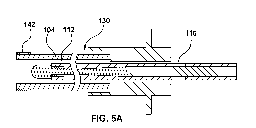
Fig. 5 is a diagrammatic cross-sectional view of a supporting member 130 with
a puncture device
112 (such as an RF guidewire) installed therein. In the embodiment of Fig. 5,
supporting member 130 has a distal
marker 142 at its distal end for indicating the position of the distal end of
supporting member 130 under imaging,
and the puncture device 112 has a radiopaque marker 104 at its distal end for
indicating the position of the distal end
of the puncture device under imaging. Figs. 5A to 5C show the steps of a
method advancing puncture device 112
through the supporting member 130. In Fig. 5A shows the puncture device 112 is
positioned to have the distal end of
proximal marker 116 at the proximal end of the hub/handle of the supporting
member while the tip of the tip of the
puncture device still inside of the lumen of the supporting member. The
puncture device is advanced to the
configuration of Fig. 5B wherein the middle of proximal marker 116 at the
proximal end of the hub/handle of the
supporting member and the tip of the puncture device lines up with the tip of
the supporting member 130. The
puncture device is further advanced to the configuration of Fig. 5C wherein
the proximal end of proximal marker
116 is at the proximal end of the hub/handle of the supporting member and the
tip of the puncture device 112
extends beyond the tip of the supporting member 130. The configuration on Fig.
5C further includes distal marker
142 of supporting member 130 lining up with radiopaque marker 104 at of the
distal end of supporting member
130, which under imaging, would confirm the relative positioning of the
puncture device and the supporting
member. Thus, Figs. 5A-5C illustrates an elongate proximal marker 116 such
that the leading edge of the marker
represents a first relative position between puncture device and supporting
member (i.e., where the puncture device
4 0 is
well within the lumen of the supporting member), the middle (or midpoint) of
the marker represents a second
relative position between puncture device and supporting member (i.e., where
the distal tip of the puncture device is
CA 03099451 2020-11-04
WO 2019/215618
PCT/IB2019/053745
18
aligned with the distal tip of the supporting member), and the trailing edge
of the marker represents a third relative
position between puncture device and supporting member (i.e., where the distal
tip of the puncture device is beyond
the distal tip of the supporting member and exposed therefrom). In an
alternative embodiment, the respective
relative positions may be marked by separate markers. In an alternative
embodiment, a plurality of separate
proximal markers may be provided to respectively identify the first relative
position, the second relative position,
and the third relative position.
[00104] In
some embodiments, the shaft of puncture device 112, radiopaque marker 104, and
proximal
marker 116 have outer diameters <= 0.035". Radiopaque marker 104 is comprised
of platinum and iridium (Pt/In
and has an inner diameter >= 0.01". In one embodiment, the mandrel of the
puncture device 112 is made of stainless
steel. In an alternative embodiment, the mandrel of the puncture device 112 is
a composite of a distal portion
comprised of a super elastic material such as Nitinol which is designed to be
kink resistant, and a proximal portion
comprised of a stiffer alloy such as stainless steel. In still another
embodiment, the mandrel is comprised of Nitinol
for greater flexibility and resistance to kinking along the entire length of
the puncture device. In the embodiment
where there is a composite construction, these materials can be welded,
pressfit or glued together. The body of
puncture device may be completely insulated with polytetrafluoroethylene
(PTFE). While typical embodiments of
puncture device 112 have an outer diameter of <= 0.035", any size outer
diameter of the puncture device is
acceptable as long as it fits within the dilator used for a transseptal
procedure. Alternative embodiments of
radiopaque marker 104, which are components of smaller diameter RF guidewires,
have an inner diameter smaller
than 0.01". While a typical embodiment of introducer 130 has an inner diameter
of >= 0.035", other inner diameter
2 0 sizes of the introducer are possible so long as the RF guidewire 100
used in a procedure can pass through.
[00105] In
one embodiment, puncture device may comprise multiple markers along its
length. These
markers may be spaced such that they con-espond with a particular length of
supporting member. Supporting
members, such as dilators, sheaths, and stylets may be of varying lengths. For
example, a sheath may be longer or
shorter depending on the needs of the particular procedure. By providing
multiple markers spaced along the length
of a puncture device, the puncture may be used with supporting members of a
variety of lengths by matching a
marker with a particular supporting member length. To make it more clear,
markers may be provided with distinct
visual or tactile features to distinguish which markers should be used with
which devices. For visual markers,
different colours, shades, surface features (reflective metallic coils,
dimpled bands, knurls, etc.) or symbols may be
used to distinguish between different markers. For markers with visual
features, a clear coating is provided over top
to secure the feature and ensure that the puncturing device has a consistent
outer surface.
Radiopaque markers
[00106] In
some embodiments, as shown in Fig. 1C and 1D, the supporting member 130
comprises one or
more radiopaque markers such as a supporting member radiopaque marker 42. In
some such examples as above, the
assembly 100 provides a supporting member 130 (for example comprising a needle
shaft 132 as provided as a part
of or defined by a reinforced dilator 30A), comprises a radiopaque marker 42,
such as at the distal tip of the
supporting member 130. In some such examples, the supporting member 130
comprises a radiopaque marker 42
embedded within the polymer of the distal tip thereof, as shown
[00107] In
a specific example, the radiopaque marker 42 comprises a radiopaque coil
embedded within the
polymer of the supporting member 130 (for example comprising a needle shaft
132 as provided as a part of or
4 0 defined
by a reinforced dilator 30A) such as within the one or more polymer layers 38
(forming the polymer shaft 39
CA 03099451 2020-11-04
WO 2019/215618
PCT/IB2019/053745
19
which in turn forms the dilator shaft 32), for example, at a distal tip
thereof (of the supporting member 130). In a
more specific example, the radiopaque coil is embedded within the one or more
polymer layers such that the one or
more polymer layers extend distally beyond the radiopaque coil.
Alignment using radiopaque markers
[00108] in some embodiments of the present invention, a substantially
flexible energy based puncturing
device 114 is provided (such as an RF guidewire) that comprises one or more
device side radiopaque markers (or in
other words one or more device radiopaque markers) at a distal end of thereof,
for example, as shown in Figs. 3B
and 3C. In some such embodiments, as noted above, the supporting member 130
also comprises a supporting
member radiopaque marker at the distal end of the supporting member 130 (as
shown in Figs. 1C and 11)). In some
such. embodiments, similar to die embodiments shown in Figs. 3B and 3C, the
one or more device radiopaque
markeis 12 are configured to co-operate with the supporting member radiopaque
marker 42 to indicate the relative
position of the substantially flexible energy based puncturing device 114
(such as an RF guidewire 10). The
embodiments, shown in Figs. 3B and 3C illustrate a dilator 30B that is
provided separately from a stylet. 64.
However, in alternative embodiments as described currently the stylet. 64 may
be a reinforcing member 34 that is
.. provided within a dilator 30A.
[0 01 0 9] In
some such embodiments, the assembly 100 comprises an initial configuration
100A, where the
substantially- flexible energy based puncturing device 114 (such as an RI,
guidewire 10) is .positionable within the
supporting member 130 such that the one or more device radiopaque markers 12
are not in alignment with the
supporting member 130 radiopaque marker 42, as shown in Fig. 3A. In sonic such
examples, multiple radiopaque
2 0
markers may be visible under imaging, including the one or more device
radiopaque markers 12 and the supporting
member radiopaque marker 42.
[0 01 1 0] in
some such embodiments, the assembly 100 comprises a first configuration 1.00B,
as shown in
Fig. 313 where the substantially flexible energy based puncturing device 114
(such as an RF guidewire 10) is
positionable within the supporting member 130 such that the one or more device
radiopaque markers 12 are in
alignment with the suppotting member 130 radiopaque marker 42, as Shown in
Fig. 3B. In some such examples, a
single radiopaque marker may be visible under imaging [including the one or
more device radiopaque markers 12
and the supporting me mber radiopaque marker 42 that may be arranged in close
proximity to one anotherl.
[0 01 11]
The assembiy 100 additionally has a second configuration 100B, where the
substantially flexible
energy based puncturing device 114 (such as an RF guidewire 10) is
positionableladvanceable within the supporting
member 130 such that the one or more device radiopaque marktrs 12 are
substantially not in alignment or
misaligned with the supporting member radiopaque marker 42. In some such
examples, the misalignment of the one
or more device radiopaque markers 12 with the supporting member radiopaque
marker 42 indicates positioning of
an energy delivery portion E 14d of the flexible energy based puncturing
device 11.4 (such as an RF electrode tip -10d
of an RF guidewire 10) beyond the supporting member (for example distal to the
distal tip or end of the supporting
member 130) for positioning against a target tissue site for puncture of
tissue. in sonic such examples, similar to Fig.
3A, multiple radiopaque markers may be visible under imaging (including the
one or more device radiopaque
markers 12 and the supporting member radiopaque marker 42, where the one or
more device radiopaque markers 12
are positioned distally to the supporting member radiopaque marker 42,
indicating that he distal electrode tip 10d is
positioned against a target tissue site (such as the septum of the heart) for
puncturing the tissue.
CA 03099451 2020-11-04
WO 2019/215618
PCT/IB2019/053745
[00112] In
some such examples, the sheath 20, and dilator 30A as well as the reinforcing
member 34 are
all radiopaque, and have radiopaque properties to enable them to visible under
imaging. In some such examples, one
or more of the sheath 20, dilator 30A, and reinforcing member 34, such as a
metal hypo-tube comprise radiopaque
materials in addition to radiopaque markers [42]. The reinforcing member 34
such as a metal shaft or hypotube is
5 also
radiopaque. In some such embodiments, polymers forming the sheath 20 and/or
the dilator 30A may comprise
polymer radiopaque filler such as barium sulfate 20% so there is contrast with
the one or more markers [12, 421 at
the distal tip. In other words this may provide visibility under imaging and
may additionally provide contrast with
the one or more markers [42, 121 which may allow the user to see the dilator
30A in relation to the RF guidewire 10
under imaging, to see whether the RF guidewire 10 is positioned in or outside
the dilator 30A [i.e., whether the
10 distal
segment of the RF guidewire 10 is distal to the dilator 30A1. In other
examples, puncturing device 114 (such
as an RF guidewire 10), sheath 20, dilator 30A, as well as reinforcing member
34 are also visible using ultrasound
imaging systems, radiopaque coil 106 and markers 12 being particularly
echogenic.
Supporting member with blunt tip
[00113] in
some embodiments of the 'Present invention, the supporting member 130
comprises a
15
substantially blunt distal tip or edge 143, as shown in Fig. 1A, in order to
provide a substantially alraumatic distal tip
143, while providing the advantages of a substantially rigid ot stiff
supporting member 130 (such as by providing
the reinforcing member 34) therein
[00114] In
one such embodiment, an overall method/workflow is provided that illustrates a
method of
carrying out a transseptal puncture procedure using an assembly 100, as
described herein above. The method
2 0
disclosed herein provides one or more advantages associated with an assembly
comprising an energy delivery
component that is provided separately from the rigid component. Details of the
method are provided herein below.
Method using example 1
[00115] As
a general overview, in one broad embodiment, with respect again to Fig. 2A-2G,
a method is
provided for carrying out a transseptal puncture, the method comprising: (i)
Advancing the RF wire into the superior
vena cava, as shown in Fig. 2B, (ii)advancing the sheath and dilator over the
wire into the superior vena cava, as
shown in Fig. 2C; (iii)withdrawing the RF wire into the dilator, as shown in
Fig. 2D; (iv) drop down from the SVC
into the heart to find the fossa, as additionally shown in Fig. 2D; (v)
tenting with the dilator; (vi) advancing RF wire
to puncture position, also with reference to Fig. 2D; (vii) puncturing and
advancing RF wire, as shown in Fig. 2E;
and (viii) crossing the sheath and dilator over the RF wire, as shown in Fig.
2F.
[00116] More specifically, in a specific embodiment of a method of the
present invention, with reference
again to Fig. 2A, a method is provided for carrying out a transseptal puncture
procedure using an assembly 100
comprising a flexible RF wire 10, a sheath 20, and a dilator 30A, the method
comprises the following steps: at step
202, [11 advancing the RF wire into the superior vena cava (SVC) to gain
access, as additionally illustrated in Figure
2B. In some such embodiments, providing the energy delivery component
(flexible RF wire) separately from the
reinforcing member allows the energy delivery component to be used as an
access wire. More specifically, the
dilator 30A can be advanced later, allowing the flexible RF wire to provide
access to the SVC without the use of an
additional access wire. This may help reduce the number of steps and
streamline the procedure, and as such may
reduce procedural time and complexity.
CA 03099451 2020-11-04
WO 2019/215618
PCT/IB2019/053745
21
[00117]
The method additionally comprises the following steps: at step 204, [2]
advancing the sheath 20
and dilator 30A combination over the flexible RF wire into the SVC. Thus, the
flexible RF wire 10 functions as an
access wire and enables the sheath 20 and dilator 30A (for example as an
assembly) to be tracked over the flexible
RF wire 10 into the SVC as shown in Fig. 2C.
[00118] The method additionally provides: at step 206, withdrawing the RF
wire into the dilator 30A. The
method may optionally comprise step 207 of using proximal markers on the RF
wire to determine the relative
positioning between the RF wire and the sheath/dilator. For example, in the
embodiment of the puncture device
shown in Figs. 5A-5C, the proximal marker may be used to determine whether the
active tip of the puncture device
is entirely within the dilator/sheath, or exposed from the dilator/sheath.
This positioning may optionally be verified
or further adjusted using visualization or mapping techniques. After
confirming the relative positioning of the wire
(e.g., that the wire is entirely within the sheath/dilator), the user may
proceed to step 208. In step 208, the method
comprises [3] performing a drop down from the SVC into the heart to locate the
fossa, as shown in Fig. 2D for
can-ying out the step of positioning the assembly 100. In one such example,
having the reinforced member 34
(within the dilator 30A) as separate from and operable independently form the
flexible RF wire provides the
additional advantage of allowing the drop down to be repeated if the fossa is
missed in the first pass. More
specifically, it eliminates the need to re-wire, in other words to re-insert
an access wire, remove the access wire and
then re-advance a rigid puncture device such as a needle into the SVC in order
to repeat the drop down. More
specifically, in an embodiment of the instant application, the dilator 30A
(and thus the reinforcing member 34) may
be partially removed or retracted along with the sheath 20 and the flexible RF
wire 10 may be re-advanced into the
SVC. The sheath 20 and the dilator 30 may then be re-advanced over the
flexible RF wire 10, as shown in Fig. 2C
and the drop down may be repeated to allow the RF wire 10 to engage the fossa.
This may help reduce procedural
time and increase safety as an additional exchange is not required. Adding an
additional exchange may add more
time and add unnecessary risk. Thus, procedural time and risk may be reduced
with the cun-ent embodiments where
the energy delivery component and the rigid component are decoupled.
[00119] The reinforcing member 34 [within the dilator 30A1 provides the
additional advantage of
providing sufficient stiffness to the assembly 100 to facilitate the drop
down, at step 208. As such the reinforcing
member 34 enables sufficient force transmission and torque to allow the
assembly 100 to engage the septum, as
illustrated in Fig. 2D. The method further comprises: at step 212 tenting with
the dilator 30A, with reference to Fig.
2D. The reinforcing member 34 provides sufficient stiffness to the assembly
100 to enable force to be imparted to
the distal end of the assembly 100, thus enabling tenting with the dilator
30A. In some examples, having the
reinforcing member 34 within the dilator 30A, allows it to be removed and re-
shaped to allow for optimizing the
position against the fossa. In some such embodiments, prior to the step of
tenting, at step 210, the physician may
assess whether the angle of the dilator 30A and/or the assembly 100 is
sufficient. If the angle is not deemed to be
sufficient, at step 211, the physician may pull out the dilator 30A and
reshape the curve. The dilator may be then be
reinserted as indicated by step 213. The procedure then may be repeated
starting at step 208, and a drop down may
be performed again using the assembly 100. Once the fossa has been located,
the physician may proceed with the
step of tenting with the dilator, at step 212. In some cases, it may be
necessary to repeat the procedure by starting at
previous steps such as steps 202, 204, or 206 before step 208 may be
performed. This is because the RF wire may
not be properly positioned to allow a drop down (step 208) to be performed
without repositioning the assembly.
CA 03099451 2020-11-04
WO 2019/215618
PCT/IB2019/053745
22
[00120]
The method additionally comprises the steps of: at step 214, advancing RF wire
10 to puncture
position and at step 216 [4] puncturing and advancing RF wire 10, as shown in
Fig. 2E. At step 214, as the RF wire
is being advanced to the puncture position (i.e., residing outside the
sheath/dilator), the user may optionally visually
or tactilely monitor the proximal marker on the RF wire to determine the
relative positioning between the RF wire
and the dilator/sheath. In one embodiment (see, e.g., Figs. 5A-5C), as the
proximal marker disappears into the
handle/hub of the dilator/sheath, the user knows that the active tip of the RF
wire is now exposed (i.e., in the
puncture position). This positioning may optionally be verified or further
adjusted using visualization or mapping
techniques. At step 216, the RF wire punctures the tissue and is advanced
therethrough. The advancement of the RF
wire 10 into the left side of the heart 500, enables anchoring of the RF wire
10 on the left side of the heart to
maintain access to the left side of the heart. The flexible RF wire 10 may
provide the additional advantage of
allowing the operator to push hard without injury as the flexible RF wire is
more flexible. The method additionally
comprises: at step 218, [5] crossing the sheath and dilator over the RF wire
10, as additionally shown in Fig. 2F. The
flexible RF wire 10 may additionally protect the open end of the sheath
20/dilator 30A so it does not push hard into
the tissue. At step 218, the sheath 20 and dilator 30A [including the
reinforcing component 34] may then be
removed.
[00121] As
outlined herein, energy delivery component is provided as a flexible RF wire
10 that is separate
from a stiff component such as a reinforcing member 34 [provided within the
dilator 30A], where the reinforcing
member 34 [with the dilator 30A] is separable from and removable from the
flexible RF wire 10. This provides the
additional advantage, in that the reinforcing member 34 [within dilator 30A]
may be removable after transseptal
2 0
puncture and access, providing a step [6] of allowing the flexible RF wire 10
to remain positioned within the left
atrium which allows for immediate anchoring of the flexible RF wire within the
left atrium, as shown in Fig. 2G. In
one such example, the RF wire may be positioned within the left superior
pulmonary vein for anchoring. This may
enable the RF wire to maintain access into the left atrium, allowing removal
of the reinforcing member 34[along
with dilator 30A to facilitate exchange of devices into the left atrium using
the flexible RF wire. This may
additionally reduce an additional exchange of the left side as it may
eliminate the need for the physician to advance
another wire after puncture to maintain access on the left side for tracking
additional devices into the left side. An
additional benefit of minimizing exchanges on the left side, in addition to
reducing procedural time and the number
of steps required, is minimizing risk of infection, embolisms and stroke. In
another example, the RF wire 10 may
have a pigtail curve at the distal end. This may enable anchoring of the RF
wire 10 in the left atrium instead of the
pulmonary vein. Alternatively, the RF wire 10 may be used to anchor in the
pulmonary vein. In some such
examples, the former method of anchoring in the left atrium may provide
additional advantages not found in the
latter method.
Using the proximal markers on the puncture device
[00122] In
some embodiments of the method, the user positions the puncture device
relative to the
supporting member using the proximal marker 116 without using an imaging
system such a fluoroscopy in a step
that can be called, 'macro-positioning'. Subsequent to the 'macro-
positioning', the user may utilize an imaging
system (e.g. fluoroscopy) for more precise positioning of the RF guidewire
relative to the introducer and the target
tissue in a step that can be called micro-positioning. By using the proximal
and distal markers, a user can perform
the early part of positioning the apparatus without fluoroscopy to thereby
reduce the amount of X-rays the user and
4 0
patient are exposed to when compared to performing the entire procedure under
fluoroscopy. In some alternative
CA 03099451 2020-11-04
WO 2019/215618
PCT/IB2019/053745
23
embodiments of the method, the part of the procedure involved with positioning
the puncture device relative to the
supporting member is performed without any fluoroscopy.
Using the same device for initial track up/access and positioning
[00123] In some embodiments of the present invention, with reference
now to Figs. 2A-2G, a method is
disclosed for puncturing tissue. The method comprises the step of: [1]
accessing a region of tissue within a patient's
body by advancing a device (such as a puncture device 110 which may be an RF
guidewire 10) into the region of
tissue, as shown in Fig. 2B. In some such examples the method of puncturing a
region of tissue comprises a method
of carrying out a transseptal puncture where the step of accessing the region
of tissue comprises advancing the
device (such as the puncture device 110) into the superior vena cava (SVC) 501
adjacent a heart 500 of the patient.
[00124] In some embodiments of the present invention, the method for
puncturing tissue additionally
comprises the step of: [3] positioning a device at a target tissue site in the
region of tissue, as shown in Fig. 2D, by
for example: [2] first tracking a supporting member 130 (such as reinforced
dilator 30A) over the puncture device
110 to support the device (such as puncture device 110) as shown in Fig. 2C,
to [3] enable advancement of the
device (such as a puncture device 110) towards a target tissue site in order
to position the device at the target tissue
site for puncturing, as shown in Fig. 2D.
[00125] In some such examples, the step of positioning the puncture
device 110 at the target tissue site
comprises performing [3] a drop down from the superior vena cava (SVC) into
the heart 500 of the patient to locate
a fossa ovalis (or in other words fossa) 504 along a septum 502 of the heart
500, by first for example (2) tracking or
advancing a supporting member 130 (such as a dilator 30A) over the device
(such as a puncture device 110) into the
2 0 SVC to (3) facilitate the drop down to position the puncture device 110
at the fossa 504.
[00126] In some such examples, as shown in Figs. 2B-2D, the steps of
accessing [1], as shown in Fig. 2B
and positioning [3], as shown in Fig. 2D, are performed using the same device
such as a puncture device 110,
wherein the puncture device 110 is usable without the supporting member 130
during the step of accessing [1] and
wherein the device is usable with the supporting member 130 during the step of
positioning [3].
Using a puncture device for initial access and positioning
[00127] In some such embodiments of the present invention, as shown in
Figs. 2B-2D, the steps of
accessing and positioning are performed using a puncture device 110.
Using the same device for initial access, positioning and puncturing
[00128] In some such embodiments of the present invention, as shown in
Fig. 2E, the method additionally
comprises: [4] a step of puncturing through the target tissue site using a
device (such as the puncture device 110)
after the step of positioning [3] as shown in Fig. 2D. The supporting member
130 supports the device (such as
puncture device 110) during puncturing [4] where the steps of accessing [1],
positioning [3] and puncturing [4] are
performed using the same device.
[00129] In some embodiments of the present invention, the step [4] of
puncturing through the target tissue
site comprises the step [4] of puncturing through the fossa 504 to gain access
to a left side of the heart 500. This
enables one or more devices of the assembly 100, such as the supporting member
130 (such as dilator 30A) and
sheath 20 of the assembly 100 to be tracked over the RF guidewire 10 into the
left side of the heart.
CA 03099451 2020-11-04
WO 2019/215618
PCT/IB2019/053745
24
Using a puncture device for initial access, positioning and puncturing
[00130] In
some such examples, as shown in Figs. 2B-2E, the steps of accessing,
positioning, and
puncturing are performed using a puncture device 110.
Using the same device for initial access, positioning and puncturing and
anchoring
[00131] In accordance with an embodiment of the present invention, the
method additionally comprises a
step of anchoring, as shown in Fig. 2E, where the step of anchoring is
performed using a device (such as the
puncture device 110) after the step of puncturing [4] through the target
tissue site, to maintain access through the
target tissue site to the other side of the target tissue site, to allow one
or more additional device [such as sheath 20
and the supporting member 130 comprising the dilator 30A1 to be tracked over
the device (such as the puncture
device 110) to the other side of the target tissue site, as shown in Fig. 2F,
where the steps of accessing, positioning,
puncturing and anchoring are performed using the same device. The puncture
device 110 such as the RF guidewire
10 may be left to maintain access to the left side of the heart, as shown in
Fig. 2G. The supporting member 130 for
example comprising the dilator 30A may be removed or retracted to allow
anchoring using the RF guidewire 10.
The RF guidewire 10 functions as a rail to guide one more devices to the left
side of the heart. In some such
examples, the RF guidewire 10 provides a substantially stiff rail to guide the
one or more devices to left side of the
heart while being substantially atraumatic to minimize damage to the tissue.
[00132] In
some such embodiments of the present invention, the step of anchoring to
maintain access
through the target tissue site comprises advancing the device (such as the
puncture device 110) through the fossa to
the left side of the heat to maintain access to the left side of the heart.
The step additionally comprises a step of
removing the supporting member 130 [such as dilator 30A1 and leaving the
puncture device 110 [such as RF
guidewire 101 to maintain access to the region of tissue such as the left side
of the heart.
[00133] As
such, in some examples, the step of anchoring comprises removing the
supporting member 130
comprising the dilator 30A to enable anchoring by allowing the RF guidewire 10
to remain positioned to maintain
access to the eft side of the heart. The sheath 20 may additionally be removed
as well.
Alternatives for the device being used for initial access, positioning and/or
puncturing - based on the base
claim these dependents depend from
[00134] In
some such embodiments of the present invention, the device comprises a
flexible puncture
device 112 where one or more of the steps of accessing, positioning,
puncturing and anchoring are performed using
the flexible puncture device 112. In some such examples, each of the steps of
accessing, positioning, puncturing and
anchoring are substantially performed using the flexible puncture device 112.
[00135] In
some such embodiments of the present invention, the device comprises a
substantially flexible
guidewire (such as a mechanical guidewire 118 or an RF guidewire 10) where one
or more of the steps of accessing,
positioning, puncturing and anchoring are performed using the substantially
flexible guidewire(such as a mechanical
guidewire 118 or an RF guidewire 10). In some such examples, each of the steps
of accessing, positioning,
puncturing and anchoring are substantially performed using substantially
flexible guidewire (such as a mechanical
guidewire 118 or an RF guidewire 10).
[00136] In
some such embodiments of the present invention, the device comprises a
flexible energy based
puncture device 114 where one or more of the steps of accessing, positioning,
puncturing and anchoring the steps
CA 03099451 2020-11-04
WO 2019/215618
PCT/IB2019/053745
are performed using the flexible energy based puncture device 114. In some
such examples, each of the steps of
accessing, positioning, puncturing and anchoring are substantially performed
substantially using flexible energy
based puncture device 114.
[00137] In
some such embodiments of the present invention, the device comprises a
flexible RF guidewire
5 10 and wherein one or more of the steps of accessing, positioning,
puncturing and anchoring are performed using the
flexible RF guidewire 10. In some such examples, each of the steps of
accessing, positioning, puncturing and
anchoring are substantially performed substantially using flexible the
flexible RF guidewire 10.
[00138] In
some such embodiments of the present invention, wherein the device comprises a
flexible
mechanical guidewire 118 having a relatively sharp distal tip 118d wherein one
or more of the steps of accessing,
10
positioning, puncturing and anchoring are performed using the flexible
mechanical guidewire 118. In some such
examples, each of the steps of accessing, positioning, puncturing and
anchoring are substantially performed
substantially using flexible mechanical guidewire 118.
Repeating steps of accessing and positioning
[00139] In
some such embodiments of the present invention, the method further comprises
repeating the
15 steps of accessing [1], shown in Fig. 2B, and positioning [3] as shown
in Fig. 2D, until the device (such as the
puncture device 110) is positioned at the desired target tissue site prior to
the step of puncturing [4], as shown in Fig.
2E.
Reshaping the supporting member
[00140] In
some such examples, repeating the step of positioning [3] as shown in Fig 2D,
further
20 comprises reshaping a curvature of the supporting member 130 after
removing the supporting member 130, and re-
tracking [2] the supporting member 130 over the device, as shown in Fig. 2C
(such as the puncture device 110 that
has been re-positioned [1] within the SVC as shown in Fig. 2B), prior to
repeating the step of positioning as shown
in Fig. 2D, which in the example shown comprises a drop-down procedure to find
the fossa 504. In a specific
example, the supporting member 130 comprises a reinforcing member 34, where
the step of positioning is
25 performed using the reinforcing member 34.
[00141] In
some such embodiments of the present invention, the method comprises reshaping
the
supporting member 130 (such as the reinforced dilator 30A). In some such
examples, the method comprises pulling
the dilator element or dilator 30A out and reshaping it. In other examples,
comprises pulling both the dilator element
30A and the sheath 20out and reshaping it.
Supporting member comprises reinforced dilator
[00142] In
some such examples re-shaping is performed using the supporting member 130
comprising a
reinforced dilator 30A where the reinforced dilator 30A comprises the
reinforcing member 34, where the step of
positioning is performed using the reinforced dilator 30A that can be re-
shaped.
Supporting member comprises a stylet
[00143] In some embodiments, alternatively, as discussed further herein
below, with respect to Figs. 4A-
4E, the step of re-shaping can be performed using the supporting member 130
comprising a stylet 60 wherein the
stylet 60 is the reinforcing member 34, and the step of positioning is
performed using the stylet 60.
CA 03099451 2020-11-04
WO 2019/215618
PCT/IB2019/053745
26
[00144] In
some such examples, the stylet element 60 can be taken out and reshaped. In
other examples,
the stylet element 60 along with the sheath 20 and/or dilator 30B may be
pulled out and re-shaped to see what the
net shape might be and then can be re-inserted therein.
[00145]
The methods outlined herein above may also be used for embodiments discussed
further herein
below having a removable stylet 60, as shown in Figs. 4A-4E.
Mapping System to visualize initial access tracking and positioning
[00146] In
some such embodiments with respect to Figs. 2A-2G, and also additionally with
reference to
embodiments shown in Figs. 4A-4E, the step of positioning is performed using a
flexible RF guidewire 10. In some
such examples, the steps of positioning, and puncturing are performed using a
flexible RF guidewire 10. Still
additionally, in some such examples, the steps of positioning, puncturing, and
anchoring are performed using a
flexible RF guidewire 10. In some such examples, a mapping system as provided
below may be used to visualize
the steps of positioning, and anchoring. In some such examples, as provided in
Figs. 2A-2G and Figs. 4A-4E, the
step of accessing may additionally be performed using the RF guidewire 10. As
such, in some such examples, a
mapping system as provided below may be used to visualize the flexible RF
guidewire 10 using a mapping system
during the steps of accessing positioning, and anchoring. In some such
examples, the method further comprises the
step of visualizing the flexible RF guidewire 10 using a mapping system during
the steps of accessing and
positioning.
[00147] As
such embodiments of the present invention provide a mapping system that is
usable to
visualize an RF guidewire 10 during a method of puncturing tissue during one
or more of the steps of accessing,
positioning, and anchoring.
[00148] In
some instances, the mapping device comprises an electro-anatomical mapping
system where the
electro-anatomical mapping system may be magnetic or impedance based to create
virtual volumes. In some
examples, the electro-anatomical mapping system is usable with other
echocardiographic imaging modalities, which
may be ultrasound. The echocardiographic imaging modalities may be used as an
overlay in maps, in other words
they may be used provide additional information to the mapping system. The
echocardiographic imaging modalities
may comprise intracardiac cardiography or FEE echorcardiographic
[00149] In
some examples, the method involves switching between a mapping mode that is
used for each
of the steps of accessing, positioning, and anchoring and the puncture mode
that is used for the step of puncturing.
[00150] In
some such examples, the method of mapping the RF guidewire 10 to visualize
using an
imaging modality, may be usable with a flexible wire with an electrode which
may or may not deliver energy which
may be used for recording purposive. In some cases it may be a passive
electrode for recording purposes.
Alternatively as discussed above, if an RF guidewire 10 is used, then the
mapping system is usable with an active
electrode such as the distal electrode tip 10d of the RF guidewire 10. As such
the recording and mapping properties
of a mapping system, are usable with a guidewire having a passive electrode or
an active electrode. In a specific
example, where a wire is provided with a passive electrode for mapping, the
wire may comprise a puncturing means
or a means to puncture tissue. In one instance the wire may comprise a
mechanical guidewire 118 that may have a
sharp distal tip 118d for puncturing tissue.
CA 03099451 2020-11-04
WO 2019/215618
PCT/IB2019/053745
27
[00151] In
some such embodiments the reinforcing member is the stylet 60 that is usable
independently
from the substantially flexible energy based puncture device 114 such as an RF
wire 10.
EXAMPLE 2
[00152] In
another example, embodiments of the present invention provides an assembly
300, as shown in
Fig. 3A, for puncturing tissue (such as creating a transseptal puncture
through a septum of a heart). Similar to
embodiments described herein above, the assembly 300 provides a puncture
device such as a substantially flexible
energy delivery puncture device 14 for puncturing tissue via deliveiy of
energy (such as flexible RF guidewire 10
and a a supporting member for supporting the substantially flexible energy
delivery puncture device, such as a
separate reinforcing member 34. In some such examples, the supporting member
comprises a reinforcing member
34. In some such embodiments, the substantially flexible energy delivery
puncture device 114 (such as RF
guidewire 10) is capable of being selectively insertable within the suppotting
member 130 to be sedectively usable in
co-operation therewith during a portion of the procedure and wherein the
substantially flexible energy delivery
puncture device 114 (such as 11F guidewire 10) is usable independently
therefrom during another portion of the
procedure, in. order to facilitate exchange and positioning vvitile providing
substantially atraurnatic puncture of
tissue.
Overall assembly
[00153] In
one such example, as illustrated in Fig. 3A, the assembly 300 comprises a
flexible energy
delivery component 114 that is provided separately from and is operable
independently from a supporting member.
In one such example, the flexible energy delivery component comprises an RF
wire 10, and the separate supporting
member 130 comprises a stylet 60 that defines a reinforcing member 34. In
other words, as provided herein below
the supporting member 130 is the reinforcing member 34 that is provided as a
stylet 60 that is usable independently
from a puncture device 110 such as a flexible puncture device 112. In still
others words, the supporting member 130
is defined by the reinforcing member 34, where in one example, the reinforcing
member 34 comprises the stylet
60.The assembly 300 additionally comprises a sheath 20 and a dilator 30B that
are usable with the flexible RF wire
10. In the particular example shown the reinforcing member 64 is also provided
separately from and removable from
the dilator 30B which in the present embodiment is provided as a flexible
dilator.
[00154]
Some such embodiments comprises a dilator 30B that is usable with the
supporting member 130
'to form a supporting member assembly Ã34 for selective use there-with during
a portion of the procedure, as shown
in Fig. 3B. In some such embodiments, as noted above, the supporting member
130 comprises a stylet 60 defining
the reinforcing member 34. In some examples, a dilator 30B is provided that is
usable with the stylet 60 for selective
use therewith to form a stylet. assembly 164, as shown in Fig. 3C.
[00155] In
some such embodiments, the puncture device 110 comprises a substantially
flexible energy
based puncture device 114. In a specific instance of this example, the
substantially flexible energy based puncture
device 114 comprises a flexible RF guidewire or wire 10. In some embodiments,
the RF guidewire Ã0 is capable of
being selectively usable in co-operation with the stylet 60 (for example by
being selectively being coupled thereto)
during a portion of the procedure, and the RF guidewire 1.0 is usable
independently from the stylet 60 during another
portion of the procedure. Where selective use of the RF guidewire 10 in
conjunction with the stylet, as well as
without the. stylet. 60, facilitates pimcdttre of tissue.
Supporting member/reinforcing member shape-ability
CA 03099451 2020-11-04
WO 2019/215618
PCT/IB2019/053745
28
[00156] In
some such embodiments of the present invention where the supporting member 130
is provided
separately from a dilator 30B., the assembly 300 provides a supporting member
130 that is shapeable to enable it to
be removed from the puncture device 110 (such as a flexible tissue puncture
device 112, for e.g. a substantially
flexible energy based tissue puncture device 114) to enable a curve of the
supporting member 130 be re-shaped to
be reinserted therewith. For example, the re-shaped supporting member 130 is
re-insertable with and/or usable with
the substantially flexible energy based tissue puncture device 114 and/or one
or more other components of the
assembly 300 such as dilator 30B and/or sheath 20), in order to optimize the
position of the assembly 300 against a
target tissue site to facilitate puncture (such as a fossa of the heart to
facilitate a transseptal puncture).
[00157] In
a specific example, the stylet 60 is shapeable to enable the stylet 60 to be
removed from the
substantially flexible puncture device to enable a curve of the stylet 60 be
re-shaped to be reinserted therewith, in
order to optimize the position of the assembly against a target tissue site.
In some such examples, the stylet 60 is
removable from the one or more components or member of the assembly 300 to be
re-shaped to be re-inserted
therewith to position the assembly 300 at the target tissue site.
[00158]
Details of the stylet 60 defining the reinforcing member 34 in use with a
dilator 30B and flexible
RF wire 10 are shown in Figs. 3B and 3C. More specifically, Figs. 3B and 3C,
illustrate a dilator 30B which in some
examples is a flexible dilator such as a standard transseptal dilator without
having a reinforcing member embedded
therein or in other words separately from the dilator 30B, the dilator 30B
comprising a proximal portion 31 that
terminates at a distal tip 41. In some embodiments, the dilator 30B may
additionally include a radiopaque marker 42
at the distal tip 41. Similar to embodiments disclosed herein above, the
dilator 30B comprises a dilator shaft 32 that
extends along the proximal portion 31. However, unlike embodiments discussed
herein above, assembly 300
provides a reinforcing member or component 34 defined by stylet 60 that is
provided separately from the dilator
30B, and functions as a removable reinforcing member that is removable from
the dilator 30B. As such, the
reinforcing member 34 is provided separately from and is removable from both
the flexible RF wire 10 and the
dilator 30B. Fig. 3B shows the assembly 300 in position for a drop down,
whereas Fig. 3C shows the assembly 300
in position for arcing to enable the transseptal puncture.
Atraumatic stylet
[00159] In
some embodiments, the stylet 60 is provided as a substantially atraumatic
stylet 68, as shown in
Fig. 5F to prevent damage to the dilator 30A that it is inserted in. In some
such examples, the stylet 68 comprises a
tapered distal tip 69 to prevent and/or help minimize skiving and to provide a
smoother feel for the user upon
insertion into a dilator during use.
[00160] In
some embodiments, as an alternative or in addition to providing a tapered
distal tip 69, the
stylet 60 is made substantially atraumatic by providing a lubricous coating 67
on the stylet 60 in order to prevent
and/or help minimize skiving and to provide a smoother feel for the user upon
insertion into a dilator during use.
[00161] In some such examples, the lubricous coating 67 comprises a P
__ 1Th coating. The PT1 , coating
may be spray coated onto the stylet 60 or it may be provided as a heat shield.
Alignment using radiopaque markers
[00162] In
some embodiments of the present invention, similar to embodiments discussed
previously with
cespect to assembly 100, the assembly 300 coniptises a substantially flexible
energy based puncturing device 114
CA 03099451 2020-11-04
WO 2019/215618
PCT/IB2019/053745
29
(such as the RF guidewire 10) that comprises one or more device radiopaque
markers 12 at a distal end of thereof.
Additionally, the supporting member assembly comprises one or more supporting
assembly radiopaque markers 42
at the distal end of a supporting member assembly 134 (for example comprising
a separate reinforcing member 34
such as a stylet 60 and a puncture device 110 such as a substantially flexible
energy based puncturing device 114. In
one such example, the supporting assembly radiopaque marker 42 is provided on
the dilator 30B of the supporting
member assembly 134. In some such examples, the one or more device radiopaque
markers 12 are configured to co-
operate with the supporting assembly radiopaque marker 42 to indicate the
relative position of the substantially
flexible energy based puncturing device 114.
[0 01 6 3] In
some such embodiments, the assembly 300 comprises an initial configuration
100A, where the
substantially flexible energy based puncturing device 114 (such as an .RF
guidewire 10) is positionable within the
supporting member assembly 134 such that the one or more device radiopaque
markers 12 are not in alignment with
the supporting assembly radiopaque marker 42, as shown in Fig. 3A. In some
such examples, multiple radiopaque
markers may be visible under imaging, including the one or more device
radiopaque markers 12 and the supporting
member radiopaque marker 42.
[00164] The assembly 300 additionally has a first configuration 100B, where
the substantially flexible
c nergy based putiounng device 114 is positionable within the supporting
member assembly 134 such that the OEC
more device radiopaque markers12 are in alignment with the supporting assembly
radiopaque marker 42. in some
such examples, a single radiopaque marker may be visible under imaging
[including the one or more device
radiopaque markers 12 and the supporting me tuber :radiopaque marker 42 that
may be arranged in close proximity to
one another].
[00165]
The assembly 300 additionally has a second configuration 10013, where the
substantially flexible
energy based puncturing device 114 (such as RF guidewire 1(i) is
positionableladvanceable within the supporting
member assembly 134 such that the one or more device radiopaque markers 12 are
substantially not in
alignmentimisaligned with the supporting assembly radiopaque marker 42. In
some such examples, the
misalignment of the one or more device radiopaque markers 12 with the
supporting assembly radiopaque marker 42
indicates positioning of an energy delivery portion I 14 (such as electrode
distal tip 10d or also referred to as distal
electrode tip /0d) of the flexible energy based puncturing device 114 (such as
an RF guidewire 10) beyond the
supporting member assembly 134 (for example distal to the distal tip or end of
the supporting member 130) for
positioning against a target tissue site for puncture of tissue,
[00166] With reference now to Fig, 3A, similar to embodiments Shown in Fig,
3A and discussed
previously, multiple radiopaque markers may be visible under imaging
,including the one or more device radiopaque
markers 12 and the swooning member radiopaque marker 42, where the one or more
device radiopaque markers 12
are positioned distally to the supporting member radiopaque marker 42,
indicating that the distal electrode tip 10d is
positioned against a target tissue site (such as the septum of the heart.) for
puncturing the tissue.
[00167] In some embodiments of the present invention, one or members or
components of the assembly 300
may be radiopaque to facilitate visualization of the assembly 300. In one such
example, the sheath 20 and/or the
dilator 30B comprise a radiopaque polymer and the stylet 60 (for example
comprising a metal shaft) is radiopaque.
As such, in some examples, the stylet 60, sheath 20 and/or the dilator 30B are
all radiopaque and thus have
radiopaque properties. In a specific example, the polymers forming the sheath
20 and/or dilator 30B comprise
4 0
radiopaque filler such as barium sulfate 20% to provide contrast with the one
or more markers 12, 42 at the distal
CA 03099451 2020-11-04
WO 2019/215618
PCT/IB2019/053745
tip, in order to allow the user to see the sheath 20 and/or the dilator 30B in
comparison to the RF guidewire 10. As
such, the present configuration may enhance visibility and may allow the user
to ascertain when the RF guidewire
10 (more specifically the electrode distal tip 10d of the RF guidewire 10) is
positioned inside or whether it extends
outside or beyond the distal tip of the dilator 30B.
5
[00168] In some embodiments of the transseptal assembly 300, the sheath 20
comprises a standard
transseptal sheath, the dilator 30B comprises a standard flexible dilator and
the flexible RF wire 10 is provided as a
0.035" wire. In some such examples, the flexible RF wire 10 may be J-tip wire
or a pigtail wire. In one particular
example, the dilator 30 comprises HDPE. The dilator 30 defines an inner
diameter that is sufficient to accommodate
the stylet 60. In one example, the stylet 60 that defines the reinforcing
member 3 4 comprises a hypo-tube such as a
10 metal hypo-tube. In a specific example, the stylet 60 comprises a metal
hypo-tube that comprises a stainless steel
hypotube. In one such example, the stainless steel hypo-tube has an ID of
greater than about 0.035".
[00169] In
some examples, the steerable sheath 20 may be an 8 French (Fr) steerable
sheath. Alternatively,
an 8.5Fr steerable sheath 20 may be provided. In some such examples, the
steerable sheath 20 may be provided with
different curvatures. In a specific example, steerable sheaths 20 may be
provided in different curvatures,
15 specifically at angles of: 37, 45, 55, 90, or 135 degrees. In a specific
instance of this example, the sheath tubing
comprises an inner P11- , liner, a braid and a Pebax outer jacket. In some
such embodiments, an 8 French (Fr)
dilator 30B is provided that is compatible with an 8 French (Fr) Sheath.
Alternatively, an 8.5 (Fr) dilator 30B may
be provided that is compatible with an 8 French (Fr) steerable sheath 20. Some
such dilators may be provided with a
64 degree curvature and an HDPE shaft. The stylet 60 may be provided as a
metal hypotube. In one such instance,
20 the
stylet 60 may have an ID of greater than about 0.038" and an OD that is less
than about 0.060". The dilator 30A
may be provided with a 50 degree or 86 degree curvature. In some examples,
materials may include HDPE and a
metal hypotube that forms the reinforcing member 34. In some such examples,
the RF wire 10 comprises a 0.035"
OD wire and may be a J-tip wire or a pigtail wire. In a specific instance of
this example, the RF wire 10 may
comprise a stainless steel core with a PT1 , coating.
25 Method [Example 2¨ removable stylet]
Using the same device for initial track up/ access and positioning
[00170] In
some embodiments of the present invention, with reference now to Figs. 4A-4G,
a method is
disclosed for puncturing tissue. The method comprises the step of: [1]
accessing a region of tissue within a patient's
body by advancing a device (such as a puncture device 110 such as an RF
guidewire 10) into the region of tissue, as
30 shown in Fig. 4B. In some such examples the method of puncturing a
region of tissue comprises a method of
can-ying out a transseptal puncture where the step of accessing the region of
tissue comprises advancing the device
(such as the puncture device 110) into the superior vena cava (SVC) 501
adjacent a heart 500 of the patient, as
shown in Fig. 4B
[00171] In
some embodiments of the present invention, the method for puncturing tissue
additionally
comprises the step of: [4] positioning a device at a target tissue site in the
region of tissue, as shown in Fig. 4D, by
for example: [3] first tracking a supporting member 130 over the puncture
device 110 to support the device (such as
puncture device 110) as shown in Fig. 4C, to [4] enable advancement of the
device (such as a puncture device 110)
towards a target tissue site in order to position the device at the target
tissue site for puncturing, as shown in Fig. 4D.
CA 03099451 2020-11-04
WO 2019/215618
PCT/IB2019/053745
31
[00172] In
some such examples, the step of positioning the puncture device 110 at the
target tissue site
comprises performing [4] a drop down from the superior vena cava (SVC) into
the heart 500 of the patient to locate
a fossa ovalis (or fossa) 504 along a septum 502 of the heart 500, by first
for example (3) tracking or advancing a
supporting member 130 (such as a stylet) over the device (such as a puncture
device 110) into the SVC to (3)
facilitate the drop down procedure, as shown in Fig. 4D, to position the
puncture device 110 at the fossa. For
example, this involves dropping down the assembly 300 from the superior vena
cava into the heart to find the fossa.
[00173] In
some examples, the step of positioning [4] is performed by first for example
additionally
comprises a step of advancing [2] a sheath 20 and dilator 30B over the device
(such as RF guidewire 10) into the
superior vena cava, prior to tracking and advancing a supporting member 130
which may comprise inserting a stylet
60 in the dilator 30B [for example until it reaches a stop], as shown in Fig
4C. In some such examples, the step of
positioning [4] is performed after a step of withdrawing the RF guidewire into
the stylet 60.
[00174] In
some such examples, as shown in Figs. 4B-4D, the steps of accessing [1], as
shown in Fig. 4B
and positioning [4], as shown in Fig. 4D, are performed using the same device
such as a puncture device 110,
wherein the puncture device 110 is usable without the supporting member 130
[comprising the stylet 601 during the
step of accessing [1] and wherein the device is usable with the supporting
member 130 [comprising the stylet 601
during the step of positioning [4].
Using a puncture device for initial access and positioning
[00175] In
some such embodiments of the present invention, as shown in Figs. 4B-4D, the
steps of
accessing and positioning are performed using a puncture device 110 [such as
an RF guidewire 101.
2 0 Using the same device for initial access, positioning and puncturing
[00176] In
some such embodiments of the present invention, as shown in Fig. 4E, the
method additionally
comprises: a step of puncturing[5] through the target tissue site using a
device (such as the puncture device 110)
after the step of positioning [4] as shown in Fig. 4D. The supporting member
130 [comprising the stylet 601
supports the device (such as puncture device 110) during puncturing [5] where
the steps of accessing [1], positioning
[4] and puncturing [5] are performed using the same device.
[00177] In
some embodiments of the present invention, the step [5] of puncturing through
the target tissue
site comprises the step [5] of puncturing through the fossa 504 to gain access
to a left side of the heart 500. This
enables one or more devices of the assembly 100, such as the supporting member
130 (such as dilator 30A) and
sheath 20 of the assembly 100 to be tracked over the RF guidewire 10 into the
left side of the heart.
[00178] In some such embodiments, the a step of puncturing [5], is
performed by first advancing the
device (such as the RF guidewire 10) and tenting with the dilator 30B, as
shown in Fig. 4D, to enable the RF
guidewire 10 to be advanced to the puncture position, in order to the puncture
the septum 502 at the fossa 504.
Using a puncture device for initial access, positioning and puncturing
[00179] In
some such examples, as shown in Figs. 2B-2E, the steps of accessing,
positioning, and
puncturing are performed using a puncture device 110.
Using the same device for initial access, positioning and puncturing and
anchoring
CA 03099451 2020-11-04
WO 2019/215618
PCT/IB2019/053745
32
[00180] In
accordance with an embodiment of the present invention, the method
additionally comprises a
step of anchoring [6], as shown in Fig. 4E, where the step of anchoring is
performed using a device (such as the
puncture device 110) after the step of puncturing [5] through the target
tissue site, to maintain access through the
target tissue site to the other side of the target tissue site, to allow one
or more additional device [such as sheath 20
and the dilator 30B1 to be advanced or tracked over the device (such as the
puncture device 110, for example an RF
guidewire 10) in order to allow crossing of the sheath 20 and dilator 30B to
the other side of the target tissue site, for
example into the left side of the heart, as shown in Fig. 4F, where the steps
of accessing [1], positioning [4],
puncturing and anchoring [5] are performed using the same device. The RF
guidewire 10 may be left to maintain
access to the left side of the heart as shown in Fig. 4G. The RF guidewire 10
functions as a rail to guide one more
devices to the left side of the heart. In some such examples, the RF guidewire
10 provides a substantially stiff rail to
guide the one or more devices to left side of the heart while being
substantially atraumatic to minimize damage to
the tissue.
[00181] In
some such embodiments of the present invention, the step of anchoring to
maintain access
through the target tissue site comprises advancing the device (such as the
puncture device 110) through the fossa to
the left side of the heat to maintain access to the left side of the heart.
[00182] In
some such examples, the step of anchoring additionally comprises removing the
stylet 60 to
enable anchoring by allowing the RF guidewire 10 to remain positioned to
maintain access to the eft side of the
heart. The sheath 20 and/or the dilator 30B may additionally be removed as
well.
[00183] In
some such embodiments, the steps of accessing, positioning, puncturing and
anchoring are
performed substantially using the wire such as the RF guidewire and the
removable stylet 60.
Using a puncture device for initial access, positioning and puncturing
[00184] In
some such embodiments of the present invention, the steps of accessing,
positioning,
puncturing and anchoring are performed using a puncture device (such as a wire
comprising an RF guidewire 10)
and a removable stylet 60
Alternatives for the device being used for initial access, positioning and/or
puncturing - based on the base
claim these dependents depend from
[00185] In
some such embodiments of the present invention, the device comprises a
flexible puncture
device 112 where one or more of the steps of accessing, positioning,
puncturing and anchoring are performed using
the flexible puncture device 112. In some such examples, each of the steps of
accessing, positioning, puncturing and
anchoring are substantially performed using the flexible puncture device 112.
[00186] In
some such embodiments of the present invention, the device comprises a
substantially flexible
guidewire (such as a mechanical guidewire 118 or an RF guidewire 10) where one
or more of the steps of accessing,
positioning, puncturing and anchoring are performed using the substantially
flexible guidewire(such as a mechanical
guidewire 118 or an RF guidewire 10). In some such examples, each of the steps
of accessing, positioning,
puncturing and anchoring are substantially performed using substantially
flexible guidewire (such as a mechanical
guidewire 118 or an RF guidewire 10).
[00187] In
some such embodiments of the present invention, the device comprises a
flexible energy based
puncture device 114 where one or more of the steps of accessing, positioning,
puncturing and anchoring the steps
CA 03099451 2020-11-04
WO 2019/215618
PCT/IB2019/053745
33
are performed using the flexible energy based puncture device 114. In some
such examples, each of the steps of
accessing, positioning, puncturing and anchoring are substantially performed
substantially using flexible energy
based puncture device 114.
[00188] In
some such embodiments of the present invention, the device comprises a
flexible RF guidewire
10 and wherein one or more of the steps of accessing, positioning, puncturing
and anchoring are performed using the
flexible RF guidewire 10. In some such examples, each of the steps of
accessing, positioning, puncturing and
anchoring are substantially performed substantially using flexible the
flexible RF guidewire 10.
[00189] In
some such embodiments of the present invention, wherein the device comprises a
flexible
mechanical guidewire 118 having a relatively sharp distal tip 118d wherein one
or more of the steps of accessing,
positioning, puncturing and anchoring are performed using the flexible
mechanical guidewire 118. In some such
examples, each of the steps of accessing, positioning, puncturing and
anchoring are substantially performed
substantially using flexible mechanical guidewire 118.
Repeating steps of accessing and positioning
[00190] In
some such embodiments of the present invention, the method further comprises
repeating the
steps of accessing [1], shown in Fig. 4B, and positioning [4] as shown in Fig.
4D, until the device (such as the
puncture device 110) is positioned at the desired target tissue site prior to
the step of puncturing [5], as shown in Fig.
4E.
Reshaping the supporting member
[00191] In
some such examples, repeating the step of positioning [4] as shown in Fig 4D,
further
comprises reshaping a curvature of the supporting member 130 after removing
the supporting member 130 [stylet
601, and re-tracking [3] the supporting member 130 [stylet 601 over the
device, as shown in Fig. 4C (such as the
puncture device 110 that has been re-positioned [1] within the SVC as shown in
Fig. 4B), prior to repeating the step
of positioning as shown in Fig. 4D, which in the example shown comprises a
drop-down procedure to find the fossa
504.In a specific example, the supporting member 130 comprises the stylet 60,
where the step of positioning is
performed using the stylet 60.
[00192] In
some such embodiments of the present invention, the method comprises reshaping
the
supporting member 130 (by pulling the stylet 60 out and re-shaping it).
Supporting member comprises a stylet
[00193] In
some embodiments, as discussed with respect to Figs. 4A-4E, the step of re-
shaping can be
performed using the supporting member 130 comprising a stylet 60 wherein the
stylet 60 is the reinforcing member
34, and the step of positioning is performed using the stylet 60.
[00194] In
some such examples, the stylet element 60 can be taken out and reshaped. In
other examples,
the stylet element 60 along with the sheath 20 and/or dilator 30B may be
pulled out and re-shaped to see what the
net shape might be and then can be re-inserted therein.
[00195] Similar to embodiments described herein above, an overall
method/workflow is provided that
illustrates a method of carrying out a transseptal puncture procedure using an
assembly 300, as described above. The
method disclosed herein provides one or more advantages associated with an
assembly comprising an energy
CA 03099451 2020-11-04
WO 2019/215618
PCT/IB2019/053745
34
delivery component that is provided separately from the rigid component.
Details of the method are provided herein
below.
[00196] As
a general overview, in one broad embodiment, as shown in Fig. 4A-4G, a method
is provided
for carrying out a transseptal puncture, the method comprising: (i) Advancing
the RF wire into the superior vena
cava, (ii)advancing the sheath and dilator over the wire into the superior
vena cava; (iii)inserting the stylet in the
dilator until it reaches a stop; (iv) withdrawing the RF wire into the stylet;
(v) dropping down from the SVC into the
heart to find the fossa; (vi) tenting with the dilator; (vii) advancing RF
wire to puncture position; (viii) puncturing
and advancing RF wire; and (ix) crossing the sheath and dilator over the RF
wire; and (x) remove stylet.
[00197]
More specifically, with reference again to Fig. 4A, a method is provided for
carrying out a
transseptal puncture procedure using an assembly 100 comprising a flexible RF
wire 10 or RF guidewire 10, a
sheath 20, a standard transseptal dilator 30B, and a stylet 60, the method
comprises the following steps: at step 402,
[1] advancing the RF wire into the superior vena cava (SVC) to gain access, as
additionally illustrated in Figure 4B.
As outlined previously, in some such embodiments, providing the energy
delivery component (flexible RF wire 10)
separately from the reinforcing member 34, allows the energy delivery
component to be used as an access wire or
starter wire. More specifically, the stylet 60 defining the reinforcing member
34 can be advanced later, allowing the
flexible RF wire 10 to provide access to the SVC without the use of an
additional access wire. This may help reduce
the number of steps and streamline the procedure, and as such may reduce
procedural time and complexity.
[00198]
The method additionally comprises the following steps: [2] at step 404,
advancing the sheath 20
and flexible dilator 30B combination over the flexible RF wire into the SVC.
As such, in this embodiment also, the
2 0
flexible RF wire 10 functions as an access wire and enables the sheath 20 and
dilator 30B (for example as an
assembly) to be tracked over the flexible RF wire 10 into the SVC as shown in
Fig. 4C. Furthermore, in one such
example a standard transseptal dilator 30B may be provided without an embedded
reinforcing member. This may
help allow the initial track up of the sheath 20 and dilator 30B to provide a
similar feel to the physician as a standard
transseptal.
[00199] The method additionally provides an additional step: at step 406,
[3] inserting the stylet 60 until a
stop within the dilator 30B is reached. At step 408, withdrawing the RF wire
into the dilator 30B and step 410,
providing a step of positioning the assembly 300 by [4] performing a drop down
from the SVC into the heart to
locate the fossa, as shown in Fig. 4D, in order to position the assembly 300
at the target tissue site such as the fossa
504 along the septum 502 of the heart 500. The reinforcing member 34 [defined
by the stylet 601 provides sufficient
stiffness to the assembly 100 to facilitate the drop down. As such the
reinforcing member 34 enables sufficient force
transmission and torque to allow the assembly 100 to engage the septum 502, as
illustrated in Figure 4D. The
method may optionally provide step 409 of using proximal markers on the RF
wire to determine the relative
positioning between the RF wire and the dilator/sheath. For example, the RF
wire may comprise at least one
proximal marker for determining whether the active tip of the RF wire is
entirely within the relative positioning
between the RF wire and the dilator/sheath. In one embodiment, the marker is
positioned at a proximal end of the
RF wire such that when the proximal marker is completely exposed from the
handle/hub of the combined assembly
(some combination of stylet, dilator, and sheath), the active tip of the RF
wire is entirely within the lumen of the
dilator/sheath. In this embodiment, when the proximal marker is no longer in
view (i.e., fully within the handle/hub
of the combined assembly), the active tip of the RF wire is exposed from the
dilator/sheath. This allows at least for
4 0 macro
adjustment of the relative positioning between the RF wire and the
dilator/sheath. In other embodiments,
CA 03099451 2020-11-04
WO 2019/215618
PCT/IB2019/053745
separate proximal markers may be provided to indicate the various states of
relative positioning (i.e., well inside
sheath/dilator, exposed from sheath/dilator, or just inside sheath/dilator).
This positioning may optionally be verified
or further adjusted using visualization or mapping techniques.
[00200] In
one such example, having the reinforced member 34 (as defined by the stylet
60) as separate
5 from
and operable independently form the flexible RF wire 10 may additionally
assist with repeatability if one or
more steps in the procedure need to be repeated. If the initial placement of
the flexible RF wire 10 against the
septum 502 is not adequate after the drop down, the sheath 20 and dilator 30B
along with the stylet 60 [and thus the
reinforcing member 341 may be partially removed or partially withdrawn and the
flexible RF wire 10 may be
repositioned within the superior vena cava (SVC). The sheath 20, dilator 30B
and the stylet 60 [and thus the
10
reinforcing member 341 may be re-advanced over the RF wire 10 to provide
adequate force transmission and torque
to reposition the RF wire 10 against the septum in a drop down, as shown in
Fig. 4D, to locate the fossa 504 prior to
RF delivery, for example during the step of positioning the assembly 300 at
the target tissue site such as the fossa
504. Thus, the reinforcing member 34 and RF wire 10 may help minimize device
exchanges by reducing the need
for reinserting an exchange wire. This may help reduce procedural time and
enhance safety by eliminating an
15
exchange. Thus, procedural time and risk may be reduced with the cun-ent
embodiments where the energy delivery
component and the rigid component are decoupled.
[00201]
Furthermore, in the embodiment described herein, a removable reinforcing
member is provided in
that the stylet 60 and thus reinforcing member 34, is removable from and
separable from the dilator 30B. By
providing a removable stiffening element by way of a removable stylet 60
allows the stylet to impart different
20
curvatures. A variable system is provided where the location of the stylet 60
within the dilator 30B may be adjusted
to leverage a more preferential location for positioning against the dilator
30B against the fossa 504. Additionally
the stylet 60 may be re-shapeable allowing and may be pulled out and manually
reshaped. In some such
embodiments, after the drop down has been performed at step 410, the physician
may assess whether the angle of
the stylet 60 and/or the assembly 300 is sufficient at step 412, prior to
tenting. If the angle is not deemed to be
25
sufficient, the physician may pull out the stylet 60 and reshape the curve, at
step 422. The procedure then may be
repeated starting at step 406 to step 412.
[00202] If
the angle is deemed to be sufficient, at step 412, the method further
comprises: at step 414
tenting with the dilator 30B, with reference to Fig. 4D. The reinforcing
member 34 provides sufficient stiffness to
the assembly 100 to enable force to be imparted to the distal end of the
assembly 100, thus enabling tenting with the
30 dilator 30B.
[00203]
The method additionally comprises the step of: at step 416, advancing RF wire
10 to puncture
position. As the RF wire is being advanced to the puncture position (i.e.,
residing outside the sheath/dilator), the user
may optionally visually or tactilely monitor the proximal marker on the RF
wire to determine the relative
positioning between the RF wire and the dilator/sheath. In one embodiment, as
the proximal marker disappears into
35 the
handle/hub of the combined assembly (i.e., some combination of stylet,
dilator, and sheath), the user knows that
the active tip of the RF wire is now exposed (i.e., in the puncture position).
This positioning may optionally be
verified or further adjusted using visualization or mapping techniques.
[00204]
and at step 418 [5] puncturing and advancing RF wire 10, as shown in Fig. 4E
to enable the RF
wire 10 to puncture through the septum 502, at the fossa 504, to access the
left side of the heart, thereby providing a
step of anchoring using the RF wire 10. In some such examples, the RF wire 10
thus positioned functions as an
CA 03099451 2020-11-04
WO 2019/215618
PCT/IB2019/053745
36
anchor to maintain access to the left side of the heart after puncturing. The
flexible RF wire 10 may provide the
additional advantage of allowing the operator to push hard without injury as
the flexible RF wire 10 is more flexible.
[00205]
The method additionally comprises: at step 420, [6] crossing the sheath 10 and
dilator 30B with
the stylet 60 therein over the RF wire 10, as additionally shown in Fig. 4F.
The flexible RF wire 10 may additionally
protect the open end of the sheath 20/dilator 30B so it does not push hard
into the tissue. At step 422, the sheath 20
and dilator 30 as well as the stylet 60 [and thus the reinforcing member 34
defined thereby] may then be removed.
[00206] As
outlined herein, energy delivery component is provided as a flexible RF wire
10 that is separate
from a stiff component such as a reinforcing member 34 [as provided by stylet
601, where the stylet 60 is separable
from and removable from the flexible RF wire 10. This provides the additional
advantage, in that the reinforcing
member 34 [defined by stylet 601 may be removable after transseptal puncture
and access, providing a step [7]
allowing the flexible RF wire 10 to remain positioned within the left atrium
which allows for immediate anchoring
of the flexible RF wire 10 within the left atrium, for example as shown in
Fig. 4G. In one such example, the RF wire
10 may be positioned within the left superior pulmonary vein for anchoring.
This may enable the RF wire 10 to
maintain access into the left atrium, allowing removal of the stylet 60 [and
thus the reinforcing member 341 to
facilitate exchange of devices into the left atrium using the flexible RF wire
10. This may additionally reduce an
additional exchange of the left side as it may eliminate the need for the
physician to advance another wire after
puncture to maintain access on the left side for tracking additional devices
into the left side. As outlined above, the
present embodiment also provides an additional benefit of minimizing risk of
infection, embolisms and stroke by
minimizing exchanges on the left side, in addition to reducing procedural time
and the number of steps required.
Lockable stylet and flexible puncture device
[00207] In
some embodiments of the present invention. the assembly 100 or 300 further
comprises a
locking feature to allow the flexible energy based puncturing device 114 (such
as RF guidewire 10) to be coupled to
the reinforcing member 34 (such as stylet 60) to form a needle assembly to
allow the flexible energy based
puncturing device 114 (such as RF guidewire 10) to be selectively usable with
the reinforcing member 34, to
provide feel of a needle while enabling use of an RF guidewire.
[00208] In
some such examples, the locking feature may enable the puncturing device 114
and the
reinforcing member 34 to be axially locked such that the puncturing device 114
and the reinforcing member 34 may
be moved back and forth together. In an additional embodiment, the locking
feature may additionally provide
rotational locking. The locking feature allows the combination to provide the
feel of a rigid RF needle while
enabling the use of an RF wire 10. The combination additionally provides the
advantages provided herein above of a
decoupled energy delivery system where a flexible energy delivery component
such as the RF wire 10 is provided
separately from a supporting member 130 such as a reinforcing member 34.
[00209] In
embodiments with a locking feature, the methods relating to Example 2
described above may
further comprise the step of locking the reinforcing member 34 and RF wire 10
together. This may be desirable at
various points in the procedure in order to provide the RF wire 10 with
sufficient stiffness and pushability for a)
dropping the apparatus down onto the fossa ovalis; orb) puncturing the septum.
[00210] As
such, in some embodiments, the systems of the present invention provide a work
flow that may
reduce the number device exchanges, facilitate repeatability, provide adequate
anchoring and enhance safety.
CA 03099451 2020-11-04
WO 2019/215618
PCT/IB2019/053745
37
[00211]
The embodiments of the invention described above are intended to be exemplary
only. The scope of
the invention is therefore intended to be limited solely by the scope of the
appended claims.
[00212] It
is appreciated that certain features of the invention, which are, for clarity,
described in the context
of separate embodiments, may also be provided in combination in a single
embodiment. Conversely, various
features of the invention, which are, for brevity, described in the context of
a single embodiment, may also be
provided separately or in any suitable subcombination.
[00213]
Although the invention has been described in conjunction with specific
embodiments thereof, it is
evident that many alternatives, modifications and variations will be apparent
to those skilled in the art. Accordingly,
it is intended to embrace all such alternatives, modifications and variations
that fall within the broad scope of the
appended claims. All publications, patents and patent applications mentioned
in this specification are herein
incorporated in their entirety by reference into the specification, to the
same extent as if each individual publication,
patent or patent application was specifically and individually indicated to be
incorporated herein by reference. In
addition, citation or identification of any reference in this application
shall not be construed as an admission that
such reference is available as prior art to the present invention.