Note: Descriptions are shown in the official language in which they were submitted.
CA 03099530 2020-11-05
ANTI-INTERLEUKIN-17A ANTIBODY, PHARMACEUTICAL COMPOSITION
AND USE THEREOF
TECHNICAL FIELD
The present invention belongs to the field of molecular immunology, and
relates to an anti-
interleukin-17A antibody, a pharmaceutical composition thereof and use
thereof. In particular, the
present invention relates to an anti-interleukin-17A monoclonal antibody.
TECHNICAL BACKGROUND
Interleukin-17A (abbreviated as IL-17A or IL 17A) is a member of IL-17
cytokine family which
has 6 members, i.e. IL-17A (the IL-17A is discovered first and also called IL-
17), IL-17B, IL-
17C, IL-17D, IL-17E (also named as IL-25) and IL-17F (Shi Peiqing et al.,
Chinese Journal
of Cell Biology 33: 345-357 (2011)). IL-17F shares about 50% homology with IL-
17A, and their
coding genes are located in the same segment of chromosome 6p12 (Gaffen et
al., Nat Rev
Immunol 9:556-67 (2009)). IL-17A and IL-17F can exist in the form of
homodimers such as IL-
17A/IL-17A and IL-17F/IL-17F, as well as the heterodimer IL-17A/IL-17F. IL-17A
and IL-17F
exhibit biological effects by binding to receptors (Wright et al., J Immunol
181: 2799-805
(2008)).
The IL-17 receptor (IL-17R) family consists of 5 members, i.e. IL-17RA, IL-
17RB, IL-17RC, IL-
17RD, and IL-17RE. Members of the IL-17 receptor family can form different
receptor
complexes, in which IL-17RA, the largest molecule discovered to date in this
family, is a
common subunit that transmits signals for at least four ligands, and exhibits
major biological
effects (Gaffen et al., Nat Rev Immunol 9: 556-67 (2009)). IL-17RA and IL-17RC
complex
mediates cell responses to IL-17A and IL-17F (Toy et al., J Immunol 177: 36-9
(2006)).
IL-17A is more critical than IL-17F in the autoimmune inflammatory response,
the key reason is
that IL17RA has a hundred times greater affinity for IL-17A than that for IL-
17F (Ely et al., Nat
1
Date Recue/Date Received 2020-11-05
CA 03099530 2020-11-05
Immunol 10 1245-51 (2009)), the response of a cell to IL-17A is 10 times
stronger than that to
IL-17F (Dubin et al., Immunity 30: 9-11(2009)). An anti-IL-17A antibody or an
anti-IL-17-
receptor antibody can be used to block the binding of the IL-17A to its
receptor thereof, thereby
blocking the biological activity of IL-17A.
IL-17A plays an important role in several autoimmune diseases (e.g. psoriasis,
psoriatic arthritis,
rheumatoid arthritis, ankylosing spondylitis, systemic lupus erythematosus,
etc.). The anti-IL-
17A monoclonal antibody Secukinumab is approved by the Food and Drug
Administration (FDA)
and the European Medicines Agency (EMA) for the treatment of moderate to
severe plaque
psoriasis, psoriatic arthritis, and ankylosing spondylitis.
Psoriasis, also known as psora, is a chronic autoimmune skin disease. The skin
histological
characteristics thereof are epidermal keratinocyte hyperproliferations,
angiogenesis, as well as
dendritic cell, macrophage, neutrophil, and T cell infiltrations. (Nestle et
al., N Engl J Med, 361:
496-509 (2009)). Psoriasis has various manifestations, among which plaque
psoriasis is the most
common type, accounting for more than 90% of all patients with psoriasis.
Psoriatic arthritis
(PsA) is a special type of psoriasis, which causes psoriasis rash as well as
pain, swelling,
tenderness, stiffness, and dyskinesia in the joints and surrounding soft
tissues. Some patients may
have sacroiliitis and (or) spondylitis with prolonged course, easy relapse and
late-stage joint
stiffness, leading to disability. The existence of psoriasis is an important
difference between
psoriatic arthritis and other inflammatory joint diseases, and the severity of
skin lesions is not
directly related to the degree of joint inflammations (Tan Zhen et al.,
Chinese Journal of
Rheumatology 20:354-357 (2016)).
The IL-17A expression is significantly increased in psoriatic pathogenic skin
tissues, and this
increase is closely related to psoriasis disease activity (Johansen et al.,
Brit J Dermatol, 160:319-
24 (2009); Lowes et al. J Invest Dermatol 128:1207-11(2008)). Among patients
with psoriasis,
the anti-IL-17A monoclonal antibody Secukinumab has shown excellent efficacy,
which can
2
Date Recue/Date Received 2020-11-05
CA 03099530 2020-11-05
significantly alleviate the disease activity of patients with psoriasis and
reduce the area of
psoriasis plaques (Langley et al., N Engl J Med, 371:326-38 (2014));
Secukinumab can also
significantly reduce arthritis symptoms and significantly improve joint
functions of patients with
psoriatic arthritis (Gottlieb et al., J Drugs Dermatol, 14821-33 (2015)).
Secukinumab is approved
by the FDA for the treatment of moderate to severe plaque psoriasis and
psoriatic arthritis.
Rheumatoid arthritis (RA) is mainly characterized by inflammatory joint
synovial fibroblast
proliferation, joint and cartilage damage, infiltrations of CD4+ helper T
cells and plasma cells
producing autoantibodies. IL-17A can cause both inflammation and bone damage
in rheumatoid
arthritis. IL-17A is highly expressed in rheumatoid synovial monocytes of
patients with
rheumatoid arthritis relative to healthy people or patients with
osteoarthritis (Sarkar et al., Clin
Exp Immunol. 177: 652-61 (2014)), cytological studies suggest that IL-17A can
stimulate bone
resorption and collagen destruction (Kitami et al., Biochimie. 92: 398-404
(2010)). IL-17A can
induce cartilage, synovial cells, macrophages and osteoblasts to secrete
proinflammatory
cytokines such as TNFa, IL-lb and IL-6, and exert biological effects. These
proinflammatory
cytokines cause sudden onset of rheumatoid arthritis and can maintain the
number of TH17 cells
through IL-17A-induced IL-6, thereby forming a positive feedback and acting
synergistically
to amplify their inflammatory effects, and establishing a chronic inflammatory
state (Ogura et al.,
Immunity 29: 628-36 (2008)). Antagonizing IL-17A can effectively alleviate
rheumatoid arthritis
symptoms. In a mouse model with collagen-induced arthritis, neutralizing IL-
17A or its receptor
thereof can resolve the symptoms of rheumatoid arthritis (Chao et al.,
Autoimmunity. 2011 May;
44 (3):243-52); IL-17 deficiency can protect a host mouse from collagen-
induced arthritis (Nakae
et al., J Immunol, 171: 6173-7 (2003)) while IL-17A overexpression can
aggravate such
conditions (Lubberts et al., Inflamm Res 51: 102-4 (2002)).
Ankylosing spondylitis (AS) is a chronic autoimmune disease. The early
pathological features of
AS are acute or chronic inflammations at the bone attachment points of
sacroiliac joints, tendons,
3
Date Recue/Date Received 2020-11-05
CA 03099530 2020-11-05
and ligaments, which could develop to discitis and facet arthritis at a later
stage; and there is a
phenomenon of decreased bone density in all patients with AS. Studies have
shown that both the
number of Th17 cells secreting IL-17A in peripheral blood and the
concentration of the IL-17A
of patients with AS are significantly elevated than those of healthy people
(Gracey et al., Arthritis
Rheumatol. 68: 679-89. (2016)). IL-17A can activate a variety of cells such as
macrophages,
dendritic cells, endothelial cells, fibroblasts, chondrocytes, and
osteoblasts, which can produce a
large number of inflammatory destructive factors (Ogura et al., Immunity 29:
628-36 (2008)). In
bone tissues, IL-17A induces osteoblasts to express receptor activator of
nuclear factor-x B
ligand (RANKL), activates osteoclasts, thus inducing bone resorption,
cumulatively exacerbating
bone loss, and causing bone destruction directly or indirectly (Gaffen, Curr
Rheumatol Rep
11:365-370 (2009)). Among patients with AS, the anti-IL-17A monoclonal
antibody
Secukinumab has shown excellent efficacy, which can significantly reduce the
symptoms and
signs of ankylosing spondylitis (Baeten et al., N Engl J Med, 373:2534-48
(2015)). Based on
these results, Secukinumab is approved by the FDA for the treatment of
ankylosing spondylitis.
Systemic lupus erythematosus (SLE) is an autoimmune disease that affects
multiple systems.
Specifically, antibodies against autoantigens appear in patients' bodies,
which attack various
tissues or organs directly or indirectly, and the most commonly affected areas
include skin, joints
and kidneys. Studies have shown that IL-17A plays a role in SLE. The ratio of
cells producing
IL-17A in peripheral blood of patients with SLE is increased, and the level of
IL-17A in serum of
patients is abnormally high (Chen et al., J Clin Immunol, 30:221-5 (2010)).
Peripheral blood
mononuclear cells of patients with SLE accompanied by renal damage can produce
more total
IgG, anti-dsDNA IgG and IL-6 when cultured with IL-17, indicating that IL-17
can participate in
B cells activation (Dong Et al., Chin Med J (Engl), 116:543-8 (2003)). It has
also been recently
found that IL-17A can cooperate with BAFF (B-cell activating factor) to
protect B cells from
apoptosis, thereby increasing the number of cells producing autoantibodies
(Onishi et al.,
Immunology 129: 311-21 (2010)).
4
Date Recue/Date Received 2020-11-05
CA 03099530 2020-11-05
SUMMARY OF THE INVENTION
After intensive study and creative effort, the inventors used mammalian cell
expression systems
to express recombinant IL-17A (24-155) as an antigen to immunize mice, and
obtained a large
number of hybridoma cell samples by fusion of mouse spleen cells and myeloma
cells. The
inventors obtained the following two hybridoma cell lines separately by
screening a large number
of the samples:
hybridoma cell line LT006 (IL-17A-13E9), which was deposited at China Center
for Type
Culture Collection (CCTCC) on September 12, 2017 with an accession number of
CCTCC NO:
C2017102;
and hybridoma cell line LT007 (IL-17A-2G2), which was deposited at China
Center for Type
Culture Collection (CCTCC) on September 12, 2017 with an accession number of
CCTCC NO:
C2017165.
The inventors surprisingly found:
the hybridoma cell line LT006 may secrete and produce a specific monoclonal
antibody (named
as 13E9) that specifically binds to IL-17A, and the monoclonal antibody can
block the binding of
IL-17A to IL-17RA very effectively;
the hybridoma cell line LT007 may secrete and produce a specific monoclonal
antibody (named
as 2G2) that specifically binds to IL-17A, and the monoclonal antibody can
block the binding of
IL-17A to IL-17RA very effectively;
furthermore, the inventors creatively prepared anti-IL-17A humanized
antibodies (named as 13E9
H ILI, 13E9 H2L2, 13E9 H3L2; and 2G2 H ILI, 2G2 H2L2, 2G2 H3L3, respectively),
all of
which may bind to human IL-17A effectively, block the binding of IL-17A to IL-
17A receptors,
and inhibit the activation of downstream signaling pathways of the IL-17A
receptors;
Date Recue/Date Received 2020-11-05
CA 03099530 2020-11-05
the antibody of the present invention has the potential to produce drugs for
preventing and/or
treating autoimmune diseases such as psoriasis, rheumatoid arthritis,
psoriatic arthritis,
ankylosing spondylitis, and systemic lupus erythematosus.
The following invention is thus provided:
one aspect of the present invention relates to a monoclonal antibody or an
antigen binding
fragment thereof, wherein,
the heavy chain variable region (VH) of the monoclonal antibody comprises:
HCDRI-HCDR3
with the amino acid sequences shown in SEQ ID NOs: 31-33 respectively, or
HCDRI-HCDR3
with the amino acid sequences shown in SEQ ID NOs: 37-39 respectively; and
the light chain variable region (VL) of the monoclonal antibody comprises:
LCDRI-LCDR3 with
the amino acid sequences shown in SEQ ID NOs: 34-36 respectively, or LCDRI-
LCDR3 with
the amino acid sequences shown in SEQ ID NOs: 40-42 respectively.
The variable regions of the light chain and the heavy chain determine the
binding of the antigen;
the variable region of each chain contains three hypervariable regions, namely
complementarity
determining regions (CDRs) (the CDRs of the heavy chain (H) include HCDR I,
HCDR2,
HCDR3, and the CDRs of the light chain (L) include LCDRI, LCDR2, LCDR3;
defined by
Kabat et al., see Sequences of Proteins of Immunological Interest, Fifth
Edition (1991), Volumes
1-3, NIH Publication 91-3242, Bethesda Md).
The amino acid sequences of the CDR regions of the monoclonal antibody in (1)
to (2) above are
analyzed by technical means well known to those skilled in the art, for
example, by a VBASE2
database:
the antibodies 13E9, 13E9 H IL I, 13E9 H2L2, and 13E9 H3L2 of the present
invention have the
same CDRs:
6
Date Recue/Date Received 2020-11-05
CA 03099530 2020-11-05
the amino acid sequences of the three CDR regions of the heavy chain variable
region are as
follows:
HCDR1: SYSFTSDYA (SEQ ID NO: 31),
HCDR2: ITYSGVT (SEQ ID NO: 32),
HCDR3: ARADYDSYYTMDY (SEQ ID NO: 33); and
the amino acid sequences of the three CDR regions of the light chain variable
region are as
follows:
LCDR1: QSLVHSNGNTY (SEQ ID NO: 34),
LCDR2: KVS (SEQ ID NO: 35),
LCDR3: SQSTHFWT (SEQ ID NO: 36).
The antibodies 2G2, 2G2 H1L1, 2G2 H2L2, and 2G2 H3L3 of the present invention
have the
same CDRs:
the amino acid sequences of the three CDR regions of the heavy chain variable
region are as
follows:
HCDR1: SEVFPIAD (SEQ ID NO: 37),
HCDR2: ILPSFGRT (SEQ ID NO: 38),
HCDR3: ARGNYGFAY (SEQ ID NO: 39); and
the amino acid sequences of the three CDR regions of the light chain variable
region are as
follows:
LCDR1: QSLLNSDGKTY (SEQ ID NO: 40),
LCDR2: LVS (SEQ ID NO: 41),
LCDR3: WQGSHFPQT (SEQ ID NO: 42).
7
Date Recue/Date Received 2020-11-05
CA 03099530 2020-11-05
In one or more embodiments of the present invention, the monoclonal antibody
or the antigen
binding fragment thereof, wherein,
the heavy chain variable region (VH) of the monoclonal antibody comprises
HCDR1-HCDR3
with the amino acid sequences shown in SEQ ID NOs: 31-33 respectively, and
the light chain variable region (VL) of the monoclonal antibody comprises
LCDR1-LCDR3 with
the amino acid sequences shown in SEQ ID NOs: 34-36 respectively.
In one or more embodiments of the present invention, the monoclonal antibody
or the antigen
binding fragment thereof, wherein,
the heavy chain variable region (VH) of the monoclonal antibody comprises
HCDR1-HCDR3
with the amino acid sequences shown in SEQ ID NOs: 37-39 respectively, and
the light chain variable region (VL) of the monoclonal antibody comprises
LCDR1-LCDR3 with
the amino acid sequences shown in SEQ ID NOs: 40-42 respectively.
In one or more embodiments of the present invention, the monoclonal antibody
or the antigen
binding fragment thereof, wherein,
the amino acid sequence of the heavy chain variable region is selected from
SEQ ID NO: 2, SEQ
ID NO: 6, SEQ ID NO: 10, SEQ ID NO: 14, SEQ ID NO: 16, SEQ ID NO: 20, SEQ ID
NO: 24,
and SEQ ID NO: 28;
and
the amino acid sequence of the light chain variable region is selected from
SEQ ID NO: 4, SEQ
ID NO: 8, SEQ ID NO: 12, SEQ ID NO: 18, SEQ ID NO: 22, SEQ ID NO: 26, and SEQ
ID NO:
30.
8
Date Recue/Date Received 2020-11-05
CA 03099530 2020-11-05
In one or more embodiments of the present invention, the monoclonal antibody
or the antigen
binding fragment thereof,
wherein the heavy chain variable region and light chain variable region are
selected from any one
of the following (1) to (8):
(1) a heavy chain variable region comprising the amino acid sequence shown in
SEQ ID NO: 2,
and
a light chain variable region comprising the amino acid sequence shown in SEQ
ID NO: 4;
(2) a heavy chain variable region comprising the amino acid sequence shown in
SEQ ID NO: 6,
and
a light chain variable region comprising the amino acid sequence shown in SEQ
ID NO: 8;
(3) a heavy chain variable region comprising the amino acid sequence shown in
SEQ ID NO: 10,
and
a light chain variable region comprising the amino acid sequence shown in SEQ
ID NO: 12;
(4) a heavy chain variable region comprising the amino acid sequence shown in
SEQ ID NO: 14,
and
a light chain variable region comprising the amino acid sequence shown in SEQ
ID NO: 12;
(5) a heavy chain variable region comprising the amino acid sequence shown in
SEQ ID NO: 16,
and
a light chain variable region comprising the amino acid sequence shown in SEQ
ID NO: 18;
(6) a heavy chain variable region comprising the amino acid sequence shown in
SEQ ID NO: 20,
and
a light chain variable region comprising the amino acid sequence shown in SEQ
ID NO: 22;
(7) a heavy chain variable region comprising the amino acid sequence shown in
SEQ ID NO: 24,
and
9
Date Recue/Date Received 2020-11-05
CA 03099530 2020-11-05
a light chain variable region comprising the amino acid sequence shown in SEQ
ID NO: 26; and
(8) a heavy chain variable region comprising the amino acid sequence shown in
SEQ ID NO: 28,
and
a light chain variable region comprising the amino acid sequence shown in SEQ
ID NO: 30.
In one or more embodiments of the present invention, the monoclonal antibody
or the antigen
binding fragment thereof, wherein the monoclonal antibody or the antigen
binding fragment
thereof is selected from a Fab, a Fab', an F(ab')2, an Fd, an Fv, a dAb, a
complementarity
determining region fragment, a single chain antibody, a humanized antibody, a
chimeric antibody
and a diabody.
In one or more embodiments of the present invention, the monoclonal antibody
or the antigen
binding fragment thereof, wherein, the monoclonal antibody binds to IL-17A
protein with an
EC50 of less than about 100 nM, such as less than about 10 nM, 5 nM, 4 nM, 3
nM, 2.5 nM, 2
nM, or less; preferably, the EC50 is measured by a competitive ELISA method.
In some embodiments of the present invention, the monoclonal antibody or the
antigen binding
fragment thereof, wherein the monoclonal antibody binds to IL-17A protein with
a KD of less
than about 10-5 M, such as less than about 10-6 M, 10-7M, 10-8 M, 10-9 M, 10-
19 M, or less;
preferably, the KD is measured by a Fortebio molecular interaction instrument.
In some embodiments of the present invention, the monoclonal antibody or the
antigen binding
fragment thereof, wherein the monoclonal antibody binds to IL-17A protein with
an EC50 of less
than about 100 nM, such as less than about 10 nM, 1 nM, 0.9 nM, 0.8 nM, 0.7
nM, 0.6 nM, 0.5
nM, 0.4 nM, 0.3 nM, 0.2 nM, 0.1 nM, or less; in particular, the EC50 is
measured by an indirect
ELISA method.
In one or more embodiments of the present invention, the monoclonal antibody
comprises non-
CDR regions, and the non-CDR regions are from species other than murine, such
as from a
human antibody.
Date Recue/Date Received 2020-11-05
CA 03099530 2020-11-05
In some embodiments of the present invention, the constant region of the
immunoglobulin is
humanized, for example, the heavy chain constant regions use Ig gamma-1 chain
C region,
ACCESSION: P01857; and the light chain constant regions use Ig kappa chain C
region,
ACCESSION: P01834.
In one or more embodiments of the present invention, the monoclonal antibody
or the antigen
binding fragment thereof, wherein:
the monoclonal antibody is produced by the hybridoma cell line LT006, which
was deposited at
China Center for Type Culture Collection (CCTCC) with an accession number of
CCTCC NO:
C2017102; or
the monoclonal antibody is produced by the hybridoma cell line LT007, which
was deposited at
China Center for Type Culture Collection (CCTCC) with an accession number of
CCTCC NO:
C2017165.
In one or more embodiments of the present invention, the monoclonal antibody
or the antigen
binding fragment thereof is used to prevent and/or treat tumors or autoimmune
diseases, or to
diagnose autoimmune diseases; preferably, the autoimmune disease is selected
from psoriasis,
rheumatoid arthritis, psoriatic arthritis, ankylosing spondylitis, and
systemic lupus erythematosus;
preferably, the psoriasis is moderate to severe plaque psoriasis.
In one or more embodiments of the present invention, the monoclonal antibody
or an antigen
binding fragment thereof is used for:
blocking the binding of IL-17A to IL-17RA,
regulating (e.g., down-regulating) IL-17A activity or level, or
inhibiting IL-6 expression in cells.
11
Date Recue/Date Received 2020-11-05
CA 03099530 2020-11-05
Another aspect of the present invention relates to an isolated nucleic acid
molecule comprising
nucleotide sequences encoding the heavy chain variable region and light chain
variable region of
any one of the monoclonal antibodies described in the present invention.
In one or more embodiments of the present invention, the isolated nucleic acid
molecule
comprises nucleotide sequences selected from any of the following (1) to (8):
(1) SEQ ID NO: 1, SEQ ID NO: 3;
(2) SEQ ID NO: 5, SEQ ID NO: 7;
(3) SEQ ID NO: 9, SEQ ID NO: 11;
(4) SEQ ID NO: 13, SEQ ID NO: 11;
(5) SEQ ID NO: 15, SEQ ID NO: 17;
(6) SEQ ID NO: 19, SEQ ID NO: 21;
(7) SEQ ID NO: 23, SEQ ID NO: 25; and
(8) SEQ ID NO: 27, SEQ ID NO: 29.
Another aspect of the present invention relates to a recombinant vector
comprising the isolated
nucleic acid molecule of the present invention. Preferably, the recombinant
vector is a
recombinant expression vector, such as a recombinant prokaryotic expression
vector or a
recombinant eukaryotic expression vector.
Another aspect of the present invention relates to a host cell comprising the
recombinant vector
of the present invention.
Another aspect of the present invention relates to a method for preparing any
one of the
monoclonal antibodies or the antigen binding fragments thereof described in
the present
invention, comprising the steps of culturing the host cell in the present
invention under
12
Date Recue/Date Received 2020-11-05
CA 03099530 2020-11-05
appropriate conditions and isolating the monoclonal antibody or the antigen
binding fragment
thereof from the cell cultures.
Another aspect of the present invention relates to a hybridoma cell line
selected from:
the hybridoma cell line LT006, which was deposited at China Center for Type
Culture Collection
(CCTCC) with an accession number of CCTCC NO: C2017102; and
the hybridoma cell line LT007, which was deposited at China Center for Type
Culture Collection
(CCTCC) with an accession number of CCTCC NO: C2017165.
Another aspect of the present invention relates to a conjugate comprising a
monoclonal antibody
or an antigen binding fragment thereof and a conjugated portion, wherein the
monoclonal
antibody is any one of the monoclonal antibodies or the antigen binding
fragments thereof
described in the present invention, the conjugated portion is a detectable
label; preferably, the
detectable label is a radioactive isotope, a luminescent substance such as a
fluorescent substance,
a colored substance, or an enzyme.
Another aspect of the present invention relates to a kit comprising any one of
the monoclonal
antibodies or the antigen binding fragments thereof described in the present
invention or
comprising the conjugate of the present invention;
preferably, the kit further comprises a second antibody which specifically
recognizes the
monoclonal antibody or the antigen binding fragment thereof; optionally, the
second antibody
further comprises a detectable label; preferably, the detectable label is a
radioactive isotope, a
luminescent substance such as a fluorescent substance, a colored substance, or
an enzyme.
13
Date Recue/Date Received 2020-11-05
CA 03099530 2020-11-05
Another aspect of the present invention relates to use of any one of the
monoclonal antibodies or
the antigen binding fragments thereof described in the present invention or
the conjugate of the
present invention in the preparation of a kit for qualitative or quantitative
detection for IL-17A.
The qualitative detection refers to detecting the presence of IL-17A in the
sample to be tested,
and the quantitative detection refers to detecting the concentration or
content of IL-17A in the
sample to be tested.
Another aspect of the present invention relates to a pharmaceutical
composition comprising any
one of the monoclonal antibodies or the antigen binding fragments thereof
described in the
present invention or the conjugate of the present invention; optionally, the
pharmaceutical
composition further comprises pharmaceutically acceptable carriers or
excipients.
Another aspect of the present invention relates to use of any one of the
monoclonal antibodies or
the antigen binding fragments thereof described in the present invention or
the conjugate of the
present invention in the preparation of a drug for preventing and/or treating
tumors or
autoimmune diseases, or use in the preparation of a drug for diagnosing
autoimmune diseases;
preferably, the autoimmune disease is selected from psoriasis, rheumatoid
arthritis, psoriatic
arthritis, ankylosing spondylitis, and systemic lupus erythematosus;
preferably, the psoriasis is
moderate to severe plaque psoriasis.
In particular, the inventors found from animal experiments (Example 11) that
13E9H3L2 can
effectively inhibit the increase in the epidermal thickness of a C57BL/6 mouse
model with
psoriasis, which is shown as the antibody drug 13E9H3L2 can significantly
inhibit the increase of
epidermal thickness of the mouse caused by IL-17A and subcutaneous injection,
having an
efficacy equivalent to the marketed monoclonal antibody drug Secukinumab for
the same target.
14
Date Recue/Date Received 2020-11-05
CA 03099530 2020-11-05
Another aspect of the present invention relates to use of any one of the
monoclonal antibodies or
the antigen binding fragments thereof described in the present invention or
the conjugate of the
present invention in the preparation of the following drugs:
drugs blocking the binding of IL-17A to IL-17RA,
drugs regulating (e.g., down-regulating) IL-17A activity or level, or
drugs inhibiting IL-6 expression in cells.
Another aspect of the present invention relates to an in vivo or in vitro
method comprising the
step of administering to a cell or a subject in need an effective amount of
any one of the
monoclonal antibodies or the antigen binding fragments thereof described in
the present invention
or the conjugate of the present invention, and the method is selected from the
following:
methods for blocking the binding of IL-17A to IL-17RA,
methods for regulating (e.g., down-regulating) IL-17A activity or level, or
methods for inhibiting IL-6 expression in fibroblasts.
In a specific embodiment of the present invention, the in vitro method is non-
therapeutic and/or
non-diagnostic.
Interleukin 6 (abbreviated as IL-6 or IL 6) can be produced by fibroblasts,
monocytes/macrophages, T lymphocytes, B lymphocytes, epithelial cells,
keratinocytes, and
various tumor cells. Interleukin 6 is an important factor regulating immune
response, and IL-6
and IL-1 can synergistically promote T cell proliferation, stimulate B cell
differentiation, and
participate in the body's inflammatory responses (Schoenborn et al. Advances
in Immunology 96:
41-101 (2007)). The in vitro experiment (Example 10) of the present invention
shows that the
anti-IL-17A antibody can significantly reduce the secretion of IL-6 and
inhibit IL-6-mediated
immune response.
Date Recue/Date Received 2020-11-05
CA 03099530 2020-11-05
Another aspect of the present invention relates to a method for treating
and/or preventing tumors
or autoimmune diseases, and the method comprises the step of administering to
a subject in need
an effective amount of any one of the monoclonal antibodies or the antigen
binding fragments
thereof described in the present invention or the conjugate of the present
invention; preferably,
the autoimmune disease is selected from psoriasis, rheumatoid arthritis,
psoriatic arthritis,
ankylosing spondylitis, and systemic lupus erythematosus; preferably, the
psoriasis is moderate to
severe plaque psoriasis.
Another aspect of the present invention relates to a method for diagnosing
autoimmune diseases,
and the method comprises the step of applying a sample to be tested (such as a
tissue sample, a
cell sample, or a blood sample) or administering to a subject in need an
effective amount of any
one of the monoclonal antibodies or the antigen binding fragments thereof
described in the
present invention or the conjugate of the present invention; preferably, the
autoimmune disease is
selected from psoriasis, rheumatoid arthritis, psoriatic arthritis, ankylosing
spondylitis, and
systemic lupus erythematosus; preferably, the psoriasis is moderate to severe
plaque psoriasis.
In the present invention, unless otherwise defined, the scientific and
technical terms used herein
have the meanings generally understood by those skilled in the art. In
addition, the laboratory
operations of cell culture, molecular genetics, nucleic acid chemistry and
immunology used
herein are the routine operations widely used in the corresponding fields.
Meanwhile, in order to
better understand the present invention, the definitions and explanations of
the relevant terms are
provided below.
As used herein, when the amino acid sequence of the IL-17A (interleukin-17A)
protein is
mentioned, it includes the full length of the IL-17A protein; also a fusion
protein of IL-17A, such
as a fragment fused to a mouse or human IgG Fe protein fragment (mFc or hFc).
However, those
skilled in the art will understand that in the amino acid sequence of the IL-
17A protein, mutations
16
Date Recue/Date Received 2020-11-05
CA 03099530 2020-11-05
or variations (including but not limited to, substitutions, deletions and/or
additions) can be
naturally generated or artificially introduced without affecting biological
functions thereof.
Therefore, in the present invention, the term "IL-17A protein" should include
all such sequences,
including their natural or artificial variants. In addition, when the sequence
fragment of the IL-
17A protein is described, the IL-17A protein also includes the corresponding
sequence fragments
in natural or artificial variants thereof.
The full-length sequence (155aa) of IL-17A is as follows, wherein the signal
peptide sequence
(23aa) is underlined.
MTPGKTSLVSLLLLLSLEAIVKAGITIPRNPGCPNSEDKNFPRTVMVNLNIHNRNTNTNPK
RS SDYYNRST SPWNLHRNEDPERYP SVIWEAKCRHLGCINADGNVDYHMNSVPIQQEILV
LRREPPHCPNSFRLEKILVSVGCTCVTPIVHHVA (SEQ ID NO: 43)
As used herein, unless otherwise defined, the IL-17R is IL-17RA; specific
protein sequence
thereof is a sequence known in the prior art, and reference may be made to the
sequence disclosed
in the existing literature or GenBank. For example, IL-17RA (CD217, NCBI Gene
ID:
NP 055154.3).
As used herein, the term ECso refers to the concentration for 50% of maximal
effect, i.e. the
concentration that can cause 50% of the maximal effect.
As used herein, the term "antibody" refers to an immunoglobulin molecule that
generally consists
of two pairs of polypeptide chains (each pair with one "light" (L) chain and
one "heavy" (H)
chain). In a general sense, the heavy chain can be interpreted as a
polypeptide chain with a larger
molecular weight in an antibody, and the light chain refers to a polypeptide
chain with a smaller
molecular weight in an antibody. Light chains are classified as lc and X light
chains. Heavy chains
are generally classified as p., 6, 7, a, or E, and isotypes of antibodies are
defined as IgM, IgD, IgG,
IgA, and IgE, respectively. In light chains and heavy chains, the variable
region and constant
region are linked by a "J" region of about 12 or more amino acids, and the
heavy chain also
17
Date Recue/Date Received 2020-11-05
CA 03099530 2020-11-05
comprises a "D" region of about 3 or more amino acids. Each heavy chain
consists of a heavy
chain variable region (VH) and a heavy chain constant region (CH). The heavy
chain constant
region consists of 3 domains (CHI, C1-12 and CH3). Each light chain consists
of a light chain
variable region (VL) and a light chain constant region (CL). The light chain
constant region
consists of one domain CL. The constant region of the antibody can mediate the
binding of
immunoglobulins to host tissues or factors, including the binding of various
cells of the immune
system (e.g., effector cells) to the first component (C lq) of classical
complement system. The VH
and VL regions can be further subdivided into highly variable regions (called
complementarity
determining regions (CDRs)), and between which conservative regions called
framework regions
(FRs) are distributed. Each VH and VL consists of 3 CDRs and 4 FRs arranged
from amino
terminus to carboxyl terminus in the following order: FRI, CDR1, FR2, CDR2,
FR3, CDR3,
FR4. The variable regions (VH and VL) of each heavy chain/light chain pair
form an antibody
binding site, respectively. The assignment of amino acids to each region or
domain follows the
definition of Kabat Sequences of Proteins of Immunological Interest (National
Institutes of
Health, Bethesda, Md. (1987 and 1991)), or Chothia & Lesk (1987) J. Mol. Biol.
196:901-917;
Chothia et al. (1989) Nature 342:878-883. In particular, the heavy chain may
also comprise more
than 3 CDRs, such as 6, 9, or 12. For example, in the bispecific antibody of
the present invention,
the heavy chain may be a ScFv with the C-terminus of the heavy chain of IgG
antibody linked to
another antibody, and in this case, the heavy chain comprises 9 CDRs. The term
"antibody" is not
restricted by any specific method for producing antibody. For example, the
antibody includes, in
particular, a recombinant antibody, a monoclonal antibody or a polyclonal
antibody. Antibodies
can be different isotypes, such as antibodies IgG (e.g., subtypes IgGI, IgG2,
IgG3 or IgG4),
IgAl, IgA2, IgD, IgE or IgM.
As used herein, the term "antigen binding fragment" refers to the polypeptide
comprising the
fragment of a full-length antibody, which maintains the ability to
specifically bind to the same
antigen to which the full-length antibody binds, and/or competing with the
full-length antibody
18
Date Recue/Date Received 2020-11-05
CA 03099530 2020-11-05
for the specific binding to antigen, which is also known as the "antigen
binding portion". See
generally, Fundamental Immunology, Ch. 7 (Paul, W., ed., 2nd edition, Raven
Press, N.Y.
(1989), which is incorporated herein by reference in its entirety for all
purposes. Antigen binding
fragment of the antibody can be produced by recombinant DNA technology or by
enzymatic or
chemical cleavage of intact antibodies. In some cases, the antigen binding
fragment includes a
Fab, a Fab', an F (ab')2, an Fd, an Fv, a dAb, a complementarity determining
region (CDR)
fragment, a single chain antibody fragment (e.g., scFv), a chimeric antibody,
a diabody and such
polypeptide, which comprises at least a portion of the antibody sufficient to
impart specific
antigen binding ability to a polypeptide.
As used herein, the term "Fd fragment" refers to an antibody fragment
consisting of VH and CHI
domains; the term "Fv fragment" refers to an antibody fragment consisting of
the VL and VH
domains of a single arm of an antibody; the term "dAb fragment" refers to an
antibody fragment
consisting of a VH domain (Ward et al., Nature 341:544-546 (1989)); the term
"Fab fragment"
refers to an antibody fragment consisting of VL, VH, CL, and CHI domains; and
the term "F (ab')2
fragment" refers to an antibody fragment comprising two Fab fragments linked
by the disulfide
bridge on a hinge region.
In some cases, the antigen binding fragment of the antibody is a single chain
antibody (e.g., scFv)
in which the VL and VI-I domains are paired to form a monovalent molecule via
a linker that
enables them to produce a single polypeptide chain (see, e.g., Bird et al.,
Science 242:423-426
(1988) and Huston et al., Proc. Natl. Acad. Sci. USA 85:5879-5883 (1988)).
Such scFv molecules
may have a general structure: NH2-VL-linker-VH-COOH or NH2-VH-linker-VL-COOH.
An
appropriate prior art linker consists of a repeating GGGGS amino acid sequence
or a variant
thereof. For example, a linker having the amino acid sequence (GGGGS)4 can be
used, but
variants thereof can also be used (Holliger et al. (1993), Proc. Natl. Acad.
Sci. USA 90: 6444-
6448). Other linkers that can be used in the present invention are described
by Alfthan et al.
19
Date Recue/Date Received 2020-11-05
CA 03099530 2020-11-05
(1995), Protein Eng. 8:725-731, Choi et al. (2001), Eur. J. Immunol. 31: 94-
106, Hu et al. (1996),
Cancer Res. 56:3055-3061, Kipriyanov et al. (1999), J. Mol. Biol. 293:41-56
and Roovers et al.
(2001), Cancer Immunol.
In some cases, the antigen binding fragment of the antibody is a diabody, that
is, a bivalent
antibody, in which the Vi-i and VL domains are expressed on a single
polypeptide chain. However,
the linker used is too short to allow the pairing of the two domains on the
same chain, thereby the
domains are forced to pair with the complementary domains on the other chain
and two antigen
binding sites are generated (see, e.g., Holliger P. et al., Proc. Natl. Acad.
Sci. USA 90:6444-6448
(1993), and Poljak RJ et al., Structure 2:1121-1123 (1994)).
Antigen binding fragments (e.g., the above mentioned antibody fragments) of
antibodies can be
obtained from given antibodies by using conventional techniques known to those
skilled in the art
(e.g., recombinant DNA technology or enzymatic or chemical cleavage), and the
antigen binding
fragments of the antibodies are screened for specificity in the same way as
for intact antibodies.
As used herein, unless otherwise clearly defined in the context, when
referring to the term
"antibody", it includes not only intact antibodies but also antigen binding
fragments of antibodies.
As used herein, the terms "mAb" and "monoclonal antibody" refer to an antibody
or a fragment
of an antibody that is derived from a group of highly homologous antibodies,
i.e. from a group of
identical antibody molecules, except for natural mutations that may occur
spontaneously. The
monoclonal antibody has a high specificity for a single epitope on an antigen.
The Polyclonal
antibody, relative to the monoclonal antibody, generally comprises at least
two or more different
antibodies which generally recognize different epitopes on an antigen.
Monoclonal antibodies can
generally be obtained by hybridoma technology first reported by Kohler et al.
(Nature, 256:495,
1975), but can also be obtained by recombinant DNA technology (for example,
see U.S.P
4,816,567).
Date Recue/Date Received 2020-11-05
CA 03099530 2020-11-05
As used herein, the term "chimeric antibody" refers to an antibody of which a
part of the light
or/and heavy chains is derived from an antibody (which may be derived from a
specific species or
belong to a specific antibody class or subclass), and the other part of the
light or/and heavy chains
are derived from another antibody (which may be derived from the same or
different species or
belong to the same or different antibody class or subclass). But in any case,
it retains the binding
activity to the target antigen (USP 4,816,567 to Cabilly et al.; Morrison et
al., Proc. Natl. Acad.
Sci. USA, 81:6851 6855 (1984)).
As used herein, the term "humanized antibody" refers to an antibody or
antibody fragment
obtained when all or a part of CDR regions of a human immunoglobulin (receptor
antibody) are
replaced by the CDR regions of a non-human antibody (donor antibody), wherein
the donor
antibody may be a non-human (e.g., mouse, rat or rabbit) antibody having
expected specificity,
affinity or reactivity. In addition, some amino acid residues in the framework
regions (FRs) of the
receptor antibody can also be replaced by the amino acid residues of
corresponding non-human
antibodies or by the amino acid residues of other antibodies to further
improve or optimize the
performance of the antibody. For more details on humanized antibodies, see,
for example, Jones
et al., Nature, 321:522 525 (1986); Reichmann et al., Nature, 332:323 329
(1988); Presta, Curr.
Op Struct. Biol., 2:593 596 (1992); and Clark, Immunol. Today 21: 397 402
(2000).
As used herein, the term "epitope" refers to a site on the antigen that an
immunoglobulin or
antibody specifically binds to. "Epitope" is also called in the art as an
"antigenic determinant".
The epitope or antigenic determinant generally consists of chemically active
surface groups of a
molecule such as amino acids or carbohydrates or sugar side chains, and
usually has specific
three-dimensional structural characteristics and specific charge
characteristics. For example, the
epitope generally includes at least 3, 4, 5, 6, 7, 8, 9, 10, 11, 12, 13, 14,
or 15 consecutive or non-
consecutive amino acids in a unique spatial conformation, which can be
"linear" or
"conformational". See, for example, Epitope Mapping Protocols in Methods in
Molecular
21
Date Recue/Date Received 2020-11-05
CA 03099530 2020-11-05
Biology, Vol. 66, G. E. Morris, Ed. (1996). In a linear epitope, all
interacting points between a
protein and an interacting molecule (e.g., an antibody) exist linearly along
the primary amino acid
sequence of the protein. In a conformational epitope, the interacting points
exist across the
protein amino acid residues that are separated from each other.
As used herein, the term "isolated" refers to obtained by artificial means
from natural state. If a
certain "isolated" substance or component appears in nature, it may be due to
the change in its
natural environment, or it is isolated from the natural environment, or both.
For example, a
certain non-isolated polynucleotide or polypeptide naturally exists in a
certain living animal, and
the same polynucleotide or polypeptide with a high purity isolated from such a
natural state is
called isolated polynucleotide or polypeptide. The term "isolated" does not
exclude the existence
of artificial or synthetic substances or other impurities that do not affect
the activity of the
substance.
As used herein, the term "E. coli expression system" refers to an expression
system consisting of
E. coli (strain) and a vector, wherein the E. coli (strain) is derived from a
commercially available
strain, such as but not limited to GI698, ER2566, BL21 (DE3), B834 (DE3), and
BLR (DE3).
As used herein, the term "vector" refers to a nucleic acid vehicle into which
a polynucleotide can
be inserted. When the vector allows for the expression of the protein encoded
by the inserted
polynucleotide, the vector is called an expression vector. A vector can be
introduced into a host
cell by transformation, transduction, or transfection so that the genetic
substance elements carried
by the vector can be expressed in the host cell. Vectors are well known to
those skilled in the art,
including, but not limited to: plasmids; phagemids; cosmids; artificial
chromosomes, such as
yeast artificial chromosomes (YAC), bacterial artificial chromosomes (BAC) or
P1-derived
artificial chromosomes (PAC); phages such as lambda phages or M13 phages, and
animal viruses,
etc. Animal viruses that can be used as vectors include, but are not limited
to, retroviruses
(including lentiviruses), adenoviruses, adeno-associated viruses, herpes
viruses (such as herpes
22
Date Recue/Date Received 2020-11-05
CA 03099530 2020-11-05
simplex virus), poxviruses, baculoviruses, papillomaviruses, and papovaviruses
(such as SV40).
A vector can contain a variety of elements that control expression, including,
but not limited to,
promoter sequences, transcription initiation sequences, enhancer sequences,
selection elements,
and reporter genes. In addition, the vector may further contain a replication
initiation site.
As used herein, the term "host cell" refers to cells that can be used to
introduce vectors,
including, but not limited to, prokaryotic cells such as E. coli or bacillus
subtilis, fungal cells
such as yeast cells or aspergillus, insect cells such as S2 drosophila cells
or Sf9, or animal cells
such as fibroblast, CHO cells, COS cells, NSO cells, HeLa cells, BHK cells,
HEK 293 cells, or
human cells.
As used herein, the term "Ku" refers to a dissociation equilibrium constant
for a specific
antibody-antigen interaction, which is used to describe the binding affinity
between the antibody
and the antigen. The smaller the equilibrium dissociation constant, the
tighter the antibody-
antigen binding, and the higher the affinity between the antibody and the
antigen. Generally,
antibodies bind to antigens with a dissociation equilibrium constant (Ku) of
less than about 10-5
M, such as less than about 10-6M, 10-7 M, 10-8M, 10-9M, 1049 M, or less, and
the dissociation
equilibrium constant can be measured by, for example, using a Fortebio
molecular interaction
instrument.
As used herein, the terms "monoclonal antibody" and "mAb" have the same
meaning and can be
used interchangeably; the terms "polyclonal antibody" and "PcAb" have the same
meaning and
can be used interchangeably; the terms "polypeptide" and "protein" have the
same meaning and
can be used interchangeably. And in the present invention, amino acids are
generally represented
by single-letter and three-letter abbreviations known in the art. For example,
alanine can be
represented by A or Ala.
As used herein, the terms "hybridoma" and "hybridoma cell line" can be used
interchangeably,
and when referring to the terms "hybridoma" and "hybridoma cell line", they
also include
23
Date Recue/Date Received 2020-11-05
CA 03099530 2020-11-05
subclones and progeny cells of the hybridoma. For example, when referring to
hybridoma cell
line LT006 or LT007, it also refers to subclones and progeny cells of the
hybridoma cell line
LT006 or LT007.
As used herein, the term "pharmaceutically acceptable carrier and/or
excipient" refers to a carrier
and/or excipient that is pharmacologically and/or physiologically compatible
with the subject and
the active ingredient, which is well known in the art (see, e.g., Remington's
Pharmaceutical
Sciences. Edited by Gennaro AR, 19th ed. Pennsylvania: Mack Publishing
Company, 1995), and
includes but is not limited to pH regulators, surfactants, adjuvants, and
ionic strength enhancers.
For example, the pH regulators include, but are not limited to, phosphate
buffer; the surfactants
include, but are not limited to, cationic, anionic, or non-ionic surfactants,
such as Tween-80; and
the ionic strength enhancers include, but are not limited to, sodium chloride.
As used herein, the term "adjuvant" refers to a non-specific immune enhancer,
which can enhance
the immune response of the body to antigens or change the type of immune
response when
delivered into the body together with the antigens or delivered into the body
in advance. There
are various adjuvants, including, but not limited to, aluminum adjuvant (e.g.,
aluminum
hydroxide), Freund's adjuvant (e.g., complete Freund's adjuvant and incomplete
Freund's
adjuvant), corynebacterium parvum, lipopolysaccharide, cytokine, etc. The
Freund's adjuvant is
the most commonly used adjuvant in animal experiments. The aluminum hydroxide
adjuvant is
used more in clinical trials.
As used herein, the term "effective amount" refers to an amount sufficient to
obtain or at least
partially obtain desired effect. For example, the effective amount for
preventing diseases (e.g.,
diseases related to IL-17A binding to IL-17A receptor or excessive IL-17A
activity, such as
autoimmune diseases) is an amount sufficient to prevent, stop, or delay the
onset of diseases (e.g.,
diseases related to IL-17A binding to IL-17A receptor or excessive IL-17A
activity, such as
autoimmune diseases); a therapeutically effective amount is an amount
sufficient to cure or at
24
Date Recue/Date Received 2020-11-05
CA 03099530 2020-11-05
least partially stop a disease and its complications in patients who have
already had the disease. It
is well within the ability of those skilled in the art to determine such an
effective amount. For
example, the amount effective for therapeutic use will depend on the severity
of the disease to be
treated, the overall state of the patient's own immune system, the general
condition of the patient
such as age, weight and gender, the manner of drug administration, and other
treatments
administered concurrently, etc.
The Beneficial Effects of The Invention
The present invention achieves at least one of the following technical
effects:
(1) the monoclonal antibodies of the present invention such as 13E9 H ILI,
13E9 H2L2, 13E9
H3L2, and 2G2 H ILI, 2G2 H2L2, 2G2 H3L3 all can specifically bind to IL-17A
very well, and
can effectively block the binding of IL-17A to the IL-17A ligand and
specifically reduce
secretion of IL-6 in the IL-17A-mediated fibroblasts;
(2) the monoclonal antibodies of the present invention, especially 13E9 H2L2,
have the same
effect as the marketed drug Secukinumab for the same target in inhibiting the
secretion of IL-6;
(3) the monoclonal antibodies of the present invention have the potential to
be applied in the
treatment and/or prevention of anti-autoimmune diseases such as psoriasis,
rheumatoid arthritis,
psoriatic arthritis, ankylosing spondylitis or systemic lupus erythematosus.
BRIEF DESCRIPTION OF THE DRAWINGS
Figure 1: SDS-PAGE detection results of the monoclonal murine antibody 13E9.
The samples of
the four lanes from left to right and their respective loading amounts are:
antibody in non-
reducing protein electrophoresis loading buffer, I lag; antibody in reducing
protein
electrophoresis loading buffer, 1 lig; Marker, 5 1.11_,; BSA, 1 jig.
Date Recue/Date Received 2020-11-05
CA 03099530 2020-11-05
Figure 2: SDS-PAGE detection results of the monoclonal murine antibody 2G2.
The samples of
the four lanes from left to right and their respective loading amounts are:
antibody in non-
reducing protein electrophoresis loading buffer, 1 g; antibody in reducing
protein
electrophoresis loading buffer, 1 g; Marker, 5 L; BSA, 1 g.
Figure 3: SDS-PAGE detection results of monoclonal humanized antibody 13E9
H3L2. The
samples of the three lanes from left to right and their respective loadings
are: BSA, 1 g; Marker,
I; antibody in reducing protein electrophoresis loading buffer, 1 g; antibody
in non-reducing
protein electrophoresis loading buffer, 1 g.
Figure 4: detection results of the kinetic characteristic parameters of
antibody 13E9 H 1L 1. In the
figure, ordinate is signal value with nm as unit; abscissa is time with sec as
unit.
Figure 5: detection results of the kinetic characteristic parameters of
antibody 13E9 H2L2. In the
figure, ordinate is signal value with nm as unit; abscissa is time with sec as
unit.
Figure 6: detection results of the kinetic characteristic parameters of
antibody 13E9 H3L2. In the
figure, ordinate is signal value with nm as unit; abscissa is time with sec as
unit.
Figure 7: detection results of the kinetic characteristic parameters of
antibody Secukinumab. In
the figure, ordinate is signal value with nm as unit; abscissa is time with
sec as unit.
Figure 8: detection results of the kinetic characteristic parameters of
antibody 2G2 H 1L 1. In the
figure, ordinate is signal value with nm as unit; abscissa is time with sec as
unit.
Figure 9: detection results of the kinetic characteristic parameters of
antibody 2G2 H2L2. In the
figure, ordinate is signal value with nm as unit; abscissa is time with sec as
unit.
Figure 10: detection results of the kinetic characteristic parameters of
antibody 2G2 H3L3. In the
figure, ordinate is signal value with nm as unit; abscissa is time with sec as
unit.
Figure 11: detection results of the kinetic characteristic parameters of
antibody Secukinumab. In
the figure, ordinate is signal value with nm as unit; abscissa is time with
sec as unit.
26
Date Recue/Date Received 2020-11-05
CA 03099530 2020-11-05
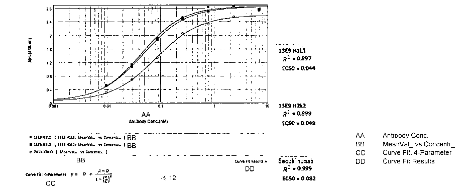
Figure 12: detection of the binding activity of antibodies 13E9 H ILI and 13E9
H2L2 to antigen
IL17A-His with an indirect ELISA method. The marketed drug Secukinumab is used
as a positive
control.
Figure 13: detection of the binding activity of antibody 13E9 H3L2 to antigen
IL17A-His by
indirect ELISA method. The marketed drug Secukinumab is used as a positive
control.
Figure 14: detection of the activity of antibodies 13E9 HIL1 and 13E9 H2L2
competing with
antigen IL17RA-His for binding by competitive ELISA method. The marketed drug
Secukinumab is used as a positive control.
Figure 15: detection of the activity of antibody 13E9 H3L2 competing with
antigen IL17RA-His
for binding by competitive ELISA method.The marketed drug Secukinumab is used
as a positive
control.
Figure 16: detection of the binding activity of antibodies 2G2 H ILI, 2G2 H2L2
and 2G2 H3L3 to
antigen IL17A-His by indirect ELISA method. The marketed drug Secukinumab is
used as a
positive control.
Figure 17: detection of the activity of antibodies 2G2 HIL1, 2G2 H2L2 and 2G2
H3L3
competing with the receptor IL17A-His (biotin) for binding to antigen IL17A-
His by competitive
ELISA method. The marketed drug Secukinumab is used as a positive control.
Figure 18: effect of antibodies 13E9 HIL1, 13E9 H2L2 and 13E9 H3L2 as well as
2G2 HIL1,
2G2 H2L2 and 2G2 H3L3 on the secretion of cytokine IL-6 by mixed lymphocytes.
Figure 19: effect of the antibody drug 13E9 H3L2 on epidermal thickness of the
C57BL/6 mouse
model with psoriasis.
27
Date Recue/Date Received 2020-11-05
CA 03099530 2020-11-05
Notes on the preservation of biological materials:
hybridoma cell line LT006 (IL17A-13E9), which was deposited at China Center
for Type Culture
Collection (CCTCC) on September 12, 2017 with an accession number of CCTCC NO:
C2017102, at Wuhan University, Wuhan, China, 430072.
hybridoma cell line LT007 (IL17A-2G2), which was deposited at China Center for
Type Culture
Collection (CCTCC) on September 12, 2017 with an accession number of CCTCC NO:
C2017165, at Wuhan University, Wuhan, China, 430072.
DETAILED DESCRIPTION
The embodiments of the present invention will be described in detail below
with reference to the
examples. Those skilled in the art will understand that the following examples
are only used to
illustrate the present invention, and should not be regarded as limiting the
scope of the present
invention. An example is performed according to the technologies or conditions
described in the
literature in the art (e.g., see, Guide to Molecular Cloning Experiments,
authored by J. Sambrook
et al., and translated by Huang Peitang et al., third edition, Science Press)
or according to the
product manual if specific technologies or conditions are not specified
therein. Reagents or
instruments used are all commercially available conventional products if the
manufacturers
thereof are not specified.
In the following examples, the BALB/C mice used were purchased from Guangdong
Medical
Laboratory Animal Center; the C57BL/6 mice used were from Nanjing Galaxy
Biopharma Co.,
Ltd.; the MRCS cells used were from Shanghai Fudan IBS Cell Center; the
monoclonal antibody
Secukinumab (Cosentyx0) used was purchased from Novartis Corporation.
28
Date Recue/Date Received 2020-11-05
CA 03099530 2020-11-05
Example 1: Preparation of anti-IL-17A antibodies 13E9 and 2G2
1. Preparation of the hybridoma cell lines LT006 and LT007
Antigen IL17A (24-155)-his used to generate the anti-IL-17A antibody is the
fusion protein of
human IL-17A (GenBank ID: Q16552) mature peptide and the His tag. Spleen cells
from the
immunized BALB/C mice (purchased from Guangdong Medical Laboratory Animal
Center) and
mouse myeloma cells were fused into hybridoma cells, and established methods
(e.g.,
"Monoclonal Antibody Production", in Basic Methods in Antibody Production and
Characterization, Eds. G.C. Howard and D.R. Bethell, Boca Raton: CRC Press,
2000) were
referred to for specific operations.
The fusion protein IL17A-His was enzyme-digested with TEV protease and
purified by column
to obtain IL-17A (24-155) protein. Indirect ELISA screening was performed in
coated
microplates with the IL-17A (24-155) protein as the antigen to obtain
hybridoma cells that
secreted new antibodies specifically bound to IL-17A (24-155).
Hybridoma cells obtained by indirect ELISA screening were screened by
competitive ELISA to
obtain hybridoma cell lines capable of secreting a monoclonal antibody that
competed with the
receptor IL-17RA (CD217, NCBI Gene ID: NP_055154.3) for binding to IL-17A, and
two stable
hybridoma cell lines were obtained by limited dilution.
Hybridoma cell line LT006 (IL17A-13E9) was deposited at China Center for Type
Culture
Collection (CCTCC) on Saturday, September 12, 2015 with an accession number of
CCTCC NO:
C2017102, at Wuhan University, Wuhan, China, 430072. The monoclonal antibody
secreted by
hybridoma cell line LT006 was named as 13E9.
Hybridoma cell line LT007 (IL17A-2G2) was deposited at China Center for Type
Culture
Collection (CCTCC) on September 12, 2017 with an accession number of CCTCC NO:
C2017165, at Wuhan University, Wuhan, China, 430072. The monoclonal antibody
secreted by
hybridoma cell line LT007 was named as 2G2.
29
Date Recue/Date Received 2020-11-05
CA 03099530 2020-11-05
2. Preparation of anti-IL-17A antibody 13E9
The above LT006 cell lines (1x10⁵ cells per well) were cultured in IMDM medium
containing
10% low IgG fetal bovine serum (containing 1% Penicillin-Streptomycin,
cultured in a cell
incubator at 37 C with 5% CO2), and the cell culture supernatant was collected
when the survival
rate was around 20% after 7 days of culturing, which was then subjected to
high-speed
centrifugation, vacuum filtration through a microporous membrane, and
purification through a
HiTrap protein A HP column to obtain antibody 13E9. The purified 13E9 samples
were detected
by SDS-PAGE electrophoresis, and the results are shown in Figure 1.
3. Preparation of anti-IL-17A antibody 2G2
The above LT007 cell lines (1x10⁵ cells per well) were cultured in IMDM medium
containing
10% low IgG fetal bovine serum (containing 1% Penicillin-Streptomycin,
cultured in a cell
incubator at 37 C with 5% CO2), and the cell culture supernatant was collected
when the survival
rate was around 20% after 7 days of culturing, which was then subjected to
high-speed
centrifugation, vacuum filtration through a microporous membrane, and
purification through a
HiTrap protein A HP column to obtain antibody 2G2. The purified 2G2 samples
were detected by
SDS-PAGE electrophoresis, and the results are shown in Figure 2.
Example 2: Sequence analysis of antibody 13E9
LT006 cells were cultured according to the method in step 2 of Example 1.
Using the cell/bacterial total RNA extraction kit (Tiangen, article number
DP430), mRNA was
extracted from the cultured LT006 cells according to the method in the kit
manual.
cDNA was synthesized according to the kit manual of the Invitrogen SuperScript
III First-
Strand Synthesis System for RT-PCR, and amplified by PCR.
The PCR-amplified products were directly TA cloned, and the kit manual of the
pEASY-T1
Cloning Kit (Transgen CT101) was referred to for specific operations.
Date Recue/Date Received 2020-11-05
CA 03099530 2020-11-05
The TA-cloned products were directly sequenced, and the sequencing results are
as follows:
nucleotide sequence of the heavy chain variable region: (360 bp)
GAAGTAAAGCTGCAGGAGTCGGGACCTGGCCTGGTGAAACCTTCTCAGTCTCTGTCCC
TCACCTGCACTGTCACTAGCTACTCATTCACCAGTGATTATGCCTGGAGCTGGATCCG
GCAGTTTCCAGGAATCAAACTGGAGTGGATGGGCTACATAACCTACAGTGGTGTCAC
TAGCTACAACCCCTCTCTCAAAAGTCGAATCTCTATCACTCGAGACACATCCAAGAAC
CAGTTCTTCCTACAGTTGAATTCTGTGACTACTGAGGACACGGCCACATATTACTGTG
CAAGGGCAGACTATGATAGCTACTATACTATGGACTACTGGGGTCAAGGAACCTCAG
TCACCGTCTCCTCA (SEQ ID NO: 1)
its encoded amino acid sequence: (120 aa)
EVKLQESGPGLVKPSQSLSLTCTVTSYSFTSDYAWSWIRQFPGIKLEWMGYITYSGVTSY
NPSLKSRISITRDTSKNQFFLQLNSVTTEDTATYYCARADYDSYYTMDYWGQGTSVTVSS
(SEQ ID NO: 2)
nucleotide sequence of the light chain variable region: (333 bp)
GACATCCAGCTGACTCAGTCTCCACTCTCCCTGCCTGTCAGTCTTGGAGATCAAGCCTC
CATCTCTTGCAGATCTAGTCAGAGCCTTGTACACAGTAATGGAAACACCTATTTACATT
GGTACCTGCAGAAGCCAGGCCAGTCTCCAAGGCTCCTGATCTACAAAGTTTCCAACCG
ATTTTCTGGGGTCCCAGACAGGTTCAGTGGCAGTGGATCAGGGACAGATTTCACACTC
AAGATCAGCAGAGTGGAGGCTGAGGATCTGGGAGTTTATTTCTGCTCTCAAAGTACAC
ATTTTTGGACGTTCGGTGGAGGCACCAAGCTGGAAATAAAA (SEQ ID NO: 3)
its encoded amino acid sequence: (111 aa)
DIQLTQSPLSLPVSLGDQASISCRSSQSLVHSNGNTYLHWYLQKPGQSPRLLIYKVSNRFS
GVPDRF SGSGSGTDFTLKISRVEAEDLGVYFCSQSTHFWTFGGGTKLEIK (SEQ ID NO: 4)
The underlined regions are the CDR regions.
31
Date Recue/Date Received 2020-11-05
CA 03099530 2020-11-05
Example 3: Design, preparation and detection of anti-IL-17A humanized
antibodies 13E9 H1L1,
13E9 H2L2 and 13E9 H3L2
1. Design of the light chain and heavy chain sequences of anti-IL-17A
humanized antibodies
13E9 H1L1, 13E9 H2L2 and 13E9 H3L2
Based on the three-dimensional crystal structure of the IL-17A protein (EMBO
J. (2001) 20 p:
5332-41) and the sequence of the antibody 13E9 obtained in Example 2, the
antibody model was
simulated by computer, and mutations were designed according to the model to
obtain the
variable region sequences of antibodies 13E9 H1L1, 13E9 H2L2 and 13E9 H3L2
(antibody
constant region sequences are from the NCBI database, in which the heavy chain
constant region
is Ig gamma-1 chain C region, ACCESSION: P01857, and the light chain constant
region is Ig
kappa chain C region, ACCESSION: P01834).
The designed variable region sequences are as follows:
(1) heavy chain and light chain sequences of humanized monoclonal antibody
13E9 H1L1
nucleotide sequence of the heavy chain variable region: (360 bp)
GATGTGCAGCTGCAGGAAAGCGGACCAGGACTGGTGAAGCCTAGCCAGACCCTGAGC
CTGACTTGCACCGTGTCCAGCTACAGCTTCACCAGCGACTACGCTTGGTCTTGGATCA
GACAGTTCCCAGGAATTGGCCTCGAGTGGATGGGCTACATCACCTACAGCGGCGTGA
CCAGCTACAACCCCAGCCTGAAGAGCAGGATCACCATCAGCCGGGACACCAGCAAGA
ACCAGTTCTTCCTGCAGCTGAACAGCGTGACAGCAGCCGATACCGCAGTGTACTATTG
CGCCAGGGCCGACTACGACAGCTACTACACCATGGACTATTGGGGCCAGGGAACCAG
CGTGACAGTGTCTAGC (SEQ ID NO: 5)
its encoded amino acid sequence: (120 aa)
DVQLQESGPGLVKPSQTLSLTCTVSSYSFTSDYAWSWIRQPPGKGLEWIGYITYSGVTSYN
PSLKSRITISRDTSKNQFFLQLSSVTAADTAVYYCARADYDSYYTMDYWGQGTSVTVSS
(SEQ ID NO: 6)
32
Date Recue/Date Received 2020-11-05
CA 03099530 2020-11-05
The underlined regions are the CDR regions.
nucleotide sequence of the light chain variable region: (333 bp)
GATGTCGTGATGACCCAGACCCCTCTGTCTCTGCCAGTGACACTGGGACAGCAGGCTA
GCATCTCTTGCAGAAGCAGCCAGAGCCTGGTGCACAGCAACGGCAACACCTACCTGC
ATTGGTACCTGCAGAAGCCAGGCCAGTCTCCTAGACTGCTGATCTACAAGGTGTCCAA
CCGGTTCAGCGGCGTGCCAGATAGATTCAGCGGAAGCGGAAGCGGCACCGACTTCAC
CCTGAAGATCAGCAGAGTGGAGGCCGAGGATCTGGGAGTGTACTTCTGCAGCCAGAG
CACCCACTTTTGGACCTTCGGCGGAGGCACCAAGCTGGAGATCAAG (SEQ ID NO: 7)
its encoded amino acid sequence: (111 aa)
DVVMT QTPL SLPVTL GQ QASI S CRS S Q SLVHSNGNTYLHWYL QKP GQ SPRLL IYKVSNRF
SGVPDRFSGSGSGTDFTLKISRVEAEDLGVYFCSQSTHFWTFGGGTKLEIK (SEQ ID NO:
8)
The underlined regions are the CDR regions.
(2) heavy chain and light chain sequences of humanized monoclonal antibody
13E9 H2L2
nucleotide sequence of the heavy chain variable region: (360 bp)
GATGTGCAGCTGCAGGAAAGCGGACCAGGACTGGTGAAGCCTAGCCAGACCCTGAGC
CTGACTTGCACCGTGTCCAGCTACAGCTTCACCAGCGACTACGCTTGGTCTTGGATCA
GACAGCCACCAGGAAAGGGACTCGAGTGGATCGGCTACATCACCTACAGCGGCGTGA
CCAGCTACAACCCCAGCCTGAAGAGCAGGATCACCATCAGCCGGGACACCAGCAAGA
ACCAGTTCTTCCTGCAGCTGTCTAGCGTGACAGCAGCCGATACCGCAGTGTACTATTG
CGCCAGGGCCGACTACGACAGCTACTACACCATGGACTATTGGGGCCAGGGAACCAG
CGTGACAGTGTCTAGC (SEQ ID NO: 9)
33
Date Recue/Date Received 2020-11-05
CA 03099530 2020-11-05
its encoded amino acid sequence: (120 aa)
DVQLQESGPGLVKPSQTL SLTC TVS SYSFT SDYAW SWIRQPP GKGLEWI GYITYSGVT SYN
PSLKSRITISRDT SKNQFFLQLSSVTAADTAVYYCARADYDSYYTMDYWGQGTSVTVSS
(SEQ ID NO: 10)
The underlined regions are the CDR regions.
nucleotide sequence of the light chain variable region: (333 bp)
GATGTCGTGATGACCCAGACCCCTCTGTCTCTGCCAGTGACACTGGGACAGCCAGCTA
GCATCTCTTGCAGAAGCAGCCAGAGCCTGGTGCACAGCAACGGCAACACCTACCTGC
ATTGGTACCTGCAGAAGCCAGGCCAGTCTCCTAGACTGCTGATCTACAAGGTGTCCAA
CCGGTTCAGCGGCGTGCCAGATAGATTCAGCGGAAGCGGAAGCGGCACCGACTTCAC
CCTGAAGATCAGCAGAGTGGAGGCCGAGGATCTGGGAGTGTACTACTGCAGCCAGAG
CACCCACTTTTGGACCTTCGGCGGAGGCACCAAGCTGGAGATCAAG (SEQ ID NO: 11)
its encoded amino acid sequence: (111 aa)
DVVMTQTPLSLPVTLGQPASISCRSSOSLVHSNGNTYLHWYLQKPGQSPRLLIYKVSNRFS
GVPDRF SGSGSGTDFTLKISRVEAEDLGVYYC SQ STHFWTFGGGTKLEIK (SEQ ID NO:
12)
The underlined regions are the CDR regions.
(3) heavy chain and light chain sequences of humanized monoclonal antibody
13E9 H3L2
nucleotide sequence of the heavy chain variable region: (360 bp)
GATGTGCAGCTGCAGGAAAGCGGACCAGGACTGGTGAAGCCTAGCCAGACCCTGAGC
CTGACTTGCACCGTGTCCAGCTACAGCTTCACCAGCGACTACGCTTGGTCTTGGATCA
GACAGCCACCAGGAAAGGGACTCGAGTGGATCGGCTACATCACCTACAGCGGCGTGA
34
Date Recue/Date Received 2020-11-05
CA 03099530 2020-11-05
CCAGCTACAACCCTAGCCTGAAGAGCCGCGTGACCATTAGCGTGGACACCAGCAAGA
ACCAGTTCTCCCTGAAGCTGAGCAGCGTGACAGCCGCCGATACAGCAGTGTACTATT
GCGCCCGGGCCGATTACGACAGCTACTACACCATGGACTATTGGGGCCAGGGAACCA
GCGTGACAGTGTCTAGC (SEQ ID NO: 13)
its encoded amino acid sequence: (120 aa)
DVQLQESGPGLVKPSQTLSLTCTVSSYSFTSDYAWSWIRQPPGKGLEWIGYITYSGVTSYN
PSLKSRVTISVDTSKNQFSLKLSSVTAADTAVYYCARADYDSYYTMDYWGQGTSVTVSS
(SEQ ID NO: 14)
The underlined regions are the CDR regions.
The nucleotide sequence of the light chain variable region is the same as the
nucleotide sequence
of the light chain variable region of 13E9 H2L2, as shown in SEQ ID NO: 11.
Its encoded amino acid sequence is also the same as the amino acid sequence of
the light chain
variable region of 13E9 H2L2, as shown in SEQ ID NO: 12.
2. Preparation of humanized antibodies 13E9 H1L1, 13E9 H2L2 and 13E9 H3L2
Heavy chain constant regions all use Ig gamma-1 chain C region, ACCESSION:
P01857; the
light chain constant regions use Ig kappa chain C region, ACCESSION: P01834.
Heavy chain cDNA and light chain cDNA of 13E9 Hi Li, heavy chain cDNA and
light chain
cDNA of 13E9 H2L2, and heavy chain cDNA and light chain cDNA of 13E9 H3L2 were
cloned
into pUC57simple (provided by Genscript) vectors, respectively, to obtain
pUC57simple-
13E9H1, pUC57simple-13E9L1, pUC57simple-13E9H2, pUC57simp1e-13E9L2 and
pUC57simple-13E9H3, respectively, and fragments containing corresponding heavy
chains and
fragments containing corresponding light chains were subcloned into pcDNA3.1
vectors,
Date Recue/Date Received 2020-11-05
CA 03099530 2020-11-05
respectively, to obtain recombinant plasmids pcDNA3.1 -13E9H1, pcDNA3.1-
13E9L1,
pcDNA3.1-13E9H2, pcDNA3.1-13E9L2, pcDNA3.1-13E9H3 and pcDNA3.1-13E9L2. Then,
the
corresponding light chain recombinant plasmids and heavy chain recombinant
plasmids
(pcDNA3.1-13E9H1 and pcDNA3.1-13E9L1; pcDNA3.1-13E9H2 and pcDNA3.1-13E9L2;
pcDNA3.1-13E9H3 and pcDNA3.1-13E9L2) were co-transfected into 293F cells, the
cell culture
was collected and purified to obtain humanized antibodies 13E9 H1L1, 13E9
H2L2, and 13E9
H3L2 respectively. The purified 13E9 H3L2 sample was detected by SDS-PAGE
electrophoresis,
and the results are shown in Figure 3.
Example 4: Sequence analysis of antibody 2G2
LT007 cells were cultured according to the method in step 3 of Example 1.
Using the cell/bacterial total RNA extraction kit (Tiangen, article number
DP430), mRNA was
extracted from the cultured LT007 cells according to the method in the kit
manual.
cDNA was synthesized according to the kit manual of the Invitrogen SuperScript
III First-
Strand Synthesis System for RT-PCR, and amplified by PCR.
The PCR-amplified products were directly TA cloned, and the kit manual of the
pEASY-T1
Cloning Kit (Transgen CT101) was referred to for specific operations.
The TA-cloned products were directly sequenced, and the sequencing results are
as follows:
nucleotide sequence of the heavy chain variable region: (351 bp)
GAGGTTCAGCTGGAGCAGTCTGGTTCTGAACTGAGGAGTCCTGGATCTTCAGTAAAG
CTTTCATGCAAGGATTTTGATTCAGAAGTCTTCCCTATTGCTGATATGAGTTGGGTTAG
GCAGAAGCCTGGGCATGGATTTGAATGGATTGGAGACATACTCCCAAGTTTTGGTAG
AACAATCTATGGAGAGAAGTTTGAGGACAAAGCCAAAGTGGATGCAGACACAGTGTC
CAACACAGCCTACTTGGAACTCAACAGTCTGACATCTGAGGACTCTGCTATCTACTAC
36
Date Recue/Date Received 2020-11-05
CA 03099530 2020-11-05
TGTGCAAGGGGTAACTACGGGTTTGCTTACTGGGGCCAAGGGACTCTGGTCACTGTCT
CTGCA (SEQ ID NO: 15)
its encoded amino acid sequence: (117 aa)
EVQLEQSGSELRSPGSSVKLSCKDFDSEVFPIADMSWVRQKPGHGFEWIGDILPSFGRTIY
GEKFEDKAKVDADTVSNTAYLELNSLTSEDSAIYYCARGNYGFAYWGQGTLVTVSA
(SEQ ID NO: 16)
The underlined regions are the CDR regions.
nucleotide sequence of the light chain variable region: (336 bp)
GATGTTTTGATGACCCAAACTCCACTCACTTTGTCGGTTATCATTGGACAACCAGCCT
CCATCTCTTGCAAGCCAAGTCAGAGCCTCTTAAATAGTGATGGAAAGACATATTTGAA
TTGGTTGTTGCAGAGGCCAGGCCAGTCTCCAAAGCGCCTAATCTATCTGGTGTCTAAA
CTGGACTCTGGAGTCCCTGACAGGTTCACTGGCAGTGGATCAGGGACAGATTTCACA
CTGAAAATCAGCAGAGTGGAGGCTGAGGATTTGGGAGTTTATTATTGCTGGCAAGGT
TCACATTTTCCTCAGACGTTCGGTGGAGGCACAAAGTTGGAAATAAAA (SEQ ID NO:
17)
its encoded amino acid sequence: (112 aa)
DVLMTQTPLTLSVIIGQPASISCKPSQSLLNSDGKTYLNWLLQRPGQSPKRLIYLVSKLDSG
VPDRFTGSGSGTDFTLKISRVEAEDLGVYYCWQGSHFPQTFGGGTKLEIK (SEQ ID NO:
18)
The underlined regions are the CDR regions.
Example 5: Design, preparation and detection of anti-IL-17A humanized
antibodies 2G2 H1L1,
2G2 H2L2 and 2G2 H3L3
37
Date Recue/Date Received 2020-11-05
CA 03099530 2020-11-05
(1) heavy chain and light chain sequences of humanized monoclonal antibody 2G2
H1L1
nucleotide sequence of the heavy chain variable region: (348 bp)
GTGCAGCTGGTGCAGAGCGGAAGCGAACTGAGAAAGCCAGGCTCCAGCGTGAAGCT
GTCTTGCAAGGACTTCGACAGCGAGGTGTTCCCCATCGCCGATATGTCTTGGGTCCGA
CAGGCTCCAGGCCAGGGATTCGAGTGGATCGGTGACATTCTGCCCAGCTTCGGAAGA
ACCAACTACGCCCAGAAGTTCGAGGGCAAGGCCAAGGTGGACGCAGACAAGAGCAC
CAACACCGCCTACCTGGAGCTGAACAGCCTGAGAAGCGAGGACACCGCCATCTACTA
TTGCGCCAGGGGCAACTACGGATTCGCCTATTGGGGCCAGGGAACACTGGTGACAGT
GTCCGCC (SEQ ID NO: 19)
its encoded amino acid sequence: (116 aa)
VQLVQ SGSELRKP GS SVKL SCKDFD SEVFPIADMSWVRQAP GQGFEWIGDILP SF GRTNY
AQKFEGKAKVDADKSTNTAYLELNSLRSEDTAIYYCARGNYGFAYWGQGTLVTVSA
(SEQ ID NO: 20)
The underlined regions are the CDR regions.
nucleotide sequence of the light chain variable region: (336 bp)
GATGTCGTGATGACCCAGACCCCTCTGTCTCTGAGCGTGACACTGGGACAGCCAGCTA
GCATCAGCTGCAGAAGCAGCCAGAGCCTGCTGAACAGCGACGGCAAGACCTACCTGA
ATTGGCTGCTGCAGAGACCAGGCCAGTCTCCTAGAAGGCTGATCTACCTGGTGTCCAA
GCTGGACAGCGGCGTGCCAGATAGATTCAGCGGAAGCGGAAGCGGCACCGACTTCAC
CCTGAAGATCAGCAGAGTGGAGGCCGAGGATCTGGGAGTGTACTACTGTTGGCAGGG
CAGCCACTTCCCTCAGACATTCGGCGGCGGCACAAAGCTGGAGATCAAG (SEQ ID NO:
21)
its encoded amino acid sequence: (112 aa)
38
Date Recue/Date Received 2020-11-05
CA 03099530 2020-11-05
DVVMTQTPLSL SVTL GQPA SI S CRS S Q SLLNSDGKTYLNWLL QRP GQ SPRRLIYLVSKLDS
GVPDRFSGSGSGTDFTLKISRVEAEDLGVYYCWQGSHFPQTFGGGTKLEIK (SEQ ID NO:
22)
The underlined regions are the CDR regions.
(2) heavy chain and light chain sequences of humanized monoclonal antibody 2G2
H2L2
nucleotide sequence of the heavy chain variable region: (348 bp)
GTGCAGCTGGTGCAGAGCGGAGCAGAAGTGAAGAAGCCAGGCTCCAGCGTGAAGCT
GTCTTGCAAGGACTTCGACAGCGAGGTGTTCCCCATCGCCGATATGTCTTGGGTCCGA
CAGGCTCCAGGCCAGGGATTCGAGTGGATCGGTGACATTCTGCCCAGCTTCGGGAGA
ACCAATTACGCCCAGAAGTTCCAGGGCAGAGTGACCGTGACCGCAGACAAGAGCACC
AACACCGCCTACCTGGAGCTGAACAGCCTGAGGAGCGAGGATACCGCCGTGTACTAT
TGCGCCAGGGGCAACTACGGCTTCGCCTATTGGGGACAGGGAACACTGGTGACAGTG
TCCGCC (SEQ ID NO: 23)
its encoded amino acid sequence: (116 aa)
VQLVQ SGAEVKKP GS SVKL S CKDFD SEVFPIADMSWVRQAP GQGFEWIGDILP SF GRTNY
AQKFQGRVTVTADKSTNTAYLELNSLRSEDTAVYYCARGNYGFAYWGQGTLVTVSA
(SEQ ID NO: 24)
The underlined regions are the CDR regions.
nucleotide sequence of the light chain variable region: (336 bp)
GATGTCGTGATGACCCAGACCCCTCTGTCTCTGAGCGTGACACTGGGACAGCCAGCTA
GCATCAGCTGCAGAAGCAGCCAGAGCCTGCTGAACAGCGACGGCAAGACCTACCTGA
ATTGGCTGCTGCAGAGACCAGGCCAGTCTCCTAGAAGGCTGATCTACCTGGTGTCCAA
39
Date Recue/Date Received 2020-11-05
CA 03099530 2020-11-05
CCTGGACAGCGGCGTGCCAGATAGATTCAGCGGAAGCGGAAGCGGCACCGACTTCAC
CCTGAAGATCAGCAGAGTGGAAGCCGAGGACGTGGGAGTGTACTACTGTTGGCAGGG
CAGCCACTTCCCTCAGACATTCGGCGGCGGCACAAAGCTGGAGATCAAG (SEQ ID NO:
25)
its encoded amino acid sequence: (112 aa)
DVVMTQTPLSL SVTL GQPA SI SCRS SO SLLNSDGKTYLNWLL QRP GQ SPRRLIYLVSNLDS
GVPDRFSGSGSGTDFTLKISRVEAEDVGVYYCWQGSHFPQTFGGGTKLEIK (SEQ ID NO:
26)
The underlined regions are the CDR regions.
(3) heavy chain and light chain sequences of humanized monoclonal antibody 2G2
H3L3
nucleotide sequence of the heavy chain variable region: (348 bp)
GTGCAGCTGGTGCAGAGCGGAGCAGAAGTGAAGAAGCCAGGCAGCAGCGTGAAGGT
GTCTTGCAAGGACTTCAGCAGCGAGGTGTTCCCCATCGCCGATATGTCTTGGGTCCGA
CAGGCTCCAGGCCAGGGACTGGAGTGGATCGGTGACATTCTGCCCAGCTTCGGGAGA
ACCAATTACGCCCAGAAGTTCCAGGGCAGAGTGACCGTGACCGCAGACAAGAGCACC
AACACCGCCTACCTGGAGCTGTCTAGCCTGAGAAGCGAGGACACCGCCGTGTACTAT
TGCGCCAGGGGCAACTACGGCTTCGCCTATTGGGGACAGGGAACACTGGTGACAGTG
TCCGCC (SEQ ID NO: 27)
its encoded amino acid sequence: (116 aa)
VQLVQSGAEVKKPGSSVKVSCKDFSSEVFPIADMSWVRQAPGQGLEWIGDILPSFGRTNY
AQKFQGRVTVTADKSTNTAYLELSSLRSEDTAVYYCARGNYGFAYWGQGTLVTVSA
(SEQ ID NO: 28)
The underlined regions are the CDR regions.
Date Recue/Date Received 2020-11-05
CA 03099530 2020-11-05
nucleotide sequence of the light chain variable region: (336 bp)
GATGTCGTGATGACCCAGACCCCTCTGTCTCTGAGCGTGACACTGGGACAGCCAGCTA
GCATCAGCTGCAGAAGCAGCCAGAGCCTGCTGAACAGCGACGGCAAGACCTACCTGA
ATTGGTTCCTGCAGAGACCAGGCCAGTCTCCTAGAAGGCTGATCTACCTGGTGTCCAA
CCTGGACAGCGGCGTGCCAGATAGATTCAGCGGAAGCGGAAGCGGCACCGACTTCAC
CCTGAAGATCAGCAGAGTGGAAGCCGAGGACGTGGGAGTGTACTACTGTTGGCAGGG
CAGCCACTTCCCTCAGACATTCGGCGGCGGCACAAAGCTGGAGATCAAG (SEQ ID NO:
29)
its encoded amino acid sequence: (112 aa)
DVVMTQTPLSL SVTL GQPA SI SCRS S Q SLLNSDGKTYLNWFL QRP GQ SPRRLIYLVSNLD S
GVPDRFSGSGSGTDFTLKISRVEAEDVGVYYCWQGSHFPQTFGGGTKLEIK (SEQ ID NO:
30)
The underlined regions are the CDR regions.
2. Preparation and SDS-PAGE electrophoresis detection of humanized antibodies
2G2 H1L1,
2G2 H2L2 and 2G2 H3L3
Heavy chain constant regions used Ig gamma-1 chain C region, ACCESSION:
P01857; the light
chain constant regions used Ig kappa chain C region, ACCESSION: P01834.
Heavy chain cDNA and light chain cDNA of 2G2 Hi Li, heavy chain cDNA and light
chain
cDNA of 2G2 H2L2, and heavy chain cDNA and light chain cDNA of 2G2 H3L3 were
cloned
into pUC57simple (provided by Genscript) vectors, respectively, to obtain
pUC57simple-2G2H1,
pUC57simp1e-2G2L1; pUC57simple-2G2H2, pUC57simp1e-2G2L2; and pUC57simp1e-
2G2H3,
pUC57simple-2G2L3, respectively. Nucleotide fragments containing corresponding
heavy chains
and nucleotide fragments containing corresponding light chains were then
subcloned into
pcDNA3.1 vectors, respectively, to obtain recombinant plasmids pcDNA3.1-2G2H1,
pcDNA3.1-
41
Date Recue/Date Received 2020-11-05
CA 03099530 2020-11-05
2G2L1, pcDNA3.1-2G2H2, pcDNA3.1-2G2L2, pcDNA3.1-2G2H3 and pcDNA3.1-2G2L3.
Then, the corresponding light chain recombinant plasmids and heavy chain
recombinant plasmids
(pcDNA3.1-2G2H1 and pcDNA3.1-2G2L1; pcDNA3.1-2G2H2 and pcDNA3.1-2G2L2;
pcDNA3.1-2G2H3 and pcDNA3.1-2G2L3) were co-transfected into 293F cells, the
cell culture
was collected and purified to obtain humanized antibodies 2G2 H1L1, 2G2 H2L2
and 2G2 H3L3
respectively. The purified samples were detected by SDS-PAGE electrophoresis.
Example 6: Measurement of kinetic parameters for the binding of 13E9 HIL1,
13E9 H2L2 and
13E9 H3L2 to the antigen IL-17A (24-155) protein
The kinetic parameters for the binding of humanized antibodies 13E9 H1L1, 13E9
H2L2 and
13E9 H3L2 to the antigen IL-17A (24-155) were measured using a Fortebio
molecular interaction
instrument.
The AR2G sensor was activated by EDC/NHS, and the antibody was fixed to the
activated AR2G
sensor by amine coupling, and the sensor was blocked with 1 M ethanolamine (pH
8.5). After the
sensor was equilibrated in PBST for 300 s, the antibody fixed on the sensor
binded to the antigen
IL-17A (24-155) protein (same as the antigen used in Example 1), in which the
antigen
concentration was 6.25-400 nM (double gradient dilution) and the binding time
was 420 s, and
the antigen and the antibody were dissociated in PBST for 600 s.
The kinetic parameters of antibodies 13E9 HIL1, 13E9 H2L2, 13E9 H3L2 and
Secukinumab are
shown in Table 1, and detection results of the kinetic characteristic
parameters are shown in
Figure 4, Figure 5, Figure 6 and Figure 7, respectively.
Table 1: the kinetic parameters of 13E9 humanized antibody
Antibody KD (M) Kon (1/Ms) Kon error Kdis (1/s) Kdis error Rmax (nm)
13E9 HIL1 9.72E-10 1.34E+05 2.60E+03 1.31E-04 9.06E-
06 0.0858-0.3326
13E9 H2L2 1.03E-09 9.86E+04 1.51E+03 1.01E-04 7.57E-
06 0.0546-0.3049
42
Date Recue/Date Received 2020-11-05
CA 03099530 2020-11-05
13E9 H3L2 5.08E-10 2.90E+05 8.22E+03 1.47E-04
1.02E-05 0.1457-0.3546
Secukinumab 6.74E-10 9.28E+04 3.03E+03 6.26E-05 1.57E-05 0.0379-0.2064
KD is affinity constant; Kon is binding rate of antigen and antibody; Kdis is
dissociation rate of
antigen and antibody; KD = Kdis/Kon.
The results show that 13E9 H ILI, 13E9 H2L2, and 13E9 H3L2 all have good
affinity to the
antigen IL-17A (24-155), and the affinity is equivalent to that of the control
antibody
Secukinumab.
Example 7: Measurement of kinetic parameters for the binding of 2G2 H1L I, 2G2
H2L2, and
2G2 H3L3 to the antigen IL-17A (24-155)
The kinetic parameters for the binding of humanized antibodies 2G2 H ILI, 2G2
H2L2 and 2G2
H3L3 to the antigen IL-17A (24-155) were measured using a Fortebio molecular
interaction
instrument.
The AR2G sensor was activated by EDC/NHS, and the antibody was fixed to the
activated AR2G
sensor by amine coupling, and the sensor was blocked with 1 M ethanolamine (pH
8.5). After the
sensor is equilibrated in PBST for 300 s, the antibody fixed on the sensor
bound to the antigen
IL-17A (24-155), in which the antigen concentration was 6.25-400 nM (double
gradient dilution)
and the binding time was 420 s, and the antigen and the antibody were
dissociated in PBST for
600 s.
The kinetic parameters of antibodies 2G2 H ILI, 2G2 H2L2, 2G2 H3L3, and
Secukinumab are
shown in Table 2, and detection results of the kinetic characteristic
parameters are shown in
Figure 8, Figure 9, Figure 10, and Figure 11, respectively.
Table 2: the kinetic parameters of 2G2 H1L1, 2G2 H2L2, 2G2 H3L3 and
Secukinumab
43
Date Recue/Date Received 2020-11-05
CA 03099530 2020-11-05
Antibody KD (M) Kon(I/Ms) Kon error Kdis (Us) Kdis error Rmax(nm)
2G2 HIL1 1.53E-09 1.46E+05 3.15E+03 2.24E-04 9.67E-
06 0.0035-0.2192
2G2 H2L2 8.43E-10 1.42E+05 2.18E+03 1.20E-04 6.73E-
06 0.0444-0.2732
2G2 H3L3 1.54E-09 1.98E+05 5.30E+03 3.06E-04 1.08E-
05 0.011-0.2109
Secukinumab 1.08E-09 1.33E+05 2.38E+03 1.43E-04 8.09E-06 0.0118-0.2584
KD is affinity constant; Kon is binding rate of antigen and antibody; Kdis is
dissociation rate of
antigen and antibody; KD = Kdis/Kon.
The results show that compared with the control antibody Secukinumab, 2G2 H2L2
has a higher
affinity to the antigen IL-17A (24-155); the affinity of 2G2 HIL1 and 2G2 H3L3
is equivalent to
that of the control antibody Secukinumab.
Example 8: Detection of the binding activity of the antibodies 13E9 HIL1, 13E9
H2L2 and 13E9
H3L2 to the antigen with an ELISA method
1. The binding activity of the antibodies 13E9 H ILI, 13E9 H2L2 and 13E9 H3L2
to the antigen
IL17A-His was detected with an indirect ELISA and compared with the marketed
drug
Secukinumab for the same target.
IL17A-His can be prepared by referring to the published sequences and
conventional technical
means in the art, or referring to the following steps:
Preparation of IL17A-His: the full-length protein sequence of human IL-17A was
found in the
NCBI protein database, and fused with His*6 purification tag. Genscript in
Nanjing was entrusted
to synthesize the nucleic acid encoding the fusion protein, and by referring
to the standard
technologies introduced in the Guide to Molecular Cloning Experiments (Second
Edition) and
using standard molecular cloning technologies such as PCR, enzyme digestion,
gel recovery,
ligation transformation, colony PCR or enzyme digestion identification, the
target gene was
subcloned into mammalian cell expression vectors, and the target gene with the
recombinant
44
Date Recue/Date Received 2020-11-05
CA 03099530 2020-11-05
expression vectors was further sequenced and analyzed. After the sequence was
verified to be
correct, a medium and large amount of endotoxin-free expression plasmids were
prepared, and
transiently transfected HEI(293 cells for protein expression. After 7 days of
culture, the cell
culture fluid was collected and affinity purified using a Ni Sepharose column
(GE), and the
quality of the resulting protein samples was determined using SDS-PAGE and SEC-
HPLC
standard analysis techniques to be up to standard.
ELISA: IL17A-His was added to the microplate and incubated at 4 C overnight;
after blocking
with 1% BSA in PBS at 37 C for 2 h, antibodies were added respectively, and
incubated at 37 C
for 30 min; and Goat Anti Human IgG (H + L)-HRP (Jackson, 109-035-088) was
added and
incubated at 37 C for 30 min; and then the color reaction was performed with
TMB (Neogen,
308177) for 5 min, and the absorbance at 450 nm was detected in a microplate
reader. The
obtained experimental data were analyzed and processed with SoftMax Pro 6.2.1
software, and
the 4-parameter fitted curve was plotted for analysis with the antibody
concentration as the
abscissa and the absorbance value as the ordinate.
The experimental results are shown in Figure 12, Figure 13, and Table 3 and
Table 4 below.
Table 3: detection results of the binding activity of 13E9 H ILI and 13E9 H2L2
to the antigen
IL17A-His
Antibody Coating antigen: IL17A-His, 1 pg/mL
concentration/
13E9 HILI 13E9 H2L2 Secukinumab
gradient
1 pg/mL 2.726 2.731 2.804 2.737 2.184 2.227
1:3 2.875 2.852 2.832 2.873 2.595 2.505
1:9 2.858 2.815 2.717 2.712 2.297 2.364
1:27 2.564 2.494 2.479 2.481 2.049 2.064
1:81 1.934 1.925 1.891 1.834 1.372 1.314
1:243 1.159 1.116 1.097 1.062 0.672 0.697
1:729 0.522 0.537 0.514 0.511 0.313 0.309
Date Recue/Date Received 2020-11-05
CA 03099530 2020-11-05
0 0.047 1 0.048 0.048 1 0.048 0.046 1 0.046
EC50 (nM) 0.044 0.048 0.082
Table 4: detection results of the binding activity of 13E9 H3L2 to the antigen
IL17A-His
Antibody Coating antigen: IL17A-His: 1 1.1,g/mL
concentration/
13E9 H3L2 Secukinumab
gradient
0.5 p,g/mL 3.074 3.068 2.929 2.929
1:3 3.175 3.026 2.924 2.885
1:9 2.944 2.895 2.660 2.666
1:27 2.472 2.393 1.947 1.862
1:81 1.610 1.617 1.140 1.092
1:243 0.786 0.783 0.521 0.522
1:729 0.343 0.348 0.252 0.253
0 0.091 0.084 0.087 0.087
EC50 (nM) 0.043 0.078
The experimental results show that the antibodies 13E9 H1L1, 13E9 H2L2 and
13E9 H3L2 all
can effectively bind to the antigen IL17A-His, and their binding efficiency is
dose-dependent.
Under the same experimental conditions, the binding EC50 of 13E9 H1L1 is 0.044
nM, the
binding EC50 of 13E9 H2L2 is 0.048 nM, and the EC50 of the marketed drug
Secukinumab for the
same target is 0.082 nM (Figure 12, Table 3); under the same experimental
conditions, the
binding EC50 of 13E9 H3L2 is 0.043 nM, and the EC50 of the marketed drug
Secukinumab for the
same target is 0.078 nM (Figure 13, Table 4).
The above experimental results show that under the same experimental
conditions, the EC50
values of 13E9 H1L1, 13E9 H2L2 and 13E9 H3L2 are all smaller than those of the
positive
control drug Secukinumab for the same target, indicating that the binding
activity of 13E9 Hi Li,
13E9 H2L2 and 13E9 H3L2 to IL17A-His is better than that of the marketed
control drug
Secukinumab for the same target.
46
Date Recue/Date Received 2020-11-05
CA 03099530 2020-11-05
2. The activity of antibodies 13E9 H ILI, 13E9 H2L2 and 13E9 H3L2 blocking the
binding of
IL17RA-His (biotin) to the antigen IL17A-His is detected with a competitive
ELISA.
IL17RA-His (biotin) can be prepared by referring to the published sequences
and conventional
technical means in the art, or referring to the following steps:
Preparation of IL17RA-His (biotin): the extracellular domain sequence of human
IL-17RA was
found in the NCBI protein database, and fused with His*6 purification tag.
Genscript in Nanjing
was entrusted to synthesize the nucleic acid encoding the fusion protein, and
by referring to the
standard technologies introduced in the Guide to Molecular Cloning Experiments
(Second
Edition) and using standard molecular cloning technologies such as PCR, enzyme
digestion, gel
recovery, ligation transformation, colony PCR or enzyme digestion
identification, the target gene
was subcloned into mammalian cell expression vectors, and the target gene with
the recombinant
expression vectors was further sequenced and analyzed. After the sequence was
verified to be
correct, a medium and large amount of endotoxin-free expression plasmids were
prepared, and
transiently transfected HEK293 cells for protein expression. After 7 days of
culture, the cell
culture was collected and affinity purified using a Ni Sepharose column (GE),
and the quality of
the resulting protein samples was determined using SDS-PAGE and SEC-HPLC
standard
analysis techniques to be up to standard. After the quality determination was
complete, the
biotinylated human IL-17RA-His protein samples were labeled and obtained with
the commercial
kit EZ-Link Sulfo-NHS-LC-Biotinylation of Thermo scientific, and the specific
preparation
method was performed according to the manual of the kit.
ELISA: IL 17A-His was added to the microplate and incubated at 4 C overnight;
after blocking
with 1% BSA in PBST at 37 C for 2 h, antibodies are added respectively, and
the antigen and the
antibody reacted at room temperature for 10 min; then the receptor IL17RA-His
(biotin) was
added which was mixed well with the antibody at a volume ration of 1:1 and
incubated at 37 C
for 30 min; and SA-HRP (KPL, 14-30-00) was added and incubated at 37 C for 30
min; and then
47
Date Recue/Date Received 2020-11-05
CA 03099530 2020-11-05
the color reaction was performed with TMB (Neogen, 308177) for 5 min, and the
absorbance at
450 nm was detected in a microplate reader. The obtained experimental data
were analyzed and
processed with SoftMax Pro 6.2.1 software, and the 4-parameter fitted curve
was plotted for
analysis with the antibody concentration as the abscissa and the absorbance
value as the ordinate.
The experimental results are shown in Figure 14, Figure 15, and Table 5 and
Table 6 below.
Table 5: detection results of the activity of 13E9 H IL land 13E9 H2L2
competing with the
receptor IL17RA-His (biotin) for binding to the antigen IL17A-His
Antibody Antigen coating: IL17A-His (20150213) 0.5 pg/mL
concentration/gr
adient 13E9 HIL1 13E9 H2L2 Secukinumab
pg/mL 0.273 0.249 0.274 0.290 0.402 0.392
1:3 0.299 0.220 0.313 0.339 0.469
0.502
1:9 0.418 0.379 0.431 0.379 0.642
0.708
1:27 0.673 0.610 0.646 0.636 0.842
0.855
1:81 0.986 1.008 1.011 1.069 1.169
1.201
1:243 1.342 1.362 1.428 1.331 1.418
1.468
1:729 1.363 1.375 1.447 1.519 1.467
1.657
0 1.495 1.406 1.429 1.561 1.610
1.495
Receptor IL17RA-His(bio) 0.1 pg/mL
EC50(nM) 1.437 1.281 1.807
48
Date Recue/Date Received 2020-11-05
CA 03099530 2020-11-05
Table 6: detection results of the activity of 13E9 H3L2 competing with the
receptor IL17RA-His
(biotin) for binding to the antigen IL17A-His
Antibody Antigen coating: IL17A-His 0.5 ug/mL
concentration/
13E9 H3L2 Secukinumab
gradient
10ug/mL 0.193 0.182 0.263 0.282
1:3 0.231 0.206 0.318 0.327
1:9 0.265 0.267 0.419 0.462
1:27 0.424 0.415 0.630 0.663
1:81 0.725 0.627 0.813 0.859
1:243 1.069 1.018 1.120 1.182
1:729 1.218 1.095 1.133 1.250
0 1.184 1.334 1.182 1.232
Receptor IL17RA-His(bio) 0.1 p,g/mL
EC50 (nM) 0.805 1.580
The experimental results show that the antibodies 13E9 HIL1, 13E9 H2L2 and
13E9 H3L2 all
can effectively block the binding of the receptor IL17RA-His (biotin) to the
antigen IL17A-His,
and the blocking efficiency is dose-dependent. Under the same experimental
conditions, the EC50
of 13E9 H ILI competing with IL17AR-His (biotin) for binding to IL17A-His is
1.437 nM, the
EC50 of 13E9 H2L2 competing with IL17AR-His (biotin) for binding to IL17A-His
is 1.281 nM,
and the EC50 of the positive control drug Secukinumab for the same target
competing with
IL17AR-His (biotin) for binding to IL17A-His is 1.807 nM (Table 5, Figure 14);
the EC50 of
13E9 H3L2 competing with IL17AR-His (biotin) for binding to IL17A-His is 0.805
nM, and the
EC50 of the marketed drug Secukinumab for the same target competing with IL
17AR-His (biotin)
for binding to IL17A-His is 1.580 nM (Table 6, Figure 15).
The above experimental results show that under the same experimental
conditions, the EC50
values of 13E9 H ILI, 13E9 H2L2 and 13E9 H3L2 competing with IL17AR-His
(biotin) for
49
Date Recue/Date Received 2020-11-05
CA 03099530 2020-11-05
binding to IL17A-His are all smaller than those of the marketed control drug
Secukinumab for
the same target, indicating the activity of 13E9 H ILI, 13E9 H2L2 and 13E9
H3L2 competing
with IL17AR-His (biotin) for binding to IL17A-His is better than that of the
marketed drug
Secukinumab for the same target.
Example 9: Detection of the binding activity of the antibodies 2G2 HIL1, 2G2
H2L2 and 2G2
H3L3 to the antigen with an ELISA method
1. The binding activity of the antibodies 2G2 H ILI, 2G2 H2L2 and 2G2 H3L3 to
the antigen
IL17A-His is detected with an indirect ELISA.
Experimental steps: IL17A-His was added to the microplate and incubated at 4
C overnight;
after blocking with 1% BSA in PBS at 37 C for 2 h, antibodies were added
respectively, and
incubated at 37 C for 30 min; and Goat Anti Human IgG (H + L)-HRP (Jackson,
109-035-088)
was added and incubated at 37 C for 30 min; and then the color reaction was
performed with
TMB (Neogen, 308177) for 5 min, and the absorbance at 450 nm was detected in a
microplate
reader. The obtained experimental data were analyzed and processed with
SoftMax Pro 6.2.1
software, and the 4-parameter fitted curve was plotted for analysis with the
antibody
concentration as the abscissa and the absorbance value as the ordinate.
The experimental results are shown in Figure 16 and Table 7 below. Wherein,
the binding ECso
of 2G2 H ILI is 0.177 nM, the binding EC50 of 2G2 H2L2 is 0.372 nM, and the
binding 50 of 2G2
H3L3 is 0.421 nM.
Table 7: detection results of the binding of 2G2 H ILI, 2G2 H2L2 and 2G2 H3L3
to the antigen
IL 17A-His
Date Recue/Date Received 2020-11-05
CA 03099530 2020-11-05
Antibody Antigen coating: IL17A-His, 1 pg/mL
concentration/gr
2G2 H1L1 2G2 H2L2 2G2 H3L3
Secukinumab
adient
1 pg/mL 3.220
3.135 3.030 2.960 2.485 2.384 3.448 3.448
1:3 3.284
3.149 2.899 2.886 2.311 2.314 3.477 3.478
1:9 2.820
2.747 2.199 2.126 1.693 1.703 3.320 3.331
1:27 2.020
1.969 1.348 1.328 0.982 1.002 2.768 2.764
1:81 1.113
1.075 0.638 0.624 0.439 0.440 1.761 1.689
1:243 0.469
0.426 0.261 0.350 0.188 0.186 0.807 0.806
1:729 0.224
0.188 0.131 0.148 0.113 0.102 0.337 0.326
0 0.061
0.057 0.058 0.068 0.063 0.060 0.062 0.060
EC50(nM) 0.177 0.372 0.421 0.090
The experimental results show that the antibodies 2G2 H1L1, 2G2 H2L2 and 2G2
H3L3 all can
effectively bind to the antigen IL17A-His, and their binding efficiency is
dose-dependent.
2. The activity of antibodies 2G2 H1L1, 2G2 H2L2 and 2G2 H3L3 competing with
the receptor
IL17A-His (biotin) for binding to the antigen IL17A-His was detected by
competitive ELISA.
Experimental steps: IL17A-His was added to the microplate and incubated at 4
C overnight;
after blocking with 1% BSA in PBST at 37 C for 2 h, antibodies were added
respectively, and
the antigen and the antibody reacted at room temperature for 10 min; then the
receptor IL17RA-
His (biotin) was added which is mixed well with the antibody at a volume
ration of 1:1 and
incubated at 37 C for 30 min; and SA-HRP (KPL, 14-30-00) was added and
incubated at 37 C
for 30 min; and then the color reaction was performed with TMB (Neogen,
308177) for 5 min,
and the absorbance at 450 nm was detected in a microplate reader. The obtained
experimental
data were analyzed and processed with SoftMax Pro 6.2.1 software, and the 4-
parameter fitted
51
Date Recue/Date Received 2020-11-05
CA 03099530 2020-11-05
curve was plotted for analysis with the antibody concentration as the abscissa
and the absorbance
value as the ordinate.
The experimental results are shown in Figure 17 and Table 8 below. Wherein,
the blocking EC50
of 2G2 H ILI is 2.264 nM, the blocking EC50 of 2G2 H2L2 is 5.408 nM, and the
blocking EC50 of
2G2 H3L3 is 5.911 nM (Figure 17, Table 8).
Table 8: detection results of the activity of 2G2 H ILI, 2G2 H2L2 and 2G2 H3L3
competing with
the receptor IL17RA-His (biotin) for binding to the antigen IL17A-His
Antibody Antigen coating: IL17A-His 0.5 p,g/mL
concentration/
2G2 HIL1 2G2 H2L2 2G2 H3L3
Secukinumab
gradient
p,g/mL 0.285 0.272 0.337 0.366 0.680 0.718 0.247
0.267
1:3 0.417 0.485 0.500 0.623 0.971 0.876
0.333 0.361
1:9 0.506 0.502 0.785 0.751 0.971 0.954
0.478 0.507
1:27 0.891 0.763 1.087 1.038 1.242 1.178
0.758 0.784
1:81 1.162 1.167 1.286 1.309 1.246 1.312
1.088 1.135
1:243 1.231 1.281 1.423 1.402 1.398 1.466
1.272 1.184
1:729 1.453 1.472 1.464 1.596 1.607 1.466
1.392 1.324
0 1.455 1.425 1.494 1.424 1.470 1.340
1.170 1.230
Receptor IL17RA-His (biotin) :0.1 p,g/mL
EC50(nM) 2.264 5.408 5.911 2.749
The experimental results show that the antibodies 2G2 HIL1, 2G2 H2L2, and 2G2
H3L3 all can
effectively block the binding of the receptor IL17RA-His (biotin) to the
antigen IL17A-His, and
the blocking efficiency is dose-dependent.
52
Date Recue/Date Received 2020-11-05
CA 03099530 2020-11-05
Example 10: Mixed human embryonic fibroblast reaction: secretion of cytokine
IL-6
MRC 5 (purchased from the IBS Cell Center, Fudan University) cells were plated
into a 96-well
plate with 5000 cells/well and cultured overnight. A mixture of IL-17 and
antibody (hIgG as a
control) incubated at 37 C for 20 min was added to the MRC 5 cells and
cultured for 48 h. After
48 h of culturing, the cell supernatant was collected, and the amount of IL-6
secreted was
detected by an ELISA kit (purchased from Dakewe Corporation).
MRC 5 cells were mixed and cultured with 13E9 H ILI, 13E9 H2L2, 13E9 H3L2, 2G2
H ILI,
2G2 H2L2 or 2G2 H3L3 (1 nM, 10 nM, 100 nM) and Secukinumab (1 nM, 10 nM, 100
nM),
respectively, and the detection results of secreted IL-6 are shown in Figure
18.
It can be seen from Figure 18 that 13E9 H ILI, 13E9 H2L2, 13E9 H3L2, and 2G2
H2L2 all can
effectively reduce the IL-6 secretion of MRC 5 cells induced by IL-17,
wherein:
the effect of 13E9 H ILI antibody on inhibiting IL-6 secretion at a
concentration of 100 nM is
better than that of the control antibody Secukinumab at the same dose, and the
inhibitory effect
on IL-6 secretion at a concentration of 10 nM is equivalent to that of the
control antibody
Secukinumab at a concentration of 100 nM;
the effects of 13E9 H2L2 antibody on inhibiting IL-6 secretion at
concentrations of 1 nM, 10 nM
and 100 nM all are equivalent to that of the control antibody Secukinumab at
the same dose;
the inhibitory effects of 13E9 H3L2 antibody on IL-6 secretion at
concentrations of 10 nM and
100 nM all are better than that of the control antibody Secukinumab at the
same dose, and the
effect at a concentration of 1 nM is equivalent to that of the control
antibody Secukinumab;
the effects of 2G2 H2L2 antibody on inhibiting IL-6 secretion at
concentrations of 1 nM and 100
nM all are equivalent to that of the control antibody Secukinumab;
the effect of 2G2 H3L3 on inhibiting IL-6 secretion at a concentration of 1 nM
is better than that
of the control antibody Secukinumab at the same dose.
53
Date Recue/Date Received 2020-11-05
CA 03099530 2020-11-05
The above results show that in a mixed human embryonic fibroblast reaction in
vitro, the
biological activity of the antibodies of the present invention in blocking the
IL-17A-mediated
secretion of IL-6 is better than or at least equivalent to that of the
marketed drug Secukinumab for
the same target.
Example 11: Effect of the antibody drug 13E9 H3L2 on epidermal thickness of
the C57BL/6
mouse psoriasis model
C57BL/6 mice were divided into 5 groups with 8 mice in each.
(1) Modeling:
normal group, C57BL/6 mice were injected intradermally with normal saline on
the smooth back
for 6 consecutive days from day 1 to day 6, 25 L/mouse;
the remaining groups of mice were injected intradermally with a recombinant
human IL-17A
from day 1 to day 4, 2 g/25 !AL/mouse, and were injected intradermally with
the recombinant
human IL-17A from day 5 to day 6, 5 p,g/25 pt/mouse.
(2) Specific grouping and administration:
normal group: normal saline, administered at a dose of 0 mg/kg, 3 times a week
for a total of 3
times;
model group: negative isotype control, administered at a dose of 50 mg/kg, 3
times a week for a
total of 3 times;
Secukinumab group: Secukinumab, administered at a dose of 50 mg/kg, 3 times a
week for a total
of 3 times;
13E9 H3L2 high dose group: administered at a dose of 50 mg/kg, 3 times a week
for a total of 3
times;
54
Date Recue/Date Received 2020-11-05
CA 03099530 2020-11-05
13E9 H3L2 low dose group: administered at a dose of 10 mg/kg, 3 times a week
for a total of 3
times.
Each group was administered subcutaneously 3 times, namely 1 day before the
modeling,
modeling day 3 and modeling day 6, respectively.
On day 7, the skins of the injection sites on the backs of the mice were fixed
to make pathological
sections and measure the epidermal thickness.
The experimental results are shown in Figure 19.
The results show that statistically, the epidermal thickness of the
Secukinumab group (50 mg/kg),
the 13E9 H3L2 high dose group (50 mg/kg) and the 13E9 H3L2 low dose group (10
mg/kg) was
significantly smaller than that of the model group (P < 0.01).
The results show that the antibody 13E9 H3L2 (50 mg/kg) shows statistically
significant
inhibitory effect on epidermal thickness in a C57BL/6 mouse psoriasis model,
and has the same
efficacy as Secukinumab (50 mg/kg); the inhibitory effect of 13E9 H3L2 (10
mg/kg) on
epidermal thickness is also statistically significant.
Although specific embodiments of the present invention have been described in
detail, those
skilled in the art will understand. Various modifications and substitutions
can be made to those
details according to all the teachings that have been disclosed, and these
changes are all within
the protection scope of the present invention. The full scope of the present
invention is given by
the appended claims and any equivalent thereof.
Date Recue/Date Received 2020-11-05