Note: Descriptions are shown in the official language in which they were submitted.
CA 03099677 2020-11-06
WO 2020/002899 PCT/GB2019/051785
Cancer-specific T-cell receptors
Technical field
The present disclosure relates to a new anti-cancer peptide; a vector encoding
same; a
pharmaceutical composition or immunogenic agent or bispecific or vaccine
comprising
said anti-cancer peptide; use of said anti-cancer peptide, vector,
pharmaceutical
composition, immunogenic agent, bispecific or vaccine to treat cancer; a
method of
treating cancer using said anti-cancer peptide, vector, pharmaceutical
composition,
immunogenic agent, bispecific or vaccine; and a combination therapeutic for
the
treatment of cancer comprising said anti-cancer peptide, vector,
pharmaceutical
composition, immunogenic agent, bispecific or vaccine.
Background
We have discovered a new class of T-cells effective for treating cancer.
It is established thinking that T-cells recognise individual cancer peptides
through their
cognate T-cell receptor. Thus, it has been thought that a single TCR
recognises a single
cancer antigenic peptide typically when presented at the cell surface in the
context of
human leukocyte antigen (H LA) class I or class II molecule.
This new work presented herein remarkably and significantly shows some T-cells
recognise different cancer antigenic peptides (of distinct sequence) using the
same T-cell
receptor (TCR) thus indicating that a single TCR has the ability to recognise
multiple and
distinct cancer antigens. This is a unique finding that goes against
conventional wisdom
and has significantly beneficial implications in the treatment of cancer which
is thought to
be a multifaceted disease.
Our work shows these T-cells can recognise multiple, distinct peptides that
are derived
from different cancer antigens when presented at the cell surface in the
context of the
same human leukocyte antigen (HLA) class I molecule. In most cases the
peptides are
presented at the surface of the same cancer cell, which has not been described
before.
It therefore appears that some rare T-cells are capable of recognising a range
of individual
cancer antigenic peptides through their cognate T-cell receptor. This novel
type of T-cell
J.
CA 03099677 2020-11-06
WO 2020/002899 PCT/GB2019/051785
utilises an identical T-cell receptor (TCR) to recognise cancer cells via
multiple different
cancer peptides. We have termed these T cells "multipronged T-cells" which,
using their
cognate TCR, can recognise and attack cancer cells via more than one antigen
and
thereby vastly reduce the chances of immune escape by cancer cells.
In 2015 about 90.5 million people had cancer. About 14.1 million new cases
occur a year
(not including skin cancer other than melanoma). It causes about 8.8 million
deaths
(15.7%) of human deaths. The most common types of cancer in males are lung
cancer,
prostate cancer, colorectal cancer and stomach cancer. In females, the most
common
types of cancer are breast cancer, colorectal cancer, lung cancer and cervical
cancer. If
skin cancer, other than melanoma, were included in total new cancers each year
it would
account for around 40% of cases. In children, acute lymphoblastic leukaemia
and brain
tumours are most common except in Africa where non-Hodgkin lymphoma occurs
more
often. In 2012, about 165,000 children under 15 years of age were diagnosed
with cancer.
The risk of cancer increases significantly with age and many cancers occur
more
commonly in developed countries. Rates are increasing as more people live to
an old age
and as lifestyle changes occur in the developing world. The financial costs of
cancer were
estimated at $1.16 trillion USD per year as of 2010. It follows that there is
a need to
provide better and safer ways of treating or eradicating this disease. An
immunotherapy
that uses the body's natural defence systems to kill aberrant tissue is
acknowledged to
be safer than chemical intervention but, to be effective, the immunotherapy
must be able
to clear the disease. Moreover, the discovery of an immunotherapy that is
effective
against any type of cancer or a number of cancers would be extremely
beneficial as not
only could it be administered to individuals suffering from many different
types of cancer
(i.e. it would have pan-population application) but it could also be
administered to a single
individual suffering from more than one type of cancer.
The T-cells and their receptors we have identified herein have the afore
advantageous
characteristics in that they are effective against more than one type of
cancer thus
safeguarding against a cancer evading the effectiveness of the immune system.
Further,
the production of these advantageous T cells and their receptors can be
brought about
by the use of the new anti-cancer peptides described herein.
2
CA 03099677 2020-11-06
WO 2020/002899 PCT/GB2019/051785
Statements of invention
According to a first aspect of the invention there is provided an isolated
anti-cancer T-cell
receptor (TCR), or a fragment thereof, that recognises a plurality of cancer
peptide
antigens when said antigens are presented at a cell surface by human leukocyte
antigen
(HLA) class I molecule and wherein said antigens are distinct from each other
and are
representative of more than one type of cancer.
According to a further aspect of the invention there is provided an anti-
cancer TCR or a
cancer specific TCR, or a fragment thereof, that recognises a plurality of
cancer antigens
wherein said TCR has a complementarity-determining region selected from the
group
comprising or consisting of:
CATSDRGQGANWDEQFF (SEC) ID NO: 1);
CASTLGGGTEAFF (SEC) ID NO: 2);
CSARDLLAETYEQYF (SEC) ID NO: 3);
CASSSSDTDTQYF (SEC) ID NO: 4);
CSVEGSLGRALRANEQFF (SEC) ID NO: 5);
CATHGGEKLFF (SEC) ID NO: 6);
CASSYVGLGSPLHF (SEC) ID NO: 7);
CSGQANTEAFF (SEC) ID NO: 8);
CASSPTTGLKTRSGYTF (SEC) ID NO: 9);
CSEGSPYNEQFF (SEC) ID NO: 10);
CASSNGFHFNTLYF (SEC) ID NO: 11);
CASSLGGGDTQYF (SEC) ID NO: 12);
CASSFAGTDTQYF (SEC) ID NO: 13);
CASSLGEGSPGELFF (SEC) ID NO: 14);
CASSQEPNWNTEAFF (SEC) ID NO: 15);
CASSFQGPGYGYTF (SEC) ID NO: 16);
CSARDTTWGLEQYF (SEC) ID NO: 17);
CATKPSGSTDTQYF (SEC) ID NO: 18);
CSARDEGIGYEQYF (SEC) ID NO: 19);
CASSSGPGELFF (SEC) ID NO: 20);
CARRTLVIVRRFYSGNTIYF (SEC) ID NO: 21);
CSARDLIGSQTYEQYF (SEC) ID NO: 22);
CSARDPIGTESYEQYF (SEC) ID NO: 23);
CSARDRAGRSPLHF (SEC) ID NO: 24);
CSVEESSGIYEQYF (SEC) ID NO: 25);
CSAREDGGQTYEQYF (SEC) ID NO: 26);
CASSWAGPVEQYF (SEC) ID NO: 27);
CASSSQGRAEQYF (SEC) ID NO: 28);
CASSSRDSLYEQYF (SEC) ID NO: 29);
CASSLGIISGQPQHF (SEC) ID NO: 30);
CASSNTGGYTQYF (SEC) ID NO: 31);
CASSQGLLLDNEQFF (SEC) ID NO: 32);
CASSSPMDSGDTDTQYF (SEC) ID NO: 33);
CASSPRSGVPQHF (SEC) ID NO: 34);
CASSFVREEGSTDTQYF (SEC) ID NO: 35);
3
CA 03099677 2020-11-06
WO 2020/002899 PCT/GB2019/051785
CSARGTESYEQYF (SEC) ID NO: 36);
CASWPGEGFGETQYF (SEC) ID NO: 37);
CSGWGQGDEKLFF (SEC) ID NO: 38);
CASSEYTSGNQPQHF (SEC) ID NO: 39);
CSARDLWTGETYEQYF (SEC) ID NO: 40);
CSATGLAGLGEQFF (SEC) ID NO: 41);
CATSDLGTGVGEQFF (SEC) ID NO: 42);
CSVGPGSTGELFF (SEC) ID NO: 43);
CASSPTGEKLFF (SEC) ID NO: 44);
CASSQEGGTWGDGYTF (SEC) ID NO: 45);
CATSDLLLAGGRSSYNEQFF (SEC) ID NO: 46);
CASSEAASGRPQTF (SEC) ID NO: 47);
CATS DATAGTSGSLYEQYF (SEC) ID NO: 48);
CASSLTGLGQPQHF (SEC) ID NO: 49);
CASSPAVLSYEQYF (SEC) ID NO: 50);
CSARESLAETYEQYF (SEC) ID NO: 51);
CASSPGLTANVLTF (SEC) ID NO: 52);
CASSLGLAGNEQYF (SEC) ID NO: 53);
CASSNGFHFNTQYF (SEC) ID NO: 54);
CASSLGILTDTQYF (SEC) ID NO: 55);
CASSFQPVDTQYF (SEC) ID NO: 56);
CSASEGIGQPQHF (SEC) ID NO: 57); and
CASSVSGGEQFF (SEC) ID NO: 58).,
or a complementarity-determining region that has at least 85% identity with
any one or
more of the afore complementarity-determining regions.
In a further preferred embodiment said complementarity-determining region has
at least
86, 87, 88, 89, 90, 91, 92, 93, 94, 95, 96, 97, 98 or 99% identity with any
one or more of
the afore complementarity-determining regions.
In a preferred embodiment of the invention said plurality of antigens are
presented at the
cell surface in the context of human leukocyte antigen (H LA) class I molecule
and, more
preferably still, said recognition occurs or is shown to occur by any one or
more of,
including any combination of, the following activities:
said TCR, or a T cell expressing said TCR, triggers or causes death of a
cancer cell
expressing any one or more of said antigens; and/or
said TCR, or a T cell expressing said TCR, triggers the production of or makes
pro-
inflammatory cytokines such as TNF and IFN gamma (this feature is useful for
reversing
the immunosuppressive tumour microenvironment); and/or
said TCR, or a T cell expressing said TCR, triggers degranulation or undergoes
degranulation; and/or
4
CA 03099677 2020-11-06
WO 2020/002899 PCT/GB2019/051785
said TCR, or a T cell expressing said TCR, upregulates any one or more of
CD107a,
Beta-chemokines (MIP lbeta) and cytokines such as Interferon gamma (IFNgamma)
and
tumour necrosis factor (TNF).
In a preferred embodiment of the invention said TCR has a complementarity-
determining
region selected from the group comprising or consisting of:
CATSDRGQGANWDEQFF (SEC) ID NO: 59);
CASTLGGGTEAFF (SEC) ID NO: 60);
CSARDLLAETYEQYF (SEC) ID NO: 61);
CASSSSDTDTQYF (SEC) ID NO: 62);
CSVEGSLGRALRANEQFF (SEC) ID NO: 63);
CATHGGEKLFF (SEC) ID NO: 64);
CASSYVGLGSPLHF (SEC) ID NO: 65);
CSGQANTEAFF (SEC) ID NO: 66);
CASSPTTGLKTRSGYTF (SEC) ID NO: 67);
CSEGSPYNEQFF (SEC) ID NO: 68);
CASSNGFHFNTLYF (SEC) ID NO: 69); and
CASSLGGGDTQYF (SEC) ID NO: 70);
or a complementarity-determining region that has at least 85% identity with
any one or
more of the afore complementarity-determining regions.
In a further preferred embodiment said complementarity-determining region has
at least
86, 87, 88, 89, 90, 91, 92, 93, 94, 95, 96, 97, 98 or 99% identity with any
one or more of
the afore complementarity-determining regions.
In a preferred embodiment of the invention said more than one types of cancer
are
selected from the group comprising or consisting of: nasopharyngeal cancer,
synovial
cancer, hepatocellular cancer, renal cancer, cancer of connective tissues,
melanoma,
lung cancer, bowel cancer, colon cancer, rectal cancer, colorectal cancer,
brain cancer,
throat cancer, oral cancer, liver cancer, bone cancer, pancreatic cancer,
choriocarcinoma,
gastrinoma, pheochromocytoma, prolactinoma, T-cell leukemia/lymphoma, tonsil,
spleen,
neuroma, von Hippel-Lindau disease, Zollinger-Ellison syndrome, adrenal
cancer, anal
cancer, bile duct cancer, bladder cancer, ureter cancer, glioma,
oligodendroglioma,
neuroblastoma, meningioma, spinal cord tumour, bone cancer, osteochondroma,
chondrosarcoma, Ewing's sarcoma, cancer of unknown primary site, carcinoid,
carcinoid
of gastrointestinal tract, fibrosarcoma, breast cancer, muscle cancer, Paget's
disease,
cervical cancer, ovarian, blood, colon cancer, rectal cancer, oesophagus
cancer, gall
CA 03099677 2020-11-06
WO 2020/002899 PCT/GB2019/051785
bladder cancer, cholangioma cancer, head cancer, eye cancer, nasopharynx
cancer,
neck cancer, kidney cancer, Wilms' tumor, liver cancer, Kaposi's sarcoma,
prostate
cancer, testicular cancer, Hodgkin's disease, non-Hodgkin's lymphoma, skin
cancer,
mesothelioma, myeloma, multiple myeloma, ovarian, endocrine, glucagonoma,
parathyroid cancer, penis cancer, pituitary cancer, soft tissue sarcoma,
retinoblastoma,
small intestine cancer, stomach cancer, thymus cancer, thyroid cancer,
trophoblastic
cancer, hydatidiform mole, uterine cancer, endometrial cancer, vagina cancer,
vulva
cancer, acoustic neuroma, mycosis fungoides, insulinoma, carcinoid syndrome,
somatostatinoma, gum cancer, heart cancer, lip cancer, meninges cancer, mouth
cancer,
nerve cancer, palate cancer, parotid gland cancer, peritoneum cancer, pharynx
cancer,
pleural cancer, salivary gland cancer, tongue cancer and tonsil cancer.
In yet a further preferred embodiment of the invention said more than one
types of cancer
are selected from the group comprising or consisting of: pancreatic, blood,
ovarian, skin,
breast, cervical, prostate, bone, lung, liver, colon and kidney.
Reference herein to cancer antigens that are distinct from each other is
reference to
cancer antigens that are representative of different types of cancer and so
reference to
antigens that are distinctly different in terms of their sequence structure or
the molecule,
typically protein, from which they are derived.
Nevertheless, despite this difference in antigen sequence the TCR of the
invention is able
to recognise a plurality of these distinct or different cancer antigens. Those
skilled in the
art will appreciate, it would be extremely difficult for cancer cells to
escape from T-cells
that were targeting them through more than one different cancer antigen as
escape would
require simultaneous mutation of all targets that lowered or ablated
presentation of all
cognate peptides.
In a preferred embodiment of the invention said human leukocyte antigen (HLA)
class I
molecule is MHC class I (A, B, or C). More specifically, said HLA is HLA A2 or
HLA A24
or HLA Al or HLA A3.
MHC class I present peptides from inside the cell. For example, in the context
of a cancer
cell, the HLA system brings fragments or peptides of the cancer-expressed
protein to the
6
CA 03099677 2020-11-06
WO 2020/002899 PCT/GB2019/051785
surface of the cell so that the cell can be recognised as cancerous and
destroyed by the
immune system. These peptides are produced from digested proteins that are
broken
down in the proteasomes. In general, these particular peptides are small
polymers, about
7-20, typically but not exclusively 9 or 10 amino acids in length. Oncogenic
antigens
presented by MHC class I system attract killer T-cells (also called CD8
positive- or
cytotoxic T-cells) that destroy the cancer cells.
In a preferred embodiment of the invention said TCR is an alpha beta (ap) TCR.
In yet a further preferred embodiment, said TCR is a soluble TCR (sTCR) and so
lacks
the transmembrane and, ideally also, intracellular domains.
In yet another preferred embodiment of the invention said TCR is part of a
chimeric
receptor having the functionality described herein. Ideally, said TCR is fused
to a TCR
constant domain or a TCR signalling domain.
In the alternative, there is provided a fragment of said TCR such as a
monomeric part
thereof, ideally a single chain form of the TCR.
In a further alternative, there is provided a fragment of said TCR such as the
complementarity determining region thereof.
According to a further aspect of the invention there is provided a T-cell
expressing said
TCR of the invention, ideally, in either a soluble form or membrane compatible
form i.e.
having a transmembrane region and intracellular region.
According to a yet further aspect of the invention there is provided a T-cell
clone
expressing said TCR of the invention, ideally, in either a soluble form and so
lacks a
transmembrane domain and, ideally also, an intracellular domain or a membrane
compatible form i.e. having a transmembrane region and, ideally also, an
intracellular
domain.
Preferably said clone is a T-cell clone CR24, GD1, GD2, VB6G4.24, CR1 or VB10
as
described herein.
7
CA 03099677 2020-11-06
WO 2020/002899 PCT/GB2019/051785
Ideally, said clone is CR24 which recognises multiple antigenic cancer
peptides, most
preferably clone CR24 recognises a plurality of said peptides selected from
the group
comprising or consisting of: EAAGIGILTV (SEQ ID NO: 71) from Melan A (residues
26-
35), LLLGIGILVL (SEQ ID NO: 72) from BST2 (residues 22-31) and NLSALGIFST (SEQ
ID NO: 73) from IMP2 (residues 367-376). Preferably, this recognition is in
the context of
HLA A2 presentation.
Ideally, said clone GD1 or GD2 recognises multiple antigenic cancer peptides,
most
preferably clone GD1 or GD2 recognises the following peptides: RLVDDFLLV (SEQ
ID
NO: 74) from human telomerase reverse transcriptase (hTERT) (residues 855-873)
and
ALKDVEERV (SEQ ID NO: 75) from melanoma associated antigen C2 (MAGE C2)
(residues 336-344). Clone GD1 was able to kill breast, blood and melanoma
cancer cell
lines.
Ideally, said clones VB6G4.24, CR1 and VB10 recognise the Melan A peptide
(EAAGIGILTV (SEQ ID NO: 71)) but not BST2 (LLLGIGILVL (SEQ ID NO: 72) or IMP2
(NLSALGIFST (SEQ ID NO: 73)) peptides (neither as exogenous peptide nor from
transduced protein expressed by MOLT3s). Since the CDR3 sequence of the beta
TCR
chain from VB6G4.24 appeared in clonotyping data for all ten cancer cell lines
in Figure
2, this clone responds to multiple cancer cells lines but not by recognition
of the IMP2 or
BST2 peptides.
According to a yet further aspect of the invention there is provided a vector
encoding said
TCR of the invention.
According to a yet further aspect of the invention there is provided a
pharmaceutical
composition or immunogenic agent or bispecific or vaccine comprising said TCR
or T-cell
or T-cell clone or vector of the invention.
In a preferred embodiment said pharmaceutical composition or immunogenic agent
or
bispecific or vaccine is for use in the treatment of cancer.
According to a yet further aspect of the invention there is provided the TCR
or T-cell or T-
cell clone or vector as disclosed herein for use in the treatment of cancer.
8
CA 03099677 2020-11-06
WO 2020/002899 PCT/GB2019/051785
According to a yet further aspect of the invention there is provided a method
of treating
cancer in an individual having or suspected of having cancer comprising
administering
said TCR or T-cell or T-cell clone or vector or pharmaceutical composition or
immunogenic agent or bispecific or vaccine to the individual to be treated.
Ideally said cancer is of any type. More ideally, said cancer is selected from
the group
comprising or consisting of: nasopharyngeal cancer, synovial cancer,
hepatocellular
cancer, renal cancer, cancer of connective tissues, melanoma, lung cancer,
bowel
cancer, colon cancer, rectal cancer, colorectal cancer, brain cancer, throat
cancer, oral
cancer, liver cancer, bone cancer, pancreatic cancer, choriocarcinoma,
gastrinoma,
pheochromocytoma, prolactinoma, T-cell leukemia/lymphoma, tonsil, spleen,
neuroma,
von Hippel-Lindau disease, Zollinger-Ellison syndrome, adrenal cancer, anal
cancer, bile
duct cancer, bladder cancer, ureter cancer, glioma, oligodendroglioma,
neuroblastoma,
meningioma, spinal cord tumour, bone cancer, osteochondroma, chondrosarcoma,
Ewing's sarcoma, cancer of unknown primary site, carcinoid, carcinoid of
gastrointestinal
tract, fibrosarcoma, breast cancer, muscle cancer, Paget's disease, cervical
cancer,
ovarian, blood, colon cancer, rectal cancer, oesophagus cancer, gall bladder
cancer,
cholangioma cancer, head cancer, eye cancer, nasopharynx cancer, neck cancer,
kidney
cancer, Wilms' tumor, liver cancer, Kaposi's sarcoma, prostate cancer,
testicular cancer,
Hodgkin's disease, non-Hodgkin's lymphoma, skin cancer, mesothelioma, myeloma,
multiple myeloma, ovarian, endocrine, glucagonoma, parathyroid cancer, penis
cancer,
pituitary cancer, soft tissue sarcoma, retinoblastoma, small intestine cancer,
stomach
cancer, thymus cancer, thyroid cancer, trophoblastic cancer, hydatidiform
mole, uterine
cancer, endometrial cancer, vagina cancer, vulva cancer, acoustic neuroma,
mycosis
fungoides, insulinoma, carcinoid syndrome, somatostatinoma, gum cancer, heart
cancer,
lip cancer, meninges cancer, mouth cancer, nerve cancer, palate cancer,
parotid gland
cancer, peritoneum cancer, pharynx cancer, pleural cancer, salivary gland
cancer, tongue
cancer and tonsil cancer.
Most preferably said cancer is pancreatic, blood, ovarian, skin, breast, bone,
kidney,
colon, cervical, liver, prostate or lung cancer.
9
CA 03099677 2020-11-06
WO 2020/002899 PCT/GB2019/051785
In a preferred method of the invention said TCR, cell, clone or vector is
administered in
combination with an anti-cancer agent such as, but not limited to, a
bispecific antibody.
Reference herein to a bispecific is reference to a bispecific monoclonal
antibody (BsMAb,
BsAb) which is an artificial protein that can simultaneously bind to two
different types of
antigen.
Alternatively still, said TCR may form part of a Bispecific antibody wherein
said bispecific
includes said TCR, for the purpose of binding to its ligand on a cancer cell,
and also an
immune cell activating component or ligand that binds and so activates an
immune cell
such as a Killer T-cell.
According to a yet further aspect of the invention there is provided the use
of said TCR
or cell or clone or vector in the manufacture of a medicament to treat cancer.
According to a yet further aspect of the invention there is provided a
combination
therapeutic for the treatment of cancer comprising:
a) said TCR or cell or clone or vector or immunogenic agent or bispecific
or vaccine
in combination with
b) a further cancer therapeutic agent.
According to a yet further aspect of the invention there is provided an anti-
cancer peptide
or peptide antigen able to elicit anti-cancer T-cells, which, ideally but not
exclusively,
recognises said TCR of the invention, or a part thereof, and which when
administered to
a subject primes the production of: anti-cancer T-cells that act as effector T-
cells and/or
T-cells that recognise a plurality of cancer antigens when said peptide
antigens are
presented at a cell surface by human leukocyte antigen (HLA) class I molecule
and
wherein said cancer antigens are distinct from each other and are
representative of more
than one type of cancer.
According to a further aspect or in a preferred embodiment an/said anti-cancer
peptide is
selected from the group comprising or consisting of:
CA 03099677 2020-11-06
WO 2020/002899 PCT/GB2019/051785
ITSAIGVLPV (SEQ ID NO: 76);
ITSAIGILPV (SEQ ID NO: 77);
MTSAIGVLPV (SEQ ID NO: 78);
QTSAIGVLPV (SEQ ID NO: 79);
MTSAIGILPV (SEQ ID NO: 80);
LTSAIGVLPV (SEQ ID NO: 81);
ITSGIGVLPV (SEQ ID NO: 82);
ITSAIGVLPI (SEQ ID NO: 83);
QTSAIGILPV (SEQ ID NO: 84);
ITSAIGVLFV (SEQ ID NO: 85)
Most ideally, said anti-cancer peptide is MTSAIGILPV. More ideally still said
peptide has
80% or 90 identity with one of the afore peptides and so includes one or two
substitutions.
deletions or additions.
According to a further aspect of the invention there is provided a vaccine
comprising said
anti-cancer peptide.
According to a further aspect of the invention there is provided a
pharmaceutical
composition or immunogenic agent or bispecific comprising said anti-cancer
peptide.
According to a further aspect of the invention there is provided a method of
treating cancer
comprising administering the anti-cancer peptide, in its native form or as a
vaccine,
pharmaceutical composition, immunogenic agent or bispecific, to a subject.
According to a further aspect of the invention there is provided the use of an
anti-cancer
peptide for use in treating cancer.
According to a further aspect of the invention there is provide the use of the
anti-cancer
peptide in the manufacture of a medicament for treating cancer.
n.
CA 03099677 2020-11-06
WO 2020/002899 PCT/GB2019/051785
In a preferred embodiment of the invention said cancer is selected from those
disclosed
herein, especially skin cancer or melanoma.
In the claims which follow and in the preceding description of the invention,
except where
the context requires otherwise due to express language or necessary
implication, the
word "comprises", or variations such as "comprises" or "comprising" is used in
an inclusive
sense i.e. to specify the presence of the stated features but not to preclude
the presence
or addition of further features in various embodiments of the invention.
All references, including any patent or patent application, cited in this
specification are
hereby incorporated by reference. No admission is made that any reference
constitutes
prior art. Further, no admission is made that any of the prior art constitutes
part of the
common general knowledge in the art.
Preferred features of each aspect of the invention may be as described in
connection with
any of the other aspects.
Other features of the present invention will become apparent from the
following examples.
Generally speaking, the invention extends to any novel one, or any novel
combination, of
the features disclosed in this specification (including the accompanying
claims and
drawings). Thus, features, integers, characteristics, compounds or chemical
moieties
described in conjunction with a particular aspect, embodiment or example of
the invention
are to be understood to be applicable to any other aspect, embodiment or
example
described herein, unless incompatible therewith.
Moreover, unless stated otherwise, any feature disclosed herein may be
replaced by an
alternative feature serving the same or a similar purpose.
Throughout the description and claims of this specification, the singular
encompasses the
plural unless the context otherwise requires. In particular, where the
indefinite article is
used, the specification is to be understood as contemplating plurality as well
as
singularity, unless the context requires otherwise.
12
CA 03099677 2020-11-06
WO 2020/002899 PCT/GB2019/051785
An embodiment of the present invention will now be described by way of example
only
with reference to the following wherein:
Figure 1 shows tumour infiltrating lymphocytes (TILs) used to cure HLA A2+
patient
MM909.24 of metastatic melanoma are capable of recognising multiple HLA A2+
cancer
cell types. (A) The TILs were tested against autologous melanoma and cancer
cell lines
of different tissue origin. (B) Chromium release cytotoxicity assay with
autologous
melanoma and the HLA A2+ cancer cell lines displayed. The cell lines are
colour coded
according to their tissue of origin (A). Specific lysis after 18 h of
incubation is displayed.
(C) TAPI-0 assay whereby TILs were incubated with the indicated HLA A2+ cancer
cell
lines for 5 h and activation assessed by detection of TNF and CD107a with
monoclonal
antibodies. The activated gate (TNF+ and/or CD107a+) was set based on the TIL
alone
control. Responding T-cells were sorted by flow cytometry and used for next
generation
sequencing of the a and 13 chains of the T-cell receptor (TCR).
Figure 2 shows T-cell receptor (TCR) 13 chains clonotypes of functional T-
cells, from the
TIL of HLA A2+ patient MM909.24, able to respond to cancer cell lines as well
as
autologous melanoma (MM909.24). Cells were sorted based on function (TAPI-0
assay
with CD107a and TNF antibodies) following 5 h of incubation with the HLA A2+
cancer
cell lines shown (Figure 1) and used for high throughout Illumina sequencing
of the TCR
chains. (A) The TCR 13 chain CDR3s are displayed on the left, with each shaded
blue
segment of the chart indicating that the CDR3 was present in the population
responding
to the cancer cell line shown at the top of the chart. Five TCRs are seen to
respond to all
cancers. (B) Shows the proportion of CDR3s that recognised the number of
cancer cell
lines shown next to each segment. For example; 2 cell lines = autologous
melanoma +
one other cancer cell line; 10 cell lines = autologous melanoma + 9 other
cancer cell lines.
Over 50% of the clonotypes that respond to HLA A2+ autologous melanoma also
respond
to 4 or more other cancer types.
Figure 3 shows a cancer epitope discovery pipeline. This figure depicts the
strategy used
to discover the peptide(s) recognised by T-cells that respond to multiple
cancer cell types.
(A) CD8 T-cells were cloned from TIL MM909.24 by limiting dilution then
screened for
cytotoxicity against autologous MM909.24 melanoma. In some cases, other cancer
cell
13
CA 03099677 2020-11-06
WO 2020/002899 PCT/GB2019/051785
types were also used during the screening. Clones of interest were expanded
and used
for further assays. (B) Combinatorial peptide library screening was performed
for key CD8
T-cell clones to reveal their amino acid residue preferences at each position
of a peptide.
The schematic shows the design of a CPL library, comprised of peptide sub-
libraries;
each sub-library has a fixed amino acid residue (open circle) (1 of the 20
proteogenic
amino acids) at a defined position of the peptide, with all other positions of
the same sub-
library being a random mix of residues (grey square). (C) The CPL data
(example shown
in Figure 5) was used to screen a cancer protein database (manuscript in
preparation) to
shortlist candidate peptides that are predicted to be recognised by the clone.
(D)
Functional testing of candidate cancer peptides to reveal those recognised by
a CD8
clone.
Figure 4 shows T-cell cone CR24 can recognise multiple HLA A2+ cancer cell
lines of
different tissue origin. TAPI-0 assays were used to assess the reactivity of
CR24 towards
the cancer cell lines shown. The percentage of reactivity (CD107a+ and/or
TNF+) is
displayed. (A) CR24 recognised HLA A2+ melanomas but not HLA A2-negative
melanomas. (B) The leukaemic cell line CIR was recognised when HLA A2 was
expressed. (C) Recognition of non-melanoma HLA A2+ cell lines of different
tissue origin
(key).
Figure 5 shows combinatorial peptide library (CPL) screen of CD8 T-cell clone
CR24.
Each sub library of a decamer CPL screen was incubated in duplicate with CR24,
with
the TAP (transporter associated with antigen processing) deficient cell line
T2 used as an
antigen presenting cell. The peptide length (10mers) preference of CR24 had
already
been determined using a sizing scan assay (data not shown). After overnight
incubation
the supernatants were harvested, and clone activation assessed by MIP1-13
ELISA. Each
graph shows one peptide position of the CPL screen, with the amino acids
(single letter
code) shown on the x-axis fixed at that particular position. The bars in green
show the
amino acid residues for one of the peptides recognised by CR24, EAAGIGILTV
from
Melan A (residues 26-35). The CPL data was run via a bespoke cancer antigen
webtool
to give candidate peptides that are most likely to be recognised by CR24
(Figure 6).
14
CA 03099677 2020-11-06
WO 2020/002899 PCT/GB2019/051785
Figure 6 shows T-cell clone CR24 recognises three distinct peptides derived
from
different cancer proteins. Of the candidate peptides identified by the
combinatorial peptide
library screen performed in Figure 4, three of peptides were recognised by
CR24;
EAAGIGILTV (Melanoma Antigen Recognised by T-cells 1/Melanocyte Antigen (MART-
1/Melan A, residues 26-35) http://www.iedb.org/epld/10987), LLLGIGILVL (Bone
marrow
stromal antigen 2 (BST2, residues 22-31) and NLSALGIFST from Insulin-like
growth
factor 2 mRNA binding protein 2 (IMP2, residues 367-376). The two amino acid
residues
common to all three peptides are shown in red in the key. The Melan A peptide
is well
described as a target of T-cells recognising melanomas. A 9-amino acid length
version of
the BST2 peptide has been described previously
(10:
https://www.ncbLnIrmnih,gow`pubmedil 6569595). The IMP2 peptide is a new
epitope that
has not previously been described (manuscript in preparation). (A) Activation
assay with
CR24 and a titration of each peptide, incubated overnight and supernatants
used for MIP-
1 13 ELISA. (B) CR24 stained with HLA A2 tetramers for each of the peptides
confirming
that the cognate TCR could engage these antigens. An optimised staining
protocol was
used. The control tetramer is HLA A2 ALWGPDPAAA (preproinsulin residues 15-
24). (C)
Activation assays with CR24 and antigen presenting cells expressing the
proteins that the
three cancer peptides are derived from. The cell line, MOLT3 (naturally HLA-A2
negative,
Melan A negative, BST2 negative and IMP2 negative) were transduced with genes
for
expression of HLA A2, Melan A, BST2, IMP2, the a2 subunit of collagen type IV
and the
anchor capsid protein from Zika virus. The collagen and Zika proteins acted as
transduction/irrelevant protein controls. CR24 was incubated overnight with
each of the
MOLT3 cell lines and supernatants harvested for TNF ELISA.
Figure 7 shows T-cell clone CR24 recognises autologous melanoma through at
least
two antigens. (A) The Melan A gene in autologous MM909.24 melanoma was
targeted
for ablation using a guide (g) RNA and CRISPR-Cas9. The wild-type Melan A
amino
acid sequence is shown with the EAAGIGILTV (SEQ ID NO: 71) peptide in blue.
Sequencing of the Melan A loci confirmed gene disruption due to an early STOP
codon
(red), at both alleles, which was downstream of the EAAGIGILTV (SEQ ID NO: 71)
sequence. (B) Intracellular staining for Melan A with an unconjugated anti-
Melan A
antibody and PE conjugated secondary antibody confirmed the absence of Melan A
CA 03099677 2020-11-06
WO 2020/002899 PCT/GB2019/051785
protein. (C&D) Activation assays (TAPI-0 with TNF and CD107a antibodies) of
TIL
MM909.24 (C) and CR24 (D) with wild-type and Melan A knock-out (KO) autologous
melanomas. Melan A peptide EAAGIGILTV (SEQ ID NO: 71) was used as a positive
control for CR24. CR24 was still capable of recognising autologous melanoma
lacking
Melan A expression, and therefore HLA A2-EAAGIGILTV (SEQ ID NO: 71)
presentation, suggesting that at least one other peptide was being recognised
by CR24,
and most likely those derived from BST2 and/or IMP2.
Figure 8 shows T-cells cross-reactive for Melan A (EAAGIGILTV (SEQ ID NO:
71)), BST2
(LLLGIGILVL (SEQ ID NO: 72)) and IMP2 (NLSALGIFST (SEQ ID NO: 73)) peptides
can
be generated from healthy donor(s). (A) CD8 T-cells from two HLA A2+ donors
(representative data from one donor is shown) were primed as separate cultures
with
Melan A, BST2 or IMP2 peptide (1). Two weeks post priming each culture was
stained
with control (ALWGPDPAAA (SEQ ID NO: 86) from preproinsulin 15-24), Melan A,
BST2
and IMP2 tetramers (2). The percentage of cells staining is shown for each
sample. (B)
Each of the primed T-cell lines was used in overnight IFNy ELISpot assay with
the cancer
cell lines; MDA-MB-231 (breast), MM909.24 (melanoma) and Saos-2 (bone). T-
cells were
also incubated alone. The number of spot forming cells (SFCs) per 50,000 cells
is shown.
Figure 9 shows that super-agonist peptide for multi-pronged T-cells primes
more cancer-
peptide specific T-cells than the wild-type peptides. Candidate super-agonists
were
designed using CPL data for CR24 (Figure 5) and a prediction algorithm
(http://wsbc.warwek.ac.ukiwsbcToolsWebpageluser cases.php); which identifies
the
peptides most likely to act as a super-agonist based on the amino acid
preferences
revealed by the CPL data (2: https://www.ncbi.nim.nih.govipubmed/22952231).
The
peptides are sequence dissimilar to the wild-type peptide and termed altered
peptide
ligands. The top ten peptides are shown in (A) and share either a Glycine at
position 6
(Altered peptide ligands (APL), 1, 3, 4, 6, 7, 8 and 10) or Glycine and
Isoleucine at
positions 6 and 7 respectively (APL peptides 2, 5 and 9), with wild-type
peptides
EAAGIGILTV (SEQ ID NO: 71) (Melan A), LLLGIGILVL (SEQ ID NO: 72) (BST2) and
NLSALGIFST (SEQ ID NO: 73) (IMP2) (shown in bold). (B) To test the APLs for
super-
agonist properties each of the WT and APL peptides were used to prime CD8+ T-
cells
16
CA 03099677 2020-11-06
WO 2020/002899 PCT/GB2019/051785
from HLA A2+ healthy donors. The magnitude of the response to each of the
peptides
was assessed by staining the T-cells with tetramers for HLA A2-EAAGIGILTV
(MeIan A)
(SEQ ID NO: 71), -LLLGIGILVL (SEQ ID NO: 72) (BST2) or -NLSALGIFST (SEQ ID NO:
73) (IMP2). Overall, APL 5 (MTSAIGILPV) (SEQ ID NO: 80) seemed to be the most
effective super-agonist at priming MeIan A, BST2 and IMP2 T-cells across all
three
donors tested, with APL 2 (ITSAIGILPV) (SEQ ID NO: 77) also exhibiting effect
across
each donor.
Figure 10 shows that super-agonist peptide number 5 (MTSAIGILPV) (SEQ ID NO:
80)
primed more CD8 T-cells from metastatic melanoma patients able to recognise WT
EAAGIGILTV MeIan A peptide (SEQ ID NO: 71). Due to the limited number of PBMCs
available from patients 37 and 12 only the MeIan A peptide was used for
comparison to
peptide number 5. Patient 37 is now deceased having not responded to
conventional or
TIL therapy. Patient 12 was undergoing therapy. (A) HLA A2-EAAGIGILTV (WT
MeIan A)
(SEQ ID NO: 71) tetramer staining data following priming of CD8+ T-cells with
EAAGIGILTV (WT) (SEQ ID NO: 71) and MTSAIGILPV (SEQ ID NO: 80) (number 5)
peptides. Irrelevant HLA A2-ALWGPDPAAA (preproinsulin) (SEQ ID NO: 86)
tetramer
used as an irrelevant control. (B) Chromium release cytotoxicity assay
performed for the
T-cell lines from patient 37 using autologous melanoma. The T-cell line to
melanoma cell
ratio displayed is based on total T-cell number. Insufficient cells were
available from
patient 12 to perform the killing assay. (C) Cytotoxicity assay as in B, but
with cell numbers
adjusted according to EAAGIGILTV (SEQ ID NO: 71) tetramer positivity shown in
(A), to
give 2 EAAGIGILTV tetramer + cell per 3 melanoma cells, for both the
EAAGIGILTV and
MTSAIGILPV primed T-cell lines. P values are displayed for an unpaired one-
tailed t-test.
Figure 11 shows summarised preliminary data from other potentially
multipronged T-
cells. T-cell clones (VB6G4.24, CR1 and VB10) also grown from TIL patient
MM909.24
recognise the Melan A peptide (EAAGIGILTV) (SEQ ID NO: 71) but not BST2
(LLLGIGILVL) (SEQ ID NO: 72) or IMP2 (NLSALGIFST) (SEQ ID NO: 73) peptides
(neither as exogenous peptide nor from transduced protein expressed by
MOLT3s). The
CDR3 sequence of the beta TCR chain from VB6G4.24 appeared in clonotyping data
for
17
CA 03099677 2020-11-06
WO 2020/002899 PCT/GB2019/051785
all ten cancer cell lines in Figure 2, suggesting that this clone responds to
multiple cancer
cells lines but not by recognition of the IMP2 or BST2 peptides.
Figure 12 shows the peptide cross-reactivity of other multipronged T-cells.
Clones GD1
and GD2 recognise different peptides than clone CR24. (A) HLA A2-restricted
clones
GD1 and GD2 grown from different donors express different T-cell receptors but
recognise the same peptides from human telomerase reverse transcriptase
(hTERT) and
MAGE C2, as shown. Only the red amino acid residues are common to each of the
peptides. Overnight activation assay with each of the clones using decreasing
concentrations of each of the peptides. Supernatants were harvested and used
for MIP-
1 p ELISA. (B) Preliminary screening of GD1 for recognition of cancer cell
lines with
different tissue origin. Overnight activation assay and MIP-1 p ELISA. (C)
Chromium
release cytotoxicity assay with cell lines identified in (B) as being good
targets of GD1.
Percent specific lysis assessed after 4 h and overnight incubation.
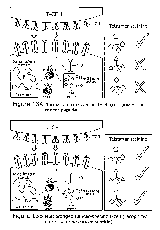
Figure 13 shows multipronged cancer specific T-cells and T-cell receptors
differ from
normal anti-cancer T-cells. (A) Conventionally, anti-cancer T-cells recognise
cancer cells
when the TCR binds to a peptide derived from cancer antigens as shown in A.
These T-
cells do not respond to other cancer-derived peptides. (B) Unusually,
multipronged anti-
cancer T-cells bear TCRs that recognise multiple different cancer peptides. It
is far more
difficult for cancer cells and a developing tumour to escape from multipronged
T-cells.
Consequently, the use of multipronged TCRs is desirable in cancer
immunotherapy
approaches.
Figure 14 shows super-agonist peptide MTSAIGILPV primed a greater proportion
of
cancer-specific T-cells leading to enhanced killing of autologous cancer. (A)
CD8 T-cells
from a renal cell carcinoma (RCC) and chronic lymphocytic leukaemia (CLL)
patient were
left unprimed or primed with MTSAIGILPV peptide for 28 days. A TAPI-0 assay
(RCC
patient) or tetramer staining (CLL patient) demonstrated the presence of
MTSAIGILPV
(SEQ ID NO: 80) specific T-cells. The MTSAIGILPV (SEQ ID NO: 80) primed CD8s
killed
more autologous cancer cells than the unprimed T-cells. (B) CD8 T-cells from
an acute
myeloid leukaemia (AML) patient and two CLL patients were left unprimed, or
primed with
either wild-type IMP-2 (NLSALGIFST) (SEQ ID NO: 73) or MTSAIGILPV (SEQ ID NO:
18
CA 03099677 2020-11-06
WO 2020/002899 PCT/GB2019/051785
80) peptide for 28 days. Analysis performed with IMP-2 tetramer revealed that
the
unprimed and IMP-2 primed conditions had similar proportions of IMP-2 specific
T-cells,
whereas MTSAIGILPV broke tolerance and induced a greater proportion of IMP-2
cells.
T-cells from CLL patient 3 were used in a killing assay and the MTSAIGILPV
(SEQ ID
NO: 80) primed T-cells killed more CLL cells than the IMP-2 primed CD8s.
Figure 15 shows a schematic of how the multipronged T-cells recognise a
plurality of
different peptides derived from the different cancer-specific antigens at the
surface of the
same cancer cell.
Figure 16 shows multipronged T-cells recognise peptides additively and at low
concentration. Multipronged T-cell clone CR24 recognizes peptides from BST2
(LLLGIGILVL) (SEQ ID NO: 72), Melan A (EAAGIGILTV) (SEQ ID NO: 71) and IMP2
(NLSALGIFST) (SEQ ID NO: 73). CR24 responded to all three individual peptides
at 10-
6 M, but responses dropped when peptides were at 10-8 M. However, CR24
exhibited
good activation when each peptide was present at 10-8 M within a mix of
peptides. This
demonstrates how multipronged T-cells can sensitively target cancer cells by
recognition
of multiple peptides from different proteins expressed by the same cell.
Detailed description
Methods and Materials
General cell culture reagents and cell lines
RMPI-1640 with 2 mM L-glutamine, 100 U/mL penicillin and 100 pg/mL
streptomycin
(termed RO) was supplemented with either 5% (R5) or 10% (R10) foetal calf
serum. T-
cell media was R10 with added 10 mM HEPES buffer, 0.5X non-essential amino
acids, 1
mM sodium pyruvate, 20-200 IU/mL of IL-2 (Aldesleukin, Proleukin, Prometheus,
San
Diego, CA, USA) and 25 ng/mL of IL-15 (Peprotech, Rocky Hill, NJ, USA). D1O-
F12 media
was made as for R10 using DMEM-F12. Unless otherwise stated tissue culture
reagents
were from Life Technologies (Carlsband, CA, USA). Cell lines C1 R, T2 and IM9
were
cultured as suspension cells in R10. Malignant melanoma cell lines Mel-526,
Mel-624,
FM-2, FM-56, SK-MEL-37 and A-375 were cultured as adherent cells in R10.
Melanoma
19
CA 03099677 2020-11-06
WO 2020/002899 PCT/GB2019/051785
MM909.24 and renal cell carcinoma RCC17 were obtained from patients treated at
the
CCIT and cultured as suspension cells in R10 and D10-F12 respectively. Other
cancer
cell lines were maintained as described by the ATCC; breast adenocarcinoma MDA-
MB-
231 (ATCCO HTB-26Tm) and MCF-7 (ATCCO HTB-22Tm); prostate adenocarcinoma
LnCAP (ATCCO CRL-1740Tm); colorectal carcinomas COLO 205 (ATCCO CCL222TM)
and HCT116 (ATCCO CCL-247Tm); lung carcinoma H69 (ATCCO HTB-119Tm); liver
hepatocellular carcinoma HepG2 (ATCCO HB-8065Tm); cervical carcinoma MS751
(ATCCO HTB-34TM); acute lymphoblastic leukaemia MOLT3 (ATCCO CRL-1552Tm);
chronic myeloid leukaemia K562 (ATCCO CRL-3344Tm); myeloma/plasmacytoma U266
(ATCCO TIB-196Tm) osteosarcomas U-2 OS (ATCCO HTB-96Tm) Saos-2 (ATCCO HTB-
85Tm) and TK143 (ATCCO CRL-8303Tm); HEK293T embryonic kidney cell (ATCCO CRL-
1573Tm); acute monocytic leukaemia THP-1 (ATCCO TIB-202Tm); and kidney
carcinoma
A-498 (ATCCO HTB-44Tm).
Melanoma tumour infiltrating lymphocytes recognise multiple cancer cell types
Stage IV metastatic melanoma patient MM909.24 underwent rapid tumour
infiltrating
therapy for at the Centre for Cancer Immunotherapy (CCIT), Herlev Hospital,
Copenhagen [1]. To date, this patient has experienced lasting remission.
Chromium
release cytotoxicity assay was used to assess reactivity towards cancer cell
lines:
autologous melanoma (MM909.24), MDA-MB-231, MCF-7, LnCAP and RCC17. Cell lines
(1 x10⁶ cells) were labelled for 1h with 30 pCi of sodium chromate (51Cr)
(Perkin Elmer,
Waltham, MA, USA), leached for lh, then cultured with TILs overnight. A 10:1
TIL to target
cell (2000 cells per well) ratio was used. After overnight incubation
supernatants were
harvested, mixed with scintillant and read using a microbeta counter and
specific lysis
calculated [2]. Further cancer cell lines were tested using a TNF processing
inhibitor-0
(TAPI-0) assay [3]; TILs were harvested from culture washed with RO and rested
overnight in R5 media. On the day of the activation assay, cells were
harvested then
counted and 100,000 incubated with 30 pM TAPI-0 (Sigma-Aldrich) anti-TNF-PE-
Vio770TM (clone cA2, Miltenyi Biotech) and anti-CD107a-PE (clone H4A3, BD
Biosciences) antibodies in wells of a 96 U well plate. Cancer cell lines were
added to give
a TIL to target cell ratio of 1:2. In addition to the cancer cell lines above
the following were
CA 03099677 2020-11-06
WO 2020/002899 PCT/GB2019/051785
also used; COLO 205, H69, HepG2, MS751 and Saos-2. The cells were incubated
for 4-
h at 37 C then stained at RT for 5 min with 2 pL of LIVE/DEAD fixable dead
cell stain
ViVid (Life Technologies) that had been diluted 1:40 using PBS. Antibodies to
detect
surface markers were added directly to each sample without washing; anti-CD8-
APC
(clone BW135/80, Miltenyi Biotech) and anti-CD3-peridinin chlorophyll (PerCP)
(clone
BW264/56, Miltenyi Biotech). Data was acquired on a BD FACS Canto ll (BD
Biosciences) and analysed with FlowJo software (TreeStar Inc., Ashland, OR,
USA).
Activated TILs (CD107a+ and/or TNF+) were sorted on a BD FACS Aria (BD
Biosciences,
San Jose, CA, USA) and used for next generation sequencing of the T-cell
receptor (TCR)
chains as previously described [4].
The strategy for identifying peptides recognised by orphan CD8 clones
T-cell clones of unknown peptide specificity (termed orphan clones) were
generated by
culturing 0.5 cells/well in of 96 U well plates in T-cell media with 50,000
irradiated (3000-
3100 cGy) allogenic peripheral blood mononuclear cells (PBMCs) from three
donors and
1-2 pg/mL of phytohaemagglutinin (PHA). PBMCs were separated from blood by
standard
density gradient centrifugation. If needed, red blood cells were lysed using
ammonium
chloride solution. Blood was procured as buffy coats' from the Welsh Blood
Service
(Pontyclun, Wales, UK). All human tissue was obtained and handled in
accordance with
Cardiff University's guidelines to comply with the UK Human Tissue Act 2004. T-
cell
clones were screened against autologous melanoma (MM909.24) and in some case
cancer cell lines of different tissue origin. Clones of interest were grown to
large number
in T25 flasks using the PBMC and PHA method as above. Combinatorial peptide
library
(CPL) and cancer antigen database screening was performed to find peptides
recognized
by orphan clones. Combinatorial peptide libraries were synthesized and used as
previously described [5,6]. Briefly, long-term storage was at -80 C as 20 mM
DMSO
stocks with 1 mM working dilutions made in sealable (silicone sealing mat,
AxyGene
AxyMatTM, Corning, New York, US) 2 mL deep round-well plates (AxyGene,
Corning)
with RO (as for R10 but with no serum), which were stored at 4 C, then
vortexed
(MixMate , Eppendorf , Hamburg, Germany) at 1300 rpm for 1 min, then
centrifuged
(400g, 5 mins) before use. Each sub-library was used at a concentration of 100
pM with
21
CA 03099677 2020-11-06
WO 2020/002899 PCT/GB2019/051785
respect to total peptide concentration. The CPL data was run via a database,
which
contains the amino acid sequences of proteins expressed by cancers (manuscript
in
preparation). The cancer antigen database will be available online as part of
the PI CPL
(peptide identification combinatorial peptide library) webtool hosted by
Warwick
University's Systems Biology
Centre
(http://wsbc.warwick.ac.uk/wsbcToolsWebpage/user_cases.php). Candidate
peptides
from the database were automatically ranked based on their likelihood of being
recognised by a clone, with the top 20 being tested in peptide titration
assays.
CR24 recognises multiple cancer cell types
HLA A2+ Melanomas, MM909.24 (autologous), Mel-526, Mel-624, and HLA A2+ non-
melanomas, CIR-HLA A2, MDA-MB-231, Saos-2, U20S, A498, TK143, HEK293T, COLO
205, HCT116, HeLa, HepG2 and THP1 were used as target cells in a TAPI-0 assay,
which is described above. HLA A2neg melanomas FM-2 and FM-56, and wild-type Cl
Rs
(HLA A2neg) were used as controls.
Combinatorial peptide library (CPL) and cancer antigen database screening of
clone CR24
CR24 was rested overnight in RO then 30,000 used per well of the decamer CPL
screen
(details above). The peptide length preference of CR24 had previously been
established
using sizing scan assays [7] (data not shown). T2 cells (60,000 per well) were
used as
antigen presenting cells. The assay was performed in R5 and supernatants
harvested for
MIP-1 [3 enzyme linked immunosorbent assay (ELISA) according to the
manufacturer's
instructions (R&D Systems, Minneapolis, MN, USA).
CR24 recognises three HLA A2 restricted peptides from different cancer
proteins
CR24 was cultured overnight in R5, then 30,000 used per well of a 96 U well
plate with
decreasing concentrations of peptides. After overnight incubation supernatants
were
used MIP-1[3 ELISA according to the manufacturer's instructions (R&D Systems,
Minneapolis, MN, USA). For tetramer analysis CR24 (20,000-50,000 per sample)
was
stained in 5 mL polypropylene tubes suitable for flow cytometry. Cells were
treated in 100
22
CA 03099677 2020-11-06
WO 2020/002899 PCT/GB2019/051785
pL of FACS buffer (PBS + 2% FBS) with 50 nM Dasatinib (a protein kinase
inhibitor) for
30 min at 372C and phycoerythrin (PE) conjugated tetramer (0.5 rig) added
directly to the
sample before being moved to ice for a further 30 min [8]. Tetramer was washed
with 3
mL of FACS buffer (700 g, 5min) then labelled with 0.5 pg (10 pg/mL) of mouse
anti-PE
unconjugated antibody (clone PE001, BioLegend, London, UK) for a further 20
min on ice
[8]. To test if CR24 could recognise endogenously express antigen MOLT3 cells
were
used to express various proteins. Codon optimised full-length human HLA A2
(IMGT/HLA
Acc No: HLA00005), MLANA (Melan A) (UniProtKB Q16655), BST2 (UniProtKB
Q10589),
IGF2BP2 (IMP2) (UniProtKB Q9Y6M1), COL6A2 (a2 subunit of collagen type VI)
(UniProtKB P12110) and Zika virus (Rio-U1) ancC (GenBank KU926309.2) genes
were
synthesized (Genewiz, South Plainfield, NJ, USA) and cloned into the 3rd
generation
lentiviral transfer vector pELNS (kindly provided by Dr. James Riley,
University of
Pennsylvania, PA, USA). The pELNS vector contains a rat CD2 (rCD2) marker gene
separated from the gene of interest by a self-cleaving 2A sequence. Lentiviral
particle
production, calcium chloride transfection and rCD2-based purification of cells
were
performed as previously described [9].
Clone CR24 is able to recognise autologous melanoma lacking Melan A expression
To demonstrate that CR24 can target autologous melanoma through multiple
antigens,
guide RNAs to ablate Melan A expression using CRISPR/Cas9 were designed using
the
cripsr.mit.edu webtool, applied and the Melan A gene sequenced to confirm
disruption
(data not shown). Intracellular staining for Melan A was performed using
Cytofix/Cytoperm TM reagents according to manufacturer's instructions (BD
Biosciences).
A primary unconjugated rabbit anti-Melan A antibody (clone EP1422Y) (Abcam,
Cambridge, UK) was used with a secondary PE conjugated goat anti-rabbit
antibody. Wild
type and Melan A KO MM909.24 melanomas were used TAPI-0 assays, as described
above, with both TILs and CR24.
23
CA 03099677 2020-11-06
WO 2020/002899 PCT/GB2019/051785
T-cells that recognise the same three peptides as CR24 are present in healthy
HLA
A2+ donors
To generate T-cell peptide lines, CD8 T-cells were purified from the PBMCs of
HLA A2+
donors using CD8 microbeads according to the manufacturer's instructions
(Miltenyi
Biotech, Bergisch Gladbach, Germany). Purified CD8 cells (3 x10⁶) were co-
incubated
with autologous CD8neg cells (6-8 x10⁶) in 24 well plates in 2 mL of T-cell
media, but with
no IL-15. 25 M of each peptide was used. The cultures had 50% of the media
changed
thrice weekly. Tetramer staining was performed as above, using 500,000 cells
per tube.
Each T-cell line was used in an IFNy enzyme linked immunosorbent spot
(ELISpot) assay
with cell lines MDA-MB-231, melanoma MM909.24 and Saos-2. 50,000 T-cells and
15,000 cancer cells were used per well. Incubation was performed for 48 h, and
the assay
developed according the manufacturer's instructions (Mabtech, Nacka Strand,
Sweden).
Super-agonist peptides prime multi-pronged T-cells for improved cancer cell
recognition.
CPL assay of CR24 was performed as described above. Candidate peptide agonists
were
designed using the CR24 CPL and an online
algorithm
(Priming of CD8 T-cells
from healthy donors, tetramer staining and chromium release cytotoxicity
assays were
performed as described above.
Other Melan A clones do not recognise the BST2 and IMP2 peptides seen by CR24
TAPI-0 and activation assays (ELISA) were performed for VB6G4.24, CR1 and
VB10, as
described above for CR24. The data was summarised in tabular from.
Clone recognition of peptides from cancer antigens hTERT and MAGE C2
Clones GD1 and GD2 were grown from the peripheral blood of different HLA A2+
healthy
donors. The clones were used in overnight activation assays with decreasing
concentrations of respective peptides, and supernatants used for MIP-1(3
ELISA, as
24
CA 03099677 2020-11-06
WO 2020/002899 PCT/GB2019/051785
described above. An overnight activation was performed with GD1 and target
cells; K562,
K562 HLA A2, CIR, CIR HLA A2, HEK 293T, MCF-7, COLO 205, U266, HCT116, Mel-
526, Mel-624, SK-MEL-37, A375, 1M9 and LnCAP. Supernatants were harvested and
used for MIP-113 ELISA. A chromium release cytotoxicity assay was performed,
as above,
with cell lines MCF-7, U266 and Mel-624. Incubation times of 4 h and
overnight, with
varying T-cell to target cell ratios were used.
Results
1. Tumour infiltrating lymphocytes (TILs) derived from a metastatic
melanoma patient
that underwent successful immunotherapy are capable of killing and recognising
autologous melanoma and HLA A2+ cancer cell lines originating from a range of
cancers:
breast, colon, lung, liver, prostate, cervix, bone and kidney (Figure 1).
2. T-cell receptor clonotyping of cancer reactive TILs revealed that the
same T-cells
recognised multiple HLA A2+ cancer cell lines (Figure 2). 50% of the T-cells
(TCRs)
recognised more than 4 cancer cell lines and, 8.6% (5 TCRs) recognised all 10
cell lines
tested. Further experiments aimed at understanding the pan cancer cell line
recognition
resulted in the discovery that a single T-cell can recognise multiple peptides
originating
from different cancer proteins.
3. In order to map the peptide specificities of the T-cells from the TILs,
the T-cells
were firstly cloned, then screened for reactivity towards various cancer cell
lines. Clone
CR24 exhibited reactivity towards autologous melanoma and cancer cell lines
from
breast, bone, kidney, blood, colon, cervix and liver (Figure 4). This
reactivity was
mediated through HLA A2 as HLA A2neg melanomas and wildtype CIR cells (HLA
A2neg)
were not recognised.
4. Combinatorial peptide library and cancer antigen database screening (as
described in Figure 3) of CR24 (Figure 5) revealed multiple peptides that were
predicted
to be seen by CR24 (data not shown), with three of them being recognised when
tested
as exogenous peptide (Figure 6). CR24 also stained with HLA A2 tetramers
containing
the three peptides (Figure 6). The peptides; EAAGIGILTV (SEQ ID NO: 71) from
Melan
A (residues 26-35), LLLGIGILVL (SEQ ID NO: 72) from BST2 (residues 22-31) and
CA 03099677 2020-11-06
WO 2020/002899 PCT/GB2019/051785
NLSALGIFST (SEQ ID NO: 73) from IMP2 (residues 367-376). These data
demonstrate
that CR24 is cross-reactive for distinct peptides derived from different
cancer proteins.
5. The peptides recognised by CR24 are processed and presented from
endogenously expressed proteins, as CR24 was capable of recognising antigen
presenting cells (MOLT3) made to stably express either Melan A, BST2 or IMP2
(Figure
6).
6. It would be extremely difficult for cancer cells to escape from T-cells
that were
targeting them through more than one different cancer antigen as escape would
require
simultaneous mutation of all targets that lowered or ablated presentation of
all cognate
peptides. To demonstrate this, we targeted autologous melanoma (MM909.24) for
ablation of the Melan A gene, which was confirmed by antibody staining to lack
Melan A
protein expression (Melan A knockout (KO)) (Figure 7). Both the TIL from
patient
MM909.24 and clone CR24 recognised the Melan A knockout melanomas (Figure 7).
For
CR24, reactivity against wild type autologous tumour was 71% and for the Melan
A KO
55%. It is highly likely that CR24 was recognising the Melan A KO melanoma
through the
BST2 and/or IMP2 peptides and therefore able to mediate destruction of the
melanoma.
7. CD8 T-cells able to recognise the Melan A, BST2 and IMP2 peptides seen
by
CR24 can be generated from the peripheral blood of healthy HLA A2+ donors
(Figure 8).
8. Super-agonists designed for multi-pronged T-cells primed a greater
proportion of CD8
T-cells capable of recognising WT Melan A (EAAGIGILTV) (SEQ ID NO: 71), BST2
(LLLGIGILVL) (SEQ ID NO: 72) and IMP2 (NLSALGIFST) (SEQ ID NO: 73) peptides,
compared to parallel priming with the WT peptides. Super-agonist MTSAIGVLVP
(SEQ
ID NO; 80) (peptide 5) seemed to be the most effective of the candidate super-
agonists
at priming (Figure 9B), eliciting Melan A, BST2 and IMP2 reactive T-cells in
all donors
tested (n=3). Additionally, MTSAIGILPV (SEQ ID NO; 80) and ITSAIGILPV (SEQ ID
NO;
77) were superior at priming Melan A (EAAGIGILTV) T-cells from metastatic
melanoma
patients compared to the WT EAAGIGILTV peptide (Figure 10A), and MTSAIGILPV
(SEQ
ID NO; 80) also in renal cell carcinoma (RCC) and chronic lymphocytic
leukaemia (CLL)
patients (figure 14A) and acute myeloid leukaemia (AML) patients (figure 14B).
26
CA 03099677 2020-11-06
WO 2020/002899 PCT/GB2019/051785
Importantly, the MTSAIGILPV (SEQ ID NO; 80) super-agonist peptide primed T-
cells
exhibited superior lysis of autologous melanoma cells than the WT Melan A
peptide
primed T-cells (Figures 10B and 10 C).
9. Clones (GD1 and GD2) grown from the peripheral blood of two healthy HLA
A2+
donors cross-react with different peptides than those recognised by CR24.
These
peptides are derived from different proteins to those recognised by the CR24 T-
cell clone;
RLVDDFLLV (SEQ ID NO: 74) from human telomerase reverse transcriptase (hTERT)
(residues 855-873) and ALKDVEERV (SEQ ID NO: 75) from melanoma associated
antigen C2 (MAGE C2) (residues 336-344). GD1 killed breast, blood and melanoma
cancer cell lines (Figure 9).
Conclusion
The current consensus view is that cancer-specific T-cells recognise cancer
cells via a
single peptide antigen presented as a peptide at the cell surface in
association with HLA
(Figure 10A). We have discovered that some, rare T-cells are able to recognise
cancer
cells through multiple peptide epitopes that differ in sequence by two or more
amino acids
and are derived from different cancer antigens (Figure 10B). Cancer escape
from this
type of multipronged T-cell is likely to be extremely difficult.
27
CA 03099677 2020-11-06
WO 2020/002899 PCT/GB2019/051785
REFERENCES
[1] Andersen R, Donia M, Ellebaek E, Borch TH, Kongsted P, Iversen TZ, et
al.
Long-Lasting complete responses in patients with metastatic melanoma after
adoptive
cell therapy with tumor-infiltrating lymphocytes and an attenuated i12
regimen. Clin
Cancer Res 2016;22:3734-45. doi:10.1158/1078-0432.CCR-15-1879.
[2] Ekeruche-Makinde J, Clement M, Cole DK, Edwards ESJ, Ladd! K, Miles JJ,
et
al. T-cell receptor-optimized peptide skewing of the T-cell repertoire can
enhance
antigen targeting. J Biol Chem 2012; 287:37269-81.
doi:10.1074/jbc.M112.386409.
[3] Haney D, Quigley MF, Asher TE, Ambrozak DR, Gostick E, Price DA, et al.
Isolation of viable antigen-specific CD8+ T cells based on membrane-bound
tumor
necrosis factor (TNF)-alpha expression. J Immunol Methods 2011;369:33-41.
doi:10.1016/j.jim.2011.04.003.1solation.
[4] Donia M, Kjeldsen JW, Andersen R, Westergaard MCW, Bianchi V, Legut M,
et
al. PD-1 + polyfunctional T cells dominate the periphery after tumor-
infiltrating
lymphocyte therapy for cancer. Clin Cancer Res 2017:clincanres.1692.2016.
doi:10.1158/1078-0432.CCR-16-1692.
[5] Wooldridge L, Ekeruche-Makinde J, Van Den Berg HA, Skowera A, Miles JJ,
Tan
MP, et al. A single autoimmune T cell receptor recognizes more than a million
different
peptides. J Biol Chem 2012;287:1168-77.
[6] Szomolay B, Liu J, Brown PE, Miles JJ, Clement M, Llewellyn-Lacey S, et
al.
Identification of human viral protein-derived ligands recognized by individual
MHCI-
restricted T-cell receptors. Immunol Cell Biol 2016;94:573-82.
doi:10.1038/icb.2016.12.
[7] Ekeruche-Makinde J, Miles JJ, van den Berg HA, Skowera A, Cole DK,
Dolton G,
et al. Peptide length determines the outcome of TCR/peptide-MHCI engagement.
Blood
2013;121:1112-23. doi:10.1182/blood-2012-06-437202.
[8] Tungatt K, Bianchi V, Crowther MD, Powell WE, Schauenburg AJ, Trimby A,
et
al. Antibody stabilization of peptide-MHC multimers reveals functional T cells
bearing
extremely low-affinity TCRs. J Immunol 2015;194:463-74.
[9] Legut M, Dolton G, Mian AA, Ottmann 0, Sewell A. CRISPR-mediated TCR
replacement generates superior anticancer transgenic T-cells. Blood 2017:blood-
2017-
05-787598. doi:10.1182/blood-2017-05-787598.
[10] Hundemer M, Schmidt S, Condomines M, Lupu A, Hose D, Moos M, et al.
Identification of a new HLA-A2-restricted T-cell epitope within HM1.24 as
28
CA 03099677 2020-11-06
WO 2020/002899
PCT/GB2019/051785
immunotherapy target for multiple myeloma. Exp Hematol 2006; 34:486-96.
doi:10.1016/j.exphem.2006.01.008.
29