Note: Descriptions are shown in the official language in which they were submitted.
CA 03099770 2020-11-09
WO 2019/217493
PCT/US2019/031221
-1-
CELL POPULATIONS AND GENE EXPRESSION ASSOCIATED WITH IN
VITRO BETA CELL DIFFERENTIATION
CROSS-REFERENCE TO RELATED APPLICATIONS
This application claims the benefit of U.S. Provisional Application No.
62/668,242, filed on May 7, 2018. The entire teachings of the above
application are
incorporated herein by reference.
BACKGROUND OF THE INVENTION
Pancreatic beta cells are regulators of blood glucose whose autoimmune
destruction or dysfunction cause Type 1 and Type 2 diabetes. Recently, in
vitro
differentiation protocols have been developed that convert pluripotent stem
cells into
pancreatic beta ce11s1-3. For instance, the `SC-beta' (stem cell-derived beta
cells)
protocoll performs a stepwise differentiation using a combination of signaling
cues
derived from those that generate beta cells in vivo. The resulting stem-cell
derived
beta cells secrete insulin in response to glucose challenges and restore
metabolic
homeostasis in animal models of diabetesl. Consequently, in vitro
differentiation
protocols are leading candidates for the development of cell-based therapies
for
diabetes.
A challenge in producing any cell type in vitro is heterogeneity of the cells
generated by directed differentiation. At each step of the process, some cells
follow
the desired path, others stray. To improve efficiency, it is important to
identify all cell
types produced during differentiation. High-throughput single-cell RNA
sequencing4
characterizes cell types by unbiased transcriptional profiling of thousands of
individual cells. Single-cell RNA sequencing has been applied to
comprehensively
CA 03099770 2020-11-09
WO 2019/217493
PCT/US2019/031221
-2-
characterize the cell types of many organs, including several studies of the
adult
human" and embryonic mouse10'11 pancreas.
Previous studies with beta cell differentiation protocols have made a number
of important observations. Co-expression of insulin and other key beta cell
markers,
combined with glucose-stimulated insulin secretion, constituted the primary
proof that
beta cells are indeed produced in vitro. Studies characterizing bulk gene
expression
profiles12'13 showed that transcriptional and epigenetic landscapes change for
thousands of genes. Petersen et a/.14 used single-cell qPCR to propose a model
for in
vitro pancreatic differentiation. None of these studies comprehensively
determined the
identities and states of all the cell types produced prior to and alongside in
vitro beta
cells.
SUMMARY OF THE INVENTION
In work described herein, single-cell RNA sequencing was used to identify
and describe the cell types produced during in vitro differentiation of
pluripotent stem
cells to pancreatic beta cells. This analysis provides an unprecedented view
of the
sequence of transcriptional changes that underlie the formation of SC-beta
cells and
reveals fate-determinative decision points and alternative pathways that cells
may
follow along the differentiation road.
Among other aspects, this analysis has revealed that the SC-beta protocol
produces stem cell-derived cells that closely resemble enterochromaffin cells
(SC-EC
cells). These SC-EC cells represent a distinct cell type produced alongside SC-
beta
cells. In vivo, enterochromaffin cells are epithelial endocrine cells that
produce and
secrete serotonin. Enterochromaffin cells and serotonin signaling may play a
key role
in the pathophysiology of several diseases, particularly those related to
intestinal
inflammation. Their transcriptional signature is described, as well as their
ability to
secrete serotonin when depolarized with KC1, demonstrating the creation of a
novel
human cell type in vitro. The invention relates in part to these non-naturally
occurring SC-EC cells, which may serve as models for screening for drugs which
may
modify, or improve, serotonin signaling in the GI tract; they may also be used
directly
for cell replacement therapy.
CA 03099770 2020-11-09
WO 2019/217493
PCT/US2019/031221
-3-
In additional aspects, work described herein identifies markers for cell types
produced by in vitro pancreatic beta cell differentiation. The single-cell RNA
sequencing results provide a detailed characterization of the full
transcriptomes of all
cell populations produced during in vitro beta cell differentiation. Using
this data,
genes can be identified that are specifically enriched in single populations
or
combinations of populations. The knowledge that these genes are specific to a
given
population can be leveraged to further develop in vitro pancreatic beta cell
differentiation methods, as well as methods for in vitro differentiation to
other cell
types (such as alpha cells).
More specifically, these genes can be used at least (i) as surface markers for
antibody-based identification and/or enrichment of specific populations, and
(ii) as
targets for genetic perturbation (such as knock-out, activation or
inhibition). This later
aspect allows for creation of 'tailored' (non-wild type) stem cell lines that
have been
genetically edited to be incapable of mis-differentiating toward undesired
fates. That
is, by controlling the gene expression of cells during the differentiation
process (at the
pluripotent stage and/or at one or more later points in the differentiation
process), one
may open or close routes of differentiation, forcing a cell down a desired
path or away
from an undesired path.
Disclosed herein are stem cell-derived enterochromaffin cells (i.e., non-
naturally occurring enterochromaffin cells).
In some embodiments, the cell expresses one or more of the following genes:
TPH1, SLC18A1, LMX1A, PAX4, DDC, TRPA1, SCN3A, ADRa2A, FEV, TAC1,
and CXCL14. In some embodiments, the cell co-expresses the genes TPH1, LMX1A,
and SLC18A1. In some embodiments, the expression of the genes is enriched
relative
to in vivo pancreatic populations. In some embodiments, the cell does not
express one
or more of the following markers: G6PC2, NPTX2, ISL1, and PDX1.
In some embodiments, the cell is capable of producing serotonin (5-HT). In
some embodiments, the cell releases serotonin in vitro upon depolarization
with KC1
and/or does not release serotonin in vitro upon stimulation with high glucose.
In some embodiments, the cell is differentiated in vitro from an endocrine
cell,
a pancreatic progenitor cell, or a pluripotent stem cell. In some aspects, the
pancreatic
progenitor cell is selected from the group consisting of a Pdxl+, NKX6-1+
pancreatic
CA 03099770 2020-11-09
WO 2019/217493
PCT/US2019/031221
-4-
progenitor cell and a Pdxl+ pancreatic progenitor cell. In some aspects, the
pluripotent stem cell is selected from the group consisting of an embryonic
stem cell
and induced pluripotent stem cell. In some embodiments, the pluripotent stem
cell is
a human cell.
Also disclosed herein are cell lines comprising the stem cell-derived
enterochromaffin cells described herein. Also disclosed herein are SC-islets
comprising one or more of the stem cell-derived enterochromaffin cells
described
herein.
Disclosed herein are methods of producing an SC-EC cell from a progenitor
cell in vitro. The methods comprise contacting a population of cells
comprising a
pancreatic progenitor cell under conditions that promote cell clustering with
at least
six EC maturation factors comprising a) a TGF-f3 signaling pathway inhibitor,
b) a
thyroid hormone signaling pathway activator, c) a y-secretase inhibitor, d) at
least one
growth factor from the EGF family, e) a retinoic acid (RA) signaling pathway
activator, and f) a sonic hedgehog (SHH) pathway inhibitor to induce the
differentiation of at least one pancreatic progenitor cell in the population
into at least
one SC-EC.
In some embodiments, the TGF-f3 signaling pathway inhibitor comprises Alk5
inhibitor II; the thyroid hormone signaling pathway activator comprises
triiodothyronine (T3); the y-secretase inhibitor comprises XXI; the at least
one growth
factor from the EGF family comprises betacellulin; the RA signaling pathway
activator comprises RA; and/or the SHH pathway inhibitor comprises Santl. In
some
embodiments, the population of cells is optionally contacted with a BMP
signaling
pathway inhibitor (e.g., LDN193189).
Also disclosed herein are methods of identifying cells (e.g., SC-f3 cells, SC-
a
cells, and/or SC-EC cells) in a population of endocrine cells. The methods
comprise
applying a diffusion pseudotime analysis to a population of endocrine cells;
identifying one or more genes expressed by one or more cells within the
population of
endocrine cells; and identifying the one or more cells as SC-f3 cells, SC-a
cells or SC-
EC cells, wherein the SC-f3 cells express at least ISL1 and ERO1B, and wherein
the
SC-EC cells express at least TPH1 and LMX1A.
CA 03099770 2020-11-09
WO 2019/217493
PCT/US2019/031221
-5-
Also disclosed herein are methods of identifying SC-f3 cells within a
population of endocrine cells. The methods comprises screening a population of
endocrine cells for cells expressing at least ISL1 and ERO1B; and identifying
cells
within the population of endocrine cells as SC-f3 cells if they express at
least ISL1 and
ERO1B .
Disclosed herein are methods of identifying SC-EC cells within a population
of endocrine cells. The methods comprise screening a population of endocrine
cells
for cells expressing at least TPH1 and LMX1A; and identifying cells within the
population of endocrine cells as SC-EC cells if they express at least TPH1 and
LMX1A.
Disclosed herein are methods for directing differentiation of a population of
cells comprising modulating expression of a regulator of cell fate during a
differentiation protocol, thereby directing differentiation of a population of
cells
towards a predetermined cell fate.
Also disclosed herein are methods for forming an enriched population of SC-f3
cells. The methods comprise applying anti-CD49a antibody and microbeads to a
solution of dissociated cells; and isolating for cells enriched in CD49a,
thereby
forming an enriched population of SC-f3 cells.
Disclosed herein are methods for producing SC-islets comprising SC-f3 cells.
The methods comprise obtaining Stage 6 clusters from a differentiation
process;
dissociating the Stage 6 clusters using a re-aggregation procedure;
resuspending and
staining dissociated single cells, wherein the cells are stained for CD49a;
adding
microbeads to a suspension of stained dissociated single cells; magnetically
separating
the single cells; and combining the separated single cells to form a cell
population
comprising an enriched yield of SC-f3 cells.
In some embodiments, the cells are stained for CD49a using anti-human
CD49a antibody. In some embodiments, the cell population shows an enriched
yield
of 70% SC-f3 cells. In some embodiments, the cell population shows an enriched
yield of 80% SC-f3 cells.
Also disclosed herein are methods for directing differentiation of a
population
of cells. The methods comprise inhibiting expression of a regulator of cell
fate during
CA 03099770 2020-11-09
WO 2019/217493
PCT/US2019/031221
-6-
a differentiation protocol, e.g.,wherein the regulator is ARX, thereby
directing
differentiation of a population of cells toward SC-f3 cells.
Also disclosed herein are methods for directing differentiation of a
population
of cells. The methods comprise inhibiting expression of a first regulator of
cell fate
during a differentiation protocol, e.g., wherein the first regulator is ARX,
and
activating expression of a second regulator of cell fate during a
differentiation
protocol, e.g., wherein the second regulator is PAX4, thereby directing
differentiation
of a population of cells toward SC-f3 cells.
Disclosed herein are methods for directing differentiation of a population of
cells comprising disrupting LMX1A during a differentiation protocol, thereby
decreasing SC-EC production and directing differentiation of a population of
cells
towards SC-f3 cells. In some embodiments, the disruption of LMX1A occurs by
knockdown using a gene editing technique. In some embodiments, the disruption
of
LMX1A occurs by knockout using a gene editing technique.
BRIEF DESCRIPTION OF THE DRAWINGS
The patent or application file contains at least one drawing executed in
color.
Copies of this patent or patent application publication with color drawings
will be
provided by the Office upon request and payment of the necessary fee.
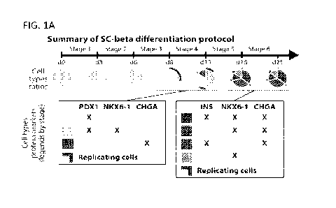
FIGS. 1A-1G demonstrate single cell RNA sequencing of in vitro beta cell
differentiation. FIG. lA provides a summary of the cell populations identified
by
flow cytometry at the end of Stages 3-6 of the Pagliuca et al. SC-beta
protocol.
PDX1: pancreatic transcription factor, NKX6.1: beta cell transcription factor,
INS:
insulin, beta cell hormone, CHGA: chromogranin A, pan endocrine marker. FIG.
1B
shows immunofluorescence imaging of a differentiated (Stage 6, day 13) SC-beta
cluster showing population heterogeneous staining for NKX6.1 and C-peptide (a
component of insulin). FIG. 1C provides a schematic of study design using
inDrops
single-cell RNA sequencing to characterize the cells sampled at different time
points
of the same differentiation. FIG. 1D shows tSNE projection of cells sampled
from the
ends of Stages 3-6 of the `x l' protocol. Cells are colored according to their
assigned
cluster using Louvain community detection. Bar along bottom of plot indicate
the
relative cluster proportions. Legend for cell colors is the same as the one
use in
CA 03099770 2020-11-09
WO 2019/217493
PCT/US2019/031221
-7-
following panel. FIG. lE provides expression profiles of developmentally
relevant
genes and markers across cell types identified in SC-beta differentiation.
Each
population has specific markers. FIG. 1F shows immunofluorescence imaging of
enterochromaffin cell marker SLC18A1. SLC18A1+ cells are present in the
cluster
periphery. FIG. 1G shows immunofluorescence imaging of non-endocrine marker
50X9. SOX9+ cells localize near center of clusters.
FIGS. 2A-2I demonstrate SC-beta cells are functional and transcriptionally
stable during extended culture in the final differentiation stage. FIG. 2A
provides a
schematic of experimental design to study functional and transcriptional
changes
during Stage 6. FIG. 2B shows glucose stimulated insulin secretion showing
consecutive low glucose (2.8 mM) and high glucose (20 mM) challenges for three
independent differentiations over a period of 5 weeks. FIG. 2C provides
stimulation
indices computed as the ratio of insulin secretion in high glucose to
secretion in low
glucose. A stim index of 1 (dashed line) represents unresponsive cells. FIG.
2D
shows tSNE plot of 38,004 cells from 6 time points spanning 5 weeks of culture
in
Stage 5. Cells are colored according to their assigned cluster. Relative
cluster
proportions for each week are displayed as vertical bars. FIG. 2E shows
correlation
of expression profiles for each cell types, broken down by week. FIG. 2F shows
tSNE projection of SC-beta cells from weeks 0 through 6, shaded by the
diffusion
pseudotime value of each cell (DPT). Dark lines show approximate contour lines
for
changes in DPT. FIG. 2G shows DPT distribution for cells from a given shows
that
cells taken from a later time are, on average, further along this process.
FIG. 2H
provides a volcano plot showing genes whose expression in beta cell correlates
with
diffusion pseudotime (q-values computed from FDR adjustment with alpha of
0.001).
FIG. 21 shows expression of selected genes shown along beta cell diffusion
pseudotime. Gray dots are measurements from individual cells, sorted by
pseudotime,
with superimposed line showing moving average.
FIGS. 3A-3E demonstrates enterochromaffin cells. FIG. 3A provides a
comparison of gene expression profiles between SC-beta and SC-EC cells.
Highlighted genes are required for serotonin synthesis or enterochromaffin
markers.
FIG. 3B shows expression levels for SC-EC enriched genes across in vitro
populations (top panel) and human pancreatic endocrine cell types (bottom).
FIG. 3C
CA 03099770 2020-11-09
WO 2019/217493
PCT/US2019/031221
-8-
provides a comparison of gene expression between WT mouse and islets and mouse
islets, 25 weeks after beta-cell specific PRC2 ablation via EED knockout.
Purple
genes are down-regulated beta-cell identity genes, red genes are part of the
serotonin/EC signature seen in (FIG. 3A). FIG. 3D shows immunofluorescence
staining for SC-EC cell markers LMX1A, TPH1, SLC18A1 showing co-localization
with serotonin (5-HT). FIG. 3E shows immunofluorescence staining for C-peptide
(INS) and SLC18A1 of grafted tissue recovered 8 weeks after transplantation of
SC-
beta differentiated clusters into a murine kidney capsule.
FIGS. 4A-4F demonstrates re-aggregation is a scalable, function-preserving
method to enrich for endocrine cells. FIG. 4A provides a schematic drawing of
re-
aggregation procedure. Cells are enzymatically dissociated and re-aggregate
during
continued suspension culture. Non endocrine cells that fail to adhere are
removed by
filtration after 4 days. FIG. 4B shows tSNE of cells sequenced from native and
re-
aggregated clusters from a single differentiation show strong depletion of the
non-
endocrine population. FIG. 4C provides representative flow cytometry analysis
for
measurement of endocrine cell abundance. Endocrine cells express CHGA. FIG. 4D
shows summary of population composition (as assayed by flow cytometry) in 60
re-
aggregated and 41 native differentiations. FIG. 4E shows stimulation index
(insulin
released at 20 mM glucose/insulin released at 2 mM) of 52 paired native vs. re-
aggregated differentiation. P-value computed using Wilcoxon Rank-sum test.
FIG.
4F shows immunofluorescence staining for C-peptide (fragment of insulin) and
SLC18A1 shows distinct neighborhoods in re-aggregated clusters.
FIGS. 5A-5H demonstrates Stage 5 time course. FIG. 5A shows tSNE
projection of 46204 cells, shaded according to whether they are present at
Stage 4
completion (day 0), Stage 5 completion (day 7) or mid Stage 5. Sampling time
(left)
and NEUROG3 expression (right). FIG. 5B provides same tSNE projection as in
FIG. 5A shaded according to assigned cell identity cluster. FIG. 5C shows
fraction of
cells from each cluster in FIG. 5B for each day of both independent
differentiations.
FIG. 5D shows diffusion pseudotime (DPT) analysis of cells in endocrine
induction,
SC-EC and SC-beta clusters. (top) schematic of cell types selected for
analysis.
(bottom) tSNE projection of selected cells, shaded by rank in DPT along each
of three
possible branches. FIG. 5E shows expression of key genes along DPT ordering
from
CA 03099770 2020-11-09
WO 2019/217493
PCT/US2019/031221
-9-
FIG. 5D. The three branches are shown sequentially. Gray dots are individual
cells
and superimposed lines are moving averages. FIG. 5F provides a heatmap showing
aggregate pattern of 635 genes ordered by DPT as in FIGS. 5D-5E. FIG. 5G
provides
a heatmap showing markers for clusters presented in FIGS. 5B-5C. FIG. 5H
provides
lineage model for the SC-beta protocol showing the primary developmental
trajectory
of the key cell types.
FIGS. 6A-6G provide comparison of two SC-beta protocol variants. FIGS.
6A-6B provide a summary of changes in Stages 3 and 4 in protocols `x1' (FIG.
6A)
and `x2' (FIG. 6B) and representative flow cytometry results at the end of
Stages 4
and 5. FIG. 6C shows tSNE projection of cells sampled from the ends of Stages
3-6
of the `x2' protocol, related to FIG. 1D. Cells are colored according to their
assigned
cluster using Louvain community detection. Legend for shading is detailed in
panel
(e). FIG. 6D provides comparison of cell populations from protocols `x1' and
`x2'.
Correlation is computed using the z-scores of TPM values of ¨2000 high-
variance
genes. Rows and columns are ordered using hierarchical clustering. FIGS. 6E-6F
show tSNE embedding of Stage 6 from three differentiations, colored by cell
type
(FIG. 6E) and by differentiation (FIG. 6F). FIG. 6G shows correlation of cell
populations derived from HUES8 (ES cells) and iPS1016/31 (iPS cells).
FIGS. 7A-7C demonstrate glucose stimulated insulin secretion. FIG. 7A
provides a summary of design for sequential GSIS assay. FIG. 7B shows complete
data for 3 independent flasks, assayed across several weeks. Circles represent
technical triplicates and bar shows mean measurement. FIG. 7C shows complete
data
for 7 batches of cadaveric human islets, run alongside samples from FIG. 7B.
FIGS. 8A-8D demonstrate Stage 6 time course. FIG. 8A shows tSNE
embedding of Stage 6 time course data shaded by sampling time (top row) and by
representative marker genes (bottom row). FIG. 8B shows expression profiles
for key
genes necessary for beta-cell function. FIGS. 8C-8D provide comparison of
global
expression between human cadaveric islet-derived beta cells and in vitro
progenitors
and SC-beta cells. Highlighted genes are the same as shown in FIG. 8B.
FIGS. 9A-9D demonstrate characterization of SC-alpha cells. FIG. 9A shows
insulin and glucagon expression in SC-beta (purple) and SC-alpha cells (red)
during
several weeks of Stage 6 (shading is a violin plot over individual cells,
connected dot
CA 03099770 2020-11-09
WO 2019/217493
PCT/US2019/031221
-10-
connect median). FIG. 9B shows expression levels of genes differentially
expressed
between cadaveric islet alpha and beta cells. FIG. 9C provides a heatmap of
expression level of genes from (c), shown for islet alpha, SC-alpha, SC-beta
and islet
beta cells. FIG. 9D shows genes up-regulated in islet beta cells are up-
regulated in
SC-beta cells, and genes up-regulated in alpha cells are up-regulated in SC-
alpha
(poly-hormonal) cells. P-value computed using Mann-Whitney U test.
FIGS. 10A-10B demonstrate characterization of SC-enterochromaffin cells.
FIG. 10A provides a schematic of serotonin synthesis from tryptophan. FIG. 10B
shows serotonin release during sequential challenges of low and high glucose
followed by KC1 depolarization. Upper panel: three batches of SC-beta
differentiated
clusters. Lower panel: two batches of human cadaveric islet controls.
FIGS. 11A-11C demonstrate characterization of non-endocrine cells from
stage 6 time course. FIG. 11A shows tSNE embedding of non-endocrine cells from
stage 6 time course, shaded by day (top row) or by genes relevant to cell
identity
(bottom row). FIG. 11B shows clusters identified by Louvain community
detection
and fraction of cells in each cluster by week of differentiation. FIG. 11C
provides
gene expression heatmap of markers for each subpopulation of non-endocrine
cells.
FIGS. 12A-12D demonstrate Stage 5 time course. FIG. 12A shows tSNE
embedding of Stage 5 time course data shaded by sampling time (top row) and by
representative marker genes (bottom row). FIG. 12B shows pseudotime ordering
of
progenitor cells from Stage 5 day 0 (top row) and day 1 (bottom row) showing
population heterogeneity among early progenitors. At day 1, NKX6.1+
progenitors
induce NEUROG3 expression. FIG. 12C provides a heatmap of 50 genes most
correlated (or anti-correlated) with pseudotime ordering of day 0 progenitors.
FIG.
12C provides a heatmap of receptors, ligands and signaling effectors that are
dynamically expressed across Stage 5 populations.
FIG. 13 provides differentiation protocols used herein.
FIG. 14 provides single-cell RNA sequencing datasets used herein.
FIG. 15 provides a summary of all cell populations identified herein.
FIGS. 16-43 re-present certain data from FIGS. 1-15 and provide additional
data.
CA 03099770 2020-11-09
WO 2019/217493
PCT/US2019/031221
-11-
FIGS. 16A-16G demonstrate single cell RNA sequencing of in vitro beta cell
differentiation. FIG. 16A provides a summary of the cell populations
identified by
flow cytometry at the end of Stages 3-6 of the Pagliuca et al. SC-beta
protocol.
PDX1: pancreatic transcription factor, NKX6.1: beta cell transcription factor,
INS:
insulin, beta cell hormone, CHGA: chromogranin A, pan endocrine marker. FIG.
16B
demonstrates using inDrops to sample cells from several time points of the
same
differentiation. FIG. 16C provides expression profiles of developmentally
relevant
genes and markers across cell types identified during SC-beta differentiation.
Shading displays mean expression (z-normalized tpm) and diameter denotes
fractional
expression. FIGS. 16D-16G shows tSNE projections of cells sampled from the
ends
of Stages 3-6 of the `x l' protocol. Cells are colored according to their
assigned
cluster. Horizontal bars indicate cell type proportions.
FIGS. 17A-17I demonstrate SC-beta cells maintain identity and gain
maturation marker expression during extended culture in Stage 6. FIG. 17A
provides
an experimental design to study functional and transcriptional changes during
Stage 6
of protocol v8. FIG. 17B shows glucose stimulated insulin secretion showing
consecutive low glucose (2.8 mM) and high glucose (20 mM) challenges for three
independent differentiations over a period of 5 weeks. FIG. 17C provides
stimulation
indices (insulin released at 20 mM glucose/insulin released at 2 mM) for data
in FIG.
17B. FIG. 17D shows tSNE projection of 38,494 cells from 6 time points
spanning 5
weeks of Stage 6. Cells are colored according to their assigned type. Vertical
bars
show population ratios in each week. FIG. 17E shows expression of endocrine
marker genes. FIG. 17F shows correlation of expression profiles for each major
cell
type, broken down by week. Cell type colors match those in (d). FIG. 17G
provides
pseudotime order of SC-beta cells shown on tSNE (top) and distribution of SC-
beta
pseudotime order stratified by sampling week (bottom). FIG. 17H provides
identification of dynamic genes along SC-beta pseudotime. Fold-change compares
start and end of pseudotime trajectory. q-values are FDR adjusted
(alpha=0.001) p-
values from likelihood ratio test comparing full and reduced models (see
methods).
FIG. 171 provides expression of selected genes shown along SC-beta pseudotime.
Each dot represents expression of a cell, sorted and shaded as in (FIG. 17G).
Line
shows result of pseudotime regression.
CA 03099770 2020-11-09
WO 2019/217493
PCT/US2019/031221
-12-
FIGS. 18A-18E provide characterization of stem cell derived-
enterochromaffin cells (SC-EC cells). FIG. 18A provides a comparison of SC-
beta
and SC-EC gene expression profiles. Blue genes are required for serotonin
synthesis
or enterochromaffin markers. FIG. 18B shows expression levels for SC-EC
enriched
genes across in vitro populations (top panel) and human pancreatic endocrine
cells
(bottom panel). FIGS. 18C-18D show immunofluorescence staining for SC-EC cell
markers showing co-localization with serotonin (5-HT) in v8 protocol. Scale
bars:
100 p.m. FIG. 18E shows immunofluorescence staining of graft tissue recovered
8
weeks after transplantation of (v4) SC-islet clusters.
FIGS. 19A-19D demonstrates purification of SC-beta cells with
ITGA1/CD49a. FIG. 19A shows expression of ITGA1/CD49a in Stage 6 time-course
data. FIG. 19B provides immunofluorescence for SC-beta (top) and endocrine
(bottom) markers of native, unsorted re-aggregated and CD49a+ sorted re-
aggregated
clusters. Scale bars: 100 p.m. FIG. 19C provides flow cytometry quantification
of SC-
beta cells (C-pep+/NKX6.1+) and SC-EC cells (SLC18A1+) fractions in three
matched conditions for 5 biologically independent v8 differentiations. FIG.
19D
provides stimulation index for the same differentiations. In (FIGS. 19C-19D),
symbol
shows mean and error bars (where shown) correspond to standard errors across 3
independently-reaggregated biological replicates. P-values are from (two-
sided)
dependent t-test.
FIGS. 20A-20I provide a high-resolution map of in vitro endocrine induction.
FIGS. 20A-20C shows tSNE projection of 51,274 cells, shaded according to (FIG.
20A) sampling time within Stage 5, (FIG. 20B) NEUROG3 expression and (FIG.
20C) assigned cell types. Arrows on FIG. 20C indicate key lineage
bifurcations.
FIG. 20D shows fraction of cells from each cluster in FIG. 20C for each day of
both
independent differentiations. FIG. 20E show tSNE shading of branch assignment
and
pseudotime value of each cell on the path from NKX6.1+ progenitors to SC-beta
and
SC-EC cells. FIG. 20F provides expression of selected marker genes along
pseudotime ordering from FIG. 20E. Dots show expression in single cells,
sorted and
shaded according to pseudotime order. Lines show regression on pseudotime for
each
branch (blue: SC-EC, purple: SC-beta). FIG. 20G shows genes with significant
branch-specific expression pattern. q-values are FDR adjusted (alpha=0.001) p-
values
CA 03099770 2020-11-09
WO 2019/217493
PCT/US2019/031221
-13-
from likelihood ratio test comparing branched and non-branched models (see
methods). FIG. 20H provides mean expression values of transcription factors
for
clusters presented in FIGS. 20C-20D. Shading displays mean expression (z-
normalized tpm) and diameter denotes fractional expression. FIG. 201 provides
proposed developmental model for the key cell types produced by SC-beta
protocol.
FIGS. 21A-21M provide comparison of two SC-beta protocol variants and
resulting cell types. FIGS. 21A-21C provide immunofluorescence imaging of
differentiated (v8, Stage 6, day 13) SC-islets showing staining of relevant
markers.
FIG. 21A shows SC-beta cells, typically positioned in the periphery, are
positive for
both NKX6.1 and C-peptide (fragment of proinsulin). FIG. 21B shows SC-EC cells
are positive for SLC18A1, an enterochromaffin cell marker. These cells are
also
present in the periphery. FIG. 21C shows non-endocrine cells, marked by 50X9,
are
most commonly found near the center of SC-islets. Scale bars: 100 p.m. FIGS.
21D-
21E show summary of changes in Stages 3 and 4 in protocols xl (FIG. 21D) and
x2
(FIG. 21E) and representative flow cytometry results at the end of Stages 4
and 6.
FIGS. 21F-21I provides tSNE projection of cells sampled from the ends of
Stages 3-6
of protocol x2. Cells in FIGS. 21F-21I are colored according to their assigned
cluster.
Horizontal bars indicate cell type proportions. (Related to FIGS. 16D-16G).
FIG. 21J
provides a comparison of cell populations from protocols xl and x2.
Correlation is
computed using the z-scores of mean tpm values (for each cluster) of 2000 high-
variance genes. Rows and columns are ordered using hierarchical clustering.
Cells are
labeled as in FIGS. 21F-21I and FIGS. 16D-16G. FIGS. 21K-21L provide tSNE
projections of Stage 6 from three differentiations, colored by cell type (FIG.
21K) and
by differentiation (FIG. 21L). FIG. 21M provides correlation of cell
populations
derived from HUES8 (ES cells, v4 and x3) and iPS1016/31 (iPS cells, v4). Same
colors as in FIG. 21K. Correlation is computed as in FIG. 21J.
FIGS. 22A-22C provide a functional assay of glucose stimulated insulin
secretion (GSIS) during Stage 6 time course. FIG. 22A provides a design for a
sequential GSIS assay. FIG. 22B provides the complete data for 3 independent
flasks,
assayed across several weeks. Circles are individual technical triplicates and
bars
show mean of those triplicates. FIG. 22C provides the complete data for
cadaveric
human islets 7 donors, run alongside samples from FIG. 22B.
CA 03099770 2020-11-09
WO 2019/217493
PCT/US2019/031221
-14-
FIGS. 23A-23F demonstrate Stage 6 SC-beta cells express characteristic beta
cell markers. FIGS. 23A-23B provide tSNE projections of Stage 6 time course
data
shaded by sampling time (FIG. 23A) and by representative marker genes (FIG.
23B).
Expression is normalized relative to maximum value and smoothed over
neighboring
cells. FIG. 23C provides expression profiles for key genes necessary for beta-
cell
function. Shading displays mean expression (tpm, log-scaled) and diameter
denotes
fractional expression. FIGS. 23D-23E provide comparisons of global expression
between human cadaveric islet-derived beta cells and in vitro progenitors
(FIG. 23D)
and SC-beta cells (FIG. 23E). Note the shift in gene expression from
progenitors to
SC-beta cells. All genes shown in all panels from FIG. 23C are circled in red.
FIG.
23F provide results from Gene Set Enrichment Analysis (GSEA) showing that gene
sets from FIG. 23C are significantly upregulated during differentiation. Value
plotted
is -log10 of the GSEA-reported FDR q-value (capped at 10), with sign showing
direction of effect (i.e, purple positive values are up-regulated in SC-beta
cells
compared to NKX6.1 progenitors).
FIGS. 24A-24D provide comparison of SC-beta and SC-alpha cells to each
other and their islet counterparts. FIG. 24A shows insulin and glucagon
expression in
SC-beta (purple distributions) and SC-alpha cells (red distributions) during
several
weeks of Stage 6, shown as violin plots of SC-beta or SC-alpha cells from that
particular time point. Connected line connects medians of each population at
each
time point. FIG. 24B shows identification of genes enriched in cadaveric islet
alpha
cells and islet beta cells from data in Baron et al. 2016. FIG. 24C provides a
heatmap
of expression level of genes from FIG. 24B, shown for islet alpha, SC-alpha,
SC-beta
and islet beta cells. FIG. 24D shows genes enriched in islet beta cells are up-
regulated in SC-beta cells, and genes enriched in alpha cells are up-regulated
in SC-
alpha cells. The displayed p-value is computed using a (two-sided) Wilcoxon
rank-
sum test. In boxplot: boxes extend from first to third quartiles, whiskers
extend from
5th to 95th percentiles, central line indicates median and box notching
indicates 95th
percentile confidence interval for median.
FIGS. 25A-25F demonstrate SC-EC cells secrete serotonin and exist in other
protocols. FIG. 25A provides a schematic of serotonin synthesis from
tryptophan.
Enterochromaffin cells use TPH1, whereas serotoninergic neurons use TPH2 for
the
CA 03099770 2020-11-09
WO 2019/217493
PCT/US2019/031221
-15-
first and rate limiting synthesis step. FIG. 25B shows serotonin release
during
sequential challenges of low and high glucose followed by KC1 depolarization.
Upper
panel: clusters from three independent SC-beta differentiation. Lower panel:
human
cadaveric islets from two donors. Symbols show values of individual replicates
for
each sample (different clusters from the same sample are split and measured
separately). p-values computed using (two-sided) Wilcoxon rank-sum test (n.s =
non-
significant with p>0.05). FIGS. 25C-25D show expression of EC marker genes
(shown in blue) is detectable in bulk RNA-sequencing (from Gupta et al.), and
enriched via sorting of NKX6.1(GFP)+ cells, shown as fold-change, mean
expression
and differential expression q-values. Positive fold-change indicates higher
expression
in NKX6.1(GFP)+ cells. Enrichment of SC-EC markers is comparable to beta cell
markers (shown in purple) and opposite of alpha cell markers (shown in red).
All
values shown are directly reproduced from results computed and deposited by
Gupta
et al. 2018. FIG. 25E provides flow cytometry showing that SLC18A1 is co-
expressed with NKX6.1+ in SC-EC cells of v8 SC-beta protocol differentiations.
This
example is representative across more than one hundred independent
differentiations.
FIG. 25F provides a comparison of gene expression between WT mouse islets and
mouse islets 25 weeks after beta-cell specific PRC2 ablation via EED knockout.
Purple genes are example down-regulated beta cell identity genes, blue genes
represent serotonin/EC signature. q-values are FDR-corrected (alpha=0.05) p-
values
from Limma differential expression analysis.
FIGS. 26A-26D provide characterization of non-endocrine cells from Stage 6
time course. FIGS. 26A-26B provide tSNE projections of non-endocrine cells
from
Stage 6 time course, shaded by collection day (FIG. 26A) or by genes relevant
to cell
identity (FIG. 26B). Expression is normalized relative to maximum value, and
smoothed over neighboring cells. FIG. 26C provides a tSNE projection shaded by
assigned cluster and bar charts of cellular fraction in each cluster by week
of
differentiation. FIG. 26D shows gene expression of population specific markers
for
each subpopulation of non-endocrine cells. Shading displays mean expression (z-
normalized tpm) and diameter denotes fractional expression.
FIGS. 27A-27K demonstrate re-aggregation is a scalable, function-preserving
method to enrich for endocrine cells. FIG. 27A provides a schematic drawing of
a re-
CA 03099770 2020-11-09
WO 2019/217493
PCT/US2019/031221
-16-
aggregation procedure to remove non-endocrine cells. Cells are enzymatically
dissociated and re-aggregated during continued suspension culture. Non-
endocrine
cells fail to adhere and are removed by filtration. FIG. 27B provides a
schematic of a
CD49a enrichment procedure to produce SC-beta enriched clusters. Dissociated
cells
are stained with anti-CD49a PE-conjugated antibody, incubated with anti-PE
magnetic microbeads and magnetically separated. The enriched cells are re-
aggregated in 6 well plates on a rocker. FIG. 27C provides a tSNE projection
of cells
sequenced from native and re-aggregated clusters from a single differentiation
showing strong depletion of the non-endocrine population. Cells in both panels
were
differentiated with protocol v8. FIG. 27D shows immunofluorescence staining
for C-
peptide, GCG and SLC18A1 showing distinct neighborhoods in re-aggregated
clusters (protocol v8). Images shown are maximum intensity projections from z-
stacks. Each panel shows separate, representative clusters stained for all
markers.
Scale bars: 100 p.m. FIGS. 27E-27F show representative flow cytometry analysis
of
endocrine cell abundance (from protocol v8), before and after re-aggregation.
Endocrine cells express CHGA. FIG. 27G shows a summary of population
composition (as assayed by flow cytometry) in 60 re-aggregated (RA) and 41
native
independent differentiations, carried out with protocol v8. Re-aggregations
were
carried out in spinner flasks. p-value computed using (two-sided) Wilcoxon
rank-sum
test. In FIG. 27G and FIG. 27H boxplots: boxes extend from first to third
quartiles,
whiskers extend from 5th to 95th percentiles, central line indicates median
and box
notching indicates 95th percentile confidence interval for median. FIG. 27H
provides
a stimulation index (insulin released at 20 mM glucose/insulin released at 2
mM) of
52 independent protocol v8 differentiations, with paired native vs. re-
aggregated
comparisons, p-value computed using (two-sided) Wilcoxon signed-rank test.
FIG.
271 provides complete data for static glucose stimulated insulin secretion
assays,
performed as in FIG. 7, corresponding to stimulation indices shown in FIG.
19D.
Circles are individual technical triplicates and bars show mean of those
triplicates.
FIG. 27J shows cynamic perifusion assay of glucose responsive insulin
secretion of
human islets, native SC-beta clusters (Stage 6, day 22, v8) and matched CD49a
magnetically sorted enriched SC-beta islets. Each point is the mean of 3
technical
replicates, with the vertical bar indicating standard error across those
triplicates. FIG.
CA 03099770 2020-11-09
WO 2019/217493
PCT/US2019/031221
-17-
27K shows area under the curve comparing the first low-glucose stimulation and
the
high-glucose stimulation, normalized to equal effective time in each
treatment.
FIGS. 28A-28F demonstrate Stage 5 time course markers and progenitor
population heterogeneity. FIGS. 28A-28B provide tSNE projections of Stage 5
time
course data shaded by collection day (FIG. 28A) and by population marker genes
(FIG. 28B). Expression is normalized relative to maximum value, and smoothed
over
neighboring cells. FIG. 28C shows a pseudotime analysis of day 0 (top) and day
1
(bottom) progenitor cells. Shading on each tSNE shows assigned pseudotime
value of
each cell. FIG. 28D shows pseudotime ordering of progenitor cells from Stage 5
day
0 (top row) and day 1 (bottom row) showing population heterogeneity among
early
progenitors. Individual cells are shown as dots, shaded as in (FIG. 28C). Gene
expression predicted from pseudotime regression shown as overlaid line. FIG.
28E
provides a summary of Stage 5 day 0 heterogeneity captured by pseudotime
analysis.
Fold-change between start and end of pseudotime ordering. q-value from
likelihood
ratio test of model with and without pseudotime. FIG. 28F provides a heatmap
of
receptors, ligands and signaling effectors that are dynamically expressed
across Stage
5 populations. Shading displays mean expression (z-normalized tpm) and
diameter
denotes fractional expression.
FIG. 29 provides expression of key marker genes across all populations from
time course datasets and cadaveric islets. Column on the left indicates origin
dataset.
Shading displays mean expression (z-normalized tpm) and diameter denotes
fractional
expression.
FIG. 30 provides expression of intestinal enteroendocrine marker genes across
all populations from time course datasets. Column on the left indicates
dataset origin.
Shading displays mean expression (z-normalized tpm) and diameter denotes
fractional
expression.
FIGS. 31A-31D demonstrate an example of flow cytometry gating strategy.
FIGS. 31A-31C show Stage 6 time course differentiation 1 (internal ID: DA-
089), at
Stage 6, day 13 of v8 protocol. FIG. 31A: Secondaries-only control. FIG. 31B:
SC-
beta cell identification via staining for C-peptide and NKX6.1. FIG. 31C:
Endocrine
cell identification via staining for CHGA and NKX6.1. Results are
representative
across more than a hundred v8 differentiations, with typical SC-beta
percentages
CA 03099770 2020-11-09
WO 2019/217493
PCT/US2019/031221
-18-
being 25-45%. FIG. 31D provides an example CD49a+ magnetic purification. Left
panel shows CD49a+ distribution prior to sorting, right panel shows
distribution after
one round of magnetic separation (see methods). Results are representative
across
more than 10 enrichment experiments.
FIG. 32 provides specification of differentiation protocols used in the study.
Summary of the different versions of the SC-beta protocol used throughout this
study.
FIG. 33 provides a summary of all cell populations identified in the study.
For
each population, key markers for their identification, which datasets they
were
identified in and, for rare populations, a description of their relation to
other
populations are listed.
FIG. 34 provides timecourse data for Stage 5 and Stage 6. Each plot
represents the value for the Zeisel et al. (Cell 2018) enrichment scores for a
given
population, in one of two data sets (Stage 6 & Stage 5 time course datasets).
The
distribution of the enrichment scores for all genes is shown as a history in
grey. The
red bars indicate the values of the score for the top markers and top TFs
selected for
each population. Note that the chosen markers have enrichment scores.
FIG. 35 provides a summary of the top 25 most enriched genes for Stages 5
and 6. The enrichment statistic from Zeisel et al. was used as a marker score
to
identify genes that are specifically enriched in a given population. This
score was
computed for each of the major populations in the Stage 5 and Stage 6 time
course
datasets, and then the top 25 (overall) genes with the highest enrichment
scores for
each population were picked out.
FIG. 36 provides a summary of the top 10 transcription factors (TF) for Stages
5 and 6. The enrichment statistic from Zeisel et al. was used as a marker
score to
identify genes that are specifically enriched in a given population. This
score was
computed for each of the major populations in the Stage 5 and Stage 6 time
course
datasets, and then the top 10 transcription factors with the highest
enrichment scores
for each population were picked out.
FIG. 37 provides a complete specification of SC-beta differentiation protocol
vi and provides a stage-by-stage and day-by-day description of cell culture
media
used.
CA 03099770 2020-11-09
WO 2019/217493
PCT/US2019/031221
-19-
FIG. 38 provides a complete specification of SC-beta differentiation protocol
v4 and provides a stage-by-stage and day-by-day description of cell culture
media
used.
FIG. 39 provides a complete specification of SC-beta differentiation protocol
v8 and provides a stage-by-stage and day-by-day description of cell culture
media
used.
FIG. 40 provides a complete specification of SC-beta differentiation protocol
x 1 and provides a stage-by-stage and day-by-day description of cell culture
media
used.
FIG. 41 provides a complete specification of SC-beta differentiation protocol
x2 and provides a stage-by-stage and day-by-day description of cell culture
media
used.
FIG. 42 provides a complete specification of SC-beta differentiation protocol
x3 and provides a stage-by-stage and day-by-day description of cell culture
media
used.
FIG. 43 provides a summary of single-cell RNA sequencing datasets
generated in the study. This table specifies protocols, cell lines, number of
inDrops
libraries, source of inDrops reagents and number of cells sequenced for each
dataset
in the study, as well as the corresponding figures.
FIG. 44 provides a chart detailing differential gene expression for all genes,
as
summarized in FIGS. 23D-23E.
FIG. 45 provides a chart detailing GSEA results (for Hallmark and custom
gene sets) between NKX6.1+ cells and SC-beta cells, as summarized in FIG. 23F.
FIG. 46 provides a chart detailing GSEA results (for Hallmark and custom
gene sets) between SC-beta cells and islet beta cells, as summarized in FIG.
23F.
FIG. 47 provides a chart detailing GSEA results (for Hallmark and custom
gene sets) between NKX6.1+ cells and islet beta cells, as summarized in FIG.
23F.
FIG. 48 provides a chart summarizing the top 25 most correlated genes, as
well as the top 25 anti-correlated genes, as identified using the pseudotime
analysis of
the Stage 6 SC-beta cells (see FIG. 17). This summary identifies genes
potentially
driving or marking the process of beta cell maturation.
CA 03099770 2020-11-09
WO 2019/217493
PCT/US2019/031221
-20-
FIG. 49 provides a chart summarizing the top 25 most correlated genes, as
well as the top 25 anti-correlated genes, as identified using the pseudotime
analysis of
the Stage 5 day 0 progenitor pool. This summary identifies genes potentially
driving
or marking the specification of the correct SC-beta progenitors.
FIG. 50 provides a chart summarizing the top 25 most correlated genes, as
well as the top 25 anti-correlated genes, as identified using the pseudotime
analysis of
the Stage 5 day 1 progenitor pool. This summary identifies genes potentially
driving
or marking the specification of the correct SC-beta progenitors.
FIG. 51 provides a chart summarizing the top 25 most correlated genes, as
well as the top 25 anti-correlated genes, as identified using the pseudotime
analysis of
the Stage 5 process of endocrine induction. This summary identifies genes
potentially
driving or marking the process of endocrine specification and branch decision.
TABLE A: provides a chart detailing the pseudotime regression analyses of
Stage 6 SC-beta cells. The chart provides the results from the regression
analysis of
Stage 6 SC-beta pseudotime for all genes (see FIG. 17).
TABLE B: provides a chart detailing the GSEA results for the pseudotime
regression analyses of Stage 6 SC-beta cells (see FIG. 52). The GSEA results
show
no relevant gene sets.
TABLE C: provides a chart detailing the pseudotime regression analyses of
Stage 5, Day 0 progenitors. The chart provides the results from the regression
analysis
of Stage 5 populations gene expression for all genes (see FIG. 28).
TABLE D: provides a chart detailing the pseudotime regression analyses of
Stage 5, Day 1 progenitors. The chart provides the results from the regression
analysis
of Stage 5 populations gene expression for all genes (see FIG. 28).
TABLE E: provides a chart detailing the pseudotime regression analyses of
Stage 5, SC-beta and SC-EC branching. The chart provides the results from the
regression analysis of Stage 5 populations gene expression for all genes (see
FIG. 20).
DETAILED DESCRIPTION OF THE INVENTION
Aspects of the disclosure relate to alternative protocols for producing stem
cell-derived beta cells (SC-f3) cells. Other aspects of the disclosure relate
to methods
for identifying, distinguishing and enriching for cells contained within SC-f3
clusters,
CA 03099770 2020-11-09
WO 2019/217493
PCT/US2019/031221
-21-
as well as methods of directing the differentiation of cells with multiple
potential
differentiation outcomes toward or away from particular differentiation
outcomes.
Still other aspects of the disclosure relate to stem cell-derived
enterochromaffin (SC-
EC) cells.
Definitions
For convenience, certain terms employed herein, in the specification,
examples and appended claims are collected here. Unless otherwise defined, all
technical and scientific terms used herein have the same meaning as commonly
understood by one of ordinary skill in the art to which this invention
belongs.
The term "differentiated cell" is meant any primary cell that is not, in its
native form, pluripotent as that term is defined herein. Stated another way,
the term
"differentiated cell" refers to a cell of a more specialized cell type derived
from a cell
of a less specialized cell type (e.g., a stem cell such as an induced
pluripotent stem
cell) in a cellular differentiation process. Without wishing to be limited to
theory, a
pluripotent stem cell in the course of normal ontogeny can differentiate first
to an
endoderm cell that is capable of forming pancreas cells and other endoderm
cell types.
Further differentiation of an endoderm cell leads to the pancreatic pathway,
where
-98% of the cells become exocrine, ductular, or matrix cells, and -2% become
endocrine cells.
As used herein, the term "somatic cell" refers to any cells forming the body
of
an organism, as opposed to germline cells. In mammals, germline cells (also
known as
"gametes") are the spermatozoa and ova which fuse during fertilization to
produce a
cell called a zygote, from which the entire mammalian embryo develops. Every
other
cell type in the mammalian body¨apart from the sperm and ova, the cells from
which
they are made (gametocytes) and undifferentiated stem cells¨is a somatic cell:
internal organs, skin, bones, blood, and connective tissue are all made up of
somatic
cells. In some embodiments the somatic cell is a "non-embryonic somatic cell",
by
which is meant a somatic cell that is not present in or obtained from an
embryo and
does not result from proliferation of such a cell in vitro. In some
embodiments the
somatic cell is an "adult somatic cell", by which is meant a cell that is
present in or
obtained from an organism other than an embryo or a fetus or results from
CA 03099770 2020-11-09
WO 2019/217493
PCT/US2019/031221
-22-
proliferation of such a cell in vitro. Unless otherwise indicated the methods
described
herein can be performed both in vivo and in vitro.
As used herein, the term "adult cell" refers to a cell found throughout the
body
after embryonic development.
The term "endoderm cell" as used herein refers to a cell which is from one of
the three primary germ cell layers in the very early embryo (the other two
germ cell
layers are the mesoderm and ectoderm). The endoderm is the innermost of the
three
layers. An endoderm cell differentiates to give rise first to the embryonic
gut and then
to the linings of respiratory and digestive tracts (e.g. the intestine), the
liver and the
pancreas.
The term "a cell of endoderm origin" as used herein refers to any cell which
has developed or differentiated from an endoderm cell. For example, a cell of
endoderm origin includes cells of the liver, lung, pancreas, thymus,
intestine, stomach
and thyroid. Without wishing to be bound by theory, liver and pancreas
progenitors
(also referred to as pancreatic progenitors) develop from endoderm cells in
the
embryonic foregut. Shortly after their specification, liver and pancreas
progenitors
rapidly acquire markedly different cellular functions and regenerative
capacities.
These changes are elicited by inductive signals and genetic regulatory factors
that are
highly conserved among vertebrates.
The term "pancreatic progenitor" or "pancreatic precursor" are used
interchangeably herein and refer to a stem cell which is capable of forming
any of
pancreatic endocrine cells, pancreatic exocrine cells, or pancreatic duct
cells. The
term "Pdxl-positive pancreatic progenitor" or "Pdxl+ pancreatic progenitor" as
used
herein refers to a cell which is a pancreatic endoderm (PE) cell. A Pdx 1-
positive
pancreatic progenitor expresses the marker Pdxl. Other markers include, but
are not
limited to Cdcpl, or Ptfla, or HNF6 or NRx2.2. The expression of Pdxl may be
assessed by any method known by the skilled person such as immunochemistry
using
an anti-Pdxl antibody or quantitative RT-PCR. The term "Pdxl-positive, NKX6-1-
positive pancreatic progenitor" or "Pdxl+, NKX6-1+ pancreatic progenitor" as
used
herein refers to a cell which is a pancreatic endoderm (PE) cell. A Pdx 1-
positive,
NKX6-1-positive pancreatic progenitor expresses the markers Pdxl and NKX6-1.
Other markers include, but are not limited to Cdcpl, or Ptfla, or HNF6 or
NRx2.2.
CA 03099770 2020-11-09
WO 2019/217493
PCT/US2019/031221
-23-
The expression of NKX6-1 may be assessed by any method known by the skilled
person such as immunochemistry using an anti-NKX6-1 antibody or quantitative
RT-
PCR.
The terms "stem cell-derived f3 cell", "SC-f3 cell", and "mature SC-f3 cell"
refer to cells (e.g., pancreatic 0 cells) that display at least one marker
indicative of a
pancreatic 0 cell, express insulin, and display a GSIS response characteristic
of an
endogenous mature 0 cell. In some embodiments, the "SC-f3 cell" comprises a
mature
pancreatic 0 cell. It is to be understood that the SC-f3 cells need not be
derived (e.g.,
directly) from stem cells, as the methods of the disclosure are capable of
deriving SC-
0 cells from any insulin-positive endocrine cell or precursor thereof using
any cell as a
starting point (e.g., one can use embryonic stem cells, induced-pluripotent
stem cells,
progenitor cells, partially reprogrammed somatic cells (e.g., a somatic cell
which has
been partially reprogrammed to an intermediate state between an induced
pluripotent
stem cell and the somatic cell from which it was derived), multipotent cells,
totipotent
cells, a transdifferentiated version of any of the foregoing cells, etc, as
the invention is
not intended to be limited in this manner). Examples of SC-f3 cells, and
methods of
obtaining such SC-f3 cells, are described in WO 2015/002724 and WO
2014/201167,
both of which are incorporated herein by reference in their entirety.
The term "exocrine cell" as used herein refers to a cell of an exocrine gland,
i.e. a gland that discharges its secretion via a duct. In particular
embodiments, an
exocrine cell refers to a pancreatic exocrine cell, which is a pancreatic cell
that
produces enzymes that are secreted into the small intestine. These enzymes
help
digest food as it passes through the gastrointestinal tract. Pancreatic
exocrine cells are
also known as islets of Langerhans, that secrete two hormones, insulin and
glucagon.
The term "phenotype" refers to one or a number of total biological
characteristics that define the cell or organism under a particular set of
environmental
conditions and factors, regardless of the actual genotype.
The term "pluripotent" as used herein refers to a cell with the capacity,
under
different conditions, to differentiate to more than one differentiated cell
type, and
preferably to differentiate to cell types characteristic of all three germ
cell layers.
Pluripotent cells are characterized primarily by their ability to
differentiate to more
than one cell type, preferably to all three germ layers, using, for example, a
nude
CA 03099770 2020-11-09
WO 2019/217493
PCT/US2019/031221
-24-
mouse teratoma formation assay. Pluripotency is also evidenced by the
expression of
embryonic stem (ES) cell markers, although the preferred test for pluripotency
is the
demonstration of the capacity to differentiate into cells of each of the three
germ
layers. It should be noted that simply culturing such cells does not, on its
own, render
them pluripotent. Reprogrammed pluripotent cells (e.g. iPS cells as that term
is
defined herein) also have the characteristic of the capacity of extended
passaging
without loss of growth potential, relative to primary cell parents, which
generally have
capacity for only a limited number of divisions in culture.
As used herein, the terms "iPS cell" and "induced pluripotent stem cell" are
used interchangeably and refers to a pluripotent stem cell artificially
derived (e.g.,
induced or by complete reversal) from a non-pluripotent cell, typically an
adult
somatic cell, for example, by inducing a forced expression of one or more
genes.
The term "progenitor" or "precursor" cell are used interchangeably herein and
refer to cells that have a cellular phenotype that is more primitive (i.e., is
at an earlier
step along a developmental pathway or progression than is a fully
differentiated cell)
relative to a cell which it can give rise to by differentiation. Often,
progenitor cells
also have significant or very high proliferative potential. Progenitor cells
can give rise
to multiple distinct differentiated cell types or to a single differentiated
cell type,
depending on the developmental pathway and on the environment in which the
cells
develop and differentiate.
The term "stem cell" as used herein, refers to an undifferentiated cell which
is
capable of proliferation and giving rise to more progenitor cells having the
ability to
generate a large number of mother cells that can in turn give rise to
differentiated, or
differentiable daughter cells. The daughter cells themselves can be induced to
proliferate and produce progeny that subsequently differentiate into one or
more
mature cell types, while also retaining one or more cells with parental
developmental
potential. The term "stem cell" refers to a subset of progenitors that have
the capacity
or potential, under particular circumstances, to differentiate to a more
specialized or
differentiated phenotype, and which retains the capacity, under certain
circumstances,
to proliferate without substantially differentiating. In one embodiment, the
term stem
cell refers generally to a naturally occurring mother cell whose descendants
(progeny)
specialize, often in different directions, by differentiation, e.g., by
acquiring
CA 03099770 2020-11-09
WO 2019/217493
PCT/US2019/031221
-25 -
completely individual characters, as occurs in progressive diversification of
embryonic cells and tissues. Cellular differentiation is a complex process
typically
occurring through many cell divisions. A differentiated cell may derive from a
multipotent cell which itself is derived from a multipotent cell, and so on.
While each
of these multipotent cells may be considered stem cells, the range of cell
types each
can give rise to may vary considerably. Some differentiated cells also have
the
capacity to give rise to cells of greater developmental potential. Such
capacity may be
natural or may be induced artificially upon treatment with various factors. In
many
biological instances, stem cells are also "multipotent" because they can
produce
progeny of more than one distinct cell type, but this is not required for
"stem-ness."
Self-renewal is the other classical part of the stem cell definition, and it
is essential as
used in this document. In theory, self-renewal can occur by either of two
major
mechanisms. Stem cells may divide asymmetrically, with one daughter retaining
the
stem state and the other daughter expressing some distinct other specific
function and
phenotype. Alternatively, some of the stem cells in a population can divide
symmetrically into two stems, thus maintaining some stem cells in the
population as a
whole, while other cells in the population give rise to differentiated progeny
only.
Formally, it is possible that cells that begin as stem cells might proceed
toward a
differentiated phenotype, but then "reverse" and re-express the stem cell
phenotype, a
term often referred to as "dedifferentiation" or "reprogramming" or
"retrodifferentiation" by persons of ordinary skill in the art. As used
herein, the term
"pluripotent stem cell" includes embryonic stem cells, induced pluripotent
stem cells,
placental stem cells, etc.
In the context of cell ontogeny, the adjective "differentiated", or
"differentiating" is a relative term meaning a "differentiated cell" is a cell
that has
progressed further down the developmental pathway than the cell it is being
compared
with. Thus, stem cells can differentiate to lineage-restricted precursor cells
(such as a
mesodermal stem cell), which in turn can differentiate into other types of
precursor
cells further down the pathway (such as a cardiomyocyte precursor), and then
to an
end-stage differentiated cell, which plays a characteristic role in a certain
tissue type,
and may or may not retain the capacity to proliferate further.
CA 03099770 2020-11-09
WO 2019/217493
PCT/US2019/031221
-26-
The term "embryonic stem cell" is used to refer to the pluripotent stem cells
of
the inner cell mass of the embryonic blastocyst (see U.S. Pat. Nos. 5,843,780,
6,200,806). Such cells can similarly be obtained from the inner cell mass of
blastocysts derived from somatic cell nuclear transfer (see, for example, U.S.
Pat.
Nos. 5,945,577, 5,994,619, 6,235,970). The distinguishing characteristics of
an
embryonic stem cell define an embryonic stem cell phenotype. Accordingly, a
cell has
the phenotype of an embryonic stem cell if it possesses one or more of the
unique
characteristics of an embryonic stem cell such that that cell can be
distinguished from
other cells. Exemplary distinguishing embryonic stem cell characteristics
include,
without limitation, gene expression profile, proliferative capacity,
differentiation
capacity, karyotype, responsiveness to particular culture conditions, and the
like.
The term "adult stem cell" or "ASC" is used to refer to any multipotent stem
cell derived from non-embryonic tissue, including fetal, juvenile, and adult
tissue.
Stem cells have been isolated from a wide variety of adult tissues including
blood,
bone marrow, brain, olfactory epithelium, skin, pancreas, skeletal muscle, and
cardiac
muscle. Each of these stem cells can be characterized based on gene
expression,
factor responsiveness, and morphology in culture. Exemplary adult stem cells
include
neural stem cells, neural crest stem cells, mesenchymal stem cells,
hematopoietic stem
cells, and pancreatic stem cells. As indicated above, stem cells have been
found
resident in virtually every tissue. Accordingly, the present invention
appreciates that
stem cell populations can be isolated from virtually any animal tissue.
The term "reprogramming" as used herein refers to the process that alters or
reverses the differentiation state of a somatic cell. The cell can either be
partially or
terminally differentiated prior to the reprogramming. Reprogramming
encompasses
complete reversion of the differentiation state of a somatic cell to a
pluripotent cell.
Such complete reversal of differentiation produces an induced pluripotent
(iPS) cell.
Reprogramming as used herein also encompasses partial reversion of a cells
differentiation state, for example to a multipotent state or to a somatic cell
that is
neither pluripotent or multipotent, but is a cell that has lost one or more
specific
characteristics of the differentiated cell from which it arises, e.g. direct
reprogramming of a differentiated cell to a different somatic cell type.
Reprogramming generally involves alteration, e.g., reversal, of at least some
of the
CA 03099770 2020-11-09
WO 2019/217493
PCT/US2019/031221
-27-
heritable patterns of nucleic acid modification (e.g., methylation), chromatin
condensation, epigenetic changes, genomic imprinting, etc., that occur during
cellular
differentiation as a zygote develops into an adult.
The term "agent" as used herein means any compound or substance such as,
but not limited to, a small molecule, nucleic acid, polypeptide, peptide,
drug, ion, etc.
An "agent" can be any chemical, entity or moiety, including without limitation
synthetic and naturally-occurring proteinaceous and non-proteinaceous
entities. In
some embodiments, an agent is nucleic acid, nucleic acid analogues, proteins,
antibodies, peptides, aptamers, oligomer of nucleic acids, amino acids, or
carbohydrates including without limitation proteins, oligonucleotides,
ribozymes,
DNAzymes, glycoproteins, siRNAs, lipoproteins, aptamers, and modifications and
combinations thereof etc. In certain embodiments, agents are small molecule
having a
chemical moiety. For example, chemical moieties included unsubstituted or
substituted alkyl, aromatic, or heterocyclyl moieties including macrolides,
leptomycins and related natural products or analogues thereof. Compounds can
be
known to have a desired activity and/or property, or can be selected from a
library of
diverse compounds.
As used herein, the term "contacting" (i.e., contacting at least one endocrine
cell or a precursor thereof with a maturation factor, or combination of
maturation
factors) is intended to include incubating the maturation factor and the cell
together in
vitro (e.g., adding the maturation factors to cells in culture). In some
embodiments,
the term "contacting" is not intended to include the in vivo exposure of cells
to the
compounds as disclosed herein that may occur naturally in a subject (i.e.,
exposure
that may occur as a result of a natural physiological process). The step of
contacting at
least one endocrine cell or a precursor thereof with a maturation factor as in
the
embodiments described herein can be conducted in any suitable manner. For
example,
the cells may be treated in adherent culture, or in suspension culture. In
some
embodiments, the cells are treated in conditions that promote cell clustering.
The
disclosure contemplates any conditions which promote cell clustering. Examples
of
conditions that promote cell clustering include, without limitation,
suspension culture
in low attachment tissue culture plates, spinner flasks, or aggrewell plates.
In some
embodiments, the inventors have observed that clusters have remained stable in
media
CA 03099770 2020-11-09
WO 2019/217493
PCT/US2019/031221
-28-
containing 10% serum. In some embodiments, the conditions that promote
clustering
include a low serum medium.
It is understood that the cells contacted with a maturation factor can also be
simultaneously or subsequently contacted with another agent, such as a growth
factor
or other differentiation agent or environments to stabilize the cells, or to
differentiate
the cells further.
The term "cell culture medium" (also referred to herein as a "culture medium"
or "medium") as referred to herein is a medium for culturing cells containing
nutrients
that maintain cell viability and support proliferation. The cell culture
medium may
contain any of the following in an appropriate combination: salt(s),
buffer(s), amino
acids, glucose or other sugar(s), antibiotics, serum or serum replacement, and
other
components such as peptide growth factors, etc. Cell culture media ordinarily
used for
particular cell types are known to those skilled in the art.
The term "cell line" refers to a population of largely or substantially
identical
cells that has typically been derived from a single ancestor cell or from a
defined
and/or substantially identical population of ancestor cells. The cell line may
have been
or may be capable of being maintained in culture for an extended period (e.g.,
months,
years, for an unlimited period of time). It may have undergone a spontaneous
or
induced process of transformation conferring an unlimited culture lifespan on
the
cells. Cell lines include all those cell lines recognized in the art as such.
It will be
appreciated that cells acquire mutations and possibly epigenetic changes over
time
such that at least some properties of individual cells of a cell line may
differ with
respect to each other. In some embodiments, a cell line comprises a stem cell
derived
cell described herein.
The term "exogenous" refers to a substance present in a cell or organism other
than its native source. For example, the terms "exogenous nucleic acid" or
"exogenous protein" refer to a nucleic acid or protein that has been
introduced by a
process involving the hand of man into a biological system such as a cell or
organism
in which it is not normally found or in which it is found in lower amounts. A
substance will be considered exogenous if it is introduced into a cell or an
ancestor of
the cell that inherits the substance. In contrast, the term "endogenous"
refers to a
substance that is native to the biological system.
CA 03099770 2020-11-09
WO 2019/217493
PCT/US2019/031221
-29-
The term "expression" refers to the cellular processes involved in producing
RNA and proteins and as appropriate, secreting proteins, including where
applicable,
but not limited to, for example, transcription, translation, folding,
modification and
processing. "Expression products" include RNA transcribed from a gene and
polypeptides obtained by translation of mRNA transcribed from a gene.
The terms "genetically modified" or "engineered" cell as used herein refers to
a cell into which an exogenous nucleic acid has been introduced by a process
involving the hand of man (or a descendant of such a cell that has inherited
at least a
portion of the nucleic acid). The nucleic acid may for example contain a
sequence that
is exogenous to the cell, it may contain native sequences (i.e., sequences
naturally
found in the cells) but in a non-naturally occurring arrangement (e.g., a
coding region
linked to a promoter from a different gene), or altered versions of native
sequences,
etc. The process of transferring the nucleic acid into the cell can be
achieved by any
suitable technique. Suitable techniques include calcium phosphate or lipid-
mediated
transfection, electroporation, and transduction or infection using a viral
vector. In
some embodiments the polynucleotide or a portion thereof is integrated into
the
genome of the cell. The nucleic acid may have subsequently been removed or
excised
from the genome, provided that such removal or excision results in a
detectable
alteration in the cell relative to an unmodified but otherwise equivalent
cell. It should
be appreciated that the term genetically modified is intended to include the
introduction of a modified RNA directly into a cell (e.g., a synthetic,
modified RNA).
Such synthetic modified RNAs include modifications to prevent rapid
degradation by
endo- and exo-nucleases and to avoid or reduce the cell's innate immune or
interferon
response to the RNA. Modifications include, but are not limited to, for
example, (a)
end modifications, e.g., 5' end modifications (phosphorylation
dephosphorylation,
conjugation, inverted linkages, etc.), 3' end modifications (conjugation, DNA
nucleotides, inverted linkages, etc.), (b) base modifications, e.g.,
replacement with
modified bases, stabilizing bases, destabilizing bases, or bases that base
pair with an
expanded repertoire of partners, or conjugated bases, (c) sugar modifications
(e.g., at
the 2' position or 4' position) or replacement of the sugar, as well as (d)
internucleoside linkage modifications, including modification or replacement
of the
phosphodiester linkages. To the extent that such modifications interfere with
CA 03099770 2020-11-09
WO 2019/217493
PCT/US2019/031221
-30-
translation (i.e., results in a reduction of 50% or more in translation
relative to the lack
of the modification¨e.g., in a rabbit reticulocyte in vitro translation
assay), the
modification is not suitable for the methods and compositions described
herein.
The term "identity" as used herein refers to the extent to which the sequence
of two or more nucleic acids or polypeptides is the same. The percent identity
between a sequence of interest and a second sequence over a window of
evaluation,
e.g., over the length of the sequence of interest, may be computed by aligning
the
sequences, determining the number of residues (nucleotides or amino acids)
within
the window of evaluation that are opposite an identical residue allowing the
introduction of gaps to maximize identity, dividing by the total number of
residues of
the sequence of interest or the second sequence (whichever is greater) that
fall within
the window, and multiplying by 100. When computing the number of identical
residues needed to achieve a particular percent identity, fractions are to be
rounded to
the nearest whole number. Percent identity can be calculated with the use of a
variety
of computer programs known in the art. For example, computer programs such as
BLAST2, BLASTN, BLASTP, Gapped BLAST, etc., generate alignments and
provide percent identity between sequences of interest. The algorithm of
Karlin and
Altschul (Karlin and Altschul, Proc. Natl. Acad. Sci. USA 87:22264-2268, 1990)
modified as in Karlin and Altschul, Proc. Natl. Acad. ScL USA 90:5873-5877,
1993
is incorporated into the NBLAST and XBLAST programs of Altschul et al.
(Altschul,
et al., J. MoI. Biol. 215:403-410, 1990). To obtain gapped alignments for
comparison
purposes, Gapped BLAST is utilized as described in Altschul et al. (Altschul,
et al.
Nucleic Acids Res. 25: 3389-3402, 1997). When utilizing BLAST and Gapped
BLAST programs, the default parameters of the respective programs may be used.
A
PAM250 or BLOSUM62 matrix may be used. Software for performing BLAST
analyses is publicly available through the National Center for Biotechnology
Information (NCBI). See the Web site having URL world-wide web address of:
"ncbi.nlm nih.gov" for these programs. In a specific embodiment, percent
identity is
calculated using BLAST2 with default parameters as provided by the NCBI.
The term "isolated" or "partially purified" as used herein refers, in the case
of
a nucleic acid or polypeptide, to a nucleic acid or polypeptide separated from
at least
one other component (e.g., nucleic acid or polypeptide) that is present with
the nucleic
CA 03099770 2020-11-09
WO 2019/217493
PCT/US2019/031221
-31-
acid or polypeptide as found in its natural source and/or that would be
present with
the nucleic acid or polypeptide when expressed by a cell, or secreted in the
case of
secreted polypeptides. A chemically synthesized nucleic acid or polypeptide or
one
synthesized using in vitro transcription/translation is considered "isolated".
The term "isolated cell" as used herein refers to a cell that has been removed
from an organism in which it was originally found or a descendant of such a
cell.
Optionally the cell has been cultured in vitro, e.g., in the presence of other
cells.
Optionally the cell is later introduced into a second organism or re-
introduced into the
organism from which it (or the cell from which it is descended) was isolated.
The term "isolated population" with respect to an isolated population of cells
as used herein refers to a population of cells that has been removed and
separated
from a mixed or heterogeneous population of cells. In some embodiments, an
isolated
population is a substantially pure population of cells as compared to the
heterogeneous population from which the cells were isolated or enriched from.
The term "substantially pure", with respect to a particular cell population,
refers to a population of cells that is at least about 75%, preferably at
least about 85%,
more preferably at least about 90%, and most preferably at least about 95%
pure, with
respect to the cells making up a total cell population.
The terms "enriching" or "enriched" are used interchangeably herein and
mean that the yield (fraction) of cells of one type is increased by at least
10% over the
fraction of cells of that type in the starting culture or preparation.
The terms "renewal" or "self-renewal" or "proliferation" are used
interchangeably herein, and are used to refer to the ability of stem cells to
renew
themselves by dividing into the same non-specialized cell type over long
periods,
and/or many months to years. In some instances, proliferation refers to the
expansion
of cells by the repeated division of single cells into two identical daughter
cells.
The term "lineages" as used herein describes a cell with a common ancestry or
cells with a common developmental fate. For example, in the context of a cell
that is
of endoderm origin or is "endodermal linage" this means the cell was derived
from an
endoderm cell and can differentiate along the endoderm lineage restricted
pathways,
such as one or more developmental lineage pathways which give rise to
definitive
CA 03099770 2020-11-09
WO 2019/217493
PCT/US2019/031221
-32-
endoderm cells, which in turn can differentiate into liver cells, thymus,
pancreas, lung
and intestine.
As used herein, the term "xenogeneic" refers to cells that are derived from
different species.
A "marker" as used herein is used to describe the characteristics and/or
phenotype of a cell. Markers can be used for selection of cells comprising
characteristics of interests. Markers will vary with specific cells. Markers
are
characteristics, whether morphological, functional or biochemical (enzymatic)
characteristics of the cell of a particular cell type, or molecules expressed
by the cell
type. Preferably, such markers are proteins, and more preferably, possess an
epitope
for antibodies or other binding molecules available in the art. However, a
marker may
consist of any molecule found in a cell including, but not limited to,
proteins (peptides
and polypeptides), lipids, polysaccharides, nucleic acids and steroids.
Examples of
morphological characteristics or traits include, but are not limited to,
shape, size, and
nuclear to cytoplasmic ratio. Examples of functional characteristics or traits
include,
but are not limited to, the ability to adhere to particular substrates,
ability to
incorporate or exclude particular dyes, ability to migrate under particular
conditions,
and the ability to differentiate along particular lineages. Markers may be
detected by
any method available to one of skill in the art. Markers can also be the
absence of a
morphological characteristic or absence of proteins, lipids etc. Markers can
be a
combination of a panel of unique characteristics of the presence and absence
of
polypeptides and other morphological characteristics.
The term "modulate" is used consistently with its use in the art, i.e.,
meaning
to cause or facilitate a qualitative or quantitative change, alteration, or
modification in
a process, pathway, or phenomenon of interest. Without limitation, such change
may
be an increase, decrease, or change in relative strength or activity of
different
components or branches of the process, pathway, or phenomenon. A "modulator"
is
an agent that causes or facilitates a qualitative or quantitative change,
alteration, or
modification in a process, pathway, or phenomenon of interest.
As used herein, the term "DNA" is defined as deoxyribonucleic acid.
The term "polynucleotide" is used herein interchangeably with "nucleic acid"
to indicate a polymer of nucleosides. Typically a polynucleotide of this
invention is
CA 03099770 2020-11-09
WO 2019/217493
PCT/US2019/031221
-33-
composed of nucleosides that are naturally found in DNA or RNA (e.g.,
adenosine,
thymidine, guanosine, cytidine, uridine, deoxyadenosine, deoxythymidine,
deoxyguanosine, and deoxycytidine) joined by phosphodiester bonds. However the
term encompasses molecules comprising nucleosides or nucleoside analogs
containing chemically or biologically modified bases, modified backbones,
etc.,
whether or not found in naturally occurring nucleic acids, and such molecules
may be
preferred for certain applications. Where this application refers to a
polynucleotide it
is understood that both DNA, RNA, and in each case both single- and double-
stranded
forms (and complements of each single-stranded molecule) are provided.
"Polynucleotide sequence" as used herein can refer to the polynucleotide
material
itself and/or to the sequence information (i.e. the succession of letters used
as
abbreviations for bases) that biochemically characterizes a specific nucleic
acid. A
polynucleotide sequence presented herein is presented in a 5' to 3' direction
unless
otherwise indicated.
The terms "polypeptide" as used herein refers to a polymer of amino acids.
The terms "protein" and "polypeptide" are used interchangeably herein. A
peptide is a
relatively short polypeptide, typically between about 2 and 60 amino acids in
length.
Polypeptides used herein typically contain amino acids such as the 20 L-amino
acids
that are most commonly found in proteins. However, other amino acids and/or
amino
acid analogs known in the art can be used. One or more of the amino acids in a
polypeptide may be modified, for example, by the addition of a chemical entity
such
as a carbohydrate group, a phosphate group, a fatty acid group, a linker for
conjugation, functionalization, etc. A polypeptide that has a non-polypeptide
moiety
covalently or non-covalently associated therewith is still considered a
"polypeptide".
Exemplary modifications include glycosylation and palmitoylation. Polypeptides
may
be purified from natural sources, produced using recombinant DNA technology,
synthesized through chemical means such as conventional solid phase peptide
synthesis, etc. The term "polypeptide sequence" or "amino acid sequence" as
used
herein can refer to the polypeptide material itself and/or to the sequence
information
(i.e., the succession of letters or three letter codes used as abbreviations
for amino
acid names) that biochemically characterizes a polypeptide. A polypeptide
sequence
CA 03099770 2020-11-09
WO 2019/217493
PCT/US2019/031221
-34-
presented herein is presented in an N-terminal to C-terminal direction unless
otherwise indicated.
The term a "variant" in referring to a polypeptide could be, e.g., a
polypeptide
at least 80%, 85%, 90%, 95%, 98%, or 99% identical to full length polypeptide.
The
variant could be a fragment of full length polypeptide. The variant could be a
naturally occurring splice variant. The variant could be a polypeptide at
least 80%,
85%, 90%, 95%, 98%, or 99% identical to a fragment of the polypeptide, wherein
the
fragment is at least 50%, 60%, 70%, 80%, 85%, 90%, 95%, 98%, or 99% as long as
the full length wild type polypeptide or a domain thereof having an activity
of
interest. In some embodiments the domain is at least 100, 200, 300, or 400
amino
acids in length, beginning at any amino acid position in the sequence and
extending
toward the C-terminus. Variations known in the art to eliminate or
substantially
reduce the activity of the protein are preferably avoided. In some
embodiments, the
variant lacks an N- and/or C-terminal portion of the full length polypeptide,
e.g., up to
10, 20, or 50 amino acids from either terminus is lacking. In some embodiments
the
polypeptide has the sequence of a mature (full length) polypeptide, by which
is meant
a polypeptide that has had one or more portions such as a signal peptide
removed
during normal intracellular proteolytic processing (e.g., during co-
translational or
post-translational processing). In some embodiments wherein the protein is
produced
other than by purifying it from cells that naturally express it, the protein
is a chimeric
polypeptide, by which is meant that it contains portions from two or more
different
species. In some embodiments wherein a protein is produced other than by
purifying
it from cells that naturally express it, the protein is a derivative, by which
is meant
that the protein comprises additional sequences not related to the protein so
long as
those sequences do not substantially reduce the biological activity of the
protein.
The term "functional fragments" as used herein is a polypeptide having an
amino acid sequence which is smaller in size than, but substantially
homologous to
the polypeptide it is a fragment of, and where the functional fragment
polypeptide
sequence is about at least 50%, or 60% or 70% or 80% or 90% or 100% or greater
than 100%, for example 1.5-fold, 2-fold, 3-fold, 4-fold or greater than 4-fold
effective
biological action as the polypeptide from which it is a fragment of.
Functional
fragment polypeptides may have additional functions that can include decreased
CA 03099770 2020-11-09
WO 2019/217493
PCT/US2019/031221
-35 -
antigenicity, increased DNA binding (as in transcription factors), or altered
RNA
binding (as in regulating RNA stability or degradation).
The term "vector" refers to a carrier DNA molecule into which a DNA
sequence can be inserted for introduction into a host cell. Preferred vectors
are those
capable of autonomous replication and/or expression of nucleic acids to which
they
are linked. Vectors capable of directing the expression of genes to which they
are
operatively linked are referred to herein as "expression vectors". Thus, an
"expression
vector" is a specialized vector that contains the necessary regulatory regions
needed
for expression of a gene of interest in a host cell. In some embodiments the
gene of
interest is operably linked to another sequence in the vector. Vectors can be
viral
vectors or non-viral vectors. Should viral vectors be used, it is preferred
the viral
vectors are replication defective, which can be achieved for example by
removing all
viral nucleic acids that encode for replication. A replication defective viral
vector will
still retain its infective properties and enters the cells in a similar manner
as a
replicating adenoviral vector, however once admitted to the cell a replication
defective viral vector does not reproduce or multiply. Vectors also encompass
liposomes and nanoparticles and other means to deliver DNA molecule to a cell.
The term "operably linked" means that the regulatory sequences necessary for
expression of the coding sequence are placed in the DNA molecule in the
appropriate
positions relative to the coding sequence so as to effect expression of the
coding
sequence. This same definition is sometimes applied to the arrangement of
coding
sequences and transcription control elements (e.g. promoters, enhancers, and
termination elements) in an expression vector. The term "operatively linked"
includes
having an appropriate start signal (e.g., ATG) in front of the polynucleotide
sequence
to be expressed, and maintaining the correct reading frame to permit
expression of the
polynucleotide sequence under the control of the expression control sequence,
and
production of the desired polypeptide encoded by the polynucleotide sequence.
The term "viral vectors" refers to the use of viruses, or virus-associated
vectors as carriers of a nucleic acid construct into a cell. Constructs may be
integrated
and packaged into non-replicating, defective viral genomes like Adenovirus,
Adeno-
associated virus (AAV), or Herpes simplex virus (HSV) or others, including
retroviral
and lentiviral vectors, for infection or transduction into cells. The vector
may or may
CA 03099770 2020-11-09
WO 2019/217493
PCT/US2019/031221
-36-
not be incorporated into the cell's genome. The constructs may include viral
sequences for transfection, if desired. Alternatively, the construct may be
incorporated
into vectors capable of episomal replication, e.g EPV and EBV vectors.
The terms "regulatory sequence" and "promoter" are used interchangeably
herein, and refer to nucleic acid sequences, such as initiation signals,
enhancers, and
promoters, which induce or control transcription of protein coding sequences
with
which they are operatively linked. In some examples, transcription of a
recombinant
gene is under the control of a promoter sequence (or other transcriptional
regulatory
sequence) which controls the expression of the recombinant gene in a cell-type
in
which expression is intended. It will also be understood that the recombinant
gene can
be under the control of transcriptional regulatory sequences which are the
same or
which are different from those sequences which control transcription of the
naturally-
occurring form of a protein. In some instances the promoter sequence is
recognized by
the synthetic machinery of the cell, or introduced synthetic machinery,
required for
initiating transcription of a specific gene.
As used herein, the term "transcription factor" refers to a protein that binds
to
specific parts of DNA using DNA binding domains and is part of the system that
controls the transfer (or transcription) of genetic information from DNA to
RNA. As
used herein, "proliferating" and "proliferation" refer to an increase in the
number of
cells in a population (growth) by means of cell division. Cell proliferation
is generally
understood to result from the coordinated activation of multiple signal
transduction
pathways in response to the environment, including growth factors and other
mitogens. Cell proliferation may also be promoted by release from the actions
of
intra- or extracellular signals and mechanisms that block or negatively affect
cell
proliferation.
The term "selectable marker" refers to a gene, RNA, or protein that when
expressed, confers upon cells a selectable phenotype, such as resistance to a
cytotoxic
or cytostatic agent (e.g., antibiotic resistance), nutritional prototrophy, or
expression
of a particular protein that can be used as a basis to distinguish cells that
express the
protein from cells that do not. Proteins whose expression can be readily
detected such
as a fluorescent or luminescent protein or an enzyme that acts on a substrate
to
produce a colored, fluorescent, or luminescent substance ("detectable
markers")
CA 03099770 2020-11-09
WO 2019/217493
PCT/US2019/031221
-37-
constitute a subset of selectable markers. The presence of a selectable marker
linked
to expression control elements native to a gene that is normally expressed
selectively
or exclusively in pluripotent cells makes it possible to identify and select
somatic cells
that have been reprogrammed to a pluripotent state. A variety of selectable
marker
genes can be used, such as neomycin resistance gene (neo), puromycin
resistance
gene (puro), guanine phosphoribosyl transferase (gpt), dihydrofolate reductase
(DHFR), adenosine deaminase (ada), puromycin-N-acetyltransferase (PAC),
hygromycin resistance gene (hyg), multidrug resistance gene (mdr), thymidine
kinase
(TK), hypoxanthine-guanine phosphoribosyltransferase (HPRT), and hisD gene.
Detectable markers include green fluorescent protein (GFP) blue, sapphire,
yellow,
red, orange, and cyan fluorescent proteins and variants of any of these.
Luminescent
proteins such as luciferase (e.g., firefly or Renilla luciferase) are also of
use. As will
be evident to one of skill in the art, the term "selectable marker" as used
herein can
refer to a gene or to an expression product of the gene, e.g., an encoded
protein.
In some embodiments the selectable marker confers a proliferation and/or
survival advantage on cells that express it relative to cells that do not
express it or that
express it at significantly lower levels. Such proliferation and/or survival
advantage
typically occurs when the cells are maintained under certain conditions, i.e.,
"selective
conditions." To ensure an effective selection, a population of cells can be
maintained
under conditions and for a sufficient period of time such that cells that do
not express
the marker do not proliferate and/or do not survive and are eliminated from
the
population or their number is reduced to only a very small fraction of the
population.
The process of selecting cells that express a marker that confers a
proliferation and/or
survival advantage by maintaining a population of cells under selective
conditions so
as to largely or completely eliminate cells that do not express the marker is
referred to
herein as "positive selection", and the marker is said to be "useful for
positive
selection". Negative selection and markers useful for negative selection are
also of
interest in certain of the methods described herein. Expression of such
markers
confers a proliferation and/or survival disadvantage on cells that express the
marker
relative to cells that do not express the marker or express it at
significantly lower
levels (or, considered another way, cells that do not express the marker have
a
proliferation and/or survival advantage relative to cells that express the
marker). Cells
CA 03099770 2020-11-09
WO 2019/217493
PCT/US2019/031221
-38-
that express the marker can therefore be largely or completely eliminated from
a
population of cells when maintained in selective conditions for a sufficient
period of
time.
A "reporter gene" as used herein encompasses any gene that is genetically
introduced into a cell that adds to the phenotype of the stem cell. Reporter
genes as
disclosed in this invention are intended to encompass fluorescent,
luminescent,
enzymatic and resistance genes, but also other genes which can easily be
detected by
persons of ordinary skill in the art. In some embodiments of the invention,
reporter
genes are used as markers for the identification of particular stem cells,
cardiovascular
stem cells and their differentiated progeny. A reporter gene is generally
operatively
linked to sequences that regulate its expression in a manner dependent upon
one or
more conditions which are monitored by measuring expression of the reporter
gene.
In some cases, expression of the reporter gene may be determined in live
cells. Where
live cell reporter gene assays are used, reporter gene expression may be
monitored at
multiple time points, e.g., 2, 3, 4, 5, 6, 8, or 10 or more time points. In
some cases,
where a live cell reporter assay is used, reporter gene expression is
monitored with a
frequency of at least about 10 minutes to about 24 hours, e.g., 20 minutes, 1
hour, 2
hours, 3 hours, 4 hours, 5 hours, 6 hours, 7 hours, 8 hours, 9 hours, 10
hours, 12
hours, 18 hours, or another frequency from any integer between about 10
minutes to
about 24 hours.
The terms "subject" and "individual" are used interchangeably herein, and
refer to an animal, for example, a human from whom cells can be obtained
and/or to
whom treatment, including prophylactic treatment, with the cells as described
herein,
is provided. For treatment of those infections, conditions or disease states
which are
specific for a specific animal such as a human subject, the term subject
refers to that
specific animal. The "non-human animals" and "non-human mammals" as used
interchangeably herein, includes mammals such as rats, mice, rabbits, sheep,
cats,
dogs, cows, pigs, and non-human primates. The term "subject" also encompasses
any
vertebrate including but not limited to mammals, reptiles, amphibians and
fish.
However, advantageously, the subject is a mammal such as a human, or other
mammals such as a domesticated mammal, e.g. dog, cat, horse, and the like, or
production mammal, e.g. cow, sheep, pig, and the like.
CA 03099770 2020-11-09
WO 2019/217493
PCT/US2019/031221
-39-
The terms "treat", "treating", "treatment", etc., as applied to an isolated
cell,
include subjecting the cell to any kind of process or condition or performing
any kind
of manipulation or procedure on the cell. As applied to a subject, the terms
refer to
providing medical or surgical attention, care, or management to an individual.
The
individual is usually ill or injured, or at increased risk of becoming ill
relative to an
average member of the population and in need of such attention, care, or
management.
As used herein, the term "treating" and "treatment" refers to administering to
a
subject an effective amount of a composition so that the subject as a
reduction in at
least one symptom of the disease or an improvement in the disease, for
example,
beneficial or desired clinical results. For purposes of this invention,
beneficial or
desired clinical results include, but are not limited to, alleviation of one
or more
symptoms, diminishment of extent of disease, stabilized (i.e., not worsening)
state of
disease, delay or slowing of disease progression, amelioration or palliation
of the
disease state, and remission (whether partial or total), whether detectable or
undetectable. Treating can refer to prolonging survival as compared to
expected
survival if not receiving treatment. Thus, one of skill in the art realizes
that a
treatment may improve the disease condition, but may not be a complete cure
for the
disease. As used herein, the term "treatment" includes prophylaxis.
Alternatively,
treatment is "effective" if the progression of a disease is reduced or halted.
"Treatment" can also mean prolonging survival as compared to expected survival
if
not receiving treatment.
As used herein, the terms "administering," "introducing" and "transplanting"
are used interchangeably in the context of the placement of cells of the
invention into
a subject, by a method or route which results in at least partial localization
of the
introduced cells at a desired site. The cells can be implanted directly to the
pancreas
or gastrointestinal tract, or alternatively be administered by any appropriate
route
which results in delivery to a desired location in the subject where at least
a portion of
the implanted cells or components of the cells remain viable. The period of
viability
of the cells after administration to a subject can be as short as a few hours,
e.g.
twenty-four hours, to a few days, to as long as several years. In some
instances, the
cells can also be administered subcutaneously, for example, in a capsule
(e.g.,
CA 03099770 2020-11-09
WO 2019/217493
PCT/US2019/031221
-40-
microcapsule) to maintain the implanted cells at the implant location and
avoid
migration of the implanted cells.
The phrases "parenteral administration" and "administered parenterally" as
used herein means modes of administration other than enteral and topical
administration, usually by injection, and includes, without limitation,
intravenous,
intramuscular, intraarterial, intrathecal, intraventricular, intracapsular,
intraorbital,
intracardiac, intradermal, intraperitoneal, transtracheal, subcutaneous,
subcuticular,
intraarticular, subcapsular, subarachnoid, intraspinal, intracerebro spinal,
and
intrasternal injection and infusion. The phrases "systemic administration,"
"administered systemically", "peripheral administration" and "administered
peripherally" as used herein mean the administration of stem cell-derived
cells and/or
their progeny and/or compound and/or other material other than directly into
the
central nervous system, such that it enters the animal's system and, thus, is
subject to
metabolism and other like processes, for example, subcutaneous administration.
The term "tissue" refers to a group or layer of specialized cells which
together
perform certain special functions. The term "tissue-specific" refers to a
source of cells
from a specific tissue.
The terms "decrease," "reduced," "reduction," "decrease," or "inhibit" are all
used herein generally to mean a decrease by a statistically significant
amount.
However, for avoidance of doubt, "reduced", "reduction" or "decrease" or
"inhibit"
means a decrease by at least 10% as compared to a reference level, for example
a
decrease by at least about 20%, or at least about 30%, or at least about 40%,
or at least
about 50%, or at least about 60%, or at least about 70%, or at least about
80%, or at
least about 90% or up to and including a 100% decrease (i.e. absent level as
compared
to a reference sample), or any decrease between 10-100% as compared to a
reference
level.
The terms "increased," "increase," "enhance," or "activate" are all used
herein
to generally mean an increase by a statically significant amount; for the
avoidance of
any doubt, the terms "increased," "increase," "enhance," or "activate" means
an
increase of at least 10% as compared to a reference level, for example an
increase of
at least about 20%, or at least about 30%, or at least about 40%, or at least
about 50%,
or at least about 60%, or at least about 70%, or at least about 80%, or at
least about
CA 03099770 2020-11-09
WO 2019/217493
PCT/US2019/031221
-41-
90% or up to and including a 100% increase or any increase between 10-100% as
compared to a reference level, or at least about a 2-fold, or at least about a
3-fold, or
at least about a 4-fold, or at least about a 5-fold or at least about a 10-
fold increase, or
any increase between 2-fold and 10-fold or greater as compared to a reference
level.
The term "statistically significant" or "significantly" refers to statistical
significance and generally means a two standard deviation (2SD) below normal,
or
lower, concentration of the marker. The term refers to statistical evidence
that there is
a difference. It is defined as the probability of making a decision to reject
the null
hypothesis when the null hypothesis is actually true. The decision is often
made using
the p-value.
As used herein the term "comprising" or "comprises" is used in reference to
compositions, methods, and respective component(s) thereof, that are essential
to the
invention, yet open to the inclusion of unspecified elements, whether
essential or not.
As used herein the term "consisting essentially of' refers to those elements
required for a given embodiment. The term permits the presence of additional
elements that do not materially affect the basic and novel or functional
characteristic(s) of that embodiment of the invention.
The term "consisting of' refers to compositions, methods, and respective
components thereof as described herein, which are exclusive of any element not
recited in that description of the embodiment.
As used in this specification and the appended claims, the singular forms "a,"
"an," and "the" include plural references unless the context clearly dictates
otherwise.
Thus for example, references to "the method" includes one or more methods,
and/or
steps of the type described herein and/or which will become apparent to those
persons
skilled in the art upon reading this disclosure and so forth.
Stem Cells
Stem cells are cells that retain the ability to renew themselves through
mitotic
cell division and can differentiate into a diverse range of specialized cell
types. The
two broad types of mammalian stem cells are: embryonic stem (ES) cells that
are
found in blastocysts, and adult stem cells that are found in adult tissues. In
a
developing embryo, stem cells can differentiate into all of the specialized
embryonic
CA 03099770 2020-11-09
WO 2019/217493
PCT/US2019/031221
-42-
tissues. In adult organisms, stem cells and progenitor cells act as a repair
system for
the body, replenishing specialized cells, but also maintain the normal
turnover of
regenerative organs, such as blood, skin or intestinal tissues. Pluripotent
stem cells
can differentiate into cells derived from any of the three germ layers.
While certain embodiments are described below in reference to the use of stem
cells, germ cells may be used in place of, or with, the stem cells to provide
at least one
differentiated cell, using similar protocols as the illustrative protocols
described
herein. Suitable germ cells can be prepared, for example, from primordial germ
cells
present in human fetal material taken about 8-11 weeks after the last
menstrual period.
Illustrative germ cell preparation methods are described, for example, in
Shamblott et
al., Proc. Natl. Acad. Sci. USA 95:13726, 1998 and U.S. Pat. No. 6,090,622.
ES cells, e.g., human embryonic stem cells (hESCs) or mouse embryonic stem
cells (mESCs), with a virtually endless replication capacity and the potential
to
differentiate into most cell types, present, in principle, an unlimited
starting material
to generate the differentiated cells for clinical therapy
(stemcells.nih.gov/info/scireport/2006rep0rt.htm, 2006).
hESC cells, are described, for example, by Cowan et al. (N Engl. J. Med.
350:1353, 2004) and Thomson et al. (Science 282:1145, 1998); embryonic stem
cells
from other primates, Rhesus stem cells (Thomson et al., Proc. Natl. Acad. Sci.
USA
92:7844, 1995), marmoset stem cells (Thomson et al., Biol. Reprod. 55:254,
1996)
and human embryonic germ (hEG) cells (Shamblott et al., Proc. Natl. Acad. Sci.
USA
95:13726, 1998) may also be used in the methods disclosed herein. mESCs, are
described, for example, by Tremml et al. (Curr Protoc Stem Cell Biol. Chapter
1:Unit
1C.4, 2008). The stem cells may be, for example, unipotent, totipotent,
multipotent, or
pluripotent. In some examples, any cells of primate origin that are capable of
producing progeny that are derivatives of at least one germinal layer, or all
three
germinal layers, may be used in the methods disclosed herein.
In certain examples, ES cells may be isolated, for example, as described in
Cowan et al. (N Engl. J. Med. 350:1353, 2004) and U.S. Pat. No. 5,843,780 and
Thomson et al., Proc. Natl. Acad. Sci. USA 92:7844, 1995. For example, hESCs
cells
can be prepared from human blastocyst cells using the techniques described by
Thomson et al. (U.S. Pat. No. 6,200,806; Science 282:1145, 1998; Curr. Top.
Dev.
CA 03099770 2020-11-09
WO 2019/217493
PCT/US2019/031221
-43-
Biol. 38:133 ff., 1998) and Reubinoff et al, Nature Biotech. 18:399, 2000.
Equivalent
cell types to hESCs include their pluripotent derivatives, such as primitive
ectoderm-
like (EPL) cells, as outlined, for example, in WO 01/51610 (Bresagen). hESCs
can
also be obtained from human pre-implantation embryos. Alternatively, in vitro
fertilized (IVF) embryos can be used, or one-cell human embryos can be
expanded to
the blastocyst stage (Bongso et al., Hum Reprod 4: 706, 1989). Embryos are
cultured
to the blastocyst stage in G1.2 and G2.2 medium (Gardner et al., Fertil.
Steril. 69:84,
1998). The zona pellucida is removed from developed blastocysts by brief
exposure to
pronase (Sigma). The inner cell masses can be isolated by immunosurgery, in
which
blastocysts are exposed to a 1:50 dilution of rabbit anti-human spleen cell
antiserum
for 30 min, then washed for 5 min three times in DMEM, and exposed to a 1:5
dilution of Guinea pig complement (Gibco) for 3 min (Solter et al., Proc.
Natl. Acad.
Sci. USA 72:5099, 1975). After two further washes in DMEM, lysed trophectoderm
cells are removed from the intact inner cell mass (ICM) by gentle pipetting,
and the
ICM plated on mEF feeder layers. After 9 to 15 days, inner cell mass-derived
outgrowths can be dissociated into clumps, either by exposure to calcium and
magnesium-free phosphate-buffered saline (PBS) with 1 mM EDTA, by exposure to
dispase or trypsin, or by mechanical dissociation with a micropipette; and
then
replated on mEF in fresh medium. Growing colonies having undifferentiated
morphology can be individually selected by micropipette, mechanically
dissociated
into clumps, and replated. ES-like morphology is characterized as compact
colonies
with apparently high nucleus to cytoplasm ratio and prominent nucleoli.
Resulting
hESCs can then be routinely split every 1-2 weeks, for example, by brief
trypsinization, exposure to Dulbecco's PBS (containing 2 mM EDTA), exposure to
type IV collagenase (about 200 U/mL; Gibco) or by selection of individual
colonies
by micropipette. In some examples, clump sizes of about 50 to 100 cells are
optimal.
mESCs cells can be prepared from using the techniques described by e.g.,
Conner et
al. (Curr. Prot. in Mol. Biol. Unit 23.4, 2003).
Embryonic stem cells can be isolated from blastocysts of members of the
primate species (U.S. Pat. No. 5,843,780; Thomson et al., Proc. Natl. Acad.
Sci. USA
92:7844, 1995). Human embryonic stem (hES) cells can be prepared from human
blastocyst cells using the techniques described by Thomson et al. (U.S. Pat.
No.
CA 03099770 2020-11-09
WO 2019/217493
PCT/US2019/031221
-44-
6,200,806; Science 282:1145, 1998; Curr. Top. Dev. Biol. 38:133 ff., 1998) and
Reubinoff et al, Nature Biotech. 18:399, 2000. Equivalent cell types to hES
cells
include their pluripotent derivatives, such as primitive ectoderm-like (EPL)
cells, as
outlined in WO 01/51610 (Bresagen).
Alternatively, in some embodiments, hES cells can be obtained from human
preimplantation embryos. Alternatively, in vitro fertilized (IVF) embryos can
be used,
or one-cell human embryos can be expanded to the blastocyst stage (Bongso et
al.,
Hum Reprod 4: 706, 1989). Embryos are cultured to the blastocyst stage in G1.2
and
G2.2 medium (Gardner et al., Fertil. Steril. 69:84, 1998). The zona pellucida
is
removed from developed blastocysts by brief exposure to pronase (Sigma). The
inner
cell masses are isolated by immunosurgery, in which blastocysts are exposed to
a 1:50
dilution of rabbit anti-human spleen cell antiserum for 30 min, then washed
for 5 min
three times in DMEM, and exposed to a 1:5 dilution of Guinea pig complement
(Gibco) for 3 min (Solter et al., Proc. Natl. Acad. Sci. USA 72:5099, 1975).
After two
further washes in DMEM, lysed trophectoderm cells are removed from the intact
inner cell mass (ICM) by gentle pipetting, and the ICM plated on mEF feeder
layers.
After 9 to 15 days, inner cell mass-derived outgrowths are dissociated into
clumps, either by exposure to calcium and magnesium-free phosphate-buffered
saline
(PBS) with 1 mM EDTA, by exposure to dispase or trypsin, or by mechanical
dissociation with a micropipette; and then replated on mEF in fresh medium.
Growing
colonies having undifferentiated morphology are individually selected by
micropipette, mechanically dissociated into clumps, and replated. ES-like
morphology
is characterized as compact colonies with apparently high nucleus to cytoplasm
ratio
and prominent nucleoli. Resulting ES cells are then routinely split every 1-2
weeks by
brief trypsinization, exposure to Dulbecco's PBS (containing 2 mM EDTA),
exposure
to type IV collagenase (200 U/mL; Gibco) or by selection of individual
colonies by
micropipette. Clump sizes of about 50 to 100 cells are optimal.
In some embodiments, human Embryonic Germ (hEG) cells are pluripotent
stem cells which can be used in the methods as disclosed herein to
differentiate into
primitive endoderm cells. hEG cells can be prepared from primordial germ cells
present in human fetal material taken about 8-11 weeks after the last
menstrual period.
Suitable preparation methods are described in Shamblott et al., Proc. Natl.
Acad. Sci.
CA 03099770 2020-11-09
WO 2019/217493
PCT/US2019/031221
-45-
USA 95:13726, 1998 and U.S. Pat. No. 6,090,622, which is incorporated herein
in its
entirety by reference.
Briefly, genital ridges are processed to form disaggregated cells. EG growth
medium is DMEM, 4500 mg/L D-glucose, 2200 mg/L mM NaHCO3; 15% ES
qualified fetal calf serum (BRL); 2 mM glutamine (BRL); 1 mM sodium pyruvate
(BRL); 1000-2000 U/mL human recombinant leukemia inhibitory factor (LIF,
Genzyme); 1-2 ng/mL human recombinant bFGF (Genzyme); and 10 [tM forskolin (in
10% DMSO). Ninety-six well tissue culture plates are prepared with a sub-
confluent
layer of feeder cells (e.g., STO cells, ATCC No. CRL 1503) cultured for 3 days
in
modified EG growth medium free of LIF, bFGF or forskolin, inactivated with
5000
rad y-irradiation -0.2 mL of primary germ cell (PGC) suspension is added to
each of
the wells. The first passage is done after 7-10 days in EG growth medium,
transferring
each well to one well of a 24-well culture dish previously prepared with
irradiated
STO mouse fibroblasts. The cells are cultured with daily replacement of medium
until
cell morphology consistent with EG cells is observed, typically after 7-30
days or 1-4
passages.
In certain examples, the stem cells can be undifferentiated (e.g. a cell not
committed to a specific lineage) prior to exposure to at least one maturation
factor
according to the methods as disclosed herein, whereas in other examples it may
be
desirable to differentiate the stem cells to one or more intermediate cell
types prior to
exposure of the at least one maturation factor (s) described herein. For
example, the
stem cells may display morphological, biological or physical characteristics
of
undifferentiated cells that can be used to distinguish them from
differentiated cells of
embryo or adult origin. In some examples, undifferentiated cells may appear in
the
two dimensions of a microscopic view in colonies of cells with high
nuclear/cytoplasmic ratios and prominent nucleoli. The stem cells may be
themselves
(for example, without substantially any undifferentiated cells being present)
or may be
used in the presence of differentiated cells. In certain examples, the stem
cells may be
cultured in the presence of suitable nutrients and optionally other cells such
that the
stem cells can grow and optionally differentiate. For example, embryonic
fibroblasts
or fibroblast-like cells may be present in the culture to assist in the growth
of the stem
cells. The fibroblast may be present during one stage of stem cell growth but
not
CA 03099770 2020-11-09
WO 2019/217493
PCT/US2019/031221
-46-
necessarily at all stages. For example, the fibroblast may be added to stem
cell
cultures in a first culturing stage and not added to the stem cell cultures in
one or
more subsequent culturing stages.
Stem cells used in all aspects of the present invention can be any cells
derived
from any kind of tissue (for example embryonic tissue such as fetal or pre-
fetal tissue,
or adult tissue), which stem cells have the characteristic of being capable
under
appropriate conditions of producing progeny of different cell types, e.g.
derivatives of
all of at least one of the 3 germinal layers (endoderm, mesoderm, and
ectoderm).
These cell types may be provided in the form of an established cell line, or
they may
be obtained directly from primary embryonic tissue and used immediately for
differentiation. Included are cells listed in the NIH Human Embryonic Stem
Cell
Registry, e.g. hESBGN-01, hESBGN-02, hESBGN-03, hESBGN-04 (BresaGen,
Inc.); HES-1, HES-2, HES-3, HES-4, HES-5, HES-6 (ES Cell International); Miz-
hES1 (MizMedi Hospital-Seoul National University); HSF-1, HSF-6 (University of
California at San Francisco); and H1, H7, H9, H13, H14 (Wisconsin Alumni
Research
Foundation (WiCell Research Institute)). In some embodiments, the source of
human
stem cells or pluripotent stem cells used for chemically-induced
differentiation into
stem cell-derived cells did not involve destroying a human embryo.
In another embodiment, the stem cells can be isolated from tissue including
solid tissue. In some embodiments, the tissue is skin, fat tissue (e.g.
adipose tissue),
muscle tissue, heart or cardiac tissue. In other embodiments, the tissue is
for example
but not limited to, umbilical cord blood, placenta, bone marrow, or chondral.
Stem cells of interest also include embryonic cells of various types,
exemplified by human embryonic stem (hES) cells, described by Thomson et al.
(1998) Science 282:1145; embryonic stem cells from other primates, such as
Rhesus
stem cells (Thomson et al. (1995) Proc. Natl. Acad. Sci. USA 92:7844);
marmoset
stem cells (Thomson et al. (1996) Biol. Reprod. 55:254); and human embryonic
germ
(hEG) cells (Shambloft et al., Proc. Natl. Acad. Sci. USA 95:13726, 1998).
Also of
interest are lineage committed stem cells, such as mesodermal stem cells and
other
early cardiogenic cells (see Reyes et al. (2001) Blood 98:2615-2625; Eisenberg
&
Bader (1996) Circ Res. 78(2):205-16; etc.) The stem cells may be obtained from
any
mammalian species, e.g. human, equine, bovine, porcine, canine, feline,
rodent, e.g.
CA 03099770 2020-11-09
WO 2019/217493
PCT/US2019/031221
-47-
mice, rats, hamster, primate, etc. In some embodiments, a human embryo was not
destroyed for the source of pluripotent cell used on the methods and
compositions as
disclosed herein.
ES cells are considered to be undifferentiated when they have not committed
to a specific differentiation lineage. Such cells display morphological
characteristics
that distinguish them from differentiated cells of embryo or adult origin.
Undifferentiated ES cells are easily recognized by those skilled in the art,
and
typically appear in the two dimensions of a microscopic view in colonies of
cells with
high nuclear/cytoplasmic ratios and prominent nucleoli. Undifferentiated ES
cells
express genes that may be used as markers to detect the presence of
undifferentiated
cells, and whose polypeptide products may be used as markers for negative
selection.
For example, see U.S. application Ser. No. 2003/0224411 Al; Bhattacharya
(2004)
Blood 103(8):2956-64; and Thomson (1998), supra., each herein incorporated by
reference. Human ES cell lines express cell surface markers that characterize
undifferentiated nonhuman primate ES and human EC cells, including stage-
specific
embryonic antigen (SSEA)-3, SSEA-4, TRA-1-60, TRA-1-81, and alkaline
phosphatase. The globo-series glycolipid GL7, which carries the SSEA-4
epitope, is
formed by the addition of sialic acid to the globo-series glycolipid GbS,
which carries
the SSEA-3 epitope. Thus, GL7 reacts with antibodies to both SSEA-3 and SSEA-
4.
The undifferentiated human ES cell lines did not stain for 5SEA-1, but
differentiated
cells stained strongly for SSEA-I. Methods for proliferating hES cells in the
undifferentiated form are described in WO 99/20741, WO 01/51616, and WO
03/020920.
A mixture of cells from a suitable source of endothelial, muscle, and/or
neural
stem cells can be harvested from a mammalian donor by methods known in the
art. A
suitable source is the hematopoietic microenvironment. For example,
circulating
peripheral blood, preferably mobilized (i.e., recruited), may be removed from
a
subject. Alternatively, bone marrow may be obtained from a mammal, such as a
human patient, undergoing an autologous transplant. In some embodiments, stem
cells
can be obtained from the subjects adipose tissue, for example using the
CELUTIONTm SYSTEM from Cytori, as disclosed in U.S. Pat. Nos. 7,390,484 and
7,429,488 which is incorporated herein in its entirety by reference.
CA 03099770 2020-11-09
WO 2019/217493
PCT/US2019/031221
-48-
In some embodiments, human umbilical cord blood cells (HUCBC) are useful
in the methods as disclosed herein. Human UBC cells are recognized as a rich
source
of hematopoietic and mesenchymal progenitor cells (Broxmeyer et al., 1992
Proc.
Natl. Acad. Sci. USA 89:4109-4113). Previously, umbilical cord and placental
blood
were considered a waste product normally discarded at the birth of an infant.
Cord
blood cells are used as a source of transplantable stem and progenitor cells
and as a
source of marrow repopulating cells for the treatment of malignant diseases
(i.e. acute
lymphoid leukemia, acute myeloid leukemia, chronic myeloid leukemia,
myelodysplastic syndrome, and nueroblastoma) and non-malignant diseases such
as
Fanconi's anemia and aplastic anemia (Kohli-Kumar et al., 1993 Br. J.
Haematol.
85:419-422; Wagner et al., 1992 Blood 79; 1874-1881; Lu et al., 1996 Crit.
Rev.
Oncol. Hematol 22:61-78; Lu et al., 1995 Cell Transplantation 4:493-503). A
distinct
advantage of HUCBC is the immature immunity of these cells that is very
similar to
fetal cells, which significantly reduces the risk for rejection by the host
(Taylor &
Bryson, 1985J. Immunol. 134:1493-1497). Human umbilical cord blood contains
mesenchymal and hematopoietic progenitor cells, and endothelial cell
precursors that
can be expanded in tissue culture (Broxmeyer et al., 1992 Proc. Natl. Acad.
Sci. USA
89:4109-4113; Kohli-Kumar et al., 1993 Br. J. Haematol. 85:419-422; Wagner et
al.,
1992 Blood 79; 1874-1881; Lu et al., 1996 Crit. Rev. Oncol. Hematol 22:61-78;
Lu et
al., 1995 Cell Transplantation 4:493-503; Taylor & Bryson, 1985J. Immunol.
134:1493-1497 Broxmeyer, 1995 Transfusion 35:694-702; Chen et al., 2001 Stroke
32:2682-2688; Nieda et al., 1997 Br. J. Haematology 98:775-777; Erices et al.,
2000
Br. J. Haematology 109:235-242). The total content of hematopoietic progenitor
cells
in umbilical cord blood equals or exceeds bone marrow, and in addition, the
highly
proliferative hematopoietic cells are eightfold higher in HUCBC than in bone
marrow
and express hematopoietic markers such as CD14, CD34, and CD45 (Sanchez-Ramos
et al., 2001 Exp. Neur. 171:109-115; Bicknese et al., 2002 Cell
Transplantation
11:261-264; Lu et al., 1993 J. Exp Med. 178:2089-2096).
In another embodiment, pluripotent cells are cells in the hematopoietic micro-
environment, such as the circulating peripheral blood, preferably from the
mononuclear fraction of peripheral blood, umbilical cord blood, bone marrow,
fetal
CA 03099770 2020-11-09
WO 2019/217493
PCT/US2019/031221
-49-
liver, or yolk sac of a mammal. The stem cells, especially neural stem cells,
may also
be derived from the central nervous system, including the meninges.
In another embodiment, pluripotent cells are present in embryoid bodies are
formed by harvesting ES cells with brief protease digestion, and allowing
small
clumps of undifferentiated human ESCs to grow in suspension culture.
Differentiation
is induced by withdrawal of conditioned medium. The resulting embryoid bodies
are
plated onto semi-solid substrates. Formation of differentiated cells may be
observed
after around about 7 days to around about 4 weeks. Viable differentiating
cells from
in vitro cultures of stem cells are selected for by partially dissociating
embryoid
bodies or similar structures to provide cell aggregates. Aggregates comprising
cells of
interest are selected for phenotypic features using methods that substantially
maintain
the cell to cell contacts in the aggregate.
In an alternative embodiment, the stem cells can be reprogrammed stem cells,
such as stem cells derived from somatic or differentiated cells. In such an
embodiment, the de-differentiated stem cells can be for example, but not
limited to,
neoplastic cells, tumor cells and cancer cells or alternatively induced
reprogrammed
cells such as induced pluripotent stem cells or iPS cells.
Cloning and Cell Culture
Illustrative methods for molecular genetics and genetic engineering that may
be used in the technology described herein may be found, for example, in
current
editions of Molecular Cloning: A Laboratory Manual, (Sambrook et al., Cold
Spring
Harbor); Gene Transfer Vectors for Mammalian Cells (Miller & Cabs eds.); and
Current Protocols in Molecular Biology (F. M. Ausubel et al. eds., Wiley &
Sons).
Cell biology, protein chemistry, and antibody techniques can be found, for
example,
in Current Protocols in Protein Science (J. E. Colligan et al. eds., Wiley &
Sons);
Current Protocols in Cell Biology (J. S. Bonifacino et al., Wiley & Sons) and
Current
protocols in Immunology (J. E. Colligan et al. eds., Wiley & Sons.).
Illustrative
reagents, cloning vectors, and kits for genetic manipulation may be
commercially
obtained, for example, from BioRad, Stratagene, Invitrogen, ClonTech, and
Sigma-
Aldrich Co.
CA 03099770 2020-11-09
WO 2019/217493
PCT/US2019/031221
-50-
Suitable cell culture methods may be found, for example, in Cell culture
methods are described generally in the current edition of Culture of Animal
Cells: A
Manual of Basic Technique (R. I. Freshney ed., Wiley & Sons); General
Techniques
of Cell Culture (M. A. Harrison & I. F. Rae, Cambridge Univ. Press), and
Embryonic
Stem Cells: Methods and Protocols (K. Turksen ed., Humana Press). Suitable
tissue
culture supplies and reagents are commercially available, for example, from
Gibco/BRL, Nalgene-Nunc International, Sigma Chemical Co., and ICN
Biomedicals.
Pluripotent stem cells can be propagated by one of ordinary skill in the art
and
continuously in culture, using culture conditions that promote proliferation
without
promoting differentiation. Exemplary serum-containing ES medium is made with
80% DMEM (such as Knock-Out DMEM, Gibco), 20% of either defined fetal bovine
serum (FBS, Hyclone) or serum replacement (WO 98/30679), 1% non-essential
amino acids, 1 mM L-glutamine, and 0.1 mM P-mercaptoethanol. Just before use,
human bFGF is added to 4 ng/mL (WO 99/20741, Geron Corp.). Traditionally, ES
cells are cultured on a layer of feeder cells, typically fibroblasts derived
from
embryonic or fetal tissue.
Scientists at Geron have discovered that pluripotent SCs can be maintained in
an undifferentiated state even without feeder cells. The environment for
feeder-free
cultures includes a suitable culture substrate, particularly an extracellular
matrix such
as Matrigel or laminin. Typically, enzymatic digestion is halted before cells
become
completely dispersed (say, - 5 min with collagenase IV). Clumps of -10 to
2,000 cells
are then plated directly onto the substrate without further dispersal.
Feeder-free cultures are supported by a nutrient medium containing factors
that support proliferation of the cells without differentiation. Such factors
may be
introduced into the medium by culturing the medium with cells secreting such
factors,
such as irradiated (4,000 rad) primary mouse embryonic fibroblasts,
telomerized
mouse fibroblasts, or fibroblast-like cells derived from pPS cells. Medium can
be
4 conditioned by plating the feeders at a density of -5-6x10 cm-2 in a serum
free
medium such as KO DMEM supplemented with 20% serum replacement and 4 ng/mL
bFGF. Medium that has been conditioned for 1-2 days is supplemented with
further
bFGF, and used to support pluripotent SC culture for 1-2 days. Features of the
feeder-
CA 03099770 2020-11-09
WO 2019/217493
PCT/US2019/031221
-51-
free culture method are further discussed in International Patent Publication
WO
01/51616; and Xu et al., Nat. Biotechnol. 19:971, 2001.
Under the microscope, ES cells appear with high nuclear/cytoplasmic ratios,
prominent nucleoli, and compact colony formation with poorly discernable cell
junctions. Primate ES cells express stage-specific embryonic antigens (SSEA) 3
and
4, and markers detectable using antibodies designated Tra-1-60 and Tra-1-81
(Thomson et al., Science 282:1145, 1998). Mouse ES cells can be used as a
positive
control for SSEA-1, and as a negative control for SSEA-4, Tra-1-60, and Tra-1-
81.
SSEA-4 is consistently present human embryonal carcinoma (hEC) cells.
Differentiation of pluripotent SCs in vitro results in the loss of SSEA-4, Tra-
1-60, and
Tra-1-81 expression, and increased expression of SSEA-1, which is also found
on
undifferentiated hEG cells.
Methods of Generating Stem Cell-Derived Cells
Aspects of the disclosure relate to generating stem cell-derived cells (e.g.,
SC-
0 cells, SC-EC cells, SC-a cells, etc.). Generally, the at least one stem cell-
derived
cell or precursor thereof, e.g., pancreatic progenitors produced according to
the
methods disclosed herein can comprise a mixture or combination of different
cells,
e.g., for example a mixture of cells such as a Pdxl+ pancreatic progenitors,
pancreatic
progenitors co-expressing Pdxl and NKX6-1, Ngn3-positive endocrine
progenitors,
endocrine cells (e.g., 13-like cells, a-like cells, EC-like cells), non-
endocrine cells,
and/or other pluripotent or stem cells.
The at least one stem cell-derived cell or precursor thereof can be produced
according to any suitable culturing protocol to differentiate a stem cell or
pluripotent
cell to a desired stage of differentiation. In some embodiments, the at least
one stem
cell-derived cell or the precursor thereof are produced by culturing at least
one
pluripotent cell for a period of time and under conditions suitable for the at
least one
pluripotent cell to differentiate into the at least one stem cell-derived cell
or the
precursor thereof.
In some embodiments, the at least one stem cell-derived cell or precursor
thereof is a substantially pure population of stem cell-derived cells or
precursors
thereof. In some embodiments, a population of stem cell-derived cells or
precursors
CA 03099770 2020-11-09
WO 2019/217493
PCT/US2019/031221
-52-
thereof comprises a mixture of pluripotent cells or differentiated cells
(e.g., a mixture
of SC-f3 cells and SC-EC cells). In some embodiments, a population SC-f3 cells
or
precursors thereof are substantially free or devoid of embryonic stem cells or
pluripotent cells or iPS cells.
In some embodiments, a somatic cell, e.g., fibroblast can be isolated from a
subject, for example as a tissue biopsy, such as, for example, a skin biopsy,
and
reprogrammed into an induced pluripotent stem cell for further differentiation
to
produce the at least one stem cell-derived cell or precursor thereof for use
in the
compositions and methods described herein. In some embodiments, a somatic
cell,
e.g., fibroblast is maintained in culture by methods known by one of ordinary
skill in
the art, and in some embodiments, propagated prior to being converted into
stem cell-
derived cells by the methods as disclosed herein.
In some embodiments, the at least one stem cell-derived cell or precursor
thereof is maintained in culture by methods known by one of ordinary skill in
the art,
and in some embodiments, propagated prior to being converted into stem cell-
derived
cells by the methods as disclosed herein.
Further, at least one stem cell-derived cell or precursor thereof, e.g.,
pancreatic
progenitor can be from any mammalian species, with non-limiting examples
including
a murine, bovine, simian, porcine, equine, ovine, or human cell. For clarity
and
simplicity, the description of the methods herein refers to a mammalian at
least one
stem cell-derived cell or precursor thereof but it should be understood that
all of the
methods described herein can be readily applied to other cell types of at
least one
stem cell-derived cell or precursor thereof. In some embodiments, the at least
one
stem cell-derived cell or precursor thereof is derived from a human
individual.
In some embodiments stem cell-derived cells may be produced using the
methods disclosed in WO 2015/002724 and WO 2014/201167, both of which are
incorporated herein by reference.
In some embodiments the methods disclosed in WO 2015/002724 and WO
2014/201167 are altered or modified (e.g., at Stages 3 and 4). In some
embodiments,
Pdxl+ pancreatic progenitor cells are obtained at Stage 3 of a differentiation
protocol
by differentiating at least some primitive gut tube cells in a population into
Pdxl+
pancreatic progenitor cells, e.g., by contacting the primitive gut tube cells
with i) at
CA 03099770 2020-11-09
WO 2019/217493
PCT/US2019/031221
-53-
least one bone morphogenic protein (BMP) signaling pathway inhibitor and ii)
at least
one retinoic acid (RA) signaling pathway activator, to induce the
differentiation of at
least some of the primitive gut tube cells into Pdxl+ pancreatic progenitor
cells,
wherein the Pdxl+ pancreatic progenitor cells express Pdxl. In other
embodiments,
Pdxl+ pancreatic progenitor cells can be obtained by differentiating at least
some
primitive gut tube cells in a population into Pdxl+ pancreatic progenitor
cells, e.g., by
contacting the primitive gut tube cells with i) at least one growth factor
from the FGF
family and ii) at least one retinoic acid (RA) signaling pathway activator, to
induce
the differentiation of at least some of the primitive gut tube cells into
Pdxl+
pancreatic progenitor cells, wherein the Pdxl+ pancreatic progenitor cells
express
Pdxl.
The disclosure contemplates the use of any BMP signaling pathway inhibitor
that induces primitive gut tube cells to differentiate into Pdxl+ pancreatic
progenitor
cells. In some embodiments, the BMP signaling pathway inhibitor comprises
LDN193189.
The disclosure contemplates the use of any growth factor from the FGF family
that induces primitive gut tube cells to differentiate into Pdxl+ pancreatic
progenitor
cells. In some embodiments, the at least one growth factor from the FGF family
comprises keratinocyte growth factor (KGF).
The disclosure contemplates the use of any RA signaling pathway activator
that induces primitive gut tube cells to differentiate into Pdxl+ pancreatic
progenitor
cells. In some embodiments, the RA signaling pathway activator comprises
retinoic
acid.
The skilled artisan will appreciate that the concentrations of agents employed
may vary. In some embodiments, the primitive gut tube cells are contacted with
the
BMP signaling pathway inhibitor at a concentration of between 20 nM ¨ 2000 nM.
In
some embodiments, the primitive gut tube cells are contacted with the BMP
signaling
pathway inhibitor at a concentration of 30 nM, 40 nM, 50 nM, 60 nM, 70 nM, 80
nM,
90 nM, 100 nM, 110 nM, 120 nM, 130 nM, 140 nM, 150 nM, 160 nM, 170 nM, 180
nM, or 190 nM. In some embodiments, the primitive gut tube cells are contacted
with
the BMP signaling pathway inhibitor at a concentration of 191 nM, 192 nM, 193
nM,
194 nM, 195 nM, 196 nM, 197 nM, 198 nM, or 199 nM. In some embodiments, the
CA 03099770 2020-11-09
WO 2019/217493
PCT/US2019/031221
-54-
primitive gut tube cells are contacted with the BMP signaling pathway
inhibitor at a
concentration of 300 nM, 400 nM, 500 nM, 600 nM, 700 nM, 800 nM, 900 nM, 1000
nM, 1100 nM, 1200 nM, 1300 nM, 1400 nM, 1500 nM, 1600 nM, 1700 nM, 1800
nM, or 1900 nM. In some embodiments, the primitive gut tube cells are
contacted
with the BMP signaling pathway inhibitor at a concentration of 210 nM, 220 nM,
230
nM, 240 nM, 250 nM, 260 nM, 270 nM, 280 nM, or 290 nM. In some embodiments,
the primitive gut tube cells are contacted with the BMP signaling pathway
inhibitor at
a concentration of 200 nM.
In some embodiments, the primitive gut tube cells are contacted with the at
least one growth factor from the FGF family at a concentration of between 5
ng/mL -
500 ng/mL. In some embodiments, the primitive gut tube cells are contacted
with the
at least one growth factor from the FGF family at a concentration of 10 ng/mL,
15
ng/mL, 20 ng/mL, 25 ng/mL, 30 ng/mL, 35 ng/mL, or 40 ng/mL. In some
embodiments, the primitive gut tube cells are contacted with the at least one
growth
factor from the FGF family at a concentration of 60 ng/mL, 65 ng/mL, 70 ng/mL,
75
ng/mL, 80 ng/mL, 85 ng/mL, 90 ng/mL, 95 ng/mL or 100 ng/mL. In some
embodiments, the primitive gut tube cells are contacted with the at least one
growth
factor from the FGF family at a concentration of 41 ng/mL, 42 ng/mL, 43 ng/mL,
44
ng/mL, 45 ng/mL, 46 ng/mL, 47 ng/mL, 48 ng/mL or 49 ng/mL. In some
embodiments, the primitive gut tube cells are contacted with the at least one
growth
factor from the FGF family at a concentration of 51 ng/mL, 52 ng/mL, 53 ng/mL,
54
ng/mL, 55 ng/mL, 56 ng/mL, 57 ng/mL, 58 ng/mL or 59 ng/mL. In some
embodiments, the primitive gut tube cells are contacted with the at least one
growth
factor from the FGF family at a concentration of 50 ng/mL.
In some embodiments, the primitive gut tube cells are contacted with the RA
signaling pathway activator at a concentration of between 0.01 i.t.M ¨ 1.0
t.M. In
some embodiments, the primitive gut tube cells are contacted with the RA
signaling
pathway activator at a concentration of 0.02 t.M, 0.03 t.M, 0.04 t.M, 0.05
t.M, 0.06
i.t.M, 0.07 t.M, 0.08 t.M, or 0.09 t.M. In some embodiments, the primitive gut
tube
cells are contacted with the RA signaling pathway activator at a concentration
of 0.20
i.t.M, 0.30 t.M, 0.40 t.M, 0.50 t.M, 0.60 t.M, 0.70 t.M, 0.80 t.M, or 0.90
t.M. In some
CA 03099770 2020-11-09
WO 2019/217493 PCT/US2019/031221
-55-
embodiments, the primitive gut tube cells are contacted with the RA signaling
pathway activator at a concentration of 0.1 t.M.
Generally, the primitive gut tube cells are maintained in a suitable culture
medium (e.g., suspension culture) for a period of time sufficient to induce
the
differentiation of at least some of the primitive gut tube cells into Pdxl+
pancreatic
progenitor cells. An exemplary suitable culture medium is shown in Table 1
below.
Table 1
Amoun
Agent t
MCDB13
1 1L
Glucose 0.44g
NaHCO3 1.23
FAF-BSA 20g
ITS-X 5mL
Glutamax 10mL
Vitamin C 0.044g
Heparin Og
P/S 10mL
In some embodiments, S3 media can be used as a suitable culture medium for
differentiating primitive gut tube cells into pancreatic progenitor cells.
In some embodiments, contacting the primitive gut tube cells is effected in
suspension culture. In some embodiments, the suspension culture is maintained
in a
spinner flask. In some embodiments, the period of time is at least 2 days. In
some
embodiments, the suspension culture is replenished every day.
In some embodiments, Pdxl+ pancreatic progenitor cells can be obtained by
differentiating at least some of the primitive gut tube cells in a population
into Pdxl+
pancreatic progenitor cells, e.g., by contacting the primitive gut tube cells
with i)
LDN193189 and ii) RA, to induce the differentiation of at least some of the
primitive
gut tube cells into Pdxl+ pancreatic progenitor cells, wherein the Pdxl+
pancreatic
progenitor cells express Pdxl.
CA 03099770 2020-11-09
WO 2019/217493
PCT/US2019/031221
-56-
In some embodiments, Pdxl+ pancreatic progenitor cells can be obtained by
differentiating at least some of the primitive gut tube cells in a population
into Pdxl+
pancreatic progenitor cells, e.g., by contacting the primitive gut tube cells
with i) KGF
and ii) RA, to induce the differentiation of at least some of the primitive
gut tube cells
into Pdxl+ pancreatic progenitor cells, wherein the Pdxl+ pancreatic
progenitor cells
express Pdx 1 . Pdx1+
In some embodiments, Pdxl+, NKX6-1+ pancreatic progenitor cell can be
obtained at Stage 4 of a differentiation protocol by differentiating at least
some Pdxl+
pancreatic progenitor cells in a population into Pdxl+, NKX6-1+ pancreatic
progenitor cells, e.g., by contacting the Pdxl+ pancreatic progenitor cells
with i) at
least one growth factor from the fibroblast growth factor (FGF) family, to
induce the
differentiation of at least some of the Pdxl+ pancreatic progenitor cells in
the
population into Pdxl+, NKX6-1+ pancreatic progenitor cells, wherein the Pdxl+,
NKX6-1+ pancreatic progenitor cells express at least NKX6-1. In other
embodiments, Pdxl+, NKX6-1+ pancreatic progenitor cell can be obtained by
differentiating at least some Pdxl+ pancreatic progenitor cells in a
population into
NKX6-1 positive pancreatic progenitor cells, e.g., by contacting the Pdxl+
pancreatic
progenitor cells with i) at least one retinoic acid (RA) signaling pathway
activator and
ii) at least one bone morphogenic protein (BMP) signaling pathway inhibitor,
to
induce the differentiation of at least some of the Pdxl+ pancreatic progenitor
cells in
the population into Pdxl+, NKX6-1+ pancreatic progenitor cells, wherein the
Pdxl+,
NKX6-1+ pancreatic progenitor cells express at least NKX6-1.
The disclosure contemplates the use of any growth factor from the FGF family
that induces Pdxl+ pancreatic progenitor cells to differentiate into Pdxl+,
NKX6-1+
pancreatic progenitor cells. In some embodiments, the at least one growth
factor from
the FGF family comprises keratinocyte growth factor (KGF).
The disclosure contemplates the use of any RA signaling pathway activator
that induces Pdxl+ pancreatic progenitor cells to differentiate into Pdxl+,
NKX6-1+
pancreatic progenitor cells. In some embodiments, the RA signaling pathway
activator comprises retinoic acid.
The disclosure contemplates the use of any BMP signaling pathway inhibitor
that induces Pdxl+ pancreatic progenitor cells to differentiate into Pdxl+,
NKX6-1+
CA 03099770 2020-11-09
WO 2019/217493
PCT/US2019/031221
-57-
pancreatic progenitor cells. In some embodiments, the BMP signaling pathway
inhibitor comprises LDN193189.
The skilled artisan will appreciate that the concentrations of agents (e.g.,
growth factors) employed may vary. In some embodiments, the Pdxl+ pancreatic
progenitor cells are contacted with the at least one growth factor from the
FGF family
at a concentration of between 1 ng/mL - 100 ng/mL. In some embodiments, the
Pdxl+ pancreatic progenitor cells are contacted with the at least one growth
factor
from the FGF family at a concentration of 5 ng/mL, 10 ng/mL, 15 ng/mL, 20
ng/mL,
25 ng/mL, 30 ng/mL, 35 ng/mL, or 40 ng/mL. In some embodiments, the Pdxl+
pancreatic progenitor cells are contacted with the at least one growth factor
from the
FGF family at a concentration of 60 ng/mL, 65 ng/mL, 70 ng/mL, 75 ng/mL, 80
ng/mL, 85 ng/mL, 90 ng/mL, 95 ng/mL or 100 ng/mL. In some embodiments, the
Pdxl+ pancreatic progenitor cells are contacted with the at least one growth
factor
from the FGF family at a concentration of 41 ng/mL, 42 ng/mL, 43 ng/mL, 44
ng/mL,
45 ng/mL, 46 ng/mL, 47 ng/mL, 48 ng/mL or 49 ng/mL. In some embodiments, the
Pdxl+ pancreatic progenitor cells are contacted with the at least one growth
factor
from the FGF family at a concentration of 51 ng/mL, 52 ng/mL, 53 ng/mL, 54
ng/mL,
55 ng/mL, 56 ng/mL, 57 ng/mL, 58 ng/mL or 59 ng/mL. In some embodiments, the
Pdxl+ pancreatic progenitor cells are contacted with the at least one growth
factor
from the FGF family at a concentration of 50 ng/mL.
In some embodiments, the Pdxl+ pancreatic progenitor cells are contacted
with the RA signaling pathway activator at a concentration of between 0.01
i.t.M ¨ 1.0
i.t.M. In some embodiments, the Pdxl+ pancreatic progenitor cells are
contacted with
the RA signaling pathway activator at a concentration of 0.02 t.M, 0.03 t.M,
0.04 t.M,
0.05 t.M, 0.06 t.M, 0.07 t.M, 0.08 t.M, or 0.09 t.M. In some embodiments, the
Pdxl+
pancreatic progenitor cells are contacted with the RA signaling pathway
activator at a
concentration of 0.20 t.M, 0.30 t.M, 0.40 t.M, 0.50 t.M, 0.60 t.M, 0.70 t.M,
0.80 t.M,
or 0.90 t.M. In some embodiments, the Pdxl+ pancreatic progenitor cells are
contacted with the RA signaling pathway activator at a concentration of 0.1
t.M.
Generally, the Pdxl+ pancreatic progenitor cells are maintained in a suitable
culture medium for a period of time sufficient to induce the differentiation
of at least
some of the Pdxl+ pancreatic progenitor cells in the population into Pdxl+,
NKX6-
CA 03099770 2020-11-09
WO 2019/217493
PCT/US2019/031221
-58-
1+ pancreatic progenitor cells. An exemplary suitable culture medium is shown
in
Table 1 above. In some embodiments, conditions that promote cell clustering
comprise a suspension culture. In some embodiments, the suspension culture is
maintained in a spinner flask. In some embodiments, the period of time is at
least 5
days. In some embodiments, the suspension culture is replenished every other
day.
In some embodiments, the maturation factors are replenished every other day.
In some embodiments, at least 10% of the Pdxl+ pancreatic progenitor cells in
the population are induced to differentiate into Pdxl+, NKX6-1+ pancreatic
progenitor cells. In some embodiments, at least 95% of the Pdxl+ pancreatic
progenitor cells are induced to differentiate into Pdxl+, NKX6-1+ pancreatic
progenitor cells.
Generally, any Pdxl+ pancreatic progenitor cell can be differentiated into a
Pdxl+, NKX6-1+ pancreatic progenitor cell. In some embodiments, the Pdxl+,
NKX6-1+ pancreatic progenitor cells express Pdxl, NKX6-1 and/or FoxA2.
In some embodiments, the Pdxl+, NKX6-1+ pancreatic progenitor cells are
obtained by contacting Pdxl+ pancreatic progenitor cells under conditions that
promote cell clustering with i) KGF to induce the differentiation of at least
some of
the Pdxl+ pancreatic progenitor cells into Pdxl+, NKX6-1+ pancreatic
progenitor
cells, wherein the Pdxl+, NKX6-1+ pancreatic progenitor cells express at least
Pdxl
and NKX6-1.
In some embodiments, the Pdxl+, NKX6-1+ pancreatic progenitor cells are
obtained by contacting Pdxl+ pancreatic progenitor cells under conditions that
promote cell clustering with i) RA and ii) LDN193189 to induce the
differentiation of
at least some of the Pdxl+ pancreatic progenitor cells into Pdxl+, NKX6-1+
pancreatic progenitor cells, wherein the Pdxl+, NKX6-1+ pancreatic progenitor
cells
express at least Pdxl and NKX6-1.
In some aspects, a method of producing an endocrine cell from a Pdxl+,
NKX6-1+ pancreatic progenitor cell (e.g. during Stage 5 of a differentiation
protocol)
comprises contacting a population of cells (e.g., under conditions that
promote cell
clustering) comprising Pdxl+, NKX6-1+ pancreatic progenitor cells with at
least two
maturation factors comprising i) a TGF-f3 signaling pathway inhibitor and ii)
a thyroid
CA 03099770 2020-11-09
WO 2019/217493
PCT/US2019/031221
-59-
hormone signaling pathway activator, to induce the differentiation of at least
one
Pdxl+, NKX6-1+ pancreatic progenitor cell in the population into an endocrine
cell.
The disclosure contemplates the use of any TGF-f3 signaling pathway inhibitor
that induces the differentiation of Pdxl+, NKX6-1+ pancreatic progenitor cells
to
differentiate into endocrine cells. In some embodiments, the TGF-f3 signaling
pathway comprises TGF-f3 receptor type I kinase signaling. In some
embodiments,
the TGF-f3 signaling pathway inhibitor comprises Alk5 inhibitor II.
The disclosure contemplates the use of any thyroid hormone signaling
pathway activator that induces the differentiation of Pdxl+, NKX6-1+
pancreatic
progenitor cells to differentiate into endocrine cells. In some embodiments,
the
thyroid hormone signaling pathway activator comprises triiodothyronine (T3).
In some embodiments, the method comprises contacting the population of
cells (e.g., Pdxl+, NKX6-1+ pancreatic progenitor cells) with at least one
additional
maturation factor. In some embodiments, the method comprises contacting the
Pdxl+
NKX6-1+ pancreatic progenitor cells with at least one of i) a SHH pathway
inhibitor,
ii) a RA signaling pathway activator, iii) a y-secretase inhibitor, iv) at
least one
growth factor from the epidermal growth factor (EGF) family, or v) a BMP
signaling
pathway inhibitor.
In some embodiments, the at least one additional maturation factor comprises
a y-secretase inhibitor. The disclosure contemplates the use of any y-
secretase
inhibitor that is capable of inducing the differentiation of Pdxl+, NKX6-1+
pancreatic
progenitor cells in a population into endocrine cells. In some embodiments,
the y-
secretase inhibitor comprises XXI.
In some embodiments, the at least one additional maturation factor comprises
a retinoic acid (RA) signaling pathway activator (e.g., a low concentration of
an RA
signaling pathway activator). The disclosure contemplates the use of any RA
signaling pathway activator that induces the differentiation of Pdxl+, NKX6-1+
pancreatic progenitor cells to differentiate into endocrine cells. In some
embodiments, the RA signaling pathway activator comprises RA.
In some embodiments, the at least one additional maturation factor comprises
a sonic hedgehog (SHH) pathway inhibitor. The disclosure contemplates the use
of
any SHH pathway inhibitor that induces the differentiation of Pdxl+, NKX6-1+
CA 03099770 2020-11-09
WO 2019/217493
PCT/US2019/031221
-60-
pancreatic progenitor cells to differentiate into endocrine cells. In some
embodiments, the SHH pathway inhibitor comprises Santl.
In some embodiments, the at least one additional maturation factor comprises
at least one growth factor from the EGF family. The disclosure contemplates
the use
of any growth factor from the EGF family that is capable of inducing the
differentiation of Pdxl+, NKX6-1+ pancreatic progenitor cells in a population
into
endocrine cells. In some embodiments, the at least one growth factor from the
EGF
family comprises betacellulin.
In some embodiments, the at least one additional maturation factor comprises
a BMP signaling pathway inhibitor. The disclosure contemplates the use of BMP
signaling pathway inhibitor that is capable of inducing the differentiation of
Pdxl+,
NKX6-1+ pancreatic progenitor cells in a population into endocrine cells. In
some
embodiments, the BMP signaling pathway inhibitor comprises LDN193189.
The skilled artisan will appreciate that the concentrations of agents employed
may vary.
In some embodiments, the Pdxl+, NKX6-1+ pancreatic progenitor cells are
contacted with the at least one TGF-f3 signaling pathway inhibitor at a
concentration
of between 100 nM ¨ 100 t.M. In some embodiments, the Pdxl+, NKX6-1+
pancreatic progenitor cells are contacted with the at least one TGF-f3
signaling
pathway inhibitor at a concentration of 10 t.M. In some embodiments, the
Pdxl+,
NKX6-1+ pancreatic progenitor cells are contacted with the at least one TGF-f3
signaling pathway inhibitor at a concentration of 100 nM, 200 nM, 300 nM, 400
nM,
500 nM, 600 nM, 700 nM, 800 nM, or 900 nM. In some embodiments, the Pdxl+,
NKX6-1+ pancreatic progenitor cells are contacted with the at least one TGF-f3
signaling pathway inhibitor at a concentration of 2 t.M, 3 t.M, 4 t.M, 5 t.M,
6 t.M, 7
i.t.M, 8 t.M, or 9 t.M. In some embodiments, the Pdxl+, NKX6-1+ pancreatic
progenitor cells are contacted with the at least one TGF-f3 signaling pathway
inhibitor
at a concentration of 9.1 t.M, 9.2 t.M, 9.3 t.M, 9.4 t.M, 9.5 t.M, 9.6 t.M,
9.7 t.M, 9.8
i.t.M or 9.9 t.M. In some embodiments, the Pdxl+, NKX6-1+ pancreatic
progenitor
cells are contacted with the at least one TGF-f3 signaling pathway inhibitor
at a
concentration of 11 t.M, 12 t.M, 13 t.M, 14 t.M, 15 t.M, 16 t.M, 17 t.M, 18
t.M, or 19
i.t.M. In some embodiments, the Pdxl+, NKX6-1+ pancreatic progenitor cells are
CA 03099770 2020-11-09
WO 2019/217493
PCT/US2019/031221
-61-
contacted with the at least one TGF-f3 signaling pathway inhibitor at a
concentration
of 10.1 t.M, 10.2 t.M, 10.3 t.M, 10.4 t.M, 10.5 t.M, 10.6 t.M, 10.7 t.M, 10.8
i.t.M or
10.9 t.M.
In some embodiments, the Pdxl+, NKX6-1+ pancreatic progenitor cells are
contacted with the thyroid hormone signaling pathway activator at a
concentration of
between 0.1 i.t.M - 10 t.M. In some embodiments, the Pdxl+, NKX6-1+ pancreatic
progenitor cells are contacted with the thyroid hormone signaling pathway
activator at
a concentration of 1 t.M. In some embodiments, the Pdxl+, NKX6-1+ pancreatic
progenitor cells are contacted with the thyroid hormone signaling pathway
activator at
a concentration of 0.2 t.M, 0.3 t.M, 0.4 t.M, 0.5 t.M, 0.6 t.M, 0.7 t.M, 0.8
t.M, or 0.9
i.t.M. In some embodiments, the Pdxl+, NKX6-1+ pancreatic progenitor cells are
contacted with the thyroid hormone signaling pathway activator at a
concentration of
1.1 t.M, 1.2 t.M, 1.3 t.M, 1.4 t.M, 1.5 t.M, 1.6 t.M, 1.7 t.M, 1.8 i.t.M or
1.9 t.M. In
some embodiments, the Pdxl+, NKX6-1+ pancreatic progenitor cells are contacted
with the thyroid hormone signaling pathway activator at a concentration of 2
t.M, 3
i.t.M, 4 t.M, 5 t.M, 6 t.M, 7 t.M, 8 t.M, or 9 t.M.
In some embodiments, the Pdxl+, NKX6-1+ pancreatic progenitor cells are
contacted with the y-secretase inhibitor at a concentration of between 0.1
i.t.M - 10
i.t.M. In some embodiments, the Pdxl+, NKX6-1+ pancreatic progenitor cells are
contacted with the y-secretase inhibitor at a concentration of 1 t.M. In some
embodiments, the Pdxl+, NKX6-1+ pancreatic progenitor cells are contacted with
the
y-secretase inhibitor at a concentration of 0.2 t.M, 0.3 t.M, 0.4 t.M, 0.5
t.M, 0.6 t.M,
0.7 t.M, 0.8 t.M, or 0.9 t.M. In some embodiments, the Pdxl+, NKX6-1+
pancreatic
progenitor cells are contacted with the y-secretase inhibitor at a
concentration of 1.1
t.M, 1.2 t.M, 1.3 t.M, 1.4 t.M, 1.5 t.M, 1.6 t.M, 1.7 t.M, 1.8 i.t.M or 1.9
t.M. In some
embodiments, the Pdxl+, NKX6-1+ pancreatic progenitor cells are contacted with
the
y-secretase inhibitor at a concentration of 2 t.M, 3 t.M, 4 t.M, 5 t.M, 6 t.M,
7 t.M, 8
iiM, or 9 i.t.M.
In some embodiments, the Pdxl+, NKX6-1+ pancreatic progenitor cells are
contacted with the at least one growth factor from the EGF family at a
concentration
of between 2 ng/mL - 200 ng/mL. In some embodiments, the Pdxl+, NKX6-1+
pancreatic progenitor cells are contacted with the at least one growth factor
from the
CA 03099770 2020-11-09
WO 2019/217493
PCT/US2019/031221
-62-
EGF family at a concentration of 3 ng/mL, 4 ng/mL, 5 ng/mL, 6 ng/mL, 7 ng/mL,
8
ng/mL, 9 ng/mL, 10 ng/mL, 11 ng/mL, 12 ng/mL, 13 ng/mL, 14 ng/mL, 15 ng/mL, 16
ng/mL, 17 ng/mL, 18 ng/mL, or 19 ng/mL. In some embodiments, the Pdxl+,
NKX6-1+ pancreatic progenitor cells are contacted with the at least one growth
factor
from the EGF family at a concentration of 30 ng/mL, 35 ng/mL, 40 ng/mL, 45
ng/mL,
50 ng/mL, 55 ng/mL, 60 ng/mL, 65 ng/mL, 70 ng/mL, 75 ng/mL, 80 ng/mL, 85
ng/mL, 90 ng/mL, 95 ng/mL, or 100 ng/mL. In some embodiments, the Pdxl+,
NKX6-1+ pancreatic progenitor cells are contacted with the at least one growth
factor
from the EGF family at a concentration of 21 ng/mL, 22 ng/mL, 23 ng/mL, 24
ng/mL,
25 ng/mL, 26 ng/mL, 27 ng/mL, 28 ng/mL or 29 ng/mL. In some embodiments, the
Pdxl+, NKX6-1+ pancreatic progenitor cells are contacted with the at least one
growth factor from the EGF family at a concentration of 20 ng/mL.
In some embodiments, the Pdxl+, NKX6-1+ pancreatic progenitor cells are
contacted with the RA signaling pathway activator at a concentration of
between 0.01
i.t.M - 1.0 t.M. In some embodiments, the Pdxl+, NKX6-1+ pancreatic progenitor
cells are contacted with the RA signaling pathway activator at a concentration
of 0.02
i.t.M, 0.03 t.M, 0.04 t.M, 0.05 t.M, 0.06 t.M, 0.07 t.M, 0.08 t.M, or 0.09
t.M. In some
embodiments, the Pdxl+, NKX6-1+ pancreatic progenitor cells are contacted with
the
RA signaling pathway activator at a concentration of 0.20 t.M, 0.30 t.M, 0.40
t.M,
0.50 t.M, 0.60 t.M, 0.70 t.M, 0.80 t.M, or 0.90 t.M. In some embodiments, the
Pdxl+, NKX6-1+ pancreatic progenitor cells are contacted with the RA signaling
pathway activator at a concentration of 0.1 t.M. In some embodiments, the
Pdxl+,
NKX6-1+ pancreatic progenitor cells are contacted with a low concentration of
a RA
signaling pathway activator.
In some embodiments, the Pdxl+, NKX6-1+ pancreatic progenitor cells are
contacted with the at least one SHH pathway inhibitor at a concentration of
between
0.1 i.t.M and 0.5 t.M. In some embodiments, the Pdxl+, NKX6-1+ pancreatic
progenitor cells are contacted with the at least one SHH pathway inhibitor at
a
concentration of 0.11 t.M, 0.12 t.M, 0.13 t.M, 0.14 t.M, 0.15 t.M, 0.16 t.M,
0.17 t.M,
0.18 t.M, 0.19 t.M, 0.2 t.M, 0.21 t.M, 0.22 t.M, 0.23 t.M, or 0.24 t.M. In
some
embodiments, the Pdxl+, NKX6-1+ pancreatic progenitor cells are contacted with
the
at least one SHH pathway inhibitor at a concentration of 0.26 t.M, 0.27 t.M,
0.28 t.M,
CA 03099770 2020-11-09
WO 2019/217493
PCT/US2019/031221
-63-
0.29 iiM, 0.30 iiM, 0.31 iiM, 0.32 iiM, 0.33 iiM, 0.34 iiM, 0.35 iiM, 0.36
iiM, 0.37
iiM, 0.38 iiM, 0.39 iiM, 0.40 iiM, 0.41 iiM, 0.42 iiM, 0.43 iiM, 0.44 iiM,
0.45 iiM,
0.46 iiM, 0.47 iiM, 0.48 iiM, or 0.49 t.M. In some embodiments, the Pdxl+,
NKX6-
1+ pancreatic progenitor cells are contacted with the at least one SHH pathway
inhibitor at a concentration of 0.25 t.M.
Generally, the population of cells is maintained in a suitable culture medium
for a period of time sufficient to induce the differentiation of at least one
of the
Pdxl+, NKX6-1+ pancreatic progenitor cells in the population into an endocrine
cell.
An exemplary culture medium is shown in Table 2.
CA 03099770 2020-11-09
WO 2019/217493
PCT/US2019/031221
-64-
Table 2
Agent Concentration
MCDB131 1L
Glucose 3.6g
NaHCO3 1.754g
FAF-BSA 20g
ITS-X 5mL
Glutamax 10mL
Vitamin C 0.044g
Heparin 10mg
P/S 10mL
In some embodiments, BE5 media can be used as a suitable culture medium
for differentiating Pdxl+, NKX6-1+ pancreatic progenitor cells into endocrine
cells.
In some embodiments, conditions that promote cell clustering comprise a
suspension culture. In some embodiments, the period of time is at least 5
days. In
some embodiments, the period of time is between 5 days and 7 days. In some
embodiments, the period of time is at least 7 days. In some embodiments, the
suspension culture is replenished every day (e.g., with maturation factors).
In some embodiments, at least 15% of the Pdxl+, NKX6-1+ pancreatic
progenitor cells in the population are induced to differentiate into endocrine
cells.
In some embodiments, at least 50% of the Pdxl+, NKX6-1+ pancreatic
progenitor cells in the population are induced to differentiate into endocrine
cells.
In some embodiments, at least 99% of the Pdxl+, NKX6-1+ pancreatic
progenitor cells in the population are induced to differentiate into endocrine
cells.
In some embodiments three classes of CHGA+ endocrine cells form in Stage 5
and/or Stage 6 of a differentiation protocol. The three classes of CHGA+
endocrine
cells may include SC-f3 cells, SC-a cells, and SC-EC cells. In some
embodiments,
non-endocrine cells (e.g., 50X9+ non-endocrine cells) form in Stage 5 and/or
Stage 6
of a differentiation protocol.
In some aspects of the disclosure, the endocrine cells formed during Stage 5
of
the differentiation protocol resemble SC-f3 cells. In some embodiments, the
endocrine
cells express at least one of the following: INS, NKX6.1, and ISL1, among
other beta
CA 03099770 2020-11-09
WO 2019/217493
PCT/US2019/031221
-65-
cell markers. In some aspects, the endocrine cells are insulin-positive
endocrine cells
that may mature into SC-f3 cells. In some embodiments, the SC-f3 cells express
INS,
NKX6.1, ISL1, PAX4, and PDX1.
In some aspects of the disclosure, endocrine cells formed during Stage 5 of
the
differentiation protocol resemble SC-EC cells. In some embodiments, the
endocrine
cells express at least one of the following: CHGA, TPH1, LMX1A, and SLC18A1.
In
some embodiments, the SC-EC cells express TPH1, LMX1A, SLC18A1, and FEV.
In some embodiments, the SC-EC cells do not express INS and PDX1.
In some aspects of the disclosure, endocrine cells formed during Stage 5 of
the
differentiation protocol resemble alpha-like cells (e.g., polyhormonal cells).
In some
embodiments, the endocrine cells express at least one of the following: GCG,
ARX,
IRX2, and INS. In some embodiments, the SC-a cells express GCG, ARX, IRX2,
CD36, and ISL1.
In some embodiments, stem cell-derived cells can be obtained at Stage 6 of a
differentiation protocol by culturing endocrine cells in an exemplary suitable
culture
medium. In some embodiments, S3 media can be used as a suitable culture medium
for maturing endocrine cells into stem cell-derived cells.
In some embodiments, stem cell-derived cells can be obtained at Stage 6 of a
differentiation protocol by culturing endocrine cells in an exemplary suitable
culture
medium with at least two maturation factors comprising i) a TGF-f3 signaling
pathway
inhibitor, and ii) a thyroid hormone signaling pathway activator, to induce
the
differentiation or maturation of at least one endocrine cell in the population
into a
stem cell-derived cell. In some embodiments, CMRLS media can be used as a
suitable culture medium for maturing endocrine cells into stem cell-derived
cells. In
some aspects, CMRLS media is supplemented with 10% FBS.
The disclosure contemplates the use of any TGF-f3 signaling pathway inhibitor
that induces the differentiation of endocrine cells to mature into stem cell-
derived
cells. In some embodiments, the TGF-f3 signaling pathway comprises TGF-f3
receptor
type I kinase signaling. In some embodiments, the TGF-f3 signaling pathway
inhibitor
comprises Alk5 inhibitor II.
The disclosure contemplates the use of any thyroid hormone signaling
pathway activator that induces the differentiation of endocrine cells to
mature into
CA 03099770 2020-11-09
WO 2019/217493
PCT/US2019/031221
-66-
stem cell-derived cells. In some embodiments, the thyroid hormone signaling
pathway activator comprises triiodothyronine (T3).
In some aspects of the disclosure, the stem cell-derived cells are obtained by
1) contacting primitive gut tube cells with RA and KGF, to induce the
differentiation
of at least some of the primitive gut tube cells into Pdxl+ pancreatic
progenitor cells;
2) contacting Pdxl+ pancreatic progenitor cells with KGF, to induce the
differentiation of at least some of the Pdxl+ pancreatic progenitor cells into
Pdxl+,
NKX6-1+ pancreatic progenitor cells; 3) contacting Pdxl+, NKX6-1+ pancreatic
progenitor cells with XXI, Alk5i, T3, RA, SANT1, and Betacellulin, to induce
the
differentiation of at least some of the Pdxl+, NKX6-1+ pancreatic progenitor
cells
into endocrine cells; and contacting the endocrine cells with Alk5i and T3, to
induce
the differentiation of at least some of the endocrine cells into stem cell-
derived cells.
In some aspects of the disclosure, the stem cell-derived cells are obtained by
1) contacting primitive gut tube cells with RA and LDN193189, to induce the
differentiation of at least some of the primitive gut tube cells into Pdxl+
pancreatic
progenitor cells; 2) contacting Pdxl+ pancreatic progenitor cells with RA and
LDN193189, to induce the differentiation of at least some of the Pdxl+
pancreatic
progenitor cells into Pdxl+, NKX6-1+ pancreatic progenitor cells; 3)
contacting
Pdxl+, NKX6-1+ pancreatic progenitor cells with XXI, Alk5i, T3, RA, SANT1, and
Betacellulin, to induce the differentiation of at least some of the Pdxl+,
NKX6-1+
pancreatic progenitor cells into endocrine cells; and contacting the endocrine
cells
with Alk5i and T3, to induce the differentiation of at least some of the
endocrine cells
into stem cell-derived cells.
In some embodiments stem cell-derived cells are purified using dissociation
(e.g., enzymatic dissociation) followed by re-aggregation. In some aspects,
the cells
are enzymatically dissociated after Stage 5. In some embodiments, re-
aggregation
results in compartmentalization of endocrine cell populations into regions of
like cells
within stem cell-derived islets. In some embodiments, a population of
differentiated
cells is enriched for a specific type of stem cell-derived cells (e.g., SC-f3
cells, SC-a
cells, or SC-EC cells).
Transcriptional Profiling of Stages of a Differentiation Protocol
CA 03099770 2020-11-09
WO 2019/217493
PCT/US2019/031221
-67-
In some aspects of the disclosure, single-cell sequencing (e.g., high
throughput
single-cell RNA sequencing) is used to provide a detailed characterization of
the full
transcriptomes of all cell populations produced using an in vitro
differentiation
protocol (e.g., an in vitro beta cell differentiation protocol). In some
embodiments,
specific genes are identified as enriching a single population of cells or
combination
of cells. In some aspects single-cell sequencing is performed at all stages of
an in
vitro differentiation protocol.
In some embodiments, sequencing is performed from the end of Stage 3
through the end of Stage 6 of a differentiation protocol (e.g., a beta cell
differentiation
protocol). In some aspects individual populations of cells are identified at
the
different stages. For example, progenitors are identified in Stages 3 and 4;
three types
of endocrine cells are identified in Stages 4, 5, and 6; and one type of non-
endocrine
cell is identified in Stages 5 and 6.
In some aspects, progenitors identified in Stage 3 of the differentiation
protocol are replicating pancreatic progenitors (e.g., Pdxl+ pancreatic
progenitors).
In some aspects, progenitors identified in Stage 4 of the differentiation
protocol
include Pdxl+, NKX6-1 pancreatic progenitors. In some aspects, endocrine cells
identified in Stage 4 of the differentiation protocol are alpha-like cells
(e.g., SC-alpha
cells). In some aspects, endocrine cells identified at Stages 5 and 6 of the
differentiation protocol are CHGA+ endocrine cells. In certain aspects, the
CHGA+
endocrine cells include SC-beta cells expressing INS, NKX6.1, ISL1, and other
beta
cell markers; alpha-like cells expressing GCG, ARX, IRX2, and INS; and SC-EC
cells expressing CHGA, TPH1, LMX1A, SLC18A1. In some aspects, non-endocrine
cells identified at Stages 5 and 6 of the differentiation protocol are 50X9+
non-
endocrine cells. In some embodiments, the differentiation protocol produces
sub-
populations of cells, including: SST+/HHEX+ cells at Stages 4, 5, and 6;
FOXJ1+
cells at Stages 5 and 6; FEV+/PAX4+ cells at Stage 5; PHOX2A+ cells at Stages
5
and 6; GAP43+ cells at Stage 6; and ONECUT3+ cells at Stage 6.
In some aspects of the disclosure, at the completion of the differentiation
protocol (e.g., after Stage 6), clusters are formed. In some embodiments the
clusters
comprise one or more cell types. In some aspects the clusters are screened to
identify
the various cells included within the cluster. In some embodiments, the
clusters are
CA 03099770 2020-11-09
WO 2019/217493
PCT/US2019/031221
-68-
screened using single-cell sequencing (e.g., high throughput single-cell RNA
sequencing) to identify the cells located with the clusters. In some aspects
the clusters
comprise one or more of SC-f3 cells, poly-hormonal cells (e.g., SC-a cells),
and SC-
EC cells.
In some embodiments, the RNA sequencing results of the individual
populations of cells (e.g., SC-f3 cells, SC-a cells, SC-EC cells, etc.) at
different time
points (e.g., Stage 3, Stage 4, Stage 5, or Stage 6) are used to identify
genes whose
expression is enriched within each individual population. In some embodiments,
an
enrichment score is calculated for each gene in the individual populations.
For
example, an enrichment score may be calculated to identify genes that are
specifically
enriched in a given population at a specific time point during
differentiation. An
enrichment score compares the expression level and the number of cells that
express a
gene, comparing all cells that are part of a given cluster with all cells that
are not
within the cluster. Methods for calculating gene enrichment are described by
Zeisel et
al. (Cell 2018), which is incorporated herein by reference in its entirety.
For example,
an enrichment score (E,j) for gene i and cluster j, is calculated as follows:
E t + lu +Ã2
LI=
4. si I 4- el
t -
where fq is the fraction of non-zero expression values in the cluster and
is the
1.
fraction of non-zero expression values for cells not in the cluster.
Similarly, is the
1.-
mean expression in the cluster and Li is the mean expression for cells not in
the
cluster. Small constants El = 0.1 and 62= 0.01 are added to prevent the
enrichment
score from going to infinity as the mean or non-zero fractions go to zero.
After
calculating the enrichment score the top genes and transcriptions factors may
be
identified for each population. In some embodiments the top 10, 15, 20, 25,
30, or 35
(overall) genes with the highest enrichment scores for each population are
selected.
In some embodiments the top 5, 6,7, 8, 9, 10, 11, 12, 13, 14, or 15
transcription
factors with the highest enrichment scores for each population are selected.
In some embodiments, SC-f3 cells at Stage 5 of the differentiation protocol
comprise one or more enriched genes selected from the group consisting of
NPTX2,
CA 03099770 2020-11-09
WO 2019/217493
PCT/US2019/031221
-69-
SLC30A8, ACVR1C, EPAS1, TMCC3, CALB2, PCDH7, CHODL, NEFM, ITGA1,
CXCL12, ISL1, G6PC2, ERO1B, SLC17A6, PCP4, PLXNA2, GAP43, RAB29,
ASPH, INS, NEFL, SYNPO, VLDLR, and LRFN2. In some embodiments, SC-f3
cells at Stage 5 of the differentiation protocol comprise one or more enriched
transcription factors selected from the group consisting of EPAS1, ISL1, OTP,
MAFA, NR3C1, EBF1, TSHZ1, MAFB, FOX01, and PAX6. In some embodiments,
SC-f3 cells at Stage 6 of the differentiation protocol comprise one or more
enriched
genes selected from the group consisting of IAPP, PCDH7, PCP4, ASB9, NEFM,
NPTX2, PRPH, TBX3, ITGA1, ACVR1C, INS, ERO1B, CALB2, G6PC2, BACE2,
CCSER1, EDARADD, PLXNA2, EPAS1, LZTS1, ERMN, TMEM196, CRTAC1,
LRFN2, and NTNG2. In some embodiments, SC-f3 cells at Stage 6 of the
differentiation protocol comprise one or more enriched transcription factors
selected
from the group consisting of TBX3, EPAS1, ISL1, HOPX, PAX4, PDX1, RXRG,
BNC2, POU2F2, and ONECUT2. In some embodiments SC-f3 cells are identified as
expressing at least one of INS, NKX6.1, PDX1, ISL1, ERO1B, and PAX4. In some
aspects SC-f3 cells express ISL1 and ERO1B.
In some embodiments, SC-a cells at Stage 5 of the differentiation protocol
comprise one or more enriched genes selected from the group consisting of ARX,
GCG, PYY, TTR, PPY, AGT, DPP4, HMP19, TMEM236, C2CD4B, SLC7A14,
NPW, ALDH1A1, GAST, AKAP12, UCN3, FRRS1L, QPCT, VAT1L, ISL1,
C2CD4A, IRX2, PLPPR5, IRX1, and ETV1. In some embodiments, SC-a cells at
Stage 5 of the differentiation protocol comprise one or more enriched
transcription
factors selected from the group consisting of ARX, ISL1, IRX2, IRX1, ETV1,
PAX6,
LHX1, JUNB, POU3F2, and HOXB2. In some embodiments, SC-a cells at Stage 6
of the differentiation protocol comprise one or more enriched genes selected
from the
group consisting of ARX, GCG, DPP4, PPY, IQGAP2, AGT, SERP1ND1, GC, PYY,
SERPINE2, HMP19, TMEM45B, CRH, ETV1, LOXL4, SERPINI1, VIM, C5orf38,
GR1N3A, SPTSSB, SSTR2, LDB2, TMEM236, BTBD11, and LPAR1. In some
embodiments, SC-a cells at Stage 6 of the differentiation protocol comprise
one or
more enriched transcription factors selected from the group consisting of ARX,
ETV1, IRX1, JUNB, IRX2, POU6F2, GLI3, POU3F2, FOSB, and EGR4. In some
CA 03099770 2020-11-09
WO 2019/217493
PCT/US2019/031221
-70-
embodiments SC-a cells are identified as expressing at least one of GCG, ARX,
IRX2, CD36, and ISL1. In some aspects SC-a cells express ARX, GCG, and IRX2.
In some embodiments, SC-EC cells at Stage 5 of the differentiation protocol
comprise one or more enriched genes selected from the group consisting of
COL5A2,
CBLN1, TPH1, STC1, ADRA2A, MME, B3GAT1, CRYBA2, DNAJC12, MGLL,
PTHLH, PRPS2, GDF6, ZPLD1, 0V052, FABP3, CNTNAP2, PALM2, NEUROD4,
FXYD2, IF16, SLC18A1, RASGRP1, LMX1A, and RTN4RL2. In some
embodiments, SC-EC cells at Stage 5 of the differentiation protocol comprise
one or
more enriched transcription factors selected from the group consisting of
NEUROD4,
LMX1A, FEV, NROB1, ZBTB7C, ASCL2, NFKBIZ, MNX1, MAFB, and TRPS1.
In some embodiments, SC-EC cells at Stage 6 of the differentiation protocol
comprise
one or more enriched genes selected from the group consisting of COL5A2,
SLC18A1, TPH1, CBLN1, MME, MGLL, STC1, 0V052, SLITRK1, PLD5, STAC,
FEV, GPC4, FATE1, BRINP3, TAC1, RASGRP1, KCNS3, CXCL14, ADH6,
LMX1A, DNAJC12, GRIA4, PRPS2, and FAM134B. In some embodiments, SC-EC
cells at Stage 6 of the differentiation protocol comprise one or more enriched
transcription factors selected from the group consisting of FEV, LMX1A,
NEUROD4, 5IX2, ASCL1, NFATC2, MNX1, NKX2-2, CASZ1, and ETS2. In some
embodiments SC-EC cells are identified as expressing at least one of TPH1,
SLC18A1, LMX1A, and PAX4. In some aspects SC-EC cells express LMX1 and
TPH1.
In some embodiments, non-endocrine cells at Stage 5 of the differentiation
protocol comprise one or more enriched genes selected from the group
consisting of
CALB1, COL9A3, CYR61, TYMS, 50X3, ADGRG6, PCLAF, RIPPLY3, GMNN,
CTGF, PLPP2, MYBL2, PHLDA3, CENPU, ID3, TK1, VCAN, ADAMTS18, C5,
AURKB, ID1, UBE2C, HMGB2, WFDC2, and DDB2. In some embodiments, non-
endocrine cells at Stage 5 of the differentiation protocol comprise one or
more
enriched transcription factors selected from the group consisting of 50X3,
MYBL2,
ID3, ID1, HMGA2, FOXMl, FOSB, FOS, HES4, and EGR2. In some embodiments,
non-endocrine cells at Stage 6 of the differentiation protocol comprise one or
more
enriched genes selected from the group consisting of FN1, UPK1B, ANXA2, LYZ,
IFITM3, FRZB, ZFP36L1, TACSTD2, MTTP, CPB1, CLPS, SPIB, SPINK1, NTS,
CA 03099770 2020-11-09
WO 2019/217493
PCT/US2019/031221
-71-
NOTCH2, GFRA3, CLDN6, SPARC, CNN2, EFEMP1, CPA2, AHNAK, LYPD6B,
SLC4A4, and TTYH1. In some embodiments, non-endocrine cells at Stage 6 of the
differentiation protocol comprise one or more enriched transcription factors
selected
from the group consisting of SPIB, TEAD2, TGIF1, TCF7L1, PTF1A, FOSL2,
50X21, KLF5, 50X2, and MECOM.
In some embodiments, the one or more enriched genes identified herein and in
FIG. 35 are used to separate and isolate specific cell populations (e.g.,
separate non-
endocrine cells from endocrine cells or separate out SC-a, SC-f3, or SC-EC
cells from
a larger population of cells). For example, cells identified as having
enriched
expression of one or more genes selected from the list consisting of IAPP,
PCDH7,
PCP4, ASB9, NEFM, NPTX2, PRPH, TBX3, ITGA1, ACVR1C, INS, ERO1B,
CALB2, G6PC2, BACE2, CCSER1, EDARADD, PLXNA2, EPAS1, LZTS1,
ERMN, TMEM196, CRTAC1, LRFN2, and NTNG2 (e.g., SC-f3 cells) may be
isolated from cells that do not exhibit the same enriched expression (e.g., SC-
a cells,
SC-EC cells, and/or non-endocrine cells). In some embodiments, additional cell-
surface markers are described in EP 3384286A1 and US 2009/0263896, both of
which are incorporated herein in their entirety.
In some embodiments, a population of differentiated cells is enriched for a
specific type of stem cell-derived cells (e.g., SC-f3 cells, SC-a cells, or SC-
EC cells).
In some aspects, a population of differentiated cells is enriched for SC-f3
cells, SC-a
cells, or SC-EC cells. In some aspects, a population of differentiated cells
is enriched
for SC-f3 cells having ITGA1 (CD49a) as an SC-f3 cell surface marker. In some
embodiments, anti-CD49a staining and magnetic microbeads are used to sort and
isolate SC-f3 cells from a population of differentiated cells. The isolated SC-
f3 cells
form enriched clusters or islets containing up to 50%, 55%, 60%, 65%, 70%,
75%,
80%, 85%, 90%, or 95% SC-f3 cells. In some aspects the clusters comprise fewer
than
10%, 9%, 8%,7%, 6%, 5%,4%, 3%, or 2% SC-EC cells.
In some embodiments, the differentiation of a population of cells to stem cell-
derived cells is directed or manipulated by targeting specific surface
markers. For
example, the differentiation process may be directed such that the resulting
population
of stem cell-derived cells is predominantly one cell type, such as SC-f3
cells. In some
aspects, the surface markers to be targeted are one or more enriched genes
and/or
CA 03099770 2020-11-09
WO 2019/217493
PCT/US2019/031221
-72-
transcription factors identified using RNA sequencing as described herein. In
some
aspects, the surface markers to be targeted are one or more enriched genes
and/or
transcription factors identified in FIGS. 35 and 36.
In some aspects a known essential factor may be inhibited or knocked-out,
thereby inhibiting development of a specific cell type (e.g., SC-f3 cells, SC-
a cells,
SC-EC cells). In other aspects a known essential factor may be activated
thereby
increasing cell development (e.g., SC-f3 cells, SC-a cells, SC-EC cells). In
some
embodiments the targeting of the essential factor, either to inhibit or
activate, occurs
using any gene editing tool known to those of skill in the art (e.g., TALENS,
CRISPR,
etc.). In some embodiments tumor suppressors (e.g., RB1 and/or NF2) may be
knocked out or inhibited thereby resulting in uncontrolled growth of stem
cells and/or
progenitors, and the failure of cells to differentiate.
In certain aspects, an increased population of SC-f3 cells is generated by
inhibiting development of SC-EC cells and increasing development of SC-f3
cells.
For example, SC-EC cell development or production is inhibited by disrupting
LMX1A, an enriched transcription factor of SC-EC cells. LMX1A may be disrupted
by either knocking down LMX1A or knocking out LMX1A (e.g., using CRISPR). By
disrupting SC-EC cell production during differentiation, the resulting
population of
differentiated cells will exhibit an increased yield of SC-f3 cells. In
another example,
PDX1, NEUROG3, and INSM1 are transcription factors known to be essential for
SC-f3 cell development. By targeting one or more of these factors (e.g.,
modulating
expression of one of the essential factors) SC-f3 cell development can be
modulated.
In some embodiments, one or more transcription factors may be identified as
controlling cell fate during a differentiation protocol. For example, PAX4 and
ARX
are example regulators of SC-f3 cell and SC-a cell differentiation. The
regulators
control the fate of the differentiated cells. In some aspects a regulator may
be targeted
using gene editing (e.g., CRISPR) to modulate expression of the regulator and
thereby
control the fate of the differentiation process. In some aspects a first
regulator may be
knocked out or inhibited, while a second regulator may be activated. In some
aspects
ARX is inhibited or knocked-out and/or PAX4 is activated thereby causing a
population of differentiating cells to form SC-f3 cells. In other aspects ARX
is
CA 03099770 2020-11-09
WO 2019/217493
PCT/US2019/031221
-73-
activated and/or PAX is knocked out or inhibited thereby causing a population
of
differentiating cells to form SC-a cells.
Stem Cell-Derived Enterochromaffin Cells
In some aspects of the disclosure, a stem cell-derived enterochromaffin cell
(SC-EC) is provided. The SC-EC cells disclosed herein share many
distinguishing
features of native EC cells, but are different in certain aspects. In some
embodiments,
the SC-EC cell is non-native, i.e., non-naturally occurring, non-endogenous
cell. As
used herein, "non-native" means that the SC-EC cells are markedly different in
certain aspects from EC cells which exist in nature, i.e., native EC cells. It
should be
appreciated, however, that these marked differences may result in the SC-EC
cells
exhibiting certain differences, but the SC-EC cells may still behave in a
similar
manner to native EC cells with certain functions altered (e.g., improved)
compared to
the native EC cells.
The SC-EC cells of the disclosure share many characteristic features of EC
cells which are important for normal EC cell function. In some embodiments,
the SC-
EC cell is capable of producing serotonin (5-HT). In some embodiments, the SC-
EC
cell releases serotonin upon depolarization with KC1. In certain aspects, the
SC-EC
cell releases serotonin in vitro upon depolarization with KC1. The SC-EC cells
do not
release serotonin upon stimulation with high glucose.
In some embodiments, the SC-EC cells express one or more of the following
genes: THP1, SLC19A1, LMX1A, PAX4, DDC, TRPA1, SCN3A, ADRa2A, FEV,
TAC1, and CXCL14. In some embodiments, the SC-EC cells co-express the genes
TPH1, LMX1A, and SLC19A1. In some embodiments, the SC-EC cells co-express
TPH1 and LMX1A. In some aspects, the SC-EC cells do not express one or more of
the following markers: G6PC2, NPTX2, ISL1, PDX, and ERO1B.
The SC-EC cells are differentiated in vitro from any starting cell as the
invention is not intended to be limited by the starting cell from which the SC-
EC cells
are derived. Exemplary starting cells include, without limitation, endocrine
cells or
any precursor thereof such as a NKX6-1+ pancreatic progenitor cell, a Pdxl+
pancreatic progenitor cell, and a pluripotent stem cell, an embryonic stem
cell, and
induced pluripotent stem cell. In some embodiments, the SC-EC cells are
CA 03099770 2020-11-09
WO 2019/217493
PCT/US2019/031221
-74-
differentiated in vitro from a reprogrammed cell, a partially reprogrammed
cell (i.e., a
somatic cell, e.g., a fibroblast which has been partially reprogrammed such
that it
exists in an intermediate state between an induced pluripotency cell and the
somatic
cell from which it has been derived), a transdifferentiated cell. In some
embodiments,
the SC-EC cells disclosed herein can be differentiated in vitro from an
endocrine cell
or a precursor thereof. In some embodiments, the SC-EC cell is differentiated
in vitro
from a precursor selected from the group consisting of a NKX6-1+ pancreatic
progenitor cell, a Pdxl+ pancreatic progenitor cell, and a pluripotent stem
cell. In
some embodiments, the pluripotent stem cell is selected from the group
consisting of
an embryonic stem cell and induced pluripotent stem cell. In some embodiments,
the
SC-EC cell or the pluripotent stem cell from which the SC- EC cell is derived
is
human. In some embodiments, the SC-EC cell is human.
In some embodiments, the SC-EC cell is not genetically modified. In some
embodiments, the SC-EC cell obtains the features it shares in common with
native EC
cells in the absence of a genetic modification of cells. In some embodiments,
the SC-
EC cell is genetically modified.
In some aspects, the disclosure provides a cell line comprising a SC-EC cell
described herein. In some aspects, the disclosure provides an SC-islet
comprising SC-
EC cells described herein.
In some embodiments, the cells described herein, e.g. a population of SC-EC
cells are transplantable, e.g., a population of SC-EC cells can be
administered to a
subject. In some embodiments, the subject who is administered a population of
SC-
EC cells is the same subject from whom a pluripotent stem cell used to
differentiate
into a SC-EC cell was obtained (e.g. for autologous cell therapy). In some
embodiments, the subject is a different subject. In some embodiments, a
subject is
suffering from an intestinal disorder such as intestinal inflammation, or is a
normal
subject. For example, the cells for transplantation (e.g. a composition
comprising a
population of SC-EC cells) can be a form suitable for transplantation, e.g.,
organ
transplantation.
The method can further include administering the cells to a subject in need
thereof, e.g., a mammalian subject, e.g., a human subject. The source of the
cells can
be a mammal, preferably a human. The source or recipient of the cells can also
be a
CA 03099770 2020-11-09
WO 2019/217493
PCT/US2019/031221
-75-
non-human subject, e.g., an animal model. The term "mammal" includes
organisms,
which include mice, rats, cows, sheep, pigs, rabbits, goats, horses, monkeys,
dogs,
cats, and preferably humans. Likewise, transplantable cells can be obtained
from any
of these organisms, including a non-human transgenic organism. In one
embodiment,
the transplantable cells are genetically engineered, e.g., the cells include
an exogenous
gene or have been genetically engineered to inactivate or alter an endogenous
gene.
A composition comprising a population of SC-EC cells can be administered to
a subject using an implantable device. Implantable devices and related
technology are
known in the art and are useful as delivery systems where a continuous, or
timed-
release delivery of compounds or compositions delineated herein is desired.
Additionally, the implantable device delivery system is useful for targeting
specific
points of compound or composition delivery (e.g., localized sites, organs).
Negrin et
al., Biomaterials, 22(6):563 (2001). Timed-release technology involving
alternate
delivery methods can also be used in this invention. For example, timed-
release
formulations based on polymer technologies, sustained-release techniques and
encapsulation techniques (e.g., polymeric, liposomal) can also be used for
delivery of
the compounds and compositions delineated herein.
For administration to a subject, a cell population produced by the methods as
disclosed herein, e.g. a population of SC-EC cells can be administered to a
subject, for
example in pharmaceutically acceptable compositions. These pharmaceutically
acceptable compositions comprise a therapeutically-effective amount of a
population
of SC-EC cells as described above, formulated together with one or more
pharmaceutically acceptable carriers (additives) and/or diluents.
As described in detail below, the pharmaceutical compositions of the present
invention can be specially formulated for administration in solid or liquid
form,
including those adapted for the following: (1) oral administration, for
example,
drenches (aqueous or non-aqueous solutions or suspensions), lozenges, dragees,
capsules, pills, tablets (e.g., those targeted for buccal, sublingual, and
systemic
absorption), boluses, powders, granules, pastes for application to the tongue;
(2)
parenteral administration, for example, by subcutaneous, intramuscular,
intravenous
or epidural injection as, for example, a sterile solution or suspension, or
sustained-
release formulation; (3) topical application, for example, as a cream,
ointment, or a
CA 03099770 2020-11-09
WO 2019/217493
PCT/US2019/031221
-76-
controlled-release patch or spray applied to the skin; (4) intravaginally or
intrarectally,
for example, as a pessary, cream or foam; (5) sublingually; (6) ocularly; (7)
transdermally; (8) transmucosally; or (9) nasally. Additionally, compounds can
be
implanted into a patient or injected using a drug delivery system. See, for
example,
Urquhart, et al., Ann. Rev. Pharmacol. Toxicol. 24: 199-236 (1984); Lewis, ed.
"Controlled Release of Pesticides and Pharmaceuticals" (Plenum Press, New
York,
1981); U.S. Pat. No. 3,773,919; and U.S. Pat. No. 35 3,270,960.
As used here, the term "pharmaceutically acceptable" refers to those
compounds, materials, compositions, and/or dosage forms which are, within the
scope
of sound medical judgment, suitable for use in contact with the tissues of
human
beings and animals without excessive toxicity, irritation, allergic response,
or other
problem or complication, commensurate with a reasonable benefit/risk ratio.
As used here, the term "pharmaceutically-acceptable carrier" means a
pharmaceutically-acceptable material, composition or vehicle, such as a liquid
or solid
filler, diluent, excipient, manufacturing aid (e.g., lubricant, talc
magnesium, calcium
or zinc stearate, or steric acid), or solvent encapsulating material, involved
in carrying
or transporting the subject compound from one organ, or portion of the body,
to
another organ, or portion of the body. Each carrier must be "acceptable" in
the sense
of being compatible with the other ingredients of the formulation and not
injurious to
the patient. Some examples of materials which can serve as pharmaceutically-
acceptable carriers include: (1) sugars, such as lactose, glucose and sucrose;
(2)
starches, such as corn starch and potato starch; (3) cellulose, and its
derivatives, such
as sodium carboxymethyl cellulose, methylcellulose, ethyl cellulose,
microcrystalline
cellulose and cellulose acetate; (4) powdered tragacanth; (5) malt; (6)
gelatin; (7)
lubricating agents, such as magnesium stearate, sodium lauryl sulfate and
talc; (8)
excipients, such as cocoa butter and suppository waxes; (9) oils, such as
peanut oil,
cottonseed oil, safflower oil, sesame oil, olive oil, corn oil and soybean
oil; (10)
glycols, such as propylene glycol; (11) polyols, such as glycerin, sorbitol,
mannitol
and polyethylene glycol (PEG); (12) esters, such as ethyl oleate and ethyl
laurate; (13)
agar; (14) buffering agents, such as magnesium hydroxide and aluminum
hydroxide;
(15) alginic acid; (16) pyrogen-free water; (17) isotonic saline; (18)
Ringer's solution;
(19) ethyl alcohol; (20) pH buffered solutions; (21) polyesters,
polycarbonates and/or
CA 03099770 2020-11-09
WO 2019/217493
PCT/US2019/031221
-77-
polyanhydrides; (22) bulking agents, such as polypeptides and amino acids (23)
serum
component, such as serum albumin, HDL and LDL; (22) C2-C12 alcohols, such as
ethanol; and (23) other non-toxic compatible substances employed in
pharmaceutical
formulations. Wetting agents, coloring agents, release agents, coating agents,
sweetening agents, flavoring agents, perfuming agents, preservative and
antioxidants
can also be present in the formulation. The terms such as "excipient",
"carrier",
"pharmaceutically acceptable carrier" or the like are used interchangeably
herein.
The phrase "therapeutically-effective amount" as used herein in respect to a
population of cells means that amount of relevant cells in a population of
cells, e.g.,
SC-EC cells, or composition comprising SC-EC cells of the present invention
which
is effective for producing some desired therapeutic effect in at least a sub-
population
of cells in an animal at a reasonable benefit/risk ratio applicable to any
medical
treatment. For example, an amount of a population of SC-EC cells administered
to a
subject that is sufficient to produce a statistically significant, measurable
change in at
least one symptom of an intestinal or gastrointestinal tract disease or
disorder (e.g.,
intestinal inflammation, irritable bowel disease, and the like). Determination
of a
therapeutically effective amount is well within the capability of those
skilled in the
art. Generally, a therapeutically effective amount can vary with the subject's
history,
age, condition, sex, as well as the severity and type of the medical condition
in the
subject, and administration of other pharmaceutically active agents.
As used herein, the term "administer" refers to the placement of a composition
into a subject by a method or route which results in at least partial
localization of the
composition at a desired site such that desired effect is produced. A compound
or
composition described herein can be administered by any appropriate route
known in
the art including, but not limited to, oral or parenteral routes, including
intravenous,
intramuscular, subcutaneous, transdermal, airway (aerosol), pulmonary, nasal,
rectal,
and topical (including buccal and sublingual) administration.
Exemplary modes of administration include, but are not limited to, injection,
infusion, instillation, inhalation, or ingestion. "Injection" includes,
without limitation,
intravenous, intramuscular, intraarterial, intrathecal, intraventricular,
intracapsular,
intraorbital, intracardiac, intradermal, intraperitoneal, transtracheal,
subcutaneous,
subcuticular, intraarticular, sub capsular, subarachnoid, intraspinal,
intracerebro
CA 03099770 2020-11-09
WO 2019/217493
PCT/US2019/031221
-78-
spinal, and intrasternal injection and infusion. In preferred embodiments, the
compositions are administered by intravenous infusion or injection.
By "treatment," "prevention," or "amelioration" of a disease or disorder is
meant delaying or preventing the onset of such a disease or disorder,
reversing,
alleviating, ameliorating, inhibiting, slowing down or stopping the
progression,
aggravation or deterioration of the progression or severity of a condition
associated
with such a disease or disorder. In one embodiment, the symptoms of a disease
or
disorder are alleviated by at least 5%, at least 10%, at least 20%, at least
30%, at least
40%, or at least 50%.
In certain embodiments, the subject is a mammal, e.g., a primate, e.g., a
human. The terms, "patient" and "subject" are used interchangeably herein.
Preferably, the subject is a mammal. The mammal can be a human, non-human
primate, mouse, rat, dog, cat, horse, or cow, but are not limited to these
examples.
Toxicity and therapeutic efficacy of administration of a compositions
comprising a population of SC-EC cells can be determined by standard
pharmaceutical procedures in cell cultures or experimental animals, e.g., for
determining the LD50 (the dose lethal to 50% of the population) and the ED50
(the
dose therapeutically effective in 50% of the population). Compositions
comprising a
population of SC-EC cells that exhibit large therapeutic indices are
preferred.
The amount of a composition comprising a population of SC-EC cells can be
tested using several well-established animal models.
In some embodiments, data obtained from the cell culture assays and in animal
studies can be used in formulating a range of dosage for use in humans. The
dosage of
such compounds lies preferably within a range of circulating concentrations
that
include the ED50 with little or no toxicity. The dosage may vary within this
range
depending upon the dosage form employed and the route of administration
utilized.
The therapeutically effective dose of a composition comprising a population of
SC-EC cells can also be estimated initially from cell culture assays.
Alternatively, the
effects of any particular dosage can be monitored by a suitable bioassay.
With respect to duration and frequency of treatment, it is typical for skilled
clinicians to monitor subjects in order to determine when the treatment is
providing
therapeutic benefit, and to determine whether to increase or decrease dosage,
increase
CA 03099770 2020-11-09
WO 2019/217493
PCT/US2019/031221
-79-
or decrease administration frequency, discontinue treatment, resume treatment
or
make other alteration to treatment regimen. The dosing schedule can vary from
once a
week to daily depending on a number of clinical factors, such as the subject's
sensitivity to the SC-EC cells. The desired dose can be administered at one
time or
divided into subdoses, e.g., 2-4 subdoses and administered over a period of
time, e.g.,
at appropriate intervals through the day or other appropriate schedule. Such
sub-doses
can be administered as unit dosage forms. In some embodiments, administration
is
chronic, e.g., one or more doses daily over a period of weeks or months.
Examples of
dosing schedules are administration daily, twice daily, three times daily or
four or
more times daily over a period of 1 week, 2 weeks, 3 weeks, 4 weeks, 1 month,
2
months, 3 months, 4 months, 5 months, or 6 months or more.
In another aspect of the invention, the methods provide use of an isolated
population of SC-EC cells as disclosed herein. In one embodiment of the
invention,
an isolated population of SC-EC cells as disclosed herein may be used for the
production of a pharmaceutical composition, for the use in transplantation
into
subjects in need of treatment.
One embodiment of the invention relates to a method of treating an intestinal
disorder or disease in a subject comprising administering an effective amount
of a
composition comprising a population of SC-EC cells as disclosed herein to a
subject
with an intestinal disorder or disease. In a further embodiment, the invention
provides
a method for treating an intestinal disorder or disease, comprising
administering a
composition comprising a population of SC-EC cells as disclosed herein to a
subject
that has, or has increased risk of developing an intestinal disorder or
disease in an
effective amount sufficient to secrete serotonin.
EXAMPLE:
Example 1: Charting in vitro beta cell differentiation by single cell RNA
sequencing
In vitro differentiation of human stem cells can produce beta cells, the
insulin-
secreting cell type whose loss underlies Type 1 Diabetes. As a step towards
mastery
of this process, transcriptional profiling of >100,000 individual cells
sampled during
CA 03099770 2020-11-09
WO 2019/217493
PCT/US2019/031221
-80-
in vitro beta cell differentiation was reported and the cell populations that
emerge
were described. Populations corresponding to beta cells, alpha-like poly-
hormonal
cells, non-endocrine cells that resemble pancreatic exocrine cells and a
previously
unreported population resembling enterochromaffin cells were resolved. It was
shown
that the beta and alpha-like cells are largely stable for weeks in culture
without
exogenous growth factors and that gene expression changes associated with in
vivo
beta cell maturation are recapitulated in vitro. The transcriptomes of stem-
cell derived
enterochromaffin cells were described and it was shown that they are capable
of
synthesizing and secreting serotonin in vitro. To remove exocrine cells, a
scalable
dissociation and re-aggregation technique was characterized that efficiently
selects for
endocrine cell types. Finally, a high-resolution sequencing time course was
used to
characterize gene expression dynamics during human pancreatic endocrine
induction
from which a lineage model of in vitro beta cell differentiation was
presented. The
results described herein provides a deeper perspective on the current state of
human
stem cell differentiation and is a jumping point for future endeavors in in
vitro
differentiation of pancreatic islet cells and their application in
regenerative medicine.
In the SC-beta protocol, human pluripotent stem cells grown in 3D clusters are
differentiated towards beta cells via 6 stages with stage specific inducing
factors
(distinct medias and growth factors). Progress and efficiency during these
stages are
measured using immunofluorescence microscopy and flow cytometry (FIG. 1A). The
first three stages of differentiation generate a nearly homogenous (-90%)
population
of progenitors expressing PDX1, a pancreatic master transcription factor
[Jennings
2013]. Thereafter, distinct populations are identifiable by staining markers
including
C-peptide (a fragment of insulin), the pan-endocrine marker CHGA, and the beta
cell
transcription factor NKX6.1 (FIG. 1A). The desired SC-beta cells express all
three
markers. Within individual clusters, CHGA+ endocrine cells form a mantle
around
the periphery (FIG. 1B).
As described herein, single cell RNA sequencing and computational analysis
methods are applied to generate a deep understanding of the in vitro beta cell
differentiation process. The goal is to first define the cell types that
emerge at
different stages of pancreatic differentiation through their gene expression
profiles
and to subsequently characterize their lineage origins and maturation
trajectories. This
CA 03099770 2020-11-09
WO 2019/217493
PCT/US2019/031221
-81-
cell by cell description defines the process of in vitro beta cell
differentiation with
precision and will guide further protocol engineering. These are critical
steps in
advancing directed differentiation of stem cells towards a treatment for
diabetes.
SC-beta differentiation produces 4 major cell populations
40,444 cells were sequenced using inDrops, sampled from the ends of Stages 3
through 6 from two differentiations done with two modified protocols. The
first
objective was to define cell populations using their full transcriptome rather
than a
preselected panel of antibodies for a few genes. Via systematic signaling
factor
subtraction (data not shown) it was discovered that significant modifications
of the
signaling factors yielded similar populations as measured by flow cytometry
but in
different ratios (FIGS. 7A-7B, FIG. 13). Thus, the second objective was to
measure
the transcriptional similarity of cells from two divergent protocols.
Throughout this
study, the fact that SC-beta differentiation is carried out in 3D suspension
culture was
leveraged to repeatedly sample the same differentiation.
Distinct populations of progenitors (in Stages 3 & 4), three types of
endocrine
cells (Stages 4, 5 & 6) and non-endocrine cells (Stages 5 & 6) were
identified. In both
protocols, it is shown that the cells at Stage 3 comprise a single population
of
replicating pancreatic progenitors (PDX1+, FIGS. 1D-1E). By the end of Stage
4,
another population of progenitors are observed as well as the first endocrine
cells,
which correspond to the SC-alpha cell population of Stages 5 and 6. Finally,
at Stages
5 and 6, three major classes of CHGA+ endocrine cells are seen (FIGS. 1D-1E):
(i)
SC-beta cells, expressing INS, NKX6.1, ISL1 and other beta cell markers (ii)
alpha-
like cells expressing GCG, ARX, IRX2 but also INS, and (iii) an endocrine cell
type
expressing CHGA, TPH1, LMX1A, SLC18A1 that most resembles enterochromaffin
cells (SC-EC). At Stages 5 and 6, non-endocrine cells form a final population
with
significant heterogeneity. Thus, two cell populations are identified with
translational
relevance corresponding to adult islet cell types (SC-beta and SC-alpha
cells),
alongside two less desirable populations (SC-EC and non-endocrine) cell types.
Although the two protocol variants tested showed the expected large
differences in cell type ratios (FIG. 1D, FIG. 6C), the gene expression of
individual
populations was highly similar across the two protocols (FIG. 6D). A single
CA 03099770 2020-11-09
WO 2019/217493
PCT/US2019/031221
-82-
population, labelled by high levels of FOXJ1+, was present in only one
protocol (FIG.
15). It was concluded that there exist protocol modifications which can
significantly
affect population ratios without affecting the identities of these
populations. It was
also sought to determine if SC-beta differentiation done with different
pluripotent
stem cells yield different populations or not. Stage 6 cells produced from
differentiation of embryonic stem cells (ESCs, line HUES 8) and induced
pluripotent
stem cells (iPSCs, line 1016/31) were sequenced and high correlations between
the
corresponding cell types of each differentiation were observed (FIGS. 6E-6G).
Together, these results establish that the in vitro beta cell differentiation
protocol
guides a lineage progression that is relatively robust to perturbation in
differentiation
factors and stem cell lines.
SC-beta cells are functional, post-mitotic and terminally differentiated
The key properties of SC-beta cells are their glucose responsiveness and their
transcriptional similarity to endogenous human beta cells. These properties
were
characterized across several weeks of Stage 6, during which cells were
cultured in
serum-free media without exogenous signaling factors (protocol v8). Single
cell RNA
sequencing, in vitro glucose stimulated insulin secretion (GSIS) and flow
cytometry
was carried out across several weeks of Stage 6, sampling at weekly intervals
from
three independent differentiations (FIG. 2A).
SC-beta clusters acquire the ability to secrete insulin in response to glucose
after approximately 2 weeks in Stage 6 culture and retain this ability for
another ¨4
weeks (FIGS. 2B-2C, FIG. 7). Functional response to glucose (stim index > 1)
was
observed in all weeks. The observed stimulation indices were in the same range
as
human islet controls run alongside SC-beta clusters, although the magnitude of
secretion was typically higher in islets. These results show for the first
time that
glucose responsiveness is a stable trait, requiring no exogenous factors or
serum.
With parallel single-cell sequencing measurements, it was observed that the
transcriptomes of each populations are highly correlated across time points.
In each
flask and time point, endocrine populations comprising SC-beta cells
(expressing INS,
NKX6.1, PDX1, ISL1, PAX4), poly-hormonal cells (GCG, INS, ARX, ISL1), and
SC-EC cells (TPH1, SLC18A1, LMX1A, PAX4) as well as CHGA- non-endocrine
CA 03099770 2020-11-09
WO 2019/217493
PCT/US2019/031221
-83-
cells were identified (FIG. 2D, FIG. 8A). Small, rare populations are present
only at
week 0, or at later time points (FIG. 15). Grouping cells by population and
time point
(FIG. 2E), much higher correlation was observed between the same cell type at
different time points, than between different cell types from the same time
point.
These results show that populations formed during differentiation are
transcriptionally
stable during extended culture.
Consistent with their capacity to secrete insulin in response to glucose, it
was
observed that SC-beta cells express key genes involved in the regulation of
insulin
gene expression, protein synthesis, packaging and secretion (FIG. 8B). These
genes,
expressed in cadaveric islet beta cells but not in the Stage 4 NKX6.1+
progenitor, are
upregulated during the emergence of SC-beta cells and stable thereafter (FIGS.
8C-
8D). There appears to be minimal cell replication as evidenced by the little
to no
expression of cell-cycle associated genes (such as TOP2A) and high expression
of the
cell cycle inhibitor CDKN1C (FIG. 8A).
Finally, it was sought to more exactly describe the refinements in SC-beta
gene expression that occurred during this time course. First, it was
determined that
further clustering of SC-beta cells revealed a single population lacking
branches or
distinct subpopulations (FIG. 2F). Then, diffusion pseudotime (DPT) [Haghverdi
2016; Wolf 2017] was used to order the cells according to their
transcriptional state.
The DPT ordering is well aligned with the real sampling time for the cells
(FIG. 2G).
Correlation of pseudotime order with gene expression identifies genes whose
expression is tuned during Stage 6 (FIG. 2H). Correlated genes include IAPP
and
known markers of beta cell maturity such as HOPX [Hrvatin 2014], NEFM [Arda
2016], BMP5 [Arda 2016] and 5IX2 [Hrvatin 2014; Arda 2016] (FIG. 2I), although
some markers of maturity (UCN3, MAFA and 5IX3) did not change in expression.
Anti-correlated genes include LDHA, whose suppression is necessary for proper
beta
cell metabolic sensing, and IGF2, a secreted peptide immediately downstream of
the
INS gene, suggesting better control of transcription at the INS genomic locus.
Thus, it
was concluded that the main axis of SC-beta cell variation in Stage 6
corresponds to
further refinement of the cell type.
Poly-hormonal cells (INS /GCG ) are immature alpha cells.
CA 03099770 2020-11-09
WO 2019/217493
PCT/US2019/031221
-84-
Poly-hormonal cells, expressing both insulin and glucagon genes, have been
reported in several in vitro pancreatic differentiation protocols. It was
demonstrated
that poly-hormonal cells represent cells whose global transcriptomes match
islet alpha
cells, but erroneously express insulin. This erroneous expression of insulin
is
corrected during the course of Stage 6 (FIG. 9A). These cells are referred to
as SC-
alpha cells. To compare SC-alpha and SC-beta cells, genes that are
differentially
expressed between human adult cadaveric alpha and beta cells were first
identified
(FIG. 9B). Genes with higher expression in alpha cells (including
transcription factors
ARX, IRX1, IRX2 and the markers DPP4, CD36 and TTR) were higher in SC-alpha
cells whereas beta cells genes were higher in SC-beta cells (FIGS. 9D-9E).
This result
is consistent with the previous finding that in vitro-derived poly-hormonal
cells
eventually resolve to mono-hormonal glucagon-expressing cells [Bruin 2014].
The
fetal pancreas has been reported to contain alpha-like (ARX+) cells co-
expressing
insulin and glucagon [Hashimoto 1988; De Krijger 1992; Teitelman 1993; Polak
2000; Riedel 2012], which become rarer in embryogenesis and are absent in the
adult
pancreas. It was concluded that SC-alpha cells observed in vitro
instantiations of these
fetal counterparts.
TPH1+ cells produced in vitro most resemble enterochromaffin cells
A broad survey identified a population of non-islet endocrine cells that
expresses TPH1, NKX6.1 and low levels insulin, but differ from SC-beta cells
by
lacking expression of beta-cell markers G6PC2, NPTX2, ISL1 and PDX1 (FIG. 1E).
It was concluded that these TPH1+ cells represent a stem-cell derived
enterochromaffin cell type (SC-EC). Enterochromaffin cells synthesize and
secrete
serotonin (5-HT) in the gut epithelium where they serve as generalized
chemosensors
[Bellono 2017]. Compared to SC-beta cells (FIG. 3A), SC-EC cells higher levels
of
genes required for serotonin synthesis (TPH1, DDC, SLC18A1, FIG. 10A), as well
as
markers of enterochromaffin cells including LMX1A [Gross 2016], TRPA1 [Nozawa
2009], SCN3A [Bellono 2017], ADRa2A [Bellono 2017], FEV [Wang 2010], TAC1
[Heitz 1976] and CXCL14 [Leja 2009]. The expression of these genes is
specifically
enriched in SC-EC cells relative to other in vitro populations, as well as
relative to in
vivo pancreatic populations (FIG. 3B). By immunostaining (FIG. 3D), SC-EC
cells
CA 03099770 2020-11-09
WO 2019/217493
PCT/US2019/031221
-85-
are verified to co-express TPH1, LMX1A and SLC18A1 and are capable of
producing
serotonin (5-HT). These cells remain stable upon transplantation of clusters
in the
kidney capsule of mice (FIG. 3E), as assayed 8 weeks after transplantation.
Using a
serotonin ELISA to measure serotonin secretion, it was determined that in
vitro
differentiated SC-islet clusters can release serotonin upon depolarization
with KC1
(FIG. 10A), but not upon stimulation with high glucose, consistent with the
expected
behaviors of EC cells [Martin 2017].
Although serotonin production via TPH1 has been reported in human beta
cells [Almaca 2016], expression of TPH1 was not observed in either in vivo or
in vitro
beta populations [Baron 2016, Xin 2016; Segerstolpe 2016; Muraro 2016; Enge
2017]. Other studies have shown that serotonin production may occur in beta
cells in
an age- or context-dependent manner, which may not be captured in these single
cell
RNA-sequencing data [Almaca 2016; Goyvaerts 2016; Ohta 2011]. While expression
of TPH1 and other EC markers was not found in RNA-Seq datasets from normal
islets, a signal of the induction of a serotonin/EC program in perturbed mouse
beta
cells was identified from recently published data [Lu 2018]. Specifically, 25
weeks
after a beta-cell specific knockout of the Polycomb repressive complex 2
(PRC2)
component EED, upregulation of Tphl, Lmxla, Slc18a1 and Trpal and
downregulation of beta cell identity genes was seen (FIG. 3C). This shows that
states
exist in which the serotonin/EC expression program can be induced in
pancreatic
islets.
Non-endocrine cells differentiate into acinar and ductal cells
Some cells do not adopt an endocrine fate during Stages 4 and 5 (FIG. 11).
These non-endocrine cells are most similar to progenitor cell types from
earlier stages
in their expression of key transcription factors (50X9, PTF1A) and lack of
endocrine
markers. By Stage 5 and 6, these cells are predominantly located in the center
of
suspension culture clusters (FIG. 1G). Whereas both in vivo and in vitro
endocrine
cells are largely post-mitotic, non-endocrine cells at later stages retain
more
expression of cell cycle associated genes (TOP2A, FIG. 8A). Although these
cells fail
to follow an endocrine program, they do not remain in a progenitor stage and
instead
continue differentiating towards exocrine pancreatic cells. During continued
culture in
CA 03099770 2020-11-09
WO 2019/217493
PCT/US2019/031221
-86-
Stage 6, changes were observed in their expression profiles, followed by a
lineage
bifurcation towards acinar and ductal cells (FIGS. 11B-11C).
Re-aggregation purifies in vitro clusters by removing non-endocrine cells
Given the concerns of transplanting non-endocrine cells with the potential to
proliferate, scalable methods for enrichment of post-mitotic endocrine cells
were
sought. Single-cell dissociation followed by controlled re-aggregation has
been used
to purify endocrine cells from neonatal pancreas [Britt 1981] and in vitro
derived
beta-like cell preparations [Agulnick 2015]. Recent methods [Agulnick 2015;
Hilderink 2015; Ramachandran 2014; Spijker 2013; Zuellig 2017] for re-
aggregation
of endocrine cells utilize micro-patterned surfaces, hanging droplets, soluble
extracellular matrix factors or RHO kinase inhibition to increase efficiency.
It was
discovered that enzymatic dissociation followed by re-aggregation after Stage
5 could
be applied to the SC-beta protocol (FIG. 4A) in the absence of any of these
additional
factors or conditions. This is a scalable, easily-implemented method for
endocrine
purification. Single-cell RNA-sequencing confirms that this re-aggregation
depletes
non-endocrine cells capable of replicating while preserving the transcriptomic
identities of the endocrine populations (FIG. 4B). As quantified by the flow
cytometry
(FIGS. 4C-4D), re-aggregated clusters are comprised of purified endocrine
cells,
showing a strong enrichment relative to native clusters. Furthermore, beta
cell
function as measured by GSIS is improved by re-aggregation (FIG. 4E).
Interestingly,
staining of clusters after re-aggregation shows marked compartmentalization of
endocrine cell populations into regions of like cells (FIG. 4F). Given the
post-mitotic
nature of endocrine cells, this cell type assortment is likely to be caused by
preferential cell type adhesion or within-cluster migration after re-
aggregation rather
than clonal expansion. In summary, re-aggregation is a scalable method to
deplete
residual non-endocrine cells in the SC-beta differentiation protocol.
A common progenitor forms SC-beta and SC-EC cells
To generate insights from single cell RNA sequencing that will translate to
fully controlling the process of SC-beta differentiation, the formation
dynamics of the
major cell populations identified must be understood. In particular, the
similarity in
CA 03099770 2020-11-09
WO 2019/217493
PCT/US2019/031221
-87-
transcriptional programs and emergence timing of SC-beta and SC-EC cells make
the
transition states during their specification especially interesting. Single
cell
sequencing can reconstruct developmental trajectories both from single
snapshots or
sequential samplings [Tusi 2018; Schiebinger 2017]. While multiple new cell
types
are present at the end of Stage 5 that were absent in Stage 4, the transition
states are
not captured by either dataset. To bridge this gap, ¨45,000 cells were chosen
to be
sequenced at daily intervals throughout the course of Stage 5 for two
independent
differentiations.
From a global perspective, individual cells in this dataset form a continuum
that connects the starting populations of Stage 5 (day 0) to the populations
present at
the end. The two extremes of this continuum are cells that have become
endocrine
(CHGA+) and those that have not (initially progenitors, later non-endocrine
cells).
NEUROG3, a transiently-expressed master regulator of in vivo endocrine
induction, is
expressed by cells bridging non-endocrine and endocrine cells within this
continuum
(FIG. 5A). Clustering summarizes the global perspective (FIG. 5B, FIG. 12A)
and
shows the gradual emergence of different states (FIG. 5C) and reveals markers
for
these states (FIG. 5G). Some cells present at the beginning of Stage 5 have
already
undergone endocrine induction towards an SC-alpha fate (marked by ARX+) or
partial endocrine induction (marked by FEV+/ISL-). The trajectory that
connects
Stage 4 progenitors to SC-beta cells appears to contain two main bifurcation
events
that are investigated in further detail (arrows in FIG. 5B).
At the conclusion of Stage 4, progenitors form a single population displaying
gene expression heterogeneity in the form of a gradient with one end 50X2+,
FRZB+, and PDX11'w and the other NKX6.1+, PTF1A+, and PDX11igh (FIGS. 12B-
12C). The latter extreme represents cells poised for endocrine induction,
which the
majority initiate over the next two days. This initiation of endocrine
induction is the
first major bifurcation of cells during Stage 5 resulting in the formation of
a transient
cell state. This state is marked by the induction of genes including the
transcription
factors NEUROG3, FEV, FOXJ1, LMX1B, MNX1, INSM1 as well as expression
changes in key effectors of signaling pathways such as Notch, Wnt, Hippo, FGF
and
EGF (FIG. 12D). This provides an unprecedented view into the cascade of events
that
CA 03099770 2020-11-09
WO 2019/217493
PCT/US2019/031221
-88-
initiate and drive pancreatic endocrine induction, something that would be
difficult if
not impossible to obtain by in vivo biopsy.
As endocrine induction progresses in S5, it yields primarily SC-beta and SC-
EC fates. The earliest emergence of these two cell types is on 55D3, which
represents
the second branching point in S5. To further explore this bifurcation, cells
from the
SC-beta, SC-EC and endocrine induction clusters were selected and diffusion
pseudotime analysis (PDT, FIG. 5D) was applied. This analysis identifies genes
correlated with progression along the two branches (FIG. 5E). Some genes
associated
with endocrine induction are transient (NEUROG3, SUSD2) while others remain
stably expressed (LMX1B, NEUROD1, PAX4). Although SC-beta and SC-EC share
expression of many genes turned on by this process, their distinct identities
are
evidenced by the unique expression of genes including ISL1, ERO1B (for SC-
beta)
and TPH1, LMX1A (for SC-EC) (FIGS. 5E-5F). Thus, SC-beta cells and SC-EC cells
are derived as a final branching point in the process of endocrine induction
that is
triggered during Stage 5.
From this high-resolution time course of Stage 5, a model was built of the
lineage relationship of cell types produced by SC-beta differentiation (FIG.
5H).
Discussion
Beta cells are front-runners in the field of regenerative medicine, promising
to
replace cells lost in disease and provide models for drug discovery. Because
directed
differentiation protocols for beta cells and other cell types produce
additional cell
types beyond the desired population, single-cell RNA sequencing is uniquely
suited
for disentangling cell type identities and origins. In this study, single cell
RNA
sequencing experiments were used to comprehensively characterize the cell
types
formed during SC-beta differentiation and their transcriptional trajectories.
By using
an unbiased approach, expected populations can be further described, as well
as
identify unexpected populations.
The stepwise differentiation of millions of human cells in a nearly
synchronous fashion provides an unprecedented opportunity to examine each cell
type
produced by SC-beta differentiation and evaluate their relevance to the goals
of stem
cell therapy. It was shown that functional SC-beta cells which are responsive
to
CA 03099770 2020-11-09
WO 2019/217493
PCT/US2019/031221
-89-
glucose in vitro constitute a single, transcriptionally stable population
under extended
culture without signaling effectors. Among the few dynamic genes within SC-
beta
cells during the last stage of differentiation, several genes previously
characterized as
markers of beta cell maturation were found. Looking beyond SC-beta cells, the
identity of poly-hormonal cells has previously been obfuscated by their
expression of
insulin but lack of other beta cell markers. Based on transcription factor
expression
and global similarity, it was concluded that these poly-hormonal cells
directly form as
alpha-like (SC-alpha) cells that initially misexpress insulin. In the context
of
transplantation, these cells may improve beta cell function through local
interactions
or autocrine signaling within SC-islets. Finally, it was shown that
progenitors that fail
endocrine induction instead mature towards pancreatic exocrine cell types.
These
seem undesirable, both because they may replicate and because they may take up
limited space within transplantation devices. To enrich for endocrine cells, a
scalable
re-aggregation method was described that depletes exocrine cells and enhances
the
glucose responsive insulin secretion of SC-beta clusters.
A surprising finding of the analysis is the existence of SC-EC cells which
would have previously been classified as a progenitor on the basis of
immunostaining
for NKX6.1, CHGA and lack of GCG. Although SC-EC cells are closely
developmentally related to SC-beta cells, they are a distinct cell type formed
from a
late bifurcation in the endocrine induction process. In vivo, enterochromaffin
cells
have not been observed in single cell studies of mouse and human islets [Baron
2016,
Xin 2016; Segerstolpe 2016; Muraro 2016; Enge 2017]. Enterochromaffin cells
can
likely arise in the human pancreas. For instance, several cases of primary
pancreatic
serotonin-producing carcinoid tumors have been reported [Tsoukalas 2017;
Kawamoto 2011], suggesting the possibility of a scarce population of
pancreatic
enterochromaffin cells. Evidence was also presented in a published dataset
that PRC2
knockout in beta cells eventually induces downregulation of beta cell markers
and
upregulation of enterochromaffin markers in adult mouse islets [Lu 2018],
possibly a
signal of beta to enterochromaffin transdifferentiation. This supports the
idea that
enterochromaffin cells are a rare alternate pancreatic fate that is ordinarily
suppressed.
This characterization provides a resource for further refining protocols for
beta
cell differentiation. For instance, hypotheses on controlling cell fate
through
CA 03099770 2020-11-09
WO 2019/217493
PCT/US2019/031221
-90-
activation or inhibition of signaling pathways may be guided by the
differential
receptor expression across cell types, as well as inferred signaling
activities based on
gene expression. Furthermore, studies of in vitro endocrine induction may
yield
insights that translate to its in vivo counterpart
Overall, a comprehensive and detailed analysis of a stem-cell derived product
developed in the pursuit of human stem cell therapy was provided. This type of
high-
resolution expression profiling is necessary to confidently assess the
identities of in
vitro produced cells on the road towards successful and safe therapies.
Example 2: Charting cellular identity during human in vitro beta cell
differentiation
This Example both re-presents certain data from Example 1 and provides
additional data.
In vitro differentiation of human stem cells can produce pancreatic beta
cells,
the insulin-secreting cell type whose loss underlies Type 1 Diabetes. As a
step toward
mastery of this process, a report on transcriptional profiling of >100,000
individual
cells sampled during in vitro beta cell differentiation is provided and
describes the
cells that emerge. Populations are resolved corresponding to beta cells, alpha-
like
poly-hormonal cells, non-endocrine cells that resemble pancreatic exocrine
cells and a
previously unreported population resembling enterochromaffin cells. It is
shown that
endocrine cells maintain their identity in culture without exogenous growth
factors
and that gene expression changes associated with in vivo beta cell maturation
are
recapitulated in vitro. A scalable re-aggregation technique is implemented to
deplete
non-endocrine cells and identify CD49a/ITGA1 as a surface marker for the beta
population allowing magnetic sorting to a purity of 80%. Finally, a high-
resolution
sequencing time course is utilized to characterize gene expression dynamics
during
human pancreatic endocrine induction from which a lineage model of in vitro
beta
cell differentiation is developed. This study provides a deeper perspective on
the
current state of human stem cell differentiation and will guide future
endeavors on
differentiation of pancreatic islet cells and their application in
regenerative medicine.
In the SC-beta protocol, human pluripotent stem cells grown in 3D clusters are
differentiated with 6 stages with specific inducing factors to produce 'SC-
islets' that
CA 03099770 2020-11-09
WO 2019/217493
PCT/US2019/031221
-91-
contain stem cell-derived beta cells. Progress and efficiency are measured
using
immunofluorescence microscopy and flow cytometry (FIG. 16A). The first three
stages of differentiation generate a nearly homogenous (-90%) population of
progenitors expressing the master transcription factor PDX1. Thereafter,
distinct
populations are identified by staining for C-peptide (a fragment of
proinsulin), the
pan-endocrine marker CHGA, and the beta cell transcription factor NKX6.1 (FIG.
16A, FIG. 21A).
Here a single cell RNA sequencing and computational analysis is applied to
generate a deep understanding of in vitro beta cell differentiation (FIG.
16B).
Emergent cell types are dfined at each stage of differentiation through their
global
gene expression profiles, creating a precise, cell-by-cell description of in
vitro beta
cell differentiation. These are critical steps in advancing directed
differentiation of
stem cells toward a treatment for diabetes.
SC-islets contain 4 major cell types
40,444 cells sampled from the ends of Stages 3 through 6 from differentiations
done with two modified protocols were sequenced to define cell populations
using
their entire transcriptomes. These two protocols use subsets of the originall
vi Stages
3 and 4 factors and yield different populations ratios at Stage 4 (FIGS. 21D-
21E, FIG.
32). Throughout this study, the fact that SC-beta differentiation is carried
out in 3D
suspension culture was leveraged to repeatedly sample the same differentiation
over
time.
The major populations identified (FIGS. 16C-16G, FIG. 29) are progenitors
(in Stages 3 & 4), three types of endocrine cells (Stages 4, 5 & 6) and one
type of
non-endocrine cell (Stages 5 & 6). In both protocols, cells at Stage 3
comprise a
single population of replicating pancreatic progenitors (PDX1+). By the end of
Stage
4, NKX6.1 progenitors are observed as well as the first alpha-like cells.
Finally, at
Stages 5 and 6, three classes of CHGA+ endocrine cells are observed: (i) SC-
beta
cells, expressing INS, NKX6.1, ISL1 and other beta cell markers, (ii) alpha-
like cells
expressing GCG, ARX, IRX2 but also INS, and (iii) an endocrine cell type
expressing
CHGA, TPH1, LMX1A, 5LC18A1 that most resembles enterochromaffin cells (SC-
EC, FIG. 21B). At Stages 5 and 6, 50X9+ non-endocrine cells (FIG. 21C) form a
CA 03099770 2020-11-09
WO 2019/217493
PCT/US2019/031221
-92-
final population with significant heterogeneity. Thus, two cell populations
were
identified with translational relevance corresponding to adult islet cell
types (SC-beta
and SC-alpha cells), alongside two other populations (SC-EC and non-endocrine
cells).
Beyond these major populations, both protocols include a small population of
SST+/HHEX+/ISL1+ cells that emerge as early as the end of Stage 4. A single
population, labelled by high levels of FOXJ1+, was present in only one
protocol (FIG.
33). Although the protocol variants showed the expected large differences in
cell type
ratios (FIGS. 16D-16G, FIGS. 21F-21I), every cell type that was shared across
protocols showed a similar gene expression signature (FIG. 21J). It is
concluded that
population ratios can be significantly affected by protocol modifications
without
altering the cell types' identities.
Finally, Stage 6 cells produced from differentiation of embryonic stem cells
(ESCs, line HUES8) were compared to induced pluripotent stem cells (iPSCs,
line
1016/31) and high correlations were observed between the corresponding cell
types
(FIGS. 21K-21M). Together, these results establish that the in vitro beta cell
differentiation protocols guide a lineage progression that is robust to
perturbation in
differentiation factors and stem cell lines.
SC-beta cells stably maintain identity
The key properties of SC-beta cells are glucose responsiveness and
transcriptional similarity to endogenous human beta cells. These properties
were
characterized across several weeks of Stage 6, using serum-free media without
exogenous signaling factors (protocol v8). Single cell RNA sequencing and in
vitro
glucose stimulated insulin secretion (GSIS) tests were carried out across
several
weeks of Stage 6, sampling at weekly intervals from three differentiations
(FIG. 17A).
SC-islets acquire glucose responsive insulin secretion in the first week of
Stage 6 and retain this ability for another ¨4 weeks (FIGS. 17B-17C, FIG. 7).
The
observed stimulation indices were in the same range as human islet controls,
although
the magnitude of secretion was higher in islets. These results show that
glucose
responsiveness is a stable trait, requiring no exogenous factors or serum.
CA 03099770 2020-11-09
WO 2019/217493
PCT/US2019/031221
-93-
In parallel, whether the Stage 6 cell populations maintain their identity
during
extended time in culture was assessed. As in the previous dataset, SC-beta, SC-
alpha,
SC-EC cells and non-endocrine cells are identified (FIGS. 17D-17E, FIGS. 23A-
23B). Small, rare populations (FIG. 33) are present only at week 0 and then
disappear
(PHOX2A+), or are first detected late in Stage 6 (GAP43+, ONECUT3+).
SST+/HHEX+ cells resembling delta cells also constitute a small population.
High
correlation is observed between the same cell type at different time points,
both in
absolute (r2> 0.8) and relative terms, as compared to other cell types from
any time
point (FIG. 17F). Importantly, for endocrine cells, no evidence is seen of
dedifferentiation toward a progenitor state nor transdifferentiation toward
alternative
fates during Stage 6. It was thus concluded that the global transcriptional
profiles,
serving as measure of identity, are maintained during extended Stage 6
culture.
Consistent with their glucose responsiveness, it is observed that SC-beta
cells
express key genes of beta cell identity15, metabolic sensing and signaling16
and insulin
synthesis, packaging and secretion17 . Broadly, these genes are expressed in
both
cadaveric islet beta cells and SC-beta cells but not in the NKX6.1+
progenitors of the
later (FIGS. 23C-23F). There appears to be minimal cell replication as
evidenced by
the negligible expression of cell-cycle associated genes (TOP2A) and high
expression
of the cell cycle inhibitor CDKN1C.
Finally, it is sought to describe the refinements in SC-beta gene expression
that occur over time. Pseudotime analysis was applied to order the cells
according to
their transcriptional state and regressed gene expression using pseudotime to
identify
dynamic genes (FIGS. 17G-17H). Genes increasing along pseudotime include IAPP
and other markers of beta cell maturity such as HOPX13, NEFM18 and
5IX213'18(FIG.
171), although some markers of maturity or age (UCN319, MAFA18 and SIX318)
were
not expressed. Decreasing genes include LDHA, whose suppression is necessary
for
proper metabolic sensing20, and IGF2, a secreted peptide downstream of the INS
gene, suggesting better transcriptional regulation of insulin's genomic locus.
In
summary, relatively subtle changes are observed in SC-beta transcriptomes
during
Stage 6, some of which correspond to known markers of maturation.
Early SC-alpha cells express insulin
CA 03099770 2020-11-09
WO 2019/217493
PCT/US2019/031221
-94-
Poly-hormonal cells, expressing both insulin and glucagon, have been reported
in several in vitro pancreatic differentiation protocols. Beyond glucagon,
these cells
express many markers of islet alpha cells, but uncharacteristically express
insulin. On
this basis, and because expression of insulin is rectified during Stage 6
(FIG. 24A),
these cells are referred to as SC-alpha cells. To explore the similarity of SC-
alpha and
SC-beta cells to their in vivo counterparts, genes differentially expressed
between
adult cadaveric alpha and beta cells were identified5 (FIG. 24B). Genes with
higher
expression in alpha cells were higher in SC-alpha cells whereas beta cell-
enriched
genes were higher in SC-beta cells (FIGS. 24C-24D). This result is consistent
with
previous findings that in vitro-derived poly-hormonal cells resolve to mono-
hormonal
glucagon-expressing cells21. Cells co-expressing insulin and glucagon have
been
observed in two contexts: human fetal pancreatic development, where
INS+/GCG+/ARX+ cells are described as alpha precursors22, and in Type 2
Diabetes,
where INS+/GCG+ cells are described as dedifferentiated beta cells23. Given
the
evidence that they are a transient state toward mono-hormonal SC-alpha cells,
in vitro
poly-hormonal cells are more likely to match the developmental INS+/GCG+/ARX+
cells.
Stem-cell derived enterochromaffin cells
This survey identified a population of endocrine cells expressing TPH1,
NKX6.1 and low levels of insulin, but lacking beta cell markers G6PC2, NPTX2,
ISL1 and PDX1. It is hypothesized that these cells are stem-cell derived
enterochromaffin cells (SC-EC). Enterochromaffin cells synthesize and secrete
serotonin (5-HT) in the gut where they serve as chemosensors24. Their
transcriptome
has been characterized via single-cell sequencing of murine intestinal
epithe1ium25
and organoids26. Compared to SC-beta cells (FIG. 18A), SC-EC cells express
genes
required for serotonin synthesis (TPH1, DDC, SLC18A1, FIG. 25A), and markers
such as LMX1A, ADRa2A, FEV, TAC1 and CXCL14. The expression of these genes
is enriched in SC-EC cells relative to both other in vitro populations, and in
vivo
pancreatic populations (FIG. 18B). By immunostaining (FIGS. 18C-18D), it is
verified that SC-EC cells co-express TPH1, LMX1A and SLC18A1 and contain
serotonin (5-HT). Like SC-beta cells, these cells survive transplantation in
the kidney
CA 03099770 2020-11-09
WO 2019/217493
PCT/US2019/031221
-95-
capsule of mice (FIG. 18E). SC-islets release serotonin upon depolarization
with KC1,
but not upon stimulation with high glucose (FIG. 25B), consistent with the
expected
behaviors of EC cells27. SC-EC cells are observed in all datasets of this
study. Also
observed is expression of SC-EC genes in bulk expression data28 from iPSC
differentiations using a different protocol (FIGS. 25C-25E), suggesting the
presence
of EC cells across other beta cell protocols and pluripotent cell lines.
Although serotonin is reportedly produced in human beta cells29, expression of
TPH1 is not observed in either in vivo or in vitro beta populations", nor are
EC cells
found in single cell profiling of the pancrea55-11. Other studies have shown
that beta
cells produce serotonin in age- or context-dependent manners, not explored in
existing
single-cell datasets29-31. However, a signal of the induction of a
serotonin/EC program
in perturbed mouse beta cells was identified from recently published data32,
suggesting a small "distance" between the beta and EC fates. Specifically, 25
weeks
after a beta-cell specific knockout of the Polycomb repressive complex 2
(PRC2)
component EED, upregulation of enterochromaffin marker genes Tphl, Lmxl a,
Slc18a1 and Trpal is noted (FIG. 25F). This analysis shows that the
serotonin/EC
program is induced in a model of beta cell dedifferentiation, suggesting a
relationship
between the beta and EC fates.
Fates of non-endocrine cells
Some cells do not adopt an endocrine fate during Stages 4 and 5 (FIG. 11).
These non-endocrine cells are similar to pancreatic progenitor cell types from
earlier
stages in their expression of key transcription factors and lack of endocrine
markers.
Whereas both in vivo and in vitro endocrine cells are largely post-mitotic,
these non-
endocrine cells retain expression of cell cycle associated genes (TOP2A, FIG.
29).
These cells do not follow endocrine commitment, nor do they remain as
progenitors
and instead appear to differentiate toward exocrine pancreatic fates. During
continued
culture in Stage 6, they split into populations that express markers of
pancreatic
acinar, mesenchymal and ductal cells (FIG. 11).
Purification of endocrine and SC-beta cells
CA 03099770 2020-11-09
WO 2019/217493
PCT/US2019/031221
-96-
Single-cell dissociation followed by controlled re-aggregation has been used
to purify endocrine cells from neonatal pancreas33 and in vitro beta cell
preparations34.
It was discovered that enzymatic dissociation followed by re-aggregation can
be
applied after Stage 5. Unlike previous methods, this approach is scalable
because it
does not require micro-patterned surfaces, hanging droplets or soluble
extracellular
matrix factors to increase efficiency. Using single-cell sequencing, flow
cytometry
and GSIS (FIGS. 27A-27H), its shown that this re-aggregation procedure
depletes
non-endocrine cells while maintaining cell identity and improving beta cell
function.
Interestingly, staining of SC-islets after re-aggregation shows marked
compartmentalization of endocrine cell populations into regions of like cells.
Beyond endocrine enrichment, ways of specifically enriching for SC-beta cells
were explored. The analysis identifies ITGA1 (CD49a) as a novel SC-beta
surface
marker (FIG. 19A). Interestingly, within the adult islet ITGA1 expression is
not
specific to beta cells5. Anti-CD49a staining and magnetic microbeads were used
to
label and efficiently sort SC-beta cells. This method produces clusters
containing up
to 80% SC-beta cells (FIGS. 19B-19C), with fewer than 5% SC-EC cells.
Comparable
purification from differentiations of an additional ESC and two iPSC lines is
observed
(data not shown). These highly purified SC-islets are responsive to glucose in
vitro
(FIG. 19D, FIGS. 27I-27K), with increased stimulation indices compared to
unsorted,
re-aggregated SC-islets in both static and dynamic GSIS, but lower secretion
magnitude compared to cadaveric islets in both. Thus, the single cell
sequencing data
has revealed a novel approach for enriching beta cells produced in vitro.
The origin and lineage of SC-beta cells
Single cell sequencing can reconstruct complex developmental trajectories
both from single snapshots or sequential samplings. SC-beta and SC-EC cells
are
absent at the end of Stage 4 and appear during the course of Stage 5. Given
shared
expression of key genes (such as PAX4, NKX6-1), it was sought to determine
whether these cells form separately during endocrine induction or whether one
is a
precursor for the other. To this end, ¨45,000 cells were sequenced at daily
intervals
throughout the course of Stage 5 for two independent differentiations.
CA 03099770 2020-11-09
WO 2019/217493
PCT/US2019/031221
-97-
From a global perspective, individual cells in this dataset form a continuum
connecting Stage 5 day 0 and day 7 populations. NEUROG3, a transiently-
expressed
master regulator of in vivo endocrine induction, is expressed by cells
bridging
endocrine and non-endocrine cells within this continuum as different cell
types
gradually emerge (FIGS. 20A-20D, FIG. 20H, FIGS. 28A-28B). Some day 0 cells
are
already endocrine, matching either SC-alpha cells (ARX+), or delta-like cells
showing
co-expression of SST and HHEX. Other day 0 cells (marked by FEV+/ISL- but
NEUROG3-) resemble NEUROG3+ cells from later timepoints and likely represent
partial endocrine induction. The trajectory that connects progenitors to SC-
beta cells
contains two bifurcation events that are explored (arrows in FIG. 20C).
The initiation of endocrine induction is the first major bifurcation of cells
during Stage 5. On day 0, progenitors form a single heterogenous population
characterized by a gradient from 50X2+, FRZB+, PDX11'w to NKX6.1+, PTF1A+,
PDX lhigh cells (FIGS. 28C-28E). Pseudotime ordering of these progenitors
identifies
335 genes correlated with the gradient. On day 1, NEUROG3+ expression is
observed
at the NKX6.1+, PTF1A+, PDX lhigh end of the gradient, and thus it is inferred
that
these genes mark progenitors most poised for endocrine induction. NEUROG3
expression is accompanied by changes in many other transcription factors and
cellular
signaling genes (FIG. 28F). Also observed, starting on day 1, is an
upregulation of
CDX2 (FIG. 28B, FIG. 28D) among a subset of the NKX6-1+ cells that have yet to
or
fail to undergo endocrine induction. The analysis reveals an axis of Stage 4
progenitor
variation, marked by NKX6.1+, PTF1A+ and PDX lhigh that predicts endocrine
induction potential.
Stage 5 endocrine induction primarily yields SC-beta and SC-EC cells, with
the earliest cells of these types emerging on day 3. Global clustering and
manifold
embedding suggest a late branching of the SC-beta and SC-EC fates. To validate
this
branching observation, diffusion pseudotime of all SC-beta, SC-EC and NEUROG3+
cells was computed (FIGS. 20E-20G). Fitted to each gene is a model
incorporating
both pseudotime and branch assignment as covariates and these models are
compared
to ones fit without branch labels. While some genes (like NEUROG3 and NKX6.1)
are dynamically expressed but show no, or little, branch dependence (FIG.
20F), 313
branch-associated genes are identified (q-val < 0.001 and fold-change >4),
including
CA 03099770 2020-11-09
WO 2019/217493
PCT/US2019/031221
-98-
many transcription factors and key SC-beta and SC-EC fate genes. The analysis
suggests that SC-beta and SC-EC cells emerge from a common NEUROG3+
induction intermediate, rather than one serving as a progenitor for the other.
Thus, this
constitutes a second fate bifurcation on the trajectory of SC-beta formation.
From this
analysis, a model is proposed for the lineage of cell types produced by SC-
beta
differentiation (FIG. 201).
Discussion
Beta cells are front-runners in the field of regenerative medicine.
Nonetheless,
directed differentiation protocols for beta cells produce other cells
alongside them. In
this study, single-cell RNA sequencing experiments are used to comprehensively
characterize cells formed during SC-beta differentiation.
The stepwise, synchronous differentiation of millions of cells provides an
unprecedented opportunity to study human developmental processes. It is shown
that
SC-beta cells respond to glucose in vitro and maintain their identity under
extended
culture without signaling modulators. Dynamic genes include several markers of
beta
cell maturation. Furthermore, the identity of poly-hormonal cells has
previously been
controversial. It is concluded that they represent alpha-like (SC-alpha) cells
that only
transiently misexpress insulin. In the context of transplantation, these cells
may
improve beta cell function through local interactions or autocrine signaling
within SC-
islets. It is shown that progenitors that fail endocrine induction progress
toward
pancreatic exocrine cell types. These seem undesirable, as they may replicate
or
occupy precious space within transplantation devices. To eliminate them, a
scalable
re-aggregation method is described that enriches endocrine cells.
Additionally, CD49a
is identified as a surface marker of SC-beta cells and highly pure SC-beta
clusters are
generated via magnetic sorting.
An unexpected finding of this analysis is the existence of SC-EC cells in
vitro.
It is shown that SC-EC cells are closely related but fundamentally distinct
from SC-
beta cells, arising from a late bifurcation of differentiation. Given this
close similarity
and their expression profile for key genes (NKX6.1+/CHGA+/GCG-), these cells
may
be misclassified as either progenitors or bona fide beta cells when analyzed
via
methods using preselected groups of genes14. In vivo, enterochromaffin cells
have not
CA 03099770 2020-11-09
WO 2019/217493
PCT/US2019/031221
-99-
been observed in studies of mouse and human islets". Nonetheless, extremely
rare
reports of primary pancreatic serotonin-producing carcinoid tumors support the
existence resident pancreatic enterochromaffin cells35. Importantly, it is
shown that
CD49a purification depletes SC-EC cells.
This study provides a resource for future development of beta cell
differentiation protocols. For instance, hypotheses on controlling cell fate
by
modulating signaling pathways may be guided by receptor expression patterns or
inferred signaling activities. Although SC-beta cells are highly similar to
cadaveric
beta cells, differences remain including the lack of expression of UCN3, MAFA,
and
5IX3. While these genes are likely expressed after transplantation in vivo,
they
represent the next milestone in the pursuit of ever more mature SC-beta cells
in vitro.
In parallel, further milestones in characterizing SC-beta differentiation will
come
from single-cell measurements of proteins, epigenetics and lineage.
Overall, a comprehensive and detailed analysis is provided of a stem-cell
product destined for human therapeutics. This type of high-resolution, single-
cell
profiling represents a necessary step on the road toward successful and safe
therapies.
Methods
Cell culture
Human pluripotent stem cell (hPSC) maintenance and differentiation was
carried out as previously describedl. Pluripotent stem cell lines were
obtained from
stocks maintained by the Melton lab or Semma Therapeutics. Lines were
identified by
DNA fingerprinting (Cell Line Genetics) and all lines tested negative on
routine
mycoplasma contamination verifications. Pluripotent stem cell lines were
maintained
in cluster suspension culture format using mTeSR1 (Stem Cell Technologies,
85850)
in 500 mL spinner flasks (Corning, VWR) spinning at 70 rpm in an incubator at
37 C,
5% CO2 and 100% humidity. Cells were passaged every 72 hours: hPSC clusters
were
dissociated to single cells using Accutase (Innovative Cell Technologies;
AT104-500)
and light mechanical disruption, counted, and seeded at 0.5 M cells/mL in
mTeSR1 +
10 i.t.M Y27632 (DNSK International, DNSK-KI-15-02).
Differentiation flasks were started 72 hours after passage by removing
mTeSR1 media and replacing with the protocol-appropriate media and growth
factor
CA 03099770 2020-11-09
WO 2019/217493
PCT/US2019/031221
-100-
or small molecule supplements (see FIG. 32 and FIGS. 37-42). Small molecules
and
signaling factors are prepared and stored as single use aliquots. During
feeds, the
differentiating clusters are allowed to gravity settle for 5-10 minutes, media
is
aspirated, and 300 mL of pre-warmed media is added. All experiments involving
human cells were approved by the Harvard University IRB and ESCRO committees.
Flow cytometry
Differentiated clusters, sampled from the suspension culture (1-2 mL), were
dissociated using TrypLE Express (Gibco; 12604013) at 37 C, mechanically
disrupted to form single cells, fixed using 4% PFA for 30 minutes at RT and
stored in
PBS at 4 C. For staining, fixed single cells were incubated in blocking
buffer for 1
hour at RT, then incubated in blocking buffer with primary antibodies (1 hr at
RT or
overnight at 4 C), washed three times with blocking buffer, incubated with
secondary
antibodies in blocking solution (1 hr at RT), washed three times and
resuspended in
PBS + 0.5% BSA (Proliant; 68700). Blocking buffer: PBS + 0.1% saponin (Sigma;
47036) + 5% donkey serum (Jackson Labs; 100181-234). Stained cells were
analyzed
using the LSR-II, Accuri C6 (BD Biosciences) or Attune NxT (Invitrogen) flow
cytometers. An example gating strategy is shown in FIG. 13. Results presented
in this
study are representative of more than a hundred independent v8
differentiations.
Immunofluorescence microscopy
Differentiated clusters were fixed in 4% PFA for 1 hour at RT, washed and
frozen in OCT and sectioned. Prior to staining, paraffin-embedded samples were
treated with Histo-Clear to remove the paraffin. All slides were rehydrated
via an
ethanol gradient and incubated in boiling antigen retrieval reagent (10 mM
sodium
citrate, pH 6.0) for 30 minutes. For staining, slides were incubated in CAS
block
(ThermoFisher; 008120) with primary antibody overnight at 4 C, washed three
time,
incubated in secondary antibody for 2 hours at RT, washed, mounted in
Vectashield
with DAPI (Vector Laboratories; H-1200) or ProLong Diamond Antifade Mountant
with DAPI, covered with coverslips and sealed with clear nail polish.
Representative
regions were imaged using Zeiss.Z2 with Apotome or Zeiss CellDiscoverer 7
CA 03099770 2020-11-09
WO 2019/217493
PCT/US2019/031221
-101-
microscopes. Images shown are representative of similar results in at least 3
biologically separate differentiations from matched or similar stages.
Antibodies
Primary antibodies (supplier; catalog number, effective dilution): rat anti-C-
peptide (DHSB; GN-ID4; 1:100), mouse anti-NKX6.1 (DHSB; F55Al2; 1:50), rabbit
anti-CHGA (Abcam; ab15160; 1:500), rabbit anti-SLC18A1 (Sigma; HPA063797;
1:300), rabbit anti-LMX1A (Sigma; HPA030088; 1:300), sheep anti-TPH1 (EMD
Millipore; AB1541; 1:100), goat anti-5-HT (Immunostar; 20079; 1:1000), rabbit
anti-
50X9 (Cell Marque; AC-0284RU0; 1:500), mouse anti-glucagon (Santa Cruz
Biotech.; SC-514592; 1:300).
Secondary antibodies (supplier; catalog number, all used at 1:300 dilution):
anti-rat 594 (Life Tech.; A21209), anti-mouse 594 (Life Tech.; A21203), anti-
mouse
647 (Life Tech.; A31571), anti-rabbit 488 (Life Tech.; A21206), anti-rabbit
594 (Life
Tech.; A21209), anti-rabbit 647 (Life Tech.; A31573), anti-goat 647 (Life
Tech.;
A21447), anti-sheep 488 (Life Tech.; A11015), anti-rat 488 (Jackson labs.; 712-
546-
153), Anti-rat 405 (Abcam; ab175670).
Transplantation studies
Transplantation of differentiated clusters was carried out as previously
describedl. Briefly, ¨500 IEQ human islets or ¨5x 10⁶ Stage 6 native (day 10,
non-
reaggregated) SC-islet clusters were transplanted under the kidney capsule of
male
SCID beige mice (Jackson labs) aged between 8 and 12 weeks. At the specified
time
after transplantation, kidneys containing grafts were dissected and fixed in
4%PFA
overnight at 4 C. The fixed kidneys were embedded in paraffin and sectioned
for
immunofluorescence staining, which was performed as described above. All
animal
studies were approved by the Harvard University IACUC.
Glucose stimulated insulin and serotonin secretion
Human islets (-400 IEQ, Prodo Laboratories) or SC-islet clusters (equivalent
to ¨4 x 10⁶ cells between 28 and 60 days of differentiation) were divided into
four
parts to collect technical triplicate and insulin/serotonin content samples.
Krebs buffer
CA 03099770 2020-11-09
WO 2019/217493
PCT/US2019/031221
-102-
(KRB) was prepared: 128 mM NaC1, 5 mM KC1, 2.7 mM CaCl2, 1.2 mM MgSO4, 1
mM Na2HPO4, 1.2 mM KH2PO4, 5 mM NaHCO3, 10 mM HEPES (Life
Technologies; 15630080), 0.1% BSA in deionized water. Clusters were washed
twice
with low-glucose (2.8mM) KRB and were then loaded into the 24 well plate
inserts
(Millicell Cell Culture Insert; PIXP01250) and fasted in low-glucose KRB for 1
hr to
remove residual insulin in 37 C incubators. Clusters were washed once in low-
glucose KRB, incubated in low-glucose KRB for 1 hour, and supernatant
collected.
Then clusters were transferred to high-glucose (20mM) KRB for 1 hour, and
supernatant collected. This sequence was repeated one additional time and
clusters
were washed once between high-glucose to second low-glucose incubation to
remove
residual glucose. Finally, clusters were incubated in KRB containing 2.8 mM
glucose
and 30 mM KC1 (depolarization challenge) for 1 hour and then supernatant
collected.
Clusters were then dispersed into single cells using TrypLE Express, and cell
number
was counted automatically by a Vi-Cell (Beckman Coulter) to normalize insulin
level
by the cell number. Supernatant samples containing secreted insulin were
processed
using the Human Ultrasensitive Insulin ELISA (ALPCO, 80-INSHUU-E01.1) and the
Serotonin ELISA (ALPCO; 17-SERHU-E01-FST).
Dynamic perifusion assay for glucose stimulated insulin secretion
Dynamic GSIS was performed as previously described 19. Non-diabetic human
islets from Prodolabs (100-250um diameter sized 25 IEQ islets were handpicked
per
sample, n = 3) and native or purified SC-beta clusters (100-250m diameter
sized 25
clusters were handpicked per sample, n=3), were assayed on a fully automated
Perifusion System (BioRep). Chambers were sequentially perifused with 2.8mM or
20mM glucose, or 2.8mM glucose with 30mM KCL in KRB buffer at a flow rate of
100u1/min. Chambers were first perifused with low glucose (2.8mM) for 1 hour
for
fasting and then 15 minutes for low glucose incubation followed by high
glucose
(20mM) challenge for 30 minutes. Samples were then perifused with low glucose
for
15 minutes followed by low glucose and 30mM KC1 for 15 minutes. Insulin
concentrations in the supernatant were determined using an Ultrasensitive
Insulin
ELISA kit (Alpco; 80-INSHUU). The insulin secretion levels were normalized by
total cell number (uIU/mL/1000 cells).
CA 03099770 2020-11-09
WO 2019/217493
PCT/US2019/031221
-103-
Re-aggregation procedure to remove non-endocrine cells
The re-aggregation procedure was optimized for scalability, in order to ensure
that the method (unlike previous related techniques3436-39) may be deployed at
scales
of several billion cells. SC-islets were dissociated into single cells at the
end of Stage
5 differentiation. 300 mL of SC-islets culture were washed in PBS and
incubated in
25mL of TrypLE Express for 20 min at 37 C. Cells were then quenched with DMEM
+ 10% FBS and spun down, before resuspending in 10mL of Stage 6 culture media.
Remaining undissociated cell clusters were mechanically dissociated using a
P1000
pipette. The single cell suspension is further diluted to a volume of 50mL
with Stage 6
media, before being passed through a 40i.tm mesh filter (pluriSelect) to
remove any
residual undissociated clusters. The dissociated single cells were counted and
seeded
into a spinner flask at a density of 1M cells/mL in Stage 6 media and cultured
in an
incubator at 37 C with 70 rpm agitation. The endocrine cells self-aggregate
into
clusters within 24 hours, while progenitor cells remain in the supernatant.
After 48
hours of culture, cells were fed by spinning down all the cells and
resuspending in
fresh Stage 6 media. Subsequent media changes were done every 48 hours using a
20
p.m mesh filter (pluriSelect). The re-aggregated clusters enriched with
endocrine cells
were collected on the 20 p.m mesh filter and reseeded back in the spinner
flask with
Stage 6 media at the original volume. Supernatant containing single cells that
passed
through the 20 p.m mesh filter were discarded.
Magnetic enrichment using CD49a/ITGA1
Stage 6 clusters (taken at Stage 6 week 2) were dissociated as in the re-
aggregation section above, starting with 75 mL of Stage 6 culture. The
dissociated
single-cells were resuspended in sorting buffer (PBS + 1% BSA + 2 mM EDTA) and
filtered through a 35 p.m mesh filter. Cells were counted and resuspended at a
density
of 10M cells per 300 i.t.L in 15 mL conical tubes. Cells were stained at room
temp for
20 minutes using a 1:100 dilution of Anti-human CD49a PE-conjugated
(BD#559596)
antibody, covered from light and agitated every 3 minutes. Stained cells were
washed
twice with 15 mL of sorting buffer by spinning down (5 min, 300 g) and
resuspending
to their initial density of 10M cells per 300 t.L. To label with microbeads,
40 i.t.L of
CA 03099770 2020-11-09
WO 2019/217493
PCT/US2019/031221
-104-
anti-PE UltraPure MACS microbreads (Miltenyi 130-105-639) were added for each
10M cells and the cell solution was incubated for 15 minutes at 4 C, agitated
every 5
minutes. The stained cells were washed twice as above and resuspended to a
target
density of 25M-30M cells per 500 t.L. Volumes of 500 0_, (containing no more
than
30M cells) were then magnetically separated on LS columns (Miltenyi 130-042-
401)
in a QuadroMACS separator (Miltenyi 130-090-976) using the recommend protocol.
Briefly, 500 0_, of cells were added to a pre-washed column, washed with 3mL
of
sorting buffer three times, removed from the separator and washed with a final
volume of 5 mL. The final cell fraction from different columns were pooled.
Successful PE enrichment was verified by live cell flow cytometry on a Attune
NxT
(Invitrogen) flow cytometer, showing enrichment of 70%+ in a typical
experiment.
An example purification result is shown in FIG. 16. Although this method was
not
used in the results presented in the paper, a second pass on an LS column will
yield
enrichment up to 90% CD49a+ cells (with downstream resulting SC-beta fractions
of
>90%), but will decrease recovered cell number. The enriched cells were
diluted in
Stage 6 media at a concentration of 0.5 M cells per mL and seeded on ultra-low
attachment 6-well plates (Corning #3471) with 2 mL of culture per well, placed
on a
rocker at 27 rpm. to carry out re-aggregation. Clusters were then fed every 48
hours
according to the normal protocol. Re-aggregation controls was carried out in
rockers
for reasons of scale, although it is noted that endocrine enrichment is less
efficient
than in spinner flasks. Typical yields were approximately 10-15M purified
cells when
starting with ¨150M total cells. Cells were assessed for function 7-9 days
post-
purification.
Preparation of differentiated cells for sequencing
Differentiated clusters were prepared for single cell RNA sequencing as
follows: 1-2 mL suspension culture was sampled from the spinner flask,
dissociated
with TrypLE Express (5-15 minutes at 37 C), quenched with cold PBS + 1% BSA
and gently dispersed with a P1000 pipette. Cells were then centrifuged (300
rpm, 3
min), resuspended in cold PBS+1% BSA and filtered through a 70 p.m mesh
filter.
Centrifugation, resuspension and filtering was repeated a total of 3 times.
Cells were
CA 03099770 2020-11-09
WO 2019/217493
PCT/US2019/031221
-105-
then counted and resuspended to the working dilution for inDrops (100,000
cells/mL)
in 1X PBS with 13% Optiprep (Sigma; D1556).
inDrops single cell RNA sequencing
Single cell RNA sequencing was carried out using the inDrops platform, as
previously described4'40. Most samples were run using `inDrops v2' barcoded
hydrogel beads (1 Cell Bio, Harvard Single Cell Core), and one experiment used
`inDrops v3' beads (Harvard Single Cell Core). Following the inDrops protocol,
each
biological sample was split into several aliquots of 1000-3000 cells after
encapsulation. At least two library aliquots were prepared separately from
each
sample, indexed using recommended index sequences, pooled and sequenced on a
NextSeq 500 (IIlumina). The first set of experiments (Stages 3-6 timecourse)
involved
sequencing several thousand cells per timepoint and provided an estimate of
the
expected cell type diversity. For the following Stage 5 and 6 time courses,
separate
flasks were used as technical replicates and measured thousands of cells from
each
individual timepoint, increasing the capacity for identifying rare populations
or subtle
changes in the major cell types.
inDrops raw data processing
Sequencing reads were processed according to the previously published
inDrops pipeline (github.com/indrops/indrops/). To run the pipeline, a
reference index
was built from the Ensembl GRCh38 human genome assembly and the GRCh38.88
transcriptome annotation. Briefly, the pipeline trims reads using Trimmomatic,
uses
Bowtie 1.1.1 to map reads to the human transcriptome, and quantifies
transcript
expression counts using the unique molecular identifiers, referred to as
UMIFMs. For
each library, the UMIFM counts matrix was filtered as follows: genes with less
than 3
counts were removed; mitochondrially encoded and under-annotated genes were
removed; cells with less than 750 (Stage 5 and 6 time courses) or 1000 (all
other
datasets) UMIFM counts were removed. Variation in the total counts of each
individual cell was removed by normalizing the sum of counts of each cell to
10,000.
These normalized counts were used as input below and were converted to TPM
values
for data presentation.
CA 03099770 2020-11-09
WO 2019/217493
PCT/US2019/031221
-106-
Dimensionality reduction and clustering
Dimensionality reduction and clustering for each dataset was performed by
broadly following a modified version of the approach presented in Zeisel et
al.
201841. Using the unnormalized counts, highly variable genes were identified
as
previously described41, by finding outliers with high coefficients of
variations as a
function of mean expression. Then, within each dataset, (depth normalized)
counts
values were further z-normalized per gene to yield z-norm values. The z-norm
values
of variable genes (per dataset) were used as input for principal component
analysis
(PCA). When computing principal components for the Stage 5 datasets, genes
correlated with cell-cycle marker TOP2A (Pearson correlation greater 0.15)
were
identified and excluded. Clustering was carried out using Leiden community
detection42, a recently published improvement on Louvain community detection.
For
community detection, a mutual kNN graph was created by keeping only the mutual
edges of the 250 (Stages 5 and 6 time course) or 100 (other datasets) nearest
neighbors of cells in the space of the first 50 PCs. When necessary, community
detection was repeated on a subset of the cells to improve the cell
annotations. It is
noted that keeping only mutual edges improved the ability to resolve
SST+/HHEX+
cells, which correspond to cluster the most difficult to correctly distinguish
in the
data. For each dataset, this dimensionality reduction procedure followed by
clustering
was carried out twice per dataset. A first pass was used to identify clusters
with lower
average library sizes, lack of expression markers (as defined using the score
in Zeisel
et al.) or clear doublet expression patterns. For the Stage 5 and 6 time
course, this first
pass of filtering was carried out once per time point, and once again for the
complete
datasets (with the full datasets used thereafter). The filtered cells were
ignored in the
second pass of clustering. After this second pass of clustering, individual
clusters
were assigned an identity (and where appropriate, merged with others) by
correlating
their expression profiles to a set of predefined marker genes for each
population. After
clusters were interpreted, a scikit-learn random forest classifier of the
clusters was
trained and used out-of-bootstrap predictions to assign final labels to the
cells. This
classifier was also used to recover cells removed in the first pass filter, by
retaining
cells whose predicted label had a 66% majority across random trees, recovering
CA 03099770 2020-11-09
WO 2019/217493
PCT/US2019/031221
-107-
approximately ¨5% of the cells across datasets. These retained cells were
incorporated in downstream analyses but ignored when finding principal
components.
tSNE projections were computed with the Python wrapper of the C Barnes-Hut t-
SNE
implementation (github.com/lvdmaaten/bhtsne), using the first 25 principal
components. To compute mean gene expression levels within a label, UMIFM
counts
were summed for all cells assigned to that label and tpm normalization was
computed
on these summed counts. The fraction of cells expressing a given gene within a
cluster was also computed, using 1% of the maximal expression of that gene (in
any
cell of the same dataset) as a threshold for qualifying as expressed. The
correlation of
groups of cells (as in FIG. 21J, FIG. 21M, FIG. 17F) was computed by first
selecting
2000 highly variables across the whole dataset, computing the mean expression
within
each group of cells (as above), z-normalizing each gene across the different
classes
and then computing Pearson r correlation coefficients between the samples for
these
2000 genes.
Diffusion pseudotime analysis
Diffusion pseudotime analysis (DPT)43 was performed using the Scanpy
package, using 100 nearest-neighbors in 10 unscaled principal components to
find
10 diffusion components. The DPT was then computed from a manually specified
root cell and cells were ordered by their rank along DPT branches (if any). In
the
Stage 5 branching analysis, cells assigned to the SC-beta or SC-EC clusters
were
assigned to that branch, while progenitor cells were randomly assigned to a
branch.
Pseudotime along each branch scales from 0 to 1 corresponding to ranked
ordering of
the cells, but adjusting the rank of the progenitors such that both branches
diverge
from the common progenitors at a value of 0.5.To identify genes whose
expression is
a function of pseudotime, a version of the BEAM45 model was implemented. For
unbranched pseudotime trajectories, two negative binomial generalized linear
models
are fit using the VGAM R package. The first is a complete model incorporating
a
natural spline function of pseudotime. The second is a reduced model which
does not
include the pseudotime spline term. For branched trajectories, a second
complete
model incorporates the branch term for each cell as a regression variable.
Fold-
changes between branches, or across the pseudotime trajectories are then
computed
CA 03099770 2020-11-09
WO 2019/217493
PCT/US2019/031221
-108-
using the regressed values. Each regression is run on all the cells being
analyzed in
that specific analysis, the resulting sample sizes for the regressions are:
10,034 (# of
SC-beta cells) for the analysis in FIGS. 17G-171, 5,131 (# of progenitors at
Stage 5,
day 0) and 5,109 (# of progenitors at Stage 5, day 1) for the analyses in
FIGS. 28C-
28E and 18,099 (# of progenitors, endocrine induction, SC-EC or SC-beta cells)
for
the analysis in FIGS. 20E-20G. As done in the BEAM publication, the likelihood
of
the data under the complete and reduced models is compared using a likelihood
ratio
test (with 3 degrees of freedom) and reported as an FDR (alpha=0.001)
corrected q-
value. It is noted that although this provides a useful relative measure of
significance,
the significance level is likely inflated because this analysis does not
account for the
fact that pseudotime values of cells were derived from some of the genes
tested in the
first place46. When reporting fold-changes derived from the pseudotime
analysis, a
floor on predicted expression (tpm=10) is enforced to prevent artificially
high fold-
changes. Then, fold-changes between the start and end of the trajectories are
calculated by comparing the mean predicted expression in the first and last 5%
of the
trajectory.
Analysis of human pancreatic islet inDrops data
Raw sequencing reads from Baron et al.5 were reprocessed as described
above, to align them the same reference as the in vitro sequencing data. UMIFM
counts were converted to tpm for expression analyses as above. Finally,
clustering
was carried out as described above to identify the same classes of cells as in
the
original publication.
Re-analysis of beta-cell EED2 knockout data
Processed RNA sequencing data was downloaded from GEO (accession
number GSE110648). The read count values were used as input to create linear
models using Voom47 and Limma48. The original data contains three different
genotypes (WT, heterozygous and homozygous EED2-floxed alleles) analyzed at
two
time points (8 and 25 weeks after induction of knock-out). All conditions have
triplicate samples, except the heterozygous and homozygous samples at 25 weeks
which have duplicates, for a total of 15 samples. A design-contrast
parameterization
CA 03099770 2020-11-09
WO 2019/217493
PCT/US2019/031221
-109-
was used to first define replicate groups across all 6 conditions in the
dataset and to
subsequently identify genes that are differentially expressed between the 25
weeks
post-EED2 KO condition for WT, heterozygous and homozygous EED2-floxed
alleles. The Benjamini-Hochberg FDR procedure with alpha=0.05 was used to
correct
for multiple hypothesis testing.
Re-analysis of sorted NKX6.1(GFP)+/- populations
Complete statistical analyses from Gupta et al.28 were downloaded from the
supplementary materials of the publications. The reported mean expression,
fold-
change and significance values were used directly to generate the relevant
figures.
Gene Set Enrichment Analysis
Gene set enrichment analysis (GSEA) was performed using GSEA 3.0 to carry
out 'pre-ranked' analyses using as input the fold-change between NKX6.1+
progenitors, SC-beta cells and islet beta cells, or the fold-change tracking
SC-beta
pseudotime expression. The analysis was run including the Hallmark
(h.all.v6.2) and
Canonical Pathway categories (c2.cp.v6.2) from MSigDB, as well as the custom
gene
sets defined in FIG. 8 in one single analysis, to ensure appropriate
correction for
multiple hypothesis testing. Set sizes as small as 5 genes were included, but
otherwise
run using the default settings.
References
1 Pagliuca, F. W. et al. Generation of functional human
pancreatic 0 cells in
vitro. Cell 159, 428-439, doi:10.1016/j.ce11.2014.09.040 (2014).
2 Rezania, A. et al. Reversal of diabetes with insulin-producing cells
derived in
vitro from human pluripotent stem cells. Nat. Biotechnol. 32, 1121-1133,
doi:10.1038/nbt.3033 (2014).
3 Russ, H. A. et al. Controlled induction of human pancreatic
progenitors
produces functional beta-like cells in vitro. EMBO J., e201591058 (2015).
4 Klein, A. M. et al. Droplet barcoding for single-cell transcriptomics
applied to
embryonic stem cells. Cell 161, 1187-1201, doi:10.1016/j.ce11.2015.04.044
(2015).
CA 03099770 2020-11-09
WO 2019/217493
PCT/US2019/031221
-110-
Baron, M. et al. A Single-Cell Transcriptomic Map of the Human and Mouse
Pancreas Reveals Inter- and Intra-cell Population Structure. Cell Syst 3, 346-
360.e344, doi:10.1016/j.cels.2016.08.011 (2016).
6 Segerstolpe, A. et al. Single-Cell Transcriptome Profiling of
Human
5 Pancreatic Islets in Health and Type 2 Diabetes. Cell Metab. 24,
593-607,
doi:10.1016/j.cmet.2016.08.020 (2016).
7 Xin, Y. et al. RNA Sequencing of Single Human Islet Cells
Reveals Type 2
Diabetes Genes. Cell Metab. 24, 608-615, doi:10.1016/j.cmet.2016.08.018
(2016).
8 Muraro, M. J. et al. A Single-Cell Transcriptome Atlas of the Human
Pancreas. Cell Syst 3, 385-394.e383, doi:10.1016/j.cels.2016.09.002 (2016).
9 Enge, M. et al. Single-Cell Analysis of Human Pancreas Reveals
Transcriptional Signatures of Aging and Somatic Mutation Patterns. Cell 171,
321-330.e314, doi:10.1016/j.ce11.2017.09.004 (2017).
10 Byrnes, L. E. et al. Lineage dynamics of murine pancreatic development
at
single-cell resolution. Nature Communications 9, 3922, doi:10.1038/s41467-
018-06176-3 (2018).
11 Scavuzzo, M. A. et al. Endocrine lineage biases arise in
temporally distinct
endocrine progenitors during pancreatic morphogenesis. Nature
Communications 9, 3356, doi:10.1038/s41467-018-05740-1 (2018).
12 Xie, R. et al. Dynamic chromatin remodeling mediated by
polycomb proteins
orchestrates pancreatic differentiation of human embryonic stem cells. Cell
Stem Cell 12, 224-237, doi:10.1016/j.stem.2012.11.023 (2013).
13 Hrvatin, S. et al. Differentiated human stem cells resemble
fetal, not adult, 0
cells. Proc. Natl. Acad. Sci. U. S. A. 111, 3038-3043,
doi:10.1073/pnas.1400709111 (2014).
14 Petersen, M. B. K. et al. Single-Cell Gene Expression Analysis
of a Human
ESC Model of Pancreatic Endocrine Development Reveals Different Paths to
13-Cell Differentiation. Stem Cell Reports 9, 1246-1261,
doi:10.1016/j.stemcr.2017.08.009 (2017).
15 Rutter, G. A., Pullen, T. J., Hodson, D. J. & Martinez-Sanchez,
A. Pancreatic
beta-cell identity, glucose sensing and the control of insulin secretion.
(2015).
CA 03099770 2020-11-09
WO 2019/217493
PCT/US2019/031221
-111-
16 Thurmond, D. C. in Mechanisms of Insulin Action 52-70
(Springer, 2007).
17 Aslamy, A. & Thurmond, D. C. Exocytosis proteins as novel
targets for
diabetes prevention and/or remediation? (2017).
18 Arda, H. E. et al. Age-Dependent Pancreatic Gene Regulation
Reveals
Mechanisms Governing Human f3 Cell Function. Cell Metab. 23, 909-920,
doi:10.1016/j.cmet.2016.04.002 (2016).
19 Blum, B. et al. Functional beta-cell maturation is marked by an
increased
glucose threshold and by expression of urocortin 3. Nat. Biotechnol. 30, 261-
264, doi:10.1038/nbt.2141 (2012).
20 Thorrez, L. et al. Tissue-specific disallowance of housekeeping genes:
the
other face of cell differentiation. Genome Res. 21, 95-105,
doi:10.1101/gr.109173.110 (2011).
21 Kelly, 0. G. et al. Cell-surface markers for the isolation of
pancreatic cell
types derived from human embryonic stem cells. Nature Biotechnology 29,
750, doi:10.1038/nbt.1931 (2011).
22 Riedel, M. J. et al. Immunohistochemical characterisation of
cells co-
producing insulin and glucagon in the developing human pancreas.
Diabetologia 55, 372-381, doi:10.1007/s00125-011-2344-9 (2012).
23 Spijker, H. S. et al. Loss of 13-Cell Identity Occurs in Type 2
Diabetes and Is
Associated With Islet Amyloid Deposits. Diabetes 64, 2928,
doi:10.2337/db14-1752 (2015).
24 Bellono, N. W. et al. Enterochromaffin Cells Are Gut
Chemosensors that
Couple to Sensory Neural Pathways. Cell 170, 185-198.e116,
doi:10.1016/j.ce11.2017.05.034 (2017).
25 Haber, A. L. et al. A single-cell survey of the small intestinal
epithelium.
Nature 551, 333-339, doi:10.1038/nature24489 (2017).
26 Griin, D. et al. Single-cell messenger RNA sequencing reveals
rare intestinal
cell types. Nature 525, 251-255, doi:10.1038/nature14966 (2015).
27 Martin, A. M. et al. The nutrient-sensing repertoires of mouse
enterochromaffin cells differ between duodenum and colon.
Neurogastroenterol. Motil. 29, doi:10.1111/nmo.13046 (2017).
CA 03099770 2020-11-09
WO 2019/217493
PCT/US2019/031221
-112-
28 Gupta, S. K. et al. NKX6.1 induced pluripotent stem cell
reporter lines for
isolation and analysis of functionally relevant neuronal and pancreas
populations. Stem Cell Research 29, 220-231, doi:10.1016/j.scr.2018.04.010
(2018).
29 Almaca, J. et al. Human Beta Cells Produce and Release Serotonin to
Inhibit
Glucagon Secretion from Alpha Cells. Cell Rep. 17, 3281-3291,
doi:10.1016/j.celrep.2016.11.072 (2016).
30 Goyvaerts, L., Schraenen, A. & Schuit, F. Serotonin competence
of mouse
beta cells during pregnancy. Diabetologia 59, 1356-1363,
doi:10.1007/s00125-016-3951-2 (2016).
31 Ohta, Y. et al. Convergence of the insulin and serotonin
programs in the
pancreatic 13-cell. Diabetes 60, 3208-3216, doi:10.2337/db10-1192 (2011).
32 Lu, T. T.-H. et al. The Polycomb-Dependent Epigenome Controls
(3 Cell
Dysfunction, Dedifferentiation, and Diabetes. Cell Metabolism 27, 1294-
1308.e1297, doi:10.1016/j.cmet.2018.04.013 (2018).
33 Britt, L. D., Stojeba, P. C., Scharp, C. R., Greider, M. H. &
Scharp, D. W.
Neonatal pig pseudo-islets. A product of selective aggregation. Diabetes 30,
580-583 (1981).
34 Agulnick, A. D. et al. Insulin-producing endocrine cells
differentiated in vitro
from human embryonic stem cells function in macroencapsulation devices in
vivo. Stem Cells Transl. Med. 4, 1214-1222 (2015).
35 Tsoukalas, N. et al. Pancreatic carcinoids (serotonin-producing
pancreatic
neuroendocrine neoplasms): Report of 5 cases and review of the literature.
Medicine 96, e6201, doi:10.1097/MD.0000000000006201 (2017).
36 Hilderink, J. et al. Controlled aggregation of primary human pancreatic
islet
cells leads to glucose-responsive pseudoislets comparable to native islets. J.
Cell. Mol. Med. 19, 1836-1846 (2015).
37 Ramachandran, K., Peng, X., Bokvist, K. & Stehno-Bittel, L.
Assessment of
re-aggregated human pancreatic islets for secondary drug screening. Br. J.
Pharmacol. 171, 3010-3022 (2014).
38 Spijker, H. S. et al. Conversion of mature human 13-cells into
glucagon-
producing a-cells. Diabetes 62, 2471-2480, doi:10.2337/db12-1001 (2013).
CA 03099770 2020-11-09
WO 2019/217493
PCT/US2019/031221
-113-
39 Zuellig, R. A. et al. Improved physiological properties of
gravity-enforced
reassembled rat and human pancreatic pseudo-islets. J. Tissue Eng. Regen.
Med. 11, 109-120 (2017).
40 Zilionis, R. et al. Single-cell barcoding and sequencing using
droplet
microfluidics. Nat. Protoc. 12, 44-73, doi:10.1038/nprot.2016.154 (2017).
41 Zeisel, A. et al. Molecular Architecture of the Mouse Nervous
System. Cell
174, 999-1014.e1022, doi:10.1016/j.ce11.2018.06.021 (2018).
42 Traag, V., Waltman, L. & Eck, N. J. v. From Louvain to Leiden:
guaranteeing
well-connected communities. arXiv (2018).
43 Haghverdi, L., Buttner, M., Wolf, F. A., Buettner, F. & Theis, F. J.
Diffusion
pseudotime robustly reconstructs lineage branching. Nat. Methods 13, 845-
848, doi:10.1038/nmeth.3971 (2016).
44 Wolf, F. A., Angerer, P. & Theis, F. J. SCANPY: large-scale
single-cell gene
expression data analysis. Genome Biol. 19, 15, doi:10.1186/s13059-017-1382-
0(2018).
45 Qiu, X. et al. Single-cell mRNA quantification and differential
analysis with
Census. Nature Methods 14, 309, doi:10.1038/nmeth.4150 (2017).
46 Zhang, J. M., Kamath, G. M. & Tse, D. N. Towards a post-
clustering test for
differential expression. bioRxiv, 463265, doi:10.1101/463265 (2018).
47 Law, C. W., Chen, Y., Shi, W. & Smyth, G. K. voom: Precision weights
unlock linear model analysis tools for RNA-seq read counts. Genome Biol. 15,
R29, doi:10.1186/gb-2014-15-2-r29 (2014).
48 Smyth, G. K. Linear models and empirical bayes methods for
assessing
differential expression in microarray experiments. Stat. Appl. Genet. Mol.
Biol. 3, Article3, doi:10.2202/1544-6115.1027 (2004).