Note: Descriptions are shown in the official language in which they were submitted.
CA 03100859 2020-11-18
WO 2020/010188
PCT/US2019/040479
USING ALTERNATING ELECTRIC FIELDS TO
INCREASE CELL MEMBRANE PERMEABILITY
CROSS REFERENCE TO RELATED APPLICATIONS
[0001] This Application claims the benefit of US Provisional Applications
62/693,811 (filed July 3,2018), 62/728,255 (filed September 7, 2018), and
62/795,136 (filed
January 22, 2019), each of which is incorporated herein by reference in its
entirety.
BACKGROUND
[0002] The treatment of glioblastoma (GBM) using alternating electric
fields is a
novel, validated therapy that has become an additional modality (after surgery
chemoradiation and chemotherapy) for anti-cancer treatments. Intermediate
frequency
alternating electric fields (100-500 kHz) have been studied in detail. Most
recently, TTFields
has been shown to prolong median survival (by 5 months) of glioblastoma
patients on
maintenance temozolomide chemotherapy. In the context of treating tumors,
alternating
electric fields at these frequencies are often referred to as "tumor treating
fields" or
"TTFields."
[0003] Many hypotheses on TTFields' mechanism exist, but the most widely
proposed ("standard") mechanism of anti-cancer action by TTFields centers upon
the
property that tubulin subunits have intrinsic dipole moments. By forcing
microtubule
structures to align along alternating electric field lines through exogenous
imposition of 200
kHz TTFields, the functionality of actively dividing cells is disrupted
through interference
with the cytoskeleton supporting mitotic spindles. Such stress ultimately
promotes impaired
cellular proliferation. Proof of concept experiments and relevant
technological developments
have occurred over the past ten years, culminating in the approval by the Food
and Drug
Administration (FDA) of a commercial, clinical TTFields device (Optune ,
Novocure Ltd.,)
for the treatment of recurrent and newly-diagnosed glioblastoma.
[0004] Over the last few years, additional details about the mechanisms of
action
have been reported. For instance, TTFields has been shown to disrupt the
localization of
septins (intracellular proteins responsible for anchoring mitotic spindles
during cellular
division) and thereby perturb mitosis. Some teams have reported prolongation
of DNA
damage by chemotherapy or radiotherapy in conjunction with TTFields while
others have
1
CA 03100859 2020-11-18
WO 2020/010188
PCT/US2019/040479
shown effects on mitochondrial function through the swelling of mitochondrial
matrices.
Other teams explored combination of chemotherapies (e.g., temozolomide) with
TTFields in
GBM patients. Such research into combination interventions has uncovered other
promising
effects against glioblastoma.
SUMMARY OF THE INVENTION
[0005] One aspect of the invention is directed to a first method for
delivering a
substance across a cell membrane of a cell. The first method comprises
applying an
alternating electric field to the cell for a period of time, wherein
application of the alternating
electric field increases permeability of the cell membrane; and introducing
the substance to a
vicinity of the cell, wherein the increased permeability of the cell membrane
enables the
substance to cross the cell membrane.
[0006] In some instances of the first method, the cell is a cancer cell. In
some
instances of the first method, the cell is a glioblastoma cell. In some
instances of the first
method, the alternating electric is applied at a frequency of about 200 kHz.
In some instances
of the first method, the alternating electric field is applied at a frequency
between 50 and 190
kHz. In some instances of the first method, the alternating electric field is
applied at a
frequency between 210 and 400 kHz. In some instances of the first method, the
alternating
electric field has a field strength of at least 1 V/cm.
[0007] In some instances of the first method, the cell is disposed in a
body of a living
subject, the alternating electric field is applied to the cell by applying an
electric field to the
subject's body, and the introducing comprises administering the substance to
the subject. In
these instances, the cell may be a cancer cell. In these instances, the cell
may be a
glioblastoma cell. In these instances, the alternating electric field may have
a frequency
between 50 and 190 kHz. In these instances, the alternating electric field may
have a
frequency between 210 and 400 kHz. In these instances, the alternating
electric field may
have a field strength of at least 1 V/cm RMS. In these instances, the
alternating electric field
may have a field strength between 1 and 4 V/cm RMS. In these instances, the
step of
introducing the substance may begin at a given time, and the step of applying
the alternating
electric field ends at least 12 hours after the given time. In these
instances, the step of
applying the alternating electric field may begin at least one hour before the
given time. In
these instances, the substance may have a molecular weight of at least 1.2
kDa. In these
instances, the substance may have a molecular weight of at least 4 kDa. In
these instances,
2
CA 03100859 2020-11-18
WO 2020/010188
PCT/US2019/040479
the substance may have a molecular weight of at least 20 kDa. In these
instances, the
substance may have at least one characteristic that ordinarily impedes the
substance from
crossing the cell membrane. In these instances, the cell may be a cancer cell
that is innately
resistant to treatment using the substance. In these instances, the cell may
comprise a
bacterium, and the substance comprises an antibiotic.
[0008] Another aspect of the invention is directed to a second method for
attacking
cancer cells. The second method comprises applying a first alternating
electric field at a first
frequency to the cancer cells for a first period of time, wherein application
of the first
alternating electric field at the first frequency to the cancer cells for the
first period of time
increases permeability of cell membranes of the cancer cells; introducing a
substance to the
cancer cells, wherein the increased permeability of the cell membranes enables
the substance
to cross the cell membranes; and applying a second alternating electric field
at a second
frequency to the cancer cells for a second period of time, wherein the second
frequency is
different from the first frequency, and wherein the second alternating
electric field at the
second frequency reduces viability of the cancer cells.
[0009] In some instances of the second method, the cancer cells comprise
glioblastoma cells, the first frequency is between 250 kHz and 350 kHz, and
the second
frequency is between 150 kHz and 250 kHz. In some instances of the second
method, the
cancer cells comprise uterine sarcoma cells, the first frequency is between
125 kHz and 175
kHz, and the second frequency is between 75 kHz and 125 kHz. In some instances
of the
second method, the cancer cells comprise breast adenocarcinoma cells, the
first frequency is
between 75 kHz and 175 kHz, and the second frequency is between 100 kHz and
300 kHz. In
some instances of the second method, the step of introducing the substance
begins at a given
time, and the step of applying the first alternating electric field ends at
least 12 hours after the
given time. In some instances of the second method, the step of applying the
first alternating
electric field begins at least one hour before the given time. In some
instances of the second
method, the second period of time comprises a plurality of non-contiguous
intervals of time
during which the second alternating electric field at the second frequency is
applied to the
cancer cells, wherein the plurality of non-contiguous intervals of time
collectively add up to
at least one week.
[0010] In some instances of the second method, the cancer cells are
disposed in a
body of a living subject, the first alternating electric field is applied to
the cancer cells by
3
CA 03100859 2020-11-18
WO 2020/010188
PCT/US2019/040479
applying a first alternating electric field to the subject's body, the second
alternating electric
field is applied to the cancer cells by applying a second alternating electric
field to the
subject's body, and the introducing comprises administering the substance to
the subject. In
some instances of the second method, the first alternating electric field has
a field strength of
at least 1 V/cm RMS. In some instances of the second method, the substance has
a molecular
weight of at least 1.2 kDa. In some instances of the second method, the
substance has a
molecular weight of at least 4 kDa. In some instances of the second method,
the substance
has a molecular weight of at least 20 kDa.
[0011] Another aspect of the invention is directed to a third method for
treating a
tumor in a subject's body and delivering a substance across cell membranes in
the subject's
body. The third method comprises applying a first alternating electric field
at a first
frequency to the subject's body for a first period of time, wherein
application of the first
alternating electric field at the first frequency to the subject's body for
the first period of time
increases permeability of the cell membranes in the subject's body;
administering the
substance to the subject, wherein the increased permeability of the cell
membranes enables
the substance to cross the cell membranes; and applying a second alternating
electric field at
a second frequency to the subject's body for a second period of time that is
at least one week
long, wherein the second frequency is different from the first frequency, and
wherein the
second alternating electric field at the second frequency inhibits growth of
the tumor.
[0012] In some instances of the third method, the tumor comprises a
glioblastoma in
the subject's brain, the first frequency is between 250 kHz and 350 kHz, and
the second
frequency is between 150 kHz and 250 kHz. In some instances of the third
method, the
second period of time comprises a plurality of non-contiguous intervals of
time during which
the second alternating electric field at the second frequency is applied to
the subject's body,
wherein the plurality of non-contiguous intervals of time collectively add up
to at least one
week. In some instances of the third method, the step of administering the
substance begins at
a given time, and the step of applying the first alternating electric field
ends at least 12 hours
after the given time. In some instances of the third method, the step of
applying the first
alternating electric field begins at least one hour before the given time.
[0013] In some instances of the third method, the substance has a molecular
weight of
at least 1.2 kDa. In some instances of the third method, the substance has a
molecular weight
4
CA 03100859 2020-11-18
WO 2020/010188
PCT/US2019/040479
of at least 4 kDa. In some instances of the third method, the substance has a
molecular weight
of at least 20 kDa.
[0014] Another aspect of the invention is directed to a first apparatus for
treating a
tumor in a subject's body and facilitating delivery of a substance across cell
membranes in
the subject's body. The first apparatus comprises an AC voltage generator
capable of
operating at a first frequency between 50 and 500 kHz and a second frequency
between 50
and 500 kHz. Wherein the second frequency is different from the first
frequency. The AC
voltage generator has a control input, and the AC voltage generator is
configured to output
the first frequency when the control input is in a first state and to output
the second frequency
when the control input is in a second state. The first apparatus also
comprises a controller
programmed to (a) place the control input in the second state so that the AC
voltage generator
outputs the second frequency, (b) accept a request to switch to the first
frequency, (c) upon
receipt of the request, place the control input in the first state so that the
AC voltage generator
outputs the first frequency for an interval of time, and (d) after the
interval of time has
elapsed, place the control input in the second state so that the AC voltage
generator outputs
the second frequency.
[0015] Some embodiments of the first apparatus further comprise a set of
electrodes
configured for affixation to the subject's body; and wiring that connects an
output of the AC
voltage generator to the set of electrodes.
[0016] In some embodiments of the first apparatus, the first frequency is
between 250
kHz and 350 kHz, and the second frequency is between 150 kHz and 250 kHz. In
some
embodiments of the first apparatus, the first frequency is between 125 kHz and
175 kHz, and
the second frequency is between 75 kHz and 125 kHz. In some embodiments of the
first
apparatus, the first frequency is between 75 kHz and 175 kHz, and the second
frequency is
between 100 kHz and 300 kHz. In some embodiments of the first apparatus, the
interval of
time is at least 12 hours. In some embodiments of the first apparatus, the
interval of time is
between 12 and 72 hours. In some embodiments of the first apparatus, the
controller is further
programmed to, subsequent to the receipt of the request, switch the control
input back and
forth between the first state and the second state.
[0017] In some embodiments of the first apparatus, the AC voltage generator
is
capable of operating at at least one additional frequency between 50 and 500
kHz, and the
CA 03100859 2020-11-18
WO 2020/010188
PCT/US2019/040479
AC voltage generator is configured to output the least one additional
frequency when the
control input is in at least one additional state, and the controller is
programmed to cycle the
control input through the second state and the at least one additional state
prior to receipt of
the request, and to cycle the control input through the second state and the
at least one
additional state after the interval of time has elapsed.
[0018] Some embodiments of the first apparatus further comprise a user
interface, and
the request is accepted via the user interface. In some embodiments of the
first apparatus, the
request is accepted via RF.
BRIEF DESCRIPTION OF THE DRAWINGS
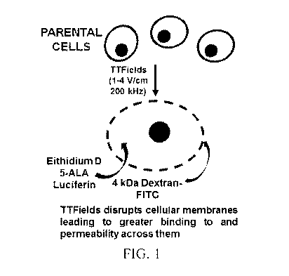
[0019] FIG. 1 is a schematic illustration showing an alternative effect of
TTFields on
modulating the integrity and thus the permeability of cellular membranes.
[0020] FIG. 2A depicts exemplary effects of TTFields on bioluminescence of
U87-
MG/eGFP-fLuc cells from bioluminescent imaging scans as a function of time in
TTFields
vs. no TTFields conditions.
[0021] FIG. 2B depicts the exemplary effects of TTFields on eGFP
fluorescence for
U87-MG/eGFP-fLuc cells as a function of time in TTFields vs. no TTFields
conditions.
[0022] FIG. 2C depicts the effect of TTFields on the fLuc bioluminescence
(fLuc-
BLI) over eGFP fluorescence (eGFP-FL) ratio for U87-MG/eGFP-fLuc cells as a
function of
length of TTFields exposure.
[0023] FIG. 2D depicts the effect of TTFields exposure vs. non-exposure on
the fLuc-
BLI/eGFP-FL ratio as a function of TTFields exposure time.
[0024] FIG. 3A depicts the exemplary effects of TTFields on time-dependent
uptake
of Ethidium D in U87-MG/eGFP-fLuc cells between no TTFields and TTFields (200
kHz).
[0025] FIGS. 3B-3D depict the exemplary impact of TTFields vs. no TTFields
conditions on the time course of Dextran-FITC uptake for 4 kDa Dextran-FITC,
20 kDa
Dextran-FITC, and 50 kDa Dextran-FITC, respectively.
6
CA 03100859 2020-11-18
WO 2020/010188
PCT/US2019/040479
[0026] FIG. 4A depicts the exemplary effect of TTFields (200 kHz) on 5-
aminolevulinic acid (5-ALA) uptake as shown by representative protoporphyrin
IX (PpIX)
fluorescence for TTFields vs. no TTFields-exposed U87-MG cells at 6 and 24
hours.
[0027] FIG. 4B depicts how PpIX fluorescence changed over time in
glioblastoma vs.
fibroblast cells in the co-culture platforms that were subjected to TTFields.
[0028] FIG. 5 provides quantification of the number and size of holes from
a SEM
comparison of plasma membrane holes in U87-MG/eGFP-fLuc cells exposed and
unexposed
to TTFields for 3 days.
[0029] FIG. 6 provides quantification of the number and size of holes from
a SEM
comparison of plasma membrane holes in normal human PCS-201ce11s exposed and
unexposed to TTFields for 3 days.
[0030] FIG. 7A-7C depict the results of experiments showing how alternating
electric
fields reversibly increase uptake in U87-MG cells of D-Luciferin, 5-ALA, and
Dextran-FITC
(4 kDa), respectively.
[0031] FIG. 7D depicts the results of an experiment that shows timing
characteristics
of the permeability that is induced by the application of TTFields to U87-MG
cells.
[0032] FIG. 8A depicts the results of an experiment showing how alternating
electric
fields affect the permeability of MDA-MB-435 cell membranes to 7-AAD.
[0033] FIG. 8B depicts the results of an experiment showing how alternating
electric
fields affect the permeability of MDA-MB-435 and MDA-MB-435 Doxycycline
resistant cell
membranes to doxorubicin.
[0034] FIG. 8C depicts the results of an experiment showing how alternating
electric
fields affect the permeability of MCF-7 and MCF-7 Mitoxantrone resistant cell
membranes to
mitoxantrone.
[0035] FIGS. 9A-9G depict the effect of TTFields on sensitivity to seven
different
combinations of substances and corresponding cell types.
7
CA 03100859 2020-11-18
WO 2020/010188
PCT/US2019/040479
[0036] FIGS. 10A and 10B each depict a suitable timing relationship between
the
application of the alternating electric field and the introduction of the
substance to the vicinity
of the cancer cell.
[0037] FIG. 11A depicts the results of an experiment to determine the
frequency that
provides the highest level of cytotoxicity to U-87 MG cells.
[0038] FIG. 11B depicts the results of an experiment to determine the
frequency that
provides the largest increase in permeability of the cell membranes of U-87 MG
cells.
[0039] FIG. 12A depicts the results of an experiment to determine the
frequency that
provides the highest level of cytotoxicity to MES-SA cells.
[0040] FIG. 12B depicts the results of an experiment to determine the
frequency that
provides the largest increase in permeability of the cell membranes of MES-SA
cells.
[0041] FIG. 12C depicts the results of an experiment to determine how 150
kHz
alternating electric fields affect the permeability of the cell membranes of
MES-SA cells to
doxorubicin.
[0042] FIG. 13A depicts the results of an experiment to determine the
frequency that
provides the highest level of cytotoxicity to MCF-7 cells.
[0043] FIG. 13B depicts the results of an experiment to determine the
frequency that
provides the largest increase in permeability of the cell membranes of MCF-7
cells.
[0044] FIG. 14 depicts the results of an experiment to determine the
frequency that
provides the largest increase in permeability of the cell membranes of
GBM39/Luc cells.
[0045] FIG. 15 is a block diagram of a dual-frequency apparatus that
generates a first
frequency for inducing cellular permeability and a second frequency for
inducing
cytotoxicity.
[0046] Various embodiments are described in detail below with reference to
the
accompanying drawings, wherein like reference numerals represent like
elements.
DETAILED DESCRIPTION OF THE PREFERRED EMBODIMENTS
8
CA 03100859 2020-11-18
WO 2020/010188
PCT/US2019/040479
[0047] As used herein, the term "reducing viability" of a cell refers to
reducing the
growth, proliferation, or survival of the cell, or increasing cytotoxicity of
the cell.
[0048] This application describes a novel approach for temporarily
increasing the
permeability of the plasma cell membranes of cancer cells using alternating
electric fields so
that substances that are ordinarily blocked by the cell membrane will be able
to cross the cell
membrane, or so that substances that are ordinarily impeded by the cell
membrane will be
able to cross the cell membrane more easily. In some of the examples described
herein, this
approach is used for temporarily increasing the permeability of glioblastoma
plasma cell
membranes using alternating electric fields so that substances that are
ordinarily impeded by
the glioblastoma cell membrane will be able to cross the glioblastoma cell
membrane more
easily.
[0049] The inventors have demonstrated that TTFields treatment, in
conjunction with
a novel anticancer compound Withaferin A, synergistically inhibited the growth
of human
glioblastoma cells. The inventors hypothesized that such a synergistic effect
is due to
increased accessibility of Withaferin A to glioblastoma cells through
TTFields' capability to
increase transiently, tumor cell membrane permeability, as depicted
schematically in FIG. 1.
In this figure, 5-ALA = 5- aminolevulinic acid; Ethidium D = ethidium bromide;
and FITC =
fluorescein isothiocyanate.
[0050] Studies were then performed that validate the hypothesis. In
particular,
evidence was found to show that TTFields exposure induced greater
bioluminescence in
human glioblastoma cells that have been modified to express luciferase
(renilla and firefly),
and that this induction is due to increased permeation of the substrates (D-
luciferin and
coelenterazine, respectively), through the plasma membrane. Increased membrane
permeability caused by TTFields exposure was also demonstrated with other
membrane-
penetrating reagents such as Dextran-FITC and Ethidium D.
[0051] Using TTFields to increase membrane permeability in glioblastoma
cells was
also shown using 5-aminolevulinic acid (5-ALA). 5-ALA is a hemoglobin
precursor that is
converted into fluorescent protoporphyrin IX (PpIX) in all mammalian cells.
However, many
malignant cells, including high-grade gliomas, have elevated hemoglobin
biosynthesis, which
is reflected in enhanced accumulation of PpIX within transformed cells and
tissues
(compared to non-cancerous cells). This property has prompted many medical
investigations
9
CA 03100859 2020-11-18
WO 2020/010188
PCT/US2019/040479
to use 5-ALA uptake (and, by consequence, its enzymatic conversion to PpIX) as
a
fluorescent biomarker for tumor cells. However, at the current level of
technology, it can be
difficult to distinguish the precise cellular margin between tumor and non-
tumor tissue
intraoperatively. Experiments described herein show that TTFields
significantly enhances the
tumor to normal cell ratio for PpIX fluorescence (brought on by 5-ALA exposure
and
uptake), and in this manner, may be used to better delineate tumor margins in
intraoperative
settings.
[0052] Further experiments using scanning electron microscopy (SEM) data
demonstrate an increase in the number and size of holes in glioblastoma cell
membranes
caused by TTFields exposure, and that the morphology of the glioblastoma cell
membrane is
perturbed when TTFields are applied. Through all modalities studied
(bioluminescence,
fluorescence, and SEM), the effects of TTFields on the GBM cell membrane
permeability
were found to be reversible after cessation of TTFields exposure.
[0053] RESULTS
[0054] Induction of TTFields increases bioluminescence (BLI) in luciferase-
expressing glioblastomas.
[0055] U87-MG/eGFP-fLuc cells were seeded on Thermanox glass coverslips,
allowed to settle and grow, and then subjected to either TTFields or no
TTFields. In this
experiment, the use of TTFields (4 V/cm, 200 kHz, 0.5-24 h duration)
significantly increased
bioluminescence intensity (BLI) of U87-MG/eGFP-fLuc cells compared to
unexposed
conditions. This increase in BLI occurred as early as 30 minutes after
commencement of
TTFields and continued to 24 h of TTFields exposure. When ROI quantification
was
performed, the time course of BLI intensity for the TTFields-exposed samples
was
significantly elevated compared to TTFields-unexposed samples (p <0.0001, two-
way
ANOVA, TTFields vs. no TTFields). Data depicting the Temporal quantification
of BLI
results of these experiments is summarized in FIG. 2A. Without being bound by
this theory, it
is believed that the elevation in bioluminescence was not due to a direct
effect of TTFields on
firefly luciferase activity because exposure of purified firefly luciferase to
200 kHz TTFields
led to over a 1000-fold loss in enzymatic activity 60 minutes after initiation
of TTFields.
[0056] FIG. 2B depicts the effect of TTFields on eGFP fluorescence in U87-
MG/eGFP-fLuc cells observed from time course of representative images (not
shown) for
CA 03100859 2020-11-18
WO 2020/010188
PCT/US2019/040479
TTFields-exposed vs. TTFields-unexposed U87-MG/eGFP-fLuc. The presence of
TTFields
did not significantly increase eGFP fluorescence (eGFP-FL) over the course of
the
experiments. When ratios of BLI over eGFP-FL was compared between TTFields vs.
no
TTFields samples, there was a significantly augmented ratio with respect to
time of TTFields
incubation for the TTFields samples, as depicted in FIGS. 2C, 2D (p <0.0001,
two-way
ANOVA, TTFields vs. no TTFields). More specifically, FIG. 2C depicts the
effect of
TTFields on the fLuc bioluminescence (fLuc-BLI) over eGFP fluorescence (eGFP-
FL) ratio
for U87-MG/eGFP-fLuc cells as a function of length of TTFields exposure; and
FIG. 2D
depicts the effect of TTFields exposure vs. non-exposure on the fLuc-BLI/eGFP-
FL ratio as a
function of TTFields exposure time (hours). TTFields significantly decreased
activity of
purified firefly luciferase compared to no TTFields (p < 0.01, two-way ANOVA,
TTFields
vs. no TTFields).
[0057] The application of TTFields over time on another patient derived
glioblastoma
cell line, GBM2/GFP-fLuc also induced a time-dependent increase in
bioluminescence in
TTFields-exposed GBM2/GFP-fLuc cells when compared to no-TTFields controls (p
<
0.0001, two-way ANOVA, TTFields vs. no TTFields). This same effect was
observed in a
murine astrocytoma cell line (KR158B) that was genetically modified to express
Renilla
luciferase-red fluorescent protein fusion protein (p < 0.0001, two-way ANOVA,
TTFields vs.
no TTFields). Renilla luciferase activity is not dependent upon ATP and
magnesium (as
opposed to firefly luciferase). Thus, it is believed that the induction of
bioluminescence by
TTFields was not due to alterations in endogenous pools of ATP.
[0058] Effect of TTFields on uptake of membrane-associating reagents.
[0059] To test if the imposition of TTFields affects cell membrane
properties, and
thus membrane permeability, the effect of TTFields on the behavior of
fluorescently tagged
reagents that bind to the cellular membrane was determined. Initially, the
impact of TTFields
on the binding of Annexin-V-APC to the membrane of U87-MG/eGFP-fLuc cells was
measured. Annexin-V-APC binding is a signature of early apoptosis which is
characterized
by ruffling of the membrane. A positive control for apoptosis (addition of 21
pM Withaferin
A to U87-MG/eGFP-fLuc cells) was used to assess the visibility of Annexin-V-
APC binding
to U87-MG/eGFP-fLuc cells, and showed that such binding could be visualized
via
fluorescence microscopy over TTFields-unexposed samples. However, when
TTFields were
applied to U87-MG/eGFP-fLuc cells, Annexin-V-APC binding was not observed at
any time
11
CA 03100859 2020-11-18
WO 2020/010188
PCT/US2019/040479
point of exposure to TTFields. It therefore appears that-TTFields did not
induce any
significant degree of apoptosis in the U87-MG cells.
[0060] Notably, ethidium D uptake was significantly increased when the U87-
MG/eGFP-fLuc cells were subjected to 200 kHz TTFields, as depicted in FIG. 3A
(p <
0.0001, two-way ANOVA, TTFields vs. no TTFields). Ethidium D permeates through
both
the plasma membrane and the nuclear membrane and intercalates into genomic
DNA. Thus,
these findings suggest that TTFields can have an effect on the permeability of
plasma
membranes in U87-MG/eGFP-fLuc cells.
[0061] Another consequence of enhanced membrane permeability by TTFields is
alterations in Dextran-FITC binding on the cell membrane. Dextran-FITC is
known to bind
and intercalate into the plasma membrane. When U87-MG cells were subjected to
1 h of 200
kHz TTFields, there was a significant uptake of Dextran-FITC of molecular
weights 4 kDa
and 20 kDa, compared to no TTFields exposure, as depicted in FIGS. 3B and 3C.
But there
was no significant difference in uptake for 50 kDa Dextran- FITC, as depicted
in FIG. 3D.
More specifically, Dextran-FITC binding was examined in the presence of
TTFields over a
timeframe of 0.5-24 h exposure, a significant increase in the uptake of 4 kDa
Dextran-FITC
was found compared to TTFields-unexposed samples (p <0.0001, two-way ANOVA,
TTFields vs. no TTFields), a significant increase in uptake of 20 kDa Dextran-
FITC under
TTFields exposure (p <0.01, TTFields vs. no TTFields) and no significant
difference in
uptake of 50 kDa Dextran-FITC under TTFields exposure (p= 0.26, not
significant, TTFields
vs. no TTFields). These data suggest that the maximum size of Dextran-FITC
that bound to
and entered the plasma membrane under TTFields exposure in this experiment was
between
about 20 and 50 kDa. In all statistical comparisons described in this
paragraph, each data
point represents n = 3 experiments. In FIGS 3A-3D, APC = allophycocyanin;
Ethidium D =
ethidium bromide; and FITC = fluorescein isothiocyanate.
[0062] Effect of TTFields on 5-aminolevulinic (5-ALA) acid uptake: single
U87-
MG culture.
[0063] Experiments were performed to determine the effects of TTFields on
uptake of
5- ALA (as measured by PpIX accumulation and its resultant fluorescence) in
glioblastoma
cells. Because it is difficult to distinguish the margin between tumor and
normal cells using
the present 5-ALA bioassay, the measurement of PpIX fluorescence was used to
address this
12
CA 03100859 2020-11-18
WO 2020/010188
PCT/US2019/040479
issue. Investigations were run to determine whether permeation of 5-ALA
through the
cellular membrane and into the glioblastoma cells could be increased with
TTFields
exposure. U87-MG cells were exposed or unexposed to TTFields, each for
durations of 6-24
h. The results, which are summarized in FIG. 4A, were as follows: TTFields
exposure
resulted in significantly increased uptake of 5-ALA into U87-MG/ eGFP-fLuc
cells as early
as 6 h of TTFields exposure (p = 0.047, Student's t-test, TTFields vs. no
TTFields) and this
increase was maintained with prolonged TTFields exposure of 24 h (p = 0.011).
[0064] To generate the data depicted in FIG. 4A, protoporphyrin IX (PpIX)
fluorescence panels were obtained for TTFields-unexposed vs. TTFields-exposed
U87-MG
cells after 6 and 24 h of exposure. Quantitation of those images in a showed
significant
increase in PpIX signals in TTFields exposed cells compared to no TTFields, at
both 6 h (p =
0.047) and 24 h (p = 0.01) time points. All monovariant statistical
comparisons between no
TTFields vs. TTFields samples done by Student's t-test for n =3 experiments
per time point.
[0065] Effect of TTFields on 5-aminolevulinic acid uptake: U87-MG GBM on
PCS-201 fibroblast co-cultures.
[0066] During glioblastoma resection in patients, 5-ALA is used to aid
neurosurgeons
in delineating between the tumors and surrounding normal brain tissue.
Likewise, to
distinguish differences in 5-ALA uptake between glioblastoma and normal cells,
a co-culture
was developed where U87-MG cells were seeded in the center of a bed of PCS-201
fibroblasts and were subjected to TTFields or to no TTFields. Fluorescent and
brightfield
photomicrographs confirmed the presence of discrete glioblastoma vs.
fibroblast cell regions
in the co-culture set-up. When co-cultures were stained with hematoxylin and
eosin (H&E),
photomicrographs revealed reduced numbers of GBM cells infiltrating into the
fibroblast
periphery for TTFields-exposed samples.
[0067] In particular, without TTFields exposure, the GBM cells formed many
pockets
of adherent neurospheres as was previously reported. Fluorescence images
showed increased
PpIX fluorescence in glioblastoma vs. fibroblast cells in the co-culture
platforms that were
subjected to TTFields for 6 h. The results, which are summarized in FIG. 4B,
were as
follows: PpIX fluorescence accumulated over time but the rate of fluorescence
intensity
increase was significantly augmented (p < 0.001, two-way ANOVA, TTFields vs.
no
TTFields) for TTFields-exposed co-cultures compared to TTFields unexposed co-
cultures. To
13
CA 03100859 2020-11-18
WO 2020/010188
PCT/US2019/040479
generate the data depicted in FIGS. 4B, fluorescent panels of 5-ALA uptake
(and subsequent
PpIX fluorescence, Ex = 558 nm, Em = 583 nm) for no TTFields and TTFields were
obtained. Duration of exposures are 2, 6, and 24 h. Quantification of time
course of PpIX
accumulation (and thus accumulation of fluorescent flux as expressed as
photons/s) in the
glioblastoma-fibroblast co-culture platform under TTFields exposed vs.
unexposed
conditions (p < 0.001). Statistical analyses consisted of two-way ANOVA for no
TTFields vs.
TTFields conditions, and n = 3 experiments per time point.
[0068] In a separate set of experiments, by 24 h of TTFields application,
the ratio of
PpIX fluorescence intensity in the U87-MG glioblastoma cells over the
surrounding PCS-201
fibroblast cells was significantly increased compared to the fluorescence
intensity ratio for
co-cultured cells under no TTFields conditions (p = 0.043, two-way ANOVA,
TTFields vs.
no TTFields).
[0069] Scanning electron micrograph (SEM) shows that TTFields alters
membrane morphology of U87-MG/eGFP-fLuc cells.
[0070] SEM images of low density (5,000 cells/coverslip) U87-MG/eGFP-fLuc
cells
that were either not exposed to TTFields or exposed to TTFields for 3 days
were obtained at
2000x, 20,000x, and 60,000x magnifications. Data obtained by reviewing these
SEM images
is summarized in FIG. 5. There was a significantly increased number of holes
greater than
51.8 nm2 in size (equivalent to 9 pixels2 on 60,000x magnification) within the
ROI of
TTFields-exposed cells (53.5 19.1) compared to the TTFields-unexposed cells
(23.9
11.0), (p = 0.0002, univariate Mann¨Whitney test). Average size of the holes
within the ROI
was also significantly greater in TTFields-exposed cells (240.6 91.7 nm2)
compared to
TTFields-unexposed cells (129.8 31.9 nm2), (p = 0.0005 (univariate
Mann¨Whitney test)).
To obtain the data depicted in FIG. 5, Quantification and comparison between
TTFields
unexposed and exposed cells of the number and size of holes was done within a
500 nm-
radius circular region of interest. The minimum hole size cut-off was based on
the 3.3 and 5.0
nm Stokes radii of 20 kDa and 50 kDa Dextran-FITCs, respectively. Coverslips
from three
experiments per condition were used, and at least 5 cells per coverslip were
analyzed for hole
count and size, in a double-blind manner.
[0071] The effects of a 24-h exposure to TTFields on the plasma membranes
of U87-
MG cells seeded at high density were also visually observed. For the no
TTFields samples,
14
CA 03100859 2020-11-18
WO 2020/010188
PCT/US2019/040479
the cell surface appeared to be covered in densely matted, elongated and
flattened membrane
extensions, similar to membrane ruffles and contiguous with the cellular
membrane. In
contrast, after 24 h of exposure to TTFields, the densely matted and elongated
structures were
replaced by short, bulbous and bleb-like structures.
[0072] For comparison, SEM images of normal human PCS-201 cells were also
obtained and analyzed. PCS-201 cells were seeded at low density (5,000 cells
per 13 mm
glass coverslip). The cells were grown under standard tissue culture
conditions (37 C, 95%
02, 5% CO2). Non-TTFields-exposed cells were left under those conditions for
the duration
of the study. Other cells were exposed to TTFields for 72 h. After 72 hours,
the SEM images
were obtained at 2000x, 20,000x, and 60,000x magnifications. Quantification
and
comparison between TTFields unexposed and exposed cells of the number and size
of holes
with area? 51.8 nm2(equivalent to a 4-nm radius circle, or 9 pixe152on the
60,000x
magnification images) within a 500 nm-radius circular region of interest. The
minimum hole
size cut-off was based on the 3.3 nm and 5.0 nm Stokes radii of 20 kDa and 50
kDa Dextran-
FITCs, respectively. The results, which are depicted in FIG. 6, were as
follows: There was no
significant difference in the number or size of holes between the TTFields
unexposed and
exposed normal human PCS-201 cells (Wilcoxon rank-sum analysis). Coverslips
from three
experiments per condition were used, and at least 5 cells per coverslip were
analyzed for hole
count and size, in a double-blind manner
[0073] The effects of a 24-h exposure to TTFields on the plasma membranes
of PCS-
201 cells were also visually observed. Unlike the situation described above
for the U87-MG
cells, TTFields did not appear to alter the membrane morphology of the PCS-201
cells.
[0074] The effect of TTFields on membrane permeability is reversible.
[0075] To assess the reversibility of the effect of TTFields on cancer
cells, U87-
MG/eGFP-fLuc cells were subjected to three conditions: (1) No TTFields
exposure, standard
cell culture conditions (37 C, 95% 02, 5% CO2), (2) TTFields exposure for 24
h and (3)
TTFields exposure for 24 h followed by no TTFields exposure for 24 h. The
readouts of BLI,
PpIX fluorescence (5-ALA product) and Dextran-FITC (4 kDa) fluorescence were
acquired.
All experimental conditions were done in triplicate. FIG. 7A summarizes the
data for BLI:
The presence of TTFields for 24 h (middle bar) significantly increased BLI
flux compared to
no TTFields exposure (left bar) (p < 0.0005, two-way ANOVA, TTFields vs. no
TTFields)
CA 03100859 2020-11-18
WO 2020/010188
PCT/US2019/040479
but this increase was significantly attenuated when the cells were re-
introduced to the no
TTFields condition for 24 h (right bar) (two-way ANOVA, p <0.005, TTFields for
24 h vs.
TTFields for 24 h followed by no TTFields for 24 h). FIG. 7B shows that a
similar pattern of
reversible readouts occurred with PpIX fluorescence (p <0.0005, two-way ANOVA,
TTFields (middle bar) vs. no TTFields (left bar) and p < 0.0004, TTFields vs.
TTFields
followed by no TTFields (right bar)). And FIG. 7C shows that a similar pattern
of reversible
readouts occurred for 4 kDa Dextran-FITC fluorescence (p < 0.05, two-way
ANOVA,
TTFields (middle bar) vs. no TTFields (left bar); and p <0.05, TTFields vs.
TTFields
followed by no TTFields (right bar)). For each experimental set, eGFP
fluorescence did not
significantly change. SEM investigations also revealed that the significant
augmentation in
both the number of holes (p = 0.007, two-way ANOVA, TTFields vs. No TTFields)
and the
size of holes (p = 0.0007, two-way ANOVA, TTFields vs. No TTFields) by
TTFields were
reversible as well, after 24 h of no exposure. Here, NS = not significant; BLI
=
bioluminescent imaging; eGFP = enhanced green fluorescence protein; fLuc =
firefly
luciferase; 5-ALA = 5 aminolevulinic acid; FITC = fluorescein isothiocyanate;
PpIX =
protoporphyrin IX; and FL = fluorescence.
[0076] To summarize, the uptake of the relevant compounds increased when
alternating electric fields were applied (as compared to when alternating
electric fields were
not applied). Each of these figures also shows that the uptake decreased
substantially after
cessation of the alternating electric fields for 24 hours. From this, we can
infer that the
increase in permeability of the cell membranes that was induced by the
alternating electric
fields is not a permanent effect, and that the permeability drops back down
after cessation of
the alternating electric fields.
[0077] FIG. 7D depicts the results of an experiment to test how quickly the
permeability drops back down after cessation of the alternating electric
fields. More
specifically, 7-Aminoactinomycin D (7-AAD) is a fluorescent chemical compound
with a
strong affinity for DNA. 7-AAD is a relatively large molecule (1270.43 g/mol,
i.e., 1.27 kDa)
that ordinarily does not readily pass through intact cell membranes. FIG. 7D
shows timing
characteristics of the permeability to 7-AAD that is induced by the
application of TTFields
for U87-MG cells. In this experiment, the cells were treated with an
alternating electric field
at 300 kHz with a field strength of 1.62 V/cm RMS for 24 hours, at an ambient
temperature
of 18 C. 7-AAD was introduced into samples at five different times: 15
minutes prior to the
16
CA 03100859 2020-11-18
WO 2020/010188
PCT/US2019/040479
cessation of the alternating electric field; immediately after cessation of
the alternating
electric field; and 15, 30, and 60 minutes after cessation of the alternating
electric field. In
each case, the cells were incubated with 7-AAD for 30 minutes after
introduction of the 7-
AAD, followed by flow cytometry analysis of the percentage of cells with
increased
accumulation of the fluorescent 7-AAD for each of the different timings. As
seen in FIG. 7D,
a significant increase in accumulation of 7-AAD was observed only in the
sample that was
incubated with 7-AAD while subjected to an alternating electric field.
[0078] Additional results for different drugs and different types of cancer
cells.
[0079] The methods described herein are not limited to the context of
glioblastoma.
To the contrary ¨ they are applicable to other types of cancer cells. More
specifically, a
substance can be delivered across a cell membrane of a cell by (a) applying an
alternating
electric field to the cell for a period of time, wherein application of the
alternating electric
field increases permeability of the cell membrane; and (b) introducing the
substance to a
vicinity of the cell. The increased permeability of the cell membrane enables
the substance to
cross the cell membrane. Notably, the methods described herein may be used to
deliver large
molecules (which ordinarily would not pass through the relevant cell membrane)
through a
cell membrane of different types of cells (i.e., cells other than
glioblastoma), including but
not limited to other types of cancer cells (e.g., MDA-MB-435 and MCF-7 cells).
[0080] FIG. 8A depicts the results of an experiment performed to determine
how
alternating electric fields affect the permeability of the cell membranes of
MDA-MB-435
human melanoma cell line cells. In this experiment, MDA-MB-435 cells were
treated with an
alternating electric field at 150 kHz with a field strength of 1.62 V/cm for
24 hours, at an
ambient temperature of 18 C and a dish temperature of 37 C. (The dish
temperature in this
and other examples is higher than the ambient temperature due to heating
caused by the
alternating electric fields.) After the first 23.75 hours, 7-AAD was added to
the culture and
incubated for 15 minutes during which time the alternating electric fields was
continued (to
complete the 24 hour period). After this 15 minute period, alternating
electric field
application was terminated and the cells were incubated at room temperature
for an additional
15 minutes. The percentage of cells with increased accumulation of the
fluorescent 7-AAD
was determined using flow cytometry analysis. ¨66% of the cells exhibited an
increased
accumulation of 7-AAD (bar 2 in FIG. 8A), as compared to less than 5% of the
cells in the
control (bar 1), which was subjected to the same conditions, except that the
alternating
17
CA 03100859 2020-11-18
WO 2020/010188
PCT/US2019/040479
electric fields were not applied. These results indicate that alternating
electric fields cause a
very significant increase in the permeability of cell membranes.
[0081] In a variation of this experiment, MDA-MB-435 human melanoma cell
line
cells were treated with an alternating electric field at 150 kHz with a field
strength of
1.62 V/cm for 24 hours, at an ambient temperature of 18 C and a dish
temperature of 37 C.
After this 24 hour period, the alternating electric fields were turned off for
15 minutes, after
which the 7-AAD was added. After waiting an additional 15 minutes, the
percentage of cells
with increased accumulation of the fluorescent 7-AAD was determined using flow
cytometry.
This time, only ¨20% of the cells exhibited an increased accumulation of 7-AAD
(bar 3 in
FIG. 8A). These results indicate that the increase in permeability of cell
membranes that is
induced by alternating electric fields is relatively short-lived, and that the
permeability
declines rapidly and dramatically after cessation of the alternating electric
fields.
[0082] FIG. 8B depicts the results of another experiment performed to
determine how
alternating electric fields affect the permeability of the cell membranes of
MDA-MB-435
human melanoma cell line cells to doxorubicin (543.52 g/mol). In this
experiment, both wild
type and doxorubicin resistant variants of MDA-MB-435 cells were treated with
an
alternating electric field at 150 kHz with a field strength of 1.62 V/cm for
23 hours. After this
23 hour period, doxorubicin at a concentration of 10 uM was added and
incubated for one
hour, during which time the alternating electric fields was continued. The
intracellular
accumulation of doxorubicin was then measured. The intracellular accumulation
of
doxorubicin increased for both the wild type cells (compare bar 1 to bar 3)
and the
doxorubicin resistant cells (compare bar 2 to bar 4).
[0083] FIG. 8C depicts the results of a similar experiment using MCF-7
human breast
adenocarcinoma cell line cells and mitoxantrone (444.481 g/mol). In this
experiment, both
wild type and mitoxantrone resistant variants of MCF-7 cells were treated with
an alternating
electric field at 150 kHz with a field strength of 1.62 V/cm for 23 hours.
After this 23 hour
period, mitoxantrone at a concentration of 2 uM was added and incubated for
one hour,
during which time the alternating electric fields was continued. The
intracellular
accumulation of mitoxantrone was then measured. The intracellular accumulation
of
mitoxantrone increased for both the wild type cells (compare bar 1 to bar 3)
and the
mitoxantrone resistant cells (compare bar 2 to bar 4).
18
CA 03100859 2020-11-18
WO 2020/010188
PCT/US2019/040479
[0084] The results described above in connection with FIGS. 8B and 8C
indicate that
the alternating electric fields improve intracellular accumulation of
chemotherapy molecules
in both wild type and drug-resistant cells, and that alternating electric
fields can
advantageously restore intra-cellular accumulation of chemotherapeutic
chemicals in cancer
cells after those cells have developed multi drug resistance to those
chemicals.
[0085] Additional experiments were performed to determine whether synergy
exists
between TTFields and various drugs for various cancer cell lines, and FIGS. 9A-
9G depict
the result of some of these experiments. More specifically, FIG. 9A shows how
applying
TTFields for 3 days improves the sensitivity of U87-MG/GFP-Luc cells to
various
concentrations of Lomustine (as compared to a control in which TTFields were
not applied).
FIG. 9B shows how applying TTFields for 3 days improves the sensitivity of
pcGBM2/GFP-
Luc cells to various concentrations of Lomustine (as compared to a control in
which TTFields
were not applied). FIG. 9C shows how applying TTFields for 3 days improves the
sensitivity
of GBM39 cells to various concentrations of Lomustine (as compared to a
control in which
TTFields were not applied). FIG. 9D shows how applying TTFields for 3 days
improves the
sensitivity of GBM39 cells to various concentrations of Temozolomide (as
compared to a
control in which TTFields were not applied). FIG. 9E shows how applying
TTFields for 3
days improves the sensitivity of GBM39/Luc cells to various concentrations of
Irinotecan (as
compared to a control in which TTFields were not applied). FIG. 9F shows how
applying
TTFields for 3 days improves the sensitivity of MDA-MB-235 cells to various
concentrations
of Doxorubicin (as compared to a control in which TTFields were not applied).
And FIG. 9G
shows how applying TTFields for 4 days improves the sensitivity of U87-MG/eGFP-
Luc
cells to various concentrations of Mannose (as compared to a control in which
TTFields were
not applied).
[0086] To date, synergy was found for the combination of TTFields plus
Withaferin
A for GBM39/Luc, U87-MG/GFP-Luc, and pcGBM2/GFP-Luc; synergy was found for the
combination of TTFields plus Lomustine for GBM39/Luc, U87-MG/GFP-Luc, and
pcGBM2/GFP-Luc; synergy was found for the combination of TTFields plus
Irinotecan for
GBM39/Luc; and synergy was found for the combination of TTFields plus Mannose
for U87-
MG/GFP-Luc. Evidence of synergy was also found for the combination of TTFields
plus
Doxorubicin for MDA-MB-235.
19
CA 03100859 2020-11-18
WO 2020/010188
PCT/US2019/040479
[0087] DISCUSSION
[0088] Previous studies have focused on the effects of TTFields on the
nucleus (e.g.,
microtubules), septin, mitochondria, and autophagy. But the experiments
described herein are
believed to be the first to report the effects of TTFields on cancer cellular
membrane
integrity, and demonstrate increased cellular membrane permeability for cancer
cells (e.g.,
multiple human GBM cell lines) in the presence of TTFields using various
evaluation
techniques (e.g., bioluminescence imaging, fluorescence imaging, and scanning
electron
microscopy).
[0089] Observations revealed increased cellular membrane permeability for
glioblastomas in the presence of TTFields across multiple human GBM cell
lines. The
approaches employed to validate the hypothesis included bioluminescence
imaging,
fluorescence imaging, and scanning electron microscopy. Observations also
revealed
increased cellular membrane permeability for other types of cancer cells in
the presence of
TTFields. Studies of TTFields in combination with chemotherapies have shown
both
therapeutic additivity and synergy. For this study, we posited that TTFields
mediates
improved accessibility to cancer cells. Several experiments showed the
reversibility of the
TTFields effect on membranes thus demonstrating a causal relationship between
TTFields
and the increase in membrane permeability. Such observations also suggest that
TTFields
could be used to tune drug accessibility to cancer cells.
[0090] The investigation into the cell permeability hypothesis of TTFields
action was
initiated partly because of observations of increased bioluminescence in
luciferase-expressing
GBM cells by TTFields. While not being bound by this theory, it is believed
that TTFields
induced increased permeability in the cellular membranes of GBM cells. It is
believed that
increased GBM cell permeability to D-luciferin as measured by BLI was not due
to the
effects of TTFields on luciferase itself, but rather due to an increased
influx of its substrate
D-luciferin into the cells engineered to express the firefly luciferase.
Furthermore, this
finding held true for both ATP-dependent (FLuc) and ATP-independent luciferase
(RLuc).
Therefore, despite a preliminary report suggesting that intracellular ATP was
increased in
CT26 colorectal carcinoma cells exposed to TTFields, the observation of
increased
glioblastoma cell membrane permeability in the setting of TTFields exposure
suggests an
independent phenomenon. An increased expression or activation of luciferase
due to
TTFields exposure could not have explained the increased BLI signal because in
these cells
CA 03100859 2020-11-18
WO 2020/010188
PCT/US2019/040479
the luciferase enzyme was controlled by the same promoter as was eGFP, and an
increase in
fluorescence signal was not observed in the same cells. However, exposure to
TTFields may
affect cellular metabolism that would be manifested by changes in ATP levels,
alterations in
membrane morphology and shifts in oxygen consumption.
[0091] Some key findings supporting the permeability hypothesis came from
the
Dextran-FITC validation experiments described above in connection with FIGS.
3B-D. The
accessibility of the cell membrane to small probes in the setting of TTFields
was tested with
FITC-labeled dextrans, which resulted in an increase in influx of 4 kDa
(Stokes' radius ¨1.4
nm) and 20 kDa (Stokes' radius ¨3.3 nm) but not 50 kDa dextrans (Stokes'
radius ¨5 nm).
This suggests that TTFields cause GBM cells to become more permeant to
substances as
large as 20 kDa, but no greater than 50 kDa. For reference, the luciferin and
coelenterazine
substrates are of small enough molecular weight to be accessible through the
membrane with
TTFields exposure. D-luciferin (substrate for Firefly luciferase) has a
molecular weight of
280.3 g/mol (-280 Da), coelenterazine H (substrate for Renilla luciferase) has
a molecular
weight of 407.5 g/mol (-408 Da), and 5-ALA has a molecular weight of 167.6
g/mol (169
Da), consistent with the Dextran-FITC findings.
[0092] The SEM findings described herein reveal that at low seeding
density, 3 days
of TTFields exposure caused a significant increase in the number and size of
holes greater
than 51.8 nm2 in area, compared to the no TTFields condition, as described
above in
connection with FIG. 5. This hole size cut-off represents a circle of radius
4.1 nm, which is
the Stokes' radius of a FITC-dextran molecule with a size of 20-40 kDa. Thus,
the difference
in cell membrane disruption visualized by SEM confirms the indirect
observations from the
FITC-dextran studies described herein.
[0093] Interestingly, exposure of normal human fibroblasts (PCS-201) to
TTFields
caused no significant increase in the number or size of cellular membrane
holes, thus
suggesting that the permeability effect may have some specificity to cancer
cells.
Qualitatively, for U87-MG cells, there was a clear onset of bulbous, bleb-like
structures due
to a 24-h exposure to TTFields under high seeding density. The appearance of
these
structures is consistent with increased permeability in the outer membrane and
the induction
of apoptosis and there appears to be little evidence of an apoptotic phenotype
with a 24-h
TTFields exposure. Furthermore, high-density PCS-201 cells displayed no such
changes with
21
CA 03100859 2020-11-18
WO 2020/010188
PCT/US2019/040479
TTFields exposure (data not shown) thus suggesting again, the specificity of
the TTFields
effect for cancer cells.
[0094] Although the cell cycle was not synchronized for the experiments,
the
doubling time of the U87-MG cells is ¨48 h and given that TTFields exert their
maximal
antiproliferative effect on dividing cells, this could explain the lack of
observed abundant
apoptosis after a 24-h TTFields exposure. An alternative interpretation may
lie in reports that
cellular blebbing may confer resistance to cellular lysis. A previous report
in unsynchronized
glioblastoma cells demonstrated that 72 h of TTFields exposure induced cell
death with a
marked proportion of Annexin V-positive cells. Using transmission electron
microscopy,
these reports described signs of autophagy including autophagosomes, swollen
mitochondria,
and a dilated endoplasmatic reticulum. In contrast, the results herein use SEM
to better
visualize the effects of TTFields specifically on the plasma cell membrane.
[0095] The increase in membrane permeability by TTFields has significant
clinical
implications. Using the co-culture platform of human GBM cells layered on top
of normal
human fibroblast cells, the impact of TTFields on the uptake of 5-
aminolevulinic acid (S-
ALA) into GBM cells was studied. TTFields exposure resulted in significantly
increased 5-
ALA uptake in the GBM cells compared to the fibroblast cells. In June 2017, 5-
ALA was
approved by the Food and Drug Administration for clinical use in the United
States to assist
neurosurgeons in delineating the tumor-normal brain border during glioma
resection.
Pretreating glioma patients with TTFields prior to 5-ALA administration will
therefore be
useful to enhance the delineation of the infiltrative tumor margin during
tumor resection.
[0096] With regard to detecting and measuring the effects of TTFields on
cancer
cells, the majority of cell culture-based studies to date have focused on cell
count/viability as
the primary readout. This is based on the prevailing understanding that
TTFields interferes
with mitosis of rapidly dividing tumor cells, which results in cancer cell
death. In addition,
computational modeling studies of TTFields in cell culture are currently
driven by cell count
as the primary outcome of the model.
[0097] Recurrence of GBM is inevitable and the median time to first
recurrence
despite standard therapy is approximately 7 months. In clinical applications
of TTFields to
patients with GBM, the data suggest that increased compliance and duration of
TTFields use
correlates with improved survival. TTFields compliance (>75% vs. <75%) was an
22
CA 03100859 2020-11-18
WO 2020/010188
PCT/US2019/040479
independent predictor of overall survival in the retrospective analysis of the
full EF-14 trial
dataset and the duration of use of TTFields was also found to affect overall
survival. Taken
together, these data may serve as clinical correlates of the observed effects
in the cell
cultured-based TTFields experimental setting. Namely, a correlation between
the length of
TTFields exposure and the duration of its effect on cell membrane permeability
after
cessation of TTFields was observed. At lengths of TTFields exposure of 0.5-3
h, the duration
in BLI augmentation (compared to no TTFields conditions) lasted about 5 min.
However, at
TTFields exposures of 12-25 h, this difference in BLI between TTFields and no
TTFields
conditions lasted for more than 20min. Likewise, a re-analysis of the data
reported by Ram et
al. shows that the percent increase in overall survival (in patients treated
with TTFields plus
temozolomide vs. temozolomide alone) jumped from 32% after 1 year of TTFields
exposure
to 551% after 5 years of TTFields exposure, respectively.
[0098] The results described herein i.e., that alternating electric fields
increase
cellular membrane permeability, are distinct from the previously reported
effects of TTFields.
This should have a significant impact on current surgical and clinical
practices in the
treatment of glioblastomas as well as other types of cancer.
[0099] In the in vitro experiments described above, the frequency of the
alternating
electric fields was 200 kHz. But in alternative embodiments, the frequency of
the alternating
electric fields could be another frequency, e.g., about 200 kHz, between 50
and 500 kHz,
between 25 kHz and 1 MHz, between 50 and 190 kHz, between 25 and 190 kHz, or
between
210 and 400 kHz.
[0100] In the in vitro experiments described above, the field strength of
the
alternating electric fields was between 1 and 4 V/cm RMS. But in alternative
embodiments,
different field strengths may be used (e.g., between 0.1 and 10 V/cm).
[0101] In the in vitro experiments described above, the alternating
electric fields were
applied for a variety of different intervals ranging from 0.5 hours to 72
hours. But in
alternative embodiments, a different duration may be used (e.g., between 0.5
hours and 14
days). In some embodiments, application of the alternating electric fields may
be repeated
periodically. For example, the alternating electric fields may be applied
every day for a two
hour duration.
23
CA 03100859 2020-11-18
WO 2020/010188
PCT/US2019/040479
[0102] In the in vitro experiments using the InovitroTM system described
herein, the
direction of the alternating electric fields was switched at one second
intervals between two
perpendicular directions. But in alternative embodiments, the direction of the
alternating
electric fields can be switched at a faster rate (e.g., at intervals between 1
and 1000 ms) or at
a slower rate (e.g., at intervals between 1 and 100 seconds).
[0103] In the in vitro experiments using the InovitroTM system described
herein, the
direction of the alternating electric fields was switched between two
perpendicular directions
by applying an AC voltage to two pairs of electrodes that are disposed 90
apart from each
other in 2D space in an alternating sequence. But in alternative embodiments
the direction of
the alternating electric fields may be switched between two directions that
are not
perpendicular by repositioning the pairs of electrodes, or between three or
more directions
(assuming that additional pairs of electrodes are provided). For example, the
direction of the
alternating electric fields may be switched between three directions, each of
which is
determined by the placement of its own pair of electrodes. Optionally, these
three pairs of
electrodes may be positioned so that the resulting fields are disposed 90
apart from each
other in 3D space. In other alternative embodiments, the electrodes need not
be arranged in
pairs. See, for example, the electrode positioning described in US patent
7,565,205, which is
incorporated herein by reference. In other alternative embodiments, the
direction of the field
remains constant.
[0104] In the in vitro experiments using the InovitroTM system described
herein, the
electrical field was capacitively coupled into the culture because the
InovitroTM system uses
conductive electrodes disposed on the outer surface of the dish sidewalls, and
the ceramic
material of the sidewalls acts as a dielectric. But in alternative
embodiments, the electric field
could be applied directly to the cells without capacitive coupling (e.g., by
modifying the
InovitroTM system configuration so that the conductive electrodes are disposed
on the
sidewall's inner surface instead of on the sidewall's outer surface).
[0105] The methods described herein can also be applied in the in vivo
context by
applying the alternating electric fields to a target region of a live
subject's body, for both
glioblastoma cells and other types of cancer cells. Imposing the electric
field in the target
region will increase the permeability of the cell membranes in the target
region, which will
enable molecules that are ordinarily blocked or impeded by the cell membrane
to pass
through the cell membrane. This may be accomplished, for example, by
positioning
24
CA 03100859 2020-11-18
WO 2020/010188
PCT/US2019/040479
electrodes on or below the subject's skin so that application of an AC voltage
between
selected subsets of those electrodes will impose the alternating electric
fields in the target
region of the subject's body.
[0106] For example, in situations where the relevant cells are located in
the subject's
lungs, one pair of electrodes could be positioned on the front and back of the
subject's thorax,
and a second pair of electrodes could be positioned on the right and left
sides of the subject's
thorax. In some embodiments, the electrodes are capacitively coupled to the
subject's body
(e.g., by using electrodes that include a conductive plate and also have a
dielectric layer
disposed between the conductive plate and the subject's body). But in
alternative
embodiments, the dielectric layer may be omitted, in which case the conductive
plates would
make direct contact with the subject's body. In another embodiment, electrodes
could be
inserted subcutaneously below a patent's skin. An AC voltage generator applies
an AC
voltage at a selected frequency (e.g., between 100 and 200 kHz) between the
right and left
electrodes for a first period of time (e.g. 1 second), which induces
alternating electric fields
where the most significant components of the field lines are parallel to the
transverse axis of
the subject's body. Then, the AC voltage generator applies an AC voltage at
the same
frequency (or a different frequency) between the front and back electrodes for
a second
period of time (e.g. 1 second), which induces alternating electric fields
where the most
significant components of the field lines are parallel to the sagittal axis of
the subject's body.
This two step sequence is then repeated for the duration of the treatment.
Optionally, thermal
sensors may be included at the electrodes, and the AC voltage generator can be
configured to
decrease the amplitude of the AC voltages that are applied to the electrodes
if the sensed
temperature at the electrodes gets too high. In some embodiments, one or more
additional
pairs of electrodes may be added and included in the sequence. In alternative
embodiments,
only a single pair of electrodes is used, in which case the direction of the
field lines is not
switched. Note that any of the parameters for this in vivo embodiment (e.g.,
frequency, field
strength, duration, direction-switching rate, and the placement of the
electrodes) may be
varied as described above in connection with the in the vitro embodiments. But
care must be
taken in the in vivo context to ensure that the electric field remains safe
for the subject at all
times.
[0107] A wide variety of applications for increasing the permeability of
cell
membranes can be readily envisioned in the in vivo context. In one example,
localized
CA 03100859 2020-11-18
WO 2020/010188
PCT/US2019/040479
enhancement of drug uptake by tumor cells can be induced by applying
alternating electric
fields to the relevant body part for a period of time (e.g., 12 or 24 hours)
prior to and during
administration of chemotherapies or other antineoplastic agents. In another
example, drug
uptake by multi drug resistant tumor cells can be restored by applying
alternating electric
fields to the relevant body part for a period of time (e.g., 12 or 24 hours)
prior to and during
administration of chemotherapies or other antineoplastic agents. In another
example,
development of multi drug resistant metastases can be prevented by applying
alternating
electric fields to regions that are prone to metastases for a period of time
(e.g., 12 or 24
hours) prior to and during administration of an appropriate drug (regardless
to whether the
subject has a primary tumor that is being treated with alternating electric
fields).
[0108] FIG. 10A depicts a first suitable relationship in timing between the
application
of the alternating electric field and the introduction of the substance to the
vicinity of the
cancer cell in the in vitro context; or between the application of the
alternating electric field
and the administration of the substance to a live patient. Based on the data
described above in
connection with FIGS. 7A-7D and 8A, and assuming that the substance is
introduced or
administered at a given time t=0, the alternating electric field can begin
after the given time
and continue for an interval of time (e.g., 12 hours) while the substance is
still available in the
vicinity of the cell. In this situation, permeability will begin to increase
after the alternating
electric field begins, and this increase in permeability will enable the
substance to enter the
relevant cells. In the context of chemotherapy, this would correspond to
administering a
chemotherapeutic agent to a patient, followed by application of the
alternating electric fields
for an interval of time (e.g., for 12 hours).
[0109] Alternatively, as depicted in FIG. 10B, the alternating electric
field can begin
before the given time (e.g., 1 hour before t=0), and continue for an interval
of time (e.g., until
12 hours following t=0) while the substance is still available in the vicinity
of the cell. In this
situation, the permeability of the relevant cells will begin to increase
before the substance
arrives in the vicinity of the cell (or before the substance is administered
to a live patient).
This will enable the substance to cross the cell membrane immediately upon its
arrival in the
vicinity of the cell. In the context of chemotherapy, this would correspond to
starting
application of the alternating electric fields, followed by the administration
of the
chemotherapeutic agent while the alternating electric fields are still being
applied, followed
by continued application of the alternating electric fields for an additional
interval of time
26
CA 03100859 2020-11-18
WO 2020/010188
PCT/US2019/040479
(e.g., until 12 hours following the time at which the chemotherapeutic agent
was
administered).
[0110] Note that the intervals of time discussed above in connection with
FIGS. 10A
and 10B can either be uninterrupted or can include breaks that are preferably
short. For
example, assuming that the interval of time is 12 hours, it could be satisfied
by a single
uninterrupted block of 12 hours. Alternatively, the 12 hour interval could be
satisfied by
applying the alternating electric fields for 6 hours, followed by a 1 hour
break, followed by
applying the alternating electric fields for an additional 6 hours (while the
substance is still
available in the vicinity of the cell). Note also that in the context of FIGS.
10A and 10B,
when the substance is administered to a live patient, the administration of
the substance may
be performed using any of a variety of approaches including but not limited to
intravenously,
orally, subcutaneously, intrathecal, intraventricularly, and intraperitonealy.
[0111] The optimal frequency, field strength, and switching characteristics
may be
determined experimentally for each combination of a given type of host cell
and a given type
of substance that is to be delivered through the cell membrane. In some
preferred
embodiments, the frequency is less than 190 kHz (e.g., between 50 and 190 kHz
or between
25 and 190 kHz. In other preferred embodiments, the frequency is between 210
and 400 kHz.
[0112] One existing approach to treating tumors (e.g., glioblastoma) is by
applying
alternating electric fields at frequencies between 50 and 500 kHz, preferably
between 100 and
300 kHz to the tumor. For glioblastoma, 200 kHz is the most preferred
frequency. Alternating
electric fields at these frequencies are referred to as TTFields, and are
described in US patents
6,868,289 and 7,565,205, each of which is incorporated herein by reference in
its entirety.
Briefly, those two applications describe disrupting dividing cells during
mitosis. The
effectiveness of TTFields is improved when the direction of the electric field
is periodically
switched, when the strength of the field in at least a portion of the tumor is
at least 1 V/cm,
and when the fields are applied for long periods of time (e.g., weeks or
months) with as few
breaks as possible.
[0113] Situations may arise where it will be desirable to treat the tumor
with TTFields
and also deliver a substance across the cell membranes of the tumor cells
(e.g., to help get a
therapeutically effective amount of a chemotherapy drug past the cell
membranes to provide
an additional line of attack against the tumor). In some situations, it may be
possible to use a
27
CA 03100859 2020-11-18
WO 2020/010188
PCT/US2019/040479
single frequency of an alternating electric field to both treat the tumor and
increase the
permeability of the cell membranes. In other situations, it may be desirable
to use alternating
electric fields with different frequencies: a first frequency that is selected
to provide
improved results for increasing the permeability of the cell membranes, and a
second
frequency that is selected to provide improved results for the anti-tumor
action of the
TTFields.
[0114] FIGS. 11A and 11B depict the results of two in vitro experiments on
U-87 MG
glioblastoma cells. More specifically, FIG. 11A depicts the results of a first
experiment to
determine the frequency that provides the highest level of cytotoxicity to U-
87 MG cells; and
FIG. 11B depicts the results of a second experiment to determine the frequency
that provides
the largest increase in permeability of the cell membranes of U-87 MG cells.
[0115] In the first experiment, the U-87 MG cells were subjected to
alternating
electric fields with a field strength of 1.62 V/cm RN/IS at different
frequencies for a period of
72 hours at an ambient temperature of 18 C. After this 72 hour period, the
number of cells
that were present in the sample for each of the different frequencies was
measured using flow
cytometry. As seen in FIG. 11A, the lowest number of cells (which indicates
the highest level
of cytotoxicity) was observed for the sample that was subjected to alternating
electric fields at
200 kHz.
[0116] In the second experiment, permeability to 7-AAD (a fluorescent
chemical with
a molecular weight of 1270.43 g/mol that ordinarily does not readily pass
through intact cell
membranes) was measured. In this experiment, the cells were treated with an
alternating
electric field at different frequencies with a field strength of 1.62 V/cm RMS
for a total of 24
hours, at an ambient temperature of 18 C and a dish temperature of 37 C.
After the first
23.75 hours, 7-AAD was added to the culture and incubated for 15 minutes
during which
time the alternating electric fields was continued (to complete the 24 hour
period). After this
15 minute period, alternating electric field application was terminated and
the cells were
incubated at room temperature for an additional 15 minutes, followed by flow
cytometry
analysis of the percentage of cells with increased accumulation of the
fluorescent 7-AAD for
each of the different frequencies. As seen in FIG. 11B, the highest percentage
of cells with
increased accumulation of 7-AAD (which indicates the highest level of
permeability) was
observed for the sample that was subjected to an alternating electric field at
300 kHz.
28
CA 03100859 2020-11-18
WO 2020/010188
PCT/US2019/040479
[0117] FIGS. 12A and 12B depict the results of two in vitro experiments
similar to
those described above in connection with FIGS. 11A/B, except that MES-SA
uterine sarcoma
cells were used. More specifically, FIG. 12A depicts the results of an
experiment to
determine the frequency that provides the highest level of cytotoxicity to MES-
SA cells. The
lowest number of MES-SA cells (which indicates the highest level of
cytotoxicity) was
observed for the sample that was subjected to alternating electric fields at
100 kHz. FIG. 12B
depicts the results of an experiment to determine the frequency that provides
the largest
increase in permeability of the cell membranes of MES-SA cells. The highest
percentage of
MES-SA cells with increased accumulation of 7-AAD (which indicates the highest
level of
permeability) was observed for the sample that was subjected to an alternating
electric field at
150 kHz.
[0118] FIG. 12C depicts the results of another experiment performed to
determine
how 150 kHz alternating electric fields affect the permeability of the cell
membranes of
MES-SA cells to doxorubicin (543.52 g/mol). In this experiment, MES-SA cells
were treated
with an alternating electric field at 150 kHz with a field strength of 1.62
V/cm RMS for 24
hours. After the first 23 hours, doxorubicin at a concentration of 10 uM was
added and
incubated for one hour during which time the alternating electric fields was
continued (to
complete the 24 hour period). The intracellular accumulation of doxorubicin
was then
measured. The intracellular accumulation of doxorubicin increased by more than
2X for the
sample that was treated with the 150 kHz alternating electric field.
[0119] FIGS. 13A and 13B depict the results of two in vitro experiments
similar to
those described above in connection with FIGS. 11A/B, except that MCF-7 breast
adenocarcinoma cells were used. More specifically, FIG. 13A depicts the
results of an
experiment to determine the frequency that provides the highest level of
cytotoxicity to MCF-
7 cells. The lowest number of MCF-7 cells (which indicates the highest level
of cytotoxicity)
was observed for the sample that was subjected to alternating electric fields
at 200 kHz. FIG.
13B depicts the results of an experiment to determine the frequency that
provides the largest
increase in permeability of the cell membranes of MCF-7 cells. The highest
percentage of
MCF-7 cells with increased accumulation of 7-AAD (which indicates the highest
level of
permeability) was observed for the sample that was subjected to an alternating
electric field at
150 kHz.
[0120] The experiments described above in connection with FIGS. 11-13
reveal that
29
CA 03100859 2020-11-18
WO 2020/010188
PCT/US2019/040479
the optimal frequency for inducing cellular permeability is different from the
optimal
frequency for inducing cytotoxicity. More specifically, for glioblastoma, the
optimal first
frequency (for inducing cellular permeability) is between 250 kHz and 350 kHz;
and the
optimal second frequency (for inducing cytotoxicity) is between 150 kHz and
250 kHz. For
uterine sarcoma, the optimal first frequency (for inducing cellular
permeability) is between
125 kHz and 175 kHz; and the optimal second frequency (for inducing
cytotoxicity) is
between 75 kHz and 125 kHz. And for breast adenocarcinoma, the optimal first
frequency
(for inducing cellular permeability) is between 75 kHz and 175 kHz; and the
optimal second
frequency (for inducing cytotoxicity) is between 100 kHz and 300 kHz. Pairs of
frequency
ranges for other types of cancer can be determined experimentally.
[0121] When different frequencies are used for inducing cellular
permeability and
inducing cytotoxicity, the cytotoxicity frequency is preferably applied for
the maximum
amount of time that can be comfortably tolerated by the patient. Preferably,
the cytotoxicity
frequency is applied for at least one week. More preferably, the cytotoxicity
frequency is
applied for many months. Optionally, the interval of time during which the
cytotoxicity
frequency is applied may be split up into a plurality of non-contiguous
intervals of time that
are separated by breaks, where the plurality of non-contiguous intervals of
time collectively
add up to at least one week. In contrast, the frequency for inducing
permeability is preferably
applied so that the permeability is high when the relevant substance is
located in the vicinity
of the target cells (e.g., as described above in connection with FIGS. 10A-
10B). The
application of these two different frequencies may be accomplished using a
single AC
voltage generator that is controllable to output a first frequency to induce
cellular
permeability at certain times and a second frequency to induce cytotoxicity at
other times.
The same set of transducer arrays (i.e., electrodes) may be used to apply the
alternating
electric fields at these two frequencies (depending on which frequency is
applied by the AC
voltage generator).
[0122] FIG. 15 is a block diagram of an apparatus that generates a first
frequency for
inducing cellular permeability and a second frequency for inducing
cytotoxicity. The
apparatus includes an AC voltage generator 44 that is similar to the
conventional Optune
field generator unit, but has the ability to operate at two different
frequencies. Each of those
frequencies is between 50 and 500 kHz. This ability may be implemented, for
example, using
relays to switch either a first set of components or a second set of
components into the
CA 03100859 2020-11-18
WO 2020/010188
PCT/US2019/040479
conventional circuit that generates the AC voltage, and adjusting the
operating frequency of
an oscillator. The AC voltage generator 44 is configured to output either the
first frequency or
the second frequency depending on the state of a control input. When the
control input is in a
first state the AC voltage generator 44 outputs the first frequency, and when
the control input
is in a second state the AC voltage generator 44 outputs the second frequency.
A controller
42 is programmed to place the control input in the second state so that the AC
voltage
generator 44 outputs the second frequency. The controller 42 is also
programmed to accept a
request to switch to the first frequency. In the embodiment depicted in FIG.
15, the request
arrives via a user interface 40 that may be implemented using any of a variety
of conventional
approaches including but not limited to a pushbutton, a touch screen, etc. In
alternative
embodiments, the request may arrive via RF (e.g. Bluetooth, WiFi, etc.) from a
tablet,
smartphone, etc.
[0123] Upon receipt of the request, the controller 42 will place the
control input in the
first state so that the AC voltage generator 44 will output the first
frequency for an interval of
time (e.g., at least 1 hour, at least 12 hours, or at least 24 hours). After
the interval of time has
elapsed, the controller 42 will place the control input in the second state so
that the AC
voltage generator 44 reverts to outputting the second frequency.
[0124] Optionally, the AC voltage generator 44 may be configured to output
one or
more additional frequencies (e.g., a third frequency, a fourth frequency,
etc.), depending on
the state of the control input. Preferably each of these additional
frequencies is selected to
induce cytotoxicity. In these embodiments, the controller 42 is programmed to
cycle the
control input through the states that cause the AC voltage generator 44 to
output the second
frequency and the one or more additional frequencies before the request
arrives. The
controller 42 is also programmed to accept a request to switch to the first
frequency. Upon
receipt of the request, the controller 42 will place the control input in the
first state so that the
AC voltage generator 44 will output the first frequency for an interval of
time (e.g., at least 1
hour, at least 12 hours, or at least 24 hours). After the interval of time has
elapsed, the
controller 42 will revert to cycling the control input through the states that
cause the AC
voltage generator 44 to output the second frequency and the one or more
additional
frequencies.
[0125] The system depicted in FIG. 15 is particularly useful when a person
has a
tumor that is being treated by combination therapy that includes TTFields and
chemotherapy.
31
CA 03100859 2020-11-18
WO 2020/010188
PCT/US2019/040479
In this situation, the system operates most of the time at the second
frequency to provide the
maximum cytotoxicity effect. But when a person visits a chemotherapy clinic
for a dose of
chemotherapy, healthcare personnel (or the user) actuates the user interface
40 to switch the
system to the first frequency that promotes permeability. In this situation,
the actuation of the
user interface could be done e.g., one hour before the expected start of the
chemotherapy, or a
short time after the actual start of the chemotherapy.
[0126] Alternatively, upon receipt of the request (e.g., from the user
interface 40), the
controller 42 can control the control input so that the AC voltage generator
44 will output the
first frequency for an interval of time (e.g., 1 hour), then switch back and
forth between the
second frequency and the first frequency (e.g., switching every hour).
Eventually (e.g., when
the relevant substance has been exhausted from the patient's bloodstream), the
controller 42
controls the control input so that the AC voltage generator 44 reverts to
outputting the second
frequency.
[0127] A set of electrodes (not shown) that are similar to the conventional
electrodes
used with Optune are connected to the output of the AC voltage generator 44.
[0128] FIG. 14 depicts the results of a yet another experiment to determine
the
frequency that provides the largest increase in permeability of the cell
membranes of
GBM39/Luc cells. In this experiment, the cells were treated with an
alternating electric field
at different frequencies in the presence of 4 kDa Dextran FITC (which
ordinarily does not
readily pass through intact cell membranes) for 2 days. As seen in FIG. 14,
the highest level
of Dextran-FITC fluorescence (which indicates the highest level of
permeability) was
observed for the sample that was subjected to an alternating electric field at
100 kHz.
[0129] The experimental data discussed in connection with FIGS. 11-14
contain
information that is useful for inducing cellular permeability to the maximum
extent possible
(to enable more of the relevant substance to cross the cell membrane) without
regard to any
cytotoxicity that may be occurring as a secondary effect. In these situations,
the alternating
electric field is preferably applied at a single frequency only, selected to
induce the highest
level of cellular permeability. In some situations (e.g., GBM39/Luc, uterine
sarcoma, and
breast adenocarcinoma), this frequency will be between 50 and 190 kHz; and in
other
situations (e.g., U-87 MG glioblastoma), this frequency will be between 210
and 400 kHz.
32
CA 03100859 2020-11-18
WO 2020/010188
PCT/US2019/040479
[0130] For those substances that ordinarily can traverse the cell membrane
to a
significant extent, the techniques described herein for increasing cell
membrane permeability
can be used to increase the quantity of the substance that will enter the
cell. This can improve
the therapeutic result provided by those substances. Examples of this class of
substances
discussed above include ethidium bromide (size = 394 Da), doxorubicin (size =
544 Da),
Mitoxantrone (size = 445 Da), etc.
[0131] And notably, the techniques described herein can also be used to
enable
substances that ordinarily could not traverse the cell membrane to a
significant extent to enter
the cell. Examples of this class of substances discussed above include (a)
compounds that are
at least 1.2 kDa (e.g., 7-AAD, whose size is 1.27 kDa), (b) compounds that are
at least 4 kDa
(e.g., 4kDa Dextran-FITC, and (c) compounds that are at least 20 kDa (e.g., 20
kDa Dextran-
FITC), (d) genetic material including but not restricted to supercoiled
plasmid DNA, siRNA,
and shRNA constructs, (e) genome editing system including but not restricted
to
meganucleases, zinc finger nucleases (ZFNs), transcription activator-like
effector-based
nucleases (TALEN) and clustered regularly interspaced short palindromic
repeats
(CRISPR/Cas9), (f) any form of an antibody including but not limited to IgG,
Fab, Fab',
F(ab')2, scFv, di-scFv, sdAb. Such antibody can be either unconjugated or
conjugated to
cytotoxic agent, toxin, fluorophore, quantum dots, and enzymes, (g) charged
molecules, and
(h) small molecules, therapeutic entities, peptides, and proteins that
typically do not permeate
the cell membrane or are being destroyed during endocytosis. Providing the
ability to get
these substances through the cell membrane means that compounds that may
previously have
been rejected as ineffective in a compound-screening process (due to their
inability to
traverse the cell membrane) may suddenly become usable for therapeutic
purposes when used
in combination with an alternating electric field that enhances cellular
permeability.
[0132] The methods described herein may also be useful beyond the context
of cancer
cells. More specifically, the methods described herein may be useful to
deliver large
molecules (which ordinarily would not pass through the relevant cell membrane)
through a
cell membrane of certain other non-cancerous cells (e.g., kidney cells, lung
cells, liver cells,
heart cells, brain cells, muscle cells, bone marrow cells, etc.). Delivery of
such drugs can be
enhanced by applying alternating electric fields to the relevant body part for
a period of time
(e.g., 24 hours) prior to and during administration those drugs. Candidates
for such drugs
33
CA 03100859 2020-11-18
WO 2020/010188
PCT/US2019/040479
include but are not limited to antiepileptic drugs and psychotropic drugs
(e.g., olanzapine, 9-
OH risperidone and other varieties of risperidone, etc.).
[0133] In yet another example, it may be possible to achieve localized
enhancement
of drug uptake in bacteria by applying alternating electric fields to the
relevant body part for a
period of time (e.g., 24 hours) prior to and during administration of a
suitable antibiotic. In
situations where a particular bacteria has evolved to be drug resistant or
multidrug-resistant
(e.g., based on a mechanism of action that involves the cell membrane), the
application of
alternating electric fields may increase the permeability of the bacteria's
cell membrane to the
point where the resistance can be overcome. Similar approaches may be used to
enhance drug
uptake to combat meningitis, pneumonia, infective endocarditis, etc. Note that
in the in vivo
context, the alternating electric fields may be applied to a target region
(e.g., the lungs) that is
tumor free. Alternatively, the alternating electric fields may be applied it
to a target region
that contains a tumor (e.g., a brain that includes a glioblastoma).
[0134] While the present invention has been disclosed with reference to
certain
embodiments, numerous modifications, alterations, and changes to the described
embodiments are possible without departing from the sphere and scope of the
present
invention, as defined in the appended claims. Accordingly, it is intended that
the present
invention not be limited to the described embodiments, but that it has the
full scope defined
by the language of the claims listed below, and equivalents thereof.
[0135] MATERIAL AND METHODS
[0136] Cell culture studies: Two patient-derived GBM lines (GBM2, GBM39), a
commercially available human GBM cell line (U87-MG from ATCC, Manassas, VA,
USA)
as well as a murine astrocytoma cell line, (KR158B; a gift from Dr. Duane
Mitchell of the
Department of Neurosurgery at the University of Florida School of Medicine)
were used.
Human U87-MG, human PCS-201 and murine KR158B glioblastoma cell lines were
grown
in DMEM (Invitrogen/ Life Technologies, Carlsbad, CA, USA)/10% FBS/ and lx
antibiotic-
antimycotic (Invitrogen/Life Technologies, Carlsbad, CA). GBM2 and GBM39 were
grown
in a defined, serum-free media whose composition has been described
previously.
[0137] Seeding of cells onto glass coverslips for TTFields experiments:
Briefly,
cells in culture were trypsinized via standard protocols and 10,000-50,000
single cells were
suspended in 200 or 75 pL of DMEM/10% FBS/lx antibiotic-antimycotic and then
were
34
CA 03100859 2020-11-18
WO 2020/010188
PCT/US2019/040479
seeded onto the center of a 22 mm or 12 mm diameter glass ThermanoxTm
coverslips
respectively (ThermoFisher Scientific, Waltham, MA, USA). The cells were
incubated
overnight in a humidified 95% air/5% CO2 incubator set at 37 C. Once the
cells became
attached to the coverslip, 2 mL or 1 mL of DMEM/10% FBS/1 x antibiotic-
antimycotic was
added per well of 6-well or 12-well plates, respectively. Unless otherwise
stated in the
Results section, the cells were left to grow on the coverslip for two to three
days (in order to
ensure cells were in the growth phase) before being transferred to ceramic
dishes of an
inovitroTM in vitro TTFields apparatus (Novocure Inc., Haifa, Israel). Growth
conditions (i.e.,
time cells allowed to grow under TTFields-exposed vs. unexposed conditions)
are specified
either in the Results section or in the corresponding figure legends.
[0138] In vitro tumor treating field apparatus: The coverslips were
transferred to a
ceramic dish of the inovitroTM system, which in turn was mounted onto
inovitroTM base plates
(Novocure Ltd., Haifa, Israel). Tumor treating fields at 200 kHz (1-4 V/cm)
were applied
through an inovitroTM power generator. Incubator ambient temperatures spanned
20-27 C
with a target temperature of 37 C in the ceramic dishes upon application of
the TTFields.
Duration of TTFields exposure lasted anywhere from 0.5 to 72 h, after which
coverslips were
removed and processed for the appropriate bioassays (see below). For
reversibility
experiments, the TTFields-exposed coverslips were transferred to a regular
incubator without
TTFields exposure for 24 h (off TTFields period to assess for reversibility of
the TTFields
effect on cell membrane permeability) prior to processing for the appropriate
bioassays.
Culture media were exchanged manually every 24 h throughout the experiments to
account
for evaporation. Corresponding control experiments (no TTFields) were done by
placing
equivalent coverslips within 6-well or 12-well plates into a conventional
humidified tissue
culture incubator (37 C, 95% air/ 5% CO2) and cells grown in parallel with
the TTFields-
exposed coverslips. Unless otherwise mentioned, all experiments were done in
at least
triplicate samples per condition and per time point.
[0139] Cell counting assay via hemocytometer: Preparation of cells for
counting
was achieved via established protocols and visualized on a Zeiss PrimoVert
benchtop
microscope (Dublin, CA, USA). Unless otherwise stated, cell counts were done
on
trypsinized, single-cell suspensions with a hemocytometer and the mean of the
four cell-
count measurements was calculated and rounded to the nearest integer.
CA 03100859 2020-11-18
WO 2020/010188
PCT/US2019/040479
[0140] Bioluminescence imaging: For all bioluminescence work, we used
genetically-modified GBM2, GBM39 and U87-MG whereby the glioblastoma cells
were
transfected with lentiviral vectors that expressed either firefly luciferase
(fLuc for GBM39) or
a fusion protein of GFP and firefly luciferase (GFP/fLuc for GBM2 and eGFP-
fLuc for U87-
MG) or a Renilla luciferase -Red Fluorescence protein fusion (RLuc-RL8 for
KR158B). Cells
were transduced using viral supernatants, and expression of luciferases was
confirmed by
measuring cellular luciferase activity (IVIS Spectrum; Perkin Elmer, Waltman,
MA) in the
presence of D-Luciferin (0.3 mg/mL final concentration) for fLuc and
coelenterazine (1
ug/mL) for rLuc.
[0141] Scanning electron microscopy (SEM): 5,000 (low seeding condition) to
50,000 (high seeding condition) U87-MG/eGFP-fLuc cells or PCS-201 fibroblast
cells were
deposited onto 13 mm glass covers lips and then prepared for TTFields
experiments. Cells
were grown under standard tissue culture incubator conditions (37 C, 95% 02,
5% CO2). At
the end of the TTFields-exposed and TTFields-unexposed experiments (1 day for
high-
seeding conditions and 3 days for low-seeding conditions), the coverslips were
processed for
SEM. All ROI analyses were performed in a blinded manner in which neither the
individual
responsible for SEM image acquisition nor the one performing data analyses
knew of the
experimental conditions for the samples. A third individual had possession of
the sample
identities.
[0142] Chemical reagents: Unless otherwise stated, all chemicals were
purchased
from Selleckchem Inc. (Houston, TX, USA), Thermo- Fisher Scientific (Waltham,
MA,
USA), or Sigma-Aldrich (St. Louis, MO, USA). Purified firefly luciferin or
firefly luciferase
(SRE0045-2MG) as well as the Ethidium D apoptosis kit (11835246001) were
purchased
from Sigma Aldrich Inc (St. Louis, MO). Dextran-FITC of molecular weights 4,
20, and 50
kDa (FD4, FD20 and FD50), were purchased from Sigma Aldrich Inc. as well. 5-
aminolevulinic acid (5-ALA, AAA16942ME) and the AnnexinV-APC kit (50712549)
were
purchased from Thermo-Fisher Scientific Inc (Waltham, MA).
[0143] Statistical analysis: The PRISM 7.0 software (GraphPad Software
Inc., La
Jolla, CA, USA) was used to determine whether the data were normally
distributed. Normally
distributed data were analyzed with two-way Student's t-test or analysis of
variance
(ANOVA) comparisons of means, while nonnormally- distributed data were
analyzed with
nonparametric analyses (e.g., Mann¨Whitney U test comparison of medians). The
level of
36
CA 03100859 2020-11-18
WO 2020/010188
PCT/US2019/040479
statistical significance was set at alpha = 0.05. Bonferroni or Dunnet post-
hoc corrections
were employed to adjust alpha for multiple comparisons. All data are presented
as range,
mean standard deviation, median (interquartile range), or percent. In all
figures, the levels
of statistically significant differences are represented by: *p < 0.05, **p <
0.01, and ***p.(
0.001.
37