Note: Descriptions are shown in the official language in which they were submitted.
CA 03101682 2020-11-25
WO 2019/231842 PCT/US2019/033911
INFRARED THERMOGRAPHY PLATFORM FOR
DETERMINING VASCULAR HEALTH OF INDIVIDUALS
Technical Field
[0001] The present disclosure relates to vascular medicine,
and more particularly, to the use of infrared thermography for determining the
vascular
health of individuals.
Background
[0002] Venous insufficiency is a common condition that manifests itself
in a very
broad range from invisible vein incompetence to massive varicose veins and
ultimately
ulcers. In excess of 40% of the adult population are affected to some extent.
[0003] Improper functioning of the vein valves in the leg, for example,
can cause
swelling and skin changes. Normally, the vein valves keep blood moving back
toward
the heart. Venous valvular failure leads to a reverse blood flow in the veins.
In addition
to swelling or skin color changes, varicose veins may form. If the condition
progresses,
leg ulcers may form. Treatment includes compression stockings, surgery, foam
sclerotherapy, heat ablation and glue closure, for example.
[0004] If not treated, the pressure and swelling may increase until the
tiniest
blood vessels in the legs (capillaries) burst. When this happens, the
overlying skin takes
on a reddish-brown color and is very sensitive to being broken if bumped or
scratched.
[0005] Tests called a venogram or a duplex ultrasound may be used to
examine
the blood circulation in a patient. During a venogram, an intravenous (IV) x-
ray contrast
solution is placed into the veins. The contrast solution causes the blood
vessels to
appear white on the X-ray image, which helps the doctor see them on the image.
The
1
SUBSTITUTE SHEET (RULE 26)
CA 03101682 2020-11-25
WO 2019/231842 PCT/US2019/033911
contrast will provide a clear X-ray picture of the blood vessels which would
otherwise be
invisible.
[0006] The duplex ultrasound may be used to identify the veins and
arteries and
test the speed and direction of blood flow in the veins. A technician will
place gel on the
skin of the patient and then press a small hand-held device (transducer)
against the
skin. The transducer sends sound waves that bounce back off tissues and
vessels to
the transducer. The sound waves are analyzed, and an image of the vessel is
created
and displayed on a monitor.
[0007] Even in view of the current tests used to examine blood flow
circulation,
there is still a need for a non-invasive and straightforward screening
methodology to
assess the vascular health of individuals.
Summary
[0008] A vascular thermography system includes at least one portable
electronic
device and a thermal image analyzer in communications therewith. The portable
electronic device includes a thermal imager configured to generate a thermal
image of
an anatomical area of a patient, a display, a processor configured to display
the thermal
image on the display, and a transceiver coupled to the processor and
configured to
transmit the thermal image. The thermal image analyzer is configured to
receive the
thermal image from the portable electronic device, determine an assessment on
vascular health of the patient based on comparing the thermal image to a
database of
thermal images, and transmit the assessment to the portable electronic device.
[0009] The vascular thermography system advantageously allows for real-
time
clinical assessments on the vascular health of patients. By using thermography
and an
intelligent database this allows for real-time clinical assessments to be
performed non-
invasively and without requiring expensive ultrasound equipment.
[0010] The vascular thermography system may further comprising a remote
storage device associated with the thermal image analyzer. The remote storage
device
may be configured to store a patient list and cataloged thermal images of
patients on
the patient list. The processor may be further configured to execute a
clinical
thermography application to display a login screen, and display vein scan
settings
2
CA 03101682 2020-11-25
WO 2019/231842 PCT/US2019/033911
based on a user of the portable electronic device logging in. The vein scan
settings
include at least one of a live view setting allowing the user to immediately
examine a
patient, a new patient setting for the user to add a new patient to the
patient list, and an
existing patient setting for the user to retrieve an existing patient from the
patient list.
[0011] The processor may be further configured to display a drop down
list so the
user can label the anatomical area being displayed in the thermal image, and
store the
labeled thermal image in the cataloged thermal images at the remote storage
device,
based on execution of the clinical thermography application.
[0012] The thermal image analyzer and the remote storage device may be
cloud-
based. Communications between the portable electronic device and both the
thermal
image analyzer and the remote storage device may be encrypted so as to be
HIPPA
(Health Insurance Portability and Accountability) compliant.
[0013] The database of thermal images may comprise a plurality of thermal
patterns corresponding to a range of vascular health conditions, and wherein
the
thermal image analyzer may be further configured to compare a thermal pattern
within
the thermal image to the plurality of thermal patterns in the database of
thermal images.
The assessment on vascular health of the patient may include statistical
probabilities of
certain disease states based on the thermal pattern within the thermal image.
[0014] The thermal image analyzer may be further configured to use
machine
learning to determine the assessment on vascular health of the patient.
[0015] The portable electronic device may further comprise an external
interface
coupled to the processor, and a housing. The housing is configured to carry
the display,
the processor, the transceiver, and the external interface. The thermal imager
may be
external the housing and removeably coupled to the external interface.
[0016] The portable electronic device may further comprises a camera
configured
to take a photograph of the same anatomical area as being displayed in the
thermal
image. The processor may be further configured to display in a side-by-side
comparison
the thermal image and the photograph of the same anatomical area.
[0017] Another aspect is directed to a vascular thermography system that
includes at least one portable electronic device comprising a thermal imager
configured
to generate a thermal image of an anatomical area of a patient, a display, a
transceiver,
3
CA 03101682 2020-11-25
WO 2019/231842 PCT/US2019/033911
and a processor coupled to the thermal imager, the display, and the
transceiver. The
processor may be configured to execute a clinical thermography application to
display
the thermal image, display a drop down list so the user can label the
anatomical area
being displayed in the thermal image, and transmit the labeled thermal image.
The
vascular thermography system may further include a remote storage device
configured
to store the labeled thermal image as a cataloged thermal image of the
patient, and a
thermal image analyzer associated with the remote storage device. The thermal
image
analyzer may be configured to receive the cataloged thermal image from the
remote
storage device, determine an assessment on vascular health of the patient
based on
comparing the cataloged thermal image to a database of thermal images, and
transmit
the assessment to the at least one portable electronic device.
[0018] Yet another aspect is directed to a method for operating a
vascular
thermography system comprising at least one portable electronic device and a
thermal
image analyzer. The portable electronic device includes a thermal imager, a
display, a
transceiver, and a processor coupled to the thermal imager, the display, and
the
transceiver. The method comprises operating the at least one portable
electronic device
to generate a thermal image of an anatomical area of a patient for display,
and transmit
the thermal image to the thermal image analyzer. The method further includes
operating
the thermal image analyzer may be operated to receive the thermal image from
the at
least one portable electronic device, determine an assessment on vascular
health of the
patient based on comparing the thermal image to a database of thermal images,
and
transmit the assessment to the at least one portable electronic device.
Brief Description of the Drawings
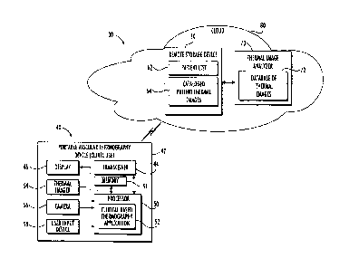
[0019] FIG. 1 is a block diagram of a clinical based infrared
thermography system in accordance with the disclosure.
[0020] FIG. 2 is a flow diagram for operating the clinical
based thermography application illustrated in FIG. 1.
[0021] FIG. 3-9 are various display screens corresponding to the flow
diagram
illustrated in FIG. 2.
[0022] FIG. 10 is a flowchart illustrating a method for operating the
infrared
4
CA 03101682 2020-11-25
WO 2019/231842 PCT/US2019/033911
thermography system as illustrated in FIG. 1.
[0023] FIG. 11 is a block diagram of a retail based infrared
thermography system in accordance with the disclosure.
Detailed Description
[0024] The present description is made with reference to the accompanying
drawings, in which exemplary embodiments are shown. However, many different
embodiments may be used, and thus the description should not be construed as
limited
to the particular embodiments set forth herein. Rather, these embodiments are
provided
so that this disclosure will be thorough and complete. Like numbers refer to
like
elements throughout.
[0025] Infrared thermography platforms as described below for determining
vascular health of individuals may be configured for clinical use or for
retail use. Clinical
use of an infrared thermography platform involves a doctor operating a
portable
vascular thermography device that is in communications with a cloud based
thermal
image analyzer to assess the vascular health of a patient. Retail use of an
infrared
thermography platform involves an individual interacting with a kiosk to
assess the
vascular health of a customer so as to recommend appropriately sized and
strength of
compression socks as necessary.
[0026] Referring initially to FIG. 1, a clinical based infrared
thermography
system 30 includes at least one portable vascular thermography device 40
operating a
clinical based thermography application 50 in communications with a remote
storage
device 60 and a thermal image analyzer 70. The portable vascular thermography
device
40 may also be referred to as a portable electronic device, and the clinical
based
infrared thermography system 30 may also be referred to as a vascular
thermography
system 30. The vascular thermography system 30 advantageously allows for real-
time
clinical assessments on the vascular health of individuals. By using
thermography and
an intelligent database this allows for real-time clinical assessments to be
performed
non-invasively and without requiring expensive ultrasound equipment.
[0027] As readily appreciated by those skilled in the art, infrared
clinical
thermography is a passive non-contact, and non-invasive method for mapping
surface
CA 03101682 2020-11-25
WO 2019/231842 PCT/US2019/033911
temperatures of the body. The differential of relative skin temperatures can
be utilized to
map and thus detect many abnormalities not seen by other diagnostic tools nor
the
human eye. The portable vascular thermography device 40 coupled with a
validated
database of thermal images 72 provides diagnostic immediacy that will enable
physicians, nurses, and even consumers to quickly know more about their
vascular
health.
[0028] The remote storage device 60 and the thermal image analyzer 70 are
cloud based 80. In other embodiments, the remote storage device 60 and the
thermal
image analyzer 70 may be co-located with the portable vascular thermography
device
40. Even though the remote storage device 60 and the thermal image analyzer 70
are
shown as separate components, they may be combined into a single component.
[0029] Communications between the portable vascular thermography device
40
and the remote storage device 60 and the thermal image analyzer 70 are
encrypted to
be HIPPA (Health Insurance Portability and Accountability Act) complainant. To
be
HIPPA compliant, the clinical based infrared thermography system 30 includes
encryption, an encrypted firewall, and role-based security to give users
different levels
of access based on their roles.
[0030] The remote storage device 60 is configured to store a patient list
62 and
cataloged patient thermal images 64 as provided by the portable vascular
thermography
device 40. The thermal images are cataloged into corresponding labeled
anatomical
areas, such as left thigh, right calf, etc. The cataloging may be performed by
the clinical
based thermography application 50 or by the thermal image analyzer 70.
[0031] The thermal image analyzer 70 is coupled to the remote storage
device 60
and is configured to provide diagnosis or an assessment of the cataloged
patient
thermal images 64 by comparing these images to the database of thermal images
72.
The thermal image analyzer 70 may be configured to use machine learning to
provide
statistical probabilities of certain disease states based on the thermal
patterns of the
cataloged patient thermal images 64.
[0032] The portable vascular thermography device 40 includes a housing
42, a
transceiver 44 carried by the housing 42 to communicate with the remote
storage
6
CA 03101682 2020-11-25
WO 2019/231842 PCT/US2019/033911
device 60 and the thermal image analyzer 70, and a display 46 carried by the
housing
42.
[0033] The display 46 and the transceiver 44 are coupled to a processor
50 also
carried by the housing 42. The processor 50 is configured to execute a
clinical based
thermography application 52.
[0034] A thermal imager 54 is carried by the housing 42 and is coupled to
the
processor 60. Optionally, the thermal imager 54 may be a separate component
that is
externally coupled to the portable vascular thermography device 40. A memory
51 is
coupled to the processor 50 for storing thermal images prior to the thermal
images
being stored in the remote storage device 60.
[0035] In addition, a camera 56 may be carried by the housing 42 and
coupled to
the processor 60. The camera 56 allows for a comparison between a patient's
thermal
image and a standard photograph of the anatomical area being examined. A user
input
device 58 also carried by the housing 42 is coupled to the processor 50.
[0036] The portable vascular thermography device 40 may be configured as
an
iPhone, iPod, iPad or Android device, for example. If the portable vascular
thermography device 40 is configured as an iPhone, for example, then the
thermal
imager 54 may be separate from the iPhone and coupled thereto via the iPhone's
USB
interface. An example external plug-in thermal imager 54 is FLIR ONE Gen 3 as
provided by FLIR Systems, Inc.
[0037] Referring now to the flow diagram 100 in FIG. 2 and to the display
screens
in FIGS. 3-9, operation of the infrared thermography application 50 by a
doctor will be
discussed. The clinical based thermography application 50 may also be referred
to as
Thermworx.
[0038] From the start, the clinical based infrared thermography
application 50 is
opened at Block 112 via a login screen 140 as shown in FIG. 3. The login
screen 140
includes a username field 142 and a password field 144. At Block 114 the
available vein
scan settings are provided via display screen 150 as shown in FIG. 4. The vein
scan
settings include a live view setting 152, a new patient setting 154 and an
existing patient
setting 156.
7
CA 03101682 2020-11-25
WO 2019/231842 PCT/US2019/033911
[0039] The live view setting 152 allows the doctor to immediately examine
a
patient at Block 116 regardless of whether the patient is an existing patient
or a new
patient. As the setting names imply, the existing patient setting 156 is for
selecting an
existing patient at Block 118 from the remote storage device 60, and the new
patient
setting 154 is for entering a new patient at Block 120 into the remote storage
device 60.
[0040] If the existing patient setting 156 is selected by the doctor,
then display
screen 160 appears as shown in FIG. 5. The doctor is to enter information into
a name
of patient field 162, a date of birth field 164 or a record number field 166,
and a
data/time stamp field 168.
[0041] With the existing patient's information having been retrieved, the
doctor
may then use the thermal imager 54 to provide a thermal image 172 of the
existing
patient's right leg, for example, as shown in FIG. 6. The doctor has access to
a save
image field 174 for saving the thermal image 172. The thermal image 172 is
saved
within the memory 51 prior to being stored in the remote storage device 60.
[0042] Prior to the thermal image 172 being saved, the doctor selects
from a drop
down list to designate or label an appropriate anatomical area to the thermal
image 172.
In this case, the appropriate anatomical area is right posterior calf.
[0043] The doctor has an option to annotate the thermal image via the
option to
annotate field 176. If the doctor wants to store the annotated thermal image
172 within
the remote storage device 60, then the store field 178 is selected. The
annotated
thermal image 172 is stored within the cataloged patient thermal images 64.
[0044] Referring back to the flow diagram 100, after an existing patient
118 or a
new patient 120 has been selected, the following steps are the same. These
steps
include providing the live thermal image 172 of the area being examined at
Block 124,
saving the thermal image 172 corresponding to an appropriate anatomical area
at Block
126, an option to annotate the thermal image 172 at Block 128, and storing the
thermal
image 172 within the remote storage device 60 at Block 130.
[0045] At Block 122 following the live view Block 116, the settings may
be
changed as provided in display screen 180 shown in FIG. 7. The setting changes
include a temperature scale field 182, a color scale field 184, a visual image
field 186,
and a live image field 189. The doctor can exit the display screen 180 by
selecting the
8
CA 03101682 2020-11-25
WO 2019/231842 PCT/US2019/033911
quit field 189.
[0046] Still referring to the flow diagram 100, when the thermal image
172 is
stored within the remote storage device 60 at Block 130, the thermal analyzer
70 may
be instructed to compare the thermal image 172 to the database of thermal
images 72.
[0047] The thermal analyzer 70 may be configured to use machine learning
to
give the doctor statistical probabilities of certain disease states based on a
thermal
pattern within the thermal image 172. In the thermal image 172 of the
patient's right
posterior calf, the thermal analyzer 70 analyzes to see how much red is in the
thermal
image. This may be suggestive of venous insufficiency, and the thermal
analyzer 70
then provides a statement such as "this area has a 95% likelihood of having
venous
insufficiency."
[0048] The thermal analyzer 70 is advantageously configured to take
advantage
that there is a unique situation in venous disease where there are incompetent
superficial veins, i.e., where the valves have failed. In legs with normal
veins, for
example, blood that fills the superficial veins is collected from the
capillaries of the skin
and subcutaneous fat.
[0049] Since the blood has come from the skin capillary network which
acts as a
heat exchanger, the temperature of blood filling the superficial veins is at
the same
temperature as the skin, and flows from distal to proximal and from
superficial to deep.
[0050] As there is no thermal differential the veins are
indistinguishable from the
skin on thermal imaging. In contrast, where valves have become incompetent
blood
flows in the opposite direction, flowing from deep to superficial and from
proximal to
distal. In this instance blood refluxing from deep is at a core temperature of
approximately 37 C, when it refluxes and fills the superficial veins the heat
conducts to
the skin. Consequently, an area overlying the varicose tributary is hotter
than the typical
skin temperature of around 30 C.
[0051] The sensitivity of thermal imaging may be as good as 0.1 C. This
makes it
easier to identify the hot areas. The hot areas are of course always losing
heat energy,
but as blood refluxes continuously during standing and activity this heat is
replenished,
and will remain hotter for a period after an individual is recumbent when
reflux ceases.
[0052] Thus, a thermal image of the leg will show only veins with reflux
and
9
CA 03101682 2020-11-25
WO 2019/231842 PCT/US2019/033911
therefore failed valves, as hot areas that are linear in nature and
serpiginous the highest
temperature follows the course of the vein. This pattern may also be seen in
arteriovenous fistula but these are rare and simply differentiated using
ultrasound.
[0053] Thermal imaging does not identify incompetent trunk veins as they
are
deep to the deep fascia of the leg and have a uniform insulating subcutaneous
layer.
Thermal imaging thus uniquely demonstrates and differentiates legs with venous
incompetence from normal limbs. Acquiring thermal images for venous disease
does
not require any preparation of the leg, is entirely non-invasive and very time
efficient.
[0054] Referring back to operation of the clinical thermography
application 52, a
side by side comparison between a thermal image 172 and a standard photograph
192
may be performed as provided in display screen 190 shown in FIG. 8. The
display
screen 190 includes the thermal image 172 of the patient's right leg and a
standard
photograph 192 of the patient's right leg as provided by the camera 58. The
thermal
image 172 may be stored alongside the standard photograph 192 in the remote
storage
device 60.
[0055] As an alternative to a thermal image, a thermal video clip 202 of
the
patient may be generated as provided in display screen 200 shown in FIG. 9.
The same
options available for a thermal image are available for a thermal video clip.
The doctor
has access to a save video clip field 204 for saving the thermal video clip
202. The
thermal video clip 202 is saved within the memory 51 prior to being stored in
the remote
storage device 60.
[0056] The doctor has an option to annotate the thermal video clip 202
via the
option to annotate field 206. If the doctor wants to store the annotated
thermal video clip
202 within the remote storage device 60, then the store field 208 is selected.
The
thermal video clip 202 is stored within the cataloged patient thermal images
64. At Block
132 the doctor quits or exits the clinical based thermography application 52.
[0057] Referring now to FIG. 10, a flowchart 300 illustrating a method
for
operating the vascular thermography system 30 will be discussed. From the
start (Block
302), the method includes generating, by the portable electronic device 40, a
thermal
image 172 of an anatomical area of a patient for display at Block 304. The
portable
electronic device 40 transmits the thermal image at Block 306. The method
further
CA 03101682 2020-11-25
WO 2019/231842 PCT/US2019/033911
includes receiving, at a thermal image analyzer 70, the transmitted thermal
image at
Block 308. The thermal image analyzer 70 determines an assessment on vascular
health of the patient based on comparing the thermal image 174 to a database
of
thermal images 72 at Block 310. The thermal image analyzer 70 then transmits
the
assessment to the portable electronic device 40 at Block 312. The method ends
at
Block 314.
[0058] Referring now to FIG. 11 a retail based vascular thermography
kiosk
220 will be discussed. The vascular thermography kiosk 220 may be placed in
shopping
malls or stores, for example, and is configured to assess the vascular health
of a
customer's legs so as to recommend appropriately sized compression socks as
necessary.
[0059] The vascular thermography kiosk 220 includes a housing 222, and
one or
more compression sock displays 240 attached to sides of the housing 222. The
housing
222 carries a display 224, a user input device 226, a processor 230, at least
one
camera 228, at least one thermal imager 250, and a customer record database
251.
The vascular thermography kiosk 220 further includes a customer feet placement
indicator 252.
[0060] The customer feet placement indicator 252 allows a customer to
stand on
foot patterns that are in alignment with the thermal imager 250 and with the
camera
228. When the system has a fixed thermal imager 250, the customer moves their
feet to
present the correct aspect to the thermal imager 250. The placement of the
feet is
guided by foot prints on the floor placed at an optimal distance from the
fixed camera
228.
[0061] The thermal imager 250 generates one or more thermal images of the
customer's legs, and the camera 228 also takes one or more picture of the
customer's
legs. The thermal imager 250 may be movable following a specific pattern to
capture
the relevant thermal images. Alternatively, there may be multiple thermal
imagers 250
thereby avoiding the need for moving parts. The customer's legs are either
bare or have
tight clothing in contact with the skin, such as socks or stockings.
[0062] The thermal images may be displayed on the display 224 for
customer
viewing. Alternatively, the thermal images may be sent by internet to the
customer
11
CA 03101682 2020-11-25
WO 2019/231842 PCT/US2019/033911
and/or to a central location for storage and cataloging and review.
[0063] The thermal image will be processed to accentuate the hot vein
pattern
and may or may not subtract areas of normal heat, such as the anterior tibia
area and
popliteal fossa. The retail based thermography application 232 may further
cooperate
with the thermal imager 250 for setting a standard temperature scale and color
pallet so
that all images are comparable and consistent.
[0064] The thermal images and the photographs of the customer's legs may
be
stored photos in a customer record database 251. Execution of the retail based
thermography application 232 by the processor 230 provides display prompts on
the
display 224. The display prompts require the customer answering a series of
clinical
questions. For example, these questions may include the following: do you have
leg
swelling Y/N, do you have leg cramps Y/N, etc.
[0065] The customer's answers to the questions, along with the thermal
image
and photograph of the customer's legs are used by the thermal image analyzer
234 to
give the customer suggestions regarding their venous health and whether or not
certain
types of compression socks might be most useful for the customer.
[0066] The thermal image analyzer 234 may be included within the
processor
230 as shown, or may be a separate processor operating in coordination with
the
processor 230. In other embodiments, the thermal image analyzer 234 may be
cloud
based wherein the vascular thermography kiosk 220 then communicates with
through
the internet. The thermal image analyzer 234 may be configured to use machine
learning to provide statistical probabilities of certain disease states based
on the thermal
patterns of the customer's thermal image. The thermal image analyzer 234 will
apply
the algorithms to determine for each leg a) are the leg vein normal or
abnormal, and b)
are compression garments indicated.
[0067] Operation of the retail based thermography application 232 allows
the
processor 230 to determine length and diameter of the customer's legs and thus
determine which size of compression stocking is most appropriate. The vascular
thermography kiosk 220 includes compression sock displays 240 for purchase
based on
the results of the analyses performed by the thermal image analyzer 234.
[0068] As discussed above, the retail based vascular thermography kiosk
220 is
12
CA 03101682 2020-11-25
WO 2019/231842 PCT/US2019/033911
a free standing autonomous system capable of deployment in public. The retail
based
vascular thermography kiosk 220 catalogs and stores customers' thermal images,
and
predicts the need for compression garments.
[0069] The retail based vascular thermography kiosk 220 is capable of
producing
a comprehensive map of a customer's incompetent venous circulation, and is
capable
of differentiating 'normal' areas of skin that are normally hotter than the
general skin
surface and disregarding them. The retail based vascular thermography kiosk
220 can
in general determine normal from abnormal limbs (with regard to veins).
[0070] The thermal image analyzers 70, 234 may be based on artificial
intelligence and machine learning. Veins of the leg, for example, are
distributed in
certain patterns and when they fail they do so in a variable recognizable
number of
patterns of varicose veins. Thermal imaging can detect these veins as broad
areas of
increased temperature above background.
[0071] The peak temperatures tend to follow the line of the vein which
has a
convoluted but linear form. Other areas of elevated temperature form a much
more fixed
pattern such as the area in the shin where the tibial bone is immediately
beneath the
skin, or behind the knee (the popliteal fossa, where subcutaneous fat is
thinner).
[0072] In both these cases they do not create a linear pattern and so an
algorithm
can be developed to improve the specificity of the venous map by following the
lines of
maximum temperature and subtraction of area of diffuse high temperature. In
this way a
simplified but more accurate vein map can be created.
[0073] Knowledge of the absolute temperature range is also important.
Body core
temperature is 37 C, typical leg temperature of normal skin in the thigh at
room
temperature 20 C is between 28-32 C. Blood that issues from the deep is at or
close to
body temperature while blood that is in normal superficial veins is at the
temperature of
the skin.
[0074] The skin capillaries act as a very efficient heat exchanger and so
the
arterial blood which arrives to feed the skin is cooled imparting its heat to
the skin
before being collected into the veins by which time there is little or no
temperature
difference. This makes normal veins invisible to thermal imaging, as there is
no
temperature gradient between skin and vein.
13
CA 03101682 2020-11-25
WO 2019/231842 PCT/US2019/033911
[0075] In contrast to varicose veins, vein valves have failed and permit
blood
(when the patient is standing) to reflux to fill veins immediately beneath the
skin
(varicose veins) at close to core temperature with little insulating fat and
so heat the skin
over the vein to a temperature greater than the surrounding skin. This is
easily within
the discriminatory range of the thermal imaging camera. Controlling the
temperature
range of the thermal imager to between 28-38 C, or more preferably 30-36 C,
improves
sensitivity and specificity of vein detection.
[0076] Many modifications and other embodiments will come to the mind of
one
skilled in the art having the benefit of the teachings presented in the
foregoing
descriptions and the associated drawings. Therefore, it is understood that the
disclosure
is not to be limited to the specific embodiments disclosed, and that
modifications and
embodiments are intended to be included within the scope of the appended
claims.
14