Note: Descriptions are shown in the official language in which they were submitted.
CA 03103272 2020-12-09
WO 2019/241700 PCT/US2019/037301
EPITHELIAZING MICROPOROUS BIOMATERIAL FOR USE IN
AVASCULAR ENVIRONMENTS AND IN CORNEAL IMPLANTS
FIELD
[0001] The present disclosure relates generally to biomaterials, and
more
specifically, to a microporous biocomposite that is suitable for surgical
implantation in an
avascular environment and which enables a stable bio-interface and sustained
epithelization directly on the surface of the microporous biocomposite in the
avascular
environment.
BACKGROUND
[0002] The cornea generally refracts and focuses light onto the retina
and
serves as a protective barrier for the intraocular components of the eye. The
cornea is
subject to a host of diseases, genetic disorders, and trauma that can cause
opacity of
what should otherwise be an optically transparent window to the retina.
[0003] Although surgical procedures exist to replace damaged or
diseased
corneas with live tissue corneas taken from donor eyes, donor corneas may not
be
available, the underlying condition of the damaged eye may be such that donor
cornea
failure or rejection is likely, and/or the patient's physiology may be such
that a donor
cornea failure or rejection is likely.
[0004] In cases where implantation of a donor cornea is not viable,
implantation of an artificial cornea is a potential alternative treatment.
Keratoprosthesis
is the surgical implantation of an artificial cornea to replace part of or all
of a damaged
or diseased cornea. The primary challenges facing keratoprostheses have been
biointegration complications and extrusion of the device from the eye. Other
complications include infection, retroprosthetic membrane formation,
inflammation,
glaucoma, lack of mechanical durability and optical fouling.
[0005] A number of approaches to solving the issue of artificial
cornea device
rejection have been attempted. One approach involves keratoprostheses having a
core
and skirt type construction. The core and skirt type devices generally have a
non-
porous optical core for visual restoration and a skirt for bio-integration
with the eye
tissue surrounding the skirt.
[0006] However, such conventional core and skirt type constructions
have not
exhibited optimal device anchoring and long-term optical patency.
1
CA 03103272 2020-12-09
WO 2019/241700 PCT/US2019/037301
SUMMARY
[0007] According to one example ("Example 1"), a biocompatible
biocomposite comprises (1) a polymer scaffold having a thickness less than
about 100
pm and including nodal structures that extend from at least one surface of
said polymer
scaffold; and (2) a hydrophilic coating on the polymer scaffold, wherein said
biocomposite is configured for the sustained viability of epithelial cells on
a surface of
said biocomposite in an avascular environment.
[0008] According to another example ("Example 2"), further to Example
1, the
polymer scaffold is a microporous biomaterial comprising an expanded
fluoropolymer
membrane having a node and fibril microstructure where the nodes are
interconnected
by the fibrils and pores are formed by voids located between the nodes and
fibrils, and
the nodal structures extend from a first surface to a second surface of said
polymer
scaffold.
[0009] According to another example ("Example 3"), further to Example
1, the
polymer scaffold is a microporous biomaterial comprising an expanded non-
fluoropolymer membrane having a node and fibril microstructure where the nodes
are
interconnected by the fibrils and pores formed by the voids located between
the nodes
and fibrils, and the nodal structures extend from a first surface to a second
surface of
said polymer scaffold.
[0010] According to another example ("Example 4"), further to Example
2 or
Example 3, the pores have a size greater than about 30 pm.
[0011] According to another example ("Example 5"), further to any of
Examples 2-4, the hydrophilic coating coats said nodes, said fibrils, and said
nodal
structures,
[0012] According to another example ("Example 6"), further to any of
Examples 2, 4, or 5, the microporous biomaterial is an expanded
polytetrafluoroethylene
(ePTFE) membrane having said node and fibril microstructure,
[0013] According to another example ("Example 7"), further to any of
Examples 2, or 4-6, the polymer scaffold is an ePTFE membrane having said
nodal
structures thereon, wherein said nodal structures are islands of ePTFE
attached to and
raising from a surface of said ePTFE membrane,
[0014] According to another example ("Example 8"), further to any of
Examples 2, or 4-7, the polymer scaffold is a three-layered structure
comprising a first
2
CA 03103272 2020-12-09
WO 2019/241700 PCT/US2019/037301
ePTFE membrane containing thereon said nodal structures, a second ePTFE
membrane containing said nodal structures, and a biocompatible adhesive
positioned
between said first and second ePTFE membranes, wherein said nodal structures
are
pillars formed of ePTFE.
[0015] According to another example ("Example 9"), further to any of
Examples 1-8, the hydrophilic coating comprises poly(tetrafluoroethylene-co-
vinyl
alcohol) or polyvinyl alcohol,
[0016] According to one example ("Example 10"), an artificial cornea
comprises: a central core including a polymeric corneal substitute; and a
microporous
biocomposite according to any one of Examples 1-9.
[0017] According to another example ("Example 11"), further to Example
10,
the central core is formed of a material configured to permit epithelial cell
growth
thereon,
BRIEF DESCRIPTION OF THE DRAWINGS
[0018] The accompanying drawings are included to provide a further
understanding of the disclosure and are incorporated in and constitute a part
of this
specification, illustrate embodiments, and together with the description serve
to explain
the principles of the disclosure.
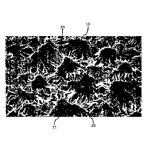
[0019] FIG. 1 is a scanning electron micrograph depicting a radially
expanded
ePTFE with a microstructure where the fibrils emanate from the nodes in all
directions in
accordance with at least one embodiment;
[0020] FIG. 2 is a scanning electron micrograph of a cross-section of
a
radially expanded ePTFE membrane that has vertically oriented nodal structures
through the membrane in accordance with at least one embodiment;
[0021] FIG. 3 is a scanning electron micrograph (SEM) of a radially
expanded
ePTFE depicting free standing nodal structures formed by plasma treatment in
accordance with at least one embodiment;
[0022] FIG. 4 is an image of an exemplary artificial cornea having
clear
cornea substitute region and a white region surrounding the clear cornea
substitute
region that includes the microporous biocomposite in accordance with at least
one
embodiment;
[0023] FIG. 5 is a schematic illustration of an artificial cornea in
accordance
with at least one embodiment;
3
CA 03103272 2020-12-09
WO 2019/241700 PCT/US2019/037301
[0024] FIG. 6 is a rear perspective view of the artificial cornea
construction of
FIG. 5 in accordance with at least one embodiment;
[0025] FIG. 7 is a top view of the artificial cornea construction of
FIG. 5 in
accordance with at least one embodiment;
[0026] FIG. 8 is a cross sectional view of the artificial cornea
construction of
FIG. 5 taken along line 7-7 in accordance with at least one embodiment;
[0027] FIG. 9 is a schematic illustration of a three-layered
biocomposite
material in accordance with at least one embodiment;
[0028] FIG. 10 is a schematic illustration of a three-layered
biocomposite
made by forming structural pillars on the surface of the ePTFE membrane in
accordance with at least one embodiment;
[0029] FIG. 11A is a scanning electron micrograph (SEM) of the top
surface of
an ePTFE membrane depicting islands of ePTFE in accordance with at least one
embodiment;
[0030] FIG. 11B is a scanning electron micrograph (SEM) of the cross-
section
of the ePTFE membrane of FIG. 11A in accordance with at least one embodiment;
[0031] FIG. 12A is a scanning electron micrograph (SEM) of a top
surface of
an ePTFE membrane suitable for use in the biocompatible biocomposite in
accordance
with at least one embodiment;
[0032] FIG. 12B is a scanning electron micrograph (SEM) of the cross-
section
of the ePTFE membrane of FIG. 12A,
[0033] FIG. 13 is a scanning electron micrograph (SEM) of an ePTFE
(vascular graft) membrane having an average nodal spacing from 35 to 100
microns
and a thickness from about 110 microns to about 170 microns in accordance with
at
least one embodiment;
[0034] FIG. 14 is a scanning electron micrograph (SEM) of an ePTFE
membrane having an average nodal spacing of 40 microns and a thickness of
approximately 300 microns in accordance with at least one embodiment;
[0035] FIG. 15A is a schematic illustration of the top view an
artificial cornea
with macro-perforations in the tissue integration skirt in accordance with at
least one
embodiment; and
[0036] FIG. 15B is a schematic illustration of the cross-sectional
view of the
artificial cornea of FIG. 15A in accordance with at least one embodiment.
4
CA 03103272 2020-12-09
WO 2019/241700 PCT/US2019/037301
DETAILED DESCRIPTION
[0037] Persons skilled in the art will readily appreciate that various
aspects of
the present disclosure can be realized by any number of methods and apparatus
configured to perform the intended functions. It should also be noted that the
accompanying drawing figures referred to herein are not necessarily drawn to
scale, but
may be exaggerated to illustrate various aspects of the present disclosure,
and in that
regard, the figures should not be construed as limiting. It is to be
appreciated that the
terms "microporous biomaterial" and "biomaterial" may be used interchangeably
herein.
In addition, the terms "microporous biocomposite", "biocomposite", and
"biocomposite
material" may be interchangeably used.
[0038] The present invention is directed to a microporous biocomposite
that is
suitable for surgical implantation in an avascular environment. The
microporous
biocomposite includes (1) a porous scaffold having a thickness less than about
100 pm
and which is formed of nodal structures that extend to at least one surface of
the porous
scaffold and (2) a hydrophilic coating on the microporous biomaterial. In
exemplary
embodiments, the nodal structures extend from a first surface (e.g., bottom
surface) of
the porous scaffold to a second surface (e.g., the top surface). In at least
one
embodiment, the porous scaffold is a microporous biomaterial with a nodal
structure
that extends from a first surface to a second surface of the microporous
biomaterial. In
some embodiments, the microporous biomaterial is radially expanded. In
additional
embodiments, the hydrophilic coating is a node and fibril coating. In some
embodiments, the nodal structures are free-standing and/or vertically
oriented. In
addition, the microporous biocomposite allows for the integration and
sustained viability
of epithelial cells on the surface thereof as well as tissue integration
(i.e., ingrowth) and
the internal colonization of the biomaterial with other cell types, such as,
for example,
corneal keratocytes and fibroblasts. In at least one embodiment, the
microporous
biocomposite may be incorporated into an artificial corneal implant or in
other avascular
mesoplants.
[0039] In exemplary embodiments, the porous scaffold is a microporous
biomaterial. The microporous biomaterial may be a microporous expanded polymer
membrane, such as a microporous expanded fluoropolymer membrane that has a
node
and fibril microstructure where the nodes are interconnected by the fibrils
and the pores
are the voids or space located between the nodes and fibrils throughout the
membrane.
In some embodiments, one or more perforation processes may be utilized to form
a
CA 03103272 2020-12-09
WO 2019/241700 PCT/US2019/037301
plurality of macro- or micro-sized discrete perforations in the microporous
biomaterial
(i.e., the polymer scaffold). An exemplary node and fibril microstructure is
expanded
polytetrafluoroethylene (ePTFE), such as is described in U.S. Patent No.
3,953,566 to
Gore. In at least one exemplary embodiment, the microporous polymer membrane
is
an expanded polytetrafluoroethylene (ePTFE) membrane. The ePTFE membranes for
use in the biocomposite are characterized by nodes interconnected by thin
fibrils. While
not intending to be limiting, expanded polytetrafluoroethylene (ePTFE)
membranes
prepared generally in accordance with the methods described in U.S. Patent No.
3,953,566 to Gore, U.S. Patent No. 7,306,729 to Bacino etal., U.S. Patent
Publication
No. 2004/0173978 to Bowen et al., U.S. Patent No. 5,476,589 to Bacino, or U.S.
Patent
No. 5,183,545 to Branca etal., U.S. Patent No. 5,476,589 to Bacino, U.S.
Patent
Publication No. 2016/0367947 to Hollenbaugh, etal., or U.S. Patent Publication
No.
2010/0381293 to Towler may be used herein. For example, FIG. 12A is a scanning
electron micrograph (SEM) of a top surface of an ePTFE membrane formed in
accordance with the teachings of U.S. Patent Publication No. 2010/0381293 to
Towler.
FIG. 12B is a cross section of the same ePTFE membrane. FIG. 13 is an ePTFE
membrane with an average nodal spacing of 35 to 100 microns (or from 60
microns to
80 microns, from 40 microns to 75 microns, or from 30 microns to 55 microns)
and a
thickness from about 110 microns to about 170 microns. Another ePTFE membrane
for
use in the microporous biocomposite is shown in FIG. 14, which is an ePTFE
membrane having an average nodal spacing of 40 microns (ranging from 30
microns to
60 microns) and a thickness of approximately 300 microns. It is to be
appreciated that
other fluoropolymer membranes are considered to be within the purview of the
invention
provided that they can be processed to form a microporous membrane that is
biocompatible and has a node and fibril microstructure.
[0040] The term "ePTFE" is utilized herein for convenience and is
meant to
include not only expanded polytetrafluoroethylene (ePTFE), but also expanded
modified
polytetrafluoroethylene (PTFE) and expanded copolymers of PTFE, such as are
described in U.S. Patent No. 5,708,044 to Branca, U.S. Patent No. 6,541,589 to
Baillie,
U.S. Patent No. 7,531,611 to Sabol etal., U.S. Patent No. 8,637,144 to Ford,
and U.S.
Patent No. 9,139,669 to Xu etal.
[0041] In some embodiments, the microporous biomaterial is an expanded
non-fluoropolymer membrane that has a node and fibril microstructure. Non-
limiting
examples of suitable expanded non-fluoropolymers include porous poly (p-
xylylene)
6
CA 03103272 2020-12-09
WO 2019/241700 PCT/US2019/037301
(ePPX) as taught in U.S. Patent Publication No. 2016/0032069, porous ultra-
high
molecular weight polyethylene (eUHMWPE) as taught in U.S. Patent No. 9,926,416
to
Sbriglia, porous ethylene tetrafluoroethylene (eETFE) as taught in U.S. Patent
No.
9,932,429 to Sbriglia, porous polylactic acid (ePLLA) as taught in U.S. Patent
No.
7,932,184 to Sbriglia, etal., porous vinylidene fluoride-co-
tetrafluoroethylene or
trifluoroethylene [VDF-co-(TFE or TrFE)] polymers as taught in U.S. Patent No.
9,441,088 to Sbriglia and copolymers and combinations thereof. Porous
hydrogels
(e.g., poly-HEMA) may be utilized herein as a non-fluoropolymer polymer. It is
to be
appreciated that other non-fluoropolymer polymers are considered to be within
the
scope of the invention so long as they can be processed to form a microporous
membrane that is biocompatible and has a node and fibril microstructure.
[0042] Woven materials, non-woven materials, and electrospun fibers or
nanofibers in the form of a sheet or other non-woven form may alternatively be
used as
a scaffold (e.g., in place of an ePTFE membrane) to form the biocomposite
material.
[0043] It is to be appreciated that reference is made herein with
respect to
expanded polytetrafluorethylene (ePTFE) for ease of discussion. However, it is
to be
understood that any suitable expanded fluoropolymer or non-fluoropolymer
membrane,
woven or non-woven material, or electrospun material may be used
interchangeably
with any "ePTFE" mentioned above.
[0044] In preparing the microporous biomaterial, the ePTFE may be
subjected
to a radial expansion, such as that described in U.S. Patent No. 5,321,109 to
Boss, et
al. The radial expansion of the ePTFE creates a unique node and fibril
microstructure
where the fibrils 20 emanate from the nodes 10 in all directions (e.g.,
radially extend),
such as is shown in FIG. 1. The radially-oriented fibrils 20 impart high
strength to
biaxial tensile loading within the plane of the membrane. In some embodiments,
the
ePTFE membrane has a thickness from about 50 pm to about 100 pm, from about 50
pm to about 90 pm, from about 50 pm to about 80 pm, or from about 50 pm to
about 70
pm. Additionally, as shown in FIG. 2, the ePTFE membrane 50 has vertically
oriented
nodal structures 30 that span the thickness of the ePTFE membrane 50 (i.e,
from the
top surface 60 of the ePTFE membrane 50 to the bottom surface 70 of the ePTFE
membrane 50). In other embodiments, the nodal structures extend to at least
one
surface of the ePTFE membrane, and may not necessarily be vertically oriented.
[0045] After the radially expanded ePTFE membrane is formed, the top
surface 60 of the ePTFE membrane 50 is subjected to a high surface energy
treatment
7
CA 03103272 2020-12-09
WO 2019/241700 PCT/US2019/037301
to alter the microstructure. One exemplary method utilized to modify the
microstructure
of the ePTFE membrane is to use a plasma treatment such as is taught in U.S.
Patent
No. 5,462,781 to Zukowski. A second exemplary method includes U.S. Patent No.
5,041,225 to Norma, which teaches a microporous ePTFE membrane where the
internal and external surfaces of the membrane are coated with a complex
formed by
the combination of a hydrophilic polymer which adheres to the membrane
structure and
a coupling agent. The coupling agent renders the ePTFE membrane substantially
hydrophilic and protein affinitive. Other process are taught in U.S. Patent
No. 5,902,745
to Butler, et al. that render an ePTFE membrane spontaneously and
substantially
completely water wettable by adsorbing and cross-linking the hydrophilic
fluoropolymer
poly(tetrafluoroethylene-co-vinyl alcohol (HPL) into the microporous void
spaces of the
ePTFE membrane and onto the surfaces of the ePTFE membrane. As used herein,
the
term "plasma treatment" is meant to include any high energy surface treatment,
such
as, but not limited to, glow discharge plasma treatment, corona treatment, ion
beam
treatment, and the like. The plasma treatment effectively removes the fibrils
from the
surface of the ePTFE membrane, leaving free standing nodal structures 40
(e.g., not
interconnected by fibrils), such as is shown in FIG. 3. These nodal structures
40 formed
by the plasma treatment rise from the bottom surface 70 of the ePTFE membrane
50 to
the top surface 60 of the ePTFE membrane. In other words, the bases 65 of the
nodal
structures 40 comprise the inner surface of the microporous biocomposite and
the tops
60 of the nodal structures 40 comprise the top surface of the microporous
biocomposite.
The plasma treatment also increases the effective pore diameter of the ePTFE
membrane 50, which allows better accessibility to the bottom surface 70 of the
ePTFE
membrane 50. The pore size of the ePTFE membrane may be greater than about 30
pm and less than about 100 pm. The inter-nodal distance at or near the top
surface 60
of the nodal structures may be from about 20 pm to about 100 pm or from about
50 pm
to about 70 pm. The plasma treatment creates an ePTFE membrane that has an
effective pore diameter that is substantially larger on the plasma treated
side (i.e., top
surface 75) then on the non-plasma treated side (i.e., bottom surface 70).
[0046] Following plasma treatment, the ePTFE membrane is rendered
hydrophilic through a coating process that preserves the nodal architecture of
the
plasma treated ePTFE membrane. In one embodiment, the ePTFE is rendered
hydrophilic (e.g., water wettable) by adsorbing and crosslinking a
hydrophilic, hydrogel
fluoropolymer such as poly(tetrafluoroethylene-co-vinyl alcohol) (HPL) onto
the nodes
8
CA 03103272 2020-12-09
WO 2019/241700 PCT/US2019/037301
and fibrils of the ePTFE microstructure as well as on the surface of the ePTFE
membrane (including the nodal structures). Such a process is taught generally
in U.S.
Patent No. 5,902,745 to Buttler, et al. In an alternative embodiment,
polyvinyl alcohol
(PVA) may be used as the hydrophilic wetting agent, rather than HPL. The
hydrophilic
coating on the nodal structures and on the nodes and fibrils of the ePTFE
membrane
forms the microporous biocomposite.
[0047] In an alternate embodiment, an ePTFE membrane may be subjected
to a pre-conditioning process to form "islands" of ePTFE on the surface of an
ePTFE
membrane. These "islands" of ePTFE are attached to and raise above the
underlying
ePTFE structure. The "islands" of ePTFE may be formed by subjecting the
precursor
ePTFE membrane to a high energy surface treatment (e.g., plasma treatment)
followed
by a heating step as taught in U.S. Patent Publication No. U.S. Patent No.
7,615,282 to
Lutz. et al. or U.S. Patent No. 7,740,020 to Lutz et al. A scanning electron
micrograph
(S EM) of the top surface of such an ePTFE membrane is depicted in FIG. 11A. A
cross-section of the ePTFE membrane is shown in the SEM of FIG. 11B. The
"islands"
act as free-standing, vertically oriented nodal structures, which may then be
subjected
to a hydrophilic coating as discussed above. In other embodiments, one or more
surface pre-conditioning processes may be utilized to form layers exhibiting a
preferred
microstructure (e.g., wrinkles, folds, or other geometric out-of-plane or
undulating
structures), such as is explained in U.S. Patent No. 9,849,629 to Zaggl. Such
surface
pre-conditionings may facilitate a bolder early inflammatory phase after
surgery,
providing an early stable interface between the artificial cornea and the eye
tissue with
which it interfaces.
[0048] The node and fibril coating of the hydrophilic polymer permits
water to
not only be absorbed into the hydrophilic coating (e.g. HPL) but also allows
the
voids/pores in the ePTFE membrane to be filled with water. The microporous
biocomposite has an adsorbed water content of less than 50% as determined by
the
formula H20 (wt%) = RA/ , ¨hydrated ¨ Wdry] / Whydrated 100% prior to
implantation. However,
after implantation, the adsorbed water content substantially increases due to
the
incorporation of water into the hydrophilic coating and into the pores of the
ePTFE
membrane. The hydrophilic coating on the ePTFE membrane allows the membrane to
"wet out" and become visually translucent. Importantly, the microporous
biocomposite
allows for the integration and sustained viability of epithelial cells on the
surface of
thereof in an avascular environment. The microporous biocomposite also allows
for
9
CA 03103272 2020-12-09
WO 2019/241700 PCT/US2019/037301
tissue integration into the internal microstructure of the ePTFE and the
sustained
viability of these tissues in an avascular environment.
[0049] In an alternate embodiment, the microporous biocomposite may be
a
three-layered structure formed of ePTFE membranes and a biocompatible adhesive
as
is depicted generally in FIG. 9. As shown in FIG. 9, the microporous
biocomposite 900
includes a first ePTFE membrane 910, a second ePTFE membrane 920, and a
biocompatible adhesive 930 such as fluorinated ethylene propylene (FEP) to
adhere the
first and second ePTFE membranes 910, 920 to each other. Pillars 940
interconnect
the top and bottom portions of the ePTFE membranes and provide structural
support for
the microporous biocomposite. As with the embodiment discussed above, each
ePTFE
membrane has a thickness (T) that is about 100 pm.
[0050] In some embodiments, depicted in FIG. 10, the pillars 940 may
be
printed or otherwise laid down on support substrates 1010,1020, such as, for
example,
ePTFE membranes. The pillars 940 may themselves be made out of ePTFE. The
support substrates 1010, 1020 are adhered to each other with a biocompatible
adhesive, such as fluorinated ethylene propylene (FEP). In such an embodiment,
the
pillars 940 have a thickness less than about 100 pm. A hydrophilic coating may
then be
applied to coat the pillars 940 and the ePTFE substrates to form a microporous
biocomposite.
[0051] In at least one embodiment, the microporous biocomposite is
incorporated into an artificial corneal implant. As used herein, the terms
"artificial
corneal implant" or "artificial cornea" are meant to include all forms of
artificial corneas,
including synthetic corneas, keratoprostheses, and the like. Turning to FIG.
4, an image
of an exemplary artificial cornea 100 is depicted. The artificial cornea 100
is an
implantable medical device that operates as a synthetic replacement for
diseased
corneas, damaged corneas, or corneas otherwise requiring replacement. The
artificial
cornea 100 includes an optical element 200 and the microporous biocomposite
300 at
least partially surrounding the optical element 200. The optical element 200
may be
formed of a synthetic polymeric material. Because of the hydrophilic coating
on the
ePTFE membrane, the ePTFE is able to wet out and become clear in the presence
of
water. Thus, when such an artificial cornea 100 is implanted in an eye,
microporous
biocomposite 300 becomes clear like the optical element 200 and is not visible
to the
naked eye. Advantageously, the microporous biocomposite does not appreciable
change its dimension (volume) after being fully absorbed by water.
Additionally, the
CA 03103272 2020-12-09
WO 2019/241700 PCT/US2019/037301
microporous biocomposite 300 permits tissue ingrowth and the attachment of
epithelial
cells directly on the microporous biocomposite in an avascular environment
whereas the
optical element is configured to resist tissue ingrowth and attachment. In
some
embodiments, the optical element 200 may be modified to facilitate cell
adhesion and/or
proliferation, such as, for example, the formation of an organized monolayer
of epithelial
cells on the optical element 200. Additionally, the optic element may be non-
porous
such that no cellular integration or infiltration may occur. As depicted in
FIG. 15A and
15B, macroscopic, discrete perforations 95 may be formed in the microporous
biocomposite 300 by any suitable perforation process known to one of skill in
the art.
[0052] FIG. 5 depicts an artificial cornea 100 according to some
embodiments. As shown, the artificial cornea 100 includes an optical element
200 and a
tissue integration skirt 300 that is formed of the microporous biocomposite.
Although
not depicted, it is to be noted that, similar to the embodiment depicted in
FIG. 4,
macroscopic, discrete perforations may be formed in the tissue integration
skirt 300.
The microporous biocomposite is positioned on the eye such that the tops 60 of
the
nodal structures 40 are facing outwardly (e.g., away from the inner portion of
the eye)
so that the nodal structures enable tissue ingrowth and the growth of
epithelial cells
directly on the surface of the biocomposite material. The spaces between the
nodal
structures permit tissue ingrowth into the biocomposite material. In addition,
the
biocomposite material allows the integration and sustained viability of other
cell types
such as, but not limited to, keratocytes and fibroblasts, and/or other corneal
cells.
[0053] The artificial cornea 100 has an anterior side 102 and a
posterior side
104 opposite the anterior side 102. When implanted, the anterior side 102
generally
faces or is otherwise exposed to an outside environment, while the posterior
side 104
faces an interior of the eye. Thus, when implanted, the artificial cornea 100
is a barrier
between the interior of the eye and the outside environment. The artificial
cornea 100
may include a front profile corresponding with a generally circular,
elliptical or ovular
shape. One or more of the anterior or posterior optical surfaces 102, 104 may
be
curved or non-curved, such that an edge profile of the artificial cornea may
correspond
with the anterior and posterior optical surfaces being curved or non-curved.
As used
herein, the term "optical surface" is meant to denote a surface which
significantly
contributes to the formation of an image in the scope of visual acuity by
being the
primary refractive surface in the optical path. In addition, the optical
surface participates
in the formation of a clear, distortion-free image.
11
WO 2019/241700 PCT/US2019/037301
[0054] In some embodiments, an outer peripheral surface 106 that extends
about the periphery of the artificial cornea 100 and may be regularly or
irregularly
shaped (e.g., scalloped, spoked, star-shaped, etc.). The artificial cornea 100
includes
an anterior optical surface 108 and a posterior optical surface 110. As
discussed in
greater detail below, the anterior and posterior optical surfaces 108, 110 of
the artificial
cornea 100 generally correspond to anterior and posterior optical surfaces
210, 214 of
the optical element 200, and are thus shaped accordingly. For example, as
shown in
FIGS. 5 and 6, the anterior side 102 is generally convex and a posterior side
104 is
generally concave.
[0055] FIG. 8 shows a cross-sectional view of the artificial cornea 100
taken
along line 8-8 of FIG. 7. As shown in FIG. 8, the artificial cornea 100
includes an
anterior side 204 and a posterior side 206. The anterior side 204 generally
faces or is
otherwise exposed to an outside environment, while the posterior side 206
faces the
eye (e.g., eye tissue and eye interior). In various examples, the anterior
side 204 is
generally convexly curved, while the posterior side 206 is generally concavely
curved.
As shown, the artificial cornea 100 may include a body (e.g., a disk) 202 that
has an
anterior protrusion 212 (extending above dashed line a¨a'), and a posterior
protrusion
216 (extending below dashed line b¨b'). As used herein, the term "protrusion"
is
meant to define a region which protrudes, projects, or otherwise extends above
or
beyond the normal body (e.g., a disk) contour or surface. The anterior and
posterior
protrusions 212, 216 may be generally circularly shaped. In some embodiment,
the
anterior and posterior protrusions 212 and 216 are dissimilarly sized and/or
shaped.
[0056] In various embodiments, the posterior side 206 of the optical
element
200 includes a posterior optical surface, such as posterior optical surface
214 located
on at least a portion of the anterior protrusion 216. In some embodiments, the
posterior
optical surface 214 operates as an interface between the optical element 200
of the
artificial cornea 100 and an interior of the eye, and defines at least a
portion of the
posterior side 206 of the optical element 200. The posterior optical surface
214
corresponds to the posterior optical surface 110 of the artificial cornea 100
(see FIG. 6).
In various embodiments, the posterior optical surface 214 is a smooth surface
capable
of high light transmission and is generally free of surface defects or
imperfection such
as scratches, pits, or gouges. In some embodiments, the posterior optical
surface 214
is generally curved or nonlinear. For example, as shown in FIG. 8, the
posterior optical
surface 214 is concave.
12
Date Recue/Date Received 2022-07-11
WO 2019/241700 PCT/US2019/037301
[0057] In some embodiments, the optical element 200 includes a posterior
protrusion or a protrusion of the body 202 that extends posteriorly from the
body 202.
For example, as shown in FIG. 8, the optical element 200 includes a posterior
protrusion 216. The posterior protrusion 216 may be a protrusion of all of or
less than
all of the posterior side 206 of the body 202. In some embodiments, the
posterior
optical surface 214 corresponds to a posterior surface of the posterior
protrusion 216.
In other embodiments, the posterior optical surface 214 extends across an
entire
posterior side 206 of the body 202 and defines the posterior side 206 of the
body 202.
[0058] In some embodiments, the anterior protrusion 212, a portion of
the
body 202, and a portion of the posterior protrusion 216 form a core portion of
the body
202 of the optical element 200. In some examples, the core portion of the body
202 is
formed of a different material than is a remainder of the body 202 of the
optical element
200. In some such examples, the core of the body 202 may be formed of an
optically
transparent and tissue ingrowth inhibiting material as described herein.
[0059] In the embodiment depicted in FIG. 8, the posterior optical
surface 214
is positioned on at least a portion of the posterior protrusion 216. The
posterior optical
surface 214 may have a concave geometry to its surface. The anterior and
posterior
optical surfaces 210, 214 are disc-shaped members that operate as an optically
transparent window to the retina when implanted in a patient's eye. As used
herein, the
term "optical element" is intended to refer to a surface which significantly
contributes to
the formation of an image in the scope of visual acuity by being the primary
refractive
surface in the optical path. The optical surface 210 is the interface between
the artificial
cornea 100 and the external environment and is located on at least a portion
of the
anterior protrusion 212. The optical element 200 is also capable of high light
transmission and is ideally largely free of surface defects. In numerous
embodiments,
the optical element 200 is optically transparent in that the optical element
200 operates
as a synthetic alternative to an otherwise normally functioning cornea.
[0060] As shown in FIG. 8, the body 202 may be generally disk shaped
with
an annular flange 218 about its outer perimeter. In some embodiments, the body
202 is
formed of a synthetic biocompatible material. An annular attachment layer 300
is
oriented around the anterior protrusion 212. As used herein, the term "disk"
is intended
to refer to a substantially circular or elliptical shape which may be flat or
have some
curvature (e.g., whether concave or tapered). Additionally, the term "annular"
as used
13
Date Recue/Date Received 2022-07-11
CA 03103272 2020-12-09
WO 2019/241700 PCT/US2019/037301
herein is intended to include any circular, elliptical, scalloped, star
shaped, spoke-like,
or any suitable geometry for the outer perimeter of the device.
[0061] FIG. 8 shows a cross-sectional view of the artificial cornea
100 taken
along line 8-8 of FIG. 7. The artificial cornea 100 includes the microporous
biocomposite 300, which is coupled to the optical element 200 without
compromising
the optical performance of the optical element 200. As shown in FIG. 8, the
microporous biocomposite 300 is coupled to the optical element 200 along the
peripheral surface 208 and the anterior surface 220, without extending across
the
anterior and posterior optical surfaces 210 and 214 of optical element 200,
and without
extending across the posterior surface 222 of optical element 200. In some
embodiments, the peripheral surface 208 thus forms or defines a first tissue
attachment
and/or ingrowth region. Similarly, in some embodiments, the anterior surface
220 forms
or defines a second tissue attachment and/or ingrowth region. The microporous
biocomposite 300 enables the attachment of epithelial cells directly on the
biocomposite. In addition, the microporous biocomposite 300 is oriented around
at least
the perimeter of the optical element 200 on peripheral surface 208 and allows
bio-
integration of tissue and cells at the perimeter of optical element 200. The
positioning of
the microporous biocomposite 300 around the perimeter of the optical surface
200
allows for the artificial cornea 100 to be naturally integrated into the
patient's eye over
time through tissue ingrowth and epithelial growth on the surface of the
biomaterial.
Additionally, the bio-integration of the microporous biocomposite 300 reduces
the
ingress of bacteria, which reduces the chance of infection.
[0062] In some embodiments, microporous biocomposite is sized and
applied
to the optical element 200. In some examples, the microporous biocomposite is
cut to
size, such as through one or more laser cutting or other suitable cutting
processes
known to those of skill in the art. The microporous biocomposite 300 is
coupled to the
optical element 200 without extending across the anterior and posterior
optical surfaces
210 and 214 of optical element 200. Additional material coating processes may
be
utilized to apply one or more drug or antimicrobial coatings, such as metallic
salts (e.g.
silver carbonate) and/or organic compounds (e.g. chlorhexidine diacetate), to
the
biocomposite so long as the coating process does not destroy the nodal
structures.
[0063] The optical element 200 may be formed from a number of suitable
materials including, but not limited to, fluoropolymers selected from a
copolymer of
tetrafluoroethylene (TFE) and perfluoroalkyl vinyl ether (PAVE), a copolymer
of
14
CA 03103272 2020-12-09
WO 2019/241700 PCT/US2019/037301
tetrafluoroethylene (TFE) and perfluoromethyl vinyl ether (PMVE), a copolymer
of
tetrafluoroethylene (TFE) and perfluoroethyl vinyl ether (PEVE), a copolymer
of
tetrafluoroethylene (TFE), a copolymer of tetrafluoroethylene (TFE) and
perfluoropropyl
vinyl ether (PPVE), a copolymer of TFE and hexafluoropropylene (FEP),
perfluoropolymers containing TFE as a comonomer, perfluoroalkoxy alkanes
(PFA),
perfluoropolyethers, or can comprise silicone, poly(methyl methacrylate)
(PMMA),
hydrogel, polyurethane, and combinations thereof.
[0064] In some embodiments, the optical element 200 may be formed from
a
material that includes a copolymer of TFE and PMVE, which is uniquely formed
to have
excellent mechanical properties while being substantially non-cross-linkable,
i.e., free of
cross-linking monomers and curing agents. The copolymer contains between 40
and
80 weight percent PMVE units and complementally between 60 and 20 weight
percent
TFE units. The lack of cross-linking systems ensures that the material is
highly pure
and, unlike some thermoset TFE/PMVE elastomers, is ideally suited as an
implantable
biomaterial. Advantages include excellent biocompatibility, high tensile
strength, high
clarity, high abrasion resistance, high purity, adequate elasticity, and ease
of processing
due to the thermoplastic and the non-cross-linkable structure of the
copolymer. The
copolymer is thermoplastic and amorphous. It also is of high strength and can
be used
as a bonding agent particularly suited for bonding porous PTFE to itself or to
other
porous substances at room or elevated temperatures. It may also be used to
bond
nonporous materials including polymers such as nonporous PTFE. U.S. Patent No.
7,049,380 to Chang, et al, further illustrates and describes such copolymers
of TFE and
PMVE.
[0065] The materials forming the body 202 of optical element 200
generally
include microstructures that minimize, inhibit, or even prevent tissue
ingrowth and
attachment. Configuring the body 202 of the optical element 200 in such a
manner
helps minimize a potential for the surrounding corneal tissue or other eye
tissue to grow
into or across the optical element 200, as the ingrowth and/or attachment of
corneal
tissue or other associated eye tissue to the body 202 of the optical element
has a
tendency to degrade or otherwise foul the optical performance of the optical
element
200. In some embodiments, in addition to a microstructure of the optical
element 200
being configured to minimize or avoid tissue ingrowth, one or more surface
texturing or
coating processes may be utilized to minimize the potential for tissue to grow
into or
across the optical element 200.
CA 03103272 2020-12-09
WO 2019/241700 PCT/US2019/037301
[0066] In some examples, the optical element 200 may have a refractive
index from 1.2 to 1.6, or from 1.3 to 1.5. In some examples, the optical
element 200
may have a light transmission in the visible light transmission range
(wavelength of from
400-700 nm) of greater than 50%, or greater than 80%. Additives such as cross-
linking
agents, biologically active substances (e.g., growth factors, cytokines,
heparin,
antibiotics or other drugs), hormones, ultraviolet absorbers, pigments, other
therapeutic
agents, etc., may be incorporated into the material forming the optical
element 200
depending on the desired performance of the device.
[0067] Turning back to FIG. 5, it is to be appreciated that the microporous
biocomposite 300 is coupled to the optical element 200 without compromising
the
optical performance of the optical element 200. That is, the microporous
biocomposite
300 is sized and shaped such that, when coupled to the optical element 200,
the
anterior and posterior optical surfaces 210 and 214 of the optical element 200
remain
unobstructed. The microporous biocomposite 300 may thus be an annularly shaped
member that, when coupled with the optical element 200, extends peripherally
about
one or more of the anterior and posterior optical surfaces 210 and 214. In
some
embodiments, the microporous biocomposite 300 may be applied to the optical
element
200 according to any known attachment methods including, but not limited to
adhesives,
thermal bonding, pressure, or molding.
[0068] In some examples, the artificial cornea 100 may be subjected to
one or
more processes to achieve a desired shape. In some examples, these processes
may
achieve a desired shape that conforms to the shape of the penetration made in
the
patient's cornea. In some examples, these processes may achieve a desired
shape
and/or contour of one or more of the optical surfaces of the artificial cornea
100 (e.g., for
proper light refraction). Such processes include the use of glass lenses made
to a
specific radius of curvature that is directly transferred to the optical
element via a
secondary molding procedure consistent with the description above. In other
examples,
a refractive surface is additionally or alternatively achieved through the use
of machined
surfaces using stainless steel or other suitable materials. In some examples,
such
surfaces could also be made to have special curvatures that offsets inherent
optical
distortions specific to the patient's eye.
[0069] In some embodiments, the microporous biocomposite 300 is
applied to
the optical element 200 such that the posterior side 206 of the optical
element 200
remains uncovered or otherwise exposed. That is, in various embodiments, the
16
CA 03103272 2020-12-09
WO 2019/241700 PCT/US2019/037301
posterior side 206 of the optical element 200 remains free from coverage by
the
microporous biocomposite 300. For example, as shown in FIG. 8, the tissue
integration
element 300 is applied to the optical element 200 such that the posterior side
206,
including the posterior optical surface 214 of the artificial cornea 100 is
exposed or not
otherwise covered by an anchoring material. Thus, in various examples, the
microporous biocomposite 300 is applied to the optical element 200 such that
the
microporous biocomposite 300 does not otherwise contact the posterior side 206
of the
optical element 200 including the posterior optical surface 214.
[0070] In some embodiments, the microporous biocomposite 300 is
applied to
the optical element 200 such that a portion of the anterior surface 210 of the
optical
element 200 is covered or otherwise concealed by the microporous biocomposite
300.
In some examples, the microporous biocomposite 300 is applied to the anterior
side 204
of the optical element 200 such that the anterior side of the artificial
cornea 100 is
smooth. In such examples, a transition between the anterior optical surface
2100f the
artificial cornea 100 and the portion of the microporous biocomposite 300
applied to the
anterior side 204 of the optical element 200 is smooth (e.g., free of
protrusions, gaps,
etc.). A smooth transition between the anterior optical surface 210 and the
tissue
integration element 300 provides that the anterior side 204 of the implanted
artificial
cornea 100 does not cause discomfort or irritation, or interfere with other
portions of the
patient's anatomy (e.g., such as the patient's eyelid). In addition, the
incorporation of
the microporous biocomposite 300 along a portion of the anterior side 204 of
artificial
cornea 100 promotes a proliferation of tissue ingrowth and epithelial
formation along a
portion of the anterior side 204of the artificial cornea 100. It is to be
appreciated that,
while the microporous biocomposite 300 is shown in FIG. 8 as being applied
across an
entirety of the peripheral surface 208 of the annular protrusion 218, in some
examples,
the microporous biocomposite 300 may applied to a portion of less than all of
the
peripheral surface 208.
[0071] In some embodiments, the microporous biocomposite 300 may be
comprised of a plurality of discrete sections that are independently and
separately
coupled to the optical element 200. For example, a first section or portion of
the
microporous biocomposite 300 may be applied to the anterior optical surface
220 while
a second distinct section or portion of the microporous biocomposite 300 is
applied to
the peripheral surface 208 of optical element 200. In some examples, these
discrete
sections or portions may be applied such that they abut or otherwise contact
one
17
CA 03103272 2020-12-09
WO 2019/241700 PCT/US2019/037301
another in a manner that facilitates a continuous coverage of the intended
portions of
the optical element 200. Thus, in some examples, a plurality of discrete
sections of
polymer material may be applied to the optical element 200 from a microporous
biocomposite 300 that is generally smooth and continuous.
[0072] In some embodiments, the artificial cornea illustrated and
described
herein is implanted in conjunction with a penetrating keratoplasty surgical
procedure
wherein a full-thickness section of tissue is removed from the diseased or
injured
cornea using a surgical cutting instrument, such as a trephine or a laser. In
various
examples, a circular full-thickness plug of the diseased or damaged cornea is
removed,
leaving a tissue bed of corneal tissue to which the artificial cornea 100 can
be affixed.
In such a configuration, a portion of or all of the posterior side 104 of the
artificial cornea
100 is suspended above the interior of the eye. That is, a portion of or all
of the
posterior side 104 of the artificial cornea 100 is not supported by the
existing corneal
tissue of the eye. In cases involving a full thickness excision of the cornea,
the cornea
is generally removed from epithelium to endothelium. In some instances, a
diseased
portion of the anterior cornea can be excised and the corneal device can be
positioned
on the residual bed of cornea to repair a defect or diseased portion.
[0073] In some embodiments, the surgical implantation method may
require
the artificial cornea 100 to be folded, deformed, or otherwise constrained
prior to being
implanted. In such examples, the artificial cornea is folded, deformed, or
otherwise
constrained and introduced into the tissue bed. In some examples, a separate
constraint may operate to maintain a deformation of the artificial cornea 100
as it is
being inserted or otherwise implanted into the tissue bed. In various
examples, the
artificial cornea 100 is sufficiently resilient such that the deformed
artificial cornea can
assume its undeformed geometry to occupy the tissue bed upon being released.
[0074] In other embodiments, the surgical implantation method requires
undersizing the trephinated hole made in the host cornea relative to the
diameter of the
artificial cornea. In some examples, this is to account for the amount by
which the
excised host cornea grows when it experiences trauma (e.g., an incision). In
some
examples, such undersizing also operates to account for retraction due to
partial
corneal melting, post-surgery. In addition, such undersizing allows the wound
to be air
and liquid tight after suturing, which helps avoid infection risks due to
ingress of
pathogens.
18
CA 03103272 2020-12-09
WO 2019/241700 PCT/US2019/037301
[0075] In various examples, after the artificial cornea is properly positioned
and
oriented within the tissue bed of the existing corneal tissue, the artificial
cornea is
mechanically coupled to the existing corneal tissue. In various examples, one
or more
sutures are utilized to mechanically fasten the artificial cornea to the
existing corneal
tissue. In some other examples, an ophthalmic glue may additionally or
alternatively be
utilized for mechanically coupling the artificial cornea to the existing
corneal tissue. In
the case of suturing, the particular surgical suturing technique (e.g.,
interrupted,
uninterrupted, combined, single, double, etc.) may vary based on a number of
surgical
indications as will be appreciated by those of skill in the art. In various
examples
involving the fastening of the artificial cornea to the existing corneal
tissue by way of
one or more sutures, the sutures generally extend into the annular flange 218
of the
optical element 200 of the artificial cornea 100. In some examples, one or
more sutures
extend through only a portion of the annular flange 218. For example, one or
more
sutures may enter the anterior side 102 of the artificial cornea 100 and exit
the artificial
cornea 100 through the peripheral surface 208 and any tissue integration skirt
material
covering the peripheral surface 208 before entering the existing corneal
tissue. In some
examples, one or more sutures additionally or alternatively extend entirely
through the
annular flange 218. For example, one or more sutures enter the anterior side
102 of the
artificial cornea 100 and exit the posterior surface 222 of the annular flange
218 before
entering the existing corneal tissue. In one such example, the suture exiting
the
posterior surface 222 of the annular flange 218 may enter existing corneal
tissue upon
which the posterior surface 222 of the annular flange 218 is resting.
[0076] Those of skill should appreciate that mechanically fastening or
affixing
(e.g., suturing) of the artificial cornea 100 to the existing corneal tissue
may be
temporary or permanent. For instance, in some examples, sutures provide
mechanical
fastening of the device after an implantation procedure to implant artificial
cornea 100,
but subsequent tissue ingrowth into the microporous biocomposite 300 operates
as a
permanent mechanism for attachment.
[0077] In various embodiments, fastening the artificial cornea 100 to the
existing
corneal tissue operates to maintain a relative position between the artificial
cornea 100
and the existing corneal tissue while corneal tissue grows into the
microporous
biocomposite 300, as those of skill will appreciate. Likewise, as those of
skill will
appreciate, fastening the artificial cornea 100 to the existing corneal tissue
operates to
maintain contact between the existing corneal tissue and the artificial cornea
100 while
19
CA 03103272 2020-12-09
WO 2019/241700 PCT/US2019/037301
corneal tissue grows into the tissue integration element 300. Such a
configuration also
operates to seal the interior of the eye from the outside environment and
potential
ingress of bacteria.
[0078] In various examples, the sutures may comprise any suitable
biocompatible
material including nylon, polypropylene, silk, polyester and fluoropolymers
such as
ePTFE and other copolymers discussed herein.
[0079] While above-discussed embodiments include configurations where the
skirt covers only a portion of the anterior surface, in some examples, the
skirt may cover
the entire anterior side including the anterior optical surface. Such a
configuration helps
facilitate the proliferation and integration of epithelial tissue across the
entire anterior
surface of the artificial cornea that is exposed to the external environment,
which would
help further biointegration. Additionally, such a configuration would increase
optic
wettability, and help minimize fouling. However, in certain cases, epithelial
tissue
growth across the entire anterior surface of the artificial cornea may be
undesirable.
For example, in certain instances, diseased tissue lacks the appropriate
morphology to
be a clear refracting surface. In such instances, the regenerated epithelium
tissue is
therefore unclear and could lead to optical fouling and should be avoided.
[0080] Example
[0081] An artificial cornea of utilizing the microporous biomaterial
was
constructed in the following manner.
[0082] A random fluorinated copolymer consisting of approximately 50%
(by
wt) tetrafluoroethylene (TFE) and 50% (by wt) perfluoromethyl vinyl ether
(PMVE) was
made by emulsion polymerization, resulting in an average emulsion particle
size of less
than 100 nanometers (particle size estimated using light scattering methods).
The
copolymer exhibited the following properties: mean tensile strength of 31 MPa
(+1- 8
MPa) and mean 100% secant modulus of 3.7 MPa (+1- 0.5 MPa).
[0083] Approximately 12 g of the TFE-PMVE copolymer were placed in a 40
mm diameter puck-shaped mold within a vacuum fixture. The TFE-PMVE copolymer
was then compressed under vacuum into a 40 mm puck of approximately 4 mm
thickness, at a temperature of about 180 C and under about 3.45 MPa pressure
for
about 20 minutes.
[0084] Subsequently, four 4 mm TFE-PMVE diameter disks (e.g., one disk
per
corneal implant) were punched from the pucks using a die cutter and used as
the
CA 03103272 2020-12-09
WO 2019/241700 PCT/US2019/037301
starting material for the molding process described below. The weight of each
disk of
starting material was generally between 100-110 mg.
[0085] A disk of TFE-PMVE was placed in a compression mold having
substantially the geometry to form a shape of the optical element 200. The
mold was
placed in a Carver press (Carver, Inc., Wabash, IN) having platens with a 523
cm2
cross-sectional area and were maintained at a temperature of 180 C. The
platens were
then brought in contact with the mold so as to apply minimal pressure on the
mold (i.e.,
only contact of the plate with the mold to enable heating of the mold). The
mold was
held under these conditions for 9 minutes. At the end of 9 minutes, the platen
pressure
was increased to 7 metric tons and maintained for 1 minute. After 1 minute,
the mold
was removed from the press and placed between heavy metal surfaces to cool.
Once
the mold had reached 25 C, the resulting molded fluoropolymer optic disk was
carefully
removed from the mold. Any excess polymer "flash," or material overflow, was
cut off
during the molding/removal process.
[0086] In order to facilitate nutritional transport through the
polymeric corneal
substitute material, holes were laser cut into the protrusion of the material,
using a CO2
laser (Model ML-9370F, Keyence, Inc., NJ). Two circles of 24 holes,
approximately 250
pm in diameter, and 16 holes, approximately 250 pm in diameter on an 8 and 7
mm
diameter circle, respectively.
[0087] Expanded polytetrafluoroethylene (ePTFE) having a density of
0.4 (+/-
0.02) g/cc, matrix tensile strength of about 14,000 psi (96 MPa) in two
orthogonal
directions, water entry pressure of 10.2 (+/- 0.6) psi (70 +/- 4 KPa) and
thickness of
about 0.1 mm was employed as the annular layers, each having an inner opening
matching the anterior and posterior protrusions, respectively, of the disk.
[0088] The ePTFE employed in the annular layers was surface treated
using
an argon plasma. Only the side of the membrane to be exposed (i.e., the side
which
would face away from the disk of TFE/PMVE polymer) was surface treated with a
hand-
held plasma treater as described in accordance with the teaching of the U.S.
Patent
Publication No. 2006/0047311 to Lutz, et al. The treated samples were heat
treated
unrestrained at 250 C for 15 minutes in a convection oven. The surface
treatment
resulted in a morphology with features having an average feature height
measurement
of about 8-15 pm and a peak-to-valley distance of about 40-50 pm.
[0089] To form the annular layers of the device, the ePTFE was then
restrained in hoops, and holes corresponding to the anterior or posterior
protrusion
21
CA 03103272 2020-12-09
WO 2019/241700 PCT/US2019/037301
diameter were laser cut using the CO2 laser. The laser spot size and intensity
were 60
pm and 20%, respectively, and the traversing speed of the laser was 200 mm/s.
Specifically, for the annular layer to be oriented on the posterior surface of
the polymeric
corneal substitute material, the surface treated side was oriented downward,
then a hole
corresponding to the posterior protrusion was cut using a CO2 laser (Model ML-
9370F,
Keyence, Inc., NJ). Correspondingly, for the annular layer to be oriented on
the anterior
side of the polymeric corneal substitute material, the surface treated side
was oriented
upward, then a hole corresponding to the anterior protrusion was cut using a
CO2 laser
(Model ML-9370F, Keyence, Inc., NJ). The disk of polymeric corneal substitute
material
was then placed so that the posterior protrusion extended through the hole in
the
annular layer (treated surface facing downward). Subsequently, the cut
membrane with
treated surface facing upward was then oriented around the anterior protrusion
of the
polymeric corneal substitute material.
[0090] The assembled layers of polymeric corneal material and ePTFE
layers
were then heated and compressed together in the following manner.
[0091] The posterior protrusion of the polymeric corneal substitute
material
(assembled with ePTFE layers) was centrally oriented so as to rest on a high
precision
planar convex BK7 lens (Edmund Optics lens with 9 mm diameter with +12 mm
focal
length). This allowed for shaping of the posterior protrusion to have a
concave
geometry. To form the precise optical surface of the anterior protrusion, a
high precision
planar concave N-SF11 lens (Edmund Optics lens with 9mm diameter, -9 mm focal
length, ground to 0.1115 in thickness) was placed centrally and a weight to
apply about
100 KPa pressure was used on the anterior lens. The ePTFE surfaces were then
compressed by precision machined parts to apply 90 KPa using gravity. Here,
the
annular portion and the optics (glass lens) of the assembly were compressed
independently. The entire assembly was then placed in a convection oven at 180
C for
45 minutes. The hot assembly was then removed from the oven and was permitted
to
cool to room temperature. The applied weight and silica lenses were then
removed,
and the CO2 laser (Model ML-9370F, Keyence, Inc., NJ) was used to cut the
outer
diameter of the device to about 9.5 mm.
[0092] The keratoprosthesis was then treated using the following
process:
1) The keratoprosthesis was immersed slowly edgewise into 100%
isopropyl alcohol and left in the solution for 5 minutes. This forced the
residual air
22
CA 03103272 2020-12-09
WO 2019/241700 PCT/US2019/037301
from the porous expanded PTFE, allowing the alcohol to fully penetrate the
porous annular and sealing region layers.
2) The keratoprosthesis was then soaked in a 2% (wt/vol) polyvinyl
alcohol (PVA)/deionized (DI) water solution for 15 minutes.
3) The keratoprosthesis was then rinsed in DI water for 15 minutes.
4) The keratoprosthesis was then placed in a 4% glutaraldehyde/2.6%
hydrochloric acid (37.6% NF grade)/DI water solution (vol/vol/vol) for 15
minutes.
5) The keratoprosthesis was then rinsed in DI water for 15 minutes.
6) The treated keratoprosthesis was then air dried.
[0093] After hydrophilic treatment, the prototypes were steam
sterilized at
110 C for 10 minutes prior to implantation.
[0094] The invention of this application has been described above both
generically and with regard to specific embodiments. It will be apparent to
those skilled
in the art that various modifications and variations can be made in the
embodiments
without departing from the scope of the disclosure. Thus, it is intended that
the
embodiments cover the modifications and variations of this invention provided
they
come within the scope of the appended claims and their equivalents.
23