Note: Descriptions are shown in the official language in which they were submitted.
CA 03105136 2020-12-24
WO 2020/011547
PCT/EP2019/067168
1
ELECTROSURGICAL INSTRUMENT
FIELD OF THE INVENTION
The invention relates to an electrosurgical instrument
for delivering microwave energy to biological tissue in order
to ablate the tissue. The instrument may comprise a probe
that is insertable through a channel of an endoscope or
catheter, or may be used in laparoscopic surgery or open
surgery. The instrument may be used in pulmonary or
gastrointestinal applications, but is not limited to such.
BACKGROUND TO THE INVENTION
Electromagnetic (EM) energy, and in particular microwave
energy, has been found to be useful in electrosurgical
operations for its ability to ablate biological tissue.
Typically, apparatus for delivering EM energy to body tissue
includes a generator comprising a source of EM energy, and an
electrosurgical instrument connected to the generator, for
delivering the energy to tissue.
Conventional electrosurgical instruments are often
designed to be inserted percutaneously into the patient's
body. However, it can be difficult to locate the instrument
percutaneously in the body, for example if the target site is
in a moving lung or a thin walled section of the
gastrointestinal (GI) tract. Other electrosurgical instruments
can be delivered to a target site by a surgical scoping device
(e.g. an endoscope) which can be run through channels in the
body such as airways or the lumen of the oesophagus or colon.
This allows for minimally invasive treatments, which can
reduce the mortality rate of patients and reduce
intraoperative and postoperative complication rates.
Tissue ablation using microwave EM energy is based on the
fact that biological tissue is largely composed of water.
Human soft organ tissue is typically between 70% and 80% water
content. Water molecules have a permanent electric dipole
moment, meaning that a charge imbalance exists across the
molecule. This charge imbalance causes the molecules to move
in response to the forces generated by application of a time
varying electric field as the molecules rotate to align their
CA 03105136 2020-12-24
WO 2020/011547
PCT/EP2019/067168
2
electric dipole moment with the polarity of the applied field.
At microwave frequencies, rapid molecular oscillations result
in frictional heating and consequential dissipation of the
field energy in the form of heat. This is known as dielectric
heating.
This principle is harnessed in microwave ablation
therapies, where water molecules in target tissue are rapidly
heated by application of a localised electromagnetic field at
microwave frequencies, resulting in tissue coagulation and
cell death. It is known to use microwave emitting probes to
treat various conditions in the lungs and other organs. For
example, in the lungs, microwave radiation can be used to
treat asthma and ablate tumours or lesions.
SUMMARY OF THE INVENTION
At its most general, the invention provides an
electrosurgical instrument for delivering microwave energy to
biological tissue, in which a pair of conductive tuning
elements are used to shape a microwave radiation profile of
the instrument so that the radiation profile (also referred to
as an "ablation profile") is constrained around the instrument
tip. The inventors have found that using such tuning elements
may result in a radiation profile that is substantially
spherical around the instrument tip, providing a well-defined
ablation volume. The inventors have also found that the tuning
elements may act to improve the efficiency with which
microwave energy can be delivered into target tissue.
According to a first aspect of the invention, there is
provided an electrosurgical instrument comprising: a coaxial
feed cable for conveying microwave energy, the coaxial feed
cable having an inner conductor, an outer conductor, and a
dielectric material separating the inner conductor and the
outer conductor; and a radiating tip disposed at a distal end
of the coaxial feed cable to receive the microwave energy, the
radiating tip comprising: an elongate conductor electrically
connected to the inner conductor and extending in a
longitudinal direction to form a microwave radiator; a
proximal tuning element electrically connected to the elongate
conductor in a proximal region of the radiating tip; a distal
tuning element electrically connected to the elongate
CA 03105136 2020-12-24
WO 2020/011547
PCT/EP2019/067168
3
conductor in a distal region of the radiating tip; and a
dielectric body disposed around the elongate conductor, the
proximal tuning element and the distal tuning element, wherein
the proximal tuning element and the distal tuning element are
spaced apart in the longitudinal direction, whereby a
microwave field emitted by the microwave radiator is shaped
around the dielectric body.
The instrument may operate to ablate target tissue in the
body. The device is particularly suited to the ablation of
tissue in the lungs, however it may be used to ablate tissue
in other organs (e.g. the uterus or the GI tract). In order to
efficiently ablate target tissue, the radiating tip should be
located as close as possible (and in many cases inside) the
target tissue. In order to reach the target tissue (e.g. in
the lungs), the device may need to be guided through
passageways (e.g. airways) and around obstacles. This means
that the instrument will ideally be as flexible as possible
and have a small cross section. Particularly, the device
should be very flexible near its tip, where it may need to be
steered along narrow passageways such as bronchioles which can
be narrow and winding.
The coaxial feed cable may be a conventional low loss
coaxial cable that is connectable at one end to an
electrosurgical generator. In particular, the inner conductor
may be an elongate conductor extending along a longitudinal
axis of the coaxial feed cable. The dielectric material may be
disposed around the inner conductor, e.g. the first dielectric
material may have a channel through which the inner conductor
extends. The outer conductor may be a sleeve made of
conductive material that is disposed on the surface of the
dielectric material. The coaxial feed cable may further
include an outer protective sheath for insulating and
protecting the cable. In some examples, the protective sheath
may be made of or coated with a non-stick material to prevent
tissue from sticking to the cable. The radiating tip is
located at the distal end of the coaxial feed cable, and
serves to deliver EM energy conveyed along the coaxial feed
cable into target tissue. The radiating tip may be permanently
attached to the coaxial feed cable, or it may be removably
attached to the coaxial feed cable. For example, a connector
may be provided at the distal end of the coaxial feed cable,
CA 03105136 2020-12-24
WO 2020/011547
PCT/EP2019/067168
4
which is arranged to receive the radiating tip and form the
required electrical connections.
The radiating tip may be generally cylindrical. The
dielectric body may be attached to a distal end of the coaxial
feed cable. In some examples, the dielectric body may comprise
a protruding portion of the dielectric material of the coaxial
feed cable that extends beyond the distal end of the coaxial
feed cable. This may simplify construction of the radiating
tip, and avoid reflections of EM energy at the boundary
between the radiating tip and the coaxial feed cable. In other
examples, a second dielectric material, different from the
dielectric material of the coaxial feed cable may be used to
form the dielectric body. The second dielectric material may
be selected to improve impedance matching with target tissue
in order to improve the efficiency with which the microwave
energy is delivered into target tissue. The radiating tip may
also include multiple different pieces of dielectric material,
which are selected and arranged to shape the radiation profile
in a desired manner.
The elongate conductor is electrically connected to the
inner conductor of the coaxial feed cable and extends within
the dielectric body so that it acts as a microwave radiator.
In other words, microwave energy conveyed to the radiating tip
from the coaxial feed cable may be radiated from the elongate
conductor. The outer conductor may terminate at the distal end
of the coaxial feed cable, such that the elongate conductor
extends beyond a distal end of the outer conductor. In this
manner, the radiating tip may act as a microwave monopole
antenna. Thus, microwave energy conveyed to the radiating tip
may be radiated from the elongate conductor into surrounding
target tissue. The elongate conductor may, for example, extend
within a channel in the dielectric body. The elongate
conductor may be any suitable conductor having an elongate
shape. For example, the elongate conductor may be a wire, rod
or strip of conductive material that extends within the
dielectric body.
The proximal tuning element may be a piece of conductive
material (e.g. metal) that is located near a proximal end of
the radiating tip. The distal tuning element may be a piece of
conductive material (e.g. metal) that is located near a distal
end of the radiating tip. Thus, the distal tuning element may
CA 03105136 2020-12-24
WO 2020/011547
PCT/EP2019/067168
be further away from the distal end of the coaxial feed cable
than the proximal tuning element. The proximal and distal
tuning elements are both electrically connected to the
elongate conductor. For example, the proximal and distal
5 tuning elements may each be disposed on or around the elongate
conductor. The proximal and distal tuning elements may be
electrically connected to the elongate conductor by any
suitable means. For example, the proximal and distal tuning
elements may be welded or soldered to the elongate conductor.
In another example, the proximal and distal tuning elements
may be connected to the elongate conductor using a conductive
adhesive (e.g. conductive epoxy). Alternatively, one or both
of the proximal and distal tuning elements may be integrally
formed with the elongate conductor (e.g. they may be
manufactured together as a single piece). The proximal and
distal tuning elements are spaced apart in a longitudinal
direction by a length of the elongate conductor. In other
words, a section of the elongate conductor is disposed between
the proximal and distal electrodes. The proximal and distal
tuning elements may be covered by a portion of the dielectric
body, so that they are isolated/protected from the
environment.
The inventors have found that a radiating tip having a
configuration as described above may reduce an impedance
mismatch between the radiating tip and surrounding target
tissue. This may reduce the amount of microwave energy that is
reflected back down the coaxial feed cable at the radiating
tip (which occurs due to impedance mismatch between the
radiating tip and the target tissue). As a result, the
efficiency with which microwave energy can be delivered into
target tissue may be improved. This may enable the amount of
energy that needs to be conveyed down the coaxial feed cable
to ablate target tissue to be reduced. This may in turn reduce
heating effects due to transmission of microwave energy along
the coaxial feed cable, such that the electrosurgical
instrument may be used for longer periods of time.
The inventors have also found that the proximal and
distal tuning elements may result in a more desirable
radiation profile of the radiating tip. In particular, the
tuning elements may shape the radiation profile such that it
is concentrated around the radiating tip, and reduce a tail of
CA 03105136 2020-12-24
WO 2020/011547
PCT/EP2019/067168
6
the radiation profile that extends back along the coaxial feed
cable. In this manner, microwave energy conveyed to the
radiating tip may be emitted from the radiating tip and ablate
surrounding target tissue in a well-defined volume around the
radiating tip. The ablation volume (i.e. a volume of tissue
that is ablated by the radiated microwave energy) may be
approximately spherical. The shape, size and location of the
tuning elements may be selected to obtain a desired microwave
radiation profile.
The proximal tuning element and the distal tuning element
may be disposed symmetrically with respect to the longitudinal
direction. For example, the proximal tuning element and the
distal tuning element may be cylindrical, e.g. having a
central axis that is collinear with a longitudinal axis of the
elongate conductor. The longitudinal axis of the elongate
conductor is an axis along the length of the elongate
conductor. For example, the proximal tuning element may be a
cylindrical piece of conductive material disposed around, and
coaxial with, the elongate conductor. This may improve the
axial symmetry of the radiation profile of the radiating tip.
In some embodiments, the proximal tuning element may be
spaced from the distal end of the coaxial feed cable in the
longitudinal direction. For example, the dielectric body may
include a spacer which is positioned between the distal end of
the coaxial feed cable and the proximal tuning element. The
inventors have found that spacing the proximal tuning element
from the distal end of the coaxial feed cable may introduce a
phase shift into the instrument. The phase shift may improve
impedance matching between the radiating tip and target
tissue, so that efficiency of microwave energy into target
tissue may be improved. The phase shift may depend on the
distance between the distal end of the coaxial feed cable and
a proximal end of the proximal tuning element.
In some embodiments, the proximal tuning element may
include a channel for receiving the elongate conductor. The
channel may serve to position the proximal tuning element
relative to the elongate conductor, and improve the connection
between the proximal tuning element and the elongate
conductor. The channel may also facilitate assembly of the
radiating tip, as the proximal tuning element may be
positioned on the elongate conductor at the desired position,
CA 03105136 2020-12-24
WO 2020/011547
PCT/EP2019/067168
7
before securing the proximal tuning element to the elongate
conductor. The channel may be a closed channel (e.g. a tunnel)
that passes through the proximal tuning element. In this
manner, the proximal tuning element may be disposed around the
elongate conductor. This may improve the axial symmetry of the
radiating tip's radiation profile. For example, where the
proximal tuning element has a cylindrical shape, the channel
may extend along the central axis of the cylinder.
Alternatively, the channel may be an open channel, e.g. it may
be a groove extending along a surface of the proximal tuning
element. The proximal tuning element may be electrically
connected to the elongate conductor in the channel in the
proximal tuning element. For example, a wall of the channel
may be in direct contact with an outer surface of the elongate
conductor. In addition or alternatively, the proximal tuning
element may be secured to the elongate conductor within the
channel (e.g. using a conductive adhesive, solder joins or
welding joins).
Similarly, the distal tuning element may include a
channel for receiving the elongate conductor. The channel in
the distal tuning element may have any of the properties
discussed above in relation to the channel in the proximal
tuning element. In particular, the channel may be open or
closed, and the distal tuning element may be electrically
connected and/or secured to the elongate conductor in the
channel in the distal tuning element.
In some embodiments, the distal tuning element may be
located at a distal end of the elongate conductor. Thus, the
distal tuning element may be located at the end of the
elongate conductor which is furthest away from the coaxial
feed cable. This may serve to concentrate the radiation
profile around the distal end of the radiating tip. This may
result in a more spherical radiation pattern. For example, the
elongate conductor may terminate at/near the distal tuning
element. In some examples, the elongate conductor may not
protrude beyond a distal end of distal tuning element. Where
the distal tuning element includes a channel, the elongate
conductor may terminate inside or at a distal end of the
channel, such that it does not protrude from the distal end of
the channel. In some cases, the channel may not extend along
the whole length of the distal tuning element, such that the
CA 03105136 2020-12-24
WO 2020/011547
PCT/EP2019/067168
8
elongate conductor terminates within the distal tuning
element. In this manner, the distal tuning element may form a
cap on the distal end of the elongate conductor.
In some embodiments, a length of the distal tuning
element in the longitudinal direction may be greater than a
length of the proximal electrode in the longitudinal
direction. The longitudinal direction corresponds to the
direction in which the elongate conductor extends. This may
serve to concentrate radiation around the distal end of the
radiating tip, which may result in a more spherical radiation
patter. For example, the distal tuning element may be twice as
long as the proximal tuning element in the longitudinal
direction.
In some embodiments, the elongate conductor may be a
distal portion of the inner conductor that extends beyond the
distal end of the coaxial feed cable. In other words, the
inner conductor may extend beyond the distal end of the
coaxial feed cable and into the dielectric body to form the
elongate conductor. This may facilitate forming the radiating
tip at the distal end of the coaxial feed cable, as it avoids
having to connect a separate conductor to the distal end of
the inner conductor.
In some embodiments, the dielectric body may include a
dielectric spacer between the proximal tuning element and the
distal tuning element. The dielectric spacer may include a
channel through which a portion of the elongate conductor
located between the proximal and distal tuning elements
extends. The dielectric spacer may include a proximal face
which is in contact with the proximal tuning element, and a
distal face which is in contact with the distal tuning
element.
In some embodiments, the dielectric body further
comprises a dielectric sheath that surrounds an outer surface
of the proximal tuning element and the distal tuning element.
The dielectric sheath may provide an outer protective layer
for protecting the radiating tip from the environment. For
example, the dielectric sheath may be made of or coated with a
non-stick material (e.g. PTFE), so that tissue does not stick
to the dielectric body. An outer surface of the dielectric
sheath may be flush with an outer surface of the coaxial feed
CA 03105136 2020-12-24
WO 2020/011547
PCT/EP2019/067168
9
cable at an interface between the coaxial feed cable and the
radiating tip.
As mentioned above, the proximal tuning element may be
spaced from the distal end of the coaxial feed cable. A
dielectric element may be disposed between the proximal tuning
element and a distal end of the coaxial feed cable. The
dielectric element may be a distal portion of the dielectric
material of the coaxial feed cable that protrudes beyond a
distal end of the outer conductor. This can assist in
ensuring a smooth and secure physical and electrical
connection between the coaxial feed cable and radiating tip.
However, it need not be essential. The dielectric element may
be a separate element, e.g. made from a different material
from the dielectric material of the coaxial feed cable.
In some embodiments, the radiating tip may further
include a distal tip mounted at a distal end of the elongate
conductor, the distal tip being made of a dielectric material.
The distal tip may be made of the same dielectric material as
the dielectric body. Alternatively, the distal tip may be made
of a different dielectric material from the rest of the
dielectric body. The dielectric material of the distal tip may
be selected to improve impedance matching between the
radiating tip and target tissue. The distal tip may be pointed
to facilitate insertion of the radiating tip into biological
tissue. In other cases, the distal tip may be rounded. The
distal tip may include a non-stick material (e.g. PTFE) on its
outer surface, to prevent tissue from sticking to it.
In some embodiments, the electrosurgical instrument may
further include a conductive field shaping element disposed at
a distal end of the coaxial feed cable, the field shaping
element being electrically connected to the outer conductor.
The field shaping element may serve to reduce back-propagation
of microwave energy down the coaxial feed cable. This may
reduce a tail of the radiation profile that extends along a
portion of the coaxial feed cable. As a result, the radiation
profile may be concentrated around the radiating tip. The
inventors have found that the tail in the radiation profile
may be more pronounced on electrosurgical instruments having
larger diameters. The field shaping element may therefore be
particularly useful for electrosurgical instruments having
larger outer diameters (e.g. greater than 2.0 mm).
CA 03105136 2020-12-24
WO 2020/011547
PCT/EP2019/067168
The field shaping element may be made of any suitable
conductive material. The field shaping element may be disposed
on a surface of the outer conductor, e.g. on an outer surface
or an inner surface of the outer conductor. The field shaping
5 element may be electrically connected to the outer conductor
via any suitable means, e.g. via a conductive epoxy, or via a
soldered or welded connection. In some cases, the field
shaping element may be integrally formed with a distal portion
of the coaxial feed cable.
10 The field shaping element may serve to increase an
effective thickness of the outer conductor in a distal portion
of the outer conductor. In some cases, the field shaping
element may be arranged symmetrically with respect to the
longitudinal direction. This may serve to provide an axially
symmetrical radiation profile. For example, the field shaping
element may be an annular sleeve of conductive material
disposed around an outer surface of the outer conductor.
In some embodiments, the field shaping element may be
formed by a distal portion of the outer conductor having an
increased thickness compared to a proximal portion of the
outer conductor. In other words, the thickness of the outer
conductor may be greater in the distal portion than in the
proximal portion.
In some embodiments, the field shaping element may have a
length in the longitudinal direction corresponding to a
quarter wavelength of the microwave energy. In other words,
the field shaping element may extend along a distal portion of
the outer conductor having a length equivalent to a quarter
wavelength of the microwave energy conveyed by the coaxial
feed cable. This may serve to minimise back-propagation of
microwave energy back down the coaxial feed cable, to improve
the efficiency of energy delivery by the radiating tip.
The electrosurgical instrument discussed above may form
part of a complete electrosurgical apparatus for treating
biological tissue. For example, the apparatus may include an
electrosurgical generator arranged to supply microwave energy;
and the electrosurgical instrument of the invention may be
connected to receive the microwave energy from the
electrosurgical generator. The electrosurgical apparatus may
further include a surgical scoping device (e.g. an endoscope)
having a flexible insertion cord for insertion into a
CA 03105136 2020-12-24
WO 2020/011547
PCT/EP2019/067168
11
patient's body, wherein the flexible insertion cord has an
instrument channel running along its length, and wherein the
electrosurgical instrument is dimensioned to fit within the
instrument channel.
In this specification "microwave" may be used broadly to
indicate a frequency range of 400 MHz to 100 GHz, but
preferably the range 1 GHz to 60 GHz. Preferred spot
frequencies for microwave EM energy include: 915 MHz, 2.45
GHz, 3.3 GHz, 5.8 GHz, 10 GHz, 14.5 GHz and 24 GHz. 5.8 GHz
may be preferred.
Herein, the terms "proximal" and "distal" refer to the
ends of the electrosurgical instrument further from and closer
to the treatment site, respectively. Thus, in use, the
proximal end of the electrosurgical instrument is closer to a
generator for providing the RF and/or microwave energy,
whereas the distal end is closer to the treatment site, i.e.
target tissue in the patient.
The term "conductive" is used herein to mean electrically
conductive, unless the context dictates otherwise.
The term "longitudinal" used below refers to the
direction along the length of the electrosurgical instrument,
parallel to the axis of the coaxial transmission line. The
term "inner" means radially closer to the centre (e.g. axis)
of the instrument. The term "outer" means radially further
from the centre (axis) of the instrument.
The term "electrosurgical" is used in relation an
instrument, apparatus or tool which is used during surgery and
which utilises microwave and/or radiofrequency electromagnetic
(EM) energy.
BRIEF DESCRIPTION OF THE DRAWINGS
Examples of the invention are discussed below with
reference to the accompanying drawings, in which:
Fig. 1 is a schematic diagram of an electrosurgical
system for tissue ablation that is an embodiment of the
invention;
Fig. 2 is a schematic cross-sectional side view of an
electrosurgical instrument that is an embodiment of the
invention;
CA 03105136 2020-12-24
WO 2020/011547
PCT/EP2019/067168
12
Fig. 3 is a diagram showing a simulated radiation profile
for an electrosurgical instrument that is an embodiment of the
invention;
Fig. 4 is a plot of the simulated return loss for an
electrosurgical instrument that is an embodiment of the
invention;
Fig. 5 shows a Smith chart having plotted thereon various
parameters calculated for an electrosurgical instrument that
is an embodiment of the invention;
Fig. 6 is a schematic cross-sectional side view of an
electrosurgical instrument that is a comparative example;
Fig. 7 is a schematic cross-sectional side view of an
electrosurgical instrument that is another comparative
example;
Fig. 8 is a diagram showing a simulated radiation profile
for the electrosurgical instrument of Fig. 6;
Fig. 9 is a plot of the simulated return loss for the
electrosurgical instrument of Fig. 6;
Fig. 10 shows a Smith chart having plotted thereon
various parameters calculated for the electrosurgical
instrument of Fig. 6;
Fig. 11 is a diagram showing a simulated radiation
profile for the electrosurgical instrument of Fig. 7;
Fig. 12 is a plot of the simulated return loss for the
electrosurgical instrument of Fig. 7;
Fig. 13 shows a Smith chart calculated for the
electrosurgical instrument of Fig. 7;
Fig. 14 is a diagram showing a simulated radiation
profile for an electrosurgical instrument that is an
embodiment of the invention;
Fig. 15 is a schematic cross-sectional side view of an
electrosurgical instrument that is an embodiment of the
invention;
Fig. 16 is a diagram showing a simulated radiation
profile for the electrosurgical instrument of Fig. 15.
DETAILED DESCRIPTION; FURTHER OPTIONS AND PREFERENCES
Fig. 1 is a schematic diagram of a complete
electrosurgical system 100 that is capable of supplying
microwave energy to the distal end of an invasive
CA 03105136 2020-12-24
WO 2020/011547
PCT/EP2019/067168
13
electrosurgical instrument. The system 100 comprises a
generator 102 for controllably supplying microwave energy. A
suitable generator for this purpose is described in WO
2012/076844, which is incorporated herein by reference. The
generator may be arranged to monitor reflected signals
received back from the instrument in order to determine an
appropriate power level for delivery. For example, the
generator may be arranged to calculate an impedance seen at
the distal end of the instrument in order to determine an
optimal delivery power level. The generator may be arranged to
deliver power in a series of pulses which are modulated to
match a patient's breathing cycle. This will allow for power
delivery to occur when the lungs are deflated.
The generator 102 is connected to an interface joint 106
by an interface cable 104. If needed, the interface joint 106
can house an instrument control mechanism that is operable by
sliding a trigger 110, e.g. to control longitudinal (back and
forth) movement of one or more control wires or push rods (not
shown). If there is a plurality of control wires, there may be
multiple sliding triggers on the interface joint to provide
full control. The function of the interface joint 106 is to
combine the inputs from the generator 102 and instrument
control mechanism into a single flexible shaft 112, which
extends from the distal end of the interface joint 106. In
other embodiments, other types of input may also be connected
to the interface joint 106. For example, in some embodiments a
fluid supply may be connected to the interface joint 106, so
that fluid may be delivered to the instrument.
The flexible shaft 112 is insertable through the entire
length of an instrument (working) channel of an endoscope 114.
The flexible shaft 112 has a distal assembly 118 (not
drawn to scale in Fig. 1) that is shaped to pass through the
instrument channel of the endoscope 114 and protrude (e.g.
inside the patient) at the distal end of the endoscope's tube.
The distal end assembly includes an active tip for delivering
microwave energy into biological tissue. The tip configuration
is discussed in more detail below.
The structure of the distal assembly 118 may be arranged
to have a maximum outer diameter suitable for passing through
the working channel. Typically, the diameter of a working
channel in a surgical scoping device such as an endoscope is
CA 03105136 2020-12-24
WO 2020/011547
PCT/EP2019/067168
14
less than 4.0 mm, e.g. any one of 2.0 mm, 2.8 mm, 3.2 mm, 3.7
mm, 3.8mm. The length of the flexible shaft 112 can be equal
to or greater than 0.3 m, e.g. 2 m or more. In other examples,
the distal assembly 118 may be mounted at the distal end of
the flexible shaft 112 after the shaft has been inserted
through the working channel (and before the instrument cord is
introduced into the patient). Alternatively, the flexible
shaft 112 can be inserted into the working channel from the
distal end before making its proximal connections. In these
arrangements, the distal end assembly 118 can be permitted to
have dimensions greater than the working channel of the
surgical scoping device 114.
The system described above is one way of introducing the
instrument into a patient's body. Other techniques are
possible. For example, the instrument may also be inserted
using a catheter.
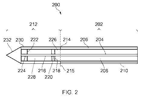
Fig. 2 shows a cross-sectional side view of an
electrosurgical instrument 200 that is an embodiment of the
invention. The distal end of the electrosurgical instrument
may correspond, for example, to the distal assembly 118
discussed above. The electrosurgical instrument 200 includes a
coaxial feed cable 202 that is connectable at its proximal end
to a generator (such as generator 102) in order to convey
microwave energy. The coaxial feed cable 202 may be the
interface cable 104 discussed above, which passes through the
flexible shaft 112. The coaxial feed cable 202 comprises an
inner conductor 204 and an outer conductor 206 which are
separated by a dielectric material 208. The coaxial feed cable
202 is preferably low loss for microwave energy. A choke (not
shown) may be provided on the coaxial feed cable 204 to
inhibit back propagation of microwave energy reflected from
the distal end and therefore limit backward heating along the
device. The coaxial feed cable 202 further includes a flexible
outer sheath 210 disposed around the outer conductor 206 to
protect the coaxial feed cable 204. The outer sheath 210 may
be made of an insulating material to electrically isolate the
outer conductor 206 from its surroundings. The outer sheath
210 may be made of, or coated with, a non-stick material such
as PTFE to prevent tissue from sticking to the instrument.
A radiating tip 212 is formed at the distal end 214 of
the coaxial feed cable 202. The dashed line 215 in Fig. 2
CA 03105136 2020-12-24
WO 2020/011547
PCT/EP2019/067168
illustrates an interface between the coaxial feed cable 202
and the radiating tip 212. The radiating tip 212 is arranged
to receive microwave energy conveyed by the coaxial feed cable
202, and deliver the energy into biological tissue. The outer
5 conductor 206 of the coaxial feed cable 202 terminates at the
distal end 214 of the coaxial feed cable 202, i.e. the outer
conductor 206 does not extend into the radiating tip 212. The
radiating tip 212 includes a distal portion 216 of the inner
conductor 204 which extends beyond the distal end of the
10 coaxial feed cable 202. In particular, the distal portion 216
of the inner conductor 204 extends beyond a distal end of the
outer conductor 206.
A proximal tuning element 218 made of a conductive
material (e.g. metal) is electrically connected to the distal
15 portion 216 of the inner conductor 204 near a proximal end of
the radiating tip 212. The proximal tuning element 218 has a
cylindrical shape, and includes a channel 220 through which
the distal portion 216 of the inner conductor 204 passes. A
diameter of the channel 220 is substantially the same as an
outer diameter of the inner conductor 204, such that the inner
conductor 204 is in contact with the proximal tuning element
218 inside the channel 220. The proximal tuning element 218
may be further secured to the inner conductor 204, e.g. using
a conductive adhesive (e.g. conductive epoxy) or by soldering
or welding. The proximal tuning element 218 is centred on the
inner conductor 204. In other words, a central axis of the
cylindrical proximal tuning element 218 is collinear with the
longitudinal axis of the inner conductor 204. In this manner,
the proximal tuning element 218 is disposed around the distal
portion 216 of the inner conductor 204 in a manner that is
symmetrical about the longitudinal axis of the inner conductor
204.
A distal tuning element 222 made of a conductive material
(e.g. metal) is electrically connected to the distal portion
216 of the inner conductor 204 near a distal end of the
radiating tip 212. Thus, the distal tuning element 222 is
located further along the inner conductor 204 than the
proximal tuning element 218. The distal tuning element 222 is
spaced apart from the proximal tuning element by a length of
the distal portion 216 of the inner conductor 204. Like the
proximal tuning element 218, the distal tuning element has a
CA 03105136 2020-12-24
WO 2020/011547
PCT/EP2019/067168
16
cylindrical shape and includes a channel 224. As can be seen
in Fig. 2, the distal portion 216 of the inner conductor 204
extends into the channel 224. The distal portion 216 of the
inner conductor 204 terminates at a distal end of the channel
224, i.e. it does not protrude beyond the distal tuning
element 222. In this manner, a distal end of the inner
conductor 204 lies flush with a distal face of the distal
tuning element 222. A diameter of the channel 224 is
substantially the same as the outer diameter of the inner
conductor 204, such that the inner conductor 204 is in contact
with the distal tuning element 222 inside the channel 224. The
distal tuning element 222 may be further secured to the inner
conductor 204, e.g. using a conductive adhesive (e.g.
conductive epoxy) or by soldering or welding. Like the
proximal tuning element 218, the distal tuning element 222 is
mounted so that it is centred on the inner conductor 204.
Both the proximal tuning element 218 and the distal
tuning element 222 have the same outer diameter. The outer
diameter of the proximal tuning element 218 and the distal
tuning element 222 may be slightly less than the outer
diameter of the electrosurgical instrument 200. In the example
shown, the distal tuning element 222 is longer than the
proximal tuning element 218 in the longitudinal direction of
the instrument. In other words, the length of inner conductor
204 in channel 224 in the distal tuning element 222 is greater
than the length of inner conductor 204 in channel 220 in the
proximal tuning element 218. For example, the distal tuning
element 222 may be approximately twice as long as the proximal
tuning element 218. By making the distal tuning element 222
longer than the proximal tuning element 218, it is possible to
concentrate microwave emission around the distal end of the
radiating tip 212.
A distal portion 226 of the dielectric material 208
extends beyond the distal end 214 of the coaxial feed cable
202 into the radiating tip 212. The distal portion 226 of the
dielectric material 208 acts as a spacer between the proximal
tuning element 218 and the distal end 214 of the coaxial feed
cable 202. In some embodiments (not shown), the dielectric
material 208 may terminate at the distal end 214 of the
coaxial feed cable 202, and a separate spacer may be provided
between the distal end 214 of the coaxial feed cable 202 and
CA 03105136 2020-12-24
WO 2020/011547
PCT/EP2019/067168
17
the proximal tuning element 218. A dielectric spacer 228 is
provided in the radiating tip 212 between the proximal tuning
element 218 and the distal tuning element 222. The dielectric
spacer 228 is a cylindrical piece of dielectric material,
having a central channel extending therethrough. Thus, the
dielectric spacer 228 may be a tube of dielectric material.
The distal portion 214 of the inner conductor 204 extends
through the channel in the dielectric spacer 228. A proximal
face of the dielectric spacer 228 is in contact with the
proximal tuning element 218, and a distal face of the
dielectric spacer 228 is in contact with the distal tuning
element 222. The dielectric spacer 228 has approximately the
same outer diameter as the proximal and distal tuning elements
218, 222.
A protective sheath 230 is provided on the outside of the
radiating tip 212. The protective sheath 230 covers the
dielectric spacer 228 and the proximal and distal tuning
elements 218, 222 to form an outer surface of the radiating
tip 212. The protective sheath 230 may be a tube made of an
insulating material. The protective sheath 230 may serve to
insulate the radiating tip 212 and protect it from the
environment. The protective sheath 230 may be made of or
coated with a non-stick material (e.g. PTFE) to prevent tissue
from sticking to it. An outer diameter of the protective
sheath 230 is substantially the same as the outer diameter of
the coaxial feed cable 202, so that the instrument has a
smooth outer surface, i.e. the radiating tip 212 has an outer
surface that is flush with an outer surface of the coaxial
feed cable 202 at the interface 215. In some embodiments (not
shown) the protective sheath 230 may be a continuation of the
outer sheath 210 of the coaxial feed cable 202. Together, the
distal portion 226 of the dielectric material 208, the
dielectric spacer 228 and the protective sheath 230 form a
dielectric body of the radiating tip 212.
The radiating tip 212 further includes a distal tip 232
located at its distal end. The distal tip 232 may be pointed
in order to facilitate insertion of the radiating tip 212 into
target tissue. However, in other embodiments (not shown), the
distal tip may be rounded or flat. The distal tip 232 may be
made of a dielectric material, e.g. the same as dielectric
material 208. In some embodiments, the material of the distal
CA 03105136 2020-12-24
WO 2020/011547
PCT/EP2019/067168
18
tip 232 may be selected to improve impedance matching with
target tissue, in order to improve the efficiency with which
the EM energy is delivered to the target tissue. The distal
tip 232 may be made of, or covered with a non-stick material
(e.g. PTFE) to prevent tissue from sticking to it.
The following are example dimensions of electrosurgical
instrument 200:
- distance from the interface 215 to the distal end of
the distal portion 216 of the inner conductor 204: 5.75 mm;
- outer diameter of proximal tuning element 218 and
distal tuning element 222: 1.5 mm;
- length of proximal tuning element 218: 0.5 mm;
- length of distal tuning element 222: 1.0 mm;
- spacing between proximal tuning element 218 and distal
tuning element 222: 3.75 mm;
- spacing between the proximal tuning element 218 and the
interface 215: 0.5 mm; and
- outer diameter of electrosurgical instrument 200: 1.85
mm.
The radiating tip 212 may act as a microwave monopole
antenna when microwave energy is conveyed to the radiating tip
212. In particular, microwave energy may be radiated from the
distal portion 216 of the inner conductor 202, so that
microwave energy can be delivered into surrounding biological
tissue. The proximal and distal tuning elements 218, 222 act
to shape the radiation profile of the radiating tip 212, and
improve impedance matching between the instrument and
surrounding target tissue, as discussed below.
Fig. 3 shows a simulated microwave radiation profile in
target tissue for the electrosurgical instrument 200
illustrated in Fig. 2. The radiation profile was simulated for
a microwave frequency of 5.8 GHz, using finite element
analysis software. The radiation profile is indicative of the
resultant shape of tissue ablated by the microwave energy. As
can be seen in Fig. 3, the radiation profile is concentrated
around the radiating tip, and defines an approximately
spherical region. In this manner, tissue may be ablated in an
approximately spherical region around the radiating tip. The
interface 215 between the radiating tip and coaxial feed cable
is shown to aid visualisation of the location and shape of the
field relative to the tip of the instrument.
CA 03105136 2020-12-24
WO 2020/011547 PCT/EP2019/067168
19
Fig. 4 shows a simulated plot of the S-parameter (also
known as the input reflection coefficient Sil, or "return
loss") against frequency of the microwave energy for the
electrosurgical instrument 200. As well known in the technical
field, the S-parameter is a measure of the return loss of
microwave energy due to impedance mismatch, and as such the 5-
parameter is indicative of the degree of impedance mismatch
between the target tissue and the radiating tip. The 5-
parameter can be defined by the equation PI = SPR, where PI is
the outgoing power in the instrument towards the tissue, PR is
the power reflected back from the tissue, and S is the 5-
parameter. As shown in Fig. 4, the S-parameter has a value of
-25.58 dB at 5.8 GHz, meaning that very little microwave
energy is reflected back from the tissue at this frequency.
This indicates a good impedance match at the operating
frequency of 5.8 GHz, and that microwave energy is efficiently
delivered from the radiating tip into the tissue at this
frequency.
Fig. 5 shows a simulated impedance Smith chart for the
electrosurgical instrument 200. The Smith chart was simulated
for a reference plane locating at the interface 215 between
the distal end of the coaxial feed cable and the radiating
tip. As well known in the technical field, the Smith chart is
a graphical representation of the S-parameter (reflection
coefficient) in the complex plane. The S-parameter may be
defined by the following equation:
z¨ 1
S = ¨
z 1
where z = Z/Zo, Z being the impedance of the radiating
tip in contact with target tissue, and Zo being a normalisation
factor. In the present case, a normalisation factor of 50 Ohm
was used, as this is a typical characteristic impedance of the
coaxial feed cable, the interface cable (e.g. interface cable
104) and the electrosurgical generator (e.g. generator 102).
In Fig. 5, the marker (labelled "1") indicates the value of
the S-parameter at 5.8 GHz. As can be seen, the value of the
S-parameter is near the unity mark (i.e. the point where z =
1). This shows a good impedance matching between the
generator, interface cable, coaxial feed cable and the antenna
CA 03105136 2020-12-24
WO 2020/011547
PCT/EP2019/067168
in contact with the target tissue. In other words, microwave
energy may be efficiently delivered from the radiating tip
into target tissue. The value of the impedance Z at 5.8 GHz is
indicated in the legend of Fig. 5, and is (54.9 + i2.9) Ohm.
5 The full circle and the empty circle next to the marker in
Fig. 5 indicate points at 6 GHz and 5.6 GHz, respectively. The
value of the impedance Z for these points is shown in the
legend of Fig. 5.
We now turn to comparative examples shown in Figs. 6-13,
10 to illustrate the effects of the proximal and distal tuning
elements in more detail. Fig. 6 shows an electrosurgical
instrument 600 which is a first comparative example, and Fig.
7 shows an electrosurgical instrument 700 which is a second
comparative example. Electrosurgical instrument 600 is similar
15 to electrosurgical instrument 200, except that electrosurgical
instrument 600 does not include a proximal tuning element. All
other features of electrosurgical instrument 600 (including
the distal tuning element) are the same as for electrosurgical
instrument 200. Electrosurgical instrument 700 is similar to
20 electrosurgical instrument 200, except that electrosurgical
instrument 700 does not include a proximal tuning element or a
distal tuning element (i.e. both tuning elements are absent).
All other features of electrosurgical instrument 700 are the
same as for electrosurgical instrument 200. Reference numerals
used in Fig. 2 are used in Figs. 6 and 7 to indicate features
corresponding to those discussed above in relation to Fig. 2.
Fig. 8 shows a simulated microwave radiation profile in
target tissue for the electrosurgical instrument 600
illustrated in Fig. 6. The radiation profile was simulated for
a microwave frequency of 5.8 GHz, using finite element
analysis software. Except for the lack of a proximal tuning
element, the dimensions of electrosurgical instrument 600 used
for the calculation were the same as those used to calculate
the radiation profile of electrosurgical instrument 200 shown
in Fig. 3. As can be seen by comparing Figs. 3 and 8, the
radiation profile of electrosurgical instrument 600 is less
spherical than the radiation profile of electrosurgical
instrument 200. In particular, the radiation profile of
electrosurgical instrument 600 includes a tail which extends
back down a longer portion of the coaxial feed cable than a
tail on the radiation profile of electrosurgical instrument
CA 03105136 2020-12-24
WO 2020/011547
PCT/EP2019/067168
21
200. Thus, the proximal tuning element acts to make the
radiation profile more spherical, and reduce the tail which
extends back down the coaxial feed cable. Such a tail may be
undesirable, as it may cause heating in the coaxial feed cable
and/or cause ablation of tissue which is outside of a target
zone.
Fig. 9 shows a simulated plot of the S-parameter against
frequency of the microwave energy for the electrosurgical
instrument 600. The plot in Fig. 9 was calculated in the same
way as the plot in Fig. 4 for electrosurgical instrument 200.
As shown in Fig. 9, the S-parameter has a value of -10.18 dB
at 5.8 GHz. This indicates a much greater return loss compared
to electrosurgical instrument 200, where the S-parameter was
found to have a value of -25.58 dB. The proximal tuning
element therefore serves to improve impedance matching.
Microwave energy may therefore be more efficiently delivered
into target tissue with electrosurgical instrument 200 than
with electrosurgical instrument 600.
Fig. 10 shows a simulated impedance Smith chart for
electrosurgical instrument 600. This was calculated in the
same way as the Smith chart for electrosurgical instrument 200
shown in Fig. 5. The marker in Fig. 10 (labelled "1")
indicates the value of the S-parameter at 5.8 GHz. As can be
seen, the marker is further away from the unity mark compared
to Fig. 5. This shows a less good impedance match between the
generator, interface cable, coaxial feed cable and the antenna
in contact with the target tissue, compared to electrosurgical
instrument 200. By comparing Figs. 5 and 10, it can be seen
that the effect of adding the proximal tuning element is to
move the marker downwards closer to the unity mark. This
indicates that the proximal tuning element introduces an
additional capacitance into the system. The shift of the
marker closer to the unity mark in Fig. 5 may also be related
to the phase shift associated with the distance between the
distal end of the coaxial feed cable and the proximal end of
the proximal tuning element. The value of the impedance Z of
electrosurgical instrument 600 at 5.8 GHz is indicated in the
legend of Fig. 10, and is (40.2 + i27.5) Ohm. The full circle
and the empty circle next to the marker in Fig. 10 indicate
points at 6 GHz and 5.6 GHz, respectively. The value of the
CA 03105136 2020-12-24
WO 2020/011547
PCT/EP2019/067168
22
impedance Z for these points is shown in the legend of Fig.
10.
Fig. 11 shows a simulated microwave radiation profile in
target tissue for the electrosurgical instrument 700
illustrated in Fig. 7. The radiation profile was simulated for
a microwave frequency of 5.8 GHz, using finite element
analysis software. Except for the lack of a proximal and
distal tuning elements, the dimensions of electrosurgical
instrument 700 used for the calculation were the same as those
used to calculate the radiation profile of electrosurgical
instrument 200 shown in Fig. 3. As can be seen by comparing
Figs. 3, 8 and 11, the radiation profile of electrosurgical
instrument 700 is even less spherical and more elongate than
the radiation profile of electrosurgical instrument 600. In
particular, the radiation profile of electrosurgical
instrument 700 is less concentrated around the distal tip of
the instrument, and has a longer tail extending back down the
coaxial feed cable. Thus, the distal tuning element acts to
make the radiation profile more spherical, and reduce the tail
which extends back down the coaxial feed cable.
Fig. 12 shows a simulated plot of the S-parameter against
frequency of the microwave energy for the electrosurgical
instrument 700. The plot in Fig. 12 was calculated in the same
way as the plot in Fig. 4 for electrosurgical instrument 200.
As shown in Fig. 12, the S-parameter has a value of -5.66 dB
at 5.8 GHz. This indicates a much greater return loss compared
to electrosurgical instruments 200 and 600, where the 5-
parameter was found to have a value of -25.58 dB and -10.18
dB, respectively. The distal tuning element therefore serves
to improve impedance matching.
Fig. 13 shows a simulated impedance Smith chart for
electrosurgical instrument 700. This was calculated in the
same way as the Smith chart for electrosurgical instrument 200
shown in Fig. 5. The marker in Fig. 13 (labelled "1")
indicates the value of the S-parameter at 5.8 GHz. As can be
seen, the marker is further away from the unity mark compared
to Fig. 5. This shows a less good impedance match between the
generator, interface cable, coaxial feed cable and the antenna
in contact with the target tissue, compared to electrosurgical
instrument 200. The marker in Fig. 13 is also further away
from the unity mark compared to Fig. 10, indicating a less
CA 03105136 2020-12-24
WO 2020/011547
PCT/EP2019/067168
23
good impedance match. The value of the impedance Z of
electrosurgical instrument 700 at 5.8 GHz is indicated in the
legend of Fig. 13, and is (20.5 - i25.7) Ohm. The full circle
and the empty circle next to the marker in Fig. 13 indicate
points at 6 GHz and 5.6 GHz, respectively. The value of the
impedance Z for these points is shown in the legend of Fig.
13.
In summary, the comparative examples show that the
presence of both the proximal and distal tuning elements in
the radiating tip serves to enhance the radiation profile of
the radiating tip, by making the radiation profile more
spherical and reducing the tail that extends back down the
coaxial feed cable. The comparative examples also show that
the proximal and distal tuning elements serve to improve
impedance matching, which may improve the efficiency with
which microwave energy can be delivered into target tissue.
The inventors have found that, as the outer diameter of
the electrosurgical instrument is increased, the tail in the
radiation profile that extends back down the coaxial feed
cable increases. This is illustrated in Fig. 14, which shows a
simulated microwave radiation profile in target tissue for an
electrosurgical instrument according to an embodiment of the
invention. The electrosurgical instrument of Fig. 14 is
similar to electrosurgical instrument 200 described above,
except that it has an outer diameter of 2.6 mm (whereas
electrosurgical instrument 200 has an outer diameter of 1.85
mm). The radiation profile was simulated for a microwave
frequency of 5.8 GHz, using finite element analysis software.
The dashed line indicated by numeral 215 in Fig. 14 shows the
position of the interface between the coaxial feed cable and
the radiating tip. As can be seen by comparing Fig. 14 with
the radiation profile for electrosurgical instrument 200, the
tail that extends back down the coaxial feed cable is larger
for the electrosurgical instrument of Fig. 14, i.e. the
electrosurgical instrument having the larger outer diameter.
The inventors have found that the tail in the radiation
profile may be suppressed by including a field shaping element
at a distal end of the coaxial feed cable. Fig. 15 shows a
cross-sectional side view of an electrosurgical instrument 900
that is an embodiment of the invention. The electrosurgical
instrument 900 is similar to electrosurgical instrument 200
CA 03105136 2020-12-24
WO 2020/011547
PCT/EP2019/067168
24
discussed above, except that it includes a field shaping
element 902, and its outer diameter is 2.6 mm. Reference
numerals used in Fig. 2 are used in Fig. 15 to indicate
features corresponding to those discussed above in relation to
Fig. 2.
The field shaping element 902 is an annular sleeve of
conductive material disposed around an outer surface of the
outer conductor 206. The field shaping element 902 is located
at the distal end of the coaxial feed cable 202, and extends
from the interface 215 along a length of the coaxial feed
cable 202. The length of the field shaping element 902
corresponds to a quarter wavelength of the microwave energy to
be conveyed by the coaxial feed cable 202. In the case where
microwave energy is at 5.8 GHz, the length of the field
shaping element 902 may be approximately 9 mm. An inner
surface of the field shaping element 902 is in contact with
the outer surface of the outer conductor 206, so that the
field shaping element 902 is electrically connected to the
outer conductor 206 along its length. Electrical connection
between the field shaping element 902 and the outer conductor
206 may be ensured by securing the field shaping element 902
to the outer conductor 206, e.g. using conductive epoxy, or by
soldering or welding them together. In some embodiments (not
shown) the field shaping element 902 may be integrally formed
with the outer conductor 206. The field shaping element 902
acts to increase an effective thickness of the outer conductor
206 in a distal region of the coaxial feed cable 202.
Fig. 16 shows a simulated microwave radiation profile in
target tissue for the electrosurgical instrument 900
illustrated in Fig. 15. The radiation profile was simulated
for a microwave frequency of 5.8 GHz, using finite element
analysis software. As can be seen by comparing Figs. 16 and
14, the radiation profile in Fig. 16 has a smaller tail
extending back down the coaxial feed cable. The radiation
profile in Fig. 16 also appears more spherical, and is more
concentrated around the radiating tip. The only difference
between the electrosurgical instrument in Fig. 14 and
electrosurgical instrument 900 is the presence of the field
shaping element 902 in electrosurgical instrument 900. Thus,
field shaping element 902 serves to reduce the tail in the
CA 03105136 2020-12-24
WO 2020/011547
PCT/EP2019/067168
radiation profile, and to concentrate emission of microwave
energy around the radiating tip.