Note: Descriptions are shown in the official language in which they were submitted.
1
ELECTRICAL IMPEDANCE HAEMATOCRIT AND HBA1C BIOSENSOR COMPRISING SAMPLE
PLATE AND SAMPLE APPARATUS
The present application is a divisional application of Canadian Patent
Application No. 2,870,354 filed
on October 10, 2014.
The present invention relates to a sample measurement system. In particular,
the present invention
relates to the measurement of properties of liquid samples of (or containing)
blood. In particular the
invention relates to a sample measurement system for measuring certain
selected properties of a liquid
substrate, such as the glucose levels in a blood sample. The invention also
relates to a sampling plate,
a measurement device, a data carrier containing software to operate the
measurement device.
There is a widespread need for improving the accuracy of sample measurement
systems such as those
enabling e.g. a diabetes sufferer to know their blood sugar levels ¨ i.e. the
concentration of glucose in
their blood.
Existing sample measurement systems use a measurement device which receives
and takes
measurement readings from a sampling plate spotted with a blood sample from a
user. The sampling
plate is often rectangular and is end-loaded with the blood sample. The blood
sample, once loaded, is
usually drawn into a sample zone having a number of sampling zones from which
measurements are
taken by the system.
Each sampling zone typically has its own particular contents. For example, the
first sampling zone may
have a glucose oxidase deposit within it, a second deposit comprising a
mixture of glucose oxidase and
a predetermined amount of glucose, while a third sampling zone may contain no
deposit. As the blood
sample is drawn over all three sampling zones, chemical reactions occur with
the deposits in each
sampling zone, resulting in discrete electrolytes. Each sampling zone bridges
a corresponding pair of
electrodes. A potential difference is established across each sampling zone,
via the electrodes, when
the sampling plate is inserted into an operating measurement device. Electric
current readings for each
sampling zone then provide measurements necessary to assess the blood sugar
(glucose) levels. For
instance, the first sampling zone may give the primary measurement, whereas
the second sampling
zone may provide a degree of calibration since a known quantity of glucose was
already present there.
The third zone may give a final check by accounting for the non-glucose
contribution to the
measurements in the first and second sampling zones.
However, in spite of these calibrations and final checks, error margins in
such blood glucose readings
are still high. Indeed, blood glucose levels are strongly influenced by the
fluctuating and transient
glucose levels in the plasma of the blood sample, which may not be
representative of the long-term
blood glucose levels of the patient and may, rather, simply indicate a recent
transient rise or drop in
Date Recue/Date Received 2021-01-11
2
blood glucose levels within the blood plasma of the patient e.g. due to recent
food consumption of other
short-term environmental factors.
The present invention aims to address this.
Blood plasma is the liquid component of blood in which the blood cells in
whole blood are normally
suspended. Blood plasma typically constitutes about 55% of the total volume of
the blood. It is the
extracellular fluid part of blood and is mostly water but contains dissolved
glucose and other contents.
The volume percentage of red blood cells in blood is known as the haematocrit
(HCT). Other terms for
this are the packed cell volume (PCV) or erythrocyte volume fraction (EVF).
Haematocrit is normally
about 45% for men and 40% for women. The haematocrit is typically calculated
by multiplying the red
blood cell count in a blood sample by the average cell volume, then dividing
the result by the whole
blood sample volume.
Glycated haemoglobin (a.k.a. haemoglobin Al c, HbAl c, or just Al c) is a form
of haemoglobin measured
primarily to identify the average plasma glucose concentration over prolonged
periods of time. It is
formed in a non-enzymatic glycation pathway by hemoglobin's exposure to plasma
glucose. Normal
levels of glucose produce a normal amount of glycated hemoglobin. As the
average amount of plasma
glucose increases, the fraction of glycated hemoglobin increases. This serves
as a marker for average
blood glucose levels over the previous months prior to the measurement. Liquid
chromatography and
capillary electrophoresis are two ways of measuring glycated haemoglobin
(HbAlc). Both methods are
complex, expensive and wholly unsuited for easy and simple implementation by a
patient.
At its most general, the invention in one aspect is a system (method and/or
apparatus) to measure
haematocrit of a liquid sample containing blood according to the electrical
impedance (e.g., resistance
and reactance) it has in response to an alternating electrical potential
difference applied across the
sample. The measured haematocrit maybe used to improve the accuracy of blood
glucose
measurements of the blood in another aspect of the invention. It has been
found that applying an
alternating potential difference (voltage) across such a sample results in a
resistance and/or a reactance
which is surprisingly responsive to haematocrit. The invention exploits this
finding. The presence of
red blood cells within a blood sample complicates the interpretation of blood
glucose measurements
using existing methods. The invention may remove or reduce that complication
to enable more accurate
blood glucose measurements to be made. At its most general, the invention in
another aspect is a
system (method and/or apparatus) to measure a level of glycated haemoglobin
(HbAlc) in a liquid
sample containing blood according to the electrical impedance (e.g.,
resistance and reactance) it has in
response to an alternating electrical potential difference applied across the
sample. It has been found
that applying an alternating potential difference (voltage) across such a
sample results in a resistance
Date Recue/Date Received 2021-01-11
3
and/or a reactance which is surprisingly responsive to HbA1c of the sample.
The invention exploits this
finding.
In a first of its aspects, the invention may provide a sampling plate
comprising a sample zone for
receiving a liquid sample. The sampling plate may have two drive electrodes
with separate respective
electrode terminals spaced by a spacing for receiving the liquid sample within
the sample zone for use
in driving an electrical signal through the sample. Two sensing electrodes may
be provided with
separate respective electrode terminals spaced between the electrode terminals
of the two drive
electrodes for use in sensing an electrical signal generated by the drive
electrodes within a the sample.
Herein, a "sampling plate" may mean any surface capable of receiving a liquid
sample in a sample
zone. Preferably, however, the sampling plate is portable. Suitably the
sampling plate may cover an
area less than 1 m2, preferably less than 50 cm2, more preferably less than 10
cm2 and most
preferably less than 5 cm2. The sampling plate may cover an area less than 500
mm2 ¨ for instance
350 mm2 where the sampling plate is 10 mm wide by 35 mm long. Suitably the
sampling plate may be
rectangular. The sampling plate may be a strip, and may be a flexible strip.
Preferably, however, the
.. sampling plate is an individual plate, preferably a rigid sampling plate.
The thickness of the sampling
plate is preferably less than 1 cm, preferably less than 1 mm, more preferably
less than 0.5 mm, most
preferably less than 0.25 mm.
The sampling plate is preferably compatible with a measurement device. For
example, the
measurement device is preferably operable to communicate with the sampling
plate to measure one or
more selected properties of the sample. Preferably the sampling plate may be
inserted into the
measurement device to allow measurements to be taken.
The two sensing electrode terminals may present to each other opposing sides
which define between
them an elongate sensing gap extending along the sample zone for receiving at
least parts of the
sample therein. The sensing electrode terminals may be substantially flat and
side-by-side to define a
substantially flat sensing gap. The width of the two sensing electrode
terminals is preferably the same.
That width is preferably about double the size of the sensing gap between
them.
The two drive electrode terminals may present to each other opposing sides
which define between
them an elongate drive gap extending along the sample zone for receiving at
least parts of the sample
therein whereby the drive electrodes are adapted to drive electrical signal
transversely across the
drive gap. The drive electrode terminals may be substantially flat and side-by-
side to define a
substantially flat drive gap.
The sensing gap may extend along the drive gap. The sensing gap and/or the
drive gap may preferably
have a substantially uniform width along at least a part of its length.
Date Recue/Date Received 2021-01-11
4
A drive electrode terminal and an adjacent sensing electrode terminal may be
arranged in/on the
sampling plate so that they present to each other opposing sides which define
between them an
elongate partitioning gap. Preferably, this partitioning gap extends along the
sample zone to define a
partition between those adjacent terminals within the sample zone. This may
apply to each of the drive
electrode terminals and their respective adjacent/neighbouring sensing
electrode terminal.
The partitioning gap preferably has a substantially uniform width along at
least a part of its length,
preferably substantially all of its length. Each partitioning gap width is
preferably the same size.
Preferably the partitioning gap width is about 1% times the width of the
sensing gap.
The sensing electrode terminals may be formed on a surface of the sensing
plate within the sample
zone. Preferably, the drive electrode terminals are formed on a surface of the
sensing plate in
common with the sensing electrode terminals within the sensing zone. The
electrodes may be formed
upon the sampling plate by a known printing process. However, for a better
degree of accuracy or
consistency a technique known as laser ablating is preferably used to remove
electrode material (e.g.
Gold) formed as a sheet/coating onto a surface of the sampling plate, where
the electrode gaps are
required. Preferably, in production, conductive electrode material may be laid
down on a surface area
on one side of the sampling plate with no gap features, and the gaps may then
be laser ablated to
define the electrodes.
The sampling plate is preferably arranged to be detachably attachable
electrically to electrical
apparatus adapted for supplying the drive current to the drive electrodes and
for taking measurements
via the sensing electrodes. In this regard, preferably each of the drive
electrodes and each of the
sensing electrodes is in electrical communication with respective electrical
contacts provided on the
sampling plate which are exposed for electrical connection simultaneously with
an external drive
current source and external sensing circuitry, respectively, of such an
apparatus.
Preferably, the width of the sensing gap is greater than about 90 microns and
less than about 160
microns. Preferably the width of the partitioning gap is about 1.5 times the
width of the sensing gap. An
alternating potential difference is preferably applied across a gap between
two electrodes designed to
be bridged by a sample being measured. It has been found that a careful
dimensioning of the sensing
gap and the drive gap enhances the accuracy of haematocrit measurement
greatly, and also permits
HbA1c to be measured. The gap size is preferably substantially smaller than a
gap size between
electrodes typically employed in existing systems designed to measure blood
glucose. Preferably, the
sensing gap is wider than the average width of a human red blood cell, but
less than the average width
of two such cells.
It is postulated, but not asserted, that the sensing gap serves to form a
generally linear array of red
blood cells along it in which the array is generally one cell in width ¨ this
being constrained by the width
Date Recue/Date Received 2021-01-11
5
of the sensing gap ¨ and that the parts of the sensing gap not occupied by red
blood cells are occupied
by blood plasma. Blood plasma is typically more electrically conductive than
are red blood cells at
certain electrical signal frequencies. By applying an oscillating voltage, red
blood cells remain mobile
(e.g. oscillate) within the gap and may not coat one or other of the
electrodes. The result may be that
there is maintained within the sensing gap a defined linear array of red blood
cells mobile within
conductive blood plasma. The proportion of red blood cells within the gap,
relative to the quantity of
blood plasma there, influences the quantity of electrically conductive
pathways (through plasma, around
blood cells) available to currents applied. This may manifest itself as an
electrical impedance value (e.g.
resistance, reactance) determined by a haematocrit value and/or an HbAl c
value, as has been
observed.
The sample zone may comprise a reagent to react with free glucose in the
liquid sample. This may be
so when the sampling plate is intended for use in measuring a value
representing the concentration of
glycated haemoglobin (HbAlc) in the liquid sample as described below. The
reagent may be a deposit
formed on one (e.g. exclusively) of the drive electrodes in the sample zone
(e.g. the one for use as an
anode) to be directly accessible to a sample therein. The deposit may be in
the form of an ink or paste.
Preferred reagents are oxidising agents. Most preferred are enzymes and
especially preferred are
glucose oxidase (G0x) and glucose dehydrogenase (GDH). Where no such reagent
is present, the
sampling plate may be used for measuring haematocrit as described below.
The sampling plate may comprise a further sample zone containing a pair of
drive electrodes as
described above, and a pair of sensing electrodes as described above. The
further sample zone may
be free of any reagent and be intended for use in measuring HCT of a blood
sample, while the other
sample zone may contain the reagent and be intended for concurrent or
sequential measurement of
HbAl c of the same sample.
Alternatively, the drive electrode terminals of the further sample zone may
comprise only one pair of
drive electrodes which may present to each other, across a respective spacing,
opposing electrode
sides extending along the sample zone. The further sample zone may contain the
reagent and be
intended for use in measuring blood glucose levels within the plasma of a
blood sample. These opposing
sides may define between them a drive gap for receiving a sample therein. This
spacing may define a
gap which is preferably greater than about 200 microns in width, and may be
between 200 microns and
400 microns in width. This dimensioning has been found to be preferable for
the electrodes in sampling
zones containing the reagent to react with free glucose in the liquid sample,
and for measuring a current
generated in response to a direct (DC) drive voltage applied across the drive
gap. The measured current
may be used to determine a measure of the glucose in the blood plasma of the
sample.
Date Recue/Date Received 2021-01-11
6
The opposing sides in the pair of drive electrodes of the further sample zone
may be of unequal length.
They may be curved. One side may be convex and the opposing side reciprocally
concave and of greater
length than the convex side. Preferably the electrode with the longer side is
used as the cathode of the
pair. This is preferable in view of the greater gap size in each further pair
of electrodes. It has been
found that electrical currents driven across those wider gaps through a blood
sample are more prone to
diffuse in a direction along the gap rather than flowing directly across the
gap un-deviated. In order to
better capture diffused charges (current) in the blood sample the electrode to
which the charges flow
when a direct (DC) voltage is applied between the electrodes, has the longer
edge. The spacing of the
drive gap may be substantially uniform along at least a part of its length.
In a second of its aspects, the invention may provide a sampling apparatus for
use in performing
electrical measurements on a liquid sample containing blood, including two
current output terminals for
outputting an alternating current signal applied therebetween, and an
alternating electrical current unit
in electrical communication with the two current output terminals for applying
thereto an alternating
electrical current of a given amplitude and frequency when a liquid sample is
in electrical connection
between the two current output terminals.
The sampling apparatus may include a voltage unit in electrical communication
with the two current
output terminals for applying therebetween a direct (DC), being most
preferably a substantially constant
(DC), electrical potential difference of a given magnitude. A first voltage
input terminal may be provided
for receiving a first electrical signal externally input thereto and a
separate second voltage input terminal
for receiving a second electrical signal externally input thereto when the
liquid sample is in electrical
connection between the first and second voltage input terminals. The apparatus
may include voltage
detector(s) for measuring a first voltage and a second voltage using said
first and second electrical
signals, respectively.
A control unit may be arranged in the sampling apparatus to control the
electrical current unit to apply
the alternating electrical current of given frequency and concurrently to
control the voltage unit and the
voltage detector(s) to measure the first and second voltages both when the
direct (e.g. substantially
constant) DC electrical potential difference is applied and when the direct DC
electrical potential
difference is not applied.
A calculating unit may be arranged in the sampling apparatus to calculate a
first electrical impedance
(e.g. reactance) value using the first and second voltages measured when the
direct (e.g. substantially
constant) DC electrical potential difference is applied, and to calculate a
second electrical impedance
(e.g. reactance) value measured when the direct DC electrical potential
difference is not applied.
Date Recue/Date Received 2021-01-11
7
The calculating unit may be arranged in the sampling apparatus to generate a
value representing the
concentration of glycated haemoglobin (HbA1c) in the liquid sample according
to the first electrical
reactance value, the second electrical reactance value and a value
representing the relative volume of
red blood cells in the liquid sample (haematocrit, HCT).
The calculating unit may be arranged to generate a value representing the
concentration of glycated
haemoglobin (HbA1c) in the blood within the sample according to the first
electrical reactance value, the
second electrical reactance value and a value of the relative volume of red
blood cells in the liquid
sample (haematocrit, HCT) according to the following formula, and store the
result and/or to output the
result to the user:
(
X
HbAlc =100x 1 1
HCT x X2 )
The haematocrit value HCT may be a contemporaneously measured value, such as
measured using a
sample of the blood on the sampling plate, or may be a predetermined value
which is generated
independently of the sampling unit.
The quantity Xi is considered to represent the reactance of a blood sample due
to glycated red blood
cells in the blood within the first sampling zone from which free glucose has
been substantially oxidized
by the reagent, whereas X2 is considered to represent the reactance of the
whole blood sample in which
both plasma and red blood cells contain glucose. The proportion of that
reactance due to red blood
cells is considered to be represented by the term (HCT)x(X2) according to the
haematocrit of the sample.
The given frequency preferably has a value in the range 500KHz to 1.5MHz, e.g.
about 1MHz. More
preferably, the given frequency has a value in the range 750KHz to 1.25MHz,
yet more preferably the
given frequency has a value in the range 850KHz to 1.15MHz, even more
preferably the given frequency
has a value in the range 900KHz to 1.1MHz, yet even more the given frequency
has a value in the range
970KHz to 1.03MHz. It has been found that a frequency of about 1MHz works
especially well, and
frequencies reasonably close to this value are desirable, though the ranges
given above have been
found to be acceptable in terms of accuracy of measurement in implementing the
invention. The value
of the direct (DC) voltage may be a value in the range from about 0.01 volts
to about 1.0 volts, or
preferably from about 0.1 volts to about 0.5 volts, or more preferably from
about 0.2 volts to about 0.3
volts ¨ e.g. about 0.25 volts.
It is postulated, but not asserted, that the presence of a direct (DC) voltage
across the drive gap, and
therefore across the sensing gap, has the effect of polarizing or physically
aligning in a common direction
those red blood cells that are not glycated, while the glycated red blood
cells are not forced into this
Date Recue/Date Received 2021-01-11
8
alignment and remain largely unaffected by the direct voltage applied across
the sample. The
consequence is felt most keenly when an alternating (AC) current is applied to
the blood sample while
the is concurrently subjected to this DC voltage. The result is believed to be
that the un-glycated red
blood cells aligned by the applied DC voltage are far less responsive to the
AC current concurrently
applied (i.e. less able to dynamically interact/oscillate in response to it)
than are the glycated red blood
cells. The result is that the portion of the impedance (e.g. reactance) of the
blood sample arising from
the un-glycated red blood cells falls dramatically, leaving the glycated red
blood cells to dominate the
impedance of the sample. By comparing this impedance value to the impedance
value of the same
sample measured when no direct (DC) voltage is applied (and thus, no un-
glycated cell alignment
occurs) provides a route to determining the proportion of glycated red blood
cells in the sample and,
from that, a measurement of HbA1c.
Alternatively, or additionally, the calculating unit may be arranged to
generate a value representing the
relative volume of red blood cells in the liquid sample (haematocrit)
according to electrical impedance
(e.g. resistance and reactance) values measured thereby from the sample. The
calculating unit may be
arranged to generate a value representing the concentration of glycated
haemoglobin in the liquid
sample according thereto.
In a third aspect, the invention may provide a sampling apparatus for use in
performing electrical
measurements on a liquid sample containing blood, the apparatus comprising two
current output
terminals for outputting an alternating current signal applied therebetween,
and an alternating electrical
current unit in electrical communication with the two current output terminals
for applying therebetween
an alternating electrical current of a given amplitude and frequency, when a
liquid sample is in electrical
connection between the two current output terminals.
This sampling apparatus may include a first voltage input terminal for
receiving a first electrical signal
externally input thereto and a separate second voltage input terminal for
receiving a second electrical
signal externally input thereto, when said liquid sample is in electrical
connection between the first and
second voltage input terminals, and a voltage detector(s) for measuring a
first voltage and a second
voltage using said first and second electrical signals, respectively.
A control unit may be arranged in this sampling apparatus to control the
electrical current unit to apply
the alternating electrical current at a first frequency and concurrently to
control the voltage detector(s)
to measure the first and second voltages, and to further control the
electrical current unit to apply the
alternating electrical current at a second frequency exceeding the first
frequency and concurrently to
control the voltage detector(s) to measure the first and second voltages. The
first frequency may be a
value (e.g. 50 KHz) within a first continuous range of values from about 1KHz
to about 150 KHz. More
preferably, the first frequency has a value in the range 25KHz to 125KHz, yet
more preferably the first
Date Recue/Date Received 2021-01-11
9
frequency has a value in the range 35KHz to 100KHz, even more preferably the
first frequency has a
value in the range 45KHz to 75KHz, yet even more the first frequency has a
value in the range 47KHz
to 53KHz. It has been found that a frequency of about 50KHz works especially
well, and frequencies
reasonably close to this value are desirable, though the ranges given above
have been found to be
acceptable in terms of accuracy of measurement in implementing the invention.
The second frequency may be a value (e.g. 1 MHz) within a second continuous
range of values from
about 500KHz to about 1.5 MHz. More preferably, the second frequency has a
value in the range
750KHz to 1.25MHz, yet more preferably the second frequency has a value in the
range 850KHz to
1.15MHz, even more preferably the second frequency has a value in the range
900KHz to 1.1MHz, yet
even more the second frequency has a value in the range 970KHz to 1.03MHz. It
has been found that
a second frequency of about 1MHz works especially well, and frequencies
reasonably close to this value
are desirable, though the ranges given above have been found to be acceptable
in terms of accuracy
of measurement in implementing the invention.
In general, the preferred range of frequencies, and the preferential frequency
within such a range, is
influenced to some extent by geometrical considerations of the sampling
process. Factors such as the
size of surface area of conductive elements/electrodes within a test area of a
sampling plate, in relation
to the size of surface area of non-conductive/non-electrode parts between
electrodes, can influence the
position and extent of the suitable AC signal frequency ranges. These surface
areas may typically be
located within a sampling area, well or zone within a sampling plate which is
between about 0.5mm and
5mm in diameter or width, or more preferably between about 1mm and 3mm, such
as about 1.6mm in
diameter or width. These dimensions enable a sample size which is large enough
to do reliable
measurement upon, but does not result in a sampling size (or sampling plate
size) which is too large for
these purposes, or for practical use generally.
This sampling apparatus may include a calculating unit arranged to calculate a
first electrical impedance
(e.g. resistance and/or reactance) value using the first and second voltages
measured at the first
frequency, and a second electrical impedance (e.g. resistance and/or
reactance) value and a reactance
value using the first and second voltages measured at the second frequency.
The first electrical
impedance may be a resistance value (Ri, ohms). The second impedance may be
comprise both a
resistive part (R2, ohms) and a reactive part (X3, ohms). The calculating unit
may be arranged to
generate a value representing the relative volume of red blood cells in the
liquid sample (haematocrit,
HCT) according to the first and second electrical resistance values (Ri, R2)
and the electrical reactance
value (X3).
The sampling apparatus may be arranged to calculate HCT according to the
following equation:
Date Recue/Date Received 2021-01-11
10
(
HCT = Aln R1 + B ln(X + X0) - C .
\ 2 )
The quantities A, B and C are preferably constants associated with a sampling
plate design in use. For
example, the values of A, B and C may each typically be within the range from
about 0.05 to about 0.5,
or preferably between about 0.1 and 0.25, or more preferably between about 0.1
and about 0.2. For
example, the electrodes of the sampling sheet may be formed from a conductive
material (e.g. a metal
such as Gold) having a sheet resistance of 5 ohms per square, the values in
question may be: A =
0.142; B = 0.155; C = 0.157. The value of the term A has been found to be
affected by the electrical
properties of the electrodes of the sampling plate (e.g. drive electrode
terminals and/or sensing electrode
terminals) within the sampling zone(s). Different properties such as
conductivity (e.g. sheet resistance),
the electrical voltages and currents applied to the electrodes in the sampling
zone(s), and the geometry
(e.g. widths) of the drive electrode terminals and sensing electrode
terminals. The value of the term B
has been found to be affected by the nature of the interaction and interface
between the blood sample
and the sampling strip surface in the sampling zone. For example, the
microscopic surface roughness
and the "wetting ability" of the surface affect the value of this term. Also,
the aspect ratio of the electrodes
within the sampling zones (e.g. the blood sample "height" as compared to the
area of the electrode
surfaces in the sampling zone over which it is arranged) can affect the value
of B ¨ thus, the three-
dimensional geometry (e.g. depth) of the sampling zone plays a role. The term
C has been found to be
affected by the geometry of the shape of the sample shape determined by the
shape of the sampling
zone, in a way similar to its influence on the term B. The electrodes may have
a sheet resistance in the
range from about 2 ohms per square to about 15 ohms per square.
Actual values, suited to a given sampling zone geometry and electrode
structure and material, may be
determined by routine calibration employing commercially available blood
samples of known HCT, as
will be apparent to the skilled person. The value of Xo may simply be zero, or
may be adjusted if
necessary to improve the predictive accuracy of the equation.
The sampling apparatus may be arranged to generate both the value representing
the relative volume
of red blood cells in the liquid sample (haematocrit) as described above, and
to generate the value
representing the concentration of glycated haemoglobin (HbA1c) in a liquid
sample as described above,
using that haematocrit.
The sampling apparatus may include the sampling plate described above. For
example, each one of
the two drive electrodes of the sampling plate may be adapted to electrically
connect to a respective
one of the two current output terminals concurrently. Furthermore, each one of
the two sensing
electrodes of the plate may be adapted to electrically connect to a respective
one of the first voltage
input terminal and the second voltage input terminal concurrently, thereby to
connect the two drive
Date Recue/Date Received 2021-01-11
11
electrodes and the two sensing electrodes to the sampling apparatus
simultaneously for electrical
communication therewith.
In another of its aspects, the invention may provide sampling apparatus (e.g.
measurement device) for
use in performing electrical measurements on a liquid sample containing blood,
the apparatus
comprising: a first output terminal arranged for outputting an alternating
(AC) electrical current; and a
second output terminal arranged outputting a direct electrical voltage applied
thereto (most preferably a
substantially constant (DC) voltage); and voltage input terminals (e.g. two)
each for receiving an input
electrical voltage signal externally input thereto; and current input
terminals (e.g. two) each for receiving
an input electrical current signal externally input thereto. The apparatus may
include a control unit
arranged to apply an alternating electrical current to the first output
terminal and concurrently to measure
a first electrical voltage at the voltage input terminals resulting therefrom
when a the liquid sample is in
electrical series connection between the first output terminal and a current
input terminal, and arranged
to apply a direct voltage (most preferably a substantially constant electrical
(DC) voltage) to the second
output terminal and concurrently to measure a second electrical current at a
current input terminal
resulting therefrom when a liquid sample is in electrical series connection
between the second output
terminal and a current input terminal. A calculating unit of the apparatus may
be arranged to calculate
electrical resistance and/or reactance values for the sample using a value of
the first electrical current
and a value of the concurrently measured first voltage, and arranged to
calculate a first calculated value
representing the relative volume of red blood cells in the liquid sample
(haematocrit) according to the
.. calculated electrical resistance and/or reactance values; and to calculate
a second calculated value
representing an amount of glucose in the liquid sample according to both the
first calculated value and
the measured second electrical current, and to output the result. The measured
second electrical current
may be measured while the direct (DC) voltage is applied to a sample zone of a
sampling plate that
contains a deposit of reagent to react with free glucose in a blood sample
when applied there for use in
.. measuring a first value for blood glucose levels within a blood sample when
there. The alternating
current may be applied to a sample zone free of such reagent and for use in
measuring haematocrit
within a blood sample when there. The haematocrit value may be used to improve
the first value for
blood glucose levels within a blood sample.
In a fourth of its aspects, the invention may provide sample measurement
method for performing
electrical measurements on a liquid sample containing blood, the method
comprising receiving the liquid
sample on a sample plate comprising electrode terminals which are separated by
a spacing adapted to
be bridged by blood from the liquid sample and which comprise a reagent to
react with free glucose in
the liquid sample, and applying to the electrodes an alternating electrical
current having a given
frequency to generate a first alternating potential difference across the
spacing between the electrode
terminals. The method may include also applying between the electrode
terminals a substantially
constant (DC) electrical potential difference of a given magnitude, and
determining a value of a first
Date Recue/Date Received 2021-01-11
12
electrical reactance of the liquid sample bridging said spacing for said given
frequency, then removing
the substantially constant (DC) electrical potential difference from between
the two electrode terminals.
The method may include applying to the electrodes the alternating electrical
current having the given
frequency, without the DC potential applied, to generate a second alternating
potential difference across
the spacing between the electrode terminals, and determining a value of a
second electrical reactance
of the liquid sample bridging the spacing for the given frequency. The method
may include generating
a value representing the concentration of glycated haemoglobin (HbA1c) in the
blood within the sample
according to the first electrical reactance value, the second electrical
reactance value and a value of the
relative volume of red blood cells in the liquid sample (haematocrit). The
given frequency preferably
has a value in the range 500KHz to 1.5MHz, e.g. about 1MHz. More preferably,
the given frequency has
a value in the range 750KHz to 1.25MHz, yet more preferably the given
frequency has a value in the
range 850KHz to 1.15MHz, even more preferably the given frequency has a value
in the range 900KHz
to 1.1MHz, yet even more the given frequency has a value in the range 970KHz
to 1.03MHz.
In a fifth aspect, the invention may provide a sample measurement method for
performing electrical
measurements on a liquid sample containing blood, the method comprising
receiving the liquid sample
on a sample plate comprising electrode terminals which are separated by a
spacing adapted to be
bridged by blood from the liquid sample, and applying to the electrodes an
alternating electrical current
having a first signal frequency to generate a first alternating potential
difference across the spacing
between the electrode terminals. This method may include determining a value
of a first electrical
resistance of the liquid sample bridging the spacing for the first signal
frequency. The method may
include applying to the electrodes an alternating electrical current having a
second signal frequency
exceeding the first signal frequency to generate a second alternating
potential difference across the
spacing between the electrode terminals, and determining a value of a second
electrical resistance and
a value of a reactance of the liquid sample bridging the spacing for the
second signal frequency. This
method may include generating a value for the relative volume of red blood
cells (haematocrit) in the
liquid sample according to the first electrical impedance value and the second
electrical impedance
value. The first frequency preferably has a value in the range 1KHz to 150KHz,
e.g. about 50 KHz. More
preferably, the first frequency has a value in the range 25KHz to 125KHz, yet
more preferably the first
frequency has a value in the range 35KHz to 100KHz, even more preferably the
first frequency has a
value in the range 45KHz to 75KHz, yet even more the first frequency has a
value in the range 47KHz
to 53KHz. The second frequency preferably has a value in the range 500KHz to
1.5MHz, e.g. about
1MHz. More preferably, the second frequency has a value in the range 750KHz to
1.25MHz, yet more
preferably the second frequency has a value in the range 850KHz to 1.15MHz,
even more preferably
the second frequency has a value in the range 900KHz to 1.1MHz, yet even more
the second frequency
has a value in the range 970KHz to 1.03MHz.
Date Recue/Date Received 2021-01-11
13
The invention in its fourth aspect may comprise generating a value
representing the concentration of
glycated haemoglobin (HbA1c) in the blood within the sample using the value
representing the relative
volume of red blood cells in the liquid sample (haematocrit) as generated
according to the invention in
its fifth aspect.
To better illustrate the invention there now follows a non-limiting examples
of embodiments of the
invention with reference to the accompanying drawings of which:
Figure 1 illustrates schematically a sampling plate attached to a sampling
unit;
Figure 2 illustrates the electrode terminals of the sampling plate in more
detail;
Figure 3 illustrates the sampling plate in isolation, in the form of a
disposable sampling strip;
Figure 4 illustrates schematically a sampling plate and sampling apparatus
according to another
embodiment of the invention, comprising two sample zones and electrode
groupings;
Figure 5 schematically illustrates equivalent circuit diagrams;
Figure 6 schematically shows a sampling plate and sampling unit containing an
ASIC comprising
circuitry components adapted to implement signal generation and reception to
and from a sampling
plate;
Figure 7 schematically shows a sampling plate and sampling unit containing an
ASIC comprising
circuitry components adapted to implement signal generation and reception to
and from a sampling
plate.
In the drawings, like items are assigned like reference symbols.
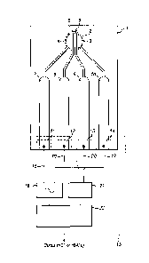
Figure 1 shows a sampling plate (1) in the form of a strip of firm and non-
conductive material (e.g.
plastic) possessing a circular sample zone (2) defined by a circular recess
formed within the strip for
receiving a liquid blood sample. Within the sample zone there are four
electrode terminals (3, 4, 5, 6)
formed upon a surface of the plate forming the floor of the sample zone and
exposed for contact with a
received sample. The electrode terminals each comprise a layer of inert
conductive material, preferably
Gold.
The four electrode terminals comprise two drive electrode terminals (3, 4)
each of which is in the shape
of a circular segment the curved edge of which coincides with a part of the
circular edge of the circular
sample zone. The straight segment edge of each one of the two drive electrode
terminals is parallel to
and opposes the straight segment edge of the other of the two drive electrode
terminals to define
between them a straight, elongate drive gap of uniform width within the sample
zone across which the
Date Recue/Date Received 2021-01-11
14
drive electrode terminals oppose each other and across which a drive current
is driven as explained in
more detail below.
Within the elongate drive gap extend two straight parallel sensing electrode
terminals (5, 6) in the form
of two strips separated from each other by a sensing gap of uniform width
defined by the spacing
between opposing side edges of each strip. The opposing edges of the two
sensing electrodes are
substantially parallel to each straight segment edge of each of the two drive
electrode terminals.
Furthermore, the straight segment edge of each one of the two drive electrode
terminals (3, 4) opposes
a correspondingly straight and parallel edge of an adjacent sensing electrode
terminal (6, 5) to define
therebetween a substantially straight and elongate partitioning gap which
extends along the sample
zone in parallel to the sensing gap. Thus, the two sensing electrodes define a
straight and uniform
sensing gap of receiving parts of the blood sample within the sensing zone,
and the two drive electrode
terminals define, together with neighbouring sensing electrode terminals, two
partitioning gaps either
side of the sending electrodes which are parallel to each other and to the
sensing gap, and which
separate the sensing electrode terminals from the drive electrode terminals
within the sensing zone.
The width of the sensing gap is preferably between about 90 microns and about
150 microns, for
example about 100 microns and is dimensioned to admit, at any point along the
sensing gap, a single
human blood cell without permitting that blood cell to bridge the gap and
concurrently contact both of
the two sensing electrodes defining the sensing gap. Rather, the gap is
dimensioned to allow a blood
cell space to oscillate within the gap between the opposing sensing electrodes
in response to an
alternating current driven transversely across the sensing gap between the two
drive electrode terminals
(3, 4). In this way, a row of blood cells may be arranged along the sensing
gap when a liquid blood
sample is received within the sensing zone and may be subject to an
alternating drive current directed
transversely (e.g. substantially perpendicular) to the row of cells.
This geometry, and the linear array of single blood cells it enables in use,
has been found to provide a
surprisingly accurate and stable means of measuring not only haematocrit (HCT)
values for the blood
sample, but also has been found to enable accurate measurement of the
concentration of glycated
haemoglobin in the blood sample (i.e. the so-called "fixed" glucose level
within blood cells, or the so-
called HbA1c value). Accurate determination of the former has been found to be
important for enabling
accurate determination of the latter ¨ i.e. one needs to know how much of the
sample is comprised of
red blood cells in order to be able to determine the quantity of fixed glucose
they carry. The present
invention could be implemented to do both simultaneously or sequentially with
reliability and accuracy.
The width of each of the two parallel partitioning gaps, either side of the
sensing electrode terminals is
preferably about 1.5 times the value of the width of the sensing gap. Again,
this gap dimension and the
parallel arrangement of blood cells the partitioning gaps enable, has been
found to assist in providing
accuracy and stability.
Date Recue/Date Received 2021-01-11
15
Each of the electrode terminals within the sensing zone is electrically
connected to a respective electrical
conductor line formed within the body of the sensing plate so as to be
electrically insulated along its
length until terminating at an exposed electrical contact zone at an end or
side of the sampling plate
distal from the sample zone. For example, the first drive electrode terminal
(3) is electrically connected
to a first drive contact zone (14) via a first (10) electrical conductor strip
(e.g. Gold). The first sensing
electrode terminal (6) is electrically connected to a first sensing contact
zone (13) via a second (9)
electrical conductor strip (e.g. Gold). Similarly, the second sensor electrode
terminal (5) is electrically
connected to a second sensing contact zone (12) via a third (8) electrical
conductor strip (e.g. Gold).
Finally, the second drive electrode terminal (4) is electrically connected to
a second drive contact zone
(11) via a fourth (7) electrical conductor strip (e.g. Gold).
These four contact zones are arrayed in a line along an edge of the sensing
plate, at the distal end of
the strip, to permit the end of the strip to be inserted into an electrical
socket/port of an electrical sensing
unit (15) to place the each one of the four contact zones simultaneously in
electrical connection with a
respective one of four (16, 17, 19, 20) electrical contact terminals of the
sensing unit.
The sensing unit maybe a handset, or part of a larger piece of equipment. The
sensing unit comprises
an alternating current source (18) arranged to generate an alternating
electrical current of selected
amplitude and selected frequency, and apply the alternating current to a first
and second sensing contact
terminals (16, 17) for application to the drive electrode terminals (3, 4) as
a drive current via the first and
second drive contact zones of the sensing plate. A control processor unit (23)
is operatively connected
to the current source (18) to control the frequency of the generated current
signal. For example, the
control processor may control the current signal frequency to be a value (e.g.
50 KHz) within a first
continuous range of values from about 1KHz to about 150 KHz, or to be a value
(e.g. 1 MHz) within a
second continuous range of values from about 500KHz to about 1.5 MHz. The
control processor is
arranged to selectively switch the frequency value from a first value within
the first range to a second
value within the second range.
A detector unit (21) is electrically connected to a first and second contact
terminals (19, 20) for receiving
voltage signals from the first and second sensing electrodes (6, 5) via the
first and second sensing
contact zones of the sensing plate.
The control processor (23) is arranged to control the electrical current
generator to apply an alternating
electrical current at a first frequency (selected from within the first range
of values) and concurrently to
control the voltage detector (21) to measure a first voltage signal received
via the first sensing electrode
and to measure a second voltage signal received via the second sensing
electrode. Subsequently, the
control processor further controls the electrical current generator to apply
an alternating electrical current
at a second frequency exceeding the first frequency (selected from the second
frequency range) and
Date Recue/Date Received 2021-01-11
16
concurrently to control the voltage detector (21) to measure a third and a
fourth voltage signal value
received from the first and second sensing electrodes respectively.
The control processor is arranged to calculate a first electrical resistance
value (R1, Ohms) using an
amplitude of the voltage difference between the first and second voltages
measured at the first
frequency and the amplitude of the associated applied alternating current, and
to calculate an electrical
impedance value ( Z2 = R2 jX3 , Ohms) using an amplitude of the voltage
difference between the third
and fourth voltages measured at the second frequency, and the amplitude of the
associated applied
alternating current. Here R represents a resistive component of impedance and
X represents a reactive
component of impedance. The quantity j = ¨1 .
Using these two impedance values, the control processor is arranged to
generate a value (HCT)
representing the relative volume of red blood cells in the liquid sample
(haematocrit) according to the
following equation.
(
HCT = Am 1 + B ln(X 3 + X0) - C
\ R21
This equation is discussed in more detail below (Equation (3)). The values of
the constants A, B, C and
Xo may be determined for a given sensing gap and/or partitioning gap width,
and electrode
structure/material for a given sensing plate, by routine calibration and
experimentation as would be
readily apparent to the skilled person. It will be appreciated that HCT values
of calibrated blood samples
may be obtained via other known methods to enable such calibration. Using this
method the standard
error of the estimated HCT values, when compared against the known micro-
haematocrit method, is
found to be less than 1.5% when measuring HCT in the range of 20 to 60%.
In a modified version of the embodiment of Figure 1, one drive electrode (4)
of the electrodes in the
sampling zone (2) may possess a deposit of an enzyme (e.g. glucose oxidase,
glucose de-hydrogenase)
to react with free glucose in the plasma component of the blood sample to
substantially oxidise it. In
that case the control processor is arranged to control the electrical current
generator to apply an
alternating electrical current at a first frequency (selected from within the
second range of values) with
a DC voltage offset (e.g. about 0.25 volts) applied across the drive
electrodes (3, 4) and concurrently to
control the voltage detector (21) to measure a first voltage signal received
via the first sensing electrode,
and to measure a second voltage signal received via the second sensing
electrode. Subsequently, the
control processor further controls the electrical current generator to apply
an alternating electrical current
.. at the same first frequency (selected from the second frequency range)
without a DC voltage offset
applied across the drive electrodes and concurrently to control the voltage
detector (21) to measure a
Date Recue/Date Received 2021-01-11
17
third and a fourth voltage signal value received from the first and second
sensing electrodes
respectively.
The control processor is arranged to calculate a first electrical reactance
value ( X1, Ohms) using an
amplitude of the voltage difference between the first and second voltages
measured at the first
frequency and the amplitude of the associated applied alternating current, and
to calculate a second
electrical reactance value (X2, Ohms) using an amplitude of the voltage
difference between the third
and fourth voltages measured at the second frequency, and the amplitude of the
associated applied
alternating current.
The control processor then calculates a value (HbA1c) representing the
concentration of glycated
haemoglobin within the blood sample according to the following equation.
(
X
HbAlc =100x 1 1
HCT x X2 )
Where HCT is a predetermined haematocrit value for the blood sample obtained
independently, e.g.
according to the invention, or otherwise.
This method has been found to provide an HbA1c measurement result with an
accuracy of 10% or
better within 20 seconds. The concentration of HbA1c depends on both the
concentration of glucose in
the blood and the lifespan of the erythrocyte (the haemoglobin cell). Because
erythrocytes are in
circulation for approximately 120 days HbA1c represents the integrated glucose
concentration over the
preceding 8 to 10 weeks, which is therefore free of the large fluctuations
that occur daily in blood glucose
concentrations in blood plasma.
Before measurement of a sample of blood is performed, the temperature of the
sampling plate is
established. This may be done by any suitable means such as would be available
and apparent to the
skilled person. Preferably, the temperature of the sampling plate may be
determined means of a
thermocouple mounted in the strip port connector. To further improve accuracy
the temperature of the
sample should preferably be maintained at 37 C 1.5 C. This may be achieved,
for example, by
environmental temperature control of the area in which sampling plates are
stored or used, or by means
of a heater (e.g. trace heating, Ohmic heating wire/strip etc, not shown)
formed in the strip electrically
connectable to a power source within the sensing unit (15, Fig.1) to
controllably heat the sampling plate.
A thermocouple (not shown) may also be formed within/upon the sampling plate,
also being arranged
to be powered by the sampling unit when the sampling plate is connected
thereto in use. This may be
used to regulate the heater (if present) and/or simply to allow the sampling
unit to determine the
temperature of the sampling plate.
Date Recue/Date Received 2021-01-11
18
It will be noted that both the reactive (X3) and resistive (R2) components of
the impedance value (Z2) are
employed in these equations when employing the higher frequency AC signals,
whereas only a resistive
component (Ri) is used at lower signal frequencies. This stems from a
consideration of the electrical
current paths which may be considered to flow across the linear arrays of
blood in the sample received
.. within the sampling and partitioning gaps of the sample zone as follows.
Figure 5 illustrates an equivalent circuit representing what is postulated to
be the conductive pathways
for electrical current driven through a blood sample between drive electrodes
of the sampling plate. This
is postulated, but not asserted, as it is useful to understanding.
A first current pathway (100) passes current across the line of blood cells
(101) within the sensing gap
through the blood plasma (102), or other added liquid, between blood cells.
This conductive path can
be considered as purely resistive in nature. A second conductive path (103)
passes through a blood
cell (104). The path through the contents of the blood cell (e.g. any glucose)
may be considered as
resistive, whereas the path through the walls of the blood cell may be
considered capacitive.
These two current pathways act in parallel and present an electrical impedance
(Z) which may be
approximated as follows.
Z = R +./X = Ra{1+ co 2 Cb2 Rb (Rb Ra) ¨ jcocRa}
1+ 02 cb2 (Rb Ray
Where R, is the resistance of the plasma, Rb is the resistance of the contents
of the blood cell including
any glucose, and Cb is the capacitance of the blood cell, where j = V-1 .
At sufficiently low frequencies, the reactive impedance of the blood cell,
arising from the capacitance of
the blood cell, is very high and prevents current flow through the cell.
Substantially only the first current
path (through plasma) is available. At sufficiently high frequencies, the
reactive impedance of the blood
cell falls and the second current pathway becomes increasingly significant.
The second pathway brings
the influence of the resistance of the content of a blood cell to bear on the
value of the impedance Z as
well as the remaining influence of the plasma due to the remaining first
current pathway. In this way,
employing the components of impedances of a blood sample at both low and high
frequencies enables
the influence of the contents of the blood cell to be probed.
It has been found that the presence of glucose within a blood cell has a
measurable effect upon the
phase of the electrical current signal passing through the sample. It is
postulated that this may be
because the presence of glucose within the blood cell increases the number of
electrons available to
react to the oscillating drive signal thereby increasing the flow of current
through the cell. This influences
the phase of the current passing through the sample. The presence of glucose
in the cell can be
Date Recue/Date Received 2021-01-11
19
considered as influencing the resistance Rb of the cell contents, according to
the equivalent circuit
model. The phase of a voltage sensed in the blood sample under such
circumstances could be
represented as:
(X ( COCbR
Phase = arctan ¨ = arctan a
1-ke)2C2R (R +R)
b b b a,)
The phase angle can be seen to be influenced by Rb, the resistance of the cell
contents. Thus, it is
postulated that this may be part of the origin of the relationship between the
phase of an electrical drive
signal within the sample, and the amount of glucose within the blood cells
(i.e. relating to HbA1c).
Figure 2 illustrates a close-up view of the electrode terminals (3, 4, 5 and
6) of figures 1õ 3 and 4. The
figure indicates a suitable sensing gap width and partitioning gap widths
either side of the sensing gap.
A sensing gap of 100 microns uniform in width is suitable. Partitioning gap
widths of 150 microns is also
suitable. Each sensing electrode terminal within the drive gap, between the
two drive electrode terminals
(3, 4) is a straight-edged flat strip of Gold having a substantially uniform
width of 200 microns. This
results in a drive gap width of 800 microns. Similarly, each drive electrode
terminal is a flat segment of
Gold.
Figure 3 illustrates an embodiment of a sensing plate of Figure 1 in the form
of a disposable strip (1).
The electrode and conductor structure of the disposable strip is as described
above with reference to
Figure 1. In addition, the end of the strip containing the drive and sensing
electrode terminals (3, 4, 5
and 6) comprises sample zone (2) for receiving the blood sample, surrounded by
an air-porous body
(27) which is in fluid communication with the sample zone wherein the air
porous body is arranged to
receive air displaced from the sample zone as the liquid blood sample is
received into the sample zone.
"In fluid communication with" may mean interfacing, where "interfacing" means
sharing a common
boundary. Preferably "in fluid communication with" refers to where the air
porous body is adjacent to
the sample zone. The air porous body may define a floor of the sample zone
and/or wall(s) of the
sample zone. The air porous body may surround the sample zone. Preferably the
air porous body
defines the sample zone, or defines an outer boundary of the sample zone.
Preferably the air porous
body defines the perimeter of the sample zone or at least part of the
perimeter of the sample zone.
Preferably the air porous body is external to the sample zone itself.
Preferably the sample zone is free
of air porous body.
Preferably the air porous body is arranged to receive displaced air as the
liquid sample approaches the
air porous body. Preferably the air porous body is arranged to receive air
displaced in the same direction
as the liquid sample travels (or spreads) into the sample zone. Preferably the
air porous body is
arranged to receive a side-ways displacement of air as the liquid sample
approaches the air porous
Date Recue/Date Received 2021-01-11
20
body in a side-ways manner. Preferably the sample zone is arranged to prevent
back flow of the liquid
sample.
An advantage of this arrangement is that the air porous body helps the liquid
sample to flow into the
sample zone with minimal air resistance, by providing a means by which air can
be directly displaced ¨
preferably in the same direction as the liquid sample enters the sample zone.
This permits the liquid
sample to enter the sample zone at a faster rate. In contrast, where such an
air porous body is absent,
air resistance retards the flow of the liquid sample into the sample zone.
Another advantage of the arrangement is that the air porous body helps the
liquid sample to spread
uniformly throughout the sample zone, thus giving greater sampling consistency
and consequently more
accurate measurements. In contrast, where the air porous body is absent, air
resistance affects the
fluid dynamics of the liquid sample by discouraging spreading (air resistance
from all sides) and instead
encouraging the liquid sample to remain collectively associated as a bulk
(aided by surface tension). As
such the liquid sample tends to flow as a bulk in a single direction since in
this way the bulk overcomes
air resistance in that particular direction. Another advantage is that
formation of air-pockets is alleviated,
which again allows for better spreading and more accurate measurements. The
liquid sample is
preferably hydrophilic, more preferably aqueous-based, and most preferably
blood. In this case, blood
glucose levels of a diabetic patient may be measured. The air porous body is
preferably substantially
impermeable to the liquid sample. The air porous body is preferably
substantially impermeable to water.
The air porous body is preferably substantially impermeable to an aqueous
liquid sample, and most
preferably substantially impermeable to blood.
The air porous body is preferably located substantially around the perimeter
of the sample zone.
Preferably a floor of the sample zone is free of air porous body. Preferably
the sample zone is free of a
roof. Where the sample zone comprises a roof, the roof is preferably free of
air porous body. The air
porous body preferably comprises hydrophobic material. Preferably the air
porous body comprises at
least 50 wt%, more preferably at least 70 wt%, and most preferably at least 90
wt% hydrophobic
material. The air porous body preferably has an average pore size between 10
and 300 microns,
preferably between 50 and 200 microns, and most preferably between 100 and 150
microns. The air
porous body preferably comprises an air porous mesh, which again is preferably
hydrophobic overall.
Such an air porous mesh preferably comprises polyether ether ketone (PEEK),
polypropylene (PP),
polyester (PET), polyvinyl idene fluoride (PVDF), ethylene
chlorotrifluoroethylene (ECTFE), ethylene co-
tetrafluoroethylene (ETFE), nylon (polyamide), or fluorinated ethylene-
propylene (FEP). The air porous
mesh preferably comprises polyester (PET). Most preferably the air porous mesh
comprises Sefar 07-
120 34.
Accordingly, where the sample zone (2) has a roof, the sample zone is
accessible via an entry port (25)
into which a blood sample (26) maybe placed. By capillary action, the blood
sample is drawn through
Date Recue/Date Received 2021-01-11
21
the entry port and into the sampling zone, displacing air into the air-porous
body (27) as it does so, to
finally occupy the sample zone covering the drive and sensing electrode
terminals there. A breathable
structure created beneath a thin polymer film covering the sample zone, as a
roof. Typically the porous
layer is a mesh made up of strands of polymer that are coated to create a
hydrophobic boundary to the
blood as it flows on to the sample zone. A geometric shape cut into the mesh
defines the sample zone
and entry port which allows the sample to fill the sample zone under capillary
action created by the thin
top film.
Figure 4 illustrates a further embodiment of the invention in which a sampling
plate (30) comprises two
sets (31, 32) of substantially identical drive electrode and sensing electrode
arrangements each being
substantially identical in structure as the electrode and conductor structure
described above with
reference to figures 1 to 3. In particular, a first electrode group (31)
comprises a pair of aforesaid drive
electrode terminals (3a, 4a) located within a first sample zone (2a) of the
sampling plate. A pair of
aforesaid sensing electrode terminals (5a, 6a) extend in parallel along the
drive gap formed between
the two drive electrodes. A respective one of four conductive strips (7a, 8a,
9a, 10a) electrically connects
a drive terminal or sensing electrode terminal to a respective one of four
separate contact zones (11a,
12a, 13a, 14a) arranged along the distal edge of the strip. A second electrode
group (32) comprises a
pair of aforesaid drive electrode terminals (3b, 4b) located within a second
sample zone (2b) of the
sampling plate. A pair of aforesaid sensing electrode terminals (5b, 6b)
extend in parallel along the
drive gap formed between the two drive electrodes. A respective one of four
conductive strips (7b, 8b,
9b, 10b) electrically connects a drive terminal or sensing electrode terminal
to a respective one of four
separate contact zones (11 b, 12b, 13b, 14) arranged along the distal edge of
the strip. Accordingly,
eight contact zones arrayed along the distal edge of the sampling strip for
insertion into a socket/port of
an electrical sensing unit (42) to place the each one of the eight contact
zones simultaneously in
electrical connection with a respective one of eight electrical contact
terminals of the sensing unit.
The first and second sample zones (2a, 2b) of the sampling plate are each in
communication with a
common single sample entry port (35) via a respective one of two sample
conduits (36, 37) which
bifurcate from the entry port and communicate with a given sample zone. The
two sample zones (2a,
2b) are each surrounded by an air-porous body (27), as described above, which
is in fluid communication
with each of the sample zones wherein the air porous body is arranged to
receive air displaced from the
sample zone as the liquid blood sample is received into the sample zone. The
air porous body defines
the entry port (35) and the sample conduits (36, 37) as well as the circular
periphery of each of the two
sample zones.
A quantity of an enzyme (38), (e.g. glucose oxidaze ("GOX") or glucose
dehydrogenase (GDH)), is
located upon one of the two drive electrode terminals (4b) in the second
sample zone (2b). The enzyme
(e.g. GOX or GDH) is placed to allow it to make contact with, and react with,
a blood sample entered
Date Recue/Date Received 2021-01-11
22
into the second sample zone. In doing so, the enzyme reacts with the blood
sample to consume any
free glucose present within the plasma of the blood sample. As a result, one
the reaction has completed,
the blood sample located within the second sample zone contains substantially
no free glucose, and
any glucose present should be substantially only fixed glucose within the red
blood cells of the blood
sample which is inaccessible to the enzyme (e.g. GOX or GDH).
In the sensing unit (42), a control processor (41) is arranged to control the
current source (18) and the
voltage detector unit (21) as described above with reference to Figure 1 when
measuring haematocrit
(HCT). The control processor (41) is arranged to control a second current
source (39) and a second
voltage detector unit (40) of the sensing unit, as described above with
reference to Figure 1 ("modified
version") when used to measure HbA1c.
Figure 6 schematically illustrates an ASIC (application specific integrated
circuit, 50) arranged for
connection to a microcontroller (55) integrated circuit for use within the
sensing unit (42, Fig.4). The
ASIC is responsive to the control signals from a control unit (55) to apply to
the second group of
electrodes (31) of the sensing strip an alternating (AC) current having a
first frequency of 1MHz and to
selectively apply a direct voltage (DC) of most preferably a substantially
constant value concurrently,
accordingly to the control signals. This is for the purposes of measuring
HbA1c in the blood sample
bridging the electrodes of that second group.
The ASIC is responsive to the control signals from a control unit (55)
selectively to apply to the first
group of electrodes (32) of the sensing strip an alternating (AC) current
having either a first frequency
.. of 1MHz or second frequency of 50 KHz according to the control signals.
This is for the purposes of
measuring haematocrit in the blood sample bridging the electrodes of that
second group.
The ASIC is arranged to receive voltage signals from the first and second
sensing electrode terminals
(5b, 6b) of the second group of electrodes (31) of the sensing plate, and to
receive voltage signals from
the first and second sensing electrode terminals (5a, 6a) of the first group
of electrodes (32) of the
sensing plate. The ASIC is responsive to the voltage signals and the current
signals to measure
peak/amplitude values for those voltages and currents for use by the
microcontroller in calculating
electrical resistance and reactance values of blood samples bridging the
electrodes of the first and
second electrode groups (31, 32) to determine values of haematocrit and HbA1c
therein.
The control unit (55) performed the functions of the control processor unit
(41) of Figure 4.
In particular, the microcontroller (55) is arranged to provide a direct (DC)
voltage level (most preferably
a substantially constant (DC) voltage level) and to output a corresponding
(DC) analogue output signal
via a digital-to-analogue converter output (56) connected to an input port
(57) of the ASIC (50).
Furthermore, the microcontroller is also arranged to produce an alternating
(AC) pulse-width modulated
square-wave analogue output signal via a pulse-width modulator (PWM, 58) as a
first signal (59) having
Date Recue/Date Received 2021-01-11
23
a frequency of 1MHz and to input the signal to a second input port (61) of the
ASIC, and to produce a
second separate modulated square-wave output signal (60) having a frequency of
50KHz and to input
the signal to a third input port (62) of the ASIC. These two pulse-width
modulated output signals are
electrical current signals which are each maintained at a predetermined
amplitude level (most preferably
substantially constant) by the microcontroller. The ASIC includes a first pre-
amplifier in the form of an
operational amplifier (63) comprising a feed-back loop (64) including a
resistor. The pre-amplifier is
arranged to receive the DC voltage signal from the microcontroller at a first
input port (67) of the pre-
amplifier, and to output an amplified value to a first output port (65) of the
ASIC. The ASIC also includes
a 1MHz sinewave filter unit (67) and a 50KHz sinewave filter unit (68) each
arranged for receiving and
filtering a respective 1MHz and a 50KHz oscillating current signal input from
the microcontroller at the
second and third input ports (61, 62) of the ASIC. Each of the sinewave filter
units is arranged to receive
the respective square-wave (PWM) signal input to it, and to alter the square-
wave shape of the signal
into substantially a sine-wave shape and to output the result as an AC sine-
wave signal. An analogue
switch unit (69) is provided in the ASIC and has two input ports for receiving
a respective one of the two
AC sinewave signals output from the 1MHz and 50KHz sinewave filter units. The
analogue switch unit
is arranged to be controlled by the microcontroller to output selectively one
of the 1MHz sinewave (AC)
signal and the 50KHz sinewave signal to a signal input port of a second pre-
amplifier (71) in the ASIC
for amplifying that output signal. In this way the microcontroller is able to
control, via the analogue switch
unit, which of the 1MHz and the 50KHz sinewave (AC) signals is ultimately
output from the second pre-
amplifier and from the ASIC.
The sampling strip (30) is shown as electrically connected to the ASIC of the
sensing unit such that a
first drive electrode terminal (4b) of a second group of electrodes (31) for
sensing HbA1c, is electrically
connected to the first output port of the ASIC, and such that a first drive
electrode terminal (4a) of a first
group of electrodes (32) for sensing haematocrit, is electrically connected to
the second output port of
the ASIC.
In addition, the analogue switch unit is arranged to be controlled by the
microcontroller to output the
1MHz sinewave (AC) signal to a second signal input port (66) of the first pre-
amplifier (63) in the ASIC
for amplifying that output signal. The first pre-amplifier unit is arranged to
selectively amplify a 1MHz
sinewave (AC) signal either with or without the concurrent presence of the DC
voltage level applied to
the first input port (67) of that amplifier. The microcontroller controls
when/if the DC voltage level is
applied to the first pre-amplifier unit.
The analogue switch unit is further arranged to output, via a third output
port (73), a signal which is
representative of the time of a peak/amplitude of the AC sinewave signal
received by the analogue
switch unit from either of the first and second sinewave filter units. This
signal is input to a phase-to-
voltage unit (74) which, as shall be explained in more detail below, is
arranged to measure a temporal
Date Recue/Date Received 2021-01-11
24
phase difference between an AC current applied to the drive electrodes of the
first and second groups
of electrodes, and an AC voltage generated between sensing electrode terminals
of the respective
groups of electrodes of the sensing plate, and to generate the result as a
signal representative of that
temporal phase difference.
The ASIC includes a peak detector unit (77) in communication with the phase-to-
voltage unit and
comprising a first and second signal input ports (82, 83) in communication
with first and second voltage
input ports (75, 76), respectively, of the ASIC for receiving voltage signals
from the sensing electrode
terminals (5a, 6a) of the first group of electrodes (32) of the sensing plate.
A third input port (84) of the
peak detector unit is arranged in communication with a second drive electrode
(3a) of the first group of
drive electrodes (32) via a first current input port (78) of the ASIC and is
arranged to receive the current
driven through the first group of electrodes between the first drive electrode
(4a) and the second drive
electrode (3a) of that group.
The peak detector unit also comprises a fourth and fifth signal input ports
(85, 86) in communication
with third and fourth voltage input ports (79, 80), respectively, of the ASIC
for receiving signals from the
sensing electrode terminals (5b, 6b) of the first group of electrodes (31) of
the sensing plate. A sixth
input port (87) of the peak detector unit is arranged in communication with a
second drive electrode (3b)
of the first group of drive electrodes (31) via a second current input port
(81) of the ASIC and is arranged
to receive the current driven through the second group of electrodes between
the first drive electrode
(4b) and the second drive electrode (3b) of that group.
Thus, the ASIC includes a first (75) and second (76) voltage input ports which
connect electrically to a
first (82) and second (83) input ports of a peak detector unit and are adapted
for receiving a respective
first and second voltages from respective of the sensing electrodes of the
first group of electrodes (32).
The ASIC also includes a third and fourth voltage input ports (79, 80) which
are similarly electrically
connected to input ports (85, 86) of the peak detector unit. A first current
input port (78) and a second
current input port (81) are each respectively connected to first and second
current input ports (84, 87)
of the peak detector unit. Each of the first to sixth input ports of the peak
detector unit are arranged to
receive respective voltage and current signals which are amplified by a
respective pair of amplifier units
(92) within the apparatus, arranged in series electrical connection along the
respective signal
transmission line leading to the input ports in question. These amplifier
units serve to amplify the
respective signals prior to receipt by the peak detector unit, and may be
formed in the ASIC if desired,
or elsewhere in the sensing unit but operably connected to the ASIC as shown
in the Figures.
The peak detector unit comprises an output port electrically connected to a
sample-and-hold unit (90)
which is arranged to receive from the peak detector unit a signal representing
a value of the peak in an
AC voltage signal received by the peak detector, and/or a value representing a
peak current value
received by the peak detector. These peak values generally represent amplitude
values of an
Date Recue/Date Received 2021-01-11
25
associated AC current and voltage signal inputs to the peak detector unit from
the electrodes of the
sampling plate (bio-sensor module). The sample-and-hold unit is operable to
momentarily retain signal
values received from the peak detector unit as and when required for
subsequent transmission to the
microcontroller.
The phase-to-voltage unit (74) possesses an output port electrically connected
to an output port of the
ASIC for outputting a voltage signal representing a measured phase difference
(measured time shift)
between the peak voltage values detected by the peak detector, and the peak
values of the AC current
signal output by the analogue switching unit.
A stop-watch unit (89) is operably connected to and controlled by the
microcontroller (55) via the control
register unit (91) and the interface (SPI) with the microcontroller. The stop-
watch unit is arranged to
measure a time interval between detected signal peaks detected by the peak
detector unit for use in
measuring a phase angle between applied current and measured voltage.
In this way, the phase-to-voltage unit and the peak detector unit provide
values representing an
amplitude of an AC voltage signal and an AC current signal received by the
peak detector unit, and a
phase difference incurred between those signals. With these measured values,
the microcontroller is
arranged to calculate values of resistance and reactance of samples within the
sampling zones of the
sampling plate (bio-sensor module).
The sample and hold unit (90) has two output ports each in communication with
a respective first and
second output port (94, 95) of the ASIC for separately outputting values
associated with the first and
second groups of electrodes (31, 32) respectively.
The voltage output port of the ASIC is arranged to be connectable (and is
shown as connected to) a
biosensor module in the form of a sampling strip such as is shown in Figure 4.
The first current output
port (65) of the ASIC is connected to a drive electrode (4b) in the sampling
zone of the sampling strip
dedicated to measuring HbA1c and the second current input port (81) is
arranged to be electrically
connected to (and is shown as connected) to the other drive electrode (3b) in
that sampling zone.
Similarly, the second current output port (72) of the ASIC is electrically
connectable to (and shown as
connected) a first drive electrode (4a) in the other sampling zone of the
sampling strip dedicated to
measuring haematocrit, whereas the other drive electrode (3a) in that sampling
zone is electrically
connectable to (as is shown as connected) the first current input port (78) of
the ASIC.
The two sensing electrodes (5a, 6a) of the sampling strip within the sampling
zone for haematocrit are
each separately connected to the first and second voltage input ports (75,
76), respectively, of the ASIC
via a respective pair of amplifier units (92). Similarly, the two sensing
electrodes (5b, 6b) in the sampling
zone of the sampling strip dedicated to measurement of HbA1c are each
separately connected to a
respective one of the third and fourth voltage input ports (79, 80) of the
ASIC. In this way, electrical
Date Recue/Date Received 2021-01-11
26
currents may be driven across the drive electrodes in the two sampling zones
via the ASIC, and resulting
voltages, and voltage/current phase differences arising from liquid samples
within those sensing zones,
may be determined.
In particular, when an AC current signal is driven across a pair of drive
electrode terminals of the first or
second group of electrode terminals, a blood sample bridging the drive gap
between those electrode
terminals responds in such a way as to present an electrical impedance (Z,
ohms). This manifests itself
in that an AC electrical potential difference is generated across the sample,
as measured by/between
the two sensing electrode terminals in the given group of terminals, which is
not in temporal phase with
the AC current signal. The sample possesses not only a resistance (R, ohms)
which is dissipative (real),
but also a reactance (X, ohms) which is non-dissipative (imaginary).
Thus, if the driving AC current of amplitude I and angular frequency co is
represented by:
1= I exp {j(wt +
where j = , and 19/ is the temporal phase of the sine wave AC current
signal, then the resulting
voltage across the two sensing electrodes of the given group of electrodes is
represented by:
V= V exp {j(wt +
where ev is the temporal phase of the sine wave AC voltage signal of amplitude
V and angular
frequency co .
The impedance of the sample is given by:
Z= Z exp{j0}
.. where Z is the magnitude of the impedance and 0 is its phase angle.
The resistance of the blood sample (R) is given by:
R = Z cos(0)
And the reactance of the blood sample (X) is given by
X = Z sin()
Date Recue/Date Received 2021-01-11
27
where Z is measurable using the known amplitudes / and V of the applied
current and resulting
voltage signals respectively. Similarly, the phase angle of the impedance can
be determined by applying
Ohm's law:
V = V exp{j(wt + ev)} =IxZ = x Z exp{j(wt + + 0)}
thus
= = co x At
where At is the time lag between a peak in the applied AC sinewave current
signal and a measured
peak in the resulting AC voltage across the blood sample.
Thus, the resistance (R) and reactance (X) values of the blood sample may be
determined from the AC
.. current peak/amplitude value (I ) as applied at a frequency co between the
two drive electrode
terminals in a sampling zone, the resulting AC voltage peak/amplitude ( V ) as
measured between the
sensing electrodes in that sampling zone, and the time lag ( At ) between
those two peaks in succession,
as follows:
V
R = ¨ cos(w x At)
Equation (1)
V 15 X
= ¨ sin(co x At) Equation (2)
These values are either known by, or measured by, the sampling unit using the
ASIC as described
above, and the microcontroller (55) is arranged to calculate these resistance
and reactance values.
A control register unit (91) is provided on the ASIC and is arranged to
control, under overall control of
the microprocessor via an interface unit (SPI), the timings and orchestration
of control signals for
operation of the amplifiers (/en signals to "enable" amplifiers), filters,
switches and other components on
the ASIC. The control register may be of a type such as would be readily
apparent to the skilled person.
A thermo-couple (88) is formed in the ASIC and is electrically connected
between a signal port of the
control register unit (91) and an earth terminal and is arranged in the
sampling device to make physical
contact with a part of the sampling plate when the sampling plate is
operatively connected to the
.. sampling device in use. The control register unit is arranged to receive
signals from the thermo-couple
Date Recue/Date Received 2021-01-11
28
representative of the temperature of the sampling plate (30) and to convey
those signals to the
microcontroller to permit the temperature of the sampling plate to be
determined by the microcontroller.
In use, the sampling unit is operable as follows. Once a user has applied a
blood sample to the first and
second sampling zones (Fig. 4; 2a, 2b) of the sampling plate (30), the blood
sample is split between the
first sampling zone (2a) dedicated to measuring haematocrit, and the second
sampling zone (2b)
dedicated to measuring HbA1c. The G0x/GDH spot (38) provided in the second
sampling zone reacts
with the free glucose present in the blood plasma of the blood sample within
that zone to substantially
oxidise it. A suitable period of time is allowed for this process (several
seconds, e.g. between 0.5 and
seconds). This has been found to be as little as 0.5 seconds. It may be slowed
by using a mediator
10 which would allow the signal transient to be recorded over a time frame
sufficient to determine the level
of free glucose in the sample. For the determination of HbA1c, it is only
required that the free glucose
is oxidised, therefore it is important at least to see that a transient has
occurred and decayed before the
HbA1c measurement sequence is started. Subsequently, the blood-bearing
sampling strip is electrically
connected to the sampling unit (if not already so connected) as shown in
Figure 4 and Figure 6.
The microcontroller shown in Figure 6 is arranged to supply to the first pre-
amplifier (63) of the ASIC
(50) an alternating (AC) electrical current having a 1MHz frequency, via the
1MHz sinewave filter unit
(67), to drive that current through the blood sample bridging the two drive
electrodes within the second
sampling zone, thereby to generate a first alternating potential difference
across the spacing between
the drive electrode terminals measurable via the two sensing electrode
terminals (5b, 6b) within that
sampling zone. This state is maintained for a period of time of several
milliseconds in duration (e.g.
between about 20 ms and about 200 ms or less). Subsequently, the
microcontroller is arranged to apply
a substantially constant direct (DC) voltage to the first pre-amplifier unit
(63) of the ASIC to output to the
first output port of the ASIC (65) a substantially constant DC offset of about
0.25 volts in combination
with the 1MHz AC current signal concurrently being applied to the second
sampling zone. The
microcontroller is arranged to then employ the peak detector unit and the
phase-to-voltage unit of the
ASIC to measure the amplitude of the voltage across the two sensing electrode
terminals (5b, 6b) in the
first sampling zone, and the time lag between the successive occurrences of a
peak in the applied AC
current and the resulting AC voltage, and to measure a first value of the
electrical reactance (Xi) of the
blood sample according to Equation (2) above.
The microcontroller is arranged to subsequently not apply the DC voltage
offset and to continue to apply
the 1MHz AC current signal to the first and second drive electrode terminals
of the second sampling
zone. After a suitable time period following removal of the DC offset voltage
(e.g. between about 20 ms
and about 200 ms or less), the microcontroller is arranged to then employ the
peak detector unit and
the phase-to-voltage unit of the ASIC to measure the amplitude of the voltage
across the two sensing
Date Recue/Date Received 2021-01-11
29
electrode terminals (5b, 6b) in the first sampling zone, and the time lag
between the successive
occurrences of a peak in the applied AC current and the resulting AC voltage,
and to measure a second
value of the electrical reactance (X2) of the blood sample according to
Equation (2) above.
Either before, during or after performing the above process on the blood
sample within the second
sampling zone, the microcontroller is arranged to determine the haematocrit
(HCT) within the blood
sample as follows.
The microcontroller is arranged to supply a 50 KHz AC current to the analogue
switch unit (69) of the
ASIC via the 50KHz sinewave filter unit (68) and to control the switch unit to
connect the 50KHz AC
current to the second output port (72) of the ASIC via the second pre-
amplifier unit (71). This causes
the AC signal to be applied to the drive electrode terminals (3a, 4a) of the
first sample zone, an
alternating electrical current having a first signal frequency of 50 KHz to
generate a first alternating
potential difference of 50 KHz across the spacing between the electrode
terminals, as measured by the
two sensing electrodes (5b, 6b) in the first sample zone.
The microcontroller is arranged to then employ the peak detector unit and the
phase-to-voltage unit of
the ASIC to measure the amplitude of the voltage across the two sensing
electrode terminals (5a, 6a)
in the first sample zone, and the time lag between the successive occurrences
of a peak in the applied
AC current and the resulting AC voltage, and to measure a first value of the
electrical resistance (Ri) of
the blood sample according to Equation (1) above.
The microcontroller is arranged to then supply a 1MHz AC current to the
analogue switch unit (69) of
the ASIC via the 1 MHz sinewave filter unit (67) and to control the switch
unit to connect the 1MHz AC
current to the second output port (72) of the ASIC via the second pre-
amplifier unit (71). This causes
the 1 MHz AC signal to be applied to the drive electrode terminals (3a, 4a) of
the first sample zone, an
alternating electrical current having a second signal frequency of 1 MHz to
generate a second alternating
potential difference of 1 MHz across the spacing between the electrode
terminals, as measured by the
two sensing electrodes (5a, 6a) in the first sample zone.
The microcontroller is arranged to then employ the peak detector unit and the
phase-to-voltage unit of
the ASIC to measure the amplitude of the voltage across the two sensing
electrode terminals (5a, 6a)
in the first sample zone, and the time lag between the successive occurrences
of a peak in the applied
AC current and the resulting AC voltage, and to measure a second value of the
electrical resistance (R2)
of the blood sample according to Equation (1) above, and to measure a third
value of the electrical
reactance (X3) of the blood sample in the first sample zone according to
Equation (2) above.
Date Recue/Date Received 2021-01-11
30
The microcontroller is arranged to subsequently calculate a value for the
relative volume of red blood
cells (haematocrit, HCT) in the liquid sample according to the first
electrical resistance value, the second
electrical resistance value and the third electrical reactance value according
to the following formula,
and store the result and/or to output the result to the user:
(
HCT = Am 1 + Bln(X + X0) - C
Equation (3)
\ R21
Where A, B and C are constants associated with the sampling plate in question.
For example, the
values of A, B and C may each typically be within the range from about 0.05 to
about 0.5, or preferably
between about 0.1 and 0.25, or more preferably between about 0.1 and about
0.2. For example, when
using electrodes formed from gold having a sheet resistance of 5 ohms per
square, and the geometry
illustrated in Figure 4, the values in question may be:
A = 0.142;
B = 0.155;
C = 0.157.
Actual values, suited to a given sampling zone geometry and electrode
structure and material, may be
determined by routine calibration employing commercially available blood
samples of known HCT, as
will be apparent to the skilled person. The value of Xo may simply be zero, or
may be adjusted if
necessary to improve the predictive accuracy of the equation. It has been
found that the term A is
correlated with the conductivity of the electrode terminals of the sensing
strip. The term B has been
found to correlate with the resistivity of the interface (e.g. the wetting)
between the blood sample and
the electrode terminals in the sample zone. This is influenced by the
electrode material (e.g. Gold), and
the quality of the structure (e.g. roughness) of the electrode surfaces. The
term C has been found to
correlate with the variability of the average blood cell size (e.g. determined
for "unfixed" or un-glycated
blood cells) within the sample. This can be strongly influenced by ethnicity,
blood type and interferences
such as those which will be readily apparent to the skilled person. Values of
C associated with different
ethnicities (or blood type or known interferences) may be stored in a look-up
table within (or accessible
by) the control processor for suitable selection during calibration.
Table 1 shows examples of values of haematocrit for a blood sample of type A
calculated according to
the above method and equations. Three groups of ten measurements were made
using the strip design
illustrated in Figures 1, 3, 4 and 6 for measuring HCT. In each of the three
groups a sample of blood
was used having a known HCT value, namely 52%, 42% and 31%. Measurements of
resistances Ri,
and R2 and reactance X3 were made at 50 KHz and 1 MHz AC signal frequency
respectively, and input
into Equation (3) to generate a measurement value of HCT in each of the
measurements. The ten
Date Recue/Date Received 2021-01-11
31
measurements for each one of the three different common (known) HCT value
shows a consistently
accurate HCT measurement.
The microcontroller is arranged to then generate a value representing the
concentration of glycated
haemoglobin (HbA1c) in the blood within the sample according to the first
electrical reactance value, the
second electrical reactance value and a value of the relative volume of red
blood cells in the liquid
sample (haematocrit, HCT) according to the following formula, and store the
result and/or to output the
result to the user:
(
X
HbAlc =100x 1 1 Equation (4)
HCT x X2 )
The quantity Xi represents the reactance of a blood sample due to glycated red
blood cells in the blood
within the second sampling zone from which free glucose has been substantially
oxidized by G0x/GDH,
whereas X2 represents the reactance of the whole blood sample in which both
plasma and red blood
cells contain glucose. The proportion of that reactance due to red blood cells
is determined by the
(HCT)x(X2) according to the haematocrit of the sample in the first sampling
zone.
Figure 7 shows an alternative form of sampling strip and sampling unit to read
the sampling strip for the
purposes of measuring haematocrit alone.
This alternative sampling strip (30A) comprises a first sampling zone
containing a first group of
electrodes identical to the electrode group illustrated in Figures 1 to 3, and
in the first sampling zone
(2a) of the sampling strip of Figure 4 and Figure 6. The first group of
electrodes comprises a pair of
drive electrode terminals (3a, 4a) defining a drive gap within which are
arranged two sensing electrode
terminals (5a, 6a) for sensing a voltage drop across a blood sample when
bridging the drive gap resulting
from the an AC current driven between the two drive electrode terminals. The
first group of electrodes,
and the circuit elements of the ASIC (50A) with which they are arranged to
electrically connect (shown
as connected in Fig. 7) are the same as those elements of the ASIC (50)
illustrated above in Figure 6.
Those circuit elements are arranged to operate, and be controlled by the
microcontroller (55A)
.. substantially as described above with reference to the first sample zone of
the sample strip of Figure 6
for determining a measure of haematocrit using equation (3) above.
However, the second sampling zone of this alternative sampling strip comprises
a single pair of drive
electrodes terminals (100, 101) defining between them a curved drive gap
(102). The drive electrode
pair is arranged to connect electrically to the ASIC to apply a DC electrical
voltage (preferably
Date Recue/Date Received 2021-01-11
32
substantially constant in value) across the curved drive gap when/if bridged
by a blood sample within
the second sensing zone. A deposit of a reagent, such as an enzyme or the
like, to react with glucose
in the blood sample (e.g. glucose oxidase (GOX) or glucose dehydrogenase
(GDH)) is provided on the
surface of the anode (e.g. electrode terminal 101) of the two drive
electrodes. Like items are assigned
like reference symbols as between figures 6 and 7.
The ASIC (50A) in this alternative design differs from the ASIC of Figure 6 in
that the analogue switch
unit (69A) does not provide a 1 MHz AC current input (66, Fig.6) to the first
pre-amplifier unit (63). The
first pre-amplifier unit (63A) does not possess a feed-back loop (64, Fig.6)
and simply receives from the
microcontroller a DC voltage signal (most preferably substantially constant),
which it amplifies and
outputs the result to a first voltage output port (65A) of the ASIC for
electrical connection to a drive
electrode (100) of the second sample zone.
The DC voltage signal is output by the microcontroller (55A) via a digital-to-
analogue converter (56) and
input to an amplifier (63A) formed within the ASIC the amplified output of
which is input to a voltage
output port (65A) of the ASIC and to a first input port (104) of an
operational amplifier unit (103) formed
in the ASIC. The operational amplifier has a second input port (105) which
serves as voltage input
terminal of the sensing unit respectively connected to an anode terminal (101)
of the second pair of drive
electrodes of the sample plate when the latter is connected to the former in
use as shown in Figure 7.
The operational amplifier has a respective output port (107) which is
connected to its second input port
.. via a feed-back loop comprising a resistor (106). As a result, the DC drive
voltage (Vdnve) applied to the
first input port of the operational amplifier by the first amplifier unit
(63A) is expressed as a
correspondingly substantially constant voltage level at the second input port
(105) of the operational
amplifier. This produces a controllably constant potential difference across
the drive electrodes of the
electrode pair (100, 101) in the second sample zone relative to a reference
voltage (Vref). Consequential
conduction through a blood sample in the second sample zone of the sampling
strip enters the
operational amplifier electrically connected to that blood sample. The result
is that a measurable current
is received by the operational amplifier at its second input port (105). This
current is output on the output
port (107) of the operational amplifier via a voltage amplifier (109) to a 12-
bit analogue to digital converter
(97B) of the microcontroller for use thereby in determining a value for the
blood glucose level within the
sample in the second sample zone, as described below. The measured current is
also input from the
operational amplifier to an input port of the peak detector unit (77) which is
arranged to detect the
occurrence of a peak in the detected current from the second sample zone, and
to communicate the
time of that occurrence to the microcontroller (55A) via a 12-bit analogue to
digital converter (97B) of
the microcontroller for use thereby in determining a value for the blood
glucose level within the sample
in the second sample zone, as described below.
Date Recue/Date Received 2021-01-11
33
It has been found that a direct (DC) voltage held between the two drive
electrodes of the second sample
zone, and thereby applied across the blood sample located there during the
enzyme (GOX/GDH)
reaction period, will cause a time-varying current to pass through the blood
sample. Variation is believed
to result, in part, from the changing (falling) quantity of free glucose
within the plasma of the sample
resulting in a changing electrical resistance of the plasma component of the
blood. Most preferably, the
DC voltage is substantially constant for simplicity, however non-constant DC
voltages (e.g. smoothly
falling or rising in a controlled way) could be employed if desired, though
this is likely to complicate
design and operation of the apparatus and so a substantially constant DC
voltage is preferred.
Starting with an initial rise (near instantaneous) in current to a peak value
at a time "tpeak", the observed
quantity of current falls monotonically as glucose is increasingly oxidised in
the blood plasma. Sample
temperature affects the rate of decay of the current ¨ lower temperatures
result in faster decay. It has
been found that the rate of fall of the observed current, following the peak
current value, is characteristic
of the amount of glucose originally present in the plasma of the blood sample
before the oxidisation
process began. The observed current decay is highly reproducible when the
process is repeated. Thus,
by performing this process initially with a sufficient plurality a blood
samples each having an
incrementally different, known quantity of glucose in its plasma component,
one may build-up a plurality
of reference curves of the type described above (or data sets representative
of them) from which a future
blood plasma glucose measurement may be made by reference. That is to say,
with the plurality of
reference curves (or representative data) one may perform a contemporaneous
blood sampling
operation as described above so as to generate a measurable current varying
generally according to
the current decay curve described above. By measuring a particular current
value at a selected time
during that current decay (i.e. a point along the contemporaneous current
decay curve) one may
subsequently identify a blood plasma glucose level associated with that
current value as derived from a
reference curve. The contemporaneously measured blood plasma glucose level may
then be concluded
to have the same glucose level. A Look-Up Table (LUT) or other storage may be
used for this purpose.
The process may include measuring a contemporaneous value "Im" of the decaying
current at a specified
time "tm" following the time "tpeak" at which the detected peak of the
measured current occurs ¨ the
specified time having also been used when generating the reference curves.
This current value then
identifies the glucose value stored in the LUT associated with the reference
curve which had the same
current value at the same specified time in its current decay phase. The
stored value from the LUT which
matches a contemporaneous value will identify the associated blood plasma
glucose level so measured.
The specified time (tm) may be between about 1 sec. and about 15 sec.
Different reference curves or
LUTs may be used according to the measured temperature of the sensing strip,
as determined by the
thermocouple described herein for example.
In this way, the microcontroller is arranged to apply a DC voltage to the pair
of drive electrodes (100,
101) in the second sample zone, and to measure the resulting current.
Date Recue/Date Received 2021-01-11
34
The sampling unit contains such a LUT as described above, and is arranged to
compare respective
contemporaneously measured current (decaying) values separately from the
second sample zone, with
stored reference current values, to identify the closest match (or interpolate
between the closest two
matches) and to retrieve an associated blood plasma glucose value "BGraw" from
the LUT associated
with that match. There may comprise as plurality of LUTs which may be
respectively associated with
reference curves generated for a common specified temperature of blood sample.
The calculating unit
may be arranged to select the appropriate LUT based on the measured
temperature of the sampling
strip at the time of the measurement at hand. A thermo-couple (not shown) may
be provided in the
sampling unit to physically contact the sampling plate (30A) in use and
provide signals to which the
.. microcontroller is responsive to determine a temperature of the sampling
plate and, from that
determination, select the most appropriate LUT for use as described above.
Alternatively, through
testing various temperatures, a temperature correction factor (Tc) maybe
determined and used to
compensate the measurement for temperature effects. One LUT for Im and tm may
then be used and
the value retrieved from the LUT using 6 and tm may then be adjusted using the
temperature correction
.. factor (Tc) as appropriate.
The microcontroller is arranged to produce an adjusted value "BGcorrected" for
the blood plasma glucose
level so retrieved according to:
BGc.cted f(BGõ,,,HCT)
Equation (5)
where f(BGõ,õHCT) is a predetermined corrective function of the measured
haematocrit value HCT
.. for the sample in the first sample zone, and of BGraw which is an
uncorrected blood plasma glucose
value measured for the blood sample in the second sample zone. The form of the
function
f(BGõ,õHCT) of the predetermined corrective function may be selected by the
user.
One example is of the form:
f(BGõ,õHCT) = BGraw ¨[mx(HCT)+ c]
where m is a positive or negative constant and c is a positive or negative
constant. These values may
be evaluated by calibration against commercially available calibration blood
samples containing known
HCT and glucose levels. This functional form exploits the finding that errors
in uncorrected glucose
measurements are typically linear to a first approximation, as a function of
HCT, and that so too is the
corrective function. Of course, other more accurate corrective functional
forms may be used such as
would be apparent to the skilled person in this field.
Date Recue/Date Received 2021-01-11
35
The ASIC controller unit (91) controls the timing and coordination of the
components formed upon the
ASIC under the master control of the microcontroller via an interface (SPI) of
the microcontroller with
which the ASIC control unit is in communication. In this way, the required
current and voltage values
may be applied to, and received from, the first sample zone containing the
first group of electrodes (3a,
4a, 5a, 6a) of the sampling plate (30A) via the ASIC to enable the
microcontroller to perform the
measurements of HCT and blood glucose as described above using equation (3).
This value of HCT is employed by the microcontroller to calculate an adjusted
value of blood glucose in
the sample of blood using the blood glucose value determined from the second
sampling zone
containing the second group of electrodes (100, 101).
Examples of the HCT value determined by the microcontroller using the ASIC and
the first group of
electrodes (3a, 4a, 5a, 6a) in the first sampling zone, as described above
with relation to Figures 1, 3
and 6, and employed in Equation (3), are given in Table 1.
Aspects of the invention are defined by the following numbered paragraphs:
Paragraph 1: A sampling apparatus for use in performing electrical
measurements on a liquid sample
containing blood, the apparatus comprising: two (or more) current output
terminals for outputting an
alternating current signal applied therebetween; an alternating electrical
current unit in electrical
communication with the two (or more) current output terminals for applying
thereto an alternating
electrical current of a given amplitude and frequency when a said liquid
sample is in electrical connection
between the two (or more) current output terminals; a voltage unit in
electrical communication with the
two (or more) current output terminals for applying therebetween a direct (DC)
electrical potential
difference of a given magnitude; a first voltage input terminal for receiving
a first electrical signal
externally input thereto and a separate second voltage input terminal for
receiving a second electrical
signal externally input thereto when said liquid sample is in electrical
connection between the first and
second voltage input terminals; voltage detector(s) for measuring a first
voltage and a second voltage
using said first and second electrical signals, respectively; a control unit
arranged to control the electrical
current unit to apply a said alternating electrical current of said given
frequency and concurrently to
control the voltage unit and the voltage detector(s) to measure said first and
second voltages both when
said direct electrical potential difference is applied and when said direct
electrical potential difference is
not applied; a calculating unit arranged to calculate a first electrical
reactance value using the first and
second voltages measured when said direct electrical potential difference is
applied, and to calculate a
second electrical reactance value measured when said direct electrical
potential difference is not
applied; wherein the calculating unit is arranged to generate a value
representing the concentration of
glycated haemoglobin (HbA1c) in the liquid sample according to the first
electrical reactance value, the
Date Recue/Date Received 2021-01-11
36
second electrical reactance value and a value representing the relative volume
of red blood cells in the
liquid sample (haematocrit).
Paragraph 2: A sampling apparatus according to Paragraph 1 in which the given
frequency has a value
in the range 500KHz to 1.5MHz.
Paragraph 3: A sampling apparatus for use in performing electrical
measurements on a liquid sample
containing blood, the apparatus comprising: two (or more) current output
terminals for outputting an
alternating current signal applied therebetween; an alternating electrical
current unit in electrical
communication with the two (or more) current output terminals for applying
therebetween an alternating
electrical current of a given amplitude and frequency, when a said liquid
sample is in electrical
connection between the two (or more) current output terminals; a first voltage
input terminal for receiving
a first electrical signal externally input thereto and a separate second
voltage input terminal for receiving
a second electrical signal externally input thereto, when said liquid sample
is in electrical connection
between the first and second voltage input terminals; voltage detector(s) for
measuring a first voltage
and a second voltage using said first and second electrical signals,
respectively; a control unit arranged
to control the electrical current unit to apply a said alternating electrical
current at a first frequency and
concurrently to control the voltage detector(s) to measure said first and
second voltages, and to further
control the electrical current unit to apply said alternating electrical
current at a second frequency
exceeding the first frequency and concurrently to control the voltage
detector(s) to measure said first
and second voltages; a calculating unit arranged to calculate a first
electrical resistance value using the
first and second voltages measured at said first frequency, and a second
electrical resistance value and
a reactance value using said first and second voltages measured at said second
frequency; wherein the
calculating unit is arranged to generate a value representing the relative
volume of red blood cells in the
liquid sample (haematocrit) according to the first and second electrical
resistance values and the
electrical reactance value.
Paragraph 4: A sampling apparatus according to Paragraph 1 and Paragraph 3
arranged to generate
both said value representing the relative volume of red blood cells in the
liquid sample (haematocrit) and
to generate said value representing the concentration of glycated haemoglobin
(HbA1c) in a liquid
sample using said haematocrit value.
Paragraph 5: A sampling apparatus according to any of Paragraphs 1 to 4
including the sampling plate
according to any of claims 1 to 14 herein in which each one of said two drive
electrodes of the sampling
plate is adapted to electrically connect to a respective one of said two (or
more) current output terminals
concurrently, and in which each one of said two sensing electrodes is adapted
to electrically connect to
a respective one of the first voltage input terminal and the second voltage
input terminal concurrently,
Date Recue/Date Received 2021-01-11
37
thereby to connect the two drive electrodes and the two sensing electrodes to
the sampling apparatus
simultaneously for electrical communication therewith.
Paragraph 6: A sampling apparatus according to any of Paragraphs 1 to 5
comprising an integrated
circuit arranged for measuring said first voltage and said second voltage, and
responsive to the control
unit to apply said alternating current accordingly.
Paragraph 7: A sampling apparatus according to any of Paragraphs 1 to 6 in
which the first frequency
has a value in the range 1KHz to 150KHz.
Paragraph 8: A sampling apparatus according to any of Paragraph 1 to 7 in
which the second frequency
has a value in the range 500KHz to 1.5MHz.
Paragraph 9: A sampling apparatus according to any of Paragraph 1 to 8 in
which said direct (DC)
voltage is substantially constant in value.
Paragraph 10: A sample measurement method for performing electrical
measurements on a liquid
sample containing blood, the method comprising: receiving the liquid sample on
a sample plate
comprising electrode terminals which are separated by a spacing adapted to be
bridged by blood from
the liquid sample and which comprise a reagent to react with free glucose in
the liquid sample; and
applying to the electrodes an alternating electrical current having a given
frequency to generate a first
alternating potential difference across the spacing between the electrode
terminals; applying between
the electrode terminals a substantially direct (DC) electrical potential
difference of a given magnitude;
determining a value of a first electrical reactance of the liquid sample
bridging said spacing for said given
.. frequency; removing the substantially direct (DC) electrical potential
difference from between the two
electrode terminals; applying to the electrodes the alternating electrical
current having said given
frequency to generate a second alternating potential difference across the
spacing between the
electrode terminals; determining a value of a second electrical reactance of
the liquid sample bridging
said spacing for said given frequency; generating a value representing the
concentration of glycated
haemoglobin (HbA1c) in the blood within the sample according to the first
electrical reactance value, the
second electrical reactance value and a value of the relative volume of red
blood cells in the liquid
sample (haematocrit).
Paragraph 11: A sample measurement method for performing electrical
measurements on a liquid
.. sample containing blood, the method comprising: receiving the liquid sample
on a sample plate
comprising electrode terminals which are separated by a spacing adapted to be
bridged by blood from
the liquid sample; and applying to the electrodes an alternating electrical
current having a first signal
Date Recue/Date Received 2021-01-11
38
frequency to generate a first alternating potential difference across the
spacing between the electrode
terminals; determining a value of a first electrical resistance of the liquid
sample bridging said spacing
for said first signal frequency; applying to the electrodes an alternating
electrical current having a second
signal frequency exceeding said first signal frequency to generate a second
alternating potential
difference across the spacing between the electrode terminals; determining a
value of a second
electrical resistance and a value of a reactance of the liquid sample bridging
said spacing for said second
signal frequency; generating a value for the relative volume of red blood
cells (haematocrit) in the liquid
sample according to the first electrical impedance value and the second
electrical impedance value.
Paragraph 12: A sample measurement method according to Paragraph 10 including
generating said
value representing the relative volume of red blood cells in the liquid sample
(haematocrit) according to
Paragraph 11.
Paragraph 13: A sample measurement method according to any of Paragraphs 10 to
12 in which said
direct (DC) voltage is substantially constant in value.
The embodiments of the invention described above are intended to be
illustrative of preferred
implementations of the invention and variants, modifications and alterations
to those implementations,
such as would be readily apparent to the skilled person, are encompassed
within the scope of the
invention as defined e.g. by the claims.
Date Recue/Date Received 2021-01-11
39
Table 1
STRIP Ri R2 X3 MEASURED HCT ACTUAL
NO. HCT
1 318.2 232.3 58 51.7049034 52
2 320.1 234.7 59 51.9084503
3 318.7 232.9 58 51.6905695
4 320.2 233 58 51.751151
318.2 232.1 58 51.7171343
6 318.3 232.2 58 51.7154794
7 318.4 232.2 58 51.7199399
8 317.5 230.5 57 51.5145174
9 319.2 232.6 58 51.7311329
320.05 232.8 58 51.7566914
Strip Ri R2 X3 Measured HCT Actual
HCT
No.
1 240 190.3 35 42.7028225 42
2 240.3 190.5 35 42.7056454
3 240.7 190.4 35 42.7367189
4 241 190.8 35.5 42.9444675
5 240.7 190.5 35 42.7292629
6 240.9 190.9 35.5 42.9311337
7 240.7 190.8 35 42.7069183
8 241 191 36 43.1463773
9 241.3 191.2 35 42.7125328
10 240 190.2 35 42.7102863
Strip Ri R2 X3 Measured HCT Actual
HCT
No.
1 195.3 165.8 18 31.4260775 31
2 193.1 163.8 18 31.4375429
3 194.7 165.2 18 31.4338656
4 194.3 165.3 18 31.3960694
5 193.1 163.7 18 31.4462147
6 193.1 163.8 18 31.4375429
7 193.1 163.7 18 31.4462147
8 192.5 163.6 18 31.4107008
9 192.7 163.6 18 31.4254464
10 192.8 163.5 18 31.4414958
Date Recue/Date Received 2021-01-11