Note: Descriptions are shown in the official language in which they were submitted.
CA 03106876 2021-01-19
WO 2020/025481
PCT/EP2019/070202
1
ELECTROSURGICAL INSTRUMENT
FIELD OF THE INVENTION
The invention relates to a device for providing
illumination and vision at the distal end of an
electrosurgical instrument.
BACKGROUND TO THE INVENTION
Conventional surgical scoping devices comprise an
insertion tube that can be manoeuvred to a treatment site in a
patient's body via a catheter or natural orifice. The
insertion tube conveys components to the treatment site. In
some examples, the insertion tube comprises an observation
channel for conveying an illumination signal and returning an
imaging signal, and a separate instrument channel for
conveying an instrument for manipulating or otherwise treating
tissue at the treatment site. It can be desirable to have
real-time vision of the treatment site during treatment.
Electrosurgical instruments are instruments that are used
to deliver radiofrequency and/or microwave frequency energy to
biological tissue, for purposes such as cutting biological
tissue or coagulating blood. Radiofrequency and/or microwave
frequency energy is typically supplied to the electrosurgical
instrument using a cable. Conventional cables used for this
purpose have a coaxial transmission line structure comprising
a solid or multi-wire cylindrical inner conductor, a tubular
layer of dielectric material around the inner conductor, and a
tubular outer conductor around the dielectric material.
When operating many electrosurgical instruments it is
common to need to provide additional supplies or components
(e.g. control means) to the electrosurgical instrument, such
as a liquid or gas feed, liquids or gases, or guide- or pull-
wires for manipulating (for example opening/closing, rotating
or extending/ retracting) part(s) of the electrosurgical
instrument.
In order to provide these additional supplies or
components to the electrosurgical instrument, additional
structures have been provided together with the conventional
cable, such as additional tubes adjacent to the conventional
CA 03106876 2021-01-19
WO 2020/025481
PCT/EP2019/070202
2
cable. For example, it is known to provide an additional tube
housing a pull-wire for the electrosurgical instrument
alongside the conventional cable, and to house the
conventional cable and the tube housing the pull-wire within a
single protective jacket/casing.
Typically, the diameter of an instrument channel of a
surgical scoping device (e.g. endoscope or laparoscope) is
less than 3 mm, e.g. 2.8 mm. It is an ongoing challenge to
provide both sufficient power and the additional supplies or
components mentioned above in a compact enough form to fit
within an instrument channel whilst maintaining flexibility
and restricting power loss to acceptable (i.e. safe) levels.
SUMMARY OF THE INVENTION
At its most general, the present invention proposes an
improved system for obtaining images of a treatment site
performing invasive electrosurgery. In one aspect, the
invention provides a chip-based images sensor at the distal
end of a cable that also conveys energy for electrosurgery.
In another aspect, both light emitting and light sensing
element are mounted at the distal end of a probe for insertion
through the instrument channel of a surgical scoping device,
whereby optical signals do not need to be conveyed through the
scoping device, because the transition from electrical signals
to optical radiation and back to electrical signals occurs at
the distal end.
Advantages of the invention include the transmission of
clear images enabling a clinician to better identify regions
of tissue which require treatment, and enabling the clinician
to more accurately perform treatment.
Particular embodiments of the invention propose combined
delivery of RF and/or microwave frequency electromagnetic
radiation for tissue treatment (e.g. ablation, coagulation or
cutting) and transmission of digital images of the treatment
site within a common structure that may form an imaging cable
of a surgical scoping device. A common structure provides a
more compact arrangement for systems in which it is desirable
to visualise electrosurgical treatment.
In some embodiments, it can enable imaging or other forms
of sensing to be available on surgical scoping devices without
CA 03106876 2021-01-19
WO 2020/025481
PCT/EP2019/070202
3
a dedicated observation channel. Furthermore, it can enable to
the provision of ultra-small diameter surgical scoping devices
opening up the possibility of electrosurgical treatment in
regions that are inaccessible to conventional instruments.
The term "optical radiation" used herein may relate to
electromagnetic radiation having a free space wavelength in
the range 100 nm to 1 mm. In some embodiments, the optical
radiation is in the visible spectrum, where it can be used to
illuminate the treatment site and provide visual assistance
for an operator. The optical radiation may be broadband, e.g.
from a white light source. In other examples, the optical
radiation may be narrow band or may have specific wavelengths
for detecting or probing certain tissue characteristics.
According to the invention, there is provided an
electrosurgical instrument for delivering radiofrequency (RF)
and/or microwave energy into biological tissue, the
electrosurgical instrument comprising: a coaxial cable for
conveying radiofrequency (RF) and/or microwave energy, the
coaxial cable having an inner conductor, an outer conductor
formed coaxially with the inner conductor, and a first
dielectric material separating the inner conductor and outer
conductor, a radiating tip portion disposed at a distal end of
the coaxial cable to receive the RF and/or microwave energy
from the coaxial cable, and an image sensor disposed at the
distal end of the coaxial cable for generating digital images
of a treatment site.
In this way, the present invention allows for direct
vision of a treatment site to enable a clinician to more
accurately see and treat tissue within an operating region,
while also providing RF and/or microwave energy for treatment
with a radiating tip. The radiating tip portion may be
delivered to the treatment site through a surgical scoping
device such as a laparoscope, bronchoscope or the like.
Preferably, the image sensor is mounted at a distal end
of an imaging cable. As explained in more detail below, it is
envisaged that the coaxial cable may run through or alongside
the imaging cable in some embodiments or, in other
embodiments, the imaging cable may run through the coaxial
cable. These arrangements in particular allow for a compact
combined vision and electrosurgical treatment device. In
particular, the combination device may be dimensioned to be
CA 03106876 2021-01-19
WO 2020/025481
PCT/EP2019/070202
4
insertable in a flexible insertion tube of an invasive
surgical scoping device. For example, it may have a maximum
outer diameter equal to or less than 3.5 mm, preferably equal
to or less than 2.8 mm. Herein, the term "surgical scoping
device" may be understood as a generic term that refers to a
class of devices used in minimally invasive procedures, where
the device typically include a rigid or flexible instrument
cord that is insertable into a patient's body. The instrument
cord is used to provide access to a treatment site for a
variety of reasons, e.g. to perform surgical procedures,
perform visual inspection or capture images, take biopsies,
etc. Examples of a surgical scoping device include an
endoscope, a bronchoscope, a laparoscope and the like.
The image sensor may be any suitable kind of chip-based
image sensor, such as a digital camera. For example, the image
sensor may be a charge-coupled device (CCD). Preferably the
image sensor is a complementary metal-oxide-semiconductor
(CMOS) sensor for capturing digital images of the treatment
site. In particular, the CMOS may have 40 kilopixels, and may
preferably have an area of 1 mm2 or less. Such a sensor may
provide high-quality images for a clinician while the small
sensor area ensures that the device is insertable through a
surgical scoping device.
Preferably, the imaging cable further comprises means for
conveying optical radiation to illuminate the treatment site.
The optical radiation may then be detected by the image sensor
to provide high quality digital images of the treatment site.
For example, the imaging cable may comprise a bundle of
optical fibres through which optical radiation may be
transmitted from a proximal end of the electrosurgical
instrument. Where optical fibres are used the electrosurgical
instrument may also comprise a coupler at the proximal end of
the imaging cable to couple optical radiation into the optical
fibres for delivery to the distal end of the electrosurgical
instrument at a treatment site.
In other examples, the electrosurgical instrument may
comprise a light source mounted at the distal end of the
imaging cable. For example, the light source may be a light
emitting diode (LED). The LED may have a broad emission
spectrum, for example it may be configured to emit white light
for imaging, or the LED may be configured to emit in a narrow
CA 03106876 2021-01-19
WO 2020/025481
PCT/EP2019/070202
band. For example, the LED may have a narrow emission
spectrum, or filters may be used to provide a narrow spectrum.
In these arrangements, the imaging cable may advantageously be
arranged to convey only electrical signals (e.g. power for the
5 light source, and data from the image sensor). By conveying
only electrical signals, the imaging cable need not convey
optical radiation for the purpose of illumination or imaging.
This may enable the imaging cable to be very compact, in
particular as wires used to carry the electrical signal may
have a smaller diameter than optical fibres used to convey
optical radiation.
In some embodiments, the inner conductor of the coaxial
cable may be hollow to form an optical channel, and the
imaging cable may be located within the optical channel. The
coaxial cable may comprise an innermost insulating layer
between the inner conductive layer and the optical channel.
Alternatively, an optical fibre bundle may form a ring,
effectively providing an innermost insulating layer of the
coaxial cable. The innermost insulating layer may prevent
interference between the optical channel and the coaxial
transmission line. Preferably, the coaxial cable may comprise
a protective sheath on the outer surface of the outer
conductor to protect the coaxial cable as it is inserted
through a surgical scoping device. The protective sheath may
be made from biocompatible material or may have a
biocompatible coating. The protective sheath may contribute to
steerability of the electrosurgical instrument. For example,
the protective sheath may comprise a distal portion and a
proximal portion, wherein the proximal portion is configured
to have greater rigidity than the distal portion. The
proximal portion may comprises an additional stiffening layer
or braiding to inhibit flexing or deformation.
Preferably the optical channel extends through a bore in
the radiating tip portion. In some examples the optical
channel may terminate at an aperture formed in an outer
surface of the radiating tip portion. This may ensure the
clinician is provided with an accurate and useful view of the
treatment site.
In other embodiments, the electrosurgical instrument may
comprise an instrument cable for conveying the coaxial cable
and the imaging cable, wherein the instrument cable comprises
CA 03106876 2021-01-19
WO 2020/025481
PCT/EP2019/070202
6
a working channel for conveying the coaxial cable. The
optical channel may thus be nested within the working channel.
Such an arrangement also provides advantages discussed above,
in particular enabling the provision of ultra-small diameter
electrosurgical instruments with combined vision and tissue
treatment abilities. However, in other examples, the
instrument cable may comprise a separate optical channel for
conveying the imaging cable, wherein the optical channel runs
adjacent to the working channel.
In some embodiments the light source and the image sensor
may be detachably mounted at the distal end of the instrument
cable. For example, the light source and the image sensor may
be affixed to a removable structure which itself may be
mounted within a distal end of the working channel and/or the
optical channel. Optionally, the removable structure may form
at least a distal portion of the working channel through which
the coaxial cable may be fed.
Preferably, the instrument cable may comprise a
protective sheath on its outer surface to protect the
instrument cable as it is inserted through a surgical scoping
device. The protective sheath may be made from biocompatible
material or may have a biocompatible coating. The protective
sheath may contribute to steerability of the electrosurgical
instrument. For example, the protective sheath may comprise a
distal portion and a proximal portion, wherein the proximal
portion is configured to have greater rigidity than the distal
portion. The proximal portion may comprises an additional
stiffening layer or braiding to inhibit flexing or
deformation.
Preferably, the coaxial cables comprises an innermost
insulating layer, the inner conductor of the coaxial cable
being formed on the innermost insulating layer. The coaxial
cable may, in some embodiments, be incorporated into the wall
of the working channel of the imaging cable and the innermost
insulating layer may be hollow to form an instrument channel.
The instrument channel may have a diameter of between 1 mm and
5 mm. The coaxial cable may alternatively be provided, at
least in part, as a liner (e.g. detachable cover) for the
working channel. The radiating instrument tip is provided at
the distal end of the coaxial cable.
CA 03106876 2021-01-19
WO 2020/025481
PCT/EP2019/070202
7
Preferably the coaxial cable may include a first terminal
that is electrically connected to the inner conductor and
which extends through the innermost insulating layer into the
instrument channel; and a second terminal that is electrically
connected to the outer conductor and which extends through the
dielectric material and innermost insulating layer into the
instrument channel. For example, the first terminal may be
located proximally from the second terminal.
The radiating instrument tip may comprises means for
connecting to the coaxial cable and the first and second
terminals. For example, the radiating tip portion comprises: a
first contact that is electrically connectable to the first
terminal; a second contact that is electrically connected to
the second terminal; and a distal bipolar transmission
structure electrically connected to the first contact and the
second contact for delivering the RF and/or microwave energy
into biological tissue. Preferably wherein the distal bipolar
transmission structure comprises a first conductive element
that is electrically connected to the first contact and a
second conductive element that is electrically connected to
the first contact. Optionally, the first contact and the
second contact are formed on a connection collar located
proximally to the bipolar transmission structure.
The electrosurgical instrument may further comprise a
handpiece which is provided for a clinician to operate the
instrument. The coaxial cable and the imaging cable may be
connected to the handpiece and extend away from it in a distal
direction. For example, the handpiece may comprise a steering
mechanism for controlling an orientation of a distal portion
of the coaxial cable and/or the imaging cable, providing a
clinician with a degree of control over the distal end of the
instrument.
In some embodiments, the handpiece may comprise a light
source and a coupler for coupling light emitted from the light
source into a proximal end of the bundle of optical fibres.
The steering mechanism may comprise: an actuator mounted
on an outer surface of the handpiece; a pull arm operably
coupled to the actuator to slide within the handpiece and a
control element extending along the coaxial cable and/or the
imaging cable, the control element being operably coupled to
the pull arm and the distal portion of the coaxial cable
CA 03106876 2021-01-19
WO 2020/025481
PCT/EP2019/070202
8
and/or the imaging cable. The control element may comprise one
or more control wires that are attached to the pull arm and
secured to the coaxial cable and/or the imaging cable at the
distal portion thereof.
Preferably the handpiece may comprise a power source, for
example a rechargeable power source. The power source may be
used for powering the image sensor and/or the light source at
the distal end of the electrosurgical device. Power from the
power source may be transmitted to the image sensor and/or
light source through a cable or wire. The cable or wire may
run through the imaging cable. For example, the imaging cable
may have an additional lumen for receiving the cable or wire
which is separate from the working channel for receiving the
coaxial cable.
Preferably the handpiece comprises a housing for
enclosing its internal components. In some embodiments, the
coaxial cable and/or imaging cable may be detachable from the
housing.
Preferably, the handpiece comprises a communication
module arranged to communicate information relating to the
digital images to a remote device. For example, the
communication module comprises a transceiver that is
communicably connectable to a wireless network. Additionally
or alternatively, the communication module may be arranged to
upload image data to a remote server.
According to a further aspect of the invention, there is
provided an electrosurgical apparatus. The apparatus comprises
an electrosurgical instrument as described above, in addition
to display device arranged to receive and display the
information relating to detected optical radiation. In
particular, the display device may be arranged to display the
digital images captured by the image sensor. For example, the
display device may be a laptop computer, tablet computer or a
smartphone.
A further aspect of the invention provides an
electrosurgical system comprising an electrosurgical generator
arranged to generated RF and/or microwave EM energy, and an
electrosurgical instrument as described above wherein the
electrosurgical wherein the electrosurgical instrument is
connected to the generator to receive the RF and/or microwave
EM energy and couple it into the coaxial cable.
CA 03106876 2021-01-19
WO 2020/025481
PCT/EP2019/070202
9
The use of optical radiation discussed herein need not be
confined to providing images of the treatment site. The
optical radiation can be used to probe the treatment site to
measure properties thereof for diagnostic purposes. For
example, the invention may be used to provide laser scattering
measurements/spectroscopy, UV reflectometry/scattering
measurements, etc.
In this specification "microwave" may be used broadly to
indicate a frequency range of 400 MHz to 100 GHz, but
preferably the range 1 GHz to 60 GHz. Specific frequencies
that have been considered are: 915 MHz, 2.45 GHz, 3.3 GHz, 5.8
GHz, 10 GHz, 14.5 GHz and 24 GHz. The device may deliver
energy at more than one of these microwave frequencies. In
contrast, this specification uses "radiofrequency" or "RF" to
indicate a frequency range that is at least three orders of
magnitude lower, e.g. up to 300 MHz, preferably 10 KHz to 1
MHz.
References herein to a "conductor" or "conductive"
material herein are to be interpreted as meaning electrically
conductive unless the context makes clear that another meaning
is intended.
BRIEF DESCRIPTION OF THE DRAWINGS
Embodiments of the invention will now be described by way
of example with reference to the accompanying drawings in
which:
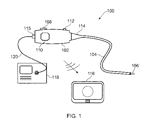
Fig. 1 is a schematic diagram of an electrosurgical
apparatus that is an embodiment of the present invention;
Fig. 2 shows a front view of a first combined vision and
treatment system;
Fig. 3 shows a cross section view of a coupler for
coupling light into a bundle of optical fibres;
Fig. 4 shows a cross-sectional view of an instrument
cable in one embodiment of the present invention;
Fig. 5 is a cross section view of an instrument tip for
use with the present invention;
Fig. 6 is an exploded view of the instrument tip of Fig.
4;
Fig. 7 is a perspective view of a vision system that may
be used in an aspect of the present invention;
CA 03106876 2021-01-19
WO 2020/025481
PCT/EP2019/070202
Fig. 8 is a perspective view of a second combined
treatment and vision system.
DETAILED DESCRIPTION; FURTHER OPTIONS AND PREFERENCES
5
Fig. 1 is a schematic view of an electrosurgical
apparatus 100 according to the present invention. The
electrosurgical apparatus 100 comprises a handpiece 102 and a
flexible instrument cable 104 extending away from the
10 handpiece 102 in a distal direction. The flexible imaging
cable is suitable for insertion into the body to access a
treatment site. The flexible instrument cable 104 may have a
biocompatible coating on its external surface so that it can
be directly inserted into tissue. The instrument cable 104
may be introduced percutaneously or in a minimally invasive
manner via a natural orifice. In some examples, the
instrument cable 104 may be used with a separate surgical
scoping device (not shown), such as a bronchoscope, endoscope,
laparoscope or the like. In other examples, the imaging cable
may be introduced through a guiding catheter. However, it may
be particular advantageous for the imaging cable to be
inserted directly (i.e. without surrounding components) to
enable it to reach regions of the body that are difficult to
access.
The instrument cable 104 in the invention has two
functions: carrying microwave electromagnetic (EM) energy
and/or radiofrequency (RF) EM energy to the treatment site,
and carrying either optical radiation or energy for powering a
light source for the purposes of imaging or sensing properties
of the treatment site. As explained in more detail below, the
instrument cable 104 of the invention provides these two
functions in a particularly compact manner, by combining the
two functions within a common structure. In some examples, an
optical channel for conveying optical radiation or energy for
powering a light source to and/or from the treatment site may
be provided within an energy conveying means for the microwave
and/or RF electromagnetic (EM) energy. For example, the
optical channel may act as an observation channel arranged to
carry optical signals to and from the treatment site to enable
an image of the treatment site to be output from the handpiece
102. In other examples, an energy conveying structure for the
CA 03106876 2021-01-19
WO 2020/025481
PCT/EP2019/070202
11
microwave and/or RF EM energy may be provided within a working
channel through an optical cable which is arranged to carry
optical signals to and from the treatment site. These examples
will be explained in more detail below.
The handpiece may include an observation port (not shown)
for viewing the image. However, in a preferred arrangement,
the handpiece 102 may be arranged to transmit the image to a
separate display device 116. The image may be transmitted via
a wireless connection, e.g. via WiFi or any other suitable
networked communication configuration. The display device 116
may be any device with a display screen that is capable of
receiving image data. The display device 116 may be portable,
e.g. a laptop or tablet computer, a smartphone, or the like.
The apparatus of the invention may include the display device,
so that the benefits of the invention can be used in locations
that do not have local display facilities.
An electrosurgical generator 118 is connected to the
handpiece 102 via a cable 120 (e.g. a coaxial cable) which
carries the RF and/or microwave energy into the handpiece 102.
The generator 118 may be of the type described in WO
2012/076844, for example. The handpiece 102 comprising a
connector port 115, which may be a QMA connector port or the
like. The connector port 115 may be arranged to electrically
connect the cable 120 to an energy conveying structure in the
instrument cable 104. This electrical connection may be
provided by a "T" connection between a coaxial cable from the
generator and a coaxial transmission line of the energy
conveying structure. Preferably there is a filter or choke
between the "T" junction and an instrument port on the
generator to prevent microwave leakage to the instrument port.
This must be placed at half a wavelength at the microwave
frequency from the "T" junction so that the "T" junction has a
high return loss, i.e. does not reflect a significant
proportion of the microwave energy back to the generator. The
proximal end of the transmission line in the energy conveying
structure is open circuit if RF energy is to be transmitted so
as not to short out the RF voltage. It is also insulated and
protected so that it does not break down for RF voltages or
expose the operator to high RF voltages.
The instrument cable 104 has at its distal end a
radiating tip 106 (or instrument tip) that is arranged to
CA 03106876 2021-01-19
WO 2020/025481
PCT/EP2019/070202
12
receive the RF and/or microwave energy from the energy
conveying means in the insertion cable 104. The radiating tip
106 includes an energy delivery portion for delivering the
received RF and/or microwave energy into biological tissue,
e.g. to assist in treatment, e.g. cutting or coagulation.
The distal end of the instrument cable 104 may be
steerable, e.g. to facilitate location of the radiating tip
106 in a desired position for treatment, and/or to enable
optical radiation to be directed as desired, e.g. to obtain
images of different parts of the treatment site or to take
measurements in different positions. As explained below, in
some examples the instrument cable 104 may include one of more
control elements (e.g. e.g. pull/push rods or control wires)
to facilitate steering. The control elements may pass out of
a proximal end of the imaging cable to engage a steering
mechanism mounted within the handpiece 102. The steering
mechanism may be operable to extend and retract the control
elements to effect action at the radiating tip. The steering
mechanism may include an actuator mounted on the handpiece
102. In this example, the actuator is a rotatable knob 110.
Rotation of the knob 110 relative to the housing can be
converted to linear motion of the control element(s) via a
suitable conversion mechanism mounted in the handpiece 102.
To limit the angle at which the proximal end of the
instrument cable 104 can be bent relative to the handpiece
102, a conical restrictor 114 is fitted over the proximal end
of the instrument cable 104. The conical restrictor 114 is
secured to a distal end of the handpiece 102 and thus limits
the movement of the cable to prevent it from experiences
unwanted stresses.
The handpiece 102 comprises a housing that contains
components associated with generating and controlling the
optical radiation that can be conveyed along the optical
channel in the instrument cable 104. For example, the
handpiece 102 may contain a power source, such as a cell or
other battery, an optical source, such as a light emitting
diode (LED) or the like, and one or more optical elements for
directing optical radiation from the optical source or from
the treatment site in a desired manner. The optical elements
may include a control interface 112 on the outer surface of
the housing, to enable a user to control the optical elements
CA 03106876 2021-01-19
WO 2020/025481
PCT/EP2019/070202
13
in use. For example, the control interface 112 may control an
intensity optical radiation delivered to the treatment site,
or may control one or more lenses to assist in focussing an
image signal received from the treatment site on to an optical
sensor. In one example, an optical detector (e.g. a camera or
the like) may be mounted in the handpiece to receive optical
radiation returned from the treatment site in order to capture
and transmit an image signal to the display device 116. In
one example, the optical components may resemble a
conventional fiberscope.
The handpiece 102 may include a power switch (not shown)
for activating and deactivating the apparatus. The handpiece
102 may include a charging port (not shown) for connecting the
power source to an external power supply to enable it to be
recharged.
Fig. 2 shows a front view of a combination treatment and
vision system 10 at the distal end of the instrument cable
104. The treatment and vision system 10 may be positioned in a
central lumen of a radiating tip 106 which may be insertable
through the instrument channel of a scoping device. The
treatment and vision system 10 may provide illumination and
vision at the distal end of the instrument cable 104 to aid a
clinician.
The treatment and vision system 10 comprises an imaging
cable, which in this example comprises a bundle of optical
fibres 12 which are configured to provide illumination to the
distal end of the instrument. The size, number and arrangement
of the optical fibres 12 may be chosen to ensure optimal light
levels. For example, the optical fibres 12 may be arranged in
a complete ring around the image sensor 14, or may be arranged
primarily on one side of the image sensor 14 as depicted in
Fig. 2. In a preferred embodiment each optical fibre 12 has a
diameter of 250 pm, though the use of smaller optical fibres
may allow more fibres to be placed into the central lumen of
the radiating tip 200 and may therefore provide more light.
Light may be coupled into a proximal end of the optical fibres
12 using collimating lenses or couplers, for example a coupler
as shown in Fig. 3. It is envisaged that any suitable light
source may be used, preferably using visible wavelengths. For
example, specific wavelengths may be selected for narrow band
imaging, such as a combination of 415 nm and 540 nm light,
CA 03106876 2021-01-19
WO 2020/025481
PCT/EP2019/070202
14
providing additional imaging modalities and allowing
clinicians to see aspects of tissue in more detail and
subsequently allow for easier identification of tissue lesions
or other anomalies.
In some embodiments, a lens may be positioned over the
bundle of optical fibres 12. The lens may be used to focus or
diffuse light emitted from the optical fibres 12 depending on
the application. Alternatively, the distal end of each of the
optical fibres 12 may be shaped so as to direct light in a
predetermined direction. For example, the distal end of an
optical fibre 12 may form a ball lens, or may be tapered as
required.
The image sensor 14 may be any chip-based image sensor
suitable for detecting light at the selected frequencies. The
image sensor 14 is configured to receive light at the distal
end of the electrosurgical instrument and transmit images to
the proximal end. For example, a cable may extend from the
image sensor to the handpiece 102 where images are transmitted
to a display 116, such as over Wi-Fi. Preferably the image
sensor 14 is a CMOS sensor, though other suitable pixel
sensors may be chosen to ensure that the display 116 receives
a good image. In particular, the image sensor 14 may be a 40
kilopixel CMOS sensor having a surface area of 1 mm2 or less.
For example, a PICORAMEDIC (TM) sensor manufactured by
Fujikura Ltd may be chosen as the image sensor 14.
The distal end of the combined treatment and vision
system 10 is preferably deflectable to provide steerability
and thus give a clinician greater control over the region
which is imaged and ensure accurate treatment by the
instrument tip 106. In one example, the system 10 may be
provided through a steerable catheter, for example a catheter
having a number of steering wires which give a clinician
control over the distal end. Alternatively, the instrument
cable 104 itself may comprise a number of steering wires to
allow the distal end to be deflected. Deflection and
steerability of the system 10 may be controlled by a clinician
manipulating the handpiece.
Fig. 3 shows a cross-sectional view of a coupler 20 which
may be positioned within the handpiece 102 of the
electrosurgical apparatus 100 to provide light to a distal end
of the instrument cable 104 through a bundle of optical fibres
CA 03106876 2021-01-19
WO 2020/025481
PCT/EP2019/070202
12. The coupler 20 comprises two light source 22, 24 which may
be LEDs. Each LED 22, 24 may have the same emission spectrum,
or they may each be chosen to provide light at specific
wavelengths. For example, LED 22 may provide light having a
5 wavelength of 540 nm (green) and LED 24 may provide light
having a wavelength of 425 nm (blue). This allows for narrow
band imaging with at least two modalities allowing clinicians
to see aspects of tissue in more detail and subsequently allow
for easier identification of tissue lesions or other
10 anomalies. Light from each light source 22, 24 is directed to
output port 26 for coupling into the bundle of optical fibres
12 by a respected channel 28. The channels 28 may metallised
to ensure good optical transmission of light from the light
sources 22, 24. Circuitry within the handpiece 102 may provide
15 for activation and dimming of each light source 22, 24
separately to tailor illumination to the response required by
the clinician.
Fig. 4 shows a cross section through the instrument cable
104 in one embodiment of the invention. In this embodiment,
the instrument cable 104 comprises a coaxial cable having an
inner conductor 11, an outer conductor 13 formed coaxially
with the inner conductor 11, and a first dielectric material
15 separating the inner conductor 11 and outer conductor 13. A
protective sheath 17 is formed on the outside of the
instrument cable 104 to protect the coaxial cable as it the
instrument cable 104 is inserted through a surgical scoping
device.
The inner conductor 11 is hollow to form an optical
channel 15. The optical channel 15 carries an imaging cable
formed from a bundle of optical fibres 12 and one or more
wires (not shown) for supplying energy to an image sensor and
for returning digital images to the handpiece of the
electrosurgical instrument. As can be seen from Fig. 4, in
this embodiment the bundle of optical fibres 12 form a ring on
the inner surface of the inner conductor 11. In this way, the
bundle of optical fibres 12 effectively form an innermost
insulating layer of the coaxial cable, which may help reduce
interference between digital image transmission through the
optical channel and the RF and/or microwave energy conveyed
through the instrument cable 104.
CA 03106876 2021-01-19
WO 2020/025481
PCT/EP2019/070202
16
A cross sectional view of an radiating tip 30, which may
be used as radiating tip 106 as described above, is shown in
Fig. 5. An exploded view of the radiating tip 30 is shown in
Fig. 6. The radiating tip 30 is configured to be mounted at
the distal end of the instrument cable 104, wherein the
instrument cable 104 comprises means for delivering RF and/or
microwave frequency energy from its proximal end to the
radiating tip 30. In particular, the instrument cable 104 may
provide an energy conveying structure such as a coaxial cable,
wherein the coaxial cable has an inner conductor, an outer
conductor and a central lumen. The radiating tip 30 may
thereby be used to perform electrosurgery, for example
ablation.
The radiating tip 30 is a bipolar structure comprising an
inner conductor 32, being a hollow metal cylinder connected to
the inner conductor of the instrument cable 104. An outer
conductor 34 is coaxial with the inner conductor 32 and is
connected to the outer conductor of the coaxial cable within
the instrument cable 104. The inner conductor 32 and outer
conductor 34 of the instrument tip 30 are separated by a
dielectric cylinder 36. The inner conductor 32 extends
longitudinally through the dielectric cylinder beyond a distal
end of the outer conductor 34. The radiating tip 30 provides a
radiating antenna structure for RF and/or microwave frequency
energy in order to treat tissue, e.g. by ablation. Each of the
inner conductor 32 and outer conductor 34 preferably comprise
silver or gold to ensure good microwave propagation.
The shape of the dielectric material 36 may also be
selected to achieve efficient delivery of energy into tissue.
For example, the dielectric 36 may be a cylindrical piece of
ceramic material, polyether ether ketone (PEEK), or PTFE
having a rounded distal tip.
The inner conductor 32 of the radiating tip 30 defines a
central lumen 38 which collinear with the central lumen of the
instrument cable 104. The central lumen 38 thus provides a
channel through which an optical cable for providing
illumination and vision may be fed, for example as discussed
above with respect to Fig. 2 or below with respected to Fig.
6. For example, the central lumen 38 may have a diameter of 5
mm or less, for example 2 mm or less.
CA 03106876 2021-01-19
WO 2020/025481
PCT/EP2019/070202
17
The radiating tip 30 may also have a biocompatible
coating covering both the inner and outer surfaces of the
radiating tip 30. The length of the radiating tip 30 is
preferably selected to be a quarter wavelength at the
predetermined operational frequency, preferably 5.8 GHz for
example. The length should be minimised for insertion through
scoping devices and to allow navigation through lumens within
a patient's body.
Fig. 7 shows perspective view of a vision system 40 which
may be provided at the distal end of an instrument cable 104.
The vision system 40 may be used to provide combined vision
and treatment, for example the vision system 40 may be
positioned within a central lumen of an radiating tip (e.g.
instrument tip 30 described above) in a similar may as
described above with respect to the first vision system 10. It
is also envisaged that the vision system 40 may be provided as
a standalone illumination and vision system which may be
delivered to a treatment site through an optical channel of an
instrument cable, an imaging cable, or through a catheter such
as a steerable catheter.
The vision system 40 comprises two light sources 42a, 42b
which are provided in this embodiment as surface mount light
emitting diodes (LEDs) each arranged generally along one side
of an image sensor 44. Other arrangements of light sources
42a, 42b and image sensor 44 may be selected to provide
optimal illumination of a treatment region. The image sensor
44 is preferably a CMOS sensor, such as a 40 kilopixel CMOS
sensor having an area of less than 1 mm2. The light sources
42a, 42b provide illumination for the image sensor 44 in place
of optical fibres as described above. Each light source 42a,
42b may have the same emission spectrum, or may be chosen to
each provide different wavelengths of light for multi-modal
imaging of the treatment region.
A support structure 46 is provide to hold the LEDs 42a,
42b and image sensor 44 in their relative positions and
strengthen the vision system 40 to allow the system 40 to be
fed through a channel, such as the optical channel of an
instrument cable. The support structure 46 may be made of any
suitable biocompatible material which is robust enough to
support the components of the vision system 30. For example,
the support structure 40 may be made of a polymer material
CA 03106876 2021-01-19
WO 2020/025481
PCT/EP2019/070202
18
such as a polycarbonate. Openings within which the light
sources 42a, 42b and the image sensor 44 are mounted may
preferably be provided in the base material by laser cutting,
though other manufacturing methods may also be suitable. The
support structure 46 may have a diameter of less than 2 mm,
such as 1.6 mm or less, to allow the vision system to be fed
through an optical channel of an instrument cable 104, or an
optical channel formed through a coaxial cable as described
above.
The support structure 46 is provided at the distal end of
a protective sheath 48 that conveys an imaging cable, which in
this case comprises wires providing energy to the light
sources 42a, 42b and image sensor 44 from the handpiece 102,
and wires carrying the signal (e.g. image data) from the image
sensor 44 to the handpiece 102 to be transmitted to a display.
The protective sheath 48 is preferably made of a biocompatible
material, and the distal end of the sheath 48 is potted to
create a sealed enclosure to prevent the ingress of liquids or
debris through or around the support structure 46.
The distal end of the vision system 40 is preferably
deflectable to provide steerability and thus give a clinician
greater control over the region which is imaged. In one
example, the system 40 may be provided through a steerable
catheter, for example a catheter having a number of steering
wires which give a clinician control over the distal end.
Alternatively, the instrument cable 104 itself may comprise a
number of steering wires to allow the distal end to be
deflected. Deflection and steerability of the system 10 may be
controlled by a clinician manipulating the handpiece 102. In
some embodiments, the protective sheath 48 may incorporate a
number of steering wires which can be manipulated to deflect
the vision system 40 by a clinician using the handpiece 102.
Fig. 8 shows a perspective view of a second combined
treatment and vision system 50 according to an aspect of the
present invention. The treatment and vision system 50
comprises two light sources 52a, 52b to provide illumination
at the distal end of an imaging cable. For example, the light
sources 52a, 52b may be light emitting diodes (LEDs) as
described above with respect to Fig. 7. The light sources 52a,
52b are arranged symmetrically on either side of an image
sensor 54, though any arrangement of the light sources 52a,
CA 03106876 2021-01-19
WO 2020/025481
PCT/EP2019/070202
19
52b may be considered to provide optimal illumination of a
treatment region. These components are held in place by a
support structure 56.
The support structure 56 strengthens the distal end of
the combined treatment and vision system 50 to ensure that the
light sources 52a, 52b and image sensor 54 are held in their
predetermined positions. The support structure 56 may be made
of any suitable biocompatible material which is robust enough
to support the components of the vision system 50. For
example, the support structure 46 may be made of a polymer
material such as a polycarbonate. Openings within which the
light sources 52a, 52b and the image sensor 54 are mounted may
preferably be provided in the base material by laser cutting,
though other manufacturing methods may also be suitable. The
support structure 56 also defines an opening 58 which is at
the distal end of a working channel through the instrument
cable 104. The working channel of the instrument cable 104 may
be used to convey a coaxial cable, and/or to deliver tools and
structures to a treatment site, and thus enables the system 50
to also provide treatment as required. The support structure
56 comprises a proximally extending extruded section
connecting the working channel through the instrument cable
104 to the opening 58.
The support structure 56 may be detachably mounted to the
distal end of the instrument cable 104. The support structure
56 may thus provide a removable structure allowing components
to be easily changed by a clinician. For example, a clinician
may wish to replace the support structure 56 to provide a
different image sensor 54 and/or light sources 52a, 52b which
are better suited to a particular treatment.
In some embodiments, the working channel may allow
electrosurgery to be carried out at the distal end of the
instrument cable 104. For example, a coaxial cable may be
inserted through the working channel to provide RF and/or
microwave frequency energy to an radiating tip at the opening
58 for electrosurgery. In other examples, the working channel
may comprise an energy delivery structure which is
incorporated into the sidewall of the working channel, and/or
which is provided as a liner (such as a detachable cover) for
the working channel. In such embodiments, the energy delivery
structure may comprise a coaxial layered structure having an
CA 03106876 2021-01-19
WO 2020/025481
PCT/EP2019/070202
innermost insulating layer, an inner conductive layer formed
on the innermost insulating layer, an outer conductive layer
formed coaxially with the inner conductive layer, and a
dielectric layer separating the inner conductive layer and the
5 outer conductive layer. In this way, the energy delivery
structure may provide a transmission line for conveying RF
and/or microwave frequency energy to an instrument tip at the
distal end of the working channel, at the opening 58.
Preferably, the energy conveying structure may include, e.g.
10 at a distal end thereof in a region proximate the opening 48,
a first terminal that is electrically connected to the inner
conductive layer and which extends through the innermost
insulating layer into the working channel, and a second
terminal that is electrically connected to the outer
15 conductive layer and which extends through the dielectric
layer and innermost insulating layer into the working channel.
The first terminal and the second terminal may be arranged to
form electrical connections (e.g. physically engage) with
corresponding contacts formed on an electrosurgical instrument
20 (e.g. a radiating tip) that is insertable in or through the
working channel. The first terminal and the second terminal
may be formed at the distal end of the inner conductive layer
and outer conductive layer respectively, preferably in a
region which is proximate the opening 48. The outer
conductive layer may extend longitudinally further in a distal
direction than the inner conductive layer, whereby the first
terminal is located proximally from the second terminal.
The working channel of the instrument cable 104 for the
vision system depicted in Fig. 7 may additionally or
alternatively receive a catheter which provides for
steerability at the distal end. For example, the catheter may
comprise a number of steering wires extending from the
handpiece 102 to the distal end of the working channel to
enable a clinician to steer the instrument cable 104.
In addition to the working channel, the instrument cable
104 may comprise an optical channel for conveying wires and/or
cables for delivery electricity from the handpiece 102 to
power the light sources 52a, 52b and image sensor 54. The
additional lumen also conveys wires and/or cables for
conveying image signals from the image sensor 54 to the
handpiece 102 to be transmitted to a display device 116.
CA 03106876 2021-01-19
WO 2020/025481
PCT/EP2019/070202
21
The distal end of the combined treatment and vision
system 50 is preferably deflectable to provide steerability
and thus give a clinician greater control over the region
which is imaged and ensure accurate treatment by an instrument
tip. In one example, the system 50 may be provided through a
steerable catheter, for example a catheter having a number of
steering wires which give a clinician control over the distal
end. Alternatively, the instrument cable 104 itself may
comprise a number of steering wires to allow the distal end to
be deflected. In another example, steering wires may be
incorporated into the walls of the working channel, enabling
deflection at the distal end of the instrument cable 104.
Alternatively, the working channel may receive an articulated
or knuckled guide wire in which sections of the guide wire may
be adjusted to provide steerability of the combined treatment
and vision system 50. Preferably, deflection and steerability
of the system 50 may be controlled by a clinician manipulating
the handpiece 102.