Note: Descriptions are shown in the official language in which they were submitted.
CA 03106997 2021-01-18
WO 2020/036845 PCT/US2019/046104
THERAPEUTIC PROTEIN SELECTION IN SIMULATED IN VIVO CONDITIONS
REFERENCE TO A SEQUENCE LISTING
[001] This application incorporates by reference the Sequence Listing
submitted in Computer
Readable Form as file 10470W001-Sequence.bd, created on August 12, 2019 and
containing 1466
bytes.
FIELD OF THE INVENTION
[002] The present invention pertains to biopharmaceuticals, and relates to the
determination of
the behavior of therapeutic biomolecules, such as antibodies, in near
physiological conditions.
BACKGROUND
[003] Proper analysis of in vitro binding equilibria is necessarily
constrained to concentrations
near the dissociation constant, typically on the order of micromolar and
below. Under these
conditions, proteins behave essentially as ideal molecules and any non-
specific interactions can
easily be ignored. Increasing the protein concentration to arbitrarily high
values leads to
macromolecular crowding, where complex formation is no longer linear with
respect to protein
concentration and the binding equations derived from the law of mass action
are better expressed
in terms of thermodynamic activity rather than concentration (Neal, B. L., D.
Asthagiri and A. M.
Lenhoff (1998). "Molecular origins of osmotic second virial coefficients of
proteins." Biophys J 75(5):
2469-2477). The magnitude of the activity coefficient depends on the
composition of the whole
solution; non-ideality arises not only from elevated levels of the protein
itself, but also from the
presence of non-interacting macromolecules and co-solutes.
[004] An understanding of the non-specific interactions between proteins under
non-ideal
conditions, which deviate significantly from those commonly employed for in
vitro characterization,
is vital to achieving a more complete picture of protein function in a
biological context. Thus, there
presently is a need for methods for determining the effect of non-specific
interactions of biological
molecules under simulated in vivo conditions.
SUMMARY OF THE INVENTION
[005] In one aspect, the present invention provides a method of determining
the effect of non-
specific interactions in simulated in vivo conditions, in which the method
includes: (a) contacting a
solution comprising a biologically relevant molecular crowding agent and a
target molecule with a
biosensor, wherein the surface of the biosensor comprises a capture molecule
that specifically
binds the target molecule; (b) allowing the target molecule to bind to the
capture molecule; and (c)
1
CA 03106997 2021-01-18
WO 2020/036845 PCT/US2019/046104
determining an amount of the target molecule bound to capture molecule using
biolayer
interferometry.
[006] In some embodiments, the method further includes comparing the amount of
the target
biomolecule bound to the capture molecule to a control threshold that
distinguishes between a
target biomolecule having attractive non-specific interactions with the other
molecules in the
solution and having repulsive non-specific interaction with the other
molecules in the solution.
[007] In various embodiments of the method, the control threshold is the
amount of binding
normalized to an amount of binding in an ideal, dilute, or semi-dilute
solution.
[008] In various embodiments of the method, the biologically relevant
molecular crowding agent
comprises human serum albumin (HSA).
[009] In some embodiments, the human serum albumin is present at a
physiologically relevant
concentration.
[0010] In some embodiments, the human serum albumin is present at a
concentration of between
about 1 g/L and about 100g/L.
[0011] In some embodiments, the method further includes determining the amount
of binding at
two or more concentrations of the biologically relevant molecular crowding
agent.
[0012] In various embodiments of the method, the biologically relevant
molecular crowding agent
comprises serum and/or plasma.
[0013] In some embodiments, the method further includes determining the amount
of binding at
two or more pHs, for example, to determine a pH dependence of binding.
[0014] In some embodiments, the method further includes determining the amount
of binding at
two or more salt concentrations, for example, to determine a salt dependence
of binding.
[0015] In various embodiments of the method, the target molecule comprises a
monoclonal
antibody and the capture molecule comprises an antigen that specifically binds
to the monoclonal
antibody.
[0016] In various embodiments of the method, the target molecule comprises an
antigen and the
capture molecule comprises a monoclonal antibody that specifically binds to
the antigen.
[0017] In various embodiments of the method, the target molecule comprises a
receptor or ligand
binding fragment thereof and the capture molecule comprises a ligand that
specifically binds to the
receptor or ligand binding fragment thereof.
[0018] In various embodiments of the method, the target molecule comprises a
ligand and the
capture molecule comprises a receptor or ligand binding fragment thereof that
specifically binds to
the ligand.
[0019] In various embodiments of the method, the capture molecule is coupled
to the surface of the
sensor with a linker.
2
CA 03106997 2021-01-18
WO 2020/036845 PCT/US2019/046104
[0020] In various embodiments of the method, the linker comprises biotin and
streptavidin or avidin.
[0021] In various embodiments of the method, the target molecule comprises an
antibody and the
capture molecule comprises anti-IgG Fc.
[0022] In various embodiments of the method, the anti-IgG Fc is anti-human IgG
Fc.
[0023] In another aspect, the present invention provides a method of selecting
a biomolecule under
simulated in vivo conditions, in which the method includes: (a) contacting a
solution simulating in
vivo conditions with a biosensor, wherein a surface of the biosensor comprises
a capture molecule
that specifically binds target biomolecules from a set of two or more target
biomolecules of interest,
and wherein the solution further comprises a biologically relevant molecular
crowding agent and a
first target biomolecule selected from the set of two or more target
biomolecules of interest; (b)
allowing the first target biomolecule to bind to the capture molecule; and (c)
determining an amount
of the first target biomolecule bound to the capture molecule using biolayer
interferometry.
[0024] In some embodiments, the method further includes: (a) contacting a
second solution
simulating in vivo conditions with the biosensor, wherein the second solution
further comprises the
biologically relevant molecular crowding agent and a second target biomolecule
selected from the
set of two or more biomolecules of interest; (b) allowing the second target
biomolecule to bind to
the capture molecule; (c) determining an amount of the second target molecule
bound to the
capture molecule using biolayer interferometry; and (d) comparing the amount
of the first
biomolecule bound to the capture molecule and the amount of the second
biomolecule bound to the
capture molecule to identify which of the first biomolecule and the second
biomolecule has a
greater amount of binding.
[0025] In some embodiments, the method further includes comparing the amount
of the first and/or
second target biomolecule bound to the capture molecule to a control threshold
that distinguishes
between a target biomolecule having attractive non-specific interactions with
the other molecules in
the solution and having repulsive non-specific interactions with the other
molecules in the solution.
[0026] In various embodiments of the method, the threshold is the amount of
binding normalized to
an amount of binding in an ideal, dilute, or semi-dilute solution.
[0027] In various embodiments of the method, the biologically relevant
molecular crowding agent
comprises human serum albumin (HSA).
[0028] In various embodiments of the method, the human serum albumin is
present at a
physiologically relevant concentration.
[0029] In various embodiments of the method, the human serum albumin is
present at a
concentration of between about 1 g/L and about 100g/L.
[0030] In some embodiments, the method further includes determining the amount
of binding at
two or more concentrations of the biologically relevant molecular crowding
agent.
3
CA 03106997 2021-01-18
WO 2020/036845 PCT/US2019/046104
[0031] In various embodiments of the method, the biologically relevant
molecular crowding agent
comprises serum and/or plasma.
[0032] In some embodiments, the method further includes determining the amount
of binding at
two or more pHs to determine a pH dependence of binding.
[0033] In some embodiments, the method further includes determining the amount
of binding at
two or more salt concentrations to determine a salt dependence of binding.
[0034] In various embodiments of the method, the target molecule comprises a
monoclonal
antibody and the capture molecule comprises an antigen that specifically binds
to the monoclonal
antibody.
[0035] In various embodiments of the method, the target molecule comprises an
antigen and the
capture molecule comprises a monoclonal antibody that specifically binds to
the antigen.
[0036] In various embodiments of the method, the target molecule comprises a
receptor or ligand
binding fragment thereof and the capture molecule comprises a ligand that
specifically binds to the
receptor or ligand binding fragment thereof.
[0037] In various embodiments of the method, the target molecule comprises a
ligand and the
capture molecule comprises a receptor or ligand binding fragment thereof that
specifically binds to
the ligand.
[0038] In various embodiments of the method, the capture molecule is coupled
to the surface of the
sensor with a linker.
[0039] In various embodiments of the method, the linker comprises biotin and
streptavidin or avidin.
[0040] In various embodiments of the method, the target molecule comprises an
antibody and the
capture molecule comprises anti-IgG Fc.
[0041] In various embodiments of the method, the anti-IgG Fc is anti-human IgG
Fc.
DESCRIPTION OF THE FIGURES
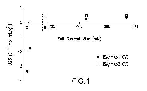
[0042] Figure 1 is a graph showing the ionic strength dependence of mAb1/HSA
and mAb2/HSA
cross-interactions measured by CG-MALS. Cross-virial coefficients (A23) were
determined by CG-
MALS for interactions between 10 g/L HSA and 10 g/L mAb1 (.) or mAb2 (0) at
increasing
concentrations of NaCI in phosphate buffer. Negative values for CVC indicate
attractive forces
between the molecules, while positive CVC values indicate repulsive forces
between the molecules.
The box indicates physiological ionic strength where the CVC values for mAb1
and mAb2 are
negative and positive, respectively.
[0043] Figure 2 is a graph showing weak cation exchange chromatography elution
profiles for
mAbs and HSA. Each mAb and HSA were assessed by analytical chromatography on a
weak
cation exchange column equilibrated with 200 mM MES, 20 mM NaCI, pH 6.5. A
gradient was
4
CA 03106997 2021-01-18
WO 2020/036845 PCT/US2019/046104
applied from 20-500 mM NaCI and is represented by a dashed line as a
percentage of a 1 M NaCI
solution. Representative elution profiles for HSA, mAb1, and mAb2 are shown.
[0044] Figures 3A and 3B are graphs showing the binding of mAb to biotinylated
antigen at 137
mM NaCI in absence of HSA measured by BLI. Binding of mAb1 (Figure 3A) and
mAb2 (Figure 3B)
to biotinylated antigen was observed using biolayer interferometry at
physiological salt
concentration in phosphate buffer. Change in wavelength in nanometers
(response, nm) is plotted
as a function of time to indicate changes in thickness of the biolayer due to
binding events.
Association and dissociation steps are shown for 1.25 nM, 2.5 nM, 5 nM, 10 nM,
20 nM, and 40 nM
mAb. Dashed traces indicate raw data and solid traces indicate fitted curves.
Data traces are
aligned at the mAb association step, and reference data was subtracted from
all sample traces.
[0045] Figures 4A and 4B are graphs showing the HSA on mAb binding to antigen
measured by
BLI is ionic-strength dependent. Binding of 40 nM mAb1 and mAb2 to
biotinylated antigen in the
absence and presence of HSA was observed by biolayer interferometry at 10
(Figure 4A) and 137
mM NaCI (Figure 4B) in phosphate buffer. Change in wavelength (response, nm)
as a function of
time indicates binding events, and only the mAb association and dissociation
steps are shown.
Biotinylated antigen was loaded onto SA tips as described above. Binding of
mAb1 was assessed
in the absence and presence of 10 g/L HSA, and mAb2 binding was assessed in
the absence and
presence of 10 g/L HSA. A minimum of 0.1 g/L HSA was used to prevent non-
specific binding to the
biosensor tip. Data traces are aligned at the mAb association step following a
baseline
measurement in equivalent concentrations of HSA. Traces for samples containing
10 g/L HSA were
corrected for a change in signal upon transitioning from association to
dissociation due to the
change in refractive index of the solution.
[0046] Figures 5A and 5B are graphs showing that the effect of HSA on mAb
binding to antigen
measured by BLI is ionic-strength dependent and mAb-specific. Binding of mAb1
(.) and mAb2 (0)
to biotinylated antigen in the presence of increasing HSA concentrations was
observed by biolayer
interferometry at 10 (Figure 5A) and 137 (Figure 5B) mM NaCI. The normalized
response (to
binding at 0.1 g/L HSA) is shown as a function of HSA concentration. The
dotted line indicates the
normal at 1.0, in order to illustrate the relationship of the data points to
this line. A minimum of 0.1
g/L HSA was used to prevent non-specific binding to the biosensor tip.
Experiments were
performed in triplicate, and the mean value and standard deviation are shown.
One-way ANOVA
was performed at each HSA concentration and p-values <0.05 are indicated with
an asterisk. All
data are summarized in Tables 2-4.
[0047] Figures 6A-60 are a set of graphs showing the effect of Ficoll 70 on
mAb binding to antigen
measured by BLI. Binding of mAb1 (.) and mAb2 (0) to biotinylated antigen in
the presence of
increasing Ficoll 70 concentrations was observed by biolayer interferometry at
10 mM (Figure 6A)
CA 03106997 2021-01-18
WO 2020/036845 PCT/US2019/046104
and 137 mM (Figure 6B) NaCI. The normalized response (to binding at 0.1 g/L
Ficoll 70) is plotted
as a function of Ficoll 70 concentration. The dotted line in Figs. 6A and 6B
indicates the normal at
1.0, in order to illustrate the relationship of the data points to this line.
Experiments were performed
in triplicate, and the mean value and standard deviation are shown. One-way
ANOVA was
performed at each HSA concentration and p-values < 0.05 are indicated with an
asterisk. All data
are summarized in Tables 5-7. Slow binding kinetics required an extension of
the time for mAb
association to antigen at very high Ficoll 70 concentrations (200 g/L and
above, Figures 60 and
6D). Binding of mAbs to biotinylated antigen in the presence of 300 g/L Ficoll
was observed by
biolayer interferometry at 10 mM (Figure 60) and 137 mM (Figure 6D) NaCI. The
aligned responses
in nm for mAb1 and mAb2 in the absence of Ficoll 70 are shown as a function of
time for an
extended association time (-3 hours). Addition of 300 g/L Ficoll 70 to mAb1
and mAb2 show
comparatively slowed binding kinetics.
[0048] Figure 7 is a graph showing that biosensor dipping into HSA solution
produces an increase
in signal in biolayer interferometry experiments. A representative sensorgram
for mAb1 binding to
an SA tip loaded with biotinylated antigen in 137 mM NaCI. Following a
baseline measurement (0-
120 s), biotinylated antigen is loaded in the presence of 0.1 g/L HSA and
allowed to equilibrate
(step A), the sensor dips into 10 g/L HSA (step B), the sensor dips into 10
g/L HSA + 40 nM mAb1
(step C), and the sensor dips into baseline buffer (step D). The magnitude of
the signal increase
observed in step B is similar to the increase in signal observed for the same
experimental set-up
with biotin-loaded SA (no antigen), indicating the signal is due to refractive
index of the high protein
concentration of HSA rather than a specific binding event to the biosensor tip
(data not shown).
[0049] Figures 8A-8H are a set of graphs showing affinity measurements with
anti-human IgG Fc
capture tips. Representative biolayer interferometry sensorgrams are presented
for mAb1 with
unlabeled antigen in 10 mM (Figure 8A) and 137 mM NaCI (Figure 8B); mAb1 with
biotinylated
antigen in 10 mM (Figure 80) and 137 mM NaCI (Figure 8D); mAb2 with unlabeled
antigen in 10
mM (Figure 8E) and 137 mM NaCI (Figure 8F); mAb2 with biotinylated antigen in
10 mM (Figure
8G) and 137 mM NaCI (Figure 8H). Biosensor tips were loaded with antibody to
achieve - 0.6 nm
response. Antigen association step was from 100-300 sec and dissociation was
600-750 sec.
[0050] Figure 9 is a graph showing mAb3 does not bind to antigen and no
changes in BLI signal
are observed. Binding of mAb3 to biotinylated antigen in the presence of
increasing HSA
concentrations was observed by biolayer interferometry at 10 (.) and 137 (0)
mM NaCI. The
observed signal (in nm), rather than the normalized response, is plotted as a
function of HSA
concentration. Experiments were performed in duplicate, and the mean value and
standard
deviation are shown.
[0051] Figure 10 is a set of schematics demonstrating the difference in ideal,
dilute, and semi-
6
CA 03106997 2021-01-18
WO 2020/036845 PCT/US2019/046104
dilute solutions.
[0052] Figure 11 is a set of schematics demonstrating that semi-dilute
solutions are not equivalent
to concentrated solutions.
[0053] Figure 12 is a schematic showing a generalized biolayer interferometry
system for
measuring antibody interactions in the presence of crowding agents and
antigen, in accordance
with disclosed embodiments.
[0054] Figure 13 is a schematic showing a generalized biolayer interferometry
system for
measuring antibody interactions in the presence of crowding agents and FcRn,
in accordance with
disclosed embodiments.
[0055] Figure 14 is graph showing the effects of HSA on antibody/FcRn binding.
[0056] Figures 15A and 15B are graphs showing the dose-response curve effects
of HSA at
different salt concentrations.
[0057] Figure 16 is graph showing the dose-response curve effects of HSA for
two different
antibodies +/- FA.
[0058] Figure 17 is a sequence alignment of wild type and the YTE mutation.
[0059] Figure 18 is graph showing the dose-response curve effects of HSA on
anti-05 mAbs
binding to FcRn.
[0060] Figure 19 is graph showing the dose-response curve effects of HSA on
anti-Zika mAbs
binding to FcRn.
[0061] Figure 20 is set of graphs showing the dose-response curve effects of
HSA on half-life
extension mutants.
DETAILED DESCRIPTION OF THE INVENTION
[0062] Before the present invention is described, it is to be understood that
this invention is not
limited to particular methods and experimental conditions described, as such
methods and
conditions may vary. It is also to be understood that the terminology used
herein is for the purpose
of describing particular embodiments only, and is not intended to be limiting,
since the scope of the
present invention will be limited only by the appended claims. Any embodiments
or features of
embodiments can be combined with one another, and such combinations are
expressly
encompassed within the scope of the present invention.
[0063] Unless defined otherwise, all technical and scientific terms used
herein have the same
meaning as commonly understood by one of ordinary skill in the art to which
this invention belongs.
As used herein, the term "about," when used in reference to a particular
recited numerical value,
means that the value may vary from the recited value by no more than 1%. For
example, as used
7
CA 03106997 2021-01-18
WO 2020/036845 PCT/US2019/046104
herein, the expression "about 100" includes 99 and 101 and all values in
between (e.g., 99.1, 99.2,
99.3, 99.4, etc.)
[0064] Although any methods and materials similar or equivalent to those
described herein can be
used in the practice or testing of the present invention, the preferred
methods and materials are
now described. All patents, applications and non-patent publications mentioned
in this specification
are incorporated herein by reference in their entireties.
Abbreviations Used Herein
[0065] AUC: Analytical Ultracentrifugation
[0066] NMR: Nuclear Magnetic Resonance
[0067] MALS: Multi-Angle Static Light Scattering
[0068] CG-MALS: Composition-Gradient Multi-Angle Light Scattering
[0069] HSA: Human Serum Albumin
[0070] mAbs: Monoclonal Antibodies
[0071] BLI: Biolayer lnterferometry
[0072] SA: Streptavidin
[0073] CVC: Cross-Virial Coefficient
[0074] MBP: Maltose Binding Protein
[0075] FcRn: Neonatal Fc Receptor
[0076] FelD1: principal cat allergen
Definitions
[0077] The term "antibody", as used herein, is intended to refer to
immunoglobulin molecules
comprised of four polypeptide chains, two heavy (H) chains and two light (L)
chains inter-connected
by disulfide bonds (i.e., "full antibody molecules"), as well as multimers
thereof (e.g. IgM) or
antigen-binding fragments thereof. Each heavy chain is comprised of a heavy
chain variable region
("HCVR" or "VH") and a heavy chain constant region (comprised of domains CH1,
CH2 and CH3). In
various embodiments, the heavy chain may be an IgG isotype. In some cases, the
heavy chain is
selected from IgG1, IgG2, IgG3 or IgG4. In some embodiments, the heavy chain
is of isotype IgG1
or IgG4, optionally including a chimeric hinge region of isotype IgG1/IgG2 or
IgG4/IgG2. Each light
chain is comprised of a light chain variable region ("LCVR or "VL") and a
light chain constant region
(CL). The VH and VL regions can be further subdivided into regions of
hypervariability, termed
complementarity determining regions (CDR), interspersed with regions that are
more conserved,
termed framework regions (FR). Each VH and VL is composed of three CDRs and
four FRs,
arranged from amino-terminus to carboxy-terminus in the following order: FR1,
CDR1, FR2, CDR2,
FR3, CDR3, FR4. The term "antibody" includes reference to both glycosylated
and non-
8
CA 03106997 2021-01-18
WO 2020/036845 PCT/US2019/046104
glycosylated immunoglobulins of any isotype or subclass. The term "antibody"
includes antibody
molecules prepared, expressed, created or isolated by recombinant means, such
as antibodies
isolated from a host cell transfected to express the antibody. For a review on
antibody structure,
see Lefranc et al., /MGT unique numbering for immunoglobulin and T cell
receptor variable
domains and Ig superfamily V-like domains, 27(1) Dev. Comp. lmmunol. 55-77
(2003); and M.
Potter, Structural correlates of immunoglobulin diversity, 2(1) Surv. lmmunol.
Res. 27-42 (1983).
[0078] The term antibody also encompasses a "bispecific antibody", which
includes a
heterotetrameric immunoglobulin that can bind to more than one different
epitope. One half of the
bispecific antibody, which includes a single heavy chain and a single light
chain and six CDRs,
binds to one antigen or epitope, and the other half of the antibody binds to a
different antigen or
epitope. In some cases, the bispecific antibody can bind the same antigen, but
at different epitopes
or non-overlapping epitopes. In some cases, both halves of the bispecific
antibody have identical
light chains while retaining dual specificity. Bispecific antibodies are
described generally in U.S.
Patent App. Pub. No. 2010/0331527(Dec. 30, 2010).
[0079] The term "antigen-binding portion" of an antibody (or "antibody
fragment"), refers to one or
more fragments of an antibody that retain the ability to specifically bind to
an antigen. Examples of
binding fragments encompassed within the term "antigen-binding portion" of an
antibody include (i)
a Fab fragment, a monovalent fragment consisting of the VL, VH, CL and CH1
domains; (ii) a
F(ab')2 fragment, a bivalent fragment comprising two Fab fragments linked by a
disulfide bridge at
the hinge region; (iii) a Fd fragment consisting of the VH and CH1 domains;
(iv) a Fv fragment
consisting of the VL and VH domains of a single arm of an antibody, (v) a dAb
fragment (Ward et al.
(1989) Nature 241:544-546), which consists of a VH domain, (vi) an isolated
CDR, and (vii) an
scFv, which consists of the two domains of the Fv fragment, VL and VH, joined
by a synthetic linker
to form a single protein chain in which the VL and VH regions pair to form
monovalent molecules.
Other forms of single chain antibodies, such as diabodies are also encompassed
under the term
"antibody" (see e.g., Holliger et at. (1993) 90 PNAS U.S.A. 6444-6448; and
Poljak et at. (1994) 2
Structure 1121-1123).
[0080] Moreover, antibodies and antigen-binding fragments thereof can be
obtained using standard
recombinant DNA techniques commonly known in the art (see Sambrook et al.,
1989).
[0081] "Fc fusion proteins" comprise part or all of two or more proteins, one
of which is an Fc
portion of an immunoglobulin molecule, which are not otherwise found together
in nature.
Preparation of fusion proteins comprising certain heterologous polypeptides
fused to various
portions of antibody-derived polypeptides (including the Fc domain) has been
described, e.g., by
Ashkenazi et at., (1991) 88 Proc. Natl. Acad. Sci. U.S.A. 10535; Byrn et at.,
(1990) 344 Nature 677;
and Hollenbaugh et at., (1992) "Construction of lmmunoglobulin Fusion
Proteins", in Current
9
CA 03106997 2021-01-18
WO 2020/036845 PCT/US2019/046104
Protocols in Immunology, Suppl. 4, pages 10.19.1-10.19.11. "Receptor Fc fusion
proteins" comprise
one or more extracellular domain(s) of a receptor coupled to an Fc moiety,
which in some
embodiments comprises a hinge region followed by a CH2 and CH3 domain of an
immunoglobulin.
In some embodiments, the Fc-fusion protein contains two or more distinct
receptor chains that bind
to one or more ligand(s). For example, Fc-fusion protein is a trap, such as
for example an IL-1 trap
(e.g., rilonacept, which contains the IL-1RAcP ligand binding region fused to
the IL-1R1
extracellular region fused to Fc of hIgGI; see U.S. Pat. No. 6,927,004), or a
VEGF trap (e.g.,
aflibercept, which contains the Ig domain 2 of the VEGF receptor Flt1 fused to
the Ig domain 3 of
the VEGF receptor Flk1 fused to Fc of hIgG1; see U.S. Pat. No. 7,087,411
(issued Aug. 8, 2006)
and U.S. Pat. No. 7,279,159 (issued Oct. 9, 2007)).
[0082] The term "human antibody", is intended to include antibodies having
variable and constant
regions derived from human germline immunoglobulin sequences. The human mAbs
of the
invention may include amino acid residues not encoded by human germline
immunoglobulin
sequences (e.g., mutations introduced by random or site-specific mutagenesis
in vitro or by somatic
mutation in vivo), for example in the CDRs and in particular CDR3. However,
the term "human
antibody", as used herein, is not intended to include mAbs in which CDR
sequences derived from
the germline of another mammalian species (e.g., mouse), have been grafted
onto human FR
sequences. The term includes antibodies recombinantly produced in a non-human
mammal, or in
cells of a non-human mammal. The term is not intended to include antibodies
isolated from or
generated in a human subject.
[0083] "Bio-layer interferometry" or "BLI" is a label-free technology for
measuring biomolecular
interactions (see, for example Current Biosensor Technologies in Drug
Discovery. Cooper, M. A.
Drug Discovery World, 2006, 68-82 and Higher-throughput, label-free, real-time
molecular
interaction analysis. Rich, R. L.; Myszka, D. G. Analytical Biochemistry,
2007, 361, 1-6). BLI is an
optical analytical technique that analyzes the interference pattern of light
reflected from two
surfaces, for example a layer of immobilized biomolecules on the biosensor
tip, and an internal
reference layer. A change in the number of target biomolecules bound to the
tip of the biosensor
causes a shift in the interference pattern that can be measured in real-time.
[0084] The binding between a capture molecule immobilized on the biosensor tip
surface and a
target biomolecule in solution produces an increase in the thickness at the
biosensor tip resulting in
a wavelength shift. Exemplary instruments for BLI can be obtained
commercially, for example from
ForteBio, Fremont California.
[0085] The term "contacting," as used herein, refers to placement in direct
physical association.
Contacting can occur in vitro with, e.g., samples, such as biological samples
containing a target
biomolecule, such as an antibody.
CA 03106997 2021-01-18
WO 2020/036845 PCT/US2019/046104
General Description
[0086] VVith regard to a biological environment, the blood is a complex,
crowded solution composed
of hundreds of different molecules. An understanding of the non-specific
interactions between
proteins under such non-ideal conditions, is vital to achieving a more
complete picture of protein
function in a biological context. Measurement of protein activity in a crowded
environment is
therefore of utmost importance, and methods such as analytical
ultracentrifugation (AUC) and
nuclear magnetic resonance (NMR) have been utilized previously to examine
molecular behavior in
crowded solutions ( Heddi, B. & Phan, A. T. Structure of human telomeric DNA
in crowded solution.
J Am Chem Soc 133, 9824-9833, doi:10.1021/ja200786q (2011), Martorell, G.,
Adrover, M., Kelly,
G., Temussi, P. A. & Pastore, A. A natural and readily available crowding
agent: NMR studies of
proteins in hen egg white. Proteins 79, 1408-1415, doi:10.1002/prot.22967
(2011), Rivas, G.,
Fernandez, J. A. & Minton, A. P. Direct observation of the self-association of
dilute proteins in the
presence of inert macromolecules at high concentration via tracer
sedimentation equilibrium:
theory, experiment, and biological significance. Biochemistry 38, 9379-9388,
doi:10.1021/bi990355z (1999), Rivas, G. & Minton, A. P. Non-ideal tracer
sedimentation
equilibrium: a powerful tool for the characterization of macromolecular
interactions in crowded
solutions. Journal of molecular recognition : JMR 17, 362-367,
doi:10.1002/jmr.708 (2004), Pielak,
G. J. et al. Protein nuclear magnetic resonance under physiological
conditions. Biochemistry 48,
226-234, doi:10.1021/bi8018948 (2009), Wright, R. T., Hayes, D. B., Stafford,
W. F., Sherwood, P.
J. & Correia, J. J. Characterization of therapeutic antibodies in the presence
of human serum
proteins by AU-FDS analytical ultracentrifugation. Analytical biochemistry
550, 72-83,
doi:10.1016/j.ab.2018.04.002 (2018)). However, these methods present some
disadvantages; AUC
is a time-consuming method with experiments that can take days, while NMR
poses limitations with
regard to ionic strength of samples, and consumption of material.
[0087] The effects of macromolecular crowding on the thermodynamic and kinetic
properties of
proteins are remarkably complex and difficult to predict ( Elcock, A. H.
Prediction of functionally
important residues based solely on the computed energetics of protein
structure. J Mol Biol 312,
885-896, doi:10.1006/jmbi.2001.5009 (2001), Candotti, M. & Orozco, M. The
Differential Response
of Proteins to Macromolecular Crowding. PLoS Comput Biol 12, e1005040,
doi:10.1371/journal.pcbi.1005040 (2016), Minton, A. P. The influence of
macromolecular crowding
and macromolecular confinement on biochemical reactions in physiological
media. The Journal of
biological chemistry 276, 10577-10580, doi:10.1074/jbc.R100005200 (2001),
Zimmerman, S. B. &
Minton, A. P. Macromolecular crowding: biochemical, biophysical, and
physiological consequences.
Annu Rev Biophys Biomol Struct 22, 27-65,
doi:10.1146/annurev.bb.22.060193.000331 (1993)). A
principal and unavoidable consequence is steric exclusion, also referred to as
the excluded volume
11
CA 03106997 2021-01-18
WO 2020/036845 PCT/US2019/046104
effect, which generally leads to a greater potential for macromolecular
association in order to
increase the volume available for all molecules. The various physicochemical
attributes of proteins
including size, shape, surface and inherent charge properties, and solvation
state contribute to the
net non-specific interactions. Furthermore, electrostatic interactions, van
der Waals forces, charge
anisotropy (local dipole moments), and hydrophobic interactions modulate the
overall effect,
possibly in opposing manners. Lastly, non-specific interactions depend greatly
on solution
conditions (e.g., pH and ionic strength; inert co-solutes) and in certain
cases may change from net
repulsive to attractive interactions (Zhang, Z., VVitham, S. & Alexov, E. On
the role of electrostatics
in protein-protein interactions. Phys Biol 8, 035001, doi:10.1088/1478-
3975/8/3/035001 (2011),
Elcock, A. H. & McCammon, J. A. Calculation of weak protein-protein
interactions: the pH
dependence of the second virial coefficient. Biophys J 80, 613-625,
doi:10.1016/S0006-
3495(01)76042-0 (2001), Blanco, M. A., Perevozchikova, T., Martorana, V.,
Manno, M. & Roberts,
C. J. Protein-protein interactions in dilute to concentrated solutions: alpha-
chymotrypsinogen in
acidic conditions. J Phys Chem B 118, 5817-5831, doi:10.1021/jp412301h
(2014)). Thus, in a
solution containing otherwise non-interacting proteins, the non-ideality that
stems from high protein
concentration may lead to an appreciable level of hetero-association or may
maintain the solutes in
a more disperse distribution.
[0088] The consequences of thermodynamic non-ideality are manifold. From the
perspective of
antibody manufacturing and formulation, in which the final presentation of the
molecule frequently
exceeds 100 g/L, non-ideality has been shown to alter a variety of protein
solution phenomena,
including viscosity, solubility, phase separation, and self-association (
Salinas, B. A. et al.
Understanding and modulating opalescence and viscosity in a monoclonal
antibody formulation. J
Pharm Sci 99, 82-93, doi:10.1002/jps.21797 (2010), Connolly, B. D. etal. Weak
interactions govern
the viscosity of concentrated antibody solutions: high-throughput analysis
using the diffusion
interaction parameter. Biophys J 103, 69-78, doi:10.1016/j.bpj.2012.04.047
(2012), Liu, J., Nguyen,
M. D., Andya, J. D. & Shire, S. J. Reversible self-association increases the
viscosity of a
concentrated monoclonal antibody in aqueous solution. J Pharm Sci 94, 1928-
1940,
doi:10.1002/jps.20347 (2005), Raut, A. S. & Kalonia, D. S. Pharmaceutical
Perspective on
Opalescence and Liquid-Liquid Phase Separation in Protein Solutions. Mo/ Pharm
13, 1431-1444,
doi:10.1021/acs.molpharmaceut.5b00937 (2016)). VVith respect to specific
environments of
biological systems including the intracellular milieu, the extracellular
matrix, and circulating blood,
macromolecular crowding not only impacts binding equilibria, but also reaction
rates, protein folding
and isomerization, protein-protein interactions, and overall cellular
homeostasis ( Spitzer, J. From
water and ions to crowded biomacromolecules: in vivo structuring of a
prokaryotic cell. Microbiol
Mol Biol Rev 75, 491-506, second page of table of contents,
doi:10.1128/MMBR.00010-11 (2011),
12
CA 03106997 2021-01-18
WO 2020/036845 PCT/US2019/046104
van den Berg, J., Boersma, A. J. & Poo!man, B. Microorganisms maintain
crowding homeostasis.
Nat Rev Microbiol 15, 309-318, doi:10.1038/nrmicro.2017.17 (2017), Zhou, H.
X., Rivas, G. &
Minton, A. P. Macromolecular crowding and confinement: biochemical,
biophysical, and potential
physiological consequences. Annu Rev Biophys 37, 375-397,
doi:10.1146/annurev.biophys.37.032807.125817 (2008). For example, theoretical
modeling of
cellular osmotic equilibrium, which affects osmotic transport into and out of
the cell, requires
consideration of non-ideal intracellular thermodynamics due to the crowded
cellular environment
(Ross-Rodriguez, L. U., Elliott, J. A. & McGann, L. E. Non-ideal solution
thermodynamics of
cytoplasm. Biopresery Biobank 10, 462-471, doi:10.1089/bio.2012.0027 (2012)).
Additionally,
macromolecular crowding can influence cellular pathology; accelerated amyloid
formation has been
demonstrated to occur in crowded environments ( Hatters, D. M., Minton, A. P.
& Howlett, G. J.
Macromolecular crowding accelerates amyloid formation by human apolipoprotein
C-II. The Journal
of biological chemistry 277, 7824-7830, doi:10.1074/jbc.M110429200 (2002),
Lashuel, H. A.,
Hartley, D., Petre, B. M., Walz, T. & Lansbury, P. T., Jr. Neurodegenerative
disease: amyloid pores
from pathogenic mutations. Nature 418, 291, doi:10.1038/418291a (2002),
Munishkina, L. A.,
Cooper, E. M., Uversky, V. N. & Fink, A. L. The effect of macromolecular
crowding on protein
aggregation and amyloid fibril formation. Journal of molecular recognition :
JMR 17, 456-464,
doi:10.1002/jmr.699 (2004)). The composition of physiological environments
often interferes with
the analytical methods typically used to characterize virial coefficients.
Thus, the phenomenon of
thermodynamic non-ideality and the consequences that follow are of both
practical and biological
significance.
[0089] Biotherapeutic proteins, such as monoclonal antibodies, are commonly
dosed at high
concentrations into the blood, which is an inherently complex and crowded
solution with substantial
protein content. The effects of macromolecular crowding and the resulting
protein non-ideality may
lead to an appreciable level of non-specific hetero-association in this
physiological landscape (see,
Figures 11 and 12). Therefore, developing a method to understand non-specific
interactions
between proteins under such non-ideal, crowded conditions, which deviate
significantly from those
commonly employed for in vitro characterization, is important to achieving a
more complete picture
of protein function in a biological context. To this end, the present
disclosure pertains to the
development of a model system to study the effects of molecular crowding on
the interaction of
biologically active molecules (e.g., antibodies, bispecific antibodies, fusion
proteins, Fc-receptor
fusion proteins) and their cognate binding partners, for example antibody-
antigen or receptor-ligand
interactions. As disclosed herein, by determining the interactions, e.g.
binding, of these molecules
in a solution that simulates an in vivo environment, information about the
behavior of these
molecules can be gleaned prior to the initiation of costly in vivo and/or
clinical trials. Such
13
CA 03106997 2021-01-18
WO 2020/036845 PCT/US2019/046104
information may be used to make informed decisions about what molecules or
compounds to
pursue as leads for eventual therapeutic agents.
[0090] Human serum albumin (HSA) is one of the most abundant proteins in the
human circulatory
system, making up roughly half the protein content in blood plasma. The
relative abundance of HSA
and its role in multiple biological processes prompted an examination of its
potential interactions
with biotherapeutics and, more importantly, how these interactions may affect
the biological activity
of these biotherapeutics in a subject. Additionally, because albumin is
negatively charged at
physiological pH this raises possibility of non-specific electrostatic
interactions between HSA and
biotherapeutics bearing a net positive charge, or large solvent exposed
positive surfaces.
[0091] Non-specific interactions between human serum albumin (HSA) and two
recombinant
monoclonal antibodies (mAbs), and the impact of these interactions on
mAb:antigen binding, are
demonstrated herein. Using biolayer interferometry (BLI), the effect of HSA on
antigen binding by
mAbs at physiological HSA concentrations was assessed to show that these non-
specific
interactions have a functional impact on mAb:antigen interactions.
Importantly, substitution of Ficoll
70 (an agent used in many molecular crowding experiments) for HSA did not
produce similar
results, demonstrating that HSA has an effect beyond molecular crowing, for
example, due to
electrostatic interactions. The presently disclosed in vitro data demonstrates
that high
concentrations of HSA in the blood serum likely leads to non-specific
interactions with mAbs in vivo,
with a potential impact on their affinity for antigen as well as with other
functionally relevant
proteins, including Fc receptors. Taken together, these results demonstrate
that BLI based methods
disclosed herein can be used to determine the impact of non-specific protein-
protein interactions on
specific biologically-relevant interactions, providing a direct method to
assess binding events in
crowded conditions.
[0092] Aspects of the present disclosure relate to a method of determining the
effect of non-specific
interactions on biomolecule behavior, such as specific binding of an antibody
to an antigen and/or
epitope thereof, under conditions that simulate in vivo conditions. For
example, the methods
disclosed herein can be used to predict if a potential therapeutic
biomolecule, such as a monoclonal
antibody, will be subject to non-specific interactions in vivo that may
inhibit its function, such as by
reducing the effective concentration, and therefore efficacy, of the potential
therapeutic
biomolecule. Thus, disclosed herein is a method of assessing the effect of non-
specific interactions
in simulated in vivo conditions (see Figure 12 for an exemplary system). In
embodiments, the
method includes contacting a solution comprising a biologically relevant
molecular crowding agent
and a target biomolecule with a biosensor. The surface of the biosensor
includes a capture
molecule that specifically binds the target molecule so that binding of the
target biomolecule to the
biosensor (mediated through the capture molecule) can be assessed and/or
determined. In various
14
CA 03106997 2021-01-18
WO 2020/036845 PCT/US2019/046104
embodiments, the biosensor is allowed to incubate in the solution for a time
sufficient for the target
biomolecule to bind to the capture molecule, for example, when equilibrium is
reached. An amount
of the target biomolecule bound to the capture molecule is then determined
using, e.g., biolayer
interferometry (see, for example, Figures 4A and 4B). In embodiments, the
amount of the target
biomolecule bound to the capture molecule is compared to a control, such as a
threshold that
distinguishes between a target biomolecule having attractive non-specific
interactions with the other
molecules in the solution and those having repulsive non-specific interactions
with the other
molecules in the solution. A target biomolecule below the threshold, that is
having non-specific
attractions, may be less desirable than one above the threshold, which does
not exhibit (or which
exhibits fewer) such non-specific interactions. Thus, the method can be used
to identify target
biomolecules that are better suited to in vivo conditions that might be found
within a subject
administered the identified target biomolecule. In some embodiments, the
threshold is a normalized
response, for example normalized to a solution that has no or very little
molecular crowding agent,
and/or an ideal, dilute, or semi-dilute solution. In some embodiments, the
biologically relevant
molecular crowing agent comprises human serum albumin (HSA) and the amount of
binding is
normalized to the binding that is observed at a low concentration of HSA, such
as between about
0.0001 g/L HSA and 0.1 g/L HSA, for example conditions that are close to or
simulate an ideal,
dilute, or semi-dilute solution. In this example, the threshold would be set
at 1 which corresponds to
no net attractive or repulsive forces. A molecule having a normalized response
less than 1 may be
considered to have non-specific interactions with the biologically relevant
molecular crowing agent,
such as with HSA, and additional studies to evaluate therapeutic potential may
be reconsidered.
Conversely, a molecule having a normalized response greater than 1 may be
considered devoid of
significant non-specific interactions with the biologically relevant molecular
crowding agent, such as
with HSA, and may be considered to have greater therapeutic potential, which
warrants additional
evaluation. In embodiments, the HSA is present in the solution at a
physiologically relevant
concentration. In other embodiments (such as those discussed above or herein
in connection with
HSA, the molecular crowding agent is selected from IgG, transferrin,
fibrinogen, IgA, a2-
macroglobulin, IgM, al-antitrypsin, haptoglobin, a1-acid glycoprotein,
apolipoprotein A-1,
apolipoprotein A-11, plasma, serum or any other protein components found in
blood or serum
samples.
[0093] In certain embodiments, the HSA is present at a concentration of
between about 1 g/L and
about 100 g/L, such as about 1,2, 3,4, 5,6, 7, 8, 9, 10, 15, 20, 25, 30, 35,
40, 45, 50, 55, 60, 65,
70, 75, 80, 85, 90, 95, or 100 g/L HSA, for example between about 35-50, 25-
40, or 10-80 g/L HSA.
In some embodiments, the method is performed at various concentrations of the
biologically
relevant molecular crowing agent, for example between about 1 g/L and about
100 g/L HSA, for
CA 03106997 2021-01-18
WO 2020/036845 PCT/US2019/046104
example to determine the response of the target molecule to increasing
concentrations of HSA
(see, for example, Figures 5A and 5B). In embodiments, the method includes
determining the
amount of binding at two or more concentrations of the molecular crowding
agent, for example to
create a dose response curve.
[0094] In embodiments, the HSA is HSA is loaded with fatty acid (FA), for
example to determine an
effect of the FA. In some embodiments, the solution includes physiological
components (or
additional physiological components), such as IgG, transferrin, fibrinogen,
IgA, a2-macroglobulin,
IgM, al-antitrypsin, haptoglobin, a1-acid glycoprotein, apolipoprotein A-1,
apolipoprotein A-11, or
any other protein components found in blood or serum samples. In embodiments,
the biologically
relevant molecular crowding agent comprises serum and/or plasma.
[0095] In embodiments, the solution is at a physiological pH, such as a single
physiological pH
around neutral pH. However, it is envisioned that the methods can be carried
out at a variety of
pHs. In certain embodiments, the method further includes determining the
amount of binding at two
or more pHs to determine a pH dependence of binding.
[0096] In embodiments, the solution is at a physiological salt concentration.
In certain
embodiments, the method further includes determining the amount of binding at
two or more salt
concentrations to determine a salt dependence of binding.
[0097] The disclosed methods can be used to determine the binding of a pair of
biologically
relevant molecules under conditions that simulate the non-specific
interactions of the in vivo
environment. In embodiments, the target molecule comprises a monoclonal
antibody and the
capture molecule comprises an antigen that specifically binds to the
monoclonal antibody, for
example with high affinity. In embodiments, the target molecule comprises an
antigen, such as
protein based immunogen and the capture molecule comprises a monoclonal
antibody that
specifically binds to the antigen. In certain embodiments, the biological
target molecule is a set of
mutant half-life extension monoclonal antibodies. In certain embodiments, the
target molecule
comprises a receptor or ligand binding fragment thereof and the capture
molecule comprises a
ligand that specifically binds to the receptor or ligand binding fragment
thereof. In certain
embodiments, the target molecule comprises a ligand and the capture molecule
comprises a
receptor or ligand binding fragment thereof that specifically binds to the
ligand.
[0098] Aspects of this disclosure further relate to a method of selecting a
biomolecule under
simulated in vivo conditions. The disclosed method can be used to screen a set
of biomolecules,
such as a set of potential candidate therapeutic monoclonal antibodies, for
additional study, for
example in preclinical or clinical trials. In embodiments, the method includes
contacting a solution
comprising a biologically relevant molecular crowding agent and a target
biomolecule (such as a
first target biomolecule) with a biosensor. The surface of the biosensor
includes a capture molecule
16
CA 03106997 2021-01-18
WO 2020/036845 PCT/US2019/046104
that specifically binds the target molecule so that binding of the target
biomolecule to the biosensor
(mediated through the capture molecule) can be assessed and/or determined. In
embodiments, the
first target biomolecule is selected from a set of two or more target
biomolecules of interest. The
biosensor is allowed to incubate in the solution for a time sufficient for the
target biomolecule to
bind to the capture molecule, for example, when equilibrium is reached. An
amount of the target
biomolecule bound to the capture molecule is then determined using biolayer
interferometry.
[0099] In embodiments, the method further includes contacting a solution, such
as a second
solution simulating in vivo conditions with the biosensor, wherein the second
solution includes a
second target biomolecule selected from the set of two or more biomolecules of
interest. The
biosensor is allowed to incubate in the solution for a time sufficient for the
second target
biomolecule to bind to the capture molecule, for example, when equilibrium is
reached. An amount
of the second target biomolecule bound to the capture molecule is then
determined using biolayer
interferometry. In embodiments, the amount of the second biomolecule bound to
the capture
molecule is compared to the amount of the first biomolecule bound to the
capture molecule to
identify which of the first biomolecule and the second biomolecule has a
greater amount of binding,
for example to rank the biomolecules (e.g., antibodies, bispecific antibodies,
or fusion proteins) in
terms of their respective binding to the capture molecule in a biologically
relevant crowding agent. It
is contemplated that this process can be repeated for any number of
biomolecules in the set of
biomolecules of interest, such as a set of monoclonal antibodies of interest.
The ranking can be
used to select monoclonal antibodies from among the set for further analysis.
In certain
embodiments, a biomolecule is selected based on in vivo compatibility. In
embodiments, the
second solution, or third, fourth, etc. is identical to the first solution
other than the presence of the
individual target biomolecule(s). Thus, the methods disclosed herein can be
used to compare the
binding of two or more target biomolecules, such as two or more monoclonal
antibodies selected
from a set of monoclonal antibodies specific for the capture molecule. In
embodiments, the amount
of the first and/or second target biomolecule bound to the capture molecule
may be compared to a
control as described above.
[00100] In certain embodiments, the biologically relevant crowding agent
(e.g., HSA) is present at
a concentration of between about 1 g/L and about 100 g/L, such as about, 1, 2,
3, 4, 5, 6, 7, 8, 9,
10, 15, 20, 25, 30, 35, 40, 45, 50, 55, 60, 65, 70, 75, 80, 85, 90, 95, or 100
g/L, for example
between about 35-50, 25-40, or 10-80 g/L. In some embodiments, the method is
performed at
various concentrations of the biologically relevant molecular crowing agent,
for example between
about 1 g/L and about 100 g/L, for example to determine the response of the
target molecules to
increasing concentrations of the crowding agent. In embodiments, the method
includes determining
the amount of binding at two or more concentrations of the molecular crowding
agent, for example
17
CA 03106997 2021-01-18
WO 2020/036845 PCT/US2019/046104
to create a dose response curve.
[00101] In embodiments, the crowding agent is HSA or HSA is loaded with fatty
acid (FA), for
example to determine an effect of the FA. In embodiments, the solution
includes physiological
components or additional physiological components, such as IgG, transferrin,
fibrinogen, IgA, a2-
macroglobulin, IgM, al-antitrypsin, haptoglobin, a1-acid glycoprotein,
apolipoprotein A-1,
apolipoprotein A-11, or any other protein components found in blood or serum
samples. In
embodiments, the biologically relevant molecular crowding agent comprises
serum and/or plasma.
[00102] In embodiments, the solution is at a physiological pH, such as a
single physiological pH
around neutral pH. However, it is envisioned that the methods can be carried
out at a variety of
pHs. In certain embodiments, the method further includes determining the
amount of binding at two
or more pHs to determine a pH dependence of binding.
[00103] In embodiments, the solution is at a physiological salt concentration.
In certain
embodiments, the method further includes determining the amount of binding at
two or more salt
concentrations to determine a salt dependence of binding.
[00104] Capture molecules may be attached to biosensors through any number of
means
including covalent attachment and/or chemical crosslinking. Target
biomolecules may then be
attached to the biosensor through specific binding to the capture probe.
Manipulation of the
biosensors, reagents, and reaction vessels may be performed robotically. The
capture of target
biomolecules by the biosensor relies on the specific recognition of target
molecules; including e.g.,
specific antibody affinities for antigens. The selected capture molecules are
immobilized on a
suitable substrate by any method available to one of skill in the art. For
example, the capture
molecules can be linked directly to a selected functional group on the
substrate. Alternatively, the
capture molecules can be linked indirectly to the substrate via a linker or
spacer. As illustrated in
Figure 12, streptavidin is coupled to the surface of the biosensor and the
capture molecule is
recruited onto the biosensor through the binding of streptavidin to biotin
(biotinylated antigen in the
example shown in Figure 12). In some cases, the selected capture molecule can
be immobilized via
linkage to streptavidin (or biotin) and then attachment to the substrate via a
biotin (or streptavidin)
moiety that is covalently linked to the substrate. In certain embodiments, the
capture molecule is
coupled to the surface of the sensor with a linker. In certain embodiments,
the linker comprises
biotin and streptavidin or avidin. In an example, the target molecule is an
antibody, such as a
humanized antibody, and the capture molecule is anti-IgG Fc, such as anti-
human IgG Fc.
EXAMPLES
[00105] The following examples are put forth so as to provide those of
ordinary skill in the art with
a complete disclosure and description of how to make and use the methods of
the invention, and
18
CA 03106997 2021-01-18
WO 2020/036845 PCT/US2019/046104
are not intended to limit the scope of what the inventors regard as their
invention. Efforts have been
made to ensure accuracy with respect to numbers used (e.g., amounts,
temperature, etc.) but some
experimental errors and deviations should be accounted for. Unless indicated
otherwise, parts are
parts by weight, molecular weight is average molecular weight, temperature is
in degrees
Centigrade, room temperature is about 25 C, and pressure is at or near
atmospheric.
Example 1: Measuring the effects of macromolecular crowding on protein
function with
biolayer interferometry
[00106] Although highly complex at the molecular level, the deviation from
ideality that is observed
from moderately high levels of protein concentration (on the order of 10 g/L)
may be conveniently
expressed with the second osmotic virial coefficient (Neal, B. L., Asthagiri,
D. & Lenhoff, A. M.
Molecular origins of osmotic second virial coefficients of proteins. Biophys J
75, 2469-2477,
doi:10.1016/S0006-3495(98)77691-X (1998)). Self (B22) and cross (B23) virial
coefficients
characterize weak, non-specific protein-protein interactions in solutions
containing single and
multiple protein species, respectively. Multi-angle static light scattering
(MALS) is a first principles
analytical method that allows for the determination of molar mass for a
variety of macromolecules,
including proteins, in the ideal limit. Static light scattering methods are
therefore commonly used to
determine the second virial coefficient, which reflects net interactions
(protein-protein and protein-
solute) and excluded volume effects for all species in solution, from the
concentration dependence
of molar mass (Alford, J. R., Kendrick, B. S., Carpenter, J. F. & Randolph, T.
W. Measurement of
the second osmotic virial coefficient for protein solutions exhibiting monomer-
dimer equilibrium.
Analytical biochemistry 377, 128-133, doi:10.1016/j.ab.2008.03.032 (2008)). In
composition-
gradient multi-angle light scattering (CG-MALS), the light scattering detector
is placed downstream
of an automated syringe pump system capable of simultaneously injecting up to
three different
solutions, each containing different molecules, as necessary (Some, D.,
Kenrick, S. in Protein
Interactions (ed Jianfeng Cai) (InTech, 2012)). In this batch mode, the weight-
average molar mass
of all solutes in solution is determined and can provide quantitative analysis
of binding interactions
with limited prior knowledge. Several implementations of CG-MALS have been
developed to
characterize specific and non-specific interactions between proteins and other
macromolecules. For
non-specific protein-protein interactions, the CG-MALS system has the
advantage of extracting
both self-virial coefficients as well as the cross-virial term from a single
experiment. The robustness
of the technique, in addition to the well-established analysis algorithm used,
enables efficient and
relatively straightforward characterization of interactions in protein
solutions over a range of
concentrations (Some, D., Pollastrini, J. & Cao, S. Characterizing Reversible
Protein Association at
Moderately High Concentration Via Composition-Gradient Static Light
Scattering. J Pharm Sci 105,
19
CA 03106997 2021-01-18
WO 2020/036845 PCT/US2019/046104
2310-2318, doi:10.1016/j.xphs.2016.05.018 (2016)).
[00107] The CG-MALS method is highly convenient for determining the degree and
nature of non-
specific interactions between two species; however, the data analysis becomes
more cumbersome
and less precise for such systems as the concentrations exceed 10 g/L. This
led to the pursuit of an
alternate method that could extend the concentration range into
physiologically relevant
concentrations, as well as expand the studies to include the impact of non-
specific interactions on
specific, functional binding events. Biolayer interferometry (BLI) is a label-
free optical technique for
measurement of specific macromolecular interactions, including determination
of kinetics and
binding affinity (Abdiche, Y., Malashock, D., Pinkerton, A. & Pons, J.
Determining kinetics and
affinities of protein interactions using a parallel real-time label-free
biosensor, the Octet. Analytical
biochemistry 377, 209-217, doi:10.1016/j.ab.2008.03.035 (2008), Fang, Y., Li,
G. & Ferrie, A. M.
Non-invasive optical biosensor for assaying endogenous G protein-coupled
receptors in adherent
cells. Journal of pharmacological and toxicological methods 55, 314-322,
doi:10.1016/j.vascn.2006.11.001 (2007), Rich, R. L. & Myszka, D. G. Survey of
the year 2006
commercial optical biosensor literature. Journal of molecular recognition :
JMR 20, 300-366,
doi:10.1002/jmr.862 (2007)). BLI analyzes the interference pattern of white
light reflected from an
internal reference layer as well as a layer of immobilized protein on a
biosensor tip (i.e., the
biolayer). Binding events increase the number of molecules on the biolayer,
producing a shift in the
interference pattern which can be monitored in real-time. This method has been
used to assess
protein-protein interactions (Shah, N. B. & Duncan, T. M. Bio-layer
interferometry for measuring
kinetics of protein-protein interactions and allosteric ligand effects.
Journal of visualized
experiments : JoVE, e51383, doi:10.3791/51383 (2014)), protein-ligand
interactions (Frenzel, D. &
VVillbold, D. Kinetic titration series with biolayer interferometry. PloS one
9, e106882,
doi:10.1371/journal.pone.0106882 (2014)), protein-nucleic acid interactions
(Park, S. et al.
Structural Basis for Interaction of the Tandem Zinc Finger Domains of Human
Muscleblind with
Cognate RNA from Human Cardiac Troponin T. Biochemistry 56, 4154-4168,
doi:10.1021/acs.biochem.7b00484 (2017), Sultana, A. & Lee, J. E. Measuring
protein-protein and
protein-nucleic Acid interactions by biolayer interferometry. Current
protocols in protein science 79,
19 25 11-26, doi:10.1002/0471140864.p51925579 (2015)) and small-molecule and
peptide
screening (Wartchow, C. A. et al. Biosensor-based small molecule fragment
screening with biolayer
interferometry. Journal of computer-aided molecular design 25, 669-676,
doi:10.1007/s10822-011-
9439-8 (2011)), among others.
[00108] Disclosed herein are methods to measure the impact of non-specific
interactions on
mAb:antigen interactions in crowded solutions, using human serum albumin (HSA)
to demonstrate
these principles in a simplified system. Albumin constitutes a significant
majority of the volume
CA 03106997 2021-01-18
WO 2020/036845 PCT/US2019/046104
fraction in serum, at a physiological concentration range of 35-50 g/L, and is
negatively charged at
physiological pH, which may lead to electrostatic association (or repulsion)
with biotherapeutics
bearing a net positive (or negative) charge or solvent exposed surface. To
this end, non-specific
interactions were investigated between HSA and two recombinant fully human
IgG4 monoclonal
antibodies (mAb1 and mAb2) that bind the same antigen, first in a binary (HSA
and mAb) system
using CG-MALS, then in a ternary interaction (HSA, mAb, antigen) system with
biolayer
interferometry (BLI). These mAbs are highly similar in sequence apart from the
complementarity-
determining region (CDR), which target different epitopes on the antigen. The
binary system
demonstrated, with well-established light scattering methodologies that non-
specific interactions
between HSA and mAbs at sub-physiological protein concentrations are both
ionic strength-
dependent as well as mAb-specific. To further elucidate the effects of these
interactions on the
functional properties of the mAbs, BLI was utilized in a non-standard manner
to assess antigen
binding by mAbs from low to physiological HSA concentrations. The BLI results
correlated with the
CG-MALS data, demonstrating that this novel use of the BLI in the presence of
high HSA
concentrations can directly assess the impact of non-specific interactions due
to crowding on a
highly specific, functional interaction such as antibody:antigen binding. The
results presented herein
demonstrate that high concentrations of HSA in the blood serum leads to non-
specific interactions
with mAbs, with a potential impact on antibody function. While the effect is
particularly apparent at
low ionic strength, it is mitigated at physiological ionic strength for this
particular set of mAbs;
however, this trend does not necessarily extend to all other mAb:antigen
systems. By utilizing this
approach at an early stage of development of a biotherapeutic, the effects of
non-specific
interactions can be easily detected; conversely, this type of investigation
can also alleviate concern
for unanticipated consequences in vivo. Using the BLI platform with an adapted
analysis in a
simple, controlled system, the inventors demonstrated that the functional
impact of non-specific
interactions can be determined, setting the stage for exploring the breadth of
consequences
macromolecular crowding and protein non-ideality may exhibit in more complex
solutions.
[00109] Materials and reagent preparation: All monoclonal antibodies used in
this study were
research grade and produced at Regeneron Pharmaceuticals, Inc. in the
PreClinical Manufacturing
and Process Development Department (Tarrytown, NY). All antibodies are fully
human IgG4
molecules and contain the mutation S108P in the hinge region in order to
recreate the IgG1 hinge
sequence to stabilize IgG4 dimer formation, and were produced in Regeneron's
proprietary cell line
cloned from Chinese hamster ovary cells. Lyophilized Human Serum Albumin
(HSA), Ficoll 70, and
solution components were obtained from Sigma-Aldrich (St. Louis, MO) or VWR
(Radnor, PA) and
were the highest grade available.
[00110] Monomeric HSA was prepared by dissolving lyophilized HSA in phosphate
buffer (1.8 mM
21
CA 03106997 2021-01-18
WO 2020/036845 PCT/US2019/046104
KH2PO4, 10 mM Na2HPO4, 2.7 mM KCI, pH 7.4) supplemented with 10 mM NaCI, and
purified with
a HiLoad 26/100 Superdex 200 size-exclusion column (GE Healthcare Little
Chalfont, UK)
equilibrated in the same buffer. Following purification, HSA was concentrated
to -100-130 g/L using
a centrifugal filter with a 10 kDa cutoff (Amicon, Billerica, MA). The
antibodies were prepared in a
similar manner for the CG-MALS experiment. HSA concentration was determined
with a SoloVPE
spectrophotometer at UVA.280 nm using an extinction coefficient of 35,700 M-
1cm-1. For BLI
measurements, the stock solution (1 g/L) of mAb was prepared by diluting a
high concentration
mAb formulation (>50 g/L) into phosphate buffer supplemented with 10 mM NaCI,
and used at a
final concentration of 40 nM in equilibrium experiments. Protein
concentrations were determined at
UVA=280 nm using an extinction coefficient of 103,555 M-1cm-1 for mAb1 and
100,700 M-1cm-1 for
mAb2. The stock solution (600 mM) of Ficoll 70 was prepared by dissolving
lyophilized Ficoll 70 into
phosphate buffer supplemented with 10 mM NaCI and gently rotated overnight to
facilitate
solubilization. The antigen was biotinylated with biotin-hydrazide (Thermo
Fisher, Waltham, MA)
following the manufacturer's labeling protocol.
[00111] The glycoprotein antigen was biotinylated on the single glycan with
biotin-hydrazide
(Thermo Fisher, Waltham, MA) following the manufacturer's labeling protocol.
Briefly, a solution of
8 g/L sodium meta-periodate (Sigma-Aldrich) was made using 0.1 M sodium
acetate pH 4.7 and
mixed with antigen, rotating in foil at room temperature for 15 min, followed
by quenching with 1%
(v/v) glycerol. Oxidized antigen was eluted through a Superdex 75 Increase
10/300 column (GE
Healthcare Little Chalfont, UK) in 0.1 M sodium phosphate pH 6Ø Fractions
containing antigen
were pooled and concentrated, then incubated with a 10-fold molar excess of
biotin-hydrazide for 2
hours at room temperature. Labeled antigen was eluted through the same column
equilibrated with
phosphate buffer at pH 7.4 supplemented with 10 mM NaCI.
[00112] Weak cation exchange chromatography: Weak cation exchange
chromatography was
performed on a ProPac WCX-10 (4 mm x 250 mm) liquid chromatography column
(Thermo Fisher)
equilibrated with 200 mM MES, 20 mM NaCI, pH 6.5. Proteins were injected neat
and 10 pg of each
sample was applied to the column on an ACQUITY UPLC system (Waters, Milford,
MA) at a flow
rate of 0.5 mlimin. A gradient ranging from 20 to 500 mM NaCI was used for
protein elution.
[00113] Composition Gradient Multi-Angle Light Scattering (CG-MALS): All
proteins were
dialyzed overnight against the appropriate buffer; all buffers were passed
through a 0.02 pm filter
and all protein samples were passed through 0.1 pm Anotop 25 Plus syringe
filters (VVhatman,
Maidstone, UK) and vacuum degassed at -25 Torr for 10 minutes prior to use.
Initial protein stock
solutions were manually diluted to approximately 10 g/L prior to filtration. A
Calypso composition
gradient system in conjunction with a miniDAWN TREOS MALS photometer and an
Optilab T-rEX
in-line differential refractometer (Calypso system and both detectors from
Wyatt Technology, Santa
22
CA 03106997 2021-01-18
WO 2020/036845 PCT/US2019/046104
Barbara, CA) was employed to collect static light scattering measurements of
HSA, mAb, and
mixtures thereof using a cross-over gradient scheme. Briefly, the Calypso pump
system was
programmed to automatically dilute and inject HSA from 1 to 10 g/L
concentrations in 1 g/L
increments (10 injections, or steps). Upon injecting undiluted (10 g/L) HSA,
the concentration of
HSA was reduced by 10% as the concentration of mAb was increased 10% (the
cross-over period)
in a series of ten injections. After injecting undiluted mAb (10 g/L), its
concentration was reduced in
1 g/L increments through a series of nine injections. While this concentration
does not reflect
physiological conditions, it is the maximum optimal concentration recommended
for determination
of the cross-virial coefficient. At each step, a 2 mL bolus of appropriately
diluted/mixed sample was
injected to fully saturate the detector flow cells; data were acquired for 90
seconds under quiescent
conditions before creating and injecting a subsequent concentration/mixture
step. Baseline
measurements were obtained immediately before and after the gradient program.
After confirming a
lack of significant angular-dependent light scattering, only data from the 90
light scattering detector
was used in the analysis. Instrument control, data acquisition, and data
analysis (Some, D.,
Kenrick, S. in Protein Interactions (ed Jianfeng Cai) (InTech, 2012), Some, D.
Light-scattering-
based analysis of biomolecular interactions. Biophys Rev 5, 147-158,
doi:10.1007/s12551-013-
0107-1 (2013)) were all performed with Calypso software (Wyatt Technology).
[00114] Biolayer interferometry: Biolayer interferometry tests were performed
using an Octet
Red96 with Streptavidin (SA, cat. number 18-5019) or anti-human IgG Fc capture
(AHC, cat.
number 18-5064) coated biosensor tips (ForteBio, Menlo Park, CA). The 96-well
plates were filled
with 200 pL of solution (buffer, antigen, HSA, or mAb) and agitated at 1,000
rpm, and all
experiments were temperature controlled at 25 C. Higher temperatures were
avoided due to
evaporation of solutions. For all tests, SA or anti-human Fc tips were
hydrated in phosphate buffer,
pH 7.4, supplemented with low (10 mM NaCI) or physiological (137 mM NaCI) salt
concentrations
for 20 minutes at room temperature. All buffers and sample solutions described
contained 0.1 g/L
HSA unless otherwise noted. Baseline subtraction was performed with tips
dipped into buffer in the
absence of analyte.
[00115] Standard experiments measuring antigen binding to antibody-loaded tips
were performed
using AHC tips. Following a baseline measurement of AHC tips in phosphate
buffer containing low
or physiological salt concentrations supplemented with 0.1 g/L HSA for 2
minutes, the tips were
incubated in 2.5 pg/mL antibody to achieve - 0.6 nm response. Antibody-loaded
tips were then
dipped into buffer to remove excess mAb for 2 min, followed by a 100-300 sec
association step with
various concentrations of unlabeled antigen, typically 2.5-50 nM. The tips
were dipped into buffer
for 750 sec for the dissociation step. The same procedure was followed for
biotinylated antigen, at
mM and 137 mM NaCI, for both mAbs.
23
CA 03106997 2021-01-18
WO 2020/036845 PCT/US2019/046104
[00116] Standard binding tests measuring antibody binding to antigen-loaded
tips were performed
using SA tips. Following a baseline measurement of SA tips in the low or
physiological salt
phosphate buffer solution containing 0.1 g/L HSA for 2 minutes, the tips were
incubated in 5 pg/mL
biotinylated antigen to achieve - 0.6 nm response. Antigen-loaded tips were
then dipped into buffer
to remove excess antigen for 2 min, followed by a 900 sec association step
with various
concentrations of unlabeled antigen, typically 2.5-50 nM. The tips were dipped
into buffer for 1800-
3600 sec for the dissociation step. The same procedure was followed for 10 mM
and 137 mM NaCI,
for both mAbs.
[00117] For steady state analysis of mAb binding to antigen in the presence of
up 50 g/L HSA, an
additional incubation step in HSA was required. Following antigen loading,
sensors were dipped
into wells containing 0.1-50 g/L HSA for equilibration for -15 min, followed
by a 2 minute incubation
in fresh solution with the same composition to establish a new baseline due to
a slight increase in
signal upon HSA incubation (see Figure 7, step B). Sensors were then dipped
into wells containing
40 nM mAb plus 0.1-50 g/L HSA for 20 minutes as a second association step. For
all equilibrium
experiments, signal response (nm) at the completion of the mAb binding to
antigen association step
was used as the metric. No kinetic analysis was performed for any experiments
measuring
mAb:antigen binding in the presence of HSA > 0.1 g/L. The data were normalized
to 1.0 by dividing
the raw response (in nm) obtained for each HSA concentration by the raw
response of mAb:antigen
binding in 0.1 g/L HSA. All experiments were performed in triplicate. One-way
analysis of variance
(ANOVA) was performed using JMP software (SAS Institute, Cary, NC) at each
condition to assess
whether differences between mAb1 and mAb2 were statistically significant
through determination of
p-values.
[00118] Experiments containing Ficoll 70 were performed in a similar manner,
substituting HSA
with Ficoll 70. For experiments containing 200 g/L Ficoll 70 or above, a
longer association time for
mAb binding to antigen was required due to increased viscosity (-1.7-3 hours)
in order to achieve
equilibrium.
[00119] Ionic strength dependence of mAbl/HSA and mAb2/HSA non-specific
interactions:
Non-specific interactions between HSA and each mAb at different ionic
strengths were examined
using composition-gradient multi-angle light scattering (CG-MALS), which is a
well-established
approach to determine the cross-virial coefficient (CVC). This approach was
applied to determine
both the degree and nature of non-specific interactions between HSA and the
mAbs prior to
analysis by BLI in order to best interpret the data. Several investigators
have pointed out, based on
rigorous thermodynamic principles, that the virial coefficient determined from
static light scattering
is not a pure self, or cross, interaction parameter, but rather it is
convolved with protein:co-solute
(i.e., buffer or electrolyte) interactions (Alford, J. R., Kendrick, B. S.,
Carpenter, J. F. & Randolph, T.
24
CA 03106997 2021-01-18
WO 2020/036845 PCT/US2019/046104
W. Measurement of the second osmotic virial coefficient for protein solutions
exhibiting monomer-
dimer equilibrium. Analytical biochemistry 377, 128-133,
doi:10.1016/j.ab.2008.03.032 (2008),
Deszczynski, M., Harding, S. E. & VVinzor, D. J. Negative second virial
coefficients as predictors of
protein crystal growth: evidence from sedimentation equilibrium studies that
refutes the designation
of those light scattering parameters as osmotic virial coefficients. Biophys
Chem 120, 106-113,
doi:10.1016/j.bpc.2005.10.003 (2006), VVinzor, D. J., Deszczynski, M.,
Harding, S. E. & VVills, P. R.
Nonequivalence of second virial coefficients from sedimentation equilibrium
and static light
scattering studies of protein solutions. Biophys Chem 128, 46-55,
doi:10.1016/j.bpc.2007.03.001
(2007)). Therefore, the preferred convention is to refer to the virial
coefficient from light scattering
analysis as A2 in order to distinguish it from the molal condition (B22).
Provided the proteins are not
highly charged and the co-solutes are simple buffers and electrolytes,
numerical differences
between A2 (used here) and B22 are minimal. Similarly, the CVC from light
scattering
measurements, or A23, is an indicator of the nature and degree of non-specific
interactions between
two species, and was measured for mAb1/HSA and mAb2/HSA interactions in
buffered solutions
containing 10-750 mM NaCI (Figure 1). A negative value for A23 indicates
attractive forces between
the two species, while a positive value indicates repulsive forces. At a
concentration of 10 mM
NaCI, both mAb1/HSA and mAb2/HSA exhibited attractive forces, with stronger
forces observed
between mAb1/HSA compared to mAb2/HSA. This phenomenon was mitigated with
increasing
ionic strength. At physiological ionic strength (-137 mM NaCI), the non-
specific interactions
between mAb1 and HSA were slightly attractive while those between mAb2 and HSA
were slightly
repulsive. This shows both the ionic-strength dependence and mAb-dependence of
non-specific
interactions with HSA. To determine the role of electrostatics in these
interactions, the molecules
were assessed by ion exchange chromatography.
[00120] Chromatographic methods show that mAb1 and mAb2 have different surface
properties: The surface properties of mAb1 and mAb2 were examined to determine
whether the
differing degree of non-specific interaction with HSA could be driven by long
range charge-charge
interactions between the molecules. Subsequently, weak cation exchange
chromatography was
performed to more specifically assess the surface properties of the two mAbs.
These experiments
showed that the retention time of mAb1 (-13 min.) was much longer than for
mAb2 (-6 min.),
indicating stronger interactions with the charged column resin (Figure 2).
Similarly, the
chromatographic profile for HSA was assessed, which eluted at a very low
retention time (-2 min.)
compared to the two mAbs, indicating a more acidic surface charge. Assessment
of the mAbs with
hydrophobic interaction chromatography showed minimal differences in the
elution volume (data
not shown), indicating that the differences in non-specific interaction
between the mAbs and HSA is
likely electrostatic in nature rather than due to a hydrophobic interaction.
Together, these data
CA 03106997 2021-01-18
WO 2020/036845 PCT/US2019/046104
demonstrate that surface properties of each mAb could play a significant role
in the degree and
nature of the non-specific interactions with HSA. To further assess these
interactions, as well as
their impact on functional properties of the mAbs, biolayer interferometry was
used to examine
binding affinity of the mAbs to antigen in the absence and presence of HSA.
[00121] The two mAbs bind to biotinylated antigen with a similar binding
affinity using
biolayer interferometry: In order to assess the effect of physiologically
relevant levels of HSA on
the binding properties of the two mAbs, biolayer interferometry (BLI) was
utilized to monitor
association of the mAbs to their common antigen. To test the system and
reagents, standard affinity
measurement experiments were performed using anti-human IgG Fc capture (AHC)
biosensor tips,
loading mAb1 or mAb2 onto the tip, and measured antigen binding (Figures 8A-
8H) with either
unmodified antigen or biotinylated antigen in low and physiological salt
conditions. The data from
these experiments are summarized in Table 1, and show strong similarity to
previously generated
Biacore surface plasmon resonance (SPR) data (data not shown) with regard to
Icon, koff, and
[00122] Table 1. Binding kinetic parameters from kinetic assays performed by
BLI with anti-human
Fc capture biosensor tips.
Biosensor Ligand Analyte Salt Ka Kd KD
(X1 05 1/Ms) (X1 0-4 its) .. (X1 0-9
M)
AHC mAb1 antigen 10 mM 12.0 0.2 6.0 0.05 0.5 0.009
AHC mAb1 antigen 137 mM 7.2 0.1 4.8 0.04 0.7 0.01
AHC mAb1 Bi-antigen 10 mM 9.7 0.07 2.0 0.02 0.2 0.002
AHC mAb1 Bi-antigen 137 mM 3.8 0.03 2.i 0.02 0.6 0.06
AHC mAb2 antigen 10 mM 4.6 0.04 0.9 0.02 0.2 0.006
AHC mAb2 antigen 137 mM 3.0 0.02 3.2 0.02 1.1 0.09
AHC mAb2 Bi-antigen 10 mM 3.0 0.01 1.3 0.006 0.4 0.002
AHC mAb2 Bi-antigen 137 mM 1.3 0.006 5.6 0.005 0.4
0.004
[00123] Standard avidity measurement experiments were performed with antigen-
loaded
biosensor tips to detect antibody binding. To do this, the antigen was site-
specifically biotinylated
and loaded onto the streptavidin-coated biosensor tip. By immobilizing the
smaller antigen rather
than the mAb, the change in response (in nm) resulting from binding of
antibody gave a more
pronounced signal, and thus improved signal to noise. Figure 3 shows the
results from the standard
BLI test performed to determine binding affinity for each mAb under ideal
solution conditions. The
slow dissociation kinetics do not enable an accurate estimate of apparent KD
but the tight binding
indicates sub-nanomolar functional binding avidity, which is consistent with
Biacore SPR data (not
shown).
26
CA 03106997 2021-01-18
WO 2020/036845 PCT/US2019/046104
[00124] Having established that the kinetic binding tests performed under
ideal solution and
experimental conditions produced consistent results to previously performed
Biacore SPR studies
(data not shown) and that the prepared reagents were fully active, mAb binding
to biotinylated
antigen was examined in the presence of high, physiologically relevant
concentrations of HSA. Due
to response level of binding to antigen at a concentration of 40 nM mAb, which
allows for
monitoring of decreases and increases in signal and is well below the
approximate physiological
dosing concentration of -550 nM, this concentration was chosen for all
subsequent binding
experiments.
[00125] The overall effect of HSA on mAb binding to antigen is ionic-strength
dependent
and mAb-specific: To investigate the effect of HSA on mAb-antigen binding, a
BLI test was
employed under non-ideal solution conditions and analyzed steady state, end-
point data. By
monitoring the steady-state response (nm) level after a lengthy (20 min)
association step with each
mAb, the level of binding to antigen achieved was determined in the presence
of HSA at or near
equilibrium. Kinetic (on and off-rate) analysis was not performed because of
the avidity format and
the added complexity imparted by increasing concentrations of HSA. A control
was also performed
with only HSA in the absence of antibody (data not shown) to demonstrate that
HSA does not
interact with the antigen. Figure 4 shows sensorgrams for mAb1 and mAb2 in the
presence and
absence of 10 g/L HSA at either 10 mM or 137 mM NaCI. These data qualitatively
show the
difference the effect of HSA has at the two ionic strength conditions for the
two mAbs,
demonstrating both the ionic-strength dependence and differences in mAb
interactions with HSA. At
low salt, the effect of HSA is greater on mAb1 than on mAb2, reflected in the
difference in response
upon addition of HSA. At physiological salt, the impact of 10 g/L HSA is
minimal on either mAb
interaction with antigen. These results correlate with the CG-MALS data
described above, and
reveal a potential effect on functional properties of mAbs.
[00126] The effect of increasing HSA concentrations is further illustrated in
the low and
physiological salt conditions (Figure 5), where the response level at
equilibrium of various HSA
concentrations was normalized to the 0.1 g/L HSA level. At 10 mM NaCI, mAb2
had a modest
decrease in response (i.e., decrease in antigen binding) with increasing HSA
concentrations
(-20%), while mAb1 exhibited a more dramatic decrease (-40%; Figure 5A, Table
2). At 137 mM
NaCI, both mAbs showed a modest increase in antigen binding at HSA
concentrations under 20
g/L; at higher HSA concentrations, the effect of HSA on antigen binding is
greater for mAb1
compared to mAb2 (Figure 5B, Table 3). In comparison to the response of mAb
binding in the
absence of HSA (denoted by the dotted line in Figure 5B), the signal for mAb1
was -10% reduced
while the signal for mAb2 was -6% enhanced in the physiological HSA range (35-
50 g/L, Table 4).
The observed binding events were specific to antigen binding; a control mAb
(mAb3) that does not
27
CA 03106997 2021-01-18
WO 2020/036845 PCT/US2019/046104
bind this antigen showed no increase in signal, or binding, in the presence of
0.1 to 50 g/L HSA
(Figure 9).
[00127] Table 2. Normalized values for binding response in the presence of HSA
in 10 mM NaCI.
HSA mAb1 mAb2
(g/L) Mean Standard Mean Standard
Deviation Deviation
0.1 1.00 0.00 1.00 0.00
1.0 0.82 0.04 0.98 0.03
10.0 0.62 0.03 0.91 0.04
20.0 0.58 0.03 0.85 0.04
35.0 0.58 0.02 0.86 0.02
50.0 0.59 0.05 0.80 0.01
[00128] Table 3. Normalized values for binding response in the presence of HSA
in 137 mM NaCI.
HSA mAb1 mAb2
(g/L) Mean Standard Mean Standard
Deviation Deviation
0.1 1.00 0.00 1.00 0.00
1.0 1.07 0.01 1.08 0.01
10.0 1.09 0.01 1.10 0.01
20.0 1.01 0.01 1.07 0.02
35.0 0.93 0.02 1.06 0.01
50.0 0.92 0.02 1.06 0.02
[00129] Table 4. Summary of results of ANOVA for mAb1 and mAb2 in HSA.
HSA (g/L) Salt concentration p-value
(mM)
0.1 10 -
1.0 10 0.0052*
10.0 10 <.0001*
20.0 10 0.0009*
35.0 10 <.0001*
50.0 10 0.0022*
0.1 137 -
1.0 137 0.1374
10.0 137 0.2435
20.0 137 0.0035*
35.0 137 0.0003*
50.0 137 0.0010*
* denotes p-value < 0.05
[00130] The crowding agent Ficoll 70 does not produce the same effect on mAb
binding to
antigen: To determine whether the observed effect of HSA on mAb/antigen
binding could be
28
CA 03106997 2021-01-18
WO 2020/036845 PCT/US2019/046104
attributed to non-specific interactions between mAb and HSA or to a more
general macromolecular
crowding effect mAb binding to antigen was assessed in the presence of
equivalent concentrations
of Ficoll 70, a highly soluble polysaccharide frequently used as a crowding
agent (Zhou, H. X.,
Rivas, G. & Minton, A. P. Macromolecular crowding and confinement:
biochemical, biophysical, and
potential physiological consequences. Annu Rev Biophys 37, 375-397,
doi:10.1146/annurev.biophys.37.032807.125817 (2008)). Ficoll 70 is a colorless
-70 kDa polymer
that does not interact specifically with proteins. Little to no change was
observed in the normalized
response of mAb1 and mAb2 binding to antigen at 10 and 137 mM NaCI at
concentrations of Ficoll
70 equivalent to those used in HSA experiments (0-50 g/L, Figure 6). Binding
was also assessed at
concentrations of Ficoll 70 more representative of those used in crowding
studies (100-300 g/L,
Figure 6). As expected, the slow kinetics of mAb binding to antigen in these
concentrations of Ficoll
70, particularly for mAb1, required an extension of the association phase to
achieve near-
equilibrium response levels (overall time was limited to -3 hours to prevent
any evaporation of well
solutions; Figures 60 and 6D). For both low and physiological salt
concentrations, a minimal and
highly similar effect on antigen binding was observed for all Ficoll
concentrations. These data
suggest that the effect on antigen binding observed with HSA is likely due to
electrostatic
interactions between HSA and the mAb, rather than the more general phenomenon
of excluded
volume effects.
[00131] Table 5. Normalized values for binding response in the presence of
Ficoll 70 in 10 mM
NaCI.
Ficoll 70 mAb1 mAb2
(g/L) Mean Standard Mean Standard
Deviation Deviation
0.1 1.00 0.00 1.00 0.00
1.0 0.99 0.01 1.02 0.06
10.0 0.96 0.01 1.04 0.11
20.0 0.94 0.00 0.98 0.01
35.0 0.95 0.01 0.97 0.03
50.0 0.94 0.01 0.96 0.00
100.0 0.89 0.01 0.96 0.00
200.0 0.87 0.02 0.95 0.01
300.0 0.93 0.02 1.04 0.01
[00132] Table 6. Normalized values for binding response in the presence of
Ficoll 70 in 137 mM
NaCI.
Ficoll 70 mAb1 mAb2
(g/L) Mean Standard Mean Standard
Deviation Deviation
0.1 1.00 0.00 1.00 0.00
1.0 0.86 0.04 1.10 0.10
29
CA 03106997 2021-01-18
WO 2020/036845 PCT/US2019/046104
10.0 0.90 0.02 1.13 0.07
20.0 1.00 0.04 1.08 0.02
35.0 0.97 0.02 1.09 0.03
50.0 1.01 0.02 1.11 0.06
100.0 0.93 0.01 0.96 0.00
200.0 0.94 0.05 1.05 0.01
300.0 0.91 0.02 1.01 0.01
[00133] Table 7. Summary of results of ANOVA for mAb1 and mAb2 in Ficoll.
Ficoll (g/L) Salt concentration p-value
(mM)
0.1 10
1.0 10 0.5394
10.0 10 0.2182
20.0 10 0.0010*
35.0 10 0.1414
50.0 10 0.0072*
100.0 10 0.0009*
200.0 10 0.0050*
300.0 10 0.0011*
0.1 137
1.0 137 0.0172*
10.0 137 0.0046*
20.0 137 0.0251*
35.0 137 0.0069*
50.0 137 0.0674
100.0 137 0.0070*
200.0 137 0.0222*
300.0 137 0.0011*
* denotes p-value < 0.05
[00134] Macromolecular crowding is ubiquitous in biology. The resulting non-
ideal interactions
between proteins in crowded solutions are predicted to profoundly affect
protein behavior and
function (Minton, A. P. The influence of macromolecular crowding and
macromolecular confinement
on biochemical reactions in physiological media. The Journal of biological
chemistry 276, 10577-
10580, doi:10.1074/jbc.R100005200 (2001), Hu, Z., Jiang, J. & Rajagopalan, R.
Effects of
macromolecular crowding on biochemical reaction equilibria: a molecular
thermodynamic
perspective. Biophys J 93, 1464-1473, doi:10.1529/biophysj.107.104646 (2007),
Wei, J., Dobnikar,
J., Curk, T. & Song, F. The Effect of Attractive Interactions and
Macromolecular Crowding on
Crystallins Association. PloS one 11, e0151159,
doi:10.1371/journal.pone.0151159 (2016)). The
specific nature of these highly non-linear effects is often difficult to
predict, as evidenced by
divergent conclusions in several reports (Kuznetsova, I. M., Turoverov, K. K.
& Uversky, V. N. What
macromolecular crowding can do to a protein. Int J Mob Sci 15, 23090-23140,
doi:10.3390/ijm5151223090 (2014), Minton, A. P. Implications of macromolecular
crowding for
CA 03106997 2021-01-18
WO 2020/036845 PCT/US2019/046104
protein assembly. Curr Opin Struct Biol 10, 34-39 (2000)). A limited number of
studies using
different macromolecular crowding agents have shown considerable consequences
for equilibrium
constants and reaction rates, often on the order of several logs (Minton, A.
P. The influence of
macromolecular crowding and macromolecular confinement on biochemical
reactions in
physiological media. The Journal of biological chemistry 276, 10577-10580,
doi:10.1074/jbc.R100005200 (2001), Kuznetsova, I. M., Turoverov, K. K. &
Uversky, V. N. What
macromolecular crowding can do to a protein. Int J Mol Sci 15, 23090-23140,
doi:10.3390/ijm5151223090 (2014), Minton, A. P. Molecular crowding: analysis
of effects of high
concentrations of inert cosolutes on biochemical equilibria and rates in terms
of volume exclusion.
Methods Enzymol 295, 127-149 (1998), Kim, J. S. & Yethiraj, A. Effect of
macromolecular crowding
on reaction rates: a computational and theoretical study. Biophys J 96, 1333-
1340,
doi:10.1016/j.bpj.2008.11.030 (2009), Jiao, M., Li, H. T., Chen, J., Minton,
A. P. & Liang, Y.
Attractive protein-polymer interactions markedly alter the effect of
macromolecular crowding on
protein association equilibria. Biophys J 99, 914-923,
doi:10.1016/j.bpj.2010.05.013 (2010)).
Together, this highlights the need for techniques capable of readily providing
information on the
effect of non-ideality in conditions closely replicating physiological
environments. Here, it was
examined how physiological concentrations of albumin affected monoclonal
antibody function with
two complimentary techniques, CG-MALS and BLI. CG-MALS, a powerful and well-
established tool
that enables measurement of the cross-virial coefficient between two species,
was used to obtain
an initial understanding of non-specific interactions in the systems. A BLI
method was then utilized,
with steady-state analysis adapted for non-ideal solution conditions, to first
replicate the CG-MALS
results, and then extend these observations by performing equilibrium
measurements of antigen
binding under physiological concentrations of HSA. While orthogonal methods
such as AUC with
fluorescence detection can measure specific interactions in non-ideal
conditions (Wright, R. T.,
Hayes, D. B., Stafford, W. F., Sherwood, P. J. & Correia, J. J.
Characterization of therapeutic
antibodies in the presence of human serum proteins by AU-FDS analytical
ultracentrifugation.
Analytical biochemistry 550, 72-83, doi:10.1016/j.ab.2018.04.002 (2018),
Wright, R. T., Hayes, D.,
Sherwood, P. J., Stafford, W. F. & Correia, J. J. AUC measurements of
diffusion coefficients of
monoclonal antibodies in the presence of human serum proteins. Eur Biophys J,
doi:10.1007/s00249-018-1319-x (2018)), BLI is advantageous as a convenient and
high-throughput
method to assess binding interactions with inherent flexibility to test many
different conditions at
high concentrations of crowding agents, and can therefore provide information
about binding in
various environments in a small set of experiments. This approach is an easy
and efficient way to
eliminate mAbs or other molecules from consideration during the screening
process, early in
discovery research.
31
CA 03106997 2021-01-18
WO 2020/036845 PCT/US2019/046104
[00135] The physicochemical complexity of the solvent-accessible surface areas
presented by
different proteins plays a fundamental role in the diversity of non-specific
macromolecular
interactions. At moderate protein concentrations 10 g/L), where the
excluded volume effect is
less prominent, electrostatics are likely the dominant intermolecular force
(Elcock, A. H. &
McCammon, J. A. Calculation of weak protein-protein interactions: the pH
dependence of the
second virial coefficient. Biophys J 80, 613-625, doi:10.1016/S0006-
3495(01)76042-0 (2001)).
Consistent with this notion, both mAb1 and mAb2, with different experimentally
observed basic
isoelectric points (differing by -0.65 pH units), were shown to interact with
HSA, possessing an
acidic isoelectric point, using CG-MALS. These are not specific interactions,
but instead non-
specific interactions between HSA and each antibody. For both antibodies, the
magnitude of
interactions with HSA were shown to be mitigated upon increasing ionic
strength, further suggesting
the primary force between the molecules is electrostatic, as electrostatic
interactions can effectively
be screened with increasing ionic strength (Roberts, D. et al. Specific ion
and buffer effects on
protein-protein interactions of a monoclonal antibody. Mol Pharm 12, 179-193,
doi:10.1021/mp500533c (2015); Roberts, D. etal. The role of electrostatics in
protein-protein
interactions of a monoclonal antibody. Mol Pharm 11,2475-2489,
doi:10.1021/mp5002334 (2014)).
Interestingly, near physiological ionic strength, mAb1 continued to exhibit
attractive interactions with
HSA while mAb2 exhibited slightly repulsive interactions with HSA. While the
two-component
system used in CG-MALS does not fully reflect the complexity of physiological
conditions and
utilizes concentrations below physiological due to technical reasons, the
analysis suggests that the
degree and nature of non-specific interactions between proteins may impact
biological function. As
the antibodies differ only in the CDR, it is possible that the difference in
the weak interactions with
HSA occur at this region. Furthermore, these non-specific interactions are
protein-dependent,
indicating the potential for a vast spectrum of functional and structural
behavior in a physiological
environment and possibly explain occasional differences observed between in
vitro results and
pharmacokinetic and clinical results. Molecular dynamics simulations of
synthetic and protein
crowders have shown that the effect of crowding on the structure, dynamics,
and interactions of
proteins within a biological network may facilitate transient interactions
that can impact functionality
(Candotti, M. & Orozco, M. The Differential Response of Proteins to
Macromolecular Crowding.
PLoS Comput Biol 12, e1005040, doi:10.1371/journal.pcbi.1005040 (2016)).
Indeed, it has been
hypothesized that evolutionary pressure minimizes non-specific protein-protein
interactions to
reduce complexity and the potential for protein promiscuity (Johnson, M. E. &
Hummer, G.
Nonspecific binding limits the number of proteins in a cell and shapes their
interaction networks.
Proc Nat! Aced Sci USA 108, 603-608, doi:10.1073/pnas.1010954108 (2011),
Deeds, E. J.,
Ashenberg, 0., Gerardin, J. & Shakhnovich, E. I. Robust protein protein
interactions in crowded
32
CA 03106997 2021-01-18
WO 2020/036845 PCT/US2019/046104
cellular environments. Proc Nat! Aced Sci U S A 104, 14952-14957,
doi:10.1073/pnas.0702766104
(2007)). In addition, several unrelated studies suggest electrostatics are the
primary driver of non-
specific interactions (Elcock, A. H. Prediction of functionally important
residues based solely on the
computed energetics of protein structure. J Mol Biol 312, 885-896,
doi:10.1006/jmbi.2001.5009
(2001), Zhang, Z., Witham, S. & Alexov, E. On the role of electrostatics in
protein-protein
interactions. Phys Biol 8, 035001, doi:10.1088/1478-3975/8/3/035001 (2011),
Gunasekaran, K. et
al. Enhancing antibody Fc heterodimer formation through electrostatic steering
effects: applications
to bispecific molecules and monovalent IgG. The Journal of biological
chemistry 285, 19637-19646,
doi:10.1074/jbc.M110.117382 (2010), Persson, B. A., Jonsson, B. & Lund, M.
Enhanced protein
steering: cooperative electrostatic and van der Waals forces in antigen-
antibody complexes. J Phys
Chem B 113, 10459-10464, doi:10.1021/jp904541g (2009), VVIodek, S. T., Shen,
T. & McCammon,
J. A. Electrostatic steering of substrate to acetylcholinesterase: analysis of
field fluctuations.
Biopolymers 53, 265-271, doi:10.1002/(SIC1)1097-0282(200003)53:3<265::AID-
BIP6>3Ø00;2-N
(2000)). The CG-MALS and chromatography data presented here further expand on
these studies
and highlight the importance of understanding surface charge properties of
proteins and the
potential effects of electrostatic interactions arising from those charges, it
is demonstrated herein
that non-specific interactions can impact functional interactions such as
antibody:antigen binding
events.
[00136] Utilizing a dip and read design rather than microfluidics, biolayer
interferometry is more
conducive to studying the impact of non-specific interactions induced by high
solute concentrations
on highly specific functional interactions, such as antibody:antigen binding.
By immobilizing the
antigen and using mAb solutions that contained increasing concentrations of
HSA, the inventors
were able to extend the conditions used for CG-MALS to examine both increased
HSA
concentration and use the method to establish a ternary interaction system,
albeit in an avidity-
based format. Importantly, the impact of physiological HSA concentrations on
antigen binding was
shown to be greater for mAb1 than for mAb2 at low ionic strength, whereas at
physiological salt
levels, the effect of HSA considerably diminished. Although the magnitude of
the difference
between the mAbs at physiological ionic strength is considerably less than
observed in 10 mM salt,
it is statistically meaningful at 20 g/L and above given the precision within
the replicates.. While
current technical issues with solution evaporation necessitate BLI testing at
ambient temperature
rather than at physiological temperature, which is in development, this
clearly suggests the BLI
approach described here is capable of replicating and expanding on the data
from light-scattering
methods, which were also performed at ambient temperature due to technical
issues. Moreover, the
non-specific interactions observed with CG-MALS at low ionic strength indeed
have a functional
impact on antibody:antigen interactions and this effect appears to plateau at
moderate HSA
33
CA 03106997 2021-01-18
WO 2020/036845 PCT/US2019/046104
concentration; for example, 50 g/L HSA does not have an appreciably larger
impact on binding than
35 g/L. It remains unclear if this is a general trend for most therapeutic
mAbs or other
biotherapeutics and continued investigation is needed. A study performed using
the biosensor
platform KinExA that tested mAbs associating with their native unpurified
antigens in serum (Bee,
C., Abdiche, Y. N., Pons, J. & Rajpal, A. Determining the binding affinity of
therapeutic monoclonal
antibodies towards their native unpurified antigens in human serum. PloS one
8, e80501,
doi:10.1371/journal.pone.0080501 (2013)) demonstrated that some mAbs show the
same apparent
affinity in buffer or serum, while others show differences in apparent
affinity. These results further
our understanding of macromolecular crowding mediated by protein co-solutes.
Several proteins
including lysozyme, RNase A, albumin, and reconstituted E. coli cytosol have
been used as
macromolecular crowding agents, often with contrasting results. For example,
the self-association
of apo-myoglobin was found to be enhanced in crowded RNase A solutions, but
not in crowded
HSA solutions (Zorrilla, S., Rivas, G., Acuna, A. U. & Lillo, M. P. Protein
self-association in crowded
protein solutions: a time-resolved fluorescence polarization study. Protein
Sci 13, 2960-2969,
doi:10.1110/ps.04809404 (2004)). Conversely, dimerization of the A34F mutant
of GB1 was
enhanced with 100 g/L BSA and diminished in 50 g/L lysozyme (Kyne, C. &
Crowley, P. B. Short
Arginine Motifs Drive Protein Stickiness in the Escherichia coli Cytoplasm.
Biochemistry 56, 5026-
5032, doi:10.1021/acs.biochem.7b00731 (2017)). The authors point to the
differences in charge
state, relative to that of A34F, as the principal driver of the observed
differences in dissociation
constants. Furthermore, weak hetero-interactions in concentrated BSA/5H3
domain solutions
slowed the translational diffusion of both proteins well beyond that expected
for the solution
viscosity (Rothe, M. et al. Transient binding accounts for apparent violation
of the generalized
Stokes-Einstein relation in crowded protein solutions. Phys Chem Chem Phys 18,
18006-18014,
doi:10.1039/c6cp01056c (2016)). This likely stems from transient binding
events on a timescale
comparable or faster than translational diffusion. Taken together with the
results presented here, it
is clear that transient interactions can have an effect on high affinity (nM-
pM) interactions, such as
antibody: antigen binding events, as well.
[00137] Synthetic polymers such as PEG, dextran, or Ficoll, are frequently
used as crowding
agents; however, the aim of the investigation is typically protein folding or
stability (Candotti, M. &
Orozco, M. The Differential Response of Proteins to Macromolecular Crowding.
PLoS Comput Biol
12, e1005040, doi:10.1371/journal.pcbi.1005040 (2016), McGuffee, S. R. &
Elcock, A. H. Diffusion,
crowding & protein stability in a dynamic molecular model of the bacterial
cytoplasm. PLoS Comput
Biol 6, e1000694, doi:10.1371/journal.pcbi.1000694 (2010), Mittal, S. & Singh,
L. R. Denatured
state structural property determines protein stabilization by macromolecular
crowding: a
thermodynamic and structural approach. PloS one 8, e78936,
doi:10.1371/journal.pone.0078936
34
CA 03106997 2021-01-18
WO 2020/036845 PCT/US2019/046104
(2013), Zhou, H. X. Polymer crowders and protein crowders act similarly on
protein folding stability.
FEBS Lett 587, 394-397, doi:10.1016/j.febslet.2013.01.030 (2013), Batra, J.,
Xu, K., Qin, S. &
Zhou, H. X. Effect of macromolecular crowding on protein binding stability:
modest stabilization and
significant biological consequences. Biophys J 97, 906-911,
doi:10.1016/j.bpj.2009.05.032 (2009),
Senske, M. et al. Protein stabilization by macromolecular crowding through
enthalpy rather than
entropy. J Am Chem Soc 136, 9036-9041, doi:10.1021/ja503205y (2014), Hong, J.
& Gierasch, L.
M. Macromolecular crowding remodels the energy landscape of a protein by
favoring a more
compact unfolded state. J Am Chem Soc 132, 10445-10452, doi:10.1021/ja103166y
(2010)).
Relatively few studies have been published on the effects polymers have on
heterogeneous
protein-protein interactions (Candotti, M. & Orozco, M. The Differential
Response of Proteins to
Macromolecular Crowding. PLoS Comput Biol 12, e1005040,
doi:10.1371/journal.pcbi.1005040
(2016), Jiao, M., Li, H. T., Chen, J., Minton, A. P. & Liang, Y. Attractive
protein-polymer interactions
markedly alter the effect of macromolecular crowding on protein association
equilibria. Biophys J
99, 914-923, doi:10.1016/j.bpj.2010.05.013 (2010), Phillip, Y. & Schreiber, G.
Formation of protein
complexes in crowded environments--from in vitro to in vivo. FEBS Lett 587,
1046-1052,
doi:10.1016/j.febslet.2013.01.007 (2013), Kozer, N., Kuttner, Y. Y., Haran, G.
& Schreiber, G.
Protein-protein association in polymer solutions: from dilute to semidilute to
concentrated. Biophys
J92, 2139-2149, doi:10.1529/biophysj.106.097717 (2007)). Here again, there is
no clear
consensus regarding the true effects of synthetic polymers and it appears the
net outcome is
specific to the system of interest. Schreiber and colleagues showed minimal
effects of PEG and
dextran on interactions between barnase and barstar or between 13-lactamase
with its protein
inhibitor, while Liang and co-workers showed considerable effects of polymer
crowding on catalase-
superoxide dismutase association (Jiao, M., Li, H. T., Chen, J., Minton, A. P.
& Liang, Y. Attractive
protein-polymer interactions markedly alter the effect of macromolecular
crowding on protein
association equilibria. Biophys J 99, 914-923, doi:10.1016/j.bpj.2010.05.013
(2010), Phillip, Y.,
Sherman, E., Haran, G. & Schreiber, G. Common crowding agents have only a
small effect on
protein-protein interactions. Biophys J 97, 875-885,
doi:10.1016/j.bpj.2009.05.026 (2009)). Although
the impact of high HSA concentration on antibody:antigen binding could simply
be due to excluded
volume effects, equivalent experiments performed in the presence of the
polysaccharide Ficoll 70
rather than HSA yielded different results, showing little to no effect on
binding even at high
concentrations. The difference is particularly apparent at low ionic strength,
which showed a
significant mAb-specific decrease in binding activity in the presence of HSA.
In Ficoll crowded
solutions, the effect is minimal and similar for the two mAbs. This suggests
that the effect of HSA
cannot be explained purely by effects on excluded volume; the complexity of
biological systems
(i.e., the surface properties of proteins) plays a significant role in
biological processes.
CA 03106997 2021-01-18
WO 2020/036845 PCT/US2019/046104
Investigations with additional systems of interest are likely to help refine
the model of protein
macromolecular crowding.
[00138] Not surprisingly, high Ficoll concentrations (100 g/L and above)
slowed the apparent
binding for both antibodies, as assessed by the time required to achieve a
steady state condition;
however, the effect was more pronounced on mAb1 than mAb2. This suggests an
additional
protein-specific effect on the binding properties of the system at Ficoll
concentrations similar to
those used for typical crowding studies in addition to the high solution
viscosity. The specific reason
for the different effects of high Ficoll concentration on the two mAbs is
unclear, but could be
attributed to differences in preferential interactions, either binding or
exclusion (Arakawa, T. &
Timasheff, S. N. Preferential interactions of proteins with solvent components
in aqueous amino
acid solutions. Arch Biochem Biophys 224, 169-177 (1983), Timasheff, S. N.
Protein-solvent
preferential interactions, protein hydration, and the modulation of
biochemical reactions by solvent
components. Proc Nat! Acad Sci U S A 99, 9721-9726, doi:10.1073/pnas.122225399
(2002)).
Notably, the effects of Ficoll on each mAb were fairly consistent between low
and high ionic
strength. Previous studies suggest that Ficoll can variably affect the thermal
stability and
conformational dynamics of molecules (Qu, Y. & Bolen, D. W. Efficacy of
macromolecular crowding
in forcing proteins to fold. Biophys Chem 101-102, 155-165 (2002), Sasahara,
K., McPhie, P. &
Minton, A. P. Effect of dextran on protein stability and conformation
attributed to macromolecular
crowding. J Mol Biol 326, 1227-1237 (2003), Stagg, L., Zhang, S. Q., Cheung,
M. S. & Wittung-
Stafshede, P. Molecular crowding enhances native structure and stability of
alpha/beta protein
flavodoxin. Proc Nat! Acad Sci U S A 104, 18976-18981,
doi:10.1073/pnas.0705127104 (2007),
Tokuriki, N. etal. Protein folding by the effects of macromolecular crowding.
Protein Sci 13, 125-
133, doi:10.1110/ps.03288104 (2004)), and differences in the CDRs between the
two mAbs could
contribute to varied Ficoll-induced effects on structure or dynamics, with a
concomitant effect on
binding. An investigation using maltose binding protein (MBP) showed that
Ficoll can bind to the
protein and compete with binding of the natural ligand, maltose (Miklos, A.
C., Sumpter, M. & Zhou,
H. X. Competitive interactions of ligands and macromolecular crowders with
maltose binding
protein. PloS one 8, e74969, doi:10.1371/journal.pone.0074969 (2013)). This
demonstrates a direct
effect of crowding agents on protein-ligand interactions and the potential
consequences of
competition from other macromolecules. However, the effects observed with
Ficoll were based on
the time required to reach equilibrium, rather than on the equilibrium
response itself, which
demonstrates that polymer (Ficoll) and protein (HSA) crowding agents do not
necessarily generate
the same result. These potential differences are likely to be dependent on the
inherent
physicochemical properties associated with the molecules being examined. An
additional
investigation is necessary to develop a better understanding of this complex
phenomenon;
36
CA 03106997 2021-01-18
WO 2020/036845 PCT/US2019/046104
furthermore, protein hydration or preferential exclusion/interaction studies
may clarify the
mechanism behind this discrepancy. Polymer crowders do not consistently
produce an effect on
ligand binding. Substitution of a protein crowder for the polymers in this
system might have a
profoundly different effect on the thermodynamics, as this would introduce
additional complexity
with electrostatic interactions and other non-ideal behavior.
[00139] The complexity and volume occupancy of biological solutions impose
significant deviations
from ideal behavior on constituent molecules. An understanding of the non-
ideal behavior and non-
specific interactions of proteins under such conditions is vital to achieving
a more complete and
accurate picture of protein function. Here, the inventors have demonstrated a
high-throughput
approach to characterize the functional impact of non-specific protein-protein
interactions using
biolayer interferometry, which allows for screening a large number of test
articles in a relatively
short time using minimal material. Specifically, the inventors investigated
the impact that
physiological concentrations of albumin have on antibody-antigen binding. Two
different antibodies
that bind the same antigen, were affected differently by the presence of
albumin, suggesting that
biotherapeutics may exhibit a range of non-specific interactions in defined
systems with albumin.
Assessment of mAb binding to other biologically relevant molecules, such as
neonatal Fc receptor
(FcRn), in the presence of HSA is paramount. Recycling of both IgG and HSA to
the bloodstream
from acidic endosomes is facilitated by FcRn in a pH-dependent manner (Vaughn,
D. E. &
Bjorkman, P. J. Structural basis of pH-dependent antibody binding by the
neonatal Fc receptor.
Structure 6, 63-73 (1998)), and while this process is well-understood, the
potential interplay
between IgG and HSA that was observed suggests a more complex process (Wang,
W. et al.
Monoclonal antibodies with identical Fc sequences can bind to FcRn
differentially with
pharmacokinetic consequences. Drug Metab Dispos 39, 1469-1477,
doi:10.1124/dmd.111.039453
(2011)). Lastly, as a largely unexplored area of biotherapeutic development,
characterizing non-
specific interactions relevant to the indication and route of administration
could serve as an
important discriminator among a pool of lead candidate molecules. The
application of biolayer
interferometry technology to a variety of biological and drug discovery
problems is expanding
(Verzijl, D., Riedl, T., Parren, P. & Gerritsen, A. F. A novel label-free cell-
based assay technology
using biolayer interferometry. Biosens Bioelectron 87, 388-395,
doi:10.1016/j.bios.2016.08.095
(2017), Kaminski, T., Gunnarsson, A. & Geschwindner, S. Harnessing the
Versatility of Optical
Biosensors for Target-Based Small-Molecule Drug Discovery. ACS Sens 2, 10-15,
doi:10.1021/acssensors.6b00735 (2017), Yang, D., Singh, A., Wu, H. & Kroe-
Barrett, R.
Determination of High-affinity Antibody-antigen Binding Kinetics Using Four
Biosensor Platforms.
Journal of visualized experiments: JoVE, doi:10.3791/55659 (2017)). As an
early discovery
research screening tool, BLI can be used to more quickly eliminate candidates
from the pipeline,
37
CA 03106997 2021-01-18
WO 2020/036845 PCT/US2019/046104
and can be beneficial in diversifying the types of assays used in discovery
research. The approach
described here is an important tool that can be used in conjunction with other
biophysical methods,
such NMR and AUC, to better investigate crowded solution phenomena.
Example 2: Selection of Antibodies from the FcRn-mediated recycling pathway
[00140] FcRn extends the half-life of IgG and serum albumin by reducing
lysosomal degradation
in endothelial cells. IgG, serum albumin and other serum proteins are
continuously internalized
through pinocytosis. Generally, serum proteins are transported from the
endosomes to
the lysosome, where they are degraded. IgG and serum albumin are bound by FcRn
at a slightly
acidic pH (<6.5), and recycled to the cell surface where they are released at
a neutral pH (>7.0) of
blood. In this way IgG and serum albumin avoid lysosomal degradation. This
mechanism provides
an explanation for the greater serum circulation half-life of IgG and serum
albumin. Thus, those
antibodies that had increased binding of FcRn under simulated in vivo
conditions would be
expected to have a longer half-life and hence may represent superior
therapeutic agents. A test
system for determining the effects of HSA on antibody FcRn binding is shown in
FIG. 13. Figure 14
shows that HSA and mAb/HSA association steps produced a specific FcRn binding
response at pH
6.0, which is fully reversible at pH 7.4. Figures 15A and 15B show that
binding by mAb1 and mAb2
to FcRn is affected similarly by HSA. The effect is the same at low and
physiological ionic strength.
HSA diminishes mAb binding to FcRn at low and physiological HSA
concentrations. The trials
described below are generally carried out as described in Example 1.
[00141] Effect of FA loaded HSA on FcRn antibody binding: It has previously
been reported
that 018:1 abolished and 016:0 strongly reduced hFcRn binding, whereas 012:0
had a lesser
effect, revealing that the recycling efficiency of FA-bound HSA is likely to
be lower than that of
ligand-free HSA. Here, experiments show (see Figure 16) that the binding of
two different mAbs
(mAb1 and mAb2) to FcRn in presence of FA:HSA is higher compared to binding
with pure HSA.
This result suggests that pure HSA binds FcRn with higher affinity than 'dirty
HSA', interfering with
mAb interaction with FcRn. HSA loaded with FA may need to continue
circulating, while ligand-free
HSA requires recycling to the cytoplasm. The change in charge state of FA-
bound HSA may result
in decreased FcRn affinity.
[00142] Differential effect of HSA on anti-05 mAbs binding to FcRn: mAb3 is an
anti-05 mAb.
The half-life extended version of this mAb (mAb4) contains a YTE mutation
(M252Y, S254T,
T256E) (see Figure 17). mAb5 and mAb6 contain HLE mutations KFF (H433K, N434F,
Y436F) and
LS (M428L, N434S), respectively. In contrast to the observations for mAb1 and
mAb2, HSA
affected various anti-05 molecules binding to FcRn differently (see Figure
18). Although all
molecules show diminished binding to FcRn in the presence of HSA, the HLE
mutants (mAb4,
38
CA 03106997 2021-01-18
WO 2020/036845 PCT/US2019/046104
mAb5, and mAb6) were affected to a lesser degree. mAb5 exhibited the least
impact of HSA on
FcRn binding, compared to the other HLE mutants.
[00143] Differential HSA effect on anti-Zika mAbs binding to FcRn: mAb7 is an
anti-Zika mAb
without Fey receptor binding activity. mAb8 = mAb7 + YTE mutation. mAb9 is an
anti-Zika virus
antibody with reduced Fey receptor binding activity. Only the YTE mutation
affects interactions with
FcRn. As shown in Figure 19, mAb9 binding to FcRn is reduced at physiological
levels of HSA;
however, mAb9 maintained a higher degree of interaction with FcRn compared to
mAb7 and mAb8
in the presence of HSA. At concentrations of 20 mg/mL HSA and above, mAb7 and
mAb9 binding
to FcRn was slightly obscured by the HSA signal (negative values).
[00144] Half-life extension mutants demonstrate a higher binding response to
FcRn than
parental mAbs in the presence of HSA: HLE mutants are designed to have a
higher binding
affinity to FcRn, and as demonstrated in Figure 20 in the presence of HSA this
rank ordering holds.
KFF mutants show highest binding response under near physiological
concentrations of HSA.
[00145] FcRn binding experiments demonstrate potential for using this platform
as a tool to
assess mAb function in physiological settings: Canonical BLI binding
experiments showed that
mAb1 and mAb2 have similar binding affinities for FcRn at both salt
concentrations. HSA has a
similar impact on FcRn binding by mAb1 and mAb2 at 10 and 137 mM NaCI. The
presence of HSA
diminished binding of mAb to FcRn. YTE mutants from anti-05 and anti-Zika
antibodies
demonstrated the increased binding affinity to FcRn as well as the reduced
impact of HSA
compared to 'wild-type'. This methodology can provide a platform to understand
mAb function in a
near-physiological setting, filling a space between Biophysical Developability
Assessments and PK
studies.
[00146] The present invention is not to be limited in scope by the specific
embodiments described
herein. Indeed, various modifications of the invention in addition to those
described herein will
become apparent to those skilled in the art from the foregoing description and
the accompanying
figures. Such modifications are intended to fall within the scope of the
appended claims.
39