Note: Descriptions are shown in the official language in which they were submitted.
CA 03107095 2021-01-20
WO 2020/021437 PCT/IB2019/056259
1
COMPOSITIONS COMPRISING BACTERIALLY DERIVED
MINICELLS AND METHODS OF USING THE SAME
CROSS-REFERENCE TO RELATED APPLICATIONS
[0001] This application claims the priority benefits under 35 USC 119 to
U.S. provisional
Application 62/702,172, filed July 23, 2018, and U.S. provisional Application
62/788,265,
filed January 4, 2019, the entire contents of which are incorporated herein by
reference in
their entirety.
BACKGROUND
[0002] Currently, most drugs used for treating cancer are administered
systemically.
Although systemic delivery of cytotoxic anticancer drugs plays a crucial role
in cancer
therapeutics, it also engenders serious problems. For instance, systemic
exposure of normal
tissues/organs to the administered drug can cause severe toxicity. This is
exacerbated by the
fact that systemically delivered cancer chemotherapy drugs often must be
delivered at very
high dosages to overcome poor bioavailability of the drugs and the large
volume of
distribution within a patient. Also, systemic drug administration can be
invasive, as it often
requires the use of a secured catheter in a major blood vessel. Because
systemic drug
administration often requires the use of veins, either peripheral or central,
it can cause local
complications such as phlebitis. Extravasation of a drug also can lead to
vesicant/tissue
damage at the local site of administration, such as is commonly seen upon
administration of
vinca alkaloids and anthracyclines.
[0003] Another challenge in cancer therapy is intrinsic or acquired clinical
tumor resistance
to chemotherapy. Intrinsic resistance exists at the time of diagnosis in
tumors that fail to
respond to first-line chemotherapy. Acquired resistance occurs in tumors that
may respond
well to initial treatment, but exhibit a resistant phenotype upon recurrence.
Such tumors gain
resistance both to previously used drugs and to new drugs, including drugs
with different
structures and mechanisms of action. The term MDR (multidrug resistance)
describes this
phenomenon in which tumor cells become cross-resistant to several structurally
unrelated
drugs after exposure to a single drug. The mechanisms for multi-drug
resistance are complex
and multifactorial, owing largely to the high level of genomic instability and
mutations in
cancer cells. Exemplary mechanisms are drug inactivation, extrusion of drug by
cell
CA 03107095 2021-01-20
WO 2020/021437 PCT/IB2019/056259
2
membrane pumps, decreased drug influx, mutations of drug targets and failure
to initiate
apoptosis. Bredel, 2001;, Chen et al., 2001; Sun et al., 2001; and White &
McCubrey, 2001.
[0004] Interactions between the immune system and malignant cells also play an
important
role in tumorigenesis. Failure of the immune system to detect and reject
transformed cells
may lead to cancer development. Tumors use multiple mechanisms to escape from
immune-
mediated rejection. Many of these mechanisms are now known on a cellular and
molecular
level. Despite this knowledge, cancer immunotherapy is still not an
established treatment in
the clinic.
[0005] Accordingly, there remains a great need for delivery systems that can
provide targeted
delivery of drugs that can reduce drug resistance, promote apoptosis, and
induce immune
responses while avoiding the problems associated with delivering these drugs
systemically.
The present invention satisfies these needs.
SUMMARY OF THE INVENTION
[0006] One embodiment of the invention relates to a composition comprising (a)
a
therapeutically effective dose of purified, intact bacterially derived
minicells comprising an
anti-neoplastic agent, and (b) an interferon type I agonist, an interferon
type II agonist, or a
combination of an interferon type I agonist and an interferon type II agonist.
The interferon
type I agonist and/or the interferon type II agonist can be optionally present
within intact
bacterially derived minicells.
[0007] In one embodiment, the composition comprises (a) a therapeutically
effective dose of
purified, intact bacterially derived minicells comprising an anti-neoplastic
agent, and (b) a
therapeutically effective dose of purified, intact bacterially derived
minicells comprising an
interferon type I agonist. In another embodiment, the composition comprises
(a) a
therapeutically effective dose of purified, intact bacterially derived
minicells comprising an
anti-neoplastic agent, and (b) a therapeutically effective dose of purified,
intact bacterially
derived minicells comprising an interferon type II agonist. In yet a further
embodiment, the
composition comprises (a) a therapeutically effective dose of purified, intact
bacterially
derived minicells comprising an anti-neoplastic agent; (b) a therapeutically
effective dose of
purified, intact bacterially derived minicells comprising an interferon type I
agonist; and (c) a
CA 03107095 2021-01-20
WO 2020/021437 PCT/IB2019/056259
3
therapeutically effective dose of purified, intact bacterially derived
minicells comprising an
interferon type II agonist.
[0008] In one embodiment, the anti-neoplastic agent and the interferon type I
agonist, the
interferon type II agonist, or the combination of an interferon type I agonist
and an interferon
type II agonist, are packaged within two or more purified, intact bacterially
derived minicells.
In one embodiment, the anti-neoplastic agent and the interferon type I
agonist, the interferon
type II agonist, or the combination of an interferon type I agonist and an
interferon type II
agonist are packaged within three separate populations of purified, intact
bacterially derived
minicells.
[0009] In one embodiment, the composition comprises the anti-neoplastic agent,
the
interferon type I agonist, and the interferon type II agonist, wherein: (a)
the anti-neoplastic
agent, the interferon type I agonist, and the interferon type II agonist are
comprised within the
same minicell; (b) the anti-neoplastic agent and the interferon type I agonist
are comprised
within a first minicell, and the interferon type II agonist is comprised
within a second
minicell; (c) the anti-neoplastic agent and the interferon type II agonist are
comprised within
a first minicell, and the interferon type I agonist is comprised within a
second minicell; (d)
the anti-neoplastic agent is comprised within a first minicell, and the
interferon type I agonist
and the interferon type II agonist are comprised within a second minicell; or
(e) the anti-
neoplastic agent is comprised within a first minicell, the interferon type I
agonist is
comprised within a second minicell, and the interferon type II agonist is
comprised within a
third minicell.
[0010] In one embodiment, the composition does not comprise an interferon type
I agonist.
[0011] In one embodiment, the anti-neoplastic agent is selected from the group
consisting of
a radionuclide, a chemotherapy drug, a functional nucleic acid, and a
polynucleotide from
which a functional nucleic acid can be transcribed. In one embodiment, the
anti-neoplastic
agent is a supertoxic chemotherapy drug. In one embodiment, the supertoxic
chemotherapy
drug is selected from the group consisting of morpholinyl anthracycline, a
maytansinoid,
ducarmycin, auristatins, calicheamicins (DNA damaging agents), a-amanitin (RNA
polymerase II inhibitor), centanamycin, pyrrolobenzodiazepine, streptonigtin,
nitrogen
mustards, nitrosorueas, alkane sulfonates, pyrimidine analogs, purine analogs,
CA 03107095 2021-01-20
WO 2020/021437 PCT/IB2019/056259
4
antimetabolites, folate analogs, anthracyclines, taxanes, vinca alkaloids,
topoisomerase
inhibitors, hormonal agents, and a combination thereof. In one embodiment, the
morpholinyl
anthracycline is selected from the group consisting of nemorubicin, PNU-
159682, idarubicin,
daunorubicin; caminomycin, and oxorubicin. In one embodiment, the supertoxic
chemotherapy drug is PNU-159682.
[0012] In one embodiment, the functional nucleic acid is selected from the
group consisting
of a siRNA, a miRNA, a shRNA, a lincRNA, an antisense RNA, and a ribozyme. In
one
embodiment, the functional nucleic acid inhibits a gene that promotes tumor
cell
proliferation, angiogenesis or resistance to chemotherapy and/or that inhibits
apoptosis or cell
cycle arrest. In some embodiments, the siRNA inhibits ribonucleotide reductase
M1 (RRM1)
expression. In some embodiments, the siRNA inhibits Polo like kinase 1 (Plkl)
expression.
In some embodiments, the miRNA is miRNA16a.
[0013] In one embodiment, the interferon type I agonist, the interferon type
II agonist, or the
combination of an interferon type I agonist and an interferon type II agonist
is an
oligonucleotide. In one embodiment, the oligonucleotide comprises a sequence
of at least
about 40 nucleotides, at least about 50 nucleotides, or at least about 60
nucleotides. In some
embodiments, the oligonucleotide is a polynucleotide product of PNPasel,
poly(I:C), poly-
ICLC, imiquimod, imidazoquiolineresquimod, cGAMP or CpG-oligodeoxynucleotides.
[0014] In one embodiment, the interferon type I agonist is selected from the
group consisting
of double stranded RNA (dsRNA), poly(dA:dT) DNAs, double stranded Z-DNA and B-
DNA, DNAs (dsDNAs) longer than 36 bp and DNA-RNA hybrids, bacterial second
messenger cyclic-di-GMP, TLR3, TLR4, TLR7, TLR8 and TLR9 agonists, STING
agonists,
and a combination thereof.
[0015] In one embodiment, the interferon type II agonist is selected from the
group
consisting of C-glycosidific form of a-galactosylceramide (a-C-GalCer), a-
galactosylceramide (a-GalCer), 12 carbon acyl form of galactosylceramide (13-
GalCer), f3-D-
glucopyranosylceramide (f3-GlcCer),1,2-Diacy1-3-0-galactosyl-sn-glycerol (BbGL-
II),
diacylglycerol containing glycolipids (G1c-DAG-s2), ganglioside (GD3),
gangliotriaosylceramide (Gg3Cer), glycosylphosphatidylinositol (GPI), a-
glucuronosylceramide (GSL-1 or GSL-4), isoglobotrihexosylceramide (iGb3),
CA 03107095 2021-01-20
WO 2020/021437 PCT/IB2019/056259
lipophosphoglycan(LPG), lyosphosphatidylcholine (LPC), a-galactosylceramide
analog
(OCH), threitolceramide, and a combination thereof In one embodiment, the
interferon type
II agonist is a-galactosylceramide (a-GalCer).
[0016] In one embodiment, the composition further comprises a bispecific
ligand bound to
the minicells comprising the anti-neoplastic agent. In one embodiment, the
composition
further comprises a bispecific ligand bound to the minicells comprising the
type I interferon
agonist. In one embodiment, the composition further comprises a bispecific
ligand bound to
the minicells comprising the type II interferon agonist.
[0017] In one embodiment, the bispecific ligand comprises a first arm that
carries specificity
for a minicell surface structure and a second arm that carries specificity for
a non-
phagocytotic mammalian cell surface receptor. In one embodiment, the minicell
surface
structure is an 0-polysaccharide component of a lipopolysaccharide on the
minicell surface.
In one embodiment, the non-phagocytotic mammalian cell surface receptor is
capable of
activating receptor-mediated endocytosis of the minicell.
[0018] In one embodiment, the bispecific ligand comprises a bispecific
antibody or antibody
fragment. In one embodiment, the antibody or antibody fragment comprises a
first
multivalent arm that carries specificity for a bacterially derived minicell
surface structure and
a second multivalent arm that carries specificity for a cancer cell surface
receptor, wherein
the cancer cell surface receptor is capable of activating receptor-mediated
endocytosis of the
minicell.
[0019] In one embodiment, the composition comprises fewer than about 1
contaminating
parent bacterial cell per 107minice11s, fewer than about 1 contaminating
parent bacterial cell
per 108minice11s, fewer than about 1 contaminating parent bacterial cell per
109minice11s,
fewer than about 1 contaminating parent bacterial cell per leminicells, or
fewer than about
1 contaminating parent bacterial cell per 1011minicells.
[0020] In one embodiment, the composition further comprises a pharmaceutically
acceptable
carrier. In one embodiment, the minicells are approximately 400 nm in
diameter. In one
embodiment, the composition is free of parent bacterial cell contamination
removable
through 200 nm filtration.
[0021] In one embodiment, the composition comprises the following amount of
minicells or
CA 03107095 2021-01-20
WO 2020/021437 PCT/IB2019/056259
6
killed bacterial cells (a) at least about 109; (b) at least about 1 x10⁹; (c)
at least about 2 x 10⁹;
(d) at least about 5 x 10⁹; (e) at least 8 x 10⁹; (f) no more than about 10";
(g) no more than
about 1 x 10"; (h) no more than about 9 x 10¹⁰; or (i) no more than about 8 x
1010
.
[0022] One embodiment of the invention relates to a method of treating a
subject in need,
comprising administering to the subject an effective amount of a composition
disclosed
herein. In one embodiment, the subject is a human, a non-human primate, a dog,
a cat, a
cow, a sheep, a horse, a rabbit, a mouse, or a rat. In one embodiment, the
subject is a human.
[0023] In one embodiment, the subject is suffering from a cancer. In one
embodiment, the
cancer is selected from the group consisting of lung cancer, breast cancer,
brain cancer, liver
cancer, colon cancer, pancreatic cancer, and bladder cancer. In one
embodiment, the cancer
is selected from the group consisting of an acute lymphoblastic leukemia;
acute myeloid
leukemia; adrenocortical carcinoma; AIDS-related cancers; AIDS-related
lymphoma; anal
cancer; appendix cancer; astrocytomas; atypical teratoid/rhabdoid tumor; basal
cell
carcinoma; bladder cancer; brain stem glioma; brain tumor; breast cancer;
bronchial tumors;
Burkitt lymphoma; cancer of unknown primary site; carcinoid tumor; carcinoma
of unknown
primary site; central nervous system atypical teratoid/rhabdoid tumor; central
nervous system
embryonal tumors; cervical cancer; childhood cancers; chordoma; chronic
lymphocytic
leukemia; chronic myelogenous leukemia; chronic myeloproliferative disorders;
colon
cancer; colorectal cancer; craniopharyngioma; cutaneous T-cell lymphoma;
endocrine
pancreas islet cell tumors; endometrial cancer; ependymoblastoma; ependymoma;
esophageal
cancer; esthesioneuroblastoma; Ewing sarcoma; extracranial germ cell tumor;
extragonadal
germ cell tumor; extrahepatic bile duct cancer; gallbladder cancer; gastric
(stomach) cancer;
gastrointestinal carcinoid tumor; gastrointestinal stromal cell tumor;
gastrointestinal stromal
tumor (GIST); gestational trophoblastic tumor; glioma; hairy cell leukemia;
head and neck
cancer; heart cancer; Hodgkin lymphoma; hypopharyngeal cancer; intraocular
melanoma;
islet cell tumors; Kaposi sarcoma; kidney cancer; Langerhans cell
histiocytosis; laryngeal
cancer; lip cancer; liver cancer; malignant fibrous histiocytoma bone cancer;
medulloblastoma; medulloepithelioma; melanoma; Merkel cell carcinoma; Merkel
cell skin
carcinoma; mesothelioma; metastatic squamous neck cancer with occult primary;
mouth
cancer; multiple endocrine neoplasia syndromes; multiple myeloma; multiple
myeloma/plasma cell neoplasm; mycosis fungoides; myelodysplastic syndromes;
CA 03107095 2021-01-20
WO 2020/021437 PCT/IB2019/056259
7
myeloproliferative neoplasms; nasal cavity cancer; nasopharyngeal cancer;
neuroblastoma;
Non-Hodgkin lymphoma; nonmelanoma skin cancer; non-small cell lung cancer;
oral cancer;
oral cavity cancer; oropharyngeal cancer; osteosarcoma; other brain and spinal
cord tumors;
ovarian cancer; ovarian epithelial cancer; ovarian germ cell tumor; ovarian
low malignant
potential tumor; pancreatic cancer; papillomatosis; paranasal sinus cancer;
parathyroid
cancer; pelvic cancer; penile cancer; pharyngeal cancer; pineal parenchymal
tumors of
intermediate differentiation; pineoblastoma; pituitary tumor; plasma cell
neoplasm/multiple
myeloma; pleuropulmonaryblastoma; primary central nervous system (CNS)
lymphoma;
primary hepatocellular liver cancer; prostate cancer; rectal cancer; renal
cancer; renal cell
(kidney) cancer; renal cell cancer; respiratory tract cancer; retinoblastoma;
rhabdomyosarcoma; salivary gland cancer; Sezary syndrome; small cell lung
cancer; small
intestine cancer; soft tissue sarcoma; squamous cell carcinoma; squamous neck
cancer;
stomach (gastric) cancer; supratentorial primitive neuroectodermal tumors; T-
cell lymphoma;
testicular cancer; throat cancer; thymiccarcinoma; thymoma; thyroid cancer;
transitional cell
cancer; transitional cell cancer of the renal pelvis and ureter; trophoblastic
tumor; ureter
cancer; urethral cancer; uterine cancer; uterine sarcoma; vaginal cancer;
vulvar cancer;
Waldenstrom macroglobulinemia; and Wilm's tumor.
[0024] In one embodiment, the brain cancer or tumor is selected from the group
consisting of
brain stem glioma, central nervous system atypical teratoid/rhabdoid tumor,
central nervous
system embryonal tumors, astrocytomas, craniopharyngioma, ependymoblastoma,
ependymoma, medulloblastoma, medulloepithelioma, pineal parenchymal tumors of
intermediate differentiation, supratentorial primitive neuroectodermal tumors
and
pineoblastoma.
[0025] In one embodiment, the composition is administered at least once a week
over the
course of several weeks. In one embodiment, the composition is administered at
least once a
week over several weeks to several months. In one embodiment, the composition
is
administered at least once a week for about 2, about 3, about 4, about 5,
about 6, about 7,
about 8, about 9, about 10, about 11, about 12, about 13, about 14, about 15,
about 16, about
17, about 18, about 19 or about 20 weeks or more. In one embodiment, the
composition is
administered about twice every week. In one embodiment, the composition is
administered
twice a week for about 2, about 3, about 4, about 5, about 6, about 7, about
8, about 9, about
CA 03107095 2021-01-20
WO 2020/021437 PCT/IB2019/056259
8
10, about 11, about 12, about 13, about 14, about 15, about 16, about 17,
about 18, about 19
or about 20 weeks or more.
[0026] Both the foregoing summary and the following description of the
drawings and
detailed description are exemplary and explanatory. They are intended to
provide further
details of the invention, but are not to be construed as limiting. Other
objects, advantages,
and novel features will be readily apparent to those skilled in the art from
the following
detailed description of the invention.
BRIEF DESCRIPTION OF THE DRAWINGS
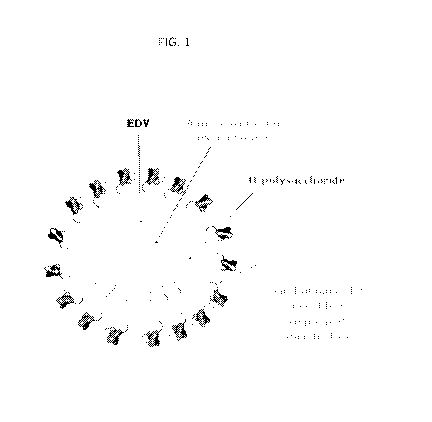
[0027] FIG. 1 is a graphical depiction of an EnGeneIC Dream Vehicle (EDV)
(e.g., a
bacterial minicell) comprising a bispecific antibody for 0-polysaccharide and
human
epidermal growth factor receptor antigens and loaded with the anti-cancer drug
PNU-159682
(an anthracycline analogue).
[0028] FIG. 2 is a graphical depiction of an EnGeneIC Dream Vehicle (EDV)
comprising 0-
polysaccharides on the surface and loaded with immunomodulatory 60mer double
stranded
DNA.
[0029] FIG. 3 is a graphical depiction of an EnGeneIC Dream Vehicle (EDV)
comprising 0-
polysaccharides on the surface and loaded with immunomodulatory alpha
galactosylceramide
(aGC).
[0030] FIG. 4 is a graphical summary of a clinical trial evaluating EGFR-
targeted and
miRNA16a loaded EDVs for treating mesothelioma patients.
[0031] FIG. 5 shows the cytotoxic effect of the indicated chemotherapy drugs
on an A549
lung cancer cell line. FIG. 4A compares the effect of the indicated
chemotherapy drugs with
the supertoxic drug PNU-159682. FIG. 4B compares the effect of doxorubicin and
PNU-
159682.
[0032] FIG. 6 shows the effect of the indicated chemotherapy drugs on adreno-
cortical
cancer cell lines ACCO1 (FIG. 6A) and ACCO7 (FIG. 6B).
[0033] FIG. 7 shows the effect of the indicated chemotherapy drugs on the MDA-
MB-468
breast cancer cell line.
[0034] FIG. 8 shows the effect of the indicated chemotherapy drugs on the
human colorectal
CA 03107095 2021-01-20
WO 2020/021437 PCT/IB2019/056259
9
cancer cell lines Caco-2 (FIG. 8A) and HCT116 (FIG. 8B).
[0035] FIG. 9 shows the effect of the indicated chemotherapy drugs on the
glioblastoma cell
line U87-MG.
[0036] FIG. 10 shows the effect of the indicated chemotherapy drugs on the
human
pancreatic cell lines MiaPaca-2 (FIG. 10A) and gemicitabine-resistant MiaPaca-
2 GemR
cells (FIG. 10B).
[0037] FIG. 11 shows the effect of EDVs targeted to EGFR and loaded with PNU-
159682
(ElEDVs682TM) or doxorubicine (EGFREDvsDoXTM) on A549 xenograft tumor growth
in
mice. Negative controls are saline only or untargeted EDVs loaded with PNU-
159682
(EDVs682Tm). Arrows indicated when the mice were treated with the indicated
saline or EDV
compositions. Asterisks indicate when the mice initially treated with saline
were
administered the EGFREDVs682TM composition
[0038] FIG. 12 shows expression of GAPDH (glyceraldehyde 3-phosphate
dehydrogenase)
(G), KSP (Kinesin Spindle Protein), Plkl (Polo like kinase 1) (P), and RRM1
(ribonucleotide
reductase enzyme 1) (R) relative to GAPDH expression in the indicated NSCLC
cell lines.
[0039] FIG. 13 shows the effect of delivering EGFR-targeted, siRRM1-packaged
EDVs to a
mesothelioma cell line (MSTO, FIG. 13A) or an adreno-cortical cancer cell line
(H295R,
FIG. 13B).
[0040] FIG. 14 shows the effect of delivering EGFR-targeted, miRNA16a
(EGFREDVmaNAmaTm), or EGFR-targeted, siRRM1-packaged EDVs (EGFREDvsiRpJlTM) on
mesothelioma xenograft tumor growth in Balb/c flu/flu mice. Negative controls
were saline
or EGFR-targeted EDVs loaded with scrambled siRNA.
[0041] FIG. 15 shows tumors isolated from mesothelioma xenograft Balb/c
flu/flu mice
treated with EGFR-targeted, miRNA16a (EGFREDVmjRNAl6aTM) or EGFR-targeted,
siRRM1-
packaged EDVs (EGFREDVsaRmiTm), as compared to saline treated or EGFR-targeted
EDVs
loaded with scrambled siRNA.
[0042] FIGS. 16A and 16B show apoptosis induced in adreno-cortical cancer
cells (ACC01)
by EGFR-targeted EDVs loaded with siRNA targeting Polo like kinase 1
(EGFREDVTmsipujm)
and ribonucleotide reductase enzyme 1 (EGFREDyjRpJ.lTM) based on measuring the
number of
CA 03107095 2021-01-20
WO 2020/021437 PCT/IB2019/056259
cellular debris (FIG. 16A) and the ratio of Annexin5 to Propidium Iodide (PI)
positive cells
(FIG. 16B). Apoptosis in untreated ACCO1 cells, in ACCO1 cells treated with
EDVs loaded
with irrelevant siRNA (EGFREDvms uc
fferaseTM), and unloaded EDVs (EGFREDVTM) ) are
included as negative controls.
[0043] FIGS. 17A-D show sub-G1 arrest (Fig. 17D) induced in adreno-cortical
cancer cells
(ACCO1) by EGFR-targeted EDVs loaded with siRNA targeting Polo like kinase 1
(EGFREDVTmsipLKTm) (FIG. 17D) and ribonucleotide reductase enzyme 1
(EGFREDVsaRmiTm)
based on measuring the number of cellular debris and the ratio of Annexin5 to
Propidium
Iodide (PI) positive cells. Apoptosis in untreated ACCO1 cells (Fig. 17A), in
ACCO1 cells
treated with EDVs loaded with irrelevant siRNA (EGFREDVTMsthuciferaseTM) (Fig.
17C), and
unloaded EDVs (EGFREDVTM) (Fig. 17B) are included as negative controls.
[0044] FIG. 18 shows the effect of A549 (lung cancer) xenograft tumor growth
in Balb/c
flu/flu mice treated with: (i) solid triangle = EGFREDVsPNJl59682TM +
EDV54omorTm, (ii) solid
circle = EGFREDvspi59682TM (iii) open square = EGFREDVsPNJl59682TM + EDVs,
(iv) open
triangle = EGFREDvspNu.159682TM EDVssomorTm, and (v) solid square = saline.
The mice were
treated with these EDVs combinations at day 24, 27, 29, 31, 34, 36, and 38
after the
xenograft implantation as indicated with up arrows. On days 36 and 38, the
saline group
mice with tumor volume of ¨ 650mm3 were treated with EGFREDVSPNU-159682TM
EDVssomer
Tm as indicated by the down arrows.
[0045] FIG. 19 shows the effect on A549 (lung cancer) xenograft tumor growth
in Balb/c
flu/flu mice treated with EDVs comprising 40mers (EGFREDVS4Omers TM) in
combination with
EDVs comprising PNU-159682 (EGFREDVsPTM) The triangles indicate treatment
days.
[0046] FIG. 20 shows the effect on A549 (lung cancer) xenograft tumor growth
in Balb/c
flu/flu mice treated with saline (negative control), IFN-y (0.5 x 10⁴ IU per
dose), EGFR-
targeted EDVs loaded with doxorubicine (EGFREDVsDox Tm), and EGFREDVsDox TM
IFN-y.
The triangles indicate treatment days.
[0047] FIG. 21 shows the effect on MBA-MB 468 (breast cancer) xenograft tumor
growth in
Balb/c flu/flu mice treated with saline (negative control), IFN-y (0.5 x 10⁴
IU per dose),
EGFR-targeted EDVs loaded with doxorubicine (EGFREDVsDox Tm), and EGFREDVsDox
Tm +
IFN-y. The triangles indicate treatment days.
CA 03107095 2021-01-20
WO 2020/021437 PCT/IB2019/056259
11
[0048] FIG. 22 shows another experiment illustrating the effect on MDA-MB 468
(breast
cancer) xenograft tumor growth in Balb/c flu/flu mice treated with saline
(negative control),
IFN-y (0.5 x 10⁴ IU per dose), EGFR-targeted EDVs loaded with doxorubicine
(EGFREDvsDoXTM) and EGFREDvsDoXTM + IFN-y. The triangles indicate treatment
days.
[0049] FIG. 23 shows the effect on doxorubicin-resistant A549 xenograft tumor
growth in
Balb/c flu/flu mice treated with saline (negative control, Group 1), EGFR-
targeted EDVs
loaded with doxorubicine (EGFREDV5DOXTM Group 2), EGFREDV5DOXTM + IFN-y (0.75
x 104 IU
per dose) (Group 3), and EGFREDV5DOXTM + IFN-y (0.5 x 10⁴ IU per dose) (Group
4). Mice in
Groups 1-3 received treatment twice per week indicated by the solid triangles.
Mice in
Group 4 were treated three times per week as indicated by open triangles.
[0050] FIGS. 24A-K show a cytokine profile of patients from a First-in-Man
clinical study
where different dosages of EGFREDV5TM loaded with paclitaxel were
administered. (FIG.
24A) = pg/mL of IL-6 measured for each of the 5 doses; (FIG. 24B) = pg/mL of
IL-8
measured for each of the 5 doses; (FIG. 24C) = pg/mL of IL-10 measured for
each of the 5
doses; (FIG. 24D) = pg/mL of TNF-a measured for each of the 5 doses; (FIG.
24E) = pg/mL
of IFN-a measured for each of the 5 doses; (FIG. 24F) = pg/mL of IFN-y
measured for each
of the 5 doses; (FIG. 24G) = pg/mL of IL-113 measured for each of the 5 doses;
(FIG. 2411) =
pg/mL of IL-2 measured for each of the 5 doses; (FIG. 241) = pg/mL of IL-4
measured for
each of the 5 doses; and (FIG. 24J) = pg/mL of IL-12 measured for each of the
5 doses.
Finally, (FIG. 24K) shows the five doses tested.
[0051] FIGS. 25A-K show a cytokine profile of patients from a First-in-Man
clinical study
where different dosages of EGFREDV5TM loaded with doxorubicin were
administered. (FIG.
25A) = pg/mL of IL-6 measured for each of the 8 doses; (FIG. 25B) = pg/mL of
IL-8
measured for each of the 8 doses; (FIG. 25C) = pg/mL of IL-10 measured for
each of the 8
doses; (FIG. 25D) = pg/mL of TNF-a measured for each of the 8 doses; (FIG.
25E) = pg/mL
of IFN-a measured for each of the 8 doses; (FIG. 25F) = pg/mL of IFN-y
measured for each
of the 8 doses; (FIG. 25G) = pg/mL of IL-113 measured for each of the 8 doses;
(FIG. 2511) =
pg/mL of IL-2 measured for each of the 8 doses; (FIG. 251) = pg/mL of IL-4
measured for
each of the 8 doses; and (FIG. 25J) = pg/mL of IL-12 measured for each of the
8 doses.
Finally, (FIG. 25K) shows the additional three doses tested, with the first
five doses being
the same as those shown in (FIG. 24K).
CA 03107095 2021-01-20
WO 2020/021437 PCT/IB2019/056259
12
[0052] FIG. 26 shows signaling pathways of cytosolic DNA sensors with DNA
challenge.
Up to now, many cytosolic DNA sensors have been defined to detect
intracellular double-
stranded DNAs. RNA polymerase III transcribes AT-rich DNAs into RNAs that are
recognized by RNA sensor RIG-I, followed by STING and IRF3 activation. DNA
sensors
DAI, IFI16, DDX41 and LSm14A sense dsDNA directly to activate STING for type I
IFN
production. In the presence of dsDNAs, cGAS catalyzes the synthesis of cGAMP,
a strong
activator of STING. With dsDNAs, LRRFIP1 initiates 0-catenin and IRF3
activation in a
STING-dependent manner. Other DNA sensors prime immune responses independently
of
STING. After recognition of dsDNAs, Sox2 triggers the activation of the
Tab2/TAK1
complex in neutrophils. When detected by dsDNAs, DHX9/36 activates NFKB and
IRF7
through MyD88. DNA sensor Ku70 triggers the activation of IRF1 and IRF7. AIM2
initiates the activation of inflammasome through ASC with DNA binding.
[0053] FIG. 27 shows RAW264.7 cell and bone marrow derived dendritic cell
(BMDC)
activation in response to EDV treatment. (FIG. 27 A) CD86 expression in RAW
cells
incubated directly with 1 g/m1LPS, Ep-EDV, Ep-EDV682, or 682. (FIG. 27 B) CD86
expression in RAW cells co-cultured with 4T1 or CT26Ep12.1 cells treated with
Ep-EDV,
Ep-EDV682, or 682. RAW cells co-cultured with Ep-EDV682 treated tumor cells
resulted in
a significant increase in CD86 expression. (FIG. 27 C) TNFa production in RAW
cell/tumor
cell co-cultures showing a significant increase in TNFa production by RAW
cells incubated
with EDV treated tumor cells. (FIG. 27 D) IL-6 production in RAW cell/tumor
cell co-
cultures showing a significant increase in IL-6 production by RAW cells
incubated with EDV
treated tumor cells. (FIG. 27 E) PCR quantitation of IFNa and IFNf3 expression
in
BMDC/4T1 co-cultures. (FIG. 27 F) PCR quantitation of IFNa and IFN0 expression
in
BMDC/CT26Ep12.1 co-cultures. Quantitation of (FIG. 27 G) CD861-li and MHC
Class IIHi
expression and (FIG. 27 H) CD8OHi expression in BMDC/tumor cell co-cultures.
(FIG. 27 I) Flow cytometic density plots of MHC Class II vs CD86 expression in
BMDC co-
cultures with EDV and drug-treated CT26Ep12.1 cells. ELISA analysis of (FIG.
27 J)
TNFa (FIG. 27 K) IL-12p40 and (FIG. 27 L) IL-6 from the supernatants of
BMDC/tumor
cell co-cultures. Data represents mean s.e.m. and analyzed by one-way ANOVA
and
Tukey's multiple comparison test.
[0054] FIG. 28 shows tumor response and macrophage activation in response to
EDV
CA 03107095 2021-01-20
WO 2020/021437 PCT/IB2019/056259
13
treatment. Tumor growth in response to Ep-EDV and Ep-EDV682 treatment in
BALB/c
mice bearing (FIG. 28 A) 4T1 or (FIG. 28 B) CT26Ep12.1 tumors. Tumor growth in
response to (FIG. 28 C) EDV-682 and EDV-EGFR682 in BALB/C nude mice T84
xenografts and (FIG. 28 D) EDV-682, EDV-EGFRDox, and EDV-EGFR682 in BALB/C
nude mice bearing A549/MDR xenografts. Green arrow indicates where EDV-EGFR682
treatment of formally saline treated mice was begun. Data (FIGS. 28A-D)
represents mean
s.e.m. and analyzed by a two way ANOVA and Tukey's multiple comparison test
(FIG. 28E)
xCELLigence RTCA of CD11b+ isolated from 4T1 tumors and co-cultured with 4T1
cells at
a 5:1 (E:T) ratio. Plot represents normalized cell index which correlates to
cell adhesion and
growth/death vs time. CD11b+ cells from 4T1 tumors undergo and initial
adhesion and
settling phase as indicated by an increase in cell index, followed by growth
or death
represented by an increasing or decreasing cell index. (FIG. 28 F) Ratio of M1
(CD86):
M2 (CD206+) macrophages in 4T1 tumors of treated mice. (FIG. 28 G) xCELLigence
RTCA of CD11b+ isolated from CT26Ep12.1 tumors and co-cultured with CT26Ep12.1
cells
at a 5:1 (E:T) ratio. (FIG. 2811) Ratio of M1 (CD86+): M2 (CD206+) macrophages
in
CT26Ep12.1 tumors of treated mice. Data (FIGS. 28F and 2811) represents mean
s.e.m.
and analyzed by one way ANOVA and Tukey's multiple comparison test.
[0055] FIG. 29 shows NK cell response to EDV treatment. (FIG. 29 A)
xCELLigence
RTCA of NK cells isolated from spleens of mice bearing 4T1 tumors co-cultured
with 4T1
cells at a 20:1 (E:T) ratio. Plot represents cell viability, calculated from
the normalized cell
index, over time. (FIG. 29B) % viability of 4T1 cells co-cultured with NK
cells from saline,
Ep-EDV, or Ep-EDV-682 treated mice 70 h following the addition of NK. (FIG.
29C)
xCELLigence RTCA of NK cells isolated from spleens of mice bearing CT26Ep12.1
tumors
co-cultured with CT26Ep12.1 cells at a 20:1 (E:T) ratio. (FIG. 29D) %
viability of
CT26Ep12.1 cells co-cultured with NK cells from saline, Ep-EDV, or Ep-EDV-682
treated
mice 50 h following the addition of NK cells (FIG. 29E) Expression of NKG2D in
NK cells
(CD45+, CD11b+, DX5+) within 4T1 tumors showing an increase in NKG2D
expression in
Ep-EDV-682 treated mice. Production of (FIG. 29F) RANTES and (FIG. 29G) TNFa
in
co-cultures of NK cells isolated from the spleens of EDV treated mice bearing
4T1 tumors
with 4T1 cells. (FIG. 2911) Quantitation of NKG2D ligands RAE-1, H60a, and
MULT-1 on
the surface of 4 different mouse tumor cell lines. (FIG. 291) xCELLigence RTCA
of NK
CA 03107095 2021-01-20
WO 2020/021437 PCT/IB2019/056259
14
cells isolated from spleens of EpEDV-682 treated mice bearing 4T1 tumors co-
cultured with
4T1 cells at a 20:1 (E:T) ratio in the presence of RAE-1 and/or H60a
inhibiting antibodies
demonstrating that both are important in NK tumor cell cytolysis. (FIG. 29J)
Quantitation of
NK cytolysis 80 h post NK cell addition showing significant inhibition of NK
cytolysis with
H60a antibody alone of in conjunction with RAE-1 inhibiting antibodies. Data
(FIGS. 29B,
29D, 29E-G, and 29J) represents mean s.e.m. and analyzed by one way ANOVA
and
Tukey's multiple comparison test.
[0056] FIG. 30 shows interstitial tumor cytokine/chemokine production and
cytokine
production by splenocyte/tumor cell co-cultures in response to EDV treatment.
ELISA
analysis of interstitial cytokines and chemokines produced in response to Ep-
EDV and Ep-
EDV682 treatment in BALB/c mice bearing a (FIG. 30A) 4T1 or (FIG. 30B)
CT26Ep12.1
tumors. Ep-EDV-682 treatment results in an increase in predominantly Thl
cytokines. Data
represents mean s.e.m. Individual cytokine data analyzed by one way ANOVA
and Tukey's
multiple comparison test. ELISA analysis of (FIG. 30C) TNFa (FIG. 30D) IL-2
(FIG. 30E)
IL-10 (FIG. 30F) IFNy and (FIG. 30G) IL-10 from the supernatants of co-
cultures of
splenocytes isolated from saline, Ep-EDV, and Ep-EDV-682 treated mice bearing
4T1 and
CT26Ep12.1 tumors with their corresponding tumor cells. Data represents mean
s.e.m.
One way ANOVA analysis and Tukey's multiple comparison used to compare groups
+ or ¨
tumor cells. T-test used to compare individual treatments with and without
tumor cells.
[0057] FIG. 31 shows T-cell function and phenotype in response to EDV
treatment. (FIG.
31A) xCELLigence RTCA of CD8+ T-cells isolated from the spleens of mice
bearing 4T1
tumors co-cultured with 4T1 cells at a 30:1 (E:T) ratio. Plot represents
normalized cell index
which correlates to cell adhesion and growth/death vs time. (FIG. 31B) %
viability of 4T1
cells co-cultured with CD8+ T-cells from saline, Ep-EDV, or Ep-EDV-682 treated
mice 30 h
following the addition of CD8+ T-cells. (FIG. 31C) xCELLigence RTCA of CD8+ T-
cells
isolated from the spleens of mice bearing CT26Ep12.1 tumors co-cultured with
CT26Ep12.1
cells at a 30:1 (E:T) ratio. Plot represents normalized cell index which
correlates to cell
adhesion and growth/death vs time. (FIG. 31D) % viability of CT26Ep12.1 cells
co-cultured
with CD8+ T-cells from saline, Ep-EDV, or Ep-EDV-682 treated mice 20 h
following the
addition of CD8+ T-cells. (FIG. 31E) Percentage of CD8+ T-cells (defined as
CD45+, CD3+,
CD8+) detected in 4T1 tumors. (FIG. 31F) Percentage of T-regs (defined as
CD45+, CD3+,
CA 03107095 2021-01-20
WO 2020/021437 PCT/IB2019/056259
CD4+, CD25+) detected in 4T1 tumors. (FIG. 31G) Numbers of T-cells in tumor
draining
lymph nodes of mice bearing 4T1 tumors shown as % of total cells. (FIG. 3111)
%CD80/MEIC Class II expression in dendritic cells in the tumor draining lymph
nodes of
mice bearing 4T1 tumors. (FIG. 311) Confocal image of the interaction between
isolated
CD8+ T-cells from Ep-EDV-682 treated mice with 4T1 cells. Red ¨ actin, green ¨
perforin,
blue (dark) ¨ DAPI; scale bar 10 p.m. Data (FIG. 31B, 31D, and 31E-H)
represents mean
s.e.m. and analyzed by one way ANOVA and Tukey's multiple comparison test
(FIG. 31B,
31D, and 31E-G) or t-test (FIG. 311I).
[0058] FIG. 32 shows Prognostic Indicators and Immunophenotyping of patient
peripheral
blood mononuclear cells (PBMCs) reveals evidence of enhanced antigen
presentation by
dendritic cells and monocytes and elevated cytotoxic CD8+ T cell content at
dose 12.
Prognostic indicators (FIG. 32A) CA19-9 and (FIG. 32B) C-reactive protein
serum levels.
Analysis of PBMC with Duraclone immunophenotyping panels for (FIG. 32C)
Monocytes
and (FIG. 32D) intermediate (CD14+CD16++) antigen presenting monocyte subtype.
Expressed as %Leukocytes. Dendritic cell subtypes including (FIG. 32E) myeloid
dendritic
(Clec9A+) cells (mDC) that drive the CD8+ Effector T cell response and (FIG.
32F)
Plasmacytoid dendritic and myeloid dendritic (professional antigen presenting
DC).
Expressed as %DC or %mDC as indicated. (FIG. 32G) CD8+ T cell subtypes.
Cytotoxic
CD8+ T cells include effectors and exhausted (PD1+) subtypes.
[0059] FIG. 33 shows a schematic of how the EDV first creates an immunogenic
tumor
microenvironment via the delivery of cytotoxic agents directly to the tumor,
then stimulates
the innate immune system either directly or indirectly towards an antitumor
phenotype, and
finally produces an adaptive response in which tumor specific cytotoxic T-
cells arise. (FIG.
33A) Ep-EDV-682 enters the tumor microenvironment via the leaky vasculature
resulting in
tumor cell apoptosis and the release of immune activating DAMPs. (FIG. 33B)
The
interaction of macrophages within the tumor microenvironment via engulfment of
apoptotic
cells or even EDVs directly, results M1 macrophage polarization and release of
inflammatory
cytokines TNFa and IL-6 (FIG. 33C) M1 macrophages are capable of further
lysing tumor
cells and release MIP-la which can recruit additional immune cells. (FIG. 33D)
Immature
dendritic cells engulf apoptotic cell bodies and released tumor antigens which
occur in
response to Ep-EDV-682 treatment and mature releasing type 1 interferons,
TNFa, IL-12p40
CA 03107095 2021-01-20
WO 2020/021437 PCT/IB2019/056259
16
and IL-6. (FIG. 33E) Mature DC then migrate to the lymph node for antigen
presentation to
T-cells. (FIG. 33F) NK cell activation also occurs in the tumor
microenvironment resulting
in release of IFNy and TNFa as well as RANTES to attract additional immune
cells. Further,
activated NK cells effectively lyse tumor cells. (FIG. 33G) The release of
RANTES and
MIP-la recruits additional T-cells, NK cells, and macrophages into the tumor
where (FIG.
3311) tumor specific CD8+ T-cells then contribute the response via tumor cell
lysis. (FIG.
331) All of these steps combine to create and effective antitumor immune
response.
[0060] FIG. 34 shows RAW264.7 and JAWS II cell activation in response to EDV
treatment.
(FIG. 34A) TNFa production by RAW264.7 cells incubated directly with EDV
formulations. (FIG. 34B) IL-6 productions by RAW264.7 cells incubated directly
with
EDV formulations. (FIG. 34 C) Flow cytometric histogram overlays of CD86 and
WIC
Class II expression in JAWS II cells co-cultures with untreated CT26Ep12.1 and
4T1 cells or
those treated with Ep-EDV, Ep-EDV-682, or 682 alone. (FIG. 34D) Quantitation
of CD86
expression as determined via flow cytometry on JAWS II cells co-cultured with
treated tumor
cells. (FIG. 34E) Quantitation of MI-IC Class II expression as determined via
flow
cytometry on JAWS II cells co-cultured with treated tumor cells. Data
represent mean
s.e.m. and are analyzed by one-way ANOVA and Tukey's multiple comparison test.
[0061] FIG. 35 shows body weight changes and macrophage activation in response
to EDV
treatment in Balb/c and Balb/c nude xenografts. % change in body weights of
mice bearing
(FIG. 35A) CT26Ep12.1 (FIG. 35 B) 4T1 (FIG. 35 C) T84 and (FIG. 35 D) A549/MDR
tumors in response to treatment. No more than 5% weight loss is seen with the
initial dose,
and weights then recover and stabilize with subsequent dosing. Data represents
mean
s.e.m. M1/M2 (CD86:CD206) ratio of macrophages in (FIG. 35E) A549/MDR and
(FIG.
35F) T84 tumors of EDV treated mice. (FIG. 35 G) xCELLigence RTCA of CD11b+
isolated from A549/MDR tumors and co-cultured with A549/MDR cells at a 5:1
(E:T) ratio.
Plot represents normalized cell index which correlates to cell adhesion and
growth/death vs
time. (FIG. 3511) % cytolysis of A549/MDR cells co-cultured with CD11b+ cells
from the
tumors of saline or EGFR-EDV-682 treated mice 6.5 h following the addition of
CD11b+
cells. (FIG. 351) Production of MIP-la in co-cultures of CD11b+ cells isolated
from treated
4T1 tumors with 4T1 cells. Data (FIGS. 35E, 35F, 3511, and 351) represents
mean s.e.m.
CA 03107095 2021-01-20
WO 2020/021437 PCT/IB2019/056259
17
and analyzed by one way ANOVA and Tukey's multiple comparison test (FIGS. 35E,
3511,
351) or t-test (FIG. 35F).
[0062] FIG. 36 shows NK cell response to EDV treatment. (FIGS. 36A)
xCELLigence
RTCA of NK cells isolated from spleens of Balb/c nude mice bearing T84 tumors
co-cultured
with T84 cells at a 10:1 (E:T) ratio. Plot represents cell viability,
calculated from the
normalized cell index, over time. (FIG. 36B) Granzyme B production in co-
cultures of NK
cells isolated from spleens of saline and EGFR-EDV-682 treated mice and T84
cells. Data
represents mean s.e.m. and analyzed by t-test. (FIG. 36C) xCELLigence RTCA of
NK
cells isolated from spleens of Balb/c nude mice bearing A549/MDR tumors co-
cultured with
A549/MDR cells at a 10:1 (E:T) ratio. Plot represents cell viability,
calculated from the
normalized cell index, over time (Saline n=5; EGFR-EDV-682 n=4).
[0063] FIG. 37 shows receptor expression and drug sensitivity screening of
patient derived
pancreatic ductal adenocarcinoma cells. (FIG. 37A) EGFR surface receptor
quantitation of
cells from the head of the tumor. (FIG. 37B) EGFR surface receptor
quantitation of cells
from the tail of the tumor. (FIG. 37C) Drug sensitivity and IC50 of first and
second line
chemotherapy drugs/drug combinations as compared to 682 sensitivity.
[0064] FIG. 38 shows single cells from the total PBMC pool (FSC v SSC) were
gated based
on forward scatter width (FSC-W) versus forward scatter area (F SC-A) then
analysed for
CD45 staining (CD45+ gate on FSC v C545+). Cell viability was 96% (Count and
viability
kit #C00162, Beckman Coulter, data not shown) with dead cells excluded based
on a FSC
threshold discriminator of 80. Total dendritic cells (DCs) were gated on
Leukocytes (CD45+)
and defined as HLA-DR+ and Lineage- (#B53351, Beckman Coulter). The lineage
negative
marker was comprised of a pool of antibodies conjugated with the same
fluorophore (PE)
raised against CD3, CD14, CD19, CD20 and CD56 used to negatively select for T
cells,
Monocytes, B cells, and NK cells, respectively. The remaining cells that were
HLA-DR+
were gated as dendritic and subdivided into plasmacytoid DCs (CD11c-CD123+) or
antigen
presenting myeloid DCs (CD11c+CD123-). The myeloid DCs (mDC) were divided into
the
three major subsets, CD1c+ mDC1, CD141+ mDC2 (Clec9A+ shown here) and CD16+
mDCs. CD14+ expression defined the monocytes (#B93604, Beckman Coulter) that
were
gated on leukocytes, then subdivided into classical (CD14+CD16-), intermediate
(CD14+CD16+) and non-classical (CD14+CD16++). T cells (#B53328, Beckman
Coulter)
CA 03107095 2021-01-20
WO 2020/021437 PCT/IB2019/056259
18
were CD3+ and gated on lymphocytes (CD45+SSC low). CD3+ T cell subsets, T
helper
(CD4+) and cytotoxic T (CD8+) were then defined. The PD1+ CD8+ cytotoxic T
cell gate
was plotted against SSC and gated on CD8+ T cells. All FSC and SSC axes are
linear while
fluorescence channel axes (all CD markers) are logarithmic or bi-exponential
(logicle',
Kaluza software, Beckman Coulter).
[0065] FIG. 39 shows Flow Cytometric analysis showing purity of cell
isolations used for
xCelligence RTCA experiments. (FIG. 39A) Isolation of CD11b+ cells from mouse
tumors.
Density plots showing isotype control vs. FSC and CD11b vs. FSC. Samples had
¨80%
purity of CD11b+ cells. (FIG. 39B) Isolation of NK cells from mouse spleens.
Density plots
show isotype control vs. FSC, NKp46 vs. FSC and CD11b vs. FSC. Samples had
¨90%
purity of NK cells. (FIG. 39C) CD8+ T-cell isolation from mouse spleens.
Density plots
show isotype control vs. FSC, CD3e vs. FSC, and CD8 vs. FSC. Samples had ¨90%
purity of
T-cells (CD3e+) with over 98% of those T-cells being CD8+.
[0066] FIG. 40 shows combination treatment using EPminicellDox and minicella-
Gc in a
syngeneic mouse model (EPCT26 colon tumors in Balb/c mice).
[0067] FIG. 41 shows combination treatment of EPminicellDox and minicella-Gc
is effective in
reducing large tumors in Balb/c mice bearing CT26 isograft.
[0068] FIG. 42 shows effect of EPminicellDox and minicella-Gc on tumor
regression in Balb/c
mice with CT26 isograft.
[0069] FIG. 43 shows different sized CT26 isografts treated with (FIGS. 43A
and 43B)
EPminicellDox and minicella-Gc, (FIGS. 43D and 43E) minicella-Gc only, (FIG.
43F)
EPminicellDox only, and (FIG. 43C) saline.
[0070] FIG. 44 shows different sized CT26 isografts treated with EPminicellDox
and minicella-
GC.
[0071] FIG. 45 shows aGC-CD1D presentation of JAWSII Cells Following
minicellaGc
treatment at various time points (FIGS. 45A-E)
DETAILED DESCRIPTION
I. Overview
[0072] The present invention is based on the discovery that compositions
comprising a
CA 03107095 2021-01-20
WO 2020/021437 PCT/IB2019/056259
19
combination of (i) an anti-neoplastic agent and a type I interferon agonist;
(ii) an anti-
neoplastic agent and a type II interferon agonist; or (iii) an anti-neoplastic
agent, a type I
interferon agonis, and and a type II interferon agonist, wherein at least the
anti-neoplastic
agent is packaged within intact bacterially derived minicells, can
synergistically improve
cancer treatment strategies.
[0073] The combination of active agent and immunomodulatory agent(s), where at
least the
anti-neoplastic agent, and optionally the type I and/or type II interferon
agonist is packaged
within intact bacterially derived minicells, results in dramatic efficacy
against cancerous
cells, as well as surprising lack of drug-resistance development in subjects.
The described
compositions avoid the toxicity associated with systemic delivery of anti-
neoplastic drugs
combined with immunomodulatory drugs such as type I and/or type II interferon
agonists to
provide synergistically improved cancer treatment strategies.
[0074] Recent advances in cancer immunotherapy have resulted in unprecedented,
durable
clinical responses in specific cancers (Emens et al., 2017; Farkona et al.,
2016; Oiseth and
Aziz, 2017; Sharma et al., 2017; Ventola, 2017). However, current
immunotherapeutic
strategies have resulted in limited success rates across a variety of tumor
types and a
significant proportion of patients who initially demonstrate encouraging tumor
regression
relapse over time (Emens et al., 2017; Mellman et al., 2011; Oiseth and Aziz,
2017; Sharma
et al., 2017; Ventola, 2017).
[0075] Described in Example 16 below is data elucidating the mechanism of the
cyto-
immuno-therapy function of a tumor-targeted nanocell therapeutic, where it
launches a dual
assault on the tumor via delivery of a super-cytotoxin combined with
engagement of multiple
arms of the immune system. This approach circumvents some of the current
pitfalls with
immunotherapies by creating an immunogenic tumor microenvironment and also
acting on
multiple immune cell subsets thereby avoiding primary and/or adaptive
resistances that may
arise in patients.
[0076] Further, a subset of patients lack tumor immunogenicity resulting from
an absence of
tumor cell antigens or lack of immune cell infiltration and therefore exhibit
no initial
response to the current strategies available (Emens et al., 2017; Oiseth and
Aziz, 2017;
Sharma et al., 2017). Thus, the identification of novel, robust
immunotherapeutic approaches
CA 03107095 2021-01-20
WO 2020/021437 PCT/IB2019/056259
may result in significantly improved clinical outcomes and remains an area of
high priority.
[0077] To mount an effective anti-tumor immune response, certain steps must be
achieved
either spontaneously or therapeutically. First, tumor cell antigens which may
be derived in
situ via tumor cell death, or delivered exogenously must be taken up by
dendritic cells (DC)
(Anguille et al., 2015; Emens et al., 2017; Jung et al., 2018; Mellman et al.,
2011). In
conjunction with antigen uptake, DCs need to receive a proper maturation
signal prompting
differentiation and enhanced processing and presentation of antigens such that
antitumor
function as opposed to tolerance is promoted (Anguille et al., 2015; Emens et
al., 2017; Jung
et al., 2018; Mellman et al., 2011; Simmons et al., 2012). These mature, tumor
antigen
loaded DCs must then effectively generate antitumor T-cell responses which can
occur via
production of tumor specific cytotoxic T-cells, triggering of NK and/or NKT
cell responses,
and enhancing T-helper type 1 responses, among others (Emens et al., 2017;
Fang et al.,
2017; Mellman et al., 2011; Sharma et al., 2017; Zitvogel et al., 2015).
Antitumor T-cells
must finally enter the tumor microenvironment, where immune suppressive
signals may be
present, and effectively perform their antitumor function (Emens et al., 2017;
Mellman et al.,
2011). Problems arising in any of these steps will impede efficacy of an
immunotherapeutic,
and can even result in total failure of the therapy (Emens et al., 2017;
Mellman et al., 2011;
Sharma et al., 2017).
[0078] Currently, the immunotherapeutic strategies which have received the
most attention
clinically include immunological checkpoint inhibitors and chimeric antigen
receptor T-cell
therapy (CAR-T) (Emens et al., 2017; Mellman et al., 2011; Oiseth and Aziz,
2017; Sharma
et al., 2017; Ventola, 2017). Checkpoint inhibitors such as cytotoxic T
lymphocyte antigen 4
(CTLA-4), and programmed cell death 1/programmed cell death 1 ligand (PD-1/PDL-
1)
function by blocking the transmission of immune-suppressive signals and direct
stimulation
to activate cytotoxic T lymphocytes within the tumor microenvironment (Dine et
al., 2017;
Jenkins et al., 2018; Sharpe, 2017). Inhibitors of these pathways have shown
dramatic
clinical results in specific cancers, but overall response rates across
different cancers remains
low (-15-25%) and immune related toxicities associated with these therapies
can be high
(Dine et al., 2017; Emens et al., 2017; Jenkins et al., 2018; Sharpe, 2017;
Ventola, 2017).
With new checkpoints continually being discovered as potential immune targets,
it is
apparent that tumors are capable of exploiting an elaborate and diverse set of
immune-
CA 03107095 2021-01-20
WO 2020/021437 PCT/IB2019/056259
21
suppressive pathways ( Dine et al., 2017; Emens et al., 2017; Farkona et al.,
2016; Jenkins et
al., 2018; Sharpe, 2017). Thus, development of resistance to checkpoint
inhibitors continues
to be a hurdle and attempts are being made to utilize combinations of more
than one
checkpoint inhibitors to overcome these issues, though this often exacerbates
associated
toxicities (Dine et al., 2017; Jenkins et al., 2018; Sharma et al., 2017;
Ventola, 2017).
[0079] The second therapy receiving widespread attention is CAR-T cell therapy
which
entails the genetic engineering of a patient's T-cells to express membrane
fusion receptors
with defined tumor antigen specificities and capable of eliciting robust T-
cell activation to
initiate killing of the target tumor cells (D'Aloia et al., 2018'; Farkona et
al., 2016; Mellman
et al., 2011; Sharma et al., 2017). This therapeutic approach has produced
unprecedented
clinical outcomes in the treatment of "liquid" hematologic cancers, but to
date has not
produced comparable responses in targeting solid malignancies due to
limitations associated
with the lack of a good specific antigen target, poor homing to the tumor,
poor extravasation
into the tumor, and lack of persistence within a hostile tumor
microenvironment (D'Aloia et
al., 2018'; Sharma et al., 2017). Practical limitations relating to the
availability of
lymphocytes from heavily pre-treated patients and long manufacturing times and
are not a
feasible treatment option for patients with rapidly advancing disease are also
present (Oiseth
and Aziz, 2017; Rezvani et al., 2017).
[0080] The EnGeneIC Dream Vector (EDV) is a bacterially-derived delivery
system
consisting of nonviable nanocells that are 400 20 nm in diameter, generated
by reactivating
polar sites of cell division in bacteria (MacDiarmid et al., 2007b). It has
been demonstrated
that these nanocells can be packaged with a cytotoxic drug, siRNA, or miRNA
and
specifically targeted to tumor cell-surface receptors via attachment of
bispecific antibodies to
the surface polysaccharide of the nanocells (MacDiarmid et al., 2009;
MacDiarmid et al.,
2007b; Reid et al., 2013). Post-intravenous administration in mouse and dog
studies has
demonstrated that they are retained in the vascular system due to their size,
but then rapidly
extravasate into the tumor via the tumor-associated leaky vasculature
(MacDiarmid et al.,
2007b; Sagnella et al., 2018). Post-tumor cell-surface receptor engagement via
the associated
bispecific antibody results in macropinocytosis into endosomes and release of
the payload via
degredation intracellularly in the lysosomes (MacDiarmid et al., 2009;
MacDiarmid et al.,
2007b; Sagnella et al., 2018). The safety of these nanocell therapeutics has
been
CA 03107095 2021-01-20
WO 2020/021437 PCT/IB2019/056259
22
demonstrated in three Phase I clinical trials with over a thousand doses
administered in
various end-stage cancer patients and 682 loaded EDVs are currently being
delivered to
patients in a phase I trial and to date have shown a promising safety profile
(2017; Kao et al.,
2015; Solomon et al., 2015; van Zandwijk et al., 2017; Whittle et al., 2015).
A. Overview of bacterial minicell delivery methods
[0081] The use of bacterial minicells to deliver chemotherapeutic agents to
cancer cells has
previously been described. This delivery method to treat cancer packages a
toxic
chemotherapy agent or drug, or functional nucleic acid, into a bacterially-
derived minicell,
which are typically about 400 nm in diameter. Typically, the minicell carries
an antibody
targeting specific cancer cells. The antibodies attach to the surface of
cancer cells and the
minicell is internalized by the cancer cell. In this way, the toxic
chemotherapy agents are not
widely distributed throughout the body, and therefore reduce the chance of
side effects and
intolerability as the toxic drug or compound is delivered inside the cancer
cell. Using
antibody-targeted minicells as a delivery vehicle for toxic chemotherapy
agents results in
much less drug needed to kill the cancer cell, thus improving the therapeutic
index.
[0082] Indeed, the present inventors have shown that minicells (or EnGeneIC
Dream
Vehicles, EDVs) can deliver chemotherapy drugs, such as paclitaxel or
doxorubicin, to
xenograft tumors in mice (Example 1), dogs (Example 2), and monkeys (Example
3). The
targeted delivery ensures that the cancer cells receive most of the
chemotherapeutic agent,
resulting in a low level of toxicity. See Examples 1-3; see also MacDiarmid et
al., 2007b;,
MacDiarmid et al., 2007a; MacDiarmid et al., 2009;, and MacDiarmid et al.,
2016.
Furthermore, the minicells do not induce a significant immune response in the
xenograft
models, and the minicells are well tolerated (Example 4). Thus, intact
bacterially derived
minicells are a well-tolerated vehicle for delivering anti-cancer drugs to
patients, with
examples including doxorubicin targeted to advanced solid tumors (Example 5),
doxorubicin
targeted to glioblastoma (Example 6), and MicroRNA-16a targeted to
mesothelioma
(Example 7).
[0083] These treatment strategies did not result in complete remission or cure
of all cancers
in all patients, however. Accordingly, there is a need for improved cancer
treatment
therapies. The present inventors discovered that using a combination of
minicells having
CA 03107095 2021-01-20
WO 2020/021437 PCT/IB2019/056259
23
three different types of payloads produced surprisingly dramatic and effective
clinical
efficacy.
[0084] Specifically, the present inventors discovered that minicells
comprising a
chemotherapy agent (in the examples below, for instance, the agent is PNU-
159682, a
supertoxic chemotherapy drug) combined with minicells comprising an interferon
type I
agonist and/or an interferon type II agonist resulted in synergistic anti-
tumor effects and was
well-tolerated by a patient suffering from late stage pancreatic cancer. See
Example 12. In
fact, the late-stage pancreatic cancer patient exhibited markedly improved
quality of life after
this treatment, which is remarkable for a patient at that stage. This triple
or duel combination
strategy provides synergistically improved treatment of cancers, particularly
late-stage
terminal cancer. The inventors also discovered that minicells comprising a
chemotherapy
agent combined with minicells comprising an interferon type II agonist
resulted in synergistic
anti-tumor effects.
[0085] It was also surprisingly discovered that a dual combination of a
minicell packaged
antineoplastic agent, in combination with a type II interferon agonist, and in
the absence of a
type I interferon agonist, resulted in dramatic efficacy against large sized
tumors. Such
results have not previously been described. It is theorized that in some
patients, combining a
type I interferon agonist and type II interferon agonist may be
counterproductive, as the two
types of interferon agonists may compete rather than synergistically act. This
data is
described in more detailed below.
[0086] The following description outlines the invention related to these
discoveries, without
limiting the invention to the particular embodiments, methodology, protocols,
or reagents
described. Likewise, terminology used here describes particular embodiments
only and does
not limit the scope of the invention.
B. Summary of the experimental results
(1) Minicell packaged antineoplastic agent in combination
with minicell packaged type I interferon agonist
[0087] In a first embodiment, described are compositions and methods relating
to a
combination of a bacterial minicell packaged antineoplastic agent in
combination with a type
I interferon agonist packaged in a bacterial minicell.
CA 03107095 2021-01-20
WO 2020/021437 PCT/IB2019/056259
24
[0088] Example 11 and FIG. 18 describe data showing the results in lung cancer
xenograft
models in mice treated with various minicell (EDVs) compositions, as
summarized in the
table below. The animals of Groups 1 and 5 were administered a combination of
a
chemotherapeutic agent (PNU 159682) packaged in an intact bacterially derived
minicell and
a type I interferon agonist (a 40mer double stranded DNA or a 50mer double-
stranded DNA),
also packaged in an intact bacterially derived minicell. All of the minicell
compositions
resulted in stabilization of tumor growth. However, the most dramatic results
were obtained
after treating the large tumor size that resulted from saline treatment in
part 1 of the
experiment. When the saline-treated control group was subsequently treated in
part 2 of the
experiment with a composition comprising a combination of minicell-packaged
anti-
neoplastic agent plus a minicell-packaged type I interferon agonist, the tumor
size was
reduced by 62% over a 5-day period.
Table 1
Group Treatment Figure Phase I Phase II Results
Results Treatment
Starting at days
36 and 38
1 EGFREDvspNu_ Fig. 18, solid Tumor growth
159682 + triangle stabilization
EDVS4Omer
2 EGFREDvspNu_ Fig. 18, solid Tumor growth
159682 circle stabilization
3 EGFREDvspNu_ Fig. 18, open Tumor growth
159682 + EDVs square stabilization
(no payload)
4 EGFREDvspNu_ Fig. 18, open Tumor growth
159682 + triangle stabilization
EDVS5Omer
Saline Fig. 18, solid tumor growth up Treatment with In 5 days,
tumors
square to a volume of ¨ EGFREDVspNu-159682 having a
large
650mm3 + EDV54Omer volume of ¨
650mm3 decreased
to ¨250 mm3 ¨ or a
62% reduction in
size in 5 days
[0089] In a follow-up of Example 11 (results shown in FIG. 19), the addition
of a type I
interferon agonist packaged in a minicell resulted in dramatic tumor size
reduction, which
was not seen when an anti-neoplastic agent packaged in a minicell was used in
the absence of
the type I interferon agonist adjuvant. The results are summarized in the
table below. These
CA 03107095 2021-01-20
WO 2020/021437 PCT/IB2019/056259
results clearly demonstrate the adjuvant effects on minicell-packaged anti-
neoplastic agents
with the addition of a minicell-packaged type I interferon agonist.
Table 2
Group Treatment Figure Results
1 EGFREDVspNu- Fig. 19, solid circle Slight tumor size reduction
(from a tumor
159682 volume of about 275 mm3 to 260 mm3)
2 solid triangle = Fig. 19, solid triangle
Significant tumor reduction, from a tumor
EGFREDue
v PNU- volume about 275 mm3 to about 175
mm3)
159682 EDV54Omer
3 Saline Fig. 19, solid square Significant tumor growth.
(ii) Minicell packaged antineoplastic agent in combination with
minicell packaged type I interferon agonist and optionally a
type II interferon agonist (not minicell packaged)
[0090] A second embodiment is directed to methods and compositions utilizing a
minicell
packaged antineoplastic agent combined with a mincell-packaged type I
interferon agonist or
a minicell-packaged anti-neoplastic agent in combination with a mincell-
packaged type I
interferon agonist, and a type II interferon agonist (free from a bacterial
minicell).
[0091] Further evidence of the dramatic and surprising effectiveness of the
compositions of
the invention is reflected in the clinical results shown in Example 12.
Specifically, Example
12 relates what happened when patients suffering from advanced solid tumors
were treated
with compositions comprising (1) a combination of a minicell-packaged anti-
neoplastic agent
and a mincell-packaged type I interferon agonist; and (2) a combination of a
minicell-
packaged anti-neoplastic agent, a mincell-packaged type I interferon agonist,
and a type II
interferon agonist.
[0092] In particular, the human clinical data detailed in Example 12
demonstrate the safety
profile of type I and type II IFN agonists used as adjuvants for minicell-
packaged anti-
neoplastic agents in human patients. See data in Table 3 below, in relation to
which the type I
interferon agonist packaged in intact, bacterially-derived minicells was 40mer
double-
stranded DNA (EDV54omer) or 60mer double-stranded DNA (EDV56omer).
[0093] Moreover, the results obtained with a stage 4 pancreatic cancer
patient, who had
exhausted all other treatment options, were remarkable. The levels of the
patient's tumor
marker (CA 19-9) dropped by more than 90% after the initial three doses,
equivalent to only
10 days of treatment. After ten doses this had dropped even further, with an
almost 95%
CA 03107095 2021-01-20
WO 2020/021437 PCT/IB2019/056259
26
reduction in tumor marker levels. The patient also demonstrated significant
weight gain, in
contrast to the cachexic state experienced by most patients presenting with
stage IV
pancreatic cancer, and reported a marked improvement in quality of life. These
results are
dramatic, particularly given the poor prognosis associated with advanced
pancreatic cancer.
[0094] In summary, five patients received a total of 69 doses of EGFR(V)ur"T
v SPNU/Dox or
EGFRooEDvspNu + EDV54omed60mer, (type I IFN agonist) Imukin (type II IFN
agonist). The
treatments were well-tolerated, and the addition of immunomodulatory adjuvants
did not
seem to change the safety profile of single-agent-loaded and targeted EDVs.
Table 3
Patient # Treatment Cancer Comments
3 patients EGFR(V)crIA
v 3PNU-159682 at 2.5 advanced solid Treatment was well
tolerated, no
X 109 + EDVsaomer at 5 x tumors unexpected adverse reactions.
One
108 patient was ultimately
withdrawn
from the study due to dose-limiting
toxicity.
1 patient EGFR(v)EDVSPNU-159682 and Stage IV pancreatic
Treatment was well-tolerated; levels
EDVS4Omer or EDV56Omer cancer of the patient's tumor marker
(CA 19
And 9) dropped by more than 90%
after
ITG-targeted EDVs loaded the first 3 doses, equivalent
to only 10
with PNU-159682 days of treatment. After 10
doses this
(aG(609)EDvsp) had dropped even further, with
an
almost 95% reduction in tumor
marker levels.
1 patient EGFR(v)EDVSpNu and recurrent and end- Treatment was well
tolerated.
EDVsoomer + Imukin (type stage adreno-cortical
II IFN agonist) cancer with a very
heavy tumor burden
(iii) Minicell packaged antineoplastic agent in
combination with type II interferon agonist
[0095] Example 13 describes the results of various studies conducted to
evaluate the
effectiveness of combining anti-neoplastic agents packaged in minicells with a
type II
interferon agonist, e.g., IFN-y. The results show that the addition of a type
II interferon
agonist augments or enhances the anticancer effect of anti-neoplastic agents
packaged in
minicells in xenograft models of various cancers, including lung cancer and
breast cancer.
Further, the data set forth in Example 13 and excerpted in Table 4 below
demonstrate that the
addition of a type II interferon agonist to a composition comprising an
antineoplastic agent
packaged in a minicell in the treatment of tumors normally resistant to the
antineoplastic
agent alone is essential to achieve tumor stabilization. Thus, combining a
minicell-packaged
CA 03107095 2021-01-20
WO 2020/021437 PCT/IB2019/056259
27
antineoplastic agent with a type II interferon agonist can overcome drug
resistance.
Table 4
Cancer Treatment Figure Results
Lung Group 1 = sterile FIG. 20, open diamonds Significant tumor
growth
cancer physiological saline
Group 2 = IFN-y (0.5 x 10⁴ FIG. 20, solid triangles no anti-tumor
efficacy
IU) per dose
= EGFREDVsnox Group 3 FIG. 20, solid squares tumor stabilisation
Group 4 = EGFREDVspo,, and FIG. 20, solid circles highly significant
tumor
IFN-y (0.5 x 10⁴ IU) per dose regression by day 43
after a
total of 6 doses
Breast Group 1 = sterile FIG. 21, open diamonds Significant tumor
growth
cancer physiological saline
Group 2 = IFN-y (0.5 x 10⁴ FIG. 21, solid triangles no anti-tumor
efficacy
IU)
Group 3 = , EGFREDv
v oDox FIG. 21, solid squares tumor stabilisation
of breast
cancer xenografts, but by
¨day 25 the tumors began to
grow again, likely due to
development of resistance to
doxorubicin
Group 4 = EGFREDVspox and FIG. 21, solid circles highly significant
tumor
IFN-y (0.5 x 10⁴ IU) per dose regression, and by day
30,
after a total of 6 doses, these
tumors were more like scar
tissue
(iv) Triple combination of a minicell-packaged antineoplastic
agent, a
minicell-packaged type I interferon agonist, and a type II
interferon agonist (either alone or minicell-packaged)
[0096] The present inventors also discovered that a triple combination of a
minicell-packaged
antineoplastic agent, a minicell-packaged type I interferon agonist, and a
type II interferon
agonist (either alone or minicell-packaged) can produce dramatic anticancer
effects.
Specifically, Example 14 details treatment of dogs with late stage endogenous
tumors (brain
cancer, sarcoma, or melanoma) with a combination of a minicell-packaged
antineoplastic
agent, a minicell-packaged type I interferon agonist, and a type II interferon
agonist. The
results show that the combination composition was well-tolerated. Moreover, in
6 of 7
evaluable animals (85.7%) the disease was stabilized, although one dog
achieved a near
partial response (29.8% reduction in tumor size).
CA 03107095 2021-01-20
WO 2020/021437 PCT/IB2019/056259
28
(v) Duel combination of a
minicell-packaged antineoplastic agent in
combination with a minicell-packaged type II interferon agonist, in
the absence of a type I interferon agonist
[0097] In another embodiment, this invention relates to the surprising
discovery that
compositions comprising a combination of a minicell-packaged antineoplastic
agent and a
minicell packaged type II interferon agonist, such as for example alpha-
galactosyl ceramide
(a-GC), and in the absence of a type I interferon agonist, demonstrates
surprising anticancer
efficacy.
[0098] In particular, Example 23 describes data illustrating the efficacy of a
dual
combination of minicell contained therapeutic and minicell contained
interferon type II
agonist against tumors. This result demonstrates that compositions lacking
interferon type I
agonists can be used to effectively treat tumors. See also, FIGS. 40 and 42.
The
experimental results showed a marked halt in tumor progression for combination
treatment
groups receiving 4minicellDox+ minicella-Gc (interferon type II agonist) as
compared to saline
and EPminicellDox treatments. This result supports the theory of an immune
adjuvant effect by
the addition of minicella-Gc treatment to EPminicellDox
[0099] Further data showed that saline treated control tumors demonstrated
dramatic tumor
regression following a treatment change to drug and a-GC EDV mediated dual
combination
therapy (FIG. 41); e.g., a combination of minicell packaged antineoplastic
agent and minicell
packaged type II interferon agonist. In particular, tumours that had reached
800mm3 dropped
to below 600mm3 in 3 days before the experiment was terminated¨ a markedly
dramatic
tumor size reduction (¨ 25%) in a short period of time. The ability for the
dual combination
composition to dramatically decrease large tumors in a short period of time
was not known
prior to the present invention.
[0100] In one embodiment of the invention, the dual combination composition
(e.g., a
minicell packaged antineoplastic agent in combination with a minicell packaged
interferon
type II agonist) can reduce a tumour's size, including the size of a large
tumor, by about 5%,
about 10%, about 15%, about 20%, about 25%, about 30%, about 35%, about 40%,
about
45%, about 50%, about 55%, about 60%, about 65%, about 70%, about 75%, about
80%,
about 85%, about 90%, about 95%, or about 100%. The reduction in tumor size
can be
measured over any suitable time period, such as about 3 days, about 5 days,
about 1 week,
CA 03107095 2021-01-20
WO 2020/021437 PCT/IB2019/056259
29
about 2 weeks, about 3 weeks, about 1, about 2, about 3, about 4, about 5,
about 6, about 7,
about 8, about 9, about 10, about 11, about 12 months, about 1.5 years, about
2 years or
longer.
C. Immunotherapy data
[0101] Example 16 details data demonstrating that minicell packaged
antineoplastic agents
targeted to a tumor cell surface receptor function as a cancer immunotherapy,
e.g., as a cyto-
immunotherapy. In particular, the example illustrates the ability of the
bacterial minicell to
activate cells of the innate immune system, including macrophages, NK cells
and dendritic
cells. This is followed by dendritic cell maturation and antigen presentation
leading to an
adaptive T-cell response in which tumor specific cytotoxic T-cells are
produced and results in
further recruitment of additional immune cells to the tumor microenvironment.
This
approach circumvents some of the current pitfalls with immunotherapies by
creating an
immunogenic tumor microenvironment and also acting on multiple immune cell
subsets
thereby avoiding primary and/or adaptive resistances that may arise in
patients.
[0102] This example therefore shows the ability of the bacterial minicell to
deliver a
cytotoxic drug within tumor cells and to also simultaneously elicit an innate
and adaptive
immune response specifically targeting the tumor.
[0103] Further immunotherapy data is shown in Example 18, which describes data
showing
that NK cells adopt an antitumor phenotype in vivo following treatment with
targeted
minicells comprising an antineoplastic agent. This is significant as NK cells
are the primary
effector cell of the innate immune system and are tightly regulated by a
balance of activating
and inhibitory signals (Morvan and Lanier, 2016; Wallace and Smyth, 2005).
Impairment of
NK cell function has been associated with increased tumor incidence, growth,
and metastasis,
and thus its importance in contributing to an antitumor immune response is
well documented
(Fang et al., 2017; Morvan and Lanier, 2016; Rezvani et al., 2017; Wallace and
Smyth,
2005).
[0104] Interesting, Example 19 details data showing that a predominantly Thl
cytokine
response within a tumor microenvironment is exhibited following treatment with
a minicell-
encapsulated antineoplastic agent (e.g., PNU-159682). Cytokine and chemokine
production
within a tumor microenvironment allows immune cells to effectively communicate
with each
CA 03107095 2021-01-20
WO 2020/021437 PCT/IB2019/056259
other to generate a coordinated response which can either be tumor promoting
or suppressing
(Belardelli and Ferrantini, 2002; Lee and Margolin, 2011). The effect of
individual cytokines
on immune response is dependent on a variety of factors including local
concentration,
cytokine receptor expression patterns and the activation state of surrounding
cells (Lee and
Margolin, 2011). Thus, many cytokines have been shown to be capable of
eliciting opposing
effects on tumor growth (Dredge et al., 2002; Landskron et al., 2014; Lee and
Margolin,
2011).
[0105] Further, Example 20 details data showing that treatment with a minicell-
encapsulated
antineoplastic agent (e.g., PNU-159682) results in the production of tumor
specific CD8+ T-
cells. Initial in vitro experiments indicated that EDV treatment can result in
dendritic cell
maturation either via direct interaction or as a result of cell death in
response to a targeted
EDV loaded with an effective chemotherapeutic. Thus, this experiment aimed to
examine if
this result could translate to DC maturation and antigen presentation in vivo
resulting in the
production of tumor specific CD8+ cytotoxic T-cells. The resulting data
demonstrated that
minicell treatment successfully elicited the production of tumor specific CD8+
T-cells. In
addition, a significant increase in overall T-cell numbers (CD3+) as well as a
significant
increase in both CD4+ and CD8+ T-cells numbers were seen in the lymph nodes of
mice
treated with minicell encapsulated antineoplastic agent (e.g., PNU-159682)
(FIG. 31G). A
significant increase in mature dendritic cells in the lymph nodes of treated
mice was also
detected (FIG. 3111), and visualization of the interaction between isolated
CD8+ T-cells from
treated mice with 4T1 cells shows that these T-cells are capable of attaching
to and expelling
perforin (green) into the tumor cell (Figure 311).
[0106] Example 21 demonstrates the ability of targeted bacterial minicells
loaded with an
antineoplastic agent (e.g., the super-cytotoxin PNU-159682) to not only
effectively deliver
this drug to the tumor site, but also behave as an immunotherapeutic by
stimulating multiple
immune cell subsets. The example demonstrates the ability of a minicell
capsulated
antineoplastic agent treatment to push immune cell subsets, including
macrophages, NK cells
and CD8+ T-cells, towards an antitumor phenotype capable of effectively
eliminating tumor
cells. When combined with the effectiveness of an antineoplastic agent, this
results in a dual
assault on the tumor.
[0107] While the idea of cancer immunotherapy has been around for decades, it
is only in
CA 03107095 2021-01-20
WO 2020/021437 PCT/IB2019/056259
31
recent times that its potential has begun to be realized with the approval of
a number of
immunotherapies (Farkona et al., 2016; Ventola, 2017). Bacterial minicells
represent a
unique, combined cyto-immunotherapy which first creates an immunogenic tumor
microenvironment via the delivery of cytotoxic agents directly to the tumor,
where it
stimulates the innate immune system either directly or indirectly towards an
antitumor
phenotype. This innate immune activation then triggers an adaptive response in
which tumor
specific cytotoxic T-cells arise (FIG. 33).
[0108] Following intravenous administration, the minicell extravasates to a
tumor via the
tumor's leaky vasculature where > 30% of the administered dose of targeted
minicells
carrying their toxic payload deposit directly into the tumor microenvironment
within a 2hr
period (MacDiarmid et al., 2007b). Targeted bacterial minicells bind to
receptors on the
tumor cells (4T1 and CT26Ep12.1 in the case of Example 21), and are then
internalized
effectively delivering their payload (antineoplastic agent) directly within
the tumor cells.
PNU-159682 is a highly potent super cytotoxin resulting in rapid apoptosis
within 24h of
being delivered to the tumor cells (FIG. 33A). The apoptotic cells and DAMP
signals
produced by bacterial minicell (e.g., Ep-EDV-682) treatment can then interact
with innate
immune cells such as tumor associated macrophages (TAMs) and stimulate
upregulation of
CD86 and the production of Thl pro-inflammatory cytokines such as TNFa and IL-
6 (FIG.
33B). These changes are typical of M1 polarization of macrophages which are
capable of
lysing tumor cells and releasing cytokines to signal activation of other
immune cell subsets,
and have thus been shown to possess antitumor characteristics (Sawa-Wejksza
and Kandefer-
Szerszen, 2018; Yuan et al., 2015).
[0109] Furthermore, the bacterial minicell itself can also interact directly
with TAMs
producing a similar M1 polarization, albeit this would be expected to occur at
very low levels
in the current system. TAMs are generally the most abundant immune cell in the
tumor
microenvironment, and it has been demonstrated that increased numbers of TAMs
are
associated with poor prognosis and increased tumor growth (Sawa-Wejksza and
Kandefer-
Szerszen, 2018). This is due in large part to the fact that TAMs mostly
consist of anti-
inflammatory M2 macrophages which have been shown to possess tumor promoting
characteristics, whereas inflammatory M1 macrophages exhibit antitumor
characteristics
(Sawa-Wejksza and Kandefer-Szerszen, 2018; Yuan et al., 2015). Example 21
demonstrates
CA 03107095 2021-01-20
WO 2020/021437 PCT/IB2019/056259
32
the ability of bacterial minicell treatment to shift the M1:M2 balance within
the tumor
microenvironment in 4 different tumor models. Despite differences in the
degree of this shift
in the different tumor models, it was shown that the increase in M1
polarization translated to
increased tumor cell lysis by TAMs isolated from the tumors of mice which had
been treated
with bacterial minicells. In addition to the phenotypic shifts to Ml, TAMs
from tumors of
bacterial minicell treated mice also secreted an increased amount of MIP-la
(FIG. 33C), a
chemokine which has been established to play a role in promoting immune cell
recruitment,
and in particular tumor infiltration by NK cells, CD4+ T-cells and CD8+ T-
cells (Allen et al.,
2018).
[0110] In addition to TAM activation, immature dendritic cells (DC) interact
either directly
with bacterial minicells, or more likely, with the apoptotic cells and DAMP
signals produced
by bacterial minicell treated tumors resulting in dendritic cell maturation
and migration to the
lymph nodes for antigen presentation. DCs have been explored as a potential
target in cancer
immunotherapies as they are known to be the most effective antigen presenting
cell and
constitute the bridge between the innate and adaptive immune system (Allen et
al., 2018).
[0111] Most current strategies for DC based immunotherapy involve ex vivo
manipulation
and priming of DCs or DC precursors, however success from this strategy has
been limited
due to a variety of factors including: development of immune tolerance,
induction of
insufficient numbers of CD8+ cytotoxic T-cells (CTL) or those with poor
antitumor efficacy,
and the suppressive nature of the tumor microenvironment (Anguille et al.,
2015; Jung et al.,
2018; Landskron et al., 2014; Oiseth and Aziz, 2017). Bacterial minicell
treatment allows for
in vivo priming and maturation of DCs within the tumor microenvironment in
response to
dying tumor cells (FIG. 33D). Immature DCs are capable of engulfing DAMPs
and/or
apoptotic tumor cell bodies produced in response to targeted, drug loaded
bacterial minicells.
These DAMPs and dying tumor cells are then processed for antigen presentation
on the DC
surface via MHC Class I and II molecules, with concomitant DC maturation.
Upregulation of
the co-stimulatory molecules CD86, CD80 and MHC Class II, which have been
identified as
markers of the DC maturation process, was shown to occur in DCs co-cultured
with bacterial
minicell treated tumor cells, along with an increase in the percentage of
mature DCs detected
in the tumor draining lymph nodes of bacterial minicell treated mice (Anguille
et al., 2015;
Cauwels et al., 2018; Simmons et al., 2012). During the maturation process,
the DCs migrate
CA 03107095 2021-01-20
WO 2020/021437 PCT/IB2019/056259
33
to the tumor draining lymph nodes for antigen presentation to T-cells thereby
increasing
production of CD4+ T-helper cells and tumor specific CD8+ CTL initiating an
adaptive
immune response to the tumor (FIG. 33E). An increase in the production of
IFNa/f3, TNFcc,
IL-12p40, and IL-6 by DCs co-cultured with bacterial minicell treated tumor
cells was
subsequently detected, in addition to a significant increase in IFNa
concentration in the tumor
microenvironment observed in both the 4T1 and CT26Ep12.1 tumor models (Example
21).
Expression levels of type 1 IFNs (IFNa/f3) and IFN stimulated genes within the
tumor
microenvironment have been shown to correlate with favorable disease outcomes
and may in
fact even be necessary for the success of cancer therapies including
immunotherapies
(Cauwels et al., 2018; Fitzgerald-Bocarsly and Feng, 2007; Zitvogel et al.,
2015). The
antitumor activity of type 1 IFNs arise indirectly via immune cell activation
of DCs, T and B
lymphocytes, NK cells, and macrophages (Cauwels et al., 2018; Fitzgerald-
Bocarsly and
Feng, 2007; Showalter et al., 2017; Zitvogel et al., 2015).
[0112] In conjunction with enhancing macrophage and DC antitumor functions,
treatment
with bacterial minicells comprising an antineoplastic agent is capable of
eliciting NK cell
activation leading to increased cytotoxicity (FIG. 33F). NK cells possess the
inherent ability
to lyse malignant cells in an antigen independent manner, thus their
activation and functional
status must be tightly controlled in order to avoid potentially adverse
effects on the host. The
ability to attract NK cells to and activate NK cells within the tumor
microenvironment is vital
to their ability to exert their antitumor function. Cytokines including IL-2,
IFNy, and IFNa
which are significantly increased in the microenvironment of Ep-EDV-682
treated tumors,
are known to activate NK cells towards both increased cytokine production and
enhanced
cytolytic function (Fang et al., 2017; Ferlazzo and Munz, 2004; Lee and
Margolin, 2011;
Morvan and Lanier, 2016; Rezvani et al., 2017). In fact, evidence indicates
that type 1 IFNs
are required for the activation of NK cell cytotoxicity (Ferlazzo and Munz,
2004; Muller et
al., 2017). Further, type 1 IFNs are capable of inducing cellular senescence
followed by
upregulation of NKG2D ligand expression in tumor cells thereby promoting their
elimination
by NK cells (Muller et al., 2017). Upregulation of the NKG2D receptor was
observed on NK
cells within the tumors of mice treated with Ep-EDV-682, and this receptor was
demonstrated
to contribute significantly to the cytolytic ability of NK cells isolated from
Ep-EDV-682
treated mice. Moreover, immature, intermediate and mature mouse NK cells
express both the
CA 03107095 2021-01-20
WO 2020/021437 PCT/IB2019/056259
34
CCR1 and CCR5 chemokine receptors that can bind the chemokines MIPla and
RANTES,
both of which are upregulated in Ep-EDV-682 treated tumors as well as by
macrophages and
NK cells from Ep-EDV-682 treated mice (Bernardini et al., 2016).
[0113] Chemokines, such as MIPla and RANTES, are responsible for the further
recruitment
of helper and effector immune cells including NK cells, macrophages, and T-
cells to the
tumor microenvironment (FIG. 33G) (Allen et al., 2018; Bernardini et al.,
2016; Zibert et
al., 2004). Following the initial innate immune response due to EDV treatment
which
encompasses macrophages, NK cells, and DCs, an adaptive immune response is
mounted in
which tumor specific CTLs and T-helper cells are produced and then recruited
to the tumor
site (FIG. 3311). Tumor specific CTLs then target and lyse tumor cells further
contributing
to the overriding antitumor environment which has been created by the other
immune cell
subsets in combination with the targeted, drug loaded EDVs. Targeted, drug
loaded EDV
treatment elicits a mainly Thl response as evidenced by the increase of Thl
cytokines
(TNFa, IFNa, IFNy, IL-2, and IL-6) within the tumor microenvironment. As
previously
mentioned, innate immune cell subsets, when activated, become a primary source
of one or
more of these particular cytokines. T-cells are similarly capable of producing
all of the
aforementioned cytokines (Belardelli and Ferrantini, 2002; Lee and Margolin,
2011).
Release of these cytokines by either innate immune cells or T-cells are
responsible for co-
stimulation, activation, growth, and increased antigen presentation of
additional immune cells
creating a feedback loop which further enhances the antitumor activity of the
immune system
FIG. 331) (Lee and Margolin, 2011).
[0114] Bacterial minicell treatment represents a unique cancer therapeutic
strategy capable of
delivering conventional and novel drug therapies directly to the tumor site
and subsequently
eliciting an antitumor immune response. A dual assault on the tumor occurs,
first through
cell death in response to the delivered therapeutic and followed by innate
immune cell
activation leading to an adaptive immune response. This type of therapy has
certain
advantages over current immunotherapy strategies in that immune cell
activation occurs both
in vivo and primarily at the tumor site, which is a rapidly changing, dynamic
environment.
Further, it creates an immunogenic tumor environment and elicits effects on
multiple immune
cell subsets avoiding problems associated with patients who show little to no
immune
response to their tumors or adaptations to therapies which only target single
immune cell
CA 03107095 2021-01-20
WO 2020/021437 PCT/IB2019/056259
subsets. The study described in Example 21 highlights the potential of
bacterial minicells as
a novel cancer immunotherapeutic, and future bacterial minicell formulations
could further
exploit its inherent immunogenic nature given the versatility of this
technology with respect
to both payload and targeting ability (MacDiarmid et al., 2007a).
D. Supertoxic antineoplastic agents
[0115] Example 17 details data demonstrating the effective delivery of a super
toxic
antineoplastic agent, e.g., PNU-159682, which is unable to be delivered using
conventional
means because of severe toxicity associated with the compound. Specifically,
Example 17
details how PNU-159682 is a super cytotoxin with IC50s for even drug-resistant
cancer cells
in the pM range (Quintieri et al., 2005), which means that the compound is
unable to be used
clinically due to the severe systemic toxicity (Staudacher and Brown, 2017).
However, when
encapsulated in a bacterial minicell, super cytotoxins such as PNU-159682 can
be effectively
delivered to the tumor with few side effects.
II. Composition Components
[0116] As noted above, the compositions of the invention comprise at least two
different
active agents, an antineoplastic agent and a type I interferon agonist, a type
II interferon
agonist, or both a type I interferon agonist and a type II interferon agonist
with the
antineoplastic agent. The three different active agents can be packaged in
one, two, or three
different minicells. The type II interferon agonist also can be included in
the methods and
compositions of the invention without being packaged in a minicell.
A. Antineoplastic or Cytotoxic Active Agents Useful in Treating
Cancer
[0117] The phrase "anti-neoplastic agent" denotes a drug, whether chemical or
biological,
that prevents or inhibits the growth, development, maturation, or spread of
neoplastic cells.
The term "antineoplastic agent" is used interchangeably with "anticancer
agent" and
"chemotherapy agent."
[0118] In the context of this disclosure, selecting an anti-neoplastic agent
for treating a given
brain tumor patient depends on several factors, in keeping with conventional
medical
practice. These factors include but are not limited to the patient's age,
Karnofsky Score, and
whatever previous therapy the patient may have received. See, generally,
PRINCIPLES AND
CA 03107095 2021-01-20
WO 2020/021437 PCT/IB2019/056259
36
PRACTICE OF NEURO-ONCOLOGY, M. Mehta (Demos Medical Publishing 2011), and
PRINCIPLES OF NEURO-ONCOLOGY, D. Schiff and P. O'Neill, eds. (McGraw-Hill
2005).
[0119] The composition can comprise at most about 1 mg of the antineoplastic
or
chemotherapeutic drug. Alternatively, the amount of the chemotherapeutic drug
can be at
most about 750 [tg, about 500 [tg, about 250 [tg, about 100 [tg, about 50 [tg,
about 10 [tg,
about 5 [tg, about 1 [tg, about 0.5 [tg, or about 0.1 [tg. In another aspect,
the composition
comprises a chemotherapeutic drug having an amount of less than about 1/1,000,
or
alternatively less than about 1/2,000, 1/5,000, 1/10,000, 1/20,000, 1/50,000,
1/100,000,
1/200,000 or 1/500,000 of the therapeutically effective amount of the drug
when used without
being packaged into minicells. Pursuant to yet another aspect of the
disclosure, the
composition can comprise at least about 1 nmol of the chemotherapeutic drug.
Accordingly,
the disclosure also encompasses embodiments where the amount of the
chemotherapeutic
drug is at least about 2 nmol, about 3 nmol, about 4 nmol, about 5 nmol, about
10 nmol,
about 20 nmol, about 50 nmol, about 100 nmol, or about 800 nmol, respectively.
[0120] In the context of this disclosure, selecting an anti-neoplastic agent
for treating a given
tumor depends on several factors. These factors include but are not limited to
the patient's
age, the stage of the tumor, and whatever previous therapy the patient may
have received.
[0121] In accordance with the disclosure, a drug can be selected from one of
the classes
detailed below for packaging into intact, bacterially derived minicells. These
drugs can also
be synthetic analogs designed from drug design and discovery efforts. Any
known
chemotherapy agent can be utilized in the compositions of the invention.
Examples of known
chemotherapy agents include, but are not limited to:
(1) alkylating agents, such as mustard gas derivatives (Mechlorethamine,
Cyclophosphamide (Cytoxan), Chlorambucil (Leukeran), Melphalan, and
Ifosfamide),
ethylenimines (Thiotepa (Thioplex) and Hexamethylmelamine), alkyl sulfonates
(Busulfan
(Myleran)), hydrazines and triazines (Altretamine (Hexalen), Procarbazine
(Matulane),
Dacarbazine (DTIC) and Temozolomide), nitrosureas (Carmustine, Lomustine and
Streptozocin), and metal salts (Carboplatin, Cisplatin (Platinol), and
Oxaliplatin),
Mechlorethamine, and Melphalan (Alkeran);
(2) Plant alkaloids, terpenoids and topoisomerase inhibitors, such as vinca
alkaloids (Vincristine (Oncovin),Vinblastine (Velban), Vindesine, and
Vinorelbine), taxanes
CA 03107095 2021-01-20
WO 2020/021437 PCT/IB2019/056259
37
(Paclitaxel (Taxol) and Docetaxel (Taxotere)), podophyllotoxins (Etoposide and
Tenisopide),
and camptothecan analogs (Irinotecan and Topotecan);
(3) antitumor antibiotics, such as anthracyclines (Doxorubicin (Adriamycin,
Rubex, Doxil), Daunorubicin, Epirubicin, Mitoxantrone, Idarubicin,
Duocarmycin, and
Dactinomycin (Cosmegen)), chromomycins (Dactinomycin and Plicamycin
(Mithramycin)),
and miscellaneous (Mitomycin and Bleomycin (Blenoxane));
(4) antimetabolites, such as folic acid antagonists (Methotrexate),
pyrimidine
antagonists (5-Fluorouracil, Foxuridine, Cytarabine, Flurouracil (5-FU),
Capecitabine, and
Gemcitabine), purine antagonists (6-Mercaptopurine (Purinethol) and 6-
Thioguanine), 6-
Thiopurines, and adenosine deaminase inhibitor (Cladribine (Leustatin),
Fludarabine,
Nelarabine and Pentostatin), Azacitidine, Thioguanine, and Cytarabine (ara-C);
(5) topoisomerase Inhibitors, such as topoisomerase I inhibitors
(Ironotecan,
topotecan), and topoisomerase II inhibitors (Amsacrine, etoposide, etoposide
phosphate,
teniposide);
(6) hormonal agents, exemplified by Estrogen and Androgen Inhibitors
(Tamoxifen and Flutamide), Gonadotropin-Releasing Hormone Agonists (Leuprolide
and
Goserelin (Zoladex)), Aromatase Inhibitors (Aminoglutethimide and Anastrozole
(Arimidex));
(7) DNA hypomethylating agents, e.g., Azacitidine, Decitabine;
(8) Poly(adenosine diphosphate [ADP]-ribose) polymerase (PARP) pathway
inhibitors, such as Iniparib, Olaparib, Veliparib;
(9) PI3K/Akt/mTOR pathway inhibitors, e.g., Everolimus;
(10) Histone deacetylase (HDAC) inhibitors, e.g., Vorinostat, Entinostat (SNDX-
275), Mocetinostat (MGCD0103), Panobinostat (LBH589), Romidepsin, Valproic
acid.
[0049] Cyclin-dependent kinase (CDK) inhibitors, e.g., Flavopiridol,
Olomoucine,
Roscovitine, Kenpaullone, AG-024322 (Pfizer), Fascaplysin, Ryuvidine,
Purvalanol A,
NU2058, BML-259, SU 9516, PD-0332991, P276-00. [0050] Heat shock protein
(HSP90)
inhibitors, e.g., Geldanamycin, Tanespimycin, Alvespimycin, Radicicol,
Deguelin, and
BIIB021;
(11) Murine double minute 2 (MDM2) inhibitors, e.g., Cis-imidazoline,
Benzodiazepinedione, Spiro-oxindoles, Isoquinolinone, Thiophene, 5-
Deazaflavin,
CA 03107095 2021-01-20
WO 2020/021437 PCT/IB2019/056259
38
Tryptamine;
(12) Anaplastic lymphoma kinase (ALK) inhibitors, e.g., Aminopyridine,
Diaminopyrimidine, Pyridoisoquinoline, Pyrrolopyrazole, Indolocarbazole,
Pyrrolopyrimidine, Dianilinopyrimidine;
(13) Poly [ADPribose] polymerase (PARP) inhibitors, illustrated by Benzamide,
Phthalazinone, Tricyclic indole, Benzimidazole, Indazole, Pyrrolocarbazole,
Phthalazinone,
Isoindolinone; and
(14) miscellaneous anticancer drugs, exemplified by Amsacrine, Asparaginase
(El-
spar), Hydroxyurea, Mitoxantrone (Novantrone), Mitotane (Lysodren),
Maytansinoid,
Retinoic acid Derivatives, Bone Marrow Growth Factors (sargramostim and
filgrastim),
Amifostine, agents disrupting folate metabolism, e.g., Pemetrexed,
ribonucleotide reductase
inhibitors (Hydroxyurea), adrenocortical steroid inhibitors (Mitotane),
enzymes
(Asparaginase and Pegaspargase), antimicrotubule agents (Estramustine), and
retinoids
(Bexarotene, Isotretinoin, Tretinoin (ATRA)).
[0122] Chemotherapy drugs that are illustrative of the small molecule drug
subcategory are
Actinomycin-D, Alkeran, Ara-C, Anastrozole, BiCNU, Bicalutamide, Bleomycin,
Busulfan,
Capecitabine, Carboplatin, Carboplatinum, Carmustine, CCNU, Chlorambucil,
Cisplatin,
Cladribine, CPT-11, Cyclophosphamide, Cytarabine, Cytosine arabinoside,
Cytoxan,
Dacarbazine, Dactinomycin, Daunorubicin, Dexrazoxane, Docetaxel, Doxorubicin,
DTIC,
Epirubicin, Ethyleneimine, Etoposide, Floxuridine, Fludarabine, Fluorouracil,
Flutamide,
Fotemustine, Gemcitabine, Hexamethylamine, Hydroxyurea, Idarubicin,
Ifosfamide,
Irinotecan, Lomustine, Mechlorethamine, Melphalan, Mercaptopurine,
Methotrexate,
Mitomycin, Mitotane, Mitoxantrone, Oxaliplatin, Paclitaxel, Pamidronate,
Pentostatin,
Plicamycin, Procarbazine, Steroids, Streptozocin, STI-571, Streptozocin,
Tamoxifen,
Temozolomide, Teniposide, Tetrazine, Thioguanine, Thiotepa, Tomudex,
Topotecan,
Treosulphan, Trimetrexate, Vinblastine, Vincristine, Vindesine, Vinorelbine,
VP-16, and
Xeloda.
[0123] Maytansinoids (molecular weight: ¨738 Daltons) are a group of chemical
derivatives
of maytansine, having potent cytotoxicity. Although considered unsafe for
human patient
use, due to toxicity concerns, maytansinoids are suitable for delivery to
brain tumor patients
via minicells, pursuant to the present invention.
CA 03107095 2021-01-20
WO 2020/021437 PCT/IB2019/056259
39
[0124] Duocarmycins (molecular weight: ¨588 Daltons) are a series of related
natural
products, first isolated from Streptomyces bacteria. They also have potent
cytotoxicity but
are considered as unsafe for human use. Like maytansinoids, duocarmycins are
suitable
chemotherapy drugs for use in the invention.
[0125] The subcategory of biologic chemotherapy drugs includes, without
limitation,
Asparaginase, AIN-457, Bapineuzumab, Belimumab, Brentuximab, Briakinumab,
Canakinumab, Cetuximab, Dalotuzumab, Denosumab, Epratuzumab, Estafenatox,
Farletuzumab, Figitumumab, Galiximab, Gemtuzumab, Girentuximab (WX-G250),
Herceptin, Ibritumomab, Inotuzumab, Ipilimumab, Mepolizumab, Muromonab-CD3,
Naptumomab, Necitumumab, Nimotuzumab, Ocrelizumab, Ofatumumab, Otelixizumab,
Ozogamicin, Pagibaximab, Panitumumab, Pertuzumab, Ramucirumab, Reslizumab,
Rituximab, REGN88, Solanezumab, Tanezumab, Teplizumab, Tiuxetan, Tositumomab,
Trastuzumab, Tremelimumab, Vedolizumab, Zalutumumab, and Zanolimumab.
[0126] In some embodiments, the anti-neoplastic agent comprises a compound
selected from
the group consisting of actinomycin-D, alkeran, ara-C, anastrozole, BiCNU,
bicalutamide,
bleomycin, busulfan, capecitabine, carboplatin, carboplatinum, carmustine,
CCNU,
chlorambucil, cisplatin, cladribine, CPT-11, cyclophosphamide, cytarabine,
cytosine
arabinoside, cytoxan, dacarbazine, dactinomycin, daunorubicin, dexrazoxane,
docetaxel,
doxorubicin, DTIC, epirubicin, ethyleneimine, etoposide, floxuridine,
fludarabine,
fluorouracil, flutamide, fotemustine, gemcitabine, hexamethylamine,
hydroxyurea,
idarubicin, ifosfamide, irinotecan, lomustine, mechlorethamine, melphalan,
mercaptopurine,
methotrexate, mitomycin, mitotane, mitoxantrone, oxaliplatin, paclitaxel,
pamidronate,
pentostatin, plicamycin, procarbazine, steroids, streptozocin, STI-571,
tamoxifen,
temozolomide, teniposide, tetrazine, thioguanine, thiotepa, tomudex,
topotecan, treosulphan,
trimetrexate, vinblastine, vincristine, vindesine, vinorelbine, VP-16, xeloda,
asparaginase,
AIN-457, bapineuzumab, belimumab, brentuximab, briakinumab, canakinumab,
cetuximab,
dalotuzumab, denosumab, epratuzumab, estafenatox, farletuzumab, figitumumab,
galiximab,
gemtuzumab, girentuximab (WX-G250), herceptin, ibritumomab, inotuzumab,
ipilimumab,
mepolizumab, muromonab-CD3, naptumomab, necitumumab, nimotuzumab, ocrelizumab,
ofatumumab, otelixizumab, ozogamicin, pagibaximab, panitumumab, pertuzumab,
ramucirumab, reslizumab, rituximab, REGN88, solanezumab, tanezumab,
teplizumab,
CA 03107095 2021-01-20
WO 2020/021437
PCT/IB2019/056259
tiuxetan, tositumomab, trastuzumab, tremelimumab, vedolizumab, zalutumumab,
zanolimumab, 5FC, accutane hoffmann-la roche, AEE788 novartis, AMG-102, anti
neoplaston, AQ4N (Banoxantrone), AVANDIA (Rosiglitazone Maleate), avastin
(Bevacizumab) genetech, BCNU, biCNU carmustine, CCI-779, CCNU, CCNU lomustine,
celecoxib (Systemic), chloroquine, cilengitide (EMD 121974), CPT -11
(CAMPTOSAR,
Irinotecan), dasatinib (BMS-354825, Sprycel), dendritic cell therapy,
etoposide (Eposin,
Etopophos, Vepesid), GDC-0449, gleevec (imatinib mesylate), gliadel wafer,
hydroxychloroquine, IL-13, IIIVIC-3G3, immune therapy, iressa (ZD-1839),
lapatinib
(GW572016), methotrexate for cancer (Systemic), novocure, OSI-774, PCV, RAD001
novartis (mTOR inhibitor), rapamycin (Rapamune, Sirolimus), R1\4P-7, RTA 744,
simvastatin, sirolimus, sorafenib, SU-101, 5U5416 sugen, sulfasalazine
(Azulfidine), sutent
(Pfizer), TARCEVA (erlotinib HC1), taxol, TEMODAR schering-plough, TGF-B anti-
sense,
thalomid (thalidomide), topotecan (Systemic), VEGF trap, VEGF-trap, vorinostat
(SAHA),
XL 765, XL184, XL765, zarnestra (tipifarnib), ZOCOR (simvastatin),
cyclophosphamide
(Cytoxan), (Alkeran), chlorambucil (Leukeran), thiopeta (Thioplex), busulfan
(Myleran),
procarbazine (Matulane), dacarbazine (DTIC), altretamine (Hexalen),
clorambucil, cisplatin
(Platinol), ifosafamide, methotrexate (MTX), 6-thiopurines (Mercaptopurine [6-
1VIP],
Thioguanine [6-TG]), mercaptopurine (Purinethol), fludarabine phosphate,
(Leustatin),
flurouracil (5-FU), cytarabine (ara-C), azacitidine, vinblastine (Velban),
vincristine
(Oncovin), podophyllotoxins (etoposide {VP- 16}and teniposide {VM-26}),
camptothecins
(topotecan and irinotecan ), taxanes such as paclitaxel (Taxol) and docetaxel
(Taxotere),
(Adriamycin, Rubex, Doxil), dactinomycin (Cosmegen), plicamycin (Mithramycin),
mitomycin: (Mutamycin), bleomycin (Blenoxane), estrogen and androgen
inhibitors
(Tamoxifen), gonadotropin-releasing hormone agonists (Leuprolide and Goserelin
(Zoladex)), aromatase inhibitors (Aminoglutethimide and Anastrozole
(Arimidex)),
amsacrine, asparaginase (El-spar), mitoxantrone (Novantrone), mitotane
(Lysodren), retinoic
acid derivatives, bone marrow growth factors (sargramostim and filgrastim),
amifostine,
pemetrexed, decitabine, iniparib, olaparib, veliparib, everolimus, vorinostat,
entinostat
(SNDX-275), mocetinostat (MGCD0103), panobinostat (LBH589), romidepsin,
valproic
acid, flavopiridol, olomoucine, roscovitine, kenpaullone, AG-024322 (Pfizer),
fascaplysin,
ryuvidine, purvalanol A, NU2058, BML-259, SU 9516, PD-0332991, P276-00,
CA 03107095 2021-01-20
WO 2020/021437 PCT/IB2019/056259
41
geldanamycin, tanespimycin, alvespimycin, radicicol, deguelin, BIIB021, cis-
imidazoline,
benzodiazepinedione, spiro-oxindoles, isoquinolinone, thiophene, 5-
deazaflavin, tryptamine,
aminopyridine, diaminopyrimidine, pyridoisoquinoline, pyrrolopyrazole,
indolocarbazole,
pyrrolopyrimidine, dianilinopyrimidine, benzamide, phthalazinone, tricyclic
indole,
benzimidazole, indazole, pyrrolocarbazole, isoindolinone, morpholinyl
anthracycline, a
maytansinoid, ducarmycin, auristatins, calicheamicins (DNA damaging agents), a-
amanitin
(RNA polymerase II inhibitor), centanamycin, pyrrolobenzodiazepine,
streptonigtin, nitrogen
mustards, nitrosorueas, alkane sulfonates, pyrimidine analogs, purine analogs,
antimetabolites, folate analogs, anthracyclines, taxanes, vinca alkaloids,
topoisomerase
inhibitors, hormonal agents, and any combination thereof.
[0127] Active agents useable in accordance with the present disclosure are not
limited to
those drug classes or particular agents enumerated above. Different discovery
platforms
continue to yield new agents that are directed at unique molecular signatures
of cancer cells;
indeed, thousands of such chemical and biological drugs have been discovered,
only some of
which are listed here. Yet, the surprising capability of intact, bacterially
derived minicells
and killed bacterial cells to accommodate packaging of a diverse variety of
active agents,
hydrophilic or hydrophobic, means that essentially any such drug, when
packaged in
minicells, has the potential to treat a cancer, pursuant to the findings in
the present disclosure.
[0128] Illustrative of the class of anti-neoplastic agents are radionuclides,
chemotherapy
drugs, and functional nucleic acids, including but not limited to regulatory
RNAs. Members
of the class are discussed further below.
i. Radionuclides
[0129] A "radionuclide" is an atom with an unstable nucleus, i.e., one
characterized by
excess energy available to be imparted either to a newly created radiation
particle within the
nucleus or to an atomic electron. Radionuclides herein may also be referred to
as
"radioisotopes," "radioimaging agents," or "radiolabels." Radionuclides can be
used imaging
and/or therapeutic purposes. They can be contained within the minicell or
attached to a
ligand, peptide, or glycolipid on the minicell outer surface. Attachments may
be directly or
via a linker, a linker containing a chelating moiety comprising chelators such
as
mercaptoacetyltriglycine (MAG3), DOTA, EDTA, HYNIC, DTPA, or crown ethers may
be
CA 03107095 2021-01-20
WO 2020/021437 PCT/IB2019/056259
42
used. The chelators may be attached directly the minicell surface component or
attached to
the minicell via a linker. Numerous radionuclides are known in the art, and a
number of them
are known to be suitable for medical use, such as yttrium-90, technetium-99m,
iodine-123,
iodine-124, iodine-125, iodine-131, rubidium-82, thallium-201, gallium-67,
fluorine-18,
xenon-133, and indium-111.
[0130] Thus, in some embodiments, the radioisotope comprises a radioisotope
selected from
the group consisting of yttrium-90, yttrium-86, terbium-152, terbium- 155,
terbium-149,
terbium-161, technetium-99m, iodine-123, iodine-131, rubidium-82, thallium-
201, gallium-
67, fluorine-18, copper-64, gallium-68, xenon-133, indium-111, lutetium-177,
and any
combination thereof
[0131] Radioisotopes useful for attaching to minicells for both imaging and
therapeutic
purposes include, for example, Iodine-131 and lutetium-177, which are gamma
and beta
emitters. Thus, these agents can be used for both imaging and therapy.
[0132] Different isotopes of the same element, for example, iodine-123 (gamma
emitter) and
iodine-131 (gamma and beta emitters), can also be used for both imaging and
therapeutic
purposes (Gerard and Cavalieri, 2002; Alzahrani et al., 2012).
[0133] Newer examples are yttrium-86/yttrium-90 or terbium isotopes (Tb):
152Tb (beta plus
emitter), 155Tb (gamma emitter), 149Tb (alpha emitter), and 161Tb (beta minus
particle)
(Muller et al., 2012; Walrand et al., 2015).
[0134] Nuclear imaging utilizes gamma and positron emitters (0+). Gamma
emitters, such as
technetium-99m (99mTc) or iodine-123 (1234 can be located using gamma cameras
(planar
imaging) or SPECT (single photon emission computed tomography) (Holman and
Tumeh,
1990).
[0135] The tissue penetration of these particles is proportional to the energy
of the
radioisotopes (Kramer-Marek and Capala, 2012). Beta particles have a potential
cytocidal
effect, but they also spare the surrounding healthy tissue due to having a
tissue penetration of
only a few millimeters. Commonly used beta emitters in routine nuclear
oncology practices
include lutetium-177 (177Lu, tissue penetration: 0.5-0.6 mm, maximum: 2 mm,
497 keV, half-
life: 6.7 days) and yttrium-90 (90Y, tissue penetration: mean 2.5 mm, maximum:
11 mm, 935
CA 03107095 2021-01-20
WO 2020/021437 PCT/IB2019/056259
43
keV, half-life: 64 hours) (Teunissen et al., 2005; Kwekkeboom et al., 2008;
Ahmadzadehfar
et al., 2010; Pillai et al., 2013; Ahmadzadehfar et al., 2016).
[0136] Radionuclides have found extensive use in nuclear medicine,
particularly as beta-ray
emitters for damaging tumor cells. In some embodiments, radionuclides are
suitably
employed as the anti-neoplastic agents.
[0137] Radionuclides can be associated with intact, bacterially derived
minicells by any
known technique. Thus, a protein or other minicell-surface moiety (see below)
can be
labeled with a radionuclide, using a commercially available labeling means,
such as use of
Pierce Iodination reagent, a product of Pierce Biotechnology Inc. (Rockford,
Ill.), detailed in
Rice et al., Semin. Nucl. Med., 41, 265-282 (2011). Alternatively,
radionuclides can be
incorporated into proteins that are inside minicells.
[0138] In the latter situation, a minicell-producing bacterial strain is
transformed with
plasmid DNA encoding foreign protein. When minicells are formed during
asymmetric cell
division, several copies of the plasmid DNA segregate into the minicell
cytoplasm. The
resultant recombinant minicells are incubated in the presence of radiolabeled
amino acids
under conditions such that foreign protein expressed inside the minicell, from
the plasmid
DNA, incorporates into the radionuclide-carrying amino acids. Pursuant to the
protocol of
Clark-Curtiss and Curtiss, Methods Enzymol., 101: 347-362 (1983), for
instance,
recombinant minicells are incubated in minimal growth medium that contains
'smethionine,
whereby newly expressed, plasmid-encoded proteins incorporate the
'smethionine. A
similar approach can be used so that recombinant minicells become packaged
with other
radiolabels, as desired.
[0139] Oligosaccharides on the minicell surface also can be radiolabeled
using, for example,
well-established protocols described by Fukuda, Curr. Protocols Molec. Biol.
(Suppl. 26),
17.5.1-17.5.8 (1994). Illustrative of such oligosaccharides that are endemic
to minicells is the
0-polysaccharide component of the lipopolysaccharide (LPS) found on the
surface of
minicells derived from Gram-negative bacteria (see below).
[0140] A preferred methodology in this regard is to radiolabel a bispecific
antibody used as a
tumor targeting ligand that is used to target minicells to specific tumors.
See US Patent
Publication 2007/0237744, the contents of which are incorporated herein by
reference. That
CA 03107095 2021-01-20
WO 2020/021437 PCT/IB2019/056259
44
is, the bispecific antibody "coated" on a minicell exposes a significant
amount of additional
surface protein for radiolabeling. Accordingly, it is possible to achieve a
higher specific
activity of the radiolabel associated with the antibody-coated minicell. By
contrast, the
radiolabeling of non-coated minicells, i.e., when the radionuclide labels only
endemic
moieties, can result in weaker labeling (lower specific activity). In one
embodiment, this
weaker labeling is thought to occur because the outer membrane-associated
proteins of
minicells derived from Gram-negative bacteria are masked by LPS, which, as
further
discussed below, comprises long chains of 0-polysaccharide covering the
minicell surface.
[0141] For treating a tumor, a composition of the disclosure would be
delivered in a dose or
in multiple doses that affords a level of in-tumor irradiation that is
sufficient at least to reduce
tumor mass, if not eliminate the tumor altogether. The progress of treatment
can be
monitored along this line, on a case-by-case basis. In general terms, however,
the amount of
radioactivity packaged in the composition typically will be on the order of
about 30 to about
50 Gy, although the invention also contemplates a higher amount of
radioactivity, such as for
example about 50 to about 200 Gy, which gives an overall range between about
30 Gy and
about 200 Gy.
[0142] In some instances, the amount of radioactivity packaged in the
composition can be
even lower than mentioned above, given the highly efficient and specific
delivery of the
minicell-bourne radionuclides to a tumor. Accordingly, in one aspect the
composition
comprises from about 20 to about 40 Gy, or about 10 to about 30 Gy, or about 1
to about 20
Gy, or less than about 10 Gy.
[0143] Some tumor targeting ligands may include a radioisotope that functions
to deliver
radiation to the tumor while the ligand binds the tumor cell. In some
embodiments, the
ligand comprises Arg-Gly-Asp (RGD) peptide, bombesin (BBN)/gastrin-releasing
peptide
(GRP), cholecystokinin (CCK)/gastrin peptide, a-melanocyte-stimulating hormone
(a-MSH),
neuropeptide Y (NPY), neutrotensin (NT), ["Ga]Ga-PSMA-HBED-CC (["Ga]Ga-PSMA-11
[PET]), [177Lu]Lu/[90y]y4-591,
[1230-MIP-1072, [1311]I-M1P-1095, "Ga or 177Lu labeled
PSMA-I&T, "Ga or 177Lu labeled DKFZ-PSMA-617 (PSMA-617), somatostatin (SST)
peptide, substance P, T140, tumor molecular targeted peptide 1 (TMTP1),
vasoactive
intestinal peptide (VIP), or any combination thereof.
CA 03107095 2021-01-20
WO 2020/021437 PCT/IB2019/056259
[0144] In some embodiments, the radioisotope is conjugated to the tumor
targeting ligand. In
some embodiments, the conjugation is via a linker. In some embodiments, the
tumor targeting
ligand comprises a peptide comprising functional group(s) for conjugation of a
radioisotope
or chelator moiety that chelates a radioisotope. The functional groups of
peptides available
for conjugation include but are not limited to the c-amino group on lysine
side chains, the
guanidinium group on arginine side chains, the carboxyl groups on aspartic
acid or glutamic
acid, the cysteine thiol, and the phenol on tyrosine. The most common
conjugation reactions
are carbodiimide/N-hydroxysuccinimidyl (EDC/NHS) mediated carboxyl and amine
coupling, maleimide conjugation to thiol groups, and diazonium modification of
the phenol
on tyrosine. The representative chemistries to couple peptides with imaging
moieties can be
found in a number of reviews (Erathodiyil and Ying, 2011; Takahashi et al.,
2008).
[0145] In some embodiments, the radioisotope functions as a radioimaging
agent. Several
radioisotopes have been used for peptide labeling including 'Tc, 124, and "In
for SPECT
imaging and r 'Cu and 68Ga for PET imaging (Chatalic et al., 2015). Generally,
these
radioisotopes are attached to the peptides via chelators. Some widely-used
chelators are
described in (Sun et al., 2017). Most therapeutic radiopharmaceuticals are
labeled with beta-
emitting isotopes (0¨).
[0146] The minicells of the present invention, targeted to the tumor cells
will also deliver
targeted radiation from the radioisotope to the tumor cell to which the
minicell is bound. In
some embodiments, the radioisotope functions as a therapeutic radiation
emitting agent, and
wherein the amount of radiation provided by the radioisotope is sufficient to
provide a
therapeutic effect on the tumor. In some embodiments, the therapeutic effect
is a reduction in
tumor size. The tumor may be reduced in size by about 100%, about 90%, about
80%, about
70%, about 60%, about 50%, about 40%, about 30%, about 20%, about 10%, or
about 5%.
[0147] Radiolabeled phosphonates have a high bone affinity and can be used for
imaging and
palliation of painful bone metastases. Depending on the degree of osseous
metabolism, the
tracer accumulates via adhesion to bones and, preferably, to osteoblastic bone
metastases.
Therapy planning requires a bone scintigraphy with technetium-99m-
hydroxyethylidene
diphosphonate (HEDP) to estimate the metabolism and the extent of the
metastases
involvement. Bisphosphonate HEDP can be labeled for therapy either with
rhenium-186
(beta-emitter, half-life: 89 hours, 1.1 MeV maximal energy, maximal range: 4.6
mm) or
CA 03107095 2021-01-20
WO 2020/021437 PCT/IB2019/056259
46
rhenium-188 (beta-emitter [to 85%, 2.1 MeV] and gamma-emitter [to 15%,155
keV],
half-life: 16.8 hours, maximal range in soft tissue: 10 mm) (Palmedo,
2007).New promising
radiopharmaceuticals for bone palliation therapy include radiolabeled
complexes of
zoledronic acid. Zoledronic acid belongs to a new, most potent generation of
bisphosphonates
with cyclic side chains. The bone affinity of zoledronic acid labeled with
scandium-46 or
lutetium-177 has shown excellent absorption (98% for [177Lu]Lu-zoledronate and
82% for
[46Sc]Sc-zoledronate), which is much higher than of bisphosphonates labeled
with
samarium-153 (maximum: 67%) (Majkowska et al., 2009). These bisphosphonates
can be
conjugated to intact minicells for use as diagnostics or treatment for bone
metastasis.
Chemotherapy Drugs
[0148] An antineoplastic agent employed in the present disclosure can also be
a
chemotherapy drug. In this description, "chemotherapeutic drug,"
"chemotherapeutic agent,"
and "chemotherapy" are employed interchangeably to connote a drug that has the
ability to
kill or disrupt a neoplastic cell. A chemotherapeutic agent can be a small
molecule drug or a
biologic drug, as further detailed below.
[0149] The "small molecule drug" subcategory encompasses compounds
characterized by
having (i) an effect on a biological process and (ii) a low molecular weight
as compared to a
protein or polymeric macromolecule. Small molecule drugs typically are about
800 Daltons
or less, with a lower limit of about 150 Daltons, as illustrated by Temodarg
(temozolomide),
at about 194 Daltons, which is used to treat glioblastoma and other types of
brain cancer. In
this context "about" indicates that the qualified molecular-weight value is
subject to variances
in measurement precision and to experimental error on the order of several
Daltons or tens of
Daltons. Thus, a small molecule drug can have a molecular weight of about 900
Daltons or
less, about 800 or less, about 700 or less, about 600 or less, about 500 or
less, or about 400
Daltons or less, e.g., in the range of about 150 to about 400 Daltons. More
specifically, a
small molecule drug can have a molecular weight of about 400 Daltons or more,
about 450
Daltons or more, about 500 Daltons or more, about 550 Daltons or more, about
600 Daltons
or more, about 650 Daltons or more, about 700 Daltons or more, or about 750
Daltons or
more. In another embodiment, the small molecule drug packaged into the
minicells has a
molecular weight between about 400 and about 900 Daltons, between about 450
and about
900 Daltons, between about 450 and about 850 Daltons, between about 450 and
about 800
CA 03107095 2021-01-20
WO 2020/021437 PCT/IB2019/056259
47
Daltons, between about 500 and about 800 Daltons, or between about 550 and
about 750
Daltons.
[0150] Specifically, suitable small molecule drugs include but are not limited
to those listed
above, such as nitrogen mustards, nitrosorueas, ethyleneimine, alkane
sulfonates, tetrazine,
platinum compounds, pyrimidine analogs, purine analogs, anti-metabolites,
folate analogs,
anthracyclines, taxanes, vinca alkaloids, and topoisomerase inhibitors, inter
alia.
Accordingly, a small molecule drug for use in the present invention can be
selected from
among any of the following, inter alia: enediynes, such as dynemicin A,
unicalamycin,
calicheamicin yl and calicheamicin-theta-1; meayamicin, a synthetic analog of
FR901464;
benzosuberene derivatives as described, for example, by Tanpure et al.,
Bioorg. Med.
Chem., 21: 8019-32 (2013); auristatins, such as auristatin E, mono-methyl
auristatin E
(MMAE), and auristatin F, which are synthetic analogs of dolastatin;
duocarmysins such as
duocarmycin SA and CC-1065; maytansine and its derivatives (maytansinoids),
such as DM1
and DM4; irinotecan (Camptosarg) and other topoisomerase inhibitors, such as
topotecan,
etoposide, mitoxantrone and teniposide; and yatakemycin, the synthesis of
which is detailed
by Okano et al., 2006.
[0151] More particularly, any one or more or all of the specific small
molecule drugs detailed
herein are illustrative of those suitable for use in this invention:
actinomycin-D, alkeran, ara-
C, anastrozole, BiCNU, bicalutamide, bisantrene, bleomycin, busulfan,
capecitabine
(Xelodag), carboplatin, carboplatinum, carmustine, CCNU, chlorambucil,
cisplatin,
cladribine, CPT-11, cyclophosphamide, cytarabine, cytosine arabinoside,
cytoxan,
dacarbazine, dactinomycin, daunorubicin, dexrazoxane, docetaxel, doxorubicin,
DTIC,
epirubicin, ethyleneimine, etoposide, floxuridine, fludarabine, fluorouracil,
flutamide,
fotemustine, gemcitabine, hexamethylamine, hydroxyurea, idarubicin,
ifosfamide, irinotecan,
lomustine, mechlorethamine, melphalan, mercaptopurine, methotrexate,
mitomycin,
mitotane, mitoxantrone, oxaliplatin, paclitaxel, pamidronate, pentostatin,
plicamycin,
procarbazine, streptozocin, STI-571, tamoxifen, temozolomide, teniposide,
tetrazine,
thioguanine, thiotepa, tomudex, topotecan, treosulphan, trimetrexate,
vinblastine, vincristine,
vindesine, vinorelbine, and VP-16.
[0152] For purposes of this description a "biologic drug" is defined, by
contrast, to denote
any biologically active macromolecule that can be created by a biological
process, exclusive
CA 03107095 2021-01-20
WO 2020/021437 PCT/IB2019/056259
48
of "functional nucleic acids," discussed below, and polypeptides that by size
qualify as small
molecule drugs, as defined above. The "biologic drug" subcategory thus is
exclusive of and
does not overlap with the small molecule drug and functional nucleic acid
subcategories.
Illustrative of biologic drugs are therapeutic proteins and antibodies,
whether natural or
recombinant or synthetically made, e.g., using the tools of medicinal
chemistry and drug
design.
Supertoxic Chemotherapy Drugs
[0153] Certain molecules that are designed for chemotherapeutic purposes fail
during pre-
clinical or clinical trials due to unacceptable toxicity. The present
inventors have shown that
packaging a highly toxic or "supertoxic" chemotherapy drug in a minicell,
followed by
systemic delivery to a tumor patient, results in delivery of the drug to tumor
cells. Further,
even after the tumor cells are broken up and the drug-containing cytoplasm is
released to the
nearby normal tissue, the result is not toxicity to normal tissue. This is
because the drug is
already bound to the tumor cellular structures, such as DNA, and can no longer
attack normal
cells. Accordingly, the present invention is particularly useful for delivery
of highly toxic
("supertoxic") chemotherapy drugs to a cancer patient.
[0154] When cancer subjects have exhausted all treatment options, the tumors
are likely to
have reached a stage of considerable heterogeneity with a high degree of
resistance to
conventional cytotoxic drugs. "Highly toxic chemotherapy drug" or "supertoxic
chemotherapy drugs" in this description refer to chemotherapy drugs that can
overcome the
resistance to conventional drugs due to their relatively low lethal dose to
normal cells as
compared to their effective dose for cancer cells.
[0155] Thus, in one aspect a highly toxic chemotherapy drug has a median
lethal dose (LD5o)
that is lower than its median effective dose (ED5o) for a targeted cancer. For
instance, a
highly toxic or supertoxic chemotherapy drug can have an LD5o that is lower
than about
500%, 400%, 300%, 250%, 200%, 150%, 120%, or 100% of the ED5o of the drug for
a
targeted cancer. In another aspect, a highly toxic or supertoxic chemotherapy
drug has a
maximum sub-lethal dose (i.e., the highest dose that does not cause serious or
irreversible
toxicity) that is lower than its minimum effective dose, e.g., about 500%,
about 400%, about
300%, about 250%, about 200%, about 150%, about 120%, about 100%, about 90%,
about
CA 03107095 2021-01-20
WO 2020/021437 PCT/IB2019/056259
49
80%, about 70%, about 60% or about 50% of the minimum effective dose. In one
embodiment, the targeted cancer can be, for example, (1) a cancer type for
which the drug is
designed, (2) the first cancer type in which a pre-clinical or clinical trial
is run for that drug,
or (3) a cancer type in which the drug shows the highest efficacy among all
tested cancers.
[0156] Illustrative, non-limiting examples of supertoxic chemotherapy drugs
include but are
not limited to maytansinoids, duocarmycins, morpholinyl anthracycline, and
their derivatives.
Maytansinoids (molecular weight: about 738 Daltons) are a group of chemical
derivatives of
maytansine, having potent cytotoxicity. Although considered unsafe for human
patient use,
due to toxicity concerns, maytansinoids are suitable for delivery to tumor
patients via
minicells, pursuant to the present invention. Duocarmycins (molecular weight:
about 588
Daltons) are a series of related natural products, first isolated from
Streptomyces bacteria.
They also have potent cytotoxicity but are considered as unsafe for human use.
Like
maytansinoids, duocarmycins are suitable chemotherapy drugs for use in the
invention.
[0157] Illustrative as well are compounds in the class of morpholinyl
anthracycline
derivatives described in international patent application WO 1998/002446.
Among such
derivatives are nemorubicin (3'-deamino-3'-[2(S)-methoxy-4-
morpholinyl]doxorubicin)
(MMDX), and its major metabolite PNU-159682 (3'-deamino-3"-4'-anhydro-[2"(S)-
methoxy-3"(R)-hydroxy-4"-morpholinyl- ] doxorubicin), as well as four other
such
derivatives described in U.S. Pat. No. 8,470,984, the contents of which are
incorporated here
by reference: 3'-deamino-3"-4'-anhydro-[2"(S)-methoxy-3"(R)-hydroxy-4"-
morpholiny1]-
idarubicin; 3'-deamino-3"-4'-anhydro-[2"(S)-methoxy-3"(R)-hydroxy-4"-
morpholiny1]-
daunorubicin; 3'-deamino-3"-4,-anhydro-[2"(S)-methoxy-3"(R)-hydroxy-4"-
morpholinyl]-
caminomycin; and 3'-deamino-3"-4'-anhydro-[2"(S)-ethoxy-3"(R)-hydroxy-4"-
morpholinyl]d-oxorubicin.
[0158] In an exemplary embodiment of the present disclosure, the minicell
comprises the
supertoxic chemotherapy drug 3'-deamino-3",4'-anhydro-[2"(S)-methoxy-3"(R)-oxy-
4"-
morpholinyl] doxorubicin (PNU-159682). The present inventors discovered that
PNU-
159682 is a potent drug that appears to overcome drug resistance in a number
of different
tumor cell lines and is much more potent than a range of conventional
chemotherapeutics in
cytotoxicity assays against many different tumor cell lines. See Examples 8
and 9. Further,
it was shown in in vivo mouse xenograft experiments that human tumor
xenografts resistant
CA 03107095 2021-01-20
WO 2020/021437 PCT/IB2019/056259
to doxorubicin can be treated effectively with IV administration of EGFR-
targeted and PNU-
159682-loaded EDVs. See Example 11. Remarkably, PNU-159682-loaded EDVs
combined
with type I interferon agonists was found to be well-tolerated and to provide
synergistic and
improved anti-cancer effect in a late-stage pancreatic cancer patient. See
Example 12.
Accordingly, in one embodiment of the present invention a composition
comprises an EGFR-
targeted minicell comprising PNU-159682 as an active anticancer drug.
[0159] Other suitable cancer chemotherapy drugs that may exhibit supertoxic
chemotherapy
properties include auristatins, calicheamicins (DNA damaging agents), a-
amanitin (RNA
polymerase II inhibitor), centanamycin, geldanamycin, pyrrolobenzodiazepine,
streptonigtin,
nitrogen mustards, nitrosorueas, ethyleneimine, alkane sulfonates, tetrazine,
platinum
compounds, pyrimidine analogs, purine analogs, antimetabolites, folate
analogs,
anthracyclines, taxanes, vinca alkaloids, topoisomerase inhibitors, and
hormonal agents, inter
alia.
iv. Biologic Chemotherapy Drugs
[0160] In another aspect, the minicells may comprise a biologic chemotherapy
drug.
Examples of such drugs include but are not limited to asparaginase, AIN-457,
bapineuzumab,
belimumab, brentuximab, briakinumab, canakinumab, cetuximab, dalotuzumab,
denosumab,
epratuzumab, estafenatox, farletuzumab, figitumumab, galiximab, gemtuzumab,
girentuximab (WX-G250), ibritumomab, inotuzumab, ipilimumab, mepolizumab,
muromonab-CD3, naptumomab, necitumumab, nimotuzumab, ocrelizumab, ofatumumab,
otelixizumab, ozogamicin, pagibaximab, panitumumab, pertuzumab, ramucirumab,
reslizumab, rituximab, REGN88, solanezumab, tanezumab, teplizumab, tiuxetan,
tositumomab, trastuzumab (Hercepting), tremelimumab, vedolizumab, zalutumumab,
and
zanolimumab.
v. Functional Nucleic Acids
[0161] "Functional nucleic acid" refers to a nucleic acid molecule that, upon
introduction
into a host cell, specifically interferes with expression of a protein. With
respect to treating
cancer, in accordance with the disclosure, it is preferable that a functional
nucleic acid
payload delivered to cancer cells via intact, bacterially derived minicells
inhibits a gene that
promotes tumor cell proliferation, angiogenesis or resistance to chemotherapy
and/or that
CA 03107095 2021-01-20
WO 2020/021437 PCT/IB2019/056259
51
inhibits apoptosis or cell-cycle arrest; i.e., a "cancer-promoting gene."
[0162] In general, functional nucleic acid molecules used in this disclosure
have the capacity
to reduce expression of a protein by interacting with a transcript for a
protein. This category
of minicell payload for the disclosure includes regulatory RNAs, such as
siRNA, shRNA,
short RNAs (typically less than 400 bases in length), micro-RNAs (miRNAs),
ribozymes and
decoy RNA, antisense nucleic acids, and LincRNA, inter alia. In this regard,
"ribozyme"
refers to an RNA molecule having an enzymatic activity that can repeatedly
cleave other
RNA molecules in a nucleotide base sequence-specific manner. "Antisense
oligonucleotide"
denotes a nucleic acid molecule that is complementary to a portion of a
particular gene
transcript, such that the molecule can hybridize to the transcript and block
its translation. An
antisense oligonucleotide can comprise RNA or DNA. The "LincRNA" or "long
intergenic
non-coding RNA" rubric encompasses non-protein coding transcripts longer than
200
nucleotides. LincRNAs can regulate the transcription, splicing, and/or
translation of genes,
as discussed by Khalil et al., 2009.
[0163] Each of the types of regulatory RNA can be the source of functional
nucleic acid
molecule that inhibits a tumor-promoting gene as described above and, hence,
that is suitable
for use according to the present disclosure. Thus, in one embodiment of the
disclosure the
intact minicells carry siRNA molecules mediating a post-transcriptional, gene-
silencing RNA
interference (RNAi) mechanism, which can be exploited to target tumor-
promoting genes.
For example, see MacDiarmid et al., 2009 (antibody-presenting minicells
deliver, with
chemotherapy drug, siRNAs that counter developing resistance to drug), and Oh
and Park,
Advanced Drug Delivery Rev., 61: 850-62 (2009) (delivery of therapeutic siRNAs
to treat
breast, ovarian, cervical, liver, lung and prostate cancer, respectively).
[0164] As noted, "siRNA" generally refers to double-stranded RNA molecules
from about 10
to about 30 nucleotides long that are named for their ability specifically to
interfere with
protein expression. Preferably, siRNA molecules are about 12 to about 28
nucleotides long,
more preferably about 15 to about 25 nucleotides long, still more preferably
about 19 to about
23 nucleotides long and most preferably about 21 to about 23 nucleotides long.
Therefore,
siRNA molecules can be, for example, about 12, about 13, about 14, about 15,
about 16,
about 17, about 18, about 19, about 20, about 21, about 22, about 23, about
24, about 25,
about 26, about 27, about 28, or about 29 nucleotides in length.
CA 03107095 2021-01-20
WO 2020/021437 PCT/IB2019/056259
52
[0165] The length of one strand designates the length of an siRNA molecule.
For instance,
an siRNA that is described as 21 ribonucleotides long (a 21-mer) could
comprise two
opposing strands of RNA that anneal for 19 contiguous base pairings. The two
remaining
ribonucleotides on each strand would form an "overhang." When a siRNA contains
two
strands of different lengths, the longer of the strands designates the length
of the siRNA. For
instance, a dsRNA containing one strand that is 21 nucleotides long and a
second strand that
is 20 nucleotides long, constitutes a 21-mer.
[0166] Tools to assist the design of siRNA specifically and regulatory RNA
generally are
readily available. For instance, a computer-based siRNA design tool is
available on the
internet at www.dharmacon.com.
[0167] In another preferred embodiment, the intact minicells of the present
disclosure carry
miRNAs, which, like siRNA, are capable of mediating a post-transcriptional,
gene-silencing
RNA interference (RNAi) mechanism. Also like siRNA, the gene-silencing effect
mediated
by miRNA can be exploited to target tumor-promoting genes. For example, see
Kota et al.,
2009 (delivery of a miRNA via transfection resulted in inhibition of cancer
cell proliferation,
tumor-specific apoptosis and dramatic protection from disease progression
without toxicity in
murine liver cancer model), and Takeshita et al., 2010 (delivery of synthetic
miRNA via
transient transfection inhibited growth of metastatic prostate tumor cells on
bone tissues).
[0168] Although both mediate RNA interference, miRNA and siRNA have noted
differences.
In this regard, "miRNA" generally refers to a class of about 17 to about 27-
nucleotide single-
stranded RNA molecules (instead of double-stranded as in the case of siRNA).
Therefore,
miRNA molecules can be, for example, about 17, about 18, about 19, about 20,
about 21,
about 22, about 23, about 24, about 25, about 26, or about 27 nucleotides in
length.
Preferably, miRNA molecules are about 21 to about 25 nucleotide long.
[0169] Another difference between miRNAs and siRNAs is that the former
generally do not
fully complement the mRNA target. In contrast, siRNA must be completely
complementary
to the mRNA target. Consequently, siRNA generally results in silencing of a
single, specific
target, while miRNA is promiscuous.
[0170] Additionally, although both are assembled into RISC (RNA-induced
silencing
complex), siRNA and miRNA differ in their respective initial processing before
RISC
CA 03107095 2021-01-20
WO 2020/021437 PCT/IB2019/056259
53
assembly. These differences are described in detail in Chu et al., 2006; and
Gregory et al.,
2006. A number of databases serve as miRNA depositories. For example, see
miRBase
(www.mirbase.org) and tarbase
(http://diana.cslab.ece.ntua.gr/DianaToolsNew/index.php?r=tarbase/index). In
conventional
usage, miRNAs typically are named with the prefix "-mir," combined with a
sequential
number. For instance, a new miRNA discovered after mouse mir-352 will be named
mouse
"mir-353." Again, tools to assist the design of regulatory RNA including miRNA
are readily
available. In this regard, a computer-based miRNA design tool is available on
the internet at
wmd2.weigelworld.org/cgi-bin/mirnatools.p1.
[0171] It is a discovery of the present inventors that miRNA16a can be
administered by
targeted minicell-mediated delivery to mesothelioma and Adreno-Cortical cancer
cells. See
Example 7. Once internalized by the cancer cells, the miRNA16a was found to
potently
inhibit cancer cell proliferation. Accordingly, in some embodiments the
minicells of the
present disclosure comprise miRNA16a. Other microRNAs useful in inhibiting the
proliferation of neoplastic cells include mir-34 family and let-7 family.
[0172] As noted above, a functional nucleic acid employed in the compositions
of the
invention can inhibit a gene that promotes tumor cell proliferation,
angiogenesis or resistance
to chemotherapy. The inhibited gene also can itself inhibit apoptosis or cell
cycle arrest.
Examples of genes that can be targeted by a functional nucleic acid are
provided below.
[0173] Functional nucleic acids of the disclosure preferably target the gene
or transcript of a
protein that promotes drug resistance, inhibits apoptosis or promotes a
neoplastic phenotype.
Successful application of functional nucleic acid strategies in these contexts
have been
achieved in the art, but without the benefits of minicell vectors. See, e.g.,
Sioud, Trends
Pharmacol. Sc., 2004;, Cap/en, Expert Op/n. Biol. Ther ., 2003; Nieth et al.,
2003; Caplen
and Mousses, 2003; Duxbury et al., 2004; Yague et al., 2004; and Duan et al.,
2004.
[0174] Proteins that contribute to drug resistance constitute preferred
targets of functional
nucleic acids. The proteins may contribute to acquired drug resistance or
intrinsic drug
resistance. When diseased cells, such as tumor cells, initially respond to
drugs, but become
refractory on subsequent treatment cycles, the resistant phenotype is
acquired. Useful targets
involved in acquired drug resistance include ATP binding cassette transporters
such as P-
CA 03107095 2021-01-20
WO 2020/021437 PCT/IB2019/056259
54
glycoprotein (P-gp, P-170, PGY1, MDR1, ABCB1, MDR-associated protein,
Multidrug
resistance protein 1), MDR-2 and MDR-3. MRP2 (multi-drug resistance associated
protein),
BCR-ABL (breakpoint cluster region--Abelson protooncogene), a STI-571
resistance-
associated protein, lung resistance-related protein, cyclooxygenase-2, nuclear
factor kappa,
XRCC1 (X-ray cross-complementing group 1), ERCC1 (excision cross-complementing
gene), GSTP1 (glutathione S-transferase), mutant .beta.-tubulin, and growth
factors such as
IL-6 are additional targets involved in acquired drug resistance.
[0175] Particularly useful targets that contribute to drug resistance include
ATP binding
cassette transporters such as P-glycoprotein, MDR-2, MDR-3, BCRP, APT11 a, and
LRP.
Useful targets also include proteins that promote apoptosis resistance. These
include Bc1-2
(B cell leukemia/lymphoma), Bc1-XL, Al/Bfl 1, focal adhesion kinase,
dihydrodiol
dehydrogenase, and p53 mutant protein.
[0176] Useful targets further include oncogenic and mutant tumor suppressor
proteins.
Illustrative of these are .beta.-Catenin, PKC-.alpha. (protein kinase C), C-
RAF, K-Ras (V12),
DP97 Dead box RNA helicase, DNMT1 (DNA methyltransferase 1), FLIP (Flice-like
inhibitory protein), C-Sfc, 53BPI, Polycomb group protein EZH2 (Enhancer of
zeste
homologue), ErbBl, HPV-16 E5 and E7 (human papillomavirus early 5 and early
7), Fortilin
& MCI1P (Myeloid cell leukemia 1 protein), DIP13.alpha. (DDC interacting
protein 13a),
MBD2 (Methyl CpG binding domain), p21, KLF4 (Kruppel-like factor 4), tpt/TCTP
(Translational controlled tumor protein), SPK1 and SPK2 (Sphingosine kinase),
P300, PLK1
(Polo-like kinase-1), Trp53, Ras, ErbBl, VEGF (Vascular endothelial growth
factor), BAG-1
(BCL2-associated athanogene 1), MRP2, BCR-ABL, STI-571 resistance-associated
protein,
lung resistance-related protein, cyclooxygenase-2, nuclear factor kappa,
XRCC1, ERCC1,
GSTP1, mutant - 13-tubulin, and growth factors.
[0177] Also useful as targets are global regulatory elements exemplified by
the cytoplasmic
polyadenylation element binding proteins (CEPBs). For instance, CEPB4 is
overexpressed in
glioblastoma and pancreatic cancers, where the protein activates hundreds of
genes associated
with tumor growth, and it is not detected in healthy cells (Oritz-Zapater et
al., 2011). In
accordance with the present description, therefore, treatment of a
glioblastoma could be
effected via administration of a composition containing intact, bacterially
derived minicells
CA 03107095 2021-01-20
WO 2020/021437 PCT/IB2019/056259
that encompass an agent that counters overexpression of CEPB4, such as an
siRNA or other
functional nucleic acid molecule that disrupts CEPB4 expression by the tumor
cells.
[0178] A further example of useful targets for functional nucleic acids
include replication
protein A (RPA), a trimeric complex composed of 70-kDa (RPA1), 32-kDa (RPA2),
and 14-
kDa (RPA3) subunits, which is essential for DNA replication in all organisms.
Iftode et al.,
1999.
[0179] Other useful targets are those important for mitosis and for the
maintenance of
genomic stability. Examples included the Polo-like kinase (PLK1), which was
found to be
overexpressed in a broad range of cancer cells. See Example 3, FIG. 12. The
inventors of
the present disclosure also found that siRNA inhibiting Plkl (siPlkl)
expression inhibits
proliferation of mesothelioma and Adreno-Cortical cancer cells. See Example
10.
Accordingly, in some embodiments, the minicells of the present disclosure
comprise Plkl.
[0180] Other useful targets are those that are involved in DNA replication and
repair.
Examples include ribonucleotide reductase (RR), which is a potential
therapeutic target for
cancer because it catalyzes the conversion of ribonucleoside 5'-diphosphates
into their
corresponding 2'-deoxyribonucleoside 5'-triphosphates that are necessary for
DNA
replication and repair. See D'Angiolella et al., 2012. Human RR comprises two
subunits,
RRM1 and RRM2, and functional nucleic acids that target both subunits are
useful in the
present invention. The inventors of the present disclosure showed that siRNA
targeting
RRM1 (siRRM1) potently inhibited mesothelioma and Adreno-Cortical cancer cell
proliferation upon delivery with minicells. See Example 10. Accordingly, in
some
embodiments the minicell comprises siRNA, which inhibits ribonucleotide
reductase M1
(RRM1) expression.
B. Type I Interferon Agonists
[0181] The present compositions can comprise a type 1 interferon agonist,
i.e., an agent that
increases the level (e.g., the activity or expression level) of type 1
interferons. Human type I
interferons (IFNs) are a large subgroup of interferon proteins that help
regulate the activity of
the immune system. Interferons bind to interferon receptors. All type I IFNs
bind to a
specific cell surface receptor complex known as the IFN-a receptor (IFNAR),
which consists
of IFNAR1 and IFNAR2 chains. Mammalian type I IFNs are designated IFN-a
(alpha), IFN-
CA 03107095 2021-01-20
WO 2020/021437 PCT/IB2019/056259
56
13 (beta), fFN-k (kappa), [FN -6 (delta), IFN-c. (epsilon), IFN-T (tau). IFN-w
(omega), and IFN-
t; (zeta, also known as limitin)
i. Oligonucleotides
[0182] FIG. 2 shows a graphical depiction of an exemplary embodiment of a
minicell
comprising immunomodulatory, 60mer double-stranded DNA. The present inventors
discovered that delivery of a type I interferon agonist, such as double-
stranded DNA with
EGFR-targeted minicells, acts as an adjuvant (i.e., it enhances) anti-tumor
efficacy of
minicells loaded with cytotoxic drugs. See Example 11. Thus, combining
minicells
packaged with the supertoxic drug PNU-159682 resulted in enhanced anti-tumor
effects, and
this treatment was well-tolerated by a late-stage pancreatic cancer patient.
See Example 12.
[0183] Expression of type I interferons (IFN) can be induced by delivering
double- stranded
DNA to target cells. Specifically, innate immune activation by cytosolic DNA
from
microbial pathogens is a potent trigger of type I IFNs and pro-inflammatory
cytokines
mediated by cytosolic DNA sensors such as cGAMP, cyclic G1V1P-AMP synthetase
(cGAS)
and IFN gamma inducible factor 16 (IFI16). See, e.g., Hansen et al., 2014; and
Unterholzner
et al., 2013. Post-binding to double stranded DNA, cGAS has the enzymatic
capacity to
produce the second messenger cyclic GMP-AMP which docks onto the endoplasmic
reticulum-bound protein stimulator of IFN genes (STING). Barber et al., 2011.
This induces
conformational changes that allow STING to homodimerize, migrate from the ER
(Dobbs et
al., 2015), and to recruit TANK-binding kinase 1 that phosphorylates STING,
resulting in the
transcription factor IFN regulatory factor 3 that initiates expression of IFN.
See Dobbs et al.,
2015; Wang et al., 2014; and Liu et al., 2015. Thus, expression of type I IFNs
can be induced
by delivering double-stranded DNA to target cells that can be recognized by
cytosolic DNA
sensors, as described above and in the cited references.
[0184] In some embodiments, the compositions disclosed herein include an
intact minicell
comprising an type I IFN agonist. In some embodiments, the type I IFN agonist
is an
oligonucleotide suitable for DNA sensor mediated induction of type I IFN, as
described
herein. In some embodiments the oligonucleotide comprises a sequence of at
least about 10,
at least about 20, at least about 30, at least about 40, at least about 50, at
least about 60, at
least about 70, at least about 80, at least about 90, at least about 100, at
least about 110, at
CA 03107095 2021-01-20
WO 2020/021437 PCT/IB2019/056259
57
least about 120, at least about 130, at least about 140, at least about 150,
at least about 160, at
least about 170, at least about 180, at least about 190, or at least about 200
nucleotides. In
another embodiment, the oligonucleotide comprises a sequence of from about 10
up to about
200 nucleotides, or any amount in-between these two values. In some
embodiments, the
oligonucleotide comprises a sequence of at least about 40 nucleotides, at
least about 50
nucleotides, or at least about 60 nucleotides.
[0185] In other embodiments, polynucleotide products of the enzyme
polynucleotide
phosphorylase (PNPase 1) may be used as synthetic inducers of IFN activity.
Field et al.,
1967. Similarly, the dsRNA mimetic polyinosinic:polycytidylic acid
(poly(I:C)), was shown
to function as an agonist for both TLR3 and MDA5. Alexopoulou et al., 2001;
and Gitlin et
al., 2006. Accordingly, in some embodiments, the oligonucleotide is a
polynucleotide
product of PNPasel, poly(I:C), poly-ICLC, imiquimod, imidazoquioline
resquimod, or CpG-
oligodeoxynucleotides.
[0186] Synthetic oligonucleotides can also be designed and used as agonists of
nucleic acid
sensors. For example, TLR9-stimulatory synthetic CpG oligodeoxynucleotides
(CpG-ODNs)
were designed based on the immune-stimulatory properties of bacterial DNA
that, in contrast
to human DNA, is rich in unmethylated CpG motifs. Krieg et al., 1995.
Optimization of
sequence features and backbone modifications led to CpG-ODN subtypes that
preferentially
activate either B cells or pDCs. Accordingly, it is contemplated herein that
the CpG-ODN can
be methylated or unmethylated, or a combination of both.
[0187] There are a number of molecules that are known to be stimulators of
type I IFN
secretion and these molecules along with their agonists are suitable for
delivery via minicells
to elicit type I IFN secretion. These molecules include but are not limited
to, double stranded
RNA (dsRNA), poly(dA:dT) DNAs, double stranded Z-DNA and B-DNA, DNAs (dsDNAs)
longer than 36 bp and DNA-RNA hybrids, bacterial second messenger cyclic-di-
G1VIP,
TLR3, TLR4, TLR7, TLR8 and TLR9 agonists, and STING Agonists, which are more
fully
described below.
Double-stranded RNA (dsRNA)
[0188] Double-stranded RNA is an inducer of type I IFN. The RNA helicases
retinoic acid¨
inducible gene I (RIG-I) and melanoma differentiation-associated gene 5 (MDA5)
are
CA 03107095 2021-01-20
WO 2020/021437 PCT/IB2019/056259
58
cytoplasmic receptors that trigger type I IFN secretion. These receptors (RIG-
I¨like
receptors) transmit signals through the mitochondria-localized adaptor
molecule IPS-1 or
MAVS and the kinases TBK1 and IKKi to activate IRF3 and induce transcription
of the type
I IFN genes (Kawai and Akira, 2010). RIG-I and MDA5 respond to viral RNAs tri-
phosphorylated in their 5' ends (Leung and Amarasinghe, 2016; Lu et al., 2010;
Marq et al.,
2011; Wang et al., 2010).
poly(dA:dT) DNAs
[0189] RNA polymerase III is a cytosolic DNA sensor for poly(dA:dT) DNAs
(Ablasser et
al., 2009). In the cytosol, RNA polymerase III converts poly(dA:dT) to RNA
with 5' tri-
phosphorylation. The converted 5'-ppp RNA then initiates the RIG-I-MAVS
pathway and
NEKB activation to elicit type I IFN secretion.
iv. Double-stranded Z-DNA and B-DNA
[0190] A cytosolic DNA sensor, DNA-dependent activator of IRFs (DAI) or Z-DNA
binding
protein 1, is known to induce type I IFN in response to the right-handed dsDNA
conformation (B-DNA) in a TBK1- and IRF3-mediated mechanism (Kawai and Akira,
2010).
RNA-polymerase III also transcribes B-DNA into 5'-ppp RNA, which then
activates type I
IFN transcription through RIG-I (Chiu et al., 2009). Once phosphorylated,
these transcription
factors help drive the expression of all genes of the type I IFN family,
thereby amplifying
type I IFN production. Many cytosolic DNA sensors have been reported to
recognize
intracellular pathogenic DNAs. See e.g. FIG. 26, excerpted from Xia et al.,
"DNA sensor
cGAS-mediated immune recognition," Protein Cell, 7(11): 777-791 (2016)).
[0191] For example, DDX41 (Zhang et al., 2011b), IFI16 (Orzalli et al., 2012;
Unterholzner
et al., 2010) and DAI (Takaoka et al., 2007) detect double stranded DNAs
(dsDNAs)
and activate the STING-TBK1-IRF3 pathway. LRRFIP1 binds dsDNA and triggers
IRF3
activation through 13-catenin (Yang et al., 2010). DHX9 and DHX36 associate
with dsDNA
and lead to NFKB activation through MyD88 (Kim et al., 2010). Ku70 binds dsDNA
to
induce type I interferon (IFN) through activation of IRF1 and IRF7 (Zhang et
al., 2011a).
AIM2 interacts with dsDNA and activates inflammasomes by recruiting ASC and
pro-
caspase-1 (Burckstummer et al., 2009; Fernandes-Alnemri et al., 2009; Hornung
et al., 2009).
Of note, Sox2 is expressed in the cytosol of neutrophils and activates the
Tab2/TAK1
CA 03107095 2021-01-20
WO 2020/021437 PCT/IB2019/056259
59
complex upon binding to dsDNA in a sequence-dependent manner (Xia et al.,
2015).
v. DNAs (dsDNAs) longer than 36 bp and DNA-RNA hybrids
[0192] cGAS is a DNA sensor that recognizes cytoplasmic DNA (Ablasser et al.,
2013a;
Ablasser et al., 2013b; Gao et al., 2013a; Li et al., 2013b; Schoggins et al.,
2014; Sun et al.,
2013; Wu et al., 2013). Double stranded DNAs (dsDNAs) longer than 36 bp are
optimal for
cGAS activation (Gao et al., 2013b). Post-DNA binding, cGAS undergoes a
conformational
change that allows ATP and GTP to come into the catalytic pocket, leading to
the synthesis
of cGAMP a strong activator of the STING-TBK1 axis (Civril et al., 2013; Gao
et al., 2013b;
Kranzusch et al., 2013; Wu et al., 2013; Zhang et al., 2014). cGAS can be
activated by
dsDNAs and DNA-RNA hybrids (Mankan et al., 2014).
vi. Bacterial second messenger cyclic-di-GMP
[0193] Bacterial second messenger cyclic-di-GMP potently induces type I IFN
via a
mechanism that is independent of DAI or other known cytoplasmic receptors but
requires
TBK1 and IRF3 (McWhirter et al., 2009).
vii. TLR3, TLR4, TLR7, TLR8 and TLR9 agonists
[0194] In some cell types, e.g., macrophages and DCs, type I IFN is produced
in response to
triggering of the transmembrane receptors Toll-like receptor 3 (TLR3) and TLR4
by
dsRNA and lipopolysaccharide, respectively. TLR3 and TLR4 signal through the
adaptor
molecule TRIF, which associates with TBK1 and activates IRF3 (Kawai and Akira,
2010).
[0195] Natural IFN-producing cells, plasmacytoid DCs (pDCs) (Colonna et al.,
2004)
preferentially express the intracellular endosomal receptors TLR7 and TLR9,
allowing them
to respond to single-stranded RNA and DNA viruses, respectively, by triggering
signal transduction through the adaptor protein MyD88 (Colonna et al., 2004).
These
receptors are efficient in inducing type I IFN only in pDCs because these
cells constitutively
express IRF7 and IRF8, and the MyD88¨IRF7 complex undergoes a spatiotemporal
regulation upon TLR ligation such that it is retained in the endosomal
compartment, where
it induces type I IFN production (Colonna et al., 2004).
[0196] The TLR4 agonist glucopyranosyl lipid adjuvant (GLA) is being tested
alone or in
combination with anti-PD-1 mAb [Immune Design 2016] (J. Meulen and S. Brady,
"Immune
CA 03107095 2021-01-20
WO 2020/021437 PCT/IB2019/056259
Design," Hum. Vaccin. Immunother., /3(1):15-16 (2017)). The TLR3 agonist Poly-
ICLC
(HiltonolTM) and the TLR7/8 agonist MEDI9197 also are being tested in patients
with
advanced accessible solid tumors (MedImmune 2016; Oncovir 201). ("Activating
the Natural
Host Defense; Hiltonol (Poly-ICLC) and Malignant Brain Tumors, A. Salzar,
Oncovir, Inc.,
www.oncovir.com/id2 (accessed July 11,2018); and Gupta et al., "Abstract
CT091: Safety
and pharmacodynamic activity of MEDI9197, a TLR 7/8 agonist, administered
intratumorally
in subjects with solid tumors," Cancer Research, AACR Annual Meeting 2017;
April 1-5,
2017 (published July 2017)). Intratumoral injection of TLR agonists such as
CpG-rich
oligodeoxynucleotides (CpG ODN, PF-3512676) along with low-dose radiotherapy
has
shown clinical responses in patients with advanced non-Hodgkin's lymphoma in a
phase I/II
clinical study [Dynavax 2016]. (Adamus et al., 2018).
viii. STING Agonists
[0197] Cyclic dinucleotides (CDNs) [cyclic di-GMP (guanosine 5'-
monophosphate), cyclic
di-AMP (adenosine 5'-monophosphate), and cyclic GMP-AMP (cGAMP)] are a class
of
pathogen-associated molecular pattern molecules (PAMPs) that activate the
TBK1/IRF3/type
1 interferon signaling axis via the cytoplasmic pattern recognition receptor
stimulator of
interferon genes (STING).
[0198] New STING agonists are being developed to elicit a type I interferon
response. One
major approach involves rational modifications of CDNs to improve efficiency,
which led to
the development of synthetic dithio-mixed linkage CDNs (Corrales et al.,
2015). One
compound (ML RR-52 CDA or ADU-S100) binds both human and mouse STING, and
showed a potent anti-tumor effect in multiple animal models (Corrales et al.,
2015). A phase
1 clinical trial of ADU-S100 in patients with cutaneously accessible solid
tumors and
lymphomas is in progress (Aduro Biotech 2016).
[0199] An analysis of the 1000 Genome Project database
(http://www.1000genomes.org/)
identified five human STING variants including the WT allele, the reference
(REF) allele
(R232H), the HAQ allele (R71H, G230A, R293Q), the AQ allele (G230A, R293Q),
and the
Q allele (R293Q) (Yi et al., 2013).
[0200] A rationally designed synthetic CDN agonist, ML RR-52 CDA, has been
developed
and exhibits enhanced stability, human STING activation, cellular uptake, and
antitumor
CA 03107095 2021-01-20
WO 2020/021437 PCT/IB2019/056259
61
efficacy, as well as low reactogenicity compared with the natural STING
ligands produced by
bacteria or host cell cGAS (Corrales et al., 2015; Fu et al., 2015).
[0201] Rp, Rp (R,R) dithio-substituted diastereomer CDNs were resistant to
digestion with
phosphodiesterase, stimulated higher expression of IFN-0 in cultured human
cells, and
induced more potent antitumor immunity as compared with CDNs that did not
contain a
dithio modification (Corrales et al., 2015; Fu et al., 2015). To increase
affinity for human
STING, ML RR-S2 CDA contains a noncanonical structure defined by a phosphate
bridge
with one 2'-5' and one 3'-5' mixed phosphodiester linkages (2',3' CDNs). The
2',3' mixed
linkage structure confers increased STING binding affinity (Gao et al., 2013b)
and is also
found in endogenous cGAMP produced by eukaryotic cGAS. ML RR-52 CDA was shown
to
broadly activate all known human STING alleles in a HEK293T cellular STING
signaling
assay and induced dose-dependent expression of IFN-0 in human peripheral blood
monocytes
(PBMCs) isolated from multiple donors with different STING genotypes,
including a donor
homozygous for the REF allele, which is known to be refractory to signaling
induced by
bacterial 3',3' CDNs (Corrales et al., 2015; Fu et al., 2015).
C. Type II Interferon Agonists
[0202] The present compositions and methods can include a type II IFN agonist,
i.e., an agent
that increases the level (e.g., the activity or expression level) of type II
interferons. The class
of type II interferons (IFNs) currently includes a member, called IFN-y
(gamma). Mature
IFN-y is an anti-parallel homodimer, which binds to the IFN-y receptor (IFNGR)
complex to
elicit a signal within its target cell. IFNGR is made up of two subunits each
of molecules
designated IFNGR1 and IFNGR2. IFN-y is involved in the regulation of the
immune and
inflammatory responses; in humans, there is only one type of interferon-gamma.
It is
produced in activated T cells and natural killer cells. IFN-y potentiates the
effects of type I
IFNs. IFN-y released by Thl cells recruits leukocytes to a site of infection,
resulting in
increased inflammation. It also stimulates macrophages to kill bacteria that
have been
engulfed. IFN-y released by Thl cells also is important in regulating the Th2
response. As
IFN-y is vitally implicated in the regulation of immune response, and its
production can lead
to autoimmune disorders.
[0203] Thus, one embodiment of the invention encompasses compositions that
comprise a
CA 03107095 2021-01-20
WO 2020/021437 PCT/IB2019/056259
62
minicell comprising a type II IFN agonist. Although minicells are derived from
bacteria, the
minicells by themselves do not activate type II interferon responses in human
patients. See
Example 15. The present inventors discovered that the addition of IFN gamma
augmented
the anti-tumor efficacy of EGFR-targeted EDVs loaded with doxorubicin and
caused tumor
regression in xenograft models. See Example 13. Furthermore, a composition
comprising (i)
EFGR targeted minicells loaded with the supertoxic chemotherapy drug PNU-
159682, (ii)
non-targeted minicells loaded with double stranded DNA comprising 60
nucleotides, and (iii)
minicells comprising the IFN gamma product Imukin was well-tolerated and
induced anti-
cancer effects in dogs suffering from late-stage endogenous tumors. See
Example 14.
[0204] Type II IFNs play an important role in anti-tumor immunity by
activating cytotoxic T
cells. See, e.g., Chikuma et at., 2017. IFN gamma cytokines are released by
innate Natural
Killer cells upon binding of natural antigen, but glycosphingolipid compounds
can function
as potent activators of both innate and acquired immune responses. The present
inventors
discovered that exposure to a glycosphingolipid induces a potent cytokine
response by innate
natural killer T (iNKT) cells, including the type II interferon, IFN-y, and a
number of
Interleukins (Thl-, Th2-, and/or Th17-type cytokines). See, e.g., Carreno et
al., 2016. iNKT
cells then induce DC maturation and display T cell helper¨like functions that
result in the
development of cytotoxic T cell responses.
[0205] Examples of glycosphingolips useful to induce a IFN type II response
are described
herein and include C-glycosidific form of a-galactosylceramide (a-C-GalCer), a-
galactosylceramide (a-GalCer), 12 carbon acyl form of galactosylceramide (13-
GalCer), f3-D-
glucopyranosylceramide (f3-GlcCer),1,2-Diacy1-3-0-galactosyl-sn-glycerol (BbGL-
II),
diacylglycerol containing glycolipids (G1c-DAG-s2), ganglioside (GD3),
gangliotriaosylceramide (Gg3Cer), glycosylphosphatidylinositol (GPI), a-
glucuronosylceramide (GSL-1 or GSL-4), isoglobotrihexosylceramide (iGb3),
lipophosphoglycan(LPG), lyosphosphatidylcholine (LPC), a-galactosylceramide
analog
(OCH), and threitolceramide. In a particular embodiment the minicell disclosed
herein
comprises a-galactosylceramide (a-GalCer) as a type II IFN agonist.
[0206] a-GC, an INF type II agonist is known to stimulate the immune system
through
activation of a type of white blood cell known as natural killer T cell (NKT
cell) (Birkholz et
al 2015). Knowing that minicells were able to facilitate presentation of a-GC
on target cells,
CA 03107095 2021-01-20
WO 2020/021437
PCT/IB2019/056259
63
as further discussed in Example 17, Applicant moved to investigate minicell
facilitated
immune activation using minicella-Gc could complement treatment consisting of
minicell
facilitated delivery of chemotherapeutic drug.
[0207] As shown in Example 18, Applicant discovered that tumor containing mice
that were
administered minicells containing the chemotherapeutic doxorubicin
(4minicellpax) and
minicells containing a-GC (minicella-Gc) displayed a marked halt in tumor
progression over
mice administered only 4minicellpax. These observations indicated that
minicell
compositions excluding the INF type I agonist and instead incorporating an INF
type II
agonist, are effective at treating tumors in mice.
[0208] The minicell can deliver type II IFN agonists directly to cells of the
immune system,
with a view to enhancing iNKT cell activation and type II interferon IFN-y
production in
vivo. Alternatively, non-targeted EDVs are taken up by phagocytic cells of the
immune
system, where they are broken down in endosomes, and aGC is presented to iNKT
cells for
immune activation. Accordingly, in some embodiments the minicell provides
targeted
delivery of type II interferon agonists. In other embodiments, the composition
disclosed
herein comprises a non-targeted minicell comprising a type II interferon
agonist.
[0209] IFN-y production is controlled by cytokines secreted by antigen
presenting cells
(APCs), most notably interleukin (IL)-12 and IL-18. These cytokines serve as a
bridge to
link infection with IFN-y production in the innate immune response. Macrophage
recognition of many pathogens induces secretion of IL-12 and chemokines. These
chemokines attract NK cells to the site of inflammation, and IL-12 promotes
IFN-y synthesis
in these cells. In macrophages, natural killer cells and T cells, the
combination of IL-12 and
IL-18 stimulation further increases IFN-y production. Accordingly, any of
these proteins or
their combinations are suitable agents for the purpose of this disclosure.
[0210] Negative regulators of IFN-gamma production include IL-4, IL-10,
transforming
growth factor 0 and glucocorticoids. Proteins or nucleic acids that inhibit
these factors will
be able to stimulate the production of IFN-y.
[0211] Also suitable for use in this context are polynucleotides that encode
IFN-y or genes
that activate the production and/or the secretion of IFN-y.
[0212] The agent that increases the level of IFN-y may also be a viral
vaccine. A number of
CA 03107095 2021-01-20
WO 2020/021437 PCT/IB2019/056259
64
viral vaccines are available that can induce IFN-y production without causing
infection or
other types of adverse effects. Illustrative of this class of viral-vaccine
agent is a flu
(influenza) vaccine.
[0213] The data show that the serum concentration of IFN-y required for
effectively
activating host immune response to tumor cells is low when the patient also
receives
administration of drug-loaded, bispecific antibody-targeted minicells or
killed bacterial cells.
Thus, in one aspect the inventive methodology results in increase of serum IFN-
y
concentration that is not higher than about 30,000 pg/mL. In another aspect,
the serum IFN-y
concentration is increased to not higher than about 5000 pg/mL, 1000 pg/mL,
900 pg/mL,
800 pg/mL, 700 pg/mL, 600 pg/mL, 500 pg/mL, 400 pg/mL, 300 pg/mL, 200 pg/mL,
or 100
pg/mL. In a further aspect, the resulting serum IFN-gamma concentration is at
least about 10
pg/mL, or at least about 20 pg/mL, 30 pg/mL, 40 pg/mL, 50 pg/mL, 60 pg/mL, 70
pg/mL, 80
pg/mL, 90 pg/mL, 100 pg/mL, 150 pg/mL, 200 pg/mL, 300 pg/mL, 400 pg/mL or 500
pg/mL.
[0214] Pursuant to some aspects, the agent is an IFN-y protein or an
engineered protein or
analog. In some aspects, the administration achieves from about 0.02 ng to 1
microgram of
IFN-y per ml of host blood. In one aspect, the achieved IFN-gamma
concentration in the host
blood is from about 0.1 ng to about 500 ng per ml, from about 0.2 ng to about
200 ng per ml,
from about 0.5 ng to about 100 ng per ml, from about 1 ng to about 50 ng per
ml, or from
about 2 ng to about 20 ng per ml.
III. Intact bacterially-derived minicells
[0215] The term "minicell" is used here to denote a derivative of a bacterial
cell that lacks
chromosomes ("chromosome-free") and is engendered by a disturbance in the
coordination,
during binary fission, of cell division with DNA segregation. Minicells are
distinct from
other small vesicles, such as so-called "membrane blebs" (about 0.2 p.m or
less in size),
which are generated and released spontaneously in certain situations but which
are not due to
specific genetic rearrangements or episomal gene expression. By the same
token, intact
minicells are distinct from bacterial ghosts, which are not generated due to
specific genetic
rearrangements or episomal gene expression. Bacterially derived minicells
employed in this
disclosure are fully intact and thus are distinguished from other chromosome-
free forms of
CA 03107095 2021-01-20
WO 2020/021437 PCT/IB2019/056259
bacterial cellular derivatives characterized by an outer or defining membrane
that is disrupted
or degraded, even removed. See U.S. Pat. No. 7,183,105 at co1.111, lines 54 et
seq. The
intact membrane that characterizes the minicells of the present disclosure
allows retention of
the therapeutic payload within the minicell until the payload is released,
post-uptake, within a
tumor cell.
[0216] Minicell or EDVs are anucleate, non-living nanoparticles produced as a
result of
inactivating the genes that control normal bacterial cell division, thereby de-
repressing polar
sites of cell. Ma et al., 2004. The de-repression means that the bacteria
divide in the centre
as well as at the poles; the polar division resulting in minicells which the
inventors of the
present disclosure have shown can function as leak-resistant, micro-reservoir
carriers that
allow efficient packaging of a range of different chemotherapeutic drugs.
Moreover, in
contrast to current stealth liposomal drug carriers like DOXIL (liposomal
doxorubicin), for
example, that can package only ¨14,000 molecules per particle (Park et al.,
Breast Cancer
Res., 4(3): 95-99 (2002), or "armed antibodies," which can carry fewer than 5
drug
molecules, EDVs can readily accommodate payloads of up to 1 million drug
molecules.
Further, EDVs can be targeted to over-expressed receptors on the surface of
cancer cells
using bispecific antibodies, see section D infra, which allows highly
significant tumor
growth-inhibition and/or regression, both in vitro and in vivo.
[0217] The minicells employed in the present invention can be prepared from
bacterial cells,
such as E. coil and S. typhymurium. Prokaryotic chromosomal replication is
linked to normal
binary fission, which involves mid-cell septum formation. In E. coil, for
example, mutation
of min genes, such as minCD, can remove the inhibition of septum formation at
the cell poles
during cell division, resulting in production of a normal daughter cell and an
chromosome-
less minicell. See de Boer et al., I Bacteriol., 174: 63-70 (1992); Raskin &
de Boer,
Bacteriol., 181: 6419-s24 (1999); Hu & Lutkenhaus, Mol. Microbio., 34: 82-90
(1999);
Harry, Mol. Microbiol., 40: 795-803 (2001).
[0218] In addition to min operon mutations, chromosome-less minicells also are
generated
following a range of other genetic rearrangements or mutations that affect
septum formation,
for example, in the divIVB1 in B. subtilis. See Reeve and Cornett, I Virol.,
15: 1308-16
(1975). Minicells also can be formed following a perturbation in the levels of
gene
expression of proteins involved in cell division/chromosome segregation. For
instance, over-
CA 03107095 2021-01-20
WO 2020/021437 PCT/IB2019/056259
66
expression of minE leads to polar division and production of minicells.
Similarly,
chromosome-less minicells can result from defects in chromosome segregation,
e.g., the smc
mutation in Bacillus subtilis (Britton et al., Genes Dev., 12: 1254-9 (1998)),
the spo0J
deletion in B. subtilis (Ireton et al., I Bacteriol., 176: 5320-29 (1994)),
the mukB mutation
in E. coil (Hiraga et al., I Bacteriol., 171: 1496-1505 (1989)), and the parC
mutation in E.
coil (Stewart and D'Ari, I Bacteriol., 174: 4513-6 (1992)). Further, CafA can
enhance the
rate of cell division and/or inhibit chromosome partitioning after replication
(Okada et al.,
Bacteriol., 176: 917-22 (1994)), resulting in formation of chained cells and
chromosome-less
minicells.
[0219] Accordingly, minicells can be prepared for the present disclosure from
any bacterial
cell, be it of Gram-positive or Gram-negative origin due to the conserved
nature of bacterial
cell division in these bacteria. Furthermore, the minicells used in the
disclosure should
possess intact cell walls (i.e., are "intact minicells"), as noted above, and
should be
distinguished over and separated from other small vesicles, such as membrane
blebs, which
are not attributable to specific genetic rearrangements or episomal gene
expression.
[0220] In a given embodiment, the parental (source) bacteria for the minicells
can be Gram
positive, or they can be Gram negative. In one aspect, the parental bacteria
are one or more
selected from Terra-/Glidobacteria (BV1), Proteobacteria (BV2), BV4 including
Spirochaetes, Sphingobacteria, and Planctobacteria. Pursuant to another
aspect, the bacteria
are one or more selected from Firmicutes (BV3) such as Bacilli, Clostridia or
Tenericutes/Mollicutes, or Actinobacteria (BV5) such as Actinomycetales or
Bifidobacteriales.
[0221] Pursuant to the invention, killed bacterial cells are non-living
prokaryotic cells of
bacteria, cyanobateria, eubacteria and archaebacteria, as defined in the 2nd
edition of
Bergey's Manual Of Systematic Biology. Such cells are deemed to be "intact" if
they possess
an intact cell wall and/or cell membrane and contain genetic material (nucleic
acid) that is
endogenous to the bacterial species. Methods of preparing killed bacterial
cells are
described, for instance, in U.S. patent application publication No.
2008/0038296, the contents
of which are incorporated herein by reference.
[0222] In yet a further aspect, the bacteria are one or more selected from
Eobacteria
CA 03107095 2021-01-20
WO 2020/021437 PCT/IB2019/056259
67
(Chloroflexi, Deinococcus-Thermus), Cyanobacteria, Thermodesulfobacteria,
thermophiles
(Aquificae, Thermotogae), Alpha, Beta, Gamma (Enterobacteriaceae), Delta or
Epsilon
Proteobacteria, Spirochaetes, Fibrobacteres, Chlorobi/Bacteroidetes,
Chlamydiae/Verrucomicrobia, Planctomycetes, Acidobacteria, Chrysiogenetes,
Deferribacteres, Fusobacteria, Gemmatimonadetes, Nitrospirae, Synergistetes,
Dictyoglomi,
Lentisphaerae Bacillales, Bacillaceae, Listeriaceae, Staphylococcaceae,
Lactobacillales,
Enterococcaceae, Lactobacillaceae, Leuconostocaceae, Streptococcaceae,
Clostridiales,
Halanaerobiales, Thermoanaerobacterales, Mycoplasmatales, Entomoplasmatales,
Anaeroplasmatales, Acholeplasmatales, Haloplasmatales, Actinomycineae,
Actinomycetaceae, Corynebacterineae, Nocardiaceae, Corynebacteriaceae,
Frankineae,
Frankiaceae, Micrococcineae, Brevibacteriaceae, and Bifidobacteriaceae.
[0223] For pharmaceutical use, a composition of the disclosure should comprise
minicells or
killed bacterial cells that are isolated as thoroughly as possible from
immunogenic
components and other toxic contaminants. Methodology for purifying bacterially
derived
minicells to remove free endotoxin and parent bacterial cells are described,
for example, in
WO 2004/113507, which is incorporated by reference herein in its entirety.
Briefly, the
purification process achieves removal of (a) smaller vesicles, such as
membrane blebs, which
are generally smaller than 0.2 p.m in size, (b) free endotoxins released from
cell membranes,
and (c) parental bacteria, whether live or dead, and their debris, which also
are sources of free
endotoxins. Such removal can be implemented with, inter al/a, a 0.2 p.m filter
to remove
smaller vesicles and cell debris, a 0.45 p.m filter to remove parental cells
following induction
of the parental cells to form filaments, antibiotics to kill live bacterial
cells, and antibodies
against free endotoxins.
[0224] Underlying the purification procedure is a discovery by the present
inventors that,
despite the difference of their bacterial sources, all intact minicells are
approximately 400 nm
in size, i.e., larger than membrane blebs and other smaller vesicles and yet
smaller than
parental bacteria. Size determination for minicells can be accomplished by
using solid-state,
such as electron microscopy, or by liquid-based techniques, e.g., dynamic
light scattering.
The size value yielded by each such technique can have an error range, and the
values can
differ somewhat between techniques. Thus, the size of minicells in a dried
state can be
measured via electron microscopy as approximately 400 nm 50 nm. Dynamic
light
CA 03107095 2021-01-20
WO 2020/021437 PCT/IB2019/056259
68
scattering can measure the same minicells to be approximately 500 nm 50 nm
in size.
Also, drug-packaged, ligand-targeted minicells can be measured, again using
dynamic light
scattering, to be approximately 400 nm to 600 nm 50 nm.
[0225] This scatter of size values is readily accommodated in practice, e.g.,
for purposes of
isolating minicells from immunogenic components and other toxic contaminants,
as
described above. That is, an intact, bacterially derived minicell is
characterized by cytoplasm
surrounded by a rigid membrane, which gives the minicell a rigid, spherical
structure. This
structure is evident in transmission-electron micrographs, in which minicell
diameter is
measured, across the minicell, between the outer limits of the rigid membrane.
This
measurement provides the above-mentioned size value of 400 nm 50 nm.
[0226] Another structural element of a killed bacterial cells or a minicell
derived from Gram-
negative bacteria is the 0-polysaccharide component of lipopolysaccharide
(LPS), which is
embedded in the outer membrane via the lipid A anchor. The component is a
chain of repeat
carbohydrate-residue units, with as many as 70 to 100 repeat units of four to
five sugars per
repeat unit of the chain. Because these chains are not rigid, in a liquid
environment, as in
vivo, they can adopt a waving, flexible structure that gives the general
appearance of seaweed
in a coral sea environment; i.e., the chains move with the liquid while
remaining anchored to
the minicell membrane.
[0227] Influenced by the 0-polysaccharide component, dynamic light scattering
can provide
a value for minicell size of about 500 nm to about 600 nm, as noted above.
Nevertheless,
minicells from Gram-negative and Gram-positive bacteria alike readily pass
through a 0.45
p.m filter, which substantiates an effective minicell size of 400 nm 50 nm.
The above-
mentioned scatter in sizes is encompassed by the present invention and, in
particular, is
denoted by the qualifier "approximately" in the phrase "approximately 400 nm
in size" and
the like.
[0228] In relation to toxic contaminants, a composition of the disclosure
preferably
comprises less than about 350 EU free endotoxin. Illustrative in this regard
are levels of free
endotoxin of about 250 EU or less, about 200 EU or less, about 150 EU or less,
about 100 EU
or less, about 90 EU or less, about 80 EU or less, about 70 EU or less, about
60 EU or less,
about 50 EU or less, about 40 EU or less, about 30 EU or less, about 20 EU or
less, about 15
CA 03107095 2021-01-20
WO 2020/021437 PCT/IB2019/056259
69
EU or less, about 10 EU or less, about 9 EU or less, about 8 EU or less, about
7 EU or less,
about 6 EU or less, about 5 EU or less, about 4 EU or less, about 3 EU or
less, about 2 EU or
less, about 1 EU or less, about 0.9 EU or less, about 0.8 EU or less, about
0.7 EU or less,
about 0.6 EU or less, about 0.5 EU or less, about 0.4 EU or less, about 0.3 EU
or less, about
0.2 EU or less, about 0.1 EU or less, about 0.05 EU or less, or about 0.01 EU
or less.
[0229] A composition of the disclosure also can comprise at least about 109
minicells or
killed bacterial cells, e.g., at least about 1 x10⁹, at least about 2 x 10⁹,
at least about 5 x 10⁹,
or at least 8 x 10⁹- In some embodiments, the composition comprises no more
than about 1011
minicells or killed bacterial cells, e.g., no more than about 1 x 10¹¹ or no
more than about 9 x
1010, or no more than about 8 x 10¹⁰
.
IV. Loading Active agents into Minicells or Killed Bacterial Cells
[0230] Active agents or anti-neoplastic agents, such as small molecular drugs,
proteins and
functional nucleic acids can be packaged into minicells directly by co-
incubating a plurality
of intact minicells with the active agent in a buffer. The buffer composition
can be varied, as
a function of conditions well known in this field, to optimize the loading of
the active agent
in the intact minicells. The buffer also may be varied in dependence on the
agent (e.g.,
dependent upon the nucleotide sequence or the length of the nucleic acid to be
loaded in the
minicells in the case of a nucleic acid payload). An exemplary buffer suitable
for loading
includes, but is not limited to, phosphate buffered saline (PBS). Once
packaged, the active
agent remains inside the minicell and is protected from degradation. Prolonged
incubation
studies with siRNA-packaged minicells incubated in sterile saline have shown,
for example,
no leakage of siRNAs.
[0231] Active agents such as functional nucleic acids or proteins that can be
encoded for by a
nucleic acid, can be introduced into minicells by transforming into the
parental bacterial cell
a vector, such as a plasmid, that encodes the active agents. When a minicell
is formed from
the parental bacterial cell, the minicell retains certain copies of the
plasmid and/or the
expression product, the anti-neoplastic agent. More details of packaging and
expression
product into a minicell is provided in WO 03/033519, the contents of which are
incorporated
into the present disclosure in its entirety by reference.
[0232] Data presented in WO 03/033519 demonstrated, for example, that
recombinant
CA 03107095 2021-01-20
WO 2020/021437 PCT/IB2019/056259
minicells carrying mammalian gene expression plasmids can be delivered to
phagocytic cells
and to non-phagocytic cells. WO 03/033519 also described the genetic
transformation of
minicell-producing parent bacterial strains with heterologous nucleic acids
carried on
episomally-replicating plasmid DNAs. Upon separation of parent bacteria and
minicells,
some of the episomal DNA segregated into the minicells. The resulting
recombinant
minicells were readily engulfed by mammalian phagocytic cells and became
degraded within
intracellular phagolysosomes. Moreover, some of the recombinant DNA escaped
the
phagolysosomal membrane and was transported to the mammalian cell nucleus,
where the
recombinant genes were expressed.
[0233] In other embodiments, multiple nucleic acids directed to different mRNA
targets can
be packaged in the same minicell. Such an approach can be used to combat drug
resistance
and apoptosis resistance. For instance, cancer patients routinely exhibit
resistance to
chemotherapeutic drugs. Such resistance can be mediated by over-expression of
genes such
as multi-drug resistance (MDR) pumps and anti-apoptotic genes, among others.
To combat
this resistance, minicells can be packaged with therapeutically significant
concentrations of
functional nucleic acid to MDR-associated genes and administered to a patient
before
chemotherapy. Furthermore, packaging into the same minicell multiple
functional nucleic
acid directed to different mRNA targets can enhance therapeutic success since
most
molecular targets are subject to mutations and have multiple alleles. More
details of directly
packaging a nucleic acid into a minicell is provided in WO 2009/027830, the
contents of
which are incorporated into the present disclosure in its entirety by
reference.
[0234] Small molecule drugs, whether hydrophilic or hydrophobic, can be
packaged in
minicells by creating a concentration gradient of the drug between an
extracellular medium
comprising minicells and the minicell cytoplasm. When the extracellular medium
comprises
a higher drug concentration than the minicell cytoplasm, the drug naturally
moves down this
concentration gradient, into the minicell cytoplasm. When the concentration
gradient is
reversed, however, the drug does not move out of the minicells. More details
of the drug
loading process and its surprising nature are found, for instance, in U.S.
Patent Application
Publication No. 2008/0051469, the contents of which are specifically
incorporated by
reference.
[0235] To load minicells with drugs that normally are not water soluble, the
drugs initially
CA 03107095 2021-01-20
WO 2020/021437 PCT/IB2019/056259
71
can be dissolved in an appropriate solvent. For example, paclitaxel can be
dissolved in a 1:1
blend of ethanol and cremophore EL (polyethoxylated castor oil), followed by a
dilution in
PBS to achieve a solution of paclitaxel that is partly diluted in aqueous
media and carries
minimal amounts of the organic solvent to ensure that the drug remains in
solution. Minicells
can be incubated in this final medium for drug loading. Thus, the inventors
discovered that
even hydrophobic drugs can diffuse into the cytoplasm or the membrane of
minicells to
achieve a high and therapeutically significant cytoplasmic drug load. This is
unexpected
because the minicell membrane is composed of a hydrophobic phospholipid
bilayer, which
would be expected to prevent diffusion of hydrophobic molecules into the
cytoplasm. The
loading into minicells of a diversity of representative small molecule drugs
has been shown,
illustrating different sizes and chemical properties: doxorubicin, paclitaxel,
fluoro-paclitaxel,
cisplatin, vinblastine, monsatrol, thymidylate synthase (TS) inhibitor OSI-
7904, irinotecan, 5-
fluorouracil, gemcitabine, and carboplatin. Across the board, moreover, the
resultant, small
molecule drug-packaged minicells show significant anti-tumor efficacy, in
vitro and in vivo.
V. Targeting Minicells to Specific Mammalian Cells and Tumors
[0236] The inventors discovered that blood vessels around tumor cells display
a loss of
integrity; that is, the vessels have large fenestrations and are "leaky," even
in the blood brain
barrier (BBB) environment. When cancer cells establish, they secrete
substances that
promote the formation of new blood vessels - a process called angiogenesis.
These blood
vessels grow quickly and, unlike normal blood vessels, they are leaky with
"holes"
(fenestrations) ranging from 50 nm to 1.2 p.m (hyperpermeable vasculature).
Drug delivery
particles such as liposomes are currently believed to effect tumor-targeting
by a passive
process involving extravasation from the leaky vasculature that supports the
tumor
microenvironment. Hobbs et al., 1998. Although it has been shown that the
abnormal tumor
microenvironment is characterised by interstitial hypertension, and that this
phenomenon may
limit access of anti-cancer antibody therapeutics, this does not appear to be
an absolute
barrier as is exemplified by immunoliposomes (Nielsen et al, 2002) and
antibody conjugated
to Quantum Dots (Gao et al., 2004). This phenomenon also holds true for the
EDV which
has the added advantage of carrying a specifically directed tumor antibody.
Following IV
injection the EDV extravasates into the tumor microenvironment and this is
followed by
active targeting via cancer cell-surface receptor engagement and endocytosis.
In contrast to
CA 03107095 2021-01-20
WO 2020/021437 PCT/IB2019/056259
72
conventional understanding, therefore, particles that are as large as
minicells, i.e., much
larger than the above-discussed consensus pore size limitations of the BBB,
nevertheless are
smaller than the fenestrations in the walls of the leaky blood vessel; hence,
they can
extravasate passively through these fenestrations and into the tumor
microenvironment.
[0237] Upon entering the tumor microenvironment, minicells are able to trigger
receptor-
mediated internalization by the host tumor cells and to be taken up by them.
Thus, a minicell
that is packaged with an anti-neoplastic agent will release the agent into the
cytoplasm of the
tumor cell, killing it.
[0238] Pursuant to a further aspect of this disclosure, the minicells or
killed bacterial cells of
a composition, as described above, are directed to a target mammalian tumor
cell via a ligand.
In some embodiments the ligand is "bispecific." That is, the ligand displays a
specificity for
both minicell and mammalian (tumor) cell components, such that it causes a
given vesicle to
bind to the target cell, whereby the latter engulfs the former. Use of
bispecific ligands to
target a minicell to a tumor cell is further described in WO 05/056749 and WO
05/079854,
and use of bispecific ligands to target a killed bacterial cell to a tumor
cell is further described
in U.S. Pat. No. 8,591,862, the respective contents of which are incorporated
here by
reference in its entirety. Once such a ligand is attached to a vesicle, the
unoccupied
specificity ("monospecificity") of the ligand pertains until it interacts with
the target (tumor)
mammalian cell. A number of tumor targeting ligands are known in the art (Hong
et al.,
2011; Hoelder et al., 2012; Galluzzi et al., 2013). Several peptides, such as
somatostatin
(SST) peptide, vasoactive intestinal peptide (VIP), Arg-Gly-Asp (RGD) peptide,
and
bombesin/gastrin-releasing peptide (BBN/GRP), have been successfully
characterized for
tumor receptor imaging (De Jong et al., 2009; Tweedle, 2009; Schottelius and
Wester 2009;
Igarashi et al., 2011; Laverman et al., 2012).
[0239] Tumor-targeting peptide sequences can be selected mainly in three
different ways: (1)
derivatization from natural proteins (Nagpal et al., 2011); (2) chemical
synthesis and
structure-based rational engineering (Andersson et al., 2000; Merrifield,
2006); and (3)
screening of peptide libraries (Gray and Brown 2013). Among the methods, phage
display
technology is a conventional but most widely used method with many advantages
such as
ease of handling and large numbers of different peptides can be screened
effectively
(Deutscher, 2010).
CA 03107095 2021-01-20
WO 2020/021437 PCT/IB2019/056259
73
[0240] Receptors that are overexpressed on tumor cells rather than on normal
cells are
excellent candidates for in vivo tumor imaging. To date, many tumor targeting
peptides and
their analogs have been identified as described below.
[0241] Arg-Gly-Asp (RGD) peptide¨RGD specifically binds to integrin receptors
(Ruoslahti, 1996). Integrins constitute two subunits (a and 13 subunits). The
integrin family,
especially av133, is associated with tumor angiogenesis and metastasis. They
are
overexpressed on endothelial cells during angiogenesis, but barely detectable
in most normal
organs. Therefore, they are widely used for diagnostic imaging.
[0242] Bombesin (BBN)/gastrin-releasing peptide (GRP)¨Amphibian BBNs and their
related peptides consist of a family of neuropeptides exhibiting various
physiological effects
such as exocrine and endocrine secretions, thermoregulation, sucrose
regulations as well as
cell growth (Ohki-Hamazaki et al., 2005). The bombesin-like peptide receptors
have 4-
subtypes: the neuromedin B receptor, the bombesin 3 receptor, the GRP
receptor, and the
bombesin 4 receptor. These receptors are overexpressed in many tumors such as
breast
cancer, ovarian cancer and gastrointestinal stromal tumors.
[0243] Cholecystokinin (CCK)/gastrin peptide¨CCK and gastrin are structurally
and
functionally similar peptides that exert a variety of physiological actions in
the
gastrointestinal tract as well as the central nervous system (Matsuno et al.,
1997). Three types
of receptors for CCK (CCK1, CCK2 and CCK2i4sv have been identified, which all
belong to
the superfamily of GPCRs. Among them, CCK2/gastrin receptors have been
frequently found
in human cancers such as stromal ovarian cancers and astrocytomas.
[0244] a-Melanocyte-stimulating hormone (a-MSH)¨a-MSHs are linear
tridecapeptides,
mainly responsible for skin pigmentation regulation (Singh and Mukhopadhyay,
2014). a-
MSHs and their analogs exhibit binding affinities to melanocortin-1 receptors
(MC-1r) which
are expressed in over 80% of human melanoma metastases, and thus, are widely
used as
vehicles for melanoma-targeted imaging and radiotherapy.
[0245] Neuropeptide Y (NPY)¨NPY is a 36 amino acid peptide and belongs to the
pancreatic polypeptide family (Tatemoto, 2004). NPY receptors are
overexpressed in various
tumors including neuroblastomas, sarcomas, and breast cancers.
[0246] Neutrotensin (NT)¨NT is a 13 amino acid peptide, targeting NT receptor
which has
CA 03107095 2021-01-20
WO 2020/021437 PCT/IB2019/056259
74
been identified in various tumors such as ductal pancreatic adenocarcinomas,
small cell lung
cancer, and medullary thyroid cancer (Tyler-McMahon et al., 2000). Therefore,
it is an
attractive candidate for cancer imaging.
[0247] Prostate Specific Membrane Antigen (PSMA) ¨ Prostate cancer cells
overexpress
PSMA on the cell surface (Silver et al., 2007; Ghosh and Heston, 2004; Mhawech-
Fauceglia
et al., 2007; Santoni et al., 2014). There are several available
radiopharmaceuticals that target
PSMA including [68Ga]Ga-PSMA-HBED-CC (also known as [-Ga]Ga-PSMA-11 [PET]), a
monoclonal antibody (mAb) [177Lu]Lu/[90Y]Y-J591 (therapy), [1231]I_mip_1072
(planar/SPECT), [1311]I-MIP-1095 (therapy), and the theranostic agents PSMA-
I&T and
DKFZ-PSMA-617 (PSMA-617), which are labeled with 68Ga for PET or with 177Lu
for
therapy.
[0248] Somatostatin (SST) peptide¨SSTs are naturally occurring cyclopeptide
hormones
with either 14 or 28 amino acids (Weckbecker et al., 2003). They can inhibit
the secretion of
insulin, glucagon and some other hormones. Somatostatin receptors (SSTRs; five
subtypes
SSTR1¨SSTR5) are overexpressed in many tumors including gliomas,
neuroendocrine
tumors and breast tumor. Neuroendocrine neoplasia (NEN) of the GEP system
originates
most frequently from the pancreas, jejunum, ileum, cecum, rectum, appendix,
and colon. The
common characteristic of all GEP-NEN is the compound features of endocrine and
nerve
cells. Well-differentiated NEN overexpresses somatostatin receptors (SSTRs),
especially the
SSTR-2 subtype.
[0249] Substance P¨Substance P is an undecapeptide belonging to a family of
neuropeptides known as tachykinins (Strand, 1999). Substance P is a specific
endogenous
ligand known for neurokinin 1 receptor (NKilt) which is found to be expressed
on various
cancer cells.
[0250] T140¨T140 is a 14 amino acid peptide with one disulfide bridge and is
an inverse
agonist of chemokine receptor type 4 (CXCR4) (Burger et al., 2005). Its
derivatives are
widely used as CXCR4 imaging agents.
[0251] Tumor molecular targeted peptide 1 (TMTP1)¨TMTP1 is a 5-amino acid
peptide
that has been found to specifically bind to highly metastatic cancer cells,
especially those
from a typical liver micrometastasis (Yang et al., 2008).
CA 03107095 2021-01-20
WO 2020/021437 PCT/IB2019/056259
[0252] Vasoactive intestinal peptide (VIP)¨VIP is a neuropeptide with 28 amino
acids
(Igarashi et al., 2011). It promotes vasodilation, cell growth and
proliferation. Its action is
mainly controlled by two receptor subtypes (VPAC1 and VPAC2). A large amount
of VIP
receptors are expressed on many tumors including adenocarcinomas of the
pancreas and
neuroendocrine tumors.
[0253] The ligand can be attached to the cell membrane of the vesicles by
virtue of the
interaction between the ligand and a component on the cell membrane, such as a
polysaccharide, a glycoprotein, or a polypeptide. The expressed ligand is
anchored on the
surface of a vesicle such that the tumor surface component-binding portion of
the ligand is
exposed so that the portion can bind the target mammalian cell surface
receptor when the
vesicle and the mammalian tumor cell come into contact.
[0254] Alternatively, the ligand can be expressed and displayed by a living
counterpart of a
bacterially derived vesicle, e.g., by the parent cell of a minicell or by a
bacterial cell before it
becomes a killed cell. In this instance the ligand does not require a
specificity to the vesicle
and only displays a specificity to a component that is characteristic of
mammalian cells. That
is, such component need not be unique to tumor cells, per se, or even to the
particular kind of
tumor cells under treatment, so long as the tumor cells present the component
on their
surface.
[0255] Upon intravenous administration, vesicles accumulate rapidly in the
tumor
microenvironment. This accumulation, occurring as a function of the above-
described leaky
tumor vasculature, effects delivery of vesicle-packaged therapeutic payload to
cells of the
tumor, which then internalize packaged vesicles.
[0256] The inventors have found that this delivery approach is applicable to a
range of
mammalian tumor cells, including cells that normally are refractory to
specific adhesion and
endocytosis of minicells. For instance, ligands that comprise an antibody
directed at an anti-
HER2 receptor or anti-EGF receptor can bind minicells to the respective
receptors on a range
of targeted non-phagocytic cells, such as lung, ovarian, brain, breast,
prostate, and skin
cancer cells.
[0257] The binding thus achieved precedes uptake of the vesicles by each type
of non-
phagocytic cells. That is, in the context of the present invention a suitable
target cell presents
CA 03107095 2021-01-20
WO 2020/021437 PCT/IB2019/056259
76
a cell surface receptor the binding of which, by a ligand on a vesicle,
elicits endocytosis of
that vesicle.
[0258] More specifically, the present inventors discovered that the
interaction between (a) the
ligand on a minicell or a killed bacterial cell and (b) a mammalian cell
surface receptor can
activate an uptake pathway, called here a "receptor-mediated endocytosis"
(rME) pathway,
into the late-endosomal/lysosomal compartment of the target host cell, such as
a tumor cell.
By this rME pathway, the inventors found, bacterially derived vesicles are
processed through
the early endosome, the late endosome and the lysosome, resulting in release
of their payload
into the cytoplasm of the mammalian host cell. Moreover, a payload that is a
nucleic acid not
only escapes complete degradation in the late-endosomal/lysosomal compartment
but also is
expressed by the host cell.
[0259] A tumor targeting ligand for this delivery approach can be
"bispecific," as described
above, because it binds to surface components on a payload-carrying vesicle
and on a target
cell, respectively, and its interaction with the latter component leads to
uptake of the vesicle
into the rME pathway. In any event, a given target cell surface receptor can
be a candidate
for binding by the ligand, pursuant to the invention, if interaction with the
component in
effect accesses an endocytic pathway that entails a cytosolic internalization
from the target
cell surface. Such candidates are readily assessed for suitability in the
invention via an assay
in which a cell type that presents on its surface a candidate component is co-
incubated in
vitro with minicells carrying a ligand that binds the candidate and that also
is joined to a
fluorescent dye or other marker amenable to detection, e.g., visually via
confocal microscopy.
(An in vitro assay of this sort is described by MacDiarmid et al., 2007b, in
the legend to FIG.
3 at page 436.) Thus, an observed internalization of the marker constitutes a
positive
indication by such an assay that the tested target cell surface receptor is
suitable for the
present invention.
[0260] In accordance with the invention, the ligand can be any polypeptide or
polysaccharide
that exhibits the desired specificity or specificities. Preferred ligands are
antibodies. In its
present use the term "antibody" encompasses an immunoglobulin molecule
obtained by in
vitro or in vivo generation of an immunogenic response. Accordingly, the
"antibody"
category includes monoclonal antibodies and humanized antibodies, such as
single-chain
antibody fragments (scFv), bispecific antibodies, etc. A large number of
different bispecific
CA 03107095 2021-01-20
WO 2020/021437 PCT/IB2019/056259
77
protein and antibody-based ligands are known, as evidenced by the review
article of
Caravella and Lugovskoy, Curr. Op/n. Chem. Biol., 14: 520-28 (2010), which is
incorporated here by reference in its entirety. Antibodies useful in
accordance with the
present disclosure can be obtained by known recombinant DNA techniques.
[0261] By way of non-limiting example, therefore, an antibody that carries
specificity for a
surface component, such as a tumor antigen, can be used to target minicells to
cells in a
tumor to be treated. Illustrative cell surface receptors in this regard
include any of the RTKs
epidermal growth factor receptor (EGFR), vascular endothelial growth factor
receptor
(VEGFR), platelet-derived growth factor receptor (PDGFR) and insulin-like
growth factor
receptor (IGFR), each of which is highly expressed in several solid tumors,
including brain
tumors, and folate receptor, which is overexpressed in some pituitary
adenomas. Such a
bispecific ligand can be targeted as well to mutant or variant receptors,
e.g., the IL-13Ra2
receptor, which is expressed in 50% to 80% of human glioblastoma multiforme
tumors, see
Wykosky et al., 2008; Jarboe et al., 2007;, Debinski et al., 2000; and Okada
et al., 1994), but
which differs from its physiological counterpart IL4R/IL13R, expressed in
normal tissues.
See Hershey, 2003. Thus, IL13Ra2 is virtually absent from normal brain cells.
See Debinski
and Gibo, 2000. Additionally, tumors that metastasize to the brain may
overexpress certain
receptors, which also can be suitable targets. For instance, Da Silva et al.,
2010, showed that
brain metastases of breast cancer expressed all members of the HER family of
RTKs. HER2
was amplified and overexpressed in 20% of brain metastases, EGFR was
overexpressed in
21% of brain metastases, HER3 was overexpressed in 60% of brain metastases and
HER4
was overexpressed in 22% of brain metastases. Interestingly, HER3 expression
was
increased in breast cancer cells residing in the brain.
[0262] Illustrative of candidate target cell surface receptors are members of
the receptor
tyrosine kinases or "RKTs," a family of transmembrane proteins that undergo
constitutive
internalization (endocytosis) at a rate similar to that of other integral
membrane proteins. See
Goh and Sorkin, 2013. The family of RKTs is described by Lemmon and
Schlessinger, Cell,
141(7): 1117-134 (2010). Exemplary RTKs are ErbB EGFR, ErbB2, ErbB3, ErbB4 Ins
InsR, IGF1R, InsRR PDGF PDGFR.alpha., PDGFR.beta., CSF1R/Fms, Kit/SCFR,
Fit3/F1k2
VEGF VEGFR1/Fitl, VEGFR2/KDR, VEGFR3/Fit4 FGF FGFR1, FGFR2, FGFR3, FGFR4
PTK7 PTK7/CCK4 Trk TrkA, TrkB, TrkC Ror Ron, Ror2 MuSK Met, Ron Axl, Mer,
Tyro3
CA 03107095 2021-01-20
WO 2020/021437 PCT/IB2019/056259
78
Tie Tiel, Tie2 Eph EphA1-8, EphA10, EphB1-4, EphB6 Ret Ryk DDR DDR1, DDR2 Ros
LMR LMR1, LMR2, LMR3 ALK, LTK STYK1 SuRTK106/STYK1.
[0263] Another candidate for suitable target cell surface receptors are the
family of
membrane-associated, high-affinity folate binding proteins (folate receptor),
which bind
folate and reduced folic acid derivatives and which mediate delivery of
tetrahydrofolate to the
interior of cells; the family of membrane-bound cytokine receptors that play a
role in the
internalization of a cognate cytokine, such as IL13; the surface antigens such
as CD20,
CD33, mesothelin and HM1.24, that are expressed on certain cancer cells and
that mediate
the internalization of cognate monoclonal antibodies, e.g., rituximab in the
instance of CD20;
and the family of adhesion receptors (integrins), which are transmembrane
glycoproteins that
are trafficked through the endosomal pathway and are major mediators of cancer
cell
adhesion. In one embodiment of the invention, the tumor cell surface receptor
comprises an
integrin, neuromedin B receptor, bombesin 3 receptor, GRP receptor, bombesin 4
receptor,
CCK2/gastrin, melanocortin-1 receptor (MC-1r), neuropeptide Y (NPY) receptor,
neutrotensin (NT) receptor, prostate specific membrane antigen (PSMA),
somatostatin (SST)
receptor, neurokinin 1 receptor (NK1R), chemokine receptor type 4 (CXCR4),
vasoactive
intestinal peptide (VIP), epidermal growth factor receptor (EGFR), vascular
endothelial
growth factor receptor (VEGFR), platelet-derived growth factor receptor
(PDGFR), insulin-
like growth factor receptor (IGFR), or any combination thereof.
[0264] According to another embodiment of the invention, the cell surface
receptor is an
antigen which is uniquely expressed on a target cell in a disease condition,
but which remains
either non-expressed, expressed at a low level or non-accessible in a healthy
condition.
Examples of such target antigens which might be specifically bound by a
targeting ligand of
the invention may advantageously be selected from EpCAM, CCR5, CD19, HER-2
neu,
HER-3, HER-4, EGFR, PSMA, CEA, MUC-1 (mucin), MUC2, MUC3, MUC4, MUC5,
MUC5, MUC7, BhcG, Lewis-Y. CD20, CD33, CD30, ganglioside GD3, 9-0-Acetyl-GD3,
GM2, Globo H, fucosyl GM1, Poly SA, GD2, Carboanhydrase IX (MN/CA IX), CD44v6,
Sonic Hedgehog (Shh), Wue-1, Plasma Cell Antigen, (membrane-bound) IgE,
Melanoma
Chondroitin Sulfate Proteoglycan (MCSP), CCR8, TNF-alpha precursor, STEAP,
mesothelin, A33 Antigen, Prostate Stem Cell Antigen (PSCA), Ly-6; desmoglein
4, E-
cadherin neoepitope, Fetal Acetylcholine Receptor, CD25, CA19-9 marker, CA-125
marker
CA 03107095 2021-01-20
WO 2020/021437 PCT/IB2019/056259
79
and Muellerian Inhibitory Substance (MIS) Receptor type II, sTn (sialylated Tn
antigen;
TAG-72), FAP (fibroblast activation antigen), endosialin, EGFRVIII, LG, SAS
and CD63.
VI. Formulations
[0265] The invention includes within its scope compositions, or formulations,
comprising
minicells having as payloads a combination of one or more of (1) an anti-
neoplastic agent, (2)
a type I IFN agonist, and/or (3) a type II IFN agonist. In compositions
comprising all three
components, the anti-neoplastic agent, the type I IFN agonist, and the type II
IFN agonist can
be comprised in one or more minicells. For example: (a) the anti-neoplastic
agent, the type I
IFN agonist, and the type II IFN agonist can be comprised within the same
minicell; (b) the
anti-neoplastic agent and the type I IFN agonist can be comprised within a
first minicell, and
the type II IFN agonist can be comprised within a second minicell; (c) the
anti-neoplastic
agent and the type II IFN agonist can be comprised within a first minicell,
and the type I IFN
agonist can be comprised within a second minicell; or (d) the anti-neoplastic
agent can be
comprised within a first minicell and the type I IFN agonist and the type II
IFN agonist can
be comprised within a second minicell, or (e) the anti-neoplastic agent can be
comprised
within a first minicell, the type I IFN agonist can be comprised within a
second minicell and
the type II IFN agonist can be comprised within a third minicell.
[0266] The invention includes within its scope compositions, or formulations,
comprising
minicells having as payloads a combination of (1) an anti-neoplastic agent and
(2) a type I
IFN agonist or a type II IFN agonist. In some embodiments, the anti-neoplastic
agent and the
type I IFN agonist or the INF II agonist can be comprised in one or more
minicells. For
example: (a) the anti-neoplastic agent and the type I IFN agonist, can be
comprised within the
same minicell; (b) the anti-neoplastic agent can be comprised within a first
minicell and the
type I IFN agonist can be comprised within a second minicell; (c) the anti-
neoplastic agent
and the type II IFN agonist can be comprised within the same minicell; or (d)
the anti-
neoplastic agent can be comprised within a first minicell and the type II IFN
agonist can be
comprised within a second minicell.
[0267] In an exemplary embodiment, the compositions disclosed herein comprise
the anti-
neoplastic agent siPlkl, the interferon type I agonist 60mer double stranded
DNA, and/or the
interferon type II agonist a-galactosyl ceramide, wherein the siPlkl, the
60mer double
CA 03107095 2021-01-20
WO 2020/021437 PCT/IB2019/056259
stranded DNA, and the a-galactosyl ceramide are comprised within one or more
minicells.
[0268] In another exemplary embodiment, the compositions disclosed herein
comprise the
anti-neoplastic agent siRRM1, the interferon type I agonist 60mer double
stranded DNA,
and/or the interferon type II agonist a-galactosyl ceramide, wherein the
siRRM1, the 60mer
double stranded DNA, and the a-galactosyl ceramide are comprised within one or
more
minicells.
[0269] In another exemplary embodiment, the compositions disclosed herein
comprise the
anti-neoplastic agent PNU-159682, the interferon type I agonist 60mer double
stranded DNA,
and/or the interferon type II agonist a-galactosyl ceramide, wherein the PNU-
159682, the
60mer double stranded DNA, and/or the a-galactosyl ceramide are comprised
within one or
more minicells.
[0270] The formulations also optionally comprise a bispecific ligand for
targeting the
minicell to a target cell. The minicell and ligand may be any of those
described herein.
Thus, the bispecific ligand of the present invention is capable of binding to
a surface
component of the minicell and to a surface component of a target mammalian
cell.
[0271] A formulation comprising minicells, drugs and optionally bispecific
ligands of the
present invention (that is, a formulation that includes such minicells, drugs
and ligands with
other constituents that do not interfere unduly with the drug or drug-
delivering quality of the
composition) can be formulated in conventional manner, using one or more
pharmaceutically
acceptable carriers or excipients.
[0272] Formulations or compositions of the disclosure can be presented in unit
dosage form,
e.g., in ampules or vials, or in multi-dose containers, with or without an
added preservative.
The formulation can be a solution, a suspension, or an emulsion in oily or
aqueous vehicles,
and can contain formulatory agents, such as suspending, stabilizing and/or
dispersing agents.
A suitable solution is isotonic with the blood of the recipient and is
illustrated by saline,
Ringer's solution, and dextrose solution. Alternatively, formulations can be
in lyophilized
powder form, for reconstitution with a suitable vehicle, e.g., sterile,
pyrogen-free water or
physiological saline. The formulations also can be in the form of a depot
preparation. Such
long-acting formulations can be administered by implantation (for instance,
subcutaneously
or intramuscularly) or by intramuscular injection. In some embodiments,
administering
CA 03107095 2021-01-20
WO 2020/021437 PCT/IB2019/056259
81
comprises enteral or parenteral administration. In some embodiments
administering
comprises administration selected from oral, buccal, sublingual, intranasal,
rectal, vaginal,
intravenous, intramuscular, and subcutaneous injection.
[0273] In some aspects, a minicell-containing composition that includes a
therapeutically
effective amount of an anti-neoplastic agent is provided. A "therapeutically
effective"
amount of an anti-neoplastic agent is a dosage of the agent in question, e.g.,
a siRNA or a
super-cytotoxic drug that invokes a pharmacological response when administered
to a
subject, in accordance with the present disclosure.
[0274] In the context of the present disclosure, therefore, a therapeutically
effective amount
can be gauged by reference to the prevention or amelioration of the tumor or a
symptom of
tumor, either in an animal model or in a human subject, when minicells
carrying a therapeutic
payload are administered, as further described below. An amount that proves
"therapeutically effective amount" in a given instance, for a particular
subject, may not be
effective for 100% of subjects similarly treated for the tumor, even though
such dosage is
deemed a "therapeutically effective amount" by skilled practitioners. The
appropriate dosage
in this regard also will vary as a function, for example, of the type, stage,
and severity of the
tumor.
[0275] When "therapeutically effective" is used to refer to the number of
minicells in a
pharmaceutical composition, the number can be ascertained based on what anti-
neoplastic
agent is packaged into the minicells and the efficacy of that agent in
treating a tumor. The
therapeutic effect, in this regard, can be measured with a clinical or
pathological parameter
such as tumor mass. A reduction or reduced increase of tumor mass,
accordingly, can be
used to measure therapeutic effects.
A. Administration Routes
[0276] Formulations of the invention can be administered via various routes
and to various
sites in a mammalian body, to achieve the therapeutic effect(s) desired,
either locally or
systemically. Delivery may be accomplished, for example, by oral
administration, by
application of the formulation to a body cavity, by inhalation or
insufflation, or by parenteral,
intramuscular, intravenous, intraportal, intrahepatic, peritoneal,
subcutaneous, intratumoral,
or intradermal administration. The mode and site of administration is
dependent on the
CA 03107095 2021-01-20
WO 2020/021437 PCT/IB2019/056259
82
location of the target cells. For example, tumor metastasis may be more
efficiently treated
via intravenous delivery of targeted minicells. Primary ovarian cancer may be
treated via
intraperitoneal delivery of targeted minicells. A combination of routes also
may be
employed. For example, in metastatic bladder cancer the cytotoxic drug-loaded
and receptor-
targeted minicells may be administered within the bladder as well as
intravenously, and the
adjuvant-packaged (receptor-targeted or non-targeted) minicells along with
targeted-drug-
packaged minicells may be administered intravenously. The in situ
administration of
targeted, drug-packaged minicells may target bladder surface-exposed tumors,
while the full
combination of minicells administered intravenously may target tissue-
localized tumors and
also elicit the anti-tumor immune response.
B. Purity
[0277] Minicells of the invention are substantially free from contaminating
parent bacterial
cells. Thus, minicell-comprising formulations preferably comprise fewer than
about 1
contaminating parent bacterial cell per 107 minicells, fewer than about 1
contaminating parent
bacterial cell per 108 minicells, fewer than about 1 contaminating parent
bacterial cell per 109
minicells, fewer than about 1 contaminating parent bacterial cell per 1010
minicells, or fewer
than about 1 contaminating parent bacterial cell per 1011 minicells.
[0278] Methods of purifying minicells are known in the art and described in
PCT/IB02/04632. One such method combines cross-flow filtration (feed flow is
parallel to a
membrane surface; Forbes, 1987) and dead-end filtration (feed flow is
perpendicular to the
membrane surface). Optionally, the filtration combination can be preceded by a
differential
centrifugation, at low centrifugal force, to remove some portion of the
bacterial cells and
thereby enrich the supernatant for minicells.
[0279] Another purification method employs density gradient centrifugation in
a biologically
compatible medium. After centrifugation, a minicell band is collected from the
gradient, and,
optionally, the minicells are subjected to further rounds of density gradient
centrifugation to
maximize purity. The method may further include a preliminary step of
performing
differential centrifugation on the minicell-containing sample. When performed
at low
centrifugal force, differential centrifugation will remove some portion of
parent bacterial
cells, thereby enriching the supernatant for minicells.
CA 03107095 2021-01-20
WO 2020/021437 PCT/IB2019/056259
83
[0280] Particularly effective purification methods exploit bacterial
filamentation to increase
minicell purity. Thus a minicell purification method can include the steps of
(a) subjecting a
sample containing minicells to a condition that induces parent bacterial cells
to adopt a
filamentous form, followed by (b) filtering the sample to obtain a purified
minicell
preparation.
[0281] Known minicell purification methods also can be combined. One highly
effective
combination of methods is as follows:
Step A: Differential centrifugation of a minicell producing bacterial cell
culture. This
step, which may be performed at 2,000 g for about 20 minutes, removes most
parent bacterial
cells, while leaving minicells in the supernatant;
Step B: Density gradient centrifugation using an isotonic and non-toxic
density
gradient medium. This step separates minicells from many contaminants,
including parent
bacterial cells, with minimal loss of minicells. Preferably, this step is
repeated within a
purification method;
Step C: Cross-flow filtration through a 0.45 [tm filter to further reduce
parent
bacterial cell contamination.
Step D: Stress-induced filamentation of residual parent bacterial cells. This
may be
accomplished by subjecting the minicell suspension to any of several stress-
inducing
environmental conditions;
Step E: Antibiotic treatment to kill parent bacterial cells;
Step F: Cross-flow filtration to remove small contaminants, such as membrane
blebs,
membrane fragments, bacterial debris, nucleic acids, media components and so
forth, and to
concentrate the minicells. A 0.2 [tm filter may be employed to separate
minicells from small
contaminants, and a 0.1 [tm filter may be employed to concentrate minicells;
Step G: Dead-end filtration to eliminate filamentous dead bacterial cells. A
0.45 um
filter may be employed for this step; and
Step H: Removal of endotoxin from the minicell preparation. Anti-Lipid A
coated
magnetic beads may be employed for this step.
C. Administration Schedules
[0282] In general, the formulations disclosed herein may be used at
appropriate dosages
defined by routine testing, to obtain optimal physiological effect, while
minimizing any
CA 03107095 2021-01-20
WO 2020/021437 PCT/IB2019/056259
84
potential toxicity. The dosage regimen may be selected in accordance with a
variety of
factors including age, weight, sex, medical condition of the patient; the
severity of the
condition to be treated, the route of administration, and the renal and
hepatic function of the
patient.
[0283] Optimal precision in achieving concentrations of minicell and drug
within the range
that yields maximum efficacy with minimal side effects may require a regimen
based on the
kinetics of the minicell and drug availability to target sites and target
cells. Distribution,
equilibrium, and elimination of a minicell or drug may be considered when
determining the
optimal concentration for a treatment regimen. The dosages of the minicells
and drugs may
be adjusted when used in combination, to achieve desired effects.
[0284] Moreover, the dosage administration of the formulations may be
optimized using a
pharmacokinetic/pharmacodynamic modeling system. For example, one or more
dosage
regimens may be chosen and a pharmacokinetic/pharmacodynamic model may be used
to
determine the pharmacokinetic/pharmacodynamic profile of one or more dosage
regimens.
Next, one of the dosage regimens for administration may be selected which
achieves the
desired pharmacokinetic/pharmacodynamic response based on the particular
pharmacokinetic/pharmacodynamic profile. See, e.g., WO 00/67776.
[0285] Specifically, the formulations may be administered at least once a week
over the
course of several weeks. In one embodiment, the formulations are administered
at least once
a week over several weeks to several months.
[0286] More specifically, the formulations may be administered at least once a
day for about
2, about 3, about 4, about 5, about 6, about 7, about 8, about 9, about 10,
about 11, about 12,
about 13, about 14, about 15, about 16, about 17, about 18, about 19, about
20, about 21,
about 22, about 23, about 24, about 25, about 26, about 27, about 28, about
29, about 30, or
about 31 days. Alternatively, the formulations may be administered about once
every day,
about once every about 2, about 3, about 4, about 5, about 6, about 7, about
8, about 9, about
10, about 11, about 12, about 13, about 14, about 15, about 16, about 17,
about 18, about 19,
about 20, about 21, about 22, about 23, about 24, about 25, about 26, about
27, about 28,
about 29, about 30 or about 31 days or more.
CA 03107095 2021-01-20
WO 2020/021437 PCT/IB2019/056259
[0287] The formulations may alternatively be administered about once every
week, about
once every about 2, about 3, about 4, about 5, about 6, about 7, about 8,
about 9, about 10,
about 11, about 12, about 13, about 14, about 15, about 16, about 17, about
18, about 19 or
about 20 weeks or more. Alternatively, the formulations may be administered at
least once a
week for about 2, about 3, about 4, about 5, about 6, about 7, about 8, about
9, about 10,
about 11, about 12, about 13, about 14, about 15, about 16, about 17, about
18, about 19 or
about 20 weeks or more.
[0288] The formulations may alternatively be administered about twice every
week, about
twice every about 2, about 3, about 4, about 5, about 6, about 7, about 8,
about 9, about 10,
about 11, about 12, about 13, about 14, about 15, about 16, about 17, about
18, about 19 or
about 20 weeks or more. Alternatively, the formulations may be administered at
least once a
week for about 2, about 3, about 4, about 5, about 6, about 7, about 8, about
9, about 10,
about 11, about 12, about 13, about 14, about 15, about 16, about 17, about
18, about 19 or
about 20 weeks or more.
[0289] Alternatively, the formulations may be administered about once every
month, about
once every about 2, about 3, about 4, about 5, about 6, about 7, about 8,
about 9, about 10,
about 11 or about 12 months or more.
[0290] The formulations may be administered in a single daily dose, or the
total daily dosage
may be administered in divided doses of two, three, or four times daily.
[0291] In a method in which minicells are administered before a drug,
administration of the
drug may occur anytime from several minutes to several hours after
administration of the
minicells. The drug may alternatively be administered anytime from several
hours to several
days, possibly several weeks up to several months after the minicells.
[0292] More specifically, the minicells may be administered at least about 1,
about 2, about
3, about 4, about 5, about 6, about 7, about 8, about 9, about 10, about 11,
about 12, about 13,
about 14, about 15, about 16, about 17, about 18, about 19, about 20, about
21, about 22,
about 23 or about 24 hours before the drug. Moreover, the minicells may be
administered at
least about 1, about 2, about 3, about 4, about 5, about 6, about 7, about 8,
about 9, about 10,
about 11, about 12, about 13, about 14, about 15, about 16, about 17, about
18, about 19,
about 20, about 21, about 22, about 23, about 24, about 25, about 26, about
27, about 28,
CA 03107095 2021-01-20
WO 2020/021437 PCT/IB2019/056259
86
about 29, about 30 or about 31 days before the administration of the drug. In
yet another
embodiment, the minicells may be administered at least about 1, about 2, about
3, about 4,
about 5, about 6, about 7, about 8, about 9, about 10, about 11, about 12,
about 13, about 14,
about 15, about 16, about 17, about 18, about 19 or about 20 weeks or more
before the drug.
In a further embodiment, the minicells may be administered at least about 1,
about 2, about 3,
about 4, about 5, about 6, about 7, about 8, about 9, about 10, about 11 or
about 12 months
before the drug.
[0293] In another embodiment, the minicell is administered after the drug. The
administration of the minicell may occur anytime from several minutes to
several hours after
administration of the drug. The minicell may alternatively be administered
anytime from
several hours to several days, possibly several weeks up to several months
after the drug.
VII. Methods of Treating Cancer
[0294] The compositions described herein may be used to treat a subject
suffering from a
cancer. The method disclosed herein comprises administering to the subject an
effective
amount of a composition according to the invention, comprising at least one
anti-neoplastic
agent, an interferon type I agonist, an interferon type II agonist, or a
combination of an
interferon type I agonist and an interferon type II agonist. The anti-
neoplastic agent, the
interferon type I agonist, the interferon type II agonist, or the combination
of an interferon
type I agonist and the interferon type II agonist are comprised in one or more
minicells.
[0295] In another aspect, the composition used to treat a subject suffering
from cancer further
comprises a pharmaceutically acceptable carrier.
[0296] In another aspect, the methods disclosed herein are useful for treating
a subject
suffering from a cancer, wherein the subject is a human, a non-human primate,
a dog, a cat, a
cow, a sheep, a horse, a rabbit, a mouse, or a rat.
[0297] In another aspect, the methods disclosed herein are useful for treating
a cancer
disease. In some embodiment the cancer comprises a lung cancer, a breast
cancer, a brain
cancer, a liver cancer, a colon cancer, a pancreatic cancer, or a bladder
cancer.
[0298] In some embodiments, the cancer comprises an acute lymphoblastic
leukemia; acute
myeloid leukemia; adrenocortical carcinoma; AIDS-related cancers; AIDS-related
CA 03107095 2021-01-20
WO 2020/021437 PCT/IB2019/056259
87
lymphoma; anal cancer; appendix cancer; astrocytomas; atypical
teratoid/rhabdoid tumor;
basal cell carcinoma; bladder cancer; brain stem glioma; brain tumor
(including brain stem
glioma, central nervous system atypical teratoid/rhabdoid tumor, central
nervous system
embryonal tumors, astrocytomas, craniopharyngioma, ependymoblastoma,
ependymoma,
medulloblastoma, medulloepithelioma, pineal parenchymal tumors of intermediate
differentiation, supratentorial primitive neuroectodermal tumors and
pineoblastoma); breast
cancer; bronchial tumors; Burkitt lymphoma; cancer of unknown primary site;
carcinoid
tumor; carcinoma of unknown primary site; central nervous system atypical
teratoid/rhabdoid
tumor; central nervous system embryonal tumors; cervical cancer; childhood
cancers;
chordoma; chronic lymphocytic leukemia; chronic myelogenous leukemia; chronic
myeloproliferative disorders; colon cancer; colorectal cancer;
craniopharyngioma; cutaneous
T-cell lymphoma; endocrine pancreas islet cell tumors; endometrial cancer;
ependymoblastoma; ependymoma; esophageal cancer; esthesioneuroblastoma; Ewing
sarcoma; extracranial germ cell tumor; extragonadal germ cell tumor;
extrahepatic bile duct
cancer; gallbladder cancer; gastric (stomach) cancer; gastrointestinal
carcinoid tumor;
gastrointestinal stromal cell tumor; gastrointestinal stromal tumor (GIST);
gestational
trophoblastic tumor; glioma; hairy cell leukemia; head and neck cancer; heart
cancer;
Hodgkin lymphoma; hypopharyngeal cancer; intraocular melanoma; islet cell
tumors; Kaposi
sarcoma; kidney cancer; Langerhans cell histiocytosis; laryngeal cancer; lip
cancer; liver
cancer; malignant fibrous histiocytoma bone cancer; medulloblastoma;
medulloepithelioma;
melanoma; Merkel cell carcinoma; Merkel cell skin carcinoma; mesothelioma;
metastatic
squamous neck cancer with occult primary; mouth cancer; multiple endocrine
neoplasia
syndromes; multiple myeloma; multiple myeloma/plasma cell neoplasm; mycosis
fungoides;
myelodysplastic syndromes; myeloproliferative neoplasms; nasal cavity cancer;
nasopharyngeal cancer; neuroblastoma; Non-Hodgkin lymphoma; nonmelanoma skin
cancer;
non-small cell lung cancer; oral cancer; oral cavity cancer; oropharyngeal
cancer;
osteosarcoma; other brain and spinal cord tumors; ovarian cancer; ovarian
epithelial cancer;
ovarian germ cell tumor; ovarian low malignant potential tumor; pancreatic
cancer;
papillomatosis; paranasal sinus cancer; parathyroid cancer; pelvic cancer;
penile cancer;
pharyngeal cancer; pineal parenchymal tumors of intermediate differentiation;
pineoblastoma; pituitary tumor; plasma cell neoplasm/multiple myeloma;
pleuropulmonary
CA 03107095 2021-01-20
WO 2020/021437 PCT/IB2019/056259
88
blastoma; primary central nervous system (CNS) lymphoma; primary
hepatocellular liver
cancer; prostate cancer; rectal cancer; renal cancer; renal cell (kidney)
cancer; renal cell
cancer; respiratory tract cancer; retinoblastoma; rhabdomyosarcoma; salivary
gland cancer;
Sezary syndrome; small cell lung cancer; small intestine cancer; soft tissue
sarcoma;
squamous cell carcinoma; squamous neck cancer; stomach (gastric) cancer;
supratentorial
primitive neuroectodermal tumors; T-cell lymphoma; testicular cancer; throat
cancer; thymic
carcinoma; thymoma; thyroid cancer; transitional cell cancer; transitional
cell cancer of the
renal pelvis and ureter; trophoblastic tumor; ureter cancer; urethral cancer;
uterine cancer;
uterine sarcoma; vaginal cancer; vulvar cancer; Waldenstrom macroglobulinemia;
or Wilm's
tumor.
[0299] In some embodiments, the brain cancer or tumor is selected from the
group consisting
of brain stem glioma, central nervous system atypical teratoid/rhabdoid tumor,
central
nervous system embryonal tumors, astrocytomas, craniopharyngioma,
ependymoblastoma,
ependymoma, medulloblastoma, medulloepithelioma, pineal parenchymal tumors of
intermediate differentiation, supratentorial primitive neuroectodermal tumors
and
pineoblastoma.
VIII. Definitions
[0300] Technical and scientific terms used herein have the meanings commonly
understood
by one of ordinary skill in the art to which the present invention pertains,
unless otherwise
defined. Materials, reagents and the like to which reference is made in the
following
description and examples are obtainable from commercial sources, unless
otherwise noted.
[0301] For convenience, the meaning of certain terms and phrases employed in
the
specification, examples, and appended claims are provided below. Other terms
and phrases
are defined throughout the specification.
[0302] The singular forms "a," "an," and "the" include plural reference unless
the context
clearly dictates otherwise.
[0303] The term "about" means that the number comprehended is not limited to
the exact
number set forth herein, and is intended to refer to numbers substantially
around the recited
number while not departing from the scope of the invention. As used herein,
"about" will be
understood by persons of ordinary skill in the art and will vary to some
extent on the context
CA 03107095 2021-01-20
WO 2020/021437 PCT/IB2019/056259
89
in which it is used. If there are uses of the term which are not clear to
persons of ordinary
skill in the art given the context in which it is used, "about" will mean up
to plus or minus
10% of the particular term.
[0304] "Individual," "subject," "host," and "patient," used interchangeably
herein, refer to
any mammalian subject for whom diagnosis, treatment, or therapy is desired. In
one
preferred embodiment, the individual, subject, host, or patient is a human.
Other subjects
may include, but are not limited to, cattle, horses, dogs, cats, guinea pigs,
rabbits, rats,
primates, and mice.
[0305] "Cancer," "neoplasm," "tumor," "malignancy" and "carcinoma," used
interchangeably herein, refer to cells or tissues that exhibit an aberrant
growth phenotype
characterized by a significant loss of control of cell proliferation. There
are several main
types of cancer. Carcinoma is a cancer that begins in the skin or in tissues
that line or cover
internal organs. Sarcoma is a cancer that begins in bone, cartilage, fat,
muscle, blood vessels,
or other connective or supportive tissue. Leukemia is a cancer that starts in
blood-forming
tissue, such as the bone marrow, and causes large numbers of abnormal blood
cells to be
produced and enter the blood. Lymphoma and multiple myeloma are cancers that
begin in
the cells of the immune system. Central nervous system cancers are cancers
that begin in the
tissues of the brain and spinal cord. The methods and compositions of this
invention
particularly apply to precancerous, malignant, pre-metastatic, metastatic, and
non-metastatic
cells.
[0306] The terms "treatment," "treating," "treat," and the like refer to
obtaining a desired
pharmacological and/or physiologic effect in a tumor patient. The effect can
be prophylactic
in terms of completely or partially preventing tumor or symptom thereof and/or
can be
therapeutic in terms of a partial or complete stabilization or cure for tumor
and/or adverse
effect attributable to the tumor. Treatment covers any treatment of a tumor in
a mammal,
particularly a human. A desired effect, in particular, is tumor response,
which can be
measured as reduction of tumor mass or inhibition of tumor mass increase. In
addition to
tumor response, an increase of overall survival, progress-free survival, or
time to tumor
recurrence or a reduction of adverse effect also can be used clinically as a
desired treatment
effect.
CA 03107095 2021-01-20
WO 2020/021437 PCT/IB2019/056259
[0307] As used herein, the term "administering" includes directly
administering to another,
self-administering, and prescribing or directing the administration of an
agent as disclosed
herein.
[0308] As used herein, the phrases "effective amount" and "therapeutically
effective amount"
mean that active agent dosage or plasma concentration in a subject,
respectively, that
provides the specific pharmacological effect for which the active agent is
administered in a
subject in need of such treatment. It is emphasized that an effective amount
of an active
agent will not always be effective in treating the conditions/diseases
described herein, even
though such dosage is deemed to be an effective amount by those of skill in
the art.
[0309] As used herein, the term "active agent" is any small molecular drug,
protein,
functional nucleic acid, or polynucleic acid encoding a functional nucleic
acid that is useful
for treating a subject. The active agent can be any of the anti-neoplastic
drugs, functional
acids, interferon type I agonists or type II agonists described herein.
[0310] The phrase "pharmaceutically acceptable" is employed herein to refer to
those
compounds, materials, compositions, and/or dosage forms which are, within the
scope of
sound medical judgment, suitable for use in vivo without excessive toxicity,
irritation,
allergic response, or other problem or complication, commensurate with a
reasonable
benefit/risk ratio.
[0311] The term "endocytosis" encompasses (1) phagocytosis and (2)
pinocytosis, itself a
category inclusive of (2a) macropinocytosis, which does not require receptor
binding, as well
as of (2b) clathrin-mediated endocytosis, (2c) caveolae-mediated endocytosis
and (2d)
clathrin- / caveolae-independent endocytosis, all of which tend to access the
late-
endosome/lysosome pathway. The interaction between the ligand on a minicell
and a
mammalian cell surface receptor, the present inventors discovered, activates a
particular
endocytosis pathway, involving receptor mediated endocytosis (rME) to the late-
endosomal/lysosomal compartment. By virtue of such an endocytosis pathway, the
present
inventors further discovered that the minicells were able to release their
payload into the
cytoplasm of the target mammalian cell. In the event the payload is an
encoding nucleic acid,
the nucleic acid not only is not completely degraded in the late-
endosomal/lysosomal
compartment, but also is expressed in the target mammalian cell.
CA 03107095 2021-01-20
WO 2020/021437 PCT/IB2019/056259
91
[0312] The following examples are illustrative only, rather than limiting, and
provide a more
complete understanding of the invention. The examples demonstrate that drug
resistant
tumor cells can be effectively treated in-vivo by (1) administration of
targeted recombinant
minicells carrying RNAi sequences designed to reduce or eliminate expression
of drug
resistance encoding gene(s), and (2) administration of targeted, drug-packaged
minicells
carrying the drug to which the cancer cells are made sensitive.
[0313] The following examples are provided to illustrate the present
invention. It should be
understood, however, that the invention is not to be limited to the specific
conditions or
details described in these examples. Throughout the specification, any and all
references to a
publicly available document, including a U.S. patent, are specifically
incorporated by
reference.
WORKING EXAMPLES
Example 1: Pre-Clinical Studies in Mice
[0314] This example showed that minicells (EDVs) provided efficient delivery
of
chemotherapy drugs and inhibited tumor growth in mice xenograft models. The
EDV
targeted technology has been tested on mouse xenograft models of various
cancers including
colon cancer, breast cancer, ovarian cancer, leukaemia, lung cancer,
mesothelioma, and
uterine cancer. In addition, various targeting moieties were utilized, as EDVs
were targeted
to tumor cell surface receptors including EGFR, Human epidermal growth factor
receptor 2
(HER2), Mesothelin (MSLN), and CD33. Finally, the tested targeted EDVs
comprised a
wide variety of cytotoxic drugs, including doxorubicin, paclitaxel, monastrol,
irinotecan,
super-cytotoxic drugs such as PNU-159682, and a novel thymidylate synthase
inhibitor (OSI-
7904). PNU-159682 is an anthracycline analogue which is thousands of times
more
cytotoxic than doxorubicin. OSI-7904 is a benzoquinazoline folate analog with
antineoplastic activity. As a thymidylate synthase inhibitor, OSI-7904
noncompetitively
binds to thymidylate synthase, resulting in inhibition of thymine nucleotide
synthesis and
DNA replication.
[0315] In all cases, tumor stabilisation or regression was observed when using
specifically-
targeted and drug-packaged EDVs, even with large (>1000 mm3) tumors (see Table
5).
Control mice treated with free drug (not packeged in an EDV) showed the
expected toxicity
CA 03107095 2021-01-20
WO 2020/021437 PCT/IB2019/056259
92
of phlebitis and eventually lost weight and died. These toxicities were not
observed
following repeat administration of EDV-packaged drug.
[0316] Surprisingly, even though the concentration of cytotoxic drug delivered
via EDVs was
up to 8,000 times less than systemic delivery, the cytotoxic drug
concentration delivered by
EDVs was sufficient to maximise anti-tumor efficacy.
Table 5: Summaries of mouse xenograft studies
Type of EDV, Dose Human cancer Results Reference
xenograft
EGFRETIAT,Dox/Pac, 1 x 10⁸ Breast cancer Tumor stabilisation or
MacDiarmid et al., 2007b;
v 3
per dose MDA-MB-468 regression Figure 4A and 4B
EGFREnw,
= v 3Dox,1 Xn iv8 Lung cancer Tumor
stabilisation or MacDiarmid et al., 2007b;
per dose A549 regression Figure 4D
cD"EDVsDox, 5 x 10⁸ Promyelocytic Tumor stabilisation or
MacDiarmid et al., 2007b;
per dose leukaemia HL- regression Figure 5A
HER2EDVSDox, 1 x 10⁸ Ovarian cancer Tumor stabilisation or
MacDiarmid et al., 2007b;
per dose SKOV3 regression Figure 5B
EGFREnw,
= v 3Dox,1 Xn iv8 Breast cancer Tumor
stabilisation or MacDiarmid et al., 2007b;
per dose (comparison MDA-MB-468 regression Figure 5C
with liposomal
doxorubicin)
EGFRE1-,
^ V spox, dose Breast cancer Tumor stabilisation or
MacDiarmid et al., 2007b;
escalation MDA-MB-468 regression Figure 5D
EGFRETIAT,
^ v 3Dox,1 X iv' Breast cancer Tumor stabilisation or
MacDiarmid et al., 2007b;
per dose (stability MDA-MB-468 regression Figure 5E
comparison of fresh vs
reconstituted EDVs)
EGFREDvsmon, 1 x 10⁸ Breast cancer Tumor stabilisation or
MacDiarmid et al., 2007a;
per dose MDA-MB-468 regression Figure lA
EGFREnw,
v 30SI-794, 1 X 10⁸ Colon cancer Tumor stabilisation or MacDiarmid et
al., 2007a;
per dose HT29 regression Figure 1B
EGFREnw,Dox/Pac, 1 x 10⁹ Colon cancer Tumor stabilisation or
MacDiarmid et al., 2007a;
v 3
per dose HCT-116 regression Figure 4a
EGFREnw, 1 x 10⁹ Colon cancer Tumor growth slowed
MacDiarmid et al., 2009; Figure
v
per dose Caco-2 (stabilisation 5a
(+1_ EGFRETIAT,
^ v3shmDR1)* /regression with
EGFRETIAT,
v3shmDRi)
EGFRETIAT,
^ v 3Dox,1 X iv9 Uterine cancer Tumor growth slowed
MacDiarmid et al., 2009; Figure
per dose MES-SA (stabilisation 6a
(+1_ EGFRETIAT,
^ v3shmDR1)* /regression with
EGFREnw,
v shmDRi)
EGFREnw,
= v 3Dox,1 Xn iv9 Breast cancer Tumor
stabilisation or Taylor et al., /114bs, 7(1): 53-65
per dose MDA-MB-468 regression 2015 (2015); Figure 7
msLNEDVspox, 1 x 10⁹ Mesothelioma Tumor stabilisation or
Alfaleh et al., PLoS One, 12: 1-
per dose H226 regression 21(2017); Figure 5a
CA 03107095 2021-01-20
WO 2020/021437 PCT/IB2019/056259
93
Example 2: Pre-Clinical Studies in Dogs
[0317] This example showed that minicells (EDVs) loaded with drugs could be
safely
administered to dogs.
[0318] Canine toxicology studies in dogs with a range of endogenous tumors
(n=41) showed
that up to 98 doses of targeted and drug-packaged EDVs could be safely
administered to a
single animal over the course of more than 2 years. There were mild spikes in
temperature
(increases of up to 1 C) with concomitant elevation of Interleukin-6 (IL-6),
Interleukin-10
(IL-10), and Tumor necrosis factor alpha (TNF-a), following doses in some
dogs, however
this was not associated with any significant adverse events.
[0319] Furthermore, pre-clinical studies in dogs showed that CD3-targeted,
doxorubicin
loaded EDVs can inhibit tumor growth. Two dogs with advanced non-Hodgkin's
lymphoma
were treated with CD3-targeted, doxorubicin-packaged EDVs, and both
demonstrated
marked tumor regression as was evident by highly significant reductions in
lymph node size.
MacDiarmid et al., 2007b. Over 60% of dogs with hemangiosarcoma showed tumor
stabilisation or regression when treated with CD33-targeted and doxorubicin-
packaged
EDVs.
[0320] In another study of dogs (n = 17) with late-stage brain cancer, the
animals were
treated with EGFR-targeted EDVs loaded with doxorubicin. MacDiarmid et al.,
2016. Up to
98 repeat doses were administered for a single dog (with 11 dogs receiving >
20 doses) at a
concentration of 1 x 10¹⁰ EGFRm i ni c el 1 sp 0 x with no signs of toxicity
observed. The objective
response rate was 23.53% (4 of 17 dogs; 95% confidence interval, 6.8-49.8%).
Of the 15
dogs evaluated for tumor response, 2 had complete responses (CR) to therapy, 2
had partial
responses (PR) to therapy (90-98.95% reduction in tumor volume), 10 had stable
disease
(SD), and 1 showed progressive disease (PD).
[0321] Bio-distribution studies using 'Iodine radio-labelled, EGFR-targeted
EDVs in two
dogs with brain cancer showed localisation of targeted EDVs to brain tumors,
suggesting that
EDVs can circumvent the blood-brain barrier to enter the tumor environment.
Some
localisation in the gastrointestinal track suggests excretion via the faeces.
CA 03107095 2021-01-20
WO 2020/021437 PCT/IB2019/056259
94
Example 3: Pre-clinical studies in Monkeys
[0322] This example showed that minicell (EDV) technology is well tolerated by
monkeys.
[0323] Three rhesus monkey trials were performed to assess the toxicity of
empty EDVs (up
to 2 x 10' per dose), EGFR-targeted, doxorubicin-loaded EDVs (up to 2 x 10'
per dose),
and EGFR-targeted, paclitaxel-loaded EDVs (up to 1 x 10¹¹ per dose). Monkeys
were treated
with EDVs once weekly for 5 weeks (35 day repeat dose testing).
[0324] As seen with dogs, there were transient spikes in temperature
(increases of up to 1 C)
with concomitant elevation of IL-6 post-dose. The inflammatory marker C-
reactive protein
was also increased at these times, however no significant toxicities or
adverse events were
observed. A mild elevation of TNF-a was seen over the course of treatment with
EGFR-
targeted, doxorubicin-loaded EDVs only. A total of 72 monkeys have been safely
administered with EDV technology.
Example 4: Inflammatory and Immunological Responses in the Pre-Clinical
Studies.
[0325] This example showed that only minor inflammatory responses were
observed in the
pre-clinical mice studies (Example 1), the pre-clinical dog studies (Example
2), and the pre-
clinical monkeys study (Example 3). These responses resolved as quickly as 4
hours post-
dose. No other significant changes in haematological or biochemical parameters
were
observed. Animals remained healthy in appearance and behaviour throughout the
course of
treatments.
[0326] Formation of anti-product antibodies was evaluated in canine studies
and in the
monkey trials. The immune responses considered with respect to administration
of targeted
EDVs were:
= Serum antibody responses to the EDV-surface exposed immunodominant
antigen
being the 0-polysaccharide component of LPS (IgG or IgM responses). Anti-0-
polysaccharide antibody responses are T-cell independent and do not exhibit
memory
responses.
= Serum antibody responses to the mouse IgG monoclonal antibody used in
construction of the BsAb to target the EDVs to tumor cell surface receptors
(e.g.,
EGFR).
CA 03107095 2021-01-20
WO 2020/021437 PCT/IB2019/056259
[0327] In dogs with hemangiosarcoma or brain cancer, the serum anti-LPS IgG
titres rose to
a mean of approximately 10,000 by Dose 3-4 of targeted and doxorubicin-
packaged EDVs.
Subsequent dosing did not result in any further elevation of titre. Anti-LPS
IgG titres in the 3
monkey trials (healthy animals) generally showed a mild rise over the first 2-
3 doses before
plateauing. The response was largely dose-dependent, rising to a maximum titre
of just over
100 for the highest dose levels of 'EDVDox These are considered weak antibody
responses
since anti-O-polysaccharide antibody titres expected in vaccines against Gram-
negative
bacteria are generally in the millions.
[0328] Anti-LPS IgM titre responses in monkey trials were also mild, rising to
just over 100
on treatment with EGFREDVDox and up to 1,000 on treatment with non-targeted
EDVs. Titres
were not augmented further after Doses 3-4.
[0329] Immunogenic responses to monoclonal antibodies used in construction of
the BsAbs
were also measured in monkey studies, with a mild rise in titre observed in
response to the
EGFR antibody in monkeys treated with EGFR-targeted EDVs (mouse IgG). These
results
suggest that administration of BsAb-targeted, drug-packaged EDVs may not
elicit significant
anti-LPS immune responses that could prevent the effectiveness of subsequent
doses. This is
particularly relevant for cancer patients, whose immune system is likely to be
compromised,
as it suggests that repeat dosing is likely to be a viable treatment option.
Example 5: First-in-Man, Phase 1 Clinical Trial Evaluating Erbitux-Targeted,
Paclitaxel-Packaged EDVs (EGFR(Erb)EDVspac) in Advanced solid Tumors
[0330] This example showed the promising result of using minicells (EDVs) to
deliver
Paclitaxel (Taxol ) to advanced solid tumors.
[0331] In this trial, it was shown that ERBITUX (cetuximab) -Targeted,
Paclitaxel-Packaged
EDVs are well tolerated in human patients, but a significant number of the
patients had to end
the study because of adverse events or dose-limiting toxicity in a clinical
trial. Furthermore,
although this treatment strategy achieved stabilization of the disease, none
of the patients in
this study exhibited a partial or complete response to the treatment. The
results of this trial
data are published in Solomon et al., 2015.
[0332] The First-in-Man trial was designed as a dose escalation study to
determine the safety,
tolerability and maximum tolerated dose or recommended phase 2 dose of EGFR-
targeted,
CA 03107095 2021-01-20
WO 2020/021437 PCT/IB2019/056259
96
paclitaxel-loaded EDVs (EGFR(Erb)EDVspac). Note that the antibody used for
targeting EDVs
to EGFR is based on the Erbitux sequence. Other objectives were to assess
immune and
inflammatory responses to IV administered EGFR(Erb)EDVspac, and to assess
response to
therapy according to RECIST criteria.
[0333] The study was conducted at 3 oncology clinics in Melbourne, Australia,
and was
registered with the Australian New Zealand Clinical Trials Registry (number
ACTRN12609000672257). The final study report is available and the study has
been
published in Solomon et al, 2015.
[0334] Patients were adults of at least 18 years of age with advanced
epithelial malignancies
for which standard curative treatment was not available.
103351 EGFR(Erb)EDVspac was administered weekly as a 20-minute IV infusion in
cycles
consisting of 5 weeks of treatment. This was followed by a treatment-free week
in which
patients underwent radiological assessment of their tumors with MRI, CT, and
or FDG-PET.
Patients could continue to receive further cycles of treatment if the tumor
remained stable or
was responding to treatment, or if they were deriving clinical benefit from
the therapy, and
they did not experience any dose limiting toxicities (DLTs) or other adverse
events (AEs)
requiring discontinuation of treatment.
[0336] A total of 236 doses were delivered over 7 dose levels: 1 x 10⁸, 1 x
109, 3 x 10⁹, 1 x
1010, 1.5 x 10¹⁰, 2 x 10¹⁰ and 5 x 10¹⁰ EGFR(Erb) targeted EDVs comprising
Paclitaxel per
dose. Twenty-two of the 28 patients completed at least 1 full cycle of
treatment (5 weekly
doses), with one patient receiving 45 doses over 9 complete cycles
(approximately 14
months). No treatment-related deaths occurred. The maximum tolerated dose was
identified
as 1 x 10¹⁰ EGFR(Erb)EDVspac, with significant toxicities observed above this
dose level,
particularly in the form of prolonged fever and transient elevation of liver
function tests
(LFT). The treatment was generally well tolerated with acceptable safety
findings in the
indicated population.
[0337] A summary of the clinical study and findings is presented in Table 6
below.
CA 03107095 2021-01-20
WO 2020/021437 PCT/IB2019/056259
97
Table 6: Summary of clinical data ¨ EGFR(Erb)EDVspac
Number of Patients Percent
Dose levels 1 x 10⁸ 6/28 21.4%
1 x 10⁹ 6/28 21.4%
3 x 10⁹ 4/28 14.3%
1 x 10¹⁹ 6/28 21.4%
1.5 x 10¹⁹ 3/28 10.7%
2 x 10¹⁹ 1/28 3.6%
x 1019 2/28 7.1%
Length of treatment' At least 1 full cycle 22/28
78.6%
<1 complete cycle 6/28 21.4%
Adverse events All treatment-related 24/28 85.7%
Rigors 16/28 57.1%
Pyrexia 13/28 46.4%
Serious adverse events 5/28 17.9%
Dose limiting toxicities 8/28 28.6%
Withdrawal due to AE 4/28 14.3%
Response2 Stable disease 10/22 45.5%
Progressive disease 12/22 55.5%
1 One full cycle consisted of 5 weekly doses.
2 Response was evaluated at completion of 1 full cycle of treatment.
[0338] The most common adverse events that were at least probably related to
study
treatment were low-grade pyrexia (fever) and rigor (chills), experienced in up
to 60% of
patients (Grade 1-2 severity). Most patients experienced mild transient
elevations of the
cytokines IL-6, IL-8 and IL-10 at 4 hours post-dose. Levels generally returned
to baseline
within 24 hours of receiving the dose. This is consistent with a minor
inflammatory response
to treatment.
[0339] Five patients experienced treatment-related adverse events that were
considered
serious, and 8 patients experienced dose limiting toxicity or adverse events
that that required
dose reduction. These events are summarised in Table 7 and described in the
narrative
below.
CA 03107095 2021-01-20
WO 2020/021437 PCT/IB2019/056259
98
Table 7: Serious adverse events or dose limiting toxicities
Number of
Adverse events Percent Serious
DLT
patients
Musculoskeletal and connective tissue disorders
Arthritis reactive 1/28 3.6%
Nervous system disorders
Syncope 1/28 3.6%
Metabolism and nutrition disorders
Hypophosphatennia 1/28 3.6%
Immune system disorders
Cytokine release syndronnel 1/28 3.6%
Investigations
Elevated liver function tests (ALT, AST) 5/28 17.9% 2* 2*
General disorders and administration site conditions
Pyrexia 2/28 7.1%
Vascular disorders
Hypotension 1/28 3.6%
[0340] One patient at the 1 x 10⁸ dose level experienced elevated LFTs meeting
the DLT
criteria. This event was not considered serious, and the definition of DLT was
amended for
subsequent dose levels. Four patients at dose levels above the MTD experienced
elevated
LFTs not meeting the amended DLT criteria, however these patients also
experienced serious
treatment-associated clinical symptoms (pyrexia, rigors, nausea, vomiting) and
the safety
committee decided on dose reduction.
[0341] Three patients were recruited into the first dose level, 1 x 10⁸
EGFR(Erb)EDVspac. One
patient experienced a grade 3 drop in phosphate levels after 3 of his 5 doses.
In each case the
levels returned to normal by 24 hours post-dose, and there were no clinical
symptoms.
Another patient experienced asymptomatic grade 3 elevations in the liver
enzymes alanine
transaminase (ALT) and aspartate transaminase (AST) at 4 hours post-Dose 3,
which
returned to baseline by the next dose. These events met the protocol's
original definition of
dose limiting toxicity (DLT), and the ongoing patients' doses were reduced to
5 x 10⁷
EGFR(Erb)EDVspac, with one patient receiving a total of 45 doses over the
study duration. A
further 3 patients were recruited to receive 1 x 10⁸ EGFR(Erb)EDVsPac, and no
drug related
adverse events were reported by these 3 individuals. The protocol's definition
of DLT was
amended to exclude biochemical abnormalities that resolved within 7 days of
treatment.
CA 03107095 2021-01-20
WO 2020/021437 PCT/IB2019/056259
99
[0342] The dose was escalated to 1 x 10⁹ EGFR(Erb)EDVspac. Two days after the
second dose
one patient experienced severe joint pain. This was accompanied by a
significant rise in the
cytokine interferon-a suggesting a viral infection. The patient was admitted
for observation
and was later diagnosed with reactive arthritis, which was classed as a
serious adverse event
(SAE). After some consideration the safety committee decided to proceed
cautiously and
defined this event as a DLT. Therefore, this cohort was also extended to 6
patients. No other
patients on the trial experienced similar events. At the completion of the
first cycle the safety
data supported escalating the dose further.
[0343] A cohort of three patients was recruited to the next dose level, 3 x
109
EGFR(Erb)EDVspac. One patient was withdrawn after receiving only 1 dose due to
rapidly
progressive disease, and subsequently died as a result of disease. A fourth
patient was
recruited at the same dose level. The patients all tolerated this dose without
any major
concerns and the safety data supported escalating the dose further. One
patient in this cohort
achieved stabilized disease after the first cycle and completed two cycles
with 10 doses in
total. One patient received only 4 of 5 doses due to disease infiltration of
his bone marrow.
[0344] A cohort of three patients was recruited to the next dose level, 1 x
1010
EGFR(Erb)EDVspac. The patients tolerated this dose level without any major
concerns and at the
completion of cycle 1 the safety data supported escalating the dose further.
Two of the three
patients achieved stabilized disease and completed three and five cycles of 15
and 25 doses
respectively.
[0345] Two patients were recruited to the next dose level, 5 x 10¹⁰
EGFR(Erb)EDVspac. They
received one dose at this level and both experienced a grade 3-4 rise in the
liver enzymes
ALT and AST. These changes were transient and as such did not meet the
protocol's
amended definition of a DLT, however, as the patients experienced other AEs
such as fever,
rigors, and nausea (in one case resulting in hospitalisation for a SAE), the
decision was made
to reduce the dose level to 1 x 10¹⁰ EGFR(Erb)EDVspac. These patients also
experienced
considerable elevation of the inflammatory markers IL-6, IL-8, IL-10 and TNF-
a. One of
these two patients went on to achieve stabilized disease and completed two
full cycles
following dose reduction.
[0346] To identify the maximum tolerate dose (MTD) one patient was recruited
to the
CA 03107095 2021-01-20
WO 2020/021437 PCT/IB2019/056259
100
intermediate dose level of 2 x 10¹⁰ EGFR(Erb)EDVspac. The patient received one
dose at this
level and similarly experienced grade 3-4 transient elevations in ALT and AST,
with fever,
rigors, nausea and vomiting, and elevation of inflammatory markers. Clinically
significant
elevations in lactate dehydrogenase (LDH) and gamma glutamyltransferase (GGT)
were also
observed. Again, though these parameters did not meet the protocol's amended
definition of
a DLT, the elevated liver enzymes were considered to be SAEs, and it was
judged clinically
appropriate to reduce the dose to 1 x 10¹⁰ EGFR(Erb)EDVspac. This patient
achieved stabilized
disease and completed four cycles receiving 19 doses.
[0347] In a further attempt to identify the MTD, three patients were recruited
to the
intermediate dose level of 1.5 x 10¹⁰ EGFR(Erb)EDVspac. One of these patients
received their
first dose without any adverse reaction however they did not continue
treatment due to
rapidly progressive disease. Another patient experienced grade 3 hypotension,
which was
considered a dose limiting serious adverse event, and hence their dose level
was reduced to 5
x 109 EGFR(Erb)EDVspac. The final patient in this cohort experienced a grade 3
rise in AST with
treatment-associated symptoms (fever, rigors, vomiting) after their first
dose, as well as
elevation of inflammatory markers, and subsequent doses were reduced to 1 x
1010
EGFR(Erb)EDVSPac.
[0348] It was concluded, therefore, that the MTD for EGFR(Erb)EDVspac was a
dose level of 1 x
1010 and a further 3 patients were recruited to this dose level. One patient
was withdrawn
from the study after one dose due to presumed cytokine release syndrome. The
patient had a
pre-existing cough and experienced occasional episodes of syncope due to a
supraclavicular
mass pressing on the brachiocephalic veins. Between 2-4 hours post-dose the
patient became
febrile, began coughing, and experienced 3-4 episodes of syncope witnessed by
staff The
patient was admitted for observation and diagnosed with cytokine release
syndrome, although
no increase in IFN-y was detected. The patient experienced elevation of IL-6,
IL-8 and IL-
10, similar to that observed in other patients at or above this dose level,
which likely
represents an inflammatory response to the bacterial component of the drug
product rather
than typical cytokine release syndrome. While this incident did represent an
SAE, the safety
committee decided this did not meet the DLT criteria due to pre-existing
disease
involvement. The remaining two patients completed the first cycle of treatment
without any
major concerns.
CA 03107095 2021-01-20
WO 2020/021437 PCT/IB2019/056259
101
[0349] No deaths resulted from treatment-emergent adverse events. Overall the
treatment
was well tolerated and there are no particular safety concerns for the
intended target
population.
[0350] Antibodies to Salmonella typhimurium (anti-LPS) and Erbitux at
screening were
negative in all patients. All patients, with the exception of one, developed
positive
Salmonella antibody titres following treatment with the EGFR(Erb)EDVspac
(27/28 = 96%).
Anti-LPS antibody titres reached a peak by Dose 3 (Day 15) and were maintained
at that
level despite repeat dosing. No patients developed positive Erbitux antibody
titres.
[0351] The 22 patients who completed cycle 1 were evaluated for tumor
response. The best
response observed was stable disease (SD) (no patients achieved a partial or
complete
response according to RECIST criteria). Stable disease was observed in 10/22
(45.5%) of
patients at the end of cycle 1, with 12/22 (55.5%) demonstrating progressive
disease (PD).
One patient in dose level 1 completed nine full treatment cycles, her disease
being alternately
stable and progressive from the end of Cycle 4. Hers was the longest time to
development of
PD, 197 days.
[0352] In conclusion, the first in man study demonstrated that EDVs packaged
with
paclitaxel are well tolerated and 45.5% of the patients exhibit disease
stabilization. This
example also demonstrates that it is desirable to improve cancer treatment
strategies to
improve survival and disease response.
Example 6: Phase 1 Clinical Trial Evaluating EGFR-Targeted, Doxorubicin-
Packaged
EDVs (EGFRW)EDVsDox) in Recurrent Glioblastoma
[0353] This is example showed that treatment with EGFR-Targeted, Doxorubicin-
Packaged
minicells (EDVs) was well tolerated in patients suffering from Recurrent
Glioblastoma. 50%
of the patients exhibited stabilization of the disease, but no patients
experienced partial or
complete response. Whittle et al., I Cl/n. Neurosci ., 22(12): 1889-1894
(2015).
[0354] The Recurrent glioblastoma trial was designed as a dose escalation
study to determine
the safety, tolerability and maximum tolerated dose or recommended phase 2
dose of EGFR-
targeted, doxorubicin-loaded EDVs (vEDVsDox). Note that the antibody used for
targeting
EDVs to EGFR is the same as the antibody for the current protocol (Vectibix-
based
sequence). Other objectives were to assess immune and inflammatory responses
to IV
CA 03107095 2021-01-20
WO 2020/021437 PCT/IB2019/056259
102
administered vEDVsDox, and to assess response to therapy according to Response
Assessment
in Neuro-Oncology (RANO) criteria.
[0355] The study began on 5th February 2013 and was concluded on 26th June
2014. It was
conducted at 4 oncology clinics in Sydney and Melbourne, Australia, and was
registered with
the Australian New Zealand Clinical Trials Registry (number
ACTRN12613000297729). A
final clinical study report is available. The study has been published based
on draft listings in
Whittle et al., I Cl/n. Neurosci., 22(12): 1889-1894 (2015).
[0356] Patients were adults of at least 18 years of age with pathologically
documented and
definitively diagnosed recurrent World Health Organization (WHO) Grade IV
glioblastoma,
who had experienced disease recurrence or progression following receipt of
standard of care
therapy (including maximum safe surgical resection, standard adjuvant
radiation/temozolomide, and maintenance temozolomide treatment).
[0357] vEDVspox was administered weekly as a 20-minute IV infusion in cycles
consisting of
8 weeks of treatment. At the end of each cycle, patients underwent
radiological assessment
of their tumors with magnetic resonance imaging (MM). Patients could continue
to receive
further cycles of treatment if the tumor remained stable or was responding to
treatment, or if
they were deriving clinical benefit from the therapy, and they did not
experience any DLTs or
other AEs requiring discontinuation of treatment.
[0358] A total of 197 doses were delivered over 3 dose levels: 2 x 10⁹, 5 x
109 and 8 x 10⁹
vEDVsDox per dose. Eight of the 14 patients completed at least 1 full cycle of
treatment (8
weekly doses), with one patient receiving 47 doses over almost 6 complete
cycles
(approximately 12 months). No treatment-related deaths occurred, and no
patients
experienced a dose limiting toxicity or other adverse events requiring
discontinuation of
treatment. A summary of the clinical study and findings is presented in Table
8 below.
Table 8: Summary of clinical data ¨ EGFRMEDVsoox
Number of Patients Percent
Dose levels 2 x 10⁹ 3/14 21.4%
x 109 3/14 21.4%
8 x 10⁹ 8/14 57.1%
Length of treatment' At least 1 full cycle 8/14 57.1%
<1 complete cycle 6/14 42.9%
CA 03107095 2021-01-20
WO 2020/021437 PCT/IB2019/056259
103
Table 8: Summary of clinical data ¨ EGFRMEDVsuox
Number of Patients Percent
Adverse events All treatment-related 13/14 92.9%
Pyrexia 7/14 50.0%
Nausea 6/14 42.9%
Rigor 6/14 42.9%
Serious adverse events 2/14 14.3%
Dose limiting toxicities 0/14 0.0%
Withdrawals due to AEs 0/14 0.0%
Best response2 Stable disease 3/6 50.0%
Progressive disease 3/6 50.0%
Survival3 > 5 months 8/8 100.0%
<5 months 0/8 0.0%
Percentage survival4 3/8 37.5%
1 One full cycle consisted of 8 weekly doses.
2 For patients who completed at least 1 cycle of treatment.
3 Median historical survival for recurrent glioblastoma is approximately 5
months.
4 Number and percentage of patients alive at 2 years.
[0359] The most common adverse events that were at least probably related to
the study
treatment were low-grade pyrexia (fever), nausea, and rigor (chills),
experienced in up to
50% of patients (generally Grade 1-2 severity). Most patients experienced mild
transient
elevations of the cytokines IL-6, IL-8, IL-10, and TNF-a at 3 hours post-dose.
Levels
generally returned to baseline within 24 hours of receiving the dose. This is
consistent with a
minor inflammatory response to treatment.
[0360] Five patients experienced treatment-related AEs of Grade 3 or higher
severity
according to the National Cancer Institute's Common Terminology Criteria for
Adverse
Events (NCI-CTCAE). These events are summarised in Table 9 below and described
in the
narrative below.
Table 9: Grade 3 or higher treatment-related adverse events (AE) to vEDVsuox
Adverse events Number of patients (N=14)
Percent Serious
Total patients with? Grade 3 severity AEs 5 35.7
Blood and lymphatic system disorders 1 7.1
Lymphopenia 1 7.1
Metabolism and nutrition disorders 2 14.3
Hypophosphatemia 2 14.3
Investigations 1 7.1
CA 03107095 2021-01-20
WO 2020/021437
PCT/IB2019/056259
104
Table 9: Grade 3 or higher treatment-related adverse events (AE) to vEDVsuox
Adverse events Number of patients (N=14)
Percent Serious
Alanine aminotransferase increased 1 7.1
Aspartate aminotransferase increased 1 7.1
Gamma-glutamyltransferase increased 1 7.1
Musculoskeletal and connective tissue disorders 1 7.1
Generalised muscle weakness 1 7.1 Yes
Vascular disorders 1 7.1
Hypotension 1 7.1 Yes
[0361] Two patients experienced adverse events of > Grade 3 severity that were
considered
serious. One patient at the 5 x 10⁹ dose level was hospitalised in the evening
following Dose
2 of Cycle 1 for a serious adverse event of Grade 3 body weakness accompanied
by Grade 1
fever. This was not considered a dose limiting toxicity as the patient was
positive for anti-
product antibodies (antibodies to the Salmonella LPS component of the EDV) at
study
enrollment. This patient went on to receive 2 further doses of treatment with
no repetition of
the event.
[0362] Another patient at the 8 x 10⁹ dose level experienced a serious adverse
event of Grade
3 symptomatic hypotension 4 hours post-Dose 1 of Cycle 1, which resulted in
hospitalisation
for IV hydration. This was not considered a DLT as it was likely attributable
to an E. Coil
urinary tract infection, for which the patient was treated. The patient
received 3 subsequent
additional doses of study drug.
[0363] Three patients experienced an adverse event of > Grade 3 severity that
were not
considered serious. One patient experienced Grade 3 increases in liver enzymes
after some
doses. These were not considered dose limiting toxicity or serious adverse
events as they
were asymptomatic and transient, returning to baseline in between doses. Two
patients
experienced Grade 3 hypophosphatemia, and in one instance this was accompanied
by Grade
4 lymphopenia. These were also not considered dose limiting toxicity or
serious adverse
events as they were transient and did not require therapeutic intervention.
[0364] Overall the treatment was well tolerated and there are no particular
safety concerns
for the intended target population.
CA 03107095 2021-01-20
WO 2020/021437 PCT/IB2019/056259
105
[0365] To evaluate immune response to the EDV treatment, antibodies to
Salmonella were
assessed at screening. Thirteen of 14 patients (93%) were negative for
Salmonella antibodies
at screening. One patient assigned to dose Level 2 was positive at screening.
All patients
showed an initial rise in antibody titre through to dose 3. The titres were
maintained with no
further augmentation over subsequent doses, despite one patient receiving a
total of 47 doses.
No patient developed antibodies to Vectibix.
[0366] To evaluate efficacy, eight patients who completed Cycle 1 were
evaluated for tumor
response. No patient experienced complete or partial remission, however 50%
demonstrated
stable disease and no patient experience disease progression. One patient was
reported to
have stable disease for the whole duration of study, receiving almost 6 full
cycles of
treatment (approximately 12 months).
[0367] The eight patients who completed at least 1 full cycle of treatment
were followed for
survival. All 8 patients survived beyond the median historical survival of 5-7
months, with a
median OS of 15.1 months (range 9.1 to >18.4). Four subjects were alive at
last contact and
were censored at this time. Two of these (14.3%) who completed at least 1
cycle of treatment
survived for > 18 months.
[0368] In conclusion, the treatment for recurrent glioblastoma with EGFR-
Targeted,
Doxorubicin-Packaged EDVs showed promising results with few adverse events and
50% of
the patients experienced stabilization of the disease. However, there is a
need for improved
treatment strategies.
Example 7: Phase 1 Clinical Trial Evaluating EGFR-Targeted EDVs Packaged with
a
MicroRNA-16 Mimic (EGFRMEDV5mutisw6a) in Mesothelioma
[0369] This example showed that EGFR-Targeted EDVs Packaged with a MicroRNA-16
gave partial response in one mesothelioma patient out of 16 patients tested
for efficacy;
whereas 62.5% of the patients experienced stabilized disease, and 31.3% of the
patients
experienced progressive disease. The trial data are published in van Zandwijk
et al., Lancet
Oncol., 18(10): 1386-1396 (2017) and Kao et al., Am. I Respir. Crit. Care
Med., /9/(12):
1467-1469 (2015). This treatment strategy was generally well tolerated.
[0370] vEDVsmaNAma was evaluated in an open-label, multi-centre, exploratory
Phase 1
study in subjects with recurrent malignant pleural mesothelioma (MPM). The
primary end
CA 03107095 2021-01-20
WO 2020/021437 PCT/IB2019/056259
106
points of the trial were to establish the maximum tolerated dose and DLTs of
vEDVsmaNA16a,
to evaluate the effect of multiple dosing, and to detect early signs of
efficacy with
vEDV5maNA16a. Note that the antibody used for targeting EDVs to EGFR was the
same as the
antibody for the current protocol (Vectibix-based sequence). Secondary
endpoints of the trial
were to assess the quality of life in patients receiving vEDVsmaNAma and to
monitor changes
in Eastern Cooperative Oncology Group (ECOG) performance status and pulmonary
function
parameters during treatment. Exploratory endpoints evaluated changes in immune
and
cytokine markers during treatment.
[0371] To be eligible, patients must have had histological or cytological
documentation of
MPM with evidence of EGFR expression in their tumor tissue. Patients included
men and
women aged 18 years or older with an ECOG performance status of 0 or 1 and a
life
expectancy of at least 3 months. Patients must have displayed disease
progression during or
following the administration of standard 1" or 2' line therapy regimens and
were required
have adequate bone marrow, liver and renal function.
[0372] vEDVsmaNAma was administered weekly or twice weekly as a 20-minute IV
infusion
in cycles consisting of 8 weeks of treatment. At the end of each cycle,
patients underwent
radiological assessment of their tumors. Tumor response was assessed according
to the
modified response evaluation criteria in solid tumors (RECIST) criteria.
Spirometry, FDG-
PET scan and CT scans were used to assess disease extent.
[0373] The trial was initiated on 2 October 2014 at three oncology clinics in
Sydney,
Australia, and was concluded on 24 November 2016. The study was registered
with the
Australian New Zealand Clinical Trials Registry (number ACTRN 12614001248651)
and on
ClinicalTrials.gov (number NCT02369198). A total of 27 patients were recruited
over 5
cohorts, with 26 patients receiving a total of 316 doses of vEDVsmaNAma (one
subject died
before receiving any treatment and was excluded from further analysis). This
study has been
published in Kao et al, 2015 and van Zandwijk et al, 2017.
[0374] The dose levels evaluated were 5 x 10⁹ once or twice weekly, and 2.5 x
109 twice
weekly. To avoid increased cytokine reactions, dose adaptation whereby the
dose was
gradually increased from 1 x 10⁹ to the Phase 1 equivalent dose was also
evaluated. A
dexamethasone (dex) adaptation whereby dex was gradually decreased for pre-
medication of
CA 03107095 2021-01-20
WO 2020/021437
PCT/IB2019/056259
107
subsequent doses was also evaluated. The MTD was identified as 5 x 10⁹
vEDV5ma16a once
weekly. The treatment was generally well tolerated with acceptable safety
findings in the
indicated population.
[0375] A summary of the clinical study and findings is presented in Table 10
below.
Table 10: Summary of clinical data ¨ EGFR","17 v SmiRNA16a
# of Patients'
Percent
Dose levels 5 x 10⁹ weekly 6/26 23.1%
x 109 twice weekly 4/26 15.4%
5 x 10⁹ weekly + dose escalation 6/26 23.1%
2.5 x 10⁹ twice weekly + dose escalation 2/26 7.7%
5 x 10⁹ weekly + dose escalation + dex adaptation 8/26 30.8%
Length of treatment2 At least 1 full
cycle 16/26 61.5%
< 1 complete cycle 10/26 38.5%
Adverse events All treatment-related 26/26 100.0%
Infusion-related reactions4 25/26 50.0%
Non-cardiac chest pain (tumor pain) 14/26 42.9%
Serious adverse events 8/26 30.8%
Dose limiting toxicities 3/26 11.5%
Withdrawals due to AEs 2/26 7.7%
Best response3 Partial response 1/16 6.3%
Stable disease 10/16 62.5%
Progressive disease 5/16 31.3%
1 One of 27 patients enrolled died before receiving treatment and was
excluded from further analyses.
2 One full cycle consisted of 8 weeks of treatment (8 weekly doses or 16
bi-weekly doses).
3 For patients who completed at least 1 cycle of treatment.
4 Infusion-related reactions included any of the following: chills,
rigors, pyrexia, tachycardia, night
sweats, or hypertension.
[0376] 16 of the 24 patients completed at least 1 full cycle of treatment,
with 2 patients
receiving at least 40 doses in total (> 5 full cycles of treatment). The best
response observed
was a partial response in 1 patient. This patient demonstrated a near complete
remission in
response to treatment with vEDV5ma16a, as described below. Ten patients
(62.5%)
demonstrated stabilized disease and 5 patients (31.3%) demonstrated
progressive disease.
The median survival was 36.5 weeks, or 8.4 months (range 9.3 ¨ >119.6 weeks),
with 9
patients (60.0%) surviving for > 6 months and 5 of these alive and well at >
12 months from
the start of treatment. See FIG. 4.
[0377] Of particular note, one patient (patient #5 from Cohort 1) displayed a
dramatic clinical
response at the end of the first cycle (see Kao et al., Am. I Respir. Cra.
Care Med., /9/(12):
CA 03107095 2021-01-20
WO 2020/021437
PCT/IB2019/056259
108
1467-1469 (2015)). At the end of the 8-week period, a "complete" metabolic
response was
evident on his PET-CT scan, and a partial response was noted on the chest CT
scan and
confirmed 4 weeks later. The objective imaging response was accompanied by a
marked
improvement in respiratory function test parameters.
[0378] The most common treatment-related adverse events were infusion-related
reactions
(96.2%) which included chills, rigors, pyrexia, tachycardia, or hypertension
(night sweats
were also included in this category). The majority of these were mild to
moderate in severity.
Non-cardiac chest pain at the tumor site was also experienced by 14 patients
(53.8%) after
infusion. These reactions were addressed by an amendment to the protocol in
which all
subjects received an adapted escalating dose schedule in Cycle 1, resulting in
fewer infusion-
related reactions and of lesser severity. Laboratory examination revealed a
transitory rise in
inflammatory cytokines and neutrophils, and a transient decrease in
lymphocytes shortly after
VEDV5miRNA16a infusion in the majority of patients, consistent with a mild
inflammatory
response.
[0379] 8 patients experienced 9 serious adverse events that were at least
possibly related to
treatment. Non-cardiac chest pain and infusion-related reactions both occurred
in 2 patients.
Three patients experienced dose limiting toxicities and an additional 2
subjects experienced
toxicities that were considered dose-limiting but did not fit the criteria for
a dose limiting
toxicity as they occurred outside of the dose limiting toxicity window. No
treatment-related
deaths occurred. These events are summarized in Table 11 below and described
in the
narrative below.
Table 11: Treatment-related SAEs or dose limiting toxicities
Number of Dose
reduction/
Adverse events Percent DLT
patients withdrawal
General disorders
Non-cardiac chest pain (tumor-related) 2/26 7.7% Y (1) Y (1)
Infusion-related reaction 2/26 7.7% Y (1) Y (1)
Dizziness, confusion, cold sweat 1/26 3.8%
Cardiac events
Ischaemia (with ECG changes) 1/26 3.8%
Cardiomyopathy (post-infusion reaction) 1/26 3.8%
Other events
Dysphagia 1/26 3.8%
CA 03107095 2021-01-20
WO 2020/021437
PCT/IB2019/056259
109
Table 11: Treatment-related SAEs or dose limiting toxicities
Number of Dose
reduction/
Adverse events Percent DLT
patients withdrawal
Anaphylactoid reaction 1/26 3.8%
[0380] Three patients were recruited into Cohort 1 (5 x 10⁹ weekly). One of
these
experienced a dose limiting toxicity of non-cardiac chest pain around the
tumor site. This
subject went on to a reduced dose, with subsequent escalation back to full
strength. This
cohort was expanded to enroll a total of 6 subjects, with no further DLTs.
[0381] Two of 2 subjects in Cohort 2 (5 x 10⁹ twice weekly) experienced
toxicities leading to
dose reduction or study withdrawal. The first was an infusion-related reaction
which was
classified as a dose limiting toxicity. This subject went on to a reduced
dose, with subsequent
escalation back to full strength. The second event was persistent ECG changes
with
concurrent coronary ischaemia, which was not classified as a dose limiting
toxicity as it
occurred in the 4th week of dosing (discussed further below). This subject was
removed from
study and no further subjects were enrolled to this cohort (maximum
administered dose).
[0382] Six subjects were enrolled to an additional cohort (Cohort 3, 5 x 10⁹
weekly + dose
escalation) where all subjects began on a reduced dose with subsequent
escalation to full
strength to minimise infusion reactions to study medication. No subjects
experienced a dose
limiting toxicity in this cohort.
[0383] Two subjects were enrolled to Cohort 4 (2.5 x 10⁹ twice weekly + dose
escalation)
and no dose limiting toxicities were experienced. However due to the
substantial clinical
burden associated with twice weekly dosing it was decided to discontinue
recruitment to this
cohort.
[0384] Nine subjects were enrolled to an additional cohort (Cohort 5, 5 x 10⁹
weekly + dose
escalation + dex adaptation) to evaluate a dosing regimen which included dose
escalation and
also gradual reduction of dexamethasone premedication. See FIG. 4. One subject
did not
receive any treatment in this cohort. Of the 8 remaining subjects, 2
experienced toxicities
leading to dose reduction or study withdrawal. The first was Takotsubo (stress-
related)
cardiomyopathy, which was classified as a dose limiting toxicity (discussed
further below).
This subject was withdrawn from study. The second was an anaphylactoid
reaction, which
CA 03107095 2021-01-20
WO 2020/021437 PCT/IB2019/056259
110
was not classified as a dose limiting toxicity as it occurred in the 7th week
of dosing. This
subject was, however, removed from the study, and recruitment to this cohort
was halted.
The maximum tolerated dose was determined to be 5 x 10⁹ vEDVsmaNAma once
weekly.
[0385] A number of cardiac events were noted in this study which had not
previously been
observed in trials of different EDV products for other indications. Two
subjects experienced
serious adverse events of cardiac events resulting in dose reduction or
withdrawal (ischaemia
and Takotsubo cardiomyopathy). The ischaemia was considered unlikely to be due
to study
treatment, as the event occurred 7 days post-dose and the subject had a
previous history of
coronary artery disease. This subject experienced ECG changes during earlier
doses. The
cardiomyopathy event was preceded by an infusion reaction, possibly as a
consequence of
dex tapering. This subject also had a history of coronary artery disease.
Three additional
subjects experienced transient ECG changes (T-wave abnormalities) post-dose,
which were
not classified as serious. These were not associated with elevated troponin,
ischaemia or
LVEF changes. All events resolved and subjects went on to receive additional
doses with no
further ECG abnormalities. Nevertheless, in view of the advanced age of
patients with
malignant pleural mesothelioma, and the morbidities associated with the
disease, these
observations collectively led the safety committee to recommend more stringent
cardiac
exclusion criteria and additional cardiac monitoring for the remainder of the
study and for
any future trials.
[0386] Overall treatment with vEDVsmaNAma was generally well tolerated up to
the
maximum tolerated dose of 5 x 10⁹ EDVs, with no significant concerns in this
particular
patient population given adequate monitoring and an adapted escalating dose
schedule.
[0387] Thus, this example showed promising results in treating mesothelioma by
using EDVs
targeting EGFR to deliver miRNA16a to the cancer cells. However, the example
results also
demonstrate a need for improved treatment strategies for mesothelioma.
Example 8: In vitro Cytotoxicity assays revealed that the supertoxic drug PNU-
159682
inhibits proliferation of tumor cell lines to a greater extent than other
chemotherapy
agents.
[0388] This example showed that PNU-159682, which is a supertoxic cytotoxic
chemotherapy agent, was a more potent inhibitor of cancer cell growth than a
wide range of
other chemotherapy drugs.
CA 03107095 2021-01-20
WO 2020/021437 PCT/IB2019/056259
111
[0389] PNU-159682 is a highly potent metabolite of the anthracycline
nemorubicin (MMDX)
and is more than 3,000-times more cytotoxic than its parent compound (MMDX and
doxorubicin); thus, PNU-159682 is considered a "supertoxic" chemotherapy drug.
Use of
such drugs is generally not possible with typical chemotherapy treatments as
the toxicity
levels of supertoxic drugs result in severe adverse events, including death.
[0390] PNU-159682 and the other indicated chemotherapeutic agents were added
to tumor
cell lines at the concentrations indicated in FIGS. 5-10.
[0391] All cells were incubated for a further 72 hrs followed by the
colorimetric MTS cell
proliferation assay (Cory et al., Cancer Commun. , 3(7): 207-12 (1991)) using
the CellTiter
96 AQueous One Solution Cell Proliferation Assay (Promega Corp., Madison, WI,
USA),
according to the manufacturer's instructions. The colorimetric measurements
were read at
490 nm..
[0392] Greater cytotoxicity: The results of these in vitro cytotoxicity assays
showed that
PNU-159682 exhibited greater cytotoxicity against the human lung cancer cell
line A549 as
compared to the cytotoxicity observed by a range of known chemotherapeutic
agents as
shown in FIG. 5A. In particular, PNU-159682 inhibited A549 cells to a much
greater degree
than doxorubicin. FIG. 5B.
[0393] Effectiveness against cancer cells known to exhibit resistance to
conventional
chemotherapy agents: Further, FIG. 6 shows that PNU-159682 and duocarmycin
exhibited
potent cytotoxic effects against two Adreno-cortical cancer cell lines derived
from Stage IV
patients that were highly resistant to doxorubicin, mitotane, paclitaxel,
oxaliplatin, and
mitoxantrone. Moreover, FIG. 7 shows that PNU-159682 inhibited proliferation
of the
human breast cancer cell line MDA-MB-468 that was resistant to doxorubicin,
paclitaxel, and
docetaxel. FIG. 8 shows that PNU-159682 inhibited proliferation of the human
colorectal
cancer cell lines Caco-2 (FIG. 8A) and HCT116 (FIG. 8B) that were resistant to
doxorubicin, cisplatin. FIG. 9 shows that PNU-159682 could inhibit
proliferation the
Glioblastoma cell line U87-MG that was resistant to doxorubicin and paclitaxel
IVAX. FIG.
10A shows that PNU-159682 could inhibit proliferation of human pancreatic cell
lines that
were gemicitabine sensitive (MiaPaca-2 cells, FIG. 10A) even though these
cells were
resistant to doxorubicin, gemzar, mathotrxate, oxaliplatin, irinotecan, and 5-
Fluoro Uracil (5-
CA 03107095 2021-01-20
WO 2020/021437 PCT/IB2019/056259
112
FU). FIG. 10B shows that PNU-159682 could inhibit proliferation of human
pancreatic cell
lines that were gemicitabine-resitant cells (MiaPaca-2 GemR cells, FIG. 10B)
even though
these cells were resistant to gemzar.
[0394] These data demonstrate that it would be highly desirable to use
supertoxic
chemotherapy agents such as PNU-159682 in cancer therapies as the drug can be
useful in
treating cancers that demonstrate resistance to conventional, non-supertoxic
chemotherapy
drugs.
Example 9: PNU- 159682 delivered with EGFR targeted EDVs can overcome drug
resistance in human lung cancer cells in a mouse xenograft model.
[0395] This example showed that using minicells (EDVs) to deliver a supertoxic
chemotherapy agent, such as PNU-159682, effectively inhibits tumor growth in a
lung cancer
xenograft model.
[0396] A549 (lung cancer) cells were made doxorubicin-resistant by continuous
culture in
the presence of doxorubicin (dox) and selecting dox-resistant clones. These
cells were then
implanted as xenografts in Balb/c flu/flu mice. When the tumor volumes reached
¨150 mm3,
4 different groups of mice (I/ = 7 per group) were treated intravenously (IV)
with (i) saline,
(ii) epidermal growth factor receptor (EGFR) targeting EDVs loaded with
doxorubicin
(EGFREDVsTmbox), (iii) EGFR targeting EDVs loaded with PNU-159682 ( EGFREDVSTM
682),
and (iv) non targeted EDVs loaded with PNU-159682 (EDVsTm682) at the time
points
indicated with solid arrows in FIG. 11. The composition of an EDV targeting
EGFR and
loaded with PNU-159682 is depicted graphically in FIG. 1.
[0397] The results depicted in FIG. 11 show that EGFREDVSTM Dox had no anti-
tumor efficacy,
and therefore, the tumors exhibited dox resistance. In contrast, mice treated
with
EGFREDvS TM
682 showed complete tumor stabilisation. When the saline treated tumors
reached tumor volumes in the range of 500 mm3 to 700 mm3, the treatment was
changed to
EGFREDvsTM 682 at the time points indicated with asterisks (*) in FIG. 11.
Surprisingly, the
results showed a highly significant anti-tumor efficacy, even in tumors having
reached a
volume in the range of 500 mm3 to 700 mm3.
CA 03107095 2021-01-20
WO 2020/021437 PCT/IB2019/056259
113
Example 10: Delivering functional DNA agents with EGFR targeted EDVs
effectively
inhibits mesothelioma (MSTO) and Adreno-cortical cancer (ACC) cancer cell
growth.
[0398] This example showed that using minicells (EDVs) to deliver siRNA
targeting Polo
like kinase 1 (P1k1), siRNA targeting ribonucleotide e reductase enzyme 1
(RRM1), or
miRNA16a can effectively inhibit growth of cancer cells.
[0399] Polo like kinase 1 (Plkl) and ribonucleotide reductase enzyme 1 (RRM1)
were shown
to be over-expressed in several non-small cell lung carcinoma (NSCLC) cell
lines including
A549, A549MDR (dox resistant A549 cell line, over-expressing the multi-drug
resistance
membrane pump, MDR), H2122, H358 and H441. FIG. 12 shows expression of GAPDH
(G), KSP (K), Plkl (P), and RRM1(R) and the expression is shown relative to
the GAPDH
expression in the indicated NSCLC cell lines.
[0400] To test if the Plkl or RRM1 are useful targets for cancer treatment,
inhibiting non-
coding small interfering RNAs (siRNAs) targeting RRM1 (siRRM1) and Plkl
(siPlkl) were
synthesized and packaged in EDVs for delivery to cancer cell lines.
[0401] siRNA targeting RR1V11 was found to inhibit proliferation of
mesothelioma and
adreno-cortical cancer cells. The EGFR-targeted, siRRM1-packaged EDVs were
transfected
into MSTO (mesothelioma cell line) or H295R (adreno-cortical cancer cell
line). Five days
post-transfection, cell proliferation was measured and the results are
depicted in FIG. 13 and
showed highly significant inhibition of cell proliferation as compared to
control transfections
with non-targeted, siRRM1-packaged EDVs or EGFR-targeted, siNonsense-packaged
EDVs.
[0402] In a mesothelioma (MSTO) xenograft study in Balb/c flu/flu mice,
intravenous (IV)
treatment with EGFR-targeted, siRRM1-packaged EDVs showed highly significant
anti-
tumor efficacy as compared to saline or EGFR-targeted, siScrambled-packaged
EDVs as
shown in FIG. 14. The tumors isolated from mice receiving the siRRM1 packaged
EDVs
were significantly smaller than tumors from mice receiving negative controls.
FIG. 15.
[0403] miRNA16a was found to inhibit proliferation of mesothelioma cancer
cells. In a
mesothelioma (MSTO) xenograft study in Balb/c flu/flu mice, intravenous (IV)
treatment with
EGFR-targeted, miRNA16a -packaged EDVs showed highly significant anti-tumor
efficacy
compared to saline or EGFR-targeted, siScrambled-packaged EDVs as shown in
FIG. 14.
CA 03107095 2021-01-20
WO 2020/021437 PCT/IB2019/056259
114
The tumors isolated from mice receiving the miRNA16a packaged EDVs were
significantly
smaller than tumors from mice receiving negative controls. FIG. 15.
[0404] siPLK loaded EDVs targeted to EGFR (EGFREDVMsinK) induced apoptosis in
cancer
patient-derived tumor Spheroids (ACC001) as shown in FIG. 16. EDVs without any
payload
(EGFREDVTM) or EDVs with RNA interference molecules targeting the irrelevant
luciferase
sequence (EGFREDVTmweiremse) were used as a negative control. Compared to
these negative
controls, EGFREDVTIVIRPLK induced apoptosis in the ACC001 spheroids as
determined by
measurements of cellular debris (FIG. 16A), and measurements of Annexin/
propidium
iodide(PI) ratio (FIG. 16B). EGFREDVFMRPLK treatment also resulted in a
significant number
of cells in sub-G1 cell cycler arrest as compared to the negative controls as
shown in FIG.
17. Thus, inhibiting Plkl expression with siRNA is an effective strategy for
inducing
apoptosis and cell cycle arrest in ACC001 adreno cortical cancer cells.
Example 11: Delivering Interferon type I agonists with EGFR targeted EDVs
augments
anti-tumor efficacy of EDVs loaded with cytotoxic drugs in a xenograft model.
[0405] This example showed that using minicells (EDVs) to deliver a
chemotherapy agent
combined with an interferon type I agonist, was an effective strategy for
treating a cancer
such as lung cancer xenografts. The interferon type I agonist can be in the
same or a different
minicell as the chemotherapy agent. In the present example, the chemotherapy
agent is the
supertoxic drug PNU-159682 and the interferon type I agonist is a 40mer double
stranded
DNA.
[0406] A549 (lung cancer) xenografts in Balb/c flu/flu mice were treated with
various EDV
combinations by intravenous injection in the tail vein as depicted in FIG. 18.
The mice were
treated with: (i) solid triangle = EGFREDVspNu-159682 + EDV54Omer, (ii) solid
circle =
EGFREDVspNu-159682, (iii) open square = EGFREDVspNu-159682 + EDVs, (iv) open
triangle =
EGFREDVspNu-159682 + EDVssomer, and (v) solid square = saline. The is a type I
interferon
agonist.
[0407] The mice were treated with these EDVs combinations at day 24, 27, 29,
31, 34, 36,
and 38 after the xenograft implantation as indicated with up arrows in FIG.
18. As shown in
FIG. 18, all combinations of EDVs tested resulted in stabilizing the tumor
growth. In
contrast, the saline treated control group exhibited tumor growth up to a
volume of ¨
CA 03107095 2021-01-20
WO 2020/021437 PCT/IB2019/056259
115
650mm3' On day 36 and 38, the saline group mice with tumor volume of 650mm3
were
treated with EGFREDVspNu-159682 + EDVssomer as indicated by the down arrows in
FIG. 18.
[0408] Treating mice having tumors with a large volume of ¨ 650mm3 with
minicells (EDVs)
comprising PNU-159682 and EGFR targeting (EGFREDVspNu-159682) combined with
EDVs
comprising 40mer double stranded DNA (EDV54omer) resulted in a dramatic
regression of the
tumors. Specifically, in just 5 days the tumor volumes decreased from ¨650 mm3
to ¨250
mm3 ¨ or a 62% reduction in size in 5 days. The results are summarized in the
table below.
Table 12
Group Treatment Figure Phase I Phase II Results
Results Treatment
Starting at
days 36 and 38
1 EGFREDvspNu_ Fig. 18, Tumor growth
159682 + solid stabilization N/A N/A
EDV54Omer triangle
2 EGFREDvspNu_ Fig. 18, Tumor growth
159682 solid circle stabilization N/A N/A
EGFREDvspNu_ Fig. 18, Tumor growth
159682 + EDVs open square stabilization N/A N/A
(no payload)
4 EGFREDvspNu_ Fig. 18, Tumor growth
159682 + open stabilization N/A N/A
EDV55Omer triangle
Saline Fig. 18, tumor growth Treatment with In 5 days,
tumors having a
solid square up to a volume EGFREDvspNu_ large volume of ¨
650mm3
of ¨ 650mm3 159682 + EDV54Omer decreased to ¨250
mm3 ¨ or
a 62% reduction in tumor
size in 5 days
[0409] Furthermore, EDVs comprising 40mer double stranded DNA (EDV54omer) in
combination with minicells (EDVs) comprising PNU-159682 and EGFR targeting
(EGFREDVspNu-159682) induced more significant regression of tumors as compared
to tumor
cells treated with EGFREDVspNu-159682 alone in a mouse xenograft model of lung
cancer, as
shown in FIG. 19. In FIG. 19, Baiblc nu/ nu mice mice were treated with (i)
solid circle =
EGFREDV5pNu-159682, (ii) solid triangle = EGFREDVspNu-159682 + EDV54omer, or
(iii) solid square
= saline. The results are summarized in the table below.
Table 13
Group Treatment Figure Results
1 EGFREDVSPNU-159682 Fig. 19, solid circle Slight tumor size
reduction (from a tumor
volume of about 275 mm3 to 260 mm3)
CA 03107095 2021-01-20
WO 2020/021437
PCT/IB2019/056259
116
Table 13
Group Treatment Figure Results
2 solid triangle = Fig. 19, solid triangle
Significant tumor reduction, from a tumor
EGFRED
v 3PNU-159682 volume about 275 mm3 to about 175
mm3)
EDVS4Omer
3 Saline Fig. 19, solid square Significant tumor growth.
[0410] In conclusion, a type I IFN agonist packaged in a minicell augments the
anti-
neoplastic effects of EGFREDVspNu-159682 treatment.
Example 12: Clinical evaluation of EDVspNu-159682 with adjuvant type I IFN
agonists
(EDV4omer or EDVS6Omer) and type II IFN agonists (Imukin).
[0411] This example showed that type I and type II IFN agonists augment the
anti-cancer
effect of EGFREDVspNu-159682 in human patients suffering from advanced solid
tumors.
Remarkably, even in an advanced stage pancreatic cancer patient, this
treatment produced a
90% drop in tumor marker levels after only 3 doses and the patient exhibited
markedly
improved life quality.
[0412] Treatment with Minicells comprising drug, type I IFN agonist, and type
II IFN
agonist: The inventors of the present disclosure performed clinical case
studies where
subjects received targeted and loaded EDVs in combination with minicells
loaded with the
type 1 IFN agonists 40mer double stranded DNA (EDV54omer) (type 1 IFN agonist)
or 60mer
double stranded DNA (EDV56omer) (type 1 IFN agonist).
[0413] Three subjects with advanced solid tumors received combination
treatment of
EGFR(V)EDVSPNU-159682 with adjuvant EDV54omer (type 1 IFN agonist) as part of
the Designer
EDV Study (Melbourne, Australia). Two additional patients received loaded and
targeted
EDVs in combination with EDV56omer (type 1 IFN agonist) under compassionate
use
legislation. One compassionate-use patient diagnosed with Stage IV pancreatic
cancer
received treatment with EGFR(V)EDVSPNU-159682 and EDV54omer (type 1 IFN
agonist) or
EDV56omer (type 1 IFN agonist), and a second compassionate use patient
diagnosed with
recurrent adreno-cortical cancer received EGFRooEDVspNu and EDV56omer+ Imukin
(type II
IFN).
Clinical Study
[0414] Minicells comprising drug and type I IFN agonist: EGFR(v)EDVspNu with
adjuvant
EDV54omer (type 1 IFN agonist) was evaluated in 3 patients in an open-label,
single centre,
CA 03107095 2021-01-20
WO 2020/021437 PCT/IB2019/056259
117
exploratory Phase 1 study in subjects with advanced solid tumors (Designer EDV
Study). To
be eligible, patients were to have histological or cytological documentation
of advanced solid
tumors with evidence of EGFR expression in their tumor tissue to facilitate
targeting.
Patients must have displayed disease progression during or following the
administration of
standard 1st, 2nd or 3rd line therapy regimens. The primary end points of the
trial were to
establish the safety and tolerability of EGFR(V)EDVSPNU-159682 with adjuvant
EDV54omer (type 1
IFN agonist).
[0415] This trial was initiated at a single centre in Melbourne, Australia,
and was registered
with the Australian New Zealand Clinical Trials Registry (number ACTRN
12617000037303).
[0416] The 3 patients in the trial received a total of 13 doses of
EGFR(v)EDV5pNu-159682 at 2.5 x
109 with EDV54omer (type 1 IFN agonist) at 5 x 10⁸ Treatment was administered
weekly as a
20-minute IV infusion in cycles consisting of 8 weeks of treatment. At the end
of each cycle,
patients were to undergo radiological assessment of their tumors.
[0417] Available safety data are limited, but the treatment has generally been
well tolerated
with no unexpected adverse reactions to the IP. As seen with administration of
other EDV
products, patients generally experienced a transient increase in the
inflammatory cytokines
IL-6, IL-8 and TNF-a, which returned to baseline between treatments doses.
Observed
changes in haematology parameters largely mirrored changes seen in previous
trials with
EDV therapeutics, including mild self-limiting elevation of white blood cells
(WBC),
elevation of neutrophils 3 hours post-dose, and a concomitant decrease in
lymphocytes and
monocytes. Parameters returned to normal at the following time point, prior to
the next dose.
Some subjects experienced a mild reduction in serum phosphate levels, which
did not require
intervention and returned to baseline between doses.
[0418] Two patients experienced related adverse events involving infusion-
related reactions,
with rigors and fever beginning approximately 1 hour post-dose. These patients
were
admitted overnight for observation and the events resolved by the following
day. One patient
was withdrawn from study due to dose limiting toxicity. The second patient
continued the
study and received additional doses.
Compassionate Use Studies
CA 03107095 2021-01-20
WO 2020/021437 PCT/IB2019/056259
118
[0419] The first compassionate use case study took place at Royal North Shore
Hospital,
Sydney. The patient was a 68 year old female who was diagnosed with Stage IV
pancreatic
cancer. She had Whipple surgery (pancreaticoduodenectomy), with gemcitabine as
first line
treatment. She also received the FOLFIRINOX treatment regime but developed
metastatic
liver disease. Her tumor cells were tested in vitro and found to be sensitive
to PNU-159682.
[0420] The patient received bi-weekly doses of EDV products including PNU-
loaded and
EGFR-targeted EDVs in combination with different immunomodulatory adjuvants.
These
were delivered IV as a 20 mL infusion. She received both EGFR(V)-targeted EDVs
and
ITG(609)-targeted EDVs comprising PNU, and also EDV54omer (type 1 IFN agonist)
or
EDV56omer (type 1 IFN agonist) as intended for use in the current protocol. In
total, the
patient received 45 doses of EGFR(V)EDV5PNU-159682 EDV54Omer (or the related
product
EDV56omer). Doses of EGFR(V)-cr"
v SPNU-159682 and ITG(6091
'EDVSPNU-159682 were escalated up to a
maximum of 2 x 10⁹ and 4 x 10⁹ respectively, and EDV54omeri60mer were given at
a set dose of
x 108.
[0421] The patient tolerated the treatment very well, with no IP-related
serious adverse
events. Preliminary results indicate a transient increase in the inflammatory
cytokines IL-6,
IL-8 and TNF-a post-dose, similar to that seen with administration of other
EDV products.
These responses were generally reduced over subsequent doses. The anti-
inflammatory
cytokine IL-10 also was transiently increased, post-dose. Interestingly, IFN-a
was increased
as well at various time points throughout the study, which is likely a
consequence of
stimulation with EDV54omer/60mer. No elevation of IFN-y was detected at 2
hours post-dose.
[0422] Remarkably, the levels of the patient's tumor marker (CA 19-9) dropped
by more than
90% after the first 3 doses, equivalent to only 10 days of treatment. After 10
doses this had
dropped even further, with an almost 95% reduction in tumor marker levels. She
also
demonstrated significant weight gain, in contrast to the cachexic state
experienced by most
patients presenting with stage IV pancreatic cancer, and reported a marked
improvement in
quality of life. The preliminary safety and efficacy results of this case
study are thus
extremely promising, particularly given the poor prognosis associated with
advanced
pancreatic cancer.
[0423] Minicells comprising drug, type I IFN agonist, and type II agonist: The
second
CA 03107095 2021-01-20
WO 2020/021437 PCT/IB2019/056259
119
compassionate use case study in an end-stage adreno-cortical cancer patient
with a very
heavy tumor burden was treated at Royal North Shore Hospital (Sydney.
[0424] The patient received 10 bi-weekly doses of EGFR(V)-E-1-"7
v SPNU-159682 (minicell
comprising an antineoplastic agent, with doses of the antineoplastic agent
ranging from 1 x
109 to 4 x 10⁹ [EDVs], EDV56omer (minicell comprising type I IFN agonist, with
doses of the
type 1 IFN agonist ranging from 5 x 10⁸ to 2 x 10⁹ [EDVs]), and Imukin (type
II IFN, with
doses of the type II IFN ranging from 5 pg (1 x 10⁵ IU) to 301..tg (6 x 10⁵
IU)).
[0425] The patient tolerated the treatment very well, experiencing only mild
elevation of
temperature up to 60 minutes post-dose. This is to be expected on
administration of EDV
products. Unfortunately, the patient had a very high disease burden including
high levels of
cortisol which is known to be a serious immune system suppressor and the CT
scans during
week 7 showed progressive disease and hence the patient was taken off the
study.
[0426] In summary, 5 patients received a total of 69 doses of
EGFR(v)EDVspNu/Dox or
EGriz(v)EDvspNuipNu + EDV54omeri60mer, (type I IFN agonist) Imukin (type II
IFN). The
treatments were well tolerated, and addition of immunomodulatory adjuvants did
not seem to
change the safety profile of single agent loaded and targeted EDVs.
Example 13: Addition of IFN-y (type II IFN agonist) Augments the Anti-Tumor
Efficacy of Epidermal Growth Factor Receptor targeted EDVs loaded with
Doxorubicin
and Cause Tumor Regression in Xenograft Models of various cancers.
[0427] This example showed that using minicells (EDVs) to deliver doxorubicin
combined
with IFN-y provides improved anti-tumor effects in mice xenograft models.
[0428] Lung cancer: To study the anti-tumor effects of combining EGFREDVsDox
and IFN-y
(type II IFN) in a lung cancer model, A549 (lung cancer) xenografts in Balb/c
flu/flu mice
were established and divided into four groups receiving different treatment
combinations by
intravenous tail vein injection. Group 1 received sterile physiological saline
(FIG. 20, open
diamonds). Group 2 received IFN-y (0.5 x 10⁴ IU) per dose (FIG. 20, solid
triangles).
Group 3 received EGFREDVsDox (FIG. 20, solid squares). Group 4 received
EGFREDVsDox
and IFN-y (0.5 x 10⁴ IU) per dose (FIG. 20, solid circles).
[0429] Mice treated with EGFREDVsDox achieved tumor stabilisation of A549 lung
cancer
xenografts (FIG. 20, solid squares). In contrast, mice treated with
EGFREDVsDox and IFN-y
CA 03107095 2021-01-20
WO 2020/021437 PCT/IB2019/056259
120
showed highly significant tumor regression by day 43 after a total of 6 doses
(FIG. 20, solid
circles). Mice treated with IFN-y alone showed no anti-tumor efficacy (FIG.
20, solid
triangles), and the tumors grew as in the saline treated group (FIG. 20, open
diamonds).
Thus, combining EGFREDVsDox and IFN-y (type II IFN) resulted in tumor
regression in a
mouse xenograft model of lung cancer, as summarized in Table 14, below.
Table 14
Group Treatment Figure Results
Group 1 sterile physiological saline FIG. 20, open no anti-tumor
efficacy, and tumors
diamonds grew
Group 2 IFN-y (0.5 x 10⁴IU) per FIG. 20, solid no anti-
tumor efficacy, and tumors
dose triangles grew
Group 3 EGFREDVsno. FIG. 20, solid tumor stabilisation
squares
Group 4 EGFRED,
V six. and 1FN-y FIG. 20, solid circles highly significant
tumor regression
(0.5 x 10⁴ IU) per dose by day 43 after a total of 6
doses
[0430] Breast cancer: To study the anti-tumor effects of combining EGFREDVsDox
and IFN-y
in a breast cancer model, MDA-MB 468 xenografts in Balb/c flu/flu mice were
established
and divided into four groups receiving different treatment combinations by
intravenous tail
vein injection. Group 1 received sterile physiological saline (FIG. 21, open
diamonds).
Group 2 received IFN-y (0.5 x 10⁴ IU) per dose (FIG. 21, solid triangles).
Group 3 received
EGFREDVsDox (FIG. 21, solid squares). Group 4 received EGFREDVsDox and IFN-y
(0.5 x 10⁴
IU) per dose (FIG. 21, solid circles).
[0431] Mice treated with EGFREDVsDox achieved tumor stabilisation of MDA-MB
468 breast
cancer xenografts, but by ¨day 25 the tumors began to grow again, likely due
to development
of resistance to doxorubicin (FIG. 21, solid squares). In contrast, mice
treated with
EGFREDVsDox and IFN-y showed highly significant tumor regression, and by day
30, after a
total of 6 doses, these tumors were more like scar tissue (FIG. 21, solid
circles). Mice
treated with IFN-y alone showed no anti-tumor efficacy (FIG. 21, solid
triangles), and the
tumors grew as in the saline treated group (FIG. 21, open diamonds). In an
additional
experiment depicted in FIG. 22, mice treated with EGFREDVsDox again achieved
tumor
regression of MDA-MB 468 breast cancer xenografts, but by ¨day 23 the tumors
began to
grow again, likely due to development of resistance to doxorubicin (FIG. 22,
solid squares).
In contrast, mice treated with EGFREDVsDox and IFN-y showed highly significant
tumor
CA 03107095 2021-01-20
WO 2020/021437 PCT/IB2019/056259
121
regression and by day 28, after a total of 3 doses, these tumors were more
like scar tissue
(FIG. 22, solid circles). Mice treated with IFN-y alone showed no anti-tumor
efficacy (FIG.
22, solid triangles), and the tumors grew, as in the saline treated group
(FIG. 22, open
diamonds). Thus, combining EGFREDVsDox and IFN-y (type II IFN) resulted in
tumor
regression in a mouse xenograft model of breast cancer, as summarized in Table
15, below.
Table 15
Group Treatment Figure Results
Group 1 sterile physiological FIG. 21, open diamonds Exps. #1 and #2: no
anti-tumor
saline FIG. 22, open diamonds efficacy, and tumors grew
Group 2 IFN-y (0.5 x 10⁴ IU) FIG. 21, solid triangles Exps. #1 and #2: no
anti-tumor
per dose FIG. 22, solid triangles efficacy, and tumors grew
Group 3 EGFREDVsDo. FIG. 21, solid squares Exp. #1: tumor
stabilisation, but by
¨day 25 the tumors began to grow
again, likely due to development of
FIG. 22, solid squares resistance to doxorubicin
Exp. #2: tumor regression, but by ¨day
23 the tumors began to grow again,
likely due to development of resistance
to doxorubicin
Group 4 EGFREDVspox and FIG. 21, solid circles Exp #1: highly
significant tumor
IFN-y (0.5 x 10⁴ IU) regression, and by day 30, after
a total
per dose of 6 doses, these tumors were
more like
scar tissue
FIG. 22, solid circles Exp. #2: highly significant
tumor
regression and by day 28, after a total
of 3 doses, these tumors were more like
scar tissue
[0432] Lung cancer: To study the anti-tumor effects of combining EGFREDVsDox
and IFN-y
(type II IFN) in a doxorubicin-resistant lung cancer model, the A549 lung
cancer cell line was
initially grown in culture in the presence of doxorubicin (Dox), and a Dox-
resistant derivative
cell line was established. Then, Dox-resistant A549 xenografts in Balb/c
flu/flu mice were
established and divided into four groups receiving different treatment
combinations by
intravenous tail vein injection. Group 1 received sterile physiological saline
two times per
week (FIG. 23, open diamonds). Group 2 received EGFREDVsDox (FIG. 23, solid
triangles).
Group 3 received EGFREDVsDox and IFN-y (0.75 x 10⁴ IU) per dose (FIG. 23,
solid squares).
Group 4 received EGFREDVsDox and IFN-y (0.5 x 10⁴ IU) per dose (FIG. 23, solid
circles). As
depicted in FIG. 23, group 1, 2, and 3 were the indicated dosages two times
per week
(indicated by solid triangles on the x axis in FIG. 23); whereas group 4 mice
were
CA 03107095 2021-01-20
WO 2020/021437 PCT/IB2019/056259
122
administered the indicated dosage three times per week (depicted by open
triangles on the x-
axis in FIG. 23). Mice treated twice or three times a week with EGFREDVsDox
and IFN-y (0.5
or 0.75 x 10⁴ IU) achieved tumor stabilisation of resistant A549 lung cancer
xenografts (FIG.
23). In contrast, mice treated with EGFREDVsDox showed no anti-tumor efficacy
and the
tumors grew as in the saline treated group. This result suggests that the
inclusion of IFN-y
(0.5 or 0.75 x 10⁴ IU) along with EGFREDVsDox in the treatment of tumors
normally resistant
to the latter alone is essential to achieve tumor stabilisation. Thus,
combining EGFREDVsDox
and IFN-y can overcome drug resistance in a lung cancer xenograft model as
summarized in
Table 16, below.
Table 16
Group Treatment Figure Results
Group 1 sterile physiological saline FIG. 23, open diamonds no anti-
tumor efficacy, and
2x per week tumors grew
Group 2 EGFREDVsDo. FIG. 23, solid triangles no anti-tumor
efficacy, and
tumors grew
Group 3 EGFRED V T
six. and IFN-y FIG. 23, solid squares tumor stabilisation
(0.75 x 10⁴ IU) per dose
Group 4 EGFREDVspox and IFN-y (0.5 FIG. 23, solid circles tumor
stabilisation
x 104 IU) per dose
Example 14: Treatment of Dogs with Late-stage Endogenous Tumors with
EGFREDirc,
v aPNU or ITGEDV5pNu-159682 + EDV54omer (type 1 IFN agonist) + Imukin (type II
IFN)
[0433] This example showed that delivering the supertoxic drug PNU-159682 and
a type I
IFN agonist (40mer double stranded DNA) with minicell technology and
additionally
interferon gamma (Imukin) (type II IFN) was well tolerated in a dog study.
[0434] A toxicology study was carried out in dogs with endogenous late-stage
cancers. Dogs
were companion animals presenting with late stage tumors. Informed consent was
obtained
from each pet owner.
[0435] Thirteen dogs with brain cancer, sarcoma, or melanoma were treated with
PNU-
loaded EDVs targeted to EGFR or ITG (integrin), in combination with
immunomodulatory
adjuvants (EDV56omeri50mer and/or Imukin). Dogs with brain cancer or sarcoma
were treated
with EGFR-targeted EDVs (n=9), and dogs with melanoma were treated with ITG-
targeted
CA 03107095 2021-01-20
WO 2020/021437 PCT/IB2019/056259
123
EDVs (n=4). A total of 185 weekly or bi-weekly doses were administered in
different
combinations with or without adjuvants, with up to 73 doses received by a
single dog. Doses
of EDVs administered were up to 5 x 10⁹ PNU-loaded and targeted EDVs, up to 2
x 109
EDV54omeri50mer, and Imukin at 25 [tg/m2 per dose. All combinations were
generally well-
tolerated, with the most common adverse events being similar to those seen on
administration
of other EDV products (mild lethargy, fever, nausea, vomiting). Addition of
immunomodulatory adjuvants did not appear to change the general spectrum or
increase the
frequency of AEs seen with EDV dosing.
[0436] Notable effects on haematological parameters included mild transitory
reduction of
leukocyte subsets at 3 hours post-dose (WBC, neutrophils, lymphocytes,
monocytes and
eosinophils). Similar changes were seen with or without the addition of
immunomodulatory
adjuvants. It is of interest to note that in other canine and human clinical
studies, neutrophils
generally increased rather than decreased at 3 hours post-dose. This mild
reduction in
neutrophils appears to be specific to treatment of dogs with PNU-loaded EDVs.
[0437] Dogs experienced transient elevation of IL-6, IL-8, IL-10, IL-12p40 and
TNF-a at 3
hours post-dose. In general, doses comprising immunomodulatory adjuvants
resulted in a
slightly higher induction of these cytokines than doses comprising PNU-loaded
EDVs alone.
They also resulted in reduced levels of IL-2 post-dose. These data provide
support for the
safety and tolerability of immunomodulatory EDV adjuvants used in combination
with anti-
neoplastic-loaded and targeted EDVs.
[0438] The best response observed was stabilized disease in 6 of 7 evaluable
animals
(85.7%), although 1 dog achieved a near partial response (29.8% reduction in
tumor size).
One dog demonstrated stabilized disease over the course of the study,
receiving 73 doses over
more than 11 months of treatment. This dog exhibited loss of vision due to the
tumor mass
pressing on the optic nerve; however, vision was restored over the course of
treatment,
demonstrating improvement in clinical symptoms.
Example 15: Targeted and Loaded EDVs Do Not Activate IFN-y in Human Clinical
Studies
[0439] This example showed that EDVs packaged with Paclitaxel or Doxorubicin
induced a
cytokine response in human patients consistent with toll like receptor
activation, but this
CA 03107095 2021-01-20
WO 2020/021437 PCT/IB2019/056259
124
EDV treatment did not induce an interferon type II response.
[0440] To gain insight into the pathways activated by administration of
targeted and loaded
EDVs, a panel of cytokines was evaluated in the First-in-Man (Example 5 above)
and
recurrent glioblastoma human clinical trials (Example 6 above).
[0441] FIG. 24 shows the cytokine response to EGFR(Erb)EDVspac treatment in
the First in Man
study (Example 5), and FIG. 25 shows the cytokine response to EGFR(v)EDVsbox
in the
recurrent glioblastoma study. As shown in FIGs. 24 and 25, IL-6, IL-8, IL-10,
and to a lesser
extent TNF-a were transiently elevated at 3-4 hours post-dose, and returned to
baseline by 24
hours or prior to the next dose for both treatments. The cytokine response to
EDVs loaded
with Paclitaxel or Doxorubicin is consistent with activation of the toll-like
receptor pathway,
and in particular TLR4 which is known to be stimulated by bacterial LPS. IL-12
was
randomly elevated in 2 patients in the First-in-Man study only (FIG. 24),
though elevations
were not consistent with dosing.
[0442] Interestingly the type I interferon IFN-a was elevated at random stages
throughout the
study in 3/22 patients in the First-in-Man study (see FIG. 24), and in 1/14
patients in the
recurrent glioblastoma study (see FIG. 25). Induction of IFN-a was not
consistent with
dosing. Interferon pathways are generally activated by viral rather than
bacterial stimuli like
the TLR pathways, so it is possible that these selected patients had
concurrent unreported
viral infections during the study (e.g. a cold). However all other cytokines
tested, including
IFN-y, were not affected by dosing with targeted and loaded EDVs. This
suggests that
activation of type II IFN-y (via addition of Imukin) may be a viable approach
for enhancing
the efficacy of targeted and loaded EDVs by stimulating different anti-tumor
pathways.
Example 16: Cyto-Immuno-Therapy for cancer: A novel pathway elicited by tumor-
targeted, supertoxic drug-packaged bacterially-derived nanocells
[0443] This example demonstrates that the EDV nanocell targeted to a tumor
cell surface
receptor functions as a cancer immunotherapy capable of a dual assault on the
tumor by
delivering the super cytotoxin PNU-159682 (682) in conjunction with activation
of the innate
and adaptive immune systems.
[0444] This example shows targeted EDV nanocells delivering 682 activated M1
macrophages and NK cells which are capable of killing tumor cells within the
CA 03107095 2021-01-20
WO 2020/021437 PCT/IB2019/056259
125
microenvironment accompanied with a predominantly Thl cytokine response. This
is
followed by dendritic cell maturation resulting in antigen presentation and
generation of
tumor specific CD8+ T-cells. The combination of super cytotoxin delivery and
activation of
both innate and adaptive antitumor immune responses, results in a potent
cancer cyto-
immunotherapeutic which has potential in clinical oncology.
[0445] This example shows for the first time on the novel cyto-immunotherapy
function of
the EDV nanocell, where it is not only capable of delivering a cytotoxic drug
within tumor
cells but at the same time of eliciting an innate and adaptive immune response
specifically
targeting the tumor. Clinically, in a compassionate use case patient, 682
loaded EDV are
shown to overcome drug resistance while stimulating adaptive immunity. Pre-
clinical studies
demonstrated that the immunotherapeutic pathway resulting from EDV treatment
encompasses an approach which addresses all the necessary steps needed to
mount an
effective antitumor response by the immune system. This example illustrates
the ability of
the EDV to activate cells of the innate immune system, including macrophages,
NK cells and
dendritic cells. This is followed by dendritic cell maturation and antigen
presentation leading
to an adaptive T-cell response in which tumor specific cytotoxic T-cells are
produced and
results in further recruitment of additional immune cells to the tumor
microenvironment.
This approach circumvents some of the current pitfalls with immunotherapies by
creating an
immunogenic tumor microenvironment and also acting on multiple immune cell
subsets
thereby avoiding primary and/or adaptive resistances that may arise in
patients.
[0446] Results: M1 macrophage polarization and dendritic cell maturation in
response
to EDV Treatment: Due to the fact that the EDV is derived from Salmonella
Typhimurium
bacteria, the outer EDV membrane contains a substantial lipopolysaccharide
(LPS) content
(MacDiarmid et al., 2007b). The interaction of high doses of LPS with
macrophages is well
known to result in macrophage activation and M1 polarization. To determine if
the EDV was
capable of eliciting a similar phenotypic response, RAW264.7 cells were
incubated with Ep-
EDV-682 and Ep-EDV and examined for changes in macrophage phenotype and
cytokine
production. Expression of the co-stimulatory CD86 expression is known to be a
phenotypic
indicator of macrophage polarization as well as a hallmark of the antitumor M1
tumor
associated macrophages (TAMs) (Dong et al., 2016).
[0447] Both Ep-EDV-682 and Ep-EDV were capable of eliciting significant
increases in the
CA 03107095 2021-01-20
WO 2020/021437 PCT/IB2019/056259
126
level of CD86 expression, most likely in response to the presence of LPS on
the EDV
surface, whereas 682 alone did not induce the same response (FIG. 27A).
Furthermore, EDV
treated RAW cells displayed a significant increase in the pro-inflammatory
cytokines TNF-a
and IL-6, which have been identified as being responsible for Thl macrophage
polarization
(Yuan et al., 2015) (FIGS. 35A and 35B). Interestingly, mouse tumor cells (4T1
and
CT26Ep12.1) which had been treated with Ep-EDV-682 followed by co-culture with
RAW264.7 cells were also able to generate a significant increase in CD86
expression on the
RAW264.7 cells (FIG. 27B) as well as a significant increase in the production
of the pro-
inflammatory cytokines TNF-a and IL-6. (FIG. 27C-27D). However, Ep-EDV or 682
treatment alone was unable to produce any significant change in CD86
expression, and 682
treatments showed no increase in the production of Thl cytokines, indicating
that cell death
in response to EDV loaded 682 was necessary to fully induce subsequent M1
macrophage
polarization.
[0448] The effect of tumor cell death in response to Ep-EDV, Ep-EDV-682, and
682
treatment on dendritic cell maturation was also examined (FIGS. 27E-271). Bone
marrow
derived dendritic cells (BMDC) were co-incubated with treated tumor cells (4T1
and
CT26Ep12.1) for 48 h, followed by assessment of the production of the type 1
interferons
IFNa and IFN(3. Increases in type 1 interferon production by dendritic cells
is well
established as a mechanism of dendritic cell maturation and enhanced antigen
presentation as
well as being vital for their interaction with other immune cell subsets
including NK cells and
T-cells (Fitzgerald-Bocarsly and Feng, 2007; Simmons et al., 2012). BMDC co-
culture with
Ep-EDV-682 treated 4T1 cells showed significant increases in both type 1
interferons with
¨100 fold increase in IFNa mRNA production and ¨70 fold increase in IF1\113
mRNA
production (FIG. 27E). Similarly, BMDC co-culture with Ep-EDV-682 treated
CT26Ep12.1
showed ¨300 fold increase in IFNa mRNA production and ¨60 fold increase in
IFNf3 mRNA
production (FIG. 27F).
[0449] In addition, to assess if differences in the drug loaded into the EDV
had any effect on
type 1 interferon production, CT26Ep12.1 were treated with Ep-EDV-Dox and
showed a
significant ¨20 fold increase in IF1\113 mRNA production, but only a slight,
non-significant
increase in IFNa mRNA production. Ep-EDV and 682 treatment were unable to
elicit
increases in type 1 interferon mRNA production in co-cultures with either cell
line.
CA 03107095 2021-01-20
WO 2020/021437 PCT/IB2019/056259
127
However, doxorubicin (Dox) treatment of CT26Ep12.1 did in fact show a
significant ¨5 fold
increase in IF1\113 mRNA production (FIG. 27F). Doxorubicin treatment of tumor
cells is
known to result in immunogenic cell death, and is therefore capable of
prompting dendritic
cell maturation. However, drug doses well above the IC50 are generally
necessary for this
type of death to occur with the drug alone (Showalter et al., 2017). Co-
incubation of BMDC
with EDV and drug treated 4T1 and Ct26Ep12.1 cells resulted in upregulation of
CD86,
MHC Class II, and CD80 within 24 h (FIGS. 27G-27I) with similar results seen
in mouse
JAWS II cells (FIGS. 35C-35E). BMDC co-cultured with EDV treated tumor cells
also
exhibited a profound and significant increase in the production of TNFcc, IL-
12p40, and IL-6
(FIGS. 27J-27L) Combined, these results indicate that EDVs loaded with 682 are
capable of
polarizing macrophages towards the M1 antitumor phenotype, as well as
promoting dendritic
cell maturation, while 682 alone is unable to elicit a similar response.
Example 17: Effective delivery of the super cytotoxin PNU-159682 generates
tumoricidal CD1113+ innate immune cells
[0450] Since 682 is a super cytotoxin with IC50s for even drug-resistant
cancer cells in the
pM range (Quintieri et al., 2005), it is unable to be used clinically due to
the severe systemic
toxicity associated with such compounds (Staudacher and Brown, 2017). However,
when
encapsulated in the EDV, super cytotoxins such as 682 can be effectively
delivered to the
tumor with few side effects as evidenced by minimal weight loss (< 5%), little
to no fur
ruffling, and no appearance of lethargy or hunched postures in Ep-EDV-682
treated mice
which correlates with previous studies involving mice treated with EDVs
carrying a variety
of therapeutic payloads (MacDiarmid et al., 2009; MacDiarmid et al., 2007a;
MacDiarmid et
al., 2007b; Sagnella et al., 2018) (FIGS. 35A-35D).
[0451] Here, significant tumor regression was seen in Ep-EDV-682 treated
BALB/c mice
bearing either 4T1 tumors in the mammary fat pad or subcutaneous CT26Ep12.1
tumors
(FIGS. 28A-28B). Ep-EDV-682 was also capable of prompting significant tumor
reduction
in athymic BALB/c nude mice bearing T84 human xenografts and drug resistant
A549/MDR
xenografts as well as elicit significant tumor reduction in large (-600 mm3)
A549/MDR
tumors (FIGS. 28C-28D).
[0452] While it has been well established that the EDV can effectively deliver
chemotherapeutics to tumors, initial in vitro experiments indicated that the
EDV can also
CA 03107095 2021-01-20
WO 2020/021437 PCT/IB2019/056259
128
behave as an immunotherapeutic in a number of ways including, but not limited
to, driving
M1 macrophage polarization. To establish if these results extended to in vivo
stimulation of
the innate immune system within the tumor microenvironment, CD11b+ immune
cells were
isolated from either 4T1 or CT26Ep12.1 mouse tumors which had been treated
with saline,
Ep-EDV, or Ep-EDV-682 and co-cultured ex vivo with their corresponding tumor
cells in an
xCELLigence real time cell analyzer (RTCA) at a 5:1 (Effector:Target) ratio
(FIGS. 28E-
28G). CD11b+ cells co-cultured with adherent 4T1 cells, showed an initial
adhesion and
settling phase resulting in an increase in the cell index followed by an
active phase in which
cell index decreased steadily if the CD11b+ cells were effective in killing
the adherent tumor
cells or increased if tumor cells were not effectively lysed and continued to
grow.
[0453] As demonstrated, CD11b+ cells isolated from the tumors of mice which
had been
treated with Ep-EDV-682 were highly effective at killing 4T1 cells, while
those isolated from
either Ep-EDV or saline treated tumors did not kill 4T1 tumor cells (FIG.
28E). Phenotyping
of macrophages within the 4T1 tumors (CD45+ CD11b+ Ly6G" Ly6C+) showed an
increase in
the M1/M2 ratio as evidenced by an increase in the ratio of CD86:CD206
expressing
macrophages (FIG. 28F). Moreover, CD11b+ from Ep-EDV-682 4T1 tumor bearing
mice
exhibited a 2-fold increase over saline and Ep-EDV treated mice in the
production of
macrophage inflammatory protein la (CCL3/MIP-1a), when co-cultured ex vivo
with 4T1
cells (FIG. 361). Localized production of MIP-la has been implicated as a
major factor
responsible for attracting immune effector cell infiltrates into the tumor
microenvironment
and potentiating an effector cell mediated antitumor immune response (Allen et
al., 2018;
Zibert et al., 2004).
[0454] CD11b+ cells co-cultured with adherent CT26Ep12.1 cells similarly
exhibited
enhanced cytolytic activity of CD11b+ cells isolated from the tumors of mice
treated with Ep-
EDV-682 (FIG. 28G). Overall, CD11b+ cells from CT26Ep12.1 tumors were more
active
than those from 4T1 tumors as indicated by the steady decrease in cell index
on the
xCELLigence RTCA for all three treatment groups. However, cytolysis was more
pronounced and began within lhr post CD11b+ cell addition in the Ep-EDV-682
treated
group falling to 42% viability at 10h post CD11b+ cell addition. In contrast,
it took nearly 5h
for cytolysis to begin in the Ep-EDV treated samples and 7h for the saline
treated group and
viability at 10h post CD11b+ cell addition was 76% and 86% respectively. Flow
Cytometric
CA 03107095 2021-01-20
WO 2020/021437 PCT/IB2019/056259
129
analysis of the CD11b+ cells isolated from the CT26Ep12.1 tumors showed a
significant
increase in the CD86:CD206 ratio (M1/M2 ratio) in the Ep-EDV-682 treated
tumors (FIG.
2811).
[0455] As with the immunocompetent mouse strains, a shift in macrophage
polarization was
also seen in a drug resistant human lung cancer xenograft (A549/MDR) in
athymic nude mice
where ¨2-3 fold increase in M1/M2 ratio in the spleens of EGFR-EDV-682 treated
mice was
observed as compared to saline treated mice or mice treated with EDVs that
were ineffective
at reducing tumor growth (FIG. 35E). Additionally, a small, but significant
increase in
M1/M2 ratio was also detected in the EGFR-EDV-682 treated T84 tumors (FIG.
35F).
Similar to both immunocompetent mouse cancer models, CD11b+ cells isolated
from the
tumors of nude mice bearing A549/MDR tumors treated with EGFR-EDV-682
exhibited
superior tumor cell cytolysis with 28% cytolysis 6.5h post CD11b+ cell
addition to
A549/MDR tumor cells as determined by xCELLigence RTCA compared to those
treated
with saline (FIGS. 35G and 3511).
Example 18: NK cells adopt an antitumor phenotype in vivo following Ep-EDV-682
treatment
[0456] The purpose of this example was to determine the impact on NK cell
function of
bacterial minicells comprising an antineoplastic agent.
[0457] To explore the effect of ED Vs carrying 682 on NK cell function, NK
cells were
isolated from spleens of EDV treated and control BALB/c mice bearing either
4T1 or
CT26Ep12.1 tumors following 2 weeks treatment with Ep-EDV-682, Ep-EDV or
saline.
Splenic NK cells were co-cultured in the xCELLigence RTCA with their
corresponding
tumor cells at an E:T ratio of 20:1 and tumor cell death was analyzed over a 3-
4 day period
(FIGS. 29A-29D).
[0458] NK cells of Ep-EDV-682 treated mice in both tumor models demonstrated
antitumor
properties via significant and potent cytolysis of the target tumor cells,
while those treated
with saline or Ep-EDV showed little to no cytolytic potential towards their
target tumor cells.
NK cells from mice bearing CT26Ep12.1 displayed rapid cytolysis of target
CT26Ep12.1
cells, dropping to nearly 60% target cell viability within the first few hours
of co-culture and
maintaining only 18% target cell viability after 50 h. NK cells from Ep-EDV
treated
CA 03107095 2021-01-20
WO 2020/021437 PCT/IB2019/056259
130
CT26Ep12.1 had a low level of cytolytic capacity, with 70-80% target cell
viability after 50h,
while NK cells from saline treated mice showed ¨82% target cell viability in
the same time
period (FIG. 29D). NK cells from Ep-EDV-682 treated mice bearing 4T1 tumors,
while still
highly active, displayed a more gradual cytolytic profile dropping to ¨36%
target cell
viability after 70h, while NK cells from saline or Ep-EDV treated mice
maintained > 90%
target cell viability (FIGS. 29A and 29B). Additionally, NK/4T1 co-cultures
with NK cells
isolated from mice treated with Ep-EDV-682 exhibited more than a 2-fold
increase in the
production of both TNFa and RANTES as compared to those from saline treated
mice
(FIGS. 29F and 29G).
[0459] NK cells isolated from the spleens of athymic mice bearing either
A549/MDR or T84
tumors demonstrated similar cytolytic profiles to the immunocompetent mouse
tumor models
(FIGS. 36A and 36C). NK cells of EGFR-EDV-682 treated mice, when co-cultured
with
their target tumor cells, resulted in under 40% target cell viability in both
the A549/MDR and
T84 tumors, while the saline controls for both maintained a target cell
viability > 70%.
Examination of Granzyme B production in the T84/NK cell co-cultures revealed
significantly
higher levels of Granzyme B in the co-cultures containing the NK cells from
the Ep-EDV-
682 treated mice (FIG. 36B).
[0460] Examination of NK cells within the 4T1 tumors via flow cytometry showed
that NK
cells (CD45+, CD11b+, DX5+) in the Ep-EDV-682 treated tumors had a significant
increase
the NKG2D expression, an NK activating receptor known to be important in tumor
immunosurveillance (FIG. 29E). Upregulation of NKG2D ligands on the tumor cell
surface
are sufficient to override NK inhibitory signals thus enabling tumor cell
cytolysis (Morvan
and Lanier, 2016). Screening of mouse tumor cell lines (including CT26Ep12.1
and 4T1) for
the NKG2D binding NK ligands RAE-1, H60a, and MULT-1 showed that both 4T1 and
CT26Ep12.1 had the highest level of expression of H60a of the 4 cell lines
screened and this
was the ligand with the highest overall expression in these two cell lines.
Further, the mouse
breast cancer cell lines 4T1 and EMT-6 cells showed the highest expression of
RAE-1 in all
cell lines screened, however, at much lower levels than H60a, while CT26Ep12.1
had only
very low levels of this ligand. Finally, with the exception of the 4T1 cells,
all other cell lines
showed no expression of MULT-1 (FIG. 2911).
CA 03107095 2021-01-20
WO 2020/021437 PCT/IB2019/056259
131
[0461] To further examine the role of these ligands and the NKG2D receptor in
the cytolytic
activity of the isolated NK cells, NK cells were isolated from the spleens of
Ep-EDV-682
treated mice bearing 4T1 tumors. NK cells were incubated with antibodies
designed to block
binding of the NKG2D receptor to its particular ligand before co-culture with
4T1 cell in the
xCELLigence RTCA system (FIG. 291). Antibodies to RAE-1 resulted in ¨13%
inhibition
of NK cytolysis of 4T1 cells, while antibodies to H60a resulted in ¨21%
inhibition, and
combination of the two antibodies inhibited ¨25% of the cytolytic ability of
the NK cells
(FIG. 29J).
Example 19: A predominantly Thl cytokine response within the tumor
microenvironment is exhibited following Ep-EDV-682 treatment
[0462] The purpose of this example was to explore the cytokine milieu within
the tumor
microenvironment following EDV treatment.
[0463] 4T1 and CT26Ep12.1 tumors were harvested 24h following the final
treatment and
gently dissociated in a non-enzymatic manner ensuring no lysis of cells so
that interstitial
tumor cytokine levels could be assessed (FIGS. 30A-30B). Due to the
significant differences
in tumor sizes, tumors were weighed and measured, and cytokine levels were
calculated per
gram of tissue to account for these size differences. Both tumors exhibited a
significant
increase in TNFa within the tumor microenvironment in response to Ep-EDV-682
treatment,
although this increase was more pronounced in the CT26Ep12.1 tumors with >10-
fold
increase following Ep-EDV-682 treatment compared to saline treatment.
Similarly, Ep-
EDV-682 treatment resulted in a significant increase in the interstitial IFNa
concentration in
both tumors with ¨2 fold increase in IFNa levels in the 4T1 tumors and a 15
fold increase in
the CT26Ep12.1 tumors.
[0464] The most prominent change in cytokine level with Ep-EDV-682 treatment
was seen in
the CT26Ep12.1 tumors where a 500 fold increase in IFNy levels occurred, while
a small, but
significant 2 fold increase in IFNy levels occurred in the 4T1 tumors. The IL-
10 level as a
result of Ep-EDV-682 treatment was significantly decreased in 4T1 tumors but
showed an
increase, albeit not significant, in the CT26Ep12.1 tumors. Significant
increases in IL-2 (-4
fold) and IL-4 (-3 fold) occurred in the 4T1 tumors, while there was no
significant change in
IL-6 levels following Ep-EDV-682 treatment. In the CT26Ep12.1 tumors of Ep-EDV-
682
treated mice, IL-2 levels significantly increased more than 150 fold, while a
significant 3 fold
CA 03107095 2021-01-20
WO 2020/021437 PCT/IB2019/056259
132
increase in IL-6 levels and a 2 fold nonsignificant increase in IL-4 was
observed. MIP-la
(CCL3) and RANTES (CCL5), which have been shown to be major determinants of
infiltration by immune cells such as antigen presenting cells, NK cells, and T-
cells, were also
examined (Allen et al., 2018). Both the 4T1 and CT26Ep12.1 exhibited increases
in the level
of the two chemokines in the Ep-EDV-682 treated tumors with a ¨3 fold
significant increase
in MIP-la levels in the CT26Ep12.1 tumors and RANTES levels in the 4T1 tumors
treated
with Ep-EDV-682. Furthermore, a 2 ¨ 2.5 fold increase in RANTES and MIP-la
levels
occurred in both the CT-26Ep12.1 and 4T1 Ep-EDV treated tumors respectively,
although
this was not significant. Generally, Ep-EDV treatment resulted in interstitial
tumor cytokine
and chemokine levels similar to the saline treated group.
[0465] In addition to examination of the interstitial tumor cytokines,
cytokine production
(TNFa, IFNy, IL-113, IL-2, and IL-10) by splenocytes from treated animals was
assessed.
Splenocytes were cultured alone or co-cultured with dispersed tumor cells from
their
corresponding mouse. Systemic treatment with saline, Ep-EDV, and Ep-EDV-682
had no
significant effect on cytokine production by splenocytes from either the 4T1
or Ct26Ep12.1
tumor model (FIGS. 30C-30G). However, when splenocytes were cultured with
their
corresponding treated tumor, this was no longer the case. TNFa production
significantly
increased in the co-cultures from mice treated with Ep-EDV-682 as compared to
splenocytes
only as well as the co-cultures from saline and Ep-EDV treated mice from both
tumor models
(FIG. 30C). In the 4T1 model, IL-2 production significantly increased in the
co-cultures
from the Ep-EDV-682 treated mice as compared to the saline treated mice (FIG.
30D).
Moreover, there was a significant decrease in IL-2 production when the
splenocytes from
saline treated mice were co-cultured with their corresponding tumor cells. IL-
2 production
increased significantly in all co-cultures as compared to the splenocytes
alone isolated from
mice bearing CT26Ep12.1 tumors. The only significant change in IL-113
production occurred
in co-cultures from the 4T1 model in which samples from saline treated mice
exhibited a
significant increase as compared to Ep-EDV-682 treated mice corresponding to
the difference
seen in vivo (FIG. 30E). IFNy production significantly decreased between
splenocytes alone
and co-cultures isolated from saline treated mice bearing 4T1 tumors and
significantly greater
in the Ep-EDV-682 treated splenocyte/tumor cell co-cultures than those from
saline or Ep-
EDV treated 4T1 tumor bearing mice (FIG. 30F). In the CT26Ep12.1 tumor model,
IFNy
CA 03107095 2021-01-20
WO 2020/021437 PCT/IB2019/056259
133
production decreased in the Ep-EDV-682 treated splenocyte/tumor cell co-
cultures as
compared to the saline and Ep-EDV co-cultures, however this was only
significant for Ep-
EDV. Finally, IL-10 production significantly increased in the saline treated
splenocyte/tumor
cell co-cultures from the CT26Ep12.1 treated mice as compared splenocytes
alone and the
EDV treatment groups (FIG. 30G).
Example 20: Ep-EDV-682 treatment lead to the production of tumor specific CD8+
T-
cells
[0466] Initial in vitro experiments indicated that EDV treatment can result in
dendritic cell
maturation either via direct interaction or as a result of cell death in
response to a targeted
EDV loaded with an effective chemotherapeutic. Thus, this experiment aimed to
examine if
this result could translate to DC maturation and antigen presentation in vivo
resulting in the
production of tumor specific CD8+ cytotoxic T-cells.
[0467] Following 2 weeks treatment, spleens were removed from 4T1 or
CT26Ep12.1 tumor
bearing mice which had been treated with saline, Ep-EDV, or Ep-EDV-682 and the
CD8+ T-
cells were isolated. CD8+ T-cells were then added to the corresponding tumor
cells and
examined for their ability to specifically recognize and kill those cells
using the
xCELLigence RTCA (FIG. 31A and 31C). CD8+ T-cells isolated from mice bearing
4T1 and
treated with saline or Ep-EDV exhibited no cytotoxicity towards 4T1 cells,
while CD8+ T-
cells isolated from the mice treated with Ep-EDV-682 induced 50% cytolysis of
the target
cells after 30h (FIG. 31B).
[0468] CD8+ T-cells isolated from mice bearing CT26Ep12.1 and treated with Ep-
EDV-682
were highly effective in killing the target CT26Ep12.1 cells, with 81%
cytotoxicity seen 20h
after the addition of the effector cells to the target cells (FIG. 31D).
Interestingly, even the
Ep-EDV treatment was able to elicit the production of tumor specific CD8+ T-
cells in the
CT26Ep12.1 model, with 40% cytotoxicity apparent after 20h, while the CD8+ T-
cells from
the saline treated mice showed no specificity towards the CT26Ep12.1 cells.
Flow analysis
of CD8+ T-cells within the 4T1 tumors showed a small, but significant increase
in the
percentage of CD8+ T-cells within the tumors (CD45+, CD3+, CD8+) of Ep-EDV-682
treated
mice (FIG. 31E). Additionally, a significant 2 fold decrease was seen in the
percentage of
regulatory T-cells (CD45+, CD3+, CD4+, CD25+) within the tumors of Ep-EDV-682
treated
mice (FIG. 31F). T-cell numbers in the tumor draining lymph nodes of 4T1 tumor
bearing
CA 03107095 2021-01-20
WO 2020/021437 PCT/IB2019/056259
134
mice were also examined via flow. A significant increase in overall T-cell
numbers (CD3+)
as well as a significant increase in both CD4+ and CD8+ T-cells numbers were
seen in the
lymph nodes of Ep-EDV-682 treated mice as compared to both the saline and Ep-
EDV
treated mice (FIG. 31G). A significant increase in mature dendritic cells in
the lymph nodes
of Ep-EDV-682 treated mice bearing 4T1 tumors was also detected (FIG. 3111).
Visualization of the interaction between isolated CD8+ T-cells from Ep-EDV-682
treated
mice with 4T1 cells shows that these T-cells are capable of attaching to and
expelling
perforin (green) into the tumor cell (Figure 311).
Example 21: Patient Response to EGFR-EDV-682 in a Case of Stage IV Pancreatic
Ductal Adenocarcinoma
[0469] This experiment pertains to the clinical observation of a compassionate
case usage of
EGFR-EDV-682 treatment in a patient (CEB) with stage IV pancreatic ductal
adenocarcinoma (PDAC).
[0470] Diagnostic evaluation of CEB included computerised axial tomography
(CT) of the
abdomen (May 2017) which revealed multiple low density liver lesions. The
tumours were
not avid on positron emission tomography (PET). Standard biochemistry and
haematology
tests were generally unremarkable. Serum CA19-9 and C-reactive (CRP) protein
were also
assessed. Low serum levels of CA 19-9, a carbohydrate antigen that is
expressed on some
gastrointestinal malignancies, particularly pancreatic cancers, have been
shown to be a
prognostic indicator of overall survival and response to therapy. Similarly,
elevated CRP
levels have also been shown to be significantly associated with poor clinical
outcomes
(Szkandera et al., 2014). Even after gemcitabine and FOLFIRINOX, CEB presented
with a
CA19-9 level of >120,000kU/L, 3,000 times higher than normal, and a
considerable elevated
CRP level of 64 mg/L.
[0471] PDAC cells obtained from resected tumor tissue from both the head and
tail of the
pancreas were examined for drug sensitivity. Both the head and tail PDAC cells
exhibited
low sensitivity with partial to no response to first and second line drugs
(FIG. 37A). In
contrast, both displayed extreme sensitivity to 682, with IC50's in the pM
range (FIG. 37A).
Further, epidermal growth factor receptor (EGFR) was found to be overexpressed
with
>200,000 copies per cell by flow cytometry (FIG. 37) and therefore, an ideal
receptor for
targeting EDVs loaded with 682 for treatment.
CA 03107095 2021-01-20
WO 2020/021437 PCT/IB2019/056259
135
[0472] CEB tolerated the EDVs carrying 682 and targeted to EGFR (E-EDV-D682)
very
well and reported a dramatic increase in well- being throughout the cycle. Her
ECOG
performance status fell from 2 to 0 during that time. There was a transient
rise in TNFa and
IL-6 at 3 hours post dose which was highest post dose 1 and was much lower in
subsequent
doses, possibly indicative of a tolerance build up. There were no changes in
haematological
parameters, and white cell count (WCC) remained normal throughout the cycle.
Biochemistry results were unremarkable, even after 14 doses. Of note was the
CA19-9
marker which steadily fell from > 120,000 kU/1 to 5,310 kU/m1 at, and the CRP
levels which
feel from 64mg/L to 7 mg/L at dose 13 (FIG. 32A and 32B).
[0473] Post dose 12, immunophenotyping of major immune cell subsets from
peripheral
blood mononuclear cells (PBMCs) revealed changes within multiple cell types
that may
support a favorable anti-tumor response (Gating strategy ¨ FIG. 38). Total
CD14+
monocytes, the precursors for macrophages and dendritic cells, were increased
from 15.28%
to 31.39% at D12 (105% increase) when compared with the screen dose (D1) (FIG.
32C),
including the intermediate (CD14+CD16++) monocyte subset (69% increase; FIG.
32D).
The intermediate monocytes demonstrate the highest capacity to present antigen
to T cells,
with superior antigen-specific induction of IL-12 and IFN-y (Ziegler-Heitbrock
and Hofer,
2013).
[0474] A 28% increase in the myeloid dendritic cells (mDC) and a 60% decrease
in the
plasmacytoid DC (pDC) were also observed (FIG. 32F). Clec9A+ myeloid dendritic
cells
(mDCs) which are responsible for driving a CD8+ effector T response also
increased (98% at
D12) (FIG. 32E). This increase was in concordance with increases in the total
cytotoxic
CD8+ T cells (60%), including effector CD8+ T cells (50% increase) at D12
(FIG. 32G).
The effector CD8+ T cell pool are CD8+ T cells that have recently interacted
with antigen
presented by monocytes, macrophages or dendritic cells and contain tumour
and/or EDV
antigen-specific T cells. Cytotoxic CD8+ T cells expressing the exhausted
programmed
death-1 (PD-1+) phenotype, indicative of prolonged cell activation and
susceptibility to PD-1
ligation by tumour cells expressing PD-L1, were decreased at D12 by 17%
compared to Dl.
This process commonly occurs within the tumour and tumour-draining lymph
nodes. The
results observed in this case study follow a similar trend to those observed
in the pre-clinical
mouse models.
CA 03107095 2021-01-20
WO 2020/021437 PCT/IB2019/056259
136
[0475] Discussion: This data demonstrates the ability of targeted EDVs loaded
with the
super-cytotoxin 682 to not only effectively deliver this drug to the tumor
site, but also behave
as an immunotherapeutic by stimulating multiple immune cell subsets. The
ability of Ep-
EDV-682 treatment to push immune cell subsets including macrophages, NK cells
and CD8+
T-cells towards an antitumor phenotype capable of effectively eliminating
tumor cells has
been demonstrated. When combined with the effectiveness of the drug, this
results in a dual
assault on the tumor.
[0476] Following intravenous administration, the EDV extravasates to the tumor
via its leaky
vasculature where > 30% of the administered dose of targeted EDVs carrying
their toxic
payload deposit directly into the tumor microenvironment within a 2hr period
(MacDiarmid
et al., 2007b). EpCAM targeted EDVs bind to surface expressed EpCAM on the
tumor cells
(4T1 and CT26Ep12.1 in this case), and are then internalized effectively
delivering their
payload (682) directly within the tumor cells. 682 is a highly potent super
cytotoxin resulting
in rapid apoptosis within 24h of being delivered to the tumor cells (FIG.
33A). The apoptotic
cells and DAMP signals produced by Ep-EDV-682 treatment can then interact with
innate
immune cells such as tumor associated macrophages (TAMs) and stimulate
upregulation of
CD86 and the production of Thl pro-inflammatory cytokines such as TNFa and IL-
6 (FIG.
33B).
[0477] Furthermore, the EDV itself can also interact directly with TAMs
producing a similar
M1 polarization, albeit this would be expected to occur at very low levels in
the current
system. Here, the ability of Ep-EDV-682 treatment to shift the M1:M2 balance
within the
tumor microenvironment in 4 different tumor models has been demonstrated.
Despite
differences in the degree of this shift in the different tumor models, it was
shown that the
increase in M1 polarization translated to increased tumor cell lysis by TAMs
isolated from
the tumors of mice which had been treated with Ep-EDV-682. In addition to the
phenotypic
shifts to Ml, TAMs from tumors of Ep-EDV-682 treated mice also secreted an
increased
amount of MIP-la (FIG. 33C), a chemokine which has been established to play a
role in
promoting immune cell recruitment, and in particular tumor infiltration by NK
cells, CD4+ T-
cells and CD8+ T-cells (Allen et al., 2018).
[0478] EDV treatment allows for in vivo priming and maturation of DCs within
the tumor
microenvironment in response to dying tumor cells (FIG. 33D). During the
maturation
CA 03107095 2021-01-20
WO 2020/021437 PCT/IB2019/056259
137
process, the DCs migrate to the tumor draining lymph nodes for antigen
presentation to T-
cells thereby increasing production of CD4+ T-helper cells and tumor specific
CD8+ CTL
initiating an adaptive immune response to the tumor (FIG. 33E).
[0479] In conjunction with enhancing macrophage and DC antitumor functions,
EDV
treatment is capable of eliciting NK cell activation leading to increased
cytotoxicity (FIG.
33F). Upregulation of the NKG2D receptor was observed on NK cells within the
tumors of
mice treated with Ep-EDV-682, and this receptor was demonstrated to contribute
significantly to the cytolytic ability of NK cells isolated from Ep-EDV-682
treated mice.
Moreover, immature, intermediate and mature mouse NK cells express both the
CCR1 and
CCR5 chemokine receptors that can bind the chemokines MIPla and RANTES, both
of
which are upregulated in Ep-EDV-682 treated tumors as well as by macrophages
and NK
cells from Ep-EDV-682 treated mice (Bernardini et al., 2016).
[0480] Chemokines, such as MIPla and RANTES, are responsible for the further
recruitment
of helper and effector immune cells including NK cells, macrophages, and T-
cells to the
tumor microenvironment (FIG. 33G) (Allen et al., 2018; Bernardini et al.,
2016; Zibert et
al., 2004). Following the initial innate immune response due to EDV treatment
which
encompasses macrophages, NK cells, and DCs, an adaptive immune response is
mounted in
which tumor specific CTLs and T-helper cells are produced and then recruited
to the tumor
site (FIG. 3311). Tumor specific CTLs then target and lyse tumor cells further
contributing
to the overriding antitumor environment which has been created by the other
immune cell
subsets in combination with the targeted, drug loaded EDVs. Targeted, drug
loaded EDV
treatment elicits a mainly Thl response as evidenced by the increase of Thl
cytokines
(TNFa, IFNa, IFNy, IL-2, and IL-6) within the tumor microenvironment. As
previously
mentioned, innate immune cell subsets, when activated, become a primary source
of one or
more of these particular cytokines. T-cells are similarly capable of producing
all of the
aforementioned cytokines (Belardelli and Ferrantini, 2002; Lee and Margolin,
2011).
Release of these cytokines by either innate immune cells or T-cells are
responsible for co-
stimulation, activation, growth, and increased antigen presentation of
additional immune cells
creating a feedback loop which further enhances the antitumor activity of the
immune system
FIG. 331) (Lee and Margolin, 2011).
CA 03107095 2021-01-20
WO 2020/021437 PCT/IB2019/056259
138
Methods and Materials
[0481] EnGeneIC Dream Vector (EDV): EDV were produced and purified from
a Salmonella enter/ca serovar Typhimurium (S. Typhimurium) minCDE- strain as
previously
described (MacDiarmid et al., 2007b). Drug loading, antibody targeting,
lyophilization, and
dose preparation have been previously described (MacDiarmid et al., 2007b;
Sagnella et al.,
2018). EDV preparations were subject to strict quality control in which EDV
size and
number were assessed using dynamic light scattering using a Zetasizer Nano
Series and
NanoSight LM20 (Malvern Instrument). Endotoxin levels were assessed using an
Endosafe
portable test system (Charles River). Drug was extracted from EDV Tm
preparations and
quantified via HPLC as previously described (MacDiarmid et al., 2007b).
[0482] Flow Cytometry: All flow cytometry was performed on a Beckman Coulter
Gallios
6C and analyzed using Kaluza software (Beckman Coulter).
[0483] Cell culture: RAW264.7 cells (ATCC) were grown to ¨70% confluence in
Dulbecco's Modified Eagle Media (DMEM) (Sigma) containing 10% FCS and passaged
using a cell scraper. Mouse tumor cell lines (4T1 and CT26) were grown in
monolayers in
RPMI-1640 media (Sigma) containing 10% FCS and passaged 2-3 times per week
using
phosphate buffered saline (PBS)/Trypsin EDTA. All cells were maintained in
culture at 37 C
in a humidified atmosphere containing 5% CO2 and routinely screened and found
to be free
of mycoplasma. EpCAM expression and receptor number in the mouse cell lines
were
quantified using flow cytometry with APC anti-mouse CD326 (Biolegend) using
Quantum
Simply Cellular anti-Rat IgG microspheres (Bangs Laboratory). As CT26 were
shown to be
negative for EpCAM, cells were transfected with a pcDNA3.1+C DYK containing
the mouse
EpCAM ORF clone (NM 008532.2) (Genescript) using Lipofectamine 2000 (Thermo
Fisher). G418 selection was used to obtain pure populations of EpCAM
expressing CT26
clones, and cells were screened as described above for EpCAM expression.
Clones were
examined for growth rate, drug sensitivity and in vivo tumorgenicity, and one
that possessed
high EpCAM expression with the above 3 parameters being similar to the
parental CT26 cell
line was selected for all subsequent studies (CT26Ep12.1).
[0484] Bone Marrow Derived DCs (BMDC): Bone marrow was isolated from the
femurs
and tibias of Balb/c mice. Following red blood cell lysis and washing, cells
were
CA 03107095 2021-01-20
WO 2020/021437 PCT/IB2019/056259
139
resuspended in AIMV + 5%FBS + 2-mercaptoethanol +penicillin/streptomycin
+20ng/m1
GM-CSF (Miltenyi Biotec) and grown for 7 days.
[0485] Treatment of RAW264.7 cells with EDVs: RAW264.7 cells were seeded in 6-
well
plates at 3 x10⁵ cells per well and incubated overnight. Media was then
replaced with fresh
media containing one of the following: 111g/mL LPS (Sigma); 100pmol PNU-159682
(Najing Levena); Ep-EDV-682 (500:1 and 1000:1 EDV: cells), Ep-EDV (5000:1
EDV:cells), or left untreated. Cells were harvested 6h and 24h post treatment
using a cell
scraper and samples were stained with DAPI (Sigma), anti-CD45 Brilliant Violet
510
(BioLegend), anti-CD86 APC-Cy7 (BioLegend), and anti-CD206 AF488 (R&D Systems)
and assessed by flow cytometry.
[0486] Macrophage and DC/Tumor Cell Co-cultures: CT26Ep12.1 and 4T1 cells were
harvested with Versene (Gibco) and cells were collected in lmL Eppendorf
tubes. Cells were
resuspended in lmL DMEM (Sigma) supplemented with 10% FBS (Bovogen)
containing:
Ep-EDV (1000:1 and 5000:1 - EDV:cells); Ep-EDV-682 (500:1 and 1000:1-
EDV:cells); Ep-
EDV-Dox (10,000:1 ¨ EDV:cell), 100pmol PNU-159682, 504 Doxorubicin, or media
alone.
Drug and EDV amounts were established via MTS and XCELLigence real time
experiments
such that chosen concentrations resulted in the initiation of cell death
within the first 24h post
treatment. Cells were then washed thoroughly with PBS to remove any non-
adherent EDV or
excess drug. Treated tumor cells were cultured overnight with either RAW264.7
or BMDC
at a 1:1 ratio of tumor cells: RAW264.7/BMDC/JAWS II. Supernatants were
collected for
ELISA analysis. RAW264.7/tumor cell co-cultures were collected using a cell
scraper and
samples were stained with DAPI (Sigma), anti-CD45 Brilliant Violet 510
(BioLegend), anti-
CD86 APC-Cy7, and anti-CD206 AF488 and assessed by flow cytometry. JAWS
II/tumor
cell and BMDC/tumor cell co-cultures were collected with versene and stained
with DAPI
(Sigma), CD1lb AF488 (Abcam), CD11 c PE (Molecular Probes), anti-CD45
Brilliant Violet
510 or PECy5 (BioLegend), anti-CD86 APC-Cy7, MHC Class II PECy5 (Thermo
Fisher),
MHC Class II Brilliant Violet 421 (BioLegend), 7-AAD (BioLegend), and/or CD80
PE
(Thermo Fisher) and assessed by flow cytometry. RNA was extracted from
BMDC/tumor
cell co-cultures using an RNAeasy Plus Mini Kit (Qiagen) according to the
manufacturer's
protocol. Briefly, cells were lysed and homogenized in RLT buffer, and passed
through a
gDNA eliminator spin column. 70% ethanol was added to the flow through and
samples were
CA 03107095 2021-01-20
WO 2020/021437 PCT/IB2019/056259
140
then passed through an RNeasy spin column, washed and eluted in RNase-free
water. RNA
concentration was determined on an Eppendorf biophotometer plus. The RNA was
used to
reverse transcribe cDNA using a SuperScriptTmVILOTmcDNA Synthesis Kit (Thermo
Fisher)
according to the manufacturer's protocol. The transcribed cDNA was diluted 1:2
for qPCR.
Each qPCR reaction contained 5uL TaqMan fast advanced master mix (Thermo
Fisher),
0.5uL 20X Taqman primer/probe mix (IFNa Mm03030145 gH, IFNbl Mm00439552 sl,
GAPDH Mm99999915 gl; Thermo Fisher) and 2.5uL of water. 84, of the mix plus
24, of
cDNA was added into 96 well plate. qPCR was performed using an Applied
Biosystems
Real-Time PCR System. Data was exported to excel and the relative quantitation
was
calculated from the AACt.
[0487] In Vivo Tumor Models: All animal work was performed in accordance with
the
EnGeneIC animal ethics guidelines under AEC 1/2016, AEC 14/2016, AEC 15/2011,
and
AEC 11/2017. For the 4T1 and CT26Ep12.1 model, female BALB/c mice were
obtained
from Animal Resources Centre at 6-8 weeks of age. For T84 and A549/MDR models
BALB/c Fox1"/ARC were obtained from Animal Resources Centre at 5-7 weeks of
age.
After at least 1 week of observation, mice were injected with 5x10⁴ 4T1 cells
per 50 .1 PBS
into the 3rd mammary fat pad on the right hand side or 2x10⁵ CT26Ep12.1 per
100 11.1 PBS
subcutaneously into the right flank of BALB/c mice. For human xenografts,
5x10⁶
A549/MDR or lx107 T84 per 100 11.1 PBS/Matrigel (Sigma) was subcutaneously
injected into
the right flank. Treatment was commenced on day 7 post tumor induction for the
4T1 model,
when the average tumor size was ¨90mm3, and on day 9 for the CT26Ep12.1 model
when the
average tumor size was ¨125mm3- Mice were treated via i.v, tail vein injection
three times
weekly for 2 weeks with one of the following treatments: Saline, lx109EpCAM
targeted
EDVs (Ep-EDV), or 1x10⁹EpCAM targeted EDVs loaded with PNU-159682 (Ep-EDV-
682).
Tumors were measured 3 times/week and tumor volume was calculated as
7c/6(Length x
Width x Height). At the end of the 2 week period, mice were humanely
euthanized and
tumors and spleens collected for ex vivo analysis. Treatment of A549/MDR and
T84 tumors
was commenced when tumors reached 100-120mm3 and 120-150mm3 respectively. Mice
were treated with Saline, lx109EGFR targeted EDVs loaded with Doxorubicin
(EGFR-EDV-
Dox), 1x10⁹EGFR targeted EDVs loaded with PNU-159682 (EGFR-EDV-682), or 1x10⁹
non-targeted EDVs loaded with PNU-159682 (EDV-682).
CA 03107095 2021-01-20
WO 2020/021437 PCT/IB2019/056259
141
[0488] Isolation of CD1113+ cells from 4T1 and CT26Ep12.1 tumors: Tumors were
dissected, weighed, and enzymatically digested using a Tissue Dissociation Kit
(Miltenyi
Biotec) at 37 C according to the manufacturer's instructions, using the
gentleiVIACSTm
Dissociator. Following dissociation, red blood cells were removed using RBC
lysis buffer
(Sigma). After washing, cells were passed through a 70[tm cell strainer to
remove any
clumps. CD11b+ cells were purified by positive selection using CD1lb MACS
beads
(Miltenyi Biotec) on LS column on the MACS separator (Miltenyi Biotec). The
purity of the
isolated CD11b+ cell population was assessed by flow-cytometry with an APC
anti-mouse
CD1lb (Biolegend) and shown to be ¨80% pure (FIG. 39A).
[0489] Isolation of NK and CD8 from Spleens: Spleens were homogenized using a
Dounce homogenizer and filtered through 70[tM mesh strainers to obtain single
cell
suspension followed by erythrocyte lysis using RBC lysis buffer. Splenocytes
were then
washed and a cell count performed before progressing to NK or CD8+ T-cell
isolation. NK
cells and CD8+ T cells were isolated from dissociated spleen cells by negative
selection using
either the NK Cell Isolation II kit (Miltenyi Biotec) or the CD8a+ T Cell
Isolation Kit
(Miltenyi Biotec), according to the manufacturer's instructions. Cells were
separated by
using an LS column on the MACS separator (Miltenyi Biotec). NK cell and CD8+ T-
cell
preparations were assessed by flow-cytometry and NK cell purity was
consistently greater
that 90% (FIG. 39B) while CD8+ T-cell purity was consistently greater than 86%
(FIG.
39C). NK cells were rested overnight in RPMI-1640 media supplemented with 10%
FBS at
37 C prior to the NK cell-mediated cytolysis assay. CD8+ T-cells were added to
tumor cells
immediately following isolation to assess CD8+ T-cell cytolysis.
[0490] XCELLigence Monitored CD11b+, CD8+, and NK cell Cytolysis of Tumor
Cells:
Cell growth characteristics and tumor cell death were monitored in real time
by the
xCELLigence DP system. To do so, circular electrodes covering the base of the
tissue
culture wells detect changes in electrical impedance and convert the impedance
values to a
Cell Index (CI). Cell Index measurements directly correspond to the strength
of cell adhesion
and cell number. Target cells (4T1, CT26Ep12.1, A549/MDR, or T84) were seeded
into an
E-Plate 16. Cells were allowed to attach and proliferate till they had reached
their
logarithmic growth phase. The effector cells (CD11b+ cells, NK cells, or CD8+
T-cells)
were added to the target cells at the following effector-to-target cell
ratios: 5:1 (CD11b+:
CA 03107095 2021-01-20
WO 2020/021437 PCT/IB2019/056259
142
tumor cell), 20:1 (NK cell: mouse tumor cell), 10:1 (NK:human tumor cell), and
30:1 (CD8+
T-cell: tumor cell). After addition of effector cells, the system took regular
measurements
(every 5 or 15 min) for 3-4 days to monitor immune cell-mediated killing of
tumor cells.
[0491] NK Cell mediated cytolysis inhibition: Mouse tumor cell lines were
initially
screened for NK cell ligand expression via flow cytometry with anti-Rae-
la/f3/y-PE (Miltenyi
Biotec), anti-H60a-PE (Miltenyi Biotec), and anti-MULT-1 PE (R&D Systems). For
NK
cell-mediated cytolysis inhibition based on these ligand expression levels,
the effector NK
cells were added to target cells in the presence of 31.tg/m1 of blocking mAb
to the following
NK cell ligands: anti-RAE-14y (R&D Systems) or anti-H60 (R&D Systems)
separately and
as mixture. xCELLigence data was transformed in Excel and exported to Prism
(GraphPad
Software) for graphing and statistical analysis.
[0492] Tumor/Spleen Flow Cytometry: Tumors and spleens were dissociated as
described
above. Following red blood cell lysis, cells were incubated with Fc block 1:10
in MACS
buffer (Miltenyi Biotec) for 10 min. After the 10 min incubation, cells were
washed once and
incubated with a primary antibody panel in MACS buffer for 15 min on ice in
the dark. Cells
were washed 2 times and then resuspended in MACS buffer for flow cytometric
analysis.
The following antibodies were used in T-cell, NK cell, and macrophage staining
panels: anti-
CD45 PECy7 (BioLegend), anti-CD45 BV510 (Biolegend), anti-CD3e APC-eFluor780
(eBioscience), anti-CD3 APC (Molecular Probes), anti-CD4 PE-TR (Abcam), anti-
CD8a
FITC (eBioscience), anti-CD8 BV510 (BioLegend), anti-CD25 PE (Abcam), anti-
CD314
(NKG2D) PE-eFluor610 (eBioscience), anti-CD335 (NKp46) PECy7 (BioLegend), anti-
CD27 BV421 (BioLegend), ant-CD183 (CXCR3) BV510 (BioLegend), anti-NKG2A/C/E
FITC (eBioscience), anti-CD1lb APC (BD Pharmingen), anti-Ly6C FITC
(BioLegend), anti-
Ly6G BV510 (BioLegend), anti-F4/80 PE Dazzle594 (BioLegend), anti-CD206 PECy7
(BioLegend), and anti-CD86 APC-Cy7 (BioLegend). Single stained controls and/or
versacomp (Beckman Coulter) beads were used for fluorescence compensation.
DAPI
(Sigma), propidium iodide (Sigma), DRAQ5 (Thermo Fisher), or Live/Dead Yellow
(Thermo
Fisher) were used for live cell detection. Unstained and isotype controls were
employed to
determine auto-fluorescence levels and confirm antibody specificity.
[0493] Cytokine and chemokine detection (tumor and splenocytes): To measure
the
interstitial cytokine and chemokine levels in the mouse tumors, tumors were
carefully
CA 03107095 2021-01-20
WO 2020/021437 PCT/IB2019/056259
143
dissected removing all skin, placed into serum free media, and weighed. Tumors
were then
gently broken up using Eppendorf micropestles (Sigma), ensuring no large
pieces were
visible. The cell suspension was centrifuged and the supernatant collected and
stored at -
80 C until analysis. For splenocyte/tumor cell co-cultures, spleens were
dissociated and
tumors were enzymatically digested as previously described. Splenocytes and
tumors from
the same mouse were then placed into tissue culture plates at a ratio of 10:1
(Splenocytes:tumor cells) and cultured for up to 72 h. Supernatant was
collected at 24, 48 and
72 h and stored at -80 C until analysis. Tumor and splenocyte supernatant was
analyzed for
mouse IL-113, TNF-a, IL-2, IL-4, IL-6, JFNa, IFNy, RANTES and MIP-la according
the
manufacturer's instructions. The IFNa kit was obtained from PBL Assay Science,
while IL-
1(3, TNF-a, IL-2, IL-4, IL-6, IFNy, RANTES (CCL5) and MIP-la (CCL3) duoset
kits were
obtained from R&D systems. Each ELISA was developed using the 3,3',5,5'-
tetramethylbenzidine (Sigma) substrate. Microwell plates were read in a Biotek
uQuant plate
reader at 450 nm with 540 nm as the reference wavelength. KC junior software
was used to
fit 4 parameter logistic curves to the standards and interpolate the samples.
The minimum
detectable concentration (MDC) of each assay was calculated by multiplying the
s.d. of the
response by 10 and dividing by the slope of the standard curve at the
inflection point.
[0494] Confocal microscopy: 4T1 cells were seeded on Lab-Tek chamber slides
(Sigma)
and left to attach and grow for 24 h. Isolated CD8+ T-cells were added to the
4T1 cells and
left for 8 h, at which time, cells were fixed in 4% paraformaldehyde. Cells
were washed and
permeabilized with 0.5% triton-x-100 in PBS (PBST). Cells were blocked with 3%
BSA for
30 min followed by incubation with the primary anti-perforin antibody (Abcam)
diluted in
PBST. After washing, cells were incubated with the secondary goat anti-rat IgG
Alexafluor
488 (Abcam), followed by incubation with AlexaFluor 568 Phalloidin (Thermo
Fisher).
Cells were mounted with Prolong Diamond Antifade with DAPI (Thermo Fisher) and
sealed
with nail polish prior to imaging. Images were acquired on a Zeiss LSM 780,
and images
were merged and processed in Image J.
[0495] Case Presentation of Stage IV Pancreatic Ductal Adenocarcinoma: A 67-
year old
Caucasian woman, CEB, whose primary symptom was jaundice, had previously
undergone a
complete Whipple procedure for pancreatic ductal adenocarcinoma (PDAC), to
remove the
pancreas, the gallbladder, the duodenum, the spleen and a portion of the
stomach and
CA 03107095 2021-01-20
WO 2020/021437 PCT/IB2019/056259
144
surrounding lymph nodes. She had tumours in both the head and tail of the
pancreas and her
disease was diagnosed as Stage IV. She was treated with gemcitabine followed
by
FOLFIRINOX at another institution but had developed extensive metastatic
disease in the
liver on treatment. At the conclusion of her chemotherapy, sixteen months
after her Whipple
procedure, she had exhausted all treatment options, her weight was down from
62kg to 45kg,
and she sought experimental EDV treatment which could be administered under
the
Australian Therapeutic Goods Administration (TGA) compassionate use scheme,
and had
been previously tested in a Phase I trial for mesothelioma (van Zandwijk et
al., 2017) and
recurrent glioblastoma (Whittle et al., 2015). CEB was dosed twice weekly for
7 weeks in
her first cycle in the oncology ward at Royal North Shore Hospital, Sydney.
However,
because of her weakened state, and to potentially build tolerance to the
lipopolysaccharide
inherent in the EDV, doses were slowly escalated within the cycle (Table 17,
below).
Table 17
Dose EDV concentration
,2
IIIIIIEN111111111111111 ........... ,4 ............... i.OXiOµ' .....
1111.11.1111311111111111111. IMMENI 5,6 MENEEMENER1 ,25 x IMMEN
7,8 1.5 XIV'
6 9O 2,0
11;1 2 2.0 xl
7
1111111111111111144341111111111111EntzumillEME
[0496] The dosed was administered over 20 min via a 20m1niki pump and
premedications
were given prior to dosing as before (van Zandwijk et al., 2017). Serum
biochemistry,
haematology and cytokine expression was evaluated pre and 3 hours post each
dose. CA19-9
and C-reactive protein levels were monitored at least bi-weekly. Peripheral
Blood
mononuclear cells (PBMCs) were examined by flow cytometry prior to dosing and
at the end
of the cycle for changes in anti-tumour immune cell numbers. Tumour tissue was
obtained
from the original surgical resection and PDAC cells were cultured and tested
for drug
sensitivity and surface receptors expression.
[0497] Statistics: All statistical analysis was performed using the GraphPad
Prism software
package. Data is represented as mean standard deviation (SD) or standard
error of the
mean (SEM). Statistical significance between 2 groups was determined by a
student's t-test.
CA 03107095 2021-01-20
WO 2020/021437 PCT/IB2019/056259
145
Statistical significance between groups of 3 or more was determined by a one
way ANOVA,
followed by the Tukey's multiple comparison test. Significance for tumor
regression studies
was determined by a two way ANOVA followed by the Tukey's multiple comparison
test.
For all tests, p values were as follows: * p < 0.05, ** p < 0.01, *** p <
0.001, and **** p <
0.0001.
Example 22: EDVocc treatment of JAWSII cells and the subsequent surface
presentation of aGC through CD1d ligand
[0498] This example contrasts EDV-delivery of aGC and free aGC against cancer
cells.
[0499] Cells used: Mouse immature monocytes JAWSII (ATCC CRL-11904').
[0500] Preparation Perfecta3D 96-Well Hanging Drop Plate: The upper and lower
side
tray reservoirs of the 3D hanging drop plates were filled with melted 1%
agarose using a
P 1000 pipette (1g agarose dissolve in 100m1 of water, dissolved in microwave
and allowed to
cool to ¨50 C). The plates were allowed to dry and settle at room temperature
for at least
30min. The outside wells of the hanging drop plate were then filled with 50 1
of sterile cell
culture media (without cells)/well.
[0501] Treatment of JAWSII spheroids with EDVGc: JAWSII cells were treated
with
1000ng/m1 aGC (positive control); empty minicells and minicellsaGc compared to
untreated
cells and collected at 8h, 16h, 24h and 48h post-treatment (FIG 46A-46D).
[0502] Dissociation of JAWSII cells into single-cell suspensions: JAWSII cells
were
grown as semi-suspension cultures in T25 or T75 flasks. The culture media was
carefully
collected into a sterile 50m1 tube by pipetting using a pipette-aid and the
culture surface of
the flask was washed 2x with 5m1 of sterile PBS, and collected in the same
sterile 50m1 tube
after each wash. The adherent cells were collected by the addition of 5m1 of
0.25%
trypsion/EDTA and incubated at 37 C for 3min or until all the cells were
lifted from the
surface of the flask. The lifted cells were carefully broken up into single
cells by gentle
pipetting using a pipette-aid and transferred into the sample sterile 50m1
tube used in previous
steps. The cell suspension was then centrifuged at 300g for 7 min and the
supernatant was
carefully decanted. The cell pellet was dissociated by flicking the bottom of
the tube with a
finger and resuspended in 5m1 of pre-warmed JAWSII culture media. The cell
suspension
was further dissociated into single cells by careful pipetting using a pipette-
aid. To determine
CA 03107095 2021-01-20
WO 2020/021437 PCT/IB2019/056259
146
the cell number, 10 1 of the cell suspension was mixed with 10 1 of trypan
blue solution and
analysed using an EVE automated cell counter.
[0503] Initial treatment preparation: 6 hanging drop suspension samples were
used for
each treatment group per time point. 5x10⁴ JAWSII cells and 5x10⁸ minicells
(1:1000
minicell to cell ratio) were used for each treatment sample and cultured in
JAWSII cell
culture media in a total volume of 50 1. Extra untreated samples were prepared
for isotype
controls. The appropriate amount of minicells were pelleted by centrifugation
at 12,000g for
7min and the supernatant was carefully removed by pipetting. Appropriate
amount of live
JAWS cells (based on the cell count from the previous section) were added to
the pelleted
minicells. The minicells were then dissociated into single- minicells -cell
suspensions by
gentle pipetting. The final volume of each sample was then made up by the
addition of sterile
culture media. For the untreated and aGC treated samples, 5x10⁴ JAWSII cells
were used for
each sample and cultured in JAWSII cell culture media in a total volume of 50
1. Appropriate
amount of live JAWSII cells were transferred into Eppendorf tubes. The final
volume of
each sample was then made up by the addition of sterile culture media.
1000ng/mL of aGC
was added directly into the cell suspension for the JAWSII cells treated with
1000ng/m1 aGC
(positive control) treatment group. The samples were then carefully seeded
into each well of
the hanging drop plates at 50 1 of treatment suspension/well and incubated at
37 C at 5%
CO2 until collection.
[0504] Staining the treated JAWSII cells with anti-alpha GalCer:mCD1d complex
monoclonal antibody: The entire content of each hanging drop well was
carefully collected
using a P200 pipette and transferred into an Eppendorf tube. A total of 6
samples were
collected for each treatment group into 1 tube. 1:1000 PE conjugated anti-
mouse alpha
GalCermCD1d complex monoclonal antibody and 1:1000 PE conjugated mouse IgG1
isotype control were added into appropriate samples and mixed by gentle
vortexing.
GalCermCD1d monoclonal antibody binds to the cell surface exposed portion of
the
GalCer:CD1d complex. The samples were then incubated at room temperature for
20min in
the dark. Samples were then pelleted by centrifugation at 350g for 5min. The
supernatant
was removed by careful pipetting and the pellets were re-suspended and washed
once in
5004, FACS buffer. The cells were then collected by centrifugation at 350g for
5min,
resuspended in 2504, FACS buffer and transferred into FACS tubes. 1tL of DAPI
was
CA 03107095 2021-01-20
WO 2020/021437 PCT/IB2019/056259
147
added into each sample and mixed by gently swirling of the tubes. The samples
were then
analyzed using a Gallios flow cytometer.
[0505] Results: Flow cytometry data (FIG. 45) showed a clear shift after
staining with anti-
GalCermCD1d for JAWSII cells treated with minicellsa-Gc and with free a-GC
compared to
JAWSII cells treated with minicells alone and untreated. This positive
staining, confirms the
successful delivery of a-GC by minicells to JAWSII cells and subsequent
antigen
presentation on the cell surface by the CD1d molecule which presents
glycolipids on the cell
surface. Presentation of a-GC is a crucial step which leads to receptor
recognition by
invariant NKT cells triggering off a type II IFN cascade essential in anti-
tumor activity.
Example 23: In vivo studies using combination treatment of 4minicelloox and
minicello,_
GC in a syngeneic mouse model (EPCT26 murine colon cancer in Balb/c mice)
[0506] This example illustrates the efficacy of minicell contained therapeutic
and minicell
contained interferon type II agonist against tumors. This result demonstrates
that
compositions lacking interferon type I agonists can be used to effectively
treat tumors.
[0507] Mice and treatments (Experiments 1-3): Balb/c mice, female, 6-7 weeks
old were
obtained from the Animal Resources Company in Western Australia. The mice were
acclimatized for one week before the experiments commenced. CT26 cells (mouse
colon
cancer) were stably transformed with a plasmid expressing EpCAM antigen and a
stable
clone (Epclone 12.1) was established. This clone expressed EpCAM on the
surface of the
cells. All animal experiments were performed in compliance with National
Health and
Medical Research Council, Australia guidelines for the care and use of
laboratory animals,
and with EnGeneIC Animal Ethics Committee approval.
[0508] CT26 (Epclone 12. 1) isografts were established by injecting 2x10⁵
cells per 100 1
PBS subcutaneously on the left flank of each mouse. The tumors grew to the
¨125mm3
starting volume within 8 days post implantation. The mice were randomly
distributed into
groups with 8 mice for each treatment group. Tumors were treated with
EPminicellDox,
minicella-Gc and EPminicellDox+ minicella-Gc (combination) compared to saline
treatment
alone.
[0509] Dosing was carried out 3x per week for 2 weeks. EPminicellDox was dosed
at lx109
minicells per dose in single and in combination treatments. minicella-Gc was
dosed at lx107in
CA 03107095 2021-01-20
WO 2020/021437 PCT/IB2019/056259
148
experiments 1 (FIG. 40) and 3 (FIG. 42) and 1x10⁸ in experiment 2 (FIG. 40);
where the
saline group was also challenged when the tumor volume reached 800mm3.
[0510] Results: All 3 experiments showed a marked halt in tumor progression
for
combination treatment groups receiving EPminicellDox+ minicella-Gc compared to
saline and
EPminicellDox treatments. This result supports the theory of an immune
adjuvant effect by the
addition of minicella-Gc treatment to EPminicellDox Treatment with minicella-
Gc alone also
showed a halt in tumor progression for all 3 experiments, though not to the
extent of the
combination treatment, as best seen in experiment 2.
[0511] In experiment 2, saline treated control tumors demonstrated dramatic
tumor
regression following a treatment change to drug and a-GC EDV mediated
combination
therapy (FIG. 41). Tumours that had reached 800mm3 dropped to below 600mm3 in
3 days
before the experiment was terminated.
[0512] Dose evaluation of different sized tumors; Mice and treatments
(Experiment 4):
CT26 (Ep clone 12.1) isograft was established by injecting subcutaneously
2x10⁵ cells/100W
PBS into the left flank of female, 6-7 weeks old Balb/c mice. The tumours were
grown to
¨200-250mm3 or 600-800mm3 before treatments commenced. The mice were
randomised
into 6 groups, 3 mice per group. Mice received one dose only. Treatment groups
included;
Saline (FIG. 43C), EPminicellDox (1x10⁹) (FIG. 43F), minicella-GC (1x10⁶)
(FIG. 43E),
minicella-GC (1x10⁷) (FIG. 43D), EPminicellDox 1x10⁹+ (1x10⁶) (FIG. 43B),
EPminicellDox (1 x10⁹) + minicella-Gc ( 1 x10⁷) (FIG. 43A)
[0513] Mice were sacrificed at 24 hrs post treatment for 200-250 mm3 (FIG. 43)
tumors and
at 16hrs and 24 hrs for 600-800 mm3 tumors (FIG. 44).
[0514] Results: The effect of minicella-Gc dosing, alone and in combination,
in CT26
syngeneic tumor bearing Balb/c mice was further investigated by treating
different sized
tumors with a single dose as described above. Interestingly it was found that
in both, mice
carrying tumors of 200-250mm3 as well as 400-600mm3, the tumours developed a
marked
necrosis (black color) within 24 hours of dosing. This effect was more
pronounced in the
larger tumours and not seen in the control groups.
[0515] In sum, this data shows that a combination of minicell contained
therapeutic and a
interferon type II agonist against demonstrates efficacy against tumors. This
result
CA 03107095 2021-01-20
WO 2020/021437 PCT/IB2019/056259
149
demonstrates that compositions lacking interferon type I agonists can be used
to effectively
treat tumors
* * * *
[0516] It will be apparent to those skilled in the art that various
modifications and variations
can be made in the methods and compositions of the present invention without
departing
from the spirit or scope of the invention. Thus, it is intended that the
present invention cover
the modifications and variations of this invention, provided they come within
the scope of the
appended claims and their equivalents.
Cited Publications
[0517] Ablasser et al., Nat. Immunol., 10 (10):1065-72 (2009).
[0518] Ablasser et al., Nature, 498:380-384 (2013a).
[0519] Ablasser et al., Nature, 503:530-534 (2013b).
[0520] Adamus et al., Contemp. Oncol (Ponzn), 22(1A):56-60 (Mar. 2018).
[0521] Aduro Biotech Inc. (2016), Novartis Pharmaceuticals. Study of the
Safety and
Efficacy of MIW815 (ADU-5100) in Patients with Advanced/Metastatic Solid
Tumors or
Lymphomas. 2020. ClinicalTrials.gov [Internet]. Bethesda (MD): National
Library of
Medicine (US). Identifier: NCT02675439. Available from:
https://ClinicalTrials.govishowNCT02675439. (cited 01 July 2016).
[0522] Ahmadzadehfar et al., "Radioembolization of liver tumors with yttrium-
90
microspheres," Semin Nucl Med. 2010;40(2): 105-121.
[0523] Ahmadzadehfar et al., "Therapeutic response and side effects of
repeated radioligand
therapy with 177Lu-PSMA-DKFZ-617 of castrate-resistant metastatic prostate
cancer,"
Oncotarget. 2016;7(11):12477-12488.
[0524] Alexopoulou et al., Nature, 413: 732-738 (2001).
[0525] Alzahrani AS, AlShaikh 0, Tuli M, Al-Sugair A, Alamawi R, Al-Rasheed
MM.
Diagnostic value of recombinant human thyrotropin-stimulated 1231 whole-body
scintigraphy
in the follow-up of patients with differentiated thyroid cancer. Clin Nucl
Med.
2012;37(3):229-234.
CA 03107095 2021-01-20
WO 2020/021437 PCT/IB2019/056259
150
[0526] Andersson L, Blomberg L, Flegel M, Lepsa L, Nilsson B, Verlander M.
Large-scale
synthesis of peptides. Pept Sci. 2000; 55:227-250.
[0527] Anguille et al., Pharmacological Reviews 67, 731-753 (2015).
[0528] Barber et al., Curr. Opin. Immunol., 23(1): 10-20 (2011).
[0529] Belardelli et al., TRENDS in Immunology 23, 201-208 (2002).
[0530] Bernardini et al., Frontiers in immunology 7, 402 (2016).
[0531] Birkholz et al. (2015), J Biol Chem. The Alpha and Omega of
Galactyosylceramides
in T Cell Immune Function. NIH.gov [Internet]. Bethesda (MD): United States
National
Library of Medicine (United States). Identifier: PMC4505449. Available from:
https://wvay.nebi.nlm.nih.gov/pmelarticles/PMC45054z19/.
[0532] Bobanga et al., Oncoimmunology 7, e1393598.
[0533] Bredel, Brain Res. Rev., 35: 161 (2001).
[0534] Britton et al., Genes Dev., 12: 1254-9 (1998).
[0535] Brody et al., I Clin. Oncol., 28:4324-4332 (2010).
[0536] Burckstummer et al., Nat. Immunol., /0:266-272 (2009).
[0537] Burger M, Hartmann T, Krome M, Rawluk J, Tamamura H, Fujii N, Kipps TJ,
Burger
JA. Small peptide inhibitors of the CXCR4 chemokine receptor (CD184)
antagonize the
activation, migration, and antiapoptotic responses of CXCL12 in chronic
lymphocytic
leukemia B cells. Blood. 2005; 106:1824-1830.
[0538] Caplen, N.J., Expert Opin. Biol. Ther., 3: 575-86 (2003).
[0539] Caplen and Mousses, Ann. 1VY Acad. Sci., 1002: 56-62 (2003).
[0540] Caravella and Lugovskoy, Curr. Opin. Chem. Biol., 14: 520-28 (2010).
[0541] Carreno et al., Clin Transl. Immunology, 5(4): e69 (2016).
[0542] Caskey et al., I Exp. Med., 208:2357- 2366 (2011).
[0543] Cauwels et al. Cancer research 78, 463-474 (2018).
CA 03107095 2021-01-20
WO 2020/021437 PCT/IB2019/056259
151
[0544] Chatalic KLS, Kwekkeboom DJ, de Jong M. Radiopeptides for imaging and
therapy:
a radiant future. J Nucl Med. 2015; 56:1809-1812.
[0545] Chen et al., Int. i Cancer, 93: 107 (2001).
[0546] Chikuma et al., Cancer Sc., 108: 574-580 (2017).
[0547] Chiu et al., Cell, /38:576-591 (2009).
[0548] Chu et al., PLoS Biology, 4: 1122-36 (2006).
[0549] Civril et al., Nature, 498:332-337 (2013).
[0550] Clark-Curtiss and Curtiss, Methods Enzymol., 101: 347-362 (1983).
[0551] Colonna etal., Nat. Immunol., 5:1219-1226 (2004).
[0552] Corrales etal., Cell Rep., 11:1018-1030 (2015).
[0553] Cory et al., Cancer Commun., 3(7): 207-12 (1991) .
[0554] D'Aloia et al., Cell death & disease 9,282 (2018).
[0555] D'Angiolella etal., Cell, 149:1023-34 (2012).
[0556] Da Silva et al., Breast Cancer Res., 12: R46 (1-13) (2010).
[0557] Debinski et al., I Neurooncol., 48: 103-11 (2000).
[0558] Debinski and Gibo, Mol. Med., 6: 440-49 (2000).
[0559] de Boer et al., I Bacteriol., 174(1): 63-70 (1992).
[0560] Deutscher SL. Phage display in molecular imaging and diagnosis of
cancer. Chem
Rev. 2010; 110:3196-3211.
[0561] Dine et al., Asia-Pacific journal of oncology nursing 4,127-135 (2017).
[0562] Dobbs et al., Cell Host Microbe, /8(2): 15-24 (2015).
[0563] Dong et al., International journal of molecular sciences 17,320 (2016).
[0564] Dowling et al., PLoS One, 8:e58164 (2013).
[0565] Dredge etal., Cancer immunology, immunotherapy: CII 51,521-531 (2002).
[0566] Duan et al., Mol. Cancer Ther ., 3: 833-8 (2004).
CA 03107095 2021-01-20
WO 2020/021437 PCT/IB2019/056259
152
[0567] Duxbury et al., Ann. Surg., 240: 667-74 (2004).
[0568] Dynavax Technologies Corporation (2016). Study of SD-101 in Combination
with
Localized Low-dose Radiation in Patients with Untreated Low-grade B-cell
Lymphoma.
2016. ClinicalTrials.gov [Internet]. Bethesda (MD): National Library of
Medicine (US).
Identifier: NCT02266147. Available from:
https://ClinicalTrials.gov/show/NCT02266147.
(cited 01 Jul 2016).
[0569] Emens et al., European journal of cancer 81, 116-129 (2017).
[0570] Erathodiyil N, Ying JY. Functionalization of inorganic nanoparticles
for bioimaging
applications. Acc Chem Res. 2011; 44:925-935. [PubMed: 21648430].
[0571] Fang et al., Seminars in immunology 31, 37-54 (2017).
[0572] Farkona, et al., BMC medicine 14, 73 (2016).
[0573] Ferlazzo et al. The Journal of Immunology 172, 1333-1339 (2004).
[0574] Fernandes-Alnemri et al., Nature, 458:509-513 (2009).
[0575] Field et al., Proc. Natl Acad. Sci. USA, 58: 1004-1010 (1967).
[0576] Fitzgerald-Bocarsly et al., Biochimie 89, 843-855 (2007).
[0577] Fu et al., Sci. Transl. Med., 7(283):283ra252 (2015).
[0578] Fukuda, Curr. Protocols Molec. Biol. (Suppl. 26), 17.5.1-17.5.8 (1994).
[0579] Gao et al., Nat Biotechnol., 22(8): 969-976 (2004).
[0580] Gao et al., Science, 341:903-906 (2013a).
[0581] Gao etal., Cell, 153:1094-1107 (2013b).
[0582] Gerard SK, Cavalieri RR. 1-123 diagnostic thyroid tumor whole-body
scanning with
imaging at 6, 24, and 48 hours. Clin Nucl Med. 2002;27(1):1-8.
[0583] Ghosh A, Heston WDW. Tumor target prostate specific membrane antigen
(PSMA)
and its regulation in prostate cancer. J Cell Biochem. 2004;91(3):528-539.
[0584] Gitlin et al., Proc. Natl Acad. Sci. USA, 103: 8459-8464 (2006).
[0585] Goh and Sorkin, Cold Spring Harb. Perspect. Biol., 5: a017459 (2013).
CA 03107095 2021-01-20
WO 2020/021437 PCT/IB2019/056259
153
[0586] Gray BP, Brown KC. Combinatorial peptide libraries: mining for cell-
binding
peptides. Chem Rev. 2013; 114:1020-1081.
[0587] Gregory et al., Methods in Molecular Biology, 342: 33-47 (2006).
[0588] Gupta et al., "Abstract CT091: Safety and pharmacodynamic activity of
MEDI9197,
a TLR 7/8 agonist, administered intratumorally in subjects with solid tumors,"
Cancer
Research, AACR Annual Meeting 2017; April 1-5,2017 (Published July 2017)).
[0589] Hansen et al., EMBO J., 33(15): 1654-66 (2014).
[0590] Harry, E.J., Mol. Microbiol., 40(4): 795-803 (2001).
[0591] Hershey, J. Allergy Cl/n. Immunol., 111: 677-90 (2003).
[0592] Hiraga et al., J. Bacteriol., 171: 1496-1505 (1989).
[0593] Hobbs et al., Proc. Natl. Acad. Sci. USA, 95(8): 4607-4612 (1998).
[0594] Holman BL, Tumeh SS. Single-photon emission computed tomography
(SPECT):
applications and potential. JAMA. 1990;263(4):561-564.
[0595] Hornung et al., Nature, 458:514-518 (2009).
[0596] Hu & Lutkenhaus, Mol. Microbio., 34(1): 82-90 (1999).
[0597] Iftode et al., Crit. Rev. Biochem. Mol. Biol., 34: 141-80 (1999).
[0598] Igarashi H, Fujimori N, Ito T, Nakamura T, Oono T, Nakamura K, Suzuki
K, Jensen
RT, Takayanagi R. Vasoactive intestinal peptide (VIP) and VIP
receptors¨elucidation of
structure and function for therapeutic applications. Int J Clin Med. 2011;
2:500-508.
[0599] Immune Design (2016), Merck Sharp & Dohme Corp. Study of Intratumoral
G100
with or without Pembrolizumab in Patients with Follicular Non-Hodgkin's
Lymphoma. 2017.
ClinicalTrials.gov [Internet]. Bethesda (MD): National Library of Medicine
(US). Identifier:
NCT02501473. Available from: https:// ClinicalTrials.gov/show/NCT02501473.
(cited 01
July 2016).
[0600] Jarboe et al., Cancer Res., 67: 7983-86 (2007).
[0601] Jenkins et al., British journal of cancer 118,9-16 (2018).
[0602] Jung et al., Translational oncology 11,686-690 (2018).
CA 03107095 2021-01-20
WO 2020/021437 PCT/IB2019/056259
154
[0603] Kao etal., Am. J. Respir. Crit. Care Med., /9/(12): 1467-1469 (2015).
[0604] Kao et al., American Journal of Respiratory and Critical Care Medicine
191,1467-
1469 (2015).
[0605] Kawai and Akira, Nat. Immunol., //:373-384 (2010).
[0606] Khalil etal., Proc Nat'l Acad. USA, 106: 11667-72 (2009).
[0607] Kim etal., Proc. Natl. Acad. Sci. USA, 107:15181-15186 (2010).
[0608] Kota et al., Cell, 137: 1005-17 (2009).
[0609] Kramer-Marek G, Capala J. The role of nuclear medicine in modern
therapy of
cancer. Tumour Biol. 2012;33(3):629-640.
[0610] Kranzusch etal., Cell Rep., 3:1362-1368 (2013).
[0611] Krieg et al., Nature, 374: 546-549 (1995).
[0612] Kwekkeboom DJ, de Herder WW, Kam BL, et al. Treatment with the
radiolabeled
somatostatin analog [177 Lu-DOTA 0, Tyr3]octreotate: toxicity, efficacy, and
survival. J Clin
Oncol. 2008;26(13):2124-2130.
[0613] Landskron etal., Journal of immunology research 2014,149185 (2014).
[0614] Lee et al., Cancers 3,3856-3893 (2011).
[0615] Lemmon and Schlessinger, Cell, 141(7): 1117-134 (2010).
[0616] Leung and Amarasinghe, Curr. Opin. Struct. Biol., 36:133-141 (2016).
[0617] Li etal., Science, 341:1390-1394 (2013b).
[0618] Liu et al., Science, 347(6227): aaa2630 (2015).
[0619] Lu etal., Structure, 18:1032-1043 (2010).
[0620] Ma et al., Mol. Microbiol., 54: 99-108 (2004).
[0621] MacDiarmid et al., PLoS One, //(4) (2016).
[0622] MacDiarmid et al. Nature biotechnology 27,643-651 (2009).
[0623] MacDiarmid etal., Cell cycle 6,2099-2105 (2007a).
CA 03107095 2021-01-20
WO 2020/021437 PCT/IB2019/056259
155
[0624] MacDiarmid et al., Cancer cell 11,431-445 (2007b).
[0625] Majkowska et al., "Complexes of low energy beta emitters 47Sc and 177Lu
with
zoledronic acid for bone pain therapy," Appl Radiat Isot. 2009;67(1):11-13.
[0626] Mankan et al., EMBO J., 33:2937-2946 (2014).
[0627] McWhirter et al., J. Exp. Med., 206:1899¨ 1911 (2009).
[0628] Marq et al., J. Biol. Chem., 286:6108-6116 (2011).
[0629] Matsuno M, Matsui T, Iwasaki A, Arakawa Y. Role of acetylcholine and
gastrin-
releasing peptide (GRP) in gastrin secretion. J Gastroenterol. 1997; 32:579-
586.
[0630] MedImmune LLC (2016). A Study of MEDI9197 Administered in Subjects with
a
Solid Tumor Cancer. 2018. ClinicalTrials.gov [Internet]. Bethesda (MD):
National Library
of Medicine (US). Identifier: NCT02556463. Available from:
https://ClinicalTrials.govishowNCT02556463. (cited 01 Jul 2016).
[0631] Mellman et al., Nature 480,480-489 (2011).
[0632] Merrifield R. Solid-phase peptide synthesis. Adv Enzymol Relat Areas
Mol Biol.
2006; 32:221-296.
[0633] Meulen and Brady, Hum. Vaccin. Immunother., /3(1):15-16 (2017).
[0634] Mhawech-Fauceglia P, Zhang S, Terracciano L, et al. Prostate-specific
membrane
antigen (PSMA) protein expression in normal and neoplastic tissues and its
sensitivity and
specificity in prostate adenocarcinoma: an immunohistochemical study using
mutiple tumour
tissue microarray technique. Histopathology. 2007;50(4):472-483.
[0635] Morvan et al., Nature reviews Cancer 16,7-19 (2016).
[0636] Muller et al., Frontiers in immunology 8,304 (2017).
[0637] Muller C, Zhernosekov K, Koster U, et al. A unique matched quadruplet
of terbium
radioisotopes for PET and SPECT and for alpha- and beta-radionuclide therapy:
an in vivo
proof-of-concept study with a new receptor-targeted folate derivative. J Nucl
Med.
2012;53(12):1951-1959.
[0638] NUMRC Clinical Trials Centre, University of Sydney Australian New
Zealand
CA 03107095 2021-01-20
WO 2020/021437 PCT/IB2019/056259
156
Clinical Trials Registry: Sydney (NSW): (2017)- Identifier ACTRN12617000037303
A
Phase 1 Study of Anti-Human EGFR (Vectibix Sequence) Targeted EDVs Carrying
the
Cytotoxic Drug PNU-159682 (EGFR(V)-EDV-PNU) with Concurrent Non-Targeted EDVs
Carrying an Immunomodulatory Adjuvant (EDV-40mer) in Subjects with Advanced
Solid
Tumours who have No Curative Treatment Options 2017 January 10;
haps://www.anzetr.org.au/ACTItN12617000037303.aspx.
[0639] Nielsen et al, Biochim. Biophys. Actaõ 1591(1-3), 109-118 (2002).
[0640] Nieth et al., FEBS Lett., 545: 144-50 (2003).
[0641] Oh and Park, Advanced Drug Delivery Rev., 61: 850-62 (2009).
[0642] Ohki-Hamazaki H, Iwabuchi M, Maekawa F. Development and function of
bombesin-like peptides and their receptors. Int J Dev Biol. 2005; 49:293-300.
[0643] Oiseth et al., Journal of Cancer Metastasis and Treatment 3,250 (2017).
[0644] Okada et al., J. Bacteriol., 176: 917-22 (1994).
[0645] Okano et al., J. Am. Chem. Soc., 128: 7136-37 (2006).
[0646] Oncovir Inc. (2016), National Institutes of Health, Icahn School of
Medicine at Mount
Sinai, Bay Hematology Oncology, Emory University, University of Pittsburgh,
National
Cancer Institute. In Situ, Autologous Therapeutic Vaccination Against Solid
Cancers with
Intratumoral Hiltonolg. 2018. ClinicalTrials.gov [Internet]. Bethesda (MD):
National
Library of Medicine (US). Identifier: NCT02423863. Available from: https://
ClinicalTrials.gov/showNCT02423863. (cited 01 Jul 2016).
[0647] Oritz-Zapater et al., Nature Medicine, 18(1):83-90 (2011).
[0648] Orzalli et al., Proc. Natl. Acad. Sci. USA, 109: E3008¨E3017 (2012).
[0649] Park et al., Breast Cancer Res., 4(3): 95-99 (2002).
[0650] Palmedo H. Radionuclide therapy of bone metastases. In: Biersack HJ,
Freeman LM,
editors. Clinical Nuclear Medicine. Berlin, Heidelberg: Springer Berlin
Heidelberg;
2007:433-442.
[0651] Pillai et al., "Production logistics of 177Lu for radionuclide
therapy," Appl Radiat
Isot. 2003;59(2-3):109-118.
CA 03107095 2021-01-20
WO 2020/021437 PCT/IB2019/056259
157
[0652] Quintieri et al., Clinical Cancer Research 11, 1608-1617 (2005).
[0653] Raskin & de Boer, J. Bacteriol., 181: 6419-6424 (1999).
[0654] Reeve and Cornett, J. Virol., 15: 1308-16 (1975).
[0655] Reid et al., Annals of oncology: official journal of the European
Society for Medical
Oncology 24, 3128-3135 (2013).
[0656] Rezvani et al., Molecular therapy the journal of the American Society
of Gene
Therapy 25, 1769-1781 (2017).
[0657] Rice et al., Semin. Nucl. Med., 41: 265-282 (2011).
[0658] Ruoslahti E. RGD and other recognition sequences for integrins. Annu
Rev Cell Dev
Biol. 1996; 12:697-715.
[0659] Sagnella et al., Molecular cancer therapeutics 17, 1012-1023 (2018).
[0660] Santoni M, Scarpelli M, Mazzucchelli R, et al. Targeting prostate-
specific membrane
antigen for personalized therapies in prostate cancer: morphologic and
molecular
backgrounds and future promises. J Blot Regul Homeost Agents. 2014;28(4):555-
563.
[0661] Sawa-Wejksza et al., Archivum immunologiae et therapiae experimentalis
66, 97-111
(2018).
[0662] Sazar, "Activating the Natural Host Defense; Hiltonol (Poly-ICLC) and
Malignant
Brain Tumors, Oncovir, Inc., www.oncovir.com/id2 (accessed July 11, 2018).
[0663] Sharma et al. Cell 168, 707-723 (2017).
[0664] Sharpe, Immunological reviews 276, 5-8 (2017).
[0665] Showalter, Cytokine 97, 123-132 (2017).
[0666] Silver DA, Pellicer I, Fair WR, Heston WD, Cordon-Cardo C. Prostate-
specific
membrane antigen expression in normal and malignant human tissues. Clin Cancer
Res.
1997;3(1):81-85.
[0667] Simmons et al. The Journal of Immunology 188, 3116-3126 (2012).
[0668] Singh M, Mukhopadhyay K. Alpha-melanocyte stimulating hormone: an
emerging
anti-inflammatory antimicrobial peptide. Biomed Res Int. 2014; 2014:874610.
CA 03107095 2021-01-20
WO 2020/021437
PCT/IB2019/056259
158
[0669] Sioud, M., Trends Pharmacol. Sci., 25: 22-8 (2004).
[0670] Schoggins et al., Nature, 505:691-695 (2014).
[0671] Solomon et al., PLos One, 10: 1-17 (2015).
[0672] Staudacher et al., British journal of cancer 117,1736-1742 (2017).
[0673] Strand, FL. Neuropeptides: Regulators of Physiological Processes. MIT
press; 1999.
[0674] Stewart and D'Ari, I Bacteriol., 174: 4513-6 (1992).
[0675] Sun et al., Science, 339(6121):786-791 (2013).
[0676] Sun et al., Biochem. Biophys. Res. Commun., 280: 788 (2001).
[0677] Sun X, Li Y, Liu T, Li Z, Zhang X, Chen X. Peptide based imaging agents
for cancer
detection. Adv Drug Deliv Rev. 110-111: 38-51(2017).
[0678] Szkandera et al., British journal of cancer 110,183-188 (2014).
[0679] Takahashi H, Emoto K, Dubey M, Castner DG, Grainger DW. Imaging surface
immobilization chemistry: correlation with cell patterning on non-adhesive
hydrogel thin
films. Adv Funct Mater. 2008; 18:2079-2088.
[0680] Takaoka et al., Nature, 448:501-505 (2007).
[0681] Takeshita et al., Molec. Ther ., 18: 181-87 (2010).
[0682] Tanpure et al., Bioorg. Med. Chem., 21: 8019-32 (2013).
[0683] Tatemoto, K. Neuropeptide Y: history and overview, Neuropeptide Y and
Related
Peptides. Springer; 2004. p. 1-21.
[0684] Teunissen et al., "Endocrine tumours of the gastrointestinal tract.
Peptide receptor
radionuclide therapy," Best Pract Res Clin Gastroenterol. 2005;19(4):595-616.
[0685] Tyler-McMahon BM, Boules M, Richelson E. Neurotensin: peptide for the
next
millennium. Regul Pept. 2000; 93:125-136.
[0686] Unterholzner et al., Nat. Immunol., 11:997-1004 (2010).
[0687] Unterholzner et al., Immunobiology, 128(11): 1312-21(2013).
[0688] van Zandwijk et al., Lancet Oncol., 18(10): 1386-1396 (2017).
CA 03107095 2021-01-20
WO 2020/021437 PCT/IB2019/056259
159
[0689] van Zandwijk et al., The Lancet Oncology 18,1386-1396 (2017).
[0690] Ventola, Pharmacy and Therapeutics 42,452-463 (2017).
[0691] Wallace et al., Springer seminars in immunopathology 27,49-64 (2005).
[0692] Walrand S, Hesse M, Renaud L, Jamar F. The impact of image
reconstruction bias
on PET/CT 90Y dosimetry after radioembolization. J Nucl Med. 2015;56(3):494-
495.
[0693] Wang et al., Nat. Struct. Mol. Biol., /7:781-787 (2010).
[0694] Wang et al., Immunity, 4/(6): 919-33 (2014).
[0695] Weckbecker G, Lewis I, Albert R, Schmid HA, Hoyer D, Bruns C.
Opportunities in
somatostatin research: biological, chemical and therapeutic aspects. Nat Rev
Drug Discov.
2003; 2:999-1017.
[0696] White & McCubrey, Leukemia, 15: 1011-1021(2001).
[0697] Whittle et al., J. Clin. Neurosci., 22(12): 1889-1894 (2015).
[0698] Whittle et al., Journal of clinical neuroscience: official journal of
the Neurosurgical
Society of Australasia 22,1889-1894 (2015).
[0699] Wu et al., Science, 339:826-830 (2013).
[0700] Wykosky et al., Clin Cancer Res., 14: 199-208 (2008).
[0701] Xia et al., Nat. Immunol., /6:366-375 (2015).
[0702] Yague et al., Gene Ther.,11: 1170-74 (2004).
[0703] Yang W, Luo D, Wang S, Wang R, Chen R, Liu Y, Zhu T, Ma X, Liu R, Xu G.
TMTP1, a novel tumor-homing peptide specifically targeting metastasis. Clin
Cancer Res.
2008; 14:5494-5502.
[0704] Yang et al., Nat. Immunol., //:487-494 (2010).
[0705] Yi et al., PLoS One, 8(10):e77846 (2013).
[0706] Yuan et al., Scientific reports 5,14273 (2015).
[0707] Zhang et al., J. Immunol., /86:4541-4545 (2011a).
[0708] Zhang et al., Nat. Immunol., /2:959-965 (2011b).
CA 03107095 2021-01-20
WO 2020/021437
PCT/IB2019/056259
160
[0709] Zhang et al., Cell Rep., 6:421-430 (2014).
[0710] Zibert et al., Human Gene Therapy 15, 21-34 (2004).
[0711] Ziegler-Heitbrock et al., Frontiers in immunology 4, 23 (2013).
[0712] Zitvogel et al., Nature reviews Immunology 15, 405-414 (2015).
[0713] U.S. Patent No. 8,591,862.
[0714] U.S. Patent No. 7,183,105.
[0715] U.S. 2008/0051469.
[0716] WO 2000/067776.
[0717] WO 2003/033519.
[0718] WO 2004/113507.
[0719] WO 2005/056749.
[0720] WO 2005/079854.
[0721] WO 2009/027830.