Note: Descriptions are shown in the official language in which they were submitted.
CA 03110040 2021-02-18
WO 2020/053188
PCT/EP2019/074058
Compositions for use to treat advanced glycation end products-dependent ocular
diseases
Field of the invention
The present invention relates to the treatment of vision impairment or
blindness due to advanced
glycation end products (AGEs)-related ocular diseases such as age-related
presbyopia, macular
degeneration (AMD), diabetic retinopathy (DR) and diabetic macular edema (DME)
in a human or
animal. Age-related presbyopia develops with anyone around the age of 40-50
years when elasticity
of the ciliary body and the lens markedly decrease through accumulation of
AGEs. Consequently,
accommodation and the ability to read nearby decrease gradually. The only
current solution for age-
related presbyopia are a pair of reading glasses. AMD is the most common cause
of irreversible loss of
sight in persons >65 years in the western world. At the time being no
treatment is available for the dry
form of AMD. The dry form of AMD is characterized by vision threatening
Drusen, which are
(sub)retinal accumulations of advanced glycation end products and
fluorophores. DR and DME are ¨
on the other hand- the most common cause of irreversible loss of sight in
persons <65 years in the
.. western world. Actual treatment options for DR and DME consist of topical
and systemic steroids, anti-
inflammatory agents, laser photocoagulation, pars plana vitrectomy and/or anti-
Vascular Endothelial
Growth Factor (VEGF) antibodies, but many patients show an inadequate
response. DR and DME are
characterized by the accumulation of AGEs in Bruch's membrane and in the
retina due to
hyperglycemia. The invention specifically relates to the administration of
fructosamine-3-kinase and
.. its cofactor(s). This results in deglycation and inactivation of AGEs and
fluorophores.
Background of the invention
Presbyopia is an ageing condition everybody is confronted with from 40-50
years, decreasing near
vision. The lens is held in position by a complex three dimensional system of
lenszonules, synthesized
by the ciliary body. During accommodation, contraction of the ciliary body
causes slackening of the
lenszonules, resulting in increased curvature of the lens, and an increase in
refractive power, owing to
elasticity of the lens capsule and the outer cortical lenslayers. With age,
AGEs accumulate in the ciliary
body, lens, and lenszonules closely associated with the collagenous material
of the vitreous, and
accommodative power decreases (1). Currently, the only treatment of age-
related presbyopia consists
in wearing a pair of reading glasses.
AMD is the leading cause of visual loss in the elderly in industrialized
countries (2); 1 in 4 individuals
aged over 75 years is affected by AMD. Late AMD can be broken down into 2
forms: the dry form (90%)
1
CA 03110040 2021-02-18
WO 2020/053188
PCT/EP2019/074058
and the wet or neovascular form (10%). Currently, treatment is only available
for the neovascular form.
This treatment consists of the intravitreal injection of anti-angiogenic
agents with no evidence of any
beneficial effect on the underlying degenerative process (3). Even under the
best circumstances when
eyes with wet AMD are treated and converted back to dry AMD, dry AMD will
likely progress over time
to vision loss. Dry AMD is associated with photoreceptor cell loss, often
preceded by a compromise to
the retinal pigment epithelium (RPE) cells.
Diabetes mellitus is predicted to affect about 300 million people by 2025. DR
and DME are
complications affecting about 25% of all patients with long-standing type 1
and type 2 diabetes
mellitus. The United Kingdom Prospective Diabetes Study and the Diabetes
Control and Complications
Trial have confirmed the relationship between chronic hyperglycemia and the
progression of DR and
DME (1, 4). In diabetes the retinal microvasculature becomes progressively
dysfunctional in response
to variable hyperglycemia. AGEs and/or late Amadori products have been
localized to retinal vessels
and neuroglia of diabetics. One of the key pathophysiological processes in
DME, DR and AMD appears
to be the formation of AGEs, leading to breakdown of the blood-retinal
barrier, and upregulation of
local inflammatory cytokines (prostaglandins, 11-6, TNF-a, PDGF-B), NFkB gene
transcription and VEGF
(5). Upregulation of VEGF causes angiogenesis with edema and bleedings in AMD,
DR and DME.
However, the actual treatment options focusing on treating complications and
on VEGF as a molecular
target, do not target the root of the problem: the treatment of AGEs.
One function of RPE cells during the visual cycle is the regeneration of 11-
cis retinal from all-trans
retinal during the phototransduction cascade in the visual cycle. Another
function of RPE cells is to
phagocytose the tips of rods and cones saturated with dysfunctional
retinaldehyde in bisretinoid
fluorophores and AGEs, and to deposit this "lipofuscin" material at their
basal lamina. Bisretinoids are
generated as a byproduct of the visual cycle and mediate RPE cell senescence
and expression of
inflammatory chemokines that drive retina degeneration (6).
In the process of protein glycation, metabolically important sugars such as
glucose and fructose react
with primary amine groups (amino-terminus and E-aminogroup of lysine), forming
adducts that can
then rearrange and react further, eventually leading to cross-links between
proteins, which often
inactivates these proteins or makes them resistant to the natural cellular
degradation machinery. This
process in which these AGEs are formed is also more generally known as the
'Maillard' reaction, which
is in fact a very complex and as yet quite incompletely understood set of
reactions. Maillard reactions
have been shown to play a major role in the formation of lipofuscin in the
retina (7).
2
CA 03110040 2021-02-18
WO 2020/053188
PCT/EP2019/074058
During the process of photoreceptor disk renewal, the outer segment tips are
shed in a diurnal manner
and removed by the RPE cells in a short burst of phagocytic activity. Cone
outer segments in the macula
are similarly removed by the RPE cells, but the process is considerably
slower. Phagocytosed outer
segment tips are digested in the extensive RPE phagolysosomal system, a
process that continues
throughout life. Solubilized waste material is then transported across the
basal infoldings of the RPE
cells into the choriocapillaris. AGEs and bisretinoid complexes are thus
formed at the level of the
photoreceptors, then digested by the RPE cells, and finally accumulate in
Drusen and in the Bruch's
membrane with age (8) and more specifically in AMD, DR and DME (9). AGEs can
be detected in early
disease as bright fluorescent dots in the retina, and with progression, AGE
accumulations become
larger and are encapsulated with calcium hydroxyapatite (10).
AGEs arise from two main sources: exogenous contributing around 30% of the
total AGEs in the body,
and endogenous contributing the remaining 70%. AGEs can thus be slowly formed
from high
concentration of blood sugar through the Mai!lard reaction or faster through
reactions with alpha-
dicarbonyls, such as methylglyoxal, glyoxal or 3-deoxyglucosane. The latter
create a burden of AGEs in
the lens and lenscapsule, ciliary body, vitreous body, retina, cornea and
optic nerve of the eye (1,5).
The high oxygen concentration and environmental oxidative stress in
vascularized parts of the eye,
such as the retina and Bruch's membrane, contribute to the processes of
oxidation that accelerate
AGEs formation, making them especially vulnerable to the accumulation of AGEs.
In DR, DME and AMD,
excess deposition of these AGEs and bisretinoid complexes at the basal lamina
damages the RPE and
induces an inflammatory and degenerative reaction resulting in retinal
atrophy, the expression of
vascular endothelial growth factor (VEGF) and subsequent neovascularization,
or both. The deposits
are dynamic structures that can increase in size and fuse in most patients or
regress very rarely (11).
There are no early biomarkers to anticipate dry AMD and there are no therapies
or cure. As the outer
retina is glucose-rich, AGE formation is high in this tissue.
Immunolocalisation of AGEs such as
pentosidine and carboxymethyllysine, and also RAGEs have been shown in the
retina already in early
AMD and in DR and DME (5,12). Moreover, photodegradation of bisretinoid
complexes generate
dicarbonyls glyoxal and methylglyoxal, that are known to modify proteins by
AGE formation (13)
The enzyme fructosamine-3-kinase has long been known to constitute part of the
natural cellular
repair capacity for the initial condensation product of glucose with protein
primary amine groups (14).
Its requirement for ATP as a co-substrate means that it requires a cellular
context to work, and this has
discouraged investigations with regard to potential therapeutic use. More
importantly, the enzymes
action on advanced glycation end products (AGEs) and bisretinoids is unknown.
3
CA 03110040 2021-02-18
WO 2020/053188
PCT/EP2019/074058
The vitreous body of the eye is a perfect reservoir for containing therapeutic
agents in treating retinal
diseases, as has already extensively been shown through the past 10 years.
Anti-Vascular Endothelial
Growth Factor antibodies (ranibizumab, bevacizumab), Vascular Endothelial
Growth Factor decoy
receptors (aflibercept), have been injected routinely into the vitreous for
the treatment of
hemorrhages in end stage AMD, diabetic retinopathy, DME, retinal vein
occlusion, pathologic myopia
since 2006 (15). The vitreous body is located between the eye lens and the
retina and consists of an
essentially acellular viscoelastic gel that contains more than 98% water and
2% hyaluronic acid,
collagens type ll and IX, fibronectin, fibrillin and opticin (16).
It is however completely unknown whether administering a deglycating enzyme
such as fructosamine-
3-kinase and its required cofactor(s) would result in the disruption of the
Advanced Glycation End
products and bisretinoid fluorophores. By deglycation of glycated byproducts
of the visual cycle, the
vicious circle of the formation of vision threatening Drusen is broken down
and a treatment for dry
AMD, DR and DME is offered. It is further completely unknown ¨in view of what
is disclosed by
W02019149648- whether administering a deglycating enzyme as fructosamine-3-
kinase and its
required cofactor(s) to the ciliary body restores accommodation in age-related
presbyopia.
Brief description of figures
Figure 1 illustrates a system for intravitreal injecting deglycating enzymes
according to an embodiment
of the present invention.
Figure 2 histology of Drusen in treated/untreated human retina. 5 micrometer
sections of human
retina with Drusen (D) were treated with saline + ATP + MgCl2 (Fig 2A) or
treated with F3K + ATP +
MgCl2 (Fig 2B). The Druse treated with saline + ATP + MgCl2 (untreated Druse)
is encircled and is still
intact and shows homogenous eosinophil material. The Druse treated with F3K +
ATP + MgCl2 (treated
Druse) is encircled and is not intact anymore. The latter shows also less
eosinophil material than the
Druse treated with saline + ATP + MgCl2. Doses used ranged between about 4,17
and 12.5 ug/m1
fructosamine-3-kinase, 2.50 and 4,17 mM ATP and 1.00 and 1.67 mM MgCl2. (R =
retina, C = choroid,
S = sclera, D = encircled Druse)
RGB intensity values were calculated of saline treated and FN3K treated Drusen
of human retinas (Fig
2C). Mean intensity values were then calculated of 10 Drusen treated with
saline + ATP + MgCl2 or with
FN3K + ATP + MgCl2 (Fig. 2D)
Figure 3 NIR of Drusen in treated/untreated human retina.
4
CA 03110040 2021-02-18
WO 2020/053188
PCT/EP2019/074058
Figure 3A: Hotelling's T2 plot of fluorescent AGEs in intraretinal Drusen
treated with saline + ATP +
MgCl2 (circles), compared to fluorescent AGEs in intraretinal Drusen treated
with F3K + ATP + MgCl2
(squares) (Fig 3A). Drusen of 5 micron sections of human retina were treated
for 6 hours with saline +
ATP + MgCl2 or with F3K + ATP + MgCl2. Doses used ranged between about 4,17
and 12.5 ug/m1
fructosamine-3-kinase, 2.50 and 4,17 mM ATP and 1.00 and 1.67 mM MgCl2. Near
infrared (NIR)
spectra were recorded off-line using a NIR spectrometer equipped with an
immobilized reflection
probe of seven 400 um fibers, an InGaAs detector and a halogen lamp
(AvaSpecNIR256-2.5-HSC with
an FCR-7UVIR400-2-BX reflection probe, Avantes). The Bruker Vertex 80v FTIR
spectrometer was
coupled to a Bruker Hyperion 2000 microscope for recording de FT-NIR
transmission microspectra.
The objective magnification of the microscope was set at 15x and the aperture
at 20x20
The background was collected with 800 co-adds. Spectra were recorded at a
resolution of 16 cm" in
the range 12000-4000 cm', and also collected with 800 co-adds. Spectral data
analysis was performed
using SIMCA software version 15.0 (MKS Data Analytics Solutions). Different
preprocessing steps were
performed to minimize irrelevant light scatter and standardize the
spectroscopic signals.
Differentiation was performed to accentuate small structural differences and
reduce baseline effects6,
standard normal variate normalization was performed to eliminate
multiplicative scaling effects and
additive baseline offset variations6'7 and finally a Savitzky-Golay based
smoothing procedure was
executed. After preprocessing, spectral data were analyzed by unsupervised
pattern recognition
methods, such as principal component analysis (PCA), and supervised pattern
recognition methods
such as partial least squares-discriminant analysis (PLS-DA).
As glycation results in a spectral shift in the near-infrared spectrum of
proteins, it is possible to observe
specific peak sharpening and spectral variations in NIR spectra due to
deglycation of proteins.
Figure 3B: Hotelling's T2 plot and spectral variations of fluorescent AGEs in
Bruch's membrane treated
with saline + ATP + MgCl2 (control) or with FN3K + ATP + MgCl2.
Figure 3C: Hotelling's T2 plot and spectral variations of subretinal Drusen
treated with saline + ATP +
MgCl2 (control) or with FN3K + ATP + MgCl2.
Figure 3D: shows mean spectra of all measured NIR spectra of AGEs in Bruch's
membrane (full lines)
and of subretinal Drusen (dotted lines) when treated with saline + ATP + MgCl2
(control) or treated
with FN3K + ATP + MgCl2
Results in Figure 3 show that FN3K + ATP + MgCl2 treatment changes NIR spectra
of AGEs at different
histological levels: in retinal Drusen (Figure 3A), in Bruch's membrane
(Figure 3B) and in subretinal
Drusen (Figure 3C)
5
CA 03110040 2021-02-18
WO 2020/053188
PCT/EP2019/074058
Figure 4: fluorometry of Drusen in treated/untreated human retina: UV-
fluorescence spectroscopy
of Drusen treated with saline + Mg + ATP (black line on top), compared to
Drusen treated with F3K +
Mg + ATP (grey line below). Drusen of 5 micron sections of human retina were
treated for 6 hours with
saline + ATP + MgCl2 or with FN3K + ATP + MgCl2. Fluorometry was performed
with UV fluorescence
spectroscopy in the range of 400 nm to 680 nm. Sharp differences were detected
specifically in the
range of AGE fluorescence (560 nm up to 680 nm).
Human neural retinas were isolated through dissection by a trained
ophthalmologist from cadaver
eyes that were rejected for corneatransplantation, within 12h post-mortem and
immediately
transferred to a sterile 6-well plate. The retinas were carefully washed five
times with 5 mL phosphate
buffered saline (PBS) solution. Subsequently, maillard type fluorescence
measurements (excitation
370 nm, emission 390-700 nm) were performed at baseline on each retina (30
different measurement
locations) using a miniature spectrometer system (Flame-S-VIS-NIR, Ocean
Optics, Largo, Fla) at fixed
distance and 900 angle. Afterwards, two milliliters of the final FN3K solution
were added to each retina
well, and human retinas were incubated for 24h at 37 C. After the treatment
procedure, all wells were
washed five times with PBS and fluorescence measurements were performed again.
In total, intraretinal AGEs of five different human retinas of cadaver eyes
were measured by
UV-fluorescence spectroscopy in vitro. Figure 4A shows fluorometry of
intraretinal AGEs of eye of
donor 1. Figure 4B shows fluorometry of intraretinal AGEs in four other
cadaver eyes (left eye of donor
.. 2, left eye of donor 3, right eye of donor 3, left eye of donor 10).
Treatment with FN3K + ATP + MgCl2 reduces fluorescence intensity of
intraretinal AGEs.
Figure 5 Tests are carried out on aged C57/1316 mice (>9 months old).
Mice were anesthetized with isoflurane 5% gas inhalation and sacrificed by
neck luxation (according
to declaration of Helsinki), both eyes were eviscerated and treated
immediately by intravitreal
injection. Of each mouse, one eye was treated with FN3K + ATP + MgCl2 and the
contralateral eye
with saline + ATP + MgCl2. Eyes were kept for 24 hours in 37 C and then
preserved in
paraformaldehyde 2% for preparation for histological sections and staining
with haematoxylin/eosin.
Drusen were present in eyes treated with saline, but no Drusen were found in
eyes treated with
FN3K+ ATP + MgCl2. Figure 5 shows round subretinal Druse (thick arrow mouse 1)
and thick flat
subretinal Druse in mouse 2 (thick arrow mouse 2) in the saline + ATP + MgCl2
treated eyes, but no
6
CA 03110040 2021-02-18
WO 2020/053188
PCT/EP2019/074058
Drusen were present in the FN3K + ATP + MgCl2 treated eye of the same animal.
Of note, the basal
lamina (situated at triangle) in the saline + ATP + MgCl2 treated eye of mouse
1 is completely
disrupted but is complete over the whole line in the FN3K + ATP + MgCl2
treated eye of the same
mouse. No Drusen are present and retinal pigment epithelial layers are intact
in the FN3K + ATP +
MgCl2 treated eyes Histology in Figure 5 shows that Drusen are dissolved by
Intravitreal injection with
FN3K + ATP + MgCl2 in an ex vivo mouse model.
Figure 6: Driisen treated by intravitreal injection of FN3K + thiosulfate +
hyaluronidase into a
human cadaver eye.
Four human cadaver eyes (waste material rejected for corneal transplantation)
were treated
intravitreally with 50 p.1 FN3K + ATP + MgCl2.. Thiosulfate (0.1 mol/L) and
hyaluronidase (5 UN!) were
added to the mixture to facilitate penetration of the hydroxyapatite crust
around the Drusen and the
vitreous respectively. Drusen (encircled) were measured by spectroscopy
(spectral domain Optical
Coherence Tomography Van Hopplynus, Heidelberg, Germany) before injection and
3 hours after
injection. The size of Drusen is significantly reduced after FN3K treatment in
a human ex vivo model.
Figure 7: FN3K treatment of retinas in ob/ob mice and wt mice by intravitreal
injection in vivo
Ob/ob mice of 26-30 weeks old were treated by intravitreal injection with 5 pl
FN3K + ATP + MgCl2 in
one eye and with 5 p.1 saline + ATP + MgCl2 in the other eye. Mice were
sacrificed after 24 hours, eyes
were collected and preserved in paraformaldehyde 2% for histology. Retinas of
ob/ob mice treated
with saline + ATP + MgCl2 showed signs of diabetic retinopathy with large
leaky vessels (large arrow),
and a very thick collagenous inner limiting membrane (triangle). Retinas of
ob/ob mice treated with
FN3K + ATP + MgCl2 showed normalization of the retina and normal
microvasculature (small arrows)
comparable with wt mice.
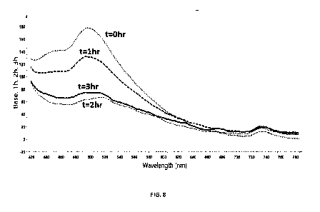
Figure 8: FN3K treatment of AGEs in the ciliary body of human cadaver eyes in
vitro
Ciliary body was dissected from human cadaver eyes (waste material rejected
for corneal
transplantation) and treated for 3 hours ex vivo with 3 mL FN3K (41.6pg/mL) +
ATP 2.5 mmol/L +
MgCl2 (1 mmol/L). Baseline Fluorometry was performed (0 hr dotted line) and
after FN3K treatment
for 1 hour (dashed line), 2 hours (full line) and 3 hours (line with stripes
and dots) using a miniature
spectrometer system (Flame-S-VIS-NIR, Ocean Optics, Largo, Fla) at fixed
distance and 900 angle.
0R400-7-VIS-BX Premium 400 micron reflection probe was used. Treatment of the
ciliary body with
FN3K reduces fluorescent signal of AGEs at 490 nm wavelength.
Figure 9: FN3K treatment of human cadaver eyes by external application of FN3K
drops ex vivo
7
CA 03110040 2021-02-18
WO 2020/053188
PCT/EP2019/074058
Human cadaver eyes (waste material rejected for corneal transplantation) were
treated within 24
hours after prelevation. For cross over experiments, always two eyes from the
same donor are used.
Fig 9A shows technique of applying FN3K drops to the intact human cadaver eye.
6 to 7 drops of
FN3K (25 pg/mL) + ATP (5 mmol/L) + MgCl2 (2 mmol/L) solution were applied
every hour for 6 hours
on one eye and saline drops were applied every hour for 6 hours on the other
eye from the same
donor. Fluorometry was performed at baseline before treatment and after
treatment using a
miniature spectrometer system (Flame-S-VIS-NIR, Ocean Optics, Largo, Fla) at
fixed distance and 900
angle. 0R400-7-VIS-BX Premium 400 micron reflection probe was used.
Fig 9B: Fluorescent signal of AGEs is lower in eye 1 than in eye 2 at the
start of the experiment (t=0
hr) Eye 1 is then treated with FN3K drops for 6 hours while eye 2 is treated
with saline drops.
Fluorescent signal of AGEs measured after 6 hours of treatment (t=6hr) however
only drops in the
FN3K treated eye. When pursuing the experiment as a cross-over experiment, eye
1 is then treated
with saline drops for another 6 hours and eye 2 with FN3K drops. Fluorescent
signal of AGEs is
measured again (t=12 hr). Fluorescent signal of AGEs decreases significantly
in eye 2 but not in eye 1.
Figure 9 shows that FN3K treatment of the intact human eye by external
application such as FN3K
drops reduces fluorescent signal of AGEs in the eye.
Detailed description of the invention
The present invention relates to the surprising finding that the
administration of a fructosamine-3-
kinase and its co-factor(s) results in less/ less dens Drusen in AMD. In other
words, the latter
administration restores light transmission and thus vision in patients with
AMD.
The present invention also relates to the surprising finding that treatment
with fructosamine-3-kinase
and its co-factor(s) reduces AGEs in the retina, in Bruch's membrane, and
subretinal. In other words, a
composition comprising FN3K and adenosine tri phosphate restores light
transmission and thus vision
in patients with AGE-dependent ocular diseases such as AMD, DR and DME.
.. The present invention also relates to the finding that treatment with
fructosamine-3-kinase and its
cofactor(s) reduces AGEs in the ciliary body. In other words, a composition
comprising FN3K and its
cofactors restores accommodation and thus near vision in individuals with age-
related presbyopia.
The present invention thus in first instance relates to a composition
comprising a fructosamine-3-
kinase and adenosine tri phosphate for use to treat AMD, DR and/or DME, age-
related presbyopia in
.. a human or an animal.
8
CA 03110040 2021-02-18
WO 2020/053188
PCT/EP2019/074058
The present invention further relates a composition for use as described above
wherein said
composition is administered by intravitreal injection.
The present invention further relates to a composition for use as described
above which further
comprises magnesium ions and/or an adenosine tri phosphate regenerating
system.
The present invention further relates to a composition comprising a
fructosamine-3-kinase and
adenosine tri phosphate regenerating system for use to treat AMD, DR and/or
DME in a human or an
animal wherein said composition is administered by intravitreal injection.
The present invention further relates a composition comprising a fructosamine-
3-kinase and
adenosine tri phosphate regenerating system for use as described above which
further comprises
magnesium ions.
The term 'a fructosamine-3-kinase' relates to enzymes classified as enzymes
2.7.1.171 in -for example-
the Brenda enzyme database (www.brenda-enzymes.org). The latter enzymes are
part of an ATP-
dependent system for removing carbohydrates from non-enzymatically glycated
proteins and catalyze
the following reaction: ATP + [protein]-N6-D-fructosyl-L-lysine = ADP +
[protein]-N6-(3-0-phospho-D-
fructosyl)-L-lysine. More specifically, the term 'a fructosamine-3-kinase'
relates to ¨as a non-limiting
example- to the human fructosamine-3-kinase having accession number or the
National Center for
Biotechnology Information (NCB!) Reference sequence number :NP_071441.1 (see
https://www.ncbi.nlm.nih.gov/protein/NP 071441). It should be further clear
that the term 'a
fructosamine-kinase' relates to the enzymes as described above, but also to
functional fragments and
variants thereof. The term "functional fragments and variants" relates to
fragments and variants of
the naturally occurring enzymes. Indeed, for many applications of enzymes,
part of the protein may be
sufficient to achieve an enzymatic effect. The same applies for variants (i.e.
proteins in which one or
more amino acids have been substituted with other amino acids, but which
retain functionality or even
show improved functionality), in particular for variants of the enzymes
optimized for enzymatic activity
(as is also described further with regard to recombinant enzymes). The term
'fragment' thus refers to
an enzyme containing fewer amino acids than the 309 amino acid sequence of the
human
fructosamine-3-kinase having NCB! Reference sequence number :NP_071441.1 and
that retains said
enzyme activity. Such fragment can -for example- be a protein with a deletion
of 10% or less of the
total number of amino acids at the C- and/or N-terminus. The term "variant"
thus refers to a protein
having at least 50 % sequence identity, preferably having at least 51-70 %
sequence identity, more
preferably having at least 71-90% sequence identity or most preferably having
at least 91, 92, 93, 94,
9
CA 03110040 2021-02-18
WO 2020/053188
PCT/EP2019/074058
95, 96, 97, 98 or 99 % sequence identity with the 309 amino acid sequence of
the human fructosamine-
3-kinase having NCB! Reference sequence number :NP_071441.1 and that retains
said enzyme activity.
Hence, orthologues, or genes in other genera and species (than the human
fructosamine-3-kinase
having NCB! Reference sequence number :NP_071441.1 ) with at least 50 %
identity at amino acid
level, and having said enzyme activity are part of the present invention. The
percentage of amino acid
sequence identity is determined by alignment of the two sequences and
identification of the number
of positions with identical amino acids divided by the number of amino acids
in the shorter of the
sequences x 100. The latter 'variant' may also differ from the protein having
NCB! Reference sequence
number :NP_071441.1 only in conservative substitutions and/or modifications,
such that the ability of
the protein to have enzymatic activity is retained. A "conservative
substitution" is one in which an
amino acid is substituted for another amino acid that has similar properties,
such that one skilled in
the art of protein chemistry would expect the nature of the protein to be
substantially unchanged. In
general, the following groups of amino acids represent conservative changes:
(1) ala, pro, gly, glu, asp,
gin, asn, ser, thr; (2) cys, ser, tyr, thr; (3) val, ile, leu, met, ala, phe;
(4) lys, arg, his; and (5) phe, tyr, trp,
his.(13,14)
Variants may also (or alternatively) be proteins as described herein modified
by, for example, the
deletion or addition of amino acids that have minimal influence on the enzymes
activity as defined
above, secondary structure and hydropathic nature of the enzyme.
The terms 'adenosine tri phosphate' (ATP) and 'magnesium ions' relate to well-
known cofactors of the
latter enzymes.
The term 'adenosine tri phosphate generating system' relates to several
enzymatic and whole-cell
based methods to regenerate ATP from ADP or AMP as are - for example-
described by Woodyer R. D.
et al. 2006 (15,16). In particular, the latter term refers to the following
four enzymes commonly used
in the regeneration of ATP from ADP: 1) the use of phosphoenolpyruvate in a
coupled reaction
catalyzed by pyruvate kinase, 2) acetylphosphate coupled with acetate kinase,
3) creatine phosphate
coupled with creatine kinase, and 4) polyphosphate coupled with polyphosphate
kinase. Preferably,
the term 'ATP generating system' refers to the usage of phosphocreatine as a
secondary energy source
and creatine kinase to transfer its phosphate group to ADP to regenerate ATP.
The usage of the latter
ATP generating systems thus limits the concentration of ATP present in the
mixture injected into the
vitreous body as is also described further.
The terms 'to treat AMD and/or DR and/or DM E' relate to stabilization and/or
improving vision of the
treated subject.
CA 03110040 2021-02-18
WO 2020/053188
PCT/EP2019/074058
The term "to treat age-related presbyopia" relates to stabilization and/or
improving nearby vision of
the treated subject.
The term 'animal' may relate to any animal.
The terms 'administration by intravitreal injection' relate to injection of
the compounds of the present
invention into the vitreous body of the eye. The intravitreal injection
technique is used under
controlled aseptic conditions. Adequate anesthesia is given prior to the
injection. For the treatment of
animal eyes, general anesthesia is used by ¨for example-inhalation anesthesia
with isoflurane 5%. For
the treatment of humans, local anesthetic drops can be used. A 32-gauge needle
can be used for
injection in smaller animal (such as a small rodent) eyes and a 30-gauge
needle in human eyes and
eyes of bigger animals such as horse and pig. In all species, the sclera is
penetrated at an angle from
450 - 90 . In mouse ¨for example-, the sclera can be penetrated at 1-1.5
millimeter from the limbus,
and in humans the sclera can be penetrated at 3-5 millimeter from the limbus.
The needle passes
through the sclera and choroid until the vitreous body is reached. The needle
does not touch the lens,
nor the retina. The composition of the present invention can be as such
delivered and the needle is
withdrawn immediately.
The present invention thus relates -in other words- to a method to treat (or
prevent) age-related
presbyopia, AMD, DR and/or DME in a subject in need thereof wherein said
method comprises
administering (for example by an injection of) a therapeutically effective
amount of a compound
comprising a fructosamine-3-kinase and adenosine tri phosphate, or, a
fructosamine-3-kinase and an
adenosine tri phosphate generating system, or, a fructosamine-3-kinase and
adenosine tri phosphate
and an adenosine tri phosphate generating system, or a fructosamine-3-kinase
and adenosine tri
phosphate and magnesium ions, or, a fructosamine-3-kinase and adenosine tri
phosphate and an
adenosine tri phosphate generating system and magnesium ions, or, a
fructosamine-3-kinase and an
adenosine tri phosphate generating system and magnesium ions to (for example
in the vitreous body
of) the eye of said subject.
The term 'a therapeutically effective amount' relates to an amount ranging
from 5 Ill (for
administering/injecting into a single mouse eye) to 50 Ill (for
administering/injecting into a single
bovine eye) taken from a therapeutic dose ranging between about 4,17 and 12.5
ug/mlfructosamine-
3-kinase, 2.50 and 4,17 mM ATP and 1.00 and 1.67 mM MgCl2. The latter
therapeutic doses can be
obtained by mixing 1:1, 1:2, 1:3 or 1:5 a solution of 25 ug/m1 fructosamine-3-
kinase with a fresh
solution of 5mM ATP/2mM MgCl2.
11
CA 03110040 2021-02-18
WO 2020/053188
PCT/EP2019/074058
0.1 mol/L thiosulfate and 5 Wm! hyaluronidase are added to the mixture when
amounts > 5 Ill are
administered (intravitreally) in an animal eye (not mouse) and human eyes.
It should be clear that besides 'injecting' said therapeutically effective
amounts 'intravitreally' ¨which
is one way of administration- also other means of administration are
envisioned such as ¨but not
limited to- external application such as via drops or gels, and, other
internal applications such as
suprachoroidal injections or subretinal injections or implants everywhere else
in the eye. Hence, and
for example, the present invention therefore relates to a composition
comprising a fructosamine-3-
kinase and ATP (and which might further comprise magnesium ions) for use to
treat age-related
presbyopia wherein said composition is administered by intravitreal injection
or as external
application.
The present invention further relates to a composition as indicated above
wherein said fructosamine-
3-kinase is a recombinant fructosamine-3-kinase. The term 'recombinant' refers
to fructosamine-3-
kinase obtained as an outcome of the expression of recombinant DNA encoding
for a fructosamine-3-
kinase inside living cells such as bacteria or yeast cells. Practitioners are
further directed to Sambrook
et al. Molecular Cloning: A laboratory Manual, 4th ed. , Cold Spring Harbor
press, Plainsview, New York
(2012) and Ausubel et al. Current Protocols in Molecular Biology (supplement
114), John Wiley & Sons,
New York (2016).
More specifically the present invention relates to a recombinant fructosamine-
3-kinase which is
obtainable by recombinant production in Pichia pastoris and, even more
specifically, wherein said
recombinant fructosamine-3-kinase obtainable by recombinant production in
Pichia pastoris has the
amino acid sequence as given by SEQ ID N 1 or SEQ ID N 2. SEQ ID N 1 is a
construct with an N-terminal
cleavable HIS-tag and a caspase 3-cleavable Asp-Glu-Val-Asp (DEVD) linker
between the His6 tag and
the protein coding sequence which allows for clean removal of the tag. SEQ ID
N 2 is the cleaved
version of SEQ ID N 1.
The amino acid sequences of SEQ ID N 1 and SEQ ID N 2 (and their encoding
nucleic acid sequences
SEQ ID N3 and SEQ ID N 4, respective) are as follows:
SEQ ID N 1:
Type: amino acid 1-letter (underlined: His6-tag, italics: linker, bold
underlined: caspase cleavage
site)
MHHHHHH VNGPGSDEVDEQLLRAELRTATLRAFGGPGAGC I SE GRAYDTDAGP VFVKVNRRTQARQMF
EGEVAS LEALRS T GLVRVP RPMKVI DLP GGGAAFVME HLKMKS LS SQASKLGEQMADLHLYNQKLREK
12
CA 03110040 2021-02-18
WO 2020/053188
PCT/EP2019/074058
LKEEENTVGRRGEGAEPQYVDKFGFHTVTCCGF IPQVNEWQDDWPTFFARHRLQAQLDLIEKDYADRE
ARELWSRLQVKIPDLFCGLEIVPALLHGDLWSGNVAEDDVGP I IYDPASFYGHSEFELAIALMFGGFP
RSFFTAYHRKIPKAPGFDQRLLLYQLFNYLNHWNHFGREYRSP SLGTMRRLLK*
SEQ ID N 3:
Type: DNA (underlined: His6-tag, italics: linker, bold underlined: caspase
cleavage site)
AT GCAT CAT CAT CAT CAT CAT GTTAACGGTCCAGGTTCTGATGAAGTTGATGAACAGTTGTTGAGAGC
TGAGTTGAGAACTGCTACTTTGAGAGCTTTTGGTGGTCCAGGTGCTGGTTGTATTTCTGAGGGTAGAG
CTTAC GATACT GACGCT GGTC CAGT TT TC GT TAAGGT TAACAGAAGAAC TCAGGC
TAGACAGATGTTC
GAGGGTGAAGTTGCTTCTTTGGAGGCTTTGAGATCCACTGGTTTGGTTAGAGTTCCAAGACCAATGAA
GGT TATCGACT TGCCAGGTGGTGGTGCTGCT TT TGTTATGGAACACT TGAAGATGAAGTCCTTGTCCT
C CCAGGC TT CTAAGT TGGGTGAACAAATGGC TGAC TT GCAC TT GTACAACCAGAAGT TGAGAGAAAAG
T TGAAAGAGGAAGAGAACACT GT TGGTAGAAGAGGTGAAGGTGCT GAGC CACAATAC GTTGACAAGTT
CGGTT TCCACACTGT TACT TGTTGTGGTT TCATCCCACAGGTTAACGAGTGGCAAGATGACTGGCCAA
C TT TC TT CGCTAGACACAGAT TGCAAGCT CAGT TGGACT TGAT CGAGAAGGAC TACGCT
GACAGAGAA
GCTAGAGAATTGTGGTCCAGATTGCAGGTTAAGATCCCAGACTTGTTCTGTGGTTTGGAGATCGTTCC
AGCTTTGTTGCACGGTGATTTGTGGTCTGGTAACGTTGCTGAAGATGACGTTGGTCCAATTATCTACG
ACCCAGCTTCTTTCTACGGTCACTCTGAATTCGAGTTGGCTATCGCTTTGATGTTCGGTGGTTTCCCA
AGATCCTTCTTCACTGCTTACCACAGAAAGATCCCAAAGGCTCCAGGTTTCGACCAGAGATTGTTGTT
GTACCAGTTGTTCAACTACTTGAACCATTGGAACCACTTCGGTAGAGAGTACAGATCTCCATCCTTGG
GTACTATGAGAAGATTGTTGAAGTAA
SEQ ID N 2 (= FN3K after N-terminal HIS-tag removal):
Type: amino acid 1-letter
EQLLRAE LRTATLRAFGGP GAGC I S EGRAYD TDAGPVFVKVNRRTQARQMFEGEVAS LEALRS TGLVR
VPRPMKVID LP GGGAAFVMEHLKMKS L S S QASKLGEQMADLHLYNQKLREKLKEEENTVGRRGEGAEP
QYVDKFGFHTVTC CGF I PQVNEWQDDWP TFFARHRLQAQLD L I EKDYADREARELWS RLQVKI PDLF C
GLEIVPALLHGDLWSGNVAEDDVGP I I YDPASFYGHSEFELAIALMFGGFPRSFF TAYHRKIPKAPGF
DQRLLLYQLFNYLNHWNHFGREYRSPSLGTMRRLLK*
SEQ ID N 4:
Type: DNA
GAACAGT TGTTGAGAGCTGAGTTGAGAACTGCTACTT TGAGAGCT TT TGGTGGTCCAGGTGCTGGTTG
TAT TTCTGAGGGTAGAGCT TACGATACTGACGCTGGTCCAGTT TTCGTTAAGGTTAACAGAAGAACTC
13
CA 03110040 2021-02-18
WO 2020/053188
PCT/EP2019/074058
AGGCTAGACAGATGTTCGAGGGTGAAGTTGCTTCTTTGGAGGCTTTGAGATCCACTGGTTTGGTTAGA
GTTCCAAGACCAATGAAGGTTATCGACTTGCCAGGTGGTGGTGCTGCTTTTGTTATGGAACACTTGAA
GATGAAGTCCTTGTCCTCCCAGGCTTCTAAGTTGGGTGAACAAATGGCTGACTTGCACTTGTACAACC
AGAAGTTGAGAGAAAAGTTGAAAGAGGAAGAGAACACTGTTGGTAGAAGAGGTGAAGGTGCTGAGCCA
CAATACGTTGACAAGTTCGGTTTCCACACTGTTACTTGTTGTGGTTTCATCCCACAGGTTAACGAGTG
GCAAGATGACTGGCCAACTTTCTTCGCTAGACACAGATTGCAAGCTCAGTTGGACTTGATCGAGAAGG
ACTACGCTGACAGAGAAGCTAGAGAATTGTGGTCCAGATTGCAGGTTAAGATCCCAGACTTGTTCTGT
GGTTTGGAGATCGTTCCAGCTTTGTTGCACGGTGATTTGTGGTCTGGTAACGTTGCTGAAGATGACGT
TGGTCCAATTATCTACGACCCAGCTTCTTTCTACGGTCACTCTGAATTCGAGTTGGCTATCGCTTTGA
TGTTCGGTGGTTTCCCAAGATCCTTCTTCACTGCTTACCACAGAAAGATCCCAAAGGCTCCAGGTTTC
GACCAGAGATTGTTGTTGTACCAGTTGTTCAACTACTTGAACCATTGGAACCACTTCGGTAGAGAGTA
CAGATCTCCATCCTTGGGTACTATGAGAAGATTGTTGAAGTAA
The present invention indeed relates ¨in addition- to the finding that the
recombinant fructosamine-
3-kinase obtainable by recombinant production in Pichia postoris and having
the amino acid sequence
as given by SEQ ID N 1 and 2 are preferred enzymes for treating AMD. Indeed,
the latter enzymes are
preferred as 1) their production in Pichia resulted in higher yields of the
enzyme compared with the
production in ¨for example- E. coli, 2) the enzymes had a higher purity when
analysed on SDS page,
and 3) the presence of endotoxin, which is known to provoke an ocular
inflammation during
intravitreal injection, can be avoided.
The following examples are provided to better illustrate the present invention
and should not be
considered as limiting the scope of the invention.
Examples
Example 1: recombinant production of fructosamine-3-kinase
A gene coding for human fructosamine-3-kinase (having accession number or the
National Center for
Biotechnology Information (NCB!) Reference sequence number :NP_071441.1 (see
https://www.ncbi.nlm.nih.gov/protein/NP 071441), codon-optimized for Pichia
postoris expression
(SEQ ID N 1), was cloned into the pKai61 P. postoris expression vector
according to Claes, K. et al. (
"Modular Integrated Secretory System Engineering in Pichia Pastoris To Enhance
G-Protein Coupled
Receptor Expression," ACS Synthetic Biology 5, no. 10 (October 21,2016): 1070-
75). The encoded gene
contains an N-terminal His6-tag (MHHHHHH) in frame with a caspase-3 cleavage
site (DEVD) and the
expression is under control of the methanol inducible A0X1 promoter. The
plasmid contains a zeocin
resistance marker for selection in bacterial as well as in yeast cells. The
vectors were linearized in the
14
CA 03110040 2021-02-18
WO 2020/053188
PCT/EP2019/074058
A0X1 promoter before transformation to P. pastoris (strain NRRL Y-11430) to
promote homologous
recombination in the endogenous A0X1 locus for stable integration into the
genome.
Stable integrants were grown shaking at 28 C in BMY buffered complex medium
(10 g/L yeast extract, 20
g/L peptone, 100 mM potassium phosphate buffer pH 6.0, 13.4 g/L YNB without
amino acids)
.. complemented with 1% glycerol. After 48 hours of growth, recombinant
expression was induced by
transfer to BMY medium complemented with 1% methanol. After 48 hours of
expression, cultures
were centrifuged, supernatant was discarded and pellets were flash frozen in
liquid nitrogen and
stored at -20 C.
Pellets were thawed and resuspended in washing buffer for protein extraction.
Pichia pastoris cells
were mechanically disrupted using 0.5 mm glass or silicia/zirkonium beads. The
cleared supernatant
was purified by Ni2+ affinity chromatography for the His6-tagged fructosamine-
3-kinase, followed by
gel filtration. The protein eluted in FN3K sample buffer (20 mM Tris-HCI pH
8.0, 150 mM NaCI, 1 mM
DTI) was identified as recombinant human fructosamine-3-kinase by SDS-PAGE and
Western blotting
with antibodies against the His6-tag and human FN3K (ThermoFisher). Enzymatic
activity was
confirmed in a kinase activity assay with a 1 deoxy 1 morpholino D fructose
substrate (R&D Systems).
Fructosamine-3-kinase aliquots were flash frozen in liquid nitrogen and stored
at -20 C.
Example 2: treatment of 5 micron slices of human eyes with Drusen.
5 micrometer sections of human retina with Drusen (D) are treated with saline
+ ATP + MgCl2 or treated
with FN3K + ATP + MgCl2. Doses used ranged between about 4,17 and 12.5 ug/m1
fructosamine-3-
kinase, 2.50 and 4,17 mM ATP and 1.00 and 1.67 mM MgCl2. Drusen are evaluated
by light microscopy
for integrity and presence of eosinophil material (Fig 2A and B).
Stained tissue sections were scanned by the Olympus dotSlide Digital Virtual
Microscopy System and
processed using the OlyVIA viewer program (Olympus Corporation, Tokyo, Japan).
For subsequent
image analysis, the freeware ImageJ v1.8.0 downloaded from the NIH website
(http://rsb.info.nih.gov/ij) was used. Red (R), green (G) and blue (B)
intensity values were calculated
using the RGB Measure plug-in. Figure 2C shows intensity values on the RGB
colour histogram of the
histological section of a Druse when treated with saline + ATP + MgCl2
(untreated) or treated with FN3K
+ ATP + MgCl2. Figure 2D shows mean value of all intensity values of 10 Drusen
treated with saline +
ATP + MgCl2 (untreated) and 10 FN3K treated DrusenNear infrared (NIR) spectra
are recorded off-line
using a NIR spectrometer equipped with an immobilized reflection probe of
seven 400 um fibers, an
InGaAs detector and a halogen lamp (AvaSpecNIR256-2.5-HSC with an FCR-7UVIR400-
2-BX reflection
CA 03110040 2021-02-18
WO 2020/053188
PCT/EP2019/074058
probe, Avantes). As glycation results in a spectral shift in the near-infrared
spectrum of proteins, it is
possible to observe specific peak sharpening and spectral variations in NIR
spectra due to deglycation
of proteins (Fig 3). Figure 3A shows the Hotelling's T2 plot of intraretinal
AGEs in human retina treated
with saline + Mg Cl2 + ATP (circles), compared to intraretinal AGEs in human
retina treated with FN3K
+ Mg C12+ ATP (squares).
Figure 3B shows NIR spectra and Hotelling's plot of AGEs in Bruch's membrane
treated with saline +
Mg Cl2 + ATP (control) compared to AGEs in Bruch's membrane treated with FN3K
+ Mg Cl2 + ATP.
Figure 3C shows NIR spectra and Hotelling's plot of AGEs in subretinal Drusen
treated with saline + Mg
C12+ ATP (control) compared to AGEs in subretinal Drusen treated with FN3K +
Mg C12+ ATP.
Figure 3D shows mean spectra of measured NIR spectra of AGEs in Bruch's
membrane (full lines) and
in subretinal Drusen (dotted lines) when treated with saline + ATP + MgCl2
(control) or treated with
FN3K + ATP + MgCl2
Fluorometry of Drusen is performed on 5 micron sections of human retina retina
treated with saline +
ATP + MgCl2 (circles) or with FN3K + ATP + MgCl2 (squares). Fluorometry is
performed with UV
fluorescence spectroscopy in the range of 400 nm to 680 nm. Differences are
detected specifically in
the range of AGE fluorescence (560 nm up to 680 nm) (Fig 4). Fluorometry is
performed on 5 different
retinas of human cadaver eyes. Figure 4 shows measurements of AGEs of
intraretinal Drusen. Figure
4A shows raw AGE fluorescence spectroscopy curves of eye 1. Figure 4B shows
AGE fluorescence
spectroscopy results of 4 other human retinas after smoothening of the curves.
.. Mean fluorescence intensities of the 4 latter human retinas are then
calculated and compared (Table
1).
Tablel. Mean fluorescence intensity 420-700 nm (a.u.) of human neural retinas
at baseline
and after ex vivo FN3K treatment
Baseline FN3K % change P-value
Eye 2 left 63.2 (55.9) 43.7 (42.4-51.3) -31.2
<0.0001
Eye 3
left 55.5 (52.4-60.5) 42.5 (41.0-44.0) -23.4
<0.0001
right 75.3 (68.2-78.9) 50.7 (45.9-56.5) -32.7
<0.0001
Eye 10 left 71.5 (50.1-95.9) 56.1 (50.4-76.7) -21.5
0.14
16
CA 03110040 2021-02-18
WO 2020/053188
PCT/EP2019/074058
Example 3 : treatment of eyes of aged C57/13I6 mice. In vivo experiment.
Tests are carried out on aged C57/13I6 mice, which show the typical AMD
lesions as Drusen. Following
FN3K treatment in one eye by intravitreal injection, mice retinas are studied
using near-infrared (NIR)
and fluorescence spectroscopy.
Histological sections are performed to evaluate the presence or absence of the
typical Drusen (Figure
5) Drusen were present when the eyes were treated with intravitreal injection
of saline + ATP + MgCl2
but were absent when eyes (contralateral eye of the same animal) were treated
with FN3K + ATP +
MgCl2
Mice from 23 months old are anesthetized during the surgical procedure with
inhalation anesthesia
(isoflurane 5%). Both eyes of the same animal are injected, one with 5
microliter fructosamine-3 kinase
+ ATP + MgCl2 (same preparation as experiment in example 2) and one with 5
microliter saline + ATP +
MgCl2. 24 hours and 1 week later, mice are sacrificed and both eyes are
enucleated. Near infrared (NIR)
spectra are recorded off-line using a NIR spectrometer equipped with an
immobilized reflection probe
of seven 400 um fibers, an InGaAs detector and a halogen lamp (AvaSpecNIR256-
2.5-HSC with an FCR-
7UVIR400-2-BX reflection probe, Avantes). As glycation results in a spectral
shift in the near-infrared
spectrum of proteins, it is possible to observe specific peak sharpening and
spectral variations in NIR
spectra due to deglycation of proteins. This allows us to distinguish
fructosamine-3-kinase-treated
from untreated eyes. The use of non-invasive NIR monitoring enables us to
assess the treatment in a
non-destructive way.
Example 4. Treatment of Drusen in human cadaver eyes by intravitreal
injection.
Human cadaver eyes (rejected for cornea transplantation) were transported on
ice and evaluated for
the presence of Drusen by Optical Coherence Tomography within 24 hours after
prelevation. When
Drusen were present, the Drusen were treated by intravitreal injection into
the eye with FN3K + ATP +
MgCl2 + 0.1 mol/L thiosulfate + 5U/m1 hyaluronidase. Thiosulfate was added to
the mixture before
intravitreal injection for optimal penetratioin of the calciumhydroxyappatite
around large subretinal
Drusen. Hyaluronidase was added to the mixture for optimal penetration of the
vitreous. Eyes were
kept at 37 C for 2 hours and Drusen were again recorded by Optical Coherence
Tomography (Figure
6). In the 4 cadaver eyes where Drusen were present, intravitreal injection
with FN3K + ATP + MgCl2
induces a clear reduction in size.
17
CA 03110040 2021-02-18
WO 2020/053188
PCT/EP2019/074058
Example 5. FN3K treatment of AGE-modified pig retina in vitro.
Pig retinas were dissected within 24 hours after prelevation. UV-fluorescence
spectroscopy of
AGES in pig retina was performed at baseline and after treatment with two of
the most prevalent
AGEs in human retina (methyglyxoal (MG), glycolaldehyde (GA) for 24 hours. Pig
retinas were then
washed with PBS and treated with saline + Mg Cl2 + ATP or with FN3K + Mg Cl2 +
ATP for 24 hours,
and UV-fluorescence spectroscopy was repeated.
Table 2. Norm, fluorescence intensities (a.u.) of neural pig retinas at
baseline, AGE-modification and after control and FN3K
treatment.
MG-AGEs GA-AGEs
Control (n=30) FN3K (n=30) Control (n=30) FN3K
(control=30)
Baseline
440 nm 5.6 (5.2-6.0) 6.6 (6.1-6.9) 6.4 (6.1-6.9) 6.7
(5.9-6.9)
490 nm 7.1 (6.5-7.4) 7.9 (7.1-8.5) 7.9 (7.3-8.4) 8.5
(8.0-91)
490/440 nm 1.3 (1.1-1.4) 1.2 (1.1-1.3) 1.2 (1.1-1.3) 1.3
(1.2-1.4)
AGE-modification
440 nm 26.2 (19.4-40.6) 30.6 (22.3-43.0) 31.2 (26.2-
50.2) 23.7 (20.4-28.9)
490 nm 28.8 (22.1-49.6) 34.2 (23.6-53.0) 55.3 (47.5-
96.0) 39.3 (31.2-46.3)
490/440 nm 1.1 (1.1-1.2) 1.2 (1.1-1.2) 1.8 (1.8-1.9) 1.6
(1.5-1.7)
Treatment
440 nm 32.5 (19.8-44.7) 22.5 (18.0-26.4) 25.1 (23.5-
32.7) 16.2 (11.0-20.0)
490 nm 35.8 (19.3-50.3) 23.4 (18.0-26.4) 40.8 (36.9-
58.6) 19.4 (13.6-28.2)
490/440 nm 1.1 (1.0-1.1) 1.1 (1.0-1.1) 1.6 (1.6-1.8) 1.3
(1.2-1.4)
Baseline vs AGE-modification (P-value)
440 nm <0.0001 <0.0001 <0.0001 <0.0001
490 nm <0.0001 <0.0001 <0.0001 <0.0001
490/440 nm <0.01 0.06 <0.0001 <0.0001
AGE-modification vs Treatment (P-value)
440 nm N.S. <0.05 <0.05 0.0001
490 nm N.S. <0.01 <0.001 <0.0001
490/440 nm <0.05 <0.001 <0.0001 <0.0001
Six porcine eyes were obtained from a local abattoir and stored at 4 C until
processing. Neural retinas
were isolated through dissection by a trained ophthalmologist within 12h post-
mortem, transferred to
a sterile 6-well plate ( Thermo scientific, Roskilde, Denmark) and stored at 4
C in RPMI-1640 medium
(Sigma-Aldrich, St. Louis, Missouri, USA). The experiment was started within
48h by removing the RPM!
medium and carefully washing the retinas five times with 5 mL phosphate
buffered saline (PBS)
solution. Subsequently, maillard type fluorescence measurements (excitation
370 nm, emission 390-
700 nm) were performed at baseline on each retina (30 different measurement
locations) using a
miniature spectrometer system (Flame-S-VIS-NIR, Ocean Optics, Largo, Fla) at
fixed distance and 90
angle. AGE modification was performed by incubation of two retina wells with 4
mL 100 mmol/L
methylglyoxal (methyl glyoxal solution -40% in H20, Sigma-Aldrich), and two
with 4 mL 100 mmol/L
18
CA 03110040 2021-02-18
WO 2020/053188
PCT/EP2019/074058
glycolaldehyde dimer (crystalline form, Sigma-Aldrich) in phosphate buffered
saline (PBS) for 24h at
37 C. After incubation, the active agents were carefully washed away (10
times) in each well with 5 mL
PBS and fluorescence measurements were performed again. Finally, in vitro
deglycation was initiated
using ATP-dependent FN3K (Fitzgerald Industries International, Acton, MA,
USA). A solution containing
0.0016 g/L ATP-dependent FN3K in PBS was added (1:1) to a mixture of 5 mmol/L
ATP and 2 mmol/L
MgCl2 (Sigma-Aldrich) in PBS. Two milliliters of the final FN3K solution were
added to one retina well
incubated with methylglyoxal, and one with glycolaldehyde and incubated for
24h at 37 C. The
remaining wells were control treated with PBS. After the treatment procedure,
all wells were washed
five times with PBS and fluorescence measurements were performed.
FN3K treatment reduced fluorescence of intraretinal AGEs in pig retinas in
vitro.
Example 6. treatment of eyes of ob/ob mice and wt mice. In vivo experiment.
Tests are carried out on aged ob/ob mice, which show the typical diabetic
lesions. Following FN3K
treatment in one eye by intravitreal injection, mice retinas are studied using
near-infrared (NIR) and
fluorescence spectroscopy.
Histological sections are performed to evaluate typical lesions in DR and DME,
such as an increase in
large leaky vessels and in thickness of collagen fibers in the inner limiting
membrane. Figure 7 shows
signs of diabetic retinopathy in ob/ob mice treated with saline + ATP + MgCl2
with large leaky vessels
(large arrow), and a very thick collagenous inner limiting membrane
(triangle). Retinas of ob/ob mice
treated with FN3K + ATP + MgCl2 showed normalization of the retina and normal
microvasculature
(small arrows) comparable with wt mice.
Mice from 30-36 weeks old are anesthetized during the surgical procedure with
inhalation anesthesia
(isoflurane 5%). Both eyes of the same animal are injected, one with 5
microliter fructosamine-3 kinase
+ ATP + MgCl2 (same preparation as experiment in example 2) and one with 5
microliter saline + ATP +
MgCl2. 24 hours later, mice are sacrificed and both eyes are enucleated. Near
infrared (NIR) spectra
are recorded off-line using a NIR spectrometer equipped with an immobilized
reflection probe of seven
400 um fibers, an InGaAs detector and a halogen lamp (AvaSpecNIR256-2.5-HSC
with an FCR-
7UVIR400-2-BX reflection probe, Avantes). As glycation results in a spectral
shift in the near-infrared
spectrum of proteins, it is possible to observe specific peak sharpening and
spectral variations in NIR
spectra due to deglycation of proteins. This allows us to distinguish
fructosamine-3-kinase-treated
19
CA 03110040 2021-02-18
WO 2020/053188
PCT/EP2019/074058
from untreated eyes. The use of non-invasive NIR monitoring enables us to
assess the treatment in a
non-destructive way.
Example 7. FN3K treatment of AGEs in the ciliary body of human cadaver eye in
vitro
Ciliary body was dissected from human cadaver eyes (waste material rejected
for corneal
transplantation) and treated for 3 hours ex vivo with 3 mL FN3K (41.6111g/mL)
+ ATP 2.5 mmol/L + MgCl2
(1 mmol/L). Fluorometry (fig.8) was performed after 1 hour, 2 hours and 3
hours of FN3K treatment
using a miniature spectrometer system (Flame-S-VIS-NIR, Ocean Optics, Largo,
Fla) at fixed distance
and 900 angle. 0R400-7-VIS-BX Premium 400 micron reflection probe was used.
Example 8 Treatment of human cadaver eyes by external application of FN3K
drops ex vivo
Human cadaver eyes (waste material rejected for corneal transplantation) were
treated within 24
hours after prelevation. For cross over experiments, always two eyes from the
same donor are used.
The technique of applying FN3K drops or saline drops to the intact human
cadaver eye consists of the
following: 6 to 7 drops of FN3K (25 Eg/mL) + ATP (5 mmol/L) + MgCl2 (2 mmol/L)
solution were applied
every hour for 6 hours on one eye and saline drops were applied every hour for
6 hours on the other
eye from the same donor. Fluorometry was performed at baseline before
treatment and 6 hours after
treatment using a miniature spectrometer system (Flame-S-VIS-NIR, Ocean
Optics, Largo, Fla) at fixed
distance and 900 angle. 0R400-7-VIS-BX Premium 400 micron reflection probe was
used. First, one eye
is treated with FN3K drops and the other eye of the same donor is treated with
saline drops. For cross
over experiments, treatment is then switched, and the FN3K treated eyes are
further on treated with
saline drops, while the eyes initially treated with FN3K are further on
treated with saline drops for 6
hours. Fluorometry is performed at baseline (start experiment, t=0 hr), after
6 hours of initial
treatment, and after 6 hours of the other treatment.
References
1. Bejarano E and Taylor A. Too sweet: problems of protein glycation in the
eye. Exp Eye Res
2019;178:255-262.
2. Wong WL et al. Global prevalence of age-related macular degeneration and
disease burden
projection for 2020 and 2040: A systematic review and meta-analysis. Lancet
Glob Health
2014;2:e106-16.
CA 03110040 2021-02-18
WO 2020/053188
PCT/EP2019/074058
3. Cheng W et al. Overview of clinical trials for dry age-related macular
degeneration. J Med Sci
2017;37:121-9.
4. Group UPDS. Risks of progression of retinopathy and vision loss related to
tight blood pressure
controle in type 2 diabetes mellitus. UKPDS 69, Arch Ophthalmol
2004;122,1631.White NH et
al. Beneficial effects of intensive therapy of diabetes during adolescence:
outcomes after the
conclusion of the Diabetes Control and Complications Trial (DCCT). J Pediat
2001;139:804-812
5. Wang J et al. Photosensitization of A2E triggers telomere dysfunction and
accelerates retinal
pigment epithelium senescence. Cell Death and Disease 2018;9:178.
6. Stitt AW. The Mai!lard Reaction in Eye Diseases Ann N Y Acad Sci
2005;1043:582-97.
7. Hollyfield J et al. Proteomic approaches to understanding age-related
macular degeneration.
Adv Exp Med Biol 2003;533:83-9.
8. Yamada Y et al. The expression of advanced glycation endproduct receptors
in RPE cells
associated with basal deposits in human maculas Exp Eye Res 2006;82:840-8.
9. Bergen AA et al. On the origin of proteins in human drusen: The meet, greet
and stick
hypothesis. Prog Retin Eye Res 2019;70:55-84.
10. Bogunovic H et al. Machine learning of the progression of intermediated
age-related macular
degeneration based on OCT imaging. Invest Ophthalmol Vis Sci 2017;58:B10141-
B10150.
11. Glenn JV and Stitt AW. The role of advanced glycation end products in
retinal ageing and
disease. Biochim Biophys Acta 2009;1790:1109-16.
12. Yoon KD et al. A novel source of methylglyoxal and glyoxal in retina:
implications for age-
related macular degeneration. PLoS One 2012;7:e41309.
13. Delpierre G, Collard F, Fortpied J, Van Schaftingen E. Fructosamine 3-
kinase is involved in an
intracellular deglycation pathway in human erythrocytes. Biochem J
2002;365:801-8.
14. Rosenfeld Pi, Brown DM, Heier JS, Boyer DS, Kaiser PK, Chung CY, Kim RY,
for the MARINA
Study Group. Ranibizumab for neovascular age-related macular degeneration. N
Engl J Med
2006;355:1419-31.
15. Halfter W, Dong S, Schurer B, Ring C, Cole GJ, Eller A. Embryonic
synthesis of the inner limiting
membrane and vitreous body. Invest Ophthalmol Vis Sci 2005;46:2202-9.
16. Delpierre G, Rider MH, Collard F, Stroobant V, Vanstapel H & Santos E
(2000) Identification,
cloning, and heterologous expression of a mammalian fructosamine-3-kinase.
Diabetes 49:
1627-1634.
17. Szwergold BS, Howell S & Beisswenger R.1(2001) Human fructosamine-3-
kinase: purification,
sequencing, substrate specificity, and evidence of activity in vivo. Diabetes
50: 2139-2147.
21
CA 03110040 2021-02-18
WO 2020/053188
PCT/EP2019/074058
18. Ryan D. Woodyer, Tyler Johannes, and Huimin Zhao, "Regeneration of
Cofactors for Enzyme
Biocatalysis in Enzyme Technology," in Enzyme Technology (Springer
Science+Business
Media,Inc. and Asiatech Publishers, Inc., 2006).
19. Andexer JN & Richter M (2015) Emerging Enzymes for ATP Regeneration in
Biocatalytic
Processes. ChemBioChem 16: 380-386.
22