Note: Descriptions are shown in the official language in which they were submitted.
CA 03112135 2021-03-07
WO 2020/058979
PCT/IL2019/051041
1
METHODS OF TREATING AMYOTROPHIC LATERAL SCLEROSIS
RELATED APPLICATION/S
This application claims the benefit of priority of Israel Patent Application
No. 261908
filed on 20 September 2018 and Israel Patent Application No. 267752 filed on
27 June 2019, the
contents of which are incorporated herein by reference in their entirety.
The contents of the above applications are all incorporated by reference as if
fully set
forth herein in their entirety.
SEQUENCE LISTING STATEMENT
The ASCII file, entitled 78818 Sequence Listing.txt, created on 19 September
2019,
comprising 22,138 bytes, submitted concurrently with the filing of this
application is
incorporated herein by reference.
FIELD AND BACKGROUND OF THE INVENTION
The present invention, in some embodiments thereof, relates to methods of
treating
Amyotrophic Lateral Sclerosis (ALS) and, more particularly, but not
exclusively, to treatment
with bacterial populations or metabolites thereof.
Amyotrophic Lateral Sclerosis (ALS) is a progressive, idiopathic
neurodegenerative
disorder characterized by premature death of motor neurons and an average
survival rate of 3-5
years from diagnosis. The majority of ALS cases are sporadic (sALS), while 10-
20% of cases are
familial (fALS), and driven by genetic mutations in genes such as superoxide
dismutase 1
(SOD1). Extensive efforts are being made to develop ALS-targeting drugs like
edaravone, but
none so far has yielded a conclusively effective disease-modifying activity.
While past
epidemiological studies did not identify clear environmental factors
correlating with ALS
occurrence and severity, the Central Nervous System (CNS) is increasingly
recognized to be
influenced by peripheral signals, such as circulatory small molecular-weight
metabolites which
may be absorbed from the GI tract to the blood stream and reach the CNS
through the brain-
blood barrier (BBB), where they can modulate metabolic, transcriptional and
epigenetic programs
in neurons and in other resident cells.
The gut microbiome, a microbial ecosystem impacting multiple host
physiological
functions, is a large potential source of such potentially bioactive CNS
disease-modulating
metabolites. Indeed, accumulating evidence suggests that the composition and
function of the gut
microbiome play significant roles in the pathogenesis of neurological
disorders such as autism,
CA 03112135 2021-03-07
WO 2020/058979
PCT/IL2019/051041
2
Parkinson's disease, Alzheimer's disease, Multiple sclerosis and epileptic
seizures. Metabolites
secreted, depleted or modified by the gut microbiome were shown to participate
in neuronal
transmission, synaptic plasticity, myelination and host complex behaviors.
Several hints suggest
that the host-gut microbiome interface may be potentially involved in the
course of ALS. A
disrupted Intestinal barrier accompanied by lower levels of colonic tight-
junction protein Zonula
occludens-1 (ZO-1) and the adherence protein E-cadherin were reported in 2
month-old SOD1-
Tg mice, potentially leading to dysbiosis hallmarked by a reduction in the
butyrate producing
bacteria Butyrivibrio fibrisolvens. Butyrate administration to SOD1-Tg mice
altered their
microbiome composition, although microbiome assessment was performed at a
single time point
and 3 animals per group, thereby precluding accurate assessment of the scope,
significance, and
mechanism of dysbiosis at this setting. 16S rDNA analysis of ALS patients
yielded conflicting
results, with one study noting a dysbiotic configuration in 6 ALS patients
compared to 5 healthy
controls, while another showing no significant compositional differences
between 25 ALS
patients and 32 healthy controls. No direct functional microbiome
investigation has been
performed in this setting.
Background art includes Richard Bedlack & The ALSUntangled Group (2018)
ALSUntangled 42: Elysium health's "basis", Amyotrophic Lateral Sclerosis and
Frontotemporal
Degeneration, 19:3-4,317-319, DOT: 10.1080/21678421.2017.1373978; and Harlan
et al, 2016,
The Journal of Biological Chemistry 291, 10836-10846.
SUMMARY OF THE INVENTION
According to an aspect of the present invention there is provided a method of
treating
ALS in a subject in need thereof comprising administering to the subject a
therapeutically
effective amount of at least two metabolites, wherein at least one of the at
least two metabolites is
selected from the group consisting of propyl 4-hydroxybenzoate,
triethanolamine, serotonin, 2-
keto-3-deoxy-gluconate, nicotinamide, N-trimethyl 5-aminovalerate,
phenylalanylglycine,
theobromine, cys-gly, glutamate, 1-palmitoy1-2-docosahexaenoyl-GPC, oxalate,
stearoyl
sphingomyelin, 1-p almitoy1-2-do co sahexaeno yl-GPC (16:0/22:6), 3 -
ureidopropionate, 1-(1-enyl-
p almito y1)-2- arachidonoyl-GPC (P-16:0/20:4), palmitoyl
sphingomyelin (d18:1/16:0),
sphingomyelin (d18:1/18:1, d18:2/18:0), pyruv ate, taurocholate, N- ac
etyltyro sine, tauro-beta-
muricholate, tauroursodeoxycholate, phenol sulfate, equol sulfate, cinnamate,
phenylpropionylglycine, 2-aminophenol sulfate, 4-allylphenol sulfate, equol
glucuronide,
palmitoleoyl-linoleoyl-glycerol, oleoyl-linolenoyl-glycerol,
1-p almitoy1-2-oleo yl-GPE,
CA 03112135 2021-03-07
WO 2020/058979
PCT/IL2019/051041
3
hydroquinone sulfate, guaiacol sulfate, diacylglycerol, palmitoyl-linoleoyl-
glycerol, gentisate and
13-HODE + 9-HODE thereby treating ALS.
According to an aspect of the present invention there is provided a use of at
least two
metabolites for treating ALS, wherein at least one of the at least two
metabolites are selected
from the group consisting of propyl 4-hydroxybenzoate, triethanolamine,
serotonin, 2-keto-3-
deoxy-gluconate, nicotinamide, N-trimethyl 5-aminovalerate,
phenylalanylglycine, theobromine,
cys-gly, glutamate, 1-palmitoy1-2-docosahexaenoyl-GPC, oxalate, stearoyl
sphingomyelin, 1-
palmitoy1-2-docosahexaenoyl-GPC (16:0/22:6), 3 -ureidopropionate, 1-(1-enyl-p
almito y1)-2-
arachidonoyl-GPC (P-16:0/20:4), palmitoyl sphingomyelin (d18:1/16:0),
sphingomyelin
(d18: 1/18: 1, d18:2/18:0), pyruvate, taurocholate, N-acetyltyrosine, tauro-
beta-muricholate,
tauroursodeoxycholate, phenol sulfate, equol sulfate, cinnamate,
phenylpropionylglycine, 2-
aminophenol sulfate, 4-allylphenol sulfate, equol glucuronide, palmitoleoyl-
linoleoyl-glycerol,
oleoyl-linolenoyl-glycerol, 1-palmitoy1-2-oleoyl-GPE, hydroquinone sulfate,
guaiacol sulfate,
diacylglycerol, palmitoyl-linoleoyl-glycerol, gentisate and 13-HODE + 9-HODE.
According to an aspect of the present invention there is provided a method of
treating
ALS in a subject in need thereof comprising administering to the subject a
therapeutically
effective amount of a probiotic comprising a bacterial population selected
from the group
consisting of Streptococcus thermophiles, Faecalibacterium prausnitzii,
Eubacterium rectale,
Bacteroides plebeius, Coprococcus, Roseburia hominis, Eubacterium ventriosum,
Lachnospiraceae, Eubacterium hallii, Bacteroidales, Bifidobacterium
pseudocatenulatum,
Anaerostipes hadrus, Akkermansia Muciniphila (AM), Anaeroplasma, Prevotella,
Distanosis,
Parabacteroides, Rikenellaceae, Alistipes, Candidatus Arthromitus,
Eggerthella, Oscillibacter,
Subdoligranulum and Lactobacillus, thereby treating ALS.
According to an aspect of the present invention there is provided a use of a
probiotic for
treating ALS, wherein the probiotic comprises a bacterial population selected
from the group
consisting of Streptococcus thermophiles, Faecalibacterium prausnitzii,
Eubacterium rectale,
Bacteroides plebeius, Coprococcus, Roseburia hominis, Eubacterium ventriosum,
Lachnospiraceae, Eubacterium hallii, Bacteroidales, Bifidobacterium
pseudocatenulatum,
Anaerostipes hadrus, Akkermansia Muciniphila (AM), Anaeroplasma, Prevotella,
Distanosis,
Parabacteroides, Rikenellaceae, Alistipes, Candidatus Arthromitus,
Eggerthella, Oscillibacter,
Subdoligranulum and Lactobacillus.
According to an aspect of the present invention there is provided a method of
treating
ALS in a subject in need thereof comprising administering to the subject a
therapeutically
effective amount of an agent that selectively decreases the amount of a
bacterial population
selected from the group consisting of Escherichia coli, Clostridium leptum,
Ruminococcus
CA 03112135 2021-03-07
WO 2020/058979
PCT/IL2019/051041
4
gnavus, Clostridium nexile, Clostridium bolteae, Bacteroides fragilis,
Catenibacterium
mitsuokai, Bifidobacterium dentium, Megasphaera, Parasutterella
excrementihominis,
Burkholderiales bacterium, Clostridium ramosum, Streptococcus
anginosus,
Flavonifractor_plautii, Methanobrevibacter smithii, Acidaminococcus
intestine,
Ruminococcus torques, Ruminococcus, Bifidobacterium, Coriobacteriaceae,
Bacteroides,
Parabacteroides, 524_7, Clostridiaceae, flavefaciens, Desulfovibrioaceae,
Allobaculum,
Sutterella, Helicobacteraceae, Coprococcus, Oscillospira in the gut microbiome
of the subject,
thereby treating the ALS.
According to an aspect of the present invention there is provided a use of an
agent that
selectively decreases the amount of a bacterial population selected from the
group consisting of
Escherichia coli, Clostridium leptum, Ruminococcus gnavus, Clostridium nexile,
Clostridium
bolteae, Bacteroides fragilis, Catenibacterium mitsuokai, Bifidobacterium
dentium,
Megasphaera, Parasutterella excrementihominis, Burkholderiales bacterium,
Clostridium
ramosum, Streptococcus anginosus, Flavonifractor_plautii, Methanobrevibacter
smithii,
Acidaminococcus intestine, Ruminococcus torques,
Ruminococcus, Bifidobacterium,
Coriobacteriaceae, Bacteroides, Parabactero ides, 524_7, Clostridiaceae,
flavefaciens,
Desulfovibrioaceae, Allobaculum, Sutterella, Helicobacteraceae, Coprococcus,
Oscillospira for
treating ALS.
According to an aspect of the present invention there is provided a method of
treating
ALS in a subject in need thereof comprising administering to the subject a
therapeutically
effective amount of a metabolite selected from the group consisting of propyl
4-hydroxybenzoate,
triethanolamine, serotonin, 2-keto-3 -deoxy-gluconate,
N-trimethyl 5- aminov alerate,
phenylalanylglycine, theobromine, cys-gly, glutamate, 1-palmitoy1-2-
docosahexaenoyl-GPC,
oxalate, stearoyl sphingomyelin, 1-p almitoy1-2-doco sahexaenoyl-GPC
(16:0/22:6), 3-
ureidopropionate, 1-(1-enyl-palmitoy1)-2-arachidonoyl-GPC (P-16:0/20:4),
palmitoyl
sphingomyelin (d18:1/16:0), sphingomyelin (d18:1/18 :1, d18:2/18:0), pyruvate,
taurocholate, N-
acetyltyrosine, tauro-beta-muricholate, tauroursodeoxycholate, phenol sulfate,
equol sulfate,
cinnamate, phenylpropionylglycine, 2-aminophenol sulfate, 4-allylphenol
sulfate, equol
glucuronide, palmitoleoyl-linoleoyl-glycerol, oleoyl-linolenoyl-glycerol, 1-
palmitoy1-2-oleoyl-
GPE, hydroquinone sulfate, guaiacol sulfate, diacylglycerol, palmitoyl-
linoleoyl-glycerol,
gentisate and 13-HODE + 9-HODE thereby treating ALS.
According to an aspect of the present invention there is provided a use of a
metabolite
selected from the group consisting of propyl 4-hydroxybenzoate,
triethanolamine, serotonin, 2-
keto-3-deoxy-gluconate, N-trimethyl 5-aminovalerate, phenylalanylglycine,
theobromine, cys-
CA 03112135 2021-03-07
WO 2020/058979
PCT/IL2019/051041
gly, glutamate, 1-palmitoyl-2-docosahexaenoyl-GPC, oxalate, stearoyl
sphingomyelin, 1-
palmitoy1-2-docosahexaenoyl-GPC (16:0/22:6), 3 -ureidopropionate, 1-(1-enyl-
palmitoy1)-2-
arachidonoyl-GPC (P-16:0/20:4), palmitoyl sphingomyelin (d18:1/16:0),
sphingomyelin
(d18 : 1/18 : 1, d18 :2/18 : 0), pyruv ate, taurocholate, N-ac etyltyro sine,
tauro-beta-muricholate,
5 tauroursodeoxycholate, phenol sulfate, equol sulfate, cinnamate,
phenylpropionylglycine, 2-
aminophenol sulfate, 4-allylphenol sulfate, equol glucuronide, palmitoleoyl-
linoleoyl-glycerol,
oleoyl-linolenoyl-glycerol, 1-palmitoyl-2-oleoyl-GPE, hydroquinone sulfate,
guaiacol sulfate,
diacylglycerol, palmitoyl-linoleoyl-glycerol, gentisate and 13-HODE + 9-HODE
for treating
ALS.
According to an aspect of the present invention there is provided a method of
diagnosing
ALS of a subject comprising analyzing microbial metabolites of the subject,
wherein a
statistically significant decrease in abundance of a microbial metabolite
selected from the group
consisting of propyl 4-hydroxybenzoate, triethanolamine, serotonin, 2-keto-3-
deoxy-gluconate,
N-trimethyl 5-aminovalerate, phenylalanylglycine, theobromine, cys-gly,
glutamate, 1-palmitoyl-
2-docosahexaenoyl-GPC, oxalate, stearoyl sphingomyelin, 1-palmitoyl-2-
docosahexaenoyl-GPC
(16:0/22:6), 3-ureidopropionate, 1-(1-enyl-palmitoy1)-2-arachidonoyl-GPC (P-
16:0/20:4),
palmitoyl sphingomyelin (d18: 1/16:0), sphingomyelin (d18: 1/18:1,
d18:2/18:0), pyruvate,
taurocholate, N-acetyltyrosine, tauro-beta-muricholate, tauroursodeoxycholate
phenol sulfate,
equol sulfate, cinnamate, phenylpropionylglycine, 2-aminophenol sulfate, 4-
allylphenol sulfate,
equol glucuronide, palmitoleoyl-linoleoyl-glycerol, oleoyl-linolenoyl-
glycerol, 1-palmitoy1-2-
oleoyl-GPE, hydroquinone sulfate, guaiacol sulfate, diacylglycerol, palmitoyl-
linoleoyl-glycerol,
gentisate and 13-HODE + 9-HODE compared to the abundance of the microbial
metabolite in a
healthy subject is indicative of ALS and/or a statistically significant
increase in abundance of a
microbial metabolite selected from the group consisting of taurourcholate
compared to the
abundance of the microbial metabolite in a healthy subject is indicative of
ALS.
According to an aspect of the present invention there is provided a method of
diagnosing
ALS of a subject comprising analyzing the amount and/or activity of
Rurninococcus in a
microbiome of the subject, wherein a statistically significant increase in
abundance and/or
activity of Rurninococcus compared to its abundance in the microbiome of a
healthy subject is
indicative of ALS.
According to embodiments of the present invention, at least one of the at
least two
metabolites is selected from the group consisting of nicotinamide, phenol
sulfate, equol sulfate
and cinnamate.
CA 03112135 2021-03-07
WO 2020/058979
PCT/IL2019/051041
6
According to embodiments of the present invention, at least one of the at
least two
metabolites is selected from the group consisting of propyl 4-hydroxybenzoate,
triethanolamine,
serotonin, 2-keto-3 -deoxy-gluconate nicotinamide,
N-trimethyl 5-aminovalerate,
phenylalanylglycine, theobromine, cys-gly, glutamate and 1-palmitoy1-2-
docosahexaenoyl-GPC.
According to embodiments of the present invention, the at least two
metabolites are
selected from the group consisting of propyl 4-hydroxybenzoate,
triethanolamine, serotonin, 2-
keto-3-deoxy-gluconate nicotinamide, N-trimethyl 5-aminovalerate,
phenylalanylglycine,
theobromine, cys-gly, glutamate and 1 -p almito y1-2-doco s ahexaeno yl-GPC .
According to embodiments of the present invention, at least one of the at
least two
metabolites is nicotinamide.
According to embodiments of the present invention, at least one of the at
least two
metabolites is comprised in a bacterial population.
According to embodiments of the present invention, the bacterial population is
selected
from the group consisting of Streptococcus thermophiles, Faecalibacterium
prausnitzii,
Eubacterium rectale, Bacteroides plebeius, Coprococcus, Roseburia hominis,
Eubacterium
ventriosum, Lachnospiraceae, Eubacterium hallii, Bacteroidales,
Bifidobacterium
pseudocatenulatum, Anaerostipes hadrus, Akkermansia Muciniphila (AM),
Anaeroplasma,
Prevotella, Distanosis, Parabacteroides, Rikenellaceae, Alistipes, Candidatus
Arthromitus,
Eggerthella, Oscillibacter, Subdoligranulum and Lactobacillus.
According to embodiments of the present invention, the bacterial population
comprises
Akkermansia Muciniphila (AM).
According to embodiments of the present invention, the bacterial population
comprises
Streptococcus the rmophiles, Faecalibacterium prausnitzii, Eubacterium
rectale, Bacteroides
plebeius, Coprococcus, Roseburia hominis, Eubacterium ventriosum,
Lachnospiraceae,
Eubacterium hallii, Bacteroidales, Bifidobacterium pseudocatenulatum and
Anaerostipes hadrus.
According to embodiments of the present invention, the bacterial population is
selected
from the group consisting of Ruminococcus, Desulfovibrioaceae, Allobaculum,
Sutterella,
Helicobacteraceae, Coprococcus and Oscillospira.
According to embodiments of the present invention, the bacterial population is
selected
from the group consisting of Escherichia coli, Clostridium leptum,
Ruminococcus gnavus,
Clostridium nexile, Clostridium bolteae, Bacteroides fragilis, Catenibacterium
mitsuokai,
Bifidobacterium dentium, Megasphaera, Parasutterella excrementihominis,
Burkholderiales
bacterium, Clostridium ramosum, Streptococcus anginosus,
Flavonifractor_plautii,
Methanobrevibacter smithii, Acidaminococcus intestine and Ruminococcus
torques.
CA 03112135 2021-03-07
WO 2020/058979
PCT/IL2019/051041
7
According to embodiments of the present invention, the bacterial population
comprises
Ruminococcus.
According to embodiments of the present invention, the Ruminococcus comprises
Ruminococcus torques or Ruminococcus gnavus.
According to embodiments of the present invention, the agent is an antibiotic.
According to embodiments of the present invention, the agent is a
bacteriophage.
According to embodiments of the present invention, the Ruminococcus comprises
Ruminococcus torques or Ruminococcus gnavus.
According to embodiments of the present invention, the method further
comprises
analyzing the amount and/or activity of at least one of the bacteria selected
from the group
consisting of Escherichia coli, Clostridium leptum, Clostridium nexile,
Clostridium bolteae,
Bacteroides fragilis, Catenibacterium mitsuokai, Bifidobacterium dentium,
Megasphaera,
Parasutterella excrementihominis, Burkholderiales bacterium, Clostridium
ramosum,
Streptococcus anginosus, Flavonifractor_plautii,
Methanobrevibacter smithii and
Acidaminococcus intestine, wherein a statistically significant increase in
abundance of the
bacteria compared to its abundance in the microbiome of a healthy subject is
indicative of ALS.
According to embodiments of the present invention, the method further
comprises
analyzing the amount and/or activity of at least one of the bacteria selected
from the group
consisting of Streptococcus thermophiles, Faecalibacterium prausnitzii,
Eubacterium rectale,
Bacteroides plebeius, Coprococcus, Roseburia hominis, Eubacterium ventriosum,
Lachnospiraceae, Eubacterium hallii, Bacteroidales, Bifidobacterium
pseudocatenulatum,
Anaerostipes hadrus, wherein a statistically significant decrease in abundance
of the bacteria
compared to its abundance in the microbiome of a healthy subject is indicative
of ALS.
According to embodiments of the present invention, the analyzing comprises
analyzing a
sample of a microbiome of the subject.
According to embodiments of the present invention, the microbiome is selected
from the
group consisting of a gut microbiome, an oral microbiome, a bronchial
microbiome, a skin
microbiome and a vaginal microbiome.
According to embodiments of the present invention, the microbiome is a gut
microbiome.
According to embodiments of the present invention, the sample comprises a
fecal sample.
According to embodiments of the present invention, the analyzing is effected
in a blood
sample of the subject.
Unless otherwise defined, all technical and/or scientific terms used herein
have the same
meaning as commonly understood by one of ordinary skill in the art to which
the invention
CA 03112135 2021-03-07
WO 2020/058979
PCT/IL2019/051041
8
pertains. Although methods and materials similar or equivalent to those
described herein can be
used in the practice or testing of embodiments of the invention, exemplary
methods and/or
materials are described below. In case of conflict, the patent specification,
including definitions,
will control. In addition, the materials, methods, and examples are
illustrative only and are not
intended to be necessarily limiting.
BRIEF DESCRIPTION OF THE SEVERAL VIEWS OF THE DRAWINGS
Some embodiments of the invention are herein described, by way of example
only, with
reference to the accompanying drawings. With specific reference now to the
drawings in detail, it
is stressed that the particulars shown are by way of example and for purposes
of illustrative
discussion of embodiments of the invention. In this regard, the description
taken with the
drawings makes apparent to those skilled in the art how embodiments of the
invention may be
practiced.
In the drawings:
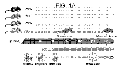
FIGs. 1A-K. Antibiotic treatment exacerbates motor symptoms in an ALS mouse
model.
(A) Experimental design. Evaluation of motor symptoms by behavioral (B)
rotarod, (C) hanging-
wire grip tests and (D) neurological scoring across the disease course.
*P<0.05, **P<0.005,
Mann-Whitney U test. The experiment was repeated 3 times, (N=5-10 mice). (E)
Histological
images and (F) quantification of lower-motor neurons in the spinal cords of
140-day old water-
and Abx-treated SOD1-Tg mice. *P<0.05, Mann-Whitney U test. (G) T2 maps and (H-
I)
quantification of T2 relaxation time in the corresponding areas between water-
and Abx-treated
SOD1-Tg mice throughout the disease progression. **P<0.005, ***P<0.0005, Mann-
Whitney U
test. The experiment was repeated twice, (N=5 mice). (J) Survival of GF (N=14)
and SPF (N=17)
SOD1-Tg mice. **P<0.005, Log-rank test. The experiment was repeated twice (K).
Survival of
Abx- and water-treated TDP43-Tg (N=10 in each group) mice. ****P<0.0001, Log-
rank test.
The experiment was repeated twice.
FIGs. 2A-H. SOD1-Tg mice develop early gut microbiome compositional and
functional
differences as compared to WT littermate controls. Weighted UniFrac PCoA on
(A) day 40 (pre-
symptomatic), (B) day 100 (disease onset) and (C) day 140 (advanced disease).
The experiment
was repeated 3 times, (N=6 mice in each group). (D) Species-level taxa summary
obtained by gut
microbiome metagenomic shotgun sequencing of WT and SOD1-Tg stool samples
during disease
progression. (E) PCA of KEGG entries of WT and SOD1-Tg microbiome. p=1.57x10-
14,
Spearman correlation coefficient. (F) Schematic representation and (G) heatmap
of bacterial gene
abundances of tryptophan metabolism. (H) Heatmap of bacterial gene abundances
of the
CA 03112135 2021-03-07
WO 2020/058979
PCT/IL2019/051041
9
nicotinamide and nicotinate biosynthesis pathway. N=6 mice, *P<0.05,
**P<0.005,
***P<0.0005, Mann-Whitney U test.
FIGs. 3A-H. Akkerrnansia rnuciniphila colonization ameliorates motor
degeneration and
increases life-span in SOD1-Tg mice. (A) Linear regression of AM relative
abundance (16S
rDNA sequencing) of SOD1-Tg and WT stool over time and (B) qPCR of AM 16S gene
copies
in fecal DNA extract (N=6 mice). Motor functions of SOD1-Tg and WT mice
treated with AM
indicated by (C) rotarod, (D) hanging-wire grip test and (E) neurological
scoring. (F) Histological
images and (G) spinal cord motor neuron quantification in 140-day old PBS- and
AM-treated
SOD1-Tg mice. *P<0.05, **P<0.005 Mann-Whitney U test. (H) Survival of PBS-, AM-
,
Prevotella rnelaninogenica (PM)- and Lactobacillus gasseri (LG)-treated mice
***P<0.0005
Log-rank test. The experiment was repeated 6 times, (N=5-26 mice).
FIGs. 4A-F. Akkerrnansia rnuciniphila treatment is associated with enhanced
nicotinamide
biosynthesis in SOD1-Tg mice. (A) Significantly increased serum metabolites in
SOD1-Tg mice
treated with AM (upper-right quadrant N=7-8 mice). (B) Serum levels
nicotinamide pathway
metabolites in SOD1-Tg and WT mice treated with AM or PBS. (C) Nicotinamide
levels in
bacterial cultures. **P<0.005, ***P<0.0005 Mann-Whitney U test. CSF
nicotinamide levels of
SOD1-Tg and WT mice treated with AM or PBS on (D) day 100 and (E) day 140.
*P<0.05,
**P<0.005, ***P<0.0005 Mann-Whitney U test. (F) Schematic representation of
the
microbiome-derived nicotinamide producing genes in AM treated SOD1-Tg fecal
samples. The
indicated genes increased in abundance following AM treatment (N=7-8 mice),
Mann Whitney U
ranksum test.
FIGs. 5A-G. Nicotinamide treatment ameliorates ALS progression in SOD1-Tg
mice. (A)
CSF and (B) sera NAM levels in NAM and vehicle treated SOD1-Tg mice (N=10
mice). Motor
performances of NAM or vehicle treated SOD1-Tg mice using subcutaneous osmotic
pumps
indicated by (C) rotarod, (D) hanging-wire grip test and (E) neurological
scoring. *P<0.05
***P<0.0005 Mann-Whitney U test. The experiment was repeated 3 times, (N=10
mice). (F)
Survival assessment of NAM and vehicle treated SOD1-Tg mice p=0.1757, Log-rank
test. (G)
neurological scoring of Abx-pretreated SOD1-Tg mice inoculated with WT or
AnadA E. coli.
***P<0.0005 Mann-Whitney U test.
FIGs. 6A-E. Uncovering potential downstream motor neuron modulatory mechanisms
of
AM and NAM treatments. (A) Heatmap of FDR-corrected differentially-expressed
genes in the
spinal cords of NAM-treated SOD1-Tg mice (N=10 mice). (B) Spearman correlation
of spinal
cord transcripts 1og2 fold change between AM- and NAM-treated SOD1-Tg mice.
(C)
Comparison of the significantly differentially-expressed genes following NAM
treatment with
CA 03112135 2021-03-07
WO 2020/058979
PCT/IL2019/051041
the KOG database classified into 4 neuropathological groups. FDR-corrected
gene set enrichment
distribution of spinal cord transcripts of (D) NAM-treated and (E) AM-treated
SOD1-Tg mice
into biological process, molecular functions and cellular components.
FIGs. 7A-F. Microbiome-derived nicotinamide metabolism is impaired in ALS
patients
5
(A) PCA of bacterial species composition (for PC1 p=3.3x10⁻⁶, Spearman
correlation coefficient)
or (B) KEGG orthology (KO) annotated bacterial genes (for PC1 p=2.8x10⁻⁹,
Spearman
correlation coefficient) obtained by metagenomic shotgun sequencing of stool
samples from ALS
patients (N=32) and healthy controls (family members, N=27). (C) KO relative
abundances of
microbiome-associated genes of the nicotinamide pathway in ALS and healthy
stool samples. (D)
10
Serum metabolites levels of tryptophan/nicotinamide pathways in ALS patients
and healthy
individuals obtained by non-targeted metabolomics. (E) Serum and (F) CSF NAM
levels of ALS
patients (N=41 for serum and 12 for CSF) and healthy controls (N=21 for serum
and 17 for CSF),
***P<0.0005, Mann Whitney U test.
FIGs. 8A-I. Antibiotic treatment exacerbates ALS symptoms in SOD1-Tg mice.
SOD1-
Tg and WT littermate control mice were untreated or treated with broad-
spectrum Abx in their
drinking water from age 40 days until the experimental end-point. On days 60,
80, 100, 120 and
140 motor performances of the mice were assessed by (A, D and G) rotarod, (B,
E and H)
hanging wire grip test and (C, F and I) neurological scoring. (N=5-10 mice),
*P<0.05,
**P<0.005, Mann-Whitney U test.
FIGs. 9A-P. The effects of antibiotic treatment on ALS symptoms in SOD1-Tg
mice.
Linear regression of motor functions over time in SOD1-Tg and WT treated
indicated by (A)
rotarod, (B) hanging-wire grip test, and (C) neurological score. (D) MRI of
brain areas and their
corresponding (E-I) quantification of T2 relaxation time between water and Abx-
treated SOD1-
Tg mice throughout ALS. *P<0.05, **P<0.005, ****P<0.00005, Mann-Whitney U
test. (J) Home
cage locomotion analysis over a period of 46 h, days 100-101 (N=5 mice).
*P=0.03.
Distributions of immune cell sub-populations in the small-intestine (K-L),
colon (M-N), spinal
cord on day 50 (0) and 140 (P) between water and Abx-treated SOD1-Tg mice.
(N=5 mice),
Mann-Whitney U test.
FIGs. 10A-D. Survival of GF- vs. SPF-SOD1-Tg mice and Abx-treated TDP43-Tg
mice.
Survival of SPF- and GF-SOD1-Tg mice that were spontaneously colonized on day
115.
*P<0.05, Log-rank test. The experiment was done twice: (A) (N=13 SPF- and 6 GF
SOD1-Tg
mice) and (B) (N=5 SPF- and 8 GF-SOD1-Tg mice). (C-D) Survival of Abx- and
water-treated
TDP43-Tg mice **P<0.005, **** P<0.0001 Log-rank test. The experiment was done
twice
(N=5-10 mice in each group).
CA 03112135 2021-03-07
WO 2020/058979
PCT/IL2019/051041
11
FIGs. 11A-0. Microbial compositional dynamics in the SOD1-Tg mouse model
across
ALS progression. (A) Taxa summary of bacterial phyla in individual WT and SOD1-
Tg mice
during ALS course and (B) genera (averaged time points) obtained by 16S rDNA
sequencing of
stool samples. (N=6 mice), the experiment was repeated 3 times. (C) Relative
abundances of
significant differentially representative genera between SOD1-Tg and WT mice
across the
disease progression. (D-M) FDR-corrected linear regression comparison of
representative
bacterial relative abundance change during ALS progression between WT and SOD1-
Tg stool.
Spearman correlation coefficient. (N) Alpha diversity of SOD1-Tg and WT
microbiomes over
time. The experiment was repeated 3 times, (N=6 mice in each group. (0) qPCR-
based
quantification of total 16S copy-number in 1 ng of DNA extracted from stool
samples of SOD1-
Tg and WT mice (N=5-6 mice).
FIGs. 12A-M. Microbial compositional dynamics in Abx-treated SOD1-Tg mouse
model
across ALS progression. (A) Taxa summary of bacterial phyla in individual Abx-
treated WT and
SOD1-Tg mice during ALS course. Weighted UniFrac PCoA on (B) day 47 (pre-Abx),
and (C-
G) days 60-140 of the disease under chronic Abx regime. (H-M) FDR corrected
volcano plots of
significantly enriched bacterial genera of Abx-treated WT and SOD1-Tg mice
during ALS
course.
FIGs. 13A-I. Microbial spontaneous colonization in Ex-GF SOD1-Tg mouse model
across ALS progression. (A) Taxa summary of bacterial genera in individual Ex-
GF WT and
SOD1-Tg undergoing spontaneous bacterial colonization during ALS course. (B-E)
Weighted
UniFrac PCoA of Ex-GF WT and SOD1-Tg mice on days 4, 5, 53 and 63 following
spontaneous
colonization. (F-I) FDR corrected volcano plots of significantly enriched
bacterial genera of Ex-
GF WT and SOD1-Tg during ALS course on days 4, 5, 53 and 63 following
spontaneous
colonization.
FIGs. 14A-E. A vivarium-affected dysbiosis in the SOD1-Tg mouse model (A)
Weighted
UniFrac PCoA and (B) Alpha diversity of WT and SOD1-Tg mice housed in a
different non-
barrier vivarium (vivarium B, Ben-Gurion University) on weeks 4, 6, 8 and 12
of age. (C)
Individual and (D) averaged taxa summary of bacterial genera in 80 days old WT
mice at
vivarium A (Weizmann Institute of Science) and vivarium B (Ben-Gurion
University). (E)
Abundance percentage summary of the top 20 highly abundant microbiome genera
in WT
animals at the two facilities and their corresponding abundances in SOD1-Tg
animals. The
comparison has performed once, (N=5-8) mice in each group.
FIGs. 15A-N. Metagenomic differences between WT and SOD1-Tg fecal microbiomes
(A) PCoA plot of bacterial composition and (B) Taxa summary representation at
the species level
CA 03112135 2021-03-07
WO 2020/058979
PCT/IL2019/051041
12
of gut microbiome of WT and S0D1-Tg mice obtained by metagenomic shotgun
sequencing.
The experiment was repeated twice (N=6 mice). (C-N) FDR-corrected linear
regression
comparison of representative bacterial relative abundance change during ALS
progression
between WT and S0D1-Tg stool. Spearman correlation coefficient.
FIGs. 16A-L. Metabolic measurements in S0D1-Tg and WT littermates
Representative
recording (A, C, E, G, I, J, K) and quantification (B, D, F, H, L) of food
intake (A, B), water
consumption (C, D), respiratory exchange ratio (E, F), 02 consumption (G, H),
Heat production
(I), locomotion (J) and speed (K, L) of 60 days old WT (N=8) and SOD1-Tg (N=7)
mice.
FIGs. 17A-L. Mono-colonization of Abx pre-treated S0D1-Tg mice with selected
ALS-
correlating microbiome strains. Motor functions of Abx pre-treated S0D1-Tg
mice treated with
PBS, Eggerthella lento (EL), Coprobacillus cateniforrnis (CC), Parabacteroides
goldsteinii (PG),
Lactobacillus rnurinus (LM), Parabacteroides distasonis (PD), Lactobacillus
gasseri (LG),
Prevotella rnelaninogenica (PM), or Akkerrnansia rnuciniphila (AM, ATCC 835)
indicated by
(A) rotarod, (B) hanging-wire grip test and (C) neurological scoring. (D-F)
Motor functions of
Abx pre-treated SOD1-Tg mice treated with PBS or Eisenbergiella tayi (ET), or
(G-I)
Subdoligranulum variabile (SV). (J-L) Motor functions of Abx pre-treated WT
littermate controls
treated with PBS, LM, PD, LG, PM or AM. (N=6-8 mice) *P<0.05, **P<0.005,
***P<0.0005
Mann-Whitney U test.
FIGs. 18A-M. The effects of Rurninococcus torques mono-colonization on ALS
progression in S0D1-Tg mice. (A) Linear regression of Rurninococcus torques
(RT) relative
abundance (16S rDNA sequencing) of S0D1-Tg and WT stool (N=6 mice). (B)
Rotarod, (C)
hanging-wire grip test and (D) neurological scoring of Abx-pretreated WT and
SOD1-Tg treated
with PBS or RT (N=5-9 mice), *P<0.05, **P<0.005, ***P<0.0005, Mann-Whitney U
test. (E)
Histological images and (F) quantification of spinal cord motor neurons of 140
days old PBS-
and RT-treated SOD1-Tg mice. (G) Brain areas and their corresponding (H-M) T2
relaxation time
quantification between PBS and RT-treated S0D1-Tg mice throughout the disease.
*P<0.05,
**P<0.005, ***P<0.0005, ****P<0.00005 Mann-Whitney U test. The experiment was
repeated
twice, (N=5 mice).
FIGs. 19A-I. Rurninococcus torques treatment exacerbates ALS symptoms in S0D1-
Tg
mice. Assessment of Abx-pretreated SOD1-Tg and WT littermate treatment with
Rurninococcus
torques (RT) in three biological repeats, by (A, D and G) rotarod, (B, E and
H) hanging wire grip
test and (C, F and I) neurological scoring. (N=5-10 mice), *P<0.05, **P<0.005,
***P<0.0005
Mann-Whitney U test.
CA 03112135 2021-03-07
WO 2020/058979
PCT/IL2019/051041
13
FIGs. 20A-0. Akkerrnansia rnuciniphila treatment attenuates ALS symptoms in
SOD1-Tg
mice. Abx-pretreated SOD1-Tg and WT littermate control mice were treated
orally with AM
(ATCC 835) or PBS as vehicle from age 60 days until the experimental end-
point. On days 60,
80, 100, 120 and 140 motor performance of the mice was assessed by (A, D, G, J
and 0) rotarod,
(B, E, H, K and M) hanging-wire grip test and (C, F, I, L and N) neurological
scoring. (N=5-26
mice), *P<0.05, **P<0.005, ***P<0.0005, Mann-Whitney U test.
FIGs. 21A-L. The effects of Akkerrnansia rnuciniphila treatment on ALS
manifestation
and microbiome composition in SOD1-Tg mice. (A-D) T2 relaxation time
quantification in PBS
and AM (ATCC 835)-treated Abx-pretreated SOD1-Tg mice at days 100 and 140.
***P<0.0005,
****P<0.00005, Mann-Whitney U test. (E) Systemic FITC-dextran measurement at
120 days
WT and SOD1-Tg treated with PBS, AM, P. Melaninogenica (PM) or L. gaseri (LG).
(F) PCoA
of bacterial species compositions in SOD1-Tg mice treated with PBS or AM. (G)
Genera
bacterial summary of SOD1-Tg treated with PBS or AM. AM relative abundance in
(H) SOD1-
Tg or (I) WT mice treated with PBS or AM. *P<0.05, ***P<0.0005, ****P<0.00005,
Mann-
Whitney ranksum test. (I) Individual and (J) averaged qPCR-based fold change
of Akkerrnansia
rnuciniphila 16S copy number in mucosal and luminal samples across the GI
tract of 140 days old
AM or PBS treated WT and SOD1-Tg mice (K) Genera bacterial summary of SOD1-Tg
or (L)
WT mice treated with PBS or AM.
FIGs. 22A-C. Akkerrnansia rnuciniphila (ATCC 2869) treatment attenuates ALS
symptoms in SOD1-Tg mice. Abx-pretreated SOD1-Tg and WT littermate control
mice were
treated orally with AM (ATCC 2869) or PBS as vehicle from age 60 days until
the experimental
end-point. On days 60, 80, 100, 120 and 140 motor performance of the mice was
assessed by (A)
rotarod, (B) hanging-wire grip test and (C) neurological scoring. (N=8-10
mice), **P<0.005,
Mann-Whitney U test.
FIGs. 23A-J. Akkerrnansia rnuciniphila treatment alters mucus properties of
SOD1-Tg
mice. Immunohistochemical assessment of distal colon mucosa of 140 days old
(A) PBS- and (B)
AM- (BAA-835) Abx-pretreated WT and SOD1-Tg mice. DNA stained with Sytox-green
(green)
and the mucus with an anti-MUC2C3 antiserum and goat anti-Ig (red). The non-
stained areas
between the epithelium and outer mucus/luminal bacteria is the inner mucus
layer, allows points
to bacteria in this. Heatmap representation of (C) total mucus proteomic
landscape and (D) AM-
related peptides and (E-J) quantification of key representative mucus
components. (N=4-8 mice),
Mann-Whitney U test.
FIGs. 24A-G. Serum metabolomic profile is affected by antibiotics or AM
treatment in
ALS SOD1-Tg mice. Heatmap representation of serum metabolites of 100 days old
(A) naïve
CA 03112135 2021-03-07
WO 2020/058979
PCT/IL2019/051041
14
SOD1-Tg and their WT littermates, (B) water or Abx-treated SOD1-Tg mice, (C)
PBS or AM-
treated SOD1-Tg mice. (D) Scoring of top six serum metabolites which
significantly altered by
Abx treatment in SOD1-Tg mice by their potential to originate of the gut
microbiome. Motor
performances of Phenol sulfate or vehicle treated SOD1-Tg mice using
subcutaneous osmotic
pumps indicated by (E) rotarod, (F) hanging-wire grip test and (G)
neurological scoring.
FIGs. 25A-B. Tryptophan and Nicotinamide metabolism are affected by
antibiotics or
AM treatment in ALS SOD1-Tg mice. Non-targeted metabolomics assessment of
typtophan
metabolism of (A) water and Abx- treated or (B) PBS and AM-treated 100 days
old SOD1-Tg
mice.
FIGs. 26A-I. Nicotinamide treatment ameliorates ALS progression in SOD1-Tg
mice.
Motor performances of NAM or vehicle treated SOD1-Tg mice using subcutaneous
osmotic
pumps indicated by (A, D and G) rotarod, (B, E and H) hanging-wire grip test
and (C, F and I)
neurological scoring (N=10 mice). *P<0.05, **P<0.005, ***P<0.0005,
****P<0.00005, Mann-
Whitney U test.
FIGs. 27A-C. Mono-inoculation of SOD1-Tg mice with gut commensal impaired in
NAM
production (A) Nicotinamide levels in WT or AnadA E. coli cultures.
***P<0.0005, Mann-
Whitney U test. Motor performances of WT or AnadA E. co/i-inoculated Abx-
pretreated SOD1-
Tg mice indicated by (B) rotarod and (C) hanging-wire grip test.
FIG. 28. NAM differentially expressed genes associated with Nuclear
respiratory factor-1
(NRF-1). Representation of spinal cord transcripts obtained by RNA-seq
analysis that changed
similarly after AM and NAM treatments of SOD1-Tg mice and share the binding
site for the
Nuclear respiratory factor-1 (NRF-1) transcription factor. The analysis was
done using the
G:Profiler platform85.
FIGs. 29A-B. Different gut microbiome composition and serum metabolites
profile in
ALS patients. (A) Taxa summary representation at the species level of gut
microbiome of healthy
family members and ALS patients obtained by metagenomic shotgun sequencing and
a table of
the top 20 changed bacterial species between ALS patients and healthy control
individuals. (B)
Top 97 differentially-represented serum metabolites between healthy
individuals (N=13) and
ALS patients (N=23) obtained by untargeted metabolomics.
DESCRIPTION OF SPECIFIC EMBODIMENTS OF THE INVENTION
The present invention, in some embodiments thereof, relates to methods of
treating
Amyotrophic Lateral Sclerosis (ALS) and, more particularly, but not
exclusively, to treatment
with bacterial populations or metabolites thereof.
CA 03112135 2021-03-07
WO 2020/058979
PCT/IL2019/051041
Before explaining at least one embodiment of the invention in detail, it is to
be understood
that the invention is not necessarily limited in its application to the
details set forth in the
following description or exemplified by the Examples. The invention is capable
of other
embodiments or of being practiced or carried out in various ways.
5 Amyotrophic Lateral Sclerosis (ALS) is an idiopathic, genetically-
influenced
neurodegenerative disorder, whose variable onset and clinical course may be
contributed by
unknown environmental factors.
The present inventors have now demonstrated that wide spectrum antibiotics-
induced
depletion of the gut microbiome in the most commonly used ALS mouse model (the
SOD1-Tg
10 mouse model) leads to worsened disease symptoms (Figures 1A-K).
Furthermore, the gut
microbiome composition and metagenomic function of SOD1-Tg mice were altered
compared to
WT littermates, even before the onset of motor clinical symptoms, resulting in
a markedly
altered systemic metabolomic profile in these mice (Figures 2A-H).
Several microbial species were identified to be correlated or anti-correlated
with disease
15 severity in SOD1-Tg mice. Of these, post-antibiotic colonization of SOD1-
Tg with anaerobic
mono-cultures of Akkerrnansia Muciniphila (AM) led to improved motor symptoms
and survival
(Figures 3A-H), while colonization with Ruminococcus was associated with
worsening disease
symptoms (Figures 14A-M and 15A-I). Furthermore, key AM-derived microbial
genes of the
Nicotinamide (NAM) biosynthetic pathway were enriched in the gut microbiome of
AM-
supplemented SOD1-Tg mice, while NA and its biosynthetic intermediates were
enriched, in this
setting, in the cerebrospinal fluid (CSF) and serum of AM-treated SOD1-Tg mice
(Figures 4A-
F). Moreover, systemic NAM supplementation of SOD1-Tg mice induced clinical
improvement
in motor neuron symptoms, coupled with distinct beneficial CNS transcriptomic
modifications
(Figures 5A-F and 6A-E). In humans, a dysbiotic gut microbiome metagenomic
configuration,
skewed serum metabolomic profile, and altered serum and CSF NAM levels were
noted in ALS
patients compared to healthy family controls (Figures 7A-E). Together, these
results suggest that
modulatory links may exist between distinct gut commensals, their modulated
metabolites and
motor manifestations in ALS animal models and potentially in humans.
Consequently, the present teachings suggest use of gut microbiome-associated
modulating agents for the treatment of ALS.
Thus, according to a first aspect of the present invention, there is provided
a method of
treating ALS in a subject in need thereof comprising administering to the
subject a
therapeutically effective amount of a therapeutically effective amount of a
metabolite selected
from the group consisting of propyl 4-hydroxybenzoate, triethanolamine,
serotonin, 2-keto-3-
CA 03112135 2021-03-07
WO 2020/058979
PCT/IL2019/051041
16
deoxy-gluconate, N-trimethyl 5-aminovalerate, phenylalanylglycine,
theobromine, cys-gly,
glutamate, 1-palmitoy1-2-docosahexaenoyl-GPC, oxalate, stearoyl sphingomyelin,
1-palmitoy1-2-
docosahexaenoyl-GPC (16:0/22:6), 3 -ureidopropionate, 1-(1-enyl-palmitoy1)-2-
arachidonoyl-
GPC
(P-16:0/20:4), palmitoyl sphingomyelin (d18: 1/16:0), sphingomyelin (d18:
1/18: 1,
d18:2/18:0), pyruvate, taurocholate, N- acetyltyro
sine, tauro-beta-muricholate,
tauroursodeoxycholate, phenol sulfate, equol sulfate, cinnamate,
phenylpropionylglycine, 2-
aminophenol sulfate, 4-allylphenol sulfate, equol glucuronide, palmitoleoyl-
linoleoyl-glycerol,
oleoyl-linolenoyl-glycerol, 1-palmitoy1-2-oleoyl-GPE, hydroquinone sulfate,
guaiacol sulfate,
diacylglycerol, palmitoyl-linoleoyl-glycerol, gentisate and 13-HODE + 9-HODE
thereby treating
ALS.
As used herein, the term "treating" includes abrogating, substantially
inhibiting, slowing
or reversing the progression of ALS, substantially ameliorating clinical or
aesthetical symptoms
of ALS or substantially preventing the appearance of clinical or aesthetical
symptoms of ALS.
As used herein, the term "treating" refers to inhibiting, preventing or
arresting the
development of a pathology (i.e. ALS) and/or causing the reduction, remission,
or regression of a
pathology. Those of skill in the art will understand that various
methodologies and assays can be
used to assess the development of a pathology or reduction, remission or
regression of a
pathology, as further disclosed herein.
Amyotrophic lateral sclerosis (ALS), also known as Lou Gehrig's disease and
Motor
Neuron Disease (MND), is a progressive, fatal, neurodegenerative disease
caused by the
degeneration of motor neurons, the nerve cells in the central nervous system
that control
voluntary muscle movement. ALS typically causes muscle weakness and atrophy
throughout the
body as both the upper and lower motor neurons degenerate, ceasing to send
messages to
muscles. Unable to function, the muscles gradually weaken, develop
fasciculations (twitches)
because of denervation, and eventually atrophy because of that denervation.
Affected subjects
may ultimately lose the ability to initiate and control all voluntary
movement; bladder and bowel
sphincters and the muscles responsible for eye movement are usually, but not
always, spared.
Cognitive or behavioral dysfunction is also associated with the disease; about
half of ALS
subjects experience mild changes in cognition and behavior, and 10 ¨ 15 % show
signs of
frontotemporal dementia. Language dysfunction, executive dysfunction, and
troubles with social
cognition and verbal memory are the most commonly reported cognitive symptoms
in ALS.
The term "ALS", as used herein, includes all of the classifications of ALS
known in the
art, including, but not limited to classical ALS (typically affecting both
lower and upper motor
neurons), Primary Lateral Sclerosis (PLS, typically affecting only the upper
motor neurons),
CA 03112135 2021-03-07
WO 2020/058979
PCT/IL2019/051041
17
Progressive Bulbar Palsy (PBP or Bulbar Onset, a version of ALS that typically
begins with
difficulties swallowing, chewing and speaking) and Progressive Muscular
Atrophy (PMA,
typically affecting only the lower motor neurons).
According to specific embodiments, ALS is classical ALS.
The term "ALS" includes sporadic and familial (hereditary) ALS, ALS at any
rate of
progression (i.e. rapid or slow progression) and ALS at any stage (e.g. prior
to onset, at onset and
late stages of ALS).
According to specific embodiments, ALS is sporadic ALS.
According to specific embodiments, ALS is familial ALS.
According to specific embodiments, ALS is rapid progression ALS.
As used herein, the phrase "rapid progression ALS" refers to ALS in which the
symptoms
progress continuously and significant degradation of motor neurons can be
observed within less
than a year with subject survival of up to 4 years from diagnosis. According
to specific
embodiments, the rapid progression ALS is characterized by a change of above
0.65 ALSFRS-R
points over a period of 1 month.
According to specific embodiments, ALS is ALS-associated depression.
As used herein, the phrase "ALS-associated depression" refers to depression
and/or
anxiety which begin following ALS onset. According to specific embodiments,
the ALS-
associated depression is part of the ALS mechanism of action and may be
attributed to e.g.
Pseudo Bulbar Affect and frontal lobe dementia. Methods of diagnosing and
monitoring
depression are well known in the art and include, but not limited to, the ALS
Depression
Inventory (ADI-12), the Beck Depression Inventory (BDI); and the Hospital
Anxiety Depression
Scale (HADS) questionnaires.
As mentioned above, the method of the invention is directed, inter alia, to
treating ALS.
The treatment may be initiated at any stage of the disease, including
following detection of ALS
symptoms.
Detection of ALS may be determined by the appearance of different symptoms
depending on which motor neurons in the body are damaged first (and
consequently which
muscles in the body are damaged first). In general, ALS symptoms include the
earliest symptoms
which are typically obvious weakness and/or muscle atrophy. Other symptoms
include muscle
fasciculation (twitching), cramping, or stiffness of affected muscles, muscle
weakness affecting
an arm or a leg and/or slurred and nasal speech. Most ALS patients experience
first symptoms in
the arms or legs. Others first notice difficulty in speaking clearly or
swallowing. Other symptoms
include difficulty in swallowing, loss of tongue mobility and respiratory
difficulties.
CA 03112135 2021-03-07
WO 2020/058979
PCT/IL2019/051041
18
The symptoms may be also classified by the part of neuronal system that is
degenerated,
namely, upper motor neurons and lower motor neurons. Symptoms of upper motor
neuron
degeneration include tight and stiff muscles (spasticity) and exaggerated
reflexes (hyperreflexia)
including an overactive gag reflex. Symptoms of lower motor neuron
degeneration include
muscle weakness and atrophy, muscle cramps, and fleeting twitches of muscles
that can be seen
under the skin (fasciculations). To be diagnosed with ALS, patients must have
signs and
symptoms of upper and/or lower motor neuron damage that cannot be attributed
to other causes.
Alternatively, treatment may be initiated at progressive stages of the
disease, e.g. when
muscle weakness and atrophy spread to different parts of the body and the
subject has increasing
problems with moving [e.g. the subject may suffer from tight and stiff muscles
(spasticity), from
exaggerated reflexes (hyperreflexia), from muscle weakness and atrophy, from
muscle cramps,
and/or from fleeting twitches of muscles that can be seen under the skin
(fasciculations)],
swallowing (dysphagia), speaking or forming words (dysarthria).
Method of monitoring ALS progression are well known in the art. Non-limiting
examples
of such methods include Physical evaluation by a physician; Weight;
Electrocardiogram (ECG);
ALS Functional Rating Scale (ALSFRS or ALSFRS-R) score; respiratory function
which can be
measured by e.g. vital capacity (forced vital capacity or slow vital
capacity); muscle strength
which can be measured by e.g. hand held dynamometry (HHD), hand grip strength
dynamometry, manual muscle testing (MMT), electrical impedance myography (EIM)
and
Maximum Voluntary Isometric Contraction Testing (MVICT); motor unit number
estimation
(MUNE); cognitive/behavior function which can be measured by e.g. the ALS
Depression
Inventory (ADI-12), the Beck Depression Inventory (BDI) and the Hospital
Anxiety Depression
Scale (HADS) questionnaires; Quality of life which can be evaluated by e.g.
the ALS
Assessment Questionnaire (ALSAQ-40); and Akt phosphorylation and pAkt:tAkt
ratio (see
International Patent Application Publication No. W02012/160563, the contents
of which are
fully incorporated herein by reference).
According to specific embodiments, the subject is monitored by ALS Functional
Rating
Scale (ALSFRS); respiratory function; muscle strength and/or cognitive
function.
According to specific embodiments, muscle strength is evaluated by a method
selected
from the group consisting of hand held dynamometry (HHD), hand grip strength
dynamometry,
manual muscle testing (MMT) and electrical impedance myography (EIIVI); each
possibility
represents a separate embodiment of the present invention.
As used herein the term "subject" refers to a human subject at any age and of
any gender
which is diagnosed with a disease (i.e., ALS) or is at risk of to develop a
disease (i.e. ALS).
CA 03112135 2021-03-07
WO 2020/058979
PCT/IL2019/051041
19
According to specific embodiments, the subject has rapid progression ALS
and/or ALS-
associated depression.
According to specific embodiments the subject fulfils the El Escorial criteria
for probable
and definite ALS, i.e. the subject presents:
1. Signs of lower motor neuron (LMN) degeneration by clinical,
electrophysiological or neuropathologic examination,
2. Signs of upper motor neuron (UMN) degeneration by clinical examination,
and
3. Progressive spread of signs within a region or to other regions,
together with the
absence of:
Electrophysiological evidence of other disease processes that might explain
the
signs of LMN and/or UMN degenerations; and
Neuroimaging evidence of other disease processes that might explain the
observed clinical and electrophysiological signs.
According to specific embodiments, the subject has an ALSFRS-R score of 26-42
prior to
treatment according to the present invention.
According to specific embodiments, the subject has a disease progression rate
greater
than 0.65 ALSFRS-R points per month over the last 3-12 months prior to
treatment according to
the present invention.
As mentioned, the method includes administering to the subject a
therapeutically
effective amount of at least one of the following bacterial metabolites:
propyl 4-
hydroxybenzoate, triethanolamine, serotonin, 2-keto-3-deoxy-gluconate, N-
trimethyl 5-
aminovalerate, phenylalanylglycine, theobromine, cys-gly, glutamate, 1-
palmitoy1-2-
docosahexaenoyl-GPC, oxalate, stearoyl sphingomyelin, 1-palmitoy1-2-
docosahexaenoyl-GPC
(16:0/22:6), 3-ureidopropionate, 1-(1-enyl-palmitoy1)-2-arachidonoyl-GPC (P-
16:0/20:4),
palmitoyl sphingomyelin (d18:1/16:0), sphingomyelin (d18:1/18:1, d18:2/18:0),
pyruvate,
taurocholate, N-acetyltyrosine, tauro-beta-muricholate, tauroursodeoxycholate,
phenol sulfate,
equol sulfate, cinnamate, phenylpropionylglycine, 2-aminophenol sulfate, 4-
allylphenol sulfate,
equol glucuronide, palmitoleoyl-linoleoyl-glycerol, oleoyl-linolenoyl-
glycerol, 1-palmitoy1-2-
oleoyl-GPE, hydroquinone sulfate, guaiacol sulfate, diacylglycerol, palmitoyl-
linoleoyl-glycerol,
gentisate and 13-HODE + 9-HODE.
According to a particular embodiment, at least one metabolite selected from
the group
consisting of propyl 4-hydroxybenzoate, triethanolamine, serotonin, 2-keto-3-
deoxy-gluconate,
N-trimethyl 5-aminovalerate, phenylalanylglycine, theobromine, cys-gly,
glutamate, 1-
palmitoy1-2-docosahexaenoyl-GPC are provided.
CA 03112135 2021-03-07
WO 2020/058979
PCT/IL2019/051041
In another embodiment, the bacterial metabolite nicotinamide is provided
together with
one of the above mentioned metabolites.
In still another embodiment, the bacterial metabolite nicotinamide is not
provided.
As used herein, the term "cinnamate" refers to cinnamic acid, salts thereof,
cinnamate
5 esters, p-dimethylaminocinnamate, cinnamaldehyde, cinnamyl acetate, cinnamyl
alcohol,
cinnamyl benzoate, cinnamyl cinnamate, cinnamyl formate, cinnamyl isobutyrate,
cinnamyl
isovalerate and cinnamyl phenylacetate and combinations thereof.
The equol of this aspect of the present invention may be (S)-equol (e.g. AUS-
131, which
is currently under development for treatment of hot flashes in menopausal
women). In one
10 embodiment, the equol is an equol salt such as equol sulfate.
Nicotinamide (NA), also known as "niacinamide", is the amide derivative form
of
Vitamin B3 (niacin). NA has the chemical formula C6H6N20.
NH2
0
N
Nicotinamide (NA)
It will be understood by the skilled reader that nicotinamide, as well as
other compounds
used in the present invention, may be capable of forming salts, complexes,
hydrates and solvates,
and that the use of such forms in the defined treatments is contemplated
herein. Nicotinamide
preparations of high purities, e.g. of 97 or 99% purity, are commercially
available. Such
commercial preparations may suitably be used for preparing nicotinamide
compositions for use
in the present methods. Furthermore, synthesis methods of nicotinamide of high
purity are
known to those skilled in the art.
According to a particular embodiment, the nicotinamide is a nicotinamide
derivative or a
nicotinamide mimic. The term "derivative of nicotinamide (NA)" as used herein
denotes a
compound which is a chemically modified derivative of the natural NA. In one
embodiment, the
chemical modification may be a substitution of the pyridine ring of the basic
NA structure (via
the carbon or nitrogen member of the ring), via the nitrogen or the oxygen
atoms of the amide
moiety. When substituted, one or more hydrogen atoms may be replaced by a
substituent and/or
a substituent may be attached to a N atom to form a tetravalent positively
charged nitrogen.
Thus, the nicotinamide of the present invention includes a substituted or non-
substituted
nicotinamide. In another embodiment, the chemical modification may be a
deletion or
CA 03112135 2021-03-07
WO 2020/058979
PCT/IL2019/051041
21
replacement of a single group, e.g. to form a thiobenzamide analog of NA, all
of which being as
appreciated by those versed in organic chemistry. The derivative in the
context of the invention
also includes the nucleoside derivative of NA (e.g. nicotinamide adenine). A
variety of
derivatives of NA are described, some also in connection with an inhibitory
activity of the PDE4
enzyme (W003/068233; W002/060875; GB2327675A), or as VEGF-receptor tyrosine
kinase
inhibitors (W001/55114). For example, the process of preparing 4-aryl-
nicotinamide derivatives
(W005/014549). Other exemplary nicotinamide derivatives are disclosed in
W001/55114 and
EP2128244.
Nicotinamide mimics include modified forms of nicotinamide, and chemical
analogs of
nicotinamide which recapitulate the effects of nicotinamide in the
differentiation and maturation
of RPE cells from pluripotent cells. Exemplary nicotinamide mimics include
benzoic acid, 3-
aminobenzoic acid, and 6-aminonicotinamide. Another class of compounds that
may act as
nicotinamide mimics are inhibitors of poly(ADP-ribose) polymerase (PARP).
Exemplary PARP
inhibitors include 3-aminobenzamide, Iniparib (BSI 201), Olaparib (AZD-2281),
Rucaparib
(AG014699, PF- 01367338), Veliparib (ABT-888), CEP 9722, MK 4827, and BMN-673.
In one embodiment, the nicotinamide is nicotinamide adenine dinucleotide
(NAD). In
another embodiment, the nicotinamide is nicotinamide riboside.
Exemplary doses of the bacterial metabolites described herein include 1 to 500
mg/kg
daily. In one embodiment of the invention the treatment comprises the daily
administration of
>10 mg/kg, e.g. the daily administration of 10-500 mg/kg.
The present inventors contemplate combinations of the above described
bacterial
metabolites, e.g. two metabolites, three metabolites, four metabolites, five
metabolites, six
metabolites, seven metabolites, eight metabolites, nine metabolites or more.
Thus, for example the combination may include:
Nicotinamide and phenol sulfate;
Nicotinamide and equol;
Nicotinamide and cinnamate;
Nicotinamide, phenol sulfate and equol;
Nicotinamide, phenol sulfate and cinnamate;
Nicotinamide, equol and cinnamate;
Nicotinamide, equol, phenol sulfate and cinnamate.
Nicotinamide and at least one of the metabolites selected from the group
consisting of
propyl 4-hydroxybenzoate, triethanolamine, serotonin, 2-keto-3-deoxy-
gluconate, N-trimethyl 5-
CA 03112135 2021-03-07
WO 2020/058979
PCT/IL2019/051041
22
aminovalerate, phenylalanylglycine, theobromine, cys-gly, glutamate and 1-
palmitoy1-2-
docosahexaenoyl-GPC.
The bacterial metabolite may be provided per se or as part of a pharmaceutical
composition, where it is mixed with suitable carriers or excipients.
As used herein a "pharmaceutical composition" refers to a preparation of one
or more of
the active ingredients described herein with other chemical components such as
physiologically
suitable carriers and excipients. The purpose of a pharmaceutical composition
is to facilitate
administration of a compound to an organism.
Herein the term "active ingredient" refers to one or more of the bacterial
metabolites
described herein accountable for the biological effect.
Hereinafter, the phrases "physiologically acceptable carrier" and
"pharmaceutically
acceptable carrier" which may be interchangeably used refer to a carrier or a
diluent that does not
cause significant irritation to an organism and does not abrogate the
biological activity and
properties of the administered compound. An adjuvant is included under these
phrases.
Herein the term "excipient" refers to an inert substance added to a
pharmaceutical
composition to further facilitate administration of an active ingredient.
Examples, without
limitation, of excipients include calcium carbonate, calcium phosphate,
various sugars and types
of starch, cellulose derivatives, gelatin, vegetable oils and polyethylene
glycols.
Techniques for formulation and administration of drugs may be found in
"Remington's
Pharmaceutical Sciences," Mack Publishing Co., Easton, PA, latest edition,
which is
incorporated herein by reference.
Suitable routes of administration may, for example, include oral, rectal,
transmucosal,
especially transnasal, intestinal or parenteral delivery, including
intramuscular, subcutaneous and
intramedullary injections as well as intrathecal, direct intraventricular,
intracardiac, e.g., into the
right or left ventricular cavity, into the common coronary artery,
intravenous, inrtaperitoneal,
intranasal, or intraocular injections.
According to a particular embodiment, the agent is administered orally or
rectally.
Alternately, one may administer the pharmaceutical composition in a local
rather than
systemic manner, for example, via injection of the pharmaceutical composition
directly into a
tissue region of a patient.
The term "tissue" refers to part of an organism consisting of cells designed
to perform a
function or functions. Examples include, but are not limited to, brain tissue,
retina, skin tissue,
hepatic tissue, pancreatic tissue, bone, cartilage, connective tissue, blood
tissue, muscle tissue,
CA 03112135 2021-03-07
WO 2020/058979
PCT/IL2019/051041
23
cardiac tissue brain tissue, vascular tissue, renal tissue, pulmonary tissue,
gonadal tissue,
hematopoietic tissue.
Pharmaceutical compositions of some embodiments of the invention may be
manufactured by processes well known in the art, e.g., by means of
conventional mixing,
dissolving, granulating, dragee-making, levigating, emulsifying,
encapsulating, entrapping or
lyophilizing processes.
Pharmaceutical compositions for use in accordance with some embodiments of the
invention thus may be formulated in conventional manner using one or more
physiologically
acceptable carriers comprising excipients and auxiliaries, which facilitate
processing of the
active ingredients into preparations which, can be used pharmaceutically.
Proper formulation is
dependent upon the route of administration chosen.
For injection, the active ingredients of the pharmaceutical composition may be
formulated in aqueous solutions, preferably in physiologically compatible
buffers such as Hank's
solution, Ringer's solution, or physiological salt buffer. For transmucosal
administration,
penetrants appropriate to the barrier to be permeated are used in the
formulation. Such penetrants
are generally known in the art.
For oral administration, the pharmaceutical composition can be formulated
readily by
combining the active compounds with pharmaceutically acceptable carriers well
known in the
art. Such carriers enable the pharmaceutical composition to be formulated as
tablets, pills,
dragees, capsules, liquids, gels, syrups, slurries, suspensions, and the like,
for oral ingestion by a
patient. Pharmacological preparations for oral use can be made using a solid
excipient,
optionally grinding the resulting mixture, and processing the mixture of
granules, after adding
suitable auxiliaries if desired, to obtain tablets or dragee cores. Suitable
excipients are, in
particular, fillers such as sugars, including lactose, sucrose, mannitol, or
sorbitol; cellulose
preparations such as, for example, maize starch, wheat starch, rice starch,
potato starch, gelatin,
gum tragacanth, methyl cellulose, hydroxypropylmethyl-cellulose, sodium
carbomethylcellulose;
and/or physiologically acceptable polymers such as polyvinylpyrrolidone (PVP).
If desired,
disintegrating agents may be added, such as cross-linked polyvinyl
pyrrolidone, agar, or alginic
acid or a salt thereof such as sodium alginate.
Dragee cores are provided with suitable coatings. For this purpose,
concentrated sugar
solutions may be used which may optionally contain gum arabic, talc, polyvinyl
pyrrolidone,
carbopol gel, polyethylene glycol, titanium dioxide, lacquer solutions and
suitable organic
solvents or solvent mixtures. Dyestuffs or pigments may be added to the
tablets or dragee
coatings for identification or to characterize different combinations of
active compound doses.
CA 03112135 2021-03-07
WO 2020/058979
PCT/IL2019/051041
24
Pharmaceutical compositions which can be used orally, include push-fit
capsules made of
gelatin as well as soft, sealed capsules made of gelatin and a plasticizer,
such as glycerol or
sorbitol. The push-fit capsules may contain the active ingredients in
admixture with filler such as
lactose, binders such as starches, lubricants such as talc or magnesium
stearate and, optionally,
stabilizers. In soft capsules, the active ingredients may be dissolved or
suspended in suitable
liquids, such as fatty oils, liquid paraffin, or liquid polyethylene glycols.
In addition, stabilizers
may be added. All formulations for oral administration should be in dosages
suitable for the
chosen route of administration.
For buccal administration, the compositions may take the form of tablets or
lozenges
formulated in conventional manner.
For administration by nasal inhalation, the active ingredients for use
according to some
embodiments of the invention are conveniently delivered in the form of an
aerosol spray
presentation from a pressurized pack or a nebulizer with the use of a suitable
propellant, e.g.,
dichlorodifluoromethane, trichlorofluoromethane, dichloro-tetrafluoroethane or
carbon dioxide.
In the case of a pressurized aerosol, the dosage unit may be determined by
providing a valve to
deliver a metered amount. Capsules and cartridges of, e.g., gelatin for use in
a dispenser may be
formulated containing a powder mix of the compound and a suitable powder base
such as lactose
or starch.
The pharmaceutical composition described herein may be formulated for
parenteral
administration, e.g., by bolus injection or continuous infusion. Formulations
for injection may be
presented in unit dosage form, e.g., in ampoules or in multidose containers
with optionally, an
added preservative. The compositions may be suspensions, solutions or
emulsions in oily or
aqueous vehicles, and may contain formulatory agents such as suspending,
stabilizing and/or
dispersing agents.
Pharmaceutical compositions for parenteral administration include aqueous
solutions of
the active preparation in water-soluble form. Additionally, suspensions of the
active ingredients
may be prepared as appropriate oily or water based injection suspensions.
Suitable lipophilic
solvents or vehicles include fatty oils such as sesame oil, or synthetic fatty
acids esters such as
ethyl oleate, triglycerides or liposomes. Aqueous injection suspensions may
contain substances,
which increase the viscosity of the suspension, such as sodium carboxymethyl
cellulose, sorbitol
or dextran. Optionally, the suspension may also contain suitable stabilizers
or agents which
increase the solubility of the active ingredients to allow for the preparation
of highly
concentrated solutions.
CA 03112135 2021-03-07
WO 2020/058979
PCT/IL2019/051041
Alternatively, the active ingredient may be in powder form for constitution
with a
suitable vehicle, e.g., sterile, pyrogen-free water based solution, before
use.
The pharmaceutical composition of some embodiments of the invention may also
be
formulated in rectal compositions such as suppositories or retention enemas,
using, e.g.,
5 conventional suppository bases such as cocoa butter or other glycerides.
Pharmaceutical compositions suitable for use in context of some embodiments of
the
invention include compositions wherein the active ingredients are contained in
an amount
effective to achieve the intended purpose. More specifically, a
therapeutically effective amount
means an amount of active ingredients (e.g. nicotinamide) effective to
prevent, alleviate or
10 ameliorate symptoms of a disorder (e.g., ALS) or prolong the survival of
the subject being
treated.
Determination of a therapeutically effective amount is well within the
capability of those
skilled in the art, especially in light of the detailed disclosure provided
herein.
For any preparation used in the methods of the invention, the therapeutically
effective
15 amount or dose can be estimated initially from in vitro and cell culture
assays. For example, a
dose can be formulated in animal models to achieve a desired concentration or
titer. Such
information can be used to more accurately determine useful doses in humans.
Toxicity and therapeutic efficacy of the active ingredients described herein
can be
determined by standard pharmaceutical procedures in vitro, in cell cultures or
experimental
20 animals. The data obtained from these in vitro and cell culture assays
and animal studies can be
used in formulating a range of dosage for use in human. The dosage may vary
depending upon
the dosage form employed and the route of administration utilized. The exact
formulation, route
of administration and dosage can be chosen by the individual physician in view
of the patient's
condition. (See e.g., Fingl, et al., 1975, in "The Pharmacological Basis of
Therapeutics", Ch. 1
25 p.1).
Dosage amount and interval may be adjusted individually to provide blood,
brain or CSF
levels of the active ingredient are sufficient to induce or suppress the
biological effect (minimal
effective concentration, MEC). The MEC will vary for each preparation, but can
be estimated
from in vitro data. Dosages necessary to achieve the MEC will depend on
individual
characteristics and route of administration. Detection assays can be used to
determine plasma
concentrations.
Depending on the severity and responsiveness of the condition to be treated,
dosing can
be of a single or a plurality of administrations, with course of treatment
lasting from several days
to several weeks or until cure is effected or diminution of the disease state
is achieved.
CA 03112135 2021-03-07
WO 2020/058979
PCT/IL2019/051041
26
The amount of a composition to be administered will, of course, be dependent
on the
subject being treated, the severity of the affliction, the manner of
administration, the judgment of
the prescribing physician, etc.
Compositions of some embodiments of the invention may, if desired, be
presented in a
pack or dispenser device, such as an FDA approved kit, which may contain one
or more unit
dosage forms containing the active ingredient. The pack may, for example,
comprise metal or
plastic foil, such as a blister pack. The pack or dispenser device may be
accompanied by
instructions for administration. The pack or dispenser may also be
accommodated by a notice
associated with the container in a form prescribed by a governmental agency
regulating the
manufacture, use or sale of pharmaceuticals, which notice is reflective of
approval by the agency
of the form of the compositions or human or veterinary administration. Such
notice, for example,
may be of labeling approved by the U.S. Food and Drug Administration for
prescription drugs or
of an approved product insert. Compositions comprising a preparation of the
invention
formulated in a compatible pharmaceutical carrier may also be prepared, placed
in an appropriate
container, and labeled for treatment of an indicated condition, as is further
detailed above.
The metabolites of the present invention may be provided in a food (such as
food bars,
biscuits, snack foods and other standard food forms well known in the art), or
in drink
formulations. Drinks can contain flavoring, buffers and the like. Nutritional
supplements
comprising the metabolites of the present invention are also contemplated.
The metabolites of this aspect of the present invention may be provided via a
probiotic
composition comprising microbes that generate the metabolites.
The term "probiotic" as used herein, refers to one or more microorganisms
which, when
administered appropriately, can confer a health benefit on the host or subject
and/or reduction of
risk and/or symptoms of a disease (such as ALS), disorder, condition, or event
in a host
organism.
Thus, according to another aspect of the present invention there is provided a
method of
treating ALS comprising administering to the subject a therapeutically
effective amount of a
bacterial composition comprising at least one of Streptococcus therrnophiles,
Faecalibacteriurn
prausnitzii, Eubacteriurn rectale, Bacteroides plebeius, Coprococcus,
Roseburia horninis,
Eubacteriurn ventriosurn, Lachnospiraceae, Eubacteriurn hallii, Bacteroidales,
Bifidobacteriurn
pseudocatenulaturn, Anaerostipes hadrus, Akkerrnansia Muciniphila (AM),
Anaeroplasrna,
Prevotella, Distanosis, Parabacteroides (e.g. Parabacteroides distasonis,
Parabacteroides
goldsteinii) Rikenellaceae, Alistipes, Candidatus Arthrornitus, Eggerthella,
Oscillibacter,
Subdoligranulurn and Lactobacillus (e.g. Lactobacillus rnurinus).
CA 03112135 2021-03-07
WO 2020/058979
PCT/IL2019/051041
27
According to a specific embodiment, the bacteria composition comprises at
least one of,
at least two of, at least three of, at least four of, at least five of
Streptococcus therrnophiles,
Faecalibacteriurn prausnitzii, Eubacteriurn rectale, Bacteroides plebeius,
Coprococcus,
Roseburia horninis, Eubacteriurn ventriosurn, Lachnospiraceae, Eubacteriurn
hallii,
Bacteroidales, Bifidobacteriurn pseudocatenulaturn and Anaerostipes hadrus.
According to a particular embodiment, the bacterial composition comprises
Akkerrnansia
Muciniphila (AM).
The probiotic microorganism may be in any suitable form, for example in a
powdered
dry form. In addition, the probiotic microorganism may have undergone
processing in order for
it to increase its survival. For example, the microorganism may be coated or
encapsulated in a
polysaccharide, fat, starch, protein or in a sugar matrix. Standard
encapsulation techniques
known in the art can be used. For example, techniques discussed in U.S. Pat.
No. 6,190,591,
which is hereby incorporated by reference in its entirety, may be used.
According to a particular embodiment, the probiotic composition is formulated
in a food
product, functional food or nutraceutical.
In some embodiments, a food product, functional food or nutraceutical is or
comprises a
dairy product. In some embodiments, a dairy product is or comprises a yogurt
product. In some
embodiments, a dairy product is or comprises a milk product.
In some embodiments, a dairy product is or comprises a cheese product. In some
embodiments, a food product, functional food or nutraceutical is or comprises
a juice or other
product derived from fruit. In some embodiments, a food product, functional
food or
nutraceutical is or comprises a product derived from vegetables. In some
embodiments, a food
product, functional food or nutraceutical is or comprises a grain product,
including but not
limited to cereal, crackers, bread, and/or oatmeal. In some embodiments, a
food product,
functional food or nutraceutical is or comprises a rice product. In some
embodiments, a food
product, functional food or nutraceutical is or comprises a meat product.
Prior to administration, the subject may be pretreated with an agent which
reduces the
number of naturally occurring microbes in the microbiome (e.g. by antibiotic
treatment).
According to a particular embodiment, the treatment significantly eliminates
the naturally
occurring gut microflora by at least 20 %, 30 % 40 %, 50 %, 60 %, 70 %, 80 %
or even 90 %.
In some particular embodiments, appropriate doses or amounts of probiotics to
be
administered may be extrapolated from dose-response curves derived from in
vitro or animal
model test systems. The effective dose or amount to be administered for a
particular individual
can be varied (e.g., increased or decreased) over time, depending on the needs
of the individual.
CA 03112135 2021-03-07
WO 2020/058979
PCT/IL2019/051041
28
In some embodiments, where bacteria are administered, an appropriate dosage
comprises at least
about 100, 200, 300, 400, 500, 600, 700, 800, 900, 1000 or more bacterial
cells. In some
embodiments, the present invention encompasses the recognition that greater
benefit may be
achieved by providing numbers of bacterial cells greater than about 1000 or
more (e.g., than
about 1500, 2000, 2500, 3000, 35000, 4000, 4500, 5000, 5500, 6000, 7000, 8000,
9000, 10,000,
15,000, 20,000, 25,000, 30,000, 40,000, 50,000, 75,000, 100,000, 200,000,
300,000, 400,000,
500,000, 600,000, 700,000, 800,000, 900,000, 1x10⁶, 2x10⁶, 3 x10⁶, 4 x10⁶, 5
x106, 6 x10⁶, 7
x106, 8 x10⁶, 9 x10⁶, 1 x10⁷, 1 x10⁸, 1 x10⁹, 1 x10¹⁰, 1 x10¹¹, 1 x10¹², 1
x1013 or more bacteria.
The present inventors have further shown that levels of particular bacterial
populations
increase in the microbiome of a subject with ALS.
Thus, according to still another aspect of the present invention there is
provided a method
of treating ALS in a subject in need thereof comprising administering to the
subject a
therapeutically effective amount of an agent that selectively decreases the
amount of a bacterial
population selected from the group consisting of Escherichia coli, Clostridium
leptum,
Clostridium nexile, Clostridium bolteae, Bacteroides fragilis, Catenibacterium
mitsuokai,
Bifidobacterium dentium, Megasphaera, Parasutterella excrementihominis,
Burkholderiales
bacterium, Clostridium ramosum, Streptococcus anginosus,
Flavonifractor_plautii,
Methanobrevibacter smithii, Acidaminococcus intestine, Ruminococcus
e.g.
Ruminococcus torques or Ruminococcus gnavus, Bifidobacterium,
Coriobacteriaceae,
Bacteroides, Parabacteroides, 524_7, Clostridiaceae, flavefaciens,
Desulfovibrioaceae,
Allobaculum, Sutterella, Helicobacteraceae, Coprococcus and Oscillospira, in
the gut
microbiome of the subject, thereby treating the ALS.
According to a particular embodiment, the bacterial population is selected
from the group
consisting of Escherichia coli, Clostridium leptum, Ruminococcus (e.g.
Ruminococcus gnavus or
Ruminococcus torques), Clostridium nexile, Clostridium bolteae, Bacteroides
fragilis,
Catenibacterium mitsuokai, Bifidobacterium dentium, Megasphaera,
Parasutterella
excrementihominis, Burkholderiales bacterium, Clostridium ramosum,
Streptococcus anginosus,
Flavonifractor_plautii, Methanobrevibacter smithii and Acidaminococcus
intestine.
According to a particular embodiment, the bacterial population is selected
from the group
consisting of Ruminococcus, Desulfovibrioaceae, Allobaculum, Sutterella,
Helicobacteraceae,
Coprococcus and Oscillospira.
In a further embodiment, the bacterial population which is down-regulated is
at least one
of the following bacteria: Bacteroides dorei, Bacteroides vulgatus,
Bacteroides xylanisolvens,
Bifidobacterium pseudolongum, Dorea, Helicobacter hepaticus,
Lactobacillus_johnsonii,
CA 03112135 2021-03-07
WO 2020/058979
PCT/IL2019/051041
29
Lactobacillus reuteri, Lactobacillus sp ASF360,
Desulfovibrio desulfuricans,
Lactobacillus vaginalis, Mucispirillum schaedleri, Parabacteroides
(e.g.
Parabacteroides johnsonii) and Ruminococcus torques.
In one embodiment, at least two of the above described species/genus are down-
regulated,
at least three of the above described species/genus are down-regulated, at
least four of the above
described species/genus are down-regulated, at least five of the above
described species/genus are
down-regulated, all of the above described species or genus are down-
regulated.
The present invention contemplates an agent which down-regulates at least one
strain, 10
% of the strains, 20 % of the strains, 30 % of the strains, 40 % of the
strains, 50 % of the strains,
60 % of the strains, 70 % of the strains, 80 % of the strains, 90 % of the
strains or all of the
strains of the above disclosed species.
As used herein, the term "downregulates" refers to an ability to reduce the
amount (either
absolute or relative amount) and/or activity (either absolute or relative
activity) of a particular
species/genus of bacteria.
In one embodiment, the agent specifically downregulates the specified
species/genus of
bacteria.
Thus, for example, the agent may reduce the amount of the specified bacterial
species/genus as compared to at least one other bacterial species/genus of the
microbiome of the
subject, by at least 2 fold. According to a particular embodiment, the agent
downregulates the
particular bacterial species/genus by at least 5 fold, 10 fold or more as
compared to at least one
other bacterial species/genus of the microbiome.
In another embodiment, the agent reduces the amount of the specified bacterial
species/genus as compared to at least 10 % of the total bacterial
species/genus of the microbiome
of the subject, by at least 2 fold. According to a particular embodiment, the
agent downregulates
the specified bacterial species/genus by at least 5 fold, 10 fold or more as
compared to at least 10
% of the total bacterial species/genus of the microbiome of the subject.
In another embodiment, the agent reduces the amount of the specified bacterial
species/genus as compared to at least 20 % of the total bacterial
species/genus of the microbiome
of the subject, by at least 2 fold. According to a particular embodiment, the
agent downregulates
the specified bacterial species/genus by at least 5 fold, 10 fold or more as
compared to at least 20
% of the total bacterial species/genus of the microbiome of the subject.
In another embodiment, the agent reduces the amount of the specified bacterial
species/genus as compared to at least 30 % of the total bacterial
species/genus of the microbiome
of the subject, by at least 2 fold. According to a particular embodiment, the
agent downregulates
CA 03112135 2021-03-07
WO 2020/058979
PCT/IL2019/051041
the specified bacterial species/genus by at least 5 fold, 10 fold or more as
compared to at least 30
% of the total bacterial species/genus of the microbiome of the subject.
In another embodiment, the agent reduces the amount of the specified bacterial
species/genus as compared to at least 40 % of the total bacterial
species/genus of the microbiome
5 .. of the subject, by at least 2 fold. According to a particular embodiment,
the agent downregulates
the specified bacterial species/genus by at least 5 fold, 10 fold or more as
compared to at least 40
% of the total bacterial species/genus of the microbiome of the subject.
In another embodiment, the agent reduces the amount of the specified bacterial
species/genus as compared to at least 50 % of the total bacterial
species/genus of the microbiome
10 of the subject, by at least 2 fold. According to a particular
embodiment, the agent downregulates
the specified bacterial species/genus by at least 5 fold, 10 fold or more as
compared to at least 50
% of the total bacterial species/genus of the microbiome of the subject.
In another embodiment, the agent reduces the amount of the specified bacterial
species/genus as compared to at least 60 % of the total bacterial
species/genus of the microbiome
15 of the subject, by at least 2 fold. According to a particular
embodiment, the agent downregulates
the specified bacterial species/genus by at least 5 fold, 10 fold or more as
compared to at least 60
% of the total bacterial species/genus of the microbiome of the subject.
In another embodiment, the agent reduces the amount of the specified bacterial
species/genus as compared to at least 70 % of the total bacterial
species/genus of the microbiome
20 of the subject, by at least 2 fold. According to a particular
embodiment, the agent downregulates
the specified bacterial species/genus by at least 5 fold, 10 fold or more as
compared to at least 70
% of the total bacterial species/genus of the microbiome of the subject.
In another embodiment, the agent reduces the amount of the specified bacterial
species/genus as compared to at least 80 % of the total bacterial
species/genus of the microbiome
25 of the subject, by at least 2 fold. According to a particular
embodiment, the agent downregulates
the specified bacterial species/genus by at least 5 fold, 10 fold or more as
compared to at least 80
% of the total bacterial species/genus of the microbiome of the subject.
In another embodiment, the agent reduces the amount of the specified bacterial
species/genus as compared to at least 90 % of the total bacterial
species/genus of the microbiome
30 of the subject, by at least 2 fold. According to a particular
embodiment, the agent downregulates
the specified bacterial species/genus by at least 5 fold, 10 fold or more as
compared to at least 90
% of the total bacterial species/genus of the microbiome of the subject.
An exemplary agent which is capable of reducing a particular bacterial genus,
species or
strain is an antibiotic.
CA 03112135 2021-03-07
WO 2020/058979
PCT/IL2019/051041
31
As used herein, the term "antibiotic agent" refers to a group of chemical
substances,
isolated from natural sources or derived from antibiotic agents isolated from
natural sources,
having a capacity to inhibit growth of, or to destroy bacteria, and other
microorganisms, used
chiefly in treatment of infectious diseases. Examples of antibiotic agents
include, but are not
limited to; Amikacin; Amoxicillin; Ampicillin; Azithromycin; Azlocillin;
Aztreonam;
Aztreonam; Carbenicillin; Cefaclor; Cefepime; Cefetamet; Cefinetazole;
Cefixime; Cefonicid;
Cefoperazone; Cefotaxime; Cefotetan; Cefoxitin; Cefpodoxime; Cefprozil;
Cefsulodin;
Ceftazidime; Ceftizoxime; Ceftriaxone; Cefuroxime; Cephalexin; Cephalothin;
Cethromycin;
Chloramphenicol; Cinoxacin; Ciprofloxacin; Clarithromycin; Clindamycin;
Cloxacillin; Co-
lo amoxiclavuanate; D alb av ancin ; Daptomycin; Dicloxacillin; Doxyc yc
line ; Enoxacin;
Erythromycin estolate; Erythromycin ethyl succinate; Erythromycin
glucoheptonate;
Erythromycin lactobionate; Erythromycin stearate; Erythromycin; Fidaxomicin;
Fleroxacin;
Gentamicin; Imipenem; Kanamycin; Lomefloxacin; Loracarbef; Methicillin;
Metronidazole;
Mezlocillin; Minocycline; Mupirocin; Nafcillin; Nalidixic acid; Netilmicin;
Nitrofurantoin;
Norfloxacin; Ofloxacin; Oxacillin; Penicillin G; Piperacillin; Retapamulin;
Rifaxamin,
Rifampin; Roxithromycin; Streptomycin; Sulfamethoxazole; Teicoplanin;
Tetracycline;
Ticarcillin; Tigecycline; Tobramycin; Trimethoprim; Vancomycin; combinations
of Piperacillin
and Tazobactam; and their various salts, acids, bases, and other derivatives.
Anti-bacterial
antibiotic agents include, but are not limited to, aminoglycosides,
carbacephems, carbapenems,
cephalosporins , cephamycins , fluoroquinolones , glycopeptides , linco s
amide s, macrolides,
monobactams, penicillins, quinolones, sulfonamides, and tetracyclines.
Antibacterial agents also include antibacterial peptides. Examples include but
are not
limited to abaecin; andropin; apidaecins; bombinin; brevinins; buforin II;
CAP18; cecropins;
ceratotoxin; defen sin s ; dermaseptin; dermcidin; drosomycin; esculentins ;
indolicidin; LL3 7 ;
magainin; maximum H5; melittin; moricin; prophenin; protegrin; and or
tachyplesins.
According to a particular embodiment, the antibiotic is a non-absorbable
antibiotic.
Other agents which are not antibiotics are also contemplated by the present
inventors.
In one embodiment, the agent which is capable of down-regulating a particular
bacterial
genus/species/strain is a bacterial population that competes with the
bacterial
genus/species/strain for essential resources. Bacterial compositions are
further described herein
below.
In still another embodiment, the agent which is capable of down-regulating a
particular
bacterial genus/species/strain is a metabolite of a competing bacterial
population (or even from
the same species/strain) that serves to decrease the relative amount of the
bacterial species/strain.
CA 03112135 2021-03-07
WO 2020/058979
PCT/IL2019/051041
32
Additional agents that can specifically reduce a particular bacterial genus,
species or
strain are known in the art and include polynucleotide silencing agents.
Preferably, the polynucleotide silencing agent of this aspect of the present
invention
targets a sequence that encodes at least one essential gene (i.e., compatible
with life) in the
bacteria. The sequence which is targeted should be specific to the particular
bacteria species that
it is desired to down-regulate. Such genes include ribosomal RNA genes (16S
and 23S),
ribosomal protein genes, tRNA-synthetases, as well as additional genes shown
to be essential
such as dnaB, fabI, folA, gyrB, murA, pytH, metG, and tufA(B).
According to an embodiment of the invention, the polynucleotide silencing
agent is
specific to the target RNA and does not cross inhibit or silence other targets
or a splice variant
which exhibits 99% or less global homology to the target gene, e.g., less than
98%, 97%, 96%,
95%, 94%, 93%, 92%, 91%, 90%, 89%, 88%, 87%, 86%, 85%, 84%, 83%, 82%, 81%
global
homology to the target gene; as determined by PCR, Western blot,
Immunohistochemistry and/or
flow cytometry.
One agent capable of downregulating an essential bacterial gene is a RNA-
guided
endonuclease technology e.g. CRISPR system.
As used herein, the term "CRISPR system" also known as Clustered Regularly
Interspaced Short Palindromic Repeats refers collectively to transcripts and
other elements
involved in the expression of or directing the activity of CRISPR-associated
genes, including
sequences encoding a Cas gene (e.g. CRISPR-associated endonuclease 9), a tracr
(trans-
activating CRISPR) sequence (e.g. tracrRNA or an active partial tracrRNA), a
tracr-mate
sequence (encompassing a "direct repeat" and a tracrRNA-processed partial
direct repeat) or a
guide sequence (also referred to as a "spacer") including but not limited to a
crRNA sequence
(i.e. an endogenous bacterial RNA that confers target specificity yet requires
tracrRNA to bind to
Cas) or a sgRNA sequence (i.e. single guide RNA).
In some embodiments, one or more elements of a CRISPR system is derived from a
type
I, type II, or type III CRISPR system. In some embodiments, one or more
elements of a CRISPR
system (e.g. Cas) is derived from a particular organism comprising an
endogenous CRISPR
system, such as Streptococcus pyo genes, Neisseria meningitides, Streptococcus
thermophilus or
Treponema denticola.
In general, a CRISPR system is characterized by elements that promote the
formation of
a CRISPR complex at the site of a target sequence (also referred to as a
protospacer in the
context of an endogenous CRISPR system).
CA 03112135 2021-03-07
WO 2020/058979
PCT/IL2019/051041
33
In the context of formation of a CRISPR complex, "target sequence" refers to a
sequence
to which a guide sequence (i.e. guide RNA e.g. sgRNA or crRNA) is designed to
have
complementarity, where hybridization between a target sequence and a guide
sequence promotes
the formation of a CRISPR complex. Full complementarity is not necessarily
required, provided
there is sufficient complementarity to cause hybridization and promote
formation of a CRISPR
complex. Thus, according to some embodiments, global homology to the target
sequence may be
of 50 %, 60 %, 70 %, 75 %, 80 %, 85 %, 90 %, 95 % or 99 %. A target sequence
may comprise
any polynucleotide, such as DNA or RNA polynucleotides. In some embodiments, a
target
sequence is located in the nucleus or cytoplasm of a cell.
Thus, the CRISPR system comprises two distinct components, a guide RNA (gRNA)
that
hybridizes with the target sequence, and a nuclease (e.g. Type-II Cas9
protein), wherein the
gRNA targets the target sequence and the nuclease (e.g. Cas9 protein) cleaves
the target
sequence. The guide RNA may comprise a combination of an endogenous bacterial
crRNA and
tracrRNA, i.e. the gRNA combines the targeting specificity of the crRNA with
the scaffolding
properties of the tracrRNA (required for Cas9 binding). Alternatively, the
guide RNA may be a
single guide RNA capable of directly binding Cas.
Typically, in the context of an endogenous CRISPR system, formation of a
CRISPR
complex (comprising a guide sequence hybridized to a target sequence and
complexed with one
or more Cas proteins) results in cleavage of one or both strands in or near
(e.g. within 1, 2, 3, 4,
5, 6, 7, 8, 9, 10, 20, 50, or more base pairs from) the target sequence.
Without wishing to be
bound by theory, the tracr sequence, which may comprise or consist of all or a
portion of a wild-
type tracr sequence (e.g. about or more than about 20, 26, 32, 45, 48, 54, 63,
67, 85, or more
nucleotides of a wild-type tracr sequence), may also form part of a CRISPR
complex, such as by
hybridization along at least a portion of the tracr sequence to all or a
portion of a tracr mate
sequence that is operably linked to the guide sequence.
In some embodiments, the tracr sequence has sufficient complementarity to a
tracr mate
sequence to hybridize and participate in formation of a CRISPR complex. As
with the target
sequence, a complete complementarity is not needed, provided there is
sufficient to be
functional. In some embodiments, the tracr sequence has at least 50 %, 60 %,
70 %, 80 %, 90 %,
95 % or 99 % of sequence complementarity along the length of the tracr mate
sequence when
optimally aligned.
Introducing CRISPR/Cas into a cell may be effected using one or more vectors
driving
expression of one or more elements of a CRISPR system such that expression of
the elements of
the CRISPR system direct formation of a CRISPR complex at one or more target
sites. For
CA 03112135 2021-03-07
WO 2020/058979
PCT/IL2019/051041
34
example, a Cas enzyme, a guide sequence linked to a tracr-mate sequence, and a
tracr sequence
could each be operably linked to separate regulatory elements on separate
vectors. Alternatively,
two or more of the elements expressed from the same or different regulatory
elements, may be
combined in a single vector, with one or more additional vectors providing any
components of
the CRISPR system not included in the first vector. CRISPR system elements
that are combined
in a single vector may be arranged in any suitable orientation, such as one
element located 5'
with respect to ("upstream" of) or 3' with respect to ("downstream" of) a
second element. The
coding sequence of one element may be located on the same or opposite strand
of the coding
sequence of a second element, and oriented in the same or opposite direction.
A single promoter
may drive expression of a transcript encoding a CRISPR enzyme and one or more
of the guide
sequence, tracr mate sequence (optionally operably linked to the guide
sequence), and a tracr
sequence embedded within one or more intron sequences (e.g. each in a
different intron, two or
more in at least one intron, or all in a single intron).
It will be appreciated that as well as treating ALS, the present inventors
further propose
testing particular bacterial species in the microbiome of the subject in order
to diagnose the
disease.
Thus, according to another aspect of the present invention there is provided a
method of
diagnosing ALS of a subject comprising analyzing the amount and/or activity of
Ruminococcus
in a microbiome of the subject, wherein a statistically significant increase
in abundance and/or
.. activity of Ruminococcus compared to its abundance in the microbiome of a
healthy subject is
indicative of ALS.
As used herein, the term "diagnosing" refers to determining the presence of a
disease,
classifying a disease, determining a severity of the disease (grade or stage),
monitoring disease
progression and response to therapy, forecasting an outcome of the disease
and/or prospects of
recovery.
Additional bacterial species/genus that may be analyzed that may aid in
diagnosis include
Akkermansia Muciniphila (AM), Anaeroplasma, Distanosis, Prevotella,
Parabacteroides (e.g.
Parabacteroides distasonis and Parabacteroides goldsteinii), Rikenellaceae,
Alistipes,
Candidatus Arthromitus, Eggerthella, Oscillibacter, Subdoligranulum,
Lactobacillus (e.g.
Lactobacillus murinus).
Additional bacterial species/genus that may be analyzed that may aid in
diagnosis include
Escherichia coli, Clostridium leptum, Clostridium nexile, Clostridium bolteae,
Bacteroides
fragilis, Catenibacterium mitsuokai, Bifidobacterium dentium, Megasphaera,
Parasutterella
excrementihominis, Burkholderiales bacterium, Clostridium ramosum,
Streptococcus anginosus,
CA 03112135 2021-03-07
WO 2020/058979
PCT/IL2019/051041
Flavonifractor_plautii, Methanobrevibacter srnithii and Acidarninococcus
intestine, wherein a
statistically significant increase in abundance of the above mentioned
bacteria compared to its
abundance in the microbiome of a healthy subject is indicative of ALS.
Further bacterial species/genus that may be analyzed that may aid in diagnosis
include
5
Streptococcus therrnophiles, Faecalibacteriurn prausnitzii, Eubacteriurn
rectale, Bacteroides
plebeius, Coprococcus, Roseburia horninis, Eubacteriurn ventriosurn,
Lachnospiraceae,
Eubacteriurn hallii, Bacteroidales, Bifidobacteriurn pseudocatenulaturn,
Anaerostipes hadrus,
wherein a statistically significant decrease in abundance of the above
mentioned bacteria
compared to its abundance in the microbiome of a healthy subject is indicative
of ALS.
10
The amount of the above bacterial species is typically decreased in a subject
with ALS as
compared to their abundance in the microbiome of a healthy subject.
The amount of the above bacterial species is typically increased in a subject
with ALS as
compared to their abundance in the microbiome of a healthy subject.
In order to diagnose a subject as having ALS, typically at least 1 (e.g.
Rurninococcus), at
15
least 2, at least 3, at least 4, at least 5, at least 6, at least 7, at least
8, at least 9 or even more of the
above disclosed species/genus are analyzed.
Typically, the increase for any of the above described bacterial species/genus
above a
predetermined level is at least 1.5 times the amount, 2 times the amount, 3
times the amount, 4
times the amount, 5 times the amount as compared to the amount of that microbe
in the
20 microbiome of a healthy subject (e.g. subject not having ALS).
Typically, the decrease for any of the above described bacterial species/genus
above a
predetermined level is at least 1.5 times the amount, 2 times the amount, 3
times the amount, 4
times the amount, 5 times less the amount as compared to the amount of that
microbe in the
microbiome of a healthy subject (e.g. subject not having ALS).
25
It will be appreciated that when comparing abundance and/or activity of a
particular
bacterial species, care should be taken to compare between microbiomes of the
same organ or
tis sue.
In one embodiment, the abundance of the above disclosed bacteria is analyzed.
Measuring a level or presence of a microbe may be effected by analyzing for
the presence
30
of microbial component or a microbial by product. Thus, for example the level
or presence of a
microbe may be effected by measuring the level of a DNA sequence. In some
embodiments, the
level or presence of a microbe may be effected by measuring 16S rRNA gene
sequences or 18S
rRNA gene sequences. In other embodiments, the level or presence of a microbe
may be effected
by measuring RNA transcripts. In still other embodiments the level or presence
of a microbe may
CA 03112135 2021-03-07
WO 2020/058979
PCT/IL2019/051041
36
be effected by measuring proteins. In still other embodiments, the level or
presence of a microbe
may be effected by measuring metabolites.
Obtaining a microbiome sample
In order to analyze the microbiome, samples are taken from a subject.
The subject is typically a mammalian subject ¨ e.g. human subject.
Thus, for example stool samples may be taken to analyze the gut microbiome,
bronchial
samples may be taken to analyze the bronchial microbiome, a saliva sample may
be taken to
analyze the oral microbiome etc. According to a particular embodiment, the
microbiome of a
subject is derived from a stool sample of the subject.
The present inventors have shown that changes in eating patterns (e.g. due to
circadian
misalignment) affect the composition of the microbiome. Therefore, preferably
samples are taken
at a fixed time in the day.
Obtaining chromosomal (genomic) DNA from microbiomes may be effected using
conventional techniques, for example as disclosed in Sambrook and Russell,
Molecular Cloning:
A Laboratory Manual, cited supra. In some cases, particularly if small amounts
of DNA are
employed in a particular step, it is advantageous to provide carrier DNA, e.g.
unrelated circular
synthetic double-stranded DNA, to be mixed and used with the sample DNA
whenever only
small amounts of sample DNA are available and there is danger of losses
through nonspecific
binding, e.g. to container walls and the like.
In one embodiment, long fragments of chromosomal DNA are obtained. Cells are
lysed
and the intact nuclei may be pelleted with a gentle centrifugation step. The
genomic DNA is then
released (e.g. through proteinase K and RNase digestion, for several hours
(e.g. 1-5 hours)). The
material can be treated to lower the concentration of remaining cellular
waste, e.g., by dialysis
for a period of time (i.e., from 2-16 hours) and/or dilution. Since such
methods need not employ
many disruptive processes (such as ethanol precipitation, centrifugation, and
vortexing), the
genomic nucleic acid remains largely intact, yielding a majority of fragments
that have lengths in
excess of 150 kilobases. In some embodiments, the fragments are from about 5
to about 750
kilobases in lengths. In further embodiments, the fragments are from about 150
to about 600,
about 200 to about 500, about 250 to about 400, and about 300 to about 350
kilobases in length.
Optionally, the target genomic DNA is then fractionated or fragmented to a
desired size
by conventional techniques including enzymatic digestion, shearing, or
sonication, with the latter
two finding particular use in the present invention.
Fragment sizes of the target nucleic acid can vary depending on the source
target nucleic
acid, and the library construction methods used, but for standard whole-genome
sequencing such
CA 03112135 2021-03-07
WO 2020/058979
PCT/IL2019/051041
37
fragments may range from 50 to 600 nucleotides in length. In another
embodiment, the
fragments are 300 to 600 or 200 to 2000 nucleotides in length. In yet another
embodiment, the
fragments are 10-100, 50-100, 50-300, 100-200, 200-300, 50-400, 100-400, 200-
400, 300-400,
400-500, 400-600, 500-600, 50-1000, 100-1000, 200-1000, 300-1000, 400-1000,
500-1000, 600-
1000, 700-1000, 700-900, 700-800, 800-1000, 900-1000, 1500-2000, 1750-2000,
and 50-2000
nucleotides in length. Longer fragments are also contemplated.
In a further embodiment, fragments of a particular size or in a particular
range of sizes
are isolated. Such methods are well known in the art. For example, gel
fractionation can be used
to produce a population of fragments of a particular size within a range of
base-pairs, for
example for 500 base pairs+50 base pairs.
In many cases, enzymatic digestion of extracted DNA is not required because
shear
forces created during lysis and extraction will generate fragments in the
desired range. In a
further embodiment, shorter fragments (1-5 kb) can be generated by enzymatic
fragmentation
using restriction endonucleases.
Quantifying Microbial Levels:
It will be appreciated that determining the abundance of microbes may be
affected by
taking into account any feature of the microbiome. Thus, the abundance of
microbes may be
affected by taking into account the abundance at different phylogenetic
levels; at the level of gene
abundance; gene metabolic pathway abundances; sub-species strain
identification; SNPs and
insertions and deletions in specific bacterial regions; growth rates of
bacteria, the diversity of the
microbes of the microbiome, as further described herein below.
In some embodiments, determining a level or set of levels of one or more types
of
microbes or components or products thereof comprises determining a level or
set of levels of one
or more DNA sequences. In some embodiments, one or more DNA sequences
comprises any
DNA sequence that can be used to differentiate between different microbial
types. In certain
embodiments, one or more DNA sequences comprises 16S rRNA gene sequences. In
certain
embodiments, one or more DNA sequences comprises 18S rRNA gene sequences. In
some
embodiments, 1, 2, 3, 4, 5, 10, 15, 20, 25, 50, 100, 1,000, 5,000 or more
sequences are amplified.
16S and 18S rRNA gene sequences encode small subunit components of prokaryotic
and
eukaryotic ribosomes respectively. rRNA genes are particularly useful in
distinguishing between
types of microbes because, although sequences of these genes differs between
microbial species,
the genes have highly conserved regions for primer binding. This specificity
between conserved
primer binding regions allows the rRNA genes of many different types of
microbes to be
amplified with a single set of primers and then to be distinguished by
amplified sequences.
CA 03112135 2021-03-07
WO 2020/058979
PCT/IL2019/051041
38
In some embodiments, a microbiota sample (e.g. fecal sample) is directly
assayed for a
level or set of levels of one or more DNA sequences. In some embodiments, DNA
is isolated
from a microbiota sample and isolated DNA is assayed for a level or set of
levels of one or more
DNA sequences. Methods of isolating microbial DNA are well known in the art.
Examples
include but are not limited to phenol-chloroform extraction and a wide variety
of commercially
available kits, including QIAamp DNA Stool Mini Kit (Qiagen, Valencia,
Calif.).
In some embodiments, a level or set of levels of one or more DNA sequences is
determined by amplifying DNA sequences using PCR (e.g., standard PCR, semi-
quantitative, or
quantitative PCR). In some embodiments, a level or set of levels of one or
more DNA sequences
is determined by amplifying DNA sequences using quantitative PCR. These and
other basic DNA
amplification procedures are well known to practitioners in the art and are
described in Ausebel
et al. (Ausubel F M, Brent R, Kingston R E, Moore D, Seidman J G, Smith J A,
Struhl K (eds).
1998. Current Protocols in Molecular Biology. Wiley: New York).
In some embodiments, DNA sequences are amplified using primers specific for
one or
more sequence that differentiate(s) individual microbial types from other,
different microbial
types. In some embodiments, 16S rRNA gene sequences or fragments thereof are
amplified using
primers specific for 16S rRNA gene sequences. In some embodiments, 18S DNA
sequences are
amplified using primers specific for 18S DNA sequences.
In some embodiments, a level or set of levels of one or more 16S rRNA gene
sequences is
determined using phylochip technology. Use of phylochips is well known in the
art and is
described in Hazen et al. ("Deep-sea oil plume enriches indigenous oil-
degrading bacteria."
Science, 330, 204-208, 2010), the entirety of which is incorporated by
reference. Briefly, 16S
rRNA genes sequences are amplified and labeled from DNA extracted from a
microbiota sample.
Amplified DNA is then hybridized to an array containing probes for microbial
16S rRNA genes.
Level of binding to each probe is then quantified providing a sample level of
microbial type
corresponding to 16S rRNA gene sequence probed. In some embodiments, phylochip
analysis is
performed by a commercial vendor. Examples include but are not limited to
Second Genome Inc.
(San Francisco, Calif.).
In some embodiments, the abundance of any of the above described bacterial
.. species/strain is determined by DNA sequencing.
Methods for sequence determination are generally known to the person skilled
in the art.
Preferred sequencing methods are next generation sequencing methods or
parallel high
throughput sequencing methods. For example, a bacterial genomic sequence may
be obtained by
using Massively Parallel Signature Sequencing (MPSS). An example of an
envisaged sequence
CA 03112135 2021-03-07
WO 2020/058979
PCT/IL2019/051041
39
method is pyrosequencing, in particular 454 pyrosequencing, e.g. based on the
Roche 454
Genome Sequencer. This method amplifies DNA inside water droplets in an oil
solution with
each droplet containing a single DNA template attached to a single primer-
coated bead that then
forms a clonal colony. Pyrosequencing uses luciferase to generate light for
detection of the
individual nucleotides added to the nascent DNA, and the combined data are
used to generate
sequence read-outs. Yet another envisaged example is Illumina or Solexa
sequencing, e.g. by
using the Illumina Genome Analyzer technology, which is based on reversible
dye-terminators.
DNA molecules are typically attached to primers on a slide and amplified so
that local clonal
colonies are formed. Subsequently one type of nucleotide at a time may be
added, and non-
incorporated nucleotides are washed away. Subsequently, images of the
fluorescently labeled
nucleotides may be taken and the dye is chemically removed from the DNA,
allowing a next
cycle. Yet another example is the use of Applied Biosystems' SOLiD technology,
which employs
sequencing by ligation. This method is based on the use of a pool of all
possible oligonucleotides
of a fixed length, which are labeled according to the sequenced position. Such
oligonucleotides
are annealed and ligated. Subsequently, the preferential ligation by DNA
ligase for matching
sequences typically results in a signal informative of the nucleotide at that
position. Since the
DNA is typically amplified by emulsion PCR, the resulting bead, each
containing only copies of
the same DNA molecule, can be deposited on a glass slide resulting in
sequences of quantities
and lengths comparable to Illumina sequencing. A further method is based on
Helicos' Heliscope
technology, wherein fragments are captured by polyT oligomers tethered to an
array. At each
sequencing cycle, polymerase and single fluorescently labeled nucleotides are
added and the
array is imaged. The fluorescent tag is subsequently removed and the cycle is
repeated. Further
examples of sequencing techniques encompassed within the methods of the
present invention are
sequencing by hybridization, sequencing by use of nanopores, microscopy-based
sequencing
techniques, microfluidic Sanger sequencing, or microchip-based sequencing
methods. The
present invention also envisages further developments of these techniques,
e.g. further
improvements of the accuracy of the sequence determination, or the time needed
for the
determination of the genomic sequence of an organism etc.
According to one embodiment, the sequencing method comprises deep sequencing.
As used herein, the term "deep sequencing" refers to a sequencing method
wherein the
target sequence is read multiple times in the single test. A single deep
sequencing run is
composed of a multitude of sequencing reactions run on the same target
sequence and each,
generating independent sequence readout.
CA 03112135 2021-03-07
WO 2020/058979
PCT/IL2019/051041
In some embodiments, determining a level or set of levels of one or more types
of
microbes comprises determining a level or set of levels of one or more
microbial RNA molecules
(e.g., transcripts). Methods of quantifying levels of RNA transcripts are well
known in the art and
include but are not limited to northern analysis, semi-quantitative reverse
transcriptase PCR,
5 quantitative reverse transcriptase PCR, and microarray analysis.
In some embodiments, determining a level or set of levels of one or more types
of
microbes comprises determining a level or set of levels of one or more
microbial polypeptides.
Methods of quantifying polypeptide levels are well known in the art and
include but are not
limited to Western analysis and mass spectrometry.
10 As mentioned herein above, as well as (or instead of) analyzing the
abundance of
microbes, the present invention also contemplates analyzing the level of
microbial products.
Examples of microbial products include, but are not limited to mRNAs,
polypeptides,
carbohydrates and metabolites.
As used herein, a "metabolite" is an intermediate or product of metabolism.
The term
15 metabolite is generally restricted to small molecules and does not
include polymeric compounds
such as DNA or proteins. A metabolite may serve as a substrate for an enzyme
of a metabolic
pathway, an intermediate of such a pathway or the product obtained by the
metabolic pathway.
In preferred embodiments, metabolites include but are not limited to sugars,
organic acids,
amino acids, fatty acids, hormones, vitamins, oligopeptides (less than about
100 amino acids in
20 length), as well as ionic fragments thereof. Cells can also be lysed in
order to measure cellular
products present within the cell. In particular, the metabolites are less than
about 3000 Daltons in
molecular weight, and more particularly from about 50 to about 3000 Daltons.
The metabolite of this aspect of the present invention may be a primary
metabolite (i.e.
essential to the microbe for growth) or a secondary metabolite (one that does
not play a role in
25 growth, development or reproduction, and is formed during the end or
near the stationary phase
of growth.
Representative examples of metabolic pathways in which the metabolites of the
present
invention are involved include, without limitation, citric acid cycle,
respiratory chain,
photosynthesis, photorespiration, glycolysis, gluconeogenesis, hexose
monophosphate pathway,
30 oxidative pentose phosphate pathway, production and 13-oxidation of
fatty acids, urea cycle,
amino acid biosynthesis pathways, protein degradation pathways such as
proteasomal
degradation, amino acid degrading pathways, biosynthesis or degradation of:
lipids, polyketides
(including, e.g., flavonoids and isoflavonoids), isoprenoids (including, e.g.,
terpenes, sterols,
steroids, carotenoids, xanthophylls), carbohydrates, phenylpropanoids and
derivatives, alkaloids,
CA 03112135 2021-03-07
WO 2020/058979
PCT/IL2019/051041
41
benzenoids, indoles, indole-sulfur compounds, porphyrines, anthocyans,
hormones, vitamins,
cofactors such as prosthetic groups or electron carriers, lignin,
glucosinolates, purines,
pyrimidines, nucleosides, nucleotides and related molecules such as tRNAs,
microRNAs
(miRNA) or mRNAs.
In some embodiments, levels of metabolites are determined by mass
spectrometry. In
some embodiments, levels of metabolites are determined by nuclear magnetic
resonance
spectroscopy, as further described herein below. In some embodiments, levels
of metabolites are
determined by enzyme-linked immunosorbent assay (ELISA). In some embodiments,
levels of
metabolites are determined by colorimetry. In some embodiments, levels of
metabolites are
determined by spectrophotometry.
According to a particular embodiment, the abundance of at least one of the
following
metabolites is analyzed: propyl 4-hydroxybenzoate, triethanolamine, serotonin,
2-keto-3-deoxy-
gluconate, nicotinamide, N-trimethyl 5-aminovalerate, phenylalanylglycine,
theobromine, cys-
gly, glutamate, 1-palmitoy1-2-docosahexaenoyl-GPC, oxalate, stearoyl
sphingomyelin, 1-
palmitoy1-2-docosahexaenoyl-GPC (16:0/22:6), 3 -ureidopropionate, 1-(1-enyl-p
almito y1)-2-
arachidonoyl-GPC (P-16:0/20:4), palmitoyl sphingomyelin (d18:1/16:0),
sphingomyelin
(d18 : 1/18 : 1, d18 :2/18 : 0), pyruv ate, taurocholate, N-ac etyltyro sine,
tauro-beta-muricholate,
tauroursodeoxycholate, phenol sulfate, equol sulfate, cinnamate,
phenylpropionylglycine, 2-
aminophenol sulfate, 4-allylphenol sulfate, equol glucuronide, palmitoleoyl-
linoleoyl-glycerol,
oleoyl-linolenoyl-glycerol, 1-palmitoy1-2-oleoyl-GPE, hydroquinone sulfate,
guaiacol sulfate,
diacylglycerol, palmitoyl-linoleoyl-glycerol, gentisate and 13-HODE + 9-HODE.
According to a particular embodiment, the amount of nicotinamide is analyzed.
According to another embodiment, the metabolite is selected from the group
consisting of
propyl 4-hydroxybenzoate, triethanolamine, serotonin, 2-keto-3-deoxy-
gluconate, nicotinamide,
N-trimethyl 5-aminovalerate, phenylalanylglycine, theobromine, cys-gly,
glutamate and 1-
p almito y1-2-doco s ahexaenoyl-GPC .
In order to diagnose a subject as having ALS, typically at least 1 (e.g.
nicotinamide), at
least 2, at least 3, at least 4, at least 5, at least 6, at least seven, at
least eight, at least nine or more
of the above disclosed metabolites is analyzed.
Typically, the increase for any of the above described metabolites above a
predetermined
level is at least 1.5 times the amount, 2 times the amount, 3 times the
amount, 4 times the
amount, 5 times the amount as compared to the amount of that metabolite in the
microbiome of a
healthy subject (e.g. subject not having ALS).
CA 03112135 2021-03-07
WO 2020/058979
PCT/IL2019/051041
42
Typically, the decrease below a predetermined level is at least 1.5 times
lower, 2 times
lower, 3 times lower, 4 times lower, 5 times lower the amount as compared to
the amount of that
metabolite in the microbiome of a healthy subject (e.g. subject not having
ALS).
As mentioned, as well as (or instead of) determining the abundance of the
specified
microbial species/strains for diagnosis of ALS, the present inventors also
contemplate analyzing
the growth dynamics of the microbes of the microbes of the microbiome.
The term "growth dynamics" refers to the growth phase of a bacterium (e.g. lag
phase,
stationary phase, exponential growth, death phase) and to the growth rate
itself.
Measuring growth dynamics can be effected using the method described in WO
2016/079731, the contents of which are incorporated herein by reference.
Other methods of analyzing bacterial growth dynamics are known in the art and
include
for example analysis of optical density of a bacterial inoculant over a period
of time.
Once a positive diagnosis has been made, additional tests may be carried out
to
corroborate the diagnosis - e.g. imaging, muscle biopsy etc. The subject may
be treated following
the diagnosis - e.g. using the bacterial populations/metabolites described
herein, or by any other
known gold-standard treatment for ALS.
As used herein the term "about" refers to 10 %.
The terms "comprises", "comprising", "includes", "including", "having" and
their
conjugates mean "including but not limited to".
The term "consisting of' means "including and limited to".
The term "consisting essentially of" means that the composition, method or
structure may
include additional ingredients, steps and/or parts, but only if the additional
ingredients, steps
and/or parts do not materially alter the basic and novel characteristics of
the claimed
composition, method or structure.
As used herein, the singular form "a", "an" and "the" include plural
references unless the
context clearly dictates otherwise. For example, the term "a compound" or "at
least one
compound" may include a plurality of compounds, including mixtures thereof.
Throughout this application, various embodiments of this invention may be
presented in
a range format. It should be understood that the description in range format
is merely for
convenience and brevity and should not be construed as an inflexible
limitation on the scope of
the invention. Accordingly, the description of a range should be considered to
have specifically
disclosed all the possible subranges as well as individual numerical values
within that range. For
example, description of a range such as from 1 to 6 should be considered to
have specifically
disclosed subranges such as from 1 to 3, from 1 to 4, from 1 to 5, from 2 to
4, from 2 to 6, from
CA 03112135 2021-03-07
WO 2020/058979
PCT/IL2019/051041
43
3 to 6 etc., as well as individual numbers within that range, for example, 1,
2, 3, 4, 5, and 6. This
applies regardless of the breadth of the range.
Whenever a numerical range is indicated herein, it is meant to include any
cited numeral
(fractional or integral) within the indicated range. The phrases
"ranging/ranges between" a first
indicate number and a second indicate number and "ranging/ranges from" a first
indicate
number "to" a second indicate number are used herein interchangeably and are
meant to include
the first and second indicated numbers and all the fractional and integral
numerals therebetween.
As used herein the term "method" refers to manners, means, techniques and
procedures
for accomplishing a given task including, but not limited to, those manners,
means, techniques
and procedures either known to, or readily developed from known manners,
means, techniques
and procedures by practitioners of the chemical, pharmacological, biological,
biochemical and
medical arts.
It is appreciated that certain features of the invention, which are, for
clarity, described in
the context of separate embodiments, may also be provided in combination in a
single
embodiment. Conversely, various features of the invention, which are, for
brevity, described in
the context of a single embodiment, may also be provided separately or in any
suitable
subcombination or as suitable in any other described embodiment of the
invention. Certain
features described in the context of various embodiments are not to be
considered essential
features of those embodiments, unless the embodiment is inoperative without
those elements.
Various embodiments and aspects of the present invention as delineated
hereinabove and
as claimed in the claims section below find experimental support in the
following examples.
EXAMPLES
Reference is now made to the following examples, which together with the above
descriptions illustrate some embodiments of the invention in a non limiting
fashion.
Generally, the nomenclature used herein and the laboratory procedures utilized
in the
present invention include molecular, biochemical, microbiological and
recombinant DNA
techniques. Such techniques are thoroughly explained in the literature. See,
for example,
"Molecular Cloning: A laboratory Manual" Sambrook et al., (1989); "Current
Protocols in
Molecular Biology" Volumes I-III Ausubel, R. M., ed. (1994); Ausubel et al.,
"Current Protocols
in Molecular Biology", John Wiley and Sons, Baltimore, Maryland (1989);
Perbal, "A Practical
Guide to Molecular Cloning", John Wiley & Sons, New York (1988); Watson et
al.,
"Recombinant DNA", Scientific American Books, New York; Birren et al. (eds)
"Genome
Analysis: A Laboratory Manual Series", Vols. 1-4, Cold Spring Harbor
Laboratory Press, New
CA 03112135 2021-03-07
WO 2020/058979
PCT/IL2019/051041
44
York (1998); methodologies as set forth in U.S. Pat. Nos. 4,666,828;
4,683,202; 4,801,531;
5,192,659 and 5,272,057; "Cell Biology: A Laboratory Handbook", Volumes I-III
Cellis, J. E.,
ed. (1994); "Culture of Animal Cells - A Manual of Basic Technique" by
Freshney, Wiley-Liss,
N. Y. (1994), Third Edition; "Current Protocols in Immunology" Volumes I-III
Coligan J. E., ed.
.. (1994); Stites et al. (eds), "Basic and Clinical Immunology" (8th Edition),
Appleton & Lange,
Norwalk, CT (1994); Mishell and Shiigi (eds), "Selected Methods in Cellular
Immunology", W.
H. Freeman and Co., New York (1980); available immunoassays are extensively
described in the
patent and scientific literature, see, for example, U.S. Pat. Nos. 3,791,932;
3,839,153; 3,850,752;
3,850,578; 3,853,987; 3,867,517; 3,879,262; 3,901,654; 3,935,074; 3,984,533;
3,996,345;
.. 4,034,074; 4,098,876; 4,879,219; 5,011,771 and 5,281,521; "Oligonucleotide
Synthesis" Gait,
M. J., ed. (1984); "Nucleic Acid Hybridization" Hames, B. D., and Higgins S.
J., eds. (1985);
"Transcription and Translation" Hames, B. D., and Higgins S. J., eds. (1984);
"Animal Cell
Culture" Freshney, R. I., ed. (1986); "Immobilized Cells and Enzymes" IRL
Press, (1986); "A
Practical Guide to Molecular Cloning" Perbal, B., (1984) and "Methods in
Enzymology" Vol. 1-
317, Academic Press; "PCR Protocols: A Guide To Methods And Applications",
Academic
Press, San Diego, CA (1990); Marshak et al., "Strategies for Protein
Purification and
Characterization - A Laboratory Course Manual" CSHL Press (1996); all of which
are
incorporated by reference as if fully set forth herein. Other general
references are provided
throughout this document. The procedures therein are believed to be well known
in the art and
are provided for the convenience of the reader. All the information contained
therein is
incorporated herein by reference.
MATERIALS AND METHODS
Mice
G93A mS0D1-Tg mice on a C57BL/6 background were used. In all experiments, age-
and
gender-matched mice were used and WT littermates as controls. Mice were 40
days of age at the
beginning of experiments. All mice were kept at a strict 24 hr reverse light-
dark cycle, with lights
being turned on from lOpm to 10am. Tryptophan-deficient diet (A10033Yi,
Research diets, NJ,
USA) was applied from the age of 40 days until the experimental end-point. For
antibiotic
.. treatment, mice were given a combination of vancomycin (0.5 g/l),
ampicillin (1 g/l), kanamycin
(1 g/l), and metronidazole (1 g/l) in their drinking water from the age of 40
days as previously
described (Levy et al., 2015). For the Akkerrnansia rnuciniphila or
Rurninococcus torques
colonization, the 40 day old mice were treated with antibiotics for two weeks
and following 2 days
CA 03112135 2021-03-07
WO 2020/058979
PCT/IL2019/051041
of wash period were gavaged with 200 ill of PBS-suspended bacteria (0.D.=0.7)
weekly until the
experimental end-point.
Administration of metabolites
For the in vivo administration of NAM and Phenol sulfate, the Alzet osmotic
minipumps
5 model 1004 (Charles River) were used (infusing the compound at a rate of
0.11 !IL/hour for 4
weeks). The pumps were filled with 100 !IL 50 mg/ml Nicotinamide (Cymit
Quimica, Barcelona,
Spain) or 33.33 mg/ml Phenol sulfate sodium salt (TLC, Ontario, Canada)
diluted in sterile water
(equivalent to 49.28 mg/kg/week of NAM and 30.8 mg/kg/week Phenol sulfate).
Vehicle control
pumps contained equivalent volume of Ultra-pure water. 6-week-old SOD1-Tg and
WT littermates
10 .. mice were anesthetized by i.p. injection of ketamine (100 mg/kg) and
xylazine (10 mg/kg), the
neck skin was shaved and sterilized with 70% ethanol, 1 cm incision was made
in the skin, the
osmotic minipumps were inserted following minimal blunt dissection and placed
above the right
hind flank. The cut was then closed with sterile surgical clips and the
animals were carefully
monitored for any signs of stress, bleeding, pain, or abnormal behavior. The
minipumps were
15 replaced every 4 weeks for three times until the mice were 5 months old.
Assessment of motor functions in mice
Rotarod: To assess motor coordination and balance, each mouse was tested with
a rotarod
device (Panlab Le8500 Harvard Apparatus, Spain), in acceleration speed mode
(increasing from 4
rpm to 40 rpm during 10 min), with a maximum test time of 5 min. The mice were
habituated on
20 .. the horizontal rotating rod and pre-trained for 3 trials before the
formal tests. Each mouse was
recorded three times at the ages of 60, 80, 100, 120 and 140 days. The
apparatus automatically
recorded the elapsed time when the mouse fell from the spindle.
Hanging wire grip test: Mice are allowed to grip with their forepaws a 2 mm
thick
horizontal metal wire (suspended 80 cm above the working surface) and the
latency to successfully
25 raise their hind legs to grip the wire is recorded. The mice are
observed for 30 sec and scored as
follows- 0 = falls off within 10 sec.; 1 = hangs onto bar by two forepaws; 2 =
attempts to climb
onto bar; 3 = hangs onto bar by two forepaws plus one or both hind paws; 4 =
hangs by all four
paws plus tail wrapped around bar; 5 = active escape to the end of bar.
Neurological scoring: Mice were neurologically scored by a system developed by
ALS
30 TDI (Hatzipetros et al., 2015): Score of 0: Full extension of hind legs
away from lateral midline
when mouse is suspended by its tail, and mouse can hold this for two seconds,
suspended two to
three times. Score of 1: Collapse or partial collapse of leg extension towards
lateral midline
(weakness) or trembling of hind legs during tail suspension. Score of 2: Toes
curl under at least
twice during walking of 12 inches, or any part of foot is dragging along cage
bottom/table. Score
CA 03112135 2021-03-07
WO 2020/058979
PCT/IL2019/051041
46
of 3: Rigid paralysis or minimal joint movement, foot not being used for
generating forward
motion. Score of 4: Mouse cannot right itself within 30 sec after being placed
on either side.
Home-cage locomotion: The locomotion of animals was quantified over a period
of 46 h in
the home cage, by automated sensing of body-heat image using an InfraMot (TSE-
Systems).
Individual animal movements were summed up every 30 min.
Survival
From the age of 130 days, mice were monitored daily. The endpoint was defined
by
reaching neurological score of 4 and/or more than 15% reduction in body
weight. The probability
of survival was calculated using the Kaplan¨ Meier method, and statistical
analysis was performed
using a log-rank test.
Cerebrospinal fluid (CSF) extraction
Mice were anesthetized by i.p. injection of ketamine (100 mg/kg) and xylazine
(10 mg/kg).
The skin of the neck was shaved, and the mouse was placed prone on the
stereotaxic instrument.
The head was secured with the head adaptors. The surgical site was swabbed
with 70% ethanol,
and a sagittal incision of the skin was made inferior to the occiput. Under
the dissection
microscope, the subcutaneous tissue and muscles (m. biventer cervicis and m.
rectus capitis
dorsalis major) were separated by blunt dissection with forceps. A pair of
micro-retractors is used
to hold the muscles apart. The dura mater was blotted dry with sterile cotton
swab. CSF was
collected using a capillary tube to penetrate into the cisterna magna through
the dura mater, lateral
to the arteria dorsalis spinalis, immediately frozen in liquid nitrogen and
stored at -80 C.
Magnetic resonance imaging (MRI)
During the MRI scanning, mice were anesthetized with Isofluorane (5% for
induction, 1-
2% for maintenance) mixed with oxygen (1 liter/min) and delivered through a
nasal mask. Once
anesthetized, the animals were placed in a head-holder to assure reproducible
positioning inside
the magnet. Respiration rate was monitored and kept throughout the
experimental period around
60-80 breaths per minute. MRI experiments were performed on 9.4 Tesla BioSpec
Magnet 94/20
USR system (Bruker, Germany) equipped with gradient coil system capable of
producing pulse
gradient of up to 40 gauss/cm in each of the three directions. All MR images
had been acquired
with a receive quadrature mouse head surface coil and transmitter linear coil
(Bruker). The T2
maps were acquired using the multi-slice spin-echo (MSME) imaging sequence
with the following
parameters: a repetition delay (TR) of 3000 ms, 16-time echo (TE) increments
(linearly from 10 to
160ms), matrix dimension of 256 x 128 (interpolated to 256 x 256) and two
averages,
corresponding to an image acquisition time of 12 min 48 sec. The T2 dataset
consisted of 16
CA 03112135 2021-03-07
WO 2020/058979
PCT/IL2019/051041
47
images per slice. Thirteen continuous slices with slice thickness of 1.00 mm
were acquired with a
field of view (FOV) of 2.0 x 2.0 cm2.
Image Analysis: A quantitative T2 map was produced from multi-echo T2-weighted
images. The multi-echo signal was fitted to a mono-exponential decay to
extract the T2 value for
each image pixel. All image analysis was performed using homemade scripts
written in Matlab
R2013B. Co-registration inter-subject and intra-subject was applied before the
MRI dataset
analysis. For optimal suitability to a mouse brain atlas (correction of head
movements image
artifacts), all images went through atlas registration: reslicing, realignment
and smoothing, using
the SPM software (version 12, UCL, London, UK). The results were reported as
mean SD. A t-
test was used to compare means of two groups. A p value of less than 0.01 was
considered
statistically significant.
Histology
Sections from the spinal cord (C3-T6) were fixed in paraformaldehyde and
embedded in
paraffin for staining with luxol fast blue and cresyl echt violet.
Subsequently, sections were
examined by a blinded researcher and cresyl echt violet positive motor neurons
in the ventral horn
were counted to evaluate neuronal survival. Colon tissues were fixed in dry
methanolic-Carnoy
and stained with the nuclear stain Sytox green and the Muc2 mucin with the
anti-MUC2C3
antiserum and goat anti-rabbit-Alexa 555 (Thermo Fisher Scientific)66
Measuring gut epithelial barrier permeability by FITC-dextran
On the day of the assay, 4 kDa fluorescein isothiocyanate (FITC)-dextran was
dissolved in
PBS to a concentration of 80 mg m1-1. Mice were fasted for 4 hours prior to
gavage with 150p1
dextran. Mice were anesthetized 3 hours following gavage and blood was
collected and
centrifuged at 1,000 x g for 12 min at 4 C. Serum was collected and
fluorescence was quantified at
an excitation wavelength of 485 nm and 535 nm emission wavelength.
Flow cytometry
WT and SOD1-Tg mice treated with Abx since 40 days of age or with water as
controls
were used for small-intestinal, colonic and spinal cord cellularity analysis
either on day 140 (for
small intestines and colons) or on days 60 and 140 (for spinal cords). Small
intestinal and colonic
samples were extensively washed from fecal matter followed by 2 mM EDTA
dissociation in 37 C
for 30 min. Following extensive shaking, the epithelial fraction was
discarded. Samples were then
digested using DNAaseI and collagenase for lamina propria analysis. Spinal
cord samples were
harvested from individual mice, homogenized and incubated with a HBSS solution
containing 2%
BSA (Sigma-Aldrich), 1 mg/ml collagenase D (Roche), and 0.15 mg/ml DNasel,
filtered through a
70 pm mesh. Homogenized sections were resuspended in 40% percoll, prior to
density
CA 03112135 2021-03-07
WO 2020/058979
PCT/IL2019/051041
48
centrifugation (1000 x g. 15 min at 20 C with low acceleration and no brake.
The isolated cells
were washed with cold PBS and resuspended in PBS containing 1% BSA for direct
cell surface
staining. Single-cell suspensions were stained with antibodies for 45 min on
ice against CD45,
CD11b, CD11c, F4/80, Ly6C, Ly6G, B220, CD3, CD4, CD8 and NK1.1. Stained cells
were
analyzed on a BD-LSRFortessa cytometer and were analyzed with FlowJo software.
Mucus proteomic analysis
For proteome analyses isolated mucus samples were incubated overnight at 37 C
in
reduction buffer (6M guanidinium hydrochloride, 0.1M Tris/HC1, pH 8.5, 5mM
EDTA, 0.1 M
DTT (Merck)) and soluble fraction was added on top of a spin-filter (10 kDa,
PALL, Port
Washington, NY) for a filter-aided sample preparation following a previous
protoco167 where 6M
GuHC1 was used instead of urea. Proteins on the filters were alkylated and
subsequently digested
for 4h with LysC (Wako, Richmond, VA) followed by an overnight trypsin
(Promega, Fitchburg,
WI) digestion. Heavy peptides (SpikeTides TQL, JPT Peptide Technologies,
Berlin, Germany) for
Muc2 absolute quantification (10 peptides, 100 fmol each68 were added before
trypsin digestion.
Peptides released from the filter after centrifugation were cleaned with
StageTip C18 columns69.
NanoLC¨MS/MS was performed on an EASY-nLC 1000 system (Thermo Fisher
Scientific),
connected to a Q Exactive HF Hybrid Quadrupole-Orbitrap Mass Spectrometer
(Thermo Fisher
Scientific) through a nanoelectrospray ion source. Peptides were separated
with an in-house
packed reverse-phase column (150 x 0.075 mm inner diameter, C18-AQ 3 p.m) by a
30 min
gradient from 10 to 45% of buffer B (A: 0.1% formic acid, B: 0.1% formic
acid/80% acetonitrile)
using a flow rate of 300 nl/min. Full mass spectra were acquired from 350-
1,600 m/z with
resolution of 60,000 (m/z 200). Up to 15 most intense peaks (charge state > 2)
were fragmented
and tandem mass spectra were acquired with a resolution of 15,000 and 20 s
dynamic exclusion.
For absolute quantification a separate targeted mass spectrometry method was
used where only
precursors and their fragments of the heavy and corresponding light peptides
were scanned with a
resolution of 30,000. Proteins were identified with the MaxQuant program
(version 1.5.7.47 ) by
searching against the mouse (downloaded 11.07.2018) UniProt protein database
supplemented
with an in-house database containing all the mouse mucin sequences
(www(dot)medkem(dot)gu(dot)se/mucinbiology/databases/). Searches were
performed with full
tryptic specificity, maximum 2 missed cleavages, precursor tolerance of 20 ppm
in the first search
used for recalibration, followed by 7 ppm for the main search and 0.5 Da for
fragment ions.
Carbamido-methylation of cysteine was set to fixed modification and methionine
oxidation and
protein N-terminal acetylation were set as variable modification. The required
false discovery rate
(FDR) was set to 1% both for peptide and protein levels and the minimum
required peptide length
CA 03112135 2021-03-07
WO 2020/058979
PCT/IL2019/051041
49
was set to six amino acids. Proteins were quantified based on MaxQuant label-
free quantification
(LFQ) option using a minimum of two peptides for quantification. Absolute
quantification of
Muc2 was performed with Skyline (version 4.2.071).
Bacterial cultures: Akkerrnansia rnuciniphila (ATCC B AA-835), A kkerrnansia
rnuciniphila (ATCC BAA-2869), Rurninococcus torques (ATCC 27756),
Lactobacillus gasseri
(ATCC 33323), Prevotella rnelaninogenica (ATCC 25845), Coprobacillus
cateniforrnis (DSM-
15921), Parabacteroides goldsteinii (DSM-19448), Lactobacillus rnurinus (DSM-
100194),
Parabacteroides distasonis (ATCC 8503), Eisenbergiella tayi (DSM-24404)
Subdoligranulurn
variabile (SDM-15176) were grown in chopped meat medium (BD 297307) under
anaerobic
conditions (Coy Laboratory Products, 75% N2, 20% CO2, 5% H2) in 37 C without
shaking.
Eggerthella lenta (DSM-15644) was grown in chopped meat medium supplemented
with 0.5%
arginine. All strains were validated for purity by whole-gene 16S sanger
sequencing. WT and
AnadA E. coli were originally obtained from the "Keio collection72" and were
grown on LB media
(WT) or LB supplemented with 30 g/ml kanamycin (AnadA). To measure bacterial
in-vitro
nicotinamide secretion, bacterial strains were grown in chopped meat medium
until stationary
phase, centrifuged and washed twice with M9 minimal medium with trace elements
and glucose (4
g/l) and resuspended in M9 for 3 hrs under anaerobic conditions. Following
centrifugation, 50 1
of the supernatant was collected for targeted nicotinamide measurements, and
protein was
extracted from the pellet using the BCA method: briefly: bacterial pellets
were homogenized in
RIPA buffer containing protease inhibitors, incubated for 45 min in 4 C and
centrifuged for 20
min, 14,000 r.p.m., at 4 C. Nicotinamide measurement in the media were then
normalized to the
total protein level in each sample.
Nucleic acid extraction
DNA purification: DNA was isolated from mouse fecal samples using PureLinkTM
Microbiome DNA Purification Kit (Invitrogen) according to manufacturer's
recommendations.
DNA was isolated from patient stool swabs using PowerSoil DNA Isolation Kit
(MOBIO
Laboratories) optimized for an automated platform.
RNA Purification: Spinal cord, colon and muscle (Vastus lateralis) samples
were harvested
from mice and snap-frozen in liquid nitrogen. Tissues were homogenized in Tri
Reagent (Sigma
Aldrich). RNA was purified using standard chloroform extraction. Two
micrograms of total RNA
were used to generate cDNA (HighCapacity cDNA Reverse Transcription kit;
Applied
Biosystems).
PCR was performed using Kapa Sybr qPCR kit (Kapa Biosystems) on a Viia7
instrument
(Applied Biosystems). PCR conditions were 95 C for 20 s, followed by 40
cycles of 95 C for 3 s
CA 03112135 2021-03-07
WO 2020/058979
PCT/IL2019/051041
and 60 C for 30 s. Data were analyzed using the AACt method with 16S serving
as the reference
housekeeping gene. 16S cycles were assured to be insensitive to the
experimental conditions.
Nucleic acid processing and library preparation
16S qPCR Protocol for Quantification of Bacterial DNA: DNA templates were
diluted to
5 1 ng/ul before amplifications with the primer sets (indicated in Table 1)
using the Fast
SybrTmGreen Master Mix (ThermoFisher) in duplicates. Amplification conditions
for Akkerrnansia
rnuciniphila were: Denaturation 95 C for 3 minutes, followed by 40 cycles of
Denaturation 95 C
for 3 seconds; annealing 66 C for 30 seconds followed by meting curve.
Amplification conditions
for total bacteria (16S rRNA) were: Denaturation 95 C for 3 minutes, followed
by 40 cycles of
10 Denaturation 95 C for 3 seconds; annealing 60 C for 30 seconds followed by
meting curve.
Duplicates with >2 cycle difference were excluded from analysis. The CT value
for any sample not
amplified after 40 cycles was defined as 40 (threshold of detection).
Table 1. Primers used in qPCR analysis.
Primer ID Sequence Target & reference
AM1 CAGCACGTGAAGGTGGGGAC Akkerrnansia rnuciniphila
16S rRNA
(SEQ ID NO: 1)
gene
AM2 CCTTGCGGTTGGCTTCAGAT Akkerrnansia rnuciniphila
16S rRNA
(SEQ ID NO: 2)
gene
F-S CAACGCGMARAACCTTACC 16S rRNA gene
(SEQ ID NO: 3)
F-AQ CTAACCGANGAACCTYACC 16S rRNA gene
(SEQ ID NO: 4)
F-UC3 ATACGCGARGAACCTTACC 16S rRNA gene
(SEQ ID NO: 5)
F-PP CNACGCGAAGAACCTTANC 16S rRNA gene
(SEQ ID NO: 6)
CA 03112135 2021-03-07
WO 2020/058979
PCT/IL2019/051041
51
R-S CGACRRCCATGCANCACCT 16S rRNA gene
(SEQ ID NO: 7)
16S rDNA Sequencing
For 16S amplicon pyrosequencing, PCR amplification was performed spanning the
V4
region using the primers 515F/806R of the 16S rRNA gene and subsequently
sequenced using
2x250 bp paired-end sequencing (Illumina MiSeq). Custom primers were added to
Illumina MiSeq
kit resulting in 253 bp fragment sequenced following paired end joining to a
depth of 110,998
66,946 reads (mean SD).
Readl: TATGGTAATTGTGTGCCAGCMGCCGCGGTAA (SEQ ID NO: 8)
Read2: AGTCAGTCAGCCGGACTACHVGGGTWTCTAAT (SEQ ID NO: 9)
Index sequence primer: ATTAGAWACCCBDGTAGTCCGGCTGACTGACTATTAGAA
(SEQ ID NO: 10)
Whole genome shotgun sequencing
100 ng of purified DNA was sheared with a Covaris E220X sonicator. IIlumina
compatible
libraries were prepared as described (Suez et al., 2014), and sequenced on the
Illumina NextSeq
platform with a read length of 80bp to a depth of 10M reads for human samples,
1M reads for AM
treated mice samples and 5M reads for the comparison between naïve WT and SOD1-
Tg mice.
RNA- Seq
Ribosomal RNA was selectively depleted by RnaseH (New England Biolabs, M0297)
according to a modified version of a published method (Adiconis et al., 2013).
Specifically, a pool
of 50bp DNA oligos (25 nM, IDT, indicated in Table 3) that is complementary to
murine rRNA18S
and 28S, was resuspended in 75 ill of 10 mM Tris pH 8Ø Total RNA (100-1000
ng in 10 ill H20)
were mixed with an equal amount of rRNA oligo pool, diluted to 2 ill and 3 ill
5x rRNA
hybridization buffer (0.5 M Tris-HC1, 1 M NaCl, titrated with HC1 to pH 7.4)
was added. Samples
were incubated at 95 C for 2 minutes, then the temperature was slowly
decreased (-0.1 C/s) to
37 C. RNAseH enzyme mix (2 ill of 10U RNAseH, 2 Ill 10 x RNAseH buffer, 1 Ill
H20, total 5 ill
mix) was prepared 5 minutes before the end of the hybridization and preheated
to 37 C. The enzyme
mix was added to the samples when they reached 37 C and they were incubated at
this temperature
for 30 minutes. Samples were purified with 2.2x SPRI beads (Ampure XP,
Beckmann Coulter)
according to the manufacturers' instructions. Residual oligos were removed
with DNAse treatment
(ThermoFisher Scientific, AM2238) by incubation with 5 ill DNAse reaction mix
(1 ill Trubo
DNAse, 2.5 ill Turbo DNAse 10 x buffer, 1.5 ill H20) that was incubated at 37
C for 30 minutes.
CA 03112135 2021-03-07
WO 2020/058979
PCT/IL2019/051041
52
Samples were again purified with 2.2x SPRI beads and suspended in 3.6 ill
priming mix (0.3 ill
random primers of New England Biolab, E7420, 3.3 ill H20). Samples were
subsequently primed at
65 C for 5 minutes. Samples were then transferred to ice and 2 ill of the
first strand mix was added
(1 ill 5x first strand buffer, NEB E7420; 0.125 ill RNAse inhibitor, NEB
E7420; 0.25 ill ProtoScript
II reverse transcriptase, NEB E7420; and 0.625 ill of 0.2 ill/m1 Actinomycin
D, Sigma, A1410). The
first strand synthesis and all subsequent library preparation steps were
performed using NEBNext
Ultra Directional RNA Library Prep Kit for Illumina (NEB, E7420) according to
the manufacturers'
instructions (all reaction volumes reduced to a quarter).
Table 3. DNA oligos used for rRNA depletion
Oligo name Sequence
AG9327 18 1 TAATGATCCTTCCGCAGGTTCACCTACGGAAACCTTGTTACGACTTT
TAC
(SEQ ID NO: 11)
AG9328 18 2 TTCCTCTAGATAGTCAAGTTCGACCGTCTTCTCAGCGCTCCGCCAGG
GCC
(SEQ ID NO: 12)
AG9329 18 3 GTGGGCCGACCCCGGCGGGGCCGATCCGAGGGCCTCACTAAACCAT
CCAA
(SEQ ID NO: 13)
AG9330 18 4 TCGGTAGTAGCGACGGGCGGTGTGTACAAAGGGCAGGGACTTAATC
AACG
(SEQ ID NO: 14)
AG9331 18 5 CAAGCTTATGACCCGCACTTACTCGGGAATTCCCTCGTTCATGGGGA
ATA
(SEQ ID NO: 15)
AG9332 18 6 NTTGCAATCCCCGATCCCCATCACGAATGGGGTTCAACGGGTTACCC
GCG
(SEQ ID NO: 16)
AG9333 18 7 CCTGCCGGCGTAGGGTAGGCACACGCTGAGCCAGTCAGTGTAGCGC
GCGT
(SEQ ID NO: 17)
AG9334 18 8 GCAGCCCCGGACATCTAAGGGCATCACAGACCTGTTATTGCTCAATC
CA 03112135 2021-03-07
WO 2020/058979 PCT/IL2019/051041
53
TCG
(SEQ ID NO: 18)
AG9335 18 9 GGTGGCTGAACGCCACTTGTCCCTCTAAGAAGTTGGGGGACGCCGA
CCGC
(SEQ ID NO: 19)
AG9336 18 10 TCGGGGGTCGCGTAACTAGTTAGCATGCCAGAGTCTCGTTCGTTATC
GGA
(SEQ ID NO: 20)
AG9337 18 11 ATTAACCAGACAAATCGCTCCACCAACTAAGAACGGCCATGCACCA
CCAC
(SEQ ID NO: 21)
AG9338 18 12 :1_, CACGGAATCGAGAAAGAGCTATCAATCTGTCAATCCTGTCCGTGTC
CGG
(SEQ ID NO: 22)
AG9339 18 13 GCCGGGTGAGGTTTCCCGTGTTGAGTCAAATTAAGCCGCAGGCTCC
ACTC
(SEQ ID NO: 23)
AG9340 18 14 2, TGGTGGTGCCCTTCCGTCAATTCCTTTAAGTTTCAGCTTTGCAACCA
TA
(SEQ ID NO: 24)
AG9341 18 15 :1_, TCCCCCCGGAACCCAAAGACTTTGGTTTCCCGGAAGCTGCCCGGCG
GGT
(SEQ ID NO: 25)
AG9342 18 16 CATGGGAATAACGCCGCCGCATCGCCGGTCGGCATCGTTTATGGTC
GGAA
(SEQ ID NO: 26)
AG9343 18 17 2, TACGACGGTATCTGATCGTCTTCGAACCTCCGACTTTCGTTCTTGAT
TA
(SEQ ID NO: 27)
AG9344 18 18 ATGAAAACATTCTTGGCAAATGCTTTCGCTCTGGTCCGTCTTGCGCC
GGT
(SEQ ID NO: 28)
AG9345 18 19 CCAAGAATTTCACCTCTAGCGGCGCAATACGAATGCCCCCGGCCGT
CCCT
CA 03112135 2021-03-07
WO 2020/058979 PCT/IL2019/051041
54
(SEQ ID NO: 29)
AG9346 18 20 CTTAATCATGGCCTCAGTTCCGAAAACCAACAAAATAGAACCGCGG
TCCT
(SEQ ID NO: 30)
AG9347 18 21 ATTCCATTATTCCTAGCTGCGGTATCCAGGCGGCTCGGGCCTGCTTT
GAA
(SEQ ID NO: 31)
AG9348 18 22 CACTCTAATTTTTTCAAAGTAAACGCTTCGGGCCCCGCGGGACACTC
AGC
(SEQ ID NO: 32)
AG9349 18 23 TAAGAGCATCGAGGGGGCGCCGAGAGGCAAGGGGCGGGGACGGGC
GGTGG
(SEQ ID NO: 33)
AG9350 18 24 CTCGCCTCGCGGCGGACCGCCCGCCCGCTCCCAAGATCCAACTACG
AGCT
(SEQ ID NO: 34)
AG9351 18 25 TTTTAACTGCAGCAACTTTAATATACGCTATTGGAGCTGGAATTACC
GCG
(SEQ ID NO: 35)
AG9352 18 26 GCTGCTGGCACCAGACTTGCCCTCCAATGGATCCTCGTTAAAGGATT
TAA
(SEQ ID NO: 36)
AG9353 18 27 AGTGGACTCATTCCAATTACAGGGCCTCGAAAGAGTCCTGTATTGTT
ATT
(SEQ ID NO: 37)
AG9354 18 28 TTTCGTCACTACCTCCCCGGGTCGGGAGTGGGTAATTTGCGCGCCTG
CTG
(SEQ ID NO: 38)
AG9355 18 29 CCTTCCTTGGATGTGGTAGCCGTTTCTCAGGCTCCCTCTCCGGAATC
GAA
(SEQ ID NO: 39)
AG9356 18 30 CCCTGATTCCCCGTCACCCGTGGTCACCATGGTAGGCACGGCGACTA
CCA
CA 03112135 2021-03-07
WO 2020/058979 PCT/IL2019/051041
(SEQ ID NO: 40)
AG9357 18 31 TCGAAAGTTGATAGGGCAGACGTTCGAATGGGTCGTCGCCGCCACG
GG
(SEQ ID NO: 41)
AG9358 18 32 GCGTGCGATCGGCCCGAGGTTATCTAGAGTCACCAAAGCCGCCGGC
GCCC
(SEQ ID NO: 42)
AG9359 18 33 GCCCCCCGGCCGGGGCCGGAGAGGGGCTGACCGGGTTGGTTTTGAT
CTGA
(SEQ ID NO: 43)
AG9360 18 34 TAAATGCACGCATCCCCCCCGCGAAGGGGGTCAGCGCCCGTCGGCA
TGTA
(SEQ ID NO: 44)
AG9361 18 35 TTAGCTCTAGAATTACCACAGTTATCCAAGTAGGAGAGGAGCGAGC
GACC
(SEQ ID NO: 45)
AG9362 18 36 AAAGGAACCATAACTGATTTAATGAGCCATTCGCAGTTTCACTGTAC
CGG
(SEQ ID NO: 46)
AG9363 18 37 CCGTGCGTACTTAGACATGCATGGCTTAATCTTTGAGACAAGCATAT
GCT
(SEQ ID NO: 47)
AG9364 18 38 TGGCTTAATCTTTGAGACAAGCATATGCTACTGGCAGGATCAACCA
GGTA
(SEQ ID NO: 48)
AG9466 5.8 1 AAGCGACGCTCAGACAGGCGTAGCCCCGGGAGGAACCCGGGGCCG
CAAGT
(SEQ ID NO: 49)
AG9467 5.8 2 GCGTTCGAAGTGTCGATGATCAATGTGTCCTGCAATTCACATTAATT
CTC
(SEQ ID NO: 50)
AG9468 5.8 3 GCAGCTAGCTGCGTTCTTCATCGACGCACGAGCCGAGTGATCCACC
GCTA
CA 03112135 2021-03-07
WO 2020/058979 PCT/IL2019/051041
56
(SEQ ID NO: 51)
AG9469 16_i NAACCCTGTTCTTGGGTGGGTGTGGGTATAATACTAAGTTGAGATGA
TAT
(SEQ ID NO: 52)
AG9470 16 2 CATTTACGGGGGAAGGCGCTTTGTGAAGTAGGCCTTATTTCTCTTGT
CCT
(SEQ ID NO: 53)
AG9471 16 3 TTCGTACAGGGAGGAATTTGAANGTAGATAGAAACCGACCTGGATT
ACTC
(SEQ ID NO: 54)
AG9472 16 4 CGGTCTGAACTCAGATCACGTAGGACTTTAATCGTTGAACAAACGA
ACCT
(SEQ ID NO: 55)
AG9473 16 5 TTAATAGCGGCTGCACCATCGGGATGTCCTGATCCAACATCGAGGTC
GTA
(SEQ ID NO: 56)
AG9474 16_6 AACCCTATTGTTGATATGGACTCTAGAATAGGATTGCGCTGTTATCC
CTA
(SEQ ID NO: 57)
AG9475 16 7 GGGTAACTTGTTCCGTTGGTCAAGTTATTGGATCAATTGAGTATAGT
AGT
(SEQ ID NO: 58)
AG9476 16 8 TCGCTTTGACTGGTGAAGTCTTAGCATGTACTGCTCGGAGGTTGGGT
TCT
(SEQ ID NO: 59)
AG9477 16 9 GCTCCGAGGTCGCCCCAACCGAAATTTTTAATGCAGGTTTGGTAGTT
TAG
(SEQ ID NO: 60)
AG9478 16 10 GACCTGTGGGTTTGTTAGGTACTGTTTGCATTAATAAATTAAAGCTC
CAT
(SEQ ID NO: 61)
AG9479 16 11 AGGGTCTTCTCGTCTTGCTGTGTTATGCCCGCCTCTTCACGGGCAGG
TCA
CA 03112135 2021-03-07
WO 2020/058979 PCT/IL2019/051041
57
(SEQ ID NO: 62)
AG9480 16 12 ATTTCACTGGTTAAAAGTAAGAGACAGCTGAACCCTCGTGGAGCCA
TTCA
(SEQ ID NO: 63)
AG9481 16 13 TACAGGTCCCTATTTAAGGAACAAGTGATTATGCTACCTTTGCACGG
TTA
(SEQ ID NO: 64)
AG9482 16 14 GGGTACCGCGGCCGTTAAACATGTGTCACTGGGCAGGCGGTGCCTC
TAAT
(SEQ ID NO: 65)
AG9483 16 15 ACTGGTGATGCTAGAGGTGATGTTTTTGGTAAACAGGCGGGGTAAG
ATTT
(SEQ ID NO: 66)
AG9484 16 16 GCCGAGTTCCTTTTACTTTTTTTAACCTTTCCTTATGAGCATGCCTGT
GT
(SEQ ID NO: 67)
AG9485 16 17 TGGGTTGACAGTGAGGGTAATAATGACTTGTTGGTTGATTGTAGATA
TTG
(SEQ ID NO: 68)
AG9486 16 18 GGCTGTTAATTGTCAGTTCAGTGTTTTAATCTGACGCAGGCTTATGC
GGA
(SEQ ID NO: 69)
AG9487 16 19 GGAGAATGTTTTCATGTTACTTATACTAACATTAGTTCTTCTATAGG
GTG
(SEQ ID NO: 70)
AG9488 16 20 ATAGATTGGTCCAATTGGGTGTGAGGAGTTCAGTTATATGTTTGGGA
TTT
(SEQ ID NO: 71)
AG9489 16 21 TTTAGGTAGTGGGTGTTGAGCTTGAACGCTTTCTTAATTGGTGGCTG
CTT
(SEQ ID NO: 72)
AG9490 16_22 TTAGGCCTACTATGGGTGTTAAATTTTTTACTCTCTCTACAAGGTTTT
TT
CA 03112135 2021-03-07
WO 2020/058979 PCT/IL2019/051041
58
(SEQ ID NO: 73)
AG9491 16 23 CCTAGTGTCCAAAGAGCTGTTCCTCTTTGGACTAACAGTTAAATTTA
CAA
(SEQ ID NO: 74)
AG9492 16_24 GGGATTTAGAGGGTTCTGTGGGCAAATTTAAAGTTGAACTAAGATT
CTA
(SEQ ID NO: 75)
AG9493 16 25 TCTTGGACAACCAGCTATCACCAGGCTCGGTAGGTTTGTCGCCTCTA
CCT
(SEQ ID NO: 76)
AG9494 16_26 ATAAATCTTCCCACTATTTTGCTACATAGACGGGTGTGCTCTTTTAG
CTG
(SEQ ID NO: 77)
AG9495 16 27 TTCTTAGGTAGCTCGTCTGGTTTCGGGGGTCTTAGCTTTGGCTCTCCT
TG
(SEQ ID NO: 78)
AG9496 16 28 CAAAGTTATTTCTAGTTAATTCATTATGCAGAAGGTATAGGGGTTAG
TCC
(SEQ ID NO: 79)
AG9497 16_29 TTGCTATATTATGCTTGGTTATAATTTTTCATCTTTCCCTTGCGGTAC
TA
(SEQ ID NO: 80)
AG9498 16 30 TATCTATTGCGCCAGGTTTCAATTTCTATCGCCTATACTTTATTTGGG
TA
(SEQ ID NO: 81)
AG9499 16 31 AATGGTTTGGCTAAGGTTGTCTGGTAGTAAGGTGGAGTGGGTTTGG
GGCT
(SEQ ID NO: 82)
AG9500 12 1 GTTCGTCCAAGTGCACTTTCCAGTACACTTACCATGTTACGACTTGT
CTC
(SEQ ID NO: 83)
AG9501 12 2 CTCTATATAAATGCGTAGGGGTTTTAGTTAAATGTCCTTTGAAGTAT
ACT
CA 03112135 2021-03-07
WO 2020/058979 PCT/IL2019/051041
59
(SEQ ID NO: 84)
AG9502 12 3 TGAGGAGGGTGACGGGCGGTGTGTACGCGCTTCAGGGCCCTGTTCA
ACTA
(SEQ ID NO: 85)
AG9503 12 4 AGCACTCTACTCTTAGTTTACTGCTAAATCCACCTTCGACCCTTAAG
TTT
(SEQ ID NO: 86)
AG9504 12 5 CATAAGGGCTATCGTAGTTTTCTGGGGTAGAAAATGTAGCCCATTTC
TTG
(SEQ ID NO: 87)
AG9505 12 6 CCACCTCATGGGCTACACCTTGACCTAACGTCTTTACGTGGGTACTT
GCG
(SEQ ID NO: 88)
AG9506 12 7 CTTACTTTGTAGCCTTCATCAGGGTTTGCTGAAGATGGCGGTATATA
GGC
(SEQ ID NO: 89)
AG9507 12 8 TGAGCAAGAGGTGGTGAGGTTGATCGGGGTTTATCGATTACAGAAC
AGGC
(SEQ ID NO: 90)
AG9508 12 9 TCCTCTAGAGGGATATGAAGCACCGCCAGGTCCTTTGAGTTTTAAGC
TGT
(SEQ ID NO: 91)
AG9509 12 10 GGCTCGTAGTGTTCTGGCGAGCAGTTTTGTTGATTTAACTGTTGAGG
TTT
(SEQ ID NO: 92)
AG9510 12 11 AGGGCTAAGCATAGTGGGGTATCTAATCCCAGTTTGGGTCTTAGCTA
TTG
(SEQ ID NO: 93)
AG9511 12 12 TGTGTTCAGATATGTTAAAGCCACTTTCGTAGTCTATTTTGTGTCAAC
TG
(SEQ ID NO: 94)
AG9512 12 13 GAGTTTTTTACAACTCAGGTGAGTTTTAGCTTTATTGGGGAGGGGGT
GAT
CA 03112135 2021-03-07
WO 2020/058979 PCT/IL2019/051041
(SEQ ID NO: 95)
AG9513 12 14 CTAAAACACTCTTTACGCCGGCTTCTATTGACTTGGGTTAATCGTGT
GAC
(SEQ ID NO: 96)
AG9514 12 15 CGCGGTGGCTGGCACGAAATTGACCAACCCTGGGGTTAGTATAGCT
TAGT
(SEQ ID NO: 97)
AG9515 12 16 TAAACTTTCGTTTATTGCTAAAGGTTAATCACTGCTGTTTCCCGTGG
G
(SEQ ID NO: 98)
AG9516 12 17 TGTGGCTAGGCTAAGCGTTTTGAGCTGCATTGCTGCGTGCTTGATGC
TTG
(SEQ ID NO: 99)
AG9517 12 18 TTCCTTTTGATCGTGGTGATTTAGAGGGTGAACTCACTGGAACGGGG
ATG
(SEQ ID NO: 100)
AG9518 12 19 CTTGCATGTGTAATCTTACTAAGAGCTAATAGAAAGGCTAGGACCA
AACC
(SEQ ID NO: 101)
AG9519 5 1 AAAGCCTACAGCACCCGGTATTCCCAGGCGGTCTCCCATCCAAGTA
CTAA
(SEQ ID NO: 102)
AG9520 5 2 CCAGGCCCGACCCTGCTTAGCTTCCGAGATCAGACGAGATCGGGCG
CGTT
(SEQ ID NO: 103)
AG9521 5 3 TTCCGAGATCAGACGAGATCGGGCGCGTTCAGGGTGGTATGGCCGT
AGAC
(SEQ ID NO: 104)
16S rDNA analysis
Overlapping paired-end FASTQ files were matched and processed in a data
curation
pipeline implemented in Qiime 2 version 2018.4.0 (Qiime2) (Caporaso et al.,
2010). Paired-end
5 sequence data were demultiplexed according to sample specific barcodes using
Qiime2 demux-emp-
paired. Trimming and amplicon sequence variant (ASV) picking were carried out
with the use of
CA 03112135 2021-03-07
WO 2020/058979
PCT/IL2019/051041
61
DADA2 (Callahan et al., 2016). Alpha rarefaction curves were plotted using
Qiime2 alpha-
rarefaction and were used to set an appropriate subsampling depth for each
comparison. Samples
were rarefied using Qiime2 feature-table rarefy (Weiss et al., 2017). Samples
with a read depth
lower than the relevant subsampling depth were excluded from the analysis.
ASV's were assigned
with taxonomic annotations using a Naive-Bayes fitted classifier trained on
August 2013, 97%
identity Greengenes rRNA database (McDonald et al., 2012). Relative abundance
tables were
calculated using Qiime2 feature-table summarize-taxa. Ordination plots were
calculated from
Unweighted and Weighted UniFrac distance matrix using principal coordinates
analysis (PCoA).
Metagenomic analysis
For metagenome analysis, metagenomic reads containing IIlumina adapters and
low-quality
reads were filtered and low-quality read edges were trimmed. Host DNA was
detected by mapping
with GEM (Marco-Sola et al., 2012) to the human or mouse genome (hg19 or mm10
respectively)
with inclusive parameters, and host reads were removed. For mice metagenomes 1
million reads
were subsampled and for humans 7-10 million reads. Relative abundances from
metagenomic
sequencing were computed using MetaPhlAn2 (Loh et al., 2016) with default
parameters.
MetaPhlAn relative abundances were capped at a level of 5x10⁻⁴. KO relative
abundance was
obtained by mapping to KEGG (Kanehisa et al., 2006) bacterial genes database
using DIAMOND
(Buchfink et al., 2015), considering only the first hit, and allowing e-value
< 0.0001. The relative
abundance of a KO was determined as the sum of all reads mapped to bacterial
genes associated
with that KO, divided by the total number of mapped reads in a sample. KO
relative abundances
were capped at a level of 2x10⁻⁵ for mice and 2x10⁻⁷ for humans. Taxa and KOs
present in less than
10% of samples were discarded.
Metabolites selection: Using the top 12 significant serum metabolites altered
by Abx in
WT and SOD1-Tg mice, we first downloaded all nucleotide sequences of KEGG
genes with
potential to synthesize or degrade the 12 metabolites. Next we built a bowtie
index of KEGG genes
and mapped to it SOD1-Tg and WT metagenome samples. Finally, we obtained all
mapped reads
and for every sample and KEGG gene, we report the number of reads mapped to
the KEGG gene
and its mean score. Scores are as defined by bowtie284 and range between 0 to -
45, where 0 denotes
perfect match.
RNAseq analysis
Data pre-processing: bcl files were converted to fastq and adaptor trimming
was
performed using bc12fastq. Then, reads were aligned to the mm10 reference
genome (UCSC) using
STAR (splice site aware alignment). Secondary alignments and PCR/optical
duplicates were
removed using samtools view -h -F 256 -F 1024. Alignments were binned to genes
using htseq-
CA 03112135 2021-03-07
WO 2020/058979
PCT/IL2019/051041
62
count (htseq-count -a 5 -s reverse -r). Transcript integrity number (TIN)
medians were calculated
using RS eQC . (tin.py.bed file: mm10
RefSeq.bed.gz downloaded from
sourceforgedotnet/proj ec ts/rseqc/files/B ED/Mou s e Mu s mu s culu s/)
Differential gene expression: For each comparison, genes with reads > 10-4 out
of total
reads and expressed in at least fifth of a group in each comparison were
included in the analysis.
Deseq2 models were fitted for each comparison separately [design: counts ¨
group + median (TIN)].
Differentially expressed genes were found using Wald-test on Deseq2 objects.
Heatmaps were
created using the regularized log transformed data (rlog).
Gene set enrichment analysis: For each gene, we calculated the following score
out of its
DESeq results: -log(padj} sign(log2FoldChange). bulk.gsea function was used
from liger package,
with the www(dot)ge-lab(dot)org/gskb/2-MousePath/MousePath GO gmtdotgmt as the
universe
model.
Non-targeted metabolomics
Sera and cecal samples were collected, immediately frozen in liquid nitrogen
and stored at -
80 C. Sample preparation and analysis was performed by Metabolon Inc. Samples
were prepared
using the automated MicroLab STAR system (Hamilton). To remove protein,
dissociated small
molecules bound to protein or trapped in the precipitated protein matrix, and
to recover chemically
diverse metabolites, proteins were precipitated with methanol. The resulted
extract was divided into
five fractions: one for analysis by UPLC-MS/MS with negative ion mode
electrospray ionization,
one for analysis by UPLC-MS/MS with positive ion mode electrospray ionization,
one for LC polar
platform, one for analysis by GC-MS and one sample was reserved for backup.
Samples were placed
briefly on a TurboVap (Zymark) to remove the organic solvent. For LC, the
samples were stored
overnight under nitrogen before preparation for analysis. For GC, each sample
was dried under
vaccum overnight before preparation for analysis.
Data extraction and compound identification: Raw data was extracted, peak-
identified
and QC processed using Metabolon's hardware and software. Compound were
identified by
comparison to library entries of purified standards or recurrent unknown
entities.
Metabolite quantification and data normalization: Peaks were quantified using
area-
under-the-curve. For studies spanning multiple days, a data normalization step
was performed to
correct variation resulting from instrument inter-day tuning differences.
Targeted metabolomics
50ng/m1 of D5-Glutamic acid and 50ng/m1 of D4-Nicotinamide (Cambridge Isotope
Laboratories) were added to all samples as internal standards. The samples (in
50% Methanol) were
CA 03112135 2021-03-07
WO 2020/058979
PCT/IL2019/051041
63
dried in a speed vac to blow off the methanol before drying to completion in a
lyophilizer. All
samples were re-dissolved in 100 1 of 0.1% formic acid.
Liquid Chromatography: Liquid chromatography was performed on a Waters Acquity
UPLC system. Metabolites were separated on an Acquity HSS T3 column (2.1 x 150
mm, 1.8 pm
particle size; Waters) at 40 C using a 10-min program. Mobile phase consisted
of (A) water and (B)
acetonitrile each containing 0.1% formic acid. Gradient conditions were: 0 to
1 min = 99.9% A
0.1% B; 1 to 6 min = 0.1% to 10.0% B; 6 to 7 min = 10% to 100% B; 7.0 to 7.2
min = 100% B; 7.2
to 10 min = 99.9% A, 0.1% B. Injection volume was 1.0 pl, and flow rate was
0.3 ml/min.
Mass Spectrometry: LC-MS/MS analysis was performed on a Waters Xevo triple
quadrupole equipped with a Zspray ESI source. MRM was performed in the
positive ion mode.
Other MS parameters included: desolvation temperature at 600 C, desolvation
gas flow at 900L/Hr,
cone gas flow at 150L/Hr nebulizer pressure at 7 Bar, capillary voltage (CV)
at 2.53kV. The MRM
transitions used were: (a) Glutamic acid: 148.1 > 84.1 and 148.1 > 102,
collision energy (CE) 15
and 11 V respectively. (b) L-D5-Glutamic acid: 153.1 > 88.1 and 153 > 107, CE
15 and 11 V
respectively. (c) Nicotinamide: 123 > 78 and 123 > 80, CE 19, 13 V
respectively and (d) D4-
Nicotinamide 127 > 81 and 127 > 84, CE 19, 17 V respectively. Argon (0.10
mg/min) was used as
collision gas. TargetLynx (Waters) was used for Qualitative and Quantitative
analysis.
Patients and control individuals
Clinical trial: The human trial was approved by the Hadassah Medical Center
Institutional
Review Board (IRB approval numbers HMO-16-0396) and Weizmann Institute of
Science Bioethics
and Embryonic Stem Cell Research oversight committee (IRB approval numbers 365-
1). Written
informed consent was obtained from all subjects.
Exclusion and inclusion criteria (human cohorts): All subjects fulfilled the
following
inclusion criteria: males and females, aged 18-70, who are currently not
following any diet regime
or dietitian consultation and are able to provide informed consent. Exclusion
criteria included: (i)
pregnancy or fertility treatments; (ii) usage of antibiotics or antifungals
within three months prior to
participation; (iii) consumption of probiotics in any form within one month
prior to participation,
(iv) chronically active inflammatory or neoplastic disease in the three years
prior to enrollment; (v)
chronic gastrointestinal disorder, including inflammatory bowel disease and
celiac disease; (vi)
myocardial infarction or cerebrovascular accident in the 6 months prior to
participation; (vii)
coagulation disorders; (viii) chronic immunosuppressive medication usage; (ix)
pre-diagnosed type I
or type II diabetes mellitus or treatment with anti-diabetic medication.
Adherence to inclusion and
exclusion criteria was validated by medical doctors.
CA 03112135 2021-03-07
WO 2020/058979 PCT/IL2019/051041
64
Table 4: Participant details
#Participant Sex Group Age (y) Weight (kg) Height (cm) ALS FRS
Relation
ALS_728 F ALS 33 39.3 170 19
ALS_747 M ALS 61 84.5 181 28
ALS_1890 M ALS 67 83.5 171 42
ALS_1447 M ALS 68 80.2 170 37
ALS_1633 M ALS 40 69.4 175 25
ALS_1640 M ALS 76 92 170 39
ALS_1641 M ALS 51 68.3 181 27
ALS_1659 F ALS 55 48.7 165 8
ALS_1671 F ALS 55 51.3 163 28
ALS_1680 F ALS 53 55.7 170 38
ALS_1717 M ALS 39 69.9 173 29
ALS_1730 F ALS 70 54.9 150 19
ALS_1731 M ALS 68 102.3 178 42
ALS_1739 M ALS 61 66.4 165 26
ALS_1745 M ALS 47 64 175 24
ALS 1753 F ALS 72 156 18
ALS_1764 M ALS 49 79.5 180 37
ALS_1781 M ALS 53 83.2 176 39
ALS_1784 F ALS 51 73.5 165 28
ALS_1787 M ALS 55 63.5 171 32
ALS_1789 M ALS 60 80.2 160 38
ALS_1799 M ALS 57 78 167 40
ALS_1814 M ALS 59 64.3 178 43
ALS_1841 M ALS 47 69.7 186 37
ALS_1779 M ALS 58 86.2 179 38
ALS_1825 M ALS 56 100 174 22
ALS_1851 M ALS 65 67 174 38
ALS_1869 M ALS 64 62.4 160 38
ALS_1883 M ALS 71 180 27
ALS_1857 F ALS 75 72.7 154 41
ALS_1823 M ALS 67 74 176 36
ALS_1888 M ALS 57 89.8 187 42
C_728 F Healthy 46 73 160 48
Wife
C_747 F Healthy 56 65 163 48
Wife
CA 03112135 2021-03-07
WO 2020/058979
PCT/IL2019/051041
C_1742 F Healthy 43 75 164 48
Mother
C_1633 F Healthy 42 80 167 48
Wife
C_1640 F Healthy 72 54 158 48
Wife
C_1641 M Healthy 48
Husband
C_1659 M Healthy 58 88 187 48
Husband
C_1671 M Healthy 55 99 185 48
Husband
C_1680 M Healthy 59 87 193 48
Husband
C_1717 F Healthy 46 75 155 48
Wife
C_1730 M Healthy 74 80 176 48
Husband
C_1731 F Healthy 67 50 162 48
Wife
C_1739 F Healthy 59 71 141 48
Wife
C_1745 F Healthy 46 65 175 48
Wife
C_1753 M Healthy 50 70 172 48
Husband
C_1764 F Healthy 44 85 163 48
Wife
C_1781 F Healthy 50 90 160 48
Wife
C_1784 M Healthy 50 104 174 48
Husband
C_1799 F Healthy 56 65 165 48
Wife
C_1814 F Healthy 55 71 163 48
Wife
C_1851 F Healthy 66 68.4 167 48
Wife
C_1833 F Healthy 48
Wife
C_1857 M Healthy 48
Husband
C_1888 F Healthy 59.5 78 160 48
Wife
C_1890 F Healthy 67 73 164 48
Wife
Statistical Analysis
Data are expressed as mean SEM. p values <0.05 were considered significant
(*p < 0.05;
**p <0.05; ***p <0.005; ****p <0.0005). Pairwise comparisons were performed
using Student's t
5 test. Mann-Whitney U test was used when the distribution was not known to be
normal. Comparison
between multiple groups was performed using ANOVA, and FDR correction was used
to adjust for
multiple comparisons. We analyzed the effect of Abx over time in control and
SOD1-Tg mice by
modeling neuro-phenotypical measurements (rotarod, grip test score and
neurological score) as a
function of time and treatment in a time-depended manner using a linear
regression:
10 Phenotype ¨ time + time x treatment + time x genotype + time x
treatment x genotype
where time is the day (60, 80, 100, 120 and 140), treatment ( Abx) and
genotype (WT or
SOD1-Tg) are binary indicators. Significance of treatment is then inferred by
the p-value of the time
CA 03112135 2021-03-07
WO 2020/058979
PCT/IL2019/051041
66
x treatment predictor. For this analysis we used python statsmodels.api.ols
version 0.8.0
stats models.
Microbial abundance change over time was evaluated using linear regression:
OUT ¨ time + time x genotype
The significance of genotype effecting OUT abundance was inferred by the p-
value of the
time x genotype predictor after 5% FDR correction for multiple OTUs.
To analyze KOs of the nicotinamide and tryptophan metabolic pathways KO levels
between
groups were compared using Mann Whitney U ranksum test. For this analysis the
python
stats.HigherLevelRanksum.directed mannwhitneyu was used.
RESULTS
An altered gut microbiome exacerbates motor symptoms in an ALS mouse model
To assess the potential modulatory role of the gut microbiome in ALS we used
the high
copy number mS0D1 G93A (herein "SOD1-Tg") mouse model for amyotrophic lateral
sclerosis
(ALS). We began our investigation by depleting the gut microbiome of male and
female SOD1-
Tg or littermate controls at our facility, by administrating a combination of
vancomycin (0.5 g/1),
ampicillin (1 g/1), neomycin (1 g/1), and metronidazole (1 g/1) (broad-
spectrum antibiotics, Abx),
that have been consistently shown to markedly deplete the indigenous
microbiome in mice25
starting at the age of 40 days (Figure 1A). Motor abilities were quantified
using multiple
methods, namely rotarod locomotor test26, hanging-wire grip test27 and
neurological scoring28.
Throughout the project, key repeat experiments were independently scored by
two blinded
researchers. Surprisingly, Abx treatment was associated with a significant and
substantial
exacerbation of motor abnormalities throughout ALS progression, compared to
the water-treated
SOD1-Tg group. Both the pooled results (N=15-30 mice per group, Figures1B-D)
and
independent results of each of the repeats ((N= 5-10 mice in each group of
each repeat, three
independent repeats, Figures 8A-I) demonstrated worsened results in the
rotarod locomotor test
(Figure 1B, Figure 8A, 8D and 8G), the hanging-wire grip test (Figure 1C,
Figure 8B, 8E and
8H) and neurological scoring (Figure 1D, Figure 8C, 8F and 81). Notably, Abx
treatment did not
affect rotarod or grip test performances in WT littermate controls at our
vivarium, as compared
to non-Abx-treated WT mice (Figures 1B-D and Figures 8A-I). A linear
regression analysis
further supported the statistically-significant negative effect of Abx
treatment on these
neuropathological measurements in SOD1-Tg mice (Figures 9A-C).
In agreement with these findings, spinal cord histopathological analysis of
neuronal
numbers (using luxol fast-blue staining) at day 140 revealed a significant
reduction in motor
CA 03112135 2021-03-07
WO 2020/058979
PCT/IL2019/051041
67
neuron cell counts in Abx-treated compared to water-treated SOD1-Tg mice
(Figures 1E-F),
suggesting an increased motor neuron cell-death following chronic Abx
exposure. Moreover, T2-
weighted magnetic resonance imaging (MRI) of the murine brain stem in areas
known to
degenerate in the SOD1-Tg model (Figure 9D29'30) demonstrated a prolonged T2
relaxation time
Abx-treated SOD1-Tg mice (Figure 1G-I, Figure 9D-I), indicative of higher
levels of free water,
enhanced brain atrophy and neurodegeneration31. Automated home-cage locomotion
system
revealed a significant reduction (p=0.03) in the activity of Abx-treated SOD1-
Tg mice on day
100 compared to water-treated SOD1-Tg controls (Figure 9J). Abx-induced
aggravation in motor
function of SOD1-Tg mice was not associated with alterations of the main
immune cell sub-
populations in spinal cord (including activated microglia), small intestine or
colon lamina
propria, compared to water-treated SOD1-Tg mice (Figure 9K-P), suggesting that
the Abx-
associated phenotypic differences were not mediated by marked immune
aberrations.
Importantly, rederivation attempts of SOD1-Tg mice into the germ-free setting
was
associated with high-rates of mortality of SOD1-Tg but not of WT littermate
controls (failed
rederivation attempts of 30 pregnant dams over a period of 18 months). Once
rederivation
succeeded, GF SOD1-Tg mice featured significantly enhanced mortality as
compared to GF WT
littermates or to colonized SOD1-Tg mice (Figure 1J, pooled results, N=9-22
mice per group,
Figures 10A-B, two independent repeats, N=5-13 per group). Enhanced mortality
remained
present even when GF mice were spontaneously colonized at Day 115, suggesting
that microbial
drivers impact ALS progression at an earlier disease stage. Moreover,
microbiome depletion by
Abx treatment substantially and significantly enhanced mortality in an
additional ALS mouse
model, TDP43-Tg mice (Figure 1K for pooled results, and Figures 10C-D for the
individual
repeats), suggesting that this detrimental microbiome depletion effect was not
confined to SOD1
mutations. Collectively, these results indicated a potential detrimental
effect of Abx-mediated
microbiome alteration (or its absence in GF mice) at our vivarium, on ALS
manifestation in
SOD1-Tg mice, suggesting that a locally dysbiotic gut microbiome configuration
may modulate
disease progression in this model.
SOD1-Tg mice develop a vivarium dependent pre-clinical dysbiosis
These suggested microbial-mediated effects on ALS neuropathology in the SOD1-
Tg
model at our vivarium presented an opportunity to identify locally-prevalent
commensal strains
potentially modulating ALS course. Indeed, assessment of fecal microbiome
composition and
function by 16s rDNA sequencing in SOD1-Tg and WT littermate controls at our
facility
indicated an early and significant microbiome compositional difference that
persisted during
disease course (Figures 2A-C, Figures 11A-C). Notably, at our vivarium,
dysbiosis in SOD1-Tg
CA 03112135 2021-03-07
WO 2020/058979
PCT/IL2019/051041
68
mice was mainly driven by the genera Akkermansia, Anaeroplasma, Prevotella,
Parabacteroides,
Rikenellaceae and Lactobacillus, which were all significantly reduced in SOD1-
Tg feces as
compared to WT littermate controls Figures 11C-G), while Ruminoccocus,
Desulfovibrioaceae,
Allobaculum, Sutterella, Helicobacteraceae, Coprococcus and Oscillospira were
enriched in their
16S rDNA abundances in the SOD1-Tg fecal microbiome (Figures 11H-M). Moreover,
the total
number of observed genera (alpha diversity) was higher in the SOD1-Tg stool at
all time-points
(Figure 11N), indicating an altered community structure in SOD1-Tg mice
compared to WT
littermates. However, total fecal bacterial load did not vary between SOD1-Tg
and WT controls
(Figure 110). Moreover, even the gut microbiome configuration of Abx-treated
SOD1-Tg and
their WT littermate controls at our vivarium yielded significantly
differential microbiome
compositions in all the examined time-points across disease progression
(Figures 12A-G), driven
by blooming of Bacteroides, Parabacteroides and Clostridiales genera in the
Abx-treated WT
microbiomes, and of Sutterella and Enterobacteraceae in the Abx-treated S0D1-
Tg mice
(Figures 12H-M). Importantly, spontaneous colonization of GF S0D1-Tg and WT
littermates at
our vivarium was associated with the development of de-novo dysbiosis (Figures
13A-I), while
these facility-dependent dysbiotic differences were not observed in a second
non-barrier (non-
SPF) vivarium featuring a near-absence of Akkermansia, Parabacteroides,
Erysipelotrichaceae
and Helicobacteraceae (Figures 14A-E). Overall, these facility-dependent
changes suggested that
a combination of murine-ALS genetic susceptibility, coupled with a locally-
prevalent
commensal signature drive early pre-clinical dysbiosis possibly contributing
to ALS modulation
at this facility.
To further assess species-level compositional and functional microbiome
differences
associated with ALS progression at our vivarium, we conducted a shotgun
metagenomic
sequencing of the fecal microbial DNA of SOD1-Tg mice, as compared to WT
littermates at
different time points. Indeed, using MetaPhlan2, significant differences were
noted in the
microbiome composition of SOD1 mice as compared with littermate controls
(Figure 2D and
Figure 15A-B), stemming from multiple species-level taxonomical differences.
For example,
Parabacteroides distasonis, Alistipes unclassified, Lactobacillus rnurinus,
Eggerthella
unclassified, Parabacteroides goldsteinii, Subdoligranulurn unclassified and
Akkermansia
rnuciniphila (Figures 15C-H and Figure 3A) were significantly decreased in the
SOD1-Tg
microbiome, whereas Helicobacter hepaticus, Lactobacillus johnsonii,
Bacteroides vulgatus,
Bifidobacteriurn pseudolongurn, Lactobacillus reuteri and Desulfovibrio
desulfuricans (Figures
15I-N) were enriched compared to WT littermate controls. Functionally, SOD1-Tg
and WT fecal
bacterial metagenomes clustered separately with respect to microbial genes
(for PC1: day 40,
CA 03112135 2021-03-07
WO 2020/058979
PCT/IL2019/051041
69
p=0.0002, day 60, p=0.0002, day 80, p=0.0005, day 100, p=0.0005, KEGG
orthology, KO,
Figure 2E), including a marked reduction in representation of genes encoding
enzymes
participating in tryptophan metabolism (Figures 2F-G) and substantial
alterations in genes
encoding enzymes involved in nicotinamide and nicotinate metabolism (Figure
2H). To rule out
that these early microbiome effects were secondary to altered metabolism in
SOD1-Tg mice, we
performed a detailed metabolic assessment at the pre-clinical day 60, and
found no significant
changes in food and water intake, respiratory exchange ratio, oxygen
consumption, locomotion,
and heat production (Figure 16A-L).
Collectively, these results demonstrated that single-genotype-housed SOD1 mice
diverge
in their gut microbial composition and function from their WT littermate
configuration at our
vivarium, even before the appearance of clinical motor neuron dysfunction
symptoms.
Commensal microbe contribution to ALS exacerbation
We next sought to determine possible causal relationships between the above
vivarium-
dependent differentially-abundant gut commensal microbes and modulation of
murine ALS-
associated motor function. In all, we tested 11 strains, including Eggerthella
lenta,
Copro bacillus cateniforrnis, Parabacteroides
goldsteinii, Lactobacillus rnurinus,
Parabacteroides distasonis, Lactobacillus gasseri, Prevotella rnelaninogenica,
Eisenbergiella
tayi (member of the Lachnospiraceae family), Subdoligranulurn variabile,
Rurninococcus
torques and Akkerrnansia rnuciniphila, all suggested by our composite 16S rDNA
and shotgun
metagenomic analysis to be correlated with severity of ALS progression in the
SOD1-Tg model
at our vivarium (Figures 11A-0 and Figures 15A-N). To this aim, we mono-
inoculated
anaerobic cultures of each of the above strains (stationary phase 0.D.=0.4-
0.7) into Abx pre-
treated SOD1-Tg and WT mice, by repeated oral administration at 6 day-
intervals for a total of
15 treatments. Mono-colonization of these mice with most of the indicated
bacteria did not affect
ALS symptoms (Figures 17A-L). Supplementation of Abx-treated SOD1-Tg mice with
two
strains, Parabacteroides distasonis (PD, Figures 17A-L) and Rurninococcus
torques (RT,
Figures 18A-M and Figures 19A-I) exacerbated disease progression, while
Lactobacillus gasseri
and Prevotella rnelaninogenica treatments (LG and PM, respectively) showed
disease-promoting
effects in some, but not all, of the behavioral tests (Figures 17A-L). Indeed,
RT levels positively
correlated with ALS progression in SOD1-Tg mice (Figure 18A), worsened upon
administration
motor functions as indicated by rotarod, grip test and neurological scores, as
indicated by the
pooled results of 4 independent treatments (N=20-40 mice per group, Figures
18B-D), albeit
some variability noted between the independently analyzed repetitions (N= 5-10
mice in each
group of each repeat, Figures 19A-I). No histological differences in neuronal
death rates (Figures
CA 03112135 2021-03-07
WO 2020/058979
PCT/IL2019/051041
18E-F), but higher early-onset (day 100) atrophy using T2-weighted MRI scans
(Figures 18G-M)
were found in RT-treated SOD1-Tg mice compared to vehicle-treated ones. Of
note, none of the
tested 11 bacterial strains affected motor abilities in WT animals (Figures
17G-I for 9 tested
bacterial strains, and Figures 18A-M and Figures 19 A-I for RT). Taken
together, these results
5 suggest that multiple commensals might contribute to motor neuron
degeneration in the SOD1-
Tg ALS mouse model.
AM colonization ameliorates murine ALS and prolongs survival
One of the differentially altered species in SOD1-Tg mice at our vivarium was
Akkerrnansia rnuciniphila (AM), with both 16S rDNA (Figure 11C) and shotgun
metagenomic
10 sequencing (Figure 15B and Figure 3A) demonstrating that it gradually
reduced in its abundance
as disease progressed in SOD1-Tg mice, as compared to stably high
representation in the WT
littermate microbiome. Decreased levels of AM 16S rDNA copies at our vivarium
were
validated in SOD1-Tg stool samples using AM-specific qPCR (Figure 3B).
Treatment of Abx
pre-treated SOD1-Tg and WT mice with an anaerobically mono-cultured AM strain
(BAA-835,
15 O.D. =0.7, stationary phase), administered orally at 6 day-intervals for
a total of 15 treatments
was associated with improved motor function in AM-treated SOD1-Tg mice as
quantified by the
rotarod, grip and neurological scoring tests and assessed in pooled samples
(N=34-62 mice per
group, Figures 3C-E) or independently from 6 repeats (N= 5-25 mice in each
group of each
repeat, Figures 17A-C and Figures 20A-0). This AM-mediated functional
improvement was
20 accompanied by a higher motor neuron survival in the AM-treated SOD1-Tg
spinal cords, as
compared to vehicle-treated Abx-pre-treated SOD1-Tg mice (Figures 3F-G,
p=0.0041).
Importantly, AM treatment significantly and substantially prolonged the life-
span of SOD1-Tg
mice compared to vehicle-treated mice or to SOD1-Tg mice treated with other
commensal
microbiome species serving as bacterial controls (Figure 3H). AM treatment
also reduced brain
25 atrophy at day 140, as indicated by lower T2 relaxation time in specific
ALS-affected brain areas
measured by MRI (Figures 21A-D). The beneficial effect of AM on ALS
progression did not
result from altered gut permeability that may be induced by this bacterium in
other contexts32, as
no differences in systemic FITC-dextran influx were found at day 120 between
AM-, PBS- and
other microbial treated SOD1-Tg and WT mice (Figure 21E). The microbiome
metagenome of
30 AM-treated SOD1-Tg mice clustered differently than that of PBS-treated SOD1-
Tg controls
(Figure 21F). As expected, AM relative abundance was significantly increased
in stool samples
of AM-treated as compared to vehicle-treated SOD1-Tg mice (Figure 21G). In
contrast, WT
mice harboring high and stable indigenous AM levels at our vivarium featured
competitive
exclusion of exogenously-administered AM whose levels rose only upon prolonged
CA 03112135 2021-03-07
WO 2020/058979
PCT/IL2019/051041
71
administration (Figure 21H). Moreover, AM was found to colonize more broadly
and efficiently
in different regions of the SOD1-Tg GI tract comparing to the WT GI tract
(Figures 21I-J).
Consequently, AM supplementation following Abx treatment altered the
microbiome
composition of both WT and SOD1-Tg mice in distinct manners (Figures 21K, L).
To further validate our results, we mono-colonized Abx-pretreated SOD1-Tg and
WT
littermates with another strain of AM (ATCC 2869). Similar to the results
observed with AM
(ATCC BAA 835), AM 2869-colonized SOD1-Tg mice presented significant
improvement in
their motor abilities (Figures 22A-C) suggesting that the observed beneficial
effect of AM on
ALS symptoms may span different AM strains. Since AM is a mucin glycan
degrading
bacterium33, we further conducted a histopathological analysis of distal colon
mucus of AM- or
PBS-treated SOD1-Tg at day 140. An intact inner mucus layer mucus was observed
in AM
supplemented and in PBS-treated SOD1-Tg mice (Figure 23A). In contrast to PBS-
treated
control SOD1-Tg mice, the AM-treated SOD1-Tg mice had bacteria penetrated the
inner mucus
and in rare cases into the crypts (Figure 23B, white arrows). A proteomic
analysis did not feature
significant differences in mucus components levels in AM-supplemented mice
(Figures 23C-J).
Collectively, assessment of multiple differentially expressed gut commensals
by their mono-
inoculation into SOD1-Tg mice identified selected commensals that adversely
(PD, RT, and
potentially LG and PM) or favorably (AM) modulate mouse-ALS disease course and
severity.
AM attenuates murine ALS by systemically elevating Nicotinamide levels
The above modulatory impacts of distinct gut commensals on murine ALS clinical
course
are likely contributed by a variety of mechanisms. As one example, we next
assessed
microbiome-induced mechanisms, potentially explaining the AM-mediated
beneficial effects on
mouse-ALS disease course at our vivarium. Given the remoteness of the gut
microbiome from
the CNS disease site, we hypothesized that intestinal microbiome-regulated
metabolites may
impact motor neuron susceptibility in SOD1-Tg mice by translocating to the
CNS9'10. To this
aim, we utilized untargeted metabolomic profiling to identify candidate
microbiome-dependent
molecules differentially abundant in sera of AM-supplemented and vehicle
controls, during the
early stage of ALS (day 100). Out of 711 serum metabolites identified in SOD1-
Tg mice, 84
metabolites were significantly altered by AM supplementation, out of which 51
were elevated by
AM treatment (Figure 4A and Figures 24A-C). Of these, the biosynthetic genes
(nucleotide
sequences, KEGG database) of only 6 metabolites were aligned to our
metagenomic index, with
two metabolites, Nicotinamide and Phenol sulfate, featuring the highest
metagenomic
probabilities to be synthesized by the WT microbiome over the SOD1-Tg
microbiome at our
vivarium (Figure 24D). Administration of Phenol sulfate to SOD1-Tg mice, using
CA 03112135 2021-03-07
WO 2020/058979
PCT/IL2019/051041
72
subcutaneously implanted slow-release mini osmotic pumps ensuring continuous
drug
administration for the duration of murine ALS course, did not affect ALS
symptoms in vivo
(Figures 24E-G).
Several key observations suggested that NAM may be involved in AM-mediated
murine-
ALS positive modulation. Marked alterations in the metagenomic NAM
biosynthetic pathway
were noted upon Abx treatment (Figure 2H). Enrichment in serum level of NAM
biosynthetic
intermediates was noted upon AM supplementation (Figure 4B). Additionally,
shotgun
metagenomic sequencing revealed that several genes of the gut microbiome-
derived tryptophan
metabolizing pathway (Figures 2F-G), which has also been shown to be involved
in generation
of NAM3435, were substantially reduced in naïve SOD1 mice, while systemic
metabolites of the
tryptophan pathway were altered upon Abx treatment or AM supplementation
(Figures 25A-B),
suggesting that microbiome modulation of tryptophan metabolism could
potentially contribute to
altered NAM levels in these setting.
To examine whether AM is able to produce and secrete NAM, we measured NAM
levels
in anaerobically-grown AM and control gram positive and negative commensal
isolates, using
targeted metabolomics. Indeed, significantly higher levels of NAM were found
in the medium of
AM cultures, compared to supernatants collected from heat-killed AM or from
other commensal
isolates (Figure 4C). To further explore the possibility that AM-
secreted/induced-NAM may
reach the CNS and affect motor neurons, we measured NAM levels in the CSF of
AM-treated as
compared to vehicle-treated SOD1-Tg and WT littermate mice at our vivarium.
Indeed, CSF
NAM levels were significantly higher in both AM-treated SOD1-Tg and WT mice
already at age
100 days (early-stage disease) (Figure 4D). During advanced stages of the
disease (day 140),
CSF NAM levels were significantly higher in AM-treated SOD1-Tg mice but not in
AM-treated
WT mice as compared to untreated controls (Figure 4E), potentially reflecting
gut colonization
stability differences noted between WT and SOD1-Tg mice (Figures 21G-L).
Importantly, 8 out
of the 10 AM genome-related genes that encode enzymes participating in NAM
metabolism,
were significantly enriched in AM-treated SOD1-Tg mice compared to vehicle-
treated SOD1-Tg
mice (Figure 4F), indicating that AM supplementation in SOD1-Tg mice may
directly modify
functional NAM biosynthesis.
To causally link increased systemic NAM levels to the associated phenotypic
effects
noted upon AM supplementation, we continuously supplemented SOD1-Tg mice with
NAM,
administered subcutaneously through implanted mini-osmotic pumps releasing NAM
at a
constant rate of 0.11 p1/hr and a cumulative dosage of 49.28 mg/kg/week. By
replacing the
pumps every 28 days, for a total of 4 times between the ages of 40-152 days,
we assured steady
CA 03112135 2021-03-07
WO 2020/058979
PCT/IL2019/051041
73
and continuous NAM administration to mice throughout the disease. Indeed, NAM
levels were
significantly increased in the CSF and sera of NAM-treated SOD1-Tg mice
compared to water-
treated controls (Figures 5A-B). Importantly, NAM-treated SOD1-Tg mice
performed
significantly better than vehicle-treated SOD1-Tg mice, in both behavioral and
neurological
motor tests, as indicated by a pooled analysis (N=30 mice per group, Figures
5C-E) or
independently in three repeats (N=10 mice in each group of each repeat,
Figures 26A-I). Of note,
NAM treatment resulted in a non-significantly trend to improve survival
(Figure 5F), possibly
reflecting insufficient dosing or exposure time, or the necessity for
integration of other AM-
mediated modulatory mechanisms (Figure 3H) in reaching the observed AM-induced
survival
.. benefit.
To examine whether NAM produced by GI bacteria is able to affect motor
abilities, we
inoculated Abx-pretreated SOD1-Tg mice with either WT E. coli as control or
with the AnadA
E. coli harboring compromised NAM production (Figure 27). Of note, E. coli is
considered a
poor colonizer of the mouse GI tract36. While AnadA E. coli supplementation
did not affect
rotarod and grip test performances (Figure 27), it significantly improved the
neurological scores
of SOD1-Tg mice compared to the WT E. coli-treated animals (Figure 5G),
suggesting that
NAM secreted from gut bacteria, even with poor colonization capacity, is able
to impact some
motor abilities in this ALS mouse model.
Potential AM and NAM mechanisms of ALS modulation
To explore potential molecular mechanisms by which AM and NAM may support
motor
neuron survival and ameliorate ALS progression in SOD1-Tg mice, we conducted
bulk RNA-
sequencing (RNA-seq) of spinal cord samples collected from AM- and NAM-treated
mice at our
vivarium and compared the transcriptional changes induced by AM- or NAM
supplementation
treatment, with their corresponding controls (PBS-treated or water-treated
controls, in AM and
NAM-treatment experiments, respectively). Overall, false discovery rate (FDR)-
corrected
expression of 213 genes significantly changed following NAM treatment of SOD1-
Tg mice
(Figure 6A). 31 of these genes also significantly correlated in their
expression pattern following
AM treatment (Figure 6B). Annotating the NAM-responsive genes to phenotype
ontology
resulted in a significant 21% fit to 4 categories related to abnormal brain
morphology,
physiology and movement, indicating that these genes may also be disease-
modifying (Figure
6C). To determine the functionality of AM- and NAM-affected transcripts, we
assigned GO
(Gene Ontology) pathways to each group of genes (Figures 6D-E). The most
significantly
enriched pathways shared between AM and NAM interventions are related to
mitochondrial
structure and function, Nicotinamide adenine dinucleotide+ (NAD ) homeostasis
and removal of
CA 03112135 2021-03-07
WO 2020/058979
PCT/IL2019/051041
74
superoxide radicals, canonical functions known to be disrupted in ALS.
Interestingly, 28.6% of
the shared genes between AM and NAM treatments were found to be regulated by
the
transcription factor Nuclear Respiratory Factor-1 (NRF-1, Figure 28), known to
control
mitochondrial biogenesis, electron transport chain activity and oxidative
stress37-41.
Dysbiosis & impaired NAM levels in human ALS patients
Finally, we examined preliminary links between the SOD1-Tg findings at our
vivarium
and features of human ALS. To this aim, we performed a human observational
study, by
collecting stool samples from 32 ALS patients and 27 healthy BMI- and Aged-
matched family
members as controls and sequencing their gut microbiome metagenomes. The
microbiome
composition of ALS patients, as quantified by shotgun metagenomic sequencing,
was
significantly different to that of healthy control household members (Figure
7A, for PC1: p =
3.3x10⁻⁶). While we did not observe any significant difference in specific
bacterial species
abundances after FDR correction, multiple compositional trends could be noted
(Figure 29A),
potentially implying that the significantly distinct global clustering of
human ALS microbiomes
stemmed from numerous accumulated small changes in bacterial abundances.
Functionally, ALS
microbiomes showed a significant difference in the global bacterial gene
content (Figure 7B, for
PC1: p = 2.88x10⁻⁹), accompanied by FDR-corrected (adjusted for these
pathways) decrease in
several key genes participating in tryptophan and in NAM metabolism, such as
Purine
nucleoside phosphorylase (K03783, punA), Nicotinamide-nucleotide amidase
(K03742,
Amuc 0430), L-aspartate oxidase (K00278, Amuc 1079) NAD synthase (K01950,
Amuc 0620), 2-oxoglutarate dehydrogenase (K00164, OGDH), Nicotinate-nucleotide
pyrophosphorylase (K00767, Amuc 1263) and Enoyl-CoA hydratase (K01782, fadJ,
Figure 7C).
Importantly, some of these significantly reduced genes were all mapped to the
A. muciniphila
genome, suggesting that, while the relative abundance of AM in the microbiome
of the examined
ALS patients was similar to that of healthy controls, the NAM-biosynthesis
capacity of distinct
AM strains could be differentially impaired in ALS.
Untargeted metabolomic profiling of sera of ALS patients revealed multiple
significantly-changed metabolites, including elevated riluzole (an ALS
exogenously
administrated treatment), creatine and 3-hydroxy-2-ethylpropionate and reduced
methyl indole
3-acetate and triethanolamine (Figure 29B). Interestingly, key molecules of
the tryptophan-
nicotinamide metabolic pathway were significantly altered in the sera of ALS
patients, among
them Indoleacetate, Kynurenine, Serotonin and circulating Nicotinamide
(Figures 7D-E),
suggesting an aberrant NAM metabolism in some of these human ALS cases. To
examine
whether these systemic aberrations may also be reflected at the CNS, we
compared the levels of
CA 03112135 2021-03-07
WO 2020/058979 PCT/IL2019/051041
NAM in the CSF of 12 ALS patients with that of 17 healthy non-household
controls. Average
NAM CSF levels of ALS patients were significantly lower than those of healthy
individuals,
with some patients featuring markedly low NAM CSF levels (Figure 7F).
CA 03112135 2021-03-07
WO 2020/058979
PCT/IL2019/051041
76
REFERENCES
1. Turner, M. R. et al. Controversies and priorities in amyotrophic lateral
sclerosis. The
Lancet Neurology 12, 310-322 (2013).
2. Andersen, P. M. & Al-Chalabi, A. Clinical genetics of amyotrophic
lateral sclerosis: What
do we really know? Nature Reviews Neurology (2011).
doi:10.1038/nrneuro1.2011.150
3. Bensimon, G., Lacomblez, L. & Meininger, V. A Controlled Trial of
Riluzole in
Amyotrophic Lateral Sclerosis. N. Engl. J. Med. 330, 585-591 (1994).
4. Group, T. W. G. on behalf of the E. (MCI-186) A. 19 S. Safety and
efficacy of edaravone
in well defined patients with amyotrophic lateral sclerosis: a randomised,
double-blind,
placebo-controlled trial. Lancet Neurol. 16, 505-512 (2017).
5. Al-Chalabi, A. & Hardiman, 0. The epidemiology of ALS: A conspiracy of
genes,
environment and time. Nature Reviews Neurology 9, 617-628 (2013).
6. Veiga-fernandes, H. & Mucida, D. Review Neuro-Immune Interactions at
Barrier
Surfaces. Cell 165, 801-811 (2016).
7. Sharon, G., Sampson, T. R., Geschwind, D. H. & Mazmanian, S. K. The
Central Nervous
System and the Gut Microbiome. Cell 167, 915-932 (2016).
8. Blacher, E., Levy, M., Tatirovsky, E. & Elinav, E. Microbiome-Modulated
Metabolites at
the Interface of Host Immunity. J. Irnrnunol. 198, 572-580 (2017).
9. Hsiao, E. Y. et al. Microbiota modulate behavioral and physiological
abnormalities
associated with neurodevelopmental disorders. Cell 155, 1451-1463 (2013).
10. Sampson, T. R. et al. Gut Microbiota Regulate Motor Deficits and
Neuroinflammation in
a Model of Parkinson's Disease. Cell 167, 1469-1480.e12 (2016).
11. Harach, T. et al. Reduction of Abeta amyloid pathology in APPPS1
transgenic mice in the
absence of gut microbiota. Sci. Rep. 7, 41802 (2017).
12. Cignarella, F. et al. Intermittent Fasting Confers Protection in CNS
Autoimmunity by
Altering the Gut Microbiota. Cell Metab. 27, 1222-1235 (2018).
13. Yadav, S. K. et al. Gut dysbiosis breaks immunological tolerance toward
the central
nervous system during young adulthood. Proc. Natl. Acad. Sci. 144, 9318-9327
(2017).
14. Berer, K. et al. Gut microbiota from multiple sclerosis patients
enables spontaneous
autoimmune encephalomyelitis in mice. Proc. Natl. Acad. Sci. 114, 10719-10724
(2017).
15. Cekanaviciute, E. et al. Gut bacteria from multiple sclerosis patients
modulate human T
cells and exacerbate symptoms in mouse models. Proc. Natl. Acad. Sci. 114,
10713-
10718 (2017).
16. Olson, C. A. et al. The Gut Microbiota Mediates the Anti-Seizure
Effects of the Ketogenic
CA 03112135 2021-03-07
WO 2020/058979
PCT/IL2019/051041
77
Diet. Cell 173, 1728-1741 (2018).
17. Rowin, J., Xia, Y., Jung, B. & Sun, J. Gut inflammation and dysbiosis in
human motor
neuron disease. Physiol. Rep. 5, 1-6 (2017).
18. Fang, X. Potential role of gut microbiota and tissue barriers in
Parkinson's disease and
amyotrophic lateral sclerosis. Int. J. Neurosci. 126, 771-776 (2016).
19. Alonso, R. et al. Evidence for fungal infection in cerebrospinal fluid
and brain tissue from
patients with amyotrophic lateral sclerosis. Int. J. Biol. Sci. 11, 546-558
(2015).
20. Wu, S., Yi, J., Zhang, Y. G., Zhou, J. & Sun, J. Leaky intestine and
impaired microbiome
in an amyotrophic lateral sclerosis mouse model. Physiol. Rep. 3, e12356
(2015).
21. Zhang, Y. guo et al. Target Intestinal Microbiota to Alleviate Disease
Progression in
Amyotrophic Lateral Sclerosis. Clin. Ther. 39, 322-336 (2017).
22. Fang, X. et al. Evaluation of the microbial diversity in amyotrophic
lateral sclerosis using
high-throughput sequencing. Front. Microbiol. 7, 1474 (2016).
23. Brenner, D. et al. The fecal microbiome of ALS patients. Neurobiol.
Aging 61, 132-137
(2018).
24. Gurney, M. E. et al. Motor neuron degeneration in mice that express a
human Cu,Zn
superoxide dismutase mutation. Science (80-. ). 264, 1772-1775 (1994).
25. Hill, D. A. et al. Metagenomic analyses reveal antibiotic-induced
temporal and spatial
changes in intestinal microbiota with associated alterations in immune cell
homeostasis.
Mucosal Immunol. (2010). doi:10.1038/mi.2009.132
26. Ralph, G. S. et al. Silencing mutant SOD1 using RNAi protects against
neurodegeneration
and extends survival in an ALS model. Nat. Med. 11, 429-433 (2005).
27. Aartsma-Rus, A. & van Putten, M. Assessing functional performance in
the mdx mouse
model. J. Vis. Exp. 27, 51303 (2014).
28. Sugiyama, K. & Tanaka, K. Spinal cord-specific deletion of the
glutamate transporter
GLT1 causes motor neuron death in mice. Biochern. Biophys. Res. Commun. 497,
689-
693 (2018).
29. Bontempi, P. et al. MRI reveals therapeutical efficacy of stem cells: An
experimental
study on the SOD l(G93A) animal model. Magn. Reson. Med. 79, 459-469 (2018).
30. Niessen, H. G. et al. In vivo quantification of spinal and bulbar motor
neuron
degeneration in the G93A-SOD1 transgenic mouse model of ALS by T2 relaxation
time
and apparent diffusion coefficient. Exp. Neurol. 201, 293-300 (2006).
31. Zang, D. W. et al. Magnetic resonance imaging reveals neuronal
degeneration in the
brainstem of the superoxide dismutase 1 transgenic mouse model of amyotrophic
lateral
CA 03112135 2021-03-07
WO 2020/058979
PCT/IL2019/051041
78
sclerosis. Eur. J. Neurosci. 20, 1745-51 (2004).
32. Chelakkot, C. et al. Akkermansia muciniphila-derived extracellular
vesicles influence gut
permeability through the regulation of tight junctions. Exp. Mol. Med. 50,
e450 (2018).
33. Derrien, M., Vaughan, E. E., Plugge, C. M. & de Vos, W. M. Akkermansia
municiphila
gen. nov., sp. nov., a human intestinal mucin-degrading bacterium. Int. J.
SysL Evol.
Microbiol. (2004). doi:10.1099/ij sØ02873-0
34. Canto, C., Menzies, K. J. & Auwerx, J. NAD+ Metabolism and the Control
of Energy
Homeostasis: A Balancing Act between Mitochondria and the Nucleus. Cell
Metabolism
22, 31-53 (2015).
35. Verdin, E. NAD+in aging, metabolism, and neurodegeneration. Science 350,
1208-1213
(2015).
36.
Sweeney, N. J., Laux, D. C. & Cohen, P. S. Escherichia coli F-18 and E. coli K-
12 eda
mutants do not colonize the streptomycin-treated mouse large intestine.
Infect. Immun.
(1996).
37. Golko-Perez, S., Amit, T., Bar-Am, 0., Youdim, M. B. H. & Weinreb, 0. A
Novel Iron
Chelator-Radical Scavenger Ameliorates Motor Dysfunction and Improves Life
Span and
Mitochondrial Biogenesis in SOD1G93AALS Mice. Neurotox. Res. 31, 230-244
(2017).
38.
Scarpulla, R. C., Vega, R. B. & Kelly, D. P. Transcriptional integration of
mitochondrial
biogenesis. Trends in Endocrinology and Metabolism 23, 459-466 (2012).
39. Bergeron, R. et al. Chronic activation of AMP kinase results in NRF-1
activation and
mitochondrial biogenesis. Am. J. Physiol. Endocrinol. Metab. 281, 1340-1346
(2001).
40. Evans, M. J. & Scarpulla, R. C. NRF-1: A trans-activator of nuclear-
encoded respiratory
genes in animal cells. Genes Dev. 4, 1023-1034 (1990).
41. Virbasius, C. M. A., Virbasius, J. V. & Scarpulla, R. C. NRF-1, an
activator involved in
nuclear-mitochondrial interactions, utilizes a new DNA-binding domain
conserved in a
family of developmental regulators. Genes Dev. 7, 2431-2445 (1993).
42. Sampson, T. R. & Mazmanian, S. K. Control of brain development, function,
and
behavior by the microbiome. Cell Host and Microbe 17, 565-576 (2015).
43. Collins, S. M., Surette, M. & Bercik, P. The interplay between the
intestinal microbiota
and the brain. Nat. Rev. Microbiol. 10, 735-742 (2012).
44. Yano, J. M. et al. Indigenous bacteria from the gut microbiota regulate
host serotonin
biosynthesis. Cell 161, 264-276 (2015).
45. Pessione, E. et al. A proteomic approach to studying biogenic amine
producing lactic acid
bacteria. in Proteomics 5, 687-698 (2005).
CA 03112135 2021-03-07
WO 2020/058979
PCT/IL2019/051041
79
46. Levy, M. et al. Microbiota-Modulated Metabolites Shape the Intestinal
Microenvironment
by Regulating NLRP6 Inflammasome Signaling. Cell 163, 1428-1443 (2015).
47. Pessione, E. et al. First evidence of a membrane-bound, tyramine and P-
phenylethylamine
producing, tyrosine decarboxylase in Enterococcus faecalis: A two-dimensional
electrophoresis proteomic study. Proteomics 9, 2695-2710 (2009).
48. Tanous, C., Chambellon, E., Sepulchre, A. M. & Yvon, M. The gene encoding
the
glutamate dehydrogenase in Lactococcus lactis is part of a remnant Tn3
transposon
carried by a large plasmid. J. Bacteriol. 187, 5019-5022 (2005).
49. Zareian, M. et al. A glutamic acid-producing lactic acid bacteria
isolated from malaysian
fermented foods. Int. J. Mol. Sci. 13, 5482-5497 (2012).
50. Han, T. C., Kwon, D. H., Hegazy, M. & Lu, C. D. Transcriptome analysis
of agmatine
and putrescine catabolism in Pseudomonas aeruginosa PA01. J. Bacteriol. 190,
1966-
1975 (2008).
51. Mazzoli, R. et al. Glutamate-induced metabolic changes in Lactococcus
lactis NCDO
2118 during GABA production: Combined transcriptomic and proteomic analysis.
Amino
Acids 39, 727-737 (2010).
52. Wong, R. K., Yang, C., Song, G. H., Wong, J. & Ho, K. Y. Melatonin
Regulation as a
Possible Mechanism for Probiotic (VSL#3) in Irritable Bowel Syndrome: A
Randomized
Double-Blinded Placebo Study. Dig. Dis. Sci. 60, 186-194 (2014).
53. Benakis, C. et al. Commensal microbiota affects ischemic stroke outcome by
regulating
intestinal y6 T cells. Nat. Med. 22, 516-523 (2016).
54. Lee, Y. K., Menezes, J. S., Umesaki, Y. & Mazmanian, S. K. Proinflammatory
T-cell
responses to gut microbiota promote experimental autoimmune encephalomyelitis.
Proc.
Natl. Acad. Sci. 108, 4615-4622 (2011).
55. Veit Rothhammer, Ivan D Mascanfroni, L. B. . . . et. Type 1 Interferons
and microbial
metabolites of tryptophan modulate astrocyte activity and inflammation via the
aryl
hydrocarbon receptor. Nat. Med. 22, 586-597 (2016).
56. Chini, E. N., Chini, C. C. S., Espindola Netto, J. M., de Oliveira, G.
C. & van Schooten,
W. The Pharmacology of CD38/NADase: An Emerging Target in Cancer and Diseases
of
Aging. Trends in Pharmacological Sciences 39, 424-436 (2018).
57. Tarrago, M. G. et al. A Potent and Specific CD38 Inhibitor Ameliorates Age-
Related
Metabolic Dysfunction by Reversing Tissue NAD+Decline. Cell Metab. 27, 1081-
1095
(2018).
58. Camacho-Pereira, J. et al. CD38 Dictates Age-Related NAD Decline and
Mitochondrial
CA 03112135 2021-03-07
WO 2020/058979
PCT/IL2019/051041
Dysfunction through an SIRT3-Dependent Mechanism. Cell Metab. 23, 1127-1139
(2016).
59. Ryu, D. et al. NAD+ repletion improves muscle function in muscular
dystrophy and
counters global parylation. Sci. Transl. Med. 8, 361ra139 (2016).
5 60. Chang, W.-T., Chen, H., Chiou, R.-J., Chen, C.-Y. & Huang, A.-M. A
novel function of
transcription factor alpha-Pal/NRF-1: increasing neurite outgrowth. Biochem.
Biophys.
Res. Commun. 334, 199-206 (2005).
61. Li, Z., Cogswell, M., Hixson, K., Brooks-Kayal, A. R. & Russek, S. J.
Nuclear
Respiratory Factor 1 (NRF-1) Controls the Activity Dependent Transcription of
the
10 GABA-A Receptor Beta 1 Subunit Gene in Neurons. bioRxiv 321257, 1-25
(2018).
62. Robertson, S. J. et al. Nodl and Nod2 signaling does not alter the
composition of
intestinal bacterial communities at homeostasis. Gut Microbes 4, 222-
231(2013).
63. Korem, Tal; Zeevi, David; Zmora, Niv; Weissbrod, Omer; Bar, Noam; Lotan-
Pompan,
Maya; Avnit-Sagi, Tali; Kosower, Noa; Malka, Gal; Rein, Michal; Suez, Jotham;
15 Goldberg, Ben Z.; Weinberger, Adina; Levy, Avraham A.; Elinav, Eran; and
Segal, E.
Bread Affects Clinical Parameters and Induces Gut Microbiome-Associated
Personal
Glycemic Responses. Cell Metab. 25, 1243-1253 (2017).
64. Zeevi, D. et al. Personalized nutrition by prediction of glycemic
responses. Cell 163,
1079-1095 (2015).
20 65. Hatzipetros, T. et al. A Quick Phenotypic Neurological Scoring
System for Evaluating
Disease Progression in the SOD1-G93A Mouse Model of ALS. J. Vis. Exp. 104, 1-6
(2015).
66. Hansson, G. C. & Johansson, M. E. V. The inner of the two Muc2 mucin-
dependent
mucus layers in colon is devoid of bacteria. Gut Microbes (2010).
25 doi:10.4161/gmic.1.1.10470
67. Wi niewski, J. R., Zougman, A., Nagaraj, N. & Mann, M. Universal sample
preparation
method for proteome analysis. Nat. Methods (2009). doi:10.1038/nmeth.1322
68. Schroeder, B. 0. et al. Bifidobacteria or Fiber Protects against Diet-
Induced Microbiota-
Mediated Colonic Mucus Deterioration. Cell Host Microbe (2018).
30 doi:10.1016/j.chom.2017.11.004
69. Rappsilber, J., Mann, M. & Ishihama, Y. Protocol for micro-
purification, enrichment, pre-
fractionation and storage of peptides for proteomics using StageTips. Nat.
Protoc. (2007).
doi:10.1038/nprot.2007.261
70. Cox, J. & Mann, M. MaxQuant enables high peptide identification rates,
individualized
CA 03112135 2021-03-07
WO 2020/058979
PCT/IL2019/051041
81
p.p.b.-range mass accuracies and proteome-wide protein quantification. Nat.
Biotechnol.
(2008). doi:10.1038/nbt.1511
71. MacLean, B. et al. Skyline: An open source document editor for creating
and analyzing
targeted proteomics experiments. Bioinforrnatics
(2010).
doi:10.1093/bioinformatics/btq054
72. Baba, T. et al. Construction of Escherichia coli K-12 in-frame, single-
gene knockout
mutants: The Keio collection. Mol. SysL Biol. (2006). doi:10.1038/msb4100050
73. Schneeberger, M. et al. Akkermansia muciniphila inversely correlates
with the onset of
inflammation, altered adipose tissue metabolism and metabolic disorders during
obesity in
mice. Sci. Rep. 5, 16643 (2015).
74. Suez, J. et al. Artificial sweeteners induce glucose intolerance by
altering the gut
microbiota. Nature (2014). doi:10.1038/nature13793
75. Adiconis, X. et al. Comparative analysis of RNA sequencing methods for
degraded or
low-input samples. Nat. Methods 10, 623-629 (2013).
76. Caporaso, J. G., Kuczynski, J. & et al. QIIME allows high throughput
community
sequencing data. Nat. Methods 7, 335-336 (2010).
77. Callahan, B. J. et al. DADA2: High-resolution sample inference from
Illumina amplicon
data. Nat. Methods 13, 581-583 (2016).
78. Weiss, S. et al. Normalization and microbial differential abundance
strategies depend
upon data characteristics. Microbiorne 5, 27 (2017).
79. McDonald, D. et al. An improved Greengenes taxonomy with explicit ranks
for ecological
and evolutionary analyses of bacteria and archaea. ISME J. 6, 610-618 (2012).
80. Marco-Sola, S., Sammeth, M., Guigo, R. & Ribeca, P. The GEM mapper:
Fast, accurate
and versatile alignment by filtration. Nat. Methods 9, 1185-1188 (2012).
81. Loh, P. R. et al. Reference-based phasing using the Haplotype Reference
Consortium
panel. Nat. Genet. 48, 1443-1448 (2016).
82. Kanehisa, M. et al. From genomics to chemical genomics: new
developments in KEGG.
Nucleic Acids Res. 34, 354-357 (2006).
83. Buchfink, B., Xie, C. & Huson, D. H. Fast and sensitive protein alignment
using
DIAMOND. Nat. Methods 12, 59-60 (2015).
84. Langmead, B. & Salzberg, S. L. Fast gapped-read alignment with Bowtie
2. Nat. Methods
(2012). doi:10.1038/nmeth.1923
85. Reimand, J., Kull, M., Peterson, H., Hansen, J. & Vilo, J. G:Profiler-a
web-based toolset
for functional profiling of gene lists from large-scale experiments. Nucleic
Acids Res.
CA 03112135 2021-03-07
WO 2020/058979
PCT/IL2019/051041
82
(2007). doi:10.1093/nar/gkm226
Although the invention has been described in conjunction with specific
embodiments
thereof, it is evident that many alternatives, modifications and variations
will be apparent to those
.. skilled in the art. Accordingly, it is intended to embrace all such
alternatives, modifications and
variations that fall within the spirit and broad scope of the appended claims.
All publications, patents and patent applications mentioned in this
specification are herein
incorporated in their entirety by reference into the specification, to the
same extent as if each
individual publication, patent or patent application was specifically and
individually indicated to
.. be incorporated herein by reference. In addition, citation or
identification of any reference in this
application shall not be construed as an admission that such reference is
available as prior art to
the present invention. To the extent that section headings are used, they
should not be construed
as necessarily limiting.
In addition, any priority document(s) of this application is/are hereby
incorporated herein
by reference in its/their entirety.