Note: Descriptions are shown in the official language in which they were submitted.
CA 03112976 2021-03-16
WO 2020/094841 PCT/EP2019/080681
1
System and Method for determining the color of
teeth and attachment for use in the system
Description
The invention relates to a system and a method for determining the color of
teeth and an
attachment for use in the system.
Color determination to determine an aesthetically matching color tone for
dental crowns and
tooth replacements has to date been largely performed using an analogous
process, wherein
a practitioner compares the adjacent teeth or the tooth to be replaced with
specified color
patterns (e.g. VITA classical Al -D40 shade guide) and approaches the matching
color in
this way. If this process is not carried out correctly, it results in a
restoration that does not
match the existing tooth situation with respect to color. If the restoration
does not match with
respect to color, work has to be redone and new patient appointments are
required.
Perfect color reproduction has also become increasingly important for patients
and is
now part of a systematic process chain of color determination, color
communication,
color reproduction and color control.
This process includes many variables and sources of error, such as fatigue of
the eyes
of the practitioner, the effect of artificial lighting, etc. For this reason,
devices which
digitally determine the tooth color have been developed specifically for color
determi-
nation. It has also recently become possible for color of teeth to be measured
using a 3D
intraoral scarmer. Such a 3D intraoral scanner is offered by Sirona Dental
Systems
GmbH, for example, under the name CEREC Omnicam.
Relatively expensive, such 3D scanners are not available in every dental
office yet, how-
ever. But most dental offices do already have more affordable 2D intraoral
cameras,
with which color determination has previously not been possible. Using
conventional
intraoral cameras, a dentist can take photographs and videos in the mouth of a
patient
but cannot determine color. There are currently also various 3D intraoral
cameras, with
CA 03112976 2021-03-16
WO 2020/094841 PCT/EP2019/080681
2
which color determination is not yet possible.
WO 2018/080413 A2 describes a light isolation apparatus which can be fitted to
a camera
or smartphone or tablet, wherein image data of teeth or their color recorded
by the camera
can be compared with reference tooth colors.
The object of the present invention is therefore to provide a system to
accurately deter-
mine the color of teeth, in particular tooth surfaces, for the production of
dental restora-
tions of teeth, which is simple and cost-effective in design and easy to use.
A method
with which an accurate determination of the color of teeth is easily possible
is to be
provided as well. An attachment, which can be used in a system for accurate
color de-
termination for a restoration of teeth, is furthermore to be provided too.
This object is achieved by a system, a method and an attachment, as
respectively specified
in Claims 1, 18, 23 and 25. In each case, advantageous further developments of
the invention
are specified in the dependent claims.
According to the invention, a system for determining the color of teeth is
provided which
comprises a 2D or 3D intraoral camera and an attachment on said camera,
wherein the at-
tachment sets the geometry of a captured image in such a way that a distance
from the camera
to the tooth surface to be measured, the spatial angle captured by the camera
and the orien-
tation of the tooth surface to be measured relative to the camera are
restricted in comparison
to a use of the camera to capture an image in the conventional manner without
an attachment.
A specific section of the tooth surface can therefore be captured, the
distance of which
to the camera and the relative orientation of which to the camera is optimum
for the
color measurement process, thus making a good determination of the tooth color
possi-
ble.
The geometry of the recorded tooth surface, namely the distance from the
camera to the
tooth surface to be measured, the spatial angle captured by the camera and the
orienta-
tion of the tooth surface to be measured relative to the camera are controlled
by means
of the attachment of the present invention. The alignment and the relative
position of
the camera to the tooth surface, and also the size of the captured surface,
are thus set.
Since a generic 2D camera also comprises integrated lighting, the distance of
the lighting
to the tooth surface and the direction of incidence relative to the surface
and to the cam-
era are implicitly defined by the attachment as well.
CA 03112976 2021-03-16
WO 2020/094841 PCT/EP2019/080681
3
The advantage of this invention lies in the fact that a 2D or 3D camera that
is already
available in the dental office can be used for the color determination. The
dentist thus
has access to an affordable digital medium that can be used in a system for
color deter-
mination, which is already available in the dental office or integrated into
the treatment
unit. He does not have to purchase any other individual devices that always
have to be
brought into the treatment room, powered up and are, in part, quite expensive
to procure.
A measurement of tooth colors is carried out using a 2D camera. In doing so,
unlike in
the case of a color measurement using a 3D camera, the 3D geometry is not also
recorded
and included in the calculation of the color. Instead, with the attachment on
the 2D cam-
era, the alignment and relative position of the camera and the lighting to the
tooth are
set such that the distance, the captured spatial angle and the orientation of
the camera
relative to the tooth surface, and with it also the orientation of the
lighting and the de-
tecting sensor, are restricted in comparison to a measurement with a camera
without
using an attachment.
A measurement of tooth colors can also be carried out with a 3D camera, if
said camera
is capable of simultaneously recording 2D color images and 3D data. In
particular, cam-
era systems in which contrast powder is used for 3D measurements are generally
not
capable of correctly determining tooth color, so that the use of the
attachment previously
described in relation to a 2D camera is helpful for determining tooth colors.
The attachment in particular comprises a spacer with a connecting means, such
as a click
connection or a plug connection, for simple fitting to the camera. It is
therefore advanta-
geously possible to fit an attachment onto the camera quickly and easily for a
color determi-
nation.
On the spacer, the attachment in particular comprises an aperture in the form
of a pinhole or
slot aperture, via which the camera captures an image of the teeth. As a
result of the spatial
position of the aperture in the recording region of the camera, the distance,
the captured
spatial angle and the orientation in relation to the camera are advantageously
restricted when
it is placed onto the tooth surface, as is desired for determining the color
of teeth.
The attachment in particular at least partially comprises a silicone support
fitted to the front
side. Said silicon support can conform at least partially to the tooth surface
during placement,
as a result of which it cannot slide away. Due to its flush fit on the tooth,
the silicone support
CA 03112976 2021-03-16
WO 2020/094841 PCT/EP2019/080681
4
can also prevent the entry of interfering scattered light, which ultimately
makes a more ac-
curate color determination of the respective tooth possible, so that the
captured color can
then be used without further correction for the color design of a tooth
replacement.
The spacer in particular comprises imaging optics, which are inserted into the
pinhole or slot
aperture or arranged behind the pinhole or slot aperture. The imaging optics
can advanta-
geously enlarge the image of the captured region on the detecting sensor of
the camera. The
color information from the pinhole could thus be magnified and therefore
measured using a
greater number of pixels and a more robust result could be achieved. The
imaging optics can
optionally fill the pinhole or slot aperture completely. According to the
invention, however,
imaging optics are not absolutely necessary.
The system in particular comprises means for color calibration, which comprise
software
and/or hardware. The system is advantageously calibrated by means of software,
which is
provided separate from the system and is respectively added to said system or
is provided in
the system as a software module.
The hardware means, in particular, comprising a color pattern composed of
reference colors.
However, it is also possible to use only white as a reference color.
The system, in particular, receives input data as color images which are
captured by the
camera, or as data which is based on the images. Captured images can thus
advantageously
be processed and used for color determination.
The system, in particular, detects changes in the image field captured by the
camera and
consequently switches to color measurement or to a color measurement mode. A
color meas-
urement can therefore be carried out advantageously quickly and without
additional inter-
mediate steps.
With said system, a color measurement is in particular initiated automatically
or by means
of foot switches. With the system according to the invention, a color
measurement can thus
advantageously be initiated intentionally when a foot switch is actuated, or
automatically
when no shaking or a uniform color is detected by the 2D camera.
The system in particular analyzes the acquired data on the basis of a color
calibration and
outputs the result in the standard colors of manufacturers of material for
tooth replacements.
A color selection for material for tooth replacement can thus advantageously
be carried out
automatically.
CA 03112976 2021-03-16
WO 2020/094841 PCT/EP2019/080681
The system in particular comprises a color calibration set with target colors
for calibration.
The color calibration set is preferably designed as a color pattern with a
variety of target
colors for calibration. Such a color pattern is preferably mounted on the
inner side of the
attachment, in particular in the direct vicinity of the pinhole or slot
aperture. By providing
such a color calibration set, color matching, including a calibration, of the
captured image
can be carried out in parallel during every recording of a tooth color via the
pinhole or slot
aperture. Aging and temperature effects, the latter of which are caused by the
camera-internal
light source, for example, can be compensated or corrected by means of a color
calibration
set mounted on the inner side of the attachment. Aging effects of the image
sensor of the
camera, for example, can also be compensated or corrected in this manner.
Changes in the
system, in particular aging effects, can also be compensated or corrected by
means of an
external color calibration set.
The invention further provides a method for color determination for a
restoration of teeth,
wherein an attachment is fitted to a 2D or 3D camera, the geometry of the
image to be cap-
tured is set by the attachment in such a way that the distance from the camera
to the surface
to be measured, the orientation of the surface to be measured relative to the
camera and the
lighting as well as the spatial angle for capturing are restricted, and an
image of a geometri-
cally defined specific section of teeth or a tooth is captured intraorally by
means of the cam-
era in conjunction with the attachment.
By fitting an attachment and subsequently setting the geometry of an image to
be captured
by the camera, an existing 2D or 3D camera can advantageously be used to
accurately de-
termine the color of teeth.
A color calibration is, in particular, carried out by capturing an image
consisting of different
reference colors. Using known reference colors, a target/actual comparison can
be carried
out and any deviations can be compensated or corrected.
The reference colors for a color calibration are preferably acquired directly
before, during or
after each color determination for a tooth, as a result of which a calibration
of the system can
be carried out virtually in real time. In such a case, the reference colors
are integrated into
the attachment. The system can alternatively be calibrated at time intervals
without the per-
formance of a color determination for a tooth directly before, during or
after. To do this, an
attachment provided with reference colors is preferably placed on the camera
at predeter-
mined intervals, for example one week, and a calibration is performed.
CA 03112976 2021-03-16
WO 2020/094841 PCT/EP2019/080681
6
The acquired data is in particular analyzed on the basis of the color
calibration and the result
of the analysis is output in the standard colors of manufacturers of material
for tooth replace-
ments. A selection of a color for material for tooth replacement can thus
advantageously be
carried out automatically.
The invention further provides an attachment for use in a system according to
the invention,
which comprises a spacer, connecting means for fitting the attachment onto a
2D or 3D
camera and a pinhole or slot aperture, via which the camera captures an image
of teeth,
wherein the spacer creates a distance between a capture device of the camera
and the pinhole
or slot aperture.
The attachment can advantageously be fitted to an existing camera without much
effort and
can be used in conjunction with said camera for an accurate color
determination for a resto-
ration of teeth.
Further, in an alternative embodiment of before mentioned system, said
attachment can be
characterized in that the camera has at least one camera opening and the
attachment com-
prises at least a holder, which provides a light tight connection to an area
of the at least one
camera opening, which is circumferential to said camera opening; and an
adapter connected
light tight to the holder and comprising at least one front side in front of
the camera opening
at the distal end to the camera opening of the optical path of the attachment,
comprising at
least one aperture and/or opening and a support at least partially adjacent
the aperture. This
embodiment offers the advantage, that different adapters can be attached to
the holder, with
for example but not limited to, different apertures and/or color reference
patterns. Further,
the light tight connection of the holder to at least the circumference of the
camera opening
prevents that light from other sources than the internal camera lighting
interferes with during
the color determination process.
As before mentioned, also within in this or other embodiments of the invention
the holder
is attached to the camera by a clamping and/or latching and/or sliding and/or
click and/or
ring and/or plug mechanism element and/or wherein the holder is removable
connected to
the camera.
In particular the holder can comprise a holder opening corresponding at least
to the camera
opening and at least one bearing surface and/or formfitting profile following
the shape of
the casing in which the camera opening is arranged providing a light tight
connection to
CA 03112976 2021-03-16
WO 2020/094841 PCT/EP2019/080681
7
the at least one camera opening and wherein said at least one bearing surface
and/or form-
fitting profile enables the correct positioning of the attachment on the
camera correspond-
ing to the camera opening.
Additionally, the adapter can comprise at least one reference pattern facing
to the at least
one camera opening, preferably located next to or around the aperture and/or
opening of
the front side.
Further within the before mentioned alternative embodiment or other
embodiments of the
invention the at least one aperture and/or opening can have different shapes,
preferably a
rectangular shape or circular shape and/or has a cross section area equal to
or smaller than
0.5 cm2, preferably smaller than 0.35 cm2 or even more preferably smaller than
0.15 cm2
but not less than 0.01 cm2.
In general, it is preferred within the invention, that the at least one
support
= comprises an elastic material, preferably silicone and/or an elastomer;
= circumferentially encloses the at least one aperture and/or opening;
and/or
= is part of the front side, wherein the front side is made at least
partially in areas of
soft and/or elastic material; and/or
= the front side is formed as of at least two layers, wherein at least the
layer facing to
tooth surface is made of an elastic or soft material, preferably an elastomer
or soft
plastic; and/or
= is recessed or protruding the front side; and/or
= is at least one area of the front side provided with a contour,
preferably a riffled
and/or other friction or grip increasing contour.
Additionally, the before mentioned alternative embodiment, but also other
embodiments
according to the invention can comprise an attachment comprising a transition
piece as
spacer, wherein the spacer is,
= a separate component, light tight connected by connecting means to the
adapter and
the holder; or
= comprised partially by the adapter and the holder; or
= comprised completely by the adapter or the holder; or
CA 03112976 2021-03-16
WO 2020/094841 PCT/EP2019/080681
8
= is comprised in a one-part element serving as attachment, comprising
holder, spacer
and adapter.
Preferably, the connecting means of the adapter, the holder and/or the spacer
are formed as
clamping and/or latching and/or sliding and/or click and/or ring and/or plug
mechanism el-
ements and/or are removable.
Finally, the spacer length of embodiments according to the invention can be
adjusted with
respect to the optical focal plane of the camera, so that the color reference
pattern and/or
aperture is located close to the focal plane of the camera, preferably within
the focal plane
of the camera.
With the system, the method and the design according to the present invention,
accurate
color determinations can be carried out easily, without the need for a user to
make major
investments.
The specified and further features and particulars of the invention will
become clearer to a
person skilled in the art in the field from the following detailed description
and the attached
drawings, which show the features of the present invention based on an example
and wherein
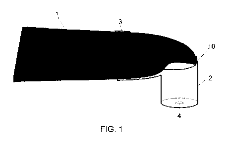
Figure 1 shows a general design of a system according to the invention having
an attach-
ment, which includes a spacer, connecting means and a pinhole aperture,
Figure 2 shows a pinhole aperture according to the present invention,
Figure 3 shows a pinhole aperture with a silicone support according to the
present inven-
tion,
Figure 4 shows a pinhole aperture with imaging optics according to the present
invention,
Figure 5 shows a pinhole aperture and a color calibration set according to the
present in-
vention,
Figure 6a shows a schematic demonstration of a system according to the
invention,
Figure 6b shows a close up of the attachment of the system shown in figure 6a
with a
holder, a spacer and an adapter,
Figure 7 shows a schematic cross section of the adapter of an attachment when
placed on
a tooth,
Figure 8 shows the schematic section of figure 7 for another embodiment with
different
CA 03112976 2021-03-16
WO 2020/094841 PCT/EP2019/080681
9
orientation of the optical axis to the tooth surface,
Figure 9 shows a perspective view of an adapter of the attachment with a
circular / pinhole
aperture,
Figure 10 shows a perspective view of an adapter of the attachment with a
rectangular /
slot aperture,
Figure 11 shows a schematic view of a front side of an adapter of an
attachment with a
support, circumferentially enclosing the aperture,
Figure 12 shows a schematic view of a front side of an adapter of an
attachment with sup-
ports adjacent to the aperture having a circular shaped base,
Figure 13 shows a schematic view of a front side of an adapter of an
attachment with sup-
ports adjacent to the aperture having a rectangular shaped base,
Figure 14 shows a side view of a holder of an attachment demonstrating the
fixation and
location of a camera therein,
Figure 15 shows a side cross section view of a holder,
Figure 16 shows a back view of the holder,
Figure 17 shows a top view of the holder,
Figure 18 shows a perspective view of the holder, showing its front, and
Figure 19 shows a perspective view of a holder, showing its back.
The present invention is explained in detail in the following, using preferred
embodiments
and with reference to the figures.
Figure 1 shows a general design of a system according to the invention for the
intraoral color
determination of teeth or tooth surfaces with a camera 1 and an attachment 10.
As can be
seen in Figure 1, the attachment 10 comprises a spacer 2, connecting means 3
and an aperture
4. The aperture 4 is preferably configured as a pinhole aperture. A slot
aperture (not shown)
can alternatively be provided as well. On the front side facing the object to
be measured, the
attachment 10 can be provided with a silicone support as will be explained in
the following.
A camera, in particular a 2D camera, which is already available in the office
of a dentist, can
be used for the color determination. The dentist thus has access to a digital
medium that can
be used in a system for color determination, which is already integrated into
the treatment
CA 03112976 2021-03-16
WO 2020/094841 PCT/EP2019/080681
unit. He does not have to purchase any other individual devices that always
have to be
brought into the treatment room, powered up and are, in part, quite expensive
to procure.
The attachment 10 can in principle also be used with a 3D camera, if said
camera is
capable of taking 2D color images. In particular for 3D camera systems that
use contrast
powder for 3D measurements, the attachment would provide real added value,
because
these camera systems are otherwise not capable of determining the tooth color
with suf-
ficient accuracy.
A measurement of tooth colors is preferably carried out using the 2D camera 1,
wherein,
unlike a color measurement using a 3D camera, the 3D geometry is not also
recorded
and included in the calculation of the color. Instead, with the attachment 10
on the 2D
camera 1, the geometry is set such that the distance, the spatial angle and
the orientation
of the camera relative to the surface to be captured are restricted in
comparison to a
measurement with a camera without using an attachment.
A measurement of tooth colors can also be carried out with a 3D camera, if
said camera
is capable of taking 2D color images. Particularly camera systems in which
contrast
powder is used for 3D measurements are generally not capable of correctly
determining
tooth color, so that the use of the attachment described in relation to a 2D
camera is
helpful for determining tooth colors.
The attachment 10 in particular comprises a spacer 2 with a connecting means
3, such as a
click connection or a plug connection, for simple fitting to the 2D camera 1.
The attachment
10 can thus be fitted onto the 2D camera 1 quickly and easily for a color
determination.
On the spacer 2, the attachment 10 comprises the aperture 4, via which the 2D
camera 1
captures an image of the teeth. The provision of the aperture 4 advantageously
restricts the
captured distance and angle, as is desired for the color determination of
teeth.
In any case, the aperture 4 is configured and spaced apart from the surface to
be measured
such that the captured surface is smaller than half the average surface of one
of the smaller
teeth, preferably an incisor. In general, the system can be used to capture
the color of every
tooth, such as for example an incisor, a canine or a molar. The captured
surface is preferably
less than 0.5 cm2. The captured surface is particularly preferably in the
range between 0.15
and 0.3 cm2. Be that as it may, the restriction of the captured surface is
intended to ensure
that only the tooth surface itself is captured, and not adjoining areas such
as the gums or
CA 03112976 2021-03-16
WO 2020/094841 PCT/EP2019/080681
11
areas beyond a tooth which could distort the result. The use of a pinhole
aperture is preferred,
whereby for some cases, for example when color gradients of the tooth surface
are to be
measured, the use of an aperture designed as a slot aperture is certainly
possible as well.
When the attachment is used in a system according to the invention, the spacer
2 creates a
defined distance between a capture device - sensor and optics - of the camera
1 and the
aperture 4. Placing the attachment on the tooth to be measured also sets the
angle between
the tooth surface and the camera. When the attachment is placed flat on the
tooth to be meas-
ured, the tooth surface is approximately parallel to the focal plane of the
camera, whereby
deviations up to about 30 are permissible, so that a sufficiently accurate
capturing of the
tooth color is generally possible. The attachment should be placed in such a
way that, during
the measurement, the tooth surface to be measured is positioned centrally
under the aperture,
and thus in the center of the camera image.
For explanation purposes, it should also be noted that a deflecting mirror is
typically a com-
ponent of a 2D or 3D intraoral camera. These deflect the imaging beam path by
a fixed angle
(107 in the case of the CEREC Omnicam specified above). The angle, which is
set by the
attachment according to the invention, is the angle between the central beam
of the imaging
optics and the surface to be measured. For the sake of simplicity, however, in
this case here
we refer to "the orientation of the surface to be measured relative to the
camera". When the
attachment is placed on the tooth with its flat side, the surface to be
measured is in a defined
orientation relative to the camera. The spatial angle, within which the camera
can capture
image data, is restricted by the aperture as well.
Figure 2 shows a single aperture in the form of a pinhole aperture 4.
Figure 3 also shows a pinhole aperture 4, for which, unlike the pinhole
aperture of Figure 2,
a silicone support 5 is provided. With the exclusion of the aperture opening,
the silicone
support 5 can cover the pinhole aperture 4 completely (not depicted) or
partially.
Since the aperture 4 forms the front end of the attachment 10 (Figure 1), the
silicone support
of said aperture rests on the tooth when the attachment is placed on said
tooth and conforms
at least partially to the surface thereof, as a result of which it can, for
one, not slide away.
On the other hand, due to its flush fit on the tooth, the silicone support can
prevent the entry
of interfering scattered light, which ultimately makes a more accurate color
determination
of the respective tooth possible, so that the captured color can then be used
without further
CA 03112976 2021-03-16
WO 2020/094841 PCT/EP2019/080681
12
correction for the color design of a tooth replacement. Lastly, the placement
of the attach-
ment also sets the angle between the tooth surface and the camera. When the
attachment is
placed on the tooth to be measured, the tooth surface is approximately
parallel to the focal
plane of the camera, whereby deviations up to about 30 do not have a
significant effect on
the result. When the attachment is placed on the tooth, the depth of the tooth
surface is ap-
proximately in the focal plane of the imaging optics of the camera.
The spacer 2 can be provided with imaging optics 6 (not shown), which are
inserted into the
hole of the pinhole aperture, for example, or arranged behind the pinhole or
slot aperture.
The imaging optics can advantageously enlarge the image of the captured region
on the de-
tecting sensor of the camera. The color information could thus be measured
using a greater
number of pixels and a more robust result could be achieved. The imaging
optics can op-
tionally fill the pinhole aperture completely. More and/or better data can
potentially be ob-
tained with the imaging optics 6 than without said optics.
According to the invention, the system further comprises means for color
calibration,
wherein the means can comprise software and/or hardware. The hardware
preferably com-
prises a color calibration set with reference colors for calibration. The
color calibration set
is in particular designed as a color pattern with a variety of target colors
for calibration. Such
a color pattern is preferably mounted on the inner side of the attachment, in
particular in the
area of the pinhole or slot aperture. By providing such a color calibration
set, color matching,
including a calibration, of the captured image can be carried out in parallel
by the software
during every recording of a tooth color via the pinhole or slot aperture.
Aging and tempera-
ture effects, the latter of which are caused by the camera-internal light
source, for example,
can be compensated or corrected by means of a color calibration set mounted on
the inner
side of the attachment. Aging effects of the image sensor of the camera, for
example, can
also be compensated or corrected in this manner.
Changes in the system, in particular aging effects, can also be compensated or
corrected by
means of an external color calibration set. To do this, the system can
comprise an additional
attachment, which is provided with reference colors and is placed onto the
camera at prede-
termined time intervals, for example at weekly intervals, to thereby perform a
calibration.
The measured values are then stored by the software to be used as a comparison
value for a
color measurement of a tooth.
A defined, well-known color pattern (reference colors) is typically used for a
calibration and,
CA 03112976 2021-03-16
WO 2020/094841 PCT/EP2019/080681
13
in a next step, the parameters responsible for the color perception of the
system are cor-
rected/optimized by means of a target/actual adjustment.
The system in particular analyzes the data acquired during the color
measurement of a tooth
on the basis of a color calibration and outputs the result in the standard
colors of manufac-
turers of material for tooth replacements. A color selection for material for
tooth replacement
can thus advantageously be carried out automatically.
A known color scale (e.g. Vita Shade Guide) is used to classify different
tooth colors,
wherein the software stores color information "learned" during measurements of
color pat-
terns/reference colors.
The color measured by the image sensor of the camera is a function of a
variety of different
system characteristics. The most important are:
- the brightness and spectrum of the light source;
- the distance from the light source to the surface and the inclination of
the surface in
relation to the light source;
- the position and alignment of the surface in the measurement field of the
sensor;
- aging and temperature effects of the sensor and the LEDs.
A calibration of the system in the previously described manner makes sense, in
particular,
to be able to take those of the previously mentioned parameters that change or
can change
over time into account. Stated more simply, the intent is to correct temporal
changes in the
color measurement properties of the system by means of the calibration. The
system could
furthermore also be designed in such a way that it comprises different
attachments, for ex-
ample attachments with different apertures and/or different coverage angles
and/or different
optics and/or differently sized coverage surfaces and/or different silicone
attachments (for
example for different teeth). For this purpose, the system can be designed
such that it cali-
brates automatically with regard to the respective attachment, which, in
particular in the
context of the previously mentioned real-time calibration that uses reference
colors inte-
grated into the attachment, is easy to accomplish.
The system receives input data as images which are captured by the camera 1,
or as data
which is based on the images captured by the camera 1. Captured images can
thus advanta-
geously be processed and used for color determination.
CA 03112976 2021-03-16
WO 2020/094841 PCT/EP2019/080681
14
The system can furthermore detect changes in the image field captured by the
camera 1 and
consequently switch to a color measurement mode. A color measurement can
therefore be
carried out advantageously quickly and without additional intermediate steps.
With said sys-
tem, a color measurement is also initiated automatically or by means of foot
switches. There-
fore, with the system according to the invention, a color measurement can
advantageously
be initiated intentionally when a foot switch is actuated, or automatically
when no shaking
or a uniform color is detected by the 2D camera 1.
The system analyzes the acquired data on the basis of a color calibration and
outputs the
result in the standard colors of manufacturers of material for tooth
replacements. This can
be carried out by means of the software. A color selection for material for
tooth replacement
can thus be carried out automatically.
For calibrating the system in the previously mentioned manner, the aperture 4
of the system
can be provided on its inner side with a color calibration set 7 composed of
reference colors.
Systems for color determination according to the present invention require a
color calibration
at regular intervals, to compensate for small deviations in the lighting
spectrum and the im-
aging system. Target colors are typically recorded within the context of such
a color calibra-
tion. These make it possible to adjust the system response by means of a
calibration. In the
aforementioned color calibration set 7, the reference colors are applied to
the inner side of
the aperture 4. In this way, for every recording of the tooth color by the
aperture 4, a color
matching, including a calibration, of the captured image can be carried out as
well.
A method for determining the color of teeth or tooth surfaces first comprises
fitting the at-
tachment 10 to the 2D camera 1. Figure 1 shows the system according to the
invention after
this step has been performed. The geometry of the image to be captured is then
set by the
attachment 10 in such a way that the distance, orientation and spatial angle
for image cap-
turing are limited for the camera in relation to a tooth surface to be
measured, and an intraoral
image of a geometrically defined specific section of teeth or of a tooth is
subsequently cap-
tured by means of the 2D camera 1 in conjunction with the attachment 10.
For the case in which only single-colored ceramics are used as a restoration
material for
teeth, a one-time image acquisition or measurement is sufficient. However, if
the use of
multicolored restoration materials, such as with color gradients for example,
is intended,
as many measurements as necessary for color determination can be performed
using the
system according to the invention.
CA 03112976 2021-03-16
WO 2020/094841 PCT/EP2019/080681
It should further be noted that the tooth color is determined from the
recorded image
data using a statistical analysis, such as averaging, for example. In
particular the image
data recorded from the interior of the (pinhole) aperture is advantageously
used for this
purpose.
A color calibration is performed by capturing an image and comparing data of
the captured
image with predefined or acquired color data, wherein the color data are
predefined by means
of the software or are based on the acquired data of the color calibration set
7 with reference
colors for calibration that is provided in the interior of the aperture 4 on
the attachment 10
(see Figure 5).
The data of the captured image is analyzed on the basis of the color
calibration and the result
of the analysis is preferably output in the standard colors of manufacturers
of material for
tooth replacements, so that the data no longer has to undergo further
processing before a
restoration material with the appropriate color variant can be selected by the
practi-
tioner/dentist.
The system is preferably designed in such a way that it recognizes the
presence of an attach-
ment on the camera. This could, for example, be accomplished on the basis of
changes in
the image field captured by the 2D camera 1 that would lead to an automatic
switchover to
color measurement. A switchover could possibly also be accomplished via a
command ini-
tiated by the user.
Turning to Figure 6a, where a schematic overview of the process of color
determination of
a tooth 40 with the above described system according to the invention is
given. The camera
with central axis 34 has a camera opening 37 with an optical axis 35 including
an angle
ranging from 75 to 135 to the center axis 34, where in the scheme of figure
6a and 6b an
angle of 90 is shown. Camera opening optical axis (see figure 6b) and
attachment optical
axis are collinear and are demonstrated in figure 6 as one axis 35. A light
tight contact of the
attachment to the area circumferential to the camera opening 37 is provided by
bearing sur-
faces 33, which prevent that stray light or light from others than the light
source of camera
1 disturb or interfere with the color determination by the camera 1 in
combination with the
attachment 10. Another realization of a light tight contact to at least the
circumferential area
of the camera opening 37 can be provided by a form fitting shaped part of the
attachment
10.
An aperture 4 in the attachment 10 defines the attachment optical axis which
correspond to
CA 03112976 2021-03-16
WO 2020/094841 PCT/EP2019/080681
16
the optical axis 35 of the camera opening 37. Further in the field of view and
the illumination
cone 36 of the camera light source, at least one color calibration pattern 7
on the inside of
the attachment 10 is located. In case of figure 6a, the patterns 7 are located
in the same plane
and adjacent to the aperture 4. On the outside at least one support 5 for a
determined and
stable contact to the surface 41 of a tooth 40, which color is to be
determined, is provided.
A support 5 can be for example the before mentioned silicone ring.
Within this configuration, the light cone 36 illuminates the tooth surface 41,
which color is
to be evaluated and at the same time the reference color patterns 7, thus
enabling referenced
or direct calibrated color determination of the tooth surface 41 by the image
recorded by the
camera 1, as previously described.
Figure 6b shows a close up cross section view of the attachment 10 of figure
6a. The optical
axis 38 of the camera opening 37 and optical axis 35 of the attachment 10 are
collinear and
thus can be represented as one axis and have the same angle to the camera
center axis 34 of
in this case 90 . In this embodiment, the attachment 10 comprises three
components, a holder
30, which provides a connection via a connecting means 3 (not shown in Fig 6b)
to the
camera 1 and at least a light tight contact to the area circumferential to the
camera opening
37, which is indicated by the bearing surfaces 33 around a corresponding
holder opening
30', an adapter 20, comprising before mentioned aperture 4 and a color
reference pattern 7
on the inside and supports 5 on the outside, that is on the side oriented to a
tooth surface in
use and a transition piece or spacer 2, which provides the corresponding
distance for the
adapter 20 to the camera opening 37 in regard to a correct imaging distance
for the built in
camera optics. The spacer 2 provides that at least the color reference pattern
7, and optionally
further the aperture 4, are located close to the focal plane of the camera 1.
It is preferred that
the color reference pattern 7 and the aperture 4 are located in the focal
plane of the camera
1. All components are removably connected, so that for example different
spacers 2 accord-
ing to the invention, corresponding to different camera optics and/or models,
can be used to
provide the correct spacing of the adapter component 20 with regard to the
focus plane of
the used camera 1.
In the same manner, this applies as well to the holder 30 and the adapter 20,
wherein for the
latter different adapters 20 with different apertures 4 can be used, while in
case of the holder
30, different holders for different camera bodies can be provided. The
connection between
CA 03112976 2021-03-16
WO 2020/094841 PCT/EP2019/080681
17
these elements are formed as light tight connections, as for example sliding,
clipping or en-
gaging mechanisms and thus preventing a not controlled and/or defined
illumination of the
reference pattern 7, and the tooth surface 41 through the aperture 4. In
alternative embodi-
ments, the spacer 2 can be part of either the holder component 30 or the
adapter component
20 or partially part of both components. Further, the complete attachment can
also be pro-
vided as one-part element.
A more detailed presentation of the interaction of the attachment 10, in
particular the adapter
20, at the tooth surface 41 is given by the schematic cross section view of
figure 7. As shown
in the embodiment of the adapter 20 in figure 7, the aperture 4 has defined
diameter d deter-
mining the actual illuminated area on the tooth surface 41, which is used for
evaluation of
the color. Here a cross section area of the aperture of at least less than or
equal to 0.5 cm2,
less than 0.35 cm2 or 0.15 cm2 is preferred, where the minimum area is not
less than 0.01
cm2. This area results in a first approximation in a corresponding illuminated
surface area of
about the same size on the tooth 40, since there is a short distance between
aperture and tooth
surface 41 with regard to the divergence of the optical system[1][2]. However,
these values
serve as guidance for those skilled in the art.
The particular area of the aperture depends on the resolution and contrast
capabilities of the
camera and the corresponding intensity of the light source. Further the
supports 5 are in
contact with the surface 41 of tooth 40, providing a defined orientation in
space of the adapter
and therefore for the system comprising of attachment 10 and camera 1. In case
of figure 7,
the attachment optical axis 36 is parallel to the normal vector n of the tooth
surface 41.
A different embodiment is shown in figure 8, where the attachment optical axis
35 is angled
with respect to the normal vector n of the tooth surface 41. However, in both
embodiments
a defined orientation of the attachment 10 to the tooth 40, in particular to
the tooth surface
41, which is to be evaluated regarding the color, is established by the
supports 5 in combi-
nation with the adapter 20.
Figure 9 gives a perspective view of the adapter of the attachment. The
adapter 20 comprises
a front side 21, which defines the distal end of the optical path to the
camera opening of the
attachment 10. At the front side 21, an aperture 4 with a circular shaped base
and reference
patterns 7 for the color determination are located. Further, the adapter
comprises a connect-
ing means 22, which provide a light tight connection together with their
counterparts on the
holder 30 shown in the embodiments of the holder of the attachment in figures
14 to 17 or
CA 03112976 2021-03-16
WO 2020/094841 PCT/EP2019/080681
18
with the respective counterparts on the spacer 2 in case of attachment 10
comprising three
components. On the outside of the front side 21 (not shown in figure 9), the
support 5 (figure
7, 8) is located.
A different embodiment of the adapter with an aperture with a rectangular/slot
shaped base,
that enables to determine a color gradient on a tooth surface, is shown in
figure 10.
Figures 11 to 13 show different embodiments of the support 5 on the outside of
a front side
21 of the adapter 20. In Figure 11 an embodiment is shown where the support 5
circumfer-
entially encloses the pinhole aperture 4, thus providing advantageously a
light tight contact
to the tooth 40, which prevents any secondary illumination of the tooth
surface 41 or that
any stray light or other unwanted light enters the attachment via the aperture
4, disturbing
the color determination. Such a support can be realized by above mentioned
silicone ring or
by another elastic material or elastomer.
In figure 12, an embodiment with a plurality of supports 5 with a round shaped
base is shown,
where the supports 5 are located adjacent to the aperture 4. Figure 13 shows
an embodiment
with a rectangular / slot formed aperture 4 and corresponding rectangular base
shaped sup-
ports 5.
In general, all these configurations of the above mentioned figures 11 to 13
may also include
that the support is partially recessed into or protruding the front side 21 or
may be partially
or completely part of the front side 21 itself. Areas of the front side 21 can
comprise elastic
materials and/or can be riffled or otherwise contoured to provide increasing
grip onto the
tooth surface 41, as for example the areas indicated as base areas of the
supports 5 on the
front side 21 in figures 11 to 13. The front side can also be formed by two
layers, the layer
on the outside facing the tooth surface 41 can be elastic and thus providing
stable and deter-
mined contact with regard to the orientation of the attachment 10 to said
tooth surface 41.
Further, the front side can serve as support itself without any of the above
mentioned modi-
fications , as a sufficient defined and stable contact to the tooth surface
can be provided by
the friction and surface pressure. However, in this case enhancements with
regard to stable
and determined contact with regard to the orientation of the attachment to the
tooth surface
41 can be achieved, if material of the front side 21 is made of soft or
partially elastic material,
such as soft plastic.
Figure 14 shows a side view of a holder 30 of an attachment 10. The previously
shown
adapter 20 is connected either directly via the connecting means 32 and 32' to
the holder or
CA 03112976 2021-03-16
WO 2020/094841 PCT/EP2019/080681
19
via corresponding connecting means of the spacer 2 (if the attachment 10 with
three compo-
nents), shown in fig. 6b. A light tight connection is provided by the
engagement of the con-
necting means 22 of the adapter and the corresponding counter parts 32 and the
additional
connecting means 32' of the holder. This enables on the one hand to use
different adapters,
e.g. with different apertures in combination with different holders, for
example for different
camera bodies and, in case of an attachment 10 with three components, see
figure 6b, differ-
ent spacers 2 and at the same time prevents that disturbing light from the
environment infers
the color determination. The holder 30 itself is connected to camera 1 by a
connecting means
3. In this embodiment, the holder 30 is adjusted to provide an attachment
optical axis 35,
which corresponds to an angled optical axis of the camera opening 37 (figure
6b) of a camera
such as the CEREC Omnicam of the applicant with an angle 31 of 107 degrees to
camera
center axis 34.
Figures 15 to 17 show different views of the holder of figure 14, where in
particular the
bearing surfaces 33 are indicated. These surfaces provide a light tight
contact of the attach-
ment to the camera 1 (not shown) in particular around the camera opening 37
(not shown).
Also the connecting means 32 and 32' are shown, which are formed as elements
for a latch-
ing 32' and sliding 32 connection between adapter 20 and holder 30. Further
also demon-
strated is the connection means 3, which provides the attachment of the holder
and therefore
the complete attachment to the camera. In this embodiment, the connecting
means 3 is
formed as a ring clamping mechanism, which has a smaller diameter than the
diameter of
the, in this case, bar shaped camera body. Other possible connections may be a
click con-
nection or a plug connection mechanism. The figures 18 and 19 show perspective
views of
the holder of figure 14.
In the previous designs and considerations, it is assumed that the camera of
the system is
provided with a light source for illuminating the tooth to be measured and, if
applicable, the
reference color pattern.
According to the invention, a system and a method for accurately determining
the color of
teeth are provided, which are simple in design and also easy to use.
Furthermore, according
to the invention, an attachment is provided, which can easily be used in the
system of the
invention.
CA 03112976 2021-03-16
WO 2020/094841
PCT/EP2019/080681
Reference Signs
1 camera
2 spacer
3 connecting means (to the camera)
4 aperture
5 support
6 imaging optics
7 color calibration set/reference pattern
8 connecting means of the spacer
10 attachment
20 adapter of the attachment
21 front side of the adapter
22 connecting means
holder of the attachment
30' holder opening
31 angle between optical axis of the attachment and the camera axis
32, 32' connecting means
33 bearing surface(s)
34 camera center axis
attachment optical axis / camera opening optical axis
36 illumination cone
37 camera opening
38 focal plane 40 tooth
41 tooth surface