Note: Descriptions are shown in the official language in which they were submitted.
CA 03113435 2021-03-18
WO 2020/068815 PCT/US2019/052680
NUCLEIC ACIDS FOR CELL RECOGNITION AND INTEGRATION
CROSS-REFERENCE
[0001] This application claims the benefit of U.S. Provisional Application
Nos. 62/875,887,
filed July 18, 2019; and 62/736,323, filed September 25, 2018, which
applications are
incorporated herein by reference in their entirety for all purposes.
SEQUENCE LISTING
[0002] The instant application contains a Sequence Listing which has been
submitted
electronically in ASCII format and is hereby incorporated by reference in its
entirety. Said
ASCII copy, created on September 23, 2019, is named 54774-702 601 SL.txt and
is 793,770
bytes in size.
BACKGROUND
[0003] A transposable element (TE or transposon) is a DNA sequence that can
change its
position within a genome, sometimes creating or reversing mutations and
altering the cell's
genetic identity and genome size. Transposable elements have previously been
associated
with various diseases including cancer, but their relationship to the onset of
the disease, if any
exists, needs to be better understood. For example, cancer is the second most
common cause
of death in the United States, with more than 1,600 cancer related deaths per
day, nearly
600,000 per year. Approximately 1.65 million new cases of cancer were
diagnosed in 2015
and cancer incidence is increasing due to demographic and lifestyle factors.
Thus, there exists
an unmet need for sensitive, effective and particularly cell-, tissue, and/or
organ-specific
compositions and methods for detection and treatment of cancer as well as
other chronic,
infectious or age-related diseases.
SUMMARY
[0004] In various aspects, the present disclosure provides nucleic acid
constructs comprising
a deoxyribonucleic acid sequence that directs a migration of the construct to
a target tissue in-
vivo (tropism). The construct can comprise two or more sequences derived from
a
transposon, for instance, a 3' sequence and a 5' sequence, for integration
into a cell of the
target tissue.
[0005] In various aspects, the disclosure describes various pharmaceutical
compositions
comprising sequences that direct a migration of the construct to a target
tissue or a target cell
-1-
CA 03113435 2021-03-18
WO 2020/068815 PCT/US2019/052680
in-vivo (tropism). In some instances the disclosure provides a pharmaceutical
composition
comprising a therapeutically effective amount of a nucleic acid construct
comprising a) a
first deoxyribonucleic acid (DNA) sequence that directs a migration of said
nucleic acid
construct to a pre-selected tissue in vivo; and b) a second deoxyribonucleic
acid sequence that
directs an integration of a region of said nucleic acid construct into a
genome of a cell from
said pre-selected tissue in vivo; which composition is formulated for
administration to a
subject. In some embodiments, said first deoxyribonucleic acid sequence is
from a same cell
type as said cell from said pre-selected tissue. In some embodiments, said
subject is a human.
In some embodiments, said first deoxyribonucleic acid sequence migrates to a
white blood
cell. In some embodiments, said first deoxyribonucleic acid sequence migrates
to a pancreatic
cell. In some embodiments, said first deoxyribonucleic acid sequence migrates
to lung cell. In
some embodiments, said nucleic acid construct crosses a nuclear membrane of
said cell from
said pre-selected tissue when administered to said subject. In some
embodiments, said first
deoxyribonucleic acid sequence has at least 90% identity to at least 12 bases
of any one of
SEQ ID NO: 203 ¨ SEQ ID NO: 277 and SEQ ID NO: 282. In some embodiments, said
first
deoxyribonucleic acid sequence is at least 400 base pairs in length. In some
embodiments,
said first deoxyribonucleic acid sequence is between 400 base pairs and 20,000
base pairs in
length. In some embodiments, said second deoxyribonucleic acid sequence has at
least 90%
homology to a transposon sequence. In some embodiments, said transposon is a
class II
transposon. In some embodiments, said class II transposon integrates itself
into said genome
of said cell from said pre-selected tissue via horizontal gene transfer. In
some embodiments,
said nucleic acid construct that comprises at least one additional
deoxyribonucleic nucleic
acid sequence. In some embodiments, said at least one additional
deoxyribonucleic nucleic
acid sequence comprises a tissue selective promoter. In some embodiments, said
at least one
additional deoxyribonucleic nucleic acid sequence comprises a second
integration signal for
incorporation into said genome. In some embodiments, said at least one
additional
deoxyribonucleic nucleic acid sequence comprises a sequence that encodes a
peptide or
protein. In some embodiments, said at least one additional deoxyribonucleic
nucleic acid
sequence comprises a sequence for ensuring that said peptide or protein is
only expressed in
said cell from said pre-selected tissue. In some embodiments, said peptide or
said protein
encodes a tumor suppressor peptide or a tumor suppressor protein. In some
embodiments,
said tumor suppressor peptide or said tumor suppressor protein is a multiple
myeloma tumor
suppressor gene. In some embodiments, said tumor suppressor peptide or said
tumor
suppressor protein is a pancreatic cancer tumor suppressor gene. In some
embodiments, said
-2-
CA 03113435 2021-03-18
WO 2020/068815 PCT/US2019/052680
tumor suppressor peptide or said tumor suppressor protein is a lung cancer
tumor suppressor
gene. In some embodiments, said tumor suppressor peptide or said tumor
suppressor protein
is any one of retinoblastoma susceptibility gene (RB), Wilms' tumors (WT1),
neurofibromatosis type-1 (NF1), familial adenomatosis polyposis coli (FAP),
von Hippel-
Lindau syndrome (VHL), wild-type p53, or super repressor p53. In some
embodiments, said
peptide or said protein encodes an antigenic protein. In some embodiments,
said antigenic
protein is translated specifically in said cell upon administration to said
subject. In some
embodiments, said pharmaceutical composition further comprises a cargo. In
some
embodiments, said cargo is a fluorophore or a radioisotope. In some
embodiments, said cargo
is a therapeutic drug. In some embodiments, the cargo is covalently attached
to said nucleic
acid construct. In some embodiments, said formulation comprises a nanoparticle
or cationic
polymer.
[0006] In various aspects, the present disclosure provides a vector comprising
a first
sequence that has at least 90% sequence identity to at least 12 bases of any
one of SEQ ID
NO: 203 ¨ SEQ ID NO: 277 or SEQ ID NO: 282, or an additional nucleic acid
sequence. In
some embodiments, said additional nucleic acid sequence has at least 90%
homology to a
transposon sequence described herein. In some embodiments, said transposon is
a class II
transposon. In some embodiments, said class II transposon integrates itself
into said genome
of said cell from said pre-selected tissue via horizontal gene transfer. In
some embodiments,
said vector comprises at least two additional nucleic acid sequences. In some
embodiments,
said at least two additional nucleic acid sequences comprise a tissue
selective promoter and a
transposon sequence. In some embodiments, said at least two additional nucleic
acid
sequences comprise at least two transposon sequences. In some embodiments,
said additional
nucleic acid sequence comprises a sequence that encodes a peptide or protein.
In some
embodiments, said peptide or said protein encodes a tumor suppressor peptide
or a tumor
suppressor protein. In some embodiments, said tumor suppressor peptide or said
tumor
suppressor protein is a multiple myeloma tumor suppressor gene. In some
embodiments, said
tumor suppressor peptide or said tumor suppressor protein is a pancreatic
cancer tumor
suppressor gene. In some embodiments, tumor suppressor peptide or said tumor
suppressor
protein is a lung cancer tumor suppressor gene. In some embodiments, said
tumor suppressor
peptide or said tumor suppressor protein is anyone of retinoblastoma
susceptibility gene
(RB), Wilms' tumors (WT1), neurofibromatosis type-1 (NF1), familial
adenomatosis
polyposis coli (FAP), von Hippel-Lindau syndrome (VHL), wild-type p53, or
super repressor
p53. In some embodiments, said vector is covalently linked to a cargo. In some
embodiments,
-3-
CA 03113435 2021-03-18
WO 2020/068815 PCT/US2019/052680
said cargo is a fluorophore or a radioisotope. In some embodiments, said cargo
is a
therapeutic drug.
[0007] In various aspects, the present disclosure provides a library of two or
more of said
vectors.
[0008] In various aspects, the present disclosure provides a method for
treating a cancer, the
method comprising: administering an effective amount of a nucleic acid
construct comprising
a nucleic acid sequence that directs migration of a cargo to a tissue of a
subject suffering
from said cancer, which composition is formulated for administration to a
subject. In some
embodiments, the cancer is a lung cancer. In some embodiments, the cancer is a
multiple
myeloma. In some embodiments, the cancer is a pancreatic cancer. In some
embodiments,
said nucleic acid construct crosses a nuclear membrane in a cell of said
tissue cell upon
administration to said subject. In some embodiments, said nucleic acid
sequence that directs
migration of said cargo to said tissue has at least 90% identity to at least
12 bases of any one
of SEQ ID NO: 203 ¨ SEQ ID NO: 277 or SEQ ID NO: 282. In some embodiments,
said
nucleic acid construct comprise a transposon for integration into a genome of
a cell of said
tissue. In some embodiments, said transposon is at least 400 base pairs in
length. In some
embodiments, said transposon is between 400 base pairs and 20,000 base pairs
in length. In
some embodiments, said transposon is a class II transposon. In some
embodiments, said
nucleic acid construct comprises at least one additional nucleic acid sequence
that is
heterologous to a cell of said tissue. In some embodiments, said at least one
additional
nucleic acid sequence comprises a tissue selective promoter. In some
embodiments, said at
least one additional nucleic acid sequence comprises a sequence that encodes a
peptide or
protein. In some embodiments, said at least one additional nucleic acid
sequence comprises
an integration signal for incorporation into a target genome. In some
embodiments, said at
least one additional nucleic acid sequence comprises a guide sequence for
targeting said cell.
In some embodiments, said at least one additional nucleic acid sequence
comprises a
sequence that encodes a peptide or a protein. In some embodiments, said
peptide or said
protein encodes a tumor suppressor peptide or a tumor suppressor protein. In
some
embodiments, said tumor suppressor peptide or said tumor suppressor protein is
a multiple
myeloma tumor suppressor gene. In some embodiments, said tumor suppressor
peptide or
said tumor suppressor protein is a pancreatic cancer tumor suppressor gene. In
some
embodiments, said tumor suppressor peptide or said tumor suppressor protein is
a lung cancer
tumor suppressor gene. In some embodiments, said tumor suppressor peptide or
said tumor
suppressor protein is anyone of retinoblastoma susceptibility gene (RB),
Wilms' tumors
-4-
CA 03113435 2021-03-18
WO 2020/068815 PCT/US2019/052680
(WT1), neurofibromatosis type-1 (NF1), familial adenomatosis polyposis coli
(FAP), von
Hippel-Lindau syndrome (VHL), wild-type p53, or super repressor p53. In some
embodiments, said peptide or said protein encodes an antigenic protein. In
some
embodiments, said antigenic protein is translated specifically in a cell of
said tissue upon
administration to said subject. In some embodiments, said nucleic acid
construct is covalently
linked to a fluorophore or a radioisotope. In some embodiments, said nucleic
acid construct is
covalently linked to a therapeutic drug.
[0009] In some instances, the present disclosure provides a nucleic acid
construct comprising
a first DNA sequence and a second DNA sequence, wherein such first DNA
sequence can be
a targeting sequence capable of directing the construct to a specific organ,
tissue, and/or cell,
and wherein the second DNA sequence can be an integration sequence capable of
integrating
at least a region or portion of the construct into a genome of a cell of the
specific organ or
tissue. In some instances, the first (cell-targeting/recognition) and second
(integration) DNA
sequence can be part of a Zip Code Sequence (ZCS), a transposon or a
transposable element.
Such ZCS or transposon can comprise a cell targeting and recognition sequence
that can
comprise a nucleotide sequence having at least about 80%, 85%, 90%, 95%, 97%,
99%, or
100% sequence identity any one of SEQ ID NO: 203 ¨ SEQ ID NO: 277 or SEQ ID
NO: 282,
or at least about 80%, 85%, 90%, 95%, 97%, 99%, or 100% sequence identity to a
fragment
thereof. Such fragment can be at least about 8, 10, 12, 15, 20, 25, or 30
nucleotides in length.
In some instances, such transposon can be a class II transposon.
[0010] In some instances, such nucleic acid construct can serve as a vehicle
for transferring
genes between cancer cells as well as used to transport a cargo (e.g., a
nucleic acid, a protein,
a small molecule, or a nanoparticle) into a cell, wherein such cell can be of
the same origin as
the cell that the first and/or second nucleic acid sequence is derived (or was
excreted) from.
As described further herein, transfer of genetic material such as ctDNA into a
cell (e.g.,
tumor cell) can alter the clonal architecture of cells and may determine the
fate of such cells
(e.g., tumor cells) to environmental changes, such as drug treatment.
[0011] In some instances, the present disclosure also provides various
surprising and
unexpected attributes of the nucleic acid constructs described herein. Such
unique attributes
can include tropism, which can be defined herein as an ability of the
construct to target and/or
enter cells that are of the same of similar origin to those from which the
first (targeting)
sequence and/or second (integration) sequence are derived from. In various
instances, such
targeting and/or integration sequences of a nucleic acid construct herein can
be derived from
a circulating tumor DNA (ctDNA). Another example of such attributes of the
constructs
-5-
CA 03113435 2021-03-18
WO 2020/068815 PCT/US2019/052680
provided herein can include ability to distinguish between cells of similar
origin and cells that
originated from a different tissue or organ. As an example, a nucleic acid
construct
comprising a first DNA sequence and a second DNA sequence that are derived
from ctDNA
that originated from multiple myeloma (MM) tumor cells can target, enter,
and/or integrate
into a genome of MM cells with high specificity compared to cells of other
tumors, such as
pancreatic cancer (PC), lung cancer, or colon cancer. Such specificity can be
at least about
80%, 85%, 90%, 95%, 97%, or 99%. Furthermore, such construct can target MM
cells from
different cell lines. As such, for example, the MM-specific nucleic acid
construct can be used
in vitro to, e.g., transport a cargo into MM cells (e.g., MM1S cells), or in
vivo to transport the
cargo into MM cells of a tumor of a subject (e.g., a rodent or a human).
[0012] The present disclosure further provides functional assays that can be
used to identify
such unique targeting and/or integration sequences (e.g., ZCSs or transposons)
that allow for
cell targeting and recognition and insertion into a genome of the cell (e.g.,
a cancer cell),
respectively. For instance, cell-specific recognition sequences and/or genomic
integration
sequences for use in delivery systems can be identified as described in
EXAMPLE 2 herein.
In some instances, a ctDNA that can be used to generate such targeting and/or
integration
sequences can be obtained from a subject, e.g., from the blood plasma of the
subject. Such
subject can be a human. The human subject can be one that has been diagnosed
with a disease
such as cancer. In some aspects, the disclosure provides a method for
identifying a nucleic
acid sequence that migrates to a tissue or a cell of substantially similar
origin (tropism)
comprising: (a) isolating circulating tumor nucleic acids from a biological
sample, thereby
producing a set of isolated tumor nucleic acids; (b) adding a barcode to said
set of isolated
circulating tumor nucleic acids, thereby providing a plurality of barcoded
tumor nucleic
acids; (c) adding at least one barcoded tumor nucleic acid from the plurality
of barcoded
tumor nucleic acids to a population of cells under conditions that allow for
integration of the
tissue tropic sequences, thereby producing a cultured population of cells; (d)
sequencing the
cultured population of cells, thereby producing a plurality of sequencing
reads; (e) analyzing
the sequencing reads by a computer to identify a presence or an absence of at
least one
sequencing read from the plurality of sequencing reads comprising a barcode;
and (f)
analyzing the sequencing reads comprising the barcode to identify a presence
or an absence
of a sequence that is present on the cultured population of cells but absent
in the population
of cells, thereby identifying the nucleic acid sequence that migrated to the
tissue or the cell of
interest.
-6-
CA 03113435 2021-03-18
WO 2020/068815 PCT/US2019/052680
[0013] In some embodiments, the present disclosure provides synthetically
generated
transposons that can be derived from such ctDNA and that can be used to
deliver a cargo
(e.g., a nucleic acid, a protein, a small molecule, or a nanoparticle) into
one or more target
cell(s). In some instances, and as described herein, such synthetically
generated transposons
or ZCS (e.g., oligo-ZCS) can be part of a nucleic acid construct that can be
used as a tool to
deliver cargo with high specificity to cells, e.g., cancer cells, both in vivo
and in vitro. Thus,
in some instances, such nucleic acid construct can be part of a delivery
system. Such delivery
system can have one or more components. Such components can include (i) a ZCS
sequence
comprising a first DNA sequence for targeting a certain cell, cell population,
or tissue, and a
second DNA sequence for integration of at least a region or portion of such
delivery system
into a genome of such target cell, cell population, or tissue; a (ii) promoter
sequence; and (iii)
one or more cargos, or any combination thereof Such one or more cargos can
include (a) one
or more nucleic acid (e.g., DNA) sequences such as genes or gene fragments
that can be
expressed in such target cells or tissue, and (b) one or more additional
cargos such as
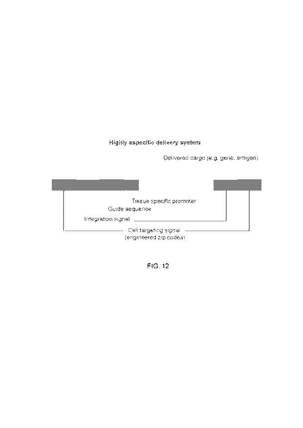
proteins, nanoparticles, or small molecules. FIG. 12 shows an example of a
delivery system
described herein. In such system, a cargo (here a gene such as an antigen-
coding gene) to be
delivered to a target cell can be coupled to a (e.g., tissue-specific)
promoter sequence and a
guide sequence, which are flanked by ZCSs and/or transposons comprising the
first and
second DNA sequences, wherein the first DNA sequence (e.g., targeting or Zip
Code
sequence) is capable of cell targeting, and the second DNA sequence is capable
of integrating
such system into a genome of the target cell once the system has reached the
nucleus of the
cell.
[0014] In some instances, the present disclosure provides nucleic acid
constructs and
systems that can transfer certain properties to target cells. Such properties
include a target
cell's response to changes in the environment. Such changes can include
exposure to certain
molecules. Such molecule can be drug molecules, such as small molecules or
proteins such as
antibodies. Examples of such transfer or properties include resistance or
sensitivity to a drug
from a patient's plasma to cancer cell lines in vitro. Such surprising and
unexpected
properties can allow for the design of non-naturally occurring nucleic acid
constructs
comprising one or more (e.g., 2, 3, 4, etc.) ZCS (or transposon) sequences and
one or more
cargo molecules such as additional nucleic acid sequences encoding for
therapeutic and/or
diagnostic proteins for the delivery and integration of such constructs, or a
portion of such
construct, into a genome of a target cell. Such target cell can be a cancer
cell and the cargo
nucleic acid can code for a variety of therapeutic proteins such as caspases
for cancer cell
-7-
CA 03113435 2021-03-18
WO 2020/068815 PCT/US2019/052680
apoptosis, antigens for immune cell recognition or enhancement of
immunotherapy, or other
proteins such as antibodies, enzymes, cytokines, signaling molecules, etc.
[0015] The ZCSs or transposon sequences described herein can comprise one or
more
transposable elements. Such transposable elements can include mammalian-wide
interspersed
repeat (MIR) and Arthrobacter luteus (ALU) such as ALUsq, as well as
derivatives or
functional fragments thereof Thus, in some instances, the present disclosure
provides
synthetic MIR oligonucleotides (oligos) that can be capable of delivering
cargo to target cells
such as tumor cells.
INCORPORATION BY REFERENCE
[0016] All publications, patents, and patent applications mentioned in this
specification are
herein incorporated by reference to the same extent as if each individual
publication, patent,
or patent application was specifically and individually indicated to be
incorporated by
reference.
BRIEF DESCRIPTION OF THE DRAWINGS
[0017] The application file contains at least one drawing executed in color.
Copies of this
patent or patent application publication with color drawing(s) will be
provided by the Office
upon request and payment of the necessary fee.
[0018] The novel features of the invention are set forth with particularity in
the appended
claims. A better understanding of the features and advantages of the present
invention will be
obtained by reference to the following detailed description that sets forth
illustrative
embodiments, in which the principles of the invention are utilized, and the
accompanying
drawings (also "Figure" and "FIG." herein), of which:
[0019] FIG. 1 shows that circulating tumor DNA (ctDNA) comprising specific
tumor cell
recognition sequences (e.g., Zip Code Sequences, also abbreviated herein as
"ZCSs") can
cross cell and nuclear membranes in multiple myeloma cells (MMls cells)
(illustrated as MM
cells + MM ctDNA), a lung cancer cell line (A549) (illustrated as LC cells +
LC ctDNA), and
a pancreatic cancer cell lines (SPAC01) (illustrated as PC cells + PC ctDNA).
[0020] FIG. 2A shows a time course of eight 3D images demonstrating capture
and
internalization of ctDNA in pancreatic cancer cells.
[0021] FIG. 2B shows a time course of eight single plane image demonstrating
capture and
internalization of ctDNA in pancreatic cancer cells.
-8-
CA 03113435 2021-03-18
WO 2020/068815 PCT/US2019/052680
[0022] FIG. 2C shows a time course of eight images from a 3D video
demonstrating capture
and internalization of ctDNA by endocytosis in pancreatic cells.
[0023] FIG. 3A shows a time course of eight images taken from a single plan
video
demonstrating nuclear localization of ctDNA in ASPC-1 cells.
[0024] FIG. 3B shows six images taken from a single plan 4-hour time course
illustrating
nuclear uptake of ctDNA in MIMI S cells. Cell membranes were labelled with
green
fluorescent protein (GFP). Nucleus is labeled with DAPI.
[0025] FIG. 4 illustrates A459 (lung cancer) and MM1 (multiple myeloma, MM)
cells
cultured with labeled ctDNA extracted from a lung cancer or MM patient. The
Figure shows
that respective ctDNA almost exclusively target cells similar to the ctDNA
cell of origin.
[0026] FIG. 5A shows a chromosome spread demonstrating ctDNA integration into
plasma
cell's chromosomes.
[0027] FIG. 5B is a graph showing effect of DNA-dependent protein kinases
(e.g.,
DNAPKcs), Ataxia telangiectasia mutated (ATM), poly-(ADP-ribose) polymerase I
(PARP-
1), and raltegravir (an integrase inhibitor) on ctDNA integration into
chromosomes.
[0028] FIG. 5C shows that chromosomal integration of ctDNA can depend on non-
homologous end joining (NHEJ) and transposases. Incorporation of labeled ctDNA
was
reduced significantly when inhibitors of DNAPKcs, ATM, PARP-1, and integrase
(raltegravir) were present. A non-significant reduction was observed with PARP-
1 inhibition.
[0029] FIG. 6 shows a scheme that illustrates the identification of consensus
sequences
(contigs). FIG. 6 discloses SEQ ID NOS 280-281, respectively, in order of
appearance.
[0030] FIG. 7 shows two cell recognition signal sequences. The left red blocks
highlight a
region for two similar variants that were present in multiple myeloma (MM)-
derived ctDNA
but not in pancreatic cancer (PC)-derived ctDNA. The red box in the right
shows an
additional nucleic acid sequence that was detected in MINI and PC (control).
[0031] FIG. 8A shows a dendogram of the identified contig sequences from
multiple
myeloma, pancreatic cancer, and control cell lines. The index contig shows
localization, type
and presence of Zip Code Sequences.
[0032] FIG. 8B shows areas of chromosomal integration and that mechanisms may
be
related to and/or mediated by transposons. Indications of such include the
observation that
most inserted contig sequences can have high content of transposons; that
contig sequences
can have different types of transposons; that integration may be configuration
dependent; and
that cellular recognition of the contig and/or ZC sequences may be receptor-
mediated.
-9-
CA 03113435 2021-03-18
WO 2020/068815 PCT/US2019/052680
[0033] FIG. 9 shows confocal microscopy images of rhodamine-labeled ctDNA
constructs
present in the tumor, including localization of the ctDNA in the nuclei, after
systemic or local
administration.
[0034] FIG. 9A shows confocal microscopy images 24 hours after administration
of
rhodamine-labeled ctDNA when injected directly into the tumor.
[0035] FIG. 9B shows confocal microscopy images 24 hours after administration
of
rhodamine-labeled ctDNA when injected when injected via the tail vein.
[0036] FIG. 9C shows confocal microscopy images 48 hours after administration
of
rhodamine-labeled ctDNA when injected when injected via the tail vein.
[0037] FIG. 9D shows confocal microscopy images of tumor-bearing control mice
that were
injected via the tail vein with phosphate buffered saline (PBS) 24 hours prior
to harvest.
[0038] FIG. 10 shows confocal microscopy images of rhodamine-labeled ctDNA
constructs
present in the tumor after systemic or local administration.
[0039] FIG. 10A shows confocal microscopy images of tumor-bearing control mice
that
were injected via the tail vein with PBS 24 hours prior to harvest.
[0040] FIG. 10B shows confocal microscopy images of rhodamine-labeled ctDNA
when
injected via the tail vein 24 hours prior to harvest.
[0041] FIG. 10C shows confocal microscopy images of rhodamine-labeled ctDNA
when
injected via the tail vein 48 hours prior to harvest.
[0042] FIG. 11 is a figure illustrating a mechanism for the endogenous
integration of
constructs comprising tissue specific zip codes.
[0043] FIG. 12 is a depiction an example of a delivery system (e.g., for a
gene or antigen of
interest) as described herein comprising a zip code region (i.e., a cell
targeting or cell
recognition sequence(s)), an integration sequence(s) or integration region, a
gene of interest
encoding for a specific protein of interest, and, optionally, a guide sequence
acting as an
additional safety measure to ensure protein of interest is only expressed in
the target cell
population. The diagram illustrates that the cell targeting signal sequence(s)
and the
integration signal sequence(s) can be present at the 3' end or at the 5' end,
or any
combination thereof
[0044] FIG. 13 shows the change in tumor size of about 50% induced by a
combination
therapy of Ganciclovir that was administered for 5 days once daily starting 48
hours after
administration of the gene construct comprising a multiple myeloma (MM) ZCS
nucleic acid
molecule (SEQ ID NO: 282) and an HSV-TK gene.
-10-
CA 03113435 2021-03-18
WO 2020/068815 PCT/US2019/052680
[0045] FIG. 13A and FIG. 13B show a comparison of tumor size before and after
5 days of
treatment with Ganciclovir.
[0046] FIG. 13C shows the change in tumor volume measured in both mice (mouse
#1 and
mouse #2) that received gene therapy with MM ZIP code-HSV-TK. Treatment with
Ganciclovir led to a significant reduction in tumor volume in both animals.
[0047] FIG. 14A and FIG. 14B show PCR results of various tissues from mouse #2
after
receiving gene therapy with MM ZIP code-HSV-TK and, 48 later, 5 days of
treatment with
Ganciclovir (100 tg/kg). The PCR results clearly show the band corresponding
to herpes
simplex virus-thymidine kinase-1 only in tumor cells and tumor tissues, and
not in any of the
other organs analyzed in this study, demonstrating the high cellular and
tissue specificity of
the Zip Code Sequence and related constructs disclosed herein. The numbers
indicate: (1)
molecular weight latter; (2) tumor; (3) lung, (4) spleen, (5) liver, (6)
pancreas, (7) brain and
(8) kidney.
[0048] FIG. 15 schematically illustrates tissue and/or organ specific "Zip
Code" sequences.
The present disclosure provides cell-, tissue-, and/or organ-specific "Zip
Code" sequences
(also abbreviated herein as "ZCS") that allow cell-, tissue-, and/or organ-
specific targeting
and/or delivery using such ZCSs.
[0049] FIG. 16A shows that multiple myeloma (MM)-derived circulating tumor DNA
(ctDNA) homes to MM cells in a cell- and tissue specific manner. Regions
highlighted in red
indicate rhodamine-labeled DNA, blue regions show DAPI staining.
[0050] FIG. 16B shows that lung cancer (LC)-derived circulating tumor DNA
(ctDNA)
homes to LC cells in a cell- and tissue specific manner. Regions highlighted
in red indicate
rhodamine-labeled DNA, blue regions show DAPI staining.
[0051] FIG. 16C shows that colon cancer (CC)-derived circulating tumor DNA
(ctDNA)
homes to CC cells in a cell- and tissue specific manner. Regions highlighted
in red indicate
rhodamine-labeled DNA, blue regions show DAPI staining.
[0052] FIG. 16D shows that pancreatic cancer (PC)-derived circulating tumor
DNA (ctDNA)
homes to PC cells in a cell- and tissue specific manner. Regions highlighted
in red indicate
rhodamine-labeled DNA, blue regions show DAPI staining.
[0053] FIG. 17A shows that multiple myeloma (MM)-derived circulating tumor DNA
(ctDNA) homes to MM cells in a cell- and tissue specific manner (MM ctDNA
shown in
red), even in the presence of competing colon cancer (CC) ctDNA (shown in
green). Solely
MM ctDNA was observed in MM cells but not CC ctDNA.
-11-
CA 03113435 2021-03-18
WO 2020/068815 PCT/US2019/052680
[0054] FIG. 17B shows that multiple myeloma (MM)-derived circulating tumor DNA
(ctDNA) homes to MM cells in a cell- and tissue specific manner (MM ctDNA
shown in
red), even in the presence of competing pancreatic cancer (PC) ctDNA (shown in
green).
Solely MM ctDNA was observed in MM cells but not PC ctDNA.
[0055] FIG. 17C shows that labeling multiple myeloma (MM)-derived circulating
tumor
DNA (ctDNA) with the dye rhodamine did not affect the ability of MM-derived
ctDNA to
accumulate in MM cells in a cell- and tissue specific manner (MM ctDNA-
rhodamine
constructs shown in red).
[0056] FIG. 17D shows that labeling multiple myeloma (MM)-derived circulating
tumor
DNA (ctDNA) with the dye Cy5 did not affect the ability of MM-derived ctDNA to
accumulate in MM cells in a cell- and tissue specific manner (MM ctDNA-Cy5
constructs
shown in green).
[0057] FIG. 18 shows that MM ctDNA integrated into chromosomal DNA of MM
cells. MM
ctDNA is shown as red dots. Chromosomal integration of MM ctDNA was validated
using
sequencing.
[0058] FIG. 19 schematically illustrates the validation of chromosomal
integration by, e.g.,
demonstrating sequence alignment matches of ctDNAs integrated into tumor
chromosomes
and those from ctDNA alone. FIG. 19 discloses SEQ ID NOS 280-281,
respectively, in order
of appearance.
[0059] FIG. 20 schematically illustrates a synthesized MM-specific Zip Code
oligonucleotide construct of the present disclosure. Such a construct can
comprise two Zip
Code sequences (e.g., about 300 bp in length) that flank, on either site, a
construct comprising
a translation element (e.g., IRES), a GFP-coding sequence, a promotor, a
luciferase-coding
sequence, and one or more rhodamine dye molecules that may be distributed
along the
construct, and may be used for tracking movement of the construct (or
fragments thereof) in
vitro and/or in vivo.
[0060] FIG. 21A shows that GFP (green, top left) expression and rhodamine
detection of the
ZCS construct (MMZipcode-PGK-GFP-MMZipcode) corresponded in their localization
within MM cells, indicating efficient delivery of the ZCS construct into MM
cells, delivery of
the transgene, and expression of said transgene.
[0061] FIG. 21B shows that use of the linear PGK-GFP construct alone does not
show any
cell-specific location of signal, confirming that the Zip Code sequences are
responsible for
the MM-cell specific uptake of the MMZipcode-PGK-GFP-MMZipcode constructs.
-12-
CA 03113435 2021-03-18
WO 2020/068815 PCT/US2019/052680
[0062] FIG. 22 shows a magnified image of cellular uptake of MMZipcode-phage-
GFP-
IRES-Luc constructs into MM cells. Image evaluation showed up to 100% gene
delivery into
MM cells using this construct, indicated by the production of GFP by up to
every MM cell
that was analyzed.
[0063] FIG. 23 shows results of an in vivo homing study of PC-derived ZCSs in
a PC
xenograft mouse model. FIG. 23A shows the two images in the first column to
the left that
show the negative control without injection of any construct. FIG. 23B shows
the two images
in the column in the middle that show accumulation of PC-derived ZCSs in PC
cells 24 hours
after administration (via the tail vein). FIG. 23C shows the two images in the
column to the
right that show accumulation of PC-derived ZCSs in PC cells 48 hours after
administration
(via the tail vein). Tissue samples obtained from the liver and spleen from
this animal showed
no uptake of PC-derived ZCSs, confirming the cellular specificity of the ZCSs
of the present
disclosure.
[0064] FIG. 24 shows results of a pancreatic cancer (PC) in vivo homing study
of PC-
derived cell-targeting nucleic acid sequences in a PC xenograft mouse model.
FIG. 24A
shows data that demonstrate significant accumulation and uptake of these PC-
targeting
nucleic acid molecules in PC cells 24, and particularly 48 hours post
injection via the tail
vein. FIG. 24B shows that uptake in tumor cells was significantly reduced when
the PC-
derived nucleic acid molecules were injected directly into the tumor,
suggesting that the cell-
targeting and/or integrating nucleic acid molecules of this disclosure may
provide improved
cell- and/or tissue recognition and uptake when administered systemically.
FIG. 24C shows
the control experiment with now ctDNA injected.
[0065] FIG. 25A shows cell viability measured in bortezomib-sensitive cells
(OMP1 and
MM1) treated with serum of a bortezomib-resistant patient and bortezomib-
resistant cells
(OMP1 and MM1) treated with serum of a bortezomib-sensitive patient. Cell
viability was
also measure in similar cells after serum was treated with DNase for 10
minutes.
[0066] FIG. 25B shows index pictures displaying the nuclear localization of
rhodamine
labelled ctDNA (red) in MM and lung, pancreas and colon cancer cell lines.
[0067] FIG. 25C shows the fold change of nuclear density measurements of
multiple cell
lines and patients derived ctDNA compared to baseline ctDNA alone density. The
data of
FIG. 25A-25C show that clinical sensitivity to bortezomib of patients can be
transmitted to
cell lines via ctDNA.
-13-
CA 03113435 2021-03-18
WO 2020/068815 PCT/US2019/052680
[0068] FIG. 26A shows time course measuring demonstrating cytoplasmic and
nuclear
localization of rhodamine-ctDNA in ASPC1 and MM1 cells. MM: Multiple myeloma,
CC:
Colon cancer and PC: Pancreatic cancer.
[0069] FIG. 26B shows index examples of tumor localization of rhodamine-ctDNA
48 hours
after tail injection (n=3).
[0070] FIG. 26C shows indexes images in cell lines matching or not the
patient's cancer
type.
[0071] FIG. 26D shows fold change of ctDNA nuclear density measurements in
cell lines
matching or not the patient's cancer type.
[0072] FIG. 26E shows indexes images of coculture of matching and unmatched
tumor type
ctDNA and cell lines.
[0073] FIG. 26F shows fold change of nuclear density measurements of coculture
of
matching and unmatched tumor type ctDNA and cell lines.
[0074] FIG. 27A shows index images of ctDNA integration into chromatids of MM,
PC and
CC cell lines.
[0075] FIG. 27B shows measurement of the chromatids with ctDNA integration
(triplicate
experiments, n=10 metaphases).
[0076] FIG. 27C shows expression of GFP in tumor cells co-cultured with a
ctDNA-CMV-
GFP-ctDNA construct in which a cargo nucleic acid sequence coding for a
Cytomegalovirus-
green fluorescent protein (CMV-GFP) was flanked by ctDNA sequences allowing
for cell
targeting and genomic integration of the cargo (CMV-GFP-coding) sequence
(right image).
The left image (control) shows that CMV-GFP was not expressed in the tumor
cells when
CMV-GFP-coding cargo nucleic acid sequence was used without ctDNA, suggesting
that the
ctDNA portions were necessary for cell targeting and expression of the cargo
nucleic
sequence.
[0077] FIG. 28A shows the number of the chromatids with ctDNA integration
(triplicate
experiments, n=10 metaphases) after treating cells (MM1S, ASPC-1, and HCT 116
cells)
with KU-55933 (ATM inhibitor, 10 DNA-PKCS inhibitor I (DNAPKcs Inhibitor,
30
NU1025 (PARP inhibitor, 200 l.M) and raltegravir (MANASE SETMAR/Integrase,
100 nM) in inhibitor.
[0078] FIG. 28B shows GFP expression in cells coculture with a TE-CMV-GFP
fragment.
[0079] FIG. 28C shows PCR of DNA extracted from HSV-TK vector, tumors controls
or
tumors of mice injected with TE-CMV-HSV-TK and organs of 1 index case of a
mice
injected with TE-CMV-HSV-TK.
-14-
CA 03113435 2021-03-18
WO 2020/068815
PCT/US2019/052680
[0080] FIG. 29A shows cell viability measured in bortezomib-sensitive cell
line MMls
treated with serum of a bortezomib-sensitive patient or same serum that had
added to the
culture media ctDNA from a bortezomib-resistant patient or when same serum was
treated
with DNAse. In addition, viability was measured in MMls cells coculture with
bortezomib-
resistant serum alone or with ctDNA of a different bortezomib-resistant
patient.
[0081] FIG. 29B shows an agarose gel of ctDNA from multiple myeloma (MM),
pancreatic
cancer (PC) and colon cancer ctDNA without or with treatment with RNase, DNase
and
proteinases.
[0082] FIG. 29C shows concordance rate single nucleotide variants between
tumor and
ctDNA measured from pancreatic cancer whole genome sequencing (n=10) and MM
exon
sequencing (n=10).
[0083] FIG. 29D shows index images of different MM cell lines and ctDNA from
multiple
MM patients.
[0084] FIG. 30A shows confocal microscopy images of the pancreatic cancer
tumors from
mice that were tail injected with rhodamine-pancreatic cancer ctDNA. Tumors
were
harvested at 24 and 48 hours post injection.
[0085] FIG. 30B shows index images from different organs of xenograft-mice
injected with
rhodamine-ctDNA (MM, Colon and pancreatic cancer) 48 hours after tail
injection (n=3).
[0086] FIG. 31A shows confocal microscopy of the MM or pancreatic cancer
tumors from
mice tail injected with rhodamine-MM ctDNA and CY5-pancreatic cancer ctDNA.
[0087] FIG. 31B shows confocal microscopy of MMls (MM), HTC116 (colon cancer)
and
ASPC1(Pancreatic cancer) cell lines culture with ctDNA derived from patients
with MM,
colon or pancreatic cancer.
[0088] FIG. 31C shows metaphase index cases exemplifying integration of ctDNA
in the
nucleus of 2 colon (HT29 and RKO) and 2 pancreatic cancer cell lines (MIA and
PANC1).
[0089] FIG. 32 shows metaphase index images of various cancer cell lines
(MM1s, ASPC1
and HT116) treated with ATM, DNAPKcs, PARP and transposase inhibitors.
DETAILED DESCRIPTION
[0090] While various embodiments of the disclosure have been shown and
described herein,
it will be obvious to those of ordinary skill that such embodiments are
provided by way of
example. Numerous variations, changes, and substitutions may occur to those of
ordinary
skill without departing from the disclosure. Moreover, various alternatives to
the
embodiments of the disclosure described herein may be employed.
-15-
CA 03113435 2021-03-18
WO 2020/068815 PCT/US2019/052680
[0091] The present disclosure provides compositions and methods for cell-,
tissue-, and/or
organ-specific targeting, uptake, nuclear localization and/or genomic
integration of a cargo
molecule (see, e.g., FIG. 15). Such cargo molecule can include nucleic acid
sequences (e.g.,
DNA sequences encoding a protein such as a therapeutic protein), amino acid
sequences
(e.g., peptide, proteins, or fragments thereof), and/or small or organic
molecules (e.g., small
molecule therapeutics or fluorescent dyes). In such instances, the
compositions and methods
herein can be used for the cell-specific delivery of cargo (e.g., nucleic acid
molecules,
proteins, peptides, or small molecules such as therapeutic and/or diagnostic
molecules, etc.)
into a target cell. Sch target cell can be a prokaryotic or a eukaryotic cell
(e.g., a tumor cell).
[0092] The compositions described herein can include nucleic acid constructs.
Such nucleic
acid construct can provide for cell-, tissue-, and/or organ-specific
targeting, uptake, nuclear
localization and/or genomic integration of a cargo molecule. Such nucleic acid
construct can
comprise a nucleic acid sequence comprising a recognition and/or an
integration sequence. In
various instances herein, such nucleic acid sequence can comprise a Zip Code
Sequence (also
abbreviated herein as "ZCS") that can provide for cell-specific targeting and
uptake of the
nucleic acid construct. Such ZCS can also comprise an integration sequence
that allows for
integration of the nucleic acid, or a portion thereof, into a genome of a
cell. In some
instances, such nucleic acid sequence that provides cell targeting and/or
genomic integration
can be or can comprise a transposon sequence.
[0093] The cell targeting (recognition) and integration sequences of the
present disclosure
can be derived from nucleic acid sequences of a biological sample (e.g., blood
or tissue
sample of a subject). Such cell targeting and integration sequences may be
derived from
circulating tumor DNA (also abbreviated herein as "ctDNA"). A ZCS of the
present
disclosure can be used to target, enter, and/or accumulate in one or more
cells and, e.g.,
target, enter, and/or accumulate in the nucleus of such cells. A ZCS can
comprise an
integration sequence which may also be derived from such ctDNA. These one or
more cells
that a recognition/targeting and integration sequence can be derived from can
be of the same
origin as the ctDNA used to produce such ZCS. For example, a DNA molecule that
stems
from and/or is derived from a nucleic acid molecule of a multiple myeloma (MM)
cell can be
used to target and/or deliver a cargo to a MM cell. Such an MM-derived ctDNA
molecule can
comprise one or more sequences that allow for MM cell recognition, cellular
uptake, nuclear
localization, and/or genomic (e.g., chromosomal) integration of the MM-derived
targeting
and integration sequences of this disclosure. In some instances, a targeting
and an integration
sequence can be part of a nucleic acid construct.
-16-
CA 03113435 2021-03-18
WO 2020/068815 PCT/US2019/052680
[0094] In some instances, the present disclosure provides delivery systems.
Such delivery
system can comprise any one or more of a (i) nucleic acid construct comprising
one or more
cell-targeting and one or more integration sequences (e.g., ZCSs); (ii) one or
more cargo
nucleic acid sequences that encode for therapeutic and/or diagnostic
molecules, such as
peptides or proteins; (iii) and one or more non-nucleic acid cargo molecules
such as small
molecules (e.g., therapeutic small molecules, dyes, etc.), proteins, peptides,
or any
combination thereof Thus, the nucleic acid constructs of this disclosure may
be particularly
useful for the delivery of genes into cells in a highly specific manner, and
in a way that is
only minimally invasive due to the surprising finding that certain nucleic
acid sequences, e.g.,
ZCSs, provide high targeting specificity on a cellular level (e.g., only cells
of a certain origin,
or cells of a certain genotype, tissue type, and/or organ type may be
targeted), and integration
sequences that allow for genomic integration and subsequent expression of a
protein such as a
therapeutic protein (e.g., tumor suppressors, apoptotic proteins, antigenic
peptides,
antibodies, enzymes, etc).
[0095] The present disclosure provides compositions and methods for the
identification,
characterization, isolation, synthesis, in vitro, and in vivo testing of cell-
and tissue-type
specific human cell-targeting nucleic acid constructs. In some instances, such
nucleic acids
comprise DNA molecules isolated from one or more regions of a tumor-cell(s) or
tumor
tissue(s). Such nucleic acid sequences (also referred herein as, Zip Code
Sequences, ZCSs, or
cell targeting signals) may be part of one or more transposons isolated from
said tumor-cells.
In some cases, a transposon sequence can be derived from ctDNA and can consist
of or
comprise such ZCS that can comprise a cell-targeting sequence and a genomic
integration
sequence. In other instances, a transposon herein can comprise or consist of a
cell-targeting or
a genomic integration sequence.
[0096] The present disclosure provides nucleic acid constructs that can
comprise a cell-
targeting and genomic integration sequence and that can circulate within a
system such as an
organisms, tolerate the activity of degradative enzymes (e.g., DNAses), and
can exclusively,
or almost exclusively (e.g., with at least 80%, 85%, 90%, 95%, 97%, or 99%
specificity for a
target cell, wherein such target cell is of the same or similar (e.g., the
same organ or tissue
type) than the cell that the cell-targeting and genomic integration sequence
are derived from,
and wherein such specificity is compared to a cell that is of different tissue
or organ type. In
an example, a MM-derived ZCS can target (and integrate) MM cells, a pancreatic
cancer
(PC)-derived ZCS can target (and integrate) PC cells, and so forth. Such
recognition can be in
vitro or in vivo and between cells of similar origin but different cell lines,
e.g., a MM-derived
-17-
CA 03113435 2021-03-18
WO 2020/068815 PCT/US2019/052680
recognition sequence can target and enter MM cells of different cell lines. In
some cases, the
nucleic acid constructs of the present disclosure can comprise portions or
fragments of such
ctDNA molecules. In some cases, the ctDNA molecules that nucleic acid
constructs can be
derived from may be obtained from a subject (e.g., a human subject). Such
circulating tumor
DNA fragments can comprise nucleic acid sequence that can have the ability to
target certain
cells or cell population and induce their integration into a cell's genome. In
various instances,
such nucleic acid sequence can be or can comprise a transposon (transposon
sequence). In
such instances, a nucleic acid construct herein can comprise one or more
transposon
sequences. Surprisingly, specific regions of these circulating tumor DNA
fragments, also
referred to herein as "zip codes," may be functioning as specific cell
targeting signals and
may be recognizing their specific cell(s) of origin, e.g., cells that
"recognize" their zip code
sequences. As an example, a ZCS of the disclosure derived from ctDNA that
originated from
a pancreatic cancer (PC) cell may have the ability to target PC cells with
high specificity.
Such zip code signals may be part of a specific "signature" of a given cell
type (e.g., MM
cells, PC cells, or any other cancer cell of other cell type). A nucleic acid
construct of the
present disclosure can comprise one or more ZCSs.
[0097] The Zip Code Sequences (ZCSs) of the present disclosure can target a
certain cell
with high specificity. A ZCS of the present disclosure can target, enter, and
localize to the
nucleus of a certain cell in the present of one or more other cells with a
specificity that is
greater than about 50%, 55%, 60%, 65%, 70,%, 75%, 80%, 85%, 90%, 91%, 92%,
93%,
94%, 95%, 96%, 97%, 98%, or greater than about 99%. As an example, a PC-
specific ZCS
targets, enters, and localizes to the nucleus of a PC cell at least 95%, 96%,
97%, 98%, or
greater than about 99% specificity over other cells that may be present in the
sample or in the
organism (e.g., a rodent or a human).
[0098] Nucleic acid systems and constructs herein including a zip code
sequence and
integration sequence may enable cell and tissue specific cargo delivery (e.g.
delivery of a
cargo or nucleic acid construct such as a transgene), and thus may
significantly reduce off-
target and unwanted side effects, for example, as compared to conventional
cargo delivery
system (e.g., nanoparticles or viruses). The methods and compositions of the
present
disclosure are derived from sequences that were originally isolated and
purified from
endogenous biological mechanisms from cancer cells (e.g., circulating tumor
DNA), and thus
may not elicit a significant immune response. The methods and compositions of
the present
disclosure may enable applications of this technology in a variety of diseases
including
chronic, infectious, and immunological diseases.
-18-
CA 03113435 2021-03-18
WO 2020/068815 PCT/US2019/052680
[0099] The nucleic acid delivery systems of the present disclosure can
comprise one or more
cell/tissue-specific Zip Code Sequences that may depend on additional nucleic
acid
sequences to become integrated into the genome of a target cell. Hence, the
human cell-
targeting or zip code sequences may be directly used to direct a cargo to a
specific cell in
some instances, and in other instances they may be part of a larger construct
that is
engineered to become integrated into the genome of a target cell, e.g., by
using an integration
sequence. In some instances, the nucleic acid constructs of the present
disclosure comprise a
Zip Code recognition sequence that can allow for targeting of specific cells.
Once inside the
nucleus, the nucleic acid constructs of the present disclosure may allow for
transposon-like
integration of a cargo nucleic acid sequence via the integration sequence.
[0100] In some instances, the nucleic acid constructs of the present
disclosure may include
one or more guide nucleic acid sequences used to ensure insertion of the
nucleic acid
constructs at a specific insertion site within a genome of a cell. Nucleic
acid delivery systems
herein may comprise a nucleic acid construct, a promoter, and a gene of
interest (e.g., a cargo
nucleic acid encoding a therapeutic protein) which expression may be under
regulatory
control of said promoter. Utilizing the self-regulatory nature of the
described system, the risk
for random translocations and unwanted double stranded DNA breaks is expected
to be
significantly reduced over conventional, vector-based technologies.
[0101] In some cases, the nucleic acid constructs of the present disclosure
may be used for
diagnostic and monitoring purposes in various chronic, infectious or inherited
(e.g., genetic)
diseases, including cancer and certain disorders related to, for example,
blood cells (e.g.,
anemia, thalassemia, hemophilia, or platelet disorders). In some cases, the
presence of a cell
and/or tissue specific recognition sequence as disclosed herein can be used as
a biomarker for
a particular disease or conditions and may be used to monitor response to a
particular
therapeutic intervention (e.g., chemotherapy, targeted therapy, immunotherapy,
or cell and
gene therapy). In other instances, a ZCS can be used as a companion
diagnostic. In such
instances, for example, the integration of a ZCS into a genome of a cell can
be used as a
measurement or marker to determine the degree of integration of a cargo
nucleic acid
sequence (e.g., a therapeutic gene sequence) into such genome. In other cases,
cell targeting
and genomic integration of a nucleic acid construct (or delivery system)
described herein can
be used as a marker for a particular biological effect. In an example, the
degree of genomic
integration of a therapeutic gene sequence can be proportional to the
integration of a ZCS or
transposon sequence of a delivery system and thus the amount of integrated
material
measured can be a marker or measurement for a therapeutic effect, e.g., cell
killing.
-19-
CA 03113435 2021-03-18
WO 2020/068815 PCT/US2019/052680
[0102] In some cases, the nucleic acid constructs of the present disclosure
may be used for
the development of novel therapeutic strategies to prevent and treat diseases
like cancer,
inflammatory diseases, autoimmune diseases, etc. For instance, drug molecules
that
specifically target a cancer type-specific (e.g., pancreatic cancer-specific,
multiple myeloma-
specific, lung cancer-specific, etc.) ZCS or transposon may be designed to
interrupt
communication between tumor cells and thus reduce tumor heterogeneity, which
may reduce
the tumor's ability to develop resistance against therapeutic interventions.
In another
example, a nucleic acid constructs of the present disclosure can be used to
elicit immune
responses in a subject upon administration of the nucleic acid constructs.
Such immune
responses can be elicited by administering nucleic acid constructs that
comprise a cargo
nucleic acid sequence coding for one or more antigenic or immunogenic peptides
or proteins,
wherein, upon expression, such immunogenic peptides or proteins can elicit an
immune
response in the subject. Thus, in some cases, the ZCSs of the present
disclosure can be used
as vaccines, such as cancer vaccines.
[0103] In some aspects, the nucleic acid constructs of the present disclosure
may be used as
delivery vehicles for a variety of cargo (e.g., drug compounds). In some
cases, the methods
and compositions of the present disclosure may be used in combination with
other modalities,
such as nanoparticles to further enhance delivery.
[0104] In some aspects, the nucleic acid constructs of the present disclosure
may be used for
the therapy and/or diagnosis of a disease or conditions. In some cases, the
nucleic acid
constructs as described herein may be used to deliver therapeutic and/or
diagnostic cargos to
a specific cell, tissue, or organ of interest. For example, the Zip Code
Sequences as described
herein may be used to visualize and/or track a disease or condition (e.g.,
cancer) in vivo, e.g.,
by delivering a chemical dye (e.g., a fluorescent dye) or a radioactive
isotope to one or more
cells associated with the disease or conditions. In yet another example, tumor
cells may be
visualized and tracked in vivo by delivering a chemical dye (e.g., a
fluorescent dye), a
radioactive isotope, or contrast agents or the like to the tumor site(s)
(e.g., primary tumor site
and metastatic sites) with high specificity by using the Zip Code Sequences as
described in
the present disclosure.
[0105] In another aspect, the compositions and methods of the present
disclosure may be
used to treat a disease or condition (e.g., cancer) by causing genetic
instability and
subsequently cell death. For example, the nucleic acid sequences comprising
one or more Zip
Code Sequences can be engineered to cause genetic instability through
insertion into a
genome of a cell. In some cases, one or more nucleic acid constructs can be
incorporated into
-20-
CA 03113435 2021-03-18
WO 2020/068815 PCT/US2019/052680
a genome. In some cases, at least two nucleic acid constructs can be
incorporated into a
genome. In some cases, at least two nucleic acid constructs can be
incorporated into a
genome. In some cases, at least five nucleic acid constructs can be
incorporated into a
genome. In some cases, at least ten nucleic acid constructs can be
incorporated into a
genome. Thus, in some cases, the nucleic acid constructs of the present
disclosure may be
cytotoxic by themselves when, for example, their intracellular concentration
is high enough
to, for example, cause genetic instability when inserted into the cell's
genome.
[0106] In some instances, one or more of the nucleic acid constructs as
described herein can
be taken up by a single cell. In some instances, at least two nucleic acid
constructs as
described herein can be taken up by a single cell. In some instances, at least
five nucleic acid
constructs as described herein can be taken up by a single cell. In some
instances, at least ten
nucleic acid constructs as described herein can be taken up by a single cell.
In some
instances, at least twenty nucleic acid constructs as described herein can be
taken up by a
single cell. In some instances, at least a hundred nucleic acid constructs as
described herein
can be taken up by a single cell.
[0107] In some cases, the nucleic acid constructs of the present disclosure
may be cytotoxic
through delivery of cytotoxic cargo to a cell, e.g. radioactive cargo. In some
cases,
radiolabeled nucleic acid constructs may cause DNA damage either from outside
the cell
(e.g., beta-radiation) or from within the cell (e.g., alpha-radiation).
[0108] The present disclosure also provides synthetic nucleic acid Zip Code
sequences that
can be used in combination with the herein described methods and compositions.
Such
synthetic nucleic acid Zip Code sequences may be derived from ctDNA. Such
synthetic
nucleic acid Zip Code sequences (also abbreviated herein as "oligo ZCSs") may
be part of a
nucleic acid construct comprising one or more other nucleic acid sequences
such as those
coding for fluorescent proteins such as green fluorescent protein (GFP), red
fluorescent
protein (RFP), or luciferase, one or more promotor sequences, and/or one or
more genes
coding for e.g., therapeutic and/or diagnostic molecules, wherein the one or
more genes may
be under the regulatory control of said promotor(s). FIG. 12 schematically
illustrates a
nucleic acid construct of the present disclosure comprising two MM-specific,
synthetic ZCSs
that flank nucleic acid sequences coding for various proteins (e.g., GFP,
luciferase, etc.).
[0109] Such synthetic oligo ZCSs can be form about 50 base pairs (bp) to about
1000 bp in
length. An oligo ZCS can be from about 100 base pairs (bp) to about 900 bp in
length. An
oligo ZCS can be from about 200 bp to about 800 bp in length. An oligo ZCS can
be from
about 300 bp to about 700 bp in length. An oligo ZCS can be from about 400 bp
to about 600
-21-
CA 03113435 2021-03-18
WO 2020/068815
PCT/US2019/052680
bp in length. An oligo ZCS can be at least about 100 bp in length. An oligo
ZCS can be at
least about 200 bp in length. An oligo ZCS can be at least about 300 bp in
length. An oligo
ZCS can be at least about 400 bp in length. An oligo ZCS can be at least about
500 bp in
length. An oligo ZCS can be at least about 1000 bp in length.
[0110] As used herein, the terms "nucleic acid" and "polynucleotide" can be
used
interchangeably herein and generally refer to a polymeric form of nucleotides
of any length,
either ribonucleotides or deoxyribonucleotides. Polynucleotides include
sequences of
deoxyribonucleic acid (DNA), ribonucleic acid (RNA), or DNA copies of
ribonucleic acid
(cDNA). The term also refers to polynucleotide polymers that comprise
chemically modified
nucleotides. A polynucleotide can be formed of D-ribose sugars, which can be
found in
nature, and L-ribose sugars, which are not found in nature.
[0111] As used herein, the term "genome" generally refers to genomic
information from a
subject, which may be, for example, at least a portion or an entirety of a
subject's hereditary
information. A genome can be encoded either in DNA or in RNA. A genome can
comprise
coding regions (e.g., that code for proteins) as well as non-coding regions. A
genome can
include the sequence of all chromosomes together in an organism. For example,
the human
genome ordinarily has a total of 46 chromosomes. All these sequences together
may
constitute a human genome.
[0112] As used herein, a polynucleotide or polypeptide has a certain percent
(%)
"sequence identity" to another polynucleotide or polypeptide, meaning that,
when aligned,
that percentage of bases or amino acids are the same, and in the same relative
position, when
comparing the two sequences. Sequence identity can be determined in a number
of different
ways. To determine sequence identity, sequences can be aligned using various
methods and
computer programs (e.g., BLAST, T-COFFEE, MUSCLE, MAFFT, etc.).
[0113] As used herein, the term "nucleic acid system," "nucleic acid delivery
system," and
"nucleic acid construct" may be used interchangeably herein and generally
refer to nucleic
acid molecule-cargo conjugates or constructs comprising a nucleic acid
molecule of the
present disclosure that is associated with (e.g., covalently or non-covalently
linked) a cargo
moiety, which can be an additional nucleic acid molecule, a peptide or
polypeptide, a
detectable moiety (e.g., a fluorescent label), a small molecule moiety, or any
combination
thereof. The term may also refer to nucleic acid system used for gene therapy
purposes, such
as systems that comprise, for example, a zip code region, an integration
region, a sequence
encoding a gene of interest, and optionally a guide sequence.
-22-
CA 03113435 2021-03-18
WO 2020/068815 PCT/US2019/052680
[0114] As used herein, the term "cargo" generally refers to a molecule that
can be coupled to
a target-specific nucleic acid molecule of the present disclosure. Such cargo
molecule can be
a nucleic acid, protein, peptide, small molecule, radionuclide, polymer, or
nanoparticle. Such
cargo molecule can be covalently or non-covalently coupled to the target-
specific nucleic
acid. In some instances, a cargo herein can be a therapeutic molecule and can
be referred to
herein as "therapeutic cargo". Therapeutic molecules include nucleic acids
with therapeutic
functions, e.g., by causing apoptosis through insertion into a target cell
genome, or by
encoding for a therapeutic protein. Therapeutic cargos further include
proteins such as
antibodies, or functional binding fragments thereof, cytokines, signaling
molecules, etc., and
small molecules such kinase inhibitors or other anticancer drugs. In other
instances, a cargo is
a diagnostic molecule and can be referred to herein as "diagnostic cargo".
Such diagnostic
cargo can be a fluorophore, a radionuclide, a contrast agent, etc.
[0115] As used herein, the term "coupled to" generally refers to covalently of
non-covalently
attaching a first molecule to a second molecule. In various instances herein,
one or more
molecule can be coupled to one another. In an example, a nucleic acid
construct of this
disclosure can comprise a first DNA sequence (e.g., a cell targeting sequence)
and a second
DNA sequence (e.g., a genomic integration sequence), wherein the first DNA
sequence can
be covalently coupled to the second DNA sequence via phosphodiester bonds. In
another
example, a delivery system herein can comprise a nucleic acid construct for
cell targeting and
genomic integration as well as one or more other nucleic acid sequences such
as a therapeutic
gene sequence coding for a therapeutic protein, a promoter sequence that can
regulate
expression of such gene sequence, and other suitable nucleic acid sequence. In
some
instances, the one or more different nucleic acid portions (e.g., nucleic acid
constructs, gene
sequence, promoter, etc.) of such delivery system can be covalently coupled to
form a linear
nucleic acid molecule. FIG. 12 illustrates an example of such linear delivery
system. Such
linear delivery can have one or more additional cargo molecule coupled to it,
either
covalently or non-covalently. FIG. 20 illustrates an example of such delivery
system where a
linear nucleic acid sequence comprising various portions (e.g., nucleic acid
constructs
sequence, gene sequence, promoter, etc.) has one or more cargo molecules (in
this case,
fluorescent dyes) coupled to it. Such one or more cargo molecules can be
coupled to the
nucleic acid sequence along the length of such sequence (e.g., as depicted in
FIG. 20), and/or
at the 3' and/or 5' ends (termini) of such nucleic acid sequence.
[0116] As used herein, the term "cell type" generally refers to a
classification used to
distinguish between morphologically or phenotypically distinct cell forms
within a genus or a
-23-
CA 03113435 2021-03-18
WO 2020/068815 PCT/US2019/052680
species. A multicellular organism may contain a number of widely differing and
specialized
cell types, such as pancreatic cells, lung cells, muscle cells and skin cells
in humans that
differ both in appearance and function yet are genetically identical. Cells
are able to be of the
same genotype, but different cell type due to the differential regulation of
the genes they
contain. Classification of a specific cell type is can be done through the use
of microscopy,
cell surface markers, functionality, or another suitable method.
[0117] The term "about," as used herein in the context of a numerical value or
range,
generally refers to 10% of the numerical value or range recited or claimed,
unless otherwise
specified.
[0118] Whenever the term "at least," "greater than," or "greater than or equal
to" precedes
the first numerical value in a series of two or more numerical values, the
term "at least,"
"greater than" or "greater than or equal to" applies to each of the numerical
values in that
series of numerical values. For example, greater than or equal to 1, 2, or 3
is equivalent to
greater than or equal to 1, greater than or equal to 2, or greater than or
equal to 3.
[0119] Whenever the term "no more than," "less than," or "less than or equal
to" precedes
the first numerical value in a series of two or more numerical values, the
term "no more
than," "less than," or "less than or equal to" applies to each of the
numerical values in that
series of numerical values. For example, less than or equal to 3, 2, or 1 is
equivalent to less
than or equal to 3, less than or equal to 2, or less than or equal to 1.
[0120] The term "pharmaceutically acceptable salt" generally refers to
physiologically and
pharmaceutically acceptable salt of a compound of the disclosure: e.g., salt
that retains the
biological activity of the parent compound and does not impart toxicological
effects thereto.
For oligomers, examples of pharmaceutically acceptable salts and their uses
are further
described in U.S. Patent No. 6,287,860, which is hereby incorporated by
reference in its
entirety.
[0121] The term "subject," as used herein, generally refers to a living member
of the animal
kingdom. The subject may be suffering from or may be suspected of suffering
from a disease
or disorder. The subject can be a member of a species comprising individuals
who naturally
suffer from the disease. The subject can be a mammal. Non-limiting examples of
mammals
can include rodents (e.g., mice and rats), primates (e.g., lemurs, monkeys,
apes, and humans),
rabbits, dogs (e.g., companion dogs, service dogs, or work dogs such as police
dogs, military
dogs, race dogs, or show dogs), horses (such as race horses and work horses),
cats (e.g.,
domesticated cats), livestock (such as pigs, bovines, donkeys, mules, bison,
goats, camels,
and sheep), and deer. The subject can be a human. The subject can be a non-
mammalian
-24-
CA 03113435 2021-03-18
WO 2020/068815 PCT/US2019/052680
animal such as a turkey, a duck, or a chicken. The subject can be a farm
animal (e.g., pig,
goat or cow). The subject can be a living organism suffering from or prone to
a disease or
condition that can be diagnosed and/or treated using the kits, methods, and
systems as
provided herein. The subject may be a patient being treated or monitored by a
healthcare
provider (e.g., a primary care physician). Alternatively, the subject may not
be a patient.
[0122] The term "diagnosis," as used herein, generally refers to a relative
probability that a
disease (e.g., an autoimmune, inflammatory autoimmune, cancer, infectious,
immune,
dysbiosis, etc.) can be present in a subject. Similarly, the term "prognosis"
generally refers to
a relative probability that a certain future outcome may occur in the subject
with respect to a
disease state.
[0123] The term "substantially the same," as used herein in the context of a
tissue tropic
nucleic acid means similar or identical in function or capability, unless
otherwise specified.
Cell- and tissue-type specific tumor recognition nucleic acid sequences (i.e.,
Zip Code
Sequences or ZCSs)
[0124] The present disclosure provides compositions and methods for the cell-,
tissue-,
and/or organ-specific targeting, uptake, and/or nuclear localization of
molecules (e.g., nucleic
acid molecules). Such molecules may comprise nucleic acid sequences such as
Zip Code
Sequences (ZCSs). Such ZCSs can target, enter, and localize to the nucleus of
cancer cells. A
cancer cell-specific ZCS of the present disclosure can be derived from ctDNA.
The ctDNA
can originate from a cancer cell. The cancer cell may be of any type of cancer
(e.g., blood
cancer, cancer that originated in the bone marrow, solid tumor, etc.),
including but not limited
to multiple myeloma, lymphoma, leukemia, pancreatic cancer, lung cancer, colon
cancer
(e.g., colorectal cancer) or brain cancer. Thus, the herein described nucleic
acid constructs
comprising such one or more ZCSs can be used as specific targeting and
delivery constructs
and/or targeting and delivery vehicles for various cargo, e.g., nucleic acid
sequences (such as
genes coding for as therapeutic and/or diagnostic molecules).
[0125] A ZCS of the disclosure can comprise one or more cell- and/or tissue-
type specific
tumor cell recognition and/or chromosomal integration sequences. These
sequences can
enable a ZCS to recognize a cell in a cell-specific manner. For example, a PC-
specific ZCS
can comprise one or more PC-specific recognition and/or integration sequences
(also referred
to herein as "recognition signals" and "integration signals," respectively)
that may allow the
PC-specific ZCS to target, enter accumulate, and/or localize to a nucleus of
PC cells with
high specificity (e.g., >95% specificity).
-25-
CA 03113435 2021-03-18
WO 2020/068815 PCT/US2019/052680
[0126] A cell-specific ZCS of the disclosure may exhibit high cell specificity
even in the
presence of ZCSs with specificity for the same and/or other cells or cell
types. For example, a
PC-specific ZCS can show high uptake, accumulation, localization to the
nucleus of, and
integration into the chromosome of a PC cell in the presence of MM-specific
ZCS (see e.g.,
FIGs. 17A-17D).
[0127] In some instances, the disclosure provides a compositions, such as a
pharmaceutical
composition, comprising an isolated nucleic acid molecule comprising a
nucleotide sequence
having at least 70%, at least 80%, at least 90%, at least 95%, or at least 99%
sequence
identity to any one or more of SEQ ID NO: 1 ¨ SEQ ID NO: 277, or at least 70%,
at least
80%, at least 90%, at least 95%, or at least 99% sequence identity to a
fragment thereof. In
some cases, the isolated nucleic acid sequence can be at least 200 base pairs
in length. In
some cases, the isolated nucleic acid sequence can be at least 400 base pairs
in length. In
some instances, said nucleic acid sequence can range from about 200 base pairs
to about 800
base pairs in length. In some instances, said nucleic acid sequence can range
from about 400
base pairs to about 2,000 base pairs in length. In some instances, said
nucleic acid sequence
can range from about 400 base pairs to about 5,000 base pairs in length. In
some instances,
said nucleic acid sequence can range from about 1,500 base pairs to about
7,200 base pairs in
length. In some instances, said nucleic acid sequence can range from about
1,900 base pairs
to about 5,800 base pairs in length. In some instances, said nucleic acid
sequence can range
from about 2,000 base pairs to about 10,000 base pairs in length. In some
instances, said
nucleic acid sequence can range from about 5,000 base pairs to about 15,000
base pairs in
length. In some instances, said nucleic acid sequence can range from about 600
base pairs to
about 16,900 base pairs in length. In some instances, said nucleic acid
sequence can range
from about 8,000 base pairs to about 18,000 base pairs in length. In some
instances, said
nucleic acid sequence can range from about 10,000 base pairs to about 20,000
base pairs in
length. In some instances, said nucleic acid sequence can range from about 400
base pairs to
about 20,000 base pairs in length.
[0128] In some instances, the disclosure provides a composition comprising an
isolated
nucleic acid with a sequence that has at least 70%, at least 80%, at least
90%, at least 95%, or
at least 95% identity to SEQ ID NO: 1 ¨ SEQ ID NO: 277, or SEQ ID NO: 282,
that
contains at least one nucleobase substitution modification relative to SEQ ID
NO: 1 ¨ SEQ
ID NO: 277, or SEQ ID NO: 282. In some cases, an isolated nucleic acid
sequence of the
present disclosure can comprise at least 10 nucleobase substitution
modifications relative to
SEQ ID NO: 1 ¨ SEQ ID NO: 277, or SEQ ID NO: 282. In some cases, an isolated
nucleic
-26-
CA 03113435 2021-03-18
WO 2020/068815 PCT/US2019/052680
acid sequence of the present disclosure can comprise at least 50 nucleobase
substitution
modifications relative to SEQ ID NO: 1 ¨ SEQ ID NO: 277, or SEQ ID NO: 282. In
some
cases, an isolated nucleic acid sequence of the present disclosure can
comprise at least 100
nucleobase substitution modifications relative to SEQ ID NO: 1 ¨ SEQ ID NO:
277, or SEQ
ID NO: 282. In some cases, an isolated nucleic acid sequence of the present
disclosure can
comprise at least 250 nucleobase substitution modifications relative to SEQ ID
NO: 1 ¨ SEQ
ID NO: 277, or SEQ ID NO: 282. In some cases, an isolated nucleic acid
sequence of the
present disclosure can comprise from about 1 to about 20 nucleobase
substitution
modifications relative to SEQ ID NO: 1 ¨ SEQ ID NO: 277, or SEQ ID NO: 282. In
some
cases, an isolated nucleic acid sequence of the present disclosure can
comprise from about 20
to about 100 nucleobase substitution modifications relative to SEQ ID NO: 1 ¨
SEQ ID NO:
277, or SEQ ID NO: 282. In some cases, an isolated nucleic acid sequence of
the present
disclosure can comprise from about 50 to about 250 nucleobase substitution
modifications
relative to SEQ ID NO: 1 ¨ SEQ ID NO: 277, or SEQ ID NO: 282. In some cases,
an isolated
nucleic acid sequence of the present disclosure can comprise from about 150 to
about 500
nucleobase substitution modifications relative to SEQ ID NO: 1 ¨ SEQ ID NO:
277, or SEQ
ID NO: 282. In some cases, an isolated nucleic acid sequence of the present
disclosure can
comprise from about 250 to about 700 nucleobase substitution modifications
relative to SEQ
ID NO: 1 ¨ SEQ ID NO: 277, or SEQ ID NO: 282. In some cases, an isolated
nucleic acid
sequence of the present disclosure can comprise from about 500 to about 750
nucleobase
substitution modifications relative to SEQ ID NO: 1 ¨ SEQ ID NO: 277, or SEQ
ID NO: 282.
In some cases, an isolated nucleic acid sequence of the present disclosure can
comprise from
about 500 to about 1,000 nucleobase substitution modifications relative to SEQ
ID NO: 1 ¨
SEQ ID NO: 277, or SEQ ID NO: 282.
[0129] In some instances, the nucleic acid constructs and systems of the
present disclosure
can comprise at least one detectable moiety (e.g., a fluorescent label or a
radioactive label)
that is attached (e.g., covalently linked or conjugated) to the nucleic acid.
In such cases, the
detectable moiety can be linked to the 3' or 5' end of the nucleic acid, or
any suitable
combination thereof The detectable moiety may include a fluorophore, such as a
fluorescent
dye or a quantum dot. In some cases, a detectable moiety may comprise a small
molecule
(e.g., a fluorescent dye), whereas in other cases a detectable moiety may
comprise a
fluorescently labeled polypeptide, a fluorescently labeled nucleic acid probe,
and/or a
fluorescently labeled polypeptide complex. In some cases, the detectable
moiety can be any
dye molecule, such as a Quasar Dye (e.g., Q570 and Q670). The detection agent
or moiety
-27-
CA 03113435 2021-03-18
WO 2020/068815 PCT/US2019/052680
may also be an unlabeled probe which can be further conjugated to an
additional labeled
probe (e.g., a labeled polypeptide or antibody). In some cases, for example, a
detectable
moiety may be indirectly conjugated or bound to the nucleic acid by a
conjugating moiety. As
described herein, a detectable moiety may be a small molecule (e.g., a dye)
which may be
directly conjugated or bound to the nucleic acid. A detectable moiety may be a
fluorescently
labeled protein or molecule which may be attached to a conjugating moiety
(e.g., a hapten
group, an azido group, an alkyne group, or tetrazine) of the nucleic acid,
using, for example,
click chemistry.
[0130] In some cases, a fluorescent small molecule as described herein may
comprise
rhodamine, rhodol, fluorescein, thiofluorescein, aminofluorescein,
carboxyfluorescein,
chlorofluorescein, methylfluorescein, sulfofluorescein, aminorhodol,
carboxyrhodol,
chlororhodol, methylrhodol, sulforhodol; aminorhodamine, carboxyrhodamine,
chlororhodamine, methylrhodamine, sulforhodamine, thiorhodamine, cyanine,
indocarbocyanine, oxacarbocyanine, thiacarbocyanine, merocyanine, cyanine 2,
cyanine 3,
cyanine 3.5, cyanine 5, cyanine 5.5, cyanine 7, oxadiazole derivatives,
pyridyloxazole,
nitrobenzoxadiazole, benzoxadiazole, pyren derivatives, cascade blue, oxazine
derivatives,
Nile red, Nile blue, cresyl violet, oxazine 170, acridine derivatives,
proflavin, acridine
orange, acridine yellow, arylmethine derivatives, auramine, crystal violet,
malachite green,
tetrapyrrole derivatives, porphin, phtalocyanine, bilirubin 1-
dimethylaminonaphthy1-5-
sulfonate, 1-anilino-8-naphthalene sulfonate, 2-p-touidiny1-6-naphthalene
sulfonate, 3-
pheny1-7-isocyanatocoumarin, N-(p-(2-benzoxazolyl)phenyl)maleimide, stilbenes,
pyrenes,
6-FAM (Fluorescein), 6-FAM (NETS Ester), 5(6)-FAM, 5-FAM, Fluorescein dT, 5-
TAMRA-
cadavarine, 2-aminoacridone, HEX, JOE (NETS Ester), MAX, TET, ROX, TAMRA,
TARMATm (NHS Ester), TEX 615, ATTOTm 488, ATTOTm 532, ATTOTm 550, ATTOTm
565, ATTOTm Rhol01, ATTOTm 590, ATTOTm 633, ATTOTm 647N, TYETm 563, TYETm
665, or TYETm 705.
[0131] A fluorescent moiety may comprise Cy3, Cy5, Cy5.5, Cy7, Q570, Alexa488,
Alexa555, Alexa594, Alexa647, Alexa680, Alexa 750, Alexa 790, Atto488,
Atto532,
Atto647N, TexasRed, CF610, Propidium iodide, Quasar 570 (Q570), Quasar 670
(Q670),
IRDye700, IRDye800, Indocyanine green, Pacific Blue dye, Pacific Green dye, or
Pacific
Orange dye.
[0132] In some cases, a fluorescent moiety may comprise a quantum dot (QD).
Quantum dots
may be a nanoscale semiconducting photoluminescent material. Exemplary QDs may
include, but are not limited to, CdS quantum dots, CdSe quantum dots, CdSe/CdS
core/shell
-28-
CA 03113435 2021-03-18
WO 2020/068815 PCT/US2019/052680
quantum dots, CdSe/ZnS core/shell quantum dots, CdTe quantum dots, PbS quantum
dots,
and/or PbSe quantum dots. As used herein, CdSe/ZnS may mean that a ZnS shell
is coated on
a CdSe core surface (a "core-shell" quantum dot). The shell materials of core-
shell QDs may
have a higher bandgap and passivate the core QDs surfaces, resulting in higher
quantum yield
and higher stability and wider applications than core QDs. A quantum dot
provided herein
may include QDot525, QDot 545, QDot 565, QDot 585, QDot 605, or QDot 655. In
some
cases, a nucleic acid as described herein may comprise a quantum dot such as
QDot525,
QDot 545, QDot 565, QDot 585, QDot 605, or QDot 655. A probe described herein
may
comprise QDot525. A probe described herein may comprise QDot 545. A probe
described
herein may comprise QDot 565. A probe described herein may comprise QDot 585.
A probe
described herein may comprise QDot 605. A probe described herein may comprise
QDot
655.
[0133] In some cases, the nucleic acid may include a conjugating moiety. The
conjugation
moiety may be attached at the 5' terminus, the 3' terminus, or at an internal
site along the
length of the nucleic acid. The conjugating moiety may be a nucleotide analog
(such as
bromodeoxyuridine). The conjugating moiety may be a conjugating functional
group. The
conjugating functional group may be an azido group or an alkyne group. The
probe may
further be derivatized through a chemical reaction such as click chemistry or
any other
bioconjugation reaction. The click chemistry may be a copper(I)-catalyzed
[3+2]-Huisgen
1,3-dipolar cyclo-addition of alkynes and azides leading to 1,2,3-triazoles.
The click
chemistry may be a copper free variant of the above reaction. The click
chemistry may be an
inverse electron-demand Diels-Alder reaction between a trans-cyclooctadiene
and a tetrazine.
[0134] In some cases, the conjugating moiety as used herein may comprise a
hapten group. A
hapten group may include digoxigenin, 2,4-dinitrophenyl, biotin, avidin, or
are selected from
azoles, nitroaryl compounds, benzofurazans, triterpenes, ureas, thioureas,
rotenones,
oxazoles, thiazoles, coumarins, cyclolignans, heterobiaryl compounds, azoaryl
compounds or
benzodiazepines. A hapten group may include biotin. The nucleic acid
comprising the
conjugating moiety may further be linked to a second nucleic acid, a
fluorescent moiety (such
as a dye such as a quantum dot), or a conjugating partner such as a polymer
(such as PEG), a
macromolecule (such as a carbohydrate, a lipid, a polypeptide), and the like.
[0135] In some aspects, a detectable label or moiety as described herein may
be detected by
light microscopy, fluorescence microscopy, or chromatography. Detection of the
detectable
label of a probe may comprise stimulating the probe or a portion thereof (such
as the
-29-
CA 03113435 2021-03-18
WO 2020/068815 PCT/US2019/052680
detectable label) with a source of radiation (such as a light source, such as
a laser). Detection
of the detectable label of a probe may also comprise an enzymatic reaction.
[0136] In some cases, the detectable label can be a radioactive label (e.g., a
radioactive
isotope) that enables the visualization of distribution of the herein
disclosed nucleic acid
constructs via positron emission tomography (PET) or single photon emission
computed
tomography (SPECT). Those nuclear detection methodologies may be coupled with
computed X-ray tomography (CT) (e.g., PET/CT or SPECT/CT). In some cases, a
detectable
moiety can comprise a radioactive isotope of at least one element. Exemplary
suitable
radiolabels include but are not limited to those described herein. In some
cases, radioisotopes
comprise 99mTc, 64cu, 67Ga, i86Re, 188Re, 153sm, 177Lb, 67cb, 1231, 1241,
1251, 11C, 13N, 150,
18F, 186Re, 188Re, 153sm, 166H0, 177Lb, 149pm, 90y, 213Bi, 103pd, 109pd,
159Gd, 140La, 198Ab,
199Ab, 169yb, 175yb, 165Dy, 166Dy, 67cb, 105Rb, 111 g
A, 89Zr, 225AC, and 192Ir.
[0137] In some aspects of the present disclosure, the radioactive isotope can
be attached to a
nucleic acid molecule as described herein using a chelator. Exemplary chelator
moieties may
include 2,2 - ,2"-(3-(4-(3-(1-(4-(1,2,4,5-tetrazin-3-yl)pheny1)-1-oxo-
5,8,11,14,17,20,23-
heptaoxa-2-azapentacosan-25-yl)thioureido)benzy1)-1,4,7-triazonane-2,5,8-
triy1)triacetic
acid; 2,2 - ,2"-(3-(4-(3-(1-(4-(1,2,4,5-tetrazin-3-yl)pheny1)-1-oxo-
5,8,11,14,17,20,23,26,29,32,35-undecaoxa-2-azaheptatriacontan-37-
yl)thioureido)benzy1)-
1,4,7-triazonane-2,5,8-triy1)triacetic acid; 2,2 - -(7-(4-(3-(1-(4-(1,2,4,5-
tetrazin-3-yl)pheny1)-
1-oxo-5,8,11,14,17,20,23,26,29,32,35-undecaoxa-2-azaheptatriacontan-37-
yl)thioureido)benzy1)-1,4,7-triazonane-1,4-diy1)diacetic acid; 2,2 - ,2"-(3-(4-
(3-(1-(4-
(1,2,4,5-tetrazin-3-yl)pheny1)-3,7-dioxo-11,14,17,20,23,26,29-heptaoxa-2,8-
diazahentriacontan-31-yl)thioureido)benzy1)-1,4,7-triazonane-2,5,8-
triy1)triacetic acid; 2,2 -
,2"-(3-(4-(3-(1-(4-(1,2,4,5-tetrazin-3-yl)pheny1)-3,7-dioxo-
11,14,17,20,23,26,29,32,35,38,41-
undecaoxa-2,8-diazatritetracontan-43-yl)thioureido)benzy1)-1,4,7-triazonane-
2,5,8-
triy1)triacetic acid; 2,2 - ,2"-(3-(4-(3-(25,28-dioxo-28-((6-(6-(pyridin-2-y1)-
1,2,4,5-tetrazin-3-
yl)pyridin-3-yl)amino)-3,6,9,12,15,18,21-heptaoxa-24-
azaoctacosyl)thioureido)benzy1)-1,4,7-
triazonane-2,5,8-triy1)triacetic acid; 2,2 - ,2"-(3-(4-(3-(37,40-dioxo-404(6-
(6-(pyridin-2-y1)-
1,2,4,5-tetrazin-3-yl)pyridin-3-yl)amino)-3,6,9,12,15,18,21,24,27,30,33-
undecaoxa-36-
azatetracontyl)thioureido)benzy1)-1,4,7-triazonane-2,5,8-triy1)triacetic acid;
2,2 - ,2"-(3-(4-
(1-(4-(6-methy1-1,2,4,5-tetrazin-3-yl)pheny1)-3-oxo-6,9,12,15,18,21,24-
heptaoxa-2-
azaheptacosan-27-amido)benzy1)-1,4,7-triazonane-2,5,8-triy1)triacetic acid;
2,2 - ,2"-(2-(4-
-30-
CA 03113435 2021-03-18
WO 2020/068815 PCT/US2019/052680
(1-(4-(6-methy1-1,2,4,5-tetrazin-3-yl)phenoxy)-3,6,9,12,15,18,21,24,27,30,33-
undecaoxahexatriacontan-36-amido)benzy1)-1,4,7-triazonane-1,4,7-
triy1)triacetic acid; 2,2 -
,2"-(3-(4-(3-(5-amino-6-((4-(6-methy1-1,2,4,5-tetrazin-3-yl)benzyl)amino)-6-
oxohexyl)thioureido)benzyl)-1,4,7-triazonane-2,5,8-triy1)triacetic acid; 2,2 -
-(7-(4-(3-(5-
amino-6-((4-6-methy1-1,2,4,5-tetrazin-3-yl)benzyl)amino)-6-
oxohexyl)thioureido)benzyl)-
1,4,7-triazonane-1,4-diy1)diacetic acid; 2,2 - ,2"-(3-(4-(3-(5-amino-6-((5-
amino-644-(6-
methy1-1,2,4,5-tetrazin-3-yl)benzyl)amino)-6-oxohexyl)amino)-6-
oxohexyl)thioureido)benzy1)-1,4,7-triazonane-2,5,8-triy1)triacetic acid; and
2,2 - ,2"-(3-(4-(3-
(5-amino-6-((5-amino-645-amino-64(4-(6-methy1-1,2,4,5-tetrazin-3-
yl)benzyl)amino)-6-
oxohexyl)amino)-6-oxohexyl)amino)-6-oxohexyl)thioureido)benzyl)-1,4,7-
triazonane-2,5,8-
triy1)triacetic acid.
[0138] In some cases, the nucleic acid molecules of the present disclosure may
be associated
(e.g., conjugated or covalently linked) with particles (e.g., nanoparticles)
or vesicles (e.g.,
exosomes) for cell and/or tissue specific targeting. For instance, cargo-
containing
nanoparticles or exosomes can be functionalized with cell-targeting nucleic
acid molecules
on their surface enabling highly specific cargo delivery.
[0139] In some instances, the disclosure provides a vector comprising an
isolated nucleic
acid comprising a sequence that has at least 70%, at least 80%, at least 90%,
at least 95%, or
at least 95% identity to SEQ ID NO: 1 - SEQ ID NO: 277, or SEQ ID NO: 282, and
a
heterologous nucleic acid sequence. In some instances, the heterologous
sequence further
comprises an integration signal. In some instances, the heterologous sequence
further
comprises a sequence encoding a gene of interest, such as a sequence encoding
an
immunogen or a sequence encoding a protein that can be expressed from the
genome. Such
protein sequences can be under the control of a tissue selective promoter, to
further promote
tissue specific expression of the protein.
[0140] In some aspects of the present disclosure, the nucleic acid sequences
and systems as
described herein can comprise various portions that can be associated with a
particular
function. For example, a system of the present disclosure can be a nucleic
acid delivery
system for a gene of interest (e.g., a transgene) comprising a cell targeting
signal, an
integration signal, a guide sequence, and a portion that encodes a gene of
interest. Optionally,
the delivery constructs of the present disclosure may comprise barcode
sequences (e.g.,
PACbio sequences) at one or both ends to identify the 3'- and 5'-ends of the
nucleic acid
sequences. The diagram in FIG. 12, for example, illustrates that a cell
targeting signal
-31-
CA 03113435 2021-03-18
WO 2020/068815 PCT/US2019/052680
sequence can be present at the 3' end and at the 5' end of a nuclei acid
delivery system
described herein. The construct may also include an integration signal
sequence and the
presence of such integration signal sequences can facilitate integration of
such nucleic acid
sequence, or at least a portion thereof, into the genome. In some cases, a ZCS
or transposon
as described herein comprises a cell targeting sequence and an integration
sequence. In some
cases, a delivery system of the present disclosure can comprise at least one
additional
sequence at both ends of the gene of interest and thus flanking the gene of
interest on both
sites, such as for example, ZCSs or transposons as shown in FIG. 12 and FIG.
20.
[0141] In some embodiments of this disclosure, a ZCS can consist of or can
comprise one or
more transposons, such as class II transposons. In such instances, a
transposon can be any
molecule capable of targeting a certain cell, cell population, or tissue
(e.g., blood cells, lung
tissue, pancreatic tissue, colonic tissue, etc.), and/or integrating itself
and/or an additional
cargo nucleic acid molecule into a genome of the target cell. In some
instance, the transposon
is nucleic acid. Such nucleic acid can comprise or consist of DNA. In
instances where a
transposon of this disclosure is a DNA sequence, the transposon can comprise a
nucleotide
sequence having at least about 80%, 85%, 90%, 95%, 97%, 99%, or 100% sequence
identity
any one of SEQ ID NO: 203 ¨ SEQ ID NO: 277 or SEQ ID NO: 282, or at least
about 80%,
85%, 90%, 95%, 97%, 99%, or 100% sequence identity to a fragment thereof. Such
fragment
can be at least about 8, 10, 12, 15, 20, 25, or 30 nucleotides in length. In
some embodiments,
a transposon can comprise or consist of a nucleotide sequence of any one of
SEQ ID NO: 203
¨ SEQ ID NO: 277 or SEQ ID NO: 282
(ACCCGGCCTTGGACACGCCATTTTCAACTCCGTGGTGCGTTTTTTTTTTTTTTTTT
TTTTTTTGTAATGGAGTTTTGCTCTTGTTGCCCAGGATGGAGTGCAAGGGATCTTG
GCTCACCACAGCCTCTGCCTCCTGGGTTCAAGTGATTCTTCTGCCTCAGCCTCCCA
AGTAGCTGGGATTATAAGCACCCACCACCACGCCCAGCTAATTTTGTATTTTTTA
GAAGAGATGGAGTTTCTCCAGTTGGCCAGGATGGTCTGTATATCCTGACCTCATG
ATCTGCCCACCA, SEQ ID NO: 282). In an example, a nucleic acid constructs
consisting
of or comprising a nucleic acid sequence set forth in SEQ ID NO: 282 can
target multiple
myeloma (MM) cell in a highly selective manner compared to cells of other
origins or tissues
and can integrate into a genome of the MM cell. Such nucleic acid construct
can be used to
direct a cargo molecule to MM cells.
[0142] In some cases, a delivery system of the disclosure can comprise at
least two, at least
three, at least four, at least five, or another suitable number of cell-
targeting sequences at
either the 3' or the 5' end of a nucleic acid delivery system. A composition
of the disclosure
-32-
CA 03113435 2021-03-18
WO 2020/068815 PCT/US2019/052680
can comprise a plurality of such delivery systems, and each delivery system
can target one or
more cell type. Thus, the methods and compositions of the present disclosure
can be used to
target one or more different target cells, one or more different target cell
population, and/or
one or more different target tissues simultaneously.
[0143] In some cases, one portion (e.g., half) of a sequence can be linked to
the 5' end of a
gene of interest and the other portion (e.g., half) of the sequence can be
linked 3' end of the
gene of interest. In some cases, a sequence can be linked directly or
indirectly to a gene of
interest. For example, in some cases, a first sequence can be linked directly
to the 5' end of a
nucleic acid sequence comprising the gene of interest, and a second sequence
can be linked
indirectly to the 3' end of the nucleic acid sequence comprising the gene of
interest. In some
cases, a composition of the present disclosure can comprise a guide sequence
that is directly
linked to a gene of interest at one terminus (e.g., 3' end) and directly
linked to a ZCS or
transposon sequence at the other terminus (e.g., 5' end), and thus indirectly
connecting the
transposon or ZCS sequence and the gene of interest (see e.g., FIG. 12, FIG.
20).
[0144] In some cases, the cell targeting signal sequence and the integration
signal sequence
are cell- and/or tissue-specific, thus allowing expression of a gene of
interest in one or more
target cell populations and/or in one or more target tissues. In some cases, a
nucleic acid
system of the disclosure further comprises a nucleic acid sequence that
encodes a gene of
interest. The gene of interest may encode for a ribonucleic acid (i.e., mRNA)
that is used by
a cell in the translation of a polypeptide or a protein. In other words, the
gene of interest may
encode a heterologous protein or polypeptide.
[0145] In further instances, the present disclosure provides nucleic acid
constructs and
nucleic acid delivery systems that can comprise one or more guide nucleic acid
sequences.
Such guide nucleic acids can be integration signal sequences, guide-RNA, guide-
DNA, or a
combination thereof Such guide nucleic acid sequences can be used, for
instance, to direct
such construct or delivery system to a specific location within a genome of a
cell for genomic
integration. Thus, in some instances, guide nucleic acid sequences herein can
be used to
select specific site of integration within a genome of a cell (e.g., a cancer
cell).
[0146] The gene of interest can encode a protein or polypeptide that performs
a particular
function. For example, the gene of interest may encode a protein or
polypeptide that is not
being produced by the cell. A gene of interest may encode a therapeutic
protein that can be
further used as an antigen for therapeutic treatment. Alternatively, a gene of
interest may
encode a therapeutic protein that can be used in gene therapy. Such
therapeutic proteins can
reduce, prevent, and/or eliminate the growth and/or survival of a cell
targeted by the nucleic
-33-
CA 03113435 2021-03-18
WO 2020/068815 PCT/US2019/052680
acid systems of the disclosure. Such cells can be cancer cells. The target
cell may be
associated with a disease or condition, and target cell-specific expression of
the gene of
interest can be achieved by using Zip Code Sequences as described herein.
In some cases, a delivery system of the disclosure can comprise at least two,
at least three, at
least four, at least five, or another suitable number of sequences encoding a
gene of interest.
[0147] In some instances, the integration of a gene of interest may be locus-
specific or non-
locus-specific. In some aspects of the present disclosure, a gene of interest
can be expressed
in a cell, and the resulting endogenous or exogenous therapeutic protein can
be retained
within the cell. In yet another aspect, the therapeutic protein of this
invention can be released
from the cell. In some cases, the rate with which the gene of interest can be
expressed can
vary depending on which promoter used, and/or whether the gene of interest is
integrated into
the genome of a target cell in a locus-specific manner. For some application,
it may be
desirable to express a protein from a constitutive promoter. In other
instances, it may be
desirable to express the protein from an inducible promoter.
[0148] In some aspects of the present disclosure, the gene of interest can
encode an
endogenous protein or functional RNA molecule that is either lacking or
functionally
impaired in a subject. In some instances, a subject carrying a mutation in a
gene, such as
cancerous mutation can be treated with this invention by supplying the lacking
or
functionally impaired protein.
[0149] In some aspects, the compositions and methods of the present disclosure
may be used
for cell-, tissue-, or organ-specific delivery of molecules that may carry out
a specific
function within a cell. For example, in some cases the nucleic acid constructs
of the present
disclosure may be used to deliver a certain gene of interest which encodes a
protein that
exhibits a certain function. In yet another example, the compositions as
described herein may
be used to inactivate a certain target gene. For instance, the compositions as
described herein
may be used to deliver interfering RNA such as small hairpin RNAs (shRNAs) to
a target
cell, which may be used for silencing that target gene via RNA interference
(RNAi).
[0150] In some cases, a cargo may be a nucleic acid. Such nucleic acid can be
a gene of
interest. Such gene of interest may encode for one or more proteins that can
exhibit a certain
function and cause a specific biological effect, such as a therapeutic effect.
In some cases, a
cargo may be, or a transgene to be delivered into a target cell may encode
for, Keratin 7,
human Cholecystokinin A receptor (Ductal pancreatic cells origin of pancreatic
cancer),
human epididymis protein 4 (HE4, ovarian cancer), GATA2 and WASP
(hematopoietic stem
cells), Prostate-specific antigen ,GFAB (Astrocytes), CD20 and CD19 (B
lymphocytes), In
-34-
CA 03113435 2021-03-18
WO 2020/068815 PCT/US2019/052680
some cases, a gene of interest may encode for HER2, HER3, CD20, VEGF, EGF,
CD38,
SLAM7, CD52, CD30, CD19, CD3, PD1, or PDLl. In some cases, a gene of interest
as
disclosed herein may encode for a nuclear factor-kappaB (NF-kappaB), or a p53
(e.g., wild-
type p53 or super repressor p53), or any combination thereof In some cases, a
gene of
interest as disclosed herein may encode for bacterial antigens such as those
derived from
Respiratory syncytial virus (RSV, e.g., RSV-A and RSV-B), Hepatitis C Virus
(HCV),
Dengue Virus (DENV), Herpes simplex virus types 1 (HSV-1) and 2 (HSV-2), Human
papillomaviruses (HPVs), or influenza-derived antigens including Hemagglutinin
fusion
peptide (HA-fp) epitope, Hemagglutinin hydrophobic pocket peptide (HA-hp)
epitope,
Hemagglutinin helix A epitopes, Neuraminidase epitopes, M2 ectodomain (M2e)
epitopes,
and nucleoprotein (NP) epitopes.
[0151] In some cases, the compositions and methods of the present disclosure
may comprise
certain nucleic acid sequences that may function as guide sequences and enable
the insertion
of a nucleic acid sequence at a specific target site of a genome.
[0152] In some aspects of the present disclosure, compositions as described
herein can
comprise a promoter (e.g., a transcriptional promoter) that may regulate the
expression of a
gene in interest in a target cell, one or more target cell populations, or one
or more target
tissues. In some cases, the transcriptional promoter is activated in response
to one or more
factors (e.g., such as p16, p21, or p53) each of which is produced within the
target cell. The
gene of interest may be operably linked to and under regulatory control of the
transcriptional
promoter. In other aspects, the promoters as described herein may be tissue-
specific and
responsive to activation by transcription factors including SP1, ETS1, and/or
ETS2. In some
cases, the transcriptional promoter can include the p2lc1pl/wafl promoter, the
p27kipl promoter,
the p571d11)2 promoter, the TdT promoter, the Rag-1 promotor, the B29
promoter, the Blk
promoter, the CDE19 promoter, the BLNK promoter, an HSP (e.g., HSP70)
promotor, the
TYR promoter, and/or the X,5 promotor. In some cases, a promoter can be globin
promoter. In
some cases, the promoter can be alpha-globin, a bet-globin, or a gamma-globin
promoter.
[0153] In some aspects of the present disclosure, a nucleic acid may include
natural or
unnatural nucleotide analogues or bases or a combination thereof. The
unnatural nucleotide
analogues or bases may comprise modifications at one or more of ribose moiety,
phosphate
moiety, nucleoside moiety, or a combination thereof. The unnatural nucleotide
analogues or
bases may comprise 2'-0-methyl, 2'-0-methoxyethyl (2'-0-M0E), 2'-0-
aminopropyl, 2'-
deoxy, T-deoxy-2'-fluoro, 2'-0-aminopropyl (2'-0-AP), 2'-0-dimethylaminoethyl
(2'-0-
DMAOE), 2'-0-dimethylaminopropyl (2'-0-DMAP), T-0- dimethylaminoethyloxyethyl
(2'-
-35-
CA 03113435 2021-03-18
WO 2020/068815 PCT/US2019/052680
O-DMAEOE), or 2'-0-N-methylacetamido (2'-0-NMA) modified, locked nucleic acid
(LNA), ethylene nucleic acid (ENA), peptide nucleic acid (PNA), 5'-
anhydrohexitol
nucleic acids (HNA), morpholino, methylphosphonate nucleotides,
thiolphosphonate
nucleotides, or 2'-fluoro N3-P5'-phosphoramidites. A nucleic acid of the
present disclosure
may further comprise one or more abasic sites. The abasic site may further be
functionalized
with a detectable moiety.
[0154] As used herein, the term "transcriptional promoter" refers to a region
of a nucleic acid
sequence that initiates transcription of a particular gene. Promoters are
generally located near
transcription start sites of genes, on the same strand and upstream on the DNA
(towards the 3'
region of the anti-sense strand, also called template strand and non-coding
strand). Promoters
can be about 100-1000 base pairs long. Promoters contain specific DNA
sequences and
response elements that provide a secure initial binding site for RNA
polymerase and for
proteins called transcription factors that recruit RNA polymerase. These
transcription factors
have specific activator or repressor sequences of corresponding nucleotides
that attach to
specific promoters and regulate gene expressions. The process is more
complicated, and at
least seven different factors are necessary for the binding of an RNA
polymerase II to the
promoter. Promoters represent critical elements that can work in concert with
other
regulatory regions (enhancers, silencers, boundary elements/insulators) to
direct the level of
transcription of a given gene. Eukaryotic transcriptional promoters comprise a
number of
essential elements, which collectively constitute a core promoter (i.e., the
minimal portion of
a promoter that is required to initiate transcription). Those elements
generally include (1) a
transcription start site (TSS), (2) an RNA polymerase binding site (in
particular an RNA
polymerase II binding site in a promoter for a gene encoding a messenger RNA),
(3) a
general transcription factor binding site (e.g., a TATA box having a consensus
sequence
TATAAA, which is a binding site for a TATA-binding protein (TBP)), (4) a B
recognition
element (BRE), (5) a proximal promoter of approximately 250 bp that contains
regulatory
elements, (6) transcription factor binding sites (e.g., an E-box having the
sequence CACGTF,
which is a binding site for basic helix- loop-helix (bHLH) transcription
factors including
BMAL11-Clock and cMyc), and (7) a distal promoter containing additional
regulatory
elements. As used herein, the term "transcriptional promoter" is distinct from
the term
"enhancer," which refers to a regulatory element that is distant from the
transcriptional start
site. Eukaryotic promoters are often categorized according to the following
classes: (1) AT-
based class, (2) CG-based class, (3) ATCG-compact class, (4) ATCG-balanced
class, (5)
ATCG-middle class, (6) ATCG-less class, (7) AT-less class, (8) CG-spike class,
(9) CG-less
-36-
CA 03113435 2021-03-18
WO 2020/068815 PCT/US2019/052680
class, and (10) ATspike class. See, Gagniuc and Ionescu-Tirgoviste, BMC
Genomics 13:512
(2012). Eukaryotic promoters can be "unidirectional" or "bidirectional."
Unidirectional
promoters regulate the transcription of a single gene and are characterized by
the presence of
a TATA box. Bidirectional promoters are short (<1 kilo base pairs (kbp)),
intergenic regions
of DNA between the 5' ends of genes in a bidirectional gene pair (i.e., two
adjacent genes
coded on opposite strands having 5' ends oriented toward one another.
Bidirectional genes are
often functionally related and because they share a single promoter, can be co-
regulated and
co-expressed. Unlike unidirectional promoters, bidirectional promoters do not
contain a
TATA box but do contain GpC islands and exhibit symmetry around a midpoint of
dominant
Cs and As on one side and Gs and Ts on the other. CCAAT boxes are common in
bidirectional promoters as are NRF-1, GABPA, YY1, and ACTACAnnTCCC (SEQ ID NO:
278) motifs.
[0155] As used herein, the term "transcription factor" refers to sequence-
specific DNA-
binding factors that bind to specific sequences within a transcriptional
promoter thereby
regulating the transcription of a nucleic acid that is in operable proximity
to and downstream
of the promoter. Transcription factors include activators, which promote
transcription, and
repressors, which block transcription by preventing the recruitment or binding
of an RNA
polymerase.
[0156] As used herein, the term "suicide gene" refers to a class of genes that
produce proteins
that induce p53-mediated apoptotic cell killing. Suicide genes that can be
employed in the
expression constructs and systems of the present disclosure include the
caspases, CASP3,
CASP8, CASP9, BAX, DNA fragmentation factor (DFF) 40, Herpes Simplex Virus
Thymidine Kinase (HSV-TK), and cytosine deaminase and inducible variants of
CASP3,
CASP8, CASP9, BAX, DFF40, Herpes HSV-TK, and cytosine deaminase.
[0157] The isolated nucleic acid can have transposon functionality, such as a
class II
transposon.
Detection of cell specific cancer Zip Code Sequences by screening biological
samples
[0158] In some instances, the present disclosure provides methods for
screening for a cancer
in a subject (e.g., a rodent or a human) by detecting cell- and tissue-type
specific cell-free
human cell targeting sequences (sometimes comprising one or more ZCSs) in a
biological
sample. In some instances the disclosure provides a method comprising:
obtaining a
biological sample of a subject; detecting whether a nucleic acid sequence that
has at least
90% sequence identity to SEQ ID NO: 1 ¨ SEQ ID NO: 277, or SEQ ID NO: 282 is
present
in said biological sample by contacting a probe with said biological sample
and detecting
-37-
CA 03113435 2021-03-18
WO 2020/068815 PCT/US2019/052680
hybridization between the probe and the nucleic acid sequence; and
characterizing a health
state of said biological sample of said subject based on whether said nucleic
acid is detected
by said hybridization. In other instances, the disclosure provides a method
comprising:
obtaining a biological sample of a subject; quantifying a level of a nucleic
acid sequence that
has at least 90% sequence identity to SEQ ID NO: 1 ¨ SEQ ID NO: 277, or SEQ ID
NO: 282
in said biological sample by one or more processes selected from: reverse
transcription,
polynucleotide amplification, or sequencing; comparing, in a computer system,
said
quantified level of said nucleic acid sequence to a reference; and
characterizing a health state
of said biological sample of said subject based on whether said nucleic acid
is detected by
said one or more processes.
[0159] In some cases, the isolated nucleic acid sequence can be at least 200
base pairs in
length. In some cases, the isolated nucleic acid sequence can be at least 400
base pairs in
length. In some instances, said nucleic acid sequence can range from about 200
base pairs to
about 800 base pairs in length. In some instances, said nucleic acid sequence
can range from
about 400 base pairs to about 2,000 base pairs in length. In some instances,
said nucleic acid
sequence can range from about 400 base pairs to about 5,000 base pairs in
length. In some
instances, said nucleic acid sequence can range from about 1,500 base pairs to
about 7,200
base pairs in length. In some instances, said nucleic acid sequence can range
from about
1,900 base pairs to about 5,800 base pairs in length. In some instances, said
nucleic acid
sequence can range from about 2,000 base pairs to about 10,000 base pairs in
length. In some
instances, said nucleic acid sequence can range from about 5,000 base pairs to
about 15,000
base pairs in length. In some instances, said nucleic acid sequence can range
from about 600
base pairs to about 16,900 base pairs in length. In some instances, said
nucleic acid sequence
can range from about 8,000 base pairs to about 18,000 base pairs in length. In
some instances,
said nucleic acid sequence can range from about 10,000 base pairs to about
20,000 base pairs
in length. In some instances, said nucleic acid sequence can range from about
400 base pairs
to about 20,000 base pairs in length.
Treatment of a disease or condition in vivo
[0160] In various aspects, the human cell-targeting nucleic acid sequences of
the present
disclosure may be used to target a specific cell, cell population, tissue, or
organ in vivo (i.e.,
in a living organism or subject) following administration. In some cases, and
due to their high
specificity for a specific cell, cell population, tissue, or organ, the human
cell-targeting
nucleic acid sequences of the present disclosure may be used in combination
with cargos to
deliver those cargos to a specific location within an organism or subject. In
some cases, the
-38-
CA 03113435 2021-03-18
WO 2020/068815 PCT/US2019/052680
delivery of such cargos may be used for therapeutic and/or diagnostic purposes
to, for
example, treat and/or detect a disease or condition or an onset of a disease
or condition with
high specificity in vivo. In some cases, the human cell-targeting nucleic acid
sequences of the
present disclosure may enable a highly targeted treatment. In some cases, the
human cell-
targeting nucleic acid sequences of the present disclosure may enable the
detection of a
disease or condition or the onset of a disease or condition including drivers
of the disease or
condition in a highly specific manner. In some cases, the diagnosis of a
disease or condition
may be non-invasive and thus a specific disease or condition may be detected
in a living
organism or subject with the use of invasive methods (e.g., surgery).
[0161] As described in further detail herein, for example, the cargo may
include nucleic acid
molecules that code for therapeutic proteins, or protein, peptide and small
molecule drugs, or
a variety of detectable moieties such as fluorescent labels or radioactive
labels that allow for
in vivo tracking of the human cell-targeting nucleic acid sequences as
disclosed herein.
[0162] Due to their modular nature (see, e.g., FIG. 12 or FIG. 20), the human
cell-targeting
nucleic acid sequences and systems of the present disclosure may be used to
prevent, treat,
and/or diagnose a variety of diseases and conditions which may include
chronic, metabolic,
and infectious diseases or conditions such as cancer, or diabetes.
[0163] In various aspects, the human cell-targeting nucleic acid sequences of
the present
disclosure may be formulated as pharmaceutical compositions and administered
to an
organism of subject using a variety of administration routes, including
systemic and local
administration. Upon administration, the human cell-targeting nucleic acid
sequences
distribute within the live organism of subject and may be detected during
circulation and
organ uptake via the detection of signals released by detectable moieties such
as fluorescent
dyes or radioactive isotopes. Upon uptake of the cell-targeting nucleic acid
sequences by the
cell, cell population, tissue, or organ in vivo, the cell-targeting nucleic
acid sequences may
deliver a cargo as described herein to the target site or multiple target
sites. Due to their high
specificity for a specific cell, cell population, tissue, or organ, the nuclei
acid constructs of
the present disclosure predominantly accumulate at the target sites and as
such may be highly
specific delivery vehicles for a variety of cargos.
Use of zip code sequences in the treatment of cancer
[0164] Many cancers, particularly solid tumors of several types, are difficult
to treat as it is
difficult to achieve a high enough level of effective drug into such tumors
while managing
negative side effects of the drugs in normal tissues. Consequently, there is a
need for
targeting drugs to, for example, solid tumors specifically to achieve a higher
effective dose of
-39-
CA 03113435 2021-03-18
WO 2020/068815 PCT/US2019/052680
drug in tumor while minimizing the level of negative side effects in other
tissues. Moreover,
there is also a need for targeting drugs specifically to rapidly growing
cancerous cell. Typical
cancer drug regimens are often limited by dose-limiting toxicities, and
although some
antibody-drug conjugates are used to target drugs to specific tumors in order
to limit off-site
toxicity, such specific therapies are not available for many solid tumors.
Provided herein are
nucleic acid constructs and delivery systems that can specifically deliver a
cargo, such as
drug, to a cell. In certain instances, the nucleic acid construct comprising
the zip code itself
possesses or induces therapeutic responses.
[0165] The present disclosure describes a class of cell-targeting nucleic acid
sequences that
can comprise one or more cell/tissue-specific ZCSs derived from cell-free
tumor DNA
specific samples that can home to, target, be directed to, accumulate in,
penetrate the
cytoplasm and nucleus of a cancerous or diseased cells. Such cell-targeting
nucleic acids of
the disclosure can specifically target one or more human cell types. Thus,
they can be used
either directly or as carriers of active drugs, nucleic acid constructs or
molecules to treat the
cancerous or diseased cells. A nucleic acid sequence that can specifically
home to, target, be
directed to, accumulate in, penetrate the cytoplasm and nucleus in one or more
specific
cancerous or diseased regions, tissues, structures or cells can have fewer off-
target and
potentially negative effects.
[0166] The present disclosure also provides a new kind of carrier that can
deliver an active
agent or detectable agent to a specific region, tissue, structure or cell that
can be used for
either or both therapeutic and imaging purposes. As described herein, an
active agent or
detectable agent can be linked to a nucleic acid sequence comprising one or
more Zip Code
Sequences of the disclosure.
[0167] The disclosure also provides a method for treating a condition of a
subject, wherein
the method comprises administrating to the subject a ZCS that homes, targets,
migrates to, is
directed to a region, tissue, or cell of the subject, for example within the
cytoplasm or nucleus
of a specific target cell, for example, a multiple myeloma, a pancreatic
cancer, or a lung
cancer cell. In some cases, the administered nucleic acid construct can cross
the nucleus of a
target cell of the subject and become integrated into the subject's cell.
[0168] In one embodiment, the method includes administering an effective
amount of a ZCS
nucleic acid construct, which can optionally comprise a therapeutic cargo, to
a subject in need
thereof.
[0169] The term "effective amount," as used herein, refers to a sufficient
amount of an agent
or a compound being administered which will relieve to some extent one or more
of the
-40-
CA 03113435 2021-03-18
WO 2020/068815 PCT/US2019/052680
symptoms of the disease or condition being treated. The result can be
reduction and/or
alleviation of the signs, symptoms, or causes of a disease, or any other
desired alteration of a
biological system. Compositions containing such agents or compounds can be
administered
for prophylactic, enhancing, and/or therapeutic treatments. An appropriate
"effective" amount
in any individual case may be determined using techniques, such as a dose
escalation study.
In some cases, a therapeutically effective amount (e.g., to prevent and/or
treat a disease or
disorder) of a nucleic acid Zip Code sequence may be from about 1 picogram
(pg) to about 1
g. A therapeutically effective amount of a ZCS may be from about 1 nanogram
(ng) to about
100 mg. A therapeutically effective amount of a ZCS may be from about 100 ng
to about 10
mg. A therapeutically effective amount of a ZCS may be from about 1 microgram
ug to about
mg. A therapeutically effective amount of a ZCS may be from about 100 ug to
about 1
mg.
[0170] A ZCS or a construct comprising one or more ZCSs may be administered in
combination with one or more other ZCSs or constructs comprising one or more
ZCSs. A
ZCS or a construct comprising one or more ZCSs may be administered in
combination with
one or more other therapeutic molecules (e.g., small molecule drugs,
immunotherapeutic
agents (e.g., immune check point blocker), therapeutic antibodies, etc.).
[0171] The methods, compositions, and kits of this disclosure may comprise a
method to
prevent, treat, arrest, reverse, or ameliorate the symptoms of a condition.
The treatment may
comprise treating a subject (e.g., an individual, a domestic animal, a wild
animal, or a lab
animal afflicted with a disease or condition) with a nucleic acid construct or
ZCS comprising
system(s) of the disclosure. The disease may be a cancer or tumor. In treating
the disease, the
nucleic acid molecules of the present disclosure may contact the tumor or
cancerous cells.
The subject may be a human. Subjects can be humans; non-human primates such as
chimpanzees, and other apes and monkey species; farm animals such as cattle,
horses, sheep,
goats, swine; domestic animals such as rabbits, dogs, and cats; laboratory
animals including
rodents, such as rats, mice and guinea pigs, and the like. A subject can be of
any age.
Subjects can be, for example, elderly adults, adults, adolescents, pre-
adolescents, children,
toddlers, infants, and fetuses in utero.
[0172] Treatment may be provided to the subject before clinical onset of
disease. Treatment
may be provided to the subject after clinical onset of disease. Treatment may
be provided to
the subject after 1 day, 1 week, 6 months, 12 months, or 2 years or more after
clinical onset
of the disease. Treatment may be provided to the subject for more than 1 day,
1 week, 1
month, 6 months, 12 months, 2 years or more after clinical onset of disease.
Treatment may
-41-
CA 03113435 2021-03-18
WO 2020/068815 PCT/US2019/052680
be provided to the subject for less than 1 day, 1 week, 1 month, 6 months, 12
months, or 2
years after clinical onset of the disease. Treatment may also include treating
a human in a
clinical trial. Treatment using the herein described ZCSs may comprise
administering a ZCS
or a construct comprising one or more ZCSs one or more times daily. In some
cases, such
daily administrations comprise the same or different amounts of the ZCS or a
construct
comprising one or more ZCSs, and/or comprise one or more other therapeutic
molecules as
described herein.
[0173] A treatment can comprise administering to a subject a pharmaceutical
composition,
such as one or more of the pharmaceutical compositions described throughout
the disclosure.
A treatment can comprise delivering a nucleic acid molecule or nucleic acid
delivery system
of the disclosure to a subject, either intravenously, subcutaneously,
intramuscularly, by
inhalation, dermally, topically, orally, sublingually, intrathecally,
transdermally, intranasally,
via a peritoneal route, or directly into the brain, e.g., via and
intracerebral ventrical route. A
treatment can comprise administering a nucleic acid molecule- or nucleic acid
delivery
system-active agent complex to a subject, either intravenously,
subcutaneously,
intramuscularly, by inhalation, dermally, topically, orally, intrathecally,
transdermally,
intransally, parenterally, orally, via a peritoneal route, nasally,
sublingually, or directly into
the brain.
[0174] In some embodiments, the present disclosure provides a method for
treating a cancer
or tumor, the method comprising administering to a subject in need thereof an
effective
amount of a nucleic acid delivery system of the present disclosure. One
example of cancers or
conditions that can be treated with a nucleic acid molecule (or nucleic acid
molecule-cargo
conjugate) of the disclosure is solid or liquid tumors. Further examples of
cancers or
conditions that can be treated with a nucleic acid construct or ZCS comprising
system(s) of
the disclosure include triple negative breast cancer, breast cancer, breast
cancer metastases,
ER/PR-positive breast cancer, metastases of any cancers described herein,
colon cancer,
colon cancer metastases, sarcomas, acute lymphoblastic leukemia, acute myeloid
leukemia,
adrenocortical carcinoma, AIDS-related cancers such as Kaposi sarcoma, AIDS-
related
lymphoma, primary CNS lymphoma, anal cancer, appendix cancer, childhood
astrocytomas,
astrocytomas, childhood atypical teratoid/rhabdiod tumor, CNS atypical
teratoid/rhabdiod
tumor, atypical teratoid/rhabdiod tumor, basal cell carcinoma, skin cancer,
bile duct cancer,
bladder cancer, bone cancer, Ewing sarcoma family of tumors, osteosarcoma,
chondroma,
chondrosarcoma, primary and metastatic bone cancer, malignant fibrous
histiocytoma,
childhood brain stem glioma, brain stem glioma, brain tumor, brain and spinal
cord tumors,
-42-
CA 03113435 2021-03-18
WO 2020/068815 PCT/US2019/052680
central nervous system embryonal tumors, childhood central nervous system
embryonal
tumors, central nervous system germ cell tumors, childhood central nervous
system germ cell
tumors, craniopharyngioma, childhood craniopharyngioma, ependymoma, childhood
ependymoma, breast cancer, bronchial tumors, childhood bronchial tumors,
burkitt
lymphoma, carcinoid tumor, gastric cancer, carcinoma of unknown primary,
cardiac tumors,
childhood cardiac tumors, primary lymphoma, cervical cancer,
cholangiocarcinoma,
chordoma, childhood chordoma, chronic lymphocytic leukemia, chronic
myelogenous
leukemia, chronic myeloproliferative neoplasms, colon cancer, colorectal
cancer, cutaneous T
cell lymphoma, ductal carcinoma in situ, endometrial cancer, esophageal
cancer,
esthesioneuroblastoma, childhood esthesioneuroblastoma, ewing sarcoma,
extracranial germ
cell tumor, childhood extracranial germ cell tumor, extragonadal germ cell
tumor, eye cancer,
intraocular melanoma, retinoblastoma, fallopian tube cancer, fibrous
histiocytoma of bone,
gallbladder cancer, gastric cancer, gastrointestinal carcinoid tumor,
gastrointestinal stromal
tumors, ovarian cancer, testicular cancer, gestational trophoblastic disease,
glioma, hairy cell
leukemia, head and neck cancer, hepatocellular cancer, histiocytosis,
Langerhans cell
histiocytosis, hodgkin lymphoma, hypopharyngeal cancer, intraocular melanoma,
melanoma,
melanoma metastases, islet cell tumors, pancreatic neuroendocrine tumors,
kidney cancer,
renal cell tumors, Wilms tumor, childhood kidney tumors, lip and oral cavity
cancer, liver
cancer, lung cancer, nonhodgkin lymphoma, macroglodulinemia, Waldenstrom
macroglodulinemia, male breast cancer, merkel cell carcinoma, metastatic
squamous neck
cancer with occult primary, midline tract carcinoma involving NUT gene, mouth
cancer,
multiple endocrine neoplasia syndromes, childhood multiple endocrine neoplasia
syndromes,
multiple myeloma/plasma cell neoplasm, mycosis fungoides, myelodysplastic
syndromes,
myelodysplastic/myeloproliferative neoplasms, multiple myeloma,
myloproliferative
neoplasms, chronic myeloproliferative neoplasms, nasal cavity and paranasal
sinus cancer,
nasopharyngeal cancer, neuorblastoma, non-small cell lung cancer,
oropharyngeal cancer,
low malignant potential tumor, pancreatic cancer, pancreatic neuroendocrine
tumors,
papillomatosis, childhood papillomatosis, paraganglioma, paranasal sinus and
nasal cavity
cancer, parathyroid cancer, penile cancer, pheochromocytoma, pharyngeal
cancer, pituitary
tumor, pleuropulmonary blastoma, childhood pleuropulmonary blastoma, primary
peritoneal
cancer, prostate cancer, rectal cancer, pregnancy-related cancer,
rhabdomyosarcoma,
childhood rhabdomyosarcoma, salivary gland cancer, Sezary syndrome, small cell
lung
cancer, small intestine caner, soft tissue sarcoma, squamous cell carcinoma,
testicular cancer,
throat cancer, thymoma, thymic carcinoma, thyroid cancer, transitional cell
cancer of the
-43-
CA 03113435 2021-03-18
WO 2020/068815 PCT/US2019/052680
renal, pelvis, and ureter, uterine cancer, urethral cancer, endometrial
cancer, uterine sarcoma,
vaginal cancer, vascular tumors, and vulvar cancers.
[0175] Exemplary carcinoma cell samples may include, but are not limited to,
cell samples
obtained from an anal cancer, appendix cancer, bile duct cancer (i.e.,
cholangiocarcinoma),
bladder cancer, brain tumor, lung cancer, breast cancer, cervical cancer,
colon cancer, cancer
of Unknown Primary (CUP), esophageal cancer, eye cancer, fallopian tube
cancer,
gastroenterological cancer, kidney cancer, liver cancer, lung cancer,
medulloblastoma,
melanoma, oral cancer, ovarian cancer, pancreatic cancer, parathyroid disease,
penile cancer,
pituitary tumor, prostate cancer, rectal cancer, skin cancer, stomach cancer,
testicular cancer,
throat cancer, thyroid cancer, uterine cancer, vaginal cancer, or vulvar
cancer.
[0176] The cancerous cell sample may comprise cells obtained from a
hematologic
malignancy. Hematologic malignancy may comprise a leukemia, a lymphoma, a
myeloma, a
non-Hodgkin's lymphoma, or a Hodgkin's lymphoma. The hematologic malignancy
may be
a T cell based hematologic malignancy. The hematologic malignancy may be a B-
cell based
hematologic malignancy. Exemplary B-cell based hematologic malignancy may
include, but
are not limited to, chronic lymphocytic leukemia (CLL), small lymphocytic
lymphoma
(SLL), high risk CLL, a non-CLL/SLL lymphoma, prolymphocytic leukemia (PLL),
follicular lymphoma (FL), diffuse large B-cell lymphoma (DLBCL), mantle cell
lymphoma
(MCL), Waldenstrom's macroglobulinemia, multiple myeloma, extranodal marginal
zone B
cell lymphoma, nodal marginal zone B cell lymphoma, Burkitt's lymphoma, non-
Burkitt high
grade B cell lymphoma, primary mediastinal B-cell lymphoma (PMBL),
immunoblastic large
cell lymphoma, precursor B-lymphoblastic lymphoma, B cell prolymphocytic
leukemia,
lymphoplasmacytic lymphoma, splenic marginal zone lymphoma, plasma cell
myeloma,
plasmacytoma, mediastinal (thymic) large B cell lymphoma, intravascular large
B cell
lymphoma, primary effusion lymphoma, or lymphomatoid granulomatosis. Exemplary
T cell
based hematologic malignancy may include, but are not limited to, peripheral T
cell
lymphoma not otherwise specified (PTCL-NOS), anaplastic large cell lymphoma,
angioimmunoblastic lymphoma, cutaneous T cell lymphoma, adult T cell
leukemia/lymphoma (ATLL), blastic NK-cell lymphoma, enteropathy-type T cell
lymphoma,
hematosplenic gamma-delta T cell lymphoma, lymphoblastic lymphoma, nasal NK/T
cell
lymphomas, treatment-related T cell lymphomas, or hairy cell leukemia.
Zip Code Sequence-mediated gene delivery can alter the phenotype of a target
cell
[0177] The methods and compositions of the present disclosure may be useful
for altering the
phenotype of a target cell and/or a target cell population. In some cases,
phenotypic alteration
-44-
CA 03113435 2021-03-18
WO 2020/068815 PCT/US2019/052680
of target cells in a subject may improve the therapeutic and/or clinical
response of the subject
to a therapeutic intervention.
[0178] The human cell-targeting sequences (Zip Codes) as described herein can
be linked to
one or more genes of interest and thus may be used for the cell- and/or tissue
¨specific
delivery of those genes without the need for viral vector systems. In some
cases, the gene of
interest may encode a specific protein or polypeptide such as a surface
receptor that ¨ once
expressed in the target cell ¨ can make the cell responsive to extracellular
stimuli to which
the cell was previously unresponsive, or against which the cell developed
resistance
mechanisms. Thus, the methods and compositions of the present disclosure can
be used to
reverse a resistance mechanism in a cell or to induce responsiveness of a cell
to certain
extracellular or exogenous stimuli. For example, the gene of interest may
encode a surface
receptor such as an estrogen receptor (ER), a human epidermal growth factor
receptor 2
(HER2) and/or 3 (HER3), or other surface markers (or biomarkers) including
cluster of
differentiation (CD) surface proteins (e.g., CD19, CD22, CD138, CD34, PD1,
CD38, or
SLAM7). The expression of such artificially introduced genes may change the
phenotype a
cell from, for example, HER-negative to HER2-positive. In some instance, for
example, a
HER2-positive primary tumor may develop HER2-negative distant lesions that
show no or
only very limited response to anti-HER2 therapy including the administration
of anti-HER2
antibodies such as trastuzumab. Thus, the methods and compositions of the
present disclosure
can be used to convert HER2-negative tumors into HER2-positive tumor and thus
increase
their susceptibility to anti-HER2 treatment. As another example, tumor cells
may
downregulate certain pathways (e.g., nucleotide synthesis or DNA repair) in
response to
targeted therapy or chemotherapy, such as gemcitabine, and thus evade these
treatment
approaches resulting in low response rates and overall survival. Hence, the
methods and
compositions of the present disclosure can be used to reverse those resistance
mechanisms
and to overexpress certain proteins which, in combination with therapeutic
drug molecules,
may significantly increase a tumor's susceptibility to a treatment.
[0179] In further aspects of the present disclosure, the compositions as
described herein can
be used to increase expression of immunomodulatory proteins in a target cell.
For example,
nucleic acid constructs and systems of the disclosure can be used to increase
the production
of immune check points such as programmed cell death receptor-1 (PD-1) and/or
programmed cell death receptor ligand 1 (PD-L1). Moreover, the nucleic acid
sequences of
the present disclosure can comprise genes encoding for proteins including, but
not limited to,
CTLA-4, BTLA, TIM-3, CCR5, CXCR4, TCR, B2M, a chimeric antigen receptor (CAR),
-45-
CA 03113435 2021-03-18
WO 2020/068815 PCT/US2019/052680
genes responsible for bioprotein production, albumin in the liver, the
hemoglobin subunit
beta gene, the hemoglobin subunit alpha 1 gene, transthyretin (TTR), CCR5,
glucocorticoid
(GR), T cell receptor (TCR), CD52, BCL11A, alpha-L iduronidase (IDUA),
iduronate-2-
sulfatase (IDS), Factor 9, PD-1/TCR-A/TCR-B, TCR/CS-1, TCR, CEP290, TCR/B2M,
CBLB, TGFbR, dystrophin, CFTR, serpinal, IL2Rg, or HBV.
Zip Code Sequence-mediated and cell-specific production of neoantigens
[0180] The human cell-targeting sequences of the present disclosure may be
used to produce
neoantigens in a target cell, a target cell population, and/or a target tissue
that can then be
targeted with specifically designed drug molecules (e.g., therapeutic
antibodies or antibody-
drug conjugates) that exhibit affinity for those neoantigens. In other words,
the ZCSs of the
present disclosure can be used to increase or to artificially induce
sensitivity of a target cell to
a drug and thus offer new and effective treatment options. As described
herein, neoantigens
are generally understood as antigens encoded by tumor-specific mutated genes.
[0181] In some aspects of the present disclosure, the methods and compositions
can be useful
to increase the efficacy of immunotherapeutic approaches. For example, current
immunotherapies targeting only show promising outcomes in a small fraction of
patients
(approximately 15-25%). One example to use the herein described compositions
in order to
enhance the ability of a subject's own immune system to more effectively
recognize and
attack tumor cells may be by producing neoantigens in cancer cells that enable
the immune
system to recognize the tumor cells as foreign. For example, the compositions
as described
herein may also be used to induce expression of bacterial or viral in tumor
cells for which a
subjects may have innate or acquired immunity (e.g., had been vaccinated
against that
specific antigen in the past) and thus may allow memory B cells to recognize
and destroy
cancer cells expressing the respective bacterial or viral antigens.
[0182] The herein disclosed methods and compositions may be used in
combination with
existing drugs such as immune checkpoint inhibitors targeting the PD-1/PD-L1
axis, or other
immune checkpoints such as cytotoxic T lymphocyte-associated molecule 4 (CTLA-
4) or
LAG-3, TIM-3, TIGIT, VISTA, or B7/H3. It has been shown that currently
available
immunotherapies are more effective in tumors with high mutational burden
(e.g., lung cancer
caused by smoking) and an increased presence of surface neoantigens that can
be detected as
foreign by T cells of the immune system. Thus, using the composition of the
present
disclosure to artificially increase the amount of non-native polypeptide
fragments (i.e.,
neoantigens) that can be recognized by immune cells may increase the efficacy
of
immunotherapies for the prevention and treatment of cancer and other diseases.
-46-
CA 03113435 2021-03-18
WO 2020/068815 PCT/US2019/052680
[0183] In other instances, the present disclosure provides nucleic acid
constructs and delivery
systems that can be used to deliver viral, bacterial, fungal, or other foreign
antigens to target
cells of a subject (e.g., a rodent or a human) in order to elicit an immune
response against
such target cell in the subject. In some cases, such target cells can be
cancer cells. Such
foreign antigens can be delivered to such target cells by, e.g., using a
delivery system of this
disclosure comprising a cell-targeting sequence, an integration sequence, and
a cargo nucleic
acid sequence encoding such foreign antigen. The cargo nucleic acid sequence
can be
expressed by the target cell upon integration of such sequence into a genome
of the target
cell.
Zip Code Sequence-mediated production of chimeric antigen receptor (CAR) T
cells
[0184] In some aspects, the human cell-targeting sequences of the present
disclosure can
comprise Zip Code sequences and may be used to express an endogenous or an
artificial
receptor in a target cell. In some cases, the artificial receptor can be a
chimeric antigen
receptor (CAR) that may be expressed in a particular target cell population
(e.g., T
lymphocytes). The high specificity for a particular target cell or cell
population and the
ability to self-integrate may enable the Zip Code sequences to be used as
targeting sequences
for efficient and reproducible integration of the CAR gene. Currently
available approaches
for producing CARs in T cells (i.e., CAR T cells) using a variety of viral
vector systems (e.g.,
adeno-viral or lentiviral vectors) generally have limited clinical utility due
to high
heterogeneity of ex vivo CAR gene integration within a T cell population,
resulting in low
reproducibility when comparing multiple batch production. Thus, the
compositions and
methods as described herein can be used to increase the efficacy and
homogeneity of CAR
gene transfection within a clinically relevant cell population (e.g., T
cells).
Zip Code Sequences prevent tumor growth and relapse of disease
[0185] The herein described nucleic acid Zip Code Sequences (ZCSs) can also be
used to
prevent tumor growth and relapse of disease. In some cases, the ZCSs of this
disclosure can
be used to bind and/or capture circulating tumor nucleic acid molecules when
administered to
a subject having or suspected of having cancer.
[0186] The composition and methods of this disclosure allow for the isolation
and/or
synthesis of cancer type-specific ZCSs. The specificity of these ZCSs to
circulating tumor
nucleic acid molecules derived from the same tumor type may result in the
formation of
hybridization complexes when a ZCS of this disclosure is contacted with a
circulating tumor
nucleic acid molecule in the subject. This may prevent the circulating tumor
nucleic acid
molecule (e.g., ctDNA) to reach its destination tissue or organ and thus may
prevent or
-47-
CA 03113435 2021-03-18
WO 2020/068815 PCT/US2019/052680
reduce the incidence of metastases formation, cellular cross-talk between
tumor cells and
other mechanisms associated with tumor growth and development.
[0187] In some cases, tumor-specific consensus sequences or consensus ZCSs as
described
herein can be used to prevent cancer in a subject and/or to prevent relapse of
a cancerous
disease. The versatility of the herein described methods may allow the
production and/or
synthesis of ZCSs that can be used for virtually any tumor type.
[0188] In some cases, the ZCSs of the present disclosure can be conjugated,
bound to, or
linked to one or more molecules that may allow for improved binding or
capturing of
circulating tumor nucleic acid molecules. Such molecules can include those
that enhance the
binding affinity of the ZCS for a circulating tumor nucleic acid molecule
associated with a
specific tumor type. These molecules can further include those chemical
moieties that alter
the biodistribution, pharmacokinetics and/or pharmacodynamics of the ZCSs. In
some cases,
a ZCS can be modified such that the removal of the circulating tumor nucleic
acid molecule
from the circulation is enhanced or accelerated, e.g., via excretion,
metabolic (e.g.,
enzymatic) transformation or inactivation.
[0189] The ZCSs can be used to determine the biodistribution of circulating
tumor nucleic
acid molecule in a subject, as well as the amount of circulating tumor nucleic
acid molecule
in the blood of said subject (e.g., by measuring the amount of bound or
hybridized ZCS,
which may be labeled with a dye).
Use of nucleic acid ZCSs for cargo delivery
[0190] Nucleic acid sequences and systems as disclosed herein can be
conjugated to an agent
used in imaging, research, therapeutics, theranostics, pharmaceuticals,
chemotherapy drugs,
chelation therapy, targeted drug delivery, and radiotherapy.
[0191] In some embodiments, a cell- or tissue-type specific ZCSs is conjugated
to or fused
with detectable agents, such as a fluorophore, a near-infrared dye, a contrast
agent, a
nanoparticle, a metal-containing nanoparticle, a metal chelate, an X-ray
contrast agent, a PET
agent, a metal, a radioisotope, a dye, radionuclide chelator, or another
suitable material that
can be used in imaging. In some cases, 1, 2, 3, 4, 5, 6, 7, 8, 9, or 10
detectable agents can be
linked to a cell- or tissue-type specific ZCSs. Non-limiting examples of
radioisotopes include
alpha emitters, beta emitters, positron emitters, and gamma emitters. In some
cases, the metal
or radioisotope is selected from the group consisting of actinium, americium,
bismuth,
cadmium, cesium, cobalt, europium, gadolinium, iridium, lead, lutetium,
manganese,
palladium, polonium, radium, ruthenium, samarium, strontium, technetium,
thallium, and
-48-
CA 03113435 2021-03-18
WO 2020/068815 PCT/US2019/052680
yttrium. In some cases, the metal is actinium, bismuth, lead, radium,
strontium, samarium, or
yttrium.
[0192] In some cases, the fluorophore is a fluorescent agent emitting
electromagnetic
radiation at a wavelength between 650 nm and 4000 nm, such emissions being
used to detect
such agent. Non-limiting examples of fluorescent dyes that could be used as a
conjugating
molecule in the present disclosure include DyLight-680, DyLight-750, VivoTag-
750,
DyLight-800, IRDye-800, VivoTag-680, Cy5.5, or indocyanine green (ICG). In
some cases,
near infrared dyes often include cyanine dyes (e.g., Cy7, Cy5.5, and Cy5).
Additional non-
limiting examples of fluorescent dyes for use as a conjugating molecule in the
present
disclosure include acradine orange or yellow, Alexa Fluors (e.g., Alexa Fluor
790, 750, 700,
680, 660, and 647) and any derivative thereof, 7-actinomycin D, 8-
anilinonaphthalene-1-
sulfonic acid, ATTO dye and any derivative thereof, auramine-rhodamine stain
and any
derivative thereof, bensantrhone, bimane, 9-10-bis(phenylethynyl)anthracene,
5,12 ¨
bis(phenylethynyl)naththacene, bisbenzimide, brainbow, calcein,
carbodyfluorescein and any
derivative thereof, 1-chloro-9,10-bis(phenylethynyl)anthracene and any
derivative thereof,
DAPI, Di0C6, DyLight Fluors and any derivative thereof, epicocconone, ethidium
bromide,
FlAsH-EDT2, Fluo dye and any derivative thereof, FluoProbe and any derivative
thereof,
Fluorescein and any derivative thereof, Fura and any derivative thereof,
GelGreen and any
derivative thereof, GelRed and any derivative thereof, fluorescent proteins
and any derivative
thereof, m isoform proteins and any derivative thereof such as for example
mCherry,
hetamethine dye and any derivative thereof, hoeschst stain, iminocoumarin,
indian yellow,
indo-1 and any derivative thereof, laurdan, lucifer yellow and any derivative
thereof, luciferin
and any derivative thereof, luciferase and any derivative thereof,
mercocyanine and any
derivative thereof, nile dyes and any derivative thereof, perylene, phloxine,
phyco dye and
any derivative thereof, propium iodide, pyranine, rhodamine and any derivative
thereof,
ribogreen, RoGFP, rubrene, stilbene and any derivative thereof, sulforhodamine
and any
derivative thereof, SYBR and any derivative thereof, synapto-pHluorin,
tetraphenyl
butadiene, tetrasodium tris, Texas Red, Titan Yellow, TSQ, umbelliferone,
violanthrone,
yellow fluroescent protein and YOYO-1. Other Suitable fluorescent dyes
include, but are not
limited to, fluorescein and fluorescein dyes (e.g., fluorescein isothiocyanine
or FITC,
naphthofluorescein, 4', 5'-dichloro-2',7' -dimethoxyfluorescein, 6-
carboxyfluorescein or
FAM, etc.), carbocyanine, merocyanine, styryl dyes, oxonol dyes,
phycoerythrin, erythrosin,
eosin, rhodamine dyes (e.g., carboxytetramethyl-rhodamine or TAMRA,
carboxyrhodamine
6G, carboxy-X-rhodamine (ROX), lissamine rhodamine B, rhodamine 6G, rhodamine
Green,
-49-
CA 03113435 2021-03-18
WO 2020/068815 PCT/US2019/052680
rhodamine Red, tetramethylrhodamine (TMR), etc.), coumarin and coumarin dyes
(e.g.,
methoxycoumarin, dialkylaminocoumarin, hydroxycoumarin, aminomethylcoumarin
(AMCA), etc.), Oregon Green Dyes (e.g., Oregon Green 488, Oregon Green 500,
Oregon
Green 514., etc.), Texas Red, Texas Red-X, SPECTRUM RED, SPECTRUM GREEN,
cyanine dyes (e.g., CY-3, Cy-5, CY-3.5, CY-5.5, etc.), ALEXA FLUOR dyes (e.g.,
ALEXA
FLUOR 350, ALEXA FLUOR 488, ALEXA FLUOR 532, ALEXA FLUOR 546, ALEXA
FLUOR 568, ALEXA FLUOR 594, ALEXA FLUOR 633, ALEXA FLUOR 660, ALEXA
FLUOR 680, etc.), BODIPY dyes (e.g., BODIPY FL, BODIPY R6G, BODIPY TMR,
BODIPY TR, BODIPY 530/550, BODIPY 558/568, BODIPY 564/570, BODIPY 576/589,
BODIPY 581/591, BODIPY 630/650, BODIPY 650/665, etc.), IRDyes (e.g., IRD40,
IRD
700, IRD 800, etc.), and the like. Additional suitable detectable agents are
described in
PCT/US14/56177. Non-limiting examples of radioisotopes include alpha emitters,
beta
emitters, positron emitters, and gamma emitters. In some embodiments, the
metal or
radioisotope is selected from the group consisting of actinium, americium,
bismuth,
cadmium, cesium, cobalt, europium, gadolinium, iridium, lead, lutetium,
manganese,
palladium, polonium, radium, ruthenium, samarium, strontium, technetium,
thallium, and
yttrium. In some embodiments, the metal is actinium, bismuth, lead, radium,
strontium,
samarium, or yttrium. In some embodiments, the radioisotope is actinium-225 or
lead-212.
[0193] The present disclosure provides cell- or tissue-type specific human
cell-targeting
nucleic acid sequences conjugated to a radiosensitizer or photosensitizer.
Examples of
radiosensitizers include but are not limited to: ABT-263, ABT-199, WEHI-539,
paclitaxel,
carboplatin, cisplatin, oxaliplatin, gemcitabine, etanidazole, misonidazole,
tirapazamine, and
nucleic acid base derivatives (e.g., halogenated purines or pyrimidines, such
as 5-
fluorodeoxyuridine). Examples of photosensitizers include but are not limited
to: fluorescent
molecules or beads that generate heat when illuminated, nanoparticles,
porphyrins and
porphyrin derivatives (e.g., chlorins, bacteriochlorins, isobacteriochlorins,
phthalocyanines,
and naphthalocyanines), metalloporphyrins, metallophthalocyanines, angelicins,
chalcogenapyrrillium dyes, chlorophylls, coumarins, flavins and related
compounds such as
alloxazine and riboflavin, fullerenes, pheophorbides, pyropheophorbides,
cyanines (e.g.,
merocyanine 540), pheophytins, sapphyrins, texaphyrins, purpurins,
porphycenes,
phenothiaziniums, methylene blue derivatives, naphthalimides, nile blue
derivatives,
quinones, perylenequinones (e.g., hypericins, hypocrellins, and cercosporins),
psoralens,
quinones, retinoids, rhodamines, thiophenes, verdins, xanthene dyes (e.g.,
eosins, erythrosins,
rose bengals), dimeric and oligomeric forms of porphyrins, and prodrugs such
as 5-
-50-
CA 03113435 2021-03-18
WO 2020/068815 PCT/US2019/052680
aminolevulinic acid. Advantageously, this approach allows for highly specific
targeting of
diseased cells (e.g., cancer cells) using both a therapeutic agent (e.g.,
drug) and
electromagnetic energy (e.g., radiation or light) concurrently.
[0194] In some aspects, the compositions and methods of the present disclosure
may be used
to induce DNA damage and/or genomic instability in a target cell. For example,
the use of
radioactively labeled nucleic acid constructs can be used to cause radiation-
induced DNA
damage within a cell that is associated with a disease or condition. In some
cases, the
administration of a specific therapeutic amount of a nucleic acid constructs
may cause a
measurable reduction in proliferating cells, for example in a tumor tissue.
Pharmaceutical compositions comprising cell- and tissue-type specific zip code
sequences
[0195] The present disclosure provides pharmaceutical compositions comprising
one or more
ZCSs as described herein. A pharmaceutical composition of the disclosure can
be a
combination of any nucleic acid construct comprising one or more of the cell-
targeting and/or
genomic integration nucleic acid sequence (e.g., ZCS) described herein with
other chemical
and/or pharmaceutically acceptable components, such as carriers, stabilizers,
diluents,
dispersing agents, suspending agents, thickening agents, antioxidants,
solubilizers, buffers,
osmolytes, salts, surfactants, amino acids, encapsulating agents, bulking
agents,
cryoprotectants, and/or excipients. The composition can facilitate
administration of any
nucleic acid construct or ZCS described herein to an organism. Compositions
can be
administered in therapeutically-effective amounts as pharmaceutical
compositions by various
forms and routes including, for example, intravenous, subcutaneous,
intramuscular, rectal,
aerosol, parenteral, ophthalmic, pulmonary, transdermal, vaginal, optic,
nasal, oral,
sublingual, inhalation, dermal, intrathecal, intranasal, and topical
administration (e.g., for use
in melanoma). A pharmaceutical composition can be administered in a local or
systemic
manner, for example, via injection of the nucleic acid molecules as described
herein directly
into an organ, optionally in a depot.
[0196] Parenteral injections can be formulated for bolus injection or
continuous infusion. The
pharmaceutical compositions as described herein can be in a form suitable for
parenteral
injection as a sterile suspension, solution or emulsion in oily or aqueous
vehicles, and can
contain formulatory agents such as suspending, stabilizing and/or dispersing
agents.
Pharmaceutical formulations for parenteral administration include aqueous
solutions of a
human cell-targeting nucleic acid sequences as described herein in water
soluble form.
Suspensions of human cell-targeting nucleic acid sequences as described herein
can be
-51-
CA 03113435 2021-03-18
WO 2020/068815 PCT/US2019/052680
prepared as oily injection suspensions. Suitable lipophilic solvents or
vehicles include fatty
oils such as sesame oil, or synthetic fatty acid esters, such as ethyl oleate
or triglycerides, or
liposomes. Aqueous injection suspensions can contain substances which increase
the
viscosity of the suspension, such as sodium carboxymethyl cellulose, sorbitol,
or dextran. The
suspension can also contain suitable stabilizers or agents that increase the
solubility and/or
reduce the aggregation of such nucleic acid molecules described herein to
allow for the
preparation of highly concentrated solutions. Alternatively, the nucleic acid
molecules and
nucleic acid-cargo constructs as described herein can be lyophilized or in
powder form for re-
constitution with a suitable vehicle, e.g., sterile pyrogen-free water, before
use. In some
cases, a nucleic acid construct or ZCS is injected directly into a tissue. A
nucleic acid
construct or ZCS described herein can be administered to a subject, home,
target, migrate to,
penetrate an organ, e.g., the pancreas, or a cell, e.g. multiple myeloma cell.
[0197] A nucleic acid molecule and nucleic acid-cargo construct of the
disclosure can be
applied directly to an organ, or an organ tissue or cells, such as the lung or
pancreas, during a
surgical procedure. The nucleic acid construct or ZCS-comprising human cell-
targeting
nucleic acid sequence described herein can be administered topically and can
be formulated
into a variety of topically administrable compositions, such as solutions,
suspensions, lotions,
gels, pastes, medicated sticks, balms, creams, and ointments. Such
pharmaceutical
compositions can contain solubilizers, stabilizers, tonicity enhancing agents,
buffers and
preservatives.
[0198] In practicing the methods of treatment or use provided herein,
therapeutically-
effective amounts of the nucleic acid construct or ZCS described herein can be
administered
in pharmaceutical compositions to a subject suffering from a cancer. In some
embodiments,
the subject is a mammal such as a human. A therapeutically-effective amount
can vary
widely depending on the severity of the disease, the age and relative health
of the subject, the
potency of the compounds used, and other factors.
[0199] Pharmaceutical compositions can be formulated using one or more
physiologically-
acceptable carriers comprising excipients and auxiliaries, which facilitate
processing of the
active compounds into preparations that can be used pharmaceutically.
Formulation can be
modified depending upon the route of administration chosen. Pharmaceutical
compositions
comprising a nucleic acid molecule or nucleic acid delivery system described
herein can be
manufactured, for example, by expressing the peptide in a recombinant system,
purifying the
peptide, lyophilizing the nucleic acid molecule, mixing, dissolving,
granulating, dragee-
making, levigating, emulsifying, encapsulating, entrapping, or compression
processes. The
-52-
CA 03113435 2021-03-18
WO 2020/068815 PCT/US2019/052680
pharmaceutical compositions can include at least one pharmaceutically
acceptable carrier,
diluent, or excipient and compounds described herein as free-base or
pharmaceutically-
acceptable salt form.
[0200] Methods for the preparation of nucleic acids and nucleic acid systems
(e.g., a ZCS
attached to a cargo moiety) as described herein comprising the compounds
described herein
include formulating the nucleic acid construct or ZCS described herein with
one or more
inert, pharmaceutically-acceptable excipients or carriers to form a solid,
semi-solid, or liquid
composition. Solid compositions include, for example, powders, tablets,
dispersible granules,
capsules, cachets, and suppositories. These compositions can also contain
minor amounts of
nontoxic, auxiliary substances, such as wetting or emulsifying agents, pH
buffering agents,
and other pharmaceutically-acceptable additives.
[0201] Non-limiting examples of pharmaceutically-acceptable excipients can be
found, for
example, in Remington: The Science and Practice of Pharmacy, Nineteenth Ed
(Easton, Pa.:
Mack Publishing Company, 1995); Hoover, John E., Remington's Pharmaceutical
Sciences,
Mack Publishing Co., Easton, Pennsylvania 1975; Liberman, H.A. and Lachman,
L., Eds.,
Pharmaceutical Dosage Forms, Marcel Decker, New York, N.Y., 1980; and
Pharmaceutical
Dosage Forms and Drug Delivery Systems, Seventh Ed. (Lippincott Williams &
Wilkins1999), each of which is incorporated by reference in its entirety.
In-vivo uptake of nucleic acid sequences
[0202] The uptake of the nucleic acid molecules and nucleic acid conjugates
(e.g., a ZCS
attached to a cargo moiety) of the present disclosure into a specific cell,
cell population,
tissue, or organ may be determined ex vivo (e.g., via tissue staining,
fluorescence, PCR, etc.)
or in vivo (e.g., tissue samples (e.g., blood samples, biopsies), non-invasive
imaging, etc.). In
the same way, the efficacy of cargo delivery may be determined ex vivo or in
vivo. Ex vivo
analyses include organ harvest and fixation (e.g., using 4% formaldehyde) of
harvested tissue
prior to analyses. Tissue samples may be analyzed using a variety of
analytical methods
including microscopy, spectroscopy, flow cytometry, polymerase chain reaction
(PCR), and
via measurements of ultrasound, electromagnetic radiation (e.g., UV/VIS, X-
ray) or
radioactivity. For example, tissue uptake may be determined by measuring
luminescence or
bioluminescence of a cell, cell population, tissue, or organ sample, or by
measuring
radioactivity of a cell, cell population, tissue, or organ sample and by
calculating uptake
values such as percent injected dose per gram (or per mole or per volume).
-53-
CA 03113435 2021-03-18
WO 2020/068815 PCT/US2019/052680
Embodiments
[0203] Embodiment 1. In some embodiments, the disclosure provides a
pharmaceutical
composition comprising a therapeutically effective amount of an isolated
nucleic acid
sequence derived from a circulating tumor DNA released by a cancerous cell
wherein said
isolated nucleic acid sequence targets a cell that is of the same cell type as
said cancerous
cell, which composition is formulated for administration to a subject.
[0204] Embodiment 2. The pharmaceutical composition of embodiment 1, wherein
said cell
is a human cell.
[0205] Embodiment 3. The pharmaceutical composition of embodiment 1, wherein
said
isolated nucleic acid sequence targets a lung cell.
[0206] Embodiment 4. The pharmaceutical composition of embodiment 1, wherein
said
isolated nucleic acid sequence targets a pancreatic cell.
[0207] Embodiment 5. The pharmaceutical composition of embodiment 1, wherein
said
isolated nucleic acid sequence targets a white blood cell.
[0208] Embodiment 6. The pharmaceutical composition of embodiment 1, wherein
said
isolated nucleic acid sequence crosses a nuclear membrane of said cell upon
administration to
said cell.
[0209] Embodiment 7. The pharmaceutical composition of embodiment 6, wherein
said
isolated nucleic acid sequence has at least 80% identity to any one of SEQ ID
NO: 203 ¨
SEQ ID NO: 277, or SEQ ID NO: 282.
[0210] Embodiment 8. The pharmaceutical composition of embodiment 7, wherein
said
isolated nucleic acid sequence integrates itself into a genome of said subject
after crossing the
nuclear membrane.
[0211] Embodiment 9. The pharmaceutical composition of embodiment 1, wherein
said
isolated nucleic acid sequence is at least 400 base pairs in length.
[0212] Embodiment 10. The pharmaceutical composition of embodiment 1, wherein
said
isolated nucleic acid sequence is between 400 base pairs and 20,000 base pairs
in length.
[0213] Embodiment 11. The pharmaceutical composition of embodiment 1, wherein
said
circulating tumor DNA released by said cancerous cell comprises a transposon.
[0214] Embodiment 12. The pharmaceutical composition of embodiment 11, wherein
said
transposon is a class II transposon.
[0215] Embodiment 13. The pharmaceutical composition of embodiment 12, wherein
said
isolated nucleic acid sequence is derived from said class II transposon.
-54-
CA 03113435 2021-03-18
WO 2020/068815 PCT/US2019/052680
[0216] Embodiment 14. The pharmaceutical composition of embodiment 1, wherein
said
isolated nucleic acid sequence is part of a nucleic acid construct that
comprises at least one
additional nucleic acid sequence that is heterologous to said cell.
[0217] Embodiment 15. The pharmaceutical composition of embodiment 14, wherein
said at
least one additional nucleic acid sequence comprises a tissue selective
promoter.
[0218] Embodiment 16. The pharmaceutical composition of embodiment 14, wherein
said at
least one additional nucleic acid sequence comprises a sequence that encodes a
peptide or
protein.
[0219] Embodiment 17. The pharmaceutical composition of embodiment 14, wherein
said at
least one additional nucleic acid sequence comprises an integration signal
sequence for
incorporation into a target genome.
[0220] Embodiment 18. The pharmaceutical composition of embodiment 14, wherein
said at
least one additional nucleic acid sequence comprises a guide sequence for
targeting said cell.
[0221] Embodiment 19. The pharmaceutical composition of embodiment 14, wherein
said at
least one additional nucleic acid sequence comprises a sequence that encodes a
peptide or a
protein.
[0222] Embodiment 20. The pharmaceutical composition of embodiment 19, wherein
said
peptide or said protein encodes a tumor suppressor peptide or a tumor
suppressor protein.
[0223] Embodiment 21. The pharmaceutical composition of embodiment 20, wherein
said
tumor suppressor peptide or said tumor suppressor protein is a multiple
myeloma tumor
suppressor gene.
[0224] Embodiment 22. The pharmaceutical composition of embodiment 20, wherein
said
tumor suppressor peptide or said tumor suppressor protein is a pancreatic
cancer tumor
suppressor gene.
[0225] Embodiment 23. The pharmaceutical composition of embodiment 20, wherein
said
tumor suppressor peptide or said tumor suppressor protein is a lung cancer
tumor suppressor
gene.
[0226] Embodiment 24. The pharmaceutical composition of embodiment 20, wherein
said
tumor suppressor peptide or said tumor suppressor protein is anyone of
retinoblastoma
susceptibility gene (RB), Wilms' tumors (WT1), neurofibromatosis type-1 (NF1),
familial
adenomatosis polyposis coli (FAP), von Hippel-Lindau syndrome (VHL), wild-type
p53, or
super repressor p53.
[0227] Embodiment 25. The pharmaceutical composition of embodiment 19, wherein
said
peptide or said protein encodes an antigenic protein.
-55-
CA 03113435 2021-03-18
WO 2020/068815
PCT/US2019/052680
[0228] Embodiment 26. The pharmaceutical composition of embodiment 25, wherein
said
antigenic protein is translated specifically in said cell upon administration
to said subject.
[0229] Embodiment 27. The pharmaceutical composition of embodiment 1, wherein
said
composition further comprises a cargo.
[0230] Embodiment 28. The pharmaceutical composition of embodiment 27, wherein
said
cargo is a fluorophore or a radioisotope.
[0231] Embodiment 29. The pharmaceutical composition of embodiment 27, wherein
said
cargo is a therapeutic drug.
[0232] Embodiment 30. The pharmaceutical composition of embodiment 27, wherein
said
formulation comprises a nanoparticle or cationic polymer.
[0233] Embodiment 31. In some embodiments, the disclosure provides a gene
delivery
system comprising: a) a first region comprising a 3' nucleic acid sequence,
wherein said 3'
cell nucleic acid sequence is derived from a transposon isolated from a cancer
cell, b) a
second region comprising a gene coding sequence; and c) a third region
comprising a 5'
nucleic acid sequence, wherein said 5' cell nucleic acid sequence is derived
from said
transposon isolated from said cancer cell.
[0234] Embodiment 32. The gene delivery system of embodiment 31, wherein said
3' cell
nucleic acid sequence or said 5' nucleic acid sequence derived from said
transposon
comprises a cell targeting signal.
[0235] Embodiment 33. The gene delivery system of embodiment 31, wherein said
3' cell
nucleic acid sequence or said 5' nucleic acid sequence derived from said
transposon
comprises a cell integration signal.
[0236] Embodiment 34. The gene delivery system of embodiment 31, wherein said
first
region further comprises an optional guide sequence.
[0237] Embodiment 35. The gene delivery system of embodiment 31, wherein said
second
region further comprises a tissue specific promoter driving the expression of
said gene of
interest.
[0238] Embodiment 36. In some embodiments, the disclosure provides an isolated
nucleic
acid comprising a sequence derived from a circulating tumor DNA released by a
cancerous
cell wherein said isolated nucleic acid sequence is taken up by a cell that is
of the same cell
type as said cancerous cell.
[0239] Embodiment 37. The isolated nucleic acid of embodiment 36, wherein said
isolated
nucleic acid is not taken up by a second cell type that is not of the same
cell type as said
cancerous cell.
-56-
CA 03113435 2021-03-18
WO 2020/068815 PCT/US2019/052680
[0240] Embodiment 38. The isolated nucleic acid of embodiment 36, wherein said
isolated
nucleic acid crosses the nuclear membrane of said cell.
[0241] Embodiment 39. The isolated nucleic acid of embodiment 36, wherein said
isolated
nucleic acid is taken up by a lung cell.
[0242] Embodiment 40. The isolated nucleic acid of embodiment 36, wherein said
isolated
nucleic acid is taken up by a pancreatic cell.
[0243] Embodiment 41. The isolated nucleic acid of embodiment 36, wherein said
isolated
nucleic acid is taken up by a lung cell.
[0244] Embodiment 42. The isolated nucleic acid of embodiment 36, wherein said
isolated
nucleic acid has a sequence that has at least 80% similarity to SEQ ID NO: 1 ¨
SEQ ID NO:
277, or SEQ ID NO: 282.
[0245] Embodiment 43. The isolated nucleic acid of embodiment 42, wherein said
isolated
nucleic acid has a sequence that has at least 80% similarity to SEQ ID NO: 1 ¨
SEQ ID NO:
277, or SEQ ID NO: 282 and contains at least one substitution modification
relative to SEQ
ID NO: 1 ¨ SEQ ID NO: 277, or SEQ ID NO: 282.
[0246] Embodiment 44. In some embodiments, the disclosure provides a vector
comprising
the isolated nucleic acid sequence of embodiment 36 and a heterologous nucleic
acid
sequence.
[0247] Embodiment 45. In some embodiments, the disclosure provides a library
of two or
more isolated nucleic acid sequences comprising two or more isolated nucleic
acid sequence
of embodiment 36.
[0248] Embodiment 46. In some embodiments, the disclosure provides an isolated
nucleic
acid comprising a sequence derived from a circulating tumor DNA released by a
cancerous
cell wherein said isolated nucleic acid sequence crosses the nuclear membrane
of a cell that is
of the same cell type as said cancerous cell and integrates itself into a
genome of said cell.
[0249] Embodiment 47. The isolated nucleic acid of embodiment 46, wherein said
isolated
nucleic acid is not taken up by a second cell type that is not of the same
cell type as said
cancerous cell.
[0250] Embodiment 48. The isolated nucleic acid of embodiment 46, wherein said
isolated
nucleic acid integrates itself into a genome of a lung cell.
[0251] Embodiment 49. The isolated nucleic acid of embodiment 46, wherein said
isolated
nucleic acid integrates itself into a genome of a pancreatic cell.
[0252] Embodiment 50. The isolated nucleic acid of embodiment 46, wherein said
isolated
nucleic acid integrates itself into a genome of a lung cell.
-57-
CA 03113435 2021-03-18
WO 2020/068815 PCT/US2019/052680
[0253] Embodiment 51. The isolated nucleic acid of embodiment 46, wherein said
isolated
nucleic acid has a sequence that has at least 80% similarity to SEQ ID NO: 203
¨ SEQ ID
NO: 277, or SEQ ID NO: 282.
[0254] Embodiment 52. The isolated nucleic acid of embodiment 51, wherein said
isolated
nucleic acid has a sequence that has at least 80% similarity to SEQ ID NO: 203
¨ SEQ ID
NO: 277, or SEQ ID NO: 282 and contains at least one substitution modification
relative to
SEQ ID NO: 203 ¨ SEQ ID NO: 277, or SEQ ID NO: 282.
[0255] Embodiment 53. In some embodiments, the present disclosure provides a
vector
comprising the isolated nucleic acid sequence of embodiment 46 and a
heterologous nucleic
acid sequence.
[0256] Embodiment 54. In some embodiments, the present disclosure provides a
library of
two or more isolated nucleic acid sequences comprising two or more human
nucleic acid
transposon sequences of embodiment 46.
[0257] Embodiment 55. In some embodiments, the present disclosure provides a
method
comprising: a) obtaining a biological sample of a subject; b) detecting
whether a nucleic acid
sequence that has at least 80% sequence identity to SEQ ID NO: 1 ¨ SEQ ID NO:
277, or
SEQ ID NO: 282 is present in said biological sample by contacting a probe with
said
biological sample and detecting hybridization between the probe and the
nucleic acid
sequence; and c) characterizing a health state of said biological sample of
said subject based
on whether said nucleic acid is detected by said hybridization.
[0258] Embodiment 56. The method of embodiment 55, wherein said nucleic acid
is a
deoxyribonucleic acid.
[0259] Embodiment 57. The method of embodiment 55, wherein said nucleic acid
is a cell-
free deoxyribonucleic acid.
[0260] Embodiment 58. The method of embodiment 55, wherein said nucleic acid
sequence
is at least 400 base pairs in length.
[0261] Embodiment 59. The method of embodiment 58, wherein said nucleic acid
sequence
is between 400 base pairs and 20,000 base pairs in length.
[0262] Embodiment 60. The method of embodiment 55, wherein said health state
is a cancer.
[0263] Embodiment 61. The method of embodiment 60, wherein said cancer is a
pancreatic
cancer.
[0264] Embodiment 62. The method of embodiment 60, wherein said cancer is a
myeloma.
[0265] Embodiment 63. The method of embodiment 55, wherein said biological
sample is
urine, saliva, or sputum.
-58-
CA 03113435 2021-03-18
WO 2020/068815 PCT/US2019/052680
[0266] Embodiment 64. The method of embodiment 55, wherein said biological
sample is a
histology tissue.
[0267] Embodiment 65. The method of embodiment 55, wherein said probe is
complementary to a region of said nucleic acid sequence.
[0268] Embodiment 66. In some embodiments, the present disclosure provides a
method
comprising: a) obtaining a biological sample of a subject; b) quantifying a
level of a nucleic
acid sequence that has at least 80% sequence identity to SEQ ID NO: 1 ¨ SEQ ID
NO: 277,
or SEQ ID NO: 282 in said biological sample by one or more processes selected
from:
reverse transcription, polynucleotide amplification, or sequencing; c)
comparing, in a
computer system, said quantified level of said nucleic acid sequence to a
reference; and d)
characterizing a health state of said biological sample of said subject based
on whether said
nucleic acid is detected by said one or more processes.
[0269] Embodiment 67. The method of embodiment 66, wherein said nucleic acid
is a
deoxyribonucleic acid.
[0270] Embodiment 68. The method of embodiment 66, wherein said nucleic acid
is a cell-
free deoxyribonucleic acid.
[0271] Embodiment 69. The method of embodiment 66, wherein said nucleic acid
sequence
is at least 400 base pairs in length.
[0272] Embodiment 70. The method of embodiment 66, wherein said nucleic acid
sequence
is between 400 base pairs and 20,000 base pairs in length.
[0273] Embodiment 71. The method of embodiment 66, wherein said health state
is a cancer.
[0274] Embodiment 72. The method of embodiment 71, wherein said cancer is a
pancreatic
cancer.
[0275] Embodiment 73. The method of embodiment 71, wherein said cancer is a
myeloma.
[0276] Embodiment 74. The method of embodiment 71, wherein said cancer is a
lung cancer.
[0277] Embodiment 75. The method of embodiment 66, wherein said biological
sample is
urine, saliva, or sputum.
[0278] Embodiment 76. The method of embodiment 66, wherein said biological
sample is a
histology tissue.
[0279] Embodiment 77. In some embodiments, the present disclosure provides a
method for
treating a cancer, the method comprising: administering an effective amount of
a
pharmaceutical composition comprising a nucleic acid sequence derived from a
human
transposon sequence and a cargo to a subject suffering from said cancer, which
composition
is formulated for administration to a subject.
-59-
CA 03113435 2021-03-18
WO 2020/068815 PCT/US2019/052680
[0280] Embodiment 78. The method of embodiment 77, wherein the cancer is a
lung cancer.
[0281] Embodiment 79. The method of embodiment 77, wherein the cancer is a
multiple
myeloma.
[0282] Embodiment 80. The method of embodiment 77, wherein the cancer is a
pancreatic
cancer.
[0283] Embodiment 81. The method of embodiment 77, wherein said isolated
nucleic acid
sequence crosses a nuclear membrane of said cell upon administration to said
cell.
[0284] Embodiment 82. The method of embodiment 77, wherein said nucleic acid
sequence
derived from said human transposon has at least 80% identity to any one of SEQ
ID NO: 1 ¨
SEQ ID NO: 277, or SEQ ID NO: 282.
[0285] Embodiment 83. The method of embodiment 77, wherein said nucleic acid
sequence
derived from said human transposon integrates itself into a genome of said
subject after
crossing the nuclear membrane.
[0286] Embodiment 84. The method of embodiment 77, wherein said nucleic acid
sequence
derived from said human transposon is at least 400 base pairs in length.
[0287] Embodiment 85. The method of embodiment 77, wherein said nucleic acid
sequence
derived from said human transposon is between 400 base pairs and 20,000 base
pairs in
length.
[0288] Embodiment 86. The method of embodiment 77, wherein said human
transposon is a
class II transposon.
[0289] Embodiment 87. The method of embodiment 77, wherein said nucleic acid
sequence
derived from said human transposon is part of a nucleic acid construct that
comprises at least
one additional nucleic acid sequence that is heterologous to said cell.
[0290] Embodiment 88. The method of embodiment 87, wherein said at least one
additional
nucleic acid sequence comprises a tissue selective promoter.
[0291] Embodiment 89. The method of embodiment 87, wherein said at least one
additional
nucleic acid sequence comprises a sequence that encodes a peptide or protein.
[0292] Embodiment 90. The method of embodiment 87, wherein said at least one
additional
nucleic acid sequence comprises an integration signal sequence for
incorporation into a target
genome.
[0293] Embodiment 91. The method of embodiment 87, wherein said at least one
additional
nucleic acid sequence comprises a guide sequence for targeting said cell.
[0294] Embodiment 92. The method of embodiment 87, wherein said at least one
additional
nucleic acid sequence comprises a sequence that encodes a peptide or a
protein.
-60-
CA 03113435 2021-03-18
WO 2020/068815 PCT/US2019/052680
[0295] Embodiment 93. The method of embodiment 92, wherein said peptide or
said protein
encodes a tumor suppressor peptide or a tumor suppressor protein.
[0296] Embodiment 94. The method of embodiment 93, wherein said tumor
suppressor
peptide or said tumor suppressor protein is a multiple myeloma tumor
suppressor gene.
[0297] Embodiment 95. The method of embodiment 93, wherein said tumor
suppressor
peptide or said tumor suppressor protein is a pancreatic cancer tumor
suppressor gene.
[0298] Embodiment 96. The method of embodiment 93, wherein said tumor
suppressor
peptide or said tumor suppressor protein is a lung cancer tumor suppressor
gene.
[0299] Embodiment 97. The method of embodiment 93, wherein said tumor
suppressor
peptide or said tumor suppressor protein is anyone of retinoblastoma
susceptibility gene
(RB), Wilms' tumors (WT1), neurofibromatosis type-1 (NF1), familial
adenomatosis
polyposis coli (FAP), von Hippel-Lindau syndrome (VHL), wild-type p53, or
super repressor
p53.
[0300] Embodiment 98. The method of embodiment 89, wherein said peptide or
said protein
encodes an antigenic protein.
[0301] Embodiment 99. The method of embodiment 98, wherein said antigenic
protein is
translated specifically in said cell upon administration to said subject.
[0302] Embodiment 100. The method of embodiment 77, wherein said composition
further
comprises a cargo.
[0303] Embodiment 101. The method of embodiment 100, wherein said cargo is a
fluorophore or a radioisotope.
[0304] Embodiment 102. The method of embodiment 100, wherein said cargo is a
therapeutic drug.
[0305] Embodiment 103. The method of embodiment 77, wherein said formulation
comprises a nanoparticle or cationic polymer.
[0306] Embodiment 104. In some embodiments, the present disclosure provides a
pharmaceutical composition comprising a therapeutically effective amount of a
nucleic acid
construct comprising a) a first deoxyribonucleic acid sequence that directs a
migration of said
nucleic acid construct to a pre-selected tissue in vivo; and b) a second
deoxyribonucleic acid
sequence that directs an integration of a region of said nucleic acid
construct into a genome of
a cell from said pre-selected tissue in vivo; which composition is formulated
for
administration to a subject.
-61-
CA 03113435 2021-03-18
WO 2020/068815 PCT/US2019/052680
[0307] Embodiment 105. The pharmaceutical composition of embodiment 104,
wherein said
first deoxyribonucleic acid sequence is from a same cell type as said cell
from said pre-
selected tissue.
[0308] Embodiment 106. The pharmaceutical composition of embodiment 104,
wherein said
subject is a human.
[0309] Embodiment 107. The pharmaceutical composition of embodiment 104,
wherein said
first deoxyribonucleic acid sequence migrates to a white blood cell.
[0310] Embodiment 108. The pharmaceutical composition of embodiment 104,
wherein said
first deoxyribonucleic acid sequence migrates to a pancreatic cell.
[0311] Embodiment 109. The pharmaceutical composition of embodiment 104,
wherein said
first deoxyribonucleic acid sequence migrates to lung cell.
[0312] Embodiment 110. The pharmaceutical composition of embodiment 104,
wherein said
nucleic acid construct crosses a nuclear membrane of said cell from said pre-
selected tissue
when administered to said subject.
[0313] Embodiment 111. The pharmaceutical composition of embodiment 104,
wherein said
first deoxyribonucleic acid sequence has at least 90% identity to at least 12
bases of any one
of SEQ ID NO: 203 ¨ SEQ ID NO: 277 and SEQ ID NO: 282.
[0314] Embodiment 112. The pharmaceutical composition of embodiment 104,
wherein said
first deoxyribonucleic acid sequence is at least 400 base pairs in length.
[0315] Embodiment 113. The pharmaceutical composition of embodiment 104,
wherein said
first deoxyribonucleic acid sequence is between 400 base pairs and 20,000 base
pairs in
length.
[0316] Embodiment 114. The pharmaceutical composition of embodiment 104,
wherein said
second deoxyribonucleic acid sequence has at least 90% homology to a
transposon sequence.
[0317] Embodiment 115. The pharmaceutical composition of embodiment 114,
wherein said
transposon is a class II transposon.
[0318] Embodiment 116. The pharmaceutical composition of embodiment 105,
wherein said
class II transposon integrates itself into said genome of said cell from said
pre-selected tissue
via horizontal gene transfer.
[0319] Embodiment 117. The pharmaceutical composition of embodiment 104,
wherein said
nucleic acid construct that comprises at least one additional deoxyribonucleic
nucleic acid
sequence.
-62-
CA 03113435 2021-03-18
WO 2020/068815 PCT/US2019/052680
[0320] Embodiment 118. The pharmaceutical composition of embodiment 117,
wherein said
at least one additional deoxyribonucleic nucleic acid sequence comprises a
tissue selective
promoter.
[0321] Embodiment 119. The pharmaceutical composition of embodiment 117,
wherein said
at least one additional deoxyribonucleic nucleic acid sequence comprises a
second integration
signal for incorporation into said genome.
[0322] Embodiment 120. The pharmaceutical composition of embodiment 117,
wherein said
at least one additional deoxyribonucleic nucleic acid sequence comprises a
sequence that
encodes a peptide or protein.
[0323] Embodiment 121. The pharmaceutical composition of embodiment 120,
wherein said
at least one additional deoxyribonucleic nucleic acid sequence comprises a
sequence for
ensuring that said peptide or protein is only expressed in said cell from said
pre-selected
tissue.
[0324] Embodiment 122. The pharmaceutical composition of embodiment 120,
wherein said
peptide or said protein encodes a tumor suppressor peptide or a tumor
suppressor protein.
[0325] Embodiment 123. The pharmaceutical composition of embodiment 122,
wherein said
tumor suppressor peptide or said tumor suppressor protein is a multiple
myeloma tumor
suppressor gene.
[0326] Embodiment 124. The pharmaceutical composition of embodiment 122,
wherein said
tumor suppressor peptide or said tumor suppressor protein is a pancreatic
cancer tumor
suppressor gene.
[0327] Embodiment 125. The pharmaceutical composition of embodiment 122,
wherein said
tumor suppressor peptide or said tumor suppressor protein is a lung cancer
tumor suppressor
gene.
[0328] Embodiment 126. The pharmaceutical composition of embodiment 122,
wherein said
tumor suppressor peptide or said tumor suppressor protein is anyone of
retinoblastoma
susceptibility gene (RB), Wilms' tumors (WT1), neurofibromatosis type-1 (NF1),
familial
adenomatosis polyposis coli (FAP), von Hippel-Lindau syndrome (VHL), wild-type
p53, or
super repressor p53.
[0329] Embodiment 127. The pharmaceutical composition of embodiment 120,
wherein said
peptide or said protein encodes an antigenic protein.
[0330] Embodiment 128. The pharmaceutical composition of embodiment 127,
wherein said
antigenic protein is translated specifically in said cell upon administration
to said subject.
-63-
CA 03113435 2021-03-18
WO 2020/068815 PCT/US2019/052680
[0331] Embodiment 129. The pharmaceutical composition of embodiment 104,
wherein said
pharmaceutical composition further comprises a cargo.
[0332] Embodiment 130. The pharmaceutical composition of embodiment 127,
wherein said
cargo is a fluorophore or a radioisotope.
[0333] Embodiment 131. The pharmaceutical composition of embodiment 127,
wherein said
cargo is a therapeutic drug.
[0334] Embodiment 132. The pharmaceutical composition of embodiment 127,
wherein said
cargo is covalently attached to said nucleic acid construct.
[0335] Embodiment 133. The pharmaceutical composition of embodiment 127,
wherein said
formulation comprises a nanoparticle or cationic polymer.
[0336] Embodiment 134. In some embodiments, the present disclosure provides a
vector
comprising a first sequence that has at least 90% identity to at least 12
bases of any one of
SEQ ID NO: 203 ¨ SEQ ID NO: 277 and SEQ ID NO: 282 and an additional nucleic
acid
sequence.
[0337] Embodiment 135. The vector of embodiment 134, wherein said additional
nucleic
acid sequence has at least 90% homology to a transposon sequence.
[0338] Embodiment 136. The vector of embodiment 135, wherein said transposon
is a class
II transposon.
[0339] Embodiment 137. The vector of embodiment 136, wherein said class II
transposon
integrates itself into said genome of said cell from said pre-selected tissue
via horizontal gene
transfer.
[0340] Embodiment 138. The vector of embodiment 134, wherein vector comprises
at least
two additional nucleic acid sequences.
[0341] Embodiment 139. The vector of embodiment 138, wherein said at least two
additional
nucleic acid sequences comprise a tissue selective promoter and a transposon
sequence.
[0342] Embodiment 140. The vector of embodiment 138, wherein said at least two
additional
nucleic acid sequences comprise at least two transposon sequences.
[0343] Embodiment 141. The vector of embodiment 134, wherein said additional
nucleic
acid sequence comprises a sequence that encodes a peptide or protein.
[0344] Embodiment 142. The vector of embodiment 141, wherein said peptide or
said protein
is a tumor suppressor peptide or a tumor suppressor protein.
[0345] Embodiment 143. The vector of embodiment 142, wherein said tumor
suppressor
peptide or said tumor suppressor protein is a multiple myeloma tumor
suppressor gene.
-64-
CA 03113435 2021-03-18
WO 2020/068815 PCT/US2019/052680
[0346] Embodiment 144. The vector of embodiment 142, wherein said tumor
suppressor
peptide or said tumor suppressor protein is a pancreatic cancer tumor
suppressor gene.
[0347] Embodiment 145. The vector of embodiment 142, wherein said tumor
suppressor
peptide or said tumor suppressor protein is a lung cancer tumor suppressor
gene.
[0348] Embodiment 146. The vector of embodiment 142, wherein said tumor
suppressor
peptide or said tumor suppressor protein is anyone of retinoblastoma
susceptibility gene
(RB), Wilms' tumors (WT1), neurofibromatosis type-1 (NF1), familial
adenomatosis
polyposis coli (FAP), von Hippel-Lindau syndrome (VHL), wild-type p53, or
super repressor
p53.
[0349] Embodiment 146. The vector of embodiment 134, wherein said vector is
covalently
linked to a cargo.
[0350] Embodiment 147. The vector of embodiment 146, wherein said cargo is a
fluorophore
or a radioisotope.
[0351] Embodiment 148. The vector of embodiment 146, wherein said cargo is a
therapeutic
drug.
[0352] Embodiment 149. In some embodiments, the present disclosure provides a
library of
two or more vectors of embodiment 134.
[0353] Embodiment 150. In some embodiments, the present disclosure provides a
method for
treating a cancer, the method comprising: administering an effective amount of
a nucleic acid
construct comprising a nucleic acid sequence that directs migration of a cargo
to a tissue of a
subject suffering from said cancer, which composition is formulated for
administration to a
subject.
[0354] Embodiment 151. The method of embodiment 150, wherein the cancer is a
lung
cancer.
[0355] Embodiment 152. The method of embodiment 150, wherein the cancer is a
multiple
myeloma.
[0356] Embodiment 153. The method of embodiment 150, wherein the cancer is a
pancreatic
cancer.
[0357] Embodiment 154. The method of embodiment 150, wherein said nucleic acid
construct crosses a nuclear membrane in a cell of said tissue cell upon
administration to said
subject.
[0358] Embodiment 155. The method of embodiment 150, wherein said nucleic acid
sequence that directs migration of said cargo to said tissue has at least 90%
identity to at least
12 bases of any one of SEQ ID NO: 203 ¨ SEQ ID NO: 277 and SEQ ID NO: 282.
-65-
CA 03113435 2021-03-18
WO 2020/068815 PCT/US2019/052680
[0359] Embodiment 155. The method of embodiment 150, wherein said nucleic acid
construct comprises a transposon for integration into a genome of a cell of
said tissue.
[0360] Embodiment 156. The method of embodiment 155, wherein said transposon
is at least
400 base pairs in length.
[0361] Embodiment 157. The method of embodiment 155, wherein said transposon
is
between 400 base pairs and 20,000 base pairs in length.
[0362] Embodiment 158. The method of embodiment 155, wherein said transposon
is a class
II transposon.
[0363] Embodiment 159. The method of embodiment 155, wherein said nucleic acid
construct comprises at least one additional nucleic acid sequence that is
heterologous to a cell
of said tissue.
[0364] Embodiment 160. The method of embodiment 159, wherein said at least one
additional nucleic acid sequence comprises a tissue selective promoter.
[0365] Embodiment 161. The method of embodiment 159, wherein said at least one
additional nucleic acid sequence comprises a sequence that encodes a peptide
or protein.
[0366] Embodiment 162. The method of embodiment 159, wherein said at least one
additional nucleic acid sequence comprises an integration signal for
incorporation into a
target genome.
[0367] Embodiment 163. The method of embodiment 159, wherein said at least one
additional nucleic acid sequence comprises a guide sequence for targeting said
cell.
[0368] Embodiment 164. The method of embodiment 159, wherein said at least one
additional nucleic acid sequence comprises a sequence that encodes a peptide
or a protein.
[0369] Embodiment 165. The method of embodiment 164, wherein said peptide or
said
protein encodes a tumor suppressor peptide or a tumor suppressor protein.
[0370] Embodiment 166. The method of embodiment 165, wherein said tumor
suppressor
peptide or said tumor suppressor protein is a multiple myeloma tumor
suppressor gene.
[0371] Embodiment 167. The method of embodiment 165, wherein said tumor
suppressor
peptide or said tumor suppressor protein is a pancreatic cancer tumor
suppressor gene.
[0372] Embodiment 168. The method of embodiment 165, wherein said tumor
suppressor
peptide or said tumor suppressor protein is a lung cancer tumor suppressor
gene.
[0373] Embodiment 169. The method of embodiment 165, wherein said tumor
suppressor
peptide or said tumor suppressor protein is anyone of retinoblastoma
susceptibility gene
(RB), Wilms' tumors (WT1), neurofibromatosis type-1 (NF1), familial
adenomatosis
-66-
CA 03113435 2021-03-18
WO 2020/068815 PCT/US2019/052680
polyposis coli (FAP), von Hippel-Lindau syndrome (VHL), wild-type p53, or
super repressor
p53.
[0374] Embodiment 170. The method of embodiment 164, wherein said peptide or
said
protein encodes an antigenic protein.
[0375] Embodiment 171. The method of embodiment 170, wherein said antigenic
protein is
translated specifically in a cell of said tissue upon administration to said
subject.
[0376] Embodiment 172. The method of embodiment 150, wherein said nucleic acid
construct is covalently linked to a fluorophore or a radioisotope.
[0377] Embodiment 173. The method of embodiment 150, wherein said nucleic acid
construct is covalently linked to a therapeutic drug.
[0378] Embodiment 174. In some embodiment, the present disclosure provides a
method for
identifying a nucleic acid sequence that migrates to a tissue or a cell of
substantially similar
origin comprising: (a) isolating circulating tumor nucleic acids from a
biological sample,
thereby producing a set of isolated tumor nucleic acids; (b) adding a barcode
to said set of
isolated circulating tumor nucleic acids, thereby providing a plurality of
barcoded tumor
nucleic acids; (c) adding at least one barcoded tumor nucleic acid from the
plurality of
barcoded tumor nucleic acids to a population of cells under conditions that
allow for
integration of the tissue tropic sequences, thereby producing a cultured
population of cells;
(d) sequencing the cultured population of cells, thereby producing a plurality
of sequencing
reads; (e) analyzing the sequencing reads by a computer to identify a presence
or an absence
of at least one sequencing read from the plurality of sequencing reads
comprising the
barcode; and (f) analyzing the sequencing reads comprising the barcode to
identify the
presence or the absence of a sequence that is present on the cultured
population of cells but
absent in the population of cells, thereby identifying the nucleic acid
sequence that migrated
to the tissue or the cell of interest.
EXAMPLES
[0379] The following examples are included to further describe certain aspects
of the present
disclosure, and do not be used to limit the scope of the disclosure.
[0380] EXAMPLE 1: identification of ctDNA that reflects tissue origin in serum
of
patients with multiple myeloma (MM), lung cancer, and pancreatic cancer
[0381] A viral extraction DNA kit (QIAamp UltraSens Virus Kit, Quiagen,
Germantown,
MD), followed by rolling circle amplification (Genomiphi V2, GE Healthcare)
was used to
-67-
CA 03113435 2021-03-18
WO 2020/068815 PCT/US2019/052680
extract large DNA fragments (approximately larger than 10kB) found in
circulation of cancer
patients. The sequence of mutational landscapes of CD138(+) sorted cells,
peripheral blood
leukocytes (PBL) cells, and serum samples from 2 patients with Multiple
Myeloma (MM)
was compared at diagnosis and again after 4 cycles of treatment.
[0382] Samples were further processed and sequenced. Briefly, patients with
MINI had higher
levels of ctDNA in diagnostic samples compared to a control sample from a non-
MM patient
(i.e., lung or pancreatic cancer) (FIG. 1). With an average depth of coverage
of ¨50 x 106
reads/exon across all samples, pairwise comparisons of base calls and
normalized sequence
depth at each position were performed. Using a varscan algorithm
(http://dkoboldt.github.io/varscan), an average 23,146 coding single
nucleotide variants
(SNVs) per ctDNA and 24,000 coding SNVs in tumor or PBL were identified, thus
characterizing ctDNA as a good source for mutation analysis of the whole
exome.
Furthermore, a comparison of the number of SNVs from amplified ctDNA and non-
amplified
DNA from tumor or PBL allowed us to determine ¨0.03% of SNVs introduced by our
amplification method (the amplification error).
[0383] Next, the level of detection (LOD) for somatic mutations in ctDNA was
elevated and
it was found that 89.8% of the somatic mutations were present in the
diagnostic DNA derived
from CD138 (+) cells, 7% are germline mutations and 3% were mutations not
found on
ctDNA.
[0384] EXAMPLE 2: identification of cell recognition and genome integration
signals
[0385] This example demonstrates the identification of nucleic acid sequences
that enable
cell and/or tissue recognition (zip codes) and the integration of the nucleic
acid into the
genome of a cell upon translocation into the nucleus of the cell (see e.g.,
FIG. 6-FIG. 7).
[0386] The process of identifying cell recognition signals (CRSs) and
integration signals
(ISs) was performed in parallel experiments. The first experiment (A) was
focused on
identifying sequences of DNA that are located at the 3' and 5' ends of
circulating tumor DNA
(ctDNA), such as the circulating tumor DNAs described in Example 1. The second
experiment (B) was focused on identifying DNA that is capable of incorporating
and
integrating into a target cell genome.
[0387] A. Identification of the sequences of DNA located in the 3' and 5' end
of the
ctDNA.
[0388] Sample processing. ctDNA was extracted from 6 multiple myeloma
patients, 1
pancreatic cancer patient, and 1 healthy volunteer as control. Subsequently,
using an Abm
-68-
CA 03113435 2021-03-18
WO 2020/068815 PCT/US2019/052680
DNA library prep Kit, both ends of the ctDNA were repaired and "A" tailing was
performed.
Once this process is finalized, specific PACBio sequence barcodes
(GCGCTCTGTGTGCT
(SEQ ID NO: 279)) were ligated to either end of the nucleic acid sequence.
Following these
steps, samples were submitted for library preparation using Illumina adapter
ligation. Whole
genome sequencing (WGS) was performed with a depth coverage of 100 million
copies using
an Illumina MiSeq instrument.
[0389] Analysis of WGS data. All reads aligned to the human mitochondrial
genome were
removed. Reads were subsampled and then assembled using Spades which resulted
in a total
of 17 million contigs. Assembled contigs were then clustered using CD-HIT at
99% identity.
The centroids of clusters that contain all multiple myeloma samples but
neither the control
nor pancreatic cancer samples were selected which results in 180 remaining
contigs that also
have PacBio barcode in either the 3' or 5' end.
[0390] B. Identification of DNA sequences capable of crossing cell and nuclear
membrane and integrating into the cell genome. Whole genome sequencing was
performed using cells from a multiple myeloma cell line, donor ctDNA extracted
from
multiple myeloma patients (772), and cells from the multiple myeloma cell line
that were co-
cultured with the extracted ctDNA (772). K-mers were then generated for all
the reads in the
fastq files from those 3 cell species. The contigs were then further filtered
by keeping those
that contain k-mers (mapped via bowtie2) that were present in the 772 co-
culture, and donor
ctDNA samples, but which were not present in the plasma cells sample of the
multiple
myeloma cell line. After these steps the number of contigs was reduced to 75.
[0391] Identification of cell recognition signals.
[0392] To this end, reads found in experiment (A) were aligned to the 75
contigs identified in
experiment (B) using Burrows-Wheeler Aligner (BWA), and called variants were
identified
using samtools/bcftools. Subsequently, based on the called variants, consensus
sequences
(contigs) were identified using bcftools for each sample to find regions of
variation that are
only specific to multiple myeloma when compared to control or pancreatic
cancer. Regions
that were not mutated in myeloma but were highly mutated in pancreatic cancer
or control
ctDNA were labeled as regions of interest to be a potential cell recognition
signal.
[0393] EXAMPLE 3: circulating tumor DNA can cross cell and nuclear membranes
[0394] ctDNA from a patient with MM, lung cancer or pancreatic cancer were
covalently
labeled with CX-rhodamine. After loading 100 ng of ctDNA into the medium,
cells were
harvested at different time points as indicated in FIG. 3B. In MM, the
appearance of ctDNA
-69-
CA 03113435 2021-03-18
WO 2020/068815 PCT/US2019/052680
in the cytoplasm was observed as early as 30 min, and nuclear localization
after 24 h, after
introduction. In the case of pancreatic cancer cell lines, ctDNA cytoplasmic
internalization
was observed within 15 minutes and nuclear localization within 50 minutes
(FIGs. 2A-2C
and FIGs. 3A-3B). Interestingly, our 3D and single plane videos highlight that
upon cellular
internalization ctDNA is surrounded by GFP (+) cellular membrane, suggesting
that ctDNA
internalization is mediated by endocytosis.
[0395] EXAMPLE 4: ctDNA induces DNA damage
[0396] MM cells and its corresponding ctDNA were evaluated and it was observed
that
ctDNA was not capable of crossing the nuclear membrane of every cell. In some
cases,
nuclear fragmentation was observed, suggesting that the load of ctDNA
introduced into the
cytoplasm or nucleus can determine the fate of the target cell. Interestingly,
double-stranded
DNA (dsDNA) breaks were identified and confirmed by positive H2AX foci in
these cells.
[0397] EXAMPLE 5: ctDNA targets only cells that are similar to its cell of
origin
[0398] This example demonstrates that ctDNA from the same origin as the target
cell rapidly
penetrates the nuclear membrane of the target cell, whereas the same ctDNA
does not enter
the nucleus of cells from a different origin (e.g., cell type or tissue).
[0399] Labeled ctDNA obtained from patients with MM, lung, or pancreatic
cancer was
added to culture medium of matching cancer cell lines (i.e., MM, lung, or
pancreatic cancer
cell lines). As shown previous examples, ctDNA was detected in the nucleus 24
h post-
introduction.
[0400] However, it was found that when ctDNA was added to a cell line
dissimilar to the
type of cancer from which the respective ctDNA was derived, the ctDNA failed
to translocate
to the cytoplasm and nucleus. This suggests the existence of a degree of
specificity in cell
recognition. Furthermore, and without being bound to any theory, it was
assumed that since
ctDNA extraction from serum is a very stringent procedure, the sequence or
inherent
modification of the ctDNA (e.g., DNA methylation pattern) may drive cell
recognition as
opposed to fragments of cellular membrane (exosomes) or proteins carrying the
ctDNA.
[0401] Thus, these results demonstrate that ctDNA from MM cells was detected
in the nuclei
of MM cells, however, the MM-derived ctDNA did not localize within the nuclei
of lung or
pancreatic cancer cells. Similarly, it was shown that ctDNA from lung cancer
patients
(adenocarcinoma) was detected in the nuclei of MM cells, however,
adenocarcinoma-derived
ctDNA did not migrate to or was introduced into MM or pancreatic cancer cells.
Lastly, it
-70-
CA 03113435 2021-03-18
WO 2020/068815 PCT/US2019/052680
was shown that ctDNA from pancreatic cancer patients only gets introduced in
the cytoplasm
and nucleus of pancreatic cancer cells but not into lung cancer or multiple
myeloma cell lines
(see, e.g., FIG. 1, FIG. 4)
[0402] In an additional experiment, it was demonstrated that tumor-derived
ctDNA homes to
tumor cells in a tumor-specific manner such that only tumor cells from which
the respective
ctDNA was derived is targeted. FIG. 16A shows that multiple myeloma (MM)-
derived
circulating tumor DNA (ctDNA) homes to MM cells in a cell- and tissue specific
manner.
Regions highlighted in red indicate rhodamine-labeled DNA, blue regions show
DAPI
staining. FIG. 16B shows that lung cancer (LC)-derived circulating tumor DNA
(ctDNA)
homes to LC cells in a cell- and tissue specific manner. Regions highlighted
in red indicate
rhodamine-labeled DNA, blue regions show DAPI staining. FIG. 16C shows that
colon
cancer (CC)-derived circulating tumor DNA (ctDNA) homes to CC cells in a cell-
and tissue
specific manner. Regions highlighted in red indicate rhodamine-labeled DNA,
blue regions
show DAPI staining. FIG. 16D shows that colon cancer (PC)-derived circulating
tumor DNA
(ctDNA) homes to PC cells in a cell- and tissue specific manner. Regions
highlighted in red
indicate rhodamine-labeled DNA, blue regions show DAPI staining.
[0403] These results demonstrate the surprising finding that tumor-derived
ctDNA homes to
tumor cells in a tumor-specific manner such that only tumor cells from which
the respective
ctDNA was derived is targeted. Thus, the Zip Code sequences described herein
can be used
as cell-, tissue-, and/or organ-specific targeting constructs to deliver
therapeutic and/or
diagnostic cargo to cells in a highly specific manner.
Cell-specific uptake of Zip Code Sequences is not affected in the presence of
competing
ctDNA
[0404] An experiment was designed to demonstrate that cell-specific (e.g.,
cells of origin)
uptake of Zip Code Sequences derived from ctDNA of a first tumor type is not
affected in the
presence of ctDNA derived from a second tumor type.
[0405] FIG. 17A shows that multiple myeloma (MM)-derived circulating tumor DNA
(ctDNA) homes to MM cells in a cell- and tissue specific manner (MM ctDNA
shown in
red), even in the presence of competing colon cancer (CC) ctDNA (shown in
green). Solely
MM ctDNA was observed in MM cells but not CC ctDNA. FIG. 17B shows that
multiple
myeloma (MM)-derived circulating tumor DNA (ctDNA) homes to MM cells in a cell-
and
tissue specific manner (MM ctDNA shown in red), even in the presence of
competing
pancreatic cancer (PC) ctDNA (shown in green). Solely MM ctDNA was observed in
MM
cells but not PC ctDNA.
-71-
CA 03113435 2021-03-18
WO 2020/068815 PCT/US2019/052680
Conjugation of cargo to ctDNA does not affect cellular uptake and nuclear
localization
[0406] An experiment was designed to demonstrate that the conjugation of cargo
(e.g.,
molecules such as fluorescent dyes) to ctDNA does not affect the ability of
the ctDNA
molecule to localize in tumor cells of its origin and further localize in the
nucleus of such
cells.
[0407] FIG. 17C shows that labeling multiple myeloma (MM)-derived circulating
tumor
DNA (ctDNA) with the dye rhodamine did not affect the ability of MM-derived
ctDNA to
accumulate in MM cells in a cell- and tissue specific manner (MM ctDNA-
rhodamine
constructs shown in red). FIG. 17D shows that labeling multiple myeloma (MM)-
derived
circulating tumor DNA (ctDNA) with the dye Cy5 did not affect the ability of
MM-derived
ctDNA to accumulate in MM cells in a cell- and tissue specific manner (MM
ctDNA-Cy5
constructs shown in green).
[0408] EXAMPLE 6: ctDNA integrates into the chromosomes of plasma cells
[0409] Rodhamine-labelled ctDNA from a first MM patient was placed in culture
of plasma
cells derived from a different MM patient. After 24 hours, the cell cycles of
the cells were
arrested with calicheamicin for 1 hour and chromosome spreads were performed.
Cellular
DNA was then labelled with DAPI staining. As shown in FIG. 5A, ctDNA labelled
bands are
incorporated into the chromosomes in multiple regions with an average of 5
integration sites
per metaphase.
[0410] Integration of ctDNA into the target cell genome can require the
presence of members
of the non-homologous end-join repair system and may function via a transposon
mediated
mechanism using non-homologous end-join repair.
[0411] For further evaluation, chromosome spreads from MM and lung cancer cell
lines were
cultured with covalently labeled ctDNA obtained from patients with MM. The
results showed
incorporation of rhodamine bands into the chromosomes of plasma cells.
Incorporation of
labeled ctDNA was reduced significantly when DNAPKcs (DNA-PKCS inhibitor I,
200 nM),
an ATM (KU-55933) or a Manase SETMAR/integrase (raltegravir, 30 nM) inhibitor
was
used, and less so when a polyADP ribose polymerase inhibitor (PARP, NU1025,
200 nM)
was used (FIG. 5B). In addition, FIG. 5C shows that chromosomal integration of
ctDNA can
depend on non-homologous end joining (NHEJ) and transposases. Incorporation of
labeled
ctDNA was reduced significantly when inhibitors of DNAPKcs, ATM, PARP-1, and
integrase (raltegravir) were present. A non-significant reduction was observed
with PARP-1
inhibition.
-72-
CA 03113435 2021-03-18
WO 2020/068815 PCT/US2019/052680
[0412] It was surprisingly found that that the contig sequence having SEQ ID
NO: 99 which
was derived from ctDNA inserts into plasma cell's genome during co-culture of
ctDNA and
plasma cells.
[0413] For example, FIG. 18 shows that MINI ctDNA integrated into chromosomal
DNA of
MM cells (MM ctDNA is shown as red dots). Chromosomal integration of M1V1
ctDNA was
validated using sequencing in this study. FIG. 19 schematically illustrates
the validation of
chromosomal integration by, e.g., demonstrating sequence alignment matches of
ctDNAs
integrated into tumor chromosomes and those from ctDNA alone.
[0414] EXAMPLE 7: the 5' and 3' ends of ctDNA frequently contain transposable
elements, and a region from such transposable elements (ZCSs) specifically
targets one
or more cell populations
[0415] This example shows that the 5' and 3' ends of ctDNA frequently contain
transposable
elements (TEs) by attaching an adapter (PACbio) to both ends of the ctDNA to
label the 5'
and 3' ends, allowing for the recognition of the beginning and end of ctDNA
upon
sequencing. The ligation was then followed by regular Illumina-based library
preparation
methodologies.
[0416] A PACbio barcode was ligated to the 5' and 3' ends of ctDNA obtained
from 4
patients with MM (MINI 2025, MINI 910, MM 2024, and MINI 772), 1 patient with
pancreatic
cancer, and a control (healthy volunteer). PACbio-labelled ctDNA underwent
standard library
preparation using an Illumina library prep kit (e.g., Illumina TruSeq Stranded
DNA Library
Prep kit). Sequences of interest underwent identification by searching for the
presence of the
PACbio barcode. Once sequences were identified and sorted out, de novo
assembly was
performed. Subsequently, the contiguous (i.e., contig) sequences were compared
between
samples, and a group of contigs was identified that were common across all
MINI samples
with more than 99% sequence similarity. FIGs. 8A-8B show results of a
clustering analysis
using consensus sequences derived from variants called on the contigs of each
sample. MINI
samples clustered together compared to control or pancreatic cancer,
suggesting that the
structure of MM contigs are very similar among each other and only present in
MM cells. By
performing circulating DNA extraction and multiple methods of amplification,
it was also
shown that in diseases like myeloma, circulating tumor DNA (ctDNA) reflects
97% of the
tumor genome.
[0417] The Gin i repbase software to analyze and identify the presence,
location and type of
transposons of all contigs. The results of the analysis demonstrated that all
contigs have a
-73-
CA 03113435 2021-03-18
WO 2020/068815 PCT/US2019/052680
high content of transposons. Interestingly, the majority of the contigs have
transposons at the
extreme ends of the sequence as shown in an index contig displaying the
location and type of
transposon (FIG. 6).
[0418] EXAMPLE 8: ctDNA sequences specifically target tumor cells and avoid
uptake
in off-target tissues in xenograft models
[0419] This example demonstrates that cancer-derived, ZCS-comprising human
cell-targeting
nucleic acid sequences specifically target tumor cells of the same origin in-
vivo while
showing very low to no uptake in off-target tissue.
[0420] In order to evaluate the ability of ctDNA to recognize a specific
target tissue in vivo, a
xenograft mouse model using the human derived pancreatic cell line MT5 was
developed.
Specifically, one million cells were injected bilaterally into the dorsum of
immune-deficient
BNX mice. After reaching a tumor volume of approximately 0.5 cm, mice were
assigned to
specific experimental arms. Mice of cohorts #1 and #2 were injected with
rhodamine-labelled
pancreatic cancer patient-derived ctDNA via the tail vein. Tumors were
harvested 24 and 48
hours post injection, respectively. Cohort #3 underwent direct injection of
rhodamine-ctDNA
construct into the tumor 24 hours prior to tumor harvesting. Cohort #4 was a
negative control
and underwent PBS tail injection 24 hours prior tumor harvesting. At harvest,
tumors and
selected organs (liver, lung and spleen) were isolated and underwent frozen
sectioning.
Tissue slides were fixed with paraformaldehyde (4% v/v) and were subsequently
stained with
4',6-diamidino-2-phenylindole (DAPI)
[0421] Confocal microscopy demonstrated that various levels of rhodamine-
labeled ctDNA
constructs were present in the tumor but not in other tissue. When injected 48
hours prior to
harvest (FIG. 9C, FIG. 10C), tumor cells showed much higher concentrations of
labeled
ctDNA compared to tumor cells that were harvested 24 hours after
administration, both via
tail vein injection (FIG. 9B, FIG. 10B) and direct tumor injection (FIG. 9A)
compared to
control mice that received PBS only (FIG. 9D, FIG. 10A). Similarly, nuclear
localization of
rhodamine-ctDNA constructs was significantly higher in tumors harvested 48
hours post
injection (FIG. 9C) compared to tumors that were harvested 24 hours after
administration
(FIG. 9A-FIG. 9B). Evaluation of rhodamine-ctDNA uptake in potential clearance
organs
such as liver, lung, or spleen showed no detectable uptake of rhodamine-ctDNA
construct in
those organs.
[0422] Thus, these results demonstrate the high specificity of ctDNA for tumor
tissue of the
same origin (pancreatic cancer in this case). The in vivo model shows that
ctDNA obtained
-74-
CA 03113435 2021-03-18
WO 2020/068815 PCT/US2019/052680
from pancreatic cancer patients exclusively targets pancreatic cancer cells
without showing
any uptake in other organs, including the liver or the reticuloendothelial
system. Hence, the
Zip Code Sequences of ctDNA may enable highly specific targeting of tissue of
the same
origin.
[0423] EXAMPLE 9: the use of ZIP code gene delivery for treating and
monitoring
multiple diseases
[0424] This example demonstrates how the use of the ZIP code gene delivery for
treating and
monitoring multiple diseases can offer significant advantages over existing
technologies
employing viral vector delivery systems.
[0425] It has previously been shown that circulating DNA (cDNA) enables the
exchange of
genetic information between cells. As shown in previous examples above, it was
demonstrated that cDNA is further capable of entering the nucleus and
integrate into the
chromosomes of target cells (see, e.g., FIG. 11). This may occur without the
need for lipid
membranes encasement (e.g., exosomes, microvesicles). Moreover, it was shown
above that
cDNA cell targeting is selective for cells of the same origin. For example, it
was shown that
cDNA that originated in the lung only penetrates the nuclear membrane of cells
from (or are
derived from) lung tissue but not the nuclear membrane of cells from other
tissue types.
Moreover, cDNA generates genomic instability upon entrance to the nucleus by
causing
double stranded DNA breaks, and thus facilitates its integration into the cell
genome.
[0426] Considering the drawbacks of using conventional viral vector systems as
gene
delivery vehicles such as risks for off-target mutagenesis, the novel
approaches of the present
disclosure using the newly identified tissue/cell-specific recognition
sequences (i.e., Zip Code
Sequences) may offer significantly reduced off-target effects during tissue
targeting and/or
cargo delivery.
[0427] EXAMPLE 10: the use of Zip Code Sequences for disease diagnostic and
treatment monitoring
[0428] The cell and tissue specific nucleic acid sequences of the present
disclosure (i.e.,
ZCSs) may be useful in the diagnosis and staging of disease and for monitoring
response to
therapeutic interventions. For example, ctDNA extracted from a subject having
a tumor can
serve as a biomarker for disease stage and severity.
[0429] Extracted ctDNA from a subject is analyzed for cell and tissue specific
recognition
signals and integration signals. The quantity of nucleic acid sequences
comprising these
-75-
CA 03113435 2021-03-18
WO 2020/068815 PCT/US2019/052680
recognition and integration signals detected per mL serum can be used to
evaluate the stage
of the disease. For example, a concentration of ctDNA-derived ZCSs can be
associated with a
metastatic disease stage.
[0430] Furthermore, the type of cell and tissue-specific recognition signals
identified in a
subject's blood as well as their concentration can provide information on
tissue of origin of
the primary tumor and possible site for metastasis or metastatic niche
formation.
[0431] Upon administration, the uptake of the nucleic acid constructs as
described herein in
the cells of the target tissue is monitored via PCR of the subject's blood
sample a reduction of
circulating nucleic acid constructs is a measure for its uptake in the target
tissue.
Alternatively, and as described above, the use of radiolabeled nucleic acid
constructs
provides a non-invasive approach for in the in vivo tracking (e.g., uptake in
the target tissue
of interest) of the constructs following systemic administration.
[0432] EXAMPLE 11: cell-specific Zip Code Sequences as a therapeutic
[0433] This example demonstrates that the ZCSs of the present disclosure can
be used for
cell and/or tissue-specific delivery of therapeutic and/or diagnostic cargo
(e.g., therapeutic
proteins or polypeptides, small molecule therapeutics, radionuclides).
Specifically, this
example demonstrates cell-specific delivery and integration of a gene of
interest that codes
for a therapeutic protein.
[0434] Cell-specific recognition sequences and genome integration sequences
are identified
as described above in EXAMPLE 2. A gene of interest coding for a therapeutic
protein (e.g.,
surface antigen CD19) can be flanked at the 3' and 5' ends by the integration
signal sequence
which is then flanked by the cell recognition sequence (see e.g., FIG. 12) to
produce the
therapeutic nucleic acid construct.
[0435] Cells of the same tissue type or origin as the ctDNA used for
identifying the targeting
and integration signals are incubated with the therapeutic nucleic acid
construct. In the case
of a CD19-coding transgene, cells are harvested and analyzed for CD19 surface
expression
using fluorescence-assisted cell sorting (FACS). Cells are also stained with
anti-CD19
antibodies and the expression density per cell is determined, which,
indirectly serves as a
measure for integration efficiency of the nucleic acid construct.
[0436] Based on the performance of the nucleic acid construct as shown in
vitro results,
several parameters of the nucleic acid delivery system may be optimized by
performing
several rounds of in vitro experiments with varying recognition and
integration sequences
-76-
CA 03113435 2021-03-18
WO 2020/068815 PCT/US2019/052680
and, optionally, the use of guide sequences for enhanced cell-specific
targeting and protein
expression.
[0437] EXAMPLE 12: tumor cell-specific Zip Code Sequences deliver suicide gene
to
tumor and induce tumor regression
[0438] This example demonstrates that tumor-specific ZCSs of the present
disclosure almost
exclusively target and accumulate in tumor cells. Moreover, such tumor-
specific ZCSs
deliver suicide genes to the tumor and induce a significant reduction in tumor
size. This
example demonstrates in vivo that multiple myeloma (MM) ZIP-code sequences
deliver
suicide genes to tumor cells and induce tumor regression in a Multiple Myeloma
xenograft
model.
[0439] In this example, a herpes simplex virus-thymidine kinase (e.g., HSV-TK)
was used as
a suicide gene for cancer therapy. The human herpes simplex virus thymidine
kinase type 1
gene (e.g., HSVtk) was used as a conditional lethal marker in the mammalian
tumor cells.
Upon transcription and translation into enzyme thymidine kinase-1, this enzyme
can convert
nucleoside analogues (e.g., ganciclovir, gemcitabine, etc.) into toxic DNA
replication
inhibitors. Thus, in the herein presented study, transfer of the herpes
simplex virus thymidine
kinase (HSV-TK) gene into cancer cells of tumor-bearing mice using the herein
disclosed
tumor-specific ZCSs was followed by treatment with ganciclovir (GCV).
Tumor xenografts model
[0440] Tumor were induced by injecting approximately five million cells of a
human-derived
Multiple Myeloma cell line (e.g., MIVIls) into the dorsum of two BNX mice.
After reaching a
tumor volume of 2 cm, mice were injected with 2 tg of the MINI zip-code
sequence (SEQ ID
NO: 282) ligated to a CMV-HSV-TK containing nucleic acid fragment. At 48 hours
post
injection, both mice were treated daily intraperitoneally with Ganciclovir
(100 pg/kg). Tumor
measurements were performed on a daily basis using electronic calipers. Tumor
volume was
calculated based on the measures of the longest and the shortest axis as well
as the width of
the tumor (see, e.g., FIG. 13).
Results
[0441] Tumor measurements demonstrated a constant reduction in tumor size in
both animals.
Mouse #1 died after 4 days of treatment due to unknown causes. FIG. 13A and
FIG. 13B
display the change in tumor volume observed in mouse #2 treated with HSV-TK
gene
delivery following 5 days of treatment with Ganciclovir. FIG. 13C and TABLE 1
below
-77-
CA 03113435 2021-03-18
WO 2020/068815 PCT/US2019/052680
show the measured changes in tumor volume in both animals that received the
gene therapy
in combination with being treated with Ganciclovir.
TABLE 1 ¨ Measurements of Tumor Volume in Therapeutic Gene Delivery Experiment
Mouse #1 Mouse #2
Days of treatment Tumor volume [mm3] Days of treatment
Tumor volume [mm3]
1 17512.2 1 17608.8
2 15005.76 2 15616.26
3 12429.2 3 14007
4 11226.6 4 10692
9276.9 5 n/a
[0442] These results clearly validated the specificity of the Zip Code
sequences provided in
the present disclosure. MM-targeting Zip-Code sequences targeted MM tumors and
delivered
a functional transgene in an in-vivo model. Combination therapy with
subsequently
administered nucleoside analogue Ganciclovir clearly demonstrated a
significant reduction in
tumor size, indicating that the ZIP Code sequence delivered the suicide gene
to tumor cells.
[0443] EXAMPLE 13: multiple Myeloma (MM)-specific Zip Code Sequences are
highly
specific for MM tumor cells
[0444] This example demonstrates that Multiple Myeloma (MM)-specific ZCSs of
the
present disclosure almost exclusively target and accumulate in tumor cells
compared to other
tissues of tumor-bearing mice.
[0445] Tumor xenografts were induced and the tumor-bearing mice treated using
a gene
therapy-nucleoside analogue combination as described above in EXAMPLE 12.
[0446] Animals were euthanized and organs including tumor, lung, and others
were obtained.
Tissue samples from each organ were prepared for PCR analysis to evaluate the
levels of
expressed herpes simplex virus-thymidine kinase-1 (HSV-TK-1) in each tissue.
[0447] FIG. 14A and FIG. 14B show PCR results of various tissues from mouse #2
after
receiving gene therapy with MM ZIP code-HSV-TK and, 48 later, 5 days of
treatment with
Ganciclovir (100 pg/kg). The PCR results clearly show the band corresponding
to herpes
simplex virus-thymidine kinase-1 only in tumor cells and tumor tissues, and
not in any of the
other organs analyzed in this study, demonstrating the high cellular and
tissue specificity of
the Zip Code Sequence and related constructs disclosed herein. The numbers
indicate: (1)
-78-
CA 03113435 2021-03-18
WO 2020/068815 PCT/US2019/052680
molecular weight latter; (2) tumor; (3) lung, (4) spleen, (5) liver, (6)
pancreas, (7) brain and
(8) kidney.
[0448] These data demonstrate the high cell ¨ and tissue specificity of the
zip-code sequences
of the present disclosure. The observed high specificity for their respective
cell and/or tissue
of origin may allow for improved gene delivery and gene therapy and
diagnostics
methodologies using the herein described zip-code nucleic acid sequences.
[0449] EXAMPLE 14: multiple Myeloma (MM)-specific Zip Code Sequences deliver
transgenes into MM cells and allow for subsequent transgene expression
[0450] This example demonstrates that the herein described Multiple Myeloma
(MM)-
specific Zip Code Sequences deliver transgenes into MM cells and allow for
subsequent
transgene expression.
[0451] FIG. 20 schematically illustrates a MM-specific Zip Code sequence
construct of the
present disclosure. Such a construct comprised two Zip Code sequences (e.g.,
about 300 bp in
length) that flank, on either site, a construct comprising a translation
element (e.g., IRES), a
GFP-coding sequence, a promotor, a luciferase-coding sequence, and one or more
rhodamine
dye molecules that was attached to the luciferase- and/or IRES-coding portion
of the
construct, and was used for tracking movement of the construct (or fragments
thereof) in
vitro and/or in vivo.
[0452] The MM-specific Zip Code construct described in FIG. 20 was evaluated
for its
ability to deliver the transgene into MM tumor cells and subsequent expression
of the
transgene.
[0453] FIG. 21A shows that GFP (green, top left) expression and rhodamine
detection of the
ZCS construct (MMZipcode-PGK-GFP-MMZipcode) corresponded in their localization
within MM cells, indicating efficient delivery of the ZCS construct into MM
cells, delivery of
the transgene, and expression of said transgene. This demonstrates that the
ZCS constructs of
the present disclosure can be used for efficient gene delivery and subsequent
expression of
said transgene.
[0454] The negative control construct comprising a linear PGK-GFP construct
without the
Zip Code sequences did not show any transgene expression. FIG. 21B shows that
use of the
linear PGK-GFP construct alone does not show any cell-specific location of
signal,
confirming that the Zip Code sequences are responsible for the MM-cell
specific uptake of
the MMZipcode-PGK-GFP-MMZipcode constructs.
-79-
CA 03113435 2021-03-18
WO 2020/068815 PCT/US2019/052680
[0455] FIG. 22 shows a magnified image of cellular uptake of MMZipcode-phage-
GFP-
IRES-Luc constructs into MM cells. Image evaluation showed up to 100% gene
delivery into
MM cells using this construct, indicated by the production of GFP by up to
every MM cell
that was analyzed.
[0456] These results demonstrate that the herein described Zip Code sequences
can be used
as highly specific and highly efficient gene delivery constructs. These
constructs not only
localize in the target cell's nuclei but also allow efficient chromosomal
integration of said
transgene as well as its subsequent expression. Therefore, the herein
described Zip Code
sequences can be used as gene delivery constructs for various applications,
e.g., as
therapeutic and/or diagnostic constructs applicable to a variety of diseases.
Importantly, such
efficient and specific gene delivery can be achieved without the use of
delivery vectors (e.g.,
viral vectors).
[0457] EXAMPLE 15: pancreatic Cancer (PC)-derived Zip Code Sequences target
and
show nuclear localization in pancreatic cancer cells in vivo
[0458] This example demonstrates that the herein disclosed PC-derived Zip Code
Sequences
target pancreatic tumor cells in vivo and show high nuclear localization.
[0459] Tumor were induced by injecting approximately five million cells of a
human-derived
pancreatic cancer cells into the dorsum of two BNX mice. After reaching a
tumor volume of
2 cm, mice were injected with 2 i.tg of PC-derived Zip Code Sequence
constructs.
[0460] FIG. 23 shows results of the in vivo homing study of PC-derived ZCSs in
a PC
xenograft mouse model. FIG. 23A shows the two images in the first column to
the left that
show the negative control without injection of any construct. FIG. 23B shows
the two images
in the column in the middle that show accumulation of PC-derived ZCSs in PC
cells 24 hours
after administration (via the tail vein). FIG. 23C shows the two images in the
column to the
right that show accumulation of PC-derived ZCSs in PC cells 48 hours after
administration
(via the tail vein). Tissue samples obtained from the liver and spleen from
this animal showed
no uptake of PC-derived ZCSs, confirming the cellular specificity of the ZCSs
of the present
disclosure.
[0461] Moreover, it was shown that systemic administration, here shown by
administration
of the constructs via the tail vein results in high uptake and nuclear
localization of these
constructs in PC cells. FIG. 24 shows results of a pancreatic cancer (PC) in
vivo homing
study of PC-derived ZCSs in a PC xenograft mouse model. FIG. 24A shows data
that
demonstrate significant accumulation and update in PC cells 24, and
particularly 48 hours
-80-
CA 03113435 2021-03-18
WO 2020/068815 PCT/US2019/052680
post injection via the tail vein. FIG. 24B shows that uptake in tumor cells
was significantly
reduced when the PC-derived ZCSs were injected directly into the tumor,
suggesting that
ZCSs derived from ctDNA may provide improved cell- and/or tissue recognition
and update
when administered systemically. FIG. 24C shows the control experiment with now
ctDNA
injected.
[0462] This data demonstrates the high cell-, tissue-, and/or organ
specificity of the
constructs described herein. These results further show genomic delivery and
integration of
these constructs in vivo, indicating the therapeutic and/or diagnostic utility
of these
constructs.
[0463] EXAMPLE 16: Zip Code Sequences can be used as cancer vaccines for in
vivo
production of immunogenic peptides or proteins
[0464] This example demonstrates that the herein disclosed nucleic acid Zip
Code Sequences
can be used to deliver nucleic acid sequences into cancer cells, wherein the
nucleic acid
sequences encode one or more immunogenic peptides, capable of eliciting immune
response
against said cancer cell in a subject suffering from the cancer.
[0465] The nucleic acid Zip Code Sequence used to deliver immunogenic peptide
encoding
nucleic acid sequences are obtained as described in EXAMPLES 1 and 2. The Zip
Code
Sequences are used to generate a nucleic acid construct (e.g., a Zip Code
construct)
comprising a nucleic acid sequence encoding for one or more immunogenic
peptides or
proteins, flanked by one or more Zip Code Sequences that comprise one or more
cell
recognition and chromosomal integration signals.
[0466] The Zip Code construct is administered to a subject suffering or is
suspected of
suffering from a cancer. The Zip Code construct homes to cancer cells upon
administration to
the subject. The Zip Code construct is integrated into the genomes of the
cancer cells,
followed by expression of the immunogenic peptides or proteins by the cancer
cells. The
expression of the immunogenic molecules is tracked using constructs comprising
a GFP
encoding sequence. The immunogenic peptides or proteins produced by the cancer
cells elicit
an immune response in the subject. The cancer is significantly reduced in
size, and eliminated
(e.g., presence of cancer/cancer cells non-detectable) shortly thereafter
(e.g., as determined
by nuclear imaging and/or imaging using labeled Zip Code sequences.
[0467] This data shows that the herein described constructs can be used as
cancer vaccines
to prevent and/or treat cancerous diseases.
-81-
CA 03113435 2021-03-18
WO 2020/068815 PCT/US2019/052680
[0468] EXAMPLE 17: synthesis, characterization, and purification of
oligonucleotide
Zip Code Sequences (oligo-ZCSs), nucleic acid constructs, and delivery systems
[0469] This example demonstrates the synthesis, characterization, and
purification of
oligonucleotide Zip Code Sequences (oligo-ZCSs), nucleic acid constructs, and
delivery
systems described herein, e.g., those comprising or consisting of any one or
more of any of
the isolated consensus sequence with SEQ ID NOs: 1-202, and/or any of the self-
integrating
nucleic acid sequences with SEQ ID NOs: 203-277, or 282.
[0470] Generally, double-stranded nucleic acid sequences were either
chemically or
enzymatically synthesized by an outside vendor. Upon receipt, the nucleic acid
molecules
were purified, e.g., using desalting methods followed by analytical
confirmation of sequence
and structure.
[0471] EXAMPLE 18: prevention of cancer and cancer relapse using the herein
described nucleic acid Zip Code Sequences
[0472] This example demonstrates that the herein described nucleic acid
constructs
comprising one or more Zip Code Sequences can be used to prevent the formation
of cancer
and/or to prevent relapse of cancer following treatment.
[0473] This study demonstrates that Zip Code Sequences of the present
disclosure (e.g.,
oligo-ZCSs) can be used to prevent cancer formation by binding and/or
capturing tumor-
associated nucleic acid molecules in the body (e.g., the circulatory system
and/or other organs
or tissues) of a subject. The subject is a rodent or a human.
[0474] To that end, a pancreatic cancer (PC)-specific ZCS molecule derived
from ctDNA of
a pancreatic cancer patient is administered (e.g., as a pharmaceutical
composition) to a group
of subjects at risk of developing a pancreatic cancer (e.g., after successful
treatment and to
prevent a relapse). The cohort of subjects receiving the (PC)-specific ZCSs
shows a
significantly lower rate (e.g., relapse rate) of developing PC.
[0475] Analyses of serum samples of these subjects show that the (PC)-specific
ZCSs bind
and capture circulating tumor-derived nucleic acid molecules in the subject,
suggesting that
disease prevention can be achieved via this therapeutic mechanism.
[0476] Thus, the herein described ZCSs can not only be used for the treatment
of cancer but
also for the prevention of cancer. Without being bound to any theory, it is
assumed that the
herein described ZCSs bind and capture circulating tumor DNA, thereby (i)
preventing or
reducing such ctDNA from reaching distant tissues and/or organs and inhibiting
metastases
-82-
CA 03113435 2021-03-18
WO 2020/068815 PCT/US2019/052680
formation; and (ii) inhibiting or reducing growth and proliferation of the
primary tumor (e.g.,
by inhibiting or reducing cellular cross-talk, formation of tumor vasculature,
etc.).
[0477] EXAMPLE 19: general experimental methods
[0478] This example describes experimental methods used herein for the
construction,
analysis and evaluation of the target (e.g., cell-) specific nucleic acid
molecules described
herein.
Clinical specimens and sample preparation
[0479] Retrospective plasma samples from 10 multiple myeloma (MM) and
pancreatic
cancer (PC) patients, four lung cancer, and four colon cancer patients were
obtained from
available stored samples in the tissue and acquisition bank at the Winship
Cancer Institute.
Ten patients with multiple myeloma were treated with bortezomib-containing
regimens or
pancreatic patients treated with gemcitabine, and samples used were selected
according to
their status of the disease (response or progressive disease). Response in MM
was determined
using International Uniform Response Criteria for Multiple Myeloma, and RECIST
criteria
were used in pancreatic cancer patients. Plasma was isolated using
centrifugation methods.
Blood was spun down at 1500 RPM for 10 minutes. The supernatant was collected
for
storage.
Cell lines and apoptosis studies
[0480] Multiple myeloma (OPM, RPMI, JK6L, KMS11, KMS12, JJN3, and MM1S),
pancreatic cancer (ASPC1, PANC1, MIA), colon cancer (HCT-116, RK8, and HCT)
and
lung cancer (A549) cell lines were grown in RPMI medium supplemented with 10%
fetal
bovine plasma, 1% L-glutamine, 1 mM sodium pyruvate, and 50 [tg/m1 penicillin-
streptomycin.
[0481] For apoptosis studies, 104 cells were treated with titrating doses of
bortezomib (doses:
0.15, 0.25, 0.5, 0.750, 1, 1.5 and 2.5 mg/mL; obtained from Sigma Aldrich) or
gemcitabine.
Twenty-four hours later, cells were stained with YO-PRO-1 and propidium iodide
(Invitrogen). Live cells were measured using an ImageXpress 5000A Automated
Acquisition
and Analysis System (PI, Molecular Devices), quantitating YO-PRO-1, and
propidium
iodide-negative cells.
ctDNA extraction and immunofluorescent labeling
[0482] Circulating tumor DNA was obtained from plasma using in-house developed
ctDNA
isolation and amplification kits. DNA was extracted following the
manufacturer's protocol,
-83-
CA 03113435 2021-03-18
WO 2020/068815 PCT/US2019/052680
except for amplification steps. Fluorescent labeling of ctDNA was performed
using Label
IT Nucleic Acid Labeling, Cx-rhodamine, or CY5.
Immunofluorescence
[0483] About 106 cells in 1 ml of RPMI medium with 10% FBS were incubated with
Rhodamine- or CY5-labelled DNA at the different time points described herein.
For live cell
imaging, the cellular membrane was labeled following the cellLight Plasma
Membrane-GFP,
Bacman 2.0 protocol (TermoFisher Cat#10607). Pictures displayed are
representative images
from triplicated experiments.
Chromosome spreads and ctDNA banding identification
[0484] Rhodamine-labeled ctDNA from 3 different patients with MM, PC, colon
cancer
(CC), and lung cancer were added to culture media of MIK PC, CC and lung
cancer (LC).
About 106 cells in 1.5 mL of culture media were co-cultured with 100 ng/mL of
rhodamine-
ctDNA construct. At 24 hours of culturing, cells were transferred to 15 ml
tube and incubated
in 10 mL media with 15 !IL Colcemid (10 g/mL) at 37 C for 20 minutes before
harvesting.
After centrifugation and media removal, cells were resuspended in prewarmed10
mL 0.075
M KC1 and incubated at 37 C for 20 minutes. Fixative (2 mL, 3:1 methanol:
acetic acid) was
added and incubated for 10 minutes before subsequent centrifugation and
aspiration. Samples
were then resuspended in 10 mL fixative and incubated at room temperature for
10 minutes,
followed by two additional washes with a fixative. Slides were prepared in
Thermotron where
temperature and humidity are controlled for optimum metaphase spreading. Three
10111 drops
of DAPI antifade were added to each slide and cover slipped. Fixed cells were
applied to
slides using serial micropipetting, 3 !IL at a time until at least 25 cells
were visible per field at
20x magnification. After drying at room temperature for 1 hour, nuclei were
stained with
4',6-diamidino-2-phenylindole (DAPI). Ten metaphase nuclei were counted per
experiment
with touching and overlapping cells excluded. The number of chromosomes with
rhodamine-
bands incorporated were counted.
Assessment of ctDNA integration with non-homologous end joining repair, the
alternative pathway, and transposase inhibitors
[0485] About 106 MIVIls, PANC1 and HCT-116 cells were treated for 2 hours with
inhibitors
of the non-homologous end join (NHEJ) repair system such as KU-55933 (ATM
inhibitor, 10
1..1M) and DNA-PKCS inhibitor I, (30 alternative repair pathway such as a
poly ADP
ribose polymerase inhibitor NU1025 (PARP, 200 M) and an integrase/transposase
inhibitor
(raltegravir, 100 nM). After treatment with inhibitors, rhodamine-labeled
ctDNA construct
was added to the culture media for 24 hours. Cells were then arrested, and
chromosome
-84-
CA 03113435 2021-03-18
WO 2020/068815 PCT/US2019/052680
spreads were obtained as noted above. Integration site counts per cell were
obtained after
counting a minimum of 20 metaphases (FIG. 32).
Xenograft experiments
[0486] A xenograft model was developed using a human-derived pancreatic cell
line (MT5),
MM cell line (MM1S), and colon cancer (HCT-116). To that end, approximately
one million
tumor cells of the respective cell line were injected bilaterally in the
dorsum of J:NU
(007850) outbred nude mice. After reaching a tumor volume of about 0.5 cm,
mice were
assigned to the specific experimental groups. Three mice underwent tail
injection with
rhodamine-labeled ctDNA construct and tumors were harvested 24 and 48 hours
post
injection, respectively. A fourth mouse underwent tail injection of rhodamine
only (not
coupled to a target-specific ctDNA, i.e., ZCS), and a fifth mouse was injected
with PBS
(control). At harvest, tumors and organs (liver, lung, small and large bowel,
pancreas and
spleen) underwent frozen section dissection. Each slide was fixed with
paraformaldehyde 4%
and stained with 4',6-diamidino-2-phenylindole (DAPI) before mounting the
coverslip.
Whole genome sequencing
[0487] ctDNA was extracted from 5 multiple myeloma (MM) and 10 pancreatic
cancer (PC)
patients using methods, as described above in this example. DNA from CD138(+)
cells was
extracted using Blood & Cell Culture DNA Mini Kit (Qiagen, MD) and Pancreatic
cancer
primary tumor DNA was obtained from FFPE samples after reviewing of tumor
slides and
core extraction of tumor-containing regions. DNA on these samples was obtained
was
extracted using QIAamp DNA FFPE Tissue Kit (Qiagen, MD). After extraction,
ctDNA was
ligated to PACBIO adaptor (GCGCTCTGTGTGCT (SEQ ID NO: 279)) following ABM
DNA Library Prep Kit for Illumina Sequencing (Applied Biological Materials
Inc. Canada).
Subsequently, PACBio labeled ctDNA underwent standard methods for library
preparation
and sequencing using Illumina protocols. Applied Biological Materials Inc
performed library
preparation and whole genome sequencing. Average target coverage was 50X. For
analysis,
all reads aligned to the human mitochondrial genome were removed. Whole genome
sequencing raw data were processed using Spades software to perform the de
novo assembly.
This analysis resulted in a total of 17 million contigs. Assembled contigs
were then clustered
using CD-HIT at 99% identity per tumor type. The centroids of clusters that
contain all MM
samples but none of the PC samples, and vice versa, were selected. From those
contigs, only
those sequences were selected that contained a PacBio barcode in either the 3'
or 5' end.
Identification of Transposable elements (TEs)
-85-
CA 03113435 2021-03-18
WO 2020/068815 PCT/US2019/052680
[0488] Contig sequences (contigs) were analyzed, and TEs were identified and
classified
using CENSOR Gin i and RepeatMasker methods, which allowed the determination
of the
locations of transposable like regions in the contigs.
Transposon linearized vector
[0489] Sequences to the transposon containing mutations shared by all MM
samples
ACCCGGCCTTGGACACGCCATTTTCAACTCCGTGGTGCGTTTTTTTTTTTTTTTTTT
TTTTTTGTAATGGAGTTTTGCTCTTGTTGCCCAGGATGGAGTGCAAGGGATCTTGG
CTCACCACAGCCTCTGCCTCCTGGGTTCAAGTGATTCTTCTGCCTCAGCCTCCCAA
GTAGCTGGGATTATAAGCACCCACCACCACGCCCAGCTAATTTTGTATTTTTTAG
AAGAGATGGAGTTTCTCCAGTTGGCCAGGATGGTCTGTATATCCTGACCTCATGA
TCTGCCCACCA (SEQ ID NO: 282) was generated by Integrated DNA Technologies, Inc
(IDT). This oligo was produced in two blocks to allow overcoming the poly T
segment. Also,
an EcoRI complementary site was added at the end to facilitate ligation to CMV-
green
fluorescent protein (GFP) or -herpes simplex virus thymidine kinase (HSVTK)
linearized
vectors.
[0490] EXAMPLE 20: plasma from multiple myeloma (MM) and pancreatic cancer
(PC) patients treated with bortezomib or gemcitabine transfers drug
sensitivity or
resistance to MM or PC cell lines
[0491] This example demonstrates that plasma from multiple myeloma (MM) and
pancreatic
cancer (PC) patients treated with bortezomib or gemcitabine, respectively, can
transfer drug
sensitivity or drug resistance observed in those patients to MM or PC cell in
vitro.
[0492] For this experiment, plasma obtained from MM or PC patients was
extracted
accordingly to their status of response to bortezomib or gemcitabine.
Subsequently, MM
sensitive cell lines (OPM1 and MM1s) to Bortezomib and PC sensitive cell lines
to
gemcitabine (MIA) were cultured for 24 hours with the plasma of patients that
had shown
resistance to the corresponding drugs. Subsequently, titrating doses of
bortezomib or
gemcitabine were added to culture media and cell survival was measured 24
hours later.
[0493] These results demonstrate that adding the plasma of patients resistant
to bortezomib
led to an increased resistance of MMls and OPM1 (FIG. 25A, top right and top
left graphs).
In contrast, when bortezomib-resistant MM cell lines (RPMI, JK6L) were
cultured with the
plasma of a patient that responded to bortezomib, cells restored their
sensitivity to bortezomib
significantly (FIG. 25A, bottom right and bottom left graphs). These
observations were
-86-
CA 03113435 2021-03-18
WO 2020/068815 PCT/US2019/052680
confirmed when the pancreatic cells (PANC1 and MIA) were co-cultured with the
plasma of
gemcitabine sensitive and resistant patients.
[0494] Moreover, in order to investigate whether ctDNA contributed to
transmitting the
response to treatment induced by the patient's plasma, the plasma of same
patients above was
treated with DNAase. The data show that this treatment restored the inherent
sensitivity of
the cells to the treatment (FIG. 29A). To further validate the contribution of
ctDNA on
transmitting drug response to cells, cell free ctDNA from bortezomib resistant
cells were
extracted and added to the media containing DNAase treated plasma of
bortezomib-sensitive
patients (FIG. 29A). Furthermore, when ctDNA extracted from bortezomib-
resistant patient
was added to the serum of a different bortezomib-resistant patient, a
significant increase in
bortezomib resistance was observed (FIG. 29A). FIG. 25C shows the fold change
of nuclear
density measurements of multiple cell lines and patients derived ctDNA
compared to baseline
ctDNA alone density. The data of FIG. 25A-25C show that clinical sensitivity
to bortezomib
of patients can be transmitted to cell lines via ctDNA.
[0495] Together, these findings suggest that ctDNA can transmit genetic
material that can
allow cells (e.g., tumor cells) to transfer information, such as information
that defines cell
fate to drug treatment. These data also demonstrate that the ZCSs described
herein can be
used deliver cargo molecules into target cells with high specificity. Without
being bound by
any theory, it is assumed that the high cell-specificity of the ZCS constructs
described herein
(e.g., specificity of MA/I-derived ZCS to MM cells) is based on the same
mechanism that
allows the herein described transfer of drug resistance information from MM-
derived ctDNA
of drug-treated patients to MM cells in vitro.
EXAMPLE 21: ctDNA incorporates into tumor cells that resemble the ctDNA cell
of
origin
[0496] This example demonstrates that ctDNA incorporates into tumor cells that
resemble
the ctDNA cell of origin.
[0497] To that end, it was evaluated whether ctDNA role in transmitting
response to drug
treatment (e.g., resistance, sensitivity, etc.) in MINI and pancreatic cell
lines is due to
horizontal transmission of genetic material. Thus, it was first confirmed that
ctDNA obtained
from the human patients actually represents DNA and reflects the tumor's
genome in the
respective cancer patients. Such DNA obtained from plasma of patients with MM,
pancreatic
and colon cancer demonstrated that only treatment with DNase degraded the band
seen in
these western blots (FIG. 29B). Moreover, ten pair tumor and ctDNA exon
sequencing from
-87-
CA 03113435 2021-03-18
WO 2020/068815 PCT/US2019/052680
MM and whole genome sequencing from PC demonstrated a high rate of similarity,
in this
case of about 90% rate of mutational landscape (FIG. 29C). Based on these
findings,
horizontal gene transfer (HGT) mediated by ctDNA was evaluated using rhodamine-
labeled
ctDNA of 4 MM, 3 PC, 3 colon cancer, and 1 lung cancer (LC) patients, which
was then
introduced in the culture of the different cell lines MM1S, ASPC1, HCT116, and
A549,
respectively, that matched the ctDNA's tumor type (FIG. 25B). FIG. 29D shows
index
images of different MM cell lines and ctDNA from multiple MM patients.
[0498] These results demonstrated that after 24 hours, ctDNA localized in the
nucleus at high
levels in most experimental conditions. It was also observed that not every
single ctDNA was
captured by cancer cell lines, suggesting that some ctDNA fragments may have
inherent
characteristics that may allow for migration and transfer between cancer
cells, e.g., allowing
an exchange of information.
[0499] Subsequently, the length of time required for ctDNA to reach the
nucleus in a solid
(PC) and liquid (MM) tumor cells was evaluated. Using the pancreatic cell line
ASPC1, it
was identified that ctDNA from the PC patient targeted the cell membrane
rapidly (see e.g.,
top left image of the ASPC1 panel of FIG. 26A, showing localization of
rhodamine-labeled
ctDNA at the membrane and even inside the cell), and internalized within
minutes to then
reach the nucleus about 10 minutes later. Moreover, MMls cells (denoted by
image panel
labeled "MM1S") took up the ctDNA within 2 hours (very left image of the MMM1S
panel
of FIG. 26A) and showed internalization of the ctDNA in the cytoplasm about 6
hours later,
reaching the nucleus as early as 8 hours with a maximum nuclear localization
at about 24
hours (very right image of the MMM1S panel of FIG. 26A).
EXAMPLE 22: ctDNA preferentially migrates to tumor xenografts resembling the
ctDNA's cell of origin
[0500] This example demonstrates that ctDNA preferentially migrates to tumor
xenografts
resembling the same tumor type they originated from, which can be referred to
herein as
"tropism".
[0501] For this experiment, a xenograft model using a human-derived pancreatic
(MT5), MM
(MM1s) and Colon cancer (HCT-116) cell lines was developed. Prior to
performing all tumor
xenograft experiments, a pilot experiment was performed to determine the
timing of the
highest concentration of ctDNA at the tumor site. To that end, 3 mice bearing
PC xenografts
were injected rhodamine-labeled PC-derived ctDNA via the tail vein. This study
identified a
-88-
CA 03113435 2021-03-18
WO 2020/068815 PCT/US2019/052680
maximum tumor localization of the rhodamine-ctDNA construct at 48 hours post-
injection
(FIG. 30A).
[0502] Subsequently, mouse tumor xenografts of MM, PC and CC (n=5 per tumor
type) were
injected with the rhodamine-ctDNA (ctDNA obtained from patients suffering from
the
respective cancer) with corresponding diagnosis (n=3 per tumor type). Two
control xenograft
mice were injected rhodamine alone as control. After 48 hours, tumors and
different organs
(liver, spleen, lung, kidney, colon, and pancreas) were harvested and frozen
sections were
made. Confocal microscopy exploring concentration of labeled ctDNA
demonstrated a high
concentration in the tumor compared to control mice (FIG. 26B and FIG. 30B).
Evaluation
of the biodistribution of ctDNA did not show any immunofluorescence signal in
any other
organs, demonstrating the high specificity of a given ctDNA to cells of its
own origin.
EXAMPLE 23: ctDNA does not incorporate into cells that are of a different cell
type
than the ctDNA's cell of origin
[0503] This example demonstrates that ctDNA does not, or not significantly,
incorporate into
cells that are of a different cell type than the ctDNA's cell of origin.
[0504] Circulating tumor DNA's (ctDNA's) specific targeting of tumor cells
raised the
possibility that ctDNA can have a selective tropism for cells that are similar
to the cell from
which the ctDNA originated. Hence, this was tested by performing a co-culture
of cell lines
that mismatched the diagnosis of the patients from which ctDNA was extracted.
Co-culturing
2 MM cell lines with ctDNA derived from PC, LC or CC patients showed that
ctDNA
clustered on the periphery of the cell membrane and failed to get internalized
(FIG. 26C and
FIG. 26D). Similar data were obtained in other experiments when cell
pancreatic and colon
cancer cell lines were co-cultured with ctDNA extracted from a mismatch tumor
type.
[0505] These surprising and unexpected findings were further validated by
adding to the
culture media containing ctDNA from patients that matched or mismatched the
tumor type of
the cell lines and measure ctDNA's nuclear localization. After labelling ctDNA
with either
CY5 or Rhodamine, ctDNA was co-cultured with MM (MM is and JK6L), CC (HT29 and
HCT-116) and PC (PANC1 and PANC1) cell lines for 24 hours. FIG. 26E and FIG.
26F
show that when ctDNA origin and cell line tumor types matched, ctDNA
significantly
accumulates in the nucleus of the cell. And, on the contrary, when tumor types
of the cell line
and ctDNA were mismatched, the ctDNA remains outside of the cell.
[0506] To recapitulate in vivo the ctDNA's property for specific cell
targeting of cells of
similar origin (tropism), the selective tropism of ctDNA was tested in 2
xenograft models
-89-
CA 03113435 2021-03-18
WO 2020/068815 PCT/US2019/052680
(MINI and PC). Triplicate experiments were performed by injecting in the tail
of each animal
rhodamine-labeled MM ctDNA and CY5-labeled PC ctDNA. Microscopy of the tumor
demonstrated that rhodamine-MM ctDNA (red fluorescence) target MINI xenografts
but failed
to target those of PC. Similarly, CY5 labelled PC ctDNA (yellow fluorescence)
highly
concentrated in PC xenograft, whereas MIM-ctDNA did not (FIG. 31A).
[0507] Together, these data provide strong evidence that ctDNA selectively
targets cancer
cell types similar to its cell of origin in an in-vivo.
EXAMPLE 24: chromosomal integration of ctDNA
[0508] This example demonstrates that ctDNA does not only target and enter the
cell and
nucleus of such cell, can be also integrated into the chromosome of such cells
that resemble
the ctDNA's cell of origin.
[0509] In this study, it was evaluated whether fragments of ctDNA are capable
of integrating
into the cell genome once they reach the nucleus. To that end, metaphase
chromosome
spreads of MM (M1\41s, RPMI, OPM1), PC (MIA, ASPC1, PANC1) and CC (HCT-116,
HCT-11, RKO) cell lines was performed by adding ctDNA to the culture medium
(N=3 per
tumor type). As shown in FIG. 27A and FIG. 27B, as well FIG. 31B and FIG. 31C,
multiple
rhodamine-ctDNA bands were identified that incorporated into the several
chromatids.
[0510] To further confirm the ctDNA's integration in the cell genome, whole
genome
sequencing (WGS) was performed as described herein of 3 different experimental
conditions:
cell lines (MM, PC and CC), ctDNA extracted from patients with cancer
diagnosis similar to
cell lines, and ctDNA co-cultured with their corresponding tumor cell line.
The sequence of
ctDNA fragments was generated by de novo assembly, which led to about 17
million contig
sequences (contigs). The list of contigs was then filtered by selecting those
with sequences
with more than 99% sequence homology in MINI, PC or CC and the capacity of
their centroid
regions to cluster into each tumor category (n=180 contigs each). After
mapping the
sequences of this contigs with the K-mers present in the co-culture ctDNA-cell
line and cell
line alone, ctDNA fragments were identified that integrated into the cell's
genome. Circus
plots demonstrate the insertion of several contigs and its corresponding
insertion site in
M1VIls and ASPC1, cell lines. Finally, the capacity of ctDNA to transport and
integrate
genetic material into target cells, was further validated by evaluating for
GFP expression in
M1VIls cells that were coculture with MINI ctDNA that had introduced in the
middle a
linearized CMV-GFP vector, such that the CMV-GFP vector encoding for GFP was
flanked
on each site by a MINI ctDNA molecule). FIG. 27C shows expression of GFP in
tumor cells
-90-
CA 03113435 2021-03-18
WO 2020/068815 PCT/US2019/052680
co-cultured with a ctDNA-CMV-GFP-ctDNA construct in which a cargo nucleic acid
sequence coding for a Cytomegalovirus-green fluorescent protein (CMV-GFP) was
flanked
by ctDNA sequences allowing for cell targeting and genomic integration of the
cargo (CMV-
GFP-coding) sequence (right image). The left image (control) shows that CMV-
GFP was not
expressed in the tumor cells when CMV-GFP-coding cargo nucleic acid sequence
was used
without ctDNA, suggesting that the ctDNA portions were necessary for cell
targeting and
expression of the cargo nucleic sequence.
[0511] Thus, these data indicate that ctDNA can mediate horizontal
transmission of genetic
material between cancer cells. Thus, these data indicate that ctDNA as well as
nucleic acids
derived therefrom as described herein, e.g., ZCSs comprising a targeting
sequence and an
integration sequence, can be used to transport a target gene (e.g., a
therapeutic target gene)
into a cell in a highly cell-specific manner, potentially obviating the need
for using viral
vectors or other conventional methods to introduce a gene of interest into a
target cell.
[0512] In addition, the specificity of cell targeting of ctDNA was
investigated comparing the
number of insertions in conditions in which ctDNA and cell type match tumor
diagnosis with
those which ctDNA does not match tumor type of the cell line.
[0513] These studies confirmed a significant increase of ctDNA insertions into
a genome of a
cell that matched the ctDNA' cell of origin, e.g., the same cancer type, etc.,
validating
ctDNA's selectivity of tissue targeting.
EXAMPLE 25: 5' and 3' transposons mediate insertion of ctDNA
[0514] This example demonstrates that 5' and 3' transposons can mediate
insertion of ctDNA
into a genome of a target cell, e.g., a cell of the same cancer type than the
cell the ctDNA is
derived from.
[0515] Transposable elements (TEs) can play an important role in the mechanism
of HGT
observed in prokaryotes and few cases of eukaryotes (insects and plants).
Thus, it was
investigated whether inhibition of transposable elements could reduce or
prevent ctDNA
integration into chromatids. To that end, cells were treated for 2 hours with
raltegravir, a
transposase inhibitor, before the addition of ctDNA into culture media. For
controls,
inhibitors of ATM (KU-55933), DNPkc (DNA-PKCS inhibitor I) and PARP (NU1025)
were
used. FIG. 28A demonstrates the level of incorporation of rhodamine (from
rhodamine-
labeled ctDNA) bands into the chromosomes in MINI (MM1S), PC (ASPC-1), and CC
(HCT
116) cell lines. The data shows that integration of ctDNA was significantly
reduced by
inhibitors of DNAPKcs (DNA-PKCS inhibitor I, 30 [tM), ATM (KU-55933, 10 [tM),
or a
transposase inhibitor (raltegravir, 100 nM) when compared to control or PARP
inhibitor
-91-
CA 03113435 2021-03-18
WO 2020/068815 PCT/US2019/052680
(NU1025, 200 M)-treated cells, confirming that transposable elements can play
a role in
inserting ctDNA into a cell's genome. Based on these results, a series of
experiments was
designed to identify the sequence characteristics of the ctDNA fragments that
may play a role
in integrating into the cell's genome. To this end, 5 MM and 10 PC ctDNA
samples were
sequenced that have been previously ligated to a PACBIO probe to label the 5'
and end 3' of
each ctDNA fragment. Following an algorithm for identification of MM and PC
sequences, it
was investigated which ctDNA fragments can play a role in inserting ctDNA into
the cell
genome by comparing the contigs list generated from the ctDNA de novo assembly
with the
sequences identified as ctDNA insertions from the co-culture experiments as
described
herein. These analyses identified contigs embedded or not in the cell genome.
Using multiple
software capable of detecting and classifying TE content in DNA sequences,
ctDNA
fragments were divided according to their capacity for integration into the
cell genome, the
content of TE and, TE localization in the contig sequence. The analysis
demonstrated that
ctDNA fragments integrated in the cell genome were highly enriched with TE and
their
localization was primarily concentrated towards both 5' and 3' ends. In
addition, ctDNA
fragments that did not insert demonstrated a significant lack of TEs and did,
when present,
not significantly localize in any of the ends of the ctDNA fragments. The
combination of the
vital role of TE in HGT observed in prokaryotes and plants, and findings
described herein in
cancer cells suggest that TEs may play a significant role in gene transfer
between cancer
cells. Thus, these TE sequences can be used in ctDNA or ctDNA-derived
constructs were
transporting a nucleic acid or gene of interest (e.g., therapeutic, apoptotic
genes, etc.) into a
target cell.
[0516] Subsequent analysis of the integrated ctDNA fragments allowed
identification of two
types of TEs that made up for about 40% of transposable like elements present
in those
fragments. About half of those were MIR retrotransposons, and the other half
were ALUsq.
Interestingly, MIR is a transposable element (TE)-derived insulator very
similar to the
Drosophila gypsy element; a TE widely uses in gene editing. To further
validate the role of
MIR in transferring genetic material between cancer cells, the TEs were
chemically
synthesized and ligated to a linearized Cytomegalovirus-green fluorescent
protein (CMV-
GFP) fragment (FIG. 28B). After ligation and purification, the TE-CMV-GFP DNA
was
placed in a MM cell line culture (MMs1) and live imaging was performed. The
results
demonstrate that within 12 hours, MMls cells expressed GFP, suggesting that
the synthesized
TE is capable of transferring and integrating genetic cargo material into the
cell genome
(FIG. 28B, right image, green fluorescence shows GFP production within cells).
This result
-92-
CA 03113435 2021-03-18
WO 2020/068815 PCT/US2019/052680
was further validated in vivo when the TE was ligated to a CMV-HSVTK and
injected into
the tail of MM xenograft mice. PCR of the tumor and different mice organs
detected HSVTK
gene exclusively present in the tumor tissue (FIG. 28C). These results,
combined with high
concentration of TE in ctDNA and the lack of integration observed in
raltegravir-treated cells,
suggest that TE play a central role in mediating ctDNA horizontal gene
transfer between
cancer cells. Thus, nucleic acid constructs comprising such transposable
elements can be used
to express genes of interest in cells in a highly cell-specific manner, while
obviating the need
to viral vectors and/or gene editing tools such TALEN or CRISPR.
[0517] While preferred embodiments of the present invention have been shown
and
described herein, it will be obvious to those skilled in the art that such
embodiments are
provided by way of example only. Numerous variations, changes, and
substitutions will now
occur to those skilled in the art without departing from the invention. It
should be understood
that various alternatives to the embodiments of the invention described herein
may be
employed in practicing the invention. It is intended that the following claims
define the
scope of the invention and that methods and structures within the scope of
these claims and
their equivalents be covered thereby.
-93-