Note: Descriptions are shown in the official language in which they were submitted.
CA 03114302 2021-03-25
WO 2020/065404 PCT/IB2019/001082
METHODS AND COMPOSITIONS FOR SELECTION OF
FUNCTIONAL APTAMERS
RELATED APPLICATIONS
This application claims the benefit of priority to U.S. Provisional Patent
Application
serial number 62/738,235, filed September 28, 2018, which is hereby
incorporated by reference
in its entirety.
BACKGROUND
Aptamers are short, single-stranded nucleic acid oligomers or peptides that
can bind to
a specific target molecule. Aptamers are typically selected from a large
random pool of
candidates in an iterative process. More recently, aptamers have been
successfully selected in
cells, in-vivo and in-vitro.
The selection of aptamers, their structure-function relationship, and their
mechanisms
of action are all poorly-understood. Although more than 100 aptamer structures
have been
solved and reported, almost no recurring structural motifs have been
identified.
A variety of different aptamer selection processes have been described for
enriching
aptamer libraries for aptamers capable of binding to a particular target.
Certain of the binding
aptamers identified from such binding-enriched pools have later been
determined to be capable
of mediating a functional effect on a cell. However, the fact that an aptamer
binds to a cell does
not mean that it will induce a desirable cellular function. For example, many
aptamers that
merely bind to a particular target cell will have no effect on that cell's
function, or may induce
a cell function completely different from the one that is desired. Moreover,
functional aptamers
that bind weakly to a target cell and/or that bind to antigens that are
expressed at low levels on
the surface of the target cell will not be enriched by conventional aptamer
selection processes.
Thus, there is a great need in the art for compositions and methods that allow
for the
direct enrichment of aptamer libraries for aptamers that mediate a desired
cellular function.
Importantly, such methods and compositions would enable the direct
identification of aptamers
able to modulate a desirable functional effect on a target cell of interest,
which would have a
profound impact on aptamer therapeutics.
1
CA 03114302 2021-03-25
WO 2020/065404 PCT/IB2019/001082
SUMMARY
Disclosed herein are compositions and methods that facilitate the direct
enrichment of
aptamer libraries for aptamers that induce a desirable cellular function. As
disclosed herein, the
methods provided herein enrich aptamer libraries for different aptamers
sequences than
conventional binding-based aptamer enrichment processes, resulting in the
production of
aptamer pools that are highly enriched for aptamers that induce a desired
cellular function. This
facilitates the identification of functional aptamers useful, for example, as
aptamer
therapeutics. Also provided herein are functionally enriched aptamer libraries
produced
according to the methods provided herein, as well as functional aptamers
identified using the
methods provided herein.
In certain aspects, provided herein are methods for generating a functionally
enriched
population of aptamers. In certain embodiments, the method comprises: (a)
contacting target
cells with a plurality of particles on which are immobilized a library of
aptamer clusters
("aptamer cluster particles"), wherein at least a subset of the immobilized
aptamer clusters bind
to at least a subset of the target cells to form cell-aptamer cluster particle
complexes; (b)
incubating the cell-aptamer cluster particle complexes for a period of time
sufficient for at least
some of the target cells in the cell-aptamer cluster particle complexes to
undergo a cell
function; (c) detecting the cell-aptamer cluster particle complexes undergoing
the cell function;
(d) separating cell-aptamer cluster particle complexes comprising target cells
undergoing the
cell function detected in step (c) from other cell-aptamer cluster particle
complexes; and (e)
amplifying the aptamers in the separated cell-aptamer cluster particle
complexes to generate a
functionally enriched population of aptamers. In some embodiments, steps (c)
and (d) are
performed using a flow cytometer.
In some embodiments, the cell function is cell viability, cell death (e.g.,
apoptosis, non-
programmed cell death), cell proliferation, gene expression (e.g., cytokine
expression), cell
morphology, cellular activation, phosphorylation, calcium mobilization,
degranulation, cellular
migration, or cellular differentiation.
In certain embodiments, the target cells are further contacted with a reporter
of the cell
function (e.g., a fluorescent reporter of the cell function) prior to and/or
during step (b). In
some embodiments, the target cell is contacted with the reporter of the cell
function prior to,
during, or after contacting the target cell with the aptamer cluster
particles. In certain
2
CA 03114302 2021-03-25
WO 2020/065404 PCT/IB2019/001082
embodiments, the target cells are modified such that they express a reporter
of the cell function
(e.g., a fluorescent protein) when they undergo the cell function. In certain
embodiments, the
cell-aptamer cluster particle complexes undergoing the cell function are
detected in step (c) by
detecting the reporter of the cell function. In certain embodiments, the
reporter of the cell
.. function is a fluorescent dye. In some embodiments, the reporter of cell
function is a
luminescent dye. In some embodiments, the fluorescent and/or luminescent dye
is a calcium
sensitive dye, a cell tracer dye, a lipophilic dye, a cell proliferation dye,
a cell cycle dye, a
metabolite sensitive dye, a pH sensitive dye, a membrane potential sensitive
dye, a
mitochondrial membrane potential sensitive dye, or a redox potential dye. In
certain
.. embodiments, the reporter of the cell function is an activation associated
marker, an oxidative
stress reporter, an immunogenic cell death marker, a necrosis marker, a marker
for cell
differentiation, an angiogenesis marker, an apoptosis marker, an autophagy
marker, a cell
viability marker, or a marker for ion concentrations. In certain embodiments,
the target cells
are not contacted with a reporter of cell function. In some embodiments, the
cells undergoing
.. the function are detected by changes in morphology and/or behavior.
In some embodiments, the method further comprises separating the aptamer
cluster
particles from the target cells in the cell-aptamer cluster particle complexes
separated in step
(d). In certain embodiments, the aptamer cluster particles in the cell-aptamer
cluster particle
complexes separated in step (d) via cell lysis and centrifugation.
In certain embodiments, the method further comprises dissociating the aptamers
from
the particles in the separated aptamer cluster particles. In some embodiments,
the aptamers in
the separated cell-aptamer cluster particles are isolated by HPLC purification
prior to step (e).
In some embodiments, the method further comprises identifying the enriched
population of
aptamers via sequencing after the step (e).
In some embodiments, the method further comprises generating the aptamer
cluster
particles prior to step (a). In certain embodiments, the step of generating
the aptamer cluster
particles comprises: (1) immobilizing a plurality of aptamers from an aptamer
library on
particle surfaces; and (2) amplifying the plurality of immobilized aptamers
locally on the
particle surfaces to form the aptamer cluster particles. In some embodiments,
the plurality of
immobilized aptamers are amplified in step (2) using emulsion PCR.
3
CA 03114302 2021-03-25
WO 2020/065404 PCT/IB2019/001082
In some embodiments, the method further comprises step (f): (i) forming
aptamer
cluster particles from the functionally enriched population of aptamers of
step (e); and (ii)
repeating steps (a) ¨ (e) using the newly formed aptamer cluster particles to
generate a further
functionally enriched population of aptamers. In some embodiments, step (f) is
repeated at
least 2 times, at least 3 times, at least 4 times, at least 5 times, at least
6 times, at least 7 times,
or at least 8 times. In some embodiments, the further enriched population of
aptamers of step
(f) has decreased sequence diversity compared to the library of aptamer
clusters of step (a) by,
for example, a factor of at least 1.1, 1.2, 1.3, 1.4, 1.5, 1.6, 1.7, 1.8, 1.9,
2.0, 2.1, 2.2, 2.3, 2.4, or
2.5. In some embodiments, each round of step (f) enriches the population of
aptamers for
aptamers that modulate the cellular function by, for example, a factor of at
least 1.1, 1.2, 1.3,
1.4, 1.5, 1.6, 1.7, 1.8, 1.9, 2.0, 2.1, 2.2, 2.3, 2.4, or 2.5.
In certain embodiments, step (f) further comprises applying a restrictive
condition in
the successive rounds of enrichment. In some embodiments, the restrictive
condition is selected
from: (i) reducing the total number of particles, (ii) reducing copy number of
aptamers per
particle, (iii) reducing the total number of target cells, and/or (iv)
reducing the incubation
period. In some embodiments, the method comprises introducing errors to the
aptamer
sequences by amplifying the population of aptamers using error-prone
polymerase.
In some embodiments, the aptamers in the aptamer clusters comprise an
unexposed
single stranded nucleic acids sequence with a molecular cap. In some
embodiments, the
aptamer clusters comprise aptamers comprising a region of conserved sequence
and a region of
randomized sequence. In some embodiments, the region of randomized sequence is
exposed
and the region of conserved sequence is capped. In certain embodiments, the
aptamer clusters
comprise aptamers comprising a chemical modification or a non-natural
nucleotide. In some
embodiments, the aptamer clusters comprise aptamers of DNA, RNA, or chemical
modifications thereof. In some embodiments, the aptamer clusters are labeled
with a
fluorescent marker or an element for allowing visibility under a light
microscope. In some
embodiments, the element for allowing visibility under a light microscope is a
nanoparticle.
In certain embodiments, at least 85% (e.g., at least 90%, or at least 95%) of
the aptamer
cluster particles used in the method individually comprise multiple copies of
no more than 3
unique aptamer sequences. In certain embodiments, at least 70% (e.g., at least
75%, at least
80%, at least 85%, at least 90%, or at least 95%) of the aptamer cluster
particles used in the
4
CA 03114302 2021-03-25
WO 2020/065404 PCT/IB2019/001082
method individually comprise multiple copies of no more than 2 unique aptamer
sequences. In
certain embodiments, at least 60% (e.g., at least 65%, at least 70%, or at
least 75%) of the
aptamer cluster particles used in the method individually comprise multiple
copies no more
than 1 unique aptamer sequence.
In some embodiments, the aptamer clusters comprise at least 2, 5, 10, 20, 30,
40, 50,
60, 70, 80, 90, 100, 200, 300, 400, 500, 600, 700, 800, 900, 103, 104, or 105
identical aptamers.
In certain embodiments, the library of aptamer clusters collectively comprises
at least 100, 200,
300, 400, 500, 600, 700, 800, 900, 103, 104, 105, 106, 107, 108, 109, 1010,
10", 1012, 1013, 1014,
1015, or 1015 distinct aptamer sequences. In certain embodiments, the library
of aptamer
clusters comprises 100 to 1014 distinct aptamer sequences.
In certain embodiments, the particles in the aptamer cluster particles are
selected from a
polymer bead, an agarose bead, a polystyrene bead, an acrylamide bead, a solid
core bead, a
porous bead, a paramagnetic bead, glass bead, controlled pore bead, a
microbead, and a
nanoparticle. In certain embodiments, the particles have an average diameter
of between about
3 nm to about 30 um (e.g., about 25 nm and about 30 um) in at least one
dimension. In certain
embodiments, the particles have an average diameter of about 3 nm, 5 nm, 10
nm, 15 nm, 20
nm, 25 nm, 50 nm, 100 nm, 250 nm, 0.5 um, 2 um, 5 um, 10 um, 20 um or 30 um in
at least
one dimension.
In certain embodiments, the library of aptamer clusters comprise aptamers that
were
previously selected via one or more processes selected from the group
consisting of binding
cell SELEX, negative SELEX, and in vitro evolution. In some embodiments, the
method
further comprises enriching an initial library of aptamers for aptamers that
bind to the target
cell to generate a binding enriched population of aptamers and then using the
binding enriched
population of aptamers to generate the aptamer cluster particles of step (a).
In some
embodiments, the step of generating the aptamer cluster particles comprises:
(1) immobilizing
a plurality of aptamers from the binding enriched population of aptamers on
particle surfaces;
and (2) amplifying the plurality of immobilized aptamers locally on the
particle surfaces to
form the aptamer cluster particles. In certain embodiments, the initial
library of aptamers are
enriched by performing one or more rounds (e.g., 1 round, 2 rounds, 3 rounds,
4 rounds, 5
rounds) of binding cell SELEX. In some embodiments, the initial library is not
enriched for
binding aptamers prior to step (a).
5
CA 03114302 2021-03-25
WO 2020/065404 PCT/IB2019/001082
In certain embodiments, the period of time in step (b) is substantial
instantaneous. In
certain embodiments, the period of time is from 1 microsecond to about 1
month. In certain
embodiments, the period of time is from about 10 minutes to about 5 days. In
certain
embodiments, the period of time is at least 10 minutes, 15 minutes, 20
minutes, 25 minutes, 30
minutes, 35 minutes, 40 minutes, 45 minutes, 50 minutes, 55 minutes, 1 hour, 2
hours, 3 hours,
4 hours, 5 hours, 6 hours, 7 hours, 8 hours, 9 hours, 10 hours, 11 hours, or
12 hours. In certain
embodiments, the period of time is no more than 15 minutes, 20 minutes, 25
minutes, 30
minutes, 35 minutes, 40 minutes, 45 minutes, 50 minutes, 55 minutes, 1 hour, 2
hours, 3 hours,
4 hours, 5 hours, 6 hours, 7 hours, 8 hours, 9 hours, 10 hours, 11 hours, or
12 hours. In some
embodiments, the period of time is from about 10 minutes to about 1 month. In
certain
embodiments, the period of time is from about 1 hour to about 24 hours. In
some embodiments,
the period of time is from about 1 hour to about 2 hours. In some embodiments,
the period of
time is from about 1.5 hours to about 24 hours. In certain embodiments, the
period of time is
from about 1.5 hours to about 2 hours.
In certain embodiments the target cell is a prokaryotic cell. In some
embodiments, the
prokaryotic cell is a bacterium. In certain embodiments, the bacterium is a
pathogenic
bacterium.
In some embodiments, the target cell is a eukaryotic cell. In certain
embodiments, the
eukaryotic cell is a mammalian cell. In some embodiments, the mammalian cell
is a cancer cell
or an immune cell. In certain embodiments, the mammalian cell is a patient-
derived cell. In
some embodiments, the patient-derived cell is a patient-derived cancer cell or
a patient-derived
immune cell.
In some embodiments, the target cell is a cancer cell (e.g., a patient derived
cancer cell).
In certain embodiments when the target cell is a cancer cell, the cell
function is cell death
and/or apoptosis. In some embodiments when the target cell is a cancer cell,
the cell function is
a modulation (e.g., an inhibition) of the expression of a ligand of an immune
checkpoint
protein (e.g., PD-L1, PD-L2).
In certain embodiments, the target cell is an immune cell (e.g., a T cell
(such as a helper
T cell, a cytotoxic T cell, an regulatory T cell), a B cell, a macrophage, a
dendritic cell). In
some embodiments when the target cell is an immune cell, the cell function is
a modulation
(e.g., enhancement or suppression) of the expression of an immune protein
(e.g., a cell surface
6
CA 03114302 2021-03-25
WO 2020/065404 PCT/IB2019/001082
immune protein, such as an immune checkpoint protein (e.g., PD-1, CTLA-4,
immune
activation markers)). In some embodiments, the immune protein is a cytokine
(e.g., an
inflammatory cytokine). In some embodiments when the target cell is an immune
cell the
function is cellular proliferation. In some embodiments when the target cell
is an immune cell
the function is cell death (e.g., apoptosis). In certain embodiments, the
function is increased
cytotoxicity by the immune cell.
In certain embodiments, the cell-aptamer cluster particle complexes, on
average,
comprise about 1 to about 6 particles per target cell. In certain embodiments,
the cell-aptamer
cluster particle complexes, on average, comprise about 2 to about 4 particles
per target cell.
In certain embodiments, the plurality of cell-aptamer cluster particle
complexes are
incubated in a single reaction volume during step (b).
In some embodiments, the cell-aptamer cluster particle complexes are separated
in step
(d) via flow cytometry, florescent microscopy, optical tweezers,
micropipettes, microfluid
separation, micromanipulation, or isolated seeding.
In certain aspects, provided herein is a functionally enriched population of
aptamers
generated by a method provided herein. In certain embodiments, the aptamer
population is
characterized by a more than 1.1 fold functional enrichment (e.g., a more than
1.5-fold
functional enrichment) compared to the aptamers in the library of aptamer
clusters before
enrichment. In some embodiments, the cell function is promoting cancer cell
death or
apoptosis. In certain embodiments, cell function is promotion of an immune
response.
In certain aspects, provided herein is a method for selecting an aptamer for
use in
personalized cancer treatment comprising selecting at least one aptamer
candidate that
promotes cell death or apoptosis of patient-derived cancer cells from the
functionally enriched
population of aptamers provided herein. In certain embodiments, the method
further comprises
sequencing the selected aptamer. In some embodiments, the method further
comprises the
sequenced aptamer. In some embodiments, the aptamer had been sequenced prior
to selection.
In certain embodiments the selection process comprises a high throughput
functional assay and
an additional sequencing of the assay's results. In some embodiments, the data
of all candidates
is then analyzed (e.g., to detect the best functional candidates in the
population).
In certain aspects, provided herein is a method for preparing a tumor delivery
system
comprising selecting at least one aptamer candidate that promotes cancer cell
death or
7
CA 03114302 2021-03-25
WO 2020/065404 PCT/IB2019/001082
apoptosis from the population of functionally enriched aptamers provided
herein and
combining the at least one aptamer with a tumor treatment for tumor localized
delivery.
In some aspects, provided herein is a method for selecting an aptamer for use
in
personalized cancer treatment comprising preparing a functionally enriched
population of
aptamers using a method provided herein and selecting at least one aptamer
candidate that
promotes cell death or apoptosis of patient-derived cancer cells from the
functionally enriched
population of aptamers. In some embodiments, the aptamer had been sequenced
prior to
selection. In some embodiments, the method further comprises sequencing the
selected
aptamer. In certain embodiments, the method further comprises synthesizing the
sequenced
aptamer.
In certain aspects, provided herein is a method of treating a cancer in a
subject
comprising (a) preparing a functionally enriched population of aptamers using
a method
provided herein using cancer cells from the subject as the target cells; (b)
selecting at least one
aptamer that promotes cell death or apoptosis of the cancer cells from the
functionally enriched
population of aptamers; and (c) administering the selected aptamer to the
subject. In some
embodiments, the method further comprises obtaining the cancer cells from the
subject.
In certain aspects, provided herein is a composition comprising a plurality of
particles
on which are immobilized a library of aptamer clusters ("aptamer clusters
particles), a target
cell, and, optionally, a reporter of cell function.
In some embodiments, the reporter of cell function is a fluorescent reporter.
In certain
embodiments, the fluorescent reporter is a membrane integrity reporter. In
some embodiments,
the fluorescent reporter is a capsid integrity reporter. In some embodiments,
the fluorescent
reporter is a protein integrity reporter. In certain embodiments, the
fluorescent reporter is a
protein denaturation reporter. In some embodiments, the fluorescent reporter
is a cell death
reporter. In some embodiments, the fluorescent reporter is a redox potential
reporter.
In some embodiments, the composition comprises at least about 10, 100, 103,
104, 105,
106, 107, or 108 aptamer clusters. In some embodiments, the composition
comprises 106 to 1011
(e.g., 106 to 109) aptamer clusters. In certain embodiments, each aptamer
cluster comprises at
least about 10, 100, 103, or 104 copies of an aptamer. In certain embodiments,
each aptamer
cluster comprises about 104 to 106 copies of an aptamer.
8
CA 03114302 2021-03-25
WO 2020/065404 PCT/IB2019/001082
In some embodiments, the aptamer clusters are labeled with a fluorescent
marker. In
some embodiments, the aptamer clusters are labeled with an element for
allowing visibility
under a light microscope. In certain embodiments, the element is a
nanoparticle. In some
embodiments, the aptamer clusters are labeled with an antisense strand. In
certain
embodiments, the antisense strand is displaced or removed upon binding of a
target.
In some embodiments, the composition further comprises an enzyme. In certain
embodiments, the enzyme is a ligase, a polymerase, a nuclease, an editing
enzyme, and/or a
restriction enzyme.
In certain embodiments of the compositions provided herein, the target cell is
a
prokaryotic cell. In some embodiments, the prokaryotic cell is a bacterium. In
certain
embodiments, the bacterium is a pathogenic bacterium.
In certain embodiments of the compositions provided herein, the target cell is
a
eukaryotic cell. In certain embodiments, the eukaryotic cell is a mammalian
cell. In some
embodiments, the mammalian cell is a cancer cell or an immune cell. In certain
embodiments,
the mammalian cell is a patient-derived cell. In some embodiments, the patient-
derived cell is a
patient-derived cancer cell or a patient-derived immune cell.
In certain embodiments of the compositions provided herein, the target cell is
a cancer
cell (e.g., a patient derived cancer cell). In certain embodiments when the
target cell is a cancer
cell, the cell function is cell death and/or apoptosis. In some embodiments
when the target cell
is a cancer cell, the cell function is a modulation (e.g., an inhibition) of
the expression of a
ligand of an immune checkpoint protein (e.g., PD-L1, PD-L2).
In certain embodiments of the compositions provided herein, the target cell is
an
immune cell (e.g., a T cell (such as a helper T cell, a cytotoxic T cell, an
regulatory T cell), a B
cell, a macrophage, a dendritic cell). In some embodiments when the target
cell is an immune
cell, the cell function is a modulation (e.g., enhancement or suppression) of
the expression of
an immune protein (e.g., a cell surface immune protein, such as an immune
checkpoint protein
(e.g., PD-1, CTLA-4, immune activation markers)). In some embodiments, the
immune protein
is a cytokine (e.g., an inflammatory cytokine). In some embodiments when the
target cell is an
immune cell the function is cellular proliferation. In some embodiments when
the target cell is
an immune cell the function is cell death (e.g., apoptosis).
9
CA 03114302 2021-03-25
WO 2020/065404 PCT/IB2019/001082
In certain aspects, provided herein are methods for selecting a population of
aptamers
that modulate a cellular function. In some embodiments, the methods comprise:
(i) incubating a
library of labeled aptamer cluster particles (e.g., a bead, such as a
microbead or a nanobead)
with target cells in a single reaction volume under conditions and for a
period of time to form
cell-aptamer cluster particle complexes; (ii) partitioning the cell-aptamer
cluster particle
complexes having altered cellular function from the cell-aptamer cluster
particle complexes
without the desired effect, the free particles and the free cells; (iii)
isolating the aptamer cluster
particles from the cell-aptamer cluster particle complexes having altered
cellular function; (iv)
dissociating the aptamers from the particles; and (v) amplifying individual
aptamer sequences
to provide a functionally enriched population of aptamers. In certain
embodiments, the
methods further comprise a step of additional functional enrichment of the
population of
aptamers by repeating the steps (i) ¨ (v) (e.g., repeating steps (i) ¨ (v) at
least 1, 2, 3, 4, 5 or
more additional rounds). In certain embodiments, the step of additional
functional enrichment
of the population of aptamers involves applying a restrictive condition (e.g.,
reducing the total
number of particles, reducing the copy number of aptamers per particle,
reducing the total
number of target cells, reducing the incubation period) in the successive
rounds. In some
embodiments, the functionally enriched population of aptamers has decreased
sequence
diversity relative to the plurality of aptamers from the incubating step by a
factor of at least 2
(e.g., a factor of at least 3, 4, 5, 6, 7, 8 or 9). In certain embodiments,
the enriched population
of aptamers has decreased sequence diversity relative to the plurality of
aptamers from the
incubating step by a factor of at least 10, 102, 103, 104, 105, or 106. In
some embodiments, the
population of aptamers of each additional round of screening is enriched by a
factor of at least
1.1, 1.2, 1.3 1.4, 1.5, 1.6, 1.7, 1.8, 1.9, 2, or 3. In some embodiments, the
number of rounds
performed are at least 2 (e.g., at least 3, at least 4, at least 5, at least
6, at least 7, at least 8, at
least 9, or at least 10). In certain embodiments, the methods further comprise
a step of
identifying the enriched population of aptamers via sequencing after the step
(v). In certain
embodiments, the methods further comprise providing a library of labeled
aptamer cluster
particles prior to step (i). In some embodiments, the library of aptamer
cluster particles is
prepared by a method comprising: (a) immobilizing a plurality of aptamers from
an aptamer
library on a particle surface; and (b) amplifying the plurality of immobilized
aptamers locally
on the particle surface to form the plurality of immobilized aptamer cluster
particles.
CA 03114302 2021-03-25
WO 2020/065404 PCT/IB2019/001082
In certain embodiments, the aptamer cluster particle comprises a cluster of
exposed
single stranded nucleic acids immobilized on the surface of a particle (e.g.,
a bead, such as a
microbead or a nanobead). In some embodiments, the exposed single stranded
nucleic acids are
non-naturally occurring. In some embodiments, the aptamer cluster particle
further comprises
an unexposed single stranded nucleic acids sequence with a molecular cap. In
some
embodiments, the molecular caps, 3'cap and/or 5' cap is an oligonucleotide
(e.g., 18
nucleotides in length) with sequence complementary to the specific PCR primer
sequence. In
some embodiments, the aptamer clusters are prepared using emulsion PCR. In
some
embodiments, each aptamer cluster particle comprises clusters of less than 10
(e.g., less than 7,
less than 5, less than 3, or less than 2) different aptamers sequences. In
some embodiments,
each aptamer cluster particle comprises a unique aptamer sequence in multiple
copies as
clusters on the surface of the particle. In some embodiments, the aptamers are
peptide
aptamers.
In some embodiments, each aptamer cluster comprises at least 2 (e.g., at least
50)
identical aptamers. In certain embodiments, the library of aptamer cluster
particles includes
100 to 10" distinct aptamers immobilized on the surface of the particles. In
some
embodiments, the library of aptamer cluster particles includes at least 108
distinct aptamers
immobilized on the surface of the particles. In some embodiments, the aptamer
sequence
comprises a region of conserved sequence and a region of randomized sequence.
In some
embodiments, the randomized sequence is exposed and the conserved sequence is
capped.
In certain embodiments, the particle is selected from the group consisting of
polymer
bead, an agarose bead, a polystyrene bead, an acrylamide bead, a solid core
bead, a porous
bead, a paramagnetic bead, glass bead, controlled pore bead, a microbead and a
nanoparticle.
In some embodiments, the particles have an average diameter of about 25 nm, 50
nm, 100 nm,
250 nm, 0.5 [tm, 2 [tm, 5 [tm, 10 [tm, 20 [tm or 30 [tm. In some embodiment,
the particles have
at least one dimension of about 25 nm, 50 nm, 100 nm, 250 nm, 0.5 [tm, 2 [tm,
5 [tm, 10 [tm,
20 [tm or 30 [tm. In some embodiments, the aptamers are previously selected
via one or more
processes selected from the group consisting of Cell SELEX, negative SELEX,
and in vitro
evolution. In certain embodiments, the aptamer sequence comprises a chemical
modification.
In some embodiments, the aptamer is a single stranded nucleic acid (e.g., DNA,
RNA, or
chemically modifications thereof). In some embodiments, the aptamer is a
single stranded
11
CA 03114302 2021-03-25
WO 2020/065404 PCT/IB2019/001082
nucleic acid hybridized to a partial-length complementary strand (e.g., the
primer sequence of
the aptamer is capped and the randomized sequence is single stranded). In some
embodiments,
the aptamer sequence comprises a non-natural nucleotide.
In certain embodiments, the period of time is sufficient to allow the aptamer
cluster to
provide an effect on the target cells. In some embodiments, the period of time
is from about 10
minutes to about 5 days (e.g., from about 1.5 hours to about 24 hours, or from
about 1.5 hours
to about 2 hours).
In certain embodiments, the target cell is a whole cell having a natural
epitope. In some
embodiments, the target cell is a prokaryotic cell (e.g., bacterium). In some
embodiments, the
target cell is a eukaryotic cell (e.g., a mammalian cell, a cancer cell, an
immune cell, or a
patient-derived cell). In certain embodiments, the target cell is in a
substantially native
environment.
In some embodiments, the target cell is detectably labeled. In certain
embodiments, the
target cell is labeled with a detectable label prior to, during, and/or after
incubating the library
of labeled aptamer cluster particles with the target cell. In some
embodiments, the detectable
label is a fluorescent dye (e.g., a calcium sensitive dye, a cell tracer dye,
a lipophilic dye, a cell
proliferation dye, a cell cycle dye, a metabolite sensitive dye, a pH
sensitive dye, a membrane
potential sensitive dye, a mitochondrial membrane potential sensitive dye, or
a redox potential
dye). In some embodiments, the detectable label is an activation associated
marker, an
oxidative stress reporter, an angiogenesis marker, an apoptosis marker, an
autophagy marker, a
cell viability marker, or a marker for ion concentrations. In some
embodiments, the cellular
function that is modulated is cell viability, apoptosis, cell proliferation,
gene expression, cell
morphology, cellular activation, phosphorylation, calcium mobilization,
degranulation, cellular
migration, or cellular differentiation. In some embodiments, the cellular
function that is
modulated is apoptosis. In certain embodiments, the cellular function that is
modulated is
apoptosis and the target cell is a cancer cell. In some other embodiments, the
cellular function
that is modulated is an immune function (e.g., immune cell activation, immune
cell
suppression, immune cell proliferation; cytokine expression, immune cell
differentiation) and
the target cell is an immune cell.
In certain embodiments, the cell-aptamer cluster particle complex comprises
about 2 to
4 particles per target cell. In some embodiments, the aptamer cluster particle
comprises about 1
12
CA 03114302 2021-03-25
WO 2020/065404 PCT/IB2019/001082
suppression, immune cell proliferation; cytokine expression, immune cell
differentiation) and
the target cell is an immune cell.
In certain embodiments, the cell-aptamer cluster particle complex comprises
about 2 to
4 particles per target cell. In some embodiments, the aptamer cluster particle
comprises about 1
to 6 clusters per particle. In some embodiments, the plurality of cell-aptamer
cluster particle
complexes are contained in a single reaction volume. In some embodiments, the
partitioning
step is by quantitating a signal from individual cells and then by physically
partitioning the
cell-aptamer cluster particle complexes having an altered cellular function
(e.g., via flow
cytometry, florescent microscopy, optical tweezers, micropipettes, microfluid
separation,
rnicromanipulation, or isolated seeding). In some embodiments, the aptamer
cluster particle is
isolated via cell lysis and centrifugation. In certain embodiments, aptamers
are isolated by
HPLC purification after being dissociated from the particles.
In certain aspects, provided herein are aptamers that induce apoptosis of
cancer cells
(e.g, triple-negative breast cancer cells). In certain embodiments, the
aptamers comprise a
sequence that is at least 90% (e.g., at least 91%, at least 92%, at least 93%,
at least 94%, at
least 95%, at least 96%, at least 97%, at least 98%, at least 99%, or 100 %)
identical to any one
of SEQ ID NOs: 1-10. In certain embodiments, the aptamers are no more than
100, 90, 80, 70,
60, 59, 58, 57, 56, 55 or 54 nucleotides in length. In some embodiments, the
aptamers comprise
RNA, DNA and/or non-natural nucleotides. In some embodiments, the aptamers are
chemically
modified. In some embodiments, the aptamers are PEGylated. In certain
embodiments, the
aptamers induce apoptosis of triple-negative breast cancer cells (e.g., TNBC9)
cells. In some
embodiments, the aptamers consist of a sequence selected from one of SEQ ID
NOs: 1-10.
In certain aspects, provided herein are pharmaceutical compositions comprising
an
aptamer provided herein. In some embodiments, provided herein are methods of
treating cancer
comprising the administration of a pharmaceutical composition provided herein.
In certain
embodiments, the cancer is breast cancer. In some embodiments, the cancer is
triple-negative
breast cancer.
BRIEF DESCRIPTION OF FIGURES
13
RECTIFIED SHEET (RULE 91) ISAIEP
CA 03114302 2021-03-25
WO 2020/065404 PCT/IB2019/001082
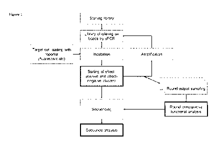
Figure I is a schematic diagram of aptamer library synthesis, sequencing and
target
identification work flow according to certain embodiments described herein.
Figure 2 is a schematic diagram of binding SELEX assay work flow described
herein.
Figure 2 discloses SEQ ID NO: 11.
Figure 3 is a schematic diagram of functional SELEX assay work flow described
herein.
Figure 4 is a schematic representation of certain aptamer structures according
to certain
exemplary embodiments provided herein.
Figure 5 is an exemplary flow cytometiy gating and sorting strategy. Panel A
is a
histogram of the microbead clustered library before incubation with the target
cells. Panel B is
a scatter plot of the microbead clustered library incubated with the target
cells followed by
incubation with a caspase-317 probe (Cas-3/7).
Figure 6 is an exemplary flow cytometry gating and sorting strategy involving
two
functional probes. Panel A is a scatter plot showing cells bound to the
clustered bead library in
the black rectangle. Panel B is a scatter plot of events from the black
rectangle in Panel A,
showing events probed for caspase-3/7 and mitochondria' membrane potential
(MitoProbe
Red).
Figure 7 shows the functional enrichment by exemplary methods provided herein
of
aptamer libraries for aptamers that induce apoptosis of the indicated cells.
The bar graph on the
left of each panel reflects the fold over increase of caspase-3/7 (Cas3/7) or
mitochondria'
membrane potential (Di1C1(5)) for each different round of functional
enrichment. The
histogram on the right of each panel shows an overlay of the enriched
libraries of the first
round (black) and final round (grey) of functional enrichment. With the
exception of Kasumi-1,
all results displayed were from functional enrichment initiated from the third
round of Binding
SELEX. Kasumi-1 results displayed were from functional enrichments initiated
from a random
library (not binding-enriched). Panel A shows results from FICT116 human
colorectal cancer
cell line. Panel B shows results from 411 murine breast cancer cell line,
Panel C shows results
from C126 murine colorectal cancer cell line, Panel D shows results from
Kasurni-1 human
acute myeloid leukemia (AML) cell line. Panel E shows results from AML1
primary AML
PBMCs from a donor, Panel F shows results from AML9 primary AML PBMCs from a
donor,
14
RECTIFIED SHEET (RULE 91) ISA/EP
CA 03114302 2021-03-25
WO 2020/065404 PCT/IB2019/001082
in logarithmic scale. The 10,000 most abundant aptamers in each Binding-Cell
SELEX round
are represented by black lines. The 10,000 most abundant aptamers in each
Functional-Cell
SELEX round are represented by blue lines. The 10 most abundant aptamers in
each Binding-
Cell SELEX round are represented by solid bold lines. The 10 most abundant
aptamers in each
functional enrichment round are represented by dashed lines. Panel A are the
profiles from the
assays performed on AML1 primary human myeloblasts. Panel B are the profiles
from the
assays performed on HCT116 colorectal cancer cell line.
Figure 9 is a comparison of caspase-3/7 activation after incubation of binding-
enriched
microbead clustered libraries or functionally-enriched clustered bead
libraries with target cells.
Percent of microbead-bound cells and caspase-3/7 positive cells were measured
by flow
cytometry. Panel A shows results after enriched-library incubation with AML1
target cells.
Panel B shows results after enriched-library incubation with HCT116 target
cells. Each data
point was measured in 4 technical replicates and significance was calculated
by Welch's one-
way t-test.
Figure 10 shows results related to the identification of a lead aptamer
candidate, E8,
from a functionally-enriched tumoricidal aptamer library. Panel A shows a
sequence
abundance plot from multiple Functional SELEX rounds. The plot shows a random
sample of
1,000 aptamer sequences out of the 10,000 most abundant aptamer sequences in
each round.
The 10 most abundant aptamer sequences are highlighted. Panel B is a
representative screening
of the 10 most abundant aptamers from Panel A for significant caspase-3/7
activation in
comparison to vehicle (V) and random oligonucleotide (R) in TNBC9 cells.
Aptamer IDs
labeled 1-10 (El, E2, ... El 0). Panel C shows selectivity of aptamer
candidate E8 to induce cell
death in TNBC9 cells (blue) in comparison to negative MCF10A target cells
(red). STA,
staurosporine; PAC, paclitaxel; Random, random oligonucleotide. Panel D shows
the effect of
E8 on MDA-MB-231 cells. Panel E shows the dose-response curve of E8 and
PEGylated-E8.
Panel F shows the effect of E8 on TNBC cells in mouse serum.
Figure 11 provides results demonstrating the biodistribution and efficacy of
E8
aptamer candidate in an animal model. Panel A shows fluorescence of E8
measured in-vivo at
0.1 h, 24 h, and 48 h after injection into NOD/SCID mice bearing MDA-MB-231-
derived
tumors. White arrows point to tumor locations. Panel B shows retention of E8
at tumor site 3 h
after intravenous injection (Ve, vehicle, K, kidney; T, tumor). Inset region
is shown magnified
CA 03114302 2021-03-25
WO 2020/065404 PCT/IB2019/001082
on the right, white arrowhead points to tumor site. Panel C shows a
quantitative measurement
of E8 fluorescent signal in tumors over a 48 h time period. Panel D shows the
efficacy of E8 in
reducing tumor volume of mice. Asterisks denote a statistically difference
with p < 0.05 (n = 8
mice/group). Panel E shows representative photographs of tumors excised from
mice sacrificed
at day 11. Panel F-G show histochemical analysis of caspase-3 activity in
tumor-derived tissue
sections (Panel F, vehicle-treated; Panel G, E8-treated). Panel H-I show TUNEL
analysis of
tumor-derived tissue sections (H, vehicle-treated; I, E8-treated).
Figure 12 are results demonstrating the efficacy of E8 in human ex-vivo organ
cultures
(EVOC). Panel A shows histological samples derived from patient 1 (P1). Panel
B shows
histological samples derived from patient 2 (P2). Graded pathological
assessment was made on
a scale of 0-4 by two blinded pathologists. White stars denote samples in
which an effect
reached a grade of at least 3. Rnd, random.
DETAILED DESCRIPTION
General
Provided herein are methods and composition related to the identification of
aptamers
that modulate a functional effect on a target cell. In certain embodiments,
the methods
comprise contacting the target cells to a plurality of aptamer clusters
immobilized on a surface
(e.g., the surface of a particle such as a bead, including a microbead, a
nanobead). Thus, in
.. some embodiments, the method comprises incubating a library of aptamer
cluster particles with
target cells in a single reaction volume under a condition and for a period of
time to form cell-
aptamer cluster particle complexes, and isolating and identifying the
population of aptamers
that modulate the cellular function.
In some embodiments, aptamers that functionally modulate a cellular function
are
identified by providing a detectable label indicative of the function being
modulated (e.g., a
fluorescent dye, such as a calcium sensitive dye, a cell tracer dye, a
lipophilic dye, a cell
proliferation dye, a cell cycle dye, a metabolite sensitive dye, a pH
sensitive dye, a membrane
potential sensitive dye, a mitochondrial membrane potential sensitive dye, or
a redox potential
dye) to the target cells, and then by physical partitioning the cell-aptamer
cluster particle
complexes having altered cell function after measuring the signal of the
detectable label. The
physical partitioning can be via, for example, flow cytometry, florescent
microscopy, optical
16
CA 03114302 2021-03-25
WO 2020/065404 PCT/IB2019/001082
tweezers, micropipettes, microfluid separation, micromanipulation, or isolated
seeding. The
aptamer cluster particles can then be isolated from the cell-aptamer cluster
particle complexes,
for example, via cell lysis and centrifugation. The individual aptamer
sequences can be
dissociated from the parties, amplified and sequenced.
In certain aspects, also provided herein are methods and compositions related
to the
creation of immobilized of aptamer clusters on a surface (e.g., a particle
surface). In some
embodiments, the aptamers (e.g., from an aptamer library disclosed herein) are
immobilized on
a particle. The particle can be made of any material. For example, in some
embodiments, the
particle is made of plastic, glass, polymer, or metal. In certain embodiments,
the particle is a
polymer bead, an agarose bead, a polystyrene bead, an acrylamide bead, a solid
core bead, a
porous bead, a paramagnetic bead, glass bead, controlled pore bead, a
microbead or a
nanoparticle. In some embodiments, the particles have at least one dimension
of an average
diameter of about 0.05, 0.1, 0.2, 0.3, 0.4, 0.5, 1, 1.5, 2, 2.5, 3, 3.5, 4,
4.5, 5, 7.5, 10, 15, 20, 25,
30, 35, 40, 45, 50, 55, 60, 65, 70, 75, 80, 85, 90, 95, 100, 105, 110, 115,
120, 125, 130, 135,
140, 145, 150, 155, 160, 165, 170, 175, 180, 185, 190, 195, or 200 microns. In
some
embodiments, the particle is coated with a blocker, such as, a polymer, a
protein, an oligo, a
lipid, and/or a chemical group. In some embodiments, the particle contains an
anchor of any
length to bind the target cells at proximity to clusters. The anchor may be a
polymer, a protein,
an oligo, a lipid, and/or a chemical group. In some embodiments, a localized
amplification
process, such as emulsion PCR is then performed to generate aptamer clusters.
The
complementary strands can be stripped in order to generate single-stranded
aptamer clusters.
The aptamer cluster particles are then ready for use in an aptamer
identification method
provided herein.
Definitions
For convenience, certain terms employed in the specification, examples, and
appended
claims are collected here.
The articles "a" and "an" are used herein to refer to one or to more than one
(e.g., to at
least one) of the grammatical object of the article. By way of example, "an
element" means one
element or more than one element.
17
CA 03114302 2021-03-25
WO 2020/065404 PCT/IB2019/001082
As used herein, the term "aptamer" refers to a short (e.g., less than 200
bases), single
stranded nucleic acid molecule (ssDNA and/or ssRNA) able to specifically bind
to a protein or
peptide target or to a topographic feature on a target cell.
As used herein, the term "aptamer cluster" refers to a collection of locally
immobilized
aptamers (e.g., at least 10) of identical sequence.
The term "binding" or "interacting" refers to an association, which may be a
stable
association, between two molecules, e.g., between an aptamer and target, e.g.,
due to, for
example, electrostatic, hydrophobic, ionic and/or hydrogen-bond interactions
under
physiological conditions.
As used herein, two nucleic acid sequences "complement" one another or are
"complementary" to one another if they base pair one another at each position.
As used herein, the term "contacting" refers to the bringing together of two
or more
molecular entities such that they can interact with each other.
As used herein, two nucleic acid sequences "correspond" to one another if they
are
both complementary to the same nucleic acid sequence.
The term "modulation" or "modulate", when used in reference to a functional
property
or biological activity or process (e.g., enzyme activity or receptor binding),
refers to the
capacity to either up regulate (e.g., activate or stimulate), down regulate
(e.g., inhibit or
suppress) or otherwise change a quality of such property, activity, or
process. In certain
instances, such regulation may be contingent on the occurrence of a specific
event, such as
activation of a signal transduction pathway, and/or may be manifest only in
particular cell
types.
As used herein, "specific binding" refers to the ability of an aptamer to bind
to a
predetermined target. In certain embodiments, an aptamer specifically binds to
the target with a
KD that is significantly less (e.g., at least 2 fold less, at least 5 fold
less, at least 10 fold less, at
least 50 fold less, at least 100 fold less, at least 500 fold less, or at
least 1000 fold less) than its
KD for binding to a non-specific and unrelated target (e.g., BSA, casein, or
an unrelated cell,
such as an FIEK 293 cell or an E. coli cell in cases where those cells were
not the target of the
process and were used as the negative target of the process). In some
embodiments, an aptamer
specifically binds to its target with an affinity corresponding to a KD of
about 10' M or less,
18
CA 03114302 2021-03-25
WO 2020/065404 PCT/IB2019/001082
about 10-7 M or less, about 10-8 M or less, about 10-9 M or less, about 10-10
M or less, about 10-
11 M or less, about 10-12 M or less, about 10-13 M or less, or about 10-14 M
or less.
As used herein, the Tm or melting temperature of two oligonucleotides is the
temperature at which 50% of the oligonucleotide/targets are bound and 50% of
the
oligonucleotide target molecules are not bound. Tm values of two
oligonucleotides are
oligonucleotide concentration dependent and are affected by the concentration
of monovalent,
divalent cations in a reaction mixture. Tm can be determined empirically or
calculated using
the nearest neighbor formula, as described in Santa Lucia, J. PNAS (USA)
95:1460-1465
(1998), which is hereby incorporated by reference.
The terms "polynucleotide" and "nucleic acid" are used herein interchangeably.
They
refer to a polymeric form of nucleotides of any length, either
deoxyribonucleotides or
ribonucleotides, or analogs thereof. Polynucleotides may have any three-
dimensional structure,
and may perform any function, known or unknown. The following are non-limiting
examples
of polynucleotides: coding or non-coding regions of a gene or gene fragment,
loci (locus)
defined from linkage analysis, exons, introns, messenger RNA (mRNA), transfer
RNA,
ribosomal RNA, ribozymes, cDNA, synthetic polynucleotides, recombinant
polynucleotides,
branched polynucleotides, plasmids, vectors, isolated DNA of any sequence,
isolated RNA of
any sequence, nucleic acid probes, and primers. A polynucleotide may comprise
modified
nucleotides, such as methylated nucleotides and nucleotide analogs. If
present, modifications to
the nucleotide structure may be imparted before or after assembly of the
polymer. The
sequence of nucleotides may be interrupted by non-nucleotide components. A
polynucleotide
may be further modified, such as by conjugation with a labeling component.
Aptamer Libraries
In certain embodiments, the methods and compositions provided herein relate to
the
identification of aptamers having desired properties from among the aptamers
present in an
aptamer library. As used herein, an aptamer library is a collection of nucleic
acid molecules
(e.g., DNA and/or RNA) having distinct sequences (e.g., at least 102, 103,
104, 105, 106, 107,
108, 109, 1010, 1011, 1012, 1013, 1014, or 10-15 distinct sequences) and
wherein at least a subset of
the nucleic acid molecules is structured such that they are capable of
specifically binding to a
19
CA 03114302 2021-03-25
WO 2020/065404 PCT/IB2019/001082
target protein, peptide, or cellular topographic feature. In some embodiments,
any library of
potential aptamers can be used in the methods and compositions provided
herein.
In some embodiments, the aptamer library used in the methods and compositions
provided herein comprises, consists of and/or consists essentially of nucleic
acid molecules
(e.g., DNA and/or RNA) having a sequence according to Formula (I):
P1-R-P2 (I),
wherein P1 is a 5' primer site sequence of about 10 to 100 bases in length,
about 10 to
50 bases in length, about 10 to 30 bases in length, about 15 to 50 bases in
length or about 15 to
30 bases in length; P2 is a 3' primer site sequence of about 10 to 100 bases
in length, about 10
to 50 bases in length, about 10 to 30 bases in length, about 15 to 50 bases in
length or about 15
to 30 bases in length; and R is a sequence comprising randomly positioned
bases of about at
least 10, 15, 20, 25, 30, 35, 40, 45, 50, 55, 60, 65, 70, 75 or 80 bases in
length and/or no more
than about 1000, 900, 800, 700, 600, 500, 400, 300, 200, 150, 120, 115, 110,
105, 100, 95, 90,
85, 80, 75, 70, 65, 60, 55 or 50 bases in length.
In one embodiment, R is a sequence comprising about 25% A. In another
embodiment,
R is a sequence comprising about 25% T. In another embodiment, R is a sequence
comprising
about 25% G. In another embodiment, R is a sequence comprising about 25% C. In
yet another
embodiment, R is a sequence comprising about 25% A, about 25% T, about 25% G,
and about
25% C.
In some embodiments, the aptamer library used in the methods and compositions
provided herein comprises, consists of and/or consists essentially of nucleic
acid molecules
(DNA and/or RNA) having a sequence according to Formula (I):
P1 -R" -P2 (I),
wherein P1 is a 5' primer site sequence of about 10 to 100 bases in length,
about 10 to
50 bases in length, about 10 to 30 bases in length, about 15 to 50 bases in
length or about 15 to
bases in length; P2 is a 3' primer site sequence of about 10 to 100 bases in
length, about 10
to 50 bases in length, about 10 to 30 bases in length, about 15 to 50 bases in
length or about 15
to 30 bases in length; and R" is a sequence of about at least 10, 15, 20, 25,
30, 35, 40, 45, 50,
55, 60, 65, 70, 75 or 80 bases in length and/or no more than about 120, 115,
110, 105, 100, 95,
30 90, 85, 80, 75, 70, 65, 60, 55 or 50 bases in length comprising randomly
positioned bases from
a biased mixture or any combination of random strings with repetitive or
biased strings.
CA 03114302 2021-03-25
WO 2020/065404 PCT/IB2019/001082
In some embodiments, the aptamer library used in the methods and compositions
provided herein comprises, consists of and/or consists essentially of nucleic
acid molecules
(DNA and/or RNA) having a sequence according to Formula II (an exemplary
schematic
representation is provided in Figure 4A),
131-S1 -L1 -S1 *-S2-L2-S2*-P2 (II),
wherein:
P1 is a 5' primer site sequence of about 10 to 100 bases in length, about 10
to 50 bases
in length, about 10 to 30 bases in length, about 15 to 50 bases in length or
about 15 to 30 bases
in length; P2 is a 3' primer site sequence of about 10 to 100 bases in length,
about 10 to 50
bases in length, about 10 to 30 bases in length, about 15 to 50 bases in
length or about 15 to 30
bases in length; Si and S2 are each independently a stem region sequence of at
least one base
(e.g., of about 4 to 40 bases in length or 4, 5, 6, 7, 8, 9, 10, 11, 12, 13,
14, 15, 16, 17, 18, 19,
20, 21, 22, 23, 24, 25, 26, 27, 28, 29, 30, 31, 32, 33, 34, 35, 36, 37, 38, 39
or 40 bases in
length); Si* is a complementary sequence to Si; S2* is a complementary
sequence to S2; Li
and L2 are each independently a Loop region sequence of at least one base
(e.g., of about 1 to
50 bases in length or 1,2, 3,4, 5, 6, 7, 8, 9, 10, 11, 12, 13, 14, 15, 16, 17,
18, 19, 20, 21, 22, 23,
24, 25, 26, 27, 28, 29, 30, 31, 32, 33, 34, 35, 36, 37, 38, 39, 40, 41, 42,
43, 44, 45, 46, 47, 48,
49 or 50 bases in length); and Sl-Ll-S1*-S2-L2-S2* is collectively about at
least 10, 15, 20,
25, 30, 35, 40, 45, 50, 55, 60, 65, 70, 75 or 80 bases in length and/or no
more than about 120,
115, 110, 105, 100, 95, 90, 85, 80, 75, 70, 65, 60, 55 or 50 bases in length.
In some embodiments, the aptamer library used in the methods and compositions
provided herein comprises, consists of and/or consists essentially of nucleic
acid molecules
(DNA and/or RNA) having a sequence according Formula III (an exemplary
schematic
representation is provided in Figure 4B):
P1-Si -Li 1 -S1*-132 (III),
wherein:
P1 is a 5' primer site sequence of about 10 to 100 bases in length, about 10
to 50 bases
in length, about 10 to 30 bases in length, about 15 to 50 bases in length or
about 15 to 30 bases
in length; P2 is a 3' primer site sequence of about 10 to 100 bases in length,
about 10 to 50
bases in length, about 10 to 30 bases in length, about 15 to 50 bases in
length or about 15 to 30
bases in length;
21
CA 03114302 2021-03-25
WO 2020/065404 PCT/IB2019/001082
Si and S2 are each independently a stem region sequence of at least one base
(e.g., of
about 4 to 40 bases in length or 4, 5, 6, 7, 8, 9, 10, 11, 12, 13, 14, is, 16,
17, 18, 19, 20, 21, 22,
23, 24, 25, 26, 27, 28, 29, 30, 31, 32, 33, 34, 35, 36, 37, 38, 39 or 40 bases
in length); Si* is a
complementary sequence to Si; S2* is a complementary sequence to S2;
Li and L2 are each independently a Loop region sequence of at least one base
(e.g., of
about 1 to 50 bases in length or 1, 2, 3,4, 5, 6, 7, 8, 9, 10, 11, 12, 13, 14,
is, 16, 17, 18, 19, 20,
21, 22, 23, 24, 25, 26, 27, 28, 29, 30, 31, 32, 33, 34, 35, 36, 37, 38, 39,
40, 41, 42, 43, 44, 45,
46, 47, 48, 49 or 50 bases in length); and
Sl-Ll-S2-L2-S2*-Ll-S1* is collectively about at least 10, 15, 20, 25, 30, 35,
40, 45,
50, 55, 60, 65, 70, 75 or 80 bases in length and/or no more than about 120,
115, 110, 105, 100,
95, 90, 85, 80, 75, 70, 65, 60, 55 or 50 bases in length.
In some embodiments, the aptamer library used in the methods and compositions
provided herein comprises, consists of and/or consists essentially of nucleic
acid molecules
(DNA and/or RNA) having a sequence according Formula IV (an exemplary
schematic
representation is provided in Figure 4C):
Pl-Lib-Ml/M2-D-Ml/M2*-Lib-P2 (IV),
wherein:
P1 is a 5' primer site sequence of about 10 to 100 bases in length, about 10
to 50 bases
in length, about 10 to 30 bases in length, about 15 to 50 bases in length or
about 15 to 30 bases
in length; P2 is a 3' primer site sequence of about 10 to 100 bases in length,
about 10 to 50
bases in length, about 10 to 30 bases in length, about 15 to 50 bases in
length or about 15 to 30
bases in length;
Lib is sequence having a formula selected from: (i) R; (ii) R"; (iii) Sl-Ll-
S1*-S2-L2-
S2*; and (iv) Sl-Ll-S2-L2-S2*-Ll-S1*;
R is a sequence comprising randomly positioned bases of about at least 10, 15,
20, 25,
30, 35, 40, 45, 50, 55, 60, 65, 70, 75 or 80 bases in length and/or no more
than about 1000,
900, 800, 700, 600, 500, 400, 300, 200, 150, 120, 115, 110, 105, 100, 95, 90,
85, 80, 75, 70, 65,
60, 55 or 50 bases in length;
R" is a sequence of about at least 10, 15, 20, 25, 30, 35, 40, 45, 50, 55, 60,
65, 70, 75 or
80 bases in length and/or no more than about 1000, 900, 800, 700, 600, 500,
400, 300, 200,
150, 120, 115, 110, 105, 100, 95, 90, 85, 80, 75, 70, 65, 60, 55 or 50 bases
in length comprising
22
CA 03114302 2021-03-25
WO 2020/065404 PCT/IB2019/001082
randomly positioned bases from a biased mixture or any combination of random
strings with
repetitive or biased strings;
Si and S2 are each independently a stem region sequence of at least one base
(e.g., of
about 4 to 40 bases in length or 4, 5, 6, 7, 8, 9, 10, 11, 12, 13, 14, is, 16,
17, 18, 19, 20, 21, 22,
23, 24, 25, 26, 27, 28, 29, 30, 31, 32, 33, 34, 35, 36, 37, 38, 39 or 40 bases
in length); Si* is a
complementary sequence to Si; S2* is a complementary sequence to S2;
Li and L2 are each independently a Loop region sequence of at least one base
(e.g., of
about 1 to 50 bases in length or 1, 2, 3,4, 5, 6, 7, 8, 9, 10, 11, 12, 13, 14,
is, 16, 17, 18, 19, 20,
21, 22, 23, 24, 25, 26, 27, 28, 29, 30, 31, 32, 33, 34, 35, 36, 37, 38, 39,
40, 41, 42, 43, 44, 45,
46, 47, 48, 49 or 50 bases in length);
wherein Sl-Ll-S1*-S2-L2-S2* is collectively about at least 10, 15, 20, 25, 30,
35, 40,
45, 50, 55, 60, 65, 70, 75 or 80 bases in length and/or no more than about
120, 115, 110, 105,
100, 95, 90, 85, 80, 75, 70, 65, 60, 55 or 50 bases in length;
D is a spacer sequence comprising at least one base (e.g., of about 1 to 20
bases in length
.. or 1, 2, 3, 4, 5, 6, 7, 8, 9, 10, 11, 12, 13, 14, 15, 16, 17, 18, 19 or 20
bases in length);
M1 is a multimer-forming domain sequence of about 10 to 18 bases in length or
10, 11,
12, 13, 14, 15, 16, 17 or 18 bases in length that enables a strand of the
sequence to interact with
another strand that contains a complementary domain; and
M2 is a complementary domain of M1 comprising a strand that interacts with a
strand
of the M1 sequence.
In some embodiments, the aptamer library used in the methods and compositions
provided herein comprises, consists of and/or consists essentially of nucleic
acid molecules
(DNA and/or RNA) having a sequence according Formula V (an exemplary schematic
representation is provided in Figure 4D):
Pl-Lib-T*-Lib-P2 (V),
wherein:
P1 is a 5' primer site sequence of about 10 to 100 bases in length, about 10
to 50 bases
in length, about 10 to 30 bases in length, about 15 to 50 bases in length or
about 15 to 30 bases
in length; P2 is a 3' primer site sequence of about 10 to 100 bases in length,
about 10 to 50
.. bases in length, about 10 to 30 bases in length, about 15 to 50 bases in
length or about 15 to 30
bases in length;
23
CA 03114302 2021-03-25
WO 2020/065404 PCT/IB2019/001082
Lib is sequence having a formula selected from: (i) R; (ii) R"; (iii) 51-L1-
51*-S2-L2-
S2*; and (iv) 51-L1-52-L2-52*-L1-51*;
R is a sequence comprising randomly positioned bases of about at least 10, 15,
20, 25,
30, 35, 40, 45, 50, 55, 60, 65, 70, 75 or 80 bases in length and/or no more
than about 1000,
900, 800, 700, 600, 500, 400, 300, 200, 150, 120, 115, 110, 105, 100, 95, 90,
85, 80, 75, 70, 65,
60, 55 or 50 bases in length;
R" is a sequence of about at least 10, 15, 20, 25, 30, 35, 40, 45, 50, 55, 60,
65, 70, 75 or
80 bases in length and/or no more than about 1000, 900, 800, 700, 600, 500,
400, 300, 200,
150, 120, 115, 110, 105, 100, 95, 90, 85, 80, 75, 70, 65, 60, 55 or 50 bases
in length comprising
randomly positioned bases from a biased mixture or any combination of random
strings with
repetitive or biased strings;
Si and S2 are each independently a stem region sequence of at least one base
(e.g., of
about 4 to 40 bases in length or 4, 5, 6, 7, 8, 9, 10, 11, 12, 13, 14, 15, 16,
17, 18, 19, 20, 21, 22,
23, 24, 25, 26, 27, 28, 29, 30, 31, 32, 33, 34, 35, 36, 37, 38, 39 or 40 bases
in length);Si* is a
complementary sequence to 51; S2* is a complementary sequence to S2;
Li and L2 are each independently a Loop region sequence of at least one base
(e.g., of
about 1 to 50 bases in length or 1,2, 3,4, 5, 6, 7, 8, 9, 10, 11, 12, 13, 14,
15, 16, 17, 18, 19, 20,
21, 22, 23, 24, 25, 26, 27, 28, 29, 30, 31, 32, 33, 34, 35, 36, 37, 38, 39,
40, 41, 42, 43, 44, 45,
46, 47, 48, 49 or 50 bases in length);
wherein 51-L1-51*-S2-L2-S2* is collectively about at least 10, 15, 20, 25, 30,
35, 40,
45, 50, 55, 60, 65, 70, 75 or 80 bases in length and/or no more than about
120, 115, 110, 105,
100, 95, 90, 85, 80, 75, 70, 65, 60, 55 or 50 bases in length;
T is a second strand bound by Watson/Crick or Hoogsteen base pairing to any
part of
the Lib sequence or T*, wherein the strand optionally contains unpaired
domains on its 5' and
3' ends (e.g., to facilitate attachment of a functional moiety to the
aptamer); and
T* is a dedicated domain sequence (e.g., of about 4 to 40 bases in length or
4, 5, 6, 7, 8,
9, 10, 11, 12, 13, 14, 15, 16, 17, 18, 19, 20, 21, 22, 23, 24, 25, 26, 27, 28,
29, 30, 31, 32, 33, 34,
35, 36, 37, 38, 39 or 40 bases in length).
In some embodiments, the aptamer library used in the methods and compositions
provided herein comprises, consists of and/or consists essentially of nucleic
acid molecules
(DNA and/or RNA) having a sequence according to a formula selected from:
24
CA 03114302 2021-03-25
WO 2020/065404 PCT/IB2019/001082
Pl-R-Pl*, P1 -S1-R-S1-P2, and P1-R-S1-R-S1-R-P2
wherein:
P1 is a 5' primer site sequence of about 10 to 100 bases in length, about 10
to 50 bases
in length, about 10 to 30 bases in length, about 15 to 50 bases in length or
about 15 to 30 bases
in length; P2 is a 3' primer site sequence of about 10 to 100 bases in length,
about 10 to 50
bases in length, about 10 to 30 bases in length, about 15 to 50 bases in
length or about 15 to 30
bases in length; Pl* is a complementary sequence to Pl.
R is a sequence comprising randomly positioned bases of about at least 10, 15,
20, 25,
30, 35, 40, 45, 50, 55, 60, 65, 70, 75 or 80 bases in length and/or no more
than about 1000,
900, 800, 700, 600, 500, 400, 300, 200, 150, 120, 115, 110, 105, 100, 95, 90,
85, 80, 75, 70, 65,
60, 55 or 50 bases in length;
Si is a stem region sequence of at least one base (e.g., of about 4 to 40
bases in length
or 4, 5, 6, 7, 8, 9, 10, 11, 12, 13, 14, 15, 16, 17, 18, 19, 20, 21, 22, 23,
24, 25, 26, 27, 28, 29,
30, 31, 32, 33, 34, 35, 36, 37, 38, 39 or 40 bases in length);
In some embodiments of the formulae above, R, P, or S comprises a CpG island
and/or
a G-quadruplex sequence.
In some embodiments of the Formulae above, R is randomly positioned bases from
any
random mixture (e.g., for canonical bases, 25% A, 25% T, 25% G, 25% C) of
about at least 10,
15, 20, 25, 30, 35, 40, 45, 50, 55, 60, 65, 70, 75 or 80 bases in length
and/or no more than
about 120, 115, 110, 105, 100, 95, 90, 85, 80, 75, 70, 65, 60, 55 or 50 bases
in length.
In one embodiment of the Formulae above, R is a sequence comprising about 25%
A.
In another embodiment, R is a sequence comprising about 25% T. In another
embodiment, R is
a sequence comprising about 25% G. In another embodiment, R is a sequence
comprising
about 25% C. In yet another embodiment, R is a sequence comprising about 25%
A, about
25% T, about 25% G, and about 25% C.
In some embodiments of the Formulae above, R" is a sequence comprising
comprises
randomly positioned bases from a biased mixture (e.g., for canonical bases,
any mixture
deviating from 25% per base). In some embodiments, R" is a sequence that
comprises about
0%, 5%, 10%, 15%, 20%, 25%, 30%, 35%, 40%, 45%, 50%, 55%, 60%, 65%, 70% or 75%
A.
In some embodiments, R" is a sequence that comprises about 0%, 5%, 10%, 15%,
20%, 25%,
30%, 35%, 40%, 45%, 50%, 55%, 60%, 65%, 70% or 75% T. In some embodiments, R"
is a
CA 03114302 2021-03-25
WO 2020/065404 PCT/IB2019/001082
sequence that comprises about 0%, 5%, 10%, 15%, 20%, 25%, 30%, 35%, 40%, 45%,
50%,
55%, 60%, 65%, 70% or 75% C. In some embodiments, R" is a sequence that
comprises about
0%, 5%, 10%, 15%, 20%, 25%, 30%, 35%, 40%, 45%, 50%, 55%, 60%, 65%, 70% or 75%
G.
In some embodiments, R" is a sequence that comprises any combination of random
strings
(string is any sequence including a single base) with repetitive or biased
strings.
In some embodiments of the Formulae above, R" is randomly positioned bases
from a
biased mixture (e.g., for canonical bases, any mixture deviating from 25% per
base); or any
combination of random strings (string is any sequence including a single base)
with repetitive
or biased strings of about at least 10, 15, 20, 25, 30, 35, 40, 45, 50, 55,
60, 65, 70, 75 or 80
.. bases in length and/or no more than about 120, 115, 110, 105, 100, 95, 90,
85, 80, 75, 70, 65,
60, 55 or 50 bases in length.
In some embodiments of the Formulae above, Si is a stem region sequence of at
least 1
base or more. In other embodiments, Si is a stem region sequence of between
about 4 to 40
bases in length or 4, 5, 6, 7, 8, 9, 10, 11, 12, 13, 14, 15, 16, 17, 18, 19,
20, 21, 22, 23, 24, 25,
26, 27, 28, 29, 30, 31, 32, 33, 34, 35, 36, 37, 38, 39 or 40 bases in length.
In some embodiments of the Formulae above, S2 is a stem region sequence of at
least 1
base or more. In other embodiments, S2 is a stem region sequence of between
about 4 to 40
bases in length or 4, 5, 6, 7, 8, 9, 10, 11, 12, 13, 14, 15, 16, 17, 18, 19,
20, 21, 22, 23, 24, 25,
26, 27, 28, 29, 30, 31, 32, 33, 34, 35, 36, 37, 38, 39 or 40 bases in length.
In some embodiments of the Formulae above, Li is a Loop region sequence of at
least
one base. In other embodiments, Li is a Loop region sequence of about 1 to 50
bases in length
or 1, 2, 3, 4, 5, 6, 7, 8, 9, 10, 11, 12, 13, 14, 15, 16, 17, 18, 19, 20, 21,
22, 23, 24, 25, 26, 27,
28, 29, 30, 31, 32, 33, 34, 35, 36, 37, 38, 39, 40, 41, 42, 43, 44, 45, 46,
47, 48, 49 or 50 bases in
length.
In some embodiments of the Formulae above, L2 is a Loop region sequence of at
least
one base. In other embodiments, L2 is a Loop region sequence of about 1 to 50
bases in length
or 1, 2, 3, 4, 5, 6, 7, 8, 9, 10, 11, 12, 13, 14, 15, 16, 17, 18, 19, 20, 21,
22, 23, 24, 25, 26, 27,
28, 29, 30, 31, 32, 33, 34, 35, 36, 37, 38, 39, 40, 41, 42, 43, 44, 45, 46,
47, 48, 49 or 50 bases in
length.
In some embodiments of the Formulae above, T may include unpaired domains on
its
5' and 3' ends, or it may be a padlock tail (e.g., a loop between two domains
paired with the
26
CA 03114302 2021-03-25
WO 2020/065404 PCT/IB2019/001082
library).The aptamers of the present disclosure may contain any number of
stems and loops,
and other structures comprised of stems and loops (e.g.,. hairpins, bulges,
etc.). In some
embodiments, the loops in the aptamer contain bases implanted in order to form
stable loop-
loop WC pairing forming a stem which is orthogonal to the main library axis.
In other
embodiments, two loops in the aptamer together form an orthogonal stem. In yet
other
embodiments, the loops in the aptamer contain bases implanted in order to form
stable
Hoogsteen pairing with an existing stem along the main library axis. In other
embodiments, the
loops in the aptamer can form Hoogsteen pairing with any stem in the aptamer.
In some embodiments of the formulae above, the aptamer sequence further
contains one
or more multimer-forming domains.
In some embodiments of the formulae above, the aptamer sequence further
contains one
or more spacers (e.g., of about 1 to 20 bases in length or 1,2, 3,4, 5, 6, 7,
8, 9, 10, 11, 12, 13,
14, 15, 16, 17, 18, 19 or 20 bases in length).
The aptamers of the present disclosure can be prepared in a variety of ways.
In one
embodiment, the aptamers are prepared through chemical synthesis. In another
embodiment,
the aptamers are prepared through enzymatic synthesis. In one embodiment, the
enzymatic
synthesis can be carried out using any enzyme that can add nucleotides to
elongate a primer,
with or without template. In some embodiments, the aptamers are prepared by
assembling
together k-mers (e.g., k>2 bases).
In some embodiments, the aptamers of the present disclosure may contain any
combination of DNA, RNA, and their natural and/or synthetic analogs. In one
embodiment, the
aptamer comprises DNA. In one embodiment, the aptamer comprises RNA.
In other embodiments, the aptamers of the present disclosure may contain any
modification on the 5' end, 3' end, or internally. Modifications of the
aptamers include, but are
not limited to, spacers, phosphorylation, linkers, conjugation chemistries,
fluorophores,
quenchers, photoreactive, and modified bases (e.g., LNA, PNA, UNA, PS,
methylation, 2-0-
methyl, halogenated, superbases, iso-dN, inverted bases, L-ribose, other
sugars as backbone,
etc.).
In some embodiments, the aptamers of the present disclosure may be conjugated
to
external, non-nucleic acid molecules on the 5' end, 3' end, or internally. Non-
limiting
examples of non-nucleic acid molecules include, but are not limited to amino
acids, peptides,
27
CA 03114302 2021-03-25
WO 2020/065404 PCT/IB2019/001082
proteins, small molecule drugs, mono- and polysaccharides, lipids, antibodies
and antibody
fragments, or a combination thereof.
The aptamers of the present disclosure may contain any domain which has a
biological
function. Non-limiting examples of biological functions of the aptamers
described herein
include, but are not limited to, acting as templates for RNA transcription,
binding to,
recognizing, and/or modulating the activity of proteins, binding to
transcription factors,
specialized nucleic acid structure (e.g., Z-DNA, H-DNA, G-quad, etc.), and
acting as an
enzymatic substrate for restriction enzymes, specific exo- and endonucleases,
recombination
sites, editing sites, or siRNA. In one embodiment, the aptamers modulate the
activity of at least
one protein. In another embodiment, the aptamers inhibit the activity of at
least one protein.
In other embodiments, the aptamers of the present disclosure may contain any
domain
for integration into a nucleic acid nanostructure built by any one of several
known methods
(Shih et al, Nature 427:618-621 (2004); Rothemund, Nature 440:297-302 (2006);
Zheng et al,
Nature 461:74-77 (2009); Dietz et al, Science 325:725-730 (2009); Wei et al,
Nature 485:623-
626 (2012); Ke et al, Science 338:1177-1183 (2012); Douglas et al, Science
335:831-834
(2012), each of which are hereby incorporated by reference). In yet other
embodiments, the
aptamers of the present disclosure may contain any domain that serves a
function in molecular
logic and computation (Seelig et al, Science 314:1585-1588 (2006); Macdonald
et al, Nano
Lett 6:2598-2603 (2006); Qian et al, Nature 475:368-372 (2011); Douglas et al,
Science
335:831-834 (2012); Amir et al, Nat Nanotechnol 9:353-357 (2014), each of
which is hereby
incorporated by reference).
In some embodiments, the aptamers of the present disclosure undergo one or
more
cycles of negative selection versus a negative control target including but
not limited to
eukaryotic or prokaryotic cell, virus or viral particle, protein, molecule,
tissue, or whole
organism, in-vivo or ex-vivo. In other embodiments, the aptamers of the
present disclosure
undergo one or more cycles of positive selection versus a positive target
(e.g., eukaryotic or
prokaryotic cell, virus or viral particle, molecule, tissue, or whole
organism, in-vivo or ex-vivo).
The aptamers of the present disclosure can be in solution or attached to a
solid phase
(e.g., surface, particles, resin, matrix, etc.). In some embodiments, the
aptamer is attached to a
surface. In one embodiment, the surface is a particle surface.
28
CA 03114302 2021-03-25
WO 2020/065404 PCT/IB2019/001082
In some embodiments, the aptamers of the present disclosure are synthesized in
an
aptamer library. The aptamer library of the present disclosure can be prepared
in a variety of
ways. In one embodiment, the aptamer library is prepared through chemical
synthesis. In
another embodiment, the aptamer library is prepared through enzymatic
synthesis. In one
embodiment, the enzymatic synthesis can be carried out using any enzyme that
can add
nucleotides to elongate a primer, with or without template.
In some embodiments, the aptamers synthesized in an aptamer library may
contain any
combination of DNA, RNA, and their natural and/or synthetic analogs. In one
embodiment, the
aptamers synthesized in an aptamer library comprise DNA. In one embodiment,
the aptamers
synthesized in an aptamer library comprise RNA.
In some embodiments, the aptamers synthesized in an aptamer library are a
nucleic acid
(e.g., DNA, RNA, natural or synthetic bases, base analogs, or a combination
thereof) collection
of 10K species (K>2), with Z copies per species (1<Z<K-1).
In other embodiments, the aptamers synthesized in an aptamer library of the
present
disclosure may contain any modification on the 5' end, 3' end, or internally.
Modifications of
the aptamers include, but are not limited to, spacers, phosphorylation,
linkers, conjugation
chemistries, fluorophores, quenchers, photoreactive modifications, and
modified bases (e.g.,
LNA, PNA, UNA, PS, methylation, 2-0-methyl, halogenated, superbases, iso-dN,
inverted
bases, L-ribose, other sugars as backbone).
In some embodiments, the aptamers synthesized in an aptamer library may be
conjugated to external, non-nucleic acid molecules on the 5' end, 3' end, or
internally. Non-
limiting examples of non-nucleic acid molecules include, but are not limited
to amino acids,
peptides, proteins, small molecule drugs, mono- and polysaccharides, lipids,
antibodies and
antibody fragments, or a combination thereof.
The aptamers synthesized in an aptamer library may contain any domain which
has a
biological function. Non-limiting examples of biological functions of the
aptamers described
herein include, but are not limited to, acting as templates for RNA
transcription, binding to,
recognizing, and/or modulating the activity of proteins, binding to
transcription factors,
specialized nucleic acid structure (e.g., Z-DNA, H-DNA, G-quad, etc.), acting
as an enzymatic
substrate for restriction enzymes, specific exo- and endonucleases,
recombination sites, editing
sites, or siRNA. In one embodiment, the aptamers synthesized in an aptamer
library modulate
29
CA 03114302 2021-03-25
WO 2020/065404 PCT/IB2019/001082
the activity of at least one protein. In another embodiment, the aptamers
synthesized in an
aptamer library inhibit the activity of at least one protein.
In other embodiments, the aptamers synthesized in an aptamer library may
contain any
domain for integration into a nucleic acid nanostructure built by one of
several known methods
(Shih et al, Nature 427:618-621 (2004); Rothemund, Nature 440:297-302 (2006);
Zheng et al,
Nature 461:74-77 (2009); Dietz et al, Science 325:725-730 (2009); Wei et al,
Nature 485:623-
626 (2012); Ke et al, Science 338:1177-1183 (2012); Douglas et al, Science
335:831-834
(2012), each of which are hereby incorporated by reference). In yet other
embodiments, the
aptamers of the present disclosure may contain any domain that serves a
function in molecular
logic and computation (Seelig et al, Science 314:1585-1588 (2006); Macdonald
et al, Nano
Lett 6:2598-2603 (2006); Qian et al, Nature 475:368-372 (2011); Douglas et al,
Science
335:831-834 (2012); Amir et al, Nat Nanotechnol 9:353-357 (2014), each of
which is hereby
incorporated by reference)
In some embodiments, the aptamers synthesized in an aptamer library undergo
one or
more cycles of negative selection versus a target (e.g., eukaryotic or
prokaryotic cell). In other
embodiments, the aptamers of the present disclosure undergo one or more cycles
of positive
selection versus a target (e.g., eukaryotic or prokaryotic cell).
The aptamers synthesized in an aptamer library can be in solution or attached
to a solid
phase (e.g., surface, particles, resin, matrix, etc.). In some embodiments,
the aptamers
synthesized in an aptamer library are attached to a surface. In one
embodiment, the surface is a
particle surface.
Immobilized Aptamer Clusters
In certain aspects, provided herein are methods for identifying aptamers that
modulate a
target cell by incubating a sample comprising the target cells with a
plurality of aptamer
clusters (e.g., clusters of aptamers from the aptamer libraries provided
herein) immobilized on
a particle surface. . In some embodiments, the surface is a bead (e.g., a
microbead, a
nanobead).
Any method known in the art can be used to generate the immobilized aptamer
clusters
on the particle surface. In certain embodiments, the surface-immobilized
aptamer clusters are
generated by first immobilizing aptamers (e.g., from an aptamer library
disclosed herein) onto
CA 03114302 2021-03-25
WO 2020/065404 PCT/IB2019/001082
the surface (e.g., the surface of a particle). In some embodiments, at least
about 102, 103, 104,
105, 106, 107, 108, 109 or 1019 distinct aptamers are immobilized onto the
surface (e.g., the
surface of a particle). Following aptamer immobilization, a localized
amplification process
(e.g., emulsion PCR, bridge amplification, or rolling circle amplification),
is then performed to
generate clusters of copies of each immobilized aptamer positioned proximal to
the
immobilization site of the original immobilized aptamer. In certain
embodiments (e.g.,
embodiments in which rolling circle amplification is performed) the aptamer
cluster is housed
in a nano-pit or pore on the surface rather than being directly immobilized on
the surface. In
some embodiments, the aptamer clusters are prepared using emulsion PCR. In
some
embodiments, amplification results in each aptamer cluster comprising at least
about 10, 20,
30, 40, 50, 60, 70, 80, 90, 100, 125, 150, 175, 200, 250, 300, 350, 400, 450,
500, 600, 700, 800,
900, 1000, 2000, 3000, 4000, 5000, 10,000, 20,000, 30,000, 40,000, 50,000,
60,000, 70,000,
80,000, 90,000, 100,000, 200,000, 300,000, 400,000, 500,000, 600,000, 700,000,
800,000,
900,000, or 1000,000 identical aptamer molecules. In certain embodiments, the
aptamer
clusters are then sequenced (e.g., by Illumina sequencing or Polonator
sequencing). If present,
complementary strands can be stripped from the aptamer cluster by washing the
surface under
conditions not amenable to strand hybridization (e.g., due to salt
concentration and/or
temperature) in order to generate clusters of single-stranded aptamers. The
surface (e.g., the
particle surface) is then ready for use in an aptamer identification method
provided herein. In
some embodiments, the immobilized aptamer clusters are prepared and/or
sequenced on one
instrument, and then transferred to a separate instrument for aptamer
identification. In other
embodiments, the aptamer clusters are prepared and/or sequenced on the same
instrument as is
used for aptamer identification.
In some embodiments, each aptamer cluster particle comprises clusters of less
than
1000 different aptamers sequences, for examples, less than 900, less than 800,
less than 700,
less than 600, less than 500, less than 400, less than 300, or less than 200,
less than 100, less
than 90, less than 80, less than 70, less than 60, less than 50, less than 40,
less than 30, or less
than 20, less than 10, less than 9, less than 8, less than 7, less than 6,
less than 5, less than 4,
less than 3, or less than 2 different aptamers sequences. In one embodiment,
each aptamer
cluster particle comprises a unique aptamer sequence in multiple copies as
clusters on the
surface of the particle. In some embodiments, each aptamer cluster comprises
at least 2
31
CA 03114302 2021-03-25
WO 2020/065404 PCT/IB2019/001082
identical aptamers (e.g., at least 10, 20, 30, 40 50 60, 70, 80, 90, 100, 150,
200, 250, 300, 350,
400, 450, 500, 550, 600, 650, 700, 750, 800, 850, 900, 950, 1,000, 2,000,
3,000, 4,000, 5,000,
6,000, 7,000, 8,000, 9,000, 10,000, 20,000, 30,000, 40,000, 50,000, 60,000,
70,000, 80,000,
90,000, 100,000, 200,000, 300,000, 400,000, 500,000, 600,000, 700,000,
800,000, 900,000, or
1,000,000 identical aptamers).
In some embodiments of the methods above, the aptamers or aptamer clusters
(e.g.,
from the aptamer library) comprise an adapter that will bring the aptamers to
surface height
(e.g., in cases where the surface is not flat, such as in particles that
include pores). In one
embodiment, the aptamers or aptamer clusters are immobilized inside pores on a
particle
surface and adapters are used to bind the aptamer to the surface in order to
bring the aptamers
to surface height. In some embodiments, the adapter is a nucleic acid adapter
(e.g., a sequence
of at least about 10, 15, 20, 25, 30, 35, 40, 45, 50, 55, 60, 65, 70, 75, 80,
85, 90, 95 or 100
bases in length). In some embodiments, a sequence complementary to the adapter
sequence is
hybridized to the adapter prior to aptamer screening. In some embodiments, the
adapter is a
chemical adapter (e.g., a polymer connecting the aptamer to the surface).
Aptamer Library Screening
In certain aspects, provided herein are methods for identifying one or more
aptamers
that specifically modulate a target cell function, the method generally
comprising: (i)
incubating the library with target cells in a single reaction volume under a
condition and for a
period of time to form cell-aptamer cluster particle complexes; (ii)
partitioning the cell-aptamer
cluster particle complexes having altered cell function from the cell-aptamer
cluster particle
complexes without the desired effect, the free particles and the free cells;
(iii) isolating the
aptamer cluster particles from the cell-aptamer cluster particle complexes
having altered
cellular function; (iv) dissociating the aptamers from the particles; and (v)
amplifying
individual aptamer sequences to provide a functionally enriched population of
aptamers.
In certain embodiments, the methods further comprise a step of enriching the
population of functional aptamers by repeating the steps (i) - (v) in
additional rounds to yield a
specific and enriched population of functional aptamers. In certain
embodiments, the step of
enriching the population of functional aptamers involves applying a
restrictive condition (e.g.,
reducing the total number of particles) in the successive rounds. In some
embodiments, the
32
CA 03114302 2021-03-25
WO 2020/065404 PCT/IB2019/001082
enriched population of aptamers has decreased sequence diversity relative to
the plurality of
aptamers from the incubating step by a factor of at least 1.5 (e.g., by a
factor of about 1.5, 1.6,
1.7. 1.8, 1.9, or 2.0). In some embodiments, the population of aptamers of
each additional
round of screening is enriched by a factor of at least 1.1 (e.g., by a factor
of about 1.1, 1.2, 1.3,
1.4, 1.5, 1.6, 1.7. 1.8, 1.9, or 2.0). The number of rounds of enrichment can
be as many as
desired. For example, in some embodiments, the number of rounds are at least 2
(e.g., at least
3,4, 5, 6, 7, 8, 9, 10, 11, 12, 13, 14, 15, 16, 17, 18, 19, 20, 21, 22, 23,
24, 25, 26, 27, 28, 29, 30,
31, 32, 33, 34, 35, 36, 37, 38, 39, 40, 41, 42, 43, 44, 45, 46, 47, 48, 49,
50, 51, 52, 53, 54, 55,
56, 57, 58, 59, 60, 61, 62, 63, 64, 65, 66, 67, 68, 69, 70, 71, 72, 73, 74,
75, 76, 77, 78, 79, 80,
81, 82, 83, 84, 85, 86, 87, 88, 89, 90, 91, 92, 93, 94, 95, 96, 97, 98, 99, or
100). In certain
embodiments, the methods further comprise a step of identifying the enriched
population of
functional aptamers via sequencing after the step (v).
The library of aptamer cluster particles can be incubated with target cells
under any
condition conductive to form cell-aptamer cluster particle complexes and to
allow the aptamer
cluster particles to provide an effect on the target cells. The condition
includes, but is not
limited to, for examples, a controlled period of time, an optimal temperature
(e.g., 37 C),
and/or an incubating medium (e.g., target cell culture medium), etc. The
period of time of
incubation can be from about 10 minutes to about 5 days, from about 30 minutes
to about 4
days, from about 1 hour to about 3 days, from about 1.5 hours to about 24
hours, or from about
1.5 hours to about 2 hours. In some embodiments, the period of time of
incubation may be, for
example, 10 min, 15 min, 30 min, 45 min, 1 hour, 2 hours, 4 hours, 6 hours, 8
hours, 12 hours,
16 hours, 24 hours, 2 days, 3 days, 4 days, 5 days, 6 days, or 7 days.
The target cells and the library of aptamer cluster particles may be mixed at
a ratio such
that the formed cell-aptamer cluster particle complexes comprise about 1 to 10
particles per
target cell (e.g., 1, 2, 3, 4, 5, 6, 7, 8, 9, or 10 particles per target
cell). In certain embodiment,
the formed cell-aptamer cluster particle complexes comprise about 2 to 4
particles per target
cell. In some embodiments, the aptamer cluster particle in the formed cell-
aptamer cluster
particle complexes comprises about 1 to 10 clusters per particle (e.g., 1, 2,
3, 4, 5, 6, 7, 8, 9, or
10 clusters per particle). In certain embodiments, the aptamer cluster
particle in the formed
cell-aptamer cluster particle complexes comprises about 1 to 6 clusters per
particle.
33
CA 03114302 2021-03-25
WO 2020/065404 PCT/IB2019/001082
In some embodiments, the target cells are labeled with and/or comprises a
detectable
label. The target (e.g., target cells) can be detectably labeled directly
(e.g., through a direct
chemical linker) or indirectly (e.g., using a detectably labeled target-
specific antibody). In
embodiments in which the target is a cell, it can be labeled by incubating the
target cell with
the detectable label under conditions such that the detectable label is
internalized by the cell. In
some embodiments, the target is detectably labeled before performing the
aptamer screening
methods described herein. In some embodiments, the target is labeled during
the performance
of the aptamer screening methods provided herein. In some embodiments, the
target is labeled
after is it is bound to an aptamer cluster (e.g., by contacting the bound
target with a detectably
labeled antibody). In some embodiments, any detectable label can be used.
Examples of
detectable labels include, but are not limited to, fluorescent moieties,
radioactive moieties,
paramagnetic moieties, luminescent moieties and/or colorimetric moieties. In
some
embodiments, the targets described herein are linked to, comprise and/or are
bound by a
fluorescent moiety. Examples of fluorescent moieties include, but are not
limited to,
Allophycocyanin, Fluorescein, Phycoerythrin, Peridinin-chlorophyll protein
complex, Alexa
Fluor 350, Alexa Fluor 405, Alexa Fluor 430, Alexa Fluor 488, Alexa Fluor 514,
Alexa Fluor
532, Alexa Fluor 546, Alexa Fluor 555, Alexa Fluor 568, Alexa Fluor 594, Alexa
Fluor 633,
Alexa Fluor 635, Alexa Fluor 647, Alexa Fluor 660, Alexa Fluor 680, Alexa
Fluor 700, Alexa
Fluor 750, Alexa Fluor 790, EGFP, mPlum, mCherry, mOrange, mKO, EYFP,
mCitrine,
Venus, YPet, Emerald, Cerulean and CyPet.
The target can be a non-molecular or a supramolecular target. Non-limiting
examples of
targets to which the aptamers of the present disclosure can bind to and/or
modulate include, but
are not limited to, cells, bacteria, fungi, archaea, protozoa, viruses, virion
particles, synthetic
and naturally-occurring microscopic particles, and liposomes. In some
embodiments, the target
.. contacted with the aptamer cluster particles is live/native. In other
embodiments, the target
contacted with the aptamer cluster particles is fixed or in a solution.
In some embodiments, the target cell is a prokaryotic cell. In some
embodiments, the
cell is a bacterial cell. non-limiting examples of bacteria include
Aspergillus, Brugia, Candida,
Chlamydia, Coccidia, Cryptococcus, Dirofilaria, Gonococcus, Histoplasma,
Klebsiella,
Legionella, Leishmania, Meningococci, Mycobacterium, Mycoplasma, Paramecium,
Pertussis,
Plasmodium, Pneumococcus, Pneumocystis, Pseudomonas, Rickettsia, Salmonella,
Shigella,
34
CA 03114302 2021-03-25
WO 2020/065404 PCT/IB2019/001082
Staphylococcus, Streptococcus, Toxoplasma and Vibriocholerae. Exemplary
species include
Neisseria gonorrhea, Mycobacterium tuberculosis, Candida albicans, Candida
tropicalis,
Trichomonas vaginalis, Haemophilus vaginalis, Group B Streptococcus sp.,
Microplasma
hominis, Hemophilus ducreyi, Granuloma inguinale, Lymphopathia venereum,
Treponema
pallidum, Brucella abortus. Brucella melitensis, Brucella suis, Brucella
canis, Campylobacter
fetus, Campylobacter fetus intestinalis, Leptospira pomona, Listeria
monocytogenes, Brucella
ovis, Chlamydia psittaci, Trichomonas foetus, Toxoplasma gondii, Escherichia
coli,
Actinobacillus Nutt& Salmonella abortus ovis, Salmonella abortus equi,
Pseudomonas
aeruginosa, Corynebacterium equi, Corynebacterium pyo genes, Actinobaccilus
seminis,
.. Mycoplasma bovigenitalium, Aspergillus fumigatus, Absidia ramosa,
Trypanosoma
equiperdum, Babesia caballi, Clostridium tetani, and Clostridium botulinum.
In some embodiments, the cell is a eukaryotic cell. In some embodiments, the
cell is an
animal cell (e.g., a mammalian cell). In some embodiments, the cell is a human
cell. In some
embodiments, the cell is from a non-human animal, such as a mouse, rat,
rabbit, pig, bovine
(e.g., cow, bull, buffalo), deer, sheep, goat, llama, chicken, cat, dog,
ferret, or primate (e.g.,
marmoset, rhesus monkey). In some embodiments, the cell is a parasite cell
(e.g., a malaria
cell, a leishmanias cell, a cryptosporidium cell or an amoeba cell). In some
embodiments, the
cell is a fungal cell, such as, e.g., Paracoccidioides brasiliensis.
In some embodiments, the cell is a cancer cell (e.g., a human cancer cell or a
patient-
derived cancer cell). In some embodiments, the cell is from any cancerous or
pre-cancerous
tumor. Non-limiting examples of cancer cells include cancer cells from the
bladder, blood,
bone, bone marrow, brain, breast, colon, esophagus, gastrointestine, gum,
head, kidney, liver,
lymph nodes, lung, nasopharynx, neck, ovary, pancreas, prostate, skin,
stomach, testis, tongue,
or uterus. In addition, the cancer may specifically be of the following
histological type, though
it is not limited to these: neoplasm, malignant, carcinoma, carcinoma,
undifferentiated, giant
and spindle cell carcinoma, small cell carcinoma, papillary carcinoma,
squamous cell
carcinoma, lymphoepithelial carcinoma, basal cell carcinoma, pilomatrix
carcinoma,
transitional cell carcinoma, papillary transitional cell carcinoma,
adenocarcinoma, gastrinoma,
malignant, cholangiocarcinoma, hepatocellular carcinoma, combined
hepatocellular carcinoma
and cholangiocarcinoma, trabecular adenocarcinoma, adenoid cystic carcinoma,
adenocarcinoma in adenomatous polyp, adenocarcinoma, familial polyposis coli,
solid
CA 03114302 2021-03-25
WO 2020/065404 PCT/IB2019/001082
carcinoma, carcinoid tumor, malignant, branchiolo-alveolar adenocarcinoma,
papillary
adenocarcinoma, chromophobe carcinoma, acidophil carcinoma, oxyphilic
adenocarcinoma,
basophil carcinoma, clear cell adenocarcinoma, granular cell carcinoma,
follicular
adenocarcinoma, papillary and follicular adenocarcinoma, nonencapsulating
sclerosing
carcinoma, adrenal cortical carcinoma, endometroid carcinoma, skin appendage
carcinoma,
apocrine adenocarcinoma, sebaceous adenocarcinoma, ceruminous adenocarcinoma,
mucoepidermoid carcinoma, cystadenocarcinoma, papillary cystadenocarcinoma,
papillary
serous cystadenocarcinoma, mucinous cystadenocarcinoma, mucinous
adenocarcinoma, signet
ring cell carcinoma, infiltrating duct carcinoma, medullary carcinoma, lobular
carcinoma,
inflammatory carcinoma, paget's disease, mammary, acinar cell carcinoma,
adenosquamous
carcinoma, adenocarcinoma w/squamous metaplasia, thymoma, malignant, ovarian
stromal
tumor, malignant, thecoma, malignant, granulosa cell tumor, malignant, and
roblastoma,
malignant, sertoli cell carcinoma, leydig cell tumor, malignant, lipid cell
tumor, malignant,
paraganglioma, malignant, extra-mammary paraganglioma, malignant,
pheochromocytoma,
glomangiosarcoma, malignant melanoma, amelanotic melanoma, superficial
spreading
melanoma, malig melanoma in giant pigmented nevus, epithelioid cell melanoma,
blue nevus,
malignant, sarcoma, fibrosarcoma, fibrous histiocytoma, malignant,
myxosarcoma,
liposarcoma, leiomyosarcoma, rhabdomyosarcoma, embryonal rhabdomyosarcoma,
alveolar
rhabdomyosarcoma, stromal sarcoma, mixed tumor, malignant, mullerian mixed
tumor,
nephroblastoma, hepatoblastoma, carcinosarcoma, mesenchymoma, malignant,
brenner tumor,
malignant, phyllodes tumor, malignant, synovial sarcoma, mesothelioma,
malignant,
dysgerminoma, embryonal carcinoma, teratoma, malignant, struma ovarii,
malignant,
choriocarcinoma, mesonephroma, malignant, hemangiosarcoma,
hemangioendothelioma,
malignant, kaposi's sarcoma, hemangiopericytoma, malignant, lymphangiosarcoma,
osteosarcoma, juxtacortical osteosarcoma, chondrosarcoma, chondroblastoma,
malignant,
mesenchymal chondrosarcoma, giant cell tumor of bone, ewing's sarcoma,
odontogenic tumor,
malignant, ameloblastic odontosarcoma, ameloblastoma, malignant, ameloblastic
fibrosarcoma, pinealoma, malignant, chordoma, glioma, malignant, ependymoma,
astrocytoma,
protoplasmic astrocytoma, fibrillary astrocytoma, astroblastoma, glioblastoma,
oligodendroglioma, oligodendroblastoma, primitive neuroectodermal, cerebellar
sarcoma, soft
tissue sarcoma, ganglioneuroblastoma, neuroblastoma, retinoblastoma, olfactory
neurogenic
36
CA 03114302 2021-03-25
WO 2020/065404 PCT/IB2019/001082
tumor, meningioma, malignant, neurofibrosarcoma, neurilemmoma, malignant,
granular cell
tumor, malignant, malignant lymphoma, Hodgkin's disease, Hodgkin's lymphoma,
paragranuloma, malignant lymphoma, small lymphocytic, malignant lymphoma,
large cell,
diffuse, malignant lymphoma, follicular, mycosis fungoides, other specified
non-Hodgkin's
lymphomas, malignant histiocytosis, multiple myeloma, mast cell sarcoma,
immunoproliferative small intestinal disease, leukemia, lymphoid leukemia,
plasma cell
leukemia, erythroleukemia, lymphosarcoma cell leukemia, myeloid leukemia,
basophilic
leukemia, eosinophilic leukemia, monocytic leukemia, mast cell leukemia,
megakaryoblastic
leukemia, myeloid sarcoma, and hairy cell leukemia.
In some embodiments, the target cell is an immune cell (e.g., a human immune
cell or a
patient-derived immune cell). As used herein, the term "immune cell" refers to
cells that play a
role in the immune response. Immune cells are of hematopoietic origin, and
include
lymphocytes, such as B cells and T cells; natural killer cells; myeloid cells,
such as monocytes,
macrophages, eosinophils, mast cells, basophils, and granulocytes.
In some embodiments, the target cell is infected with a virus. For example, in
some
embodiments, the virus is HIV, hepatitis A, hepatitis B, hepatitis C, herpes
virus (e.g., HSV-1,
HSV-2, CMV, HAV-6, VZV, Epstein Barr virus), adenovirus, influenza virus,
flavivirus,
echovirus, rhinovirus, coxsackie virus, coronavirus, respiratory syncytial
virus, mumps virus,
rotavirus, measles virus, rubella virus, parvovirus, vaccinia virus, HTLV,
dengue virus,
papillomavirus, molluscum virus, poliovirus, rabies virus, JC virus, or Ebola
virus
In some embodiments, the cellular function that is modulated is cell
viability, cell
proliferation, gene expression, cellular morphology, cellular activation,
phosphorylation,
calcium mobilization, degranulation, cellular migration, and/or cellular
differentiation. In
certain embodiments, the target is linked to, bound by or comprises a
detectable label that
allows for the detection of a biological or chemical effect on the target. In
some embodiments,
the detectable label is a fluorescent dye. Non-limiting examples of
fluorescent dyes include,
but are not limited to, a calcium sensitive dye, a cell tracer dye, a
lipophilic dye, a cell
proliferation dye, a cell cycle dye, a metabolite sensitive dye, a pH
sensitive dye, a membrane
potential sensitive dye, a mitochondrial membrane potential sensitive dye, and
a redox
potential dye. In one embodiment, the target is labeled with a calcium
sensitive dye, a cell
tracer dye, a lipophilic dye, a cell proliferation dye, a cell cycle dye, a
metabolite sensitive dye,
37
CA 03114302 2021-03-25
WO 2020/065404 PCT/IB2019/001082
a pH sensitive dye, a membrane potential sensitive dye, a mitochondrial
membrane potential
sensitive dye, or a redox potential dye.
In certain embodiments, the target cell is labeled with an activation
associated marker,
an oxidative stress reporter, an angiogenesis marker, an apoptosis marker, an
autophagy
marker, a cell viability marker, or a marker for ion concentrations. In yet
another embodiment,
the target cell is labeled with an activation associated marker, an oxidative
stress reporter, an
angiogenesis marker, an apoptosis marker, an autophagy marker, a cell
viability marker, or a
marker for ion concentrations prior to exposure of aptamers to the target.
In some embodiments, the target cell is labeled after to exposure of aptamers
to the
target. In one embodiment, the target is labeled with fluorescently-labeled
antibodies, annexin
V, antibody fragments and artificial antibody-based constructs, fusion
proteins, sugars, or
lectins. In another embodiment, the target cell is labeled with fluorescently-
labeled antibodies,
annexin V, antibody fragments and artificial antibody-based constructs, fusion
proteins, sugars,
or lectins after exposure of aptamers to the target.
In some embodiments, the target cell is labeled with a fluorescent dye. Non-
limiting
examples of fluorescent dyes include, but are not limited to, a calcium
sensitive dye, a cell
tracer dye, a lipophilic dye, a cell proliferation dye, a cell cycle dye, a
metabolite sensitive dye,
a pH sensitive dye, a membrane potential sensitive dye, a mitochondrial
membrane potential
sensitive dye, and a redox potential dye.
In some embodiments, the target cell is labeled with a calcium sensitive dye,
a cell
tracer dye, a lipophilic dye, a cell proliferation dye, a cell cycle dye, a
metabolite sensitive dye,
a pH sensitive dye, a membrane potential sensitive dye, a mitochondrial
membrane potential
sensitive dye, or a redox potential dye. In certain embodiments, the target
cell is labeled with
an activation associated marker, an oxidative stress reporter, an angiogenesis
marker, an
apoptosis marker, an autophagy marker, a cell viability marker, or a marker
for ion
concentrations. In yet another embodiment, target cell is labeled with an
activation associated
marker, an oxidative stress reporter, an angiogenesis marker, an apoptosis
marker, an
autophagy marker, a cell viability marker, or a marker for ion concentrations
prior to exposure
of aptamers to the cell. In some embodiments, the target cell is labeled after
to exposure of
aptamers to the target. In one embodiment, the target cell is labeled with a
fluorescently-
labeled antibody or antigen-binding fragment thereof, annexin V, a
fluorescently-labeled fusion
38
CA 03114302 2021-03-25
WO 2020/065404 PCT/IB2019/001082
protein, a fluorescently-labeled sugar, or fluorescently labeled lectin. In
one embodiment, the
target cell is labeled with a fluorescently-labeled antibody or antigen-
binding fragment thereof,
annexin V, a fluorescently-labeled fusion protein, a fluorescently-labeled
sugar, or
fluorescently labeled lectin after exposure of aptamers to the cell.
In some embodiments, any reporter of cellular function can be used in the
methods
provided herein.
In certain embodiments, the cellular function is bacterial membrane integrity.
Examples
of reporters of bacterial membrane integrity include, but are not limited to,
the LIVE/DEAD
BacLight Bacterial Viability Kit. In certain embodiments, the cellular
function is expression of
bacterial oxidases and reductases. Examples of reporters of expression of
bacterial oxidases
and reductases include, but are not limited to, the BacLight RedoxSensor
Bacterial Vitality
Assay. In certain embodiments, the cellular function is a change in bacterial
membrane
potential. Examples of reporters of change in bacterial membrane potential
include, but are not
limited to, the BacLight Bacterial Membrane Potential Kit.
In certain embodiments, the cellular function is apoptosis. Exemplary
apoptosis
reporters are provided in Table 14.
In some embodiments, the reporter of cellular function is an antibody. In
certain
embodiments, the antibody is labeled with a fluorescent moiety. Examples of
fluorescent
moieties include, but are not limited to, Allophycocyanin, Fluorescein,
Phycoerythrin,
Peridinin-chlorophyll protein complex, Alexa Fluor 350, Alexa Fluor 405, Alexa
Fluor 430,
Alexa Fluor 488, Alexa Fluor 514, Alexa Fluor 532, Alexa Fluor 546, Alexa
Fluor 555, Alexa
Fluor 568, Alexa Fluor 594, Alexa Fluor 633, Alexa Fluor 635, Alexa Fluor 647,
Alexa Fluor
660, Alexa Fluor 680, Alexa Fluor 700, Alexa Fluor 750, Alexa Fluor 790, EGFP,
mPlum,
mCherry, mOrange, mKO, EYFP, mCitrine, Venus, YPet, Emerald, Cerulean and
CyPet.
In certain embodiments, the cellular function is autophagy and the antibody
binds to a
marker of autophagy (e.g., LC3, p62).
In some embodiments, the cellular function is cell proliferation and the
antibody binds
to a proliferation marker (e.g., Ki67, MCM2, PCNA).
In some embodiments, the cellular function is tumor antigen expression and the
antibody binds to a tumor antigen (e.g., Prostate-specific antigen (PSA),
Prostate Membrane
Antigen (PSMA)Cancer antigen 15-3 (CA-15-3), Carcinoembryonic antigen (CEA),
Cancer
39
CA 03114302 2021-03-25
WO 2020/065404 PCT/IB2019/001082
antigen 125 (CA-125), Alpha-fetoprotein (AFP), NY-ESO-1, MAGEA-A3, WT1, hTERT,
Tyrosinase, gp100, MART-1, melanA, B catenin, CDC27, HSP70-2-m, HLA-A2-R170J,
AFP,
EBV-EBNA, HPV16-E7, MUC-1, HER-2/neu, Mammaglobin-A).
In some embodiments, the cellular function is expression of an immune
checkpoint
.. protein and the antibody binds to an immune checkpoint receptor and/or an
immune checkpoint
receptor ligand (e.g., B7-H3 (CD276), B7-H4 (VTCN1), B7-H5 (VISTA), BTLA
(CD272),
CD96 (Tactile), CD112 (Nectin-2), CD134 (0X40), CD137 (4-1BB), CD137L (4-
1BBL),
CD152 (CTLA-4), CD155 (PVR), CD223 (LAG3), CD226 (DNAM1), CD252 (0X40L),
CD258 (LIGHT), CD273 (PD-L2), CD274 (PD-L1), CD278 (ICOS), CD279 (PD-1), CD357
.. (GITR), DR3 (TNFRSF25), Galectin-9, GITRL, HVEM, ICOSL (B7-H2), IDO, TIGIT,
TIM3,
TL1A, VSIG4).
In some embodiments, the cellular function is cytokine expression and the
antibody
binds to a cytokine (e.g., IL-6, TNF-a, IFN-y, IL-113, IL-3, IL-4, IL-5, IL-
13, GM-CSF, IL-2,
IL-8, GRO-a, IL-10, MCP-1, MIP-1, MCP-3, MIG, IL-12, CCL5).
In some embodiments, the cellular function is a macrophage function and the
antibody
binds to a marker of macrophage function (e.g., IDO, CD163, CD206, Arginase-1,
CD204
(MSR-1), CD369, GPNMB, VSIG4, Marco, MerTK, Osteopontin, Axl, VISTA).
In some embodiments, the cellular function is a dendritic cell function and
the antibody
binds to a marker of dendritic cell function (e.g., CD103, CD11 b, XCR1, CD80,
CD86).
Figure 1 provides an exemplary workflow illustrating certain embodiments of
the
methods provided herein. The workflow begins with an initial aptamer library
(e.g., an aptamer
library provided herein) chosen and prepared as though for Illumina
sequencing. The library
can be, for example, newly synthesized, or an output of a previous selection
process. This
process can involve one or more positive selection cycles, one or more
negative selection
cycles, or both, in either combination and sequence.
Figure 3 provides an exemplary diagram of some of the stages in the process.
Figure 31
illustrates sequential binding SELEX stage in order to select binding specific
aptamers to the
target cell and the Functional SELEX performed in order to enrich the
population with binding
aptamers which provide a functional effect on a cell. Many options are
possible for any number
of binding and functional SELEX may be repeated. Figure 311 illustrates the
step of combining
the aptamer cluster particles described herein (see B) and the cells (see A)
to form aptamer
CA 03114302 2021-03-25
WO 2020/065404 PCT/IB2019/001082
cluster particle cell complexes (see C) which are detectably labeled. Figure
3111 is an
illustration of resulting flow cytometry results which detect intensity of
florescent marker of
cellular function (y-axis) as well as particle complexing with cells (x-axis)
which allows one to
partition the cell-aptamer cluster particle complexes having altered cellular
function (labeled as
ii) from the cell-aptamer cluster particle complexes without the desired
effect (labeled as iv),
the free particles (v) and the free cells, both with desired effect (labeled
as i) and without
desired function (labeled iii).
The prepared library is mounted on particles, such as beads. Emulsion PCR
(ePCR)
amplification turns each single sequence from the initial library into a
cluster of at least, e.g.,
10,000 copies of the same sequence. The library of aptamer cluster particles
are then incubated
with target cells. The target cells can be labeled prior to introduction into
the aptamer cluster
particles with a fluorescent dye, for the purpose of reporting a biological or
chemical effect on
the target cells. The target cells and the library of aptamer cluster
particles are incubated for a
certain amount of time to allow the effect to take place. Fluorescent dyes or
markers for
reporting the biological or chemical effect (e.g., cell activation, apoptosis,
etc.) can then be
pumped into the target cells (See Figure 1). In some embodiments, the reporter
is added to the
cells before the incubation. In some embodiments the reporter is added during
the incubation.
In certain embodiments the reporter is added after incubation. In some
embodiments a second
reporter is used (e.g., before incubation) to mark cells expressing the wanted
phenotype (e.g.
apoptosis) with no relation to the incubation process with the aptamers. In
certain
embodiments, the second reporter helps distinguish false positives. In some
embodiments a
second (or third) reporter is used (e.g., a reporter that works via a
different mechanism) in
order to make sure the phenotype detected is not false positive. Effect
positive clusters are then
sorted away from the effect-negative clusters and corresponding functional
aptamer sequences
.. are analyzed. The sorted positive clusters can also be amplified and
immobilized to the surface
of particles as the initial library for additional rounds of screening. A
portion of the enriched
functional aptamers after each round of screening is subjected to output
sampling and
comparative functional analysis before the identification of the aptamers by
sequencing.
.. Other Compositions and Methods
41
CA 03114302 2021-03-25
WO 2020/065404 PCT/IB2019/001082
In certain aspects, also provided herein are compositions comprising
functionally
enriched population of aptamers, such as the population of aptamers selected
using the aptamer
library screening methods described herein. In some embodiments, the
functional enriched
population of aptamers is characterized by a more than 1.1-fold increase in
function compared
to a control aptamer, for example, more than 1.2-fold, more than 1.3-fold,
more than 1.4-fold,
more than 1.5-fold, more than 1.6-fold, more than 1.7-fold, more than 1.8-
fold, more than 1.9-
fold, more than 2.0-fold, more than 2.1-fold, more than 2.2-fold, more than
2.3-fold, more than
2.4-fold, more than 2.5-fold, more than 2.6-fold, more than 2.7-fold, more
than 2.8-fold, more
than 2.9-fold, more than 3-fold, more than 3.5-fold, more than 4-fold, more
than 4.5-fold, more
.. than 5-fold, more than 5.5-fold, more than 6-fold, more than 6.5-fold, more
than 7-fold, more
than 7.5-fold, more than 8-fold, more than 8.5-fold, more than 9-fold, more
than 9.5-fold, or
more than 10-fold increase in function compared to the control library. The
control library may
be, for example, one or more nonfunctional aptamers, a random pool of
aptamers, a library of
aptamer clusters before any functional screening or enrichment, or a
population of aptamers
that do not modulate the specific cellular function of interest. In certain
embodiment, the
function is measured as the quantitative fluorescence of the detectable label
of cellular
function, quantitative luminescence of the detectable label of the cellular
function, or a
morphological change in the cell. In some embodiments, the function measured
is cancer cell
death or apoptosis, for example, cell death or apoptosis of tumor-derived
cells. Thus, a tumor
derived personalized cell modifying population of aptamers is also encompassed
by the present
invention. In some other embodiments, the function measured is immune cell
activation/deactivation or other phenotypic switching/skewing/polarization,
In certain aspects, provided herein are compositions comprising aptamer
clusters (e.g.,
a clustered aptamer library generated during the performance of a method
provided herein). In
certain embodiments, the aptamer clusters are immobilized on a solid support
(e.g., a particle
surface). In certain embodiments, the composition further comprises a target
cell (e.g., a cancer
cell, an immune cell, a bacterial cell). In certain embodiments, the
composition further
comprises a reporter of cell function. In certain embodiments, the reporter is
a fluorescent
reporter (e.g., a membrane integrity reporter, a capsid integrity reporter, a
protein integrity
reporter, a protein denaturation reporter, a cell death reporter, or a redox
potential reporter).
42
CA 03114302 2021-03-25
WO 2020/065404 PCT/IB2019/001082
In some embodiments, the composition comprises at least about 102 aptamer
clusters
(e.g., at least about 5 x 10², 103, 5 x 10³, 104, 5 x 10⁴, 105, 5 x 10⁵, 106,
5 x 10⁶, 107, 5 x 10⁷,
108, 5 x 10⁸, or 109 aptamer clusters). In certain embodiments, the
composition comprises at
least about 106 aptamer clusters. In some embodiments, the composition
comprises 105 to 1010
aptamer clusters (e.g., 105to 5 x 10⁹, 105to 109, 105to 5 x 10⁸, 105to 108,
105to 5 x 10⁷, 105to
107, 105to 5 x 10⁶, 105to 106, 105to 5 x 10⁵, 5 x 10⁵ ot Inv),
u
106 to 1010,5 x 106 to 1010, 107 to
1010,5 x 107 to 1010,
108 to 1010, 5 x 10⁸ to 1010, 109 to 1010,5 x 109 to 1010,5 x 105 to 5 x 10⁹,
106 to 109, 5 x 10⁶ to 5 x 10⁸, 107 to 108 aptamer clusters). In certain
embodiments, the
composition comprises 106 to 109 aptamer clusters. In some embodiments, each
aptamer cluster
comprises at least about 2 copies of an aptamer (e.g., at least about 103, 5 x
103, 104, 5 x 10⁴,
105, 5 x 10⁵, or 106 copies of an aptamer). In certain embodiments, each
aptamer cluster
comprises at least about 104 copies of an aptamer. In some embodiments, each
aptamer cluster
comprises 103 - 107 of aptamers (e.g., 103 to 5 x 10⁶, 103 to 106, 103 to 5 x
105, 103 to 105, 103 to
5 x 10⁴, 103 to 104, 103 to 5 x 10³, 5 x 10³ to 107, 104 to 107, 5 x 10⁴ to
107, 105 to 107, 5 x 10⁵ to
107, 106 to 107, 5 x 10⁶ to 107, 5 x 10³ to 5 x 10⁶, 104 to 106, or 5 x 10⁴ to
5 x 10⁵ aptamers per
cluster). In certain embodiments, each aptamer cluster comprises 104 to 106 of
aptamers.
In some embodiments, the target can be a cell of any type (e.g. prokaryotic
cell, such as
a bacterium or archaea, or a eukaryotic cell, such as an animal cell, a plant
cell, a protozoan
cell, a mammalian cell), a virus, etc.
In some embodiments, the reporter of cell function is a fluorescent reporter.
In some
embodiments, the fluorescent reporter of function is a cell death reporter, a
redox potential
reporter, a membrane integrity reporter. Examples of cell death reporters are
7-AAD, and
Annexin V fluorophore. In some embodiments, the fluorescent reporter of
function is a virus
reporter, such as a capsid integrity reporter (e.g., a reporter for measuring
the capsid integrity
and or functions of a virus). In some embodiments, the fluorescent reporter of
function is a
protein reporter, such as a protein integrity reporter (i.e., a reporter for
measuring a protein's
structural integrity and stability) or a protein denaturation reporter (i.e.,
a reporter to detect
protein denaturation).
In some aspects, methods for selecting an aptamer for cancer treatment are
also
provided. For example, in some embodiments, a method for selecting an aptamer
for use in
personalized cancer treatment comprising: a) providing a population of
aptamers characterized
43
CA 03114302 2021-03-25
WO 2020/065404 PCT/IB2019/001082
by a more than 1.5-fold increase in promoting cell death or apoptosis of the
patient-derived
cancer cells compared to a control aptamer; b) selecting at least one aptamer
candidate from
the population of aptamers; and c) formulating the at least one aptamer for
use in the
personalized cancer treatment. In some embodiments, the functionally enriched
population of
aptamers is prepared using the aptamer library screening methods described
herein.
In some aspects, methods for preparing a tumor targeted delivery system
comprising: a)
providing a population of aptamers characterized by a more than 1.1-fold
(e.g., 1.5-fold)
increase in promoting cell death or apoptosis of the patient-derived cancer
cells compared to a
control aptamer; b) selecting at least one aptamer candidate from the
population of aptamers;
and c) combining the at least one aptamer with a tumor treatment for a tumor
localized
delivery. In some embodiments, the functionally enriched population of
aptamers is prepared
using the aptamer library screening methods described herein.
Aptamers that Induce Apoptosis of Cancer Cells
In certain aspects, provided herein are aptamers that selectively bind to
and/or
selectively kill cancer cells (e.g., breast cancer cells, such as triple-
negative breast cancer
cells), including by inducing apoptosis. In some aspects, provided herein are
pharmaceutical
compositions comprising such aptamers, methods of using such aptamers to treat
cancer and/or
to kill cancer cells and methods of making such aptamers.
In certain aspects, provided herein are aptamers comprising a nucleic acid
sequence that
is at least 60% identical (e.g., at least 65% identical, at least 70%
identical, at least 75%
identical, at least 80% identical, at least 85% identical, at least 90%
identical, at least 92%
identical, at least 94% identical, at least 96% identical, at least 98%
identical) to any one of
SEQ ID NOs: 1-10 (Table 1). In some embodiments, the aptamers comprise a
nucleic acid
sequence of any one of SEQ ID NOs: 1-10. In certain embodiments, the aptamers
comprise at
least 20 (e.g., at least 25, at least 30, at least 35, at least 40, at least
41, at least 42, at least 43, at
least 44, at least 45, at least 46, at least 47, at least 48, at least 49, at
least 50, at least 51, at least
52, at least 53) consecutive nucleotides of any one of SEQ ID NO: 1-10. In
certain
embodiments, the aptamers provided herein comprise a nucleic acid sequence of
any one of
SEQ ID NOs: 1-10. In some embodiments, the aptamers provided herein have a
sequence
44
CA 03114302 2021-03-25
WO 2020/065404 PCT/IB2019/001082
consisting essentially of SEQ ID NOs: 1-10. In certain embodiments, the
aptamers provided
herein have a sequence consisting of SEQ ID NO: 1-10.
Table 1: Exemplary Aptamer Sequences. In certain embodiments the thymine bases
can be
replaced with uracil bases (e.g., for RNA aptamers).
Seq ID No. Sequence
1 TAAGGGTAGCAAT GC GT TAGT C GCT TAAAAT TCGATTT GCGCATAACACC
T CAT
2 CACAAGGGCAGTACT CT C GAGAT TAAT GT GTACAT GCACT C GCGAAAT
GT T GAG
3 TGCGTAGTATAACCGCTAATCAATCGTACAATGTAACCTTGACCGCACACGGCC
4 CACACAGCGACAGCATAGT CT C GTACT GGCT TAAAACATGAAGT T GC GAT
TAAT
5 AACACC GCTAT C TAT CGT CAT GT CAGGC GT GTAC T T GACT TACAT
CTAT T GACC
6 ACATCACAT TTGCCT GCGAT CAAGC TAACAC GCAT GATAC CAT CAT GAT
TAAC C
7 TT GCTGCTCGGATCAGGCAAGACGCTACCCACAACTCGGTT TGTAAGACTACAC
8 CGGACT CAC GCAAGAGCGT T T GGCAGT GTAAAAC T GT T TAACGTAT C
T GC T CGC
9 AT T GCGAGAT CACTAT GT
TTTAGTCTAGGCTAGCACGCTACTTGGGACTGTAGA
CACGACGAGATACCGTGGTCCT TTGGACGCGAAT GT CAT T TAGCACT TAGCATT
The terms "identical" or "percent identity," in the context of two or more
nucleic acids,
refer to two or more sequences or subsequences that are the same or have a
specified
percentage of nucleotides that are the same (i.e., about 60% identity,
preferably 65%, 70%,
10 75%, 80%, 85%, 90%, 91%, 92%, 93%, 94%, 95%, 96%, 97%, 98%, 99%, or
higher identity
over a specified region, when compared and aligned for maximum correspondence
over a
comparison window or designated region) as measured using a BLAST or BLAST 2.0
sequence comparison algorithms with default parameters described below, or by
manual
alignment and visual inspection (see, e.g. , NCBI web site
.. http://www.ncbi.nlm.nih.gov/BLAST/ or the like).
In certain embodiments, the aptamers are no more than 100 nucleotides in
length (e.g.,
no more than 90 nucleotides in length, no more than 85 nucleotides in length,
no more than 80
nucleotides in length, no more than 75 nucleotides in length, no more than 70
nucleotides in
length, no more than 65 nucleotides in length, no more than 60 nucleotides in
length, no more
than 59 nucleotides in length, no more than 58 nucleotides in length, no more
than 57
nucleotides in length, no more than 56 nucleotides in length, no more than 55
nucleotides in
length, or no more than 54 nucleotides in length.
CA 03114302 2021-03-25
WO 2020/065404 PCT/IB2019/001082
In some embodiments, the aptamers provided herein are able to bind to a cancer
cell
(e.g., a human cancer cell). In some embodiments, the aptamers provided herein
are able to
induce cell death (e.g., apoptosis) of a cancer cell (e.g., a human cancer
cell) when contacted to
the cancer cell. In some embodiments, the cancer cell is a patient-derived
cancer cell. In some
embodiments, the cancer cell is a solid tumor cell. In certain embodiments,
the cancer cell is a
carcinoma cell. In some embodiments, the cancer cell is a breast cancer cell.
In some
embodiments, cancer cell is a triple-negative breast cancer cell (i.e., a
breast cancer cell that
does not express the genes for estrogen receptor (ER), progesterone receptor
(PR) and
HER/2neu). In some embodiments, the aptamers induce cell death when contacted
to a the
cancer cell in vitro. In certain embodiments, the aptamers induce cell death
when contacted to a
the cancer cell in vivo (e.g., in a human and/or an animal model).
In some embodiments, the aptamers provided herein comprise one or more
chemical
modifications. Exemplary modifications are provided in Table 2.
Table 2: Exemplary chemical modifications.
Terminal Sugar ring Nitrogen base Backbone
biotin 2'-OH BzdU Phosphorothioate
(RNA)
Inverted-dT 2'-0Me Naphtyl
Methylphosphorothioate
PEG (0.5-40kDa) 2'-F Triptamino Phosphorodithioate
Cholesteryl 2' -NH2 Isobutyl Triazole
Albumin LNA 5-Methyl Cytosine Amide (PNA)
Chitin (0.5-40kDa) UNA Alkyne Alkyne
(dibenzocyclooctyne) (dibenzocyclooctyne)
Chitosan (0.5-40kDa) 2'-F ANA Azide Azide
Cellulose (0.5-40kDa) L-DNA Maleimide Maleimide
Terminal amine CeNA
(alkyne chain with
amine)
Alkyne TNA
(dibenzocyclooctyne)
Azide HNA
Thiol
Maleimide
NHS
46
CA 03114302 2021-03-25
WO 2020/065404 PCT/IB2019/001082
In certain embodiments, the aptamers comprise a terminal modification. In some
embodiments, the aptamers are chemically modified with poly-ethylene glycol
(PEG) (e.g.,
0.5-40 kDa) (e.g., attached to the 5' end of the aptamer). In some
embodiments, the aptamers
comprise a 5' end cap (e.g., is an inverted thymidine, biotin, albumin,
chitin, chitosan,
cellulose, terminal amine, alkyne, azide, thiol, maleimide, NHS). In certain
embodiments, the
aptamers comprise a 3' end cap (e.g., is an inverted thymidine, biotin,
albumin, chitin,
chitosan, cellulose, terminal amine, alkyne, azide, thiol, maleimide, NHS).
In certain embodiments, the aptamers provided herein comprise one or more
(e.g., at
least 1, 2, 3, 4, 5, 6, 7, 8, 9, 1011, 12, 13, 14, 15, 16, 17, 18, 19, 20, 21,
22, 23, 24, 25, 26, 27,
28, 29, 30 31, 32, 33, 34, 35, 36, 37, 38, 39, 40, 41, 42, 43, 44, 45, 46, 47,
48, 49, 50, 51, 52,
53, or 54) modified sugars. In some embodiments, the aptamers comprise one or
more 2' sugar
substitutions (e.g. a 2'-fluoro, a 2'-amino, or a 2'-0-methyl substitution).
In certain
embodiments, the aptamers comprise locked nucleic acid (LNA), unlocked nucleic
acid (UNA)
and/or 2'deozy-2'fluoro-D-arabinonucleic acid (2'-F ANA) sugars in their
backbone.
In certain embodiments, the aptamers comprise one or more (e.g., at least 1,
2, 3, 4, 5,
6, 7, 8, 9, 10 11, 12, 13, 14, 15, 16, 17, 18, 19, 20, 21, 22, 23, 24, 25, 26,
27, 28, 29, 3031, 32,
33, 34, 35, 36, 37, 38, 39, 40, 41, 42, 43, 44, 45, 46, 47, 48, 49, 50, 51,
52, 53, or 54)
methylphosphonate internucleotide bonds and/or a phosphorothioate
internucleotide bonds. In
certain embodiments, the aptamers comprise one or more (e.g., at least 1, 2,
3, 4, 5, 6, 7, 8, 9,
1011, 12, 13, 14, 15, 16, 17, 18, 19, 20, 21, 22, 23, 24, 25, 26, 27, 28, 29,
30 31, 32, 33, 34, 35,
36, 37, 38, 39, 40, 41, 42, 43, 44, 45, 46, 47, 48, 49, 50, 51, 52, 53, or 54)
triazole
internucleotide bonds. In certain embodiments, the aptamers are modified with
a cholesterol or
a dialkyl lipid (e.g., on their 5' end).
In some embodiments, the aptamers comprise one or more modified bases (e.g.,
BzdU,
Naphtyl, Triptamino, Isobutyl, 5-Methyl Cytosine, Alkyne (dibenzocyclooctyne,
Azide,
Maleimide).
In certain embodiments, the aptamers provided herein are DNA aptamers (e.g., D-
DNA
aptamers or R-DNA aptamers). In some embodiments, the aptamers provided herein
are RNA
aptamers (e.g., D-RNA aptamers or R-RNA aptamers). In some embodiments, the
aptamers
comprise a mixture of DNA and RNA.
47
CA 03114302 2021-03-25
WO 2020/065404 PCT/IB2019/001082
Aptamers may be synthesized by methods which are well known to the skilled
person.
For example, aptamers may be chemically synthesized, e.g. on a solid support.
Solid phase
synthesis may use phosphoramidite chemistry. Briefly, a solid supported
nucleotide is
detritylated, then coupled with a suitably activated nucleoside
phosphoramidite to form a
phosphite triester linkage. Capping may then occur, followed by oxidation of
the phosphite
triester with an oxidant, typically iodine. The cycle may then be repeated to
assemble the
aptamer.
.. Therapeutic Methods
In certain aspects, provided herein are pharmaceutical compositions comprising
an
aptamer (e.g., a therapeutically effective amount of an aptamer) provided
herein. In some
embodiments, the pharmaceutical compositions further comprises a
pharmaceutically
acceptable carrier. In some embodiments, the pharmaceutical composition is
formulated for
parenteral administration.
In certain embodiments, the pharmaceutical composition is for use in treating
cancer. In
some embodiments, the cancer is a solid tumor. In certain embodiments, the
cancer is a
carcinoma. In some embodiments, the cancer is a breast cancer. In some
embodiments, the
breast cancer is triple-negative breast cancer.
"Pharmaceutically acceptable carrier" refers to a substance that aids the
administration
of an active agent to and absorption by a subject and can be included in the
compositions
described herein without causing a significant adverse toxicological effect on
the patient. Non-
limiting examples of pharmaceutically acceptable excipients include water,
NaCl, normal
saline solutions, lactated Ringer's, normal sucrose, normal glucose, binders,
fillers,
disintegrants, lubricants, coatings, sweeteners, flavors, salt solutions (such
as Ringer's
solution), alcohols, oils, gelatins, carbohydrates such as lactose, amylase or
starch, fatty acid
esters, hydroxymethy cellulose, polyvinyl pyrrolidine, and colors, and the
like. Such
preparations can be sterilized and, if desired, mixed with auxiliary agents
such as lubricants,
preservatives, stabilizers, wetting agents, emulsifiers, salts for influencing
osmotic pressure,
buffers, coloring, and/or aromatic substances and the like that do not
deleteriously react with
48
CA 03114302 2021-03-25
WO 2020/065404 PCT/IB2019/001082
the compositions described herein. One of skill in the art will recognize that
other
pharmaceutical excipients are useful.
In some embodiments, provided herein are methods of treating cancer comprising
the
administration of a pharmaceutical composition comprising one or more aptamer
provided
herein. In certain embodiments, the cancer is breast cancer. In some
embodiments, the cancer is
triple-negative breast cancer. Thus, in certain aspects, provided herein is a
method of delivering
an aptamer and/or pharmaceutical composition described herein to a subject.
In certain embodiments, the pharmaceutical compositions and aptamers described
herein can be administered in conjunction with any other conventional anti-
cancer treatment,
such as, for example, radiation therapy and surgical resection of the tumor.
These treatments
may be applied as necessary and/or as indicated and may occur before,
concurrent with or after
administration of the pharmaceutical compositions, dosage forms, and kits
described herein.
In certain embodiments, the method comprises the administration of multiple
doses of the
aptamer. Separate administrations can include any number of two or more
administrations
(e.g., doses), including two, three, four, five or six administrations. One
skilled in the art can
readily determine the number of administrations to perform, or the
desirability of performing
one or more additional administrations, according to methods known in the art
for monitoring
therapeutic methods and other monitoring methods provided herein. In some
embodiments, the
doses may be separated by at least 1,2, 3,4, 5, 6, 7, 8, 9, 10, 11, 12, 13,
14, 15, 16, 17, 18, 19,
20, 21, 22, 23, 24, 25, 26, 27, 28, 29 or 30 days or 1, 2, 3, or 4 weeks.
Accordingly, the
methods provided herein include methods of providing to the subject one or
more
administrations of a bacterium, where the number of administrations can be
determined by
monitoring the subject, and, based on the results of the monitoring,
determining whether or not
to provide one or more additional administrations. Deciding on whether or not
to provide one
.. or more additional administrations can be based on a variety of monitoring
results, including,
but not limited to, indication of tumor growth or inhibition of tumor growth,
appearance of new
metastases or inhibition of metastasis, the subject's anti-bacterium antibody
titer, the subject's
anti-tumor antibody titer, the overall health of the subject and/or the weight
of the subject.
The time period between administrations can be any of a variety of time
periods. The
.. time period between administrations can be a function of any of a variety
of factors, including
monitoring steps, as described in relation to the number of administrations,
the time period for
49
CA 03114302 2021-03-25
WO 2020/065404 PCT/IB2019/001082
a subject to mount an immune response and/or the time period for a subject to
clear the bacteria
from normal tissue. In one example, the time period can be a function of the
time period for a
subject to mount an immune response; for example, the time period can be more
than the time
period for a subject to mount an immune response, such as more than about one
week, more
than about ten days, more than about two weeks, or more than about a month; in
another
example, the time period can be less than the time period for a subject to
mount an immune
response, such as less than about one week, less than about ten days, less
than about two
weeks, or less than about a month. In another example, the time period can be
a function of the
time period for a subject to clear the bacteria from normal tissue; for
example, the time period
.. can be more than the time period for a subject to clear the bacteria from
normal tissue, such as
more than about a day, more than about two days, more than about three days,
more than about
five days, or more than about a week.
The effective dose of an aptamer described herein is the amount of the aptamer
that is
effective to achieve the desired therapeutic response for a particular
patient, composition, and
mode of administration, with the least toxicity to the patient. The effective
dosage level can be
identified using the methods described herein and will depend upon a variety
of
pharmacokinetic factors including the activity of the particular compositions
administered, the
route of administration, the time of administration, the rate of excretion of
the particular
compound being employed, the duration of the treatment, other drugs, compounds
and/or
.. materials used in combination with the particular compositions employed,
the age, sex, weight,
condition, general health and prior medical history of the patient being
treated, and like factors
well known in the medical arts. In general, an effective dose of a cancer
therapy will be the
amount of the therapeutic agent which is the lowest dose effective to produce
a therapeutic
effect. Such an effective dose will generally depend upon the factors
described above.
Examples of routes of administration include oral administration, rectal
administration,
topical administration, inhalation (nasal) or injection. Administration by
injection includes
intravenous (IV), intralesional, peritumoral, intramuscular (IM), and
subcutaneous (SC)
administration. The compositions described herein can be administered in any
form by any
effective route, including but not limited to oral, parenteral, enteral,
intravenous, intratumoral,
intraperitoneal, topical, transdermal (e.g., using any standard patch),
intradermal, ophthalmic,
(intra)nasally, local, non-oral, such as aerosol, inhalation, subcutaneous,
intramuscular, buccal,
CA 03114302 2021-03-25
WO 2020/065404 PCT/IB2019/001082
sublingual, (trans)rectal, vaginal, intra-arterial, and intrathecal,
transmucosal (e.g., sublingual,
lingual, (trans)buccal, (trans)urethral, vaginal (e.g., trans- and
perivaginally), implanted,
intravesical, intrapulmonary, intraduodenal, intragastrical, and
intrabronchial. In preferred
embodiments, the bacterial compositions described herein are administered
orally, rectally,
topically, intravesically, by injection into or adjacent to a draining lymph
node, intravenously,
by inhalation or aerosol, or subcutaneously.
The dosage regimen can be any of a variety of methods and amounts, and can be
determined by one skilled in the art according to known clinical factors. As
is known in the
medical arts, dosages for any one patient can depend on many factors,
including the subject's
species, size, body surface area, age, sex, immunocompetence, tumor dimensions
and general
health, the particular microorganism to be administered, duration and route of
administration,
the kind and stage of the disease, for example, tumor size, and other
compounds such as drugs
being administered concurrently.
The methods of treatment described herein may be suitable for the treatment of
a
primary tumor, a secondary tumor or metastasis, as well as for recurring
tumors or cancers. The
dose of the pharmaceutical compositions described herein may be appropriately
set or adjusted
in accordance with the dosage form, the route of administration, the degree or
stage of a target
disease, and the like.
In some embodiments, the dose administered to a subject is sufficient to
prevent cancer,
delay its onset, or slow or stop its progression or prevent a relapse of a
cancer, or contribute to
the overall survival of the subject. One skilled in the art will recognize
that dosage will depend
upon a variety of factors including the strength of the particular compound
employed, as well
as the age, species, condition, and body weight of the subject. The size of
the dose will also be
determined by the route, timing, and frequency of administration as well as
the existence,
nature, and extent of any adverse side-effects that might accompany the
administration of a
particular compound and the desired physiological effect.
Suitable doses and dosage regimens can be determined by conventional range-
finding
techniques known to those of ordinary skill in the art. Generally, treatment
is initiated with
smaller dosages, which are less than the optimum dose of the compound.
Thereafter, the
dosage is increased by small increments until the optimum effect under the
circumstances is
reached. An effective dosage and treatment protocol can be determined by
routine and
51
CA 03114302 2021-03-25
WO 2020/065404 PCT/IB2019/001082
conventional means, starting e.g., with a low dose in laboratory animals and
then increasing the
dosage while monitoring the effects, and systematically varying the dosage
regimen as well.
Animal studies are commonly used to determine the maximal tolerable dose
("Mm") of
bioactive agent per kilogram weight. Those skilled in the art regularly
extrapolate doses for
efficacy, while avoiding toxicity, in other species, including humans.
In accordance with the above, in therapeutic applications, the dosages of the
aptamers
provided herein may vary depending on the specific aptamer, the age, weight,
and clinical
condition of the recipient patient, and the experience and judgment of the
clinician or
practitioner administering the therapy, among other factors affecting the
selected dosage.
Generally, the dose should be sufficient to result in slowing, and preferably
regressing, the
growth of the tumors and most preferably causing complete regression of the
cancer.
Examples of cancers that may treated by methods described herein include, but
are not
limited to, hematological malignancy, acute nonlymphocytic leukemia, chronic
lymphocytic
leukemia, acute granulocytic leukemia, chronic granulocytic leukemia, acute
promyelocytic
leukemia, adult T-cell leukemia, aleukemic leukemia, a leukocythemic leukemia,
basophilic
leukemia, blast cell leukemia, bovine leukemia, chronic myelocytic leukemia,
leukemia cutis,
embryonal leukemia, eosinophilic leukemia, Gross' leukemia, Rieder cell
leukemia, Schilling's
leukemia, stem cell leukemia, subleukemic leukemia, undifferentiated cell
leukemia, hairy-cell
leukemia, hemoblastic leukemia, hemocytoblastic leukemia, histiocytic
leukemia, stem cell
leukemia, acute monocytic leukemia, leukopenic leukemia, lymphatic leukemia,
lymphoblastic
leukemia, lymphocytic leukemia, lymphogenous leukemia, lymphoid leukemia,
lymphosarcoma cell leukemia, mast cell leukemia, megakaryocytic leukemia,
micromyeloblastic leukemia, monocytic leukemia, myeloblastic leukemia,
myelocytic
leukemia, myeloid granulocytic leukemia, myelomonocytic leukemia, Naegeli
leukemia,
plasma cell leukemia, plasmacytic leukemia, promyelocytic leukemia, acinar
carcinoma,
acinous carcinoma, adenocystic carcinoma, adenoid cystic carcinoma, carcinoma
adenomatosum, carcinoma of adrenal cortex, alveolar carcinoma, alveolar cell
carcinoma, basal
cell carcinoma, carcinoma basocellulare, basaloid carcinoma, basosquamous cell
carcinoma,
bronchioalveolar carcinoma, bronchiolar carcinoma, bronchogenic carcinoma,
cerebriform
carcinoma, cholangiocellular carcinoma, chorionic carcinoma, colloid
carcinoma, comedo
carcinoma, corpus carcinoma, cribriform carcinoma, carcinoma en cuirasse,
carcinoma
52
CA 03114302 2021-03-25
WO 2020/065404 PCT/IB2019/001082
cutaneum, cylindrical carcinoma, cylindrical cell carcinoma, duct carcinoma,
carcinoma
durum, embryonal carcinoma, encephaloid carcinoma, epiennoid carcinoma,
carcinoma
epitheliale adenoides, exophytic carcinoma, carcinoma ex ulcere, carcinoma
fibrosum,
gelatiniform carcinoma, gelatinous carcinoma, giant cell carcinoma, signet-
ring cell carcinoma,
carcinoma simplex, small-cell carcinoma, solanoid carcinoma, spheroidal cell
carcinoma,
spindle cell carcinoma, carcinoma spongiosum, squamous carcinoma, squamous
cell
carcinoma, string carcinoma, carcinoma telangiectaticum, carcinoma
telangiectodes,
transitional cell carcinoma, carcinoma tuberosum, tuberous carcinoma,
verrucous carcinoma,
carcinoma villosum, carcinoma gigantocellulare, glandular carcinoma, granulosa
cell
carcinoma, hair-matrix carcinoma, hematoid carcinoma, hepatocellular
carcinoma, Hurthle cell
carcinoma, hyaline carcinoma, hypernephroid carcinoma, infantile embryonal
carcinoma,
carcinoma in situ, intraepidermal carcinoma, intraepithelial carcinoma,
Krompecher's
carcinoma, Kulchitzky-cell carcinoma, large-cell carcinoma, lenticular
carcinoma, carcinoma
lenticulare, lipomatous carcinoma, lymphoepithelial carcinoma, carcinoma
medullare,
medullary carcinoma, melanotic carcinoma, carcinoma molle, mucinous carcinoma,
carcinoma
muciparum, carcinoma mucocellulare, mucoepidermoid carcinoma, carcinoma
mucosum,
mucous carcinoma, carcinoma myxomatodes, naspharyngeal carcinoma, oat cell
carcinoma,
carcinoma ossificans, osteoid carcinoma, papillary carcinoma, periportal
carcinoma,
preinvasive carcinoma, prickle cell carcinoma, pultaceous carcinoma, renal
cell carcinoma of
kidney, reserve cell carcinoma, carcinoma sarcomatodes, schneiderian
carcinoma, scirrhous
carcinoma, carcinoma scroti, chondrosarcoma, fibrosarcoma, lymphosarcoma,
melanosarcoma,
myxosarcoma, osteosarcoma, endometrial sarcoma, stromal sarcoma, Ewing' s
sarcoma, fascial
sarcoma, fibroblastic sarcoma, giant cell sarcoma, Abemethy's sarcoma, adipose
sarcoma,
liposarcoma, alveolar soft part sarcoma, ameloblastic sarcoma, botryoid
sarcoma, chloroma
sarcoma, chorio carcinoma, embryonal sarcoma, Wilms' tumor sarcoma,
granulocytic sarcoma,
Hodgkin's sarcoma, idiopathic multiple pigmented hemorrhagic sarcoma,
immunoblastic
sarcoma of B cells, lymphoma, immunoblastic sarcoma of T-cells, Jensen's
sarcoma, Kaposi's
sarcoma, Kupffer cell sarcoma, angiosarcoma, leukosarcoma, malignant
mesenchymoma
sarcoma, parosteal sarcoma, reticulocytic sarcoma, rhabdosarcoma, serocystic
sarcoma,
synovial sarcoma, telangiectaltic sarcoma, Hodgkin's Disease, Non-Hodgkin's
Lymphoma,
multiple myeloma, neuroblastoma, bladder cancer, breast cancer, ovarian
cancer, lung cancer,
53
CA 03114302 2021-03-25
WO 2020/065404
PCT/IB2019/001082
colorectal cancer, rhabdomyosarcoma, primary thrombocytosis, primary
macroglobulinemia,
small-cell lung tumors, primary brain tumors, stomach cancer, colon cancer,
malignant
pancreatic insulanoma, malignant carcinoid, premalignant skin lesions,
testicular cancer,
lymphomas, thyroid cancer, neuroblastoma, esophageal cancer, genitourinary
tract cancer,
malignant hypercalcemia, cervical cancer, endometrial cancer, adrenal cortical
cancer,
Harding-Passey melanoma, juvenile melanoma, lentigo maligna melanoma,
malignant
melanoma, acral-lentiginous melanoma, amelanotic melanoma, benign juvenile
melanoma,
Cloudman's melanoma, S91 melanoma, nodular melanoma subungal melanoma,
superficial
spreading melanoma, plasmacytoma, colorectal cancer, rectal cancer.
In some embodiments, the methods and compositions provided herein relate to
the
treatment of a sarcoma. The term "sarcoma" generally refers to a tumor which
is made up of a
substance like the embryonic connective tissue and is generally composed of
closely packed
cells embedded in a fibrillar, heterogeneous, or homogeneous substance.
Sarcomas include, but
are not limited to, chondrosarcoma, fibrosarcoma, lymphosarcoma,
melanosarcoma,
myxosarcoma, osteosarcoma, endometrial sarcoma, stromal sarcoma, Ewing' s
sarcoma, fascial
sarcoma, fibroblastic sarcoma, giant cell sarcoma, Abemethy's sarcoma, adipose
sarcoma,
liposarcoma, alveolar soft part sarcoma, ameloblastic sarcoma, botryoid
sarcoma, chloroma
sarcoma, chorio carcinoma, embryonal sarcoma, Wilms' tumor sarcoma,
granulocytic sarcoma,
Hodgkin's sarcoma, idiopathic multiple pigmented hemorrhagic sarcoma,
immunoblastic
sarcoma of B cells, lymphoma, immunoblastic sarcoma of T-cells, Jensen's
sarcoma, Kaposi's
sarcoma, Kupffer cell sarcoma, angiosarcoma, leukosarcoma, malignant
mesenchymoma
sarcoma, parosteal sarcoma, reticulocytic sarcoma, Rous sarcoma, serocystic
sarcoma, synovial
sarcoma, and telangiectaltic sarcoma.
Additional exemplary neoplasias that can be treated using the methods and
compositions described herein include Hodgkin's Disease, Non-Hodgkin's
Lymphoma,
multiple myeloma, neuroblastoma, breast cancer, ovarian cancer, lung cancer,
rhabdomyosarcoma, primary thrombocytosis, primary macroglobulinemia, small-
cell lung
tumors, primary brain tumors, stomach cancer, colon cancer, malignant
pancreatic insulanoma,
malignant carcinoid, premalignant skin lesions, testicular cancer, lymphomas,
thyroid cancer,
neuroblastoma, esophageal cancer, genitourinary tract cancer, malignant
hypercalcemia,
cervical cancer, endometrial cancer, and adrenal cortical cancer.
54
CA 03114302 2021-03-25
WO 2020/065404 PCT/IB2019/001082
In some embodiments, the cancer treated is a melanoma. The term "melanoma" is
taken
to mean a tumor arising from the melanocytic system of the skin and other
organs. Non-
limiting examples of melanomas are Harding-Passey melanoma, juvenile melanoma,
lentigo
maligna melanoma, malignant melanoma, acral-lentiginous melanoma, amelanotic
melanoma,
benign juvenile melanoma, Cloudman's melanoma, S91 melanoma, nodular melanoma
subungal melanoma, and superficial spreading melanoma.
Particular categories of tumors that can be treated using methods and
compositions
described herein include lymphoproliferative disorders, breast cancer, ovarian
cancer, prostate
cancer, cervical cancer, endometrial cancer, bone cancer, liver cancer,
stomach cancer, colon
cancer, colorectal cancer, pancreatic cancer, cancer of the thyroid, head and
neck cancer,
cancer of the central nervous system, cancer of the peripheral nervous system,
skin cancer,
kidney cancer, as well as metastases of all the above. Particular types of
tumors include
hepatocellular carcinoma, hepatoma, hepatoblastoma, rhabdomyosarcoma,
esophageal
carcinoma, thyroid carcinoma, ganglioblastoma, fibrosarcoma, myxosarcoma,
liposarcoma,
chondrosarcoma, osteogenic sarcoma, chordoma, angiosarcoma, endotheliosarcoma,
Ewing's
tumor, leimyosarcoma, rhabdotheliosarcoma, invasive ductal carcinoma,
papillary
adenocarcinoma, melanoma, pulmonary squamous cell carcinoma, basal cell
carcinoma,
adenocarcinoma (well differentiated, moderately differentiated, poorly
differentiated or
undifferentiated), bronchioloalveolar carcinoma, renal cell carcinoma,
hypernephroma,
hypernephroid adenocarcinoma, bile duct carcinoma, choriocarcinoma, seminoma,
embryonal
carcinoma, Wilms' tumor, testicular tumor, lung carcinoma including small
cell, non-small and
large cell lung carcinoma, bladder carcinoma, glioma, astrocyoma,
medulloblastoma,
craniopharyngioma, ependymoma, pinealoma, retinoblastoma, neuroblastoma, colon
carcinoma, rectal carcinoma, hematopoietic malignancies including all types of
leukemia and
lymphoma including: acute myelogenous leukemia, acute myelocytic leukemia,
acute
lymphocytic leukemia, chronic myelogenous leukemia, chronic lymphocytic
leukemia, mast
cell leukemia, multiple myeloma, myeloid lymphoma, Hodgkin' s lymphoma, non-
Hodgkin' s
lymphoma.
Cancers treated in certain embodiments also include precancerous lesions,
e.g., actinic
keratosis (solar keratosis), moles (dysplastic nevi), acitinic chelitis
(farmer's lip), cutaneous
CA 03114302 2021-03-25
WO 2020/065404 PCT/IB2019/001082
horns, Barrett's esophagus, atrophic gastritis, dyskeratosis congenita,
sideropenic dysphagia,
lichen planus, oral submucous fibrosis, actinic (solar) elastosis and cervical
dysplasia.
Cancers treated in some embodiments include non-cancerous or benign tumors,
e.g., of
endodermal, ectodermal or mesenchymal origin, including, but not limited to
cholangioma,
colonic polyp, adenoma, papilloma, cystadenoma, liver cell adenoma,
hydatidiform mole, renal
tubular adenoma, squamous cell papilloma, gastric polyp, hemangioma, osteoma,
chondroma,
lipoma, fibroma, lymphangioma, leiomyoma, rhabdomyoma, astrocytoma, nevus,
meningioma,
and ganglioneuroma.
EXAMPLES
Example 1 ¨Reagents for use in examples 2 and 3
To make binding buffer (x10), 50 mg tRNA was weighed and distributed into a 50
ml
tube, and 5 ml of azide solution (10% azide in PBSX1), 0.5m1 MgCl2 (1M) and
44.5 mL
.. PBSX1 were added. Medium with 10% human serum was made by adding 10% human
serum
to the standard growth medium of the target cancer cells (e.g., DMEM, IMDM,
RPMI etc.).
Random DNA library was generated by dissolving DNA library in DNase/RNase free
ultra-
pure water (UPW) to a final concentration of 1 mM and aliquot to Eppendorf
tubes. Aptamer
capping (cap 3' and cap 5') solution was made by preparing oligonucleotide
sequences
complementary to the primer sequence and dissolving each of cap 3' and cap 5'
in
DNase/RNase free UPW to a final concentration of 100 mM and aliquot the volume
to
Eppendorf tubes. A tube of both cap 3' and cap 5' in a concentration of 50 mM
was prepared.
The above solutions were made fresh and stored at 4 C, and then pre-warmed to
room
temperature before each experiment.
Example 2 ¨Binding CELL-SELEX: selection of binding aptamers to target cells
(see Figure
2)
a. Binding SELEX on Cancer cells, Round #1
Cells were prepared a day in advance. Cells were split in a ratio of 1:2 or
1:3 depending
on the growth rate of the cells being tested. Cells were counted with PI and
run at the cytoflex.
56
CA 03114302 2021-03-25
WO 2020/065404 PCT/IB2019/001082
The pool of aptamers was prepared in a DNase/RNase free Eppendorf tube as
described
in Table 3.
Table 3: library preparation for round 1 of binding SELEX
Reagent Stock concentration Volume hill Final
concentration
CAP 3' 1mM 21.45 42.9 [IM
CAP 5' 1mM 21.45 42.9 [IM
PBS ( -Ca - X10 5.56 X1
Mg) -----------------
--------------------------------------------------- t -- -
Binding X10 6.18 X1
buffer +
NaN3
Random 1mM 7.15 14.3 laM
Library
The pool of aptamers was denatured by heating it at 95 C for 5 min., spin
down, and
then cool on ice for 10 min. 438.21 p.1 medium plus 10% human serum was added
to the pool
of aptamers, and the tube was incubated at room temperature for at least 10
min. 5.0E6 cells
were transferred into an Eppendorf tube and centrifuged at 300xg for 5 min.
The supernatant
was discarded and the cells were resuspended in 1 ml of medium. The cells were
centrifuged at
300xg for 5 min again and the supernatant was discarded. The pool of aptamers
was then added
to the cell pellet, mixed gently, and incubated at 37 C, 50 rpm for 1 hour.
Cells were then
centrifuged at 300xg for 5 min, and the unbound sequences were removed by
removing most
supernatant (leaving only about 10111 supernatant in the tube). The cells were
then washed
using 1m1 medium twice by repeating the steps of suspending cells with 1 ml
medium,
centrifuging the cells at 300xg for 5 min, removing most supernatant (leaving
only about 10111
supernatant in the tube), and transferring cells to a new tube twice. To elute
the bound DNA,
590 L UPW was added to the pellet and then heated at 95 C for 10 min. The
reaction was then
centrifuged at 13.1xg for 5 min at room temperature, and the supernatant was
transferred into a
new tube.
PCR calibration was prepared with the following conditions: 30% and 60 %
template,
and 36 cycles, primer ratiol :10, and the Tm is the lower Tm of the 2 primers.
The reaction
57
CA 03114302 2021-03-25
WO 2020/065404 PCT/IB2019/001082
was assembled according to the Table 4, vortexed and spin down. Experiment was
done on ice
and quickly after herculase addition.
Table 4: PCR calibration
................................................................. 5
60 % template 30% template
tube number xl x5 (200 xl .... x5 (200)
UPW 1.7 8.5 7.7 38.5
Buffer 4 20 4 20
dNTPs 1.6 8 1.6 8
For primer 0.25 1.25 0.25 1.25
100uM
Rev primer 10uM 0.25 1.25 0.25 1.25
Elution 12 6
Herculase 0.2 1 0.2 1
t-
100 20 100
8 PCR tubes were prepared, 4 for 60% template and 4 for 30% template. 120 UPW
15 was added to the negative of 60% template and 6p1 UPW was added to the
negative of 30%
template. The same volumes of elution were added to the test tubes. 8 1 of the
PCR calibration
mix was added to each 60% template tube and 140 of the mix was added to each
30%
template tube. Marker of lib was prepared by diluting the marker with UPW to a
concentration
of 1004 and mixing with the loading dye. Marker can be stored at 4 C and used
for calibrating
20 the library. Run the PCR reaction using the program as follows (the Tm
can change based on
the primers Tm):
Table 5: PCR program for binding
Temp. Time C/cles
95 3 min 1 75.
95 30 sec 36
Tm=56* 30 sec 36
72 30 sec 36
4 infinite oo
20 IA of each sample was loaded to a 3% agarose gel with 4 IA loading dye. The
strongest band of single strand at the size of the marker was isolated. The
PCR reaction
58
CA 03114302 2021-03-25
WO 2020/065404 PCT/IB2019/001082
condition that gave rise to the strongest band was used to run the large-scale
PCR reaction. For
example, the condition of 30% template, 30 cycles, 1:10, and Tm=56 C was used
for the large-
scale PCR reaction.
Table 6: Large scale PCR
30% template 1:10 _____________
............................... x35
UPW 673.75
Buffer 350
dNTPs 140
For primer 100uM 21.875
Rev primer 10uM 21.875
Elution 525
Herculase 17.5
50 each
The sample was concentrated with 10k Amicon Ultra Centrifugal Filters. The
column
was inserted into the designated tube and 500[11 of the sample was added into
each tube. The
tube was centrifuged at 14,000xg for 20 min. Each column was transferred
upside down into a
new Eppendorf tube and the new tube was centrifuged at 1000xg for 2 min.
Samples in all
tubes were merged and about 80 to 100 ill in total obtained.
HPLC vial with insert was prepared. The volume of the sample was measured and
up to
1020 of sample was transfer into the insert. The sample was ran through the
HPLC vial until
completion, which took about 20 min. The results from HPLC was analyzed. The
fractions
were ran on 3% agarose gel and the desired fractions were merged. DNA was
cleaned and the
concentration was determined using Nano drop.
b. Binding SELEX on Cancer cells, Round #2
Round #2 was performed by repeating steps of round #1 with changes described
below.
The pool of aptamers was prepared in a DNase/RNase free Eppendorf tube as
described in
Table 7.
59
CA 03114302 2021-03-25
WO 2020/065404 PCT/IB2019/001082
Table 7: Pool preparation for round #2
'
Calculation Example : Final
: volume :
Elution :i Cng X 1000 76.5n9 X 1000 58.36 iii
1.1.1 Ri
CP-m _ ¨ 29700 C
:
iim = 29700
,
:
= 4.76[tM :
:
:
: :
V
Cum X Elution
,¨ = 0.5 [iM x 300 II/ ,
:
,
4.76vm x VEtution :
= = 0.5M [i.
,
' , , :
: ,
,
x SOO I
.==
VElution = 58.36 u/
Mix caps V
Elution X CElution 58.36 / X 2.5701 :
: 911./
5'+3' 5 0 pi x 3 = Vmtx caps
0 pi ___________________________________________________ x 3 = 911/
' = Vmix caps :
.== :
:
:
PBSx10 i V
Elution + Vmix caps 58.36 / + 9 ./ 7.48 /
9 9
= VPBSX10 _____________________________________________ = 7.48 /
: = VPBSX10
:
Binding :i 8.3141/
bufferx1 i VElution + Vmix caps + VPBSX10 58.36 / + 9111 + 7.48 /
0 9 9 :
:
:
= VBBX10 = 8.3 1 [it
:
:
: :
:.==
: ..........................................................................
Medium i If needed add x volume in order to
216.810
+10% reach 300 n.ltotal
human
1
:
:
serum :
:
:
Total
300 / i
volume
........................................................................... 1
5.0E6 cells were transferred into an Eppendorf tube (this number can be
changed
according to cell type however should be more restricted than previous
conditions). The
5 Eppendorf tube was incubate at 37 C, 50 rpm for 50 min (rather than lhr
as in round #1).
Washing and Elution steps were identical to Round #1. Negative selection was
performed, for
example, against PBMCs. 800 Binding BufferX1 was added without azide to the
pellet. The
bound DNA was eluted by heating at 95 C for 10 min and centrifuged at 13.1xg
for 5 min at
room temperature. The supernatant was transferred into a new tube. The eluted
DNA was
CA 03114302 2021-03-25
WO 2020/065404 PCT/IB2019/001082
denatured by heating at 95 C for 5 min (spin down) and then cooling on ice for
10 min. 220 IA
medium plus 10% human serum was added. A vial of PBMCs was thawed and counted,
and an
amount of 5.0E6 cells was aliquot into an Eppendorf tube. The cells were
Centrifuged at 300xg
for 5 min. Supernatant was discarded and 1 ml of clean medium (without
supplements) was
added to suspend the cells. The cells were centrifuged at 300xg for 5 min
again. The
supernatant was discarded and fresh medium was added to the pellet. The
Eppendorf tube was
incubated at 37 C, 50 rpm for 1 hour. The cells were centrifuged at 300xg for
10 min. The sup
was transferred into a new tube. The amplification and purification was done
by repeat
procedures of round #1 with 150[11 from the elution (total of 500 ill sample).
c. Binding SELEX on Cancer cells, Round #3
Round #3 was performed by repeating steps of round #2 with increased
restriction of
conditions: for example, 3.0E6 cells were used for incubation and the
incubation time was
reduced to 40 min.
Example 3 ¨ Functional SELEX for suspended cells: Selection of the functional
aptamers to
target cells (see Figure 3)
Log PCR calibration was prepared for the supernatant from round #3 in Example
3.
Samples were prepared using Table 8 with 8, 10, 12 and 14 cycles of PCR (the
first 3 cycles
with Tm 56 C and the rest with the Tm of the primers). 20 ill of each sample
was loaded to 3%
agarose gel with 4 ill loading dye.
Table 8: Log PCR calibration
X1 (2O 1) template X9 ( 1) X1 ( 20 1) template 30 X9
15%
UPW 9.2 82.8 6.2 55.8
Herculase buffer 4 36 4 36
x5
dNTPs 10 mM 1.6 14.4 1.6 14.4
Primer P1- for 10 1 9 1 9
Primer A-rev 10 1 9 1 9
111M
Enzyme herculase 0.2 1.8 0.2 1.8
61
CA 03114302 2021-03-25
WO 2020/065404 PCT/IB2019/001082
Sample 3 3 each 6 6
each
tot 20
Large scale PCR reactions were run, for example, using conditions of 15%
template, 12
cycles, and Tm=56 C.
Table 9: Large scale PCR
____________________________________ X3 (1501q) template 15% ,
UPW 69
Herculase buffer 30
dNTPs 10 mM 12
Primer P1- for 10 7.5
Primer A-rev 10 7.5
Enzyme herculase 1.5
Sample 22.5
Total j150
Samples from all PCR tubes were merged, and 1020 of sample was purified using
EIPLC. The concentration of purified DNA was measured in Qubit.
Beads were prepared using 2 nIVI of Invitrogen's Template beads for ion
TorrentTm
according to Table 10.
Table 10: Beads preparation
Volume 411)
Invitrogen's Template beads for ion TorrentTm in UPW 25
2nM
CapFull 3' ATTO 100 iuM 5
CapFull 5' 100 p.M 5
PBS X10 3.89
Bining bufferX10 (no azide) 4.32
The purified DNA sample was denatured by heating at 95 C for 5 min, and
centrifuged
down and then cooled on ice for 10 min. The tube was incubated at room
temperature for 30
min, at 90/60rpm. 800 IA medium plus10% human serum was added to the pool.
62
CA 03114302 2021-03-25
WO 2020/065404 PCT/IB2019/001082
5.0E6 cells was transferred into an Eppendorf tube. Two controls were
prepared:
Negative (cells only) and Positive (cells and toxin). The cells were
centrifuged at 300xg for 5
min. the supernatant was discarded and 1 ml of clean medium (without
supplements) was
added. the cells were centrifuged at 300xg for 5 min. The supernatant was
discarded and the
beads pool was added to the pellet and mixed gently. The Eppendorf tube was
incubated at
37 C, 90/60 rpm for 1 hour, and then centrifuged at 300xg for 5 min. the
supernatant was
removed. 800 IA medium plus 10% human serum and 0.8 IA caspase 3/7 green were
added.
The tube was Incubated 15 min at 37 C. Two FACS tubes coated with BSA labeled
as dead
complex and live complex were prepared. The samples were ran in BD
FACSMelodyTm Cell
Sorter (controls were ran first to adjust the gain). The sample was sorted
based on yield for
round 1 and purity for round 2 and on. Both sorted cells, the dead cancer
cells in complex with
bead aptamer in tube #1, and the live cancer cells in complex with bead
aptamer in tube #2,
were obtained. The bound DNA was eluted by heating at 95 C for 10 min and
centrifuged at
300xg for 5 min at room temperature. The supernatant was transferred into a
new tube.
PCR calibration was prepared with the following conditions: 15% template
(upscale to
30% and more if not enough), and14 to 22 cycles with the first 3 cycles
unchanged (Table 11)
and using the lower Tm of the 2 primers as the Tm.
Table 11: PCR calibration before beads
XI ( 20 p.1) template
15%
UPW 9.2
Herculase buffer 4
dNTPs 10 mM 1.6
Primer P1- for 10 1
Primer A-rev 10 1
Enzyme herculase 0.2
Sample 3
Total 20
The PCR reaction was performed with the program as follows:
63
CA 03114302 2021-03-25
WO 2020/065404 PCT/IB2019/001082
Table 12: PCR program
Cycles Time Temp C
1 3 min 95
x3 30 sec 95
x3 30 sec 56
x3 30 sec 72
X7 30 sec 95
X7 30 sec Tm=67.2*
X7 30 sec 72
1 00 4
20 ill of each sample was loaded to 3% agarose gel with 4 IA loading dye.
Large scale
PCR was ran as follows:
Table 13: Large scale PCR
X1 ( 50 p.1) template X4 (200
UPW 23 92
Herculase buffer 10 40
dNTPs 10 mM 4 16
Primer P1- for 10 2.5 10
11M
Primer A-rev 10 2.5 10
111M
Enzyme herculase 0.5 2
Sample 7.5 30
Total 50 200
An HPLC vial with insert was prepared. The volume of the sample was measured
and the
volume (1020) was transferred into the insert. The vial was closed with the
cap and placed in location
PlAl. The sample was ran through HPLC until completion, which took about 20
min. the results from
HPLC were analyzed and if needed, the fractions were ran on 3% agarose gel.
The concentration was
measured in Qubit and 2 nM beads were prepared. Procedures in round #2 were
repeated as many
rounds as needed. Most abundant functional aptamers in the resulting pool was
sequenced based on
standard methods. The identified aptamers were validated in apoptosis assay
using cancer cells.
Example 4 ____ Exemplary Functional Cell-SELEX procedure
64
CA 03114302 2021-03-25
WO 2020/065404 PCT/IB2019/001082
Three rounds of binding SELEX on cancer cells were performed (e.g., as
described in
Example 2). Specifically, A ssDNA library constructed of a random core flanked
by constant
regions is folded in the presence of constant region-complementary
oligonucleotides (termed
caps). Folding was carried out by incubation at 95 C for 5 min, cooling on
ice for 10 min, and
an additional 10 min incubation at 37 C. Folded library and cells were
incubated together in
the target cell medium supplemented with 10% human serum for lh. Library
concentration in
the incubation step was set to 500 nM. After each round, the sample was washed
to dilute
unbound candidates 104 - fold for the first selection round and 106 -fold from
the second
round forth. To prepare the next round's input library, the bound fraction was
eluted by
incubation at 95 C for 10 min. From the 2nd round on a negative selection was
added. The
eluted library was folded again and incubated with the non-target cells as
described above, this
time the unbound fraction is taken as an input for an asymmetric PCR (aPCR)
process. ssDNA
was purified from the aPCR product using preparative HPLC on an Agilent 1100
instrument.
Samples of output libraries from all rounds were stored for evaluation.
Following three rounds of binding SELEX, the binding-enriched library is
amplified
unto microbeads in a water-in-oil emulsion such that each picoliter droplet
contains, on
average, a single sequence of the binding-enriched library. Emulsion PCR is
then carried out,
via IonTorrent OneTouch, to amplify each aptamer onto the surface of the bead
inside the
droplet picoreactor. This is followed by breaking of the emulsion. The aptamer
bead library is
comprised of 101\8 microbeads, each clustered with multiple copies of a single
oligo. This
library is used in the first round of the Functional SELEX. The bead library
is prepared for
incubation with the cells with a rapid thermal ramp including oligo Caps that
complement the
flanking regions of the library, these contain a fluorescence functional label
(Figure 5A).
A bead can hold several candidates. The more candidates each bead holds, the
bigger
variety of candidates can be screened for a given number of beads. However,
increasing the
number of candidates on a single bead reduces the effective concentration for
each of the
candidates. To generate the clustered beads the Ion Proton sample prep Ion JTM
Hi-Q TM 0T2
200 Kit and an Ion OneTouchTm automated sample prep system was used. The
protocol
supplied with the kit is optimized for proton sequencing technology, where the
creation of a
high percentage of monoclonal beads is a priority. Instead of the
manufacturer's recommended
use of a 6-8 IA of a 100 pM stock to generate about 10% templated beads, 1 ill
of a 2 nM stock
CA 03114302 2021-03-25
WO 2020/065404 PCT/IB2019/001082
was used to generate about 40% templated beads. The Poisson distribution that
fits this percent
of templated beads shows that > 0.75 of the population of templated beads are
monoclonal, ¨
0.195 of the templated beads are biclonal and only ¨ 0.05 of this population
holds 3 oligo
candidates or above. With the number of beads per reaction as the limiting
factor, this
.. translates to ¨ 642 x 106 oligos represented over ¨ 500 x106 beads. With
the exception of
higher than the recommended amount of template, the Ion JTM Hi-Q TM 0T2 200
Kit user
manual instructions were followed. Enrichment QC was done using the Ion
SphereTM Quality
Control Kit according to the manufacturer instructions. Ion spheres were
labeled using Cy5
conjugated caps in order to help with their detection in the melody FACS.
Target cancer cells were prepared a day in advance by splitting the cells at a
ratio of
1:2/1:3 to reach 90% confluence in a culture dish. Two controls were prepared:
cells only
(negative control) and cells with toxin (positive control). The cell culture
media was changed
to condition medium 1 hour before the bead library was added. Culture media
was aspirated
and the aptamer bead library, which was diluted in medium containing 10% human
serum, was
added to the cells and incubated for 1.5-2 hours at 37 C under gentle shaking
conditions.
Unbound aptamers were collected from the media supernatant and transferred to
a new tube.
The target cells containing bound aptamers were gently lifted off the plate
and merged into one
collection tube. The cells were incubated with 1.5 [IL of functional probe
CellEvent Caspase-
3/7 from a stock concentration of 500 nM for 15 minutes at 37 C in the dark.
In some cases,
.. two functional probes were incubated simultaneously on target cell and bead
library mixture,
such as CellEvent Caspase-3/7 and MitoProbe Dilcl (Figure 6). Functional
labeling was also
carried out in which the cells that were initially stained with one functional
probe were
followed by incubation with a second functional probe to better differentiate
between signal to
noise in the FACS analysis. Exemplary alternative probes that can be used in
Functional Cell
Selex procedures are provided in Table 14.
Table 14: Exemplary probes
Probe Name Distributer CAT#
CellEvent Caspase-3/7 Invitrogen C10423
MitoProbe Dilc1(5) Invitrogen M34151
66
CA 03114302 2021-03-25
WO 2020/065404 PCT/IB2019/001082
Annexin V BioLegend 640945
Violet Ratiometric Invitrogen A35137
Membrane Asymmetry
Violet Live Cells BD Pharmigen 565521
Caspase
Caspase-8 (active) abcam ab65614
Caspase-9 (active) abcam ab65615
MitoProbe Di0C2(3) Invitrogen M34150
CellTrace Calcein Invitrogen C34 858
Violet
Two FACS tubes pre-coated with BSA were labeled as dead complex (tube #1) and
live
complex (tube #2). The samples were ran in BD FACSMelodyTm Cell Sorter
(controls were ran
first to adjust the gain). The sample was sorted based on yield for round 1
and purity for round
2 and so on. Only morphological intact cells were gated on during sorting.
Cells which were
bound to beads and were positive for caspase-3/7 staining (clust+/cas+) were
considered
'Positive Events.' Cells that were bound to beads but negative for caspase3/7
staining (clust+)
were also collected for future analysis and classified as 'Negative Events.'
An exemplary
gating strategy is illustrated in Figure 5B. Both sorted cells, the dead
cancer cells in complex
with bead aptamer in tube #1, and the live cancer cells in complex with bead
aptamer in tube
#2, were obtained. The bound DNA was eluted by heating at 95 C for 10 min sand
centrifuged
at 300xg for 5 min at room temperature. The supernatant was transferred into a
new tube.
Events that were positive for the caspase-3/7 dye and bead label were
amplified by emulsion
PCR to obtain a bead library for the next round of Functional SELEX.
Functional SELEX and
emulsion PCR were repeated until a functional enrichment was observed in the
target cell
population incubated with the beads.
Example 5 ¨Validation of cytotoxicity on different tumor cells by functionally-
enriched
aptamer library
Multiple rounds of the functional cell SELEX process described in Example 7
were
performed on the following cancer cell lines:
67
CA 03114302 2021-03-25
WO 2020/065404 PCT/IB2019/001082
A. HCT116 human colorectal cancer cell line
B. 4T1 murine breast cancer cell line
C. CT26 murine colorectal cancer cell line
D. Kasumi-1 human acute myeloid leukemia (AML) cell line
E. AML1 primary AML myeloblasts from a donor
F. AML9 primary AML myeloblasts from a donor
G. CLL1 primary chronic lymphocytic leukemia (CLL) lymphocytes from a donor
With the exception of the Kasumi-1 cells, the functional cell SELEX process
was
initiated following three rounds of Binding SELEX and caspase-3/7 apoptosis
was measured.
For Kasumi-1 cells, Functional SELEX was initiated from a random library (not
binding-
enriched) and mitochondrial membrane potential (MitoProbe Di1C1(5)) was
measured.
Functional enrichment is validated via a Functional Assay in which bead
libraries from
all/most Functional SELEX rounds are incubated with the target cell population
and the
apoptosis enrichment is compared across the different bead populations (Figure
7). With the
exception of Kasumi-1, all of the functional processes displayed were
performed with
Caspase3/7 as an apoptosis probe, and were initiated with a library which is
enriched for
binding, after three rounds of Binding SELEX. Kasumi-1 was performed with
MitoProbe
Di1C1(5) mitochondrial membrane potential probe, and the initial functional
library was
random library (not binding-enriched).
A final round of Functional SELEX was carried out in which the final clustered
microbead library was incubated with the positive target cells and then with
negative counter
selection cells. For human suspended cells (primary or cell lines), PBMCs from
a healthy
donor were used at negative counter selection cells. For human adherent cell
lines, MCF10a
cell line or PBMCs from a healthy donor were used at negative counter
selection cells. For
mouse adherent cell lines, freshly isolated splenocytes were used at negative
counter selection
cells. Positive events that appeared during counter selection were used to
remove promiscuous
functional leads during Illumina sequencing analysis.
Example 6 ¨Comparison of binding-enriched aptamer libraries to functionally-
enriched
.. aptamer libraries
68
CA 03114302 2021-03-25
WO 2020/065404 PCT/IB2019/001082
Libraries from all Functional SELEX rounds generated above, including the
negative
events of each round and the positive events from the counter selection cells,
were prepared for
Illumina sequencing. Sequencing was performed on Illumina NextSeq 500
sequencer using
NextSeq 500/550 High Output Kit. Sequencing abundance profiles compared
aptamers of
Binding Cell-SELEX rounds to aptamers of Functional Cell-SELEX rounds for
selection
carried out in AML1 primary human myeloblasts and HCT116 colorectal cancer
cell line. Each
SELEX process initiated with 10' log abundance and completed with 10 or 10'
log
abundance for the final enriched aptamer library. As seen in Figure 8, for
both tissue sources,
there was very little intersection observed for the 10,000 most abundant
aptamers between
binding-enriched and functionally enriched libraries at the final SELEX round.
There was also
no intersection observed for the 10 most abundant aptamers between the binding-
enriched and
functionally enriched libraries at the final SELEX round.
Final enriched libraries from Binding SELEX were also compared to final
enriched
libraries of Functional SELEX for their caspase-3/7 activation ability after
selection in AML1
and HCT116 tissue/cell sources. Binding-enriched libraries or functionally-
enriched libraries
were clustered onto microbeads and incubated with target cells for 2 h at 37 C
followed by
incubation with caspase-3/7 probe. Percent of microbead-bound and caspase-3/7-
positive cells
were gated on and measured by flow cytometry. Functionally enriched libraries
demonstrated an
increase in caspase-3/7 activity. For AML1 target cells, round #7 of the
binding-enriched library
was compared to round #7 of the functionally-enriched library, and incubation
with the
functionally-enriched library showed a 1.5-fold increase in caspase-3/7
(Figure 9, Panel A). For
HCT116 target cells, round #7 of the binding-enriched library was compared to
round #8 of the
functionally-enriched library, and incubation with the functionally-enriched
library showed a 2-
fold increase in caspase-3/7 (Figure 9, Panel B).
Example 7 ¨Selection of lead aptamer oligonucleotide candidate and functional
validation
Functional enrichment of aptamers that mediate apoptosis of patient-derived
xenograft
(PDX)-derived triple negative breast cancer (TNBC) cells, termed TNBC9
(Example 8) was
followed by selection of lead molecules. Based on sequencing analysis (Figure
10, Panel A),
the 10 most abundant sequences in the functionally enriched aptamer population
were selected,
69
CA 03114302 2021-03-25
WO 2020/065404 PCT/IB2019/001082
functionally-enriched library, and incubation with the functionally-enriched
libraty showed a 2-
fold increase in caspase-3/7 (Figure 9, Panel 14
Example 7 ----Selection of lead aptamer oligonacleotide candidate and
functional validation
Functional enrichment of aptamers that mediate apoptosis of patient-derived
xenograft
(PDX)-derived triple negative breast cancer (114E1C) cells, termed TNBC9
(Example 8) was
followed by selection of lead molecules. Based on sequencing analysis (Figure
10, Panel A),
the 10 most abundant sequences in the functionally enriched aptamer population
were selected,
synthesized, and folded. The sequences of the selected candidate aptamers are
provided in
Table 15,
Table 15: Sequences of candidate aptamers identified from pool of aptamers
functionally
enriched for induction of apoptosis of triple negative breast cancer cells.
Aptamer Seq ID No. Sequence
El TAAG G G TAG CAKE GC GTTAGT C G C. TT AAAATT C GAT TTGC
GCATAACAC CT CAT
E2 4 2 CACAAGGGCAGTACT CT C GAGAT T AA T GT GT A CAT G CAC T C
GC GAAAT GTT GAG
E3 3 T GC GTAGTATAACCG,CTAAT CAATCGTACAAT GTAAC C`.1"r GAC
CGCACACGGCC
E4 4 CACACAGCGACAGCATAGT CT C GT..kC TGGCTTA.AAACAT
GAAGTTGCGAVTAAT
ES 5 AACACC GCTAT C TAT C GT C.'AT GT CAG C GT GTACT T GAC T
TACAT C TAT T GACC
E6 ACAT CACAT TTGC CT GC GAT CAAG CTAACAC G CAT GATAC CAT
CAT GAT TAACC
El 7 TT G CT G CT C GGATCAGGCAAGAC GCTACC CACAAC T C. GGT T
G TAAGACTACAC
E8 8 CGGACT CAC G CAAGAGC GT TTGGCAGT GTAAAACT GTT TAAC G
TAT CT G CT C GC
E9 9 ATT G CGAGA.T CACTAT &En:MGT CTAGGCTAGCAC GCTACTT
GGGACT GTAGA
E10 10 CAC GI:C.' GAGATACCGTGGT CCrfT GGACGCC:11kAT GT CAT
TTAG CAC T TAGC:AT T
The effectiveness of the selected candidate aptamers in target killing was
measured on
TNBC9 cells. Specifically, candidate aptamers were synthesized, folded,
directly tested for
induction of caspase-3/7 activation in TNBC9 cells. As seen in Panel B of
Figure 10, all of the
selected candidate aptamers induced significant levels of apoptosis in these
cells as compared
to vehicle alone or random oligonucleotides,
Aptamer E8 was identified as the most effective of the selected aptamers at
inducing
apoptosis (Figure 10, Panel B), and was selected for further analysis.
Notably, the observed
B5088688.3
RECTIFIED SHEET (RULE 91) ISA/EP
CA 03114302 2021-03-25
WO 2020/065404 PCT/IB2019/001082
Targets. Ther. 10, 3453-3465 (2017), each of which is hereby incorporated by
reference.). E8
demonstrated remarkable selectivity at the target cell level, killing TNBC9
but not MCF10A
cells, which were used as negative targets in the in-vitro evolution process
(Figure 10, Panel
C). E8 was not exclusive to TNBC9 and showed a remarkable effect on MDA-MB-231
cells as
.. well (Figure 10, Panel D). In preparation for in-vivo testing, these
effects were re-validated
using E8 modified with poly-ethylene glycol (PEG), a modification that extends
in-vivo
stability and half-life of the oligonucleotide, demonstrating that the effect
was retained with
PEG (Figure 10, Panel E). In addition, E8 retained function in mouse serum
(Figure 10, Panel
F).
Example 8 ¨Biodistribution and efficacy of lead aptamer candidate in an animal
model
The in-vivo biodistribution of aptamer E8 (described in Example 10) was
determined
using fluorescently-labeled E8. The E8 was labeled as previously described for
aptamer in vivo
imaging probes (Bouvier-Miiller, A. & Duconge, F. Application of aptamers for
in vivo
molecular imaging and theranostics. Adv. Drug Deliv. Rev. 134, 94-106 (2018);
Kryza, D. et
al. Ex Vivo and In Vivo Imaging and Biodistribution of Aptamers Targeting the
Human Matrix
MetalloProtease-9 in Melanomas. PLoS One 11, e0149387 (2016); Theodorou, I. et
al. In Vitro
and In Vivo Imaging of Fluorescent Aptamers. Methods MoL Biol. 1380, 135-150
(2016), each
of which is hereby incorporated by reference in its entirety). The aptamer
molecule was
modified at the 5' with Cy5.5 and at the 3' with poly-ethylene glycol (PEG), a
modification
that extends the in-vivo stability and half-life of the oligonucleotide. The
fluorescently-labeled
E8 was injected intravenously in two doses (6 and 60 mg/kg) into NOD/SCID mice
in which
MDA-MB-231 tumors were induced on the right hind limb. Fluorescence was
measure in-vivo
at 0.1 h, 24 h, and 48 h after injection. The E8 lead aptamer candidate
localized and was
significantly retained in tumors at 24 hours and 48 hours post-injection
(Figure 11, Panels A-
C). Specifically, E8 retention levels peaked at 1-3 h post-injection and then
fall, but were still
maintained up to 48 h post-injection.
Varying concentrations of E8 aptamer candidate were incubated with red blood
cells
and, separately, with PBMCs from healthy donors to ensure E8 administration is
applicable in
a clinical setting. No effects of blood agglutination or red blood cell
hemolysis were measured.
71
CA 03114302 2021-03-25
WO 2020/065404 PCT/IB2019/001082
Only minor release of certain cytokines was detected after cytokine antibody
array assay on
PBMCs.
To evaluate the efficacy of E8 on tumor volume, the PEGylated aptamer was
injected
once every 2 days during the course of an 11-day period, at a dose of 100mg/kg
(equivalent in
molar terms to standard chemotherapy). Over this 11-day period, tumor volumes
decreased
significantly in E8-treated animals compared to the vehicle-treated animals,
where tumors
extracted from E8-treated animals exhibited macroscopic signs of tissue death
(final volumes
on day 11: 168 39 vs 301 51 mm3 in E8-treated animals and vehicle-treated
ones,
respectively) (Figure 11, Panel D). Tumors extracted from E8-treated animals
exhibited
macroscopic signs of tissue death (Figure 11, Panel E). Histochemical analysis
of caspase-3
activity in tumor-derived tissue sections of vehicle-treated and E8-treated
animals showed
significant staining in tumors of E8-treated animals (Figure 11, Panels F-I).
TUNEL analysis,
which measures apoptotic DNA fragmentation, additionally demonstrated the
increase in cell
death from tumor-derived tissue sections of E8-treated animals. No significant
changes of
physical appearance or body weight was observed following injection of E8
compared to the
PBS control.
Example 9 ¨Efficacy of lead aptamer candidate in human ex-vivo organ cultures
(EVOC)
The efficacy of aptamer E8 (described in Example 10) was evaluated in human ex-
vivo
organ cultures (EVOC). EVOCs were freshly derived from two representative TNBC
patients
and were prepared by Curesponse. 250 [tm wide tissue slices were plated onto a
24 well plate
in culture medium at high oxygen conditions. Cancer cells of the primary tumor
were kept
viable for up to 14 days.
The E8 aptamer and other chemotherapies (palbociclib, everolimus, fulvestrant)
were
administered at concentrations of 20-50 [IM to the EVOCs. Following day 1,
sample medium
was replaced and a second dose of the same concentration of therapies were
administered.
After 5 days, samples were fixed with 4% w/v paraformaldehyde and histological
sections
were prepared and stained with hematoxylin-eosin. Effects were graded by 2
blinded
pathologists on a 0-4 scale. Pathological assessment showed that E8 candidate
had a significant
effect (grades 3-4 on a 0-4 scale) on tumor cells in EVOC samples derived from
2 patients that
both showed resistance to at least one chemotherapy (Figure 12).
72
CA 03114302 2021-03-25
WO 2020/065404 PCT/IB2019/001082
Incorporation by Reference
All publications, patents, and patent applications mentioned herein are hereby
incorporated by reference in their entirety as if each individual publication,
patent or patent
application was specifically and individually indicated to be incorporated by
reference. In case
of conflict, the present application, including any definitions herein, will
control.
Equivalents
Those skilled in the art will recognize, or be able to ascertain using no more
than
routine experimentation, many equivalents to the specific embodiments of the
invention
described herein. Such equivalents are intended to be encompassed by the
following claims.
73
k
CA 03114302 2021-03-25 , i
Y
SEQUENCE LISTING
<110> AUGMANITY NANO LTD
AUMMUNE LTD.
<120> METHODS AND COMPOSITIONS FOR SELECTION OF FUNCTIONAL
APTAMERS
<130> P19421
<140> PCT/I62019/001082
<141> 2019-09-27
<150> US 62/738,235
<151> 2018-09-28
<160> 11
<170> ASCII TEXT
<210> 1
<211> 54
<212> DNA
<213> Artificial Sequence
<220>
<221> source
<223> /note="Description of Artificial Sequence: Synthetic
oligonucleotide"
<400> 1
taagggtagc aatgcgttag tcgcttaaaa ttcgatttgc gcataacacc tcat
54
<210> 2
<211> 54
<212> DNA
<213> Artificial Sequence
<220>
<221> source
<223> /note="Description of Artificial Sequence: Synthetic
oligonucleotide"
<400> 2
cacaagggca gtactctcga gattaatgtg tacatgcact cgcgaaatgt tgag
54
<210> 3
<211> 54
<212> DNA
<213> Artificial Sequence
, i
CA 03114302 2021-03-25 f
I
.,,
<220>
<221> source
<223> /note="Description of Artificial Sequence: Synthetic
oligonucleotide"
<400> 3
tgcgtagtat aaccgctaat caatcgtaca atgtaacctt gaccgcacac ggcc
54
<210> 4
<211> 54
<212> DNA
<213> Artificial Sequence
<220>
<221> source
<223> /note="Description of Artificial Sequence: Synthetic
oligonucleotide"
<400> 4
cacacagcga cagcatagtc tcgtactggc ttaaaacatg aagttgcgat taat
54
<210> 5
<211> 54
<212> DNA
<213> Artificial Sequence
<220>
<221> source
<223> /note="Description of Artificial Sequence: Synthetic
oligonucleotide"
<400> 5
aacaccgcta tctatcgtca tgtcaggcgt gtacttgact tacatctatt gacc
54
<210> 6
<211> 54
<212> DNA
<213> Artificial Sequence
<220>
<221> source
<223> /note="Description of Artificial Sequence: Synthetic
oligonucleotide"
<400> 6
acatcacatt tgcctgcgat caagctaaca cgcatgatac catcatgatt aacc
54
=
CA 03114302 2021-03-25
<210> 7
<211> 54
<212> DNA
<213> Artificial Sequence
<220>
<221> source
<223> /note="Description of Artificial Sequence: Synthetic
oligonucleotide"
<400> 7
ttgctgctcg gatcaggcaa gacgctaccc acaactcggt ttgtaagact acac 54
<210> 8
<211> 54
<212> DNA
<213> Artificial Sequence
<220>
<221> source
<223> /note="Description of Artificial Sequence: Synthetic
oligonucleotide"
<400> 8
cggactcacg caagagcgtt tggcagtgta aaactgttta acgtatctgc tcgc 54
<210> 9
<211> 54
<212> DNA
<213> Artificial Sequence
<220>
<221> source
<223> /note="Description of Artificial Sequence: Synthetic
oligonucleotide"
<400> 9
attgcgagat cactatgttt tagtctaggc tagcacgcta cttgggactg taga 54
<210> 10
<211> 54
<212> DNA
<213> Artificial Sequence
<220>
<221> source
<223> /note="Description of Artificial Sequence: Synthetic
oligonucleotide"
r
r
CA 03114302 2021-03-25 T
<400> 10
cacgacgaga taccgtggtc ctttggacgc gaatgtcatt tagcacttag catt
54
<210> 11
<211> 28
<212> DNA
<213> Artificial Sequence
<220>
<221> source
<223> /note="Description of Artificial Sequence: Synthetic
oligonucleotide"
<400> 11
actggtaccg ctacccgtat aaggtcaa
28