Note: Descriptions are shown in the official language in which they were submitted.
CA 03114467 2021-03-26
ANTI PD-Li ANTIBODY AND USE THEREOF
FIELD
100011 The present invention relates to the field of biomedicine, specifically
to an anti-PD-Li
antibody or antigen-binding fragment thereof, and medical use thereof
BACKGROUND
100021 Recently, in the field of tumor therapy, increasing efforts have been
dedicated to utilizing
the body's immune system to defense tumors. This method of inhibition and
killing of tumor cells
by mobilizing the body's immune system is called as tumor immunotherapy. Tumor
immunotherapy, including cell immunotherapy, tumor vaccines, passive
immunotherapy targeting
tumors, and immune checkpoint inhibitors, is currently the most promising
research direction in
the field of tumor therapy and has yielded a number of prospective research
results.
100031 Immune checkpoint refers to a signaling pathway that controls the
intensity of T cell
immune response by balancing costimulatory and co-suppressive signals
(Reference 1). Immune
checkpoints can maintain immune tolerance by regulating the intensity of the
autoimmune response
under normal circumstances. When the body is invaded by tumors, however, the
activation of
immune checkpoints can inhibit autoimmunity, which favors the growth and
escape of tumor cells.
Immune checkpoints such as CTLA-4, programmed death receptor-1 (PD-
1)/programmed death
ligand-1 (PD-L1) and TIM-3 are the key negatively regulatory molecules,
playing an important
role in tumors' immune evasion. Blocking the negatively regulatory pathway of
immune
checkpoints with a specific antibody and rebuilding the ability of body's
immune system to
recognize and kill tumor cells have achieved good therapeutic outcomes in
tumor immunotherapy.
Among the known immune checkpoints, PD-1/PD-L1 has drawn high attention in
tumor immune
research and therapy.
100041 PD-1 (programmed death 1, programmed death receptor 1, CD279), a member
of CD28
superfamily, is an important immunosuppressive molecule. PD-1 is expressed on
activated T cells,
16655911.1
34273/87
- 1 -
Date Recue/Date Received 2021-03-26
CA 03114467 2021-03-26
B cells, NK cells, monocytes and some tumor cells. PD-1 is a transmembrane
protein of 288 amino
acids encoded by PDCD1 gene. PD-1 mainly comprises an extracellular
region¨immunoglobulin
variable region (IgV)-like domain, a transmembrane region and an intracellular
region. The
intracellular region comprises C-terminal and N-terminal amino acid residues,
and contains two
separate phosphorylation sites located in immunoreceptor tyrosine based
inhibitory motif (ITIM)
and immunoreceptor tyrosine based switch motif (ITSM). The binding of the
extracellular IgV-like
domain of PD-1 to its ligand allows changes in ITSM followed by recruitment of
SHP2 signals,
resulting in the activation of downstream pathways (Reference 2).
100051 There are two natural ligands for PD-1, PD-Li and PD-L2 (Reference 3).
PD-Li
(Programmed death ligand 1, CD274, B7-H1) is a 40 kDa transmembrane protein
encoded by
CD274 gene, and induced to be expressed on T cells, B cells, dendritic cells,
macrophages,
mesenchymal stem cells, bone marrow-derived mast cells and non-hematopoietic
cells, and it may
be rapidly upregulated in tumor tissues and other tissues in response to
interferon and other
inflammatory factors (Reference 4). Upon activation of PD-1/PD-L1 pathway,
immune system is
suppressed in cancer, pregnancy, tissue transplantation and autoimmune
diseases. PD-L2
(Programmed death ligand 2, CD273, B7-DC) is limited to be expressed and
upregulated mainly
in activated macrophages, dendritic cells, and mast cells (Reference 5).
Although PD-Li and PD-
L2 share 37% sequence homology, their regulatory effects are different due to
the difference in
their main expression cells. Unlike PD-L2, PD-Li is expressed on a variety of
tumor cells, making
PD-Li being a main ligand for studying PD-1/PD-L pathway in the field of tumor
immunotherapy.
[0006] Since PD-Li can also bind to CD80 (belonging to the immunoglobulin
superfamily,
CD28 and CTLA4 as its ligands, playing an important role in autoimmune
monitoring, humoral
immune response and transplantation response) (Reference 6), inhibition of PD-
Li may relieve the
interference with CD80 to thereby enhance T cell activity. From the
perspective of drug safety, PD-
L2, another receptor of PD-1, has an affinity for PD-1 that is three times
higher than the affinity of
PD-Li for PD-1 (Reference 7), and PD-Li blockers do not bind to PD-L2.
Theoretically, a PD-1
antibody blocks the interaction of PD-1 with both PD-Li and PD-L2, while a PD-
Li antibody only
blocks the interaction of PD-1 with PD-L1, reserving the interaction of PD-1
with PD-L2. And also,
16655911.1
34273/87
- 2 -
Date Recue/Date Received 2021-03-26
CA 03114467 2021-03-26
PD-L2 is essential for maintaining the immune tolerance in the lung and
gastrointestinal tract.
Taken together, a PD-Li antibody may have fewer side effects on lung and
gastrointestinal tract
than a PD-1 antibody. As compared with PD-1 blockers, PD-Li blockers may have
a better
performance in terms of effectiveness and safety (Reference 8).
100071 Currently, three PD-Li antibody drugs have been approved by US FDA for
marketing (as
shown in Table 1).
Table 1 ¨ PD-Li immune checkpoint inhibitors approved by FDA
Trade name Drug Time to market
Manufacturer Main approved
indications
Tecentriq Atezolizumab 2016 Roche
Urothelial cancer, non-
small cell lung cancer
B avencio Avelumab 2017 Merck/Pfizer
and Merkel cell
carcinoma
Imfinzi Durvalumab 2017 AstraZeneca
[0008] Atezolizumab was approved by US FDA for the first time in May 2016, and
was approved
for many indications in the following two years (as shown in Table 2).
Table 2 ¨ Approved indications of Atezolizumab
Time Approved indications
2016.05.18 (1) Patients who develop disease progression during or after
platinum-containing
chemotherapy, and (2) Patients with locally advanced or metastatic urothelial
cancer who develop disease progression within 12 months after platinum-
containing chemotherapy as a neoadjuvant or adjuvant treatment
2016.10.18 Patients with metastatic non-small cell lung cancer (NSCLC)
who develop
progression after platinum-containing chemotherapy
2017.04.17 (1) Patients not suitable for cisplatin-containing
chemotherapy, and (2) Patients
with locally advanced or metastatic urothelial cancer who develop disease
16655911.1
34273/87
- 3 -
Date Recue/Date Received 2021-03-26
CA 03114467 2021-03-26
progression during or after any platinum-containing chemotherapy, or within 12
months after a neoadjuvant or adjuvant chemotherapy
2018.06.19 (1) Patients not suitable for cisplatin-containing
chemotherapy and with tumors
expressing PD-Li (PD-Li-stained tumor infiltrating immunocyte (IC) covering
>5% of the tumor area), (2) Patients with tumors expressing PD-Li but not
meeting any platinum-containing chemotherapy conditions, and (3) Patients with
locally advanced or metastatic urothelial cancer who develop disease
progression
during or after any platinum-containing chemotherapy, or within 12 months
after
a neoadjuvant or adjuvant chemotherapy
2018.07.02 (1) Patients not suitable for cisplatin-containing chemotherapy by
FDA-approved
test and with tumors expressing PD-Li (PD-Li-stained tumor infiltrating
immunocyte (IC) covering >5% of the tumor area), (2) Patients not meeting any
platinum-containing chemotherapy conditions, and (3) Patients with locally
advanced or metastatic urothelial cancer who develop disease progression
during
or after any platinum-containing chemotherapy, or within 12 months after a
neoadjuvant or adjuvant chemotherapy
[0009] In 2019, Atezolizumab was further approved for the first-line treatment
of three refractory
advanced cancers, including Atezolizumab+ Bevacizumab + chemotherapy approved
by European
Union for the first-line treatment of advanced non-squamous non-small cell
lung cancer, and a
combination of Atezolizumab with chemotherapy approved by the United States
for the first-line
treatment of PD-Li-positive advanced triple-negative breast cancer as well as
for the first-line
treatment of advanced small cell lung cancer.
[0010] The other two PD-Li antibody drugs on the market, Avelumab and
Durvalumab, have
also been approved successively for the treatment of for example metastatic
MERKEL cell
carcinoma, locally advanced or metastatic urothelial cancer, or surgically
unresectable stage III
non-small cell lung cancer. See Tables 3-4 for details.
16655911.1
34273/87
- 4 -
Date Recue/Date Received 2021-03-26
CA 03114467 2021-03-26
Table 3 ¨ Approved indications of Avelumab
Time Approved indications
2017.03.23 Patients with metastatic MERKEL cell carcinoma (MCC)
2017.05.09 Patients with locally advanced or metastatic urothelial cancer
(mUC) who
develop disease progression during or after platinum-containing chemotherapy
2017.05.09 Patients with locally advanced or metastatic urothelial cancer
(mUC) who
develop disease progression within 12 months after platinum-containing
chemotherapy before surgery (neoadjuvant treatment) or after surgery (adjuvant
treatment)
Table 4 ¨ Approved indications of Durvalumab
Time Approved indications
2017.05.01 Patients with locally advanced or metastatic urothelial cancer
who develop
disease progression 12 months after platinum-containing chemotherapy or
adjuvant chemotherapy
2018.02.16 Patients with surgically unresectable stage III non-small cell
lung cancer, and
with no amelioration of disease in the concurrent treatment with platinum-
containing chemotherapy and radiotherapy
[0011] Patent application CN102245640A also disclosed a PD-Li antibody and use
thereof for
enhancing T cell function to upregulate cell-mediated immune response and
provided a method for
the treatment of T cell dysfunction, including infection (e.g. acute and
chronic) and tumor
immunization. Cancers targeted by tumor immunization include breast cancer,
lung cancer, colon
cancer, ovarian cancer, melanoma, bladder cancer, kidney cancer, liver cancer,
salivary cancer,
stomach cancer, glioma, thyroid cancer, thymic cancer, epithelial cancer, head
and neck cancer,
16655911.1
34273/87
- 5 -
Date Recue/Date Received 2021-03-26
CA 03114467 2021-03-26
gastric and pancreatic cancer.
[0012] From the current approved indications of each PD-Li antibody and the
existing technical
literatures, different PD-Li antibodies are directed to totally different
indications, wherein three
marketed antibodies have experienced several failures in phase III clinical
trial, such as Bavencio's
three trials for indications of ovarian cancer: JAVELIN Ovarian 100, JAVELIN
Ovarian 200 and
JAVELIN Ovarian PARP 100, suggesting there are still a large number of
substantial clinical needs
that have not been met currently. Therefore, there is clinical urgency in
developing more PD-Li
inhibitors that are more effective and applicable to more indications,
especially monoclonal
antibodies targeting PD-Li.
SUMMARY
[0013] The present invention provides an anti-PD-Li antibody, as well as a
polynucleotide, a
polynucleotide combination, an expression vector, and an expression vector
combination encoding
the antibody. The present invention further provides a conjugate or a
pharmaceutical composition
comprising the above-mentioned anti-PD-Li antibody. The present invention
further provides use
of the above-mentioned polynucleotide, polynucleotide combination, expression
vector, expression
vector combination, conjugate or pharmaceutical composition of the anti-PD-Li
antibody for a
medicament for treatment or prevention of cancer.
[0014] The present invention provides an isolated anti-PD-Li antibody or
antigen-binding
fragment thereof, wherein the anti-PD-Li antibody or antigen-binding fragment
thereof comprises
a heavy chain variable region and a light chain variable region. The heavy
chain variable region
and/or the light chain variable region comprises a CDR sequence identical to
that of an antibody
defined by the following sequence or obtained by 1-2 amino acid substitutions
of the CDR sequence
of the antibody defined by the following sequence:
(1) an amino acid sequence of a heavy chain variable region as shown in SEQ ID
NO: 31;
and/or
(2) an amino acid sequence of a light chain variable region as shown in SEQ ID
NO: 32.
16655911.1
34273/87
- 6 -
Date Recue/Date Received 2021-03-26
CA 03114467 2021-03-26
100151 In a specific embodiment, according to different determination methods
or system
identifications, the complementarity determining regions CDRs 1-3 of the
corresponding heavy
chain and light chain variable regions are as shown in the Table 5.
Table 5 ¨ CDRs 1-3 Amino acid sequences of heavy chain and light chain
variable regions
Category System CDR1 CDR2 CDR3
SEQ ID NO: 1 SEQ ID NO: 2 SEQ ID NO: 3
IMGT
GFSLSRYS IWGVGTT ARNWGTADYFDY
SEQ ID NO: 7 SEQ ID NO: 8 SEQ ID NO: 9
Kabat
RYSVH
MIWGVGTTDYNSALKS NWGTADYFDY
Heavy SEQ ID NO: 13 SEQ ID NO: 14 SEQ ID NO:
15
C othia
chain GFSLSRY WGVGT
NWGTADYFDY
SEQ ID NO: 19 SEQ ID NO: 20 SEQ ID NO:
21
AbM
GFSLSRYSVH MIWGVGTTD
NWGTADYFDY
SEQ ID NO: 25 SEQ ID NO: 26 SEQ ID NO:
27
Contact
SRYSVH WLGMIWGVGTTD ARNWGTADYFD
SEQ ID NO: 4 SEQ ID NO: 5 SEQ ID NO: 6
IMGT
KSVHTSGYSY LAS QHSGELPYT
SEQ ID NO: 10 SEQ ID NO: 11 SEQ ID NO:
12
Kabat
RASKSVHTSGYSYMH LASNLES QHSGELPYT
Light SEQ ID NO: 16 SEQ ID NO: 17 SEQ ID NO:
18
C othia
chain RASKSVHTSGYSYMH LASNLES QHSGELPYT
SEQ ID NO: 22 SEQ ID NO: 23 SEQ ID NO:
24
AbM
RASKSVHTSGYSYMH LASNLES QHSGELPYT
SEQ ID NO: 28 SEQ ID NO: 29 SEQ ID NO:
30
Contact
HT SGYSYMHWY LLIYLASNLE QHSGELPY
[0016] Further, the present invention provides an isolated anti-PD-Li antibody
or antigen-
binding fragment thereof, which in some specific embodiments, comprises a
heavy chain and light
chain variable region, wherein:
(1) for the heavy chain variable region, CDR1 comprises an amino acid sequence
as shown in
16655911.1
34273/87
- 7 -
Date Recue/Date Received 2021-03-26
CA 03114467 2021-03-26
SEQ ID NO: 1, 7, 13, 19 or 25 or obtained by 1 or 2 amino acid substitutions
of SEQ ID NO: 1, 7,
13, 19 or 25; CDR2 comprises an amino acid sequence as shown in SEQ ID NO: 2,
8, 14, 20 or 26
or obtained by 1 or 2 amino acid substitutions of SEQ ID NO: 2, 8, 14, 20 or
26; CDR3 comprises
an amino acid sequence as shown in SEQ ID NO: 3, 9, 15, 21 or 27 or obtained
by 1 or 2 amino
acid substitutions of SEQ ID NO: 3, 9, 15, 21 or 27; and/or
(2) for the light chain variable region, CDR1 comprises an amino acid sequence
as shown in
SEQ ID NO: 4, 10, 16, 22 or 28 or obtained by 1 or 2 amino acid substitutions
of SEQ ID NO: 4,
10, 16, 22 or 28; CDR2 comprises an amino acid sequence as shown in SEQ ID NO:
5, 11, 17, 23
or 29 or obtained by 1 or 2 amino acid substitutions of SEQ ID NO: 5, 11, 17,
23 or 29; CDR3
comprises an amino acid sequence as shown in SEQ ID NO: 6, 12, 18, 24 or 30 or
obtained by 1
or 2 amino acid substitutions of SEQ ID NO: 6, 12, 18, 24 or 30.
[0017] Further, the present invention provides an isolated anti-PD-Li antibody
or antigen-
binding fragment thereof. In some specific embodiments,
(1) CDRs 1-3 of the heavy chain variable region comprise amino acid sequences
of SEQ ID
NOs: 1-3 or obtained by 1 or 2 amino acid substitutions of SEQ ID NOs: 1-3,
and/or CDRs 1-3 of
the light chain variable region comprise amino acid sequences of SEQ ID NOs: 4-
6 or obtained by
1 or 2 amino acid substitutions of SEQ ID NOs: 4-6; or
(2) CDRs 1-3 of the heavy chain variable region comprise amino acid sequences
of SEQ ID
NOs: 7-9 or obtained by 1 or 2 amino acid substitutions of SEQ ID NOs: 7-9,
and/or CDRs 1-3 of
the light chain variable region comprise amino acid sequences of SEQ ID NOs:
10-12 or obtained
by 1 or 2 amino acid substitutions of SEQ ID NOs: 10-12; or
(3) CDRs 1-3 of the heavy chain variable region comprise amino acid sequences
of SEQ ID
NOs: 13-15 or obtained by 1 or 2 amino acid substitutions of SEQ ID NOs: 13-
15, and/or CDRs 1-
3 of the light chain variable region comprise amino acid sequences of SEQ ID
NOs: 16-18 or
obtained by 1 or 2 amino acid substitutions of SEQ ID NOs: 16-18; or
(4) CDRs 1-3 of the heavy chain variable region comprise amino acid sequences
of SEQ ID
NOs: 19-21 or obtained by 1 or 2 amino acid substitutions of SEQ ID NOs: 19-
21, and/or CDRs 1-
16655911.1
34273/87
- 8 -
Date Recue/Date Received 2021-03-26
CA 03114467 2021-03-26
3 of the light chain variable region comprise amino acid sequences of SEQ ID
NOs: 22-24 or
obtained by 1 or 2 amino acid substitutions of SEQ ID NOs: 22-24; or
(5) CDRs 1-3 of the heavy chain variable region comprise amino acid sequences
of SEQ ID
NOs: 25-27 or obtained by 1 or 2 amino acid substitutions of SEQ ID NOs: 25-
27, and/or CDRs 1-
3 of the light chain variable region comprise amino acid sequences of SEQ ID
NOs: 28-30 or
obtained by 1 or 2 amino acid substitutions of SEQ ID NOs: 28-30.
[0018] In some specific embodiments, the present invention provides an
antibody or antigen-
binding fragment thereof, wherein the CDRs 1-3 of the heavy chain variable
region comprise amino
acid sequences of SEQ ID NOs: 1-3; the CDRs 1-3 of the light chain variable
region comprise
amino acid sequences of SEQ ID NOs: 4-6.
[0019] In some specific embodiments, the present invention provides an
antibody or antigen-
binding fragment thereof, comprising variable regions selected from the
following group:
(1) a heavy chain variable region comprising a sequence as shown in SEQ ID NO:
31, or
comprising the same CDRs 1-3 as in SEQ ID NO: 31 and more tha 80%, 85%, 90%,
95%, 96%,
97%, 98%, or 99% identical to SEQ ID NO: 31; and/or
(2) a light chain variable region comprising a sequence as shown in SEQ ID NO:
32, or
comprising the same CDRs 1-3 as in SEQ ID NO: 32 and more than 80%, 85%, 90%,
95%, 96%,
97%, 98%, or 99% identical to SEQ ID NO: 32.
[0020] The heavy chain variable region of the anti-PD-Li antibody of the
present invention
comprises an amino acid sequence of (SEQ ID NO: 31):
QVQLQESGPG LVKPSETLSL TCTVSGFSLS RYSVHWIRQP PGKGLEWLGM IWGVGTTDYN 60
SALKSRLTIS KDTSKNQFSL KLSSVTAADT AVYYCARNWG TADYFDYWGQ GTTVTVSSAS 120
[0021] The light chain variable region of the anti-PD-Li antibody of the
present invention
comprises an amino acid sequence of (SEQ ID NO: 32):
16655911.1
34273/87
- 9 -
Date Recue/Date Received 2021-03-26
CA 03114467 2021-03-26
DIVLTQSPAS LAVSPGQRAT ITCRASKSVH TSGYSYMHWY QQKPGQPPKL LIYLASNLES 60
GVPARFSGSG SGTDFTLTIN PVEANDTANY YCQHSGELPY TFGGGTKVEI KRT
113
100221 In some specific embodiments, the present invention provides an
antibody or antigen-
binding fragment thereof, comprising (1) a heavy chain variable region with an
sequence as shown
in SEQ ID NO: 31; and/or (2) a light chain variable region with a sequence as
shown in SEQ ID
NO: 32.
100231 In some specific embodiments, the present invention provides an
antibody or antigen-
binding fragment thereof, comprising (1) a heavy chain with an amino acid
sequence as shown in
SEQ ID NO: 33; and/or (2) a light chain with an amino acid sequence as shown
in SEQ ID NO: 34.
[0024] The antibody provided by the present invention may be a monoclonal
antibody, chimeric
antibody, humanized antibody, bi specific antibody, multi specific antibody,
or Fab fragment, F(ab')
fragment, F(ab')2 fragment, Fv fragment, dAb, Fd, single chain antibody
(scFv). Further, the
antibody is a humanized monoclonal antibody.
[0025] The antibody provided by the present invention further comprises a
human or murine
constant region, and the constant region may further be selected from IgGl,
IgG2, IgG3, and IgG4.
The IgG2 comprises IgG2A and IgG2B.
[0026] The present invention further provides an isolated polynucleotide
encoding the above-
mentioned antibody or antigen-binding fragment thereof, or a polynucleotide
combination
encoding a heavy chain or a part thereof and a light chain or a part thereof
of the above-mentioned
antibody or antigen-binding fragment thereof
[0027] The present invention further provides a nucleic acid construct or
vector, comprising the
above-mentioned polynucleotide encoding the anti-PD-Li antibody or antigen-
binding fragment
thereof.
100281 The present invention further provides a host cell comprising the above-
mentioned
nucleic acid construct or vector. The cell may be a prokaryotic cell,
eukaryotic cell, yeast cell,
mammalian cell, E. coli cell or CHO cell, NSO cell, Sp2/0 cell, BHK cell.
16655911.1
34273/87
- 10 -
Date Recue/Date Received 2021-03-26
CA 03114467 2021-03-26
100291 The present invention further provides an antibody-drug conjugate,
comprising the anti-
PD-Li antibody or antigen-binding fragment thereof of the present invention
and a drug toxin. The
drug toxin may be selected from chemicals, toxins, polypeptides, enzymes,
isotopes, cytokines,
antibodies, or other bioactive substances, or a mixture thereof that can
inhibit cell growth or kill
cells directly, indirectly, or by activating the body's immune response to
thereby treat tumors,
preferably interleukins, tumor necrosis factors, chemokines, nanoparticles,
MMAE, MMAF, DM1,
DM4, calicheamicin, duocarmycin, doxorubicin.
100301 The present invention further provides a pharmaceutical composition,
comprising the
anti-PD-Li antibody or antigen-binding fragment thereof and/or the above-
mentioned conjugate of
the present invention, and a pharmaceutically acceptable carrier.
[0031] The present invention further provides a method for the manufacture of
an anti-PD-Li
antibody, comprising culturing the above host cell under a condition suitable
for a vector encoding
the anti-PD-Li antibody or antigen-binding fragment to be expressed, and
recovering the antibody
or fragment.
.. [0032] The present invention further provides a method for enhancing T cell
function, comprising
administering an effective amount of the above-mentioned pharmaceutical
composition of the
present invention to dysfunctional T cells.
100331 In another aspect, the present invention provides a method for the
treatment or prevention
of cancer, comprising administering a therapeutically effective amount of the
antibody,
polynucleotide, polynucleotide combination, expression vector, conjugate
and/or pharmaceutical
composition of the present invention to a subject in need thereof.
100341 In yet another aspect, the present invention provides use of the anti-
PD-Li antibody or
antigen-binding fragment thereof, the polynucleotide, the polynucleotide
combination, the
corresponding nucleic acid construct or vector encoding the antibody or
antigen-binding fragment
thereof, the antibody-drug conjugate or the pharmaceutical composition of the
present invention
for the manufacture of a medicament for use in the treatment or prevention of
cancer.
[0035] In yet another aspect, the present invention provides the antibody,
polynucleotide,
16655911.1
34273/87
- 11 -
Date Recue/Date Received 2021-03-26
CA 03114467 2021-03-26
polynucleotide combination, expression vector, conjugate and/or pharmaceutical
composition of
the present invention, for use in the treatment or prevention of cancer.
[0036] Further, the cancer is a solid tumor.
100371 Further, the solid tumor is lung cancer, colorectal cancer, breast
cancer, ovarian cancer,
melanoma, bladder cancer, urothelial cancer, kidney cancer, liver cancer,
salivary cancer, stomach
cancer, gliomas, thyroid cancer, thymic cancer, epithelial cancer, head and
neck cancer, gastric and
pancreatic cancer.
100381 Further, the lung cancer is non-small cell lung cancer.
100391 Further, the ovarian cancer is triple negative breast cancer.
[0040] Yet another aspect of the present invention provides a method for the
treatment of T cell
dysfunction, comprising administering a therapeutically effective amount of
the above-mentioned
pharmaceutical composition of the present invention to a patient with T cell
dysfunction. The T cell
dysfunction comprises infection and tumor immunization. The tumor immunization
is caused by a
cancer selected from breast cancer, lung cancer, colon cancer, ovarian cancer,
melanoma, bladder
.. cancer, kidney cancer, liver cancer, salivary cancer, stomach cancer,
glioma, thyroid cancer, thymic
cancer, epithelial cancer, head and neck cancer, and gastric and pancreatic
cancer.
[0041] The present invention further provides use of the anti-PD-Li antibody
or antigen-binding
fragment thereof, the polynucleotide, the polynucleotide combination, the
corresponding nucleic
acid construct or vector encoding the antibody or antigen-binding fragment
thereof, the antibody-
drug conjugate or the pharmaceutical composition of the present invention for
the manufacture of
a medicament for use in treatment or prevention of cancer.
100421 The anti-PD-Li monoclonal antibody provided by the present invention is
a novel
monoclonal antibody targeting PD-Li with a new CDR sequence and amino acid
sequence. The
anti-PD-Li monoclonal antibody provided by the present invention has a
surprising affinity and
specificity to human PD-L1, and has significant advantages in competing with
human PD-1 to bind
to human PD-Li. The anti-PD-Li monoclonal antibody provided by the present
invention exhibits
unexpected tumor inhibitory effects in in vitro animal models of both non-
small cell lung cancer
16655911.1
34273/87
- 12 -
Date Recue/Date Received 2021-03-26
CA 03114467 2021-03-26
and colorectal cancer, and provides a better option for inhibiting tumor
progression.
BRIEF DESCRIPTION OF DRAWINGS
100431 FIG. 1 shows an affinity binding and dissociation curve of hAAG5D8 to
human PD-L1,
where the curve 1 indicates 100 nM hAAG5D8, the curve 2 indicates 50 nM
hAAG5D8, and the
curve 3 indicates 25 nM hAAG5D8.
[0044] FIG. 2 shows a curve of competing between hAAG5D8 and human PD-1 for
binding.
100451 FIG. 3 shows the binding ability of hAAG5D8 to various B7 family
proteins (PD-L1, PD-
L2, B7-H3, PD-1 and CD80).
[0046] FIG. 4 shows the growth curve of tumor after administration of hAAG5D8
(5 mg/kg),
MPDL3820A (5 mg/kg, positive control) or human IgG1 (5 mg/kg, negative
control) in a
subcutaneous xenograft tumor model of human non-small cell lung cancer.
[0047] FIG. 5 shows the change in tumor volume after administration of hAAG5D8
(1 mg/kg, 3
mg/kg, or 9 mg/kg), MPDL3820A (10 mg/kg, positive control) or PBS (negative
control) in a
subcutaneous xenograft tumor model of human colorectal cancer.
DETAILED DESCRIPTION
Definitions
[0048] Unless defined otherwise, all technical and scientific terms used
herein have the same
meaning as understood by those ordinarily skilled in the art. With regard to
the definitions and
terms in the art, reference may be made to Current Protocols in Molecular
Biology (Ausubel). The
standard three- and/or one-letter code used for expressing one of 20 common L-
amino acids in the
art is adopted as the abbreviation of an amino acid residue.
[0049] In the present invention, a method for determining or numbering the
complementarity
.. determining region (CDR) of an antibody's variable domain includes IMGT,
Kabat, Chothia, AbM
16655911.1
34273/87
- 13 -
Date Recue/Date Received 2021-03-26
CA 03114467 2021-03-26
and Contact, which are well known in the art.
[0050] For the purposes of the present invention, the "consistency",
"identity" or "similarity"
between two nucleic acid or amino acid sequences refers to the percentage of
identical nucleotides
or identical amino acid residues between the two sequences to be compared
after optimal alignment.
The percentage is purely statistical and the differences between the two
sequences are randomly
distributed and cover their full length. Sequence comparison between two
nucleic acid or amino
acid sequences are usually performed by comparing these sequences after they
have been optimally
matched, and the comparison can be performed on a segment or on a "comparison
window". In
addition to manual implementation, the optimal alignment for comparing
sequences can also be
performed by the local homology algorithm of Smith and Waterman (1981) [Ad.
App. Math. 2:
482], the local homology algorithm of Neddleman and Wunsch (1970) [J. Mol.
Biol. 48: 443], the
similarity search method of Pearson and Lipman (1988) [Proc. Natl. Acad. Sci.
USA 85: 2444), or
a computer software using these algorithms (GAP, BESTFIT, FASTA and TFASTA in
the
Wisconsin Genetics Software Package, Genetics Computer Group, 575 Science Dr.,
Madison, WI,
or BLAST N or BLAST P comparison software).
[0051] As used herein, "antibody" is used in a broadest sense and encompasses
various
antibodies including, but not limited to, a monoclonal antibody, a polyclonal
antibody, and a
multispecific antibody (e.g., a bispecific antibody). As used herein, "antigen-
binding fragment"
refers to an antibody fragment consisting of or comprising a partial sequence
of a heavy or light
variable chain of an antibody from which it is derived, wherein the partial
sequence is capable of
retaining the same binding specificity as the antibody from which it is
derived and a sufficient
affinity, preferably equal to at least 1/100, more preferably at least 1/10 of
the affinity of the
antibody from which it is derived. Such a functional fragment comprises a
minimum of 5 amino
acids, preferably 10, 15, 25, 50 or 100 contiguous amino acids of the antibody
sequence from which
it is derived, including (particularly) Fab, F(ab'), F(ab')2, Fv, dAb, Fd, a
complementarity
determining region (CDR) fragment, a single chain antibody (scFv), and a
bivalent single chain
antibody, that contains at least an immunoglobulin fragment enough to allow a
specific antigen to
bind to the polypeptide. The above fragments can be prepared by a synthetic or
enzymatic method,
16655911.1
34273/87
- 14 -
Date Recue/Date Received 2021-03-26
CA 03114467 2021-03-26
or by chemical cleavage of an intact immunoglobulin, or can be genetically
engineered by
recombinant DNA technology. The preparation methods thereof are well known in
the art. A heavy
chain contains a heavy chain variable region (abbreviated as VH) and a heavy
chain constant region.
The heavy chain constant region contains three domains, CHL CH2 and CH3. A
light chain
contains a light chain variable region (abbreviated as VL) and a light chain
constant region. The
light chain constant region contains a domain, CL. VH and VL regions can be
further subdivided
into multiple regions with high variability, called as complementarity
determining regions (CDRs),
interspersed with more conservative regions called as framework regions (FRs).
Each VH and VL
is composed of three CDRs and four FRs, which are arranged from the amino
terminal to the
carboxy terminal in the following order: FR1, CDR1, FR2, CDR2, FR3, CDR3, FR4.
These
variable regions of the heavy and light chains contain a binding domain that
interacts with an
antigen. The constant region of an antibody can mediate binding of an
immunoglobulin to a host
tissue or factor, including various cells in the immune system (such as
effector cells) and the first
component of the classical complement system (Clq). Chimeric or humanized
antibodies are also
.. encompassed by the antibodies according to the present invention.
[0052] The term "humanized antibody" refers to an antibody that contains a CDR
region derived
from a non-human antibody, with the rest deriving from one (or several) human
antibody. Moreover,
in order to retain binding affinity, some residues at the backbone (called FR)
segment can be
modified (Jones et al., Nature, 321: 522-525, 1986; Verhoeyen et al., Science,
239: 1534-1536,
1988; Riechmann et al., Nature, 332: 323-327, 1988). Humanized antibodies or
fragments thereof
according to the present invention can be prepared by techniques known to
those skilled in the art
(e.g., described in the document Singer et al., J. Immun. 150: 2844-2857,
1992; Mountain et al.,
Biotechnol. Genet. Eng. Rev., 10: 1-142, 1992; or Bebbington et al.,
Bio/Technology, 10: 169-175,
1992).
[0053] The term "chimeric antibody" refers to an antibody in which the
variable region sequence
is from one species while the constant region sequence is from another
species, for example, an
antibody in which the variable region sequence is from a mouse antibody while
the constant region
sequence is from a human antibody. A chimeric antibody or a fragment thereof
according to the
16655911.1
34273/87
- 15 -
Date Recue/Date Received 2021-03-26
CA 03114467 2021-03-26
present invention can be prepared by using genetic recombination technology.
For example, the
chimeric antibody can be produced by cloning a recombinant DNA comprising a
promoter and a
sequence encoding a variable region of a non-human, especially a murine
monoclonal antibody
according to the present invention, and a sequence encoding a constant region
of a human antibody.
The chimeric antibody of the present invention encoded by such a recombinant
gene will be, for
example, a murine-human chimera whose specificity is determined by the
variable region derived
from murine DNA, and the isotype is determined by the constant region derived
from human DNA.
For methods for preparing a chimeric antibody, for example, reference can be
made to the document
Verhoeyn et al. (BioEssays, 8:74, 1988).
[0054] The term "monoclonal antibody" refers to a preparation of an antibody
molecule
consisting of a single molecule. Monoclonal antibody compositions display a
single binding
specificity and affinity for a particular epitope.
[0055] The term an "isolated" nucleic acid molecule refers to a nucleic acid
molecule identified
and separated from at least one contaminant nucleic acid molecules, and is
generally associated
with the contaminant nucleic acid molecule in the natural source of an
antibody nucleic acid. An
isolated nucleic acid molecule is different in form or environment from when
it is found in nature,
and therefore different from that existing in natural cells. However, an
isolated nucleic acid
molecule comprises a nucleic acid molecule contained in cells where an
antibody is usually
expressed, and where for example, it is located on a different chromosomal
position from that in a
natural cell.
[0056] Generally, in order to prepare a monoclonal antibody or functional
fragment thereof,
especially a murine-derived monoclonal antibody or functional fragment
thereof, reference can be
made to the technology especially described in the manual "Antibodies" (Harlow
and Lane,
Antibodies: A Laboratory Manual, Cold Spring Harbor Laboratory, Cold Spring
Harbor NY, pp.
726, 1988) or the technique for preparation from hybridoma cells described by
Kohler and Milstein
(Nature, 256: 495-497, 1975).
Examples
16655911.1
34273/87
- 16 -
Date Recue/Date Received 2021-03-26
CA 03114467 2021-03-26
100571 The embodiments of the present invention will be described in detail
below in conjunction
with examples. However, it will be understood by those skilled in the art that
the following
examples are only used to illustrate the present invention and should not be
regarded as limiting
the scope of the present invention.
100581 Example 1 Production of anti-PD-Li antibody
[0059] Through extensive screening of anti-PD-Li antibodies obtained after
immunizing mice,
a candidate murine antibody mAAG5D8 was determined. After sequence alignment
in the antibody
variable region database, a human IgG1 framework region with a high homology
to the murine PD-
Li antibody mAAG5D8 was determined. For mAAG5D8, a variety of humanized
antibodies was
.. further designed and compared for their affinity, and finally a candidate
humanized anti-PD-Li
antibody hAAG5D8 was determined.
Table 6 ¨ CDRs 1-3 amino acid sequences of heavy chain and light chain
variable regions of
anti-PD-Li humanized antibody hAAG5D8 (determined by IMGT method)
CDR1 SEQ NO:1 GF SLSRYS
Heavy chain CDR2 SEQ ID NO:2 IWGVGTT
CDR3 SEQ ID NO:3 ARNWGTADYFDY
CDR1 SEQ ID NO:4 KSVHTSGYSY
Light chain CDR2 SEQ ID NO:5 LAS
CDR3 SEQ ID NO:6 QHSGELPYT
.. 100601 The heavy chain variable region of anti-PD-Li humanized antibody
hAAG5D8
comprises an amino acid sequence of (SEQ ID NO: 31):
QVQLQESGPG LVKPSETLSL TCTVSGFSLS RYSVHWIRQP PGKGLEWLGM IWGVGTTDYN 60
SALKSRLTIS KDTSKNQFSL KLSSVTAADT AVYYCARNWG TADYFDYWGQ GTTVTVSSAS 120
[0061] The light chain variable region of anti-PD-Li humanized antibody
hAAG5D8 comprises
an amino acid sequence of (SEQ ID NO: 32):
16655911.1
34273/87
- 17 -
Date Recue/Date Received 2021-03-26
CA 03114467 2021-03-26
DIVLTQSPAS LAVSPGQRAT ITCRASKSVH TSGYSYMHWY QQKPGQPPKL LIYLASNLES 60
GVPARFSGSG SGTDFTLTIN PVEANDTANY YCQHSGELPY TFGGGTKVEI KRT
113
100621 The amino acid sequences of the heavy chain and light chain of anti-PD-
Li humanized
antibody hAAG5D8 are shown in SEQ ID NO: 33 and SEQ ID NO: 34, respectively.
[0063] Example 2 Comparison of affinity of hAAG5D8 to human PD-Li
[0064] The affinity of hAAG5D8 to human PD-Li was detected using biolayer
interferometry
(BLI). The wells in A-D columns 1, 3, and 5 of a black 96-well plate were
added with a PBS
solution as baseline 1, baseline 2 and a dissociation solution, respectively.
The wells in column 2
were added with a PD-Li solution (R&D company), in column 4 with a hAAG5D8
solution (The
concentrations were as follows: 100 nM, 50 nM, 25 nM, and 0 nM), in column 10
with an imidazole
solution, in column 11 with water, and in column 12 with a nickel sulfate
solution. Ni-NTA probe
(Fortebio) was employed in the experiment. First, the probe was dipped in A-D
column 1 for 180
seconds to stabilize the baseline. The Ni-NTA probe was then dipped in A-D
column 2 for 300
seconds to immobilize PD-Li on the probe. The Ni-NTA probe was then dipped in
A-D column 3
for 120 seconds to stabilize the baseline. The Ni-NTA probe was then dipped in
A-D column 4 for
600 seconds to allow the binding of hAAG5D8 with a different concentration to
the immobilized
PD-Li protein on the probe. The Ni-NTA probe was then dipped in A-D column 5
for 600 seconds
to allow the antibody to dissociate spontaneously. Finally, the Ni-NTA probe
was dipped in A-D
columns 10, 11, and 12 successively to force the immobilized PD-Li
dissociating from the probe.
Data analysis was performed by Data analysis 7.0 to obtain the binding and
dissociation
equilibrium constant KD.
[0065] Results and conclusions: The binding and dissociation equilibrium
constant KD value and
affinity binding and dissociation curve of hAAG5D8 to human PD-Li are shown in
Table 7 and
FIG. 1, respectively. The experimental results show that the affinity of
hAAG5D8 to human PD-
Li is much higher than the affinity of PD-1 to PD-Li of 8.2 [1,M (Molecular
Interactions of
Antibody Drugs Targeting PD-1, PD-L1, and CTLA-4 in Immuno-Oncology, Hyun Tae
Lee et al.,
Molecules 2019, 24, 1190; doi:10.3390/mo1ecu1es24061190, March 26, 2019, last
paragraph on
16655911.1
34273/87
- 18 -
Date Recue/Date Received 2021-03-26
CA 03114467 2021-03-26
page 4).
Table 7 ¨ Affinity kinetic constant of anti-PD-Li antibody binding to PD-Li
Main parameter Ko(M)
Affinity kinetic constant 7.62E-11 3.32E-11
100661 Example 3 Competing between hAAG5D8 and human PD-1 for binding
100671 The ability of hAAG5D8 to compete with PD-1 to bind to PD-Li was tested
by ELISA.
PD-Li (Fc Tag) was diluted to 1 g/mL with a coating buffer (6 mM Na2CO3, 14
mM NaHCO3),
and then added 100 tL to each well followed by overnight incubation at 4 C.
The plate was then
washed with PBST, and added with 250 pL of a blocking solution (3% BSA/PBST)
to each well to
block for 2h at 25 C. Afterwards, the plate was washed with PBST and added
with 50 pL of human
PD-1 at a concentration of 6 [tg/mL as well as 50 pL of a gradiently diluted
hAAG5D8 solution (at
a concentration of 8000 ng/mL, 2000 ng/mL, 500 ng/mL, 250 ng/mL, 125 ng/mL,
62.5 ng/mL,
31.25 ng/mL, 6.25 ng/mL, or 1.25 ng/mL) to each well followed by mixing well
and incubation at
25 C for 2h. Next, the plate was washed with PBST and added with 100 pL of an
enzyme-labeled
secondary antibody (goat anti-human IgG-Fc-EIRP diluted 1:5000) to each well,
followed by
incubation at 25 C for lh. Finally, color development was performed and values
were read at 450
nm. A four-parameter equation was used for curve fitting to calculate the ECso
value.
100681 Results and conclusions: ECso value and the competitive binding curve
of hAAG5D8
competitively binding to human PD-Li are shown in Table 8 and FIG. 2,
respectively. Experimental
results show that hAAG5D8 can effectively block the binding of PD-1 to PD-Li.
Table 8 ¨ Competing between hAAG5D8 and human PD-1 for binding (pM, Mean SD)
Main parameter Anti-PD-L1 antibody
ECso of competing with human PD-1 715.56 61.58
16655911.1
34273/87
- 19 -
Date Recue/Date Received 2021-03-26
CA 03114467 2021-03-26
100691 Example 4 Affinity of hAAG5D8 to a variety of other B7 family proteins
[0070] The affinity of hAAG5D8 to a variety of other B7 family proteins (PD-
L1, PD-L2, B7-
H3, PD-1 and CD80) was tested by ELISA. hAAG5D8 was diluted to 100 g/mL with
a coating
buffer (6 mM Na2CO3, 14 mM NaHCO3), and then added 100 [IL to each well
followed by
overnight incubation at 4 C. The plate was then washed with PBST, and added
with 250 [EL of a
blocking solution (3% BSA/PBST) to each well to block for 2h at 25 C.
Afterwards, the plate was
washed with PBST and added with 100 [EL of a protein sample (rhPD-L1, rhPD-L2,
rhB7-H3,
rhPD-1 or rhCD80) at a concentration of 5 [tg/mL to each well followed by
incubation at 25 C for
2h. Next, the plate was washed with PB ST and added with 100 [IL of an enzyme-
labeled secondary
antibody (the secondary antibody anti-His-tag diluted 1:5000) to each well
followed by incubation
at 25 C for lh. Finally, the plate was washed with PBST and then color
development is performed.
Values were read at 450 nm.
[0071] Results and conclusions: The binding of hAAG5D8 to a B7 family protein
(PD-L1, PD-
L2, B7-H3, PD-1 or CD80) is shown in FIG. 3. The results show that hAAG5D8 can
specifically
bind to PD-Li but not PD-L2, and hAAG5D8 does not bind to other proteins of
the same family
with related functions (B7-H3, PD-1, and CD80). Accordingly, hAAG5D8 binds to
PD-Li with
extremely high specificity.
100721 Example 5 Study on the therapeutic efficacy of hAAG5D8 on
subcutaneously
transplanted tumor in human non-small cell lung cancer HCC827 NCG mice
[0073] Twelve highly immunodeficient male NCG mice aged 4-5 weeks, weighing 20-
26 g
(purchased from Model Animal Research Center of Nanjing University) were
injected with 100 11.1
of human peripheral blood mononuclear cells PBMC (ix 107 cells/100 1,
isolated from blood of
healthy donors) via tail vein injection. Three days after tail vein injection,
mice were inoculated
subcutaneously with human non-small cell lung cancer HCC827 cells (ATCC) on
the right flank.
Three days after inoculation (an average tumor volume of 57 mm3), the mice
were randomly
divided into 3 groups (A-1 group, A-2 group, and A-3 group) with 4 mice in
each group, and
administered according to the dosage regimen shown in Table 9. Both positive
control and negative
control were set in this experiment, wherein the positive control was
MPDL3820A (i.e.
16655911.1
34273/87
- 20 -
Date Recue/Date Received 2021-03-26
CA 03114467 2021-03-26
Atezolizumab) and the negative control was Human IgG1 (Crown Bioscience
(Taicang) Co., Ltd.,
lot no. AB160083). On the day of the fourth administration (that is, the 13th
day after inoculation
of tumor cells), the experimental mice were euthanized under CO2 and then
tumors were taken.
The relative tumor inhibition (TGIRTv) and the tumor growth inhibition
(TGIATv) were calculated
for evaluation of drug efficacy.
[0074] The equation for calculating the relative tumor inhibition: TGIRTv = 1 -
TRTv/CRTv (%).
(TRTv is the relative tumor volume (TV) of the positive control group or
treatment group at a
specific time point, CRTV is the relative tumor volume of the negative control
group at a specific
time point, TRTv/CRTv is the percentage value of the relative tumor volume of
the positive control
group or treatment group and the relative tumor volume of the negative control
group, RTV=Vt/Vo,
Vo is the tumor volume of the animal at the time of grouping, and Vt is the
tumor volume of the
animal after treatment.)
[0075] The equation for calculating the tumor growth inhibition: TGIATv% = (1 -
AT/AC) x 100%.
(AT = the average tumor volume of the positive control group or treatment
group at a specific time
point - the average tumor volume of the positive control group or the
treatment group at the
beginning of administration, and AC = the average tumor volume of the negative
control group at
a specific time point - the average tumor volume of the negative control group
at the beginning of
administration.)
[0076] Results and conclusions: The tumor inhibitory effects of hAAG5D8 on
human non-small
cell lung cancer models are shown in Table 10 and FIG. 4, respectively. Table
10 shows the relative
tumor inhibition and tumor growth inhibition of hAAG5D8 on human non-small
cell lung cancer
cells, and FIG. 4 shows tumor growth curves after administration. Experimental
results show that
hAAG5D8 has a significant inhibitory effect on human non-small cell lung
cancer.
Table 9 ¨ Dosage regimen for studying the tumor inhibitory activity of hAAG5D8
on human
non-small cell lung cancer
Group Subject Drug Dose (mg/kg) Route Volume Period
16655911.1
34273/87
- 21 -
Date Recue/Date Received 2021-03-26
CA 03114467 2021-03-26
1 A-1 group Human IgG1 5 Intravenous injection 10 L/g BIW
x 4 times
2 A-2 group MPDL3820A 5 Intravenous injection 10 L/g BIW
x 4 times
3 A-3 group hAAG5D8 5 Intravenous injection 10 L/g BIW
x 4 times
Note: Group 1 is the negative control group, Group 2 is the positive control
group, and Group 3 is the treatment
group.
Table 10 ¨ Relative tumor inhibition rand tumor growth inhibition of hAAG5D8
on human
non-small cell lung cancer tumor cells
Drug TGIRTv(%) TGLTv(%)
Human IgG1 (5 mg/kg)
MPDL3820A (5 mg/kg) 25.3 49.5
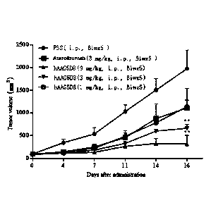
hAAG5D8 (5 mg/kg) 35.9 63.3
[0077] Example 6 Study on the therapeutic efficacy of hAAG5D8 on
subcutaneously
transplanted tumor in mouse colorectal cancer MC38 transgenic mice
[0078] Each of 30 human PD-1 transgenic female C57 mice (Animal Experiment
Center of
.. Tongji University) was implanted subcutaneously 3 x10⁶ mouse colorectal
cancer cells expressing
human PD-Li (Tongji University, code: MC38-hPD-L1) on the left flank. When the
tumor volume
reached about 100 mm3, the mice were randomly divided into 5 groups: C-1 group
(negative control
group, PBS, 6 mice), C-2 group (positive control group, Atezolizumab, 3 mg/kg,
6 mice), C-3 group
(administration group, hAAG5D8, 1 mg/kg, 6 mice), C-4 group (administration
group, hAAG5D8,
3 mg/kg, 6 mice) and C-5 group (administration group, hAAG5D8, 9 mg/kg, 6
mice). The mice
were administered intraperitoneally 2 times a week, 5 times in total. The
tumor volume was
measured twice a week until the 16th day after the beginning of
administration.
16655911.1
34273/87
- 22 -
Date Recue/Date Received 2021-03-26
CA 03114467 2021-03-26
100791 Calculation equations:
[0080] Tumor volume: TV = D1 xD22/2, wherein D1 and D2 represent tumor long
diameter and
tumor short diameter, respectively.
100811 Relative tumor volume: RTV=TvT/Tvi, where Tvi is the tumor volume
before
administration, and TvT is the tumor volume on each measurement.
[0082] Relative tumor proliferation: T/C(%) = TRTv/CRTv x 100%, wherein TRTV
represents the
RTV of the treatment group or positive control group, and CRTV represents the
RTV of the negative
control group.
100831 Relative tumor inhibition: TGIRTv(%) = (1 - TRTv/CRTv) x 100%.
[0084] Results and conclusions: The tumor inhibitory effects of hAAG5D8 on
mouse colorectal
cancer are shown in Table 11 and FIG. 5. The results show that hAAG5D8 at a
concentration of 3
mg/kg or above has a significant tumor inhibitory effect on subcutaneously
transplanted tumors of
mouse colorectal cancer. At the dose of 3 mg/kg, the tumor inhibitory effect
of the anti-PD-Li
antibody has been significantly superior to that of Atezolizumab.
Table 11 ¨ Tumor volume changes in tumor-bearing mice after administration
Dose Tumor volume TV (mm3)
Group Tiny/Clay(%)
TGIuTv(%)
(mg/kg) __________________________________________
DO D16
PBS 99.9 13.2 1974.2 413.2
Atezolizumab 3 95.9 16.8 1111.6 435.2* 53
47
hAAG5D8 1 95.5 9.8 1138.8 144.9* 64
36
hAAG5D8 3 90.7 12.4 664.8 92.0** 39
61
hAAG5D8 9 91.2 15.9 319.1 190.4** 14
86
Note: The day of administration was defined as DO; *P<0.05, **P<0.01 vs PBS
16655911.1
34273/87
- 23 -
Date Recue/Date Received 2021-03-26
CA 03114467 2021-03-26
REFERENCES
1. CHEN L, FLIES D B. Molecular mechanisms of T cell co-stimulation and co-
inhibition [J].
Nature reviews Immunology, 2013, 13(4): 227-42.
2. OHAEGBULAM K C, AS SAL A, LAZAR-MOLNAR E, et al. Human cancer immunotherapy
with antibodies to the PD-1 and PD-Li pathway [J]. Trends in molecular
medicine, 2015, 21(1):
24-33.
3. ISHIDA M, IWAI Y, TANAKA Y, et al. Differential expression of PD-Li and PD-
L2, ligands
for an inhibitory receptor PD-1, in the cells of lymphohematopoietic tissues
[J]. Immunology
letters, 2002, 84(1): 57-62.
4. TAUBE J M, ANDERS RA, YOUNG G D, et al. Colocalization of inflammatory
response with
B7-hl expression in human melanocytic lesions supports an adaptive resistance
mechanism of
immune escape [J]. Science translational medicine, 2012, 4(127): 127ra37.
5. TSENG S Y, TSUJI M, GORSKI K, et al. B7-DC, a new dendritic cell molecule
with potent
costimulatory properties for T cells [J]. The Journal of experimental
medicine, 2001, 193(7):
839-46.
6. BUTTE M J, KEIR M E, PHAMDUY T B, et al. Programmed death-1 ligand 1
interacts
specifically with the B7-1 costimulatory molecule to inhibit T cell responses
[J]. Immunity,
2007, 27(1): 111-22.
7. CHENG X, VEVERKA V, RADHAKRISHNAN A, et al. Structure and interactions of
the
human programmed cell death 1 receptor [J]. The Journal of biological
chemistry, 2013,
288(17): 11771-85.
8. An Liu, Discovery on small molecular inhibitors of PD-1/PD-L1 pathway [D],
Jilin: Jilin
University School of Pharmaceutical Sciences, 2016, 15.
16655911.1
34273/87
- 24 -
Date Recue/Date Received 2021-03-26