Note: Descriptions are shown in the official language in which they were submitted.
CA 03114647 2021-03-26
WO 2020/069509
PCT/US2019/053826
METHODS, APPARATUSES, AND SYSTEMS FOR 3-D PHENOTYPING AND
PHYSIOLOGICAL CHARACTERIZATION OF BRAIN LESIONS AND
SURROUNDING TISSUE
CROSS-REFERENCE TO RELATED APPLICATIONS
[0001] The present application claims the benefit of priority of U.S.
Provisional
Application No. 62/738,270 filed September 28, 2018, which is hereby
incorporated by
reference in its entirety.
TECHNICAL FIELD
[0002] The present application relates generally to methods,
apparatuses, and systems for
characterizing a brain lesion, and more particularly, but not by way of
limitation, to
three-dimensionally (3D) phenotyping and physiologically characterizing brain
lesions and the
surrounding tissue.
BACKGROUND
[0003] Multiple sclerosis (MS) is an autoimmune inflammatory disorder of
the central
nervous system that results in injury to myelin, nerve fibers, and glial
cells, affecting nearly 1
million individuals in the United States. Symptoms frequently include focal
neurologic deficits,
including visual, motor and sensory disturbances. Autonomic dysregulation and
cognitive
impairment might also occur. Clinicians depend on magnetic resonance imaging
(MRI) for MS
diagnosis, disease surveillance in the presence or absence of treatment, and
prediction of future
clinical outcomes. Disease progression, punctuated by the development of
contrast-enhancing
or new T2-lesions commonly results in a change in disease-modifying treatment.
Currently,
conventional MRI techniques employed in MS are limited by false positives due
to high
sensitivity to white-matter hyper-intensities and reduced specificity
regarding disease origin.
Such techniques are also limited by the forced perspectives in two-dimensional
(2D) planes
(axial, sagittal and coronal). In addition, the magnitude of axonal and glial
injury resulting from
in-situ demyelination in existing or newly developed T2- hyper-intensities is
unclear.
[0004] Recently, three-dimensional (3D) phenotyping of MS lesions has
provided a more
comprehensive view of their shape and surface features than has previously
been possible. This
additional perspective has led to the observation that some MS lesions assume
an amorphous
structure with complex surface features. Such observations suggest that
important information
regarding MS diagnosis and disease progression might be possible based on
lesion features
- 1 -
CA 03114647 2021-03-26
WO 2020/069509
PCT/US2019/053826
including their shape, symmetry, and surface texture. Understanding the degree
of
microstructural injury, physiologic dysfunction, and myelin repair is
essential for ascertaining
the clinical significance of these features.
[0005] A variety of techniques have indicated changes to the structural
and functional
characteristics of tissue both inside and outside of lesions. For instance,
there is evidence that
myelin injury extends beyond the lesion boundaries at varying distances from
MS lesion
centers. Magnetization transfer imaging measures a ratio of the macromolecule-
bound protons
(i.e., myelin) to free water protons in tissue. Thus, a higher magnetization
transfer ratio (MTR)
suggests more myelin. Using this technique, Bagley and colleagues showed
increasing MTR
at increasing distances from MS lesion centers. This result suggests that
myelin injury extends
beyond the lesion boundary and this injury gradually reduces moving outwards
from the lesion
center. In addition, studies using diffusion tensor imaging showed altered
white-matter
integrity in tissue outside MS lesions. Such results suggest that MS-related
myelin and axonal
injury exist beyond lesion boundaries. Histopathological studies have also
identified different
immunological injury patterns within lesions. Thus, there is substantial
evidence that
heterogeneity in myelination and axonal injury exists inside and outside MS
lesions. Currently,
no imaging markers are capable of characterizing this heterogeneity.
[0006] Calibrated dual-echo functional MRI (cfMRI) provides a means by
which to
characterize lesion heterogeneity because it allows near-simultaneous measures
of blood-
oxygen-level-dependent signal (BOLD) and cerebral blood flow (CBF), permitting
calculation
of the cerebral metabolic rate of oxygen (CMR02) using the deoxyhemoglobin
dilution model.
Because cfMRI provides metabolic measures and has high spatial resolution, it
allows
metabolic characterization of lesions. In cfMRI, the T2*-weighted BOLD signal
results from
local magnetic field susceptibility effects of paramagnetic deoxyhemoglobin
and diamagnetic
oxyhemoglobin in the veins, physiologically providing a measure of venous
blood oxygen
content voxelwise. The acquired BOLD signal depends on upstream factors
including 1)
arterial CBF, 2) cellular oxygen extraction from the capillaries and, 3)
CMR02, thus making
BOLD signal a biomarker of physiologic integrity.
SUMMARY
[0007] This disclosure includes implementations of methods and
configurations of
apparatuses and systems for three-dimensionally phenotyping and
physiologically
characterizing brain lesions and tissue encompassing surrounding boundaries.
Non-limiting
- 2 -
CA 03114647 2021-03-26
WO 2020/069509
PCT/US2019/053826
examples of conditions that benefit from this disclosure include, but are not
limited to, multiple
sclerosis, aging, small vessel disease, migraine headaches, and other non-
specific white matter
lesion etiologies.
[0008] The clinical management of multiple sclerosis (MS) currently
involves disease
characterization based on two-dimensional forced-perspectives of magnetic
resonance imaging
(MRI) data. Such views fail to provide an understanding of the complexity of
lesion shape and
surface texture, the magnitude of injury within and around lesions, the extent
of alterations in
the underlying metabolism, and the potential for self-remyelination and
recovery. In the
present disclosure, a novel three-dimensional (3D) lesion phenotyping approach
was utilized
and coupled with physiologic measures to study the metabolic and physiologic
profiles from
tissue within and around lesions and their impacts on lesion shape and surface
texture. A non-
invasive biomarker called blood-oxygen-level-dependent (BOLD) slope was
identified to
metabolically characterize brain lesions. BOLD slope is defined as the rate of
change in venous
blood oxygen content from the lesion tissue to its surrounding brain tissue.
Metabolically
active lesions demonstrating positive BOLD slopes had higher cerebral
metabolic rate of
oxygen and higher cerebral blood flow compared to inactive lesions
demonstrating negative
slopes. Results indicated that metabolically active lesions with more intact
tissue and myelin
architecture have more symmetrical shapes and more complex surface textures
compared to
metabolically inactive lesions with less intact tissue and myelin
architecture. The association
of lesions' shapes and surface features with their metabolic signatures
suggest the prospect for
immediate translation of MRI data to clinical management by providing
information related to
metabolic activity, lesion age, and risk for disease reactivation and self-
repair. The present
disclosure further provides a platform for disease surveillance and outcome
quantification
involving therapeutics aimed at myelin repair. The metabolic information
acquired from the
periphery of MRI lesions may inform on disease advancement or stability,
prompting a switch
from one disease modifying therapy to another agent. This may involve the use
of treatments
that are more highly effective, including chemotherapeutic medications or
potent
immunomodulatory regimens aimed at suppressing disease activity or treatments
associated
with better safety profiles. The method may also allow for the determination
of treatment
effects from prescribed therapies or investigational medications aimed at
myelin, axonal, or
tissue repair. An alternate approach to the use of these data may involve the
cessation of
treatment in certain age groups if the acquired findings suggest disease
stability. Additionally,
the metabolic profiles from these lesions and their surrounding tissue may
inform on the risk
- 3 -
CA 03114647 2021-03-26
WO 2020/069509
PCT/US2019/053826
for more advanced brain aging, specifically regional brain volume reductions
involving
surrounding tissue or total brain volumes.
[0009] Some embodiments include a system for determining characteristics
of a brain
lesion and tissue encompassing boundaries surrounding the brain lesion in
three dimensions,
the system having a computer system comprising at least one processor
configured to receive
data from a magnetic resonance imaging (MRI) machine configured to generate
one or more
series of images corresponding to a structural and a functional characteristic
of a brain lesion
and tissue encompassing one or more enlarged boundaries surrounding the brain
lesion, the
brain lesion having an outer boundary and at least part of each of the one or
more boundaries
.. surrounding the brain lesion being offset by a given distance from the
outer boundary of the
brain lesion; segment the received data to isolate the portion of the received
data corresponding
to the brain lesion and the tissue surrounding the brain lesion within the one
or more enlarged
boundaries; create, based on the segmented data, one or more three-dimensional
(3D) models
of the brain lesion and the tissue surrounding the brain lesion within the one
or more enlarged
boundaries; analyze, based on the one or more 3D models, one or more 3D
phenotypic
characteristics of the brain lesion and a slope of a blood oxygen level
dependent (BOLD) signal
from within the brain lesion through the one or more enlarged boundaries; and
determine, based
on the one or more 3D phenotypic characteristics and the slope, indicators of
one or more
characteristics selected from the group of characteristics consisting of:
lesion age, extent of
.. injury, remyelination capacity, tissue integrity within the brain lesion,
tissue integrity within
tissue surrounding the brain lesion, and metabolic activity of the brain
lesion within tissue
surrounding the brain lesion.
[0010] In some configurations, a majority of each of the one or more
boundaries
surrounding the brain lesion can be offset by a given distance from the outer
boundary of the
brain lesion. In some configurations, all of each of the one or more
boundaries surrounding
the brain lesion can be offset by a given distance from the outer boundary of
the brain lesion.
[0011] In some configurations, the one or more series of images are
generated from one or
more structural imaging sequences and one or more functional imaging
sequences. The one or
more structural imaging sequences may include fluid attenuated inversion
recovery (FLAIR),
magnetization-prepared rapid acquisition gradient-echo (MPRAGE), and diffusion
kurtosis
imaging sequences. Other structural and/or functional imaging sequences may be
included to
further enhance structural and/or functional details of the one or more series
of images. In
- 4 -
CA 03114647 2021-03-26
WO 2020/069509
PCT/US2019/053826
some configurations, the one or more functional imaging sequences include
pseudo-continuous
arterial spin labeling (pCASL) or continuous arterial spin labeling (CASL) to
generate images
corresponding to cerebral blood flow (CBF) and functional imaging sequences to
generate
blood oxygen level dependent (BOLD) data.
[0012] In some configurations, segmentation is performed on three-
dimensional (3D) fluid
attenuated inversion recovery (FLAIR) images via implementing geodesic active
contour
methodology.
[0013] In some configurations, the received data includes a series of
two-dimensional
(2D) images, and the one or more three-dimensional (3D) models is derived from
the series of
2D images. In some configurations, each of the series of two-dimensional (2D)
images is given
a thickness and assembled to define the one or more 3D models capable of being
exported into
stereolithographic format.
[0014] In some configurations, the one or more 3D models include
segmented data from
3D Ti-weighted, T2-weighted, and fluid attenuated inversion recovery (FLAIR)
images. In
some configurations, the one or more 3D models can further include segmented
data from
3D T2-weighted fluid attenuated inversion recovery (3D T2 FLAIR), Ti-weighted
magnetization-prepared rapid acquisition gradient-echo (MPRAGE), and diffusion
kurtosis
(DK) images.
[0015] In some configurations, one or more processors can be configured
to isolate the
brain lesion to create the one or more three-dimensional (3D) models of the
brain lesion and
the tissue encompassing the one or more enlarged boundaries surrounding the
brain lesion
based on the segmented data generated from 3D T2-weighted fluid attenuated
inversion
recovery (3D T2 FLAIR).
[0016] In some configurations, one or more processors can be configured
to create the one
or more three-dimensional (3D) models of the brain lesion and the tissue
encompassing the one
or more enlarged boundaries surrounding the brain lesion based on the
segmented data
generated from Ti-weighted magnetization-prepared rapid acquisition gradient-
echo
(MPRAGE) imaging.
- 5 -
CA 03114647 2021-03-26
WO 2020/069509
PCT/US2019/053826
[0017] In some configurations, one or more processors can be configured
to determine an
indicator of tissue integrity within the brain lesion by measuring white
matter microstructure
integrity via diffusion kurtosis imaging (DKI).
[0018] In some configurations, the slope of the blood oxygen level
dependent (BOLD)
signal is calculated using the formula
Er-region(B LDi ¨ BOLD)(Ti ¨ rT)
BOLD slope =
Er
=region(BOLDi ¨ BOLD)
where regions are the brain lesions and their associated perimeters, n is the
number of regions,
BOLD, is the average BOLD signal in the region and BOLD is the average BOLD
signal across
all regions, T, is the thickness of the concentric voxel layer.
[0019] In some configurations, a cerebral metabolic rate of oxygen (CMR02)
is calculated
using the formula
ABOLD ACMR021(3 [ ACBF[
BOLD al
________________________________ = M (1 ________
CMR0210] [ CBFoi
where a = 0.38 is an empirically-derived constant linking CBF and cerebral
blood volume; 0 =
1.3 is an empirically-derived constant related to vascular exchange and
susceptibility of
deoxyhemoglobin at 3T; and M is a subject-specific scaling factor dependent
upon the washout
of resting deoxyhemoglobin determined by a hypercapnia calibration experiment.
The
hypercapnia induced changes in the blood oxygen level dependent (BOLD) signal
and the
cerebral blood flow (CBF) can be used to calculate a subject-specific scaling
factor M using
the formula
ABOLD
M = BOLD
CBF )"-(3
CBF0)
- 6 -
CA 03114647 2021-03-26
WO 2020/069509
PCT/US2019/053826
where the subject-specific scaling factor M and the average blood oxygen level
dependent
(BOLD) and the cerebral blood flow (CBF) data can be used to calculate CMR02
within and
around the brain lesion using the formula
1
7 ABOLD \ F a
--
CMR02 ______________________________________________________ = 1 BOLDgn, ( CBF
)1 R
CMR02ign, M i CBFgn.,)
\ /
[0020] In some configurations, the one or more 3D phenotypic
characteristics include
lesion volume, lesion surface texture, and/or lesion shape. Manifold harmonics
transform
(MHT) descriptors can be used to quantify lesion shape from a 3D lesion
geometry via
eigenfunctions of Laplace-Beltrami operators. In some configurations, one or
more processors
can be configured to sort eigenvalues in ascending order and select one or
more eigenvectors
corresponding to the smallest eigenvalues to reconstruct an original shape of
the brain lesion.
[0021] Some implementations of the present methods include a method of
determining
characteristics of brain lesions and tissue encompassing boundaries
surrounding the brain
lesion in a patient, the method including scanning a portion of the patient
with a magnetic
resonance imaging (MRI) machine configured to generate data corresponding to a
structural
and a functional characteristic of a brain lesion of the patient and tissue
encompassing one or
more enlarged boundaries surrounding the brain lesion, the brain lesion having
an outer
boundary and at least part of each of the one or more boundaries surrounding
the brain lesion
being offset by a given distance from the outer boundary of the brain lesion;
segmenting the
generated data to isolate the portion of the generated data corresponding to
the brain lesion and
the tissue surrounding the brain lesion within the one or more enlarged
boundaries; creating,
based on the segmented data, one or more three-dimensional (3D) models of the
brain lesion
and the tissue surrounding the brain lesion within the one or more enlarged
boundaries;
analyzing, based on the one or more 3D models, one or more 3D phenotypic
characteristics of
the brain lesion and a slope of a blood oxygen level dependent (BOLD) signal
from within the
.. brain lesion through the one or more enlarged boundaries; and determining,
based on the one
or more 3D phenotypic characteristics and the slope, indicators of one or more
characteristics
selected from the group of characteristics consisting of: lesion age, extent
of injury,
remyelination capacity, tissue integrity within the brain lesion, tissue
integrity within tissue
- 7 -
CA 03114647 2021-03-26
WO 2020/069509
PCT/US2019/053826
surrounding the brain lesion, and metabolic activity of the brain lesion
within tissue
surrounding the brain lesion.
[0022] In some implementations, a majority of each of the one or more
boundaries
surrounding the brain lesion can be offset by a given distance from the outer
boundary of the
brain lesion. In some implementations, all of each of the one or more
boundaries surrounding
the brain lesion can be offset by a given distance from the outer boundary of
the brain lesion.
In some implementations, the one or more enlarged boundaries surrounding the
brain lesion
each include a region defined as a 3mm concentric voxel layer. In some
implementations, the
one or more enlarged boundaries surrounding the brain lesion include a first
boundary, a second
boundary, a third boundary, and a fourth boundary.
[0023] In some implementations, scanning includes one or more structural
imaging
sequences and one or more functional imaging sequences. In some
implementations, the one
or more structural imaging sequences include fluid attenuated inversion
recovery (FLAIR),
magnetization-prepared rapid acquisition gradient-echo (MPRAGE), and diffusion
kurtosis
imaging sequences. In some implementations, the one or more functional imaging
sequences
comprise pseudo-continuous arterial spin labeling (pCASL) or continuous
arterial spin labeling
(CASL) to generate images corresponding to cerebral blood flow (CBF) and
functional
imaging sequences to generate blood oxygen level dependent (BOLD) data.
[0024] In some implementations, segmentation can be performed on three-
dimensional
(3D) fluid attenuated inversion recovery (FLAIR) images via implementing
geodesic active
contour methodology. In some implementations, the generated data includes a
series of two-
dimensional (2D) images, and the one or more three-dimensional (3D) models is
derived from
the series of 2D images. In some implementations, each of the series of two-
dimensional (2D)
image is given a thickness and assembled to define the one or more 3D models
capable of being
exported into stereolithographic format. In some implementations, the one or
more 3D models
include segmented data from 3D Ti-weighted, T2-weighted, and fluid attenuated
inversion
recovery (FLAIR) images. In some implementations, the one or more 3D models
further
include segmented data from 3D T2- weighted fluid attenuated inversion
recovery (3D T2
FLAIR), Ti-weighted magnetization-prepared rapid acquisition gradient-echo
(MPRAGE),
-- and diffusion kurtosis (DK) images.
- 8 -
CA 03114647 2021-03-26
WO 2020/069509
PCT/US2019/053826
[0025] In some implementations, scanning includes 3D T2-weighted fluid
attenuated
inversion recovery (3D T2 FLAIR) to isolate the brain lesion to create the one
or more three-
dimensional (3D) models of the brain lesion and the tissue encompassing the
one or more
enlarged boundaries surrounding the brain lesion. In some implementations,
scanning further
includes Ti-weighted magnetization-prepared rapid acquisition gradient-echo
(MPRAGE)
imaging to produce anatomical images of the brain lesion and the tissue
encompassing the one
or more enlarged boundaries surrounding the brain lesion. In some
implementations, scanning
further includes diffusion kurtosis imaging (DKI) to measure white matter
microstructure
integrity within the brain lesion.
[0026] In some implementations, analyzing includes calculating a cerebral
blood flow
(CBF) value and a cerebral metabolic rate of oxygen (CMR02) value.
[0027] In some implementations, the slope of the blood oxygen level
dependent (BOLD)
signal is calculated using the formula
EP =region
(1301i ¨ BOLD)(Ti ¨ rT)
i
BOLD slope =
E P (BOLDi ¨ BOLD)
i=region
where regions are the brain lesions and their associated perimeters, n is the
number of regions,
BOLD, is the average BOLD signal in the region and BOLD is the average BOLD
signal across
all regions, T, is the thickness of the concentric voxel layer.
[0028] In some implementations, a cerebral metabolic rate of oxygen
(CMR02) is
calculated using the formula
ABOLD BOLD ACMR02r [ ACBF[al
_____________________________ = M (1 ________
CMR0210] [ CBFoi
where a = 0.38 is an empirically-derived constant linking CBF and cerebral
blood volume; 0 =
1.3 is an empirically-derived constant related to vascular exchange and
susceptibility of
deoxyhemoglobin at 3T; and M is a subject-specific scaling factor dependent
upon the washout
of resting deoxyhemoglobin determined by a hypercapnia calibration experiment.
The
hypercapnia induced changes in the blood oxygen level dependent (BOLD) signal
and the
cerebral blood flow (CBF) can be used to calculate a subject-specific scaling
factor M using
the formula
- 9 -
CA 03114647 2021-03-26
WO 2020/069509
PCT/US2019/053826
ABOLD
M = BOLD
( CBF )"-(3
CBF0)
where the subject-specific scaling factor M and the average blood oxygen level
dependent
(BOLD) and the cerebral blood flow (CBF) data can be used to calculate CMR02
within and
around the brain lesion using the formula
1
/ ABOLD \ 13 cc
--
CMR02 ______________________________________________________ = 1 BOLDgn, ( CBF
)1 R
CMR02ign, M / CBFgn.,)
\ /
[0029]
In some implementations, the one or more 3D phenotypic characteristics
include
lesion volume, lesion surface texture, and/or lesion shape. Manifold harmonics
transform
(MHT) descriptors can be used to quantify lesion shape from a 3D lesion
geometry via
eigenfunctions of Laplace-Beltrami operators. In some implementations,
eigenvalues are
sorted in ascending order and one or more eigenvectors corresponding to the
smallest
eigenvalues are selected to reconstruct an original shape of the brain lesion.
[0030]
Some implementations of the present methods include a method of treating
brain
lesions in a patient, the method including administering a treatment to the
patient in response
to a determination of one or more physiological characteristics of the brain
lesions by any one
of the disclosed methods. In some implementations, the treatment is switched,
based on the
determination step, from a disease modifying therapeutic agent to a different
disease modifying
therapeutic agent.
In some implementations, the treatment includes one or more
chemotherapeutic drugs and/or immunomodulatory agents.
[0031]
In some implementations, the method further includes determining, based on
the
one or more 3D phenotypic characteristics and the slope, treatment effects
from one or more
prescribed therapies and/or one or more investigational medications aimed at
myelin, axonal,
and/or tissue repair.
[0032]
In some implementations, the method further includes cessation of the
treatment in
certain age groups if an association of the one or more 3D phenotypic
characteristics and the
slope suggest disease stability.
- 10 -
CA 03114647 2021-03-26
WO 2020/069509
PCT/US2019/053826
[0033] In some implementations, lesion age, extent of injury,
remyelination capacity, tissue
integrity within the brain lesion, tissue integrity within tissue encompassing
one or more
enlarged boundaries surrounding the brain lesion, and metabolic activity of
the brain lesion and
tissue encompassing one or more enlarged boundaries surrounding the brain
lesion are
determined using artificial intelligence, machine learning, and/or deep
learning techniques.
[0034] The term "coupled" is defined as connected, although not
necessarily directly, and
not necessarily mechanically; two items that are "coupled" may be unitary with
each other.
The terms "a" and "an" are defined as one or more unless this disclosure
explicitly requires
otherwise. The term "substantially" is defined as largely but not necessarily
wholly what is
specified (and includes what is specified; e.g., substantially 90 degrees
includes 90 degrees and
substantially parallel includes parallel), as understood by a person of
ordinary skill in the art.
In any configuration or implementation of the present devices, apparatuses,
kits, and methods,
the term "substantially" may be substituted with "within [a percentage] of'
what is specified,
where the percentage includes 0.1, 1, 5, and/or 10 percent.
[0035] The terms "comprise" (and any form of comprise, such as "comprises"
and
"comprising"), "have" (and any form of have, such as "has" and "having"),
"include" (and any
form of include, such as "includes" and "including") and "contain" (and any
form of contain,
such as "contains" and "containing") are open-ended linking verbs. As a
result, an apparatus,
device, or kit that "comprises," "has," "includes" or "contains" one or more
elements possesses
those one or more elements, but is not limited to possessing only those
elements. Likewise, a
method that "comprises," "has," "includes" or "contains" one or more steps
possesses those
one or more steps, but is not limited to possessing only those one or more
steps.
[0036] Further, an apparatus, device, or structure that is configured in
a certain way is
configured in at least that way, but it can also be configured in other ways
than those
specifically described.
[0037] Any configuration or implementation of any of the present
devices, apparatuses,
kits, and methods can consist of or consist essentially of ¨ rather than
comprise/include/contain/have ¨ any of the described steps, elements, and/or
features. Thus,
in any of the claims, the term "consisting of' or "consisting essentially of'
can be substituted
for any of the open-ended linking verbs recited above, in order to change the
scope of a given
claim from what it would otherwise be using the open-ended linking verb.
- 11-
CA 03114647 2021-03-26
WO 2020/069509
PCT/US2019/053826
[0038] Details associated with the configurations described above and
others are presented
below.
BRIEF DESCRIPTION OF THE DRAWINGS
[0039] The following drawings illustrate by way of example and not
limitation. For the
sake of brevity and clarity, every feature of a given structure is not always
labeled in every
figure in which that structure appears. Identical reference numbers do not
necessarily indicate
an identical structure. Rather, the same reference number may be used to
indicate a similar
feature or a feature with similar functionality, as may non-identical
reference numbers. The
figures are drawn to scale (unless otherwise noted), meaning the sizes of the
depicted elements
are accurate relative to each other for at least the configurations depicted
in the figures.
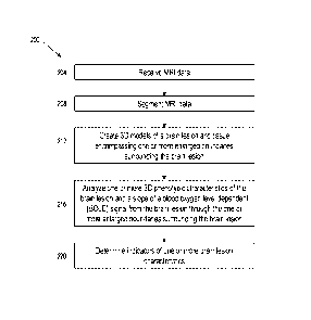
[0040] FIG. 1 depicts an exemplary system for determining
characteristics of brain lesions
according to an embodiment of the disclosure.
[0041] FIG. 2 depicts an exemplary method for determining
characteristics of brain lesions
according to an embodiment of the disclosure.
[0042] FIG. 3A shows a three-dimensional representation of a multiple
sclerosis (MS)
brain lesion isolated in 3D using geodesic active contours.
[0043] FIG. 3B shows a representation of the MS brain lesion of FIG. 3A
and its
surrounding boundaries (Perimeters 1-2) as 3mm concentric layers mirroring the
three-
dimensional (3D) shape around the MS brain lesion.
[0044] FIG. 4A shows the mean normalized blood oxygen level dependent
(BOLD) signal
in MS brain lesions and their associated Perimeters 1-4 (Pen) in focal MS
(denoted as solid
line with squares in the graph) and simulated MS brain lesions (denoted as
solid line with
triangles in the graph).
[0045] FIG. 4B shows the mean normalized cerebral blood flow (CBF) in
lesions and their
associated Perimeters 1-4 in focal MS and simulated MS brain lesions.
[0046] FIG. 5A shows the mean normalized BOLD signal in the MS brain
lesions and their
Perimeters (Pen) 1-4 for metabolically active (solid line with squares) and
inactive lesions
(solid line with triangles).
- 12-
CA 03114647 2021-03-26
WO 2020/069509
PCT/US2019/053826
[0047] FIG. 5B shows the significant differences in BOLD signal between
positive and
negative BOLD slope MS brain lesion types.
[0048] FIG. 5C shows the relationship of cerebral metabolic rate of
oxygen (CMR02) from
the MS brain lesion to its Perimeters (Pen) 1-4 in metabolically active brain
lesions and
metabolically inactive brain lesions.
[0049] FIG. 5D shows the significant differences in CMR02 between
metabolically active
and metabolically inactive MS brain lesion types.
[0050] FIG. 5E shows the significantly higher CBF in metabolically
active lesions than
inactive lesions.
[0051] FIG. 5F depicts the significant presence of more intact white matter
microstructure
(e.g., myelin) in metabolically active lesions compared to inactive lesions as
measured by
presence of kurtosis tensors.
[0052] FIG. 6A shows examples of metabolically active and inactive MS
brain lesions in
2D and 3D views demonstrating the marked underrepresentation of the MS brain
lesion shape
and texture in 2D forced perspectives of MRI.
[0053] FIG. 6B shows the probability density functions of the two lesion
types,
metabolically active (solid black line) and inactive (solid grey line),
obtained from
bootstrapping the cube root of randomly sampled lx106 tetrahedron areas on the
lesion surface.
[0054] FIG. 6C depicts a bar graph showing mean lesion volumes of the
two lesion types.
[0055] FIG. 6D depicts a bar graph showing mean surface area-to-volume
ratio for the two
lesion types.
[0056] FIG. 6E shows Log transformed manifold harmonics transform (MHT)
descriptors
plotted as a function of their eigenvalues for each lesion.
[0057] FIG. 6F depicts bar graphs showing mean MHT descriptors of low (0-
100),
mid (101-200) and high (201-300) eigenvalues for the two lesion types.
[0058] FIG. 7 shows an example of a reconstructed lesion model by using
different
numbers of eigenvectors: 6, 10, 50, 100, 300, and the original shape,
respectively.
- 13 -
CA 03114647 2021-03-26
WO 2020/069509
PCT/US2019/053826
[0059] FIG. 8 shows the definition of the angles and area covered by
each vertex in order
to compute Laplacian eigenvalues for each manifold mesh of a lesion.
[0060] FIG. 9A depicts a graph of mean BOLD signals in MS lesions and
their perimeters
and in non-specific white matter (NSWM) lesions and their perimeters.
[0061] FIG. 9B depicts a graph of mean cerebral blood flow (CBF) in MS
lesions and their
perimeters and NSWM lesions and their perimeters.
[0062] FIG. 9C depicts a bar graph of mean BOLD slope for MS lesions and
NSWM
lesions.
[0063] FIG. 9D depicts a receiver operator characteristic (ROC) curve
for a model.
[0064] FIG. 10A shows a three-dimensional illustration of an MS lesion and
its perimeters.
[0065] FIG. 10B shows a three-dimensional illustration of an NSWM lesion
and its
perimeters.
DESCRIPTION OF ILLUSTRATIVE EMBODIMENTS
[0066] Currently, the clinical management of MS patients is limited by
2D forced
perspectives of MRI views that markedly underrepresent the complexity of
lesion shape and
texture. Observations made from the 2D perspective fail to appreciate the
magnitude of injury
within and around MS lesions, the extent of alterations in the underlying
metabolism, the
potential for self-remyelination and recovery, and the long-term outcomes
related to the impact
of lesions on their surrounding brain tissue.
[0067] The present disclosure describes a practical and innovative approach
to assessing
physiologic data from the lesion tissue and one or more enlarged boundaries
surrounding the
brain lesion. In some implementations, the boundaries can be defined as
surrounding
concentric perimeters extending from the surface of a 3D MS lesion. The
association of lesion
shape and surface features with its metabolic signatures may aid in the
immediate translation
of MRI data to clinical management by providing information related to
metabolic activity.
The inclusion of an unconventional lesion-isolation technique enabled the
direct extraction of
lesions in 3D without reconstruction through 2D slices and allowed for lesion
traits to be
phenotyped.
- 14 -
CA 03114647 2021-03-26
WO 2020/069509
PCT/US2019/053826
[0068] In some implementations of the present configurations, the novel
3D approach to
the characterization of MS lesion phenotypes offers a more accurate reflection
of the
underlying microstructural and physiologic injury on an individualized level
well beyond the
capabilities of routine MRI studies. For example, lesions with a more
spherical shape and
complex surface features demonstrating a positive BOLD slope are metabolically
active,
suggesting a greater potential for in-situ remyelination. Such findings could
not have been
achieved with a 2D approach. In addition, the short acquisition time for BOLD
slope and
minimal degree of post processing required to calculate these outcomes further
increases the
potential for the disclosed methods, apparatuses, and systems to be clinically
adopted in the
management of MS patients. Further, the present disclosure provides a platform
for disease
surveillance as well as quantifying outcomes involving therapeutics aimed at
myelin repair.
[0069] Referring now to the drawings, FIG. 1 depicts an exemplary 3D
imaging and brain
lesion representation system 100 according to an embodiment of the disclosure.
In the
embodiment shown, an MRI device 102 may be provided. The MRI device 102 may be
a 2D
MRI device, a 3D MRI device, or one or more MRI devices providing both 2D and
3D imaging
capabilities. A processing device 104 may be capable of receiving 2D and/or 3D
images taken
by the MRI device. Processing device 104 may be a part of a computer system
that may include
standard components such as a hard drive, monitor, printer, keyboard, and
mouse, among
others, that may enable a user to interact with the processing device 104. In
the embodiment
shown, processing device 104 may include one or more of a segmentation
application 106, a
3D imaging application 108, and one or more databases 110. In some
embodiments,
segmentation application 106 may be configured to receive one or more MRI
images from MRI
device 102, segment the one or more MRI images into one or more regions, and
enable a
selection of one or more regions. These selected regions may be referred to as
regions of
interest (ROI). In some embodiments, the selection of ROI may be done
automatically by
processing device 104. In some embodiments, the selection of ROI may be done
by a user.
[0070] In some embodiments, the selected ROI may be exported by
segmentation
application 106 and imported into 3D image application 108. In some
embodiments, 3D image
application 108 may generate one or more 3D maximum intensity projections
(MIP) images of
the selected ROI. In some embodiments, the selected ROI may correspond to one
or more
focal brain lesions. In some embodiments, the selected ROI may be converted to
stereolithography (.stl) format and/or displayed as 3D orthographic images to
enable
- 15 -
CA 03114647 2021-03-26
WO 2020/069509
PCT/US2019/053826
orthographic views. The one or more 3D images may be displayed to a user and
3D image
application 108 may enable a user to view and manipulate the one or more 3D
images. In some
embodiments, image manipulation capabilities may include capabilities to
rotate, zoom, mark,
color, and select the one or more images. In some embodiments, one or more
databases 110
may contain information corresponding to various brain lesion characteristics.
Examples of
these brain lesion characteristics may include shape or geometric
characteristics, size
characteristics, topographical characteristics, volume characteristics,
surface area
characteristics and the like. In some embodiments, the brain lesion
characteristics may be
associated with one or more etiologies. Examples of these etiologies may
include MS, aging,
small vessel disease, migraine headaches, and other non-specific white matter
lesion etiologies.
In the embodiment shown, processing device 104 may be configured to send data
corresponding to the one or more 3D images to a 3D printing device 112. 3D
printing device
112 may create a 3D physical representation of the received one or more 3D
images.
[0071] FIG. 2 depicts an exemplary method 200 for creating 3D
representations of brain
lesions according to an embodiment of the disclosure. In one embodiment of the
disclosure,
method 200 may be implemented by system 100. In the embodiment shown in FIG.
2, method
200 may begin at step 204 by receiving one or more 2D and/or 3D MRI images. In
some
embodiments, 3D MRI images may be created from one or more received 2D MRI
images.
Method 200 may continue at step 208 by segmenting the received one or more 2D
and/or 3D
MRI images. In some embodiments, segmenting step 208 may include segmenting
the one or
more 2D and/or 3D MRI images into one or more regions of interest (ROI). The
one or more
ROI may correspond to one or more brain lesions. In some embodiments, brain
lesions may
be segmented in 3D format using a maximum intensity projection (MIP) 3D file.
In this way,
the computer system and/or a user may manipulate a 3D object in 2D space and
may select one
or more ROI. Isolating lesions from 3D MRI images may allow for a better
appreciation of
both the geometric and surface characteristics of brain lesions. In a 2D view,
a variety of
signals may influence pixel intensities that may result in pixel
misclassification. Isolating
lesions from 3D images may overcome some of these shortcomings of 2D lesion
isolation.
[0072] Method 200 may continue at step 212 by creating one or more 3D
models of brain
lesions. In some embodiments, the one or more 3D brain lesion models may be
orthographic
images or M1P images. Method 200 may continue at step 216 by analyzing of one
or more
3D phenotypic characteristics of the brain lesion and a slope of a blood
oxygen dependent
- 16-
CA 03114647 2021-03-26
WO 2020/069509
PCT/US2019/053826
(BOLD) signal from the brain lesion through one or more enlarged boundaries
surrounding the
brain lesion. For example, a computer system may analyze the one or more brain
lesion images
to determine one or more characteristics of the brain lesion. A user may also
analyze the one
or more brain lesion images by interacting with the computer system. In some
embodiments,
metadata may be used to denote a type or category of a brain lesion
characteristic. In some
embodiments, brain lesion characteristics may include geometric
characteristics. Geometric
characteristics may provide insights into a size and shape of a brain lesion.
Examples of
geometric characteristics may include lesion symmetry/asymmetry, surface
morphology (e.g.,
amorphous, ovoid), the existence of lobes and/or protrusions, and other shape
characteristics
(e.g., tapered/wedge, spherocylindrical). In some embodiments, brain lesion
characteristics
may include surface characteristics. Surface characteristics may provide
insights into lesion
surface traits and lesion properties not associated with geometry. Examples of
surface
characteristics may include the existence of surface microstructures, surface
topography (e.g.,
steepness/sheerness of surface peaks and valleys), surface irregularities, and
a non-uniform
distribution of mass of the lesion. In some embodiments, the computer system
may engage in
machine learning to generate descriptive surface, shape, and signal
characteristics from the
entire lesion or sections within lesions in order to more efficiently and
accurately classify lesion
types.
[0073] Method 200 may continue at step 220 by determining indicators of
one or more
brain lesion characteristics. In some embodiments, a computer system may
compare the one
or more brain lesion characteristics to one or more previously stored brain
lesion characteristics
to determine possible matches. In some embodiments, one or more previously
stored brain
lesion characteristics may correspond to one or more brain lesion etiologies.
In instances where
the analyzed one or more brain lesion characteristics match one or more
previously stored brain
lesion characteristics, the computer system may determine one or more possible
etiologies of
the one or more brain lesions. In some embodiments, a user may be able to
determine one or
more possible etiologies of the one or more brain lesions based on each of
their one or more
brain lesions characteristics.
A. Experimental Results
[0074] Multimodal neuroimaging methods coupled with novel lesion-
phenotyping
methods were used to study the relationship between lesion 3D shape and
texture and the
metabolic and physiologic profiles from within and around lesions in one or
more enlarged
- 17 -
CA 03114647 2021-03-26
WO 2020/069509
PCT/US2019/053826
boundaries (e.g., concentric perimeters) in multiple sclerosis (MS) patients.
Lesion
phenotyping and physiologic characterization allowed the study of the impact
of lesions on
surrounding tissue and identification of lesion characteristics within and
around lesion tissue,
resulting in identification of an association of lesions' shapes and surface
features with their
metabolic signatures. Such associations aid in the prospect for immediate
translation of 3D
MRI data to clinical management by providing information related to metabolic
activity, lesion
age, and risk for disease reactivation and self-repair. Further, the disclosed
methods,
apparatuses, and systems provide a platform for disease surveillance and
outcome
quantification involving therapeutics aimed at myelin repair.
1. Participants
[0075]
A study cohort was comprised of 23 relapsing-remitting MS patients (female=17
(74%); median age=55 years (range=29-61)), and median disease duration=11
years
(range = 1-30). A total of 109 MS lesions and 27 simulated lesions created
from 4 age- and
sex-matched healthy control (HC) brains were studied. The simulated lesions in
HC brains
were location-matched to focal lesions in MS patient brains. Table 1 below
summarizes the
baseline demographic and clinical data from the study cohort.
- 18 -
CA 03114647 2021-03-26
WO 2020/069509
PCT/US2019/053826
TABLE 1
Characteristics MS patients (N=23)
Age (years) 55 (29-61)
Median (range)
Female sex 17 (74%)
No. (%)
Disease duration (years) 11(1-30)
Median (range)
Patients on disease modifying 16 (69.6%)
therapy
No. (%)
Age at diagnosis (years) 38 (26-54)
Median (range)
Time since last acute exacerbation 2.8 (0.4-13.3)
(years)
Median (range)
EDSS score 2.5 (1-7.5)
Median (range)
Total lesion volume 3.035 (0.12-26.32)
Median (range)
[0076] Referring now to FIGs. 1A-1B, in some implementations the
physiology around a
multiple sclerosis (MS) brain lesion 100 was studied in one or more enlarged
boundaries
(e.g., concentric 3mm layers in 3D) surrounding the brain lesion. As best
shown in FIG. 3A,
an exemplary MS brain lesion 100 and its perimeters can be represented in 3D
using geodesic
active contour methodology. In some implementations, the layer immediately
adjacent to the
lesion may be designated as Perimeter 1 and each layer extending out from
perimeter 1 may be
sequentially numbered as Perimeters 2-4. As shown in FIG. 3B, exemplary MS
brain lesion
100 has a Perimeter 1 (104) and a Perimeter 2 (108), but can also have one or
more additional
perimeters associated with the brain lesion.
[0077] In some implementations, to test whether the physiologic
influences of a lesion on
its surrounding tissue were lesion-specific, blood oxygen level dependent
(BOLD) signal and
cerebral blood flow (CBF) was compared in focal lesions and their surrounding
perimeters in
- 19-
CA 03114647 2021-03-26
WO 2020/069509
PCT/US2019/053826
MS patients to BOLD signal and CBF in simulated lesions and their perimeters
in healthy
controls (HCs). To account for individual differences in BOLD signal and CBF,
average values
in lesions and their perimeters were normalized to their respective grey
matter values.
[0078] In some implementations, alterations in the blood oxygenation of
surrounding brain
tissue was observed in the one or more enlarged boundaries (e.g., concentric
perimeters)
surrounding focal MS lesions. It was determined that MS lesions alter
surrounding tissue blood
oxygenation without altering blood flow. As shown in FIG. 4A, focal MS lesions
showed
sequential reductions in BOLD signal from the lesion center outward to the
perimeters
(F(1.234, 133.243) = 17.222, p<0.0005, partial 1-12 = 0.138). Simulated
lesions showed no such
differences (F(1.122, 29.160) =2.088, p<0.158). There were significant
differences in BOLD
signal between focal MS lesions and simulated lesions (Mms=1.18, SDms=0.097,
Kic=1.12,
Sthic=0.094, F(1, 134) = 7.351, p<0.008, partial 112 = 0.052) and perimeter 1
(Mms=0.17,
SDms=0.092, Kic=1.13, Sthic=0.084, F(1, 134) = 4.065, p<0.05). There were no
differences
between focal and simulated lesions in perimeters 2, 3 and 4. Statistical
analyses revealed a
significant interaction between lesion types (focal versus simulated lesions)
and brain regions
(lesion and its perimeters), F(1.225, 162.258) = 8.942, p<0.002, partial ri2 =
0.063. Statistics
were obtained using a two-way mixed ANOVA model. All p-values were corrected
for
multiple comparisons in the model using Bonferroni methods (*, **, *** =
p<0.05, 0.005,
0.0005).
[0079] As shown in FIG. 4B, the mean normalized cerebral blood flow (CBF)
in lesions
and their associated Perimeters 1-4 in focal MS and simulated MS brain lesions
were
determined. There were no significant differences in CBF between focal MS
brain lesions and
simulated MS brain lesions in all regions. Statistical analysis revealed no
significant
interaction between lesion types and brain regions (p<0.471).
[0080] In some implementations, plotting the mean BOLD signal in 109 MS
lesions and
surrounding Perimeters 1-4 indicated two characteristic types of lesions: (i)
those with a
decreasing trend, or (ii) those with a similar or increasing trend in BOLD
signal from each
lesion to its perimeters. The BOLD slope was calculated as the change in BOLD
signal from
each focal lesion to its associated Perimeters 1-4. It was determined that the
BOLD slope
distinguishes these two characteristic lesion types.
- 20 -
CA 03114647 2021-03-26
WO 2020/069509
PCT/US2019/053826
[0081] In some implementations, to test the hypothesis that the BOLD
signal significantly
changes from each lesion to its perimeter, two-way mixed ANOVA was performed,
described
in further detail below. As shown in FIG. 5A, positive BOLD slope lesions
(nMA=33) had
significantly increasing BOLD signal from lesions to their perimeters
(F(1.563, 50.002) =
57.040, p<0.0005, partial 1-12 = 0.641, depicted as solid line with black
circles). Negative
BOLD slope lesions (nMI=76) had significantly decreasing BOLD signal from
lesions to their
perimeters (F(1.354, 101.548) = 65.324, p<0.0005, partial 112 = 0.466,
depicted as solid grey
line with squares). As shown in FIG. 5B, in some implementations there were
significant
differences in BOLD signal between positive and negative BOLD slope lesion
types in the
lesion tissue (MmA=1.12, SDmA=0.085, Mi\/11.20, SDmi=0.092, F(1, 107) =
18.406,
p<0.0005, partial 1-12 = 0.147) and Perimeter 1 (Mmi6,=1.13, SDmA=0.09,
Mmi=1.18,
SDmi=0.089, F(1, 107) = 8.102, p<0.005, partial 1-12 = 0.070;). There were no
differences
between lesion types in Perimeters 2, 3 and 4. Statistics were obtained using
a two-way mixed
ANOVA model. All p-values were corrected for multiple comparisons in the model
using
.. Bonferroni methods (* ,**, *** = p <0.05, 0.005, 0.0005).
[0082] As summarized in Table 2 below, there were no significant
differences in the spatial
distribution (classified as juxtacortical, subcortical, deep white matter,
periventricular; p<0.06)
or location (classified as those present in frontal, parietal, temporal or
occipital lobe; p<0.24)
between the two lesion types.
- 21 -
CA 03114647 2021-03-26
WO 2020/069509 PCT/US2019/053826
TABLE 2
Characteristics Metabolically Metabolically Statistics
active (N=33) inactive (N=76)
Definition BOLD slope Positive Negative
p<0.0005
Lesion location Frontal lobe 39.39% 35.52% p<0.24
Temporal lobe 3.03% 13.15%
Parietal lobe 57.57% 47.36%
Occipital lobe - 3.9%
Lesion type Juxtacortical - 3.94% p<0.06
Subcortical 39.39% 28.94%
Deep white 51.15% 36.84%
matter
Periventricular 9.09% 30.26%
Physiologic CMR02 0.69 (0.19) 0.47 (0.25)
p<0.0005
properties Mean (SD)
CBF 0.85 (0.32) 0.68 (0.39) p<0.03
Mean (SD)
Microstructural White matter 0.88 (0.08) 0.81 (0.09)
p<0.0005
properties microstructure
like myelin
3D phenotyping Lesion volume 1.27 (0.33) 1.08 (0.31) p<0.005
(cm)
Mean (SD)
Surface texture Rough Smooth
p<0.0005
Lesion shape Less complex More complex
p<0.0005
[0083] In some implementations, CMR02, which reflects the amount of
cellular oxygen
utilization, was calculated from the BOLD signal and CBF in the lesions and
their perimeters
1-4 using the deoxyhemoglobin dilution model (see Materials and Methods).
Normalized
lesion CMR02represents CMR02 in the lesion relative to that in the native
brain grey matter.
To test the hypothesis that lesions with a positive BOLD slope were
metabolically active
(nmA=33) and those with a negative BOLD slope were metabolically inactive
(nmi=76),
normalized CMR02 in each lesion and its perimeters were compared between the
two lesion
- 22 -
CA 03114647 2021-03-26
WO 2020/069509
PCT/US2019/053826
types. It was determined that positive BOLD slope lesions are more
metabolically active than
negative BOLD slope lesions.
[0084] As shown in FIG. 5C, in metabolically inactive lesions, CMR02
sequentially
increased significantly from the lesion to its perimeters F(1.307, 98.012) =
114.181, p<0.0005,
partial 112 = 0.604). In metabolically active lesions, CMR02 showed no
differences moving
from the lesion to its perimeters F(1.277, 40.87) = 2.360, p<0.13, partial 112
= 0.069). Statistical
analyses revealed a significant interaction in CMR02 between the lesion types
and brain
regions, F(1.308, 139.99) = 26.543, p<0.0005, partial ri2 = 0.199.
[0085] As shown in FIG. 5D, metabolically active lesion type had
significantly higher
CMR02 in the lesion tissue (MmA=0.69, SDmA=0.194, Mmi=0.47, SDmi=0.257, F(1,
107) =
18.217, p<0.0005, partial 112 = 0.145), Perimeter 1 (MmA=0.69, SDmA=0.167,
Mmi=0.54,
SDmi=0.223, F(1, 107) = 10.891, p<0.001, partial 1-12 = 0.092) than
metabolically inactive
lesion type. There were no significant differences in CMR02 between the lesion
types in
Perimeter 2 (MmA=0.70, SDmA=0.144, Mmi=0.63, SDmi=0.209, p<0.08), Perimeter 3
(MmA=0.72, SDmA=0.146, Mmi=0.70, SDmi=0.208, p<0.61) and Perimeter 4
(MmA=0.73,
SDmA=0.161, Mmi=0.76, SDmi=0.206, p<0.50).
[0086] It was hypothesized that metabolically active lesions would have
higher CBF than
inactive lesions. As shown in FIG. 5E, in some implementations one-way ANOVA
revealed
significantly higher CBF in metabolically active lesions than inactive lesions
(MmA=0.85,
SDmA=0.323, Mmi=0.68, SDmi=0.395, F(1, 107)=4.590, p<0.03, partial 1-12 =
0.04). There
were no differences in CBF between the lesion types in Perimeter 1 (MmA=0.86,
SDmA=0.291,
Mm0.736, SDmi=0.291, p<0.07), Perimeter 2 (MmA=0.92, SDmA=0.266, Mmi=0.82,
SDmi=0.3 15, p<0.14), Perimeter 3 (MmA=1.01, SDmA=0.267, Mmi=0.923,
SDmi=0.315,
p<0.13), and Perimeter 4 (MmA=1.08, SDmA=0.278, Mmi=0.99, SDmi=0.319, p<0.19).
Thus, it
was determined that metabolically active lesions have higher blood flow than
inactive lesions.
[0087] In some implementations, the intactness of the underlying white
matter
microstructure like myelin was assessed using diffusion kurtosis tensors. It
was determined
that metabolically active lesions have more intact white matter microstructure
than inactive
lesions. As shown in FIG. 5F, mean kurtosis (K mean) was significantly higher
in metabolically
mean,
active lesions (M=0.88, SD=0.085) than metabolically inactive lesions (M=0.81,
SD=0.098),
t(107)=3.626, p<0.0005. Axial kurtosis (Kax) and radial kurtosis (Krad) was
significantly higher
-23 -
CA 03114647 2021-03-26
WO 2020/069509
PCT/US2019/053826
in metabolically active lesions (Ma,s=0.76, SDax=0.098, Mrad=1.02,
SDrad=0.128) than
metabolically inactive lesions (Max=0.70, SDa,s=0.072, Mrad=0.923,
SDrad=0.144;
tax(107)=3.619, pa,,<0.0005, trad(107)=3.476, prad<0.001). Presence of higher
kurtotis tensors
in metabolically active than inactive lesions indicated the presence of more
intact white matter
microstructure like myelin in metabolically active lesions compared to
inactive lesions.
[0088] Referring now to FIG. 6A, examples of metabolically inactive
(e.g., 112a, 112b,
112c) and active (e.g., 116a, 116b, 116c) MS brain lesions in 2D and 3D views
(e.g., 112d,
116d) demonstrating the marked underrepresentation of the MS brain lesion
shape and texture
in 2D forced perspectives of MRI. As shown in FIG. 6A, in some implementations
a significant
difference in surface complexity between metabolically active lesions 116d and
inactive lesions
112d was observed. It was determined that metabolically active lesions have
more complex
surface features than inactive lesions. As best depicted in FIG. 6D, higher
surface area-to-
volume ratios were demonstrated in metabolically active lesions (MmA=1.27 cm-
1,
SDmA=0.335) as compared to inactive lesions (Mmi=1.08 cm-1, SDmi=0.31), 0107),
2.888,
p<0.005).
[0089] As best depicted in FIGs. 6A-6B, to evaluate specifically for the
presence of unique
surface features between groups, probability distributions of the cube root of
the tetrahedron
area were obtained by randomly sampling 1 x 10⁶ tetrahedrons from the surface
of each lesion.
L2-norm-based bootstrap test was used to test for differences in the
probability distribution
between the two lesion types. The difference in these functions was
significant (p<0.0001).
As shown in FIG. 6B, the test demonstrated significant differences in the
probability
distribution between metabolically active and inactive lesions, Tn =29.1,
p<0.0001. Such
differences indicated that metabolically active lesions have more complex
surface features than
metabolically inactive lesion.
[0090] In some implementations, the volume differences between
metabolically active and
inactive lesions were compared. It was determined that metabolically active
lesions are smaller
and less complex in shape than inactive lesions. As shown in FIG. 6C,
metabolically active
lesions (MmA=135.62 mm3, SDmA=133.99 mm3) were significantly smaller than
metabolically
inactive lesions (Mm291.59 mm3, SIDm338.59 mm3; t(106.4)=-3.443, p<0.001). In
some
implementations, manifold harmonics transforms (MHT) were used to assess for
shape
differences between lesion groups with varying metabolic activity. As shown in
FIG. 6E,
metabolically inactive lesions (shown in grey) demonstrated increased higher
frequency
- 24 -
CA 03114647 2021-03-26
WO 2020/069509
PCT/US2019/053826
characteristics suggesting greater variation from a symmetric shape compared
to metabolically
active lesions (shown in black).
[0091] As shown in FIG. 6F, low frequency eigenvalues (1-100) with
features supportive
of dynamic shapes, manifold harmonics transform (MHT) descriptors were
significantly
greater for metabolically inactive lesions relative to metabolically active
lesions (p<0.0001;
TA=10.489, df1=2.342, df2=210.1358). Similarly, for mid frequency (101-200;
p<0.0001;
TA=8.259, df1=2.664, df2=239.0421), and high frequency (201-300) eigenvalues
(p<0.0001;
TA=9.728, df1=2.683, df2=240.7007), associated with shapes deviating from
shape symmetry,
MHT descriptors were significantly greater for metabolically inactive lesions
relative to active
lesions.
[0092] In some implementations of the present methods, a non-invasive
biomarker, BOLD
slope, was identified through a novel technique of assessing physiologic data
from the lesion
tissue and one or more enlarged boundaries (e.g., surrounding concentric
perimeters) extending
from the surface of a 3D MS lesion. Obtaining the BOLD slope can be used to
clinically
characterize metabolism in and around lesions. In some implementations, as
shown in Table
2, lesions with a positive BOLD slope are metabolically active and are
associated with (1)
increased CBF, (2) more intact white matter microstructure like myelin (3)
more complex
surface texture, and, (4) less complex shape features than metabolically
inactive lesions. In
some implementations, the association of lesion shape and surface features
with its metabolic
signatures suggest the prospect for immediate translation of MRI data to
clinical management
by providing information related to metabolic activity.
[0093] Focal injury to brain tissue resulting from MS is associated with
demyelinating
lesions that are metabolically heterogeneous. Currently, the clinical
management of MS
patients is limited by 2D forced perspectives of MRI views that markedly
underrepresents the
complexity of lesion shape and texture. Observations made from the 2D
perspective fail to
appreciate the magnitude of injury within and around MS lesions, the extent of
alterations in
the underlying metabolism, the potential for self-remyelination and recovery,
and the long-
term outcomes related to the impact of lesions on their surrounding brain
tissue.
[0094] In some implementations, the inclusion of a lesion-isolation
technique enabled the
direct extraction of lesions in 3D without reconstruction through 2D slices
and allowed for
lesion traits to be phenotyped. In this way, the findings indicate that
specific 3D lesion traits
- 25 -
CA 03114647 2021-03-26
WO 2020/069509
PCT/US2019/053826
may inform the underlying physiology in the lesion tissue and the surrounding
brain
parenchyma. For example, more complex surface textures were observed in
metabolically
active lesions than inactive lesions. Additionally, lesion texture complexity
might result from
cellular activity related to inflammation and tissue remodeling in and around
MS lesions.
Acute lesions feature increased cellular activity compared to chronic lesions.
Histopathological studies have previously demonstrated that acute lesions are
associated with
irregular lesion borders compared to chronic lesions. The results show that
irregular lesion
borders in metabolically active lesions are apparent as complex surface
texture when viewed
using 3D MRI. As remyelination is more robust in active than inactive lesions,
alterations in
lesion surface texture might reflect the greater potential of new lesions to
undergo myelin repair
compared to older lesions.
[0095] Beyond differences in surface texture between lesion types, shape
differences were
also identified. Lesions with greater metabolic activity were found to be less
complex in shape,
having more spherical and symmetrical characteristics when compared to
metabolically
inactive lesions. This finding might be a reflection of differences in lesion
vascularity, lesion
age, and extent of myelination, between the two lesion types. The results
indicate that acute,
metabolically active lesions have smaller volumes, and limited shape
complexities, as well as
complex surface features, compared to inactive lesions. Such newly formed
lesions would be
expected to have lower volumes and limited shape complexities when compared to
existing
lesions. Newer lesions would also have an increased potential for disease
reactivation,
enlargement over time, and self-repair. Chronic, metabolically inactive
lesions have larger
volumes, more shape complexities, and less complex surface features compared
to active
lesions. Such older lesions would be expected to have higher volumes and more
shape
complexities due to the reduced edema surrounding chronic lesions, gliosis,
and alterations in
the surrounding brain parenchyma resulting from MS-related secondary
degenerative changes.
[0096] The extent of metabolism in and around MS lesions appears to
reflect the impact of
focal MS lesions on their surrounding brain tissue, microscopic inflammation
near the lesion
borders, or the physiologic response to MS-related injury, and mediators of
myelin repair.
Consistent with the presently disclosed classification of lesions as
metabolically active or
inactive, immunopathology on demyelinating MS lesions in humans extending from
lesion
center to the periphery have identified two characteristic lesion types by the
detection of
elevated intra- versus extra-lesional oligodendrocyte number. Thus, metabolic
activity in
- 26 -
CA 03114647 2021-03-26
WO 2020/069509
PCT/US2019/053826
lesion tissue may reflect oligodendrocyte activity, and therefore, a greater
capacity to
remyelinate when compared to inactive lesions with fewer oligodendrocytes.
[0097] The metabolic impact of lesions on adjacent tissue may be an
important contributor
to the heterogeneity observed in MS-related injury. Tissue within MS lesions
are subjected to
a virtual hypoxic state caused by an imbalance between energy demand and
supply. This
hypoxic state may be due to impaired mitochondrial energy production,
reductions in CBF
itself, or a combination of these factors. It was observed that MS lesions
impair surrounding
venous blood oxygenation without altering arterial blood flow. This
observation suggested
that the impaired venous oxygenation was mediated by the diversity and
activity of cells within
and around lesions.
[0098] The cellular diversity in and around MS lesions drive physiologic
processes. Such
diversity is reflected in the metabolism within and around lesions.
Metabolically active lesions
demonstrated higher CMR02 compared to metabolically-inactive lesions. Rates of
de- and
remyelination vary based on lesion age and are impacted by enzymatic
mechanisms following
oxidative stress and hypoxic injury. Myelin biosynthesis is a metabolically
demanding process.
This process requires mitochondrial oxidative phosphorylation for ATP
production (high
CMR02) and glycolysis to provide the substrates needed for myelination. These
dynamic
factors are significant in affecting the lesion shape and surface
characteristics and its associated
surrounding brain tissue.
[0099] The present disclosure shows that there is evidence for two lesion
types
distinguished by shape and surface texture. Smaller acute lesions with rough
surface textures,
are characterized by an abundance of repair-related metabolic activity which,
if supported by
myelin-repair therapies, could improve or resolve over time. Larger chronic
lesions with
smoother textures are characterized by a paucity of repair-related metabolic
activity would be
expected to remain static over time. The results suggest that studying lesion
metabolism along
with their 3D shape and texture informs the capacity for remyelination.
[0100] The 3D approach to the study of MS lesion phenotype offers a more
accurate
reflection of the underlying microstructural and physiologic injury on an
individualized level
well beyond the capabilities of routine MRI studies. Lesions with a more
spherical shape and
complex surface features demonstrating a positive BOLD slope are metabolically
active,
suggesting a greater potential for in-situ remyelination. Such findings could
not have been
- 27 -
CA 03114647 2021-03-26
WO 2020/069509
PCT/US2019/053826
achieved with a 2D approach. In addition, the short acquisition time for BOLD
slope and
minimal degree of post processing required to calculate these outcomes further
increases its
potential in the clinical management of MS patients. The findings provide a
platform not only
for disease surveillance but for quantifying outcomes involving therapeutics
aimed at myelin
repair.
B. Materials and Methods
1. Research Participants
[0101] The study group was ascertained from patients evaluated in the
Clinical Center for
Multiple Sclerosis at the University of Texas Southwestern (UTSW) Medical
Center and from
nearby MS support groups. HCs were recruited in the Dallas-Fort Worth
Metroplex area.
Inclusion criteria were comprised of (i) male or female patients between the
ages of 18 and 65
with (ii) a confirmed diagnosis of a relapsing-remitting disease course based
on 2010
McDonald criteria having (iii) an Expanded Disability Status Scale (EDSS)
score less than 7.5.
Patients were also required to be (iv) clinically stable on disease modifying
therapy or (v)
treatments for comorbid psychiatric illness (i.e., depression, generalized
anxiety disorder), if
present, for at least 90 days, (vi) at least 30 days past their most recent
clinical exacerbation
and (vii) exposure to their last glucocorticosteroid treatment. Exclusion
criteria included (i)
left-handed patients, (ii) pregnant or nursing women, (iii) history of smoking
or
cardiopulmonary illness due to the use of carbogen (5% CO2 and 95% room air),
and (iv)
contraindications to MRI scanning.
2. MRI Data Acquisition
[0102] The study was approved by the University of Texas Southwestern
Medical Center
Institutional Review Board. Informed written consent was obtained from all
patients prior to
study participation. MRI scans were performed on a 3T MRI scanner (Philips
Medical System,
Cleveland, Ohio) equipped with a 32-channel phased array head coil at the
University of Texas
Southwestern Advanced Imaging Research Center. In some implementations,
participants first
underwent a hypercapnia calibration experiment, followed by resting MRI scans
wherein they
focused their attention on a central fixation cross for the scan duration.
During the resting scan,
a dual-echo calibrated functional MRI (cfMRI) pulse sequence was implemented.
Following
the rest scan, high resolution 3D T2- weighted fluid attenuated inversion
recovery (3D T2
- 28 -
CA 03114647 2021-03-26
WO 2020/069509
PCT/US2019/053826
FLAIR), Ti-weighted magnetization-prepared rapid acquisition gradient-echo
(MPRAGE), and
diffusion kurtosis imaging (DKI) were performed.
[0103] In the hypercapnia calibration experiment, participants underwent
a 10-minute scan
using dual echo fMRI for calibration (to calculate M; see section 4.5).
Participants were given
a mouth-piece and a nose clip to ensure that they were only able to breathe by
mouth. The
mouth-piece delivered either room air or carbogen (5% CO2 and 95% room air).
The first
4 minutes consisted of room air (normocapnia portion), and the latter 6
minutes consisted of
carbogen solution (hypercapnia portion). Normocapnia and hypercapnia portions
of the
experiment were controlled manually using a valve switch. End-tidal CO2,
breathing rate, heart
rate, and arterial 02 saturation from participants were monitored during both
conditions to
ensure patient safety.
[0104] In some implementations, anatomical MPRAGE images were acquired
for all
participants using a lmm isotropic resolution sequence (repetition time (TR) =
8.1ms (fast field
gradient echo), echo time (TE) = 3.7ms, sagittal slice orientation, 12 flip
angle, 256 x 256 x
160 mm field of view (FOV)).
[0105] In some implementations, high resolution, 3D T2 FLAIR images were
acquired to
isolate MS lesions in 3D space to study their shape and surface topology (TR =
4800ms TE =
344ms 1.1mm3 isotropic resolution with no slice gap, 250 x 250 x 179.3mm FOV,
sagittal slice
orientation).
[0106] In some implementations, dual-echo cfMRI included both pseudo-
continuous
arterial spin labeling (pCASL; Echo 1, to obtain CBF) and BOLD images (Echo
2). This
technique permitted the near-simultaneous acquisition of BOLD and CBF data.
The
parameters used were as follows: Echo 1: labeling duration 1400ms, labeling RF
flip angle 18 ,
labeling gap = 63.5mm, 3.44 x 3.44 x 6mm voxel size, TR = 4,006ms, TE = 13ms,
1450ms post
label delay, Omm slice gap. Echo 2: 90 flip angle, 3.44 x 3.44 x 6mm voxel
size,
TR = 4,006ms, TE = 30ms, Omm slice gap.
[0107] In some implementations, DKI data were used to measure white
matter
microstructure integrity. Data were acquired using a single-shot echo planar
imaging (EPI)
sequence with repetition time (TR) = 6500ms, echo time (TE) = 62ms, resolution
= 2.0 x
2.0mm2, field of view (FOV) = 224 x 224mm2, slice thickness = 2.20mm, number
of slices =
- 29 -
CA 03114647 2021-03-26
WO 2020/069509
PCT/US2019/053826
62 axial, gap = Omm, SENSE-reduction factor =2.3, and scan time of
approximately 15 min.
Three b-shells were acquired (b = 0 s/mm2, 1000s/mm2 and 2500s/mm2) across 30
directions.
3. 3D lesion reconstruction using high resolution FLAIR
[0108] In some implementations, as best shown in FIG. 3A, lesion
segmentation was
performed using in-house designed software allowing for its direct extraction
in 3D space.
Focal brain lesions (n=109) were identified from simultaneously viewed 3D high-
resolution
Ti_weighted, T2-weighted, and FLAIR sequences. All segmentations were
performed on
supratentorial lesions from 3D T2 FLAIR images by implementing geodesic active
contour
methodology. All selected ROI files were exported into stereolithography
format for further
analysis.
4. Cerebral physiology in and around MS lesions
a. Regions of Interest
[0109] Cerebral physiology was studied in the lesion and regions around
the lesion. In
some implementations, as shown in FIG. 3B, regions around the lesion were
defined as 3mm
voxel layers concentrically (e.g., 104, 108). The first concentric layer
immediately adjacent to
the surface of the lesion constituted perimeter 1 (e.g., 104). The second,
third and fourth layer
surrounding the surface of the lesion constituted perimeters 2, 3 and 4
respectively. Regions
in the perimeters 1-4 that fell within ventricles and cranium were removed.
b. Blood oxygenation and cerebral blood flow (CBF)
[0110] In some implementations, Echo 1 and Echo 2 data were pre-processed
using
Analysis of Functional Neuroimages software. Data were despiked and registered
to the fifth
functional volume of each dataset's Echo 2 sequence using a heptic polynomial
interpolation
method to correct for motion. CBF was estimated from Echo 1 images (control
and label)
using surround subtraction. Echo 2 data were registered to each participant's
anatomical data.
The transformation matrix from this registration was then applied to Echo 1
data. Data were
then visually inspected and corrected for alignment errors. In some
implementations, Echoes
1 and 2 data were then spatially smoothed using a Gaussian kernel (FWHM = 8
mm) and high-
pass filtered (0.0156 Hz).
-30-
CA 03114647 2021-03-26
WO 2020/069509
PCT/US2019/053826
[0111] In some implementations, Echo 1 data (CBF) were then converted to
physiologic
units in m1/100g/min using Buxton's General Kinetic Model for Perfusion
Quantification.
Control images from Echo 1 were used to calculate the equilibrium
magnetization of arterial
blood (Mo) using asl calib program. Cerebrospinal fluid (CSF) in the
ventricles was used as a
reference tissue to calculate Mo due to minimal partial volume effects. CSF
ROT was obtained
in native space based on surface based atlas using FreeSurfer following
cortical reconstruction.
Estimated values of CBF were masked within range [0-200] m1/100g/min to
exclude
implausible physiologic values. Baseline CBF and BOLD values were then
averaged across
time to reduce variability and maximize statistical power. Lesion and
Perimeter 1-4 masks
were applied to average baseline CBF and BOLD maps to obtain average blood
oxygenation
and CBF in and around MS lesions.
c. BOLD slope calculation
[0112] BOLD slope is the rate of change of BOLD signal from each focal
MS lesion
through its associated Perimeters 1-4. In some implementations, BOLD slope in
these lesions
were calculated using the formula:
Ell-region(B Lpi ¨ BOLD)(Ti ¨ T)
BOLD slope = ______________________________________________ Eq. 1
E P (BOLDi
i=region
where regions are the lesions and their associated perimeters, n is the number
of regions,
BOLD, is the average BOLD signal in the region and BOLD is the average BOLD
signal across
all regions, T, is the thickness of the concentric layer.
d. Cerebral Metabolic Rate of Oxygen (CMR02)
[0113] Cerebral metabolic rate of oxygen (CMR02) reflects the rate of
cellular oxygen
consumption. In some implementations, dual echo fMRI provided near-
simultaneous measures
of CBF and BOLD. Together, CBF and BOLD along with biophysical modeling
procedures
allowed for estimation of the CMR02 using the deoxyhemoglobin dilution model
of BOLD
signal change (see Equation 2,).
ABOLD ACMR02r [ ACBF
BOLD [)
Eq. 2
_______________________ = M (1 CMR0210] [ CBE
-31 -
CA 03114647 2021-03-26
WO 2020/069509
PCT/US2019/053826
where a = 0.38 is an empirically-derived constant linking CBF and cerebral
blood volume,
and f3 = 1.3 is an empirically-derived constant related to vascular exchange
and susceptibility
of deoxyhemoglobin at 3T. M is a subject-specific scaling factor dependent
upon the washout
of resting deoxyhemoglobin determined by a calibration experiment.
[0114] Hypercapnia alters vasculature independent of neural activity.
Hypercapnia
induced through CO2 inhalation causes vasodilation resulting in maximum CBF
and BOLD
signal. In the hypercapnic physiologic state, cellular oxygen utilization
approximates to zero
(i.e. ACMR02=0). In some implementations, hypercapnia induced changes in BOLD
and CBF,
measured using dual-echo fMRI, were used to calculate, subject-dependent
scaling factor M
using Equation 3.
ABOLD
BOLD
M = Eq. 3
( CBF )"-(3
CBF0)
e. Calculating M and CMR02
[0115] In some implementations, data from the hypercapnia scan (Echo 1
and Echo 2) were
processed in a method similar to that described in section 4.b. Hypercapnia
induced changes
in BOLD signal (ABOLD) and CBF (ACBF) from normocapnic baseline were
calculated. In
order to yield a local estimate of maximum ABOLD and ACBF signal, overlapping
top 30% of
the ABOLD and top 30% of the ACBF were utilized to calculate M from equation
3. Using M,
CMR02 in and around MS lesions were calculated using the average BOLD and CBF
data
obtained from resting dual-echo fMRI (see Equation 4).
1
/ ABOLD \ F a
--
CMR02 BOLDgn, ( CBF )1 R
__________________________ = 1 __________________ Eq. 4
CMR02ign, M i CBFgn.,)
\ /
5. Diffusion metrics
[0116] In some implementations, DKI images were corrected for eddy-
current distortions
and motion using FMRIB Software Library (FSL v5Ø9; Oxford, UK) EDDY tool and
co-
registered via Analysis of Functional Neuroimages package (AFNI) to each
participants'
MPRAGE anatomical image. Diffusion and kurtosis tensors were estimated using
the
-32-
CA 03114647 2021-03-26
WO 2020/069509
PCT/US2019/053826
Diffusion Kurtosis Estimator (DKE) software and DTI and DKI indices were
calculated: mean
diffusivity (MD), axial diffusivity (AD), radial diffusivity (RD), fractional
anisotropy (FA),
mean kurtosis (MK), axial kurtosis (AK), and radial kurtosis (RK).
6. Analysis of Lesion Shape and Surface Characteristics
[0117] In some implementations, lesion size, shape, and texture were
measured using
3D lesion stereolithography files. In some implementations, lesion texture was
estimated by
analyzing the probability density function corresponding to the cube root of a
tetrahedron area
obtained by randomly sampling 1x10⁶ tetrahedrons from the surface of studied
lesions. The
probability density function was separated into 300 equally spaced bins.
7. Manifold Harmonics Transform
[0118] In some implementations, manifold harmonics transform (MHT)
descriptors were
used to quantify lesion shape. The MHT utilizes the eigenvectors of the
Laplace¨Beltrami
operators to convert a 3D lesion geometry into frequency space. Such
conversion permitted
quantification of lesion shape differences. For numerical computation of
eigenvalues and
eigenvectors, a finite element modeling method was used to compute a discrete
Laplacian for
each manifold mesh of a lesion. Finally, the eigenvalues were sorted in
ascending order and
the first 300 eigenvectors were picked corresponding to the smallest
eigenvalues to reconstruct
the original shape of lesion. As shown in FIG. 7, a reconstructed lesion model
is created by
using different numbers of eigenvectors: 6, 10, 50, 100, 300, and original
shape, respectively.
a. S.1 Laplace¨Beltrami operator
[0119] For a closed surface S, let A denote its Laplace¨Beltrami
differential operator. The
manifold harmonics spectrum is defined as a family of eigenvalues of the
Helmholtz equation:
Af = ¨Af Eq. S1
[0120] The "-" sign is required for eigenvalues to be positive. We
assume that eigenvalues
are distinct and in ascending order:
Ao = 0 < A1 < A2 < === Eq. S2
[0121] The eigenvectors [4o, cp1, (p2 =.].
corresponding to its different eigenvalues are
orthogonal and can be used to reconstruct any given function:
- 33 -
CA 03114647 2021-03-26
WO 2020/069509
PCT/US2019/053826
f = co(P0 + + C2(P2 Eq. S3
[0122] Thus, eigenvectors of continuous Laplace-Beltrami operator give
an orthogonal
basis for the space of function defined on the surface.
b. S.2 Discrete Calculation
[0123] In order to get Laplacian eigenvalues from discrete mesh, finite
element method
was used. Based on the definition of the discrete Laplace-Beltrami
differential operator, given
a function f defined on the surface, the value of Af is approximated as:
1 cot ai +
2 v(pi) ¨ f (pi)] Eq. S4
Si
- JEN(i)
[0124] As shown if FIG. 8, the angles and area covered by each vertex in
order to compute
Laplacian eigenvalues for each manifold mesh of a lesion are defined. Si is
the covered region
by each vertex. By incorporating Eq. S4 and using the discrete column vector
form, the
eigenvalue problem of Eq. Si can be written as:
S-1MH = 2LH Eq.S5
where M is called the stiffness matrix defined by:
= (cot(13i,1) + cot(0i,1))
2 Eq. S6
= KJ
and diagonal lumped mass matrix S is defined by the neighboring triangles t of
each vertex
and their area It':
¨ ¨ Eq.S7
¨
3
tEst(i)
After eigenvalue decomposition using ARPACK, for each manifold mesh surface,
we get a
set of eigenvalues A and their corresponding vectors H.
c. S.3 Manifold Harmonics Transform (MHT) descriptor
- 34-
CA 03114647 2021-03-26
WO 2020/069509
PCT/US2019/053826
[0125]
The geometrical coordinates of a manifold mesh x, y or z was taken as a
linear
combination of the "hat" basis functions (pi defined on the triangulated
surface with n vertices
of a 3D lesion.
n
X = 1 Xi (Pi Eq. 5
where xi is the geometric coordinate x at vertex i.
[0126]
The MHT converts the lesion geometry x, y or z in the "hat" basis function
(4)j)
into different frequencies of manifold harmonics (Hk). The MHT of x is given
by:
n
54 = < x, Hk > = xTsHk = 1 X .S= =1-0 Eq. 6
t t,t t
[0127]
where x is the vector of [xl, x2, ..., xn] and S is the lumped mass matrix
(see supplemental). The inverse MHT transforms the frequencies [11,312,
==='5m] back to
geometric space coordinates [xl, x2, ..., xn]. The reconstructed coordinate x
at vertex i is given
by:
In
Xi = 1 i< Eq. 7
k=1
[0128]
To make the descriptor invariant to rotation of the 3D lesion, the
coefficient
magnitude was taken as embedding function
,\1 ei = + 5,1 + 2i2 Eq. 8
[0129]
The embedded MHT vector "e" = [el, e2, ..., em] was taken as the descriptor
to
express the shape for each lesion.
8.
Conventional lesion measures (lesion burden, lesion location and lesion
type)
[0130]
In some implementations, lesion burden was calculated using the lesion
prediction
algorithm (Schmidt, 2017, Chapter 6.1) as implemented in the LST toolbox
version 2Ø15
- 35 -
CA 03114647 2021-03-26
WO 2020/069509
PCT/US2019/053826
(www.statistical-modelling.de/lst.html) for SPM. In some implementations,
anatomical lesion
location and lesion type were manually defined by an MS specialist (e.g.,
DØ).
9. Statistical analyses
[0131] All analyses were performed in R (version 3.4.3) and SPSS
(version 24.0). Two-
way mixed ANOVA models were performed to test the effects of between- or
within-subject
factors on physiologic variables, BOLD, CBF or CMR02. The between-subjects
factors were
type (simulated/focal lesions or MA/MI), and the within-subjects factors were
regions namely
lesion and Perimeters 1-4. For all models, there were no outliers, as assessed
by boxplot. The
data were normally distributed, as assessed by Shapiro-Wilk' s test of
normality. There were
homogeneities of variance, as assessed by Levene's test. Mauchly's test of
sphericity indicated
that the assumption of sphericity was violated for the two-way interaction.
Post-hoc tests were
performed using one-way ANOVA and they were corrected for multiple comparison
using
Bonferroni. Metabolically active and inactive lesion type differences for
kurtosis tensors were
tested using one-way ANOVA.
[0132] Since the probability distribution of the cube root of the area of
the tetrahedron
obtained from sampling a million data points from the lesion surface is non-
linear, the data was
log quantile density (LQD) transformed. Due to Gaussian assumption violation
of functional
ANOVA, L2-norm-based bootstrap tests based on 10,000 bootstrap samples to test
for mean
LQD differences between the two groups was chosen. The L2-norm-based bootstrap
test was
.. performed via the fdANOVA package in R. Group differences in the MHT
descriptors was
tested using multivariate nonparametric analyses due to evidence against the
multivariate
normality assumption of MANOVA. Multivariate nonparametric analyses were
performed
using the npmv package in R.
C. MS and NSWM disease states
[0133] As is known in the art, the diagnostic criteria for MS requires the
presence of white
matter lesions seen on MRI with appropriate size, morphology, and spatial or
temporal
dissemination pattern. As is also known, such criteria, however, are limited
by false positive
diagnosis due to the presence of similar MRI findings in NSWM disease states
such as
migraines and small vessel diseases. The co-existence of age-related vascular
changes (i.e.,
NSWM changes) has been recognized in MS patients, and these comorbidities pose
a further
diagnostic challenge.
- 36 -
CA 03114647 2021-03-26
WO 2020/069509
PCT/US2019/053826
[0134] A study was conducted to assess whether accessing BOLD signal and
CBF within
and around lesions in 3D would inform on the origin and distinguish MS and
NSWM lesions
irrespective of disease states. The study cohort is described in Table 3.
TABLE 3
Characteristics MS patients (N=23) NSWM patients
(N=13)
Age (years) 50.1 (29.6-61.4) 53.9 (37.8-64.4)
Median (range)
Female sex 17 (74%) 13 (100%)
No. (%)
Disease duration (years) 11.3 (1.2-30.8)
Median (range)
Patients on disease modifying 16 (69.6%)
therapy
No. (%)
Age at diagnosis (years) 38 (26-54)
Median (range)
Time since last acute exacerbation 2.8 (0.4-13.3)
(years)
Median (range)
EDSS score 2.5 (1-7.5)
Median (range)
Total lesion volume (ml) 3.035 (0.12-26.32) 1.3428 (0.27-
13.0863)
Median (range)
[0135] 105 NSWM lesions from 13 NSWM disease (NSWMD) patients and 143 MS
lesions from 23 relapsing-remitting MS patients were studied. The inclusion
criteria for
NSWMD patients were as follows: (i) male or female patients between the ages
of 18 and 65,
(ii) a history of migraine headaches or small vessel disease risk factors,
(iii) focal bilateral
supratentorial white matter abnormalities on MRI that are atypical for in-situ
demyelination
- 37 -
CA 03114647 2021-03-26
WO 2020/069509
PCT/US2019/053826
(confirmed by a board-certified neuroradiologist) and, (iv) the exclusion of a
diagnosis of MS
by a specialist (D.T.0) based on clinical impressions, radiological features,
and the results from
other paraclinical studies.
[0136] In this study, the average BOLD signal in the lesion, perimeter-
1, and perimeter-2
were obtained as the contrast-to-noise ratio of Echo 2 data. The contrast-to-
noise ratio was
calculated as the difference between the mean Echo-2 signal in the ROT and the
mean Echo-2
signal in the extracranium (noise) divided by the standard deviation of the
Echo-2 signal in the
extracranium.
1. BOLD signal within and around was altered in MS lesions compared to
NSWMD lesions
[0137] In some implementations, an assessment was made of the changes in
blood
oxygenation within and around lesions by testing for changes in the BOLD
signal sequentially
moving from lesions to their perimeters between MS and NSWMD lesions. The
hypothesis of
group-differences in BOLD signal changes from lesions to their perimeters was
tested using a
3 (Region) by 2 (Group) mixed ANOVA. For MS patients, a significant reduction
in the BOLD
signal from the lesion to their perimeters was observed, F(1.104,
156.775)=29.290, p<0.0005,
partial q2=0.171, as shown in FIG. 9A. FIG. 9A illustrates mean BOLD signal in
lesions and
their perimeters for MS and NSWM lesions. No such changes in the BOLD signal
were
observed in NSWMD lesions, F(1.084, 112.698)=0.7321, p=0.4043, partial
q2=0.0069. There
was a significant Group by Region interaction in BOLD signal, F(1.101,
270.748)=10.614,
p=0.0008, partial q2=0.041. NSWMD lesions had significantly higher BOLD
signals within
lesion tissue (MMS=11.55, SDMS=1.74, MNSWM=12.40, SDNSWM=1.51), F(1,
246)=16.045, p=0.0001, partial q2=0.061, and at perimeter 1 (MMS=11.45,
SDMS=1.67,
MNSWM=12.38, SDNSWM=1.49), F(1, 246)=20.274, p<0.0005, partial q2=0.078, when
compared to MS lesions.
2. BOLD slope was significantly lower in NSWMD lesions compared to MS
lesions
[0138] As explained above, BOLD slope can be an indicator of metabolic
capacity within
and around MS lesions. In some implementations, metabolic differences between
MS and
NSWM lesions were assessed by testing for group-differences in BOLD slope
using an
independent-sample t-test. BOLD slope was significantly lower in MS lesions
(MMS= -
- 38 -
CA 03114647 2021-03-26
WO 2020/069509
PCT/US2019/053826
0.0963, SDMS= 0.2073) compared to NSWMD lesions (MNNSWMD= -0.0142,
SDNSWMD= 0.1656, t(246)= -3.347, p= 0.0009), as shown in FIG. 9C. FIG. 9C
illustrates a
bar graph representing mean BOLD slope for MS and NSWM lesions.
3. CBF was not altered within and around MS and NSWMD lesions
[0139] In some implementations, an assessment was made of the changes in
CBF within
and around lesions by testing for changes in CBF sequentially moving from
lesions to their
perimeters between MS and NSWMD lesions. The hypothesis of group-differences
in CBF
changes from lesions to their perimeters was tested using a 3 (Region) by 2
(Group) mixed
ANOVA. In some implementations, CBF sequentially reduced from perimeters to
lesions in
MS and NSWM, as shown in FIG. 9B. FIG. 9B illustrates the mean CBF in lesions
and their
perimeters for MS and NSWM lesions. CBF significantly reduced moving from
perimeters to
their lesions for both MS, F(1.117, 158.595)=15.487, p<0.0005, partial
q2=0.098 and NSWM
lesions, F(1.050, 109.233)=5.182, p=0.0063, partial q2=0.047. There was no
significant Group
x Region interaction in CBF, F(1.085, 266.994)=0.428, p=0.5299, partial
q2=0.002. There
were no group-differences in CBF in the lesion tissue, and perimeter 1 (all p
>0.05).
4. BOLD signal within and around lesions significantly distinguished
NSWM from MS lesions
[0140] In some implementations, an assessment was made of whether blood
oxygenation
within and around MS lesions could inform on the origin of the observed white
matter lesions
(i.e., MS, NSWMD). To test this hypothesis, binomial logistic regression was
performed with
Groups as a dependent variable and BOLD Signal in the Lesion, Perimeter 1,
Perimeter 2,
BOLD Signal Differences between Perimeter 1 and Lesion, and BOLD Signal
Differences
between Perimeter 2 and Perimeter 1 were independent variables. In some
implementations,
the logistic regression model was statistically significant, x2(3) = 54.670, p
< .0005. The model
explained 27.0% (Nagelkerke R2) of the variance in disease states. The
specificity of the model
in identifying NSWM lesion was 78.9%. The sensitivity was 50.0%, the positive
predictive
value (i.e., the probability that a lesion classified as NSWM truly is NSWM)
was 62.9%, and
the negative predictive value (i.e., the probability that a lesion classified
as not NSWM is truly
not NSWM) was 68.7%. Of the five predictor variables only two were
statistically significant:
BOLD signal in lesion (B=0.597, SE=0.108, p<0.0005) and BOLD signal difference
between
perimeter 1 and lesion (B=0.4152, SE=1.365, p=0.0023). FIG. 9D shows the
receiver operator
- 39 -
CA 03114647 2021-03-26
WO 2020/069509
PCT/US2019/053826
characteristic (ROC) curve for the model. The area under the ROC curve was
0.761 (95% CI,
0.701 to 0.820).
[0141] FIG. 10A illustrates a 3D illustration of a MS lesion and its
perimeters. FIG. 10B
illustrates a 3D illustration of a NSWM lesion and its perimeters. The grey
gradient represents
change in BOLD signal from perimeter-2 towards the lesion.
[0142] Thus, the study undertook a novel 3D approach to investigate the
integrity of
surrounding brain tissue by assessing the physiology within lesions and their
surroundings
exact to the 3D shape of lesions. This approach was applied to distinguish two
disease states
that might, at times, yield similar-appearing radiological data. The utility
of BOLD signal
within and around MS lesions to distinguish the two disease states at the
level of individual
lesion was identified. Thus, this technique shows promise for clinical utility
to distinguish the
two disease states and effectively adds to other methods that aim to improve
the specificity in
identifying the etiology of central nervous system lesions to optimize the
quality of medical
management provided to patients.
[0143] The above specification and examples provide a complete description
of the
structure and use of exemplary configurations. Although certain configurations
have been
described above with a certain degree of particularity, or with reference to
one or more
individual configurations, those skilled in the art could make numerous
alterations to the
disclosed configurations without departing from the scope of this invention.
As such, the
various illustrative configurations of the present devices, apparatuses, kits,
and methods are not
intended to be limited to the particular forms disclosed. Rather, they include
all modifications
and alternatives falling within the scope of the claims, and configurations
other than the one
shown may include some or all of the features of the depicted configuration.
For example,
components may be combined as a unitary structure, and/or connections may be
substituted.
Further, where appropriate, aspects of any of the examples described above may
be combined
with aspects of any of the other examples described to form further examples
having
comparable or different properties and addressing the same or different
problems. Similarly,
it will be understood that the benefits and advantages described above may
relate to one
configuration or may relate to several configurations.
- 40 -
CA 03114647 2021-03-26
WO 2020/069509 PCT/US2019/053826
[0144] The claims are not intended to include, and should not be
interpreted to include,
means-plus- or step-plus-function limitations, unless such a limitation is
explicitly recited in a
given claim using the phrase(s) "means for" or "step for," respectively.
- 41 -
CA 03114647 2021-03-26
WO 2020/069509 PCT/US2019/053826
REFERENCES
The following references, to the extent that they provide exemplary procedural
or other details
supplementary to those set forth herein, are specifically incorporated herein
by reference.
[1] T. Vos, "Articles Global, regional, and national burden of neurological
disorders during
1990-2015: a systematic analysis for the Global Burden of Disease Study 2015,"
Lancet
Neurol, vol. 16, pp. 877-97,2017.
[2] L. K. Fisniku et al., "Disability and T2 MRI lesions: a 20-year follow-
up of patients
with relapse onset of multiple sclerosis," Brain, vol. 131, no. 3, pp. 808-
817, Feb. 2008.
[3] A. Rovira and A. Leon, "MR in the diagnosis and monitoring of multiple
sclerosis: An
overview," Eur J Radiol, vol. 67, no. 3, pp. 409-14,2008.
[4] Y. Ge, "Multiple sclerosis: the role of MR imaging," AJNR Am J
Neuroradiol, vol. 27,
no. 6, pp. 1165-1176,2006.
[5] S. Datta, B. R. Sajja, R. He, R. K. Gupta, J. S. Wolinsky, and P. A.
Narayana,
"Segmentation of gadolinium-enhanced lesions on MRI in multiple sclerosis," J.
Magn. Reson.
Imaging, vol. 25, no. 5, pp. 932-937,2007.
[6] F. Fazekas, F. Barkhof, and M. Filippi, "Unenhanced and enhanced
magnetic resonance
imaging in the diagnosis of multiple sclerosis.," J. Neurol. Neurosurg.
Psychiatry, vol. 64,
no. Suppl 1, pp. S2--5,1998.
[7] R. Geraldes et al., "The current role of MRI in differentiating
multiple sclerosis from
its imaging mimics," Nat. Rev. Neurol., vol. 14, no. 4, pp. 199-213, Mar.
2018.
[8] A. J. Solomon et al., "The contemporary spectrum of multiple sclerosis
misdiagnosis:
A multicenter study.," Neurology, vol. 87, no. 13, pp. 1393-9, Sep. 2016.
[9] D. Goldberg-Zimring, H. Azhari, S. Miron, and A. Achiron, "3-D surface
reconstruction of multiple sclerosis lesions using spherical harmonics," Magn.
Reson. Med.,
vol. 46, no. 4, pp. 756-766, Oct. 2001.
- 42 -
CA 03114647 2021-03-26
WO 2020/069509 PCT/US2019/053826
[10] B. D. Newton et al., "Three-Dimensional Shape and Surface Features
Distinguish
Multiple Sclerosis Lesions from Nonspecific White Matter Disease," J.
Neuroimaging, vol. 27,
no. 6, pp. 613-619,2017.
[11] D. Goldberg-Zimring, A. Achiron, C. R. G. Guttmann, and H. Azhari, "Three-
Dimensional Analysis of the Geometry of Individual Multiple Sclerosis Lesions:
Detection of
Shape Changes Over Time Using Spherical Harmonics."
[12] M. R. Hansen et al., "Post-gadolinium 3-dimensional spatial, surface,
and structural
characteristics of glioblastomas differentiate pseudoprogression from true
tumor progression,"
J. Neurooncol., pp. 1-8, Jun. 2018.
[13] L. J. Bagley, R. I. Grossman, S. L. Galetta, G. P. Sinson, M. Kotapka,
and J. C.
McGowan, "Characterization of white matter lesions in multiple sclerosis and
traumatic brain
injury as revealed by magnetization transfer contour plots," Am. J.
Neuroradiol., vol. 20, no.
6, pp. 977-981,1999.
[14] J. T. Chen et al., "Local magnetization transfer ratio signal
inhomogeneity is related to
subsequent change in MTR in lesions and normal-appearing white-matter of
multiple sclerosis
patients," Neuroimage, vol. 25, no. 4, pp. 1272-1278,2005.
[15] S. D. Wolff and R. S. Balaban, "Magnetization transfer contrast (MTC) and
tissue water
proton relaxation in vivo," Magn. Reson. Med., vol. 10, no. 1, pp. 135-144,
Apr. 1989.
[16] N. M. Moll et al., "Multiple sclerosis normal-appearing white matter:
Pathology-
imaging correlations," Ann. Neurol., vol. 70, no. 5, pp. 764-773,2011.
[17] J. Lee, R. Fox, A. Chang, and R. M. Ransohoff, "Multiple Sclerosis Normal-
Appearing
White Matter: Pathology- Imaging Correlations," Ann Neurol, vol. 70, no. 5,
pp. 764-773,
2012.
[18] C. F. Lucchinetti, W. Bruck, J. E. Parisi, B. W. Scheithauer, M.
Rodriguez, and
H. Las smann, "Heterogeneity of Multiple Sclerosis Lesions: Implications for
the Pathogenesis
of Demyelination," Ann. Neurol., vol. 47, no. 6, pp. 707-717,2000.
[19] C. Lucchinetti et al., "Heterogeneity of Multiple Sclerosis Lesions:
Implications for the
Pathogenesis of Demyelination."
-43 -
CA 03114647 2021-03-26
WO 2020/069509 PCT/US2019/053826
[20] P. Patrikios et al., "Remyelination is extensive in a subset of
multiple sclerosis
patients," Brain, vol. 129, no. 12, pp. 3165-3172, Jun. 2006.
[21] B. D. Trapp, J. Peterson, R. M. Ransohoff, R. Rudick, S. Mork, and L. Bo,
"Axonal
Transection in the Lesions of Multiple Sclerosis," N. Engl. J. Med., vol. 338,
no. 5, pp. 278-
285, Jan. 1998.
[22] R. D. Hoge, J. Atkinson, B. Gill, G. R. Crelier, S. Marrett, and G. B.
Pike, "Investigation
of BOLD Signal Dependence on Cerebral Blood Flow and Oxygen Consumption: The
Deoxyhemoglobin Dilution Model."
[23] R. B. Buxton, K. Uludag, D. J. Dubowitz, and T. T. Liu, "Modeling the
hemodynamic
response to brain activation," 2004.
[24] C. J. Gauthier and R. D. Hoge, "Magnetic resonance imaging of resting OEF
and
CMR02 using a generalized calibration model for hypercapnia and hyperoxia,"
Neuroimage,
vol. 60, no. 2, pp. 1212-1225,2012.
[25] C. J. Gauthier and R. D. Hoge, "A generalized procedure for calibrated
MRI
incorporating hyperoxia and hypercapnia," Hum. Brain Mapp., vol. 34, no. 5,
pp. 1053-1069,
2013.
[26] N. De Stefano, P. M. Matthews, J. P. Antel, M. Preul, G. Francis, and D.
L. Arnold,
"Chemical pathology of acute demyelinating lesions and its correlation with
disability," Ann.
Neurol., vol. 38, no. 6, pp. 901-909, Dec. 1995.
[27] M. Filippi et al., "Association between pathological and MRI findings in
multiple
sclerosis," 2012.
[28] F. Barkhof et al., "Remyelinated Lesions in Multiple Sclerosis," Arch.
Neurol., vol. 60,
no. 8, p. 1073, Aug. 2003.
[29] J. W. Prineas, R. 0. Barnard, T. Revesz, E. E. Kwon, L. Sharer, and E.-S.
Cho,
"Multiple sclerosis," Brain, vol. 116, no. 3, pp. 681-693, Jun. 1993.
[30] D. McAlpine and A. Compston, "McAlpine's Multiple Sclerosis," in McAlpine
' s
Multiple Sclerosis, 2005, pp. 83-91.
- 44 -
CA 03114647 2021-03-26
WO 2020/069509 PCT/US2019/053826
[31] D. Goldberg-Zimring, B. Shalmon, K. H. Zou, H. Azhari, D. Nass, and A.
Achiron,
"Assessment of Multiple Sclerosis Lesions with Spherical Harmonics: Comparison
of
MR Imaging and Pathologic Findings 1."
[32] C. Lucchinetti, W. Bruck, J. Parisi, B. Scheithauer, M. Rodriguez, and H.
Lassmann,
"A quantitative analysis of oligodendrocytes in multiple sclerosis lesions. A
study of 113
cases," Brain, vol. 122, no. 12, pp. 2279-2295,1999.
[33] B. D. Trapp and P. K. Stys, "Virtual hypoxia and chronic necrosis of
demyelinated
axons in multiple sclerosis," www.thelancet.com/neurology, vol. 8,2009.
[34] R. Dutta et al., "Mitochondrial Dysfunction as a Cause of Axonal
Degeneration in
Multiple Sclerosis Patients," Ann Neurol, vol. 59, pp. 478-489,2006.
[35] P. Belov et al., "Lower Arterial Cross-Sectional Area of Carotid and
Vertebral Arteries
and Higher Frequency of Secondary Neck Vessels Are Associated with Multiple
Sclerosis.,"
AJNR. Am. J. Neuroradiol., vol. 39, no. 1, pp. 123-130, Jan. 2018.
[36] A. Chang, A. Nishiyama, J. Peterson, J. Prineas, and B. D. Trapp, "NG2-
positive
oligodendrocyte progenitor cells in adult human brain and multiple sclerosis
lesions.,"
J. Neurosci., vol. 20, no. 17, pp. 6404-6412,2000.
[37] M. L. Cuzner and W. T. Norton, "Biochemistry of demyelination," in Brain
Pathology,
1996, vol. 6, no. 3, pp. 231-242.
[38] J. J. Harris and D. Attwell, "Cellular/Molecular The Energetics of CNS
White Matter."
[39] L. I. Sanchez-Abarca, A. Tabernero, and J. M. Medina, "Oligodendrocytes
use lactate
as a source of energy and as a precursor of lipids," Glia, vol. 36, no. 3, pp.
321-329, Dec. 2001.
[40] S. Y. Lunt and M. G. Vander Heiden, "Aerobic Glycolysis: Meeting the
Metabolic
Requirements of Cell Proliferation," Annu. Rev. Cell Dev. Biol., vol. 27, no.
1, pp. 441-464,
2011.
[41] S. Mi, R. Blake Pepinsky, and D. Cadavid, "Blocking LINGO-1 as a Therapy
to
Promote CNS Repair: From Concept to the Clinic," CNS Drugs, vol. 27, no. 7,
pp. 493-503,
Jul. 2013.
- 45 -
CA 03114647 2021-03-26
WO 2020/069509 PCT/US2019/053826
[42] S. Mi et al., "LINGO-1 antagonist promotes spinal cord remyelination and
axonal
integrity in MOG-induced experimental autoimmune encephalomyelitis," Nat.
Med., vol. 13,
no. 10, pp. 1228-1233, Oct. 2007.
[43] F. Sedel et al., "High doses of biotin in chronic progressive multiple
sclerosis: A pilot
study," Mull. Scler. Relat. Disord., vol. 4, no. 2, pp. 159-169, Mar. 2015.
[44] D. K. Jones, M. A. Horsfield, and A. Simmons, "Optimal strategies for
measuring
diffusion in anisotropic systems by magnetic resonance imaging," Magn. Reson.
Med., vol. 42,
no. 3, pp. 515-525,1999.
[45] R. W. Cox, "AFNI: software for analysis and visualization of functional
magnetic
resonance neuroimages," Comput. Biomed. Res., vol. 29, no. 29, pp. 162-
173,1996.
[46] T. T. Liu and E. C. Wong, "A signal processing model for arterial spin
labeling
functional MRI," Neuroimage, vol. 24, no. 1, pp. 207-215, Jan. 2005.
[47] D. C. Alsop et al., "Recommended implementation of arterial spin-labeled
perfusion
MRI for clinical applications: A consensus of the ISMRM perfusion study group
and the
European consortium for ASL in dementia," Magn. Reson. Med., vol. 73, no. 1,
pp. 102-116,
Jan. 2015.
[48] R. B. Buxton, E. C. Wong, and L. R. Frank, "Dynamics of blood flow and
oxygenation
changes during brain activation: The balloon model," Magn. Reson. Med., vol.
39, no. 6,
pp. 855-864, Jun. 1998.
[49] R. S. Desikan et al., "An automated labeling system for subdividing the
human cerebral
cortex on MRI scans into gyral based regions of interest," Neuroimage, vol.
31, no. 3,
pp. 968-980, Jul. 2006.
[50] A. Merola et al., "Mapping the pharmacological modulation of brain oxygen
metabolism: The effects of caffeine on absolute CMR02 measured using dual
calibrated
fMRI," Neuroimage, vol. 155, pp. 331-343, Jul. 2017.
[Si] B. Ances, F. Vaida, R. Ellis, and R. Buxton, "Test¨retest stability of
calibrated
BOLD-fMRI in HIV¨ and HIV+ subjects," Neuroimage, vol. 54, no. 3, pp. 2156-
2162, Feb.
2011.
- 46 -
CA 03114647 2021-03-26
WO 2020/069509 PCT/US2019/053826
[52] 0. Leontiev and R. B. Buxton, "Reproducibility of BOLD, perfusion, and
CMR02
measurements with calibrated-BOLD fMRI," Neuroimage, vol. 35, no. 1, pp. 175-
184, Mar.
2007.
[53] J. L. Hutchison, H. Lu, and B. Rypma, "Neural Mechanisms of Age-Related
Slowing:
The CBF/ CMR02 Ratio Mediates Age-Differences in BOLD Signal and Human
Performance," Cereb. Cortex, vol. 23, no. 10, pp. 2337-2346, Oct. 2013.
[54] J. L. R. Andersson, M. S. Graham, E. Zsoldos, and S. N. Sotiropoulos,
"Incorporating
outlier detection and replacement into a non-parametric framework for movement
and
distortion correction of diffusion MR images," Neuroimage, vol. 141, pp. 556-
572, Nov. 2016.
[55] J. L. R. Andersson and S. N. Sotiropoulos, "An integrated approach to
correction for
off-resonance effects and subject movement in diffusion MR imaging,"
Neuroimage, vol. 125,
pp. 1063-1078, Jan. 2016.
[56] A. Tabesh, J. H. Jensen, B. A. Ardekani, and J. A. Helpern, "Estimation
of tensors and
tensor-derived measures in diffusional kurtosis imaging," Magn. Reson. Med.,
vol. 65, no. 3,
pp. 823-836,2011.
[57] B. Vallet and B. Levy, "Spectral Geometry Processing with Manifold
Harmonics,"
Comput. Graph. Forum, vol. 27, no. 2, pp. 251-260, Apr. 2008.
[58] A. Bunn and M. Korpela, "Crossdating in dp1R," 2013.
[59] A. Petersen and H.-G. Muller, "Functional data analysis for density
functions by
transformation to a Hilbert space," Ann. Stat., vol. 44, no. 1, pp. 183-218,
Feb. 2016.
[60] J.-T. Zhang, Analysis of Variance for Functional Data. Chapman and
Hall/CRC, 2013.
[61] U. Munzel and E. Brunner, "Nonparametric Tests in the Unbalanced
Multivariate
One-Way Design," Biometrical J., vol. 42, no. 7, pp. 837-854, Nov. 2000.
[62] A. R. Ellis, W. W. Burchett, S. W. Harrar, and A. C. Bathke,
"Nonparametric Inference
for Multivariate Data: The R Package npmv," JSS J. StaL Softw., vol. 76,2017.
[63] A. J. Thompson, B. L Banwell, F. Barkhof, et al., "Diagnosis of multiple
sclerosis:
2017 revisions of the McDonald criteria," Lancet. Neurol. 17:162-173,2018.
- 47 -
CA 03114647 2021-03-26
WO 2020/069509 PCT/US2019/053826
[64] S. Liu, J. Kullnat, D. Bourdette et al., "Prevalence of brain magnetic
resonance imaging
meeting Barkhof and McDonald criteria for dissemination in space among
headache patients,"
Mult. Schler. J.
[65] U. Seneviratne, W. Chong, P.H. Billimoria, "Brain white matter
hyperintensities in
migraine: Clinical and radiological correlates," (lin. Neurol. Neruosurg,
2013.
[66] M. Absinta, M.A. Rocca, B. Colombo, et al., "Patients with migraine do
not have MRI
visible cortical lesions," J. Neurol., 2012.
[67] G. Akman-Demir, M. Mutlu, A. Kiyat-Atamer, et al., "Behcet's disease
patients with
multiple sclerosis-like features: discriminative value of Barkhof criteria,"
(lin. Exp.
Rheumatol., 33:S80-4.
[68] S.S. Kim, D.P. Richman, W.O. Johnson, et al., "Limited utility of current
MRI criteria
for distinguishing multiple sclerosis from common mimickers: Primary and
secondary CNS
vasculitis, lupus and Sjogren' s syndrome," Mult. Scler., 2014.
[69] R. Schmidt, C. Enziner, S. Ropele, et al., "Subcortical vascular
cognitive impairment:
Similarities and differences with multiple sclerosis," J. Neurol. Sci., 2006.
[70] R. Geraldes, M. M. Esiri, G.C. Deluca, J. Palace, "Age-related small
vessel disease: a
potential contributor to neurodegeneration in multiple sclerosis."
[71] M. Welvaert, Y. Rosseel, "On the definition of signal-to-noise ratio
and contrast-to-
noise ratio for fMRI data," PLoS One 8, 2013.
[72] J. S. Hyde, B. B. Biswal, A. Jesmanowicz, "High-resolution fMRI using
multislice
partial k-space GR-EPI with cubic voxels," Magn. Reson. Med., 46:114-125,
2001.
[73] L. L. Wald, "The future of acquisition speed, coverage, sensitivity,
and resolution,"
Neuroimage, 62:1221-1229, 2012.
[74] D. K. Sivakolundu, M.R. Hansen, K. L. West, et al., "Three-Dimensional
Lesion
Phenotyping and Physiologic Characterization Inform Remyelination Ability in
Multiple
Sclerosis," J. Neuroimaging, 00:1-10, 2019.
- 48 -