Note: Descriptions are shown in the official language in which they were submitted.
CA 03115531 2021-04-06
WO 2020/081875 PCT/US2019/056831
Proto-Antigen-Presenting Synthetic Surfaces, Activated T Cells, and Uses
Thereof
[0001] This application claims the benefit of priority of US Provisional
Patent Application No. 62/747,569,
filed October 18, 2018, which is incorporated by reference herein for all
purposes.
[0002] The present application is filed with a Sequence Listing in
electronic format. The Sequence Listing
is provided as a file entitled "2019-10-17_01149-0016_Seq_List_5T25.txt"
created on October 17, 2019, which
is 2 KB in size. The information in the electronic format of the sequence
listing is incorporated herein by
reference in its entirety.
INTRODUCTION AND SUMMARY
[0003] lmmunotherapy offers a potentially powerful approach to treating
cancers successfully. T
lymphocyte activation is one aspect of preparing tumor-targeting cytotoxic T
lymphocytes for use in
immunotherapy. Identifying immunogenic antigen peptide sequences from tumor-
associated antigens or other
disease-associated antigens that can be used to activate T lymphocytes can
facilitate such activation.
[0004] T lymphocytes become activated though exposure to an antigen presented
by a major
histocompatibility complex (MHC) together with one or more coactivating
stimuli. The MHC generally binds
tightly to a peptide antigen and does not fold properly without a peptide
antigen, meaning that preparation of
an MHC bound to a peptide antigen of interest for use in T cell activation has
been non-trivial, including in
situations where there are multiple possible antigens of interest that one
desires to evaluate for
immunogenicity. Heuristic models based on known antigens can be used to
identify potential novel peptide
antigens, but these models may suffer from a high false-positive rate.
Accordingly, there is a need for rapid
verification of the immunogenicity of peptide antigens. More generally, T cell
activation may be improved by
using more reproducible and better characterizable technologies.
[0005] As discussed futher herein, exchange factors, such as dipeptides (e.g.,
Glycine-Xaa where Xaa is
Leu, Phe, Val, Arg, Met, norleucine, homoleucine, or cyclohexylalanine) can
react with an MHC, which is
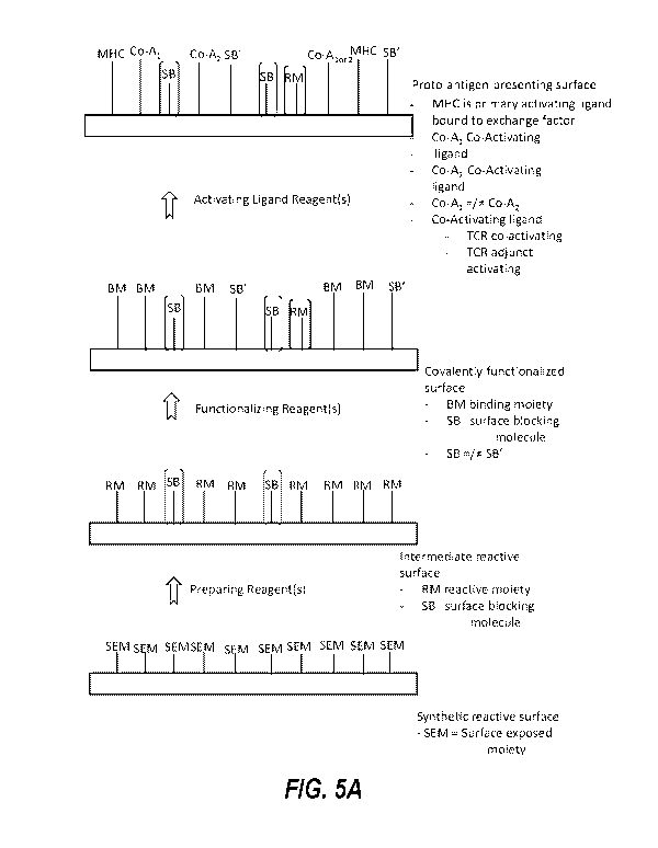
already or subsequently becomes surface-associated, to generate a proto-
antigen-presenting surface. Such
proto-antigen-presenting surfaces can then serve as substrates for generating
antigen-presenting surfaces
through displacement of the exchange factor with a peptide antigen. The
antigen-presenting surfaces can then
activate T cells if the peptide antigen is immunogenic. Thus, T cell
activation provides a readout of peptide
antigen immunogenicity.
[0006] The presently disclosed proto-antigen-presenting surfaces and related
methods and uses can
provide benefits such as more rapid evaluation of peptide antigen
immunogenicity because the relatively
laborious process of folding the MHC need not be performed with a peptide
antigen of interest and need not be
performed with each individual member of a set of peptide antigens being
evaluated for immunogenicity. For
example, an exemplary method comprises folding an MHC with an initial peptide,
which may or may not be a
peptide antigen of interest or may be any of the initial peptides described
herein, and preparing a proto-
antigen-presenting surface by associating the MHC with a suitable surface and
contacting the MHC with an
1
CA 03115531 2021-04-06
WO 2020/081875 PCT/US2019/056831
exchange factor to displace the initial peptide. The contacting step may occur
before or after the associating
step. An antigen-presenting surface can be prepared by contacting the proto-
antigen-presenting surface with
one or more peptide antigens of interest (e.g., one or more pools of peptide
antigens) such that the one or
more peptide antigens of interest displace the exchange factor and become
associated with the MHC. The
resulting surface can then be used to evaluate peptide antigen immunogenicity,
e.g., by determining whether
or to what extent it activates T lymphocytes. Additional embodiments include
kits for preparing proto-antigen-
presenting surfaces or antigen-presenting surfaces comprising an exchange
factor and an MHC associated
with a surface; methods of using antigen-presenting surfaces prepared as
described herein to activate T
lymphocytes; T lymphocytes prepared according to such methods; and methods of
using such T lymphocytes,
e.g., to treat diseases such as cancer. Further additional embodiments are
described below.
[0007] Embodiment 1 is a kit for generating an antigen-presenting surface,
the kit comprising:
(a) a covalently functionalized synthetic surface;
(b) a primary activating molecule that includes a major histocompatibility
complex (MHC) molecule
configured to bind to a T cell receptor (TCR), and a first reactive moiety
configured to react with or bind
to the covalently functionalized surface;
and
(c) at least one of: an exchange factor (e.g., provided separately from the
primary activating molecule);
and an exchange factor bound to the MHC molecule or an initial peptide bound
to the MHC molecule,
optionally wherein the initial peptide is non-immunogenic.
[0008] Embodiment 2 is the kit of embodiment 2 further comprising one or
more of:
at least one co-activating molecule that includes a second reactive moiety
configured to react with or
bind to the covalently functionalized surface, wherein each co-activating
molecule is selected from a
TCR co-activating molecule and an adjunct TCR activating molecule;
a surface-blocking molecule capable of covalently binding to the covalently
functionalized synthetic
surface;
a buffer suitable for performing an exchange reaction; and
instructions for performing an exchange reaction wherein a peptide antigen
displaces the exchange
factor.
[0009] Embodiment 3 is the kit of embodiment 1 or 2 comprising: the
exchange factor, wherein the
exchange factor is provided separately from the primary activating molecule;
and the initial peptide bound to
the MHC molecule.
[0010] Embodiment 4 is a method of forming a proto-antigen-presenting
surface, the method comprising:
synthesizing a plurality of major histocompatibility complex (MHC) molecules
in the presence of initial
peptide, thereby forming a plurality of complexes each comprising an MHC
molecule and an initial
peptide; or
2
CA 03115531 2021-04-06
WO 2020/081875 PCT/US2019/056831
synthesizing a plurality of major histocompatibility complex (MHC) molecules
in the presence of
exchange factor, thereby forming a plurality of complexes each comprising an
MHC molecule and an
exchange factor; or
reacting a plurality of MHC molecules with exchange factor, thereby forming a
plurality of complexes
each comprising an MHC molecule and an exchange factor;
wherein: (i) a plurality of primary activating molecules comprise the MHC
molecules and first reactive
moieties, or (ii) a plurality of primary activating molecules is prepared by
adding first reactive moieties
to the MHC molecules, and
the method further comprises reacting the first reactive moieties of the
plurality of primary activating
molecules with a first plurality of binding moieties disposed on a covalently
functionalized synthetic
surface, thereby forming the proto-antigen-presenting surface;
optionally wherein the initial peptide is non-immunogenic.
[0011] Embodiment 5 is the method of embodiment 4 further comprising
reacting the plurality of MHC
molecules synthesized in the presence of the initial peptide with exchange
factor, optionally in the presence of
a peptide antigen.
[0012] Embodiment 6 is a method of analyzing stability of a complex
comprising a major histocompatibility
complex (MHC) molecule and a peptide antigen, wherein the MHC molecule is
configured to bind to a T cell
receptor (TCR), the method comprising:
contacting a plurality of the MHC molecules with the peptide antigen and an
exchange factor, thereby
forming peptide antigen-bound MHC molecules, optionally wherein an initial
peptide is bound to the
MHC molecules before contact with the peptide antigen and exchange factor;
wherein
(i) a plurality of primary activating molecules comprise the MHC molecules and
first reactive moieties
or (ii) a plurality of primary activating molecules is prepared by adding
first reactive moieties to the
MHC molecules, and the method further comprises reacting the first reactive
moieties of the plurality of
primary activating molecules with a first plurality of binding moieties
disposed on a covalently
functionalized synthetic surface; and
measuring total binding and/or an extent of dissociation of the peptide
antigen from the MHC molecule.
[0013] Embodiment 7 is the method of embodiment 6, wherein measuring total
binding and/or the extent of
dissociation comprises measuring binding of an agent to the MHC molecule,
wherein the agent specifically
binds to (i) the initial peptide, and/or (ii) a peptide-bound conformation of
the MHC molecule.
[0014] Embodiment 8 is the method of embodiment 6 or 7, wherein the agent
comprises an antibody,
optionally wherein the antibody is produced by clone W6/32.
[0015] Embodiment 9 is the method of any one of embodiments 6-8, wherein
measuring total binding
and/or the extent of dissociation comprises performing flow cytometry.
3
CA 03115531 2021-04-06
WO 2020/081875 PCT/US2019/056831
[0016] Embodiment 10 is the method of any one of embodiments 6-9, wherein the
agent does not
recognize a peptide-unbound conformation of the MHC molecule.
[0017] Embodiment 11 is the method of any one of embodiments 6-10, wherein the
method further
comprises determining one or more kinetic parameters of the peptide antigen-
bound MHC molecule.
[0018] Embodiment 12 is the method of embodiment 11, wherein the one or more
kinetic parameters
comprise a half-life.
[0019] Embodiment 13 is the method of any one of embodiments 6-12, wherein the
method results in
identification of a peptide with a half-life of at least about 4 hours (e.g.,
at least about 6, 8, 10, 12, 14, 16, or 18
hours).
[0020] Embodiment 14 is the method of any one of embodiments 6-13, wherein the
method results in
identification of a peptide with a half-life in the range of about 4 to about
40 hours (e.g., about 4 to about 10
hours, about 10 to about 15 hours, about 15 to about 20 hours, about 20 to
about 25 hours, about 25 to about
30 hours, about 30 to about 35, or about 35 to about 40 hours).
[0021] Embodiment 15 is the method of any one of embodiments 6-14, wherein the
one or more kinetic
parameters comprise an off-rate.
[0022] Embodiment 16 is a method of analyzing stability of a plurality of
complexes each comprising a
histocompatibility complex (MHC) molecule and a peptide antigen, comprising
performing the method of any
one of embodiments 6-15 with each of a plurality of different peptide
antigens.
[0023] Embodiment 17 is the kit of any one of embodiments 1-3 or the method of
any one of embodiments
4-16, wherein the initial peptide comprises at least 4 or 5 amino acid
residues; or has a length of about 4, 5, 6,
7, 8, 9, 10, 11, 12, 13, 14, 15, 16, 17, or amino acid residues (e.g., ranging
from 8 to 10 amino acid residues,
or 13 to 18 amino acid residues).
[0024] Embodiment 18 is the method or kit of any one of embodiments 1-17,
wherein the initial peptide
comprises a lysine as the fourth or fifth amino acid residue.
[0025] Embodiment 19 is the method or kit of any one of embodiments 1-18,
wherein the initial peptide
comprises a label.
[0026] Embodiment 20 is the method or kit of embodiment 19, wherein the
label is attached to the fourth or
fifth amino acid residue (e.g., lysine).
[0027] Embodiment 21 is the method or kit of embodiment 19 or 20, wherein
the label is a fluorescent
label.
[0028] Embodiment 22 is the method or kit of any one of embodiments 1-21,
wherein the initial peptide has
a sequence comprising or consisting of a sequence from a naturally occurring
(e.g., mammalian or human)
polypeptide.
[0029] Embodiment 23 is the method or kit of any one of embodiments 1-22,
wherein the sequence of the
initial peptide consists of sequence that appears in a wild-type (e.g.,
mammalian or human) polypeptide.
4
CA 03115531 2021-04-06
WO 2020/081875 PCT/US2019/056831
[0030] Embodiment 24 is the method or kit of any one of embodiments 1-23,
wherein the initial peptide is
non-immunogenic.
[0031] Embodiment 25 is the method or kit of any one of embodiments 1-24,
wherein the sequence of the
initial peptide comprises or consists of sequence from a highly conserved
protein (e.g., a protein with a below
average mutation rate; optionally wherein the mutation rate is at least one or
two standard deviations below
the average amino acid mutation rate in the organism).
[0032] Embodiment 26 is the method or kit of any one of embodiments 1-25,
wherein the sequence of the
initial peptide comprises or consists of sequence from a cytoskeletal
polypeptide, e.g., an actin or tubulin
polypeptide.
[0033] Embodiment 27 is the method or kit of any one of embodiments 1-25,
wherein the sequence of the
initial peptide comprises or consists of any one of SEQ ID NOs: 1-4.
[0034] Embodiment 28 is the method or kit of any one of embodiments 1-25,
wherein the sequence of the
initial peptide comprises or consists of sequence from a ribosomal
polypeptide, e.g., the RPSA, RPS2, RPL3,
RPL4, RPL5, RPL6, RPL7A, or RPPO polypeptides.
[0035] Embodiment 29 is the method or kit of any one of embodiments 1-28,
wherein the initial peptide
binds the MHC molecule with high affinity, a low off-rate, and/or a long half-
life, optionally wherein the binding
of the initial peptide to the MHC molecule has a half-life of at least about
4, 6, 8, 10, 12, 14, 16, 18, 20, 24, 28,
32, 36, or 48 hours, or the binding of the initial peptide to the MHC molecule
has a half-life in the range of
about 4-12, 8-16, 12-20, 20-28, 24-32, 28-36, 32-40, 36-48, or 48-72 hours.
[0036] Embodiment 30 is the method or kit of any one of the preceding
embodiments, wherein the
covalently functionalized synthetic surface presents a plurality of azido
groups.
[0037] Embodiment 31 is the method or kit of embodiment 30, wherein the
first reactive moieties are
configured to react with the azido groups of the covalently functionalized
synthetic surface so as to form
covalent bonds.
[0038] Embodiment 32 is the method or kit of any one of embodiments 1-29,
wherein the covalently
functionalized synthetic surface presents a plurality of biotin-binding
agents, and wherein the first reactive
moieties are configured to specifically bind to the biotin-binding agent.
[0039] Embodiment 33 is the method or kit of embodiment 32, wherein the
first reactive moieties comprise
or consist essentially of biotin.
[0040] Embodiment 34 is the method or kit of embodiment 32 or 33, wherein
the biotin-binding agent is
covalently attached to the covalently functionalized synthetic surface.
[0041] Embodiment 35 is the method or kit of embodiment 32 or 33, wherein
the biotin-binding agent is
noncovalently attached to the covalently functionalized synthetic surface
through biotin functionalities.
[0042] Embodiment 36 is the method or kit of any one of embodiments 32-35,
wherein the biotin-binding
agent is streptavidin.
CA 03115531 2021-04-06
WO 2020/081875 PCT/US2019/056831
[0043] Embodiment 37 is the method of any one of embodiments 4-36, wherein
a plurality of co-activating
molecular ligands, each including a TCR co-activating molecule or an adjunct
TCR activating molecule, are
present on the covalently functionalized synthetic surface or are added to the
covalently functionalized
synthetic surface by reacting a plurality of co-activating molecules, each
including second reactive moiety and
a TCR co-activating molecule or an adjunct TCR activating molecule, with a
second plurality of binding
moieties of the covalently functionalized synthetic surface configured for
binding the second reactive moieties.
[0044] Embodiment 38 is the kit of any one of embodiments 30-31, or the method
of embodiment 37,
wherein the covalently functionalized synthetic surface presents a plurality
of azido groups, and wherein the
second reactive moieties are configured to react with the azido groups of the
covalently functionalized
synthetic surface so as to form covalent bonds.
[0045] Embodiment 39 is the method or kit of any one of embodiments 32-38,
wherein the covalently
functionalized synthetic surface presents a plurality of biotin-binding
agents, and wherein the second reactive
moieties are configured to specifically bind to the biotin-binding agent.
[0046] Embodiment 40 is a proto-antigen-presenting surface, the surface
comprising:
[0047] a plurality of primary activating molecular ligands, wherein each
primary activating molecular ligand
includes a major histocompatibility complex (MHC) molecule configured to bind
to a T cell receptor (TCR) of a
T cell and wherein an exchange factor or an initial peptide is bound to the
MHC molecules, optionally wherein
the initial peptide is non-immunogenic; and
a plurality of co-activating molecular ligands each including a TCR co-
activating molecule or an adjunct TCR
activating molecule.
[0048] Embodiment 41 is the proto-antigen-presenting surface of embodiment
40, wherein each of the
plurality of primary activating molecular ligands and the plurality of co-
activating molecular ligands is
specifically bound to the antigen presenting surface.
[0049] Embodiment 42 is the surface, kit, or method of any one of the
preceding embodiments, wherein the
exchange factor comprises Leu, Phe, Val, Arg, Met, Lys, Ile, homoleucine,
cyclohexylalanine, or Norleucine as
its C-terminal amino acid residue.
[0050] Embodiment 43 is the surface, kit, or method of any one of the
preceding embodiments, wherein the
exchange factor comprises a free N-terminal amine.
[0051] Embodiment 44 is the surface, kit, or method of any one of the
preceding embodiments, wherein the
exchange factor comprises Gly, Ala, Ser, or Cys as its penultimate C-terminal
residue.
[0052] Embodiment 45 is the surface, kit, or method of embodiment 44, wherein
the exchange factor
comprises Gly as its penultimate C-terminal residue.
[0053] Embodiment 46 is the surface, kit, or method of any one of the
preceding embodiments, wherein the
exchange factor is 2, 3, 4, or 5 amino acid residues in length.
6
CA 03115531 2021-04-06
WO 2020/081875 PCT/US2019/056831
[0054] Embodiment 47 is the surface, kit, or method of embodiment 46,
wherein the exchange factor is 2
amino acid residues in length.
[0055] Embodiment 48 is the surface, kit, or method of any one of the
preceding embodiments, wherein the
exchange factor comprises a linkage between its C-terminal and penultimate C-
terminal residues which is a
peptide bond, lactam, or piperazinone.
[0056] Embodiment 49 is the surface, kit, or method of embodiment 48, wherein
the exchange factor
comprises a peptide bond between its C-terminal and penultimate C-terminal
residues.
[0057] Embodiment 50 is the surface, kit, or method of any one of the
preceding embodiments, wherein the
covalently functionalized synthetic surface or the proto-antigen-presenting
surface further comprises at least
one plurality of surface-blocking molecular ligands covalently attached to the
surface.
[0058] Embodiment 51 is the surface, kit, or method of embodiment 50,
wherein:
(i) each of the plurality of surface-blocking molecular ligands includes a
hydrophilic moiety, an amphiphilic
moiety, a zwitterionic moiety, and/or a negatively charged moiety;
(ii) each of the plurality of surface-blocking molecular ligands includes a
linker and a terminal surface-blocking
group, optionally wherein the linkers of the plurality of surface-blocking
molecular ligands are of the same
length or are of different lengths; or
(iii) each of the plurality of surface-blocking molecular ligands includes a
linker and a terminal surface-blocking
group, wherein the terminal surface-blocking group comprises a hydrophilic
moiety, amphiphilic moiety,
zwitterionic moiety, and/or negatively charged moiety, optionally wherein the
linkers of the plurality of surface-
blocking molecular ligands are of the same length or are of different lengths;
(iv) each of the plurality of surface-blocking molecular ligands is covalently
bound to the covalently
functionalized synthetic surface or the proto-antigen-presenting surface
and/or
(v) the plurality of the surface-blocking molecular ligands and may include 2,
3, or 4 different surface-blocking
groups and/or 2, 3, 4, or more different lengths of linkers, chosen in any
combination.
[0059] Embodiment 52 is the surface, kit, or method of embodiment 50 or 51,
wherein:
(i) the plurality of surface-blocking molecular ligands all have the same
terminal surface-blocking group; or
(ii) the plurality of surface-blocking molecular ligands have a mixture of
terminal surface-blocking groups;
optionally wherein each of the plurality of surface-blocking molecular ligands
includes a polyethylene glycol
(PEG) moiety, a carboxylic acid moiety, or a combination thereof.
[0060] Embodiment 53 is the surface, kit, or method of embodiment 52, wherein
the PEG moiety of each of
the surface-blocking molecular ligands has a backbone linear chain length of
about 10 atoms to about 100
atoms.
[0061] Embodiment 54 is the surface, kit, or method of embodiment 52 or 53,
wherein the PEG moiety
comprises a carboxylic acid moiety.
7
CA 03115531 2021-04-06
WO 2020/081875 PCT/US2019/056831
[0062] Embodiment 55 is the surface, kit, or method of embodiment 54, wherein
the PEG moiety
comprises (PEG)4-000H.
[0063] Embodiment 56 is the surface, kit, or method of any one of the
preceding embodiments, wherein a
plurality of biotin or biotin-binding agent functionalities is attached to the
covalently functionalized synthetic
surface or the proto-antigen-presenting surface via a linker.
[0064] Embodiment 57 is the surface, kit, or method of embodiment 56,
wherein the linker linking the biotin
or biotin-binding agent functionality has a length of about 20 Angstroms to
about 100 Angstroms.
[0065] Embodiment 58 is the surface, kit, or method of embodiment 56 or 57,
wherein the linker links the
biotin or biotin-binding agent functionality to the covalently functionalized
synthetic surface or the proto-
antigen-presenting surface through a series of about 15, 20, 25, 30, 35, 40,
45, 50, 55, 60, 65, 70, 75, 80, 85,
95, 100, 200 bond lengths, or any number of bond lengths therebetween.
[0066] Embodiment 59 is the surface, kit, or method of any one of
embodiments 56-58, wherein the linker
of each biotin or biotin-binding agent functionality includes a polyethylene
glycol (PEG) moiety.
[0067] Embodiment 60 is the surface, kit, or method of embodiment 59,
wherein the PEG linker includes a
(PEG)13 repeating sequence, optionally wherein the covalently functionalized
synthetic surface or the proto-
antigen-presenting surface includes the plurality of biotin-binding agent
functionalities.
[0068] Embodiment 61 is the surface, kit, or method of embodiment 59,
wherein the PEG linker includes a
(PEG)4 repeating sequence, optionally wherein the covalently functionalized
synthetic surface or the proto-
antigen-presenting surface includes the plurality of biotin functionalities.
[0069] Embodiment 62 is the surface, kit, or method of any one of
embodiments 56-61, wherein the biotin-
binding agent functionalities are streptavidin moieties.
[0070] Embodiment 63 is the surface, kit, or method of embodiment 62,
wherein the at least one plurality of
streptavidin moieties is disposed upon the covalently functionalized synthetic
surface or the proto-antigen-
presenting surface in a density from about 4X 10² to about 3X 10⁴ molecules
per square micron, in each
portion or sub-region where it is attached.
[0071] Embodiment 64 is the surface, kit, or method of embodiment 62,
wherein the at least one plurality of
streptavidin moieties is disposed upon the covalently functionalized synthetic
surface or the proto-antigen-
presenting surface in a density from about 5X 10³ to about 3X 10⁴ molecules
per square micron, in each
portion or sub-region where it is attached.
[0072] Embodiment 65 is the surface, kit, or method of embodiment 62,
wherein the at least one plurality of
streptavidin moieties is disposed upon the covalently functionalized synthetic
surface or the proto-antigen-
presenting surface from about 6X 10² to about 5X 10³ molecules per square
micron, about 5X 10³ to about 2X
104 molecules per square micron, about 1X 10⁴ to about 2X 10⁴ molecules per
square micron, or about 1.25X
104 to about 1.75X 10⁴ molecules per square micron, in each portion or sub-
region where it is attached.
8
CA 03115531 2021-04-06
WO 2020/081875 PCT/US2019/056831
[0073] Embodiment 66 is the surface, kit, or method of any one of
embodiments 56-65, wherein the at least
one plurality of biotin-binding agent or biotin moieties is disposed upon
substantially all of the covalently
functionalized synthetic surface or the proto-antigen-presenting surface.
[0074] Embodiment 67 is the surface, kit, or method of any one of embodiments
56-65, wherein the
covalently functionalized synthetic surface or the proto-antigen-presenting
surface further includes a first
portion and a second portion, wherein the distribution of the at least one
plurality of biotin-binding agent or
biotin functionalities is located in the first portion of the covalently
modified synthetic surface, and the
distribution of the at least one plurality of the surface-blocking molecular
ligands is located in the second
portion.
[0075] Embodiment 68 is the surface, kit, or method of embodiment 67,
wherein a second plurality of
surface-blocking molecular ligands is disposed in the first portion of the
covalently functionalized synthetic
surface or the proto-antigen-presenting surface.
[0076] Embodiment 69 is the surface, kit, or method of embodiment 67 or 68,
wherein the first portion of
the covalently functionalized synthetic surface or the proto-antigen-
presenting surface further includes a
plurality of first regions, each first region including at least a subset of
the plurality of the biotin-binding agent or
biotin functionalities, wherein each of the plurality of first regions is
separated from another of the plurality of
first regions by the second region configured to substantially exclude the
streptavidin or biotin functionalities.
[0077] Embodiment 70 is the surface, kit, or method of embodiment 69,
wherein each of the plurality of first
regions including at least the subset of the plurality of the streptavidin or
biotin functionalities has an area of
about 0.10 square microns to about 4.0 square microns.
[0078] Embodiment 71 is the surface, kit, or method of embodiment 69,
wherein the area of each of the
plurality of first regions including at least the subset of the plurality of
the primary activating molecular ligands
is about 4.0 square microns to about 0.8 square microns.
[0079] Embodiment 72 is the surface, kit, or method of any one of the
preceding embodiments, wherein the
covalently functionalized synthetic surface or the proto-antigen-presenting
surface includes glass, polymer,
metal, ceramic, and/or a metal oxide.
[0080] Embodiment 73 is the surface, kit, or method of any one of the
preceding embodiments, wherein the
covalently functionalized synthetic surface or the proto-antigen-presenting
surface is a wafer, an inner surface
of a tube, or an inner surface of a microfluidic device.
[0081] Embodiment 74 is the surface, kit, or method of embodiment 73,
wherein the tube is a glass or
polymer tube.
[0082] Embodiment 75 is the surface, kit, or method of any one of
embodiments 1-71, wherein the
covalently functionalized synthetic surface or the proto-antigen-presenting
surface is a bead.
[0083] Embodiment 76 is the surface, kit, or method of embodiment 75,
wherein the bead includes a
magnetic material.
9
CA 03115531 2021-04-06
WO 2020/081875
PCT/US2019/056831
[0084] Embodiment 77 is the surface, kit, or method of embodiment 75 or 76,
wherein the bead has a
surface area within 10% of the surface area of a sphere of an equal volume or
diameter.
[0085] Embodiment 78 is the surface, kit, or method of embodiment 73,
wherein the covalently
functionalized synthetic surface or the proto-antigen-presenting surface is at
least one inner surface of a
microfluidic device.
[0086] Embodiment 79 is the surface, kit, or method of embodiment 78,
wherein the inner surface of the
microfluidic device is within a chamber of the microfluidic device.
[0087] Embodiment 80 is the surface, kit, or method of any one of embodiments
69-71, wherein each of
the plurality of first regions including at least a subset of the plurality of
biotin-binding agent or biotin
functionalities includes at least one surface within a chamber of the
microfluidic device.
[0088] Embodiment 81 is the surface, kit, or method of embodiment 79 or 80,
wherein the chamber is a
sequestration pen.
[0089] Embodiment 82 is the surface, kit, or method of embodiment 81,
wherein the microfluidic device
further comprises a flow region for containing a flow of a first fluidic
medium; and the sequestration pen
comprises an isolation region for containing a second fluidic medium, the
isolation region having a single
opening, wherein the isolation region of the sequestration pen is an unswept
region of the microfluidic device;
and a connection region fluidically connecting the isolation region to the
flow region; optionally wherein the
microfluidic device comprises a microfluidic channel comprising at least a
portion of the flow region.
[0090] Embodiment 83 is the surface, kit, or method of embodiment 82,
wherein the microfluidic device
comprises a microfluidic channel comprising at least a portion of the flow
region, and the connection region
comprises a proximal opening into the microfluidic channel having a width W00
n ranging from about 20 microns
to about 100 microns and a distal opening into the isolation region, and
wherein a length Lon of the connection
region from the proximal opening to the distal opening is at least 1.0 times a
width con o. w f the proximal
¨
opening of the connection region.
[0091] Embodiment 84 is the surface, kit, or method of embodiment 83,
wherein the length Lcon of the
connection region from the proximal opening to the distal opening is at least
1.5 times the width Wcon of the
proximal opening of the connection region.
[0092] Embodiment 85 is the surface, kit, or method of embodiment 83,
wherein the length Lcon of the
connection region from the proximal opening to the distal opening is at least
2.0 times the width Wcon of the
proximal opening of the connection region.
[0093] Embodiment 86 is the surface, kit, or method of any one of
embodiments 83-85, wherein the width
Wcon of the proximal opening of the connection region ranges from about 20
microns to about 60 microns.
[0094] Embodiment 87 is the surface, kit, or method of any one of
embodiments 83-86, wherein the length
Lcon of the connection region from the proximal opening to the distal opening
is between about 20 microns and
about 500 microns.
CA 03115531 2021-04-06
WO 2020/081875 PCT/US2019/056831
[0095] Embodiment 88 is the surface, kit, or method of any one of embodiments
83-87, wherein a width of
the microfluidic channel at the proximal opening of the connection region is
between about 50 microns and
about 500 microns.
[0096] Embodiment 89 is the surface, kit, or method of any one of
embodiments 83-88, wherein a height of
the microfluidic channel at the proximal opening of the connection region is
between 20 microns and 100
microns.
[0097] Embodiment 90 is the surface, kit, or method of any one of embodiments
82-89, wherein the volume
of the isolation region ranges from about 2 x10⁴ to about 2 x 10⁶ cubic
microns.
[0098] Embodiment 91 is the surface, kit, or method of any one of
embodiments 82-90, wherein the
proximal opening of the connection region is parallel to a direction of the
flow of the first medium in the flow
region.
[0099] Embodiment 92 is the surface, kit, or method of any one of
embodiments 82-91, wherein the
microfluidic device comprises an enclosure comprising a base, a microfluidic
circuit structure disposed on the
base, and a cover which collectively define a microfluidic circuit, and the
microfluidic circuit comprises the flow
region, the microfluidic channel, and the sequestration pen.
[00100] Embodiment 93 is the surface, kit, or method of embodiment 92,
wherein the cover is an integral
part of the microfluidic circuit structure.
[00101] Embodiment 94 is the surface, kit, or method of any one of
embodiments 82-93, wherein the
microfluidic circuit further comprises one or more inlets through which the
first medium can be input into the
flow region and one or more outlets through which the first medium can be
removed from the flow region.
[00102] Embodiment 95 is the surface, kit, or method of any one of
embodiments 92-94, wherein barriers
defining the microfluidic sequestration pen extend from a surface of the base
of the microfluidic device to a
surface of the cover of the microfluidic device.
[00103] Embodiment 96 is the surface, kit, or method of any one of
embodiments 92-95, wherein the cover
and the base are part of a dielectrophoresis (DEP) mechanism for selectively
inducing DEP forces on a micro-
object.
[00104] Embodiment 97 is the surface, kit, or method of any one of
embodiments 92-96, wherein the
microfluidic device further comprises a first electrode, an electrode
activation substrate, and a second
electrode, wherein the first electrode is part of a first wall of the
enclosure and the electrode activation
substrate and the second electrode is part of a second wall of the enclosure,
wherein the electrode activation
substrate comprises a photoconductive material, semiconductor integrated
circuits, or phototransistors.
[00105] Embodiment 98 is the surface, kit, or method of embodiment 97,
wherein the first wall of the
microfluidic device is the cover, and wherein the second wall of the
microfluidic device is the base.
[00106] Embodiment 99 is the surface, kit, or method of embodiment 97 or
98, wherein the electrode
activation substrate comprises phototransistors.
11
CA 03115531 2021-04-06
WO 2020/081875 PCT/US2019/056831
[00107] Embodiment 100 is the surface, kit, or method of any one of
embodiments 92-99, wherein the cover
and/or the base is transparent to light.
[00108] Embodiment 101 is the surface, kit, or method of any one of
embodiments 78-100, wherein the
covalently functionalized surface or the proto-antigen-presenting surface
includes a portion configured to
exclude biotin-binding agent or biotin functionalities which is disposed at at
least one surface of a microfluidic
channel of the microfluidic device.
[00109] Embodiment 102 is the surface, kit, or method of any one of
embodiments 37 or 40-101, wherein
the plurality of co-activating molecular ligands comprises TCR co-activating
molecules and adjunct TCR
activating molecules.
[00110] Embodiment 103 is the surface, kit, or method of embodiment 102,
wherein a ratio of the TCR co-
activating molecules to the adjunct TCR activating molecules of the plurality
of co-activating molecular ligands
is about 100:1 to about 1:100.
[00111] Embodiment 104 is the surface, kit, or method of embodiment 102,
wherein a ratio of the TCR co-
activating molecules to the adjunct TCR activating molecules of the plurality
of co-activating molecular ligands
is about 100:1 to about 90:1, about 90:1 to about 80:1, about 80:1 to about
70:1, about 70:1 to about 60:1,
about 60:1 to about about 50:1, about 50:1 to about 40:1, about 40:1 to about
30:1, about 30:1 to about 20:1,
about 20:1 to about 10:1, about 10:1 to about 1:1, about 1:1 to about 1:10,
about 1:10 to about 1:20, about
1:20 to about 1:30, about 1:30 to about 1:40, about 1:40 to about 1:50, about
1:50 to about 1:60, about 1:60 to
about 1:70, about 1:70 to about 1:80, about 1:80t0 about 1:90, or about 1:90t0
about 1:100.
[00112] Embodiment 105 is the surface, kit, or method of embodiment 102,
wherein a ratio of the TCR co-
activating molecules to the adjunct TCR activating molecules of the plurality
of co-activating molecular ligands
is about 10:1 to about 1:20.
[00113] Embodiment 106 is the surface, kit, or method of embodiment 102,
wherein a ratio of the TCR co-
activating molecules to the adjunct TCR activating molecules of the plurality
of co-activating molecular ligands
is about 10:1 to about 1:10.
[00114] Embodiment 107 is the surface, kit, or method of any one of the
preceding embodiments, wherein
the MHC molecule includes an MHC protein sequence and a beta microglobulin.
[00115] Embodiment 108 is the surface, kit, or method of embodiment 107,
wherein the MHC molecule
comprises a human leukocyte antigen A (HLA-A) heavy chain.
[00116] Embodiment 109 is the surface, kit, or method of embodiment 108,
wherein the HLA-A heavy chain
is an HLA-A*01, HLA-A*02, HLA-A*03, HLA-A*11, HLA-A*23, HLA-A*24, HLA-A*25,
HLA-A*26, HLA-A*29,
HLA-A*30, HLA-A*31, HLA-A*32, HLA-A*33, HLA-A*34, HLA-A*43, HLA-A*66, HLA-
A*68, HLA-A*69, HLA-
A*74, or HLA-A*80 heavy chain.
[00117] Embodiment 110 is the surface, kit, or method of embodiment 107,
wherein the MHC molecule
comprises a human leukocyte antigen B (HLA-B) heavy chain.
12
CA 03115531 2021-04-06
WO 2020/081875 PCT/US2019/056831
[00118] Embodiment 111 is the surface, kit, or method of embodiment 110,
wherein the HLA-B heavy chain
is an HLA-B*07, HLA-B*08, HLA-B*13, HLA-B*14, HLA-B*15, HLA-B*18, HLA-B*27,
HLA-B*35, HLA-B*37,
HLA-B*38, HLA-B*39, HLA-B*40, HLA-B*41, HLA-B*42, HLA-B*44, HLA-B*45, HLA-
B*46, HLA-B*47, HLA-
B*48, HLA-B*49, HLA-B*50, HLA-B*51, HLA-B*52, HLA-B*53, HLA-B*54, HLA-B*55,
HLA-B*56, HLA-B*57,
HLA-B*58, HLA-B*59, HLA-B*67, HLA-B*73, HLA-B*78, HLA-B*81, HLA-B*82, or HLA-
B*83 heavy chain.
[00119] Embodiment 112 is the surface, kit, or method of embodiment 107,
wherein the MHC molecule
comprises a human leukocyte antigen C (HLA-C) heavy chain.
[00120] Embodiment 113 is the surface, kit, or method of embodiment 112,
wherein the HLA-C heavy chain
is an HLA-C*01, HLA-C*02, HLA-C*03, HLA-C*04, HLA-C*05, HLA-C*06, HLA-C*07,
HLA-C*08, HLA-C*12,
HLA-C*14, HLA-C*15, HLA-C*16, HLA-C*17, or HLA-C*18 heavy chain.
[00121] Embodiment 114 is the surface, kit, or method of any one of
embodiments 2-3 or 37-113, wherein
the TCR co-activating molecule includes a protein.
[00122] Embodiment 115 is the surface, kit, or method of embodiment 114,
wherein the TCR co-activating
molecule further comprises a site-specific C-terminal biotin moiety.
[00123] Embodiment 116 is the surface, kit, or method of embodiment 114 or
115, wherein the TCR co-
activating protein molecule includes a CD28 binding protein or a fragment
thereof which retains binding ability
with CD28.
[00124] Embodiment 117 is the surface, kit, or method of embodiment 116,
wherein the CD28 binding
protein includes a CD80 molecule or a fragment thereof, wherein the fragment
retains binding ability to CD28.
[00125] Embodiment 118 is the surface, kit, or method of embodiment 114 or
115, wherein the TCR co-
activating molecule includes an anti-CD28 antibody or fragment thereof,
wherein the fragment retains binding
activity with CD28.
[00126] Embodiment 119 is the surface, kit, or method of any one of
embodiments 2-3 or 37-116, wherein
the adjunct TCR activating molecule is configured to provide adhesion
stimulation.
[00127] Embodiment 120 is the surface, kit, or method of any one of
embodiments 2-3 or 37-119, wherein
the adjunct TCR activating molecular ligand includes a CD2 binding protein or
a fragment thereof, wherein the
fragment retains binding ability with CD2.
[00128] Embodiment 121 is the surface, kit, or method of embodiment 120,
wherein the CD2 binding protein
further comprises a site-specific C-terminal biotin moiety.
[00129] Embodiment 122 is the surface, kit, or method of any one of
embodiments 120 or 121, wherein the
adjunct TCR activating molecular ligand includes a CD58 molecule or fragment
thereof, wherein the fragment
retains binding activity with CD2.
[00130] Embodiment 123 is the surface, kit, or method of any one of
embodiments 120 or 121, wherein the
adjunct TCR activating molecule includes an anti-CD2 antibody or a fragment
thereof, wherein the fragment
retains binding activity with CD2.
13
CA 03115531 2021-04-06
WO 2020/081875 PCT/US2019/056831
[00131] Embodiment 124 is the proto-antigen-presenting surface of any one
of embodiments 40-123,
wherein the plurality of primary activating molecular ligands is disposed upon
at least a portion of the antigen-
presenting surface at a density from about 4X 10² to about 3X 10⁴ molecules
per square micron, in each
portion or sub-region where it is attached.
[00132] Embodiment 125 is the proto-antigen-presenting surface of
embodiment 124, wherein the plurality
of primary activating molecular ligands is disposed upon at least a portion of
the antigen-presenting surface at
a density from about 4X 10² to about 2X 10³ molecules per square micron.
[00133] Embodiment 126 is the proto-antigen-presenting surface of
embodiment 124, wherein the plurality
of primary activating molecular ligands is disposed upon at least a portion of
the antigen-presenting surface at
a density from about 2X 10³ to about 5X 10³ molecules per square micron.
[00134] Embodiment 127 is the proto-antigen-presenting surface of
embodiment 124, wherein the plurality
of primary activating molecular ligands is disposed upon at least a portion of
a surface of the antigen-
presenting surface at a density from about 5X 10³ to about 2X 10⁴ molecules
per square micron, about 1X 10⁴
to about 2X 10⁴ molecules per square micron, or about 1.25X 10⁴ to about 1.75X
104 molecules per square
micron.
[00135] Embodiment 128 is the proto-antigen-presenting surface of any one
of embodiments 124-127,
wherein the plurality of primary activating molecular ligands is disposed upon
substantially all of the antigen-
presenting surface at the stated density.
[00136] Embodiment 129 is the proto-antigen-presenting surface of any one
of embodiments 40-128,
wherein the plurality of co-activating molecular ligands is disposed upon at
least a portion the antigen-
presenting surface at a density from about 5X 10² to about 2X 10⁴ molecules
per square micron or about 5X
102 to about 1.5 X 10 molecules per square micron.
[00137] Embodiment 130 is the proto-antigen-presenting surface of
embodiment 129, wherein the plurality
of co-activating molecular ligands is disposed upon at least a portion of the
antigen-presenting surface at a
density from about 5X 10³ to about 2X 10⁴ molecules per square micron, about
5X 10³ to about 1.5X 10⁴
molecules per square micron, about 1X 10⁴ to about 2X 10⁴ molecules per square
micron, about 1X 10⁴ to
about 1.5X 10⁴ molecules per square micron, about 1.25X 10⁴ to about 1.75X 10⁴
molecules per square
micron, or about 1.25X 10⁴ to about 1.5X 10⁴ molecules per square micron.
[00138] Embodiment 131 is the proto-antigen-presenting surface of any one
of embodiments 40-128,
wherein the plurality of co-activating molecular ligands is disposed upon at
least a portion of the antigen-
presenting surface at a density from about 2X 10³ to about 5X 10³ molecules
per square micron.
[00139] Embodiment 132 is the proto-antigen-presenting surface of any one
of embodiments 40-128,
wherein the plurality of co-activating molecular ligands is disposed upon at
least a portion of a surface of the
antigen-presenting surface at a density from about 5X 10² to about 2X 10³
molecules per square micron.
14
CA 03115531 2021-04-06
WO 2020/081875 PCT/US2019/056831
[00140] Embodiment 133 is the proto-antigen-presenting surface of any one
of embodiments 129-132,
wherein the plurality of co-activating molecular ligands is disposed upon
substantially all of the antigen-
presenting surface at the stated density.
[00141] Embodiment 134 is the proto-antigen-presenting surface of any one
of embodiments 40-133,
wherein a ratio of the primary activating molecular ligands to the co-
activating molecular ligands present on the
antigen-presenting surface is about 1:10 to about 2:1, about 1:5 to about 2:1,
about 1:2 to about 2:1, about
1:10 to about 1:1, about 1:5 to about 1:1, about 1:1 to about 2:1, or about
1:2 to about 1:1.
[00142] Embodiment 135 is the proto-antigen-presenting surface of any one
of embodiments 40-134,
wherein each of the plurality of primary activating molecular ligands is
noncovalently bound to a binding
moiety, and further wherein the binding moiety is covalently bound to the
antigen-presenting surface.
[00143] Embodiment 136 is the proto-antigen-presenting surface of
embodiment 135, wherein each of the
plurality of primary activating molecular ligands comprises a biotin and is
noncovalently bound to a biotin-
binding agent, and further wherein the biotin-binding agent is covalently
bound to the antigen-presenting
surface.
[00144] Embodiment 137 is the proto-antigen-presenting surface of any one
of embodiments 40-136,
wherein each of the plurality of primary activating molecular ligands is
noncovalently bound to a binding
moiety, and further wherein the binding moiety is noncovalently bound to the
antigen-presenting surface.
[00145] Embodiment 138 is the proto-antigen-presenting surface of
embodiment 137, wherein each of the
plurality of primary activating molecular ligands comprises a biotin moiety,
the binding moiety comprises a
biotin-binding agent, and the biotin-binding agent is noncovalently bound to a
second biotin moiety covalently
attached to the antigen-presenting surface.
[00146] Embodiment 139 is the proto-antigen-presenting surface of 136 or
138, wherein the biotin-binding
agent is streptavidin.
[00147] Embodiment 140 is the proto-antigen-presenting surface of any one
of embodiments 40-139,
wherein each of the plurality of co-activating molecular ligands is non-
covalently attached to a streptavidin and
the streptavidin is non-covalently attached to a streptavidin binding
molecule, further wherein the streptavidin
binding molecule is covalently attached via a linker to the proto-antigen-
presenting surface, optionally wherein
the streptavidin binding molecule comprises biotin.
[00148] Embodiment 141 is the proto-antigen-presenting surface of any one
of embodiments 40-140,
wherein each of the plurality of co-activating molecular ligands is covalently
connected to the surface via a
linker.
[00149] Embodiment 142 is the proto-antigen-presenting surface of
embodiment 140 or 141, wherein the
linker links the streptavidin binding molecule and/or co-activating molecular
ligands to the surface through a
series of about 15, 20, 25, 30, 35, 40, 45, 50, 55, 60, 65, 70, 75, 80, 85,
95, 100, 200 bond lengths, or any
number of bond lengths therebetween bonds.
CA 03115531 2021-04-06
WO 2020/081875 PCT/US2019/056831
[00150] Embodiment 143 is the proto-antigen-presenting surface of any one
of embodiments 40-140,
wherein each of the plurality of co-activating molecular ligands is non-
covalently attached to a streptavidin
moiety; and the streptavidin moiety is covalently attached to the antigen-
presenting surface.
[00151] Embodiment 144 is the proto-antigen-presenting surface of
embodiment 143, wherein the
streptavidin moiety is linked by a linker to the surface through a series of
about 15, 20, 25, 30, 35, 40, 45, 50,
55, 60, 65, 70, 75, 80, 85, 95, 100, 200 bond lengths, or any number of bond
lengths therebetween.
[00152] Embodiment 145 is the proto-antigen-presenting surface of any one
of embodiments 40-144,
wherein the proto-antigen-presenting surface further comprises a plurality of
surface-blocking molecular
ligands.
[00153] Embodiment 146 is the proto-antigen-presenting surface of
embodiment 145, wherein:
(i) each of the plurality of surface-blocking molecular ligands includes a
hydrophilic moiety, an amphiphilic
moiety, a zwitterionic moiety, and/or a negatively charged moiety;
(ii) each of the plurality of surface-blocking molecular ligands includes a
linker and a terminal surface-blocking
group, optionally wherein the linkers of the plurality of surface-blocking
molecular ligands are of the same
length or are of different lengths; or
(iii) each of the plurality of surface-blocking molecular ligands includes a
linker and a terminal surface-blocking
group, wherein the terminal surface-blocking group comprises a hydrophilic
moiety, amphiphilic moiety,
zwitterionic moiety, and/or negatively charged moiety, optionally wherein the
linkers of the plurality of surface-
blocking molecular ligands are of the same length or are of different lengths.
[00154] Embodiment 147 is the proto-antigen-presenting surface of
embodiment 145 or 146, wherein:
(i) the plurality of surface-blocking molecular ligands all have the same
terminal surface-blocking group; or
(ii) the plurality of surface-blocking molecular ligands have a mixture of
terminal surface-blocking groups;
optionally wherein each of the plurality of surface-blocking molecular ligands
includes a polyethylene glycol
(PEG) moiety, a carboxylic acid moiety, or a combination thereof, further
optionally wherein the PEG moiety of
each of the surface-blocking molecular ligands has a backbone linear chain
length of about 10 atoms to about
100 atoms.
[00155] Embodiment 148 is the proto-antigen-presenting surface of any one
of embodiments 145-147,
wherein:
(i) each of the plurality of surface-blocking molecular ligands is covalently
connected to the antigen-presenting
surface; and/or
(ii) the plurality of the surface-blocking molecular ligands and may include
2, 3, or 4 different surface-blocking
groups and/or 2, 3, 4, or more different lengths of linkers, chosen in any
combination.
[00156] Embodiment 149 is the proto-antigen-presenting surface of any one
of embodiments 40-148, further
including a plurality of adhesion stimulatory molecular ligands, optionally
wherein each adhesive molecular
ligand includes a ligand for a cell adhesion receptor comprising an ICAM
protein sequence.
16
CA 03115531 2021-04-06
WO 2020/081875 PCT/US2019/056831
[00157] Embodiment 150 is the proto-antigen-presenting surface of
embodiment 149, wherein the adhesion
stimulatory molecular ligand is covalently connected to the antigen-presenting
surface via a linker.
[00158] Embodiment 151 is the proto-antigen-presenting surface of
embodiment 150, wherein the adhesion
stimulatory molecular ligand is non-covalently attached to a streptavidin
moiety, wherein the streptavidin
moiety is covalently attached via a linker to the antigen-presenting surface.
[00159] Embodiment 152 is the proto-antigen-presenting surface of
embodiment 150, wherein the adhesion
stimulatory molecular ligand is non-covalently attached to a streptavidin,
wherein the streptavidin is
noncovalently attached to a biotin and the biotin is covalently attached via a
linker to the antigen-presenting
surface.
[00160] Embodiment 153 is the proto-antigen-presenting surface of any one
of embodiments 40-152,
wherein the ratio of the TCR co-activating molecules to the adjunct TCR
activating molecules of the plurality of
co-activating molecular ligands is from about 3:1 to about 1:3.
[00161] Embodiment 154 is the proto-antigen-presenting surface of any one
of embodiments 40-153,
wherein the ratio of the TCR co-activating molecules to the adjunct TCR
activating molecules of the plurality of
co-activating molecular ligands is about 1:2 to about 2:1.
[00162] Embodiment 155 is the proto-antigen-presenting surface of any one
of embodiments 40-154,
wherein the ratio of the TCR co-activating molecules to the adjunct TCR
activating molecules of the plurality of
co-activating molecular ligands is about 1:1.
[00163] Embodiment 156 is the proto-antigen-presenting surface of any one
of embodiments 40-155, further
including a plurality of growth-stimulatory molecular ligands, wherein each of
the growth-stimulatory molecular
ligands includes a growth factor receptor ligand.
[00164] Embodiment 157 is the proto-antigen-presenting surface of
embodiment 156, wherein the growth
factor receptor ligand includes a cytokine or fragment thereof, wherein the
fragment retains receptor binding
ability, optionally wherein the cytokine comprises IL-21.
[00165] Embodiment 158 is the proto-antigen-presenting surface of any one
of embodiments 40-127, 129-
132, or 134-157, further including a first portion and a second portion,
wherein the distribution of the plurality of
primary activating molecular ligands and the distribution of the plurality of
co-activating molecular ligands are
located in the first portion of the antigen-presenting surface, and the second
portion is configured to
substantially exclude the primary activating molecular ligands.
[00166] Embodiment 159 is the proto-antigen-presenting surface of
embodiment 158, wherein at least one
plurality of surface-blocking molecular ligands is located in the second
portion of the at least one inner surface
of the antigen-presenting surface.
[00167] Embodiment 160 is the proto-antigen-presenting surface of
embodiment 158 or 159, wherein the
first portion of the antigen-presenting surface further includes a plurality
of first regions, each first region
including at least a subset of the plurality of the primary activating
molecular ligands, wherein each of the
17
CA 03115531 2021-04-06
WO 2020/081875 PCT/US2019/056831
plurality of first regions is separated from another of the plurality of first
region by the second portion
configured to substantially exclude primary activating molecular ligands.
[00168] Embodiment 161 is the proto-antigen-presenting surface of
embodiment 160, wherein each of the
plurality of first regions including the at least a subset of the plurality of
the primary activating molecular ligands
further includes a subset of the plurality of the co-activating molecular
ligands.
[00169] Embodiment 162 is the proto-antigen-presenting surface of
embodiment 160 or 161, wherein each
of the plurality of first regions including at least the subset of the
plurality of the primary activating molecular
ligands has an area of about 0.10 square microns to about 4.0 square microns.
[00170] Embodiment 163 is the proto-antigen-presenting surface of any one
of embodiments 160-162,
wherein the area of each of the plurality of first regions including at least
the subset of the plurality of the
primary activating molecular ligands is about 4.0 square microns to about 0.8
square microns.
[00171] Embodiment 164 is the proto-antigen-presenting surface of any one
of embodiments 160-163,
wherein each of the plurality of first regions further includes at least a
subset of a plurality of adhesion
stimulatory molecular ligands, and optionally wherein each of the adhesion
stimulatory molecular ligands
includes a ligand for a cell adhesion receptor comprising an ICAM protein
sequence.
[00172] Embodiment 165 is the proto-antigen-presenting surface of any one
of embodiments 158-164,
wherein the second portion configured to substantially exclude the primary
activating molecular ligands is also
configured to substantially exclude co-activating molecular ligands.
[00173] Embodiment 166 is the proto-antigen-presenting surface of any one
of embodiments 158-165,
wherein the second portion configured to substantially exclude the primary
activating molecular ligands is
further configured to include a plurality of growth stimulatory molecular
ligands, wherein each of the growth
stimulatory molecular ligands includes a growth factor receptor ligand.
[00174] Embodiment 167 is the proto-antigen-presenting surface of any one
of embodiments 158-166,
wherein the second portion configured to substantially exclude the primary
activating molecular ligands
includes a plurality of adhesion stimulatory molecular ligands, wherein each
of the adhesion stimulatory
molecular ligands includes a ligand for a cell adhesion receptor including an
ICAM protein sequence.
[00175] Embodiment 168 is the proto-antigen-presenting surface of any one
of embodiments 158-167,
which is an antigen-presenting surface of a microfluidic device and each of
the plurality of first regions
including at least a subset of the plurality of primary activating molecular
ligands is disposed at least one
surface within a chamber of the antigen-presenting microfluidic device.
[00176] Embodiment 169 is the kit or method of embodiment 68, wherein the
second plurality of surface-
blocking molecular ligands limits the density of functionalizing moieties of
an antigen-presenting synthetic
surface formed from the covalently functionalized synthetic surface.
[00177] Embodiment 170 is the method of any one of embodiments 4-5, 18-123
or 169, further comprising
reacting a plurality of surface-blocking molecules with a first additional
plurality of binding moieties of the
18
CA 03115531 2021-04-06
WO 2020/081875 PCT/US2019/056831
covalently functionalized surface, wherein each of the binding moieties of the
first additional plurality is
configured for binding the surface-blocking molecule.
[00178] Embodiment 171 is the method of any one of embodiments 4-5, 18-123,
or 169-170, further
comprising reacting a plurality of adhesion stimulatory molecular ligands,
wherein each adhesion stimulatory
molecular ligand includes a ligand for a cell adhesion receptor including an
ICAM protein sequence, with a
second additional plurality of binding moieties of the covalently
functionalized surface, wherein each of the
binding moieties of the second additional plurality is configured for binding
with the cell adhesion receptor
ligand molecule.
[00179] Embodiment 172 is the kit of any one of embodiments 1-3, 17-123, or
169, further comprising a
plurality of surface-blocking molecules, wherein the covalently functionalized
surface further comprises a first
additional plurality of binding moieties configured for binding the surface-
blocking molecule.
[00180] Embodiment 173 is the kit of any one of embodiments 1-3, 17-123,
169, or 172, further comprising a
plurality of adhesion stimulatory molecular ligands, wherein each adhesion
stimulatory molecular ligand
includes a ligand for a cell adhesion receptor including an ICAM protein
sequence, and the covalently
functionalized surface further comprises a second additional plurality of
binding moieties configured for binding
the cell adhesion receptor ligand molecule.
[00181] Embodiment 174 is the kit of any one of embodiments 1-3, 17-123,
169, or 172-173, further
comprising a peptide antigen.
[00182] Embodiment 175 is a method of preparing an antigen-presenting
surface comprising a peptide
antigen, the method comprising reacting the peptide antigen with a proto-
antigen-presenting surface according
to any one of embodiments 40-168, wherein the exchange factor or initial
peptide is substantially displaced
and the peptide antigen becomes associated with the MHC molecules.
[00183] Embodiment 176 is the kit or method of embodiment 174 or 175,
wherein the peptide antigen
comprises a tumor-associated antigen.
[00184] Embodiment 177 is the kit or method of any one of embodiments 174-
176, wherein the peptide
antigen comprises a segment of amino acid sequence from a protein expressed on
the surface of a tumor cell.
[00185] Embodiment 178 is the kit or method of embodiment 177, wherein the
segment comprises 5, 6, 7, 8,
9, or 10 amino acid residues or is 5, 6, 7, 8, 9, or 10 amino acid residues in
length.
[00186] Embodiment 179 is the kit or method of embodiment 177 or 178,
wherein the protein expressed on
the surface of a tumor cell is CD19, CD20, CLL-1, TRP-2, LAGE-1, HER2, EphA2,
FOLR1, MAGE-Al,
mesothelin, SOX2, PSM, 0A125, or T antigen.
[00187] Embodiment 180 is the kit or method of any one of embodiments 174-
179, wherein the peptide
antigen is a neoantigenic peptide.
[00188] Embodiment 181 is the kit or method of any one of embodiments 174-
180, wherein the peptide
antigen is 7, 8, 9, 10, or 11 amino acids in length.
19
CA 03115531 2021-04-06
WO 2020/081875 PCT/US2019/056831
[00189] Embodiment 182 is the kit or method of embodiment 181, wherein the
peptide antigen is 8, 9, or 10
amino acids in length.
[00190] Embodiment 183 is the method of any one of embodiments 175-182,
further comprising contacting
a T lymphocyte with the antigen-presenting surface comprising the peptide
antigen.
[00191] Embodiment 184 is the method of embodiment 183, wherein a plurality
of T lymphocytes are
contacted with the antigen-presenting surface.
[00192] Embodiment 185 is the method of embodiment 183 or 184, wherein a
sample comprising
unactivated T cells is enriched for T cells prior to activation.
[00193] Embodiment 186 is the method of any one of embodiments 183-185,
wherein a sample comprising
unactivated T cells is enriched for CD8+ T cells prior to activation.
[00194] Embodiment 187 is the method of embodiment 185 or 186, wherein the
sample comprising
unactivated T cells is a peripheral blood sample.
[00195] Embodiment 188 is the method of any one of embodiments 185-187,
wherein the sample is from a
subject in need of treatment for cancer.
[00196] Embodiment 189 is the method of any one of embodiments 183-188,
wherein the T lymphocyte or
the plurality of T lymphocytes is CD8+.
[00197] Embodiment 190 is the method of any one of embodiments 183-189,
wherein the T lymphocyte or
the plurality of T lymphocytes are obtained from a subject in need of treating
a cancer.
[00198] Embodiment 191 is the method of any one of embodiments 183-190,
wherein the T lymphocyte
becomes an activated T lymphocyte following contact with the antigen-
presenting surface.
[00199] Embodiment 192 is the method of any one of embodiments 184-191,
wherein a plurality of the T
lymphocytes become activated T lymphocytes following contact with the antigen-
presenting surface.
[00200] Embodiment 193 is the method of any one of embodiments 191-192,
wherein the activated T
lymphocyte(s) is 0D28+.
[00201] Embodiment 194 is the method of any one of embodiments 191-193,
wherein the activated T
lymphocyte(s) is CD45R0+.
[00202] Embodiment 195 is the method of any one of embodiments 191-194,
wherein the activated T
lymphocyte(s) is 0D127+.
[00203] Embodiment 196 is the method of any one of embodiments 191-195,
wherein the activated T
lymphocyte(s) is 0D197+.
[00204] Embodiment 197 is the method of any one of embodiments 192-196,
wherein the method produces
a population of T cells, wherein at least about 1%, about 1.5%, about 2%,
about 3%, about 4%, about 5%,
about 6%, about 7%, about 8%, about 9%, or about 10% of the population of T
cells are antigen-specific T
cells.
CA 03115531 2021-04-06
WO 2020/081875 PCT/US2019/056831
[00205] Embodiment 198 is the method of embodiment 197, wherein 1%-2%, 2%-3%,
3%-4%, 4%-5%, 5%-
6%, 6%-7%, 7%-8%, 8%-9%, 9%-10%, 10%-11%, or 11%-12% of the T cells are
antigen-specific T cells
wherein each of the foregoing values are modified by "about."
[00206] Embodiment 199 is the method of embodiment 197 or 198, wherein at
least about 65%, about 70%,
about 75%, about 80%, about 85%, about 86%, about 87%, about 88%, about 89%,
about 90%, about 91%,
about 92%, about 93%, about 94%, about 95%, about 96%, about 97%, or about 98%
of the antigen-specific T
cells are CD45R0+/CD28High cells.
[00207] Embodiment 200 is the method of any one of embodiments 197-199,
further comprising rapidly
expanding the antigen-specific T cells to provide an expanded population of
antigen-specific T cells.
[00208] Embodiment 201 is the method of any one of embodiments 192-200,
further comprising separating
activated T lymphocytes from unactivated T lymphocytes.
[00209] Embodiment 202 is the method of embodiment 201, wherein separating
activated T cells includes
detecting a plurality of surface biomarkers of the activated T cells.
[00210] Embodiment 203 is the kit, surface, or method of any one of the
preceding embodiments, wherein
the MHC molecule is a MHC Class I molecule.
[00211] Embodiment 204 is the kit, surface, or method of any one of
embodiments 1-203, wherein the MHC
molecule is a MHC Class II molecule.
[00212] Embodiment 205 is One or more activated T lymphocytes produced by the
method of any one of
embodiments 183-204.
[00213] Embodiment 206 is a population of T cells comprising activated T
cells produced by the method of
any one of embodiments 183-204.
[00214] Embodiment 207 is the cell or population of embodiment 205 or 206,
wherein the activated T cells
are CD45R0+.
[00215] Embodiment 208 is the cell or population of any one of embodiments
203-207, wherein the
activated T cells are CD28+.
[00216] Embodiment 209 is the cell or population of any one of embodiments
203-208, wherein the
activated T cells are CD28high.
[00217] Embodiment 210 is the cell or population of any one of embodiments
203-209, wherein the
activated T cells are CD127+.
[00218] Embodiment 211 is the cell or population of any one of embodiments
203-210, wherein the
activated T cells are CD197+.
[00219] Embodiment 212 is the cell or population of any one of embodiments
203-211, wherein the
activated T cells are CD8+.
[00220] Embodiment 213 is a microfluidic device comprising the cell or
population of any one of
embodiments 203-212.
21
CA 03115531 2021-04-06
WO 2020/081875 PCT/US2019/056831
[00221] Embodiment 214 is a pharmaceutical composition comprising the cell
or population of any one of
embodiments 203-212.
[00222] Embodiment 215 is a method of screening a plurality of peptide
antigens for T-cell activation, the
method comprising:
reacting a plurality of different peptide antigens with a plurality of proto-
antigen-presenting surfaces according
to any one of embodiments 40-168, thereby substantially displacing the
exchange factors or initial peptides
and forming a plurality of antigen-presenting surfaces;
contacting a plurality of T cells with the antigen-presenting surfaces; and
monitoring the T cells for activation, wherein activation of a T cell
indicates that a peptide antigen associated
with the surface with which the T cell was contacted is able to contribute to
T cell activation.
[00223] Embodiment 216 is the method of embodiment 215, wherein the proto-
antigen-presenting surfaces
are reacted separately with the plurality of different peptide antigens,
thereby generating a plurality of different
antigen-presenting surfaces.
[00224] Embodiment 217 is the method of embodiment 215, wherein the proto-
antigen-presenting surfaces
are reacted separately with pools of members of the plurality of different
peptide antigens, thereby generating
a plurality of different antigen-presenting surfaces.
[00225] Embodiment 218 is the method of embodiment 217, wherein the pools of
members of the plurality of
different peptide antigens comprise overlapping pools.
[00226] Embodiment 219 is the method of embodiment 217, wherein the pools of
members of the plurality of
different peptide antigens comprise non-overlapping pools.
[00227] Embodiment 220 is the method of any one of embodiments 215-219,
wherein the plurality of proto-
antigen-presenting surfaces is a plurality of proto-antigen-presenting beads.
[00228] Embodiment 221 is the method of embodiment 220, wherein T cells are
contacted separately with
members of the plurality of different antigen-presenting beads.
[00229] Embodiment 222 is the method of embodiment 220, wherein T cells are
contacted with a pool of the
different antigen-presenting beads.
[00230] Embodiment 223 is the method of embodiment 220, wherein T cells are
contacted with a plurality of
pools of the different antigen-presenting beads.
[00231] Embodiment 224 is the method of embodiment 223, wherein the
plurality of pools of the different
antigen-presenting beads comprises overlapping pools.
[00232] Embodiment 225 is the method of embodiment 223, wherein the
plurality of pools of the different
antigen-presenting beads comprises non-overlapping pools.
[00233] Embodiment 226 is the method of any one of embodiments 220-225,
wherein the T cells are in
wells of a well plate when contacted with the antigen-presenting beads.
22
CA 03115531 2021-04-06
WO 2020/081875 PCT/US2019/056831
[00234] Embodiment 227 is the method of any one of embodiments 220-225,
wherein the T cells are in a
microfluidic device when contacted with the antigen-presenting beads.
[00235] Embodiment 228 is the method of any one of embodiments 220-225,
wherein the T cells are in
sequestration pens of a microfluidic device when contacted with the antigen-
presenting beads.
[00236] Embodiment 229 is the method of any one of embodiments 220-228,
further comprising (i)
determining that T cells contacted with a pool of antigen-presenting beads
underwent activation and (ii)
contacting additional T cells with a member or subset of members of the pool,
or with one or more additional
antigen-presenting surfaces comprising the same peptide antigen or peptide
antigens as a member or subset
of members of the pool.
[00237] Embodiment 230 is the method of any one of embodiments 215-219,
wherein the plurality of proto-
antigen-presenting surfaces is a plurality of proto-antigen-presenting
surfaces of a microfluidic device.
[00238] Embodiment 231 is the method of embodiment 230, wherein the
plurality of proto-antigen-
presenting surfaces of the microfluidic device are separated by regions of non-
antigen-presenting surface.
[00239] Embodiment 232 is the method of embodiment 230 or 231, wherein the
plurality of proto-antigen-
presenting surfaces of a microfluidic device are in sequestration pens of the
microfluidic device.
[00240] Embodiment 233 is the method of any one of embodiments 230-232,
wherein individual antigen-
presenting surfaces of the microfluidic device comprise pools of peptide
antigens and the method further
comprises (i) determining that T cells contacted with one or more of the
antigen-presenting surfaces of the
microfluidic device underwent activation and (ii) contacting additional T
cells with one or more additional
antigen-presenting surfaces comprising a member or subset of members of the
peptide antigens associated
with the one or more antigen-presenting surfaces of the microfluidic device.
[00241] Embodiment 234 is the method of any one of embodiments 215-219,
wherein the plurality of proto-
antigen-presenting surfaces is a plurality of proto-antigen-presenting
surfaces in wells of one or more well
plates.
[00242] Embodiment 235 is the method of embodiment 234, wherein the wells
comprise non-antigen-
presenting regions.
[00243] Embodiment 236 is the method of embodiment 234 or 235, wherein
individual antigen-presenting
surfaces of the one or more well plates comprise pools of peptide antigens and
the method further comprises
(i) determining that T cells contacted with one or more of the antigen-
presenting surfaces one or more well
plates underwent activation and (ii) contacting additional T cells with one or
more additional antigen-presenting
surfaces comprising a member or subset of members of the peptide antigens
associated with the one or more
antigen-presenting surfaces of the one or more well plates.
[00244] Embodiment 237 is the method of any one of embodiments 215-235,
wherein the T cells include
CD8+ T cells.
23
CA 03115531 2021-04-06
WO 2020/081875 PCT/US2019/056831
[00245] Embodiment 238 is the method of any one of embodiments 215-237,
wherein monitoring the T cells
for activation comprises detecting a CD45R0+ activated T cell.
[00246] Embodiment 239 is the method of any one of embodiments 215-238,
wherein monitoring the T cells
for activation comprises detecting a 0D28+ activated T cell.
[00247] Embodiment 240 is the method of any one of embodiments 215-239,
wherein monitoring the T cells
for activation comprises detecting a CD28high activated T cell.
[00248] Embodiment 241 is the method of any one of embodiments 215-240,
wherein monitoring the T cells
for activation comprises detecting a CD127+ activated T cell.
[00249] Embodiment 242 is the method of any one of embodiments 215-241,
wherein monitoring the T cells
for activation comprises detecting a CD197+ activated T cell.
BRIEF DESCRIPTION OF THE DRAWINGS
[00250] Figure 1A illustrates an example of a system for use with a
microfluidic device and associated control
equipment according to some embodiments of the disclosure.
[00251] Figures 1B and 10 illustrate a microfluidic device according to
some embodiments of the disclosure.
[00252] Figures 2A and 2B illustrate sequestration pens according to some
embodiments of the disclosure.
[00253] Figure 20 illustrates a detailed sequestration pen according to
some embodiments of the disclosure.
[00254] Figures 2D-F illustrate sequestration pens according to some other
embodiments of the disclosure.
[00255] Figure 2G illustrates a microfluidic device according to an
embodiment of the disclosure.
[00256] Figure 2H illustrates a coated surface of the microfluidic device
according to an embodiment of the
disclosure.
[00257] Figure 3A illustrates a specific example of a system for use with a
microfluidic device and associated
control equipment according to some embodiments of the disclosure.
[00258] Figure 3B illustrates an imaging device according to some
embodiments of the disclosure.
[00259] Figure 4 is a graphical representation of T cell activation
pathways according to an embodiment of
the disclosure.
[00260] Figures 5A and 5B are schematic representations of preparation of
antigen-presenting surfaces
according to various embodiments of the disclosure.
[00261] Figure 6 is a schematic representation of the process of preparing
an antigen presenting surface
according to an embodiment of the disclosure
[00262] Figures 7A and 7B are scanning electron micrograph representations
of patterned antigen presenting
surfaces according to some embodiments of the disclosure.
[00263] Figures 8A-8D are graphical representations of various
characterization parameters for activation of
T lymphocytes at 7 days of culturing, according to some embodiments of the
disclosure.
24
CA 03115531 2021-04-06
WO 2020/081875 PCT/US2019/056831
[00264] Figure 9 is a graphical representation of the distribution of
characteristics for activated T lymphocytes
according to some embodiments of the disclosure.
[00265] Figure 10 is a graphical representation of the distribution of
activated T lymphocytes after a first period
of stimulation and culturing, comparing the use of antigen-presenting bead
activation to dendritic cell activation,
according to one embodiment of the disclosure.
[00266] Figure 11 is a graphical representation of the distribution of
activated T lymphocytes after a second
period of stimulation and culturing, comparing the use of antigen-presenting
bead activation to dendritic cell
activation, according to one embodiment of the disclosure.
[00267] Figure 12 is a graphical representation of various characterization
parameters for activation of T
lymphocytes at 7 and 14 days, compared to dendritic cell activation.
[00268] Figure 13 is a graphical representation of Fourier Transform
Infrared spectra of a covalently
functionalized polystyrene bead at selected steps of the functionalization.
[00269] Figures 14A-14D are graphical representations of various
characterization parameters for activation
of T cells, according to an embodiment of the disclosure.
[00270] Figures 15A-15E are graphical representations of cell product
characterization according to an
embodiment of the disclosure.
[00271] Figure 16 is a graphical representation of cell product
characterization according to an embodiment
of the disclosure.
[00272] Figure 17 is a graphical representation of cytotoxicity experiments
according to one embodiment of
the disclosure.
[00273] Figures 18A-180 are graphical representations of cell product
characterization according to an
embodiment of the disclosure.
[00274] Figures 19A-19F are graphical representations of the
characterization of activation using an antigen-
presenting surface according to some embodiments of the disclosure.
[00275] Figures 20A-20I are graphical representations of the
characterization of activation using an antigen-
presenting surface according to some embodiments of the disclosure.
[00276] Figures 21A-21F are graphical representations of characterization
of activation using antigen-
presenting surfaces according to some embodiments of the disclosure.
[00277] Figures 22A-22B are images of target cells taken at selected time
points after being contacted with T
lymphocytes and a Caspase 3 substrate in an antigen specific cytotoxicity
assay according to some
embodiments of the disclosure.
[00278] Figure 220 is a graphical representation of the course of an
antigen specific cytotoxicity assay
according to some embodiments of the disclosure.
[00279] Figures 23A-23E are graphical representations of the
characterization of the cellular product obtained
using an antigen-presenting surface according to some embodiments of the
disclosure.
CA 03115531 2021-04-06
WO 2020/081875 PCT/US2019/056831
[00280] Figure 24 shows a quantitation of the peptide switching for the
indicated peptides (SEQ ID NOs: 5
and 6 from left to right).
[00281] Figures 25A-B show a time course of median fluorescence intensity
versus time for binding of a
conformationally sensitive antibody which only recognizes pHLAs in the folded,
complex conformation to pHLA
beads loaded with either SLYSYFQKV (SEQ ID NO: 5) (Fig. 25A) or SLLPIMWQLY
(SEQ ID NO: 6) (Fig. 25B)
peptides.
[00282] Figures 26A-D show levels of surface markers for cultured cells
following culture with standard pHLA
or switched pHLA.
[00283] Figures 27A-C show frequencies of types of T cells following
culture with standard pHLA or switched
pHLA.
DETAILED DESCRIPTION OF CERTAIN EMBODIMENTS
[00284] This specification describes exemplary embodiments and applications of
the disclosure. The
disclosure, however, is not limited to these exemplary embodiments and
applications or to the manner in which
the exemplary embodiments and applications operate or are described herein.
Moreover, the figures may
show simplified or partial views, and the dimensions of elements in the
figures may be exaggerated or
otherwise not in proportion. In addition, as the terms "on," "attached to,"
"connected to," "coupled to," or
similar words are used herein, one element (e.g., a material, a layer, a
substrate, etc.) can be "on," "attached
to," "connected to," or "coupled to" another element regardless of whether the
one element is directly on,
attached to, connected to, or coupled to the other element or there are one or
more intervening elements
between the one element and the other element. Also, unless the context
dictates otherwise, directions (e.g.,
above, below, top, bottom, side, up, down, under, over, upper, lower,
horizontal, vertical, "x," "y," "z," etc.), if
provided, are relative and provided solely by way of example and for ease of
illustration and discussion and
not by way of limitation. In addition, where reference is made to a list of
elements (e.g., elements a, b, c), such
reference is intended to include any one of the listed elements by itself, any
combination of less than all of the
listed elements, and/or a combination of all of the listed elements. The term
"or" is used in an inclusive sense,
i.e., equivalent to "and/or," unless the context dictates otherwise. It is
noted that, as used in this specification
and the appended claims, the singular forms "a," "an," and "the," and any
singular use of any word, include
plural referents unless expressly and unequivocally limited to one referent.
As used herein, the terms
"comprise," "include," and grammatical variants thereof are intended to be non-
limiting, such that recitation of
items in a list is not to the exclusion of other like items that can be
substituted or added to the listed items.
Section divisions in the specification are for ease of review only and do not
limit any combination of elements
discussed. In case of any contradiction or conflict between material
incorporated by reference and the
expressly described content provided herein, the expressly described content
controls.
[00285] Where dimensions of microfluidic features are described as having a
width or an area, the
dimension typically is described relative to an x-axial and/or y-axial
dimension, both of which lie within a plane
26
CA 03115531 2021-04-06
WO 2020/081875 PCT/US2019/056831
that is parallel to the substrate and/or cover of the microfluidic device. The
height of a microfluidic feature may
be described relative to a z-axial direction, which is perpendicular to a
plane that is parallel to the substrate
and/or cover of the microfluidic device. In some instances, a cross sectional
area of a microfluidic feature,
such as a channel or a passageway, may be in reference to a x-axial/z-axial, a
y-axial/z-axial, or an x-axial/y-
axial area.
[00286] For the purposes of this specification and appended claims, unless
otherwise indicated, all numbers
expressing quantities, percentages, or proportions, and other numerical values
used in the specification and
claims, are to be understood as being modified in all instances by the term
"about," to the extent they are not
already so modified. "About" indicates a degree of variation that does not
substantially affect the properties of
the described subject matter, e.g., within 10%, 5%, 2%, or 1%. Accordingly,
unless indicated to the contrary,
the numerical parameters set forth in the following specification and attached
claims are approximations that
may vary depending upon the desired properties sought to be obtained. At the
very least, and not as an
attempt to limit the application of the doctrine of equivalents to the scope
of the claims, each numerical
parameter should at least be construed considering the number of reported
significant digits and by applying
ordinary rounding techniques.
Definitions.
[00287] As used herein, "substantially" means sufficient to work for the
intended purpose. The term
"substantially" thus allows for minor, insignificant variations from an
absolute or perfect state, dimension,
measurement, result, or the like such as would be expected by a person of
ordinary skill in the field but that do
not appreciably affect overall performance. When used with respect to
numerical values or parameters or
characteristics that can be expressed as numerical values, "substantially"
means within ten percent.
[00288] The term "ones" means more than one. As used herein, the term
"plurality" can be 2, 3, 4, 5, 6, 7,
8,9, 10, or more.
[00289] As used herein, "alkyl" refers to a straight or branched
hydrocarbon chain radical consisting solely of
carbon and hydrogen atoms, containing no unsaturation, having from one to six
carbon atoms (e.g., 01-06
alkyl). Whenever it appears herein, a numerical range such as "1 to 6" refers
to each integer in the given
range; e.g., "1 to 6 carbon atoms" means that the alkyl group may consist of 1
carbon atom, 2 carbon atoms, 3
carbon atoms, etc., up to and including 6 carbon atoms, although the present
definition also covers the
occurrence of the term "alkyl" where no numerical range is designated. In some
embodiments, it is a Ci-03
alkyl group. Typical alkyl groups include, but are in no way limited to,
methyl, ethyl, propyl, isopropyl, n-butyl,
iso-butyl, sec-butyl isobutyl, tertiary butyl, pentyl, isopentyl, neopentyl,
hexyl, and the like. The alkyl is attached
to the rest of the molecule by a single bond, for example, methyl (Me), ethyl
(Et), n-propyl, 1-methylethyl (iso-
propyl), n-butyl. n-pentyl, 1,1-dimethylethyl (t-butyl), hexyl, and the like.
[00290] Unless stated otherwise specifically in the specification, an alkyl
group may be optionally substituted
by one or more substituents which independently are: aryl, arylalkyl,
heteroaryl, heteroarylalkyl, hydroxy, halo.
27
CA 03115531 2021-04-06
WO 2020/081875 PCT/US2019/056831
cyano, trifluoromethyl, trifluoromethoxy, nitro, trimethylsilanyl, -OR', -SR',
-0C(0)-R', -N(R)2, -C(0)R',
-C(0)OR', -0C(0)N(R)2, -C(0)N(R)2, -N(R)C(0)OR', -N(R')C(0)R', -
N(R')C(0)N(R')2.
N(R')C(NR')N(F)2, -N(R)S(0)tR'(where t is 1 or 2), -S(0)tOR'(where t is 1 or
2), -S(0)tN(R')2 (where t is 1
or 2), or P03(R')2 where each R' is independently hydrogen, alkyl,
fluoroalkyl, aryl, aralkyl, heterocycloalkyl, or
heteroaryl.
[00291] As referred to herein, a fluorinated alkyl moiety is an alkyl
moiety having one or more hydrogens of
the alkyl moiety replaced by a fluoro substituent. A perfluorinated alkyl
moiety has all hydrogens attached to
the alkyl moiety replaced by fluoro substituents.
[00292] As referred to herein, a "halo" moiety is a bromo, chloro, or
fluoro moiety.
[00293] As referred to herein, an "olefinic" compound is an organic
molecule which contains an "alkene"
moiety. An alkene moiety refers to a group consisting of at least two carbon
atoms and at least one carbon-
carbon double bond. The non-alkene portion of the molecule may be any class of
organic molecule, and in
some embodiments, may include alkyl or fluorinated (including but not limited
to perfluorinated) alkyl moieties,
any of which may be further substituted.
[00294] As used herein, "air' refers to the composition of gases predominating
in the atmosphere of the
earth. The four most plentiful gases are nitrogen (typically present at a
concentration of about 78% by volume,
e.g., in a range from about 70-80%), oxygen (typically present at about 20.95%
by volume at sea level, e.g. in
a range from about 10% to about 25%), argon (typically present at about 1.0%
by volume, e.g. in a range from
about 0.1% to about 3%), and carbon dioxide (typically present at about 0.04%,
e.g., in a range from about
0.01% to about 0.07%). Air may have other trace gases such as methane, nitrous
oxide or ozone, trace
pollutants and organic materials such as pollen, diesel particulates and the
like. Air may include water vapor
(typically present at about 0.25%, or may be present in a range from about
10ppm to about 5% by volume). Air
may be provided for use in culturing experiments as a filtered, controlled
composition and may be conditioned
as described herein.
[00295] As used herein, the term "plurality" can be 2, 3, 4, 5, 6, 7, 8, 9,
10, or more.
[00296] As used herein, the term "disposed" encompasses within its meaning
"located."
[00297] As used herein, a "microfluidic device" or "microfluidic apparatus"
is a device that includes one or
more discrete microfluidic circuits configured to hold a fluid, each
microfluidic circuit comprised of fluidically
interconnected circuit elements, including but not limited to region(s), flow
path(s), channel(s), chamber(s),
and/or pen(s), and at least one port configured to allow the fluid (and,
optionally, micro-objects suspended in the
fluid) to flow into and/or out of the microfluidic device. Typically, a
microfluidic circuit of a microfluidic device will
include a flow region, which may include a microfluidic channel, and at least
one chamber, and will hold a volume
of fluid of less than about 1 mL, e.g., less than about 750, 500, 250, 200,
150, 100, 75, 50, 25, 20, 15, 10, 9, 8,
7, 6, 5, 4, 3, or 2 pL. In certain embodiments, the microfluidic circuit holds
about 1-2, 1-3, 1-4, 1-5, 2-5, 2-8, 2-
10, 2-12, 2-15, 2-20, 5-20, 5-30, 5-40, 5-50, 10-50, 10-75, 10-100, 20-100, 20-
150, 20-200, 50-200, 50-250, or
28
CA 03115531 2021-04-06
WO 2020/081875 PCT/US2019/056831
50-300 pL. The microfluidic circuit may be configured to have a first end
fluidically connected with a first port
(e.g., an inlet) in the microfluidic device and a second end fluidically
connected with a second port (e.g., an
outlet) in the microfluidic device.
[00298] As used herein, a "nanofluidic device" or "nanofluidic apparatus"
is a type of microfluidic device having
a microfluidic circuit that contains at least one circuit element configured
to hold a volume of fluid of less than
about 1 pL, e.g., less than about 750, 500, 250, 200, 150, 100, 75, 50, 25,
20, 15, 10, 9, 8, 7, 6, 5, 4, 3, 2, 1 nL
or less. A nanofluidic device may comprise a plurality of circuit elements
(e.g., at least 2, 3, 4, 5, 6, 7, 8, 9, 10,
15, 20, 25, 50, 75, 100, 150, 200, 250, 300, 400, 500, 600, 700, 800, 900,
1000, 1500, 2000, 2500, 3000, 3500,
4000, 4500, 5000, 6000, 7000, 8000, 9000, 10,000, or more). In certain
embodiments, one or more (e.g., all) of
the at least one circuit elements is configured to hold a volume of fluid of
about 100 pL to 1 nL, 100 pL to 2 nL,
100 pL to 5 nL, 250 pL to 2 nL, 250 pL to 5 nL, 250 pL to 10 nL, 500 pL to 5
nL, 500 pL to 10 nL, 500 pL to 15
nL, 750 pL to 10 nL, 750 pL to 15 nL, 750 pL to 20 nL, 1 to 10 nL, 1 to 15 nL,
1 to 20 nL, 1 to 25 nL, or 1 to 50
nL. In other embodiments, one or more (e.g., all) of the at least one circuit
elements are configured to hold a
volume of fluid of about 20 nL to 200nL, 100 to 200 nL, 100 to 300 nL, 100 to
400 nL, 100 to 500 nL, 200 to 300
nL, 200 to 400 nL, 200 to 500 nL, 200 to 600 nL, 200 to 700 nL, 250 to 400 nL,
250 to 500 nL, 250 to 600 nL, or
250 to 750 nL.
[00299] A microfluidic device or a nanofluidic device may be referred to
herein as a "microfluidic chip" or a
"chip"; or "nanofluidic chip" or "chip".
[00300] A "microfluidic channel" or "flow channel" as used herein refers to
flow region of a microfluidic
device having a length that is significantly longer than both the horizontal
and vertical dimensions. For
example, the flow channel can be at least 5 times the length of either the
horizontal or vertical dimension, e.g.,
at least 10 times the length, at least 25 times the length, at least 100 times
the length, at least 200 times the
length, at least 500 times the length, at least 1,000 times the length, at
least 5,000 times the length, or longer.
In some embodiments, the length of a flow channel is about 100,000 microns to
about 500,000 microns,
including any value therebetween. In some embodiments, the horizontal
dimension is about 100 microns to
about 1000 microns (e.g., about 150 to about 500 microns) and the vertical
dimension is about 25 microns to
about 200 microns, (e.g., from about 40 to about 150 microns). It is noted
that a flow channel may have a
variety of different spatial configurations in a microfluidic device, and thus
is not restricted to a perfectly linear
element. For example, a flow channel may be, or include one or more sections
having, the following
configurations: curve, bend, spiral, incline, decline, fork (e.g., multiple
different flow paths), and any
combination thereof. In addition, a flow channel may have different cross-
sectional areas along its path,
widening and constricting to provide a desired fluid flow therein. The flow
channel may include valves, and the
valves may be of any type known in the art of microfluidics. Examples of
microfluidic channels that include
valves are disclosed in U.S. Patents 6,408,878 and 9,227,200, each of which is
herein incorporated by
reference in its entirety.
29
CA 03115531 2021-04-06
WO 2020/081875 PCT/US2019/056831
[00301] As used herein, the term "obstruction" refers generally to a bump
or similar type of structure that is
sufficiently large so as to partially (but not completely) impede movement of
target micro-objects between two
different regions or circuit elements in a microfluidic device. The two
different regions/circuit elements can be,
for example, a microfluidic sequestration pen and a microfluidic channel, or a
connection region and an
isolation region of a microfluidic sequestration pen.
[00302] As used herein, the term "constriction" refers generally to a
narrowing of a width of a circuit element
(or an interface between two circuit elements) in a microfluidic device. The
constriction can be located, for
example, at the interface between a microfluidic sequestration pen and a
microfluidic channel, or at the
interface between an isolation region and a connection region of a
microfluidic sequestration pen.
[00303] As used herein, the term "transparent" refers to a material which
allows visible light to pass through
without substantially altering the light as is passes through.
[00304] As used herein, the term "micro-object" refers generally to any
microscopic object that may be isolated
and/or manipulated in accordance with the present diclosure. Non-limiting
examples of micro-objects include:
inanimate micro-objects such as microparticles; microbeads (e.g., polystyrene
beads, LuminexTM beads, or the
like); magnetic beads; microrods; microwires; quantum dots, and the like;
biological micro-objects such as cells;
biological organelles; vesicles, or complexes; synthetic vesicles; liposomes
(e.g., synthetic or derived from
membrane preparations); lipid nanorafts, and the like; or a combination of
inanimate micro-objects and biological
micro-objects (e.g., microbeads attached to cells, liposome-coated micro-
beads, liposome-coated magnetic
beads, or the like). Beads may include moieties/molecules covalently or non-
covalently attached, such as
fluorescent labels, proteins, carbohydrates, antigens, small molecule
signaling moieties, or other
chemical/biological species capable of use in an assay. Lipid nanorafts have
been described, for example, in
Ritchie et al. (2009) "Reconstitution of Membrane Proteins in Phospholipid
Bilayer Nanodiscs," Methods
Enzymol., 464:211-231.
[00305] As used herein, the term "cell" is used interchangeably with the
term "biological cell." Non-limiting
examples of biological cells include eukaryotic cells, plant cells, animal
cells, such as mammalian cells,
reptilian cells, avian cells, fish cells, or the like, prokaryotic cells,
bacterial cells, fungal cells, protozoan cells, or
the like, cells dissociated from a tissue, such as muscle, cartilage, fat,
skin, liver, lung, neural tissue, and the
like, immunological cells, such as T cells, B cells, natural killer cells,
macrophages, and the like, embryos (e.g.,
zygotes), oocytes, ova, sperm cells, hybridomas, cultured cells, cells from a
cell line, cancer cells, infected
cells, transfected and/or transformed cells, reporter cells, and the like. A
mammalian cell can be, for example,
from a human, a mouse, a rat, a horse, a goat, a sheep, a cow, a primate, or
the like.
[00306] A colony of biological cells is "clonal" if all of the living cells
in the colony that are capable of
reproducing are daughter cells derived from a single parent cell. In certain
embodiments, all the daughter cells
in a clonal colony are derived from the single parent cell by no more than 10
divisions. In other embodiments,
all the daughter cells in a clonal colony are derived from the single parent
cell by no more than 14 divisions. In
CA 03115531 2021-04-06
WO 2020/081875 PCT/US2019/056831
other embodiments, all the daughter cells in a clonal colony are derived from
the single parent cell by no more
than 17 divisions. In other embodiments, all the daughter cells in a clonal
colony are derived from the single
parent cell by no more than 20 divisions. The term "clonal cells" refers to
cells of the same clonal colony.
[00307] As used herein, a "colony" of biological cells refers to 2 or more
cells (e.g. about 2 to about 20,
about 4 to about 40, about 6 to about 60, about 8 to about 80, about 10 to
about 100, about 20 to about 200,
about 40 to about 400, about 60 to about 600, about 80 to about 800, about 100
to about 1000, or greater than
1000 cells).
[00308] As used herein, the term "maintaining (a) cell(s)" refers to
providing an environment comprising both
fluidic and gaseous components and, optionally a surface, that provides the
conditions necessary to keep the
cells viable and/or expanding.
[00309] As used herein, the term "expanding" when referring to cells,
refers to increasing in cell number.
[00310] As referred to herein, "gas permeable" means that the material or
structure is permeable to at least
one of oxygen, carbon dioxide, or nitrogen. In some embodiments, the gas
permeable material or structure is
permeable to more than one of oxygen, carbon dioxide and nitrogen and may
further be permeable to all three
of these gases.
[00311] A "component" of a fluidic medium is any chemical or biochemical
molecule present in the medium,
including solvent molecules, ions, small molecules, antibiotics, nucleotides
and nucleosides, nucleic acids,
amino acids, peptides, proteins, sugars, carbohydrates, lipids, fatty acids,
cholesterol, metabolites, or the like.
[00312] As used herein in reference to a fluidic medium, "diffuse" and
"diffusion" refer to thermodynamic
movement of a component of the fluidic medium down a concentration gradient.
[00313] The phrase "flow of a medium" means bulk movement of a fluidic medium
primarily due to any
mechanism other than diffusion. For example, flow of a medium can involve
movement of the fluidic medium
from one point to another point due to a pressure differential between the
points. Such flow can include a
continuous, pulsed, periodic, random, intermittent, or reciprocating flow of
the liquid, or any combination
thereof. When one fluidic medium flows into another fluidic medium, turbulence
and mixing of the media can
result.
[00314] The phrase "substantially no flow" refers to a rate of flow of a
fluidic medium that, when averaged
overtime, is less than the rate of diffusion of components of a material
(e.g., an analyte of interest) into or
within the fluidic medium. The rate of diffusion of components of such a
material can depend on, for example,
temperature, the size of the components, and the strength of interactions
between the components and the
fluidic medium.
[00315] As used herein in reference to different regions within a
microfluidic device, the phrase "fluidically
connected" means that, when the different regions are substantially filled
with fluid, such as fluidic media, the
fluid in each of the regions is connected so as to form a single body of
fluid. This does not mean that the fluids
(or fluidic media) in the different regions are necessarily identical in
composition. Rather, the fluids in different
31
CA 03115531 2021-04-06
WO 2020/081875 PCT/US2019/056831
fluidically connected regions of a microfluidic device can have different
compositions (e.g., different
concentrations of solutes, such as proteins, carbohydrates, ions, or other
molecules) which are in flux as
solutes move down their respective concentration gradients and/or fluids flow
through the device.
[00316] As used herein, a "flow path" refers to one or more fluidically
connected circuit elements (e.g.
channel(s), region(s), chamber(s) and the like) that define, and are subject
to, the trajectory of a flow of medium.
A flow path is thus an example of a swept region of a microfluidic device.
Other circuit elements (e.g., unswept
regions) may be fluidically connected with the circuit elements that comprise
the flow path without being subject
to the flow of medium in the flow path.
[00317] As used herein, "isolating a micro-object" confines a micro-object
to a defined area within the
microfluidic device.
[00318] A microfluidic (or nanofluidic) device can comprise "swept" regions
and "unswept" regions. As used
herein, a "swept" region is comprised of one or more fluidically
interconnected circuit elements of a microfluidic
circuit, each of which experiences a flow of medium when fluid is flowing
through the microfluidic circuit. The
circuit elements of a swept region can include, for example, regions,
channels, and all or parts of chambers. As
used herein, an "unswept" region is comprised of one or more fluidically
interconnected circuit element of a
microfluidic circuit, each of which experiences substantially no flux of fluid
when fluid is flowing through the
microfluidic circuit. An unswept region can be fluidically connected to a
swept region, provided the fluidic
connections are structured to enable diffusion but substantially no flow of
media between the swept region and
the unswept region. The microfluidic device can thus be structured to
substantially isolate an unswept region
from a flow of medium in a swept region, while enabling substantially only
diffusive fluidic communication
between the swept region and the unswept region. For example, a flow channel
of a micro-fluidic device is an
example of a swept region while an isolation region (described in further
detail below) of a microfluidic device is
an example of an unswept region.
[00319] As used herein, a "non-sweeping" rate of fluidic medium flow means a
rate of flow in a flow region,
such as a microfluidic channel, which is sufficient to permit components of a
second fluidic medium in an
isolation region of the sequestration pen to diffuse into the first fluidic
medium in the flow region and/or
components of the first fluidic medium to diffuse into the second fluidic
medium in the isolation region; and
further wherein the first medium does not substantially flow into the
isolation region.
[00320] As used herein, "synthetic surface" refers to an interface between a
support structure and a
gaseous/liquid medium, where the synthetic surface is prepared by non-
biological processes. The synthetic
surface may have biologically derived materials connected to it, e.g., primary
and co-activating molecules as
described herein, to provide an antigen-presenting synthetic surface, provided
that the synthetic surface is not
expressed by a biological organism. Typically, the support structure is solid,
such as the non-surface
exposed portions of a bead, a wafer, or a substrate, cover or circuit material
of a microfluidic device and does
not enclose a biological nucleus or organelle.
32
CA 03115531 2021-04-06
WO 2020/081875 PCT/US2019/056831
[00321] As used herein, "co-activating" refers to a binding interaction
between a biological macromolecule,
fragment thereof, or synthetic or modified version thereof and a T cell, other
than the primary T cell
receptor/antigen:MHC binding interaction, that enhances a productive immune
response to produce activation
of the T cell. Co-activating interactions are antigen-nonspecific
interactions, e.g., between a T-cell surface
protein able to engage in intracellular signaling such as CD28, CD2, ICOS,
etc., and an agonist thereof. "Co-
activation" and "co-activating" as used herein is equivalent to the terms co-
stimulation and co-stimulating,
respectively.
[00322] As used herein, a "TCR co-activating molecule" is a biological
macromolecule, fragment thereof, or
synthetic or modified version thereof that binds to one or more co-receptors
on a T Cell that activate distal
signaling molecules which amplify and/or complete the response instigated by
antigen specific binding of the
TCR. In one example, signaling molecules such as transcription factors Nuclear
Factor kappa B (NF kB) and
Nuclear factor of activated T cells (NFAT) are activated by the TCR co-
activating molecule. The TCR co-
activating molecule can be, for example, an agonist of the CD28 receptor,
which signals through the
phosphoinositide 3 kinase (PI3K)/Akt pathway. See FIG. 4.
[00323] As used herein, "CD28high" refers to a phenotype of high CD28 surface
expression in a T cell.
Those skilled in the art are familiar with the CD28high phenotype and
appropriate ways of identifying
CD28high T cells. Unless otherwise indicated, CD28high T cells include T cells
that meet any of the following
criteria. In some embodiments, a CD28high T cell is a T cell that expresses
higher levels of CD28 than a
resting CD8+ T cell. A CD28high T cell may also express higher levels of CD28
than an irrelevant non-antigen
specific T cell. In some embodiments, CD28high T cells are a population in
which the level of surface CD28
which can be measured by FACS is equal to or greater than the level of surface
CD28 present on circulating
memory T cells which can be measured by FACS. In some embodiments, a CD28high
T cell has a level of
surface CD28 equal to or greater than the level of surface CD28 present on
circulating memory T cells from
the same sample or individual. Expression of surface CD28 can be determined by
FACS and the mean (e.g.,
geometric mean) or median level of surface CD28 present on circulating memory
T cells can be used for
determining whether a given T cell is CD28high. In some embodiments, a
CD28high T cell is a T cell that
expresses CD28 at a significantly higher level than expression typical of
naïve CD8 T cells from the same
sample or individual, e.g., higher than 75%, 80%, 85%, 87.5%, 90%, 92.5%, or
95% of the naïve T cells. Naïve
CD8 T cells can be identified and characterized by known methods, e.g., flow
cytometrically, as CD8+ cells
expressing detectable CD28 and minimal or no CD45RO.
[00324] As used herein, a "TCR adjunct activating molecule" stimulates
classes of signaling molecules
which amplify the antigen-specific TCR interaction and are distinct from the
TCR co-activating molecules. For
example, TCR proximal signaling by phosphorylation of the TCR proximal
signaling complex is one route by
which TCR adjunct activating molecules can act. The TCR adjunct activating
molecule may be, for example,
an agonist of the CD2 receptor. See FIG. 4.
33
CA 03115531 2021-04-06
WO 2020/081875 PCT/US2019/056831
[00325] As used herein, an "activated T cell" is a T cell that has
experienced antigen (or a descendant
thereof) and is capable of mounting an antigen-specific response to that
antigen. Activated T cells are
generally positive for at least one of 0D28, CD45RO, CD127, and CD197.
[00326] As used herein, a "biotin functionality" refers to a moiety of a
larger molecule or ligand wherein the
moiety comprises a covalently bound form of biotin (and may further comprise a
linker). In general, a molecule
that is biotinylated comprises a biotin functionality.
[00327] As used herein, a "molecular ligand" refers to a surface-associated
form of a molecule. The surface-
association may be a covalent or noncovalent association.
[00328] As used herein, an "exchange factor" refers to a compound of the
general formula A-B, wherein A
comprises one or more amino acid residues and B comprises a C-terminal amino
acid residue, wherein the
side chain of the C-terminal amino acid residue comprises at least three non-
hydrogen atoms (e.g., carbon,
nitrogen, oxygen, and/or sulfur). A and B may be but are not necessarily
linked by a peptide bond formed
between the carboxyl of the first amino acid residue and the amine of the
second amino acid residue. The
amino acid residues may be but are not necessarily members of the set of 20
canonical naturally occurring
amino acids. For example, nonstandard amino acids such as homoleucine,
norleucine, cyclohexylalanine, and
the like are encompassed. Modified amino acid residues, e.g., wherein the
residues comprise an alternative
linkage such as a lactam or piperazinone in place of a simple peptide bond are
also encompassed, as are
peptide-like compounds as described in US2014/0370524. Exchange factors can
bind in the antigen-binding
pocket of a major histocompatibility complex but have sufficiently low
affinity to be displaced by peptide
antigens suitable for binding to and presentation by the MHC. When present in
excess relative to a peptide
antigen already bound to the MHC, exchange factors can displace the already
bound peptide and then be
displaced in turn by a new peptide antigen, thereby catalyzing a peptide
exchange reaction.
[00329] As used herein, a "peptide antigen" (also sometimes referred to as
an antigenic peptide) refers to a
peptide that can bind in the antigen-binding pocket (also known as the antigen-
binding groove or peptide-
binding groove) of a major histocompatibility complex (MHC). In some
embodiments, a peptide antigen is able
to contribute to activation of a T lymphocyte, such as a cytotoxic T
lymphocyte (e.g., which can be a naïve T
cell, a central memory T cell, or the like), when the peptide antigen is bound
in the antigen-binding pocket of a
major histocompatibility complex (MHC), e.g., a class I MHC. In some
embodiments, the peptide antigen is a
candidate peptide that may or may not be able to contribute to activation of a
T lymphocyte, such as a
cytotoxic T lymphocyte, when the peptide antigen is bound in the antigen-
binding pocket of a major
histocompatibility complex (MHC), e.g., a class I MHC.
[00330] As used herein, an "initial peptide" refers to a peptide that can
bind to an MHC molecule and then
undergo displacement from the MHC molecule in an exchange reaction in the
presence of an exchange factor
and an incoming peptide antigen.
34
CA 03115531 2021-04-06
WO 2020/081875 PCT/US2019/056831
[00331] As used herein, a peptide is "non-immunogenic" when it is not capable
of generating an adaptive
immune response in the in the organism from which it originated, which may be
a mammal, such as a human.
Non-immunogenic peptides include peptides against which the organism's immune
system has been tolerized.
[00332] As used herein, a "non-antigen-presenting surface" refers to a
surface or region of a larger surface
substantially free of primary activating molecular ligands.
Overview.
[00333] lmmunotherapy for cancer is a promising development, but requires
specifically activated T
lymphocytes which are compatible with the subject of the therapy. However,
current approaches for activating
T lymphocytes present several disadvantageous aspects. These include the need
to identify suitable peptide
antigens for use in activation using laborious approaches involving generating
dendritic cells or otherwise
preparing MHCs comprising candidate peptide antigens so that one can evaluate
their immunogenicity.
Dendritic cells must be obtained from donor sources, limiting throughput.
Dendritic cells must be matured for
each sequence of T lymphocyte activation, which requires a lead time of about
7 days. Irradiation of dendritic
cells is also required, which limits where such processing can be performed.
On the other hand, synthetic
antigen-presentation approaches have used folding reactions to prepare MHCs
comprising candidate peptide
antigens, which require considerable time and effort.
[00334] Replacing the use of autologous antigen presenting dendritic cells
and folding reactions with proto-
antigen presenting synthetic surfaces for generating MHCs complexed with
peptide antigens for evaluating
immunogenicity and activating T lymphocytes may afford more rapid or cost-
effective results or may enable
greater reliability in stimulating and expanding T lymphocytes for a
therapeutically relevant population by
facilitating identification of more immunogenic peptide antigens. Proto-
antigen presenting synthetic surfaces
may be engineered for antigen-specific activation of T lymphocytes upon
reaction with a peptide antigen,
providing more controllable, characterizable, reproducible and/or more rapid
development of populations of
activated T lymphocytes having desirable phenotypes for treatment of cancer.
Antigen-presenting synthetic
surfaces generated from such proto-antigen presenting synthetic surfaces can
also allow for more control and
selectivity over T cell activation, including more precise targeting of
desired T cell phenotypes following
activation, e.g., enrichment of particular forms of memory T cells.
Furthermore, proto-antigen-presenting
synthetic surfaces can also exploit economies of scale and/or provide
reproducibility to a greater degree than
using autologous antigen presenting dendritic cells or folding reactions to
prepare MHCs comprising a peptide
antigen. As such, this technology can make cellular therapies available to
patients in need thereof in greater
numbers and/or in less time. Providing T cells useful for cellular therapies
more rapidly can be especially
important for patients with advanced disease. The structure of such proto-
antigen-presenting synthetic
surfaces and their methods of preparation and use are described herein. In
some embodiments, the proto-
antigen-presenting synthetic surfaces comprise primary activating ligands in
combination with TCR co-
activating molecules and/or adjunct TCR activating molecules, which serve to
activate T cells together with the
CA 03115531 2021-04-06
WO 2020/081875 PCT/US2019/056831
MHC upon formation of a complex with a peptide antigen. In some embodiments,
the proto-antigen-presenting
synthetic surfaces and their methods of preparation and use provide one or
more of the foregoing advantages,
or at least provide the public with a useful choice.
Proto-antigen-presenting synthetic surfaces.
[00335] A proto-antigen-presenting synthetic surface is provided herein for
activating a T lymphocyte (T cell)
comprising a plurality of primary activating molecular ligands, wherein each
primary activating molecular ligand
includes a major histocompatibility complex (MHC) molecule configured to bind
to a T cell receptor (TCR) of a
T cell and wherein an exchange factor or an initial peptide is bound to the
MHC molecules, optionally wherein
the initial peptide is non-immunogenic. The exchange factor or initial peptide
may have any of the features
described herein for exchange factors or initial peptides, respectively. In
some embodiments, the exchange
factor or initial peptide is bound in the antigen-binding pocket of the MHC.
In some embodiments, each of the
plurality of primary activating molecular ligands and the plurality of co-
activating molecular ligands is
specifically bound to the antigen presenting synthetic surface. Each primary
activating molecular ligand can
comprise a major histocompatibility complex (MHC) molecule configured to bind
to a T cell receptor of the T
cell. In some embodiments, the MHC molecule is an MHC Class I molecule. In
some other embodiments, the
MHC molecule is an MHC Class II molecule. In some embodiments, the plurality
of co-activating molecular
ligands comprises a plurality of T cell receptor (TCR) co-activating molecules
and a plurality of adjunct TCR
activating molecules. In some embodiments, the T cell receptor (TCR) co-
activating molecules and the adjunct
TCR activating molecules are present in a ratio of about 1:100 to about 100:1,
e.g., about 20:1 to about 1:20,
or about 10:1 to about 1:20. In some embodiments, one or more of the plurality
of co-activating molecular
ligands is a TCR co-activating molecule which can activate signaling molecules
such as transcription factors
Nuclear Factor kappa B (NF kB) and Nuclear factor of activated T cells (NFAT).
In some embodiments, the
TCR co-activating molecule is an agonist of the CD28 receptor, which signals
through the phosphoinositide 3
kinase (PI3K)/Akt pathway. In some embodiments, one or more of the plurality
of co-activating molecular
ligands is a TCR adjunct activating molecule which activates TCR proximal
signaling, e.g., by phosphorylation
of the TCR proximal signaling complex. The TCR adjunct activating molecule may
be, for example, an agonist
of the CD2 receptor. Exemplary pathways that can be activated through the CD28
and CD2 receptors (and
additional details) are shown in FIG. 4. The proto-antigen-presenting
synthetic surface may be a proto-antigen-
presenting bead, proto-antigen-presenting wafer, a proto-antigen-presenting
inner surface of a tube (e.g.,
glass or polymer tube), or a proto-antigen-presenting inner surface of a
microfluidic device. The proto-antigen-
presenting microfluidic device may be any microfluidic device as described
herein, and may have any
combination of features described herein.
[00336] In various embodiments, the proto-antigen-presenting synthetic
surface is configured to generate an
antigen-presenting synthetic surface that can activate a T lymphocyte in
vitro. The primary activating molecular
ligand may comprise a MHC molecule having an amino acid sequence and may be
connected covalently to
36
CA 03115531 2021-04-06
WO 2020/081875 PCT/US2019/056831
the proto-antigen-presenting synthetic surface via a C-terminal connection.
The MHC molecule may present
a N-terminal portion of the MHC molecule oriented away from the surface,
thereby facilitating specific binding
of the MHC molecule with the TCR of a T lymphocyte disposed upon the surface.
The MHC molecule may
include a MHC peptide. Clusters of at least four of the MHC molecules may be
disposed at locations upon the
proto-antigen-presenting synthetic surface such that when the surface is
exposed to an aqueous environment,
an MHC tetramer may be formed.
[00337] In some embodiments, each of the plurality of primary activating
molecular ligands may be
covalently connected to the antigen presenting synthetic surface via a linker.
In some embodiments, an MHC
molecule of a primary activating molecular ligand may be connected to the
proto-antigen-presenting synthetic
surface through a covalent linkage. Covalent linkages can be formed, for
example, using Click chemistry and
an appropriate Click reagent pair. Likewise, other ligands described herein,
such as co-activating molecular
ligands (comprising TCR co-activating molecules and/or adjunct TCR activating
molecules), growth stimulatory
molecular ligands, and additional stimulatory molecular ligands may be
covalently connected to the surface of
the antigen presenting synthetic surface via a linker, and the linkage can be
formed using Click chemistry and
an appropriate Click reagent pair.
[00338] In other embodiments, the MHC molecule may be connected to the antigen
presenting synthetic
surface noncovalently through a coupling group (CG), such as a
biotin/streptavidin binding pair interaction. In
some embodiments, one member of the coupling group is covalently associated
with the surface (e.g.,
streptavidin). Further examples of coupling groups include, but are not
limited to biotin/avidin,
biotin/NeutrAvidin, and digoxygenin/anti-digoxygenin. Streptavidin, avidin,
and NeutrAvidin represent examples
of biotin-binding agents. Likewise, other ligands described herein, such as co-
activating molecular ligands
(comprising TCR co-activating molecules and/or adjunct TCR activating
molecules), growth stimulatory
molecular ligands, and additional stimulatory molecular ligands may be
noncovalently coupled to the antigen
presenting synthetic surface, and the coupling group may include biotin or
digoxygenin.
[00339] In some embodiments, one member of the CG binding pair may itself be
covalently bound to the
surface, e.g., through one or more linkers. The covalent linkange to the
surface can be through a series of
about 15, 20, 25, 30, 35, 40, 45, 50, 55, 60, 65, 70, 75, 80, 85, 95, 100, 200
bond lengths, or any number of
bond lengths therebetwteen. In some embodiments, the member of the CG binding
pair covalently bound to
the surface is bound through a Click reagent pair. This may also be true for
CG binding pair members involved
in associating other ligands described herein (such as co-activating molecular
ligands, TCR co-activating
molecules, adjunct TCR activating molecules, growth stimulatory molecules, and
additional stimulatory
molecules) with the surface. Further, since some binding pair members such as
streptavidin have multiple
binding sites (e.g., four in streptavidin), a primary activating molecular
ligand may be coupled to the antigen
presenting synthetic surface by a biotin/streptavidin/biotin linkage. Again,
this may also be true for CG binding
pair members involved in associating other ligands described herein (such as
co-activating molecular ligands,
37
CA 03115531 2021-04-06
WO 2020/081875 PCT/US2019/056831
TCR co-activating molecules, adjunct TCR activating molecules, growth
stimulatory molecules, and additional
stimulatory molecules) with the surface.
[00340] In some embodiments, a first member of the CG binding pair is
covalently associated with the
primary activating molecular ligand and a second member of the CG binding pair
is non-covalently associated
with the surface. For example, the first member of the CG binding pair can be
a biotin covalently associated
with the primary activating molecular ligand; and the second member of the CG
binding pair can be a
streptavidin non-covalently associated with the surface (e.g., through an
additional biotin, wherein the
additional biotin is covalently associated with the surface). In some
embodiments, the biotin covalently
associated with the surface is linked to the surface through a series of about
15, 20, 25, 30, 35, 40, 45, 50, 55,
60, 65, 70, 75, 80, 85, 95, 100, 200 bond lengths, or any number of bond
lengths therebetwteen. For
example, the biotin covalently associated with the surface may be linked to
the surface through a series of one
or more linkers having a total length as described. Again, this may also be
true for CG binding pair members
involved in associating other ligands described herein (such as co-activating
molecular ligands, TCR co-
activating molecules, adjunct TCR activating molecules, growth stimulatory
molecules, and additional
stimulatory molecules) with the surface. Noncovalently associating the second
member of the CG binding pair,
such as streptavidin, with the surface may facilitate loading ligands such as
primary activating molecular
ligands, co-activating molecular ligands, TCR co-activating molecules, and
adjunct TCR activating molecules
at greater densities than if the second member of the CG binding pair is
covalently associated with the surface.
[00341] The primary activating molecular ligand (e.g., comprising a MHC
molecule) further includes an
exchange factor as described herein, e.g., in the section concerning exchange
factors. Any suitable exchange
factor may be used.
[00342] The antigen presenting synthetic surface includes a plurality of co-
activating molecular ligands each
comprising a TCR co-activating molecule or an adjunct TCR activating molecule.
In some embodiments, the
plurality of co-activating molecular ligands include a plurality of TCR co-
activating molecules. In some
embodiments, the plurality of co-activating molecular ligands include a
plurality of adjunct TOE activating
molecules. In other embodiments, the plurality of co-activating molecular
ligands may include TCR co-
activating molecules and adjunct TCR activating molecules. The TCR co-
activating molecules and the adjunct
TCR activating molecules can be present in a ratio of one to the other such as
100:1 to 1:100, 10:1 to 1:20, 5:1
to 1:5, 3:1 to 1:3, 2:1 to 1:2, or the like, wherein each of the foregoing
values can be modified by "about." In
some embodiments, the plurality of co-activating molecular ligands may include
TCR co-activating molecules
and adjunct TCR activating molecules in a ratio ranging from about 3:1 to
about 1:3.
[00343] The TCR co-activating molecule or adjunct TCR activating molecule
may include a protein, e.g., an
antibody or a fragment thereof. In some embodiments, the TCR co-activating
molecule may be a 0D28
binding molecule (e.g., including a 0D80 molecule) or a fragment thereof which
retains binding ability to 0D28.
In some embodiments, the TCR co-activating molecule may be a 0D28 binding
molecule (e.g., including a
38
CA 03115531 2021-04-06
WO 2020/081875 PCT/US2019/056831
CD80 molecule) or a fragment thereof which specifically binds to 0D28. In some
embodiments, the TCR co-
activating molecule may be a 0D28 binding molecule (e.g., including a CD80
molecule) or a 0D28-binding
fragment thereof. In some embodiments, the TCR co-activating molecule may
include an anti-0D28 antibody
or a fragment thereof (e.g., a 0D28-binding fragment).
[00344] In some embodiments, each of the plurality of co-activating
molecular ligands may be covalently
connected to the proto-antigen-presenting synthetic surface via a linker. In
other embodiments, each of the
plurality of co-activating molecular ligands may be noncovalently bound to a
linker covalently bound to the
proto-antigen-presenting synthetic surface. The TCR co-activating molecule or
adjunct TCR activating
molecule may be connected to the covalently modified surface noncovalently
through a CG, such as a
biotin/streptavidin binding pair interaction. For example, the TCR co-
activating molecule or adjunct TCR
activating molecule may further comprise a site-specific C-terminal biotin
moiety that interacts with a
streptavidin, which may be associated covalently or noncovalently with the
surface as described herein. A
site-specific C-terminal biotin moiety can be added to a TCR co-activating
molecule or adjunct TCR activating
molecule using known methods, e.g., using a biotin ligase such as the BirA
enzyme. See, e.g., Fairhead et al.,
Methods Mol Biol 1266:171-184, 2015. Further examples of coupling groups
include biotin/avidin,
biotin/NeutrAvidin, and digoxygenin/anti-digoxygenin. In some embodiments, one
of the CG binding pair may
itself be covalently bound to the surface, e.g., through a linker, as
described above. See the examples for
exemplary TCR co-activating molecules or adjunct TCR activating molecules.
[00345] In some other embodiments, the co-activating molecular ligands of
the proto-antigen-presenting
synthetic surface may include a plurality of adjunct TCR activating molecules,
e.g., in addition to or instead of a
TCR co-activating molecule as described herein. In some further embodiments,
there may be additional co-
activating molecular ligands. In some embodiments, the adjunct TCR activating
molecules or additional co-
activating molecular ligands comprise one or more of a CD2 agonist, a CD27
agonist, or a CD137 agonist. For
example, the adjunct TCR activating molecule may be a CD2 binding protein or a
fragment thereof, where the
fragment retains binding ability with CD2. In some embodiments, the adjunct
TCR activating molecule may be
CD58 or a fragment thereof which retains binding ability with CD2. The adjunct
TCR activating molecule may
be a CD2 binding protein (e.g., CD58) or a fragment thereof, where the
fragment specifically binds CD2. The
adjunct TCR activating molecule may be a CD2 binding protein (e.g., CD58) or a
CD2-binding fragment
thereof. The adjunct TCR activating molecules or additional co-activating
molecular ligands may each be an
antibody to CD2, CD27, or CD137, or there may be any combination of such
antibodies. The adjunct TCR
activating molecules or additional co-activating molecular ligands may
alternatively each be a fragment of an
antibody to CD2, CD27, or CD137, or any combination thereof. Varlilumab (CDX-
1127) is an exemplary anti-
CD27 antibody. Utomilumab (PF-05082566) is an exemplary anti-CD137 antibody.
CD70 or an extracellular
portion thereof may also be used as a CD27 agonist. TNFSF9, also known as
CD137L, or an extracellular
portion thereof may also be used as a CD137 agonist. In some embodiments, the
adjunct TCR activating
39
CA 03115531 2021-04-06
WO 2020/081875 PCT/US2019/056831
molecules comprise an agonist of CD2, such as an anti-CD2 antibody. In some
embodiments, each of the
adjunct TCR activating molecules may be covalently connected to the surface
via a linker. In other
embodiments, each of the adjunct TCR activating molecules may be noncovalently
bound to a linker covalently
bound to the surface, e.g., through a CG, such as a biotin/streptavidin
binding pair interaction. For example,
the adjunct TCR activating molecules may comprise a site-specific C-terminal
biotin moiety as discussed
above that interacts with a streptavidin, which may be associated covalently
or noncovalently with the surface
as described herein. Further examples of coupling groups include
biotin/avidin, biotin/NeutrAvidin, and
digoxygenin/anti-digoxygenin. In some embodiments, one of the CG binding pair
may itself be covalently
bound to the surface, e.g., through a linker.
[00346] The proto-antigen-presenting synthetic surface may further include
at least one growth stimulatory
molecular ligand. The growth stimulatory molecular ligand may be a protein or
peptide. The growth
stimulatory protein or peptide may be a cytokine or fragment thereof. The
growth stimulatory protein or
peptide may be a growth factor receptor ligand. The growth stimulatory
molecular ligand may comprise IL-21
or a fragment thereof. In some embodiments, the growth stimulatory molecular
ligand may be connected to
the proto-antigen-presenting synthetic surface via a covalent linker. In other
embodiments, the growth
stimulatory molecular ligand may be connected to the proto-antigen-presenting
synthetic surface through a
CG, such as a biotin/streptavidin binding pair interaction. Further examples
of coupling groups include
biotin/avidin, biotin/NeutrAvidin, and digoxygenin/anti-digoxygenin. In some
embodiments, one of the CG
binding pair may itself be covalently bound to the surface, e.g., through a
linker. In other embodiments, the
growth stimulatory molecular ligand may be attached to a surface either
covalently or via a biotin/streptavidin
binding interaction, where the surface is not the same surface as the proto-
antigen-presenting synthetic
surface having MHC molecules connected thereto. For example, the surface to
which the growth stimulatory
molecular ligand is attached can be a second surface of a microfluidic device
also comprising a first, proto-
antigen-presenting synthetic surface.
[00347] In yet other embodiments, there may be additional growth
stimulatory molecular ligands, which may
be one or more cytokines, or fragments thereof. In some embodiments,
additional stimulatory molecular
ligands including, but not limited to IL-2 or IL-7 may be connected to a the
proto-antigen-presenting synthetic
surface or to another surface that is not the proto-antigen-presenting
synthetic surface, as discussed above
with respect to growth stimulatory molecular ligands.
[00348] In some embodiments, the proto-antigen-presenting synthetic surface
comprises an adhesion
stimulatory molecular ligand, which is a ligand for a cell adhesion receptor
including an ICAM protein
sequence.
[00349] The additional stimulatory molecular ligands and/or adhesion
stimulatory molecular ligands may be
covalently connected to a surface or may be noncovalently connected to a
surface through a CG, such as a
biotin/streptavidin binding pair interaction. Further examples of coupling
groups include biotin/avidin,
CA 03115531 2021-04-06
WO 2020/081875 PCT/US2019/056831
biotin/NeutrAvidin, and digoxygenin/anti-digoxygenin. In some embodiments, one
of the CG binding pair may
itself be covalently bound to the surface, e.g., through a linker via a
biotin/streptavidin binding interaction.
[00350] In some embodiments, the proto-antigen-presenting synthetic surface
comprises a plurality of
surface-blocking molecular ligands, which may include a linker and a terminal
surface-blocking group. The
linker can include a linear chain of 6 or more atoms (e.g., 7, 8, 9, 10, 11,
12, 13, 14, 15, 20, 25, 30, 35, 40, 45,
50, 75, 100, or more atoms) covalently linked together. Optionally, the linker
has a linear structure. The
terminal surface-blocking group may be a hydrophilic moiety, an amphiphilic
moiety, a zwitterionic moiety, or a
negatively charged moiety. In some embodiments, the terminal blocking group
comprises a terminal hydroxyl
group. In some embodiments, the terminal blocking group comprises a terminal
carboxyl group. In some
embodiments, the terminal blocking group comprises a terminal zwitterionic
group. The plurality of surface-
blocking molecular ligands may have all the same terminal surface-blocking
group or may have a mixture of
terminal surface-blocking groups. Without being bound by theory, the terminal
surface-blocking group as well
as a hydrophilic linker of the surface-blocking molecular ligand may interact
with water molecules in the
aqueous media surrounding the proto-antigen-presenting synthetic surface to
create a more hydrophilic
surface overall.This enhanced hydrophilic nature may render the contact
between the proto-antigen-presenting
synthetic surface and a cell more compatible and more similar to natural
intercellular interactions and/or cell-
extracellular fluidic environment in-vivo. The linker can comprise, for
example, a polymer. The polymer may
include a polymer including alkylene ether moieties. A wide variety of
alkylene ether containing polymers may
be suitable for use on the surfaces described herein. One class of alkylene
ether containing polymers is
polyethylene glycol (PEG Mw <100,000Da), which are known in the art to be
biocompatible. In some
embodiments, a PEG may have an Mw of about 88Da, 100Da, 132Da, 176Da, 200Da,
220Da, 264Da, 308Da,
352Da, 396Da, 440Da, 500Da, 600Da, 700Da, 800Da, 900Da, 1000Da, 1500Da,
2000Da, 5000Da, 10,000Da
or 20,000Da, or may have a Mw that falls within a range defined by any two of
the foregoing values. In some
embodiments, the PEG polymer has a polyethylene moiety repeat of about 3, 4,
5, 10, 15, 25 units, or any
value therebetween. In some embodiments, the PEG is a carboxyl substituted PEG
moiety. In some
embodiments, the PEG is a hydroxyl substituted PEG moiety. In some
embodiments, each of the plurality of
surface-blocking molecular ligands may have a linker having the same length as
the linkers of the other
ligands of the plurality. In other embodiments, the linkers of the plurality
of surface-blocking molecular ligands
may have varied lengths. In some embodiments, the surface-blocking group and
the length of the linker may
be same for each of the plurality of surface-blocking molecular ligands.
Alternatively, the surface blocking
group and the length of the linker may vary within the plurality of the
surface-blocking molecular ligands and
may include 2, 3, or 4 different surface-blocking groups and/or 2, 3, 4, or
more different lengths, chosen in any
combination. In general, the surface-blocking molecular ligands have a length
and/or structure that is
sufficiently short so as not to sterically hinder the binding and/or function
of the primary activating molecular
ligands and the co-activating molecular ligands. For example, in some
embodiments, the length of the surface-
41
CA 03115531 2021-04-06
WO 2020/081875 PCT/US2019/056831
blocking molecular ligands is equal to or less than the length of the other
linkers bound to the surface (e.g.,
linkers that connect coupling groups, primary activating molecular ligands, co-
stimulating molecular ligands, or
other ligands). In some embodiments, the length of the surface-blocking
molecular ligands is about 1 or more
angstroms (e.g., about 2, 3, 4, 5, 10, 15, 20, 25, 30, 35, 40, 45, 50, 75,
100, or more angstroms) less than the
length of the other linkers bound to the surface (e.g., linkers that connect
coupling groups, primary activating
molecular ligands, co-stimulating molecular ligands, or other ligands). In
some embodiments, the length of the
surface-blocking molecular ligands is about 1 to about 100 angstroms (e.g.,
about 2 to about 75, about 3 to
about 50, about 4 to about 40, or about 5 to about 30 angstroms) less than the
length of the other linkers
bound to the surface. When the surface-blocking molecular ligands have a
length that is the same or
somewhat less than the length of the other linkers bound to the surface, the
resulting surface effectively
presents the ligands attached to the other linkers in a manner that is readily
available for coupling and/or
interacting with cells. With respect to proto-antigen-presenting beads,
including a surface-blocking molecular
ligand such as a hydrophilic polymer, e.g., a PEG or PEO polymer and/or
ligands comprising terminal hydroxyl
or carboxyl groups, may beneficially reduce aggregation of the beads through
hydrophobic interactions. The
surface-blocking molecular ligands can be attached to the surface after the
primary and other (e.g.,
coactivating, adjunct, etc.) ligands discussed above or may be introduced
before any of the activating or co-
activating species are attached to the surface, as set forth in any
embodiments disclosed herein.
[00351] The proto-antigen-presenting synthetic surface may comprise glass,
metal, a polymer, or a metal
oxide. In some embodiments, the proto-antigen-presenting synthetic surface is
a surface of a wafer having
any kind of configuration, a surface of a bead, at least one inner surface of
a fluidic circuit containing device
(e.g., microfluidic device) configured to contain a plurality of cells, or an
inner surface of a tube (e.g., glass or
polymer tube). In some embodiments, the wafer having a proto-antigen-
presenting synthetic surface
configured to activate T lymphocytes may be sized to fit within a well of a
standard 48, 96 or 384 wellplate. In
various embodiments, beads having a proto-antigen-presenting synthetic surface
configured to activate T
lymphocytes may be disposed for use within a wellplate or within a fluidic
circuit containing device. In some
embodiments, the density of the plurality of primary activating molecular
ligands on the proto-antigen-
presenting synthetic surface (or in each portion or sub-region where it is
attached) may be from about 50 to
about 500 molecules per square micron; about 4X 10² to about 2X 10³ molecules
per square micron; about
1x10³ to about 2X10⁴ molecules per square micron; about 5 x10³ to about 3x10⁴
molecules per square micron;
about 4X 10² to about 3X 10⁴ molecules per square micron; about 4X 10² to
about 2X 10³ molecules per
square micron; about 2X 10³ to about 5X 10³ molecules per square micron; about
5X 10³ to about 2X 10⁴
molecules per square micron; about 1X 10⁴ to about 2X 10⁴ molecules per square
micron; or about 1.25X 10⁴
to about 1.75X 10⁴ molecules per square micron.
[00352] In some embodiments, the density of the plurality of co-activating
molecular ligands on the proto-
antigen-presenting synthetic surface (or in each portion or sub-region where
it is attached) is from about 20 to
42
CA 03115531 2021-04-06
WO 2020/081875 PCT/US2019/056831
about 250 molecules per square micron; about 2X10² to about 1X 10³ molecules
per square micron; about 500
to about 5x10³ molecules per square micron; about 1x10³ to about 1x10⁴
molecules per square micron; about
5X 10² to about 2X 10⁴ molecules per square micron; about 5X 10² to about 1.5
X 104 molecules per square
micron; about 5X 10³ to about 2X 10⁴ molecules per square micron, about 5X 10³
to about 1.5X 10⁴ molecules
per square micron, about 1X 10⁴ to about 2X 10⁴ per square micron, about 1X
104 to about 1.5X 10⁴ per
square micron, about 1.25X 10⁴ to about 1.75X 10⁴, or about 1.25X 10⁴ to about
1.5X 10⁴ per square micron.
[00353] Without wishing to be bound by any particular theory, certain
experiments have indicated that it may
be advantageous to provide and use beads for T cell activation that have
relatively defined surface-area to
volume ratios. Such beads may present the relevant ligands in a more
accessible way so that they interact
more efficiently with T cells during activation. Such beads may provide a
desired degree of T cell activation
with fewer ligands needed than beads with higher surface-area to volume ratios
and/or may provide a higher
degree of T cell activation or more T cells with desired features (e.g.,
antigen specificity and/or marker
phenotypes described herein) than beads with higher surface-area to volume
ratios. An ideally spherical solid
has the lowest possible surface-area to volume ratio. Accordingly, in some
embodiments, the bead surface-
area is within 10% of the surface-area of a sphere of an equal size (volume or
diameter), and is referred to
herein as "substantially spherical." For example, for a bead with a 2.8 pm
diameter (1.4 pm radius), the
corresponding ideal sphere would have a surface area of 4-rrr2=24.63 pm2. A
substantially spherical 2.8 pm
diameter bead with a surface-area within 10% of the surface-area of an ideal
sphere of an equal volume or
diametervvould therefore have a surface-area less than or equal to 27.093 pm2.
It is noted that certain
commercially available beads are reported as having higher surface areas; for
example, Dynabeads M-270
Epoxy are described in their product literature as having a specific surface
area of 2-5 m2/g and a 2.8 pm
diameter, and the literature also indicates that 1 mg of beads is 6-7 x 10⁷
beads. Multiplying the specific
surface area by 1 mg/6-7 x 107 beads gives a surface area per bead of 28 to 83
pm2 per bead, which is more
than 10% greater than the surface-area of an ideal sphere with a 2.8 pm
diameter. Polymer beads having a
surface area more than 10% greater than the surface-area of an ideal sphere
are referred to herein as a
"convoluted bead." In some embodiments, a polymer bead may be either
substantially spherical or
convoluted. In some embodiments, the polymer bead is not convoluted, but is
substantially spherical.
[00354] Unpatterned surface. In various embodiments, the proto-antigen-
presenting synthetic surface may
be a unpatterned surface having a plurality of primary activating molecular
ligands distributed evenly thereon.
The primary activating molecular ligands can comprise MHC molecules, each of
which may include a tumor
associated antigen. The unpatterned surface may further include a plurality of
co-activating molecular ligands
(e.g., TCR co-activating molecules and/or adjunct TCR activating molecules)
distributed evenly thereon. The
co-activating molecular ligands may be as described above for proto-antigen-
presenting surfaces, in any
combination. The density of the primary activating molecular ligands and the
co-activating molecular ligands
may the same ranges as described above for proto-antigen-presenting surfaces.
The unpatterned proto-
43
CA 03115531 2021-04-06
WO 2020/081875 PCT/US2019/056831
antigen-presenting synthetic surface may further include additional growth
stimulatory, adhesive, and/or
surface-blocking molecular ligands, as described above for proto-antigen-
presenting surfaces, each of which
(if present) can be evenly distributed on the upatterned surface. For example,
the unpatterned surface can
include an adjunct stimulatory molecule such as IL-21 connected to the
surface. The primary activating
molecular ligands, co-activating molecular ligands, and/or additional ligands
may be linked to the surface as
described above for the proto-antigen-presenting surfaces. As used herein, a
surface having a ligand
"distributed evenly" thereon is characterized in that no portion of the
surface having a size of 10% the total
surface area, or greater, has a statistically significant higher concentration
of ligand as compared to the
average ligand concentration of the total surface area of the surface.
[00355] Patterned surface. In various embodiments, the proto-antigen-
presenting synthetic surface may be
patterned and may have a plurality of regions, each region including a
plurality of the primary activating
molecular ligands comprising MHC molecules, where the plurality of regions is
separated by a region
configured to substantially exclude the primary activating molecular ligands.
The proto-antigen-presenting
synthetic surface may be a planar surface. In some embodiments, each of the
plurality of regions including the
at least a plurality of the primary activating molecular ligands may further
include a plurality of the co-activating
molecular ligands, e.g., a TCR co-activating molecule and/or an adjunct TCR
activating molecule. The co-
activating molecular ligands may be any of the co-activating molecular ligands
as described above and in any
combination. The primary activating molecular ligands and/or co-activating
molecular ligands may be linked to
the surface as described above for the proto-antigen-presenting surfaces. The
density of the primary activating
molecular ligands and/or the co-activating molecular ligands in each of the
regions containing the primary
activating molecular ligands and/or the co-activating molecular ligands may be
in the same range as the
densities described above for proto-antigen-presenting surfaces. In some
embodiments, each of the plurality
of regions comprising at least the plurality of the primary activating
molecular ligands has an area of about
0.10 square microns to about 4.0 square microns. In other embodiments, the
area of each of the plurality of
regions may be about 0.20 square microns to about 0.8 square microns. The
plurality of regions may be
separated from each other by about 2 microns, about 3 microns, about 4
microns, or about 5 microns. The
pitch between each region of the plurality and its neighbor may be about 2
microns, about 3 microns, about 4
microns, about 5 microns, or about 6 microns. SeeFIGS. 7A and 7B showing two
embodiments of a patterned
surface.
[00356] In various embodiments, the region configured to substantially
exclude the primary activating
molecular ligands comprising MHC molecules may also be configured to
substantially exclude TCR co-
activating molecules and/or adjunct TCR activating molecules.
[00357] In some embodiments, the region configured to substantially exclude
the primary activating
molecular ligands and optionally the TCR co-activating molecules and/or
adjunct TCR activating molecules
may also be configured to include one or more of surface-blocking molecular
ligands, growth stimulatory
44
CA 03115531 2021-04-06
WO 2020/081875 PCT/US2019/056831
molecules, additional stimulatory molecules, and adhesion stimulatory
molecular ligands. In some
embodiments, the growth stimulatory molecules and/or additional stimulatory
molecules include a cytokine or
fragment thereof, and may further include IL-21 or fragment thereof. In some
embodiments, the region
configured to substantially exclude the primary activating molecular ligands
and optionally the TCR co-
activating molecules and/or adjunct TCR activating molecules may further be
configured to include one or
more supportive moieties. The supportive moieties may provide adhesive motifs
to support T lymphocyte
growth or may provide hydrophilic moieties providing a generally supportive
environment for cell growth. The
moiety providing adhesive support may include a peptide sequence including a
RGD motif. In other
embodiments, the moiety providing adhesive support may be an ICAM sequence. A
moiety providing
hydrophilicity may be a moiety such as a PEG moiety or carboxylic acid
substituted PEG moiety.
[00358] Microfluidic device. In some embodiments, a microfluidic device
comprises a patterned proto-
antigen-presenting synthetic surface having a plurality of regions according
to any of the foregoing
embodiments. While the proto-antigen-presenting surface of microfluidic device
may be any microfluidic (or
nanofluidic) device as described herein, the disclosure is not so limited.
Other classes of microfluidic devices,
including but not limited to microfluidic devices including microwells or
microchambers such as described in
W02014/153651, W02016/115337, or W02017/124101, may be modified to either
incorporate an antigen
presenting surface as described in this section, or may be used in combination
with the proto-antigen-
presenting beads or proto-antigen-presenting wafers as described herein in the
methods described in this
disclosure.
[00359] In some embodiments, the proto-antigen-presenting synthetic surface
is an inner surface of a
microfluidic device comprising one or more sequestration pens and a channel.
At least part of a surface within
one or more such sequestration pens may comprise a plurality of primary
activating molecular ligands and a
plurality of co-activating molecular ligands, e.g., comprising TCR co-
activating molecules and/or adjunct TCR
activating molecules. The primary activating molecular ligands and the co-
activating molecular ligands may be
any described above for proto-antigen-presenting surfaces, and may be present
in any concentration or
combination as described above. The nature of the ligands attachment to the
surface of the microfluidic
device may be any described above as for proto-antigen-presenting surfaces. In
some embodiments, this
surface within the one or more such sequestration pens can further comprise
one or more of surface-blocking
molecular ligands, growth stimulatory molecular ligands, additional
stimulatory molecular ligands, and
adhesion stimulatory molecular ligands. At least part of a surface of the
channel may comprise surface-
blocking molecular ligands, e.g., any of the regions configured to
substantially exclude the primary activating
molecular ligands described herein. In some embodiments, the surface of the
channel comprises surface-
blocking molecular ligands and optionally other non-stimulatory ligands, but
is substantially free of other
ligands present on the surface of the sequestration pen, e.g., primary
activating molecular ligands and co-
activating molecular ligands.
CA 03115531 2021-04-06
WO 2020/081875 PCT/US2019/056831
[00360] Modulation of cell-to-surface adhesion. In some embodiments, it can
be useful to modulate the
capacity for cells to adhere to surfaces within the microfluidic device. A
surface that has substantially
hydrophilic character may not provide anchoring points for cells requiring
mechanical stress of adherence to
grow and expand appropriately. A surface that presents an excess of such
anchoring moieties may prevent
successfully growing adherent cells from being exported from within a
sequestration pen and out of the
microfluidic device. In some embodiments, a covalently bound surface
modification comprises surface contact
moieties to help anchor adherent cells. The structures of the surfaces
described herein and the methods of
preparing them provide the ability to select the amount of anchoring moieties
that may be desirable for a
particular use. A very small percentage of adherent type motifs may be needed
to provide a sufficiently
adhesion enhancing environment. In some embodiments, the adhesion enhancing
moieties are prepared
before cells are introduced to the microfluidic device. Alternatively, an
adhesion enhancing modified surface
may be provided before introducing cells, and a further addition of another
adhesion enhancing moiety may be
made, which is designed to attach to the first modified surface either
covalently or non-covalently (e.g., as in
the base of biotin/streptavidin binding).
[00361] In some embodiments, adhesion enhancing surface modifications may
modify the surface in a
random pattern of individual molecules of a surface modifying ligand. In some
other embodiments, a more
concentrated pattern of adhesion enhancing surface modifications may be
introduced by using polymers
containing multiple adhesion enhancing motifs such as positively charged
lysine side chains, which can create
small regions of surface modification surrounded by the remainder of the
surface, which may have hydrophilic
surface modifications to modulate the adhesion enhancement. This may be
further elaborated by use of
dendritic polymers, having multiple adhesion enhancing ligands. A dendritic
polymer type surface modifying
compound or reagent may be present in a very small proportion relative to a
second surface modification
having only hydrophilic surface contact moieties, while still providing
adhesion enhancement. Further a
dendritic polymer type surface modifying compound or reagent may itself have a
mixed set of end
functionalities which can additionally modulate the behavior of the overall
surface.
[00362] In some embodiments, it may be desirable to provide regioselective
introduction of surfaces. For
example, in the context of a microfluidic device comprising a microfluidic
channel and sequestration pens, it
may be desirable to provide a first type of surface within the microfluidic
channel while providing a surface
within the sequestration pens opening off of the channel that provides the
ability to both culture adherent-type
cells successfully as well as easily export them (e.g., using
dielectrophoretic or other forces) when desired. In
some embodiments, the adhesion enhancing modifications may include cleavable
moieties. The cleavable
moieties may be cleavable under conditions compatible with the cells being
cultured within, such that at any
desired timepoint, the cleavable moiety may be cleaved and the nature of the
surface may alter to be less
enhancing for adhesion. The underlying cleaved surface may be usefully non-
fouling such that export is
enhanced at that time. While the examples discussed herein focus on modulating
adhesion and motility, the
46
CA 03115531 2021-04-06
WO 2020/081875 PCT/US2019/056831
use of these regioselectively modified surfaces are not so limited. Different
surface modifications for any kind
of benefit for cells being cultured therein may be incorporated into the
surface having a first and a second
surface modification according to the disclosure.
[00363] Exemplary adherent motifs that may be used include poly-L-lysine,
amine and the like, and the
tripeptide sequence RGD, which is available as a biotinylated reagent and is
easily adaptable to the methods
described herein. Other larger biomolecules that may be used include
fibronectin, laminin or collagen,
amongst others. A surface modification having a structure of Formula XXVI as
defined in W02017/205830,
including a polyglutamic acid surface contact moiety, can induce adherent
cells to attach and grow viably.
Another motif that may assist in providing an adherent site is an Elastin Like
Peptide (ELP), which includes a
repeat sequence of VPGXG, where X is a variable amino acid which can modulate
the effects of the motif.
[00364] In some embodiments, in the context of a microfluidic device
comprising a microfluidic channel and
sequestration pens, a surface of the flow region (e.g., microfluidic channel)
may be modified with a first
covalently bound surface modification and a surface of the at least one
sequestration pen may be modified
with a second covalently bound surface modification, wherein the first and the
second covalently bound
surface modification have different surface contact moieties, different
reactive moieties, or a combination
thereof. The first and the second covalently bound surface modifications may
be selected from any of Formula
XXX, Formula V, Formula VII, Formula XXXI, Formula VIII, and/or Formula IX,
all of which are as defined in
W02017/205830. When the first and the second covalently bound surface
modifications both include
functionalized surface of Formula XXX, Formula V, or Formula VII as defined in
W02017/205830, then
orthogonal reaction chemistries are selected for the choice of the first
reactive moiety and the second reactive
moiety. In various embodiments, all the surfaces of the flow region may be
modified with the first covalent
surface modification and all the surfaces of the at least one sequestration
pen may be modified with the
second covalent modification.
[00365] The proto-antigen-presenting surfaces described herein can be used to
prepare an antigen-
presenting surface that presents a peptide antigen, e.g., by reacting the
peptide antigen with the proto-antigen-
presenting surface, wherein the exchange factor or initial peptide is
substantially displaced and the peptide
antigen becomes associated with the MHC molecules.
Exchange Factors.
[00366] Exchange factors are provided in various kits and surfaces
described herein and are used in various
methods and uses described herein. The following description is provided with
respect to all disclosed
embodiments herein involving exchange factors.
[00367] An exchange factor is a compound of the general formula A-B, wherein A
comprises one or more
amino acid residues and B comprises a C-terminal amino acid residue, wherein
the side chain of the C-
terminal amino acid residue comprises at least three non-hydrogen atoms (e.g.,
carbon, nitrogen, oxygen,
47
CA 03115531 2021-04-06
WO 2020/081875 PCT/US2019/056831
and/or sulfur). In some embodiments, A and B are linked by a peptide bond. In
some embodiments, A and B
are linked through an alternative linkage, such as a lactam or piperazinone.
In some embodiments, one or
more amino acid residues of the exchange factor are nonstandard amino acid
residues (i.e., different from the
20 canonical amino acid residues that are specified by the standard genetic
code). Exemplary nonstandard
amino acid residues include norleucine, homoleucine, and cyclohexylalanine (in
which a proton of the methyl
side chain of alanine is substituted with a cyclohexyl). In some embodiments,
the penultimate residue from the
C-terminus of the exchange factor (e.g., the N-terminal residue of a
dipeptide) has a side chain comprising 0,
1, or 2 non-hydrogen atoms (e.g., G, A, S, or C). The penultimate residue from
the C-terminus is the residue
immediately adjacent to the C-terminal residue. In some embodiments, the N-
terminal residue of the exchange
factor (e.g., the N-terminal residue of a dipeptide) has a free N-terminal
amine. In some embodiments, the C-
terminal residue of the exchange factor is Leu, Phe, Ile, Val, Arg, or Met. In
some embodiments, the C-terminal
residue of the exchange factor is homoleucine, norleucine, or
cyclohexylalanine. In some embodiments, the
penultimate residue from the C-terminus of the exchange factor is Gly. In some
embodiments, the penultimate
residue from the C-terminus of the exchange factor is Ala. In some
embodiments, the exchange factor is a
dipeptide, such as GL, GF, GV, GR, GM, G(homoleucine), G(cyclohexylalanine),
G(Norleucine), GK, GI, AL,
AF, AV, AR, AM, A(homoleucine), A(cyclohexylalanine), A(Norleucine), AK, or
Al. The A or G in any of the
foregoing may alternatively be substituted with S or C.
[00368] See Saini et al., Proc Nat'l Acad Sci USA (2013) 110, 15383-88, and
Saini et al., Proc Nat'l Acad
Sci USA (2015) 112, 202-07, for discussion of exemplary exchange factors and
their use to displace an initial
peptide from an MHC and then undergo displacement by a subsequent peptide.
Major Histocompatibility Complexes (MHC).
[00369] The following description is provided with respect to any
embodiment (e.g., surface, kit, use, or
method) described herein involving an MHC. In some embodiments, the MHC
molecule is an MHC Class I
molecule. In some embodiments, the MHC molecule is an MHC Class II molecule.
[00370] Many different MHC Class I alleles are known and have been sequenced.
MHC Class I sequences
for the HLA-A, HLA-B, and HLA-C heavy chains are available, e.g., through the
hla.alleles.org website (see
hla.alleles.org/data/hla-a.html, hla.alleles.org/data/hla-b.html, and
hla.alleles.org/data/hla-c.html for links to
HLA nucleotide and amino acid sequences). In some embodiments, the MHC
comprises an HLA-A. In some
embodiments, the MHC comprises an HLA-B. In some embodiments, the MHC
comprises an HLA-C.
[00371] In some embodiments, the HLA-A is an HLA-A*01, HLA-A*02, HLA-A*03, HLA-
A*11, HLA-A*23,
HLA-A*24, HLA-A*25, HLA-A*26, HLA-A*29, HLA-A*30, HLA-A*31, HLA-A*32, HLA-
A*33, HLA-A*34, HLA-
A*43, HLA-A*66, HLA-A*68, HLA-A*69, HLA-A*74, or HLA-A*80.
[00372] In some embodiments, the HLA-B is an HLA-B*07, HLA-B*08, HLA-B*13, HLA-
B*14, HLA-B*15,
HLA-B*18, HLA-B*27, HLA-B*35, HLA-B*37, HLA-B*38, HLA-B*39, HLA-B*40, HLA-
B*41, HLA-B*42, HLA-
48
CA 03115531 2021-04-06
WO 2020/081875 PCT/US2019/056831
B*44, HLA-B*45, HLA-B*46, HLA-B*47, HLA-B*48, HLA-B*49, HLA-B*50, HLA-B*51,
HLA-B*52, HLA-B*53,
HLA-B*54, HLA-B*55, HLA-B*56, HLA-B*57, HLA-B*58, HLA-B*59, HLA-B*67, HLA-
B*73, HLA-B*78, HLA-
B*81, HLA-B*82, or HLA-B*83.
[00373] In some embodiments, the HLA-C is an HLA-C*01, HLA-C*02, HLA-C*03, HLA-
C*04, HLA-C*05,
HLA-C*06, HLA-C*07, HLA-C*08, HLA-C*12, HLA-C*14, HLA-C*15, HLA-C*16, HLA-
C*17, or HLA-C*18.
[00374] In some embodiments, an initial peptide is bound to an MHC
molecule, e.g., in a kit described
herein or during or at the beginning of a method described herein (e.g.,
before an exchange reaction). The
initial peptide may be any of the initial peptides described herein.
Initial Peptides.
[00375] An initial peptide is provided in various kits and surfaces
described herein and is used in various
methods and uses described herein. The following description is provided with
respect to all disclosed
embodiments herein involving initial peptides.
[00376] In some embodiments, the initial peptide comprises at least 4 or 5
amino acid residues. In some
embodiments, the initial peptide has a length of about 4, 5, 6, 7, 8, 9, 10,
11, 12, 13, 14, 15, 16, 17, or 18
amino acid residues. In some embodiments, the initial peptide has a length
that ranges from 8 to 10 amino
acid residues or 13 to 15 amino acid residues.
[00377] In some embodiments, the initial peptide comprises a lysine as the
fourth or fifth amino acid residue
(counting from the N-terminus, i.e., wherein the N-terminal residue is the
first residue). In some embodiments,
the initial peptide comprises a label. In some embodiments, the fourth or
fifth amino acid residue (e.g., lysine)
is labeled. In some embodiments, the label is a fluorescent label. In some
embodiments, the label is
radioactive. In some embodiments, the label is a chemical moiety (e.g.,
dinitrophenyl (DNP)) which can be
specifically bound by an detection agent (e.g., a labeled antibody) when the
initial peptide is bound to MHC.
Where an initial peptide is labeled, it can facilitate monitoring of the
progress of an exchange reaction to
exchange an initial peptide initially bound to an MHC molecule for a peptide
antigen in the presence of an
exchange factor, wherein the extent of exchange of the initial peptide for the
peptide antigen can be
determined by detecting the extent to which the label is associated with the
MHC molecule.
[00378] In some embodiments, the initial peptide comprises a sequence from
a naturally occurring (e.g.,
mammalian or human) polypeptide. In some embodiments, the sequence of the
initial peptide consists of
sequence from a naturally occurring (e.g., mammalian or human) polypeptide
(e.g., a sequence that appears
in a wild-type (e.g., mammalian or human) polypeptide). In some embodiments,
the initial peptide is non-
immunogenic in the organism from which it originated, e.g., in a mammal or in
humans. In some embodiments,
the sequence of the initial peptide comprises or consists of sequence from a
highly conserved protein (e.g., a
protein with a below average mutation rate; in some embodiments the mutation
rate is at least one or two
standard deviations below the average amino acid mutation rate in the
organism). In some embodiments, the
49
CA 03115531 2021-04-06
WO 2020/081875 PCT/US2019/056831
sequence of the initial peptide comprises or consists of sequence from a
cytoskeletal polypeptide, e.g., an
actin or tubulin polypeptide. In some embodiments, the sequence of the initial
peptide comprises or consists of
sequence from a ribosomal polypeptide, e.g., the RPSA, RPS2, RPL3, RPL4, RPL5,
RPL6, RPL7A, or RPPO
polypeptides. Ribosomal and cytoskeletal polypeptides are examples of highly
conserved polypeptides, which
should be non-immunogenic because of tolerization. It can be beneficial to use
such polypeptides because, in
the event that a residual amount of the initial polypeptide remains bound to
the MHC molecule following an
exchange reaction, the MHC molecules comprising the initial polypeptide will
not result in stimulation of
antigen-specific T cells because T cells specific for tolerized polypeptides
generally do not exist.
[00379] Exemplary initial peptide sequences are shown in Table 1 below.
Protein Source UniProt ID Peptide Position Sequence SEQ ID NO
ACTB P60709 46-54 GMGQKDSYV 1
ACTB P60709 312-320 RMQKEITAL 2
MYH9 P35579 653-661 QLAKLMATL 3
TBA1A Q71U36 397-405 LMYAKRAFV 4
[00380] An initial peptide may be used that binds the MHC molecule with
high affinity and/or a low off-rate or
long half-life. In some embodiments, the binding of the initial peptide to the
MHC molecule has a half-life of at
least about 4, 6, 8, 10, 12, 14,16, 18, 20, 24, 28, 32, 36, 0r48 hours. In
some embodiments, the binding of the
initial peptide to the MHC molecule has a half-life in the range of about 4-
12, 8-16, 12-20, 20-28, 24-32, 28-36,
32-40, 36-48, or 48-72 hours. Half-lives may be determined in the absence of
an exchange factor and/or using
the conditions described for the stability assay of Example 27. It can be
beneficial to the stability of the MHC to
be bound to an initial peptide with high affinity and/or a low off-rate or
long half-life. For example, a MHC
molecule including a beta macroglobulin (e.g., a beta-2-microglobulin) may
lose the beta microglobulin subunit
if the initial peptide dissociates prior to an exchange reaction, which may
adversely impact the function of the
MHC molecule. The high affinity and/or a low off-rate or long half-life does
not pose a substantial obstacle to
the exchange reaction because the exchange reaction can be driven by
stoichiometry (i.e., an excess of the
exchange factor and peptide antigen).
Peptide Antigens.
[00381] Peptide antigens are provided in various kits and surfaces
described herein and are used in various
methods and uses described herein. The following description is provided with
respect to all disclosed
embodiments herein involving peptide antigens. As noted herein, peptide
antigens include candidate peptide
antigens that may or may not be immunogenic when presented by an MHC, in
addition to peptide antigens
with known or verifiable immunogenicity, where immunogenicity refers to the
ability of a peptide antigen to
contribute to activation of a T lymphocyte, such as a cytotoxic T lymphocyte,
when the peptide antigen is
bound in the antigen-binding pocket of a major histocompatibility complex
(MHC), e.g., a class I MHC.
CA 03115531 2021-04-06
WO 2020/081875 PCT/US2019/056831
[00382] In some embodiments, a peptide antigen is 7-11 amino acid residues
in length, e.g., 7, 8, 9, 10, or
11 amino acid residues in length. In some embodiments, a peptide antigen is
8,9, or 10 amino acid residues in
length. In some embodiments, a peptide antigen comprises a tumor associated
antigen. Some non-limiting
examples of tumor associated antigens include MART1 (peptide sequence
ELAGIGILTV (SEQ ID NO: 7)), for
melanoma, NYES01 (peptide sequence SLLMWITQV (SEQ ID NO: 8)), involved in
melanoma and some
carcinomas, 5L045A2, TCL1, and VCX3A, but the disclosure is not so limited.
Additional examples of tumor
associated antigens include peptides comprising a segment of amino acid
sequence from a protein expressed
on the surface of a tumor cell such as CD19, CD20, CLL-1, TRP-2, LAGE-1, HER2,
EphA2, FOLR1, MAGE-
Al, mesothelin, 50X2, PSM, 0A125, T antigen, etc. The peptide can be from an
extracellular domain of the
tumor associated antigen. An antigen is considered tumor associated if it is
expressed at a higher level on a
tumor cell than on a healthy cell of the type from which the tumor cell was
derived. The T cell which recognizes
this tumor associated antigen is an antigen specific T cell. Any tumor
associated antigen may be utilized in the
antigen presenting surface described herein. In some embodiments, the tumor
associated antigen is a
neoantigenic peptide, e.g., encoded by a mutant gene in a tumor cell. For
detailed discussion of neoantigenic
peptides, see, e.g., US 2011/0293637.
[00383] Upon reaction with a proto-antigen-presenting surface, the peptide
antigen (e.g., tumor associated
antigen) may become noncovalently associated with the primary activating
molecular ligand (e.g., MHC
molecule), e.g., through binding in the antigen-binding pocket of the MHC
molecule. Such binding may involve
displacement of the previous occupant of the pocket (an exchange factor, or an
initial peptide whose
displacement is catalyzed by an exchange factor). The peptide antigen may be
presented by the primary
activating molecular ligand (e.g., MHC molecule) in an orientation which can
initiate activation of a T
lymphocyte.
[00384] In some embodiments, a population of peptide antigens is provided,
e.g., in one or more pools. For
example, such a population can be prepared from material from a tumor sample,
and may be enriched for
tumor associated antigens and/or neoantigenic peptides. The one or more pools
can be used together with
one or more proto-antigen-presenting surfaces described herein to generate a
population of antigen-
presenting surfaces, e.g., for use in screening the members of the population
of peptide antigens for
immunogenicity.
Methods of forming a proto-antigen-presenting synthetic surface.
[00385] A method of forming a proto-antigen-presenting synthetic surface for
activating a T lymphocyte (T
cell), is provided, comprising: synthesizing a plurality of major
histocompatibility complex (MHC) molecules in
the presence of initial peptide, thereby forming a plurality of complexes each
comprising an MHC molecule and
an initial peptide; or synthesizing a plurality of major histocompatibility
complex (MHC) molecules in the
presence of exchange factor, thereby forming a plurality of complexes each
comprising an MHC molecule and
51
CA 03115531 2021-04-06
WO 2020/081875 PCT/US2019/056831
an exchange factor; or reacting a plurality of MHC molecules with exchange
factor, thereby forming a plurality
of complexes each comprising an MHC molecule and an exchange factor; wherein:
a plurality of primary activating molecules comprise the MHC molecules and
first reactive moieties, and the
method further comprises reacting the first reactive moieties of the plurality
of primary activating molecules
with a first plurality of binding moieties disposed on a covalently
functionalized synthetic surface, thereby
forming the proto-antigen-presenting surface.
[00386] In some embodiments, the method further comprises reacting the
plurality of MHC molecules
synthesized in the presence of the initial peptide with exchange factor,
optionally in the presence of a peptide
antigen.
[00387] In some embodiments, before reacting a plurality of MHC molecules
with the exchange factor, an
initial peptide is bound to the MHC molecule. The initial peptide may be any
of the embodiments of initial
peptide described elsewhere herein.
[00388] In some embodiments, a plurality of co-activating molecular
ligands, each including a TCR co-
activating molecule or an adjunct TCR activating molecule, are present on the
covalently functionalized
synthetic surface or are added to the covalently functionalized synthetic
surface by reacting a plurality of co-
activating molecules, each including second reactive moiety and a TCR co-
activating molecule or an adjunct
TCR activating molecule, with a second plurality of binding moieties of the
covalently functionalized synthetic
surface configured for binding the second reactive moieties.
[00389] In some embodiments, the covalently functionalized synthetic
surface presents a plurality of azido
groups. In such embodiments, the first reactive moieties can be configured to
react with the azido groups of
the covalently functionalized synthetic surface so as to form covalent bonds.
Where present, the second
reactive moieties can also be configured to react with the azido groups of the
covalently functionalized
synthetic surface so as to form covalent bonds.
[00390] In some embodiments, the covalently functionalized synthetic
surface presents a plurality of biotin-
binding agents, and wherein the first reactive moieties are configured to
specifically bind to the biotin-binding
agent. In some such embodiments, the first reactive moieties comprise or
consist essentially of biotin. Where
present, the second reactive moieties can also comprise or consist essentially
of biotin. The biotin-binding
agent may be covalently attached to the covalently functionalized synthetic
surface or noncovalently attached
to the covalently functionalized synthetic surface, e.g., through biotin
functionalities.
[00391] The covalently functionalized synthetic surface used to prepare a
proto-antigen-presenting surface
may be any of the surface types described herein, e.g., a bead, wafer, inner
surface of a microfluidic device, or
tube (e.g., glass or polymer tube). The surface material may comprise, e.g.,
metal, glass, ceramic, polymer, or
a metal oxide. The microfluidic device may be any microfluidic device as
described herein, and may have any
combination of features. The bead can be a bead with a surface-area that is
within 10% of the surface-area of
a sphere of an equal volume or diameter, as discussed herein in the section
regarding proto-antigen-
52
CA 03115531 2021-04-06
WO 2020/081875 PCT/US2019/056831
presenting synthetic surfaces. In some embodiments, the bead may be a bead
having a surface area that
exceeds 10% of the surface area of a sphere of an equal volume or diameter, as
discussed herein for antigen
presenting surfaces. In some embodiments, the bead is not a bead that has a
surface area that exceeds 10%
of the surface area of a sphere of an equal volume or diameter, as discussed
herein for antigen presenting
surfaces.
[00392] The primary activating molecules and co-activating molecules may each
be any such molecule
described herein, and any combination thereof may be used. Thus, a primary
activating molecule can
comprise an MHC molecule and, optionally, an initial peptide or exchange
factor; and a co-activating molecule
can comprise any of the TCR co-activating molecules described herein or any of
the adjunt TCR activating
molecules described herein. As noted above, the MHC molecules may be
synthesized in the presence of the
exchange factor, e.g., so that they bind the exchange factor in the antigen-
binding pocket upon folding.
Alternatively, the MHC molecules may be reacted with the exchange factor after
synthesis. This approach can
displace an initially bound peptide from the antigen-binding pocket.
[00393] Where the MHC molecules are synthesized in the presence of the
exchange factor, they are
subsequently incorporated into primary activating molecules through a process
comprising adding a first
reactive moiety (e.g., biotin or moieties configured to react with azido
groups), as discussed in detail elsewhere
herein. Such a method further comprises reacting the first reactive moieties
of the plurality of primary activating
molecules with a first plurality of binding moieties disposed on a covalently
functionalized synthetic surface,
thereby forming the proto-antigen-presenting surface.
[00394] Where the MHC molecules are reacted with the exchange factor after
synthesis, such a reaction
may occur before or after being incorporated into primary activating molecules
through a process comprising
adding a first reactive moiety (e.g., biotin), as discussed in detail
elsewhere herein. Such a reaction may also
occur before or after reacting the first reactive moieties of the plurality of
primary activating molecules with a
first plurality of binding moieties disposed on a covalently functionalized
synthetic surface. That is, the
exchange factor can be reacted with the MHC molecules in solution or when they
are already associated with
a surface.
[00395] In some embodiments, reacting a plurality of primary activating
molecules with a first plurality of
binding moieties of a covalently functionalized synthetic surface comprising
binding moieties comprises
forming a noncovalent association between the primary activating molecules and
the binding moieties. For
example, the primary activating molecules can comprise biotin and the binding
moieties can comprise a biotin-
binding agent such as streptavidin (e.g., which may be covalently bound to the
surface or which may be non-
covalently bound to a second biotin which itself is covalently bound to the
surface). In some embodiments, the
biotin-binding agent such as streptavidin is linked to the surface through a
series of about 15, 20, 25, 30, 35,
40, 45, 50, 55, 60, 65, 70, 75, 80, 85, 95, 100, 200 bond lengths, or any
number of bond lengths
therebetwteen. For example, the biotin-binding agent may be linked to the
surface through a series of one or
53
CA 03115531 2021-04-06
WO 2020/081875 PCT/US2019/056831
more linkers having a selected length as described. In another example, both
the binding moieties and the
primary activating molecules can comprise biotin and a free, multivalent
biotin-binding agent, such as
streptavidin, can be used as a noncovalent linking agent. Any other suitable
noncovalent binding pair, such as
those described elsewhere herein, can also be used.
[00396] Alternatively, reacting a plurality of primary activating molecules
with a first plurality of binding
moieties of a covalently functionalized synthetic surface comprising binding
moieties can comprise forming a
covalent bond. For example, an azide-alkyne reaction (such as any of those
described elsewhere herein) can
be used to form the covalent bond, where the primary activating molecules and
the binding moieties comprise,
respectively, an azide and an alkyne, or an alkyne and an azide. Other
reaction pairs may be used, as is
known in the art, including but not limited to maleimide and sulfides. More
generally, exemplary functionalities
useful for forming covalent bonds include azide, carboxylic acid and active
esters thereof, succiniimide ester,
maleimide, keto, sulfonyl halides, sulfonic acid, dibenzocyclooctyne, alkene,
alkyne, and the like. Skilled
artisans are familiar with appropriate combinations and reaction conditions
for forming covalent bonds using
such moieties.
[00397] Where the covalently functionalized synthetic surface comprises a
covalently associated biotin, the
surface can further comprise noncovalently associated biotin-binding agent
(e.g., streptavidin), such that the
surface can be reacted with primary activating molecules and co-activating
molecules that comprise biotin
moieties. In some embodiments, the method of preparing a proto-antigen-
presenting synthetic surface
comprises reacting a covalently functionalized synthetic surface comprising a
covalently associated biotin with
a biotin-binding agent (e.g., streptavidin), and then with primary activating
molecules and co-activating
molecules comprising biotin moieties. In some embodiments, the biotin of the
covalently functionalized surface
is linked to the surface through a series of about 15, 20, 25, 30, 35, 40, 45,
50, 55, 60, 65, 70, 75, 80, 85, 95,
100, 200 bond lengths, or any number of bond lengths therebetween..
[00398] In some embodiments, the reaction provides any of the densities
described herein of primary
activating molecular ligands on the surface, such as about 4X 10² to about 3X
104, 4X 10² to about 2X 10³,
about 5X 10³ to about 3X 10⁴, about 5X 10³ to about 2X 10⁴, or about 1X 10⁴ to
about 2X 10⁴ molecules per
square micron.
[00399] In some embodiments, reacting a plurality of co-activating
molecules, each comprising: a T cell
receptor (TCR) co-activating molecule; or an adjunct TCR activating molecule,
with a second plurality of
binding moieties of the covalently functionalized synthetic surface comprises
forming a noncovalent
association between the co-activating molecules and the binding moieties. Any
of the embodiments described
above or set forth in any embodiments disclosed herein with respect to primary
activating molecules involving
noncovalent binding pairs such as biotin and a biotin-binding agent such as
streptavidin may be used.
[00400] Alternatively, reacting a plurality of co-activating molecules with
a second plurality of binding
moieties of the covalently functionalized synthetic surface can comprise
forming a covalent bond. For example,
54
CA 03115531 2021-04-06
WO 2020/081875 PCT/US2019/056831
an azide-alkyne reaction (such as any of those described elsewhere herein) can
be used to form the covalent
bond, where the primary activating molecules and the binding moieties
comprise, respectively, an azide and
an alkyne, or an alkyne and an azide.
[00401] In some embodiments, the reaction provides any of the densities
described herein of co-activating
molecular ligands on the surface, such as from about 4X 10² to about 3X 10⁴,
4X 10² to about 2X 10³, about
5X 10³ to about 3X 10⁴, about 5X 10³ to about 2X 10⁴, or about lx 104 to about
2X 10⁴ molecules per square
micron.
[00402] In some embodiments, the reaction provides TCR co-activating molecules
and adjunct TCR
activating molecules on the surface in any of the ratios described herein,
such as 100:1 to 1:100, 10:1 to 1:20,
5:1 to 1:5, or 3:1 to 1:3, wherein each of the foregoing values can be
modified by "about."
[00403] In some embodiments, the reactions described above or set forth in
any embodiments disclosed
herein provide primary activating molecular ligands and co-activating
molecular ligands on the surface in any
of the ratios described herein, such as about 1:1 to about 2:1; about 1:1; or
about 3:1 to about 1:3.
[00404] In some embodiments, a method of preparing a proto-antigen-
presenting surface further comprises
reacting a plurality of surface-blocking molecules with a third plurality of
binding moieties of the covalently
functionalized surface, wherein each of the binding moieties of the third
plurality is configured for binding the
surface-blocking molecule. Any surface-blocking molecule described elsewhere
herein may be used. Any of
the reaction approaches described herein for forming noncovalent associations
or a covalent bond may be
used.
[00405] In some embodiments, a method of preparing a proto-antigen-
presenting surface further comprises
reacting a plurality of adhesion stimulatory molecular ligands, wherein each
adhesion stimulatory molecular
ligand includes a ligand for a cell adhesion receptor including an ICAM
protein sequence, with a fourth plurality
of binding moieties of the covalently functionalized bead, wherein each of the
binding moieties of the fourth
plurality is configured for binding with the cell adhesion receptor ligand
molecule. Any of the reaction
approaches described herein for forming noncovalent associations or a covalent
bond may be used.
[00406] In some embodiments, a method of preparing a proto-antigen-
presenting surface further comprises
producing the intermediate reactive surface. This can include, e.g., reacting
at least a first portion of surface-
exposed moieties disposed at a surface of a synthetic reactive surface with a
plurality of intermediate
preparation molecules including reactive moieties, thereby producing the
intermediate reactive surface.
Methods of preparing a covalently functionalized surface, which can be used as
the intermediate reactive
surface, are described in detail elsewhere herein. Producing the intermediate
reactive surface can comprise
any of the features described herein with respect to methods of preparing a
covalently functionalized surface.
[00407] In some embodiments, the methods further comprise modulating the
capacity for cells to adhere to
surfaces within the microfluidic device, e.g., by providing anchoring points
for cells requiring mechanical stress
of adherence to grow and expand appropriately. This can be accomplished by
introducing a covalently bound
CA 03115531 2021-04-06
WO 2020/081875 PCT/US2019/056831
surface modification comprising surface contact moieties to help anchor
adherent cells. Any of the surface
contact moieties described elsewhere herein can be used.
[00408] The covalently functionalized synthetic surface can comprise
moieties suitable for use in any of the
reactions described herein.
Methods of preparing a covalently functionalized surface.
[00409] In some embodiments, preparation of a proto-antigen-presenting
surface from a covalently
functionalized surface further comprises preparing a covalently functionalized
surface including a plurality of
streptavidin or biotin functionalities and at least a first plurality of
surface-blocking molecular ligands. In some
embodiments, preparing the covalently functionalized surface comprises
reacting at least a first subset of
reactive moieties of an intermediate reactive synthetic surface with a
plurality of linking reagents, each linking
reagent including streptavidin or biotin; and reacting at least a second
subset of reactive moieties of the
intermediate reactive synthetic surface with a plurality of surface-blocking
molecules, thereby providing the
covalently functionalized synthetic surface including at the least one
plurality of streptavidin or biotin
functionalities and at the least first plurality of surface-blocking molecular
ligands. Generally only one or the
other of a linking reagent including streptavidin or a linking reagent
including biotin is used. The intermediate
reactive synthetic surface may be any of the surface types described herein,
e.g., a bead, wafer, inner surface
of a microfluidic device, or tube (e.g., glass or polymer tube). The surface
material may comprise, e.g., metal,
glass, ceramic, polymer, or a metal oxide. The microfluidic device may be any
microfluidic device as described
herein, and may have any combination of features. The bead can be a bead with
a surface-area is within 10%
of the surface-area of a sphere of an equal volume or diameter, as discussed
herein in the section regarding
proto-antigen-presenting synthetic surfaces.
[00410] FIGS. 5A and 5B show the structure of a proto-antigen-presenting
synthetic surface as it is
constructed from an unmodified surface according to certain exemplary methods,
comprising adding the
activating, co-activating and surface-blocking molecular ligands in one or
more steps. FIG. 5A shows the
process and structure for a proto-antigen-presenting synthetic surface having
a single region, while FIG. 5B
shows the process and structure of each intermediate and final product for a
proto-antigen-presenting
synthetic surface having two regions.
[00411] Turning to Fig. 5A, the schematic representation illustrates an
exemplary procedure for preparing a
proto-antigen-presenting surface starting with a synthetic reactive surface
comprising a plurality of surface-
exposed moieties (SEM). Reactive moieties RM and surface-blocking molecular
ligands SB, if added at this
point in the preparation, are introduced by reacting the SEMs with appropriate
preparing reagent(s), providing
an intermediate reactive surface. The reactive moieties RM introduced to the
intermediate reactive surface
may be any reactive moiety described herein and may be linked to the
intermediate reactive surface by any
linker described herein. The intermediate reactive surface includes at least
reactive moieties RM, and, in some
56
CA 03115531 2021-04-06
WO 2020/081875 PCT/US2019/056831
embodiments, may include surface-blocking molecular ligands SB, which may be
any surface-blocking
molecular ligand as described herein.
[00412] The intermediate reactive surface is then treated with
functionalizing reagents including binding
moieties BM, where the functionalizing reagents react with the reactive
moieties RM to introduce binding
moiety BM ligands. The binding moieties so introduced may be any binding
moiety BM described herein. The
binding moiety BM may be streptaviding or biotin. In some embodiments, the
binding moiety BM is
streptavidin which is covalently attached via a linker to the covalently
functionalized surface, through a reaction
with a reactive moiety RM. In some other embodiments, the covalently
functionalized surface may introduce a
streptavidin binding moiety non-covalently, in a two part structure. This two
part structure is introduced by
contacting the intermediate reactive surface with a first functionalizing
reagent to introduce a biotin moiety
covalently attached via a linker through reaction with the reactive moieties
RM. Subsequent introduction of
streptavidin, as a second functionalizing reagent, provides the covalently
functionalized surface wherein the
binding moiety BM, streptavidin, is non-covalently attached to a biotin moiety
which itself is covalently attached
to the surface. Surface-blocking molecular ligands SB' may be introduced at
the same time as the introduction
of the binding moieties or may be introduced to the covalently functionalized
surface subsequent to the
introduction of the binding moieties. The surface-blocking molecular ligands
SB' may be any surface-blocking
molecular ligand as described herein and may be the same as or different from
surface-blocking molecular
ligands SB, if surface-blocking molecular ligands SB are present. In some
embodiments, surface-blocking
molecular ligands SB may be present and there may be no surface-blocking
molecular ligands SB'.
Alternatively, there may be surface-blocking molecular ligands SB' but no
surface-blocking molecular ligands
SB. In some embodiments, both surface-blocking molecular ligands SB and SB'
are present. Without being
bound by theory, there may be some reactive moieties RM left unreacted upon
the covalently functionalized
surface but there are insufficient numbers of reactive moieties RM present to
prevent the product antigen
presenting synthetic surface from functioning. Primary activating ligands MHC
and Co-Activating Ligands Co-
Ai and Co-A2 are introduced by reacting the binding moieties BM of the
covalently functionalized surface with
appropriate activating ligand reagents, providing the proto-antigen-presenting
synthetic surface in the case
where the MHC at the time of reaction with the binding moieties comprises an
exchange factor. Alternatively,
the MHC at the time of reaction with the binding moieties may comprise an
initial peptide and the proto-
antigen-presenting surface can be provided by contacting the MHC (already
associated with the surface) with
an exchange factor, e.g., at molar excess under conditions suitable for
displacement of the initial peptide by
the exchange factor. Co-Ai and Co-A2 may be the same or different co-
activating ligands. For example, Co-Ai
and Co-A2 can comprise one, the other, or collectively both of a TCR co-
activating molecule and a TCR
adjunct activating molecule. Co-Ai and/or Co-A2, may be any combination of TCR
co-activating molecule and
a TCR adjunct activating molecule as described herein. In some embodiments,
the primary activating ligand
MHC may be introduced to the covalently functionalized surface, before the
covalently functionalized surface is
57
CA 03115531 2021-04-06
WO 2020/081875 PCT/US2019/056831
contacted with the co-activating ligands Co-Ai and/or Co-A2. In other
embodiments, the primary activating
ligand MHC may be introduced to the covalently functionalized surface
concurrently with or subsequently to
the introduction of the Co-Activating ligands Co-Ai and Co-A2 In some
embodiments, not shown in FIG 5A,
after introduction of the primary activating ligand MHC and co-activating
ligands Co-Ai and/or Co-A2, surface-
blocking molecular ligands SB may be introduced to the antigen presenting
synthetic surface by reacting
surface-blocking molecules with remaining reactive moieties RM still present
on the proto-antigen-presenting
synthetic surface. Also included but not illustrated in FIG. 5A, is the
introduction of Secondary Ligands SL,
which may be one or more growth stimulatory molecular ligands and/or adhesion
stimulatory molecular
ligands. Secondary Ligands SL may be any of these classes of ligands.
[00413] Fig. 5B provides a schematic illustration of an exemplary procedure
for preparing a proto-antigen-
presenting surface comprising first and second regions starting with a
synthetic reactive surface comprising a
plurality of surface-exposed moieties (SEM). The surface exposed moieties SEM
in Region 1 may be different
from the surface exposed moieties SEM2 in Region 2, as shown in FIG. 6, where
different materials may be
present at the surface of the synthetic reactive surface. Reactive moieties RM
are introduced in region 1 and
substantially not in region 2, while reactive moieties RM2 are introduced in
region 2,and substantially not in
region 1, due to the use of orthogonal chemistries for each of SEM and SEM2.
For example, as shown in FIG
6, the SEM of region 1 may be reacted with an alkoxysiloxane reagent
comprising an azide RM, while the
SEM2 of region 2 may be reacted with a phosphonic acid reagent comprising an
alkynyl RM. Surface-blocking
molecular ligands SBi are introduced in region 1, and substantially not in
region 2, by reacting the SEMs with
appropriate preparing reagent(s (e.g, for a surface like region 1 of FIG. 6,
the reagent would be an
alkoxysiloxane reagnt including a surface-blocking group SB). An intermediate
reactive surface having
differentiated reactive moieties result from this process. Based on the
differentiated reactive moieties RM and
RM2, further orthogonal chemistries can introduce binding moieties BM and
surface-blocking molecular ligands
SBi' in region 1 and not substantially in region 2, and surface-blocking
molecular ligands 5132 are introduced in
region 2, and not substantially in region 1. Thus, a covalently functionalized
surface having two different
regions is provided. The SBi' may be the same as or different from SBi; SBi'
may be the same as or different
from 5132; and 5132 may be the same as or different from SBi. Primary
activating ligands MHC and Co-
Activating Ligands Co-Ai and Co-A2 are introduced in region 1 by reacting
binding moieties BM with
appropriate activating ligand reagents, and secondary ligands SL are formed in
region 2 by reacting RMs with
appropriate reagent(s), providing the proto-antigen-presenting synthetic
surface in the case where the MHC at
the time of reaction with the binding moieties comprises an exchange factor.
Alternatively, the MHC at the time
of reaction with the binding moieties may comprise an initial peptide and the
proto-antigen-presenting surface
can be provided by contacting the MHC (already associated with the surface)
with an exchange factor, e.g., at
molar excess under conditions suitable for displacement of the initial peptide
by the exchange factor.
Secondary Ligands SL may be any of the classes of molecular ligands as
described for FIG. 5A. The primary
58
CA 03115531 2021-04-06
WO 2020/081875 PCT/US2019/056831
activating ligand MHC may be introduced before introducing the Co-Activating
Ligands, similarly to the process
described for FIG. 5A. Co-Ai and Co-A2 may be the same or different co-
activating ligands. For example, Co-
Ai and Co-A2 can comprise one, the other, or collectively both of a TCR co-
activating molecule and a TCR
adjunct activating molecule. Each of SEM, RM, SB, primary activating ligand
MHC, Co-Activating Ligands Co-
Ai and Co-A2, and secondary ligands SL may be any SEM, RM, SB, primary
activating ligand MHC, Co-
Activating Ligands Co-Ai and Co-A2, and secondary ligands SL described herein.
[00414] In embodiments in which the linking reagents include biotin, the
method can further comprise
noncovalently associating streptavidin with the biotin. In such embodiments,
with reference to FIG. 5A, the
conversion of a reactive moiety RM to a binding moiety BM can comprise
covalently attaching a biotin
(corresponding to the additional biotin in the above description) through
reaction with the RM and then
associating a streptavidin noncovalently with the covalently attached biotin.
[00415] In some embodiments, the reactive moieties of an intermediate
reactive synthetic surface are linked
to the surface through a series of 5, 10, 11, 12, 13, 14, 15, 16, 17, 18, 19,
20, or, in some embodiments,
greater numbers of bonds. For example, the reactive moieties can be linked
through a series of 15 bonds,
e.g., using (11-(X)undecyl)trimethoxy silane, where X is the reactive moiety
(e.g., X can be azido). With
respect to linking reagents including biotin, biotin can then be covalently
associated using a linking reagent
such as one having the general structure DBCO-PEG4-biotin (commercially
available from BroadPharm). In
some embodiments, the biotin is linked to the surface through a series of
about 15, 20, 25, 30, 35, 40, 45, 50,
55, 60, 65, 70, 75, 80, 85, 95, 100, 200 bond lengths, or any number of bond
lengths therebetvvteen. . With
respect to linking reagents including streptavidin, streptavidin can then be
covalently associated using a linking
reagent such as one having the general structure DBCO-PEG13-succinimide,
followed by reaction of
streptavidin with the succinimide. In some embodiments, the streptavidin is
linked to the surface through a
series of about 15, 20, 25, 30, 35, 40, 45, 50, 55, 60, 65, 70, 75, 80, 85,
95, 100, 200 bond lengths, or any
number of bond lengths therebetwteen. . The number of bonds through which a
moiety is linked to a surface
can be varied, e.g., by using reagents similar to those mentioned above but
with alkylene and/or PEG chains
of different lengths.
[00416] In some embodiments, the reactive moieties of at least first region
of the intermediate reactive
synthetic surface include azide moieties. In some embodiments, covalent bonds
are formed through an azide-
alkyne reaction, such as any azide-alkyne reaction described elsewhere herein.
[00417] In some embodiments, the covalently functionalized synthetic
surface includes a second region
wherein the plurality of streptavidin functionalities is excluded. In some
embodiments, the at least first plurality
of surface-blocking molecular ligands are disposed in the second region of the
covalently functionalized
synthetic surface.
59
CA 03115531 2021-04-06
WO 2020/081875 PCT/US2019/056831
[00418] In some embodiments, a method further includes reacting a second
plurality of surface-blocking
molecules with a second subset of reactive moieties in the at least first
region of the intermediate reactive
synthetic surface.
[00419] In some embodiments, the reacting of the plurality of streptavidin
functionalities and the reacting of
the at least first plurality of surface-blocking molecules is performed at a
plurality of sub-regions of the at least
first region of the covalently prepared synthetic surface including reactive
moieties.
[00420] In some embodiments, the second portion of the reactive synthetic
surface includes surface
exposed moieties configured to substantially not react with the pluralities of
the primary activating and co-
activating molecules.
[00421] In some embodiments, a method further includes preparing the
intermediate reactive synthetic
surface, including: reacting at least a first surface preparing reagent
including azide reactive moieties with
surface-exposed moieties disposed at at least a first region of a reactive
synthetic surface.
[00422] In some embodiments, the surface-exposed moieties are nucleophilic
moieties. In some
embodiments, the nucleophilic moiety of the surface is a hydroxide, amino or
thiol. In some other
embodiments, the nucleophilic moiety of the surface may be a hydroxide.
[00423] In some embodiments, the surface-exposed moieties are displaceable
moieties.
[00424] In some embodiments, where two modifying reagents are used, the
reaction of the first modifying
reagent and the reaction of the second modifying reagent with the surface may
occur at random locations
upon the surface. In other embodiments, the reaction of the first modifying
reagent may occurs within a first
region of the surface and reaction of the second modifying reagent may occur
within a second region of the
surface abutting the first region. For example, the surfaces within the
channel of a microfluidic device may be
selectively modified with a first surface modification and the surfaces within
the sequestration pen, which abut
the surfaces within the channel, may be selectively modified with a second,
different surface modification.
[00425] In yet other embodiments, the reaction of the first modifying
reagent may occurs within a plurality of
first regions separated from each other on the at least one surface, and the
reaction of the second modifying
reaction may occur at a second region surrounding the plurality of first
regions separated from each other.
[00426] In various embodiments, modification of one or more surfaces of a
microfluidic device to introduce a
combination of a first surface modification and a second surface modification
may be performed after the
microfluidic device has been assembled. For one nonlimiting example, the first
and second surface
modification may be introduced by chemical vapor deposition after assembly of
the microfluidic device. In
another nonlimiting example, a functionalized surface having a first surface
modification having a first reactive
moiety and a second surface modification having a second, orthogonal reactive
moiety may be introduced.
Differential conversion to two different surface modifying ligands having two
different surface contact moieties
can follow.
CA 03115531 2021-04-06
WO 2020/081875 PCT/US2019/056831
[00427] In some embodiments, at least one of the combination of first and
second surface modification may
be performed before assembly of the microfluidic device. In some embodiments,
modifying the at least one
surface may be performed after assembly of the microfluidic device.
[00428] In some embodiments, a covalently functionalized surface is
prepared comprising a binding agent.
In some embodiments, the distribution of the plurality of binding agent (e.g.,
plurality of multivalent binding
agent, such as a tetravalent binding agent, e.g., streptavidin
functionalities, which may be covalently
associated or noncovalently associated with a covalently bound biotin) on the
covalently functionalized
synthetic surface is from about 6X 10² to about 5X 10³ molecules per square
micron, in each region where it is
attached. In some embodiments, the distribution of the plurality of binding
agent (e.g., plurality of multivalent
binding agent, such as a trivalent binding agent) is about about 1.5X 10³ to
about 1X 10⁴, about 1.5X 10³ to
about 7.5X 10³, or about 3X 10³ to about 7.5X 10 molecules per square micron,
in each region where it is
attached. In some embodiments, the distribution of the plurality of binding
agent (e.g., plurality of multivalent
binding agent, such as a divalent binding agent) is about 2.5X 10³ to about
1.5X 10⁴, about 2.5X 10³ to about
lx 104, or about 5X 10³ to about 1X 10⁴ molecules per square micron, in each
region where it is attached. In
some embodiments, the distribution of the plurality of binding agent (e.g.,
plurality of monovalent binding
agent) is about 5X 10³ to about 3X 10⁴, about 5X 10³ to about 2X 10⁴, or about
1X 10⁴ to about 2X 10⁴
molecules per square micron, in each region where it is attached..
[00429] In some embodiments, a covalently functionalized surface is
prepared comprising a binding agent,
in which the distribution of the plurality of binding agent (e.g.,
streptavidin functionalities, which may be
covalently associated or noncovalently associated with a covalently bound
biotin) on the covalently
functionalized synthetic surface is from about 1x10⁴ to about lx 106 molecules
per square micron, in each
region where it is attached.
[00430] In some embodiments, a combined method comprising preparing a
covalently functionalized
surface and then preparing a proto-antigen-presenting synthetic surface is
provided. As such, any suitable
combination of steps for preparing the covalently functionalized surface and
steps for preparing the proto-
antigen-presenting synthetic surface may be used.
Additional aspects of surface preparation and covalently functionalized
surfaces.
[00431] Any method of preparing a surface described herein, including methods
of preparing an proto-
antigen-presenting synthetic surface, may further comprise one or more of the
following aspects. A covalently
functionalized surface may further comprise one or more of the following
aspects applicable to such surfaces,
such as reactive groups.
[00432] Azide-alkyne reactions. In some embodiments, covalent bonds are formed
by reacting an alkyne,
such as an acyclic alkyne, with an azide. For example, a "Click" cyclization
reaction may be performed, which
is catalyzed by a copper (I) salt. When a copper (I) salt is used to catalyze
the reaction, the reaction mixture
61
CA 03115531 2021-04-06
WO 2020/081875 PCT/US2019/056831
may optionally include other reagents which can enhance the rate or extent of
reaction. When an alkyne, e.g.,
of a surface modifying reagent or a functionalized surface is a cyclooctyne,
the "Click" cyclization reaction with
an azide of the corresponding functionalized surface or the surface modifying
reagent may be copper-free. A
"Click" cyclization reaction can thereby be used to couple a surface modifying
ligand to a functionalized
surface to form a covalently modified surface.
[00433] Copper catalysts. Any suitable copper (I) catalyst may be used. In
some embodiments, copper (I)
iodide, copper (I) chloride, copper (I) bromide or another copper (I) salt. In
other embodiments, a copper (II)
salt may be used in combination with a reducing agent such as ascorbate to
generate a copper (I) species in
situ. Copper sulfate or copper acetate are non-limiting examples of a suitable
copper (II) salt. In other
embodiments, a reducing agent such as ascorbate may be present in combination
with a copper (I) salt to
ensure sufficient copper (I) species during the course of the reaction. Copper
metal may be used to provide
Cu(I) species in a redox reaction also producing Cu(II) species. Coordination
complexes of copper such as
[CuBr(PPh3)3], silicotungstate complexes of copper, [Cu(CH3CN)4]PF6, or
(Eto)3P Cul may be used. In yet
other embodiments, silica supported copper catalyst, copper nanoclusters or
copper /cuprous oxide
nanoparticles may be employed as the catalyst.
[00434] Other reaction enhancers. As described above, reducing agents such as
sodium ascorbate may
be used to permit copper (I) species to be maintained throughout the reaction,
even if oxygen is not rigorously
excluded from the reaction. Other auxiliary ligands may be included in the
reaction mixture, to stabilize the
copper (I) species. Triazolyl containing ligands can be used, including but
not limited to tris(benzy1-1H-1,2,3-
triazol-4-y1) methylamine (TBTA) or 3 [tris(3-
hydroxypropyltriazolylmethyl)amine (THPTA). Another class of
auxiliary ligand that can be used to facilitate reaction is a sulfonated
bathophenanthroline, which is water
soluble, as well, and can be used when oxygen can be excluded. Other chemical
couplings as are known in
the art may be used to couple a surface modifying reagent to a functionalized
surface.
[00435] Cleaning the surface. The surface to be modified may be cleaned before
modification to ensure
that the nucleophilic moieties on the surface are freely available for
reaction, e.g., not covered by oils or
adhesives. Cleaning may be accomplished by any suitable method including
treatment with solvents including
alcohols or acetone, sonication, steam cleaning and the like. Alternatively,
or in addition, such pre-cleaning
can include cleaning (e.g., of the cover, the microfluidic circuit material,
and/or the substrate in the context of
components of a microfluidic device) in an oxygen plasma cleaner, which can
remove various impurities, while
at the same time introducing an oxidized surface (e.g. oxides at the surface,
which may be covalently modified
as described herein). Alternatively, liquid-phase treatments, such as a
mixture of hydrochloric acid and
hydrogen peroxide or a mixture of sulfuric acid and hydrogen peroxide (e.g.,
piranha solution, which may have
a ratio of sulfuric acid to hydrogen peroxide from about 3:1 to about 7:1) may
be used in place of an oxygen
62
CA 03115531 2021-04-06
WO 2020/081875 PCT/US2019/056831
plasma cleaner. This can advantageously provide more sites for modification on
the surface, thereby providing
a more closely packed modified surface layer.
[00436] Components of microfluidic devices. A surface of a material that may
be used as a component of
a microfluidic device may be modified before assembly thereof. Alternatively,
a partially or completely
constructed microfluidic device may be modified such that all surfaces that
will contact biomaterials including
biomolecules and/or micro-objects (which may include biological micro-objects)
are modified at the same time.
In some embodiments, the entire interior of a device and/or apparatus may be
modified, even if there are
differing materials at different surfaces within the device and/or apparatus.
This discussion also applies to the
methods of preparing an proto-antigen-presenting synthetic surface described
herein.
[00437] When an interior surface of a microfluidic device reacted with a
surface modifying reagent, the
reaction may be performed by flowing a solution of the surface modifying
reagent into and through the
microfluidic device.
[00438] Surface modifying reagent solutions and reaction conditions. In
various embodiments, the
surface modifying reagent may be used in a liquid phase surface modification
reaction, e.g., wherein the
surface modifying reagent is provided in solution, such as an aqueous
solution. Other useful solvents include
aqueous dimethyl sulfoxide (DMSO), DMF, acetonitrile, or an alcohol may be
used. For example, surfaces
activated with tosyl groups or labeled with epoxy groups can be modified in
liquid phase reactions. Reactions
to couple biotin or proteins such as antibodies, MHCs, or streptavidin to a
binding moiety can also be
performed as liquid phase reactions.
[00439] The reaction may be performed at room temperature or at elevated
temperatures. In some
embodiments, the reaction is performed at a temperature in a range from about
15 C to about 60 C; about
15 C to about 55 C; about 15 C to about 50 C; about 20 C to about 45 C. In
some embodiments, the
reaction to convert a functionalized surface of a microfluidic device to a
covalently modified surface is
performed at a temperature of about 15 C, 20 C, 25 C, 30 C, 35 C, 40 C, 45 C,
50 C, 55 C, or about 60 C.
[00440] Alternatively, a surface modifying reagent may be used in a vapor
phase surface modification
reaction. For example, silica surfaces and other surfaces comprising hydroxyl
groups can be modified in a
vapor phase reaction. In some embodiments, a surface (e.g., a silicon surface)
is treated with plasma (e.g.,
using an oxygen plasma cleaner; see the Examples for exemplary treatment
conditions). In some
embodiments, a surface, such as a plasma treated and/or silicon surface, is
reacted under vacuum with a
preparing reagent, e.g., comprising a methoxysilane and an azide, such as (11-
azidoundecyl) trimethoxy
silane. The preparing reagent can be provided initially in liquid form in a
vessel separate from the surface and
can be vaporized to render it available for reaction with the surface. A water
source such as a hydrated salt,
e.g., magnesium sulfate heptahydrate can also be provided, e.g., in a further
separate vessel. For example,
foil boat(s) in the bottom of a vacuum reactor can be used as the separate
vessel(s). Exemplary reaction
63
CA 03115531 2021-04-06
WO 2020/081875 PCT/US2019/056831
conditions and procedures include pumping the chamber to about 750 mTorr using
a vacuum pump and then
sealing the chamber. The vacuum reactor can then be incubated at a higher-than
ambient temperature for an
appropriate length of time, e.g., by placing it within an oven heated at 110 C
for 24- 48 h. Following the
reaction period, the chamber can be allowed to cool and an inert gas such as
argon can be introduced to the
evacuated chamber. The surface can be rinsed with one or more appropriate
liquids such as acetone and/or
isopropanol, and then dried under a stream of inert gas such as nitrogen.
Confirmation of introduction of the
modified surface can be obtained using techniques such as ellipsometry and
contact angle goniometry.
[00441] Additional modified surfaces, surface-modifying reagents, and related
methods that can be
employed in accordance with this disclosure are described in W02017/205830,
published November 30, 2017,
which is incorporated herein by reference for all purposes.
Methods of activating a T lymphocyte.
[00442] A method of activating T lymphocytes is provided, comprising:
preparing an antigen-presenting
surface as described herein; contacting a plurality of T lymphocytes with the
antigen-presenting synthetic
surface; and, culturing the plurality of T lymphocytes in contact with the
proto-antigen-presenting synthetic
surface, thereby converting at least a portion of the plurality of T
Lymphocytes to activated T lymphocytes.
Any proto-antigen-presenting surface described herein may be used to generate
the antigen-presenting
surface. In some embodiments, the MHC molecule is an MHC Class 1 molecule. In
various embodiments, the
plurality of MHC molecules may each include an amino acid sequence, and
further may be connected to the
surface via a C-terminal connection of the amino acid sequence. Alternatively,
the MHC molecule can be
connected to the surface through a noncovalent association. Any noncovalent
association can be used, e.g.,
biotinylation of the MHC and binding thereof to streptavidin on the surface.
In various embodiments, the MHC
molecule may further include a peptide antigen following displacement of an
exchange factor, such as any of
the exchange factors described herein. In some embodiments, the peptide
antigen is a tumor associated
antigen, e.g., any of the tumor associated antigens described herein.
[00443] In some embodiments, the co-activating molecules may be connected
to the proto-antigen-
presenting synthetic surface, as described herein. The T cell receptor (TCR)
co-activating molecule or an
adjunct TCR activating molecule of the plurality of co-activating molecules
may be any TCR co-activating
molecule or any adjunct TCR activating molecule as described herein and may be
provided in any ratio
described herein.
[00444] In various embodiments the method may further include contacting
the plurality of T lymphocytes
with a plurality of growth stimulatory molecular ligands. In some embodiments,
each of the growth stimulatory
molecular ligands may includs a growth factor receptor ligand. In some
embodiments, contacting the plurality
of T lymphocytes with the plurality of growth stimulatory molecular ligands
may be performed after a first
period of culturing of at least one day. In some embodiments, the plurality of
growth stimulatory molecular
ligands may include IL-21 or a fragment thereof. In various embodiments, the
plurality of growth stimulatory
64
CA 03115531 2021-04-06
WO 2020/081875 PCT/US2019/056831
molecular ligands may be connected to the antigen-presenting synthetic
surface. In some embodiments, the
plurality of growth stimulatory molecular ligands may be connected to a
surface (e.g., of a bead) that is a
different surface than the antigen-presenting synthetic surface including the
biomolecules including MHC
molecules. In some embodiments, the plurality of growth stimulatory molecular
ligands may be connected to
the antigen-presenting synthetic surface including MHC molecules.
[00445] In various embodiments, the method may include using antigen
presenting surfaces on beads.
When beads having antigen presenting surfaces are used, the ratio of beads to
T lymphocytes may be about
1:1; about 3:1; about 5:1; about 7:1 or about 10:1. The beads may have antigen
presenting MHC molecules
and anti-0D28 antibodies attached thereto in any method as described herein.
In some embodiments, IL-21
may also be attached to the antigen presenting surface of the bead. In other
embodiments, IL-21 may be
attached to a second bead that has IL-21 as the only biomolecule contributing
to activation.
[00446] In other embodiments, the method may be performed using a planar
surface which may be
patterned or unpatterned.
[00447] In various embodiments, a first period of culturing may be
performed for 4, 5, 6, 7, or 8 days.
During the first period of culturing, growth stimulatory molecules such as IL-
21, IL-2, and/or IL-7 may be added
in solution or may be added on bead to feed the T lymphocytes.
[00448] At the end of a first period of culture, the population of cells
may include a mixture of unactivated
and activated T lymphocytes. Flow cytometry using multiple cell surface
markers can be performed to
determine the extent of activation and the phenotype of the cells analyzed.
[00449] A second period of culture can be performed. If the antigen presenting
surfaces are beads, a
second aliquot of beads containing the primary activating molecular ligand
including the MHC molecule, which
includes the tumor associated antigen and co-activating molecules (e.g., TCR
co-activating molecules and/or
adjunct TCR activating molecules, such as anti-0D28 antibodies and/ot anti-CD2
antibodies, respectively) may
be provided to the T lymphocutes, e.g., by addition to the wellplate, chamber
of the fluidic circuit containing
device, or microfluidic device having sequestration pens as described herein.
The antigen presenting beads
may further include additional growth stimulatory molecules, e.g., IL-21,
connected thereto. The antigen
presenting beads may be added to the cells being cultured in about a 1:1;
about 3:1, about 5:1; or about 10:1
ratio to the cells. In some embodiments, a second aliquot of IL-21 may be
added as a second set of beads
having IL-21 connected thereto, or further, may be added as a solution. IL-2
and IL-7 may also be added
during the second period of culturing to activate additional numbers of T
lymphocytes.
[00450] When a patterned or unpatterned wafer, inner surface of a fluidic
circuit containing device, inner
surface of a tube, or inner surface of a microfluidic device having
sequestration pens is used, a second period
of culturing may be accomplished by continuing to culture in contact with same
antigen presenting surface.
Alternatively, a new antigen presenting surface may be brought into contact
with the T lymphocytes resultant
from the first period of culturing. In other embodiments, antigen presenting
beads, like any described above or
CA 03115531 2021-04-06
WO 2020/081875 PCT/US2019/056831
set forth in any embodiments disclosed herein, may be added to the wells or
interior chamber of a fluidic circuit
containing device or the sequestration pens of a microfluidic device. Growth
stimulatory molecules such as IL-
21, IL-2, IL-7, or a combination thereof may be added in solution or on beads.
In some embodiments, IL-2 and
IL-7 are added.
[00451] At the conclusion of the second culturing period, flow cytometry
analysis can be performed to
determine the extent of activation and to determine the phenotype of the
further activated T lymphocytes
present at that time.
[00452] In some embodiments, a third period of culturing may be included.
The third period may have any of
the features described herein with respect to the second period. In some
embodiments, the third period is
performed in the same way as the second period. For example, and all of the
actions employed in the second
period of culturing may be repeated to further activate T lymphocytes in the
wells of the wellplate, in a tube, or
in the chamber of a fluidic circuit containing device or a microfluidic device
having sequestration pens.
[00453] In some embodiments, the T lymphocytes being activated comprise CD8+T
lymphocytes, such as
naïve CD8+T lymphocytes. In some embodiments, the T lymphocytes being
activated are enriched for CD8+
T lymphocytes, such as naïve CD8+T lymphocytes. Alternatively, in some
embodiments, the T lymphocytes
being activated comprise CD4+T lymphocytes, such as naïve CD4+T lymphocytes.
In some embodiments,
the T lymphocytes being activated are enriched for CD4+T lymphocytes, such as
naïve CD4+T lymphocytes.
CD4+T lymphocytes can be used, e.g., if T cells specific for a Class II-
restricted antigen are desired.
[00454] In some embodiments, the method produces activated T lymphocytes that
are CD45R0+. In some
embodiments, the method produces activated T lymphocytes that are CD28+. In
some embodiments, the
method produces activated T lymphocytes that are CD28+ CD45R0+. In some
embodiments, the method
produces activated T lymphocytes that are CD197+. In some embodiments, the
method produces activated T
lymphocytes that are CD127+. In some embodiments, the method produces
activated T lymphocytes that are
positive for CD28, CD45RO, CD127 and CD197, or at least any combination of
three of the foregoing markers,
or at least any combination of two of the foregoing markers. The activated T
lymphocytes with any of the
foregoing phenotypes can further be CD8+. In some embodiments, any of the
foregoing phenotypes that is
CD28+ comprises a CD28high phenotype.
[00455] In some embodiments, the method produces a population of T cells
comprising antigen-specific T
cells, wherein at least 65%, 70%, 75%, 80%, 85%, 86%, 87%, 88%, 89%, 90%, 91%,
92%, 93%, 94%, 95%,
96%, 97%, or 98% of the antigen-specific T cells are CD45R0+/CD28High cells,
wherein each of the foregoing
values can be modified by "about." Alternatively or in addition, in some
embodiments, the method produces a
population of T cells wherein at least 1%, 1.5%, 2%, 3,%, 4%, 5%, 6%, 7%, 8%,
9%, or 10% of the T cells are
antigen-specific T cells; or wherein 1%-2%, 2%-3%, 3%-4%, 4%-5%, 5%-6%, 6%-7%,
7%-8%, 8%-9%, 9%-
10%, 10%-11%, or 11%-12% of the T cells are antigen-specific T cells, wherein
each of the foregoing values
can be modified by "about." The content of the population of T cells can be
determined on the "crude" product
66
CA 03115531 2021-04-06
WO 2020/081875 PCT/US2019/056831
of the method following contact with the antigen-presenting surface and
optionally further expansion steps, i.e.,
before/without enriching or separating product T cells having a particular
phenotype. The determination of
antigen-specificity and/or T cell marker phenotype can exclude dead cells.
[00456] In some embodiments, the method provides a population of T cells in
which the fraction of T cells
that are antigen-specific is increased relative to the starting population.
Cells and Compositions.
[00457] An activated T lymphocyte produced by any method described herein is
provided.
[00458] In some embodiments, the activated T lymphocytes are CD45R0+. In some
embodiments, the
activated T lymphocytes are 0D28+. In some embodiments, the activated T
lymphocytes are 0D28+
CD45R0+. In some embodiments, the activated T lymphocytes are 0D197+. In some
embodiments, the
activated T lymphocytes are 0D127+. In some embodiments, the activated T
lymphocytes are positive for
0D28, CD45RO, 0D127 and 0D197, at least any combination of three of the
foregoing markers, or at least
any combination of two of the foregoing markers. The activated T lymphocytes
with any of the foregoing
phenotypes can further be CD8+. In some embodiments, any of the foregoing
phenotypes that is 0D28+
comprises a CD28high phenotype.
[00459] In some embodiments, a population of T cells comprising activated T
cells produced by any method
described herein is provided. The population can have any of the features
described above for T cell
populations.
[00460] In some embodiments, a microfluidic device is provided comprising a
population of T cells provided
herein. The microfluidic device can be any of the antigen-presenting
microfluidic devices or other microfluidic
devices described herein.
[00461] In some embodiments, a pharmaceutical composition is provided
comprising a population of T cells
provided herein. The pharmaceutical composition can further comprise, e.g.,
saline, glucose, and/or Human
Serum AlbuminThe composition may be an aqueous composition and can be provided
in frozen or liquid form.
A pharmaceutical composition can be provided as a single dose, e.g., within a
syringe, and can comprise 10
million, 100 million, 1 billion, or 10 billion cells. The number of cells
administered is indication specific, patient
specific (e.g., size of patient), and will also vary with the purity and
phenotype of the administered cells.
Methods of Treatment.
[00462] Provided herein is a method of treating a subject in need of
treating a cancer; including: obtaining a
sample comprising T lymphocytes from the subject; separating the T lymphocytes
from other cells in the
sample; contacting the T lymphocytes with a antigen-presenting synthetic
surface including MHC molecules,
wherein the antigen-presenting synthetic surface is prepared according to any
method described herein, where
the MHC molecules include an antigen specific for the cancer of the subject;
producing a plurality of T
lymphocytes activated to be specific against the cancer of the subject;
separating the plurality of specific
67
CA 03115531 2021-04-06
WO 2020/081875 PCT/US2019/056831
activated T lymphocytes from non-activated T lymphocytes; and, introducing the
plurality of specific activated T
lymphocytes into the subject. Also provided herein is a plurality of specific
activated T lymphocytes for use in
treating a cancer, wherein the plurality is prepared by a method including:
obtaining a sample comprising T
lymphocytes from the subject; separating the T lymphocytes from other cells in
the sample; contacting the T
lymphocytes with a antigen-presenting synthetic surface including MHC
molecules according to any method
described herein, where the MHC molecules inlcude an antigen specific for the
cancer of the subject;
producing a plurality of T lymphocytes activated to be specific against the
cancer of the subject; and
separating the plurality of specific activated T lymphocytes from non-
activated T lymphocytes. Also provided
herein is the use of a plurality of specific activated T lymphocytes for the
manufacture of a medicament for
treating a cancer, wherein the plurality is prepared by a method including:
obtaining a sample comprising T
lymphocytes from the subject; separating the T lymphocytes from other cells in
the sample; contacting the T
lymphocytes with a antigen-presenting synthetic surface including MHC
molecules according to any method
described herein, where the MHC molecules inlcude an antigen specific for the
cancer of the subject;
producing a plurality of T lymphocytes activated to be specific against the
cancer of the subject; and
separating the plurality of specific activated T lymphocytes from non-
activated T lymphocytes.
[00463] Also provided is a method of treating a subject in need of treating
a cancer; including introducing a
plurality of specific activated T lymphocytes into the subject, wherein the
plurality of specific activated T
lymphocytes were produced by a method described herein. Also provided is a
method of treating a subject in
need of treating a cancer, including introducing a population of specific
activated T lymphocytes described
herein into the subject. Such methods can further comprise separating
activated T lymphocytes from non-
activated T lymphocytes. Also provided is a plurality of specific activated T
lymphocytes for use in treating a
subject in need of treating a cancer, wherein the plurality of specific
activated T lymphocytes were produced by
a method described herein. Also provided is a population of specific activated
T lymphocytes described herein
for use in treating a subject in need of treating a cancer. Also provided is a
use of a plurality of specific
activated T lymphocytes for the manufacture of a medicament for treating a
subject in need of treating a
cancer, wherein the plurality of specific activated T lymphocytes were
produced by a method described herein.
Also provided is a use of a population of specific activated T lymphocytes
described herein for the manufacture
of a medicament for treating a subject in need of treating a cancer. Such a
plurality or population of specific
activated T lymphocytes can be further prepared by separating activated T
lymphocytes from non-activated T
lymphocytes.
[00464] In some embodiments, separating the plurality of specific activated
T lymphocytes may further
include detecting surface biomarkers of the specific activated T lymphocytes.
[00465] In some embodiments, the specific activated T lymphocytes are
autologous (i.e., derived from the
subject to which they are to be administered).
68
CA 03115531 2021-04-06
WO 2020/081875 PCT/US2019/056831
[00466] In various embodiments, the methods or the preparation of the
plurality or population of specific
activated T lymphocytes may further include rapidly expanding the activated T
lymphocytes to provide an
expanded population of activated T lymphocytes. In some embodiments, the rapid
expansion may be
performed after separating the specific activated T lymphocytes from the non-
activated T lymphocytes. The
generation of sufficient levels of T lymphocytes may be achieved using rapid
expansion methods described
herein or known in the art. See, e.g., the Examples below; Riddell, US
5,827,642; Riddell et al., US Patent No.
6040177; and Yee and Li, PCT Patent App. Pub. No. W02009/045308 A2.
[00467] Uses of T cells in treatment of human subjects (e.g., for adoptive
cell therapy) are known in the art.
T cells prepared according to the methods described herein can be used in such
methods. For example,
adoptive cell therapy using tumor-infiltrating lymphocytes including MART-1
antigen specific T cells have been
tested in the clinic (Powell et al., Blood 105:241-250, 2005). Also,
administration of T cells coactivated with
anti-0D3 monoclonal antibody and IL-2 was described in Chang et al., J.
Clinical Oncology 21:884-890, 2003.
Additional examples and/or discussion of T cell administration for the
treatment of cancer are provided in
Dudley et al., Science 298:850-854, 2002; Roszkowski et al., Cancer Res 65(4):
1570-76, 2005; Cooper et
al., Blood 101: 1637-44, 2003; Yee, US Patent App. Pub. No. 2006/0269973; Yee
and Li, PCT Patent App.
Pub. No. W02009/045308 A2; Gruenberg et al., US Patent App. Pub. No.
2003/0170238; Rosenberg, US
Patent No. 4,690,915; and Alajez et al., Blood 105:4583-89, 2005.
[00468] In some embodiments, the cells are formulated by first harvesting
them from their culture medium,
and then washing and concentrating the cells in a medium and container system
suitable for administration (a
"pharmaceutically acceptable" carrier) in a treatment-effective amount.
Suitable infusion medium can be any
isotonic rnedium formulation, typically normal saline, Norrnosol R (Abbott) or
Plasma-Lyte A (Baxter), but also
5% dextrose in water or Ringer's lactate can be utilized. The infusion medium
can be supplemented with
human serum albumin.
[004691 In some embodiments; the number of cells in the composition is at
least 109; or at least 1010 cells. In
some embodiments, a single dose can comprise at least 10 million, 100 mon, 1
bon, or 10 bon cells. The
number of cells administered is indication specific; patient specific (e.g.,
size of patient), and 1,svill also vary with
the purity and phenotype of the administered cells. The number of cells ;mill
depend upon the ultimate use for
which the corposition is intended as will the type of cells included therein.
For example, if cells that are
specific for a particular antigen are desired, then the population will
contain greater than 70%, generally
greater than 80%; 85% and 90-95% of such cells. For uses provided herein, the
cells are generally in a volume
of a liter or less, can be 500 ms or less, even 250 ms or 100 ms or less.
Hence the density of the desired
cells may be greater than 106 cells/nil, greater than 107 cells/all, or
108cellsiml or greater. The clinically
relevant number of immune cells can be apportioned into multiple infusions
that cumulatively equal or exceed
109, 1010 or 1011 cells.
69
CA 03115531 2021-04-06
WO 2020/081875 PCT/US2019/056831
[00470] In some embodiments, T lymphocytes described herein or prepared
according to a method
described herein may be used to confer immunity to individuals against a tumor
or cancer cells. By "immunity"
is meant a lessening of one or more physical symptoms associated with cancer
cells or a tumor against an
antigen of which the lymphocytes have been activated. The cells may be
administered by infusion, with each
infusion in a range of at least 106 to 1010 cellsiim2, e.g., in the range of
at least 107 to 109 cells/m2. The clones
may be administered by a single infusion, or by multiple infusions over a
range of time. However, since
different individuals are expected to vary in responsiveness, the type and
amount of cells infused, as well as
the number of infusions and the time range over which multiple infusions are
given are determined by the
attending physician, and can be determined by examination.
[00471] Following the transfer of cells back into patients, methods may be
employed to maintain their
viability by treating patients with cytokines that could include IL-21 and IL-
2 (Bear et al., Cancer Immunol.
Immunother.50:269-74, 2001; and Schultze et al., Br. J. Haematol. 113:455-60,
2001). In another embodiment,
cells are cultured in the presence of IL-21 before administration to the
patient. See, e.g., Yee, US Patent App.
Pub. No. 2006/0269973. IL-21 can increase T cell frequency in a population
comprising activated T cells to
levels that are high enough for expansion and adoptive transfer without
further antigen-specific T cell
enrichment. Accordingly, such a step can further decrease the time to therapy
and/or obviate a need for further
selection and/or cloning.
Kits for generating an antigen-presenting synthetic surface.
[00472] A kit is also provided for generating an antigen-presenting
synthetic surface for activating a T
lymphocyte (T cell), including: a covalently functionalized surface, such as
any covalently functionalized
synthetic surface described herein; a primary activating molecule that
includes a major histocompatibility
complex (MHC) molecule configured to bind to a T cell receptor (TCR), and a
first reactive moiety configured
to react with or bind to the covalently functionalized surface; and at least
one of: an exchange factor (e.g.,
provided separately from the primary activating molecule); and an exchange
factor bound to the MHC
molecule or an initial peptide bound to the MHC molecule, optionally wherein
the initial peptide is non-
immunogenic. The kit may further comprise at least one co-activating molecule
that includes a second reactive
moiety configured to react with or bind to the covalently functionalized
surface, wherein each co-activating
molecule is selected from a TCR co-activating molecule and an adjunct TCR
activating molecule and/or an
exchange factor. In some embodiments, the exchange factor is provided
separately from the primary activating
molecule. For example, where an initial peptide (e.g., any of the initial
peptides described herein) is bound to
the MHC molecule, the exchange factor may be provided as a separate reagent.
Alternatively, the exchange
factor may be bound to the MHC molecule, e.g., bound in the antigen-binding
pocket of the MHC molecule. In
some embodiments, the covalently functionalized surface comprises a plurality
of first coupling agents. The
first coupling agent may be a biotin-binding agent. The primary activating
molecular ligand may be configured
to bind a first subset of the plurality of first coupling agents. The biotin-
binding agent may be streptavidin. In
CA 03115531 2021-04-06
WO 2020/081875 PCT/US2019/056831
some embodiments, each of the plurality of MHC molecules may further include
at least one biotin
functionality. Other coupling chemistries may be used, as is known in the art,
wherein other site specific
protein tags may be attached to the MHC protein, which are configured to
covalently attach to recognition
protein based species attached to the bead. These coupling strategies can
provide the equivalent site specific
and specifically orienting attachment of the MHC molecule as provided by C-
terminal biotinylation of the MHC
molecule. The covalently functionalized synthetic surface may be a wafer, a
bead, at least one inner surface
of a microfluidic device, or a tube.
[00473] Such a kit may be intended for use with one or more peptide
antigens supplied by the user. In some
embodiments, the kit further includes a buffer suitable for performing an
exchange reaction wherein a peptide
antigen displaces the exchange factor and/or instructions for performing an
exchange reaction wherein a
peptide antigen displaces the exchange factor. Exemplary conditions for
performing an exchange reaction
wherein a peptide antigen displaces the exchange factor include those
described, e.g., in Saini et al., Proc
Nat'l Acad Sci USA (2013) 110, 15383-88, and Saini et al., Proc Nat'l Acad Sci
USA (2015) 112, 202-07.
[00474] In some embodiments, the kit further comprises a surface-blocking
molecule capable of covalently
binding to the covalently functionalized synthetic surface. For example, the
surface-blocking molecule can be a
PEG acid such as (PEG)4-COOH. Other surface-blocking molecules, such as those
described elsewhere
herein, may also be provided.
[00475] The kit may further include a reagent including a plurality of co-
activating molecules, each
configured to bind one of a second subset of the plurality of first coupling
agents, e.g., noncovalently or
covalently associated biotin-binding agents of the covalently functionalized
synthetic surface. In some
embodiments, each of the plurality of co-activating molecules may include a
biotin functionality. Each of the co-
activating molecules may include a T cell receptor (TCR) co-activating
molecule, an adjunct TCR activating
molecule, or any combination thereof. In some embodiments, the reagent is
provided in individual containers
containing the T cell receptor (TCR) co-activating molecule and/or an adjunct
TCR activating molecule.
Alternatively, the reagent including the plurality of co-activating molecules
may be provided in one container
containing the TCR co-activating molecules and/or the adjunct TCR activating
molecules of the plurality of co-
activating molecular ligands in a ratio from about 100:1 to 1:100. In some
embodiments the reagent including
the plurality of co-activating molecules includes a mixture of TCR co-
activating molecules and adjunct TCR
activating molecules wherein the ratio of the TCR co-activating molecules to
the adjunct TCR activating
molecules of the plurality of co-activating molecular ligands is 100:1 to
90:1, 90:1 to 80:1, 80:1 to 70:1, 70:1 to
60:1, 60:1 to 50:1, 50:1 to 40:1, 40:1 to 30:1, 30:1 to 20:1, 20:1 to 10:1,
10:1 to 1:1, 1:1 to 1:10, 1:10 to 1:20,
1:20 to 1:30, 1:30 to 1:40, 1:40 to 1:50, 1:50 to 1:60, 1:60 to 1:70, 1:70 to
1:80, 1:80 to 1:90, or 1:90 to 1:100,
wherein each of the foregoing values is modified by "about". In some
embodiments, the reagent including a
plurality of co-activating molecules contains the TCR co-activating molecules
and the adjunct TCR activating
molecules of the plurality of co-activating molecular ligands in a ratio from
about 20:1 to about 1:20.
71
CA 03115531 2021-04-06
WO 2020/081875 PCT/US2019/056831
[00476] In some embodiments, the kit for preparing an antigen presenting
synthetic surface may further
include a reagent including adhesion stimulatory molecules, wherein each
adhesion stimulatory molecule
includes a ligand for a cell adhesion receptor including an ICAM protein
sequence configured to react with a
third subset of the plurality of noncovalently or covalently associated biotin-
binding agent functionalities of the
covalently functionalized synthetic surface. In some embodiments, the adhesion
stimulatory molecule may
include a biotin functionality.
[00477] In some embodiments, the kit for preparing an antigen presenting
synthetic surface may further
include a reagent including growth stimulatory molecules, wherein each growth
stimulatory molecule may
include a growth factor receptor ligand. In some embodiments, the growth
factor receptor ligand may include a
cytokine or a fragment thereof. In some embodiments, the cytokine may include
IL-21 or a fragment thereof.
In some embodiments, the growth stimulatory molecule may be attached to a
covalently modified bead.
[00478] In some embodiments, the kit for preparing an antigen presenting
synthetic surface may further
include a reagent including one or more additional growth-stimulatory
molecules. In some embodiments, the
one or more additional growth-stimulatory molecules include IL2 and/or IL7, or
fragments thereof. In some
embodiments, the growth stimulatory molecule may be attached to a covalently
modified bead.
Kits for activating T lymphocytes.
[00479] Also provided is a kit for activating T lymphocytes, including a
proto-antigen-presenting synthetic
surface as described herein. The kit can further comprise instructions for
performing an exchange reaction
wherein a peptide antigen displaces the exchange factor bound to the primary
activating ligand of the proto-
antigen-presenting synthetic surface and/or a buffer suitable for performing
an exchange reaction wherein a
peptide antigen displaces the exchange factor. Such a kit may be intended for
use with one or more peptide
antigens supplied by the user. The kit can further comprise growth stimulatory
molecules, wherein each growth
stimulatory molecule may include a growth factor receptor ligand. The growth
stimulatory molecules can be
provided as free molecules, attached to the antigen presenting synthetic
surface (in the same or a different
region than the primary activating molecular ligand), or attached to a
different covalently modified synthetic
surface. For example, the kit can further comprise a plurality of covalently
modified beads comprising an
adjunct stimulatory molecule. In some embodiments, the growth factor receptor
ligand molecule may include
a cytokine or a fragment thereof. In some embodiments, the growth factor
receptor ligandmay include IL-21.
In other embodiments, the kit may include one or more additional (e.g., a
second or second and third) growth
stimulatory molecules). In some embodiments, the one or more additional growth
stimulatory molecules may
include IL-2 and/or IL-7, or fragments thereof. Additional growth stimulatory
molecules can be provided as a
free molecule, attached to the antigen presenting synthetic surface (in the
same or a different region than the
primary activating molecular ligand), or attached to a different covalently
modified synthetic surface, such as a
bead.
72
CA 03115531 2021-04-06
WO 2020/081875 PCT/US2019/056831
Methods of screening a plurality of peptide antigens for T cell activation.
[00480] Also provided herein are methods of screening a plurality of
peptide antigens for T cell activation.
The proto-antigen-presenting surfaces can be used to rapidly generate antigen-
presenting surfaces comprising
various peptide antigens of interest, e.g., which may be immunogenic in the
context of T cell activation. Such
methods can comprise reacting a plurality of different peptide antigens with a
plurality of proto-antigen-
presenting surfaces, such as any proto-antigen-presenting surfaces described
herein, thereby substantially
displacing the exchange factors or initial peptides and forming a plurality of
antigen-presenting surfaces;
contacting a plurality of T cells with the antigen-presenting surfaces; and
monitoring the T cells for activation,
wherein activation of a T cell indicates that a peptide antigen associated
with the surface with which the T cell
was contacted is able to contribute to T cell activation.
[00481] The proto-antigen-presenting surfaces can be any of the surface types
described herein, such as
beads, surfaces of a microfluidic device, well plate, etc. To be clear, where
the surfaces are surfaces of a
larger article such as a microfluidic device or well plate, the plurality of
surfaces may be surfaces at different
locations on a single article (e.g., well plate or microfluidic device) or
surfaces of different articles. For
example, a plurality of proto-antigen-presenting surfaces of a microfluidic
device can be separated by regions
of non-antigen-presenting surface. In some embodiments, the proto-antigen-
presenting surfaces can be in
different sequestration pens of the microfluidic device while the non-antigen-
presenting surface can be in a
channel or region connecting the openings of the sequestration pens.
[00482] In some embodiments, the proto-antigen-presenting surfaces are
reacted separately with the
plurality of different peptide antigens, thereby generating a plurality of
different antigen-presenting surfaces. In
this approach, individual surfaces comprise an individual peptide antigen and
thus the extent of T cell
activation attributable to that surface provides a readout of the
immunogenicity of that particular peptide
antigen.
[00483] In some embodiments, the proto-antigen-presenting surfaces are
reacted separately with pools of
members of the plurality of different peptide antigens, thereby generating a
plurality of different antigen-
presenting surfaces. In this approach, the individual antigen-presenting
surfaces comprise more than one
peptide antigen, and thus the extent of T cell activation attributable to that
surface provides a readout of the
immunogenicity of one or more of the peptide antigens associated with the
surface. The particular peptide
antigen or antigens responsible for the T cell activation can be identified by
further analysis, e.g., using the
approach of preparing individual surfaces comprising individual antigens
described above.
[00484] The pools can be overlapping or non-overlapping pools. Overlapping
pools provide more
information about the individual peptide antigens being tested, in that when
activation occurs with a subset of
tested surfaces, it can be possible to identify a subset of peptide antigens
most likely to be responsible based
on which antigens were present on the surfaces that exhibited activation. Non-
overlapping pools provide more
bandwidth, in that a greater total number of peptide antigens can be tested
using a given number of pools,
73
CA 03115531 2021-04-06
WO 2020/081875 PCT/US2019/056831
pool sizes, and surfaces when the pools are non-overlapping. A possible
workflow for identifying immunogenic
peptide antigens from an initial candidate set is to first perform screening
using non-overlapping pools, then
generate overlapping sub-pools from members of the initial pool sets that
showed activation, and then screen
individual peptide antigens that the overlapping sub-pool results indicate are
potentially immunogenic.
[00485] Where beads are used as the surface, T cells may be contacted
separately with members of the
plurality of different antigen-presenting beads. For example, an individual
bead can be contacted with one or
more T cells, e.g., in a chamber, such as a sequestration pen or well, while
other individual beads are
contacted with other T cells in other chambers. Alternatively, T cells can be
contacted with a pool of the
different antigen-presenting beads. In another alternative, T cells can
contacted with a plurality of pools of the
different antigen-presenting beads. For example, T cells in a first chamber
(such as a sequestration pen or
well) can be contacted with a first pool and T cells in a second chamber (such
as a sequestration pen or well)
can be contacted with a second pool. The first and second pools may be
overlapping or non-overlapping.
[00486] In some embodiments, the plurality of proto-antigen-presenting
surfaces is a plurality of proto-
antigen-presenting surfaces in wells of one or more well plates. In such
embodiments, the wells may also
comprise non-antigen-presenting regions. This can be beneficial through
reducing the amount of reagents
needed to prepare the antigen-presenting surfaces within the wells and/or
through avoiding overstimulation of
the T cells.
[00487] Monitoring the T cells for activation in any screening method
described herein may comprise
detecting one or more of various markers consistent with activation (e.g., in
combination with being antigen-
specific). For example, T cells that are CD45R0+, 0D28+, CD28High, 0D127+,
and/or 0D197+ may be
detected. In some embodiments, the T cells are or include CD8+T cells.
Methods of analyzing stability of a complex comprising a major
histocompatibility complex (MHC)
molecule and a peptide antigen
[00488] Also provided herein are methods of analyzing stability of a complex
comprising a major
histocompatibility complex (MHC) molecule configured to bind to a T cell
receptor (TCR) and a peptide
antigen. In some embodiments, the method comprises contacting a plurality of
the MHC molecules with the
peptide antigen and an exchange factor, thereby forming peptide antigen-bound
MHC molecules. An initial
peptide (e.g., as described elsewhere herein) may be bound to the MHC molecule
before contact with the
peptide antigen and exchange factor. The contacting step may be performed over
a period of time sufficient for
the peptide antigen to substantially displace the initial peptide from the MHC
molecules and/or become for the
MHC molecules to become bound to the peptide antigen, e.g., at room
temperature for about 4 hours or more,
or under refrigeration (e.g., about 4 C) overnight or for about 10, 12, or 15
hours or more. In some
embodiments, a plurality of primary activating molecular ligands comprise the
MHC molecules and the plurality
of primary activating molecular ligands are specifically bound to a covalently
functionalized synthetic surface.
In other embodiments, (1) a plurality of primary activating molecules comprise
the MHC molecules and first
74
CA 03115531 2021-04-06
WO 2020/081875 PCT/US2019/056831
reactive moieties or (2) a plurality of primary activating molecules is
prepared by adding first reactive moieties
to the MHC molecules; and the method further comprises reacting the first
reactive moieties of the plurality of
primary activating molecules with a first plurality of binding moieties
disposed on a covalently functionalized
synthetic surface. The method further comprises measuring total binding and/or
an extent of dissociation of the
peptide antigen from the MHC molecule. The covalently functionalized surface
may be any such surface
described herein. In some embodiments, the covalently functionalized surface
is the surface of a bead.
[00489] Measuring the total binding and/or extent of dissociation can
comprise, e.g., measuring binding of
an agent (e.g., antibody, such as that produced by Biolegend Clone W6/32) to
the MHC molecule, wherein the
agent specifically binds to (i) the initial peptide, and/or (ii) a peptide-
bound conformation of the MHC molecule.
The peptide-bound conformation is the conformation that exists when a peptide
(e.g., an antigenic peptide or
an initial peptide) is bound in the peptide binding cleft formed by the alpha
chain of the MHC molecule.
Typically, for a MHC Class I molecule, the peptide binding cleft binds to
peptides having a length of 8-10
amino acid residues, whereas for an MHC Class II molecule, the peptide binding
cleft binds to peptides having
a length of 13-18 amino acid residues. In some embodiments, a beta
microglobulin (e.g., beta-2-microglobulin)
is part of the MHC molecule in its peptide-bound conformation. The beta
microglobulin may dissociate from the
MHC molecule as part of a transition to a peptide-unbound conformation, e.g.,
simultaneous with or upon
dissociation of the peptide antigen from the MHC molecule. Thus, the agent can
be used to discriminate
between MHC molecules that retain the peptide antigen and those that do not.
The agent may be labeled
directly (e.g., by conjugation to a label) or indirectly (e.g., by binding of
a secondary antibody comprising a
label). The label may be a fluorescent label.
[00490] Various approaches for measuring label (e.g., fluorescence) levels
associated with a surface may
be employed. In some embodiments, measuring the total binding and/or extent of
dissociation comprises
performing flow cytometry. Flow cytometry can rapidly and accurately quantify
the amount of a labeled agent
as discussed above that is bound to an MHC molecule associated with an
appropriate solid support, such as a
bead. Observing changes in such binding over time can permit analysis of
stability, e.g., in terms of an
appropriate kinetic parameter, such as a half-life or off-rate.
[00491] Such methods can be useful to evaluate the suitability of a peptide
antigen for preparing and using
an antigen-presenting surface as described herein. Peptide antigens that form
more stable complexes with
MHC molecules can provide more effective stimulation of T cells because the
complexes are longer lived and
therefore have more time to interact with the T cells. For example, in some
embodiments, a peptide antigen is
identified as being capable of forming a complex with an MHC molecule that has
a half-life of at least about 4
hours (e.g., at least about 6, 8, 10, 12, 14, 16, or 18 hours), or a half-life
in the range of about 4 to about 40
hours (e.g., about 4 to about 10 hours, about 10 to about 15 hours, about 15
to about 20 hours, about 20 to
about 25 hours, about 25 to about 30 hours, about 30 to about 35, or about 35
to about 40 hours).
CA 03115531 2021-04-06
WO 2020/081875 PCT/US2019/056831
Additional aspects of microfluidic device structure, loading, and operation;
related systems.
[00492] Microfluidic devices and uses thereof described herein may have any
of the following features, and
can be used in conjunction with systems described below.
[00493] Methods of loading. Loading of biological micro-objects or micro-
objects such as, but not limited
to, beads, can involve the use of fluid flow, gravity, a dielectrophoresis
(DEP) force, electrowetting, a magnetic
force, or any combination thereof as described herein. The DEP force can be
generated optically, such as by
an optoelectronic tweezers (OET) configuration and/or electrically, such as by
activation of
electrodes/electrode regions in a temporal/spatial pattern. Similarly,
electrowetting force may be provided
optically, such as by an opto-electro wetting (OEW) configuration and/or
electrically, such as by activation of
electrodes/electrode regions in a temporal spatial pattern.
[00494] Microfluidic devices and systems for operating and observing such
devices. Figure 1A
illustrates an example of a microfluidic device 100 and a system 150 which can
be used for maintaining,
isolating, assaying or culturing biological micro-objects. A perspective view
of the microfluidic device 100 is
shown having a partial cut-away of its cover 110 to provide a partial view
into the microfluidic device 100. The
microfluidic device 100 generally comprises a microfluidic circuit 120
comprising a flow path 106 through which
a fluidic medium 180 can flow, optionally carrying one or more micro-objects
(not shown) into and/or through
the microfluidic circuit 120. Although a single microfluidic circuit 120 is
illustrated in Figure 1A, suitable
microfluidic devices can include a plurality (e.g., 2 or 3) of such
microfluidic circuits. Regardless, the
microfluidic device 100 can be configured to be a nanofluidic device. As
illustrated in Figure 1A, the
microfluidic circuit 120 may include a plurality of microfluidic sequestration
pens 124, 126, 128, and 130, where
each sequestration pens may have one or more openings in fluidic communication
with flow path 106. In
some embodiments of the device of Figure 1A, the sequestration pens may have
only a single opening in
fluidic communication with the flow path 106. As discussed further below, the
microfluidic sequestration pens
comprise various features and structures that have been optimized for
retaining micro-objects in the
microfluidic device, such as microfluidic device 100, even when a medium 180
is flowing through the flow path
106. Before turning to the foregoing, however, a brief description of
microfluidic device 100 and system 150 is
provided.
[00495] As generally illustrated in Figure 1A, the microfluidic circuit 120
is defined by an enclosure 102.
Although the enclosure 102 can be physically structured in different
configurations, in the example shown in
Figure 1A the enclosure 102 is depicted as comprising a support structure 104
(e.g., a base), a microfluidic
circuit structure 108, and a cover 110. The support structure 104,
microfluidic circuit structure 108, and cover
110 can be attached to each other. For example, the microfluidic circuit
structure 108 can be disposed on an
inner surface 109 of the support structure 104, and the cover 110 can be
disposed over the microfluidic circuit
structure 108. Together with the support structure 104 and cover 110, the
microfluidic circuit structure 108 can
define the elements of the microfluidic circuit 120.
76
CA 03115531 2021-04-06
WO 2020/081875 PCT/US2019/056831
[00496] The support structure 104 can be at the bottom and the cover 110 at
the top of the microfluidic circuit
120 as illustrated in Figure 1A. Alternatively, the support structure 104 and
the cover 110 can be configured in
other orientations. For example, the support structure 104 can be at the top
and the cover 110 at the bottom of
the microfluidic circuit 120. Regardless, there can be one or more ports 107
each comprising a passage into or
out of the enclosure 102. Examples of a passage include a valve, a gate, a
pass-through hole, or the like. As
illustrated, port 107 is a pass-through hole created by a gap in the
microfluidic circuit structure 108. However,
the port 107 can be situated in other components of the enclosure 102, such as
the cover 110. Only one port
107 is illustrated in Figure 1A but the microfluidic circuit 120 can have two
or more ports 107. For example,
there can be a first port 107 that functions as an inlet for fluid entering
the microfluidic circuit 120, and there can
be a second port 107 that functions as an outlet for fluid exiting the
microfluidic circuit 120. Whether a port 107
function as an inlet or an outlet can depend upon the direction that fluid
flows through flow path 106.
[00497] The support structure 104 can comprise one or more electrodes (not
shown) and a substrate or a
plurality of interconnected substrates. For example, the support structure 104
can comprise one or more
semiconductor substrates, each of which is electrically connected to an
electrode (e.g., all or a subset of the
semiconductor substrates can be electrically connected to a single electrode).
The support structure 104 can
further comprise a printed circuit board assembly ("PCBA"). For example, the
semiconductor substrate(s) can
be mounted on a PCBA.
[00498] The microfluidic circuit structure 108 can define circuit elements
of the microfluidic circuit 120. Such
circuit elements can comprise spaces or regions that can be fluidly
interconnected when microfluidic circuit 120
is filled with fluid, such as flow regions (which may include or be one or
more flow channels), chambers, pens,
traps, and the like. In the microfluidic circuit 120 illustrated in Figure 1A,
the microfluidic circuit structure 108
comprises a frame 114 and a microfluidic circuit material 116. The frame 114
can partially or completely enclose
the microfluidic circuit material 116. The frame 114 can be, for example, a
relatively rigid structure substantially
surrounding the microfluidic circuit material 116. For example, the frame 114
can comprise a metal material.
[00499] The microfluidic circuit material 116 can be patterned with
cavities or the like to define circuit elements
and interconnections of the microfluidic circuit 120. The microfluidic circuit
material 116 can comprise a flexible
material, such as a flexible polymer (e.g. rubber, plastic, elastomer,
silicone, polydimethylsiloxane ("PDMS"), or
the like), which can be gas permeable. Other examples of materials that can
compose microfluidic circuit
material 116 include molded glass, an etchable material such as silicone (e.g.
photo-patternable silicone or
"PPS"), photo-resist (e.g., 5U8), or the like. In some embodiments, such
materials¨and thus the microfluidic
circuit material 116¨can be rigid and/or substantially impermeable to gas.
Regardless, microfluidic circuit
material 116 can be disposed on the support structure 104 and inside the frame
114.
[00500] The cover 110 can be an integral part of the frame 114 and/or the
microfluidic circuit material 116.
Alternatively, the cover 110 can be a structurally distinct element, as
illustrated in Figure 1A. The cover 110 can
comprise the same or different materials than the frame 114 and/or the
microfluidic circuit material 116. Similarly,
77
CA 03115531 2021-04-06
WO 2020/081875 PCT/US2019/056831
the support structure 104 can be a separate structure from the frame 114 or
microfluidic circuit material 116 as
illustrated, or an integral part of the frame 114 or microfluidic circuit
material 116. Likewise, the frame 114 and
microfluidic circuit material 116 can be separate structures as shown in
Figure 1A or integral portions of the
same structure.
[00501] In some embodiments, the cover 110 can comprise a rigid material.
The rigid material may be glass
or a material with similar properties. In some embodiments, the cover 110 can
comprise a deformable material.
The deformable material can be a polymer, such as PDMS. In some embodiments,
the cover 110 can comprise
both rigid and deformable materials. For example, one or more portions of
cover 110 (e.g., one or more portions
positioned over sequestration pens 124, 126, 128, 130) can comprise a
deformable material that interfaces with
rigid materials of the cover 110. In some embodiments, the cover 110 can
further include one or more electrodes.
The one or more electrodes can comprise a conductive oxide, such as indium-tin-
oxide (ITO), which may be
coated on glass or a similarly insulating material. Alternatively, the one or
more electrodes can be flexible
electrodes, such as single-walled nanotubes, multi-walled nanotubes,
nanowires, clusters of electrically
conductive nanoparticles, or combinations thereof, embedded in a deformable
material, such as a polymer (e.g.,
PDMS). Flexible electrodes that can be used in microfluidic devices have been
described, for example, in U.S.
2012/0325665 (Chiou et al.), the contents of which are incorporated herein by
reference. In some embodiments,
the cover 110 can be modified (e.g., by conditioning all or part of a surface
that faces inward toward the
microfluidic circuit 120) to support cell adhesion, viability and/or growth.
The modification may include a coating
of a synthetic or natural polymer. In some embodiments, the cover 110 and/or
the support structure 104 can be
transparent to light. The cover 110 may also include at least one material
that is gas permeable (e.g., PDMS or
PPS).
[00502] Figure 1A also shows a system 150 for operating and controlling
microfluidic devices, such as
microfluidic device 100. System 150 includes an electrical power source 192,
an imaging device (incorporated
within imaging module 164, and not explicitly illustrated in Figure 1A), and a
tilting device (part of tilting module
166, and not explicitly illustrated in Figure 1A).
[00503] The electrical power source 192 can provide electric power to the
microfluidic device 100 and/or tilting
device 190, providing biasing voltages or currents as needed. The electrical
power source 192 can, for example,
comprise one or more alternating current (AC) and/or direct current (DC)
voltage or current sources. The imaging
device 194 (part of imaging module 164, discussed below) can comprise a
device, such as a digital camera, for
capturing images inside microfluidic circuit 120. In some instances, the
imaging device 194 further comprises a
detector having a fast frame rate and/or high sensitivity (e.g. for low light
applications). The imaging device 194
can also include a mechanism for directing stimulating radiation and/or light
beams into the microfluidic circuit
120 and collecting radiation and/or light beams reflected or emitted from the
microfluidic circuit 120 (or micro-
objects contained therein). The emitted light beams may be in the visible
spectrum and may, e.g., include
fluorescent emissions. The reflected light beams may include reflected
emissions originating from an LED or a
78
CA 03115531 2021-04-06
WO 2020/081875 PCT/US2019/056831
wide spectrum lamp, such as a mercury lamp (e.g. a high pressure mercury lamp)
or a Xenon arc lamp. As
discussed with respect to Figure 3B, the imaging device 194 may further
include a microscope (or an optical
train), which may or may not include an eyepiece.
[00504] System 150 further comprises a tilting device 190 (part of tilting
module 166, discussed below)
configured to rotate a microfluidic device 100 about one or more axes of
rotation. In some embodiments, the
tilting device 190 is configured to support and/or hold the enclosure 102
comprising the microfluidic circuit 120
about at least one axis such that the microfluidic device 100 (and thus the
microfluidic circuit 120) can be held
in a level orientation (i.e. at 00 relative to x- and y-axes), a vertical
orientation (i.e. at 90 relative to the x-axis
and/or the y-axis), or any orientation therebetween. The orientation of the
microfluidic device 100 (and the
microfluidic circuit 120) relative to an axis is referred to herein as the
"tilt" of the microfluidic device 100 (and the
microfluidic circuit 120). For example, the tilting device 190 can tilt the
microfluidic device 100 at 0.1 , 0.2 ,
0.303 0.403 0.503 0.603 0.703 0.803 0.903 103 203 303 403 503 1003 1503 2003
2503 3003 3503 4003 4503 5003 5503 6003
65 , 70 , 75 , 80 , 90 relative to the x-axis or any degree therebetween. The
level orientation (and thus the x-
and y-axes) is defined as normal to a vertical axis defined by the force of
gravity. The tilting device can also tilt
the microfluidic device 100 (and the microfluidic circuit 120) to any degree
greater than 90 relative to the x-axis
and/or y-axis, or tilt the microfluidic device 100 (and the microfluidic
circuit 120) 180 relative to the x-axis or the
y-axis in order to fully invert the microfluidic device 100 (and the
microfluidic circuit 120). Similarly, in some
embodiments, the tilting device 190 tilts the microfluidic device 100 (and the
microfluidic circuit 120) about an
axis of rotation defined by flow path 106 or some other portion of
microfluidic circuit 120.
[00505] In some instances, the microfluidic device 100 is tilted into a
vertical orientation such that the flow
path 106 is positioned above or below one or more sequestration pens. The term
"above" as used herein in the
context of microfluidic devices denotes that the flow path 106 is positioned
higher than the one or more
sequestration pens on a vertical axis defined by the force of gravity (i.e. an
object in a sequestration pen above
a flow path 106 would have a higher gravitational potential energy than an
object in the flow path). The term
"below" as used herein in the context of microfluidic devices denotes that the
flow path 106 is positioned lower
than the one or more sequestration pens on a vertical axis defined by the
force of gravity (i.e. an object in a
sequestration pen below a flow path 106 would have a lower gravitational
potential energy than an object in the
flow path).
[00506] In some instances, the tilting device 190 tilts the microfluidic
device 100 about an axis that is parallel
to the flow path 106. Moreover, the microfluidic device 100 can be tilted to
an angle of less than 90 such that
the flow path 106 is located above or below one or more sequestration pens
without being located directly above
or below the sequestration pens. In other instances, the tilting device 190
tilts the microfluidic device 100 about
an axis perpendicular to the flow path 106. In still other instances, the
tilting device 190 tilts the microfluidic
device 100 about an axis that is neither parallel nor perpendicular to the
flow path 106.
79
CA 03115531 2021-04-06
WO 2020/081875 PCT/US2019/056831
[00507] System 150 can further include a media source 178. The media source
178 (e.g., a container,
reservoir, or the like) can comprise multiple sections or containers, each for
holding a different fluidic medium
180. Thus, the media source 178 can be a device that is outside of and
separate from the microfluidic device
100, as illustrated in Figure 1A. Alternatively, the media source 178 can be
located in whole or in part inside the
enclosure 102 of the microfluidic device 100. For example, the media source
178 can comprise reservoirs that
are part of the microfluidic device 100.
[00508] Figure 1A also illustrates simplified block diagram depictions of
examples of control and monitoring
equipment 152 that constitute part of system 150 and can be utilized in
conjunction with a microfluidic device
100. As shown, examples of such control and monitoring equipment 152 include a
master controller 154
comprising a media module 160 for controlling the media source 178, a motive
module 162 for controlling
movement and/or selection of micro-objects (not shown) and/or medium (e.g.,
droplets of medium) in the
microfluidic circuit 120, an imaging module 164 for controlling an imaging
device 194 (e.g., a camera,
microscope, light source or any combination thereof) for capturing images
(e.g., digital images), and a tilting
module 166 for controlling a tilting device 190. The control equipment 152 can
also include other modules 168
for controlling, monitoring, or performing other functions with respect to the
microfluidic device 100. As shown,
the equipment 152 can further include a display device 170 and an input/output
device 172.
[00509] The master controller 154 can comprise a control module 156 and a
digital memory 158. The control
module 156 can comprise, for example, a digital processor configured to
operate in accordance with machine
executable instructions (e.g., software, firmware, source code, or the like)
stored as non-transitory data or signals
in the memory 158. Alternatively, or in addition, the control module 156 can
comprise hardwired digital circuitry
and/or analog circuitry. The media module 160, motive module 162, imaging
module 164, tilting module 166,
and/or other modules 168 can be similarly configured. Thus, functions,
processes acts, actions, or steps of a
process discussed herein as being performed with respect to the microfluidic
device 100 or any other microfluidic
apparatus can be performed by any one or more of the master controller 154,
media module 160, motive module
162, imaging module 164, tilting module 166, and/or other modules 168
configured as discussed above.
Similarly, the master controller 154, media module 160, motive module 162,
imaging module 164, tilting module
166, and/or other modules 168 may be communicatively coupled to transmit and
receive data used in any
function, process, act, action or step discussed herein.
[00510] The media module 160 controls the media source 178. For example, the
media module 160 can
control the media source 178 to input a selected fluidic medium 180 into the
enclosure 102 (e.g., through an
inlet port 107). The media module 160 can also control removal of media from
the enclosure 102 (e.g., through
an outlet port (not shown)). One or more media can thus be selectively input
into and removed from the
microfluidic circuit 120. The media module 160 can also control the flow of
fluidic medium 180 in the flow path
106 inside the microfluidic circuit 120. For example, in some embodiments
media module 160 stops the flow of
CA 03115531 2021-04-06
WO 2020/081875 PCT/US2019/056831
media 180 in the flow path 106 and through the enclosure 102 prior to the
tilting module 166 causing the tilting
device 190 to tilt the microfluidic device 100 to a desired angle of incline.
[00511] The motive module 162 can be configured to control selection,
trapping, and movement of micro-
objects (not shown) in the microfluidic circuit 120. As discussed below with
respect to Figures 1B and 10, the
enclosure 102 can comprise a dielectrophoresis (DEP), optoelectronic tweezers
(OET) and/or opto-
electrowetting (OEW) configuration (not shown in Figure 1A), and the motive
module 162 can control the
activation of electrodes and/or transistors (e.g., phototransistors) to select
and move micro-objects (not shown)
and/or droplets of medium (not shown) in the flow path 106 and/or
sequestration pens 124, 126, 128, 130.
[00512] The imaging module 164 can control the imaging device 194. For
example, the imaging module 164
can receive and process image data from the imaging device 194. Image data
from the imaging device 194 can
comprise any type of information captured by the imaging device 194 (e.g., the
presence or absence of micro-
objects, droplets of medium, accumulation of label, such as fluorescent label,
etc.). Using the information
captured by the imaging device 194, the imaging module 164 can further
calculate the position of objects (e.g.,
micro-objects, droplets of medium) and/or the rate of motion of such objects
within the microfluidic device 100.
[00513] The tilting module 166 can control the tilting motions of tilting
device 190. Alternatively, or in addition,
the tilting module 166 can control the tilting rate and timing to optimize
transfer of micro-objects to the one or
more sequestration pens via gravitational forces. The tilting module 166 is
communicatively coupled with the
imaging module 164 to receive data describing the motion of micro-objects
and/or droplets of medium in the
microfluidic circuit 120. Using this data, the tilting module 166 may adjust
the tilt of the microfluidic circuit 120 in
order to adjust the rate at which micro-objects and/or droplets of medium move
in the microfluidic circuit 120.
The tilting module 166 may also use this data to iteratively adjust the
position of a micro-object and/or droplet of
medium in the microfluidic circuit 120.
[00514] In the example shown in Figure 1A, the microfluidic circuit 120 is
illustrated as comprising a
microfluidic channel 122 and sequestration pens 124, 126, 128, 130. Each pen
comprises an opening to channel
122, but otherwise is enclosed such that the pens can substantially isolate
micro-objects inside the pen from
fluidic medium 180 and/or micro-objects in the flow path 106 of channel 122 or
in other pens. The walls of the
sequestration pen extend from the inner surface 109 of the base to the inside
surface of the cover 110 to provide
enclosure. The opening of the pen to the microfluidic channel 122 is oriented
at an angle to the flow 106 of
fluidic medium 180 such that flow 106 is not directed into the pens. The flow
may be tangential or orthogonal to
the plane of the opening of the pen. In some instances, pens 124, 126, 128,
130 are configured to physically
corral one or more micro-objects within the microfluidic circuit 120.
Sequestration pens in accordance with the
present disclosure can comprise various shapes, surfaces and features that are
optimized for use with DEP,
OET, OEW, fluid flow, and/or gravitational forces, as will be discussed and
shown in detail below.
[00515] The microfluidic circuit 120 may comprise any number of
microfluidic sequestration pens. Although
five sequestration pens are shown, microfluidic circuit 120 may have fewer or
more sequestration pens. As
81
CA 03115531 2021-04-06
WO 2020/081875 PCT/US2019/056831
shown, microfluidic sequestration pens 124, 126, 128, and 130 of microfluidic
circuit 120 each comprise differing
features and shapes which may provide one or more benefits useful for
maintaining, isolating, assaying or
culturing biological micro-objects. In some embodiments, the microfluidic
circuit 120 comprises a plurality of
identical microfluidic sequestration pens.
[00516] In the embodiment illustrated in Figure 1A, a single channel 122
and flow path 106 is shown.
However, other embodiments may contain multiple channels 122, each configured
to comprise a flow path 106.
The microfluidic circuit 120 further comprises an inlet valve or port 107 in
fluid communication with the flow path
106 and fluidic medium 180, whereby fluidic medium 180 can access channel 122
via the inlet port 107. In some
instances, the flow path 106 comprises a single path. In some instances, the
single path is arranged in a zigzag
pattern whereby the flow path 106 travels across the microfluidic device 100
two or more times in alternating
directions.
[00517] In some instances, microfluidic circuit 120 comprises a plurality
of parallel channels 122 and flow
paths 106, wherein the fluidic medium 180 within each flow path 106 flows in
the same direction. In some
instances, the fluidic medium within each flow path 106 flows in at least one
of a forward or reverse direction. In
some instances, a plurality of sequestration pens is configured (e.g.,
relative to a channel 122) such that the
sequestration pens can be loaded with target micro-objects in parallel.
[00518] In some embodiments, microfluidic circuit 120 further comprises one
or more micro-object traps 132.
The traps 132 are generally formed in a wall forming the boundary of a channel
122, and may be positioned
opposite an opening of one or more of the microfluidic sequestration pens 124,
126, 128, 130. In some
embodiments, the traps 132 are configured to receive or capture a single micro-
object from the flow path 106.
In some embodiments, the traps 132 are configured to receive or capture a
plurality of micro-objects from the
flow path 106. In some instances, the traps 132 comprise a volume
approximately equal to the volume of a
single target micro-object.
[00519] The traps 132 may further comprise an opening which is configured to
assist the flow of targeted
micro-objects into the traps 132. In some instances, the traps 132 comprise an
opening having a height and
width that is approximately equal to the dimensions of a single target micro-
object, whereby larger micro-objects
are prevented from entering into the micro-object trap. The traps 132 may
further comprise other features
configured to assist in retention of targeted micro-objects within the trap
132. In some instances, the trap 132 is
aligned with and situated on the opposite side of a channel 122 relative to
the opening of a microfluidic
sequestration pen, such that upon tilting the microfluidic device 100 about an
axis parallel to the microfluidic
channel 122, the trapped micro-object exits the trap 132 at a trajectory that
causes the micro-object to fall into
the opening of the sequestration pen. In some instances, the trap 132
comprises a side passage 134 that is
smaller than the target micro-object in order to facilitate flow through the
trap 132 and thereby increase the
likelihood of capturing a micro-object in the trap 132.
82
CA 03115531 2021-04-06
WO 2020/081875 PCT/US2019/056831
[00520] In some embodiments, dielectrophoretic (DEP) forces are applied
across the fluidic medium 180 (e.g.,
in the flow path and/or in the sequestration pens) via one or more electrodes
(not shown) to manipulate,
transport, separate and sort micro-objects located therein. For example, in
some embodiments, DEP forces are
applied to one or more portions of microfluidic circuit 120 in order to
transfer a single micro-object from the flow
path 106 into a desired microfluidic sequestration pen. In some embodiments,
DEP forces are used to prevent
a micro-object within a sequestration pen (e.g., sequestration pen 124, 126,
128, or 130) from being displaced
therefrom. Further, in some embodiments, DEP forces are used to selectively
remove a micro-object from a
sequestration pen that was previously collected in accordance with the
embodiments of the current disclosure.
In some embodiments, the DEP forces comprise optoelectronic tweezer (OET)
forces.
[00521] In other embodiments, optoelectrowetting (OEW) forces are applied
to one or more positions in the
support structure 104 (and/or the cover 110) of the microfluidic device 100
(e.g., positions helping to define the
flow path and/or the sequestration pens) via one or more electrodes (not
shown) to manipulate, transport,
separate and sort droplets located in the microfluidic circuit 120. For
example, in some embodiments, OEW
forces are applied to one or more positions in the support structure 104
(and/or the cover 110) in order to transfer
a single droplet from the flow path 106 into a desired microfluidic
sequestration pen. In some embodiments,
OEW forces are used to prevent a droplet within a sequestration pen (e.g.,
sequestration pen 124, 126, 128, or
130) from being displaced therefrom. Further, in some embodiments, OEW forces
are used to selectively
remove a droplet from a sequestration pen that was previously collected in
accordance with the embodiments
of the current disclosure.
[00522] In some embodiments, DEP and/or OEW forces are combined with other
forces, such as flow and/or
gravitational force, so as to manipulate, transport, separate and sort micro-
objects and/or droplets within the
microfluidic circuit 120. For example, the enclosure 102 can be tilted (e.g.,
by tilting device 190) to position the
flow path 106 and micro-objects located therein above the microfluidic
sequestration pens, and the force of
gravity can transport the micro-objects and/or droplets into the pens. In some
embodiments, the DEP and/or
OEW forces can be applied prior to the other forces. In other embodiments, the
DEP and/or OEW forces can
be applied after the other forces. In still other instances, the DEP and/or
OEW forces can be applied at the same
time as the other forces or in an alternating manner with the other forces.
[00523] Figures 1B, 10, and 2A-2H illustrates various embodiments of
microfluidic devices that can be used
in the practice of the embodiments of the present disclosure. Figure 1B
depicts an embodiment in which the
microfluidic device 200 is configured as an optically-actuated electrokinetic
device. A variety of optically-
actuated electrokinetic devices are known in the art, including devices having
an optoelectronic tweezer (OET)
configuration and devices having an opto-electrowetting (OEW) configuration.
Examples of suitable OET
configurations are illustrated in the following U.S. patent documents, each of
which is incorporated herein by
reference in its entirety: U.S. Patent No. RE 44,711 (Wu et al.) (originally
issued as U.S. Patent No. 7,612,355);
and U.S. Patent No. 7,956,339 (Ohta et al.). Examples of OEW configurations
are illustrated in U.S. Patent No.
83
CA 03115531 2021-04-06
WO 2020/081875 PCT/US2019/056831
6,958,132 (Chiou et al.) and U.S. Patent Application Publication No.
2012/0024708 (Chiou et al.), both of which
are incorporated by reference herein in their entirety. Yet another example of
an optically-actuated electrokinetic
device includes a combined OET/OEW configuration, examples of which are shown
in U.S. Patent Publication
Nos. 20150306598 (Khandros et al.) and 20150306599 (Khandros et al.) and their
corresponding PCT
Publications W02015/164846 and W02015/164847, all of which are incorporated
herein by reference in their
entirety.
[00524] Examples of microfluidic devices having pens in which biological
micro-objects can be placed,
cultured, and/or monitored have been described, for example, in US
2014/0116881 (application no. 14/060,117,
filed October 22, 2013), US 2015/0151298 (application no. 14/520,568, filed
October 22, 2014), and US
2015/0165436 (application no. 14/521,447, filed October 22, 2014), each of
which is incorporated herein by
reference in its entirety. US application nos. 14/520,568 and 14/521,447 also
describe exemplary methods of
analyzing secretions of cells cultured in a microfluidic device. Each of the
foregoing applications further
describes microfluidic devices configured to produce dielectrophoretic (DEP)
forces, such as optoelectronic
tweezers (OET) or configured to provide opto-electro wetting (OEW). For
example, the optoelectronic tweezers
device illustrated in Figure 2 of US 2014/0116881 is an example of a device
that can be utilized in embodiments
of the present disclosure to select and move an individual biological micro-
object or a group of biological micro-
objects.
[00525] Microfluidic device motive configurations. As described above, the
control and monitoring
equipment of the system can comprise a motive module for selecting and moving
objects, such as micro-objects
or droplets, in the microfluidic circuit of a microfluidic device. The
microfluidic device can have a variety of motive
configurations, depending upon the type of object being moved and other
considerations. For example, a
dielectrophoresis (DEP) configuration can be utilized to select and move micro-
objects in the microfluidic circuit.
Thus, the support structure 104 and/or cover 110 of the microfluidic device
100 can comprise a DEP
configuration for selectively inducing DEP forces on micro-objects in a
fluidic medium 180 in the microfluidic
circuit 120 and thereby select, capture, and/or move individual micro-objects
or groups of micro-objects.
Alternatively, the support structure 104 and/or cover 110 of the microfluidic
device 100 can comprise an
electrowetting (EW) configuration for selectively inducing EW forces on
droplets in a fluidic medium 180 in the
microfluidic circuit 120 and thereby select, capture, and/or move individual
droplets or groups of droplets.
[00526] One example of a microfluidic device 200 comprising a DEP
configuration is illustrated in Figures 1B
and 10. While for purposes of simplicity Figures 1B and 10 show a side cross-
sectional view and a top cross-
sectional view, respectively, of a portion of an enclosure 102 of the
microfluidic device 200 having a
region/chamber 202, it should be understood that the region/chamber 202 may be
part of a fluidic circuit element
having a more detailed structure, such as a growth chamber, a sequestration
pen, a flow region, or a flow
channel. Furthermore, the microfluidic device 200 may include other fluidic
circuit elements. For example, the
microfluidic device 200 can include a plurality of growth chambers or
sequestration pens and/or one or more
84
CA 03115531 2021-04-06
WO 2020/081875 PCT/US2019/056831
flow regions or flow channels, such as those described herein with respect to
microfluidic device 100. A DEP
configuration may be incorporated into any such fluidic circuit elements of
the microfluidic device 200, or select
portions thereof. It should be further appreciated that any of the above or
below described microfluidic device
components and system components may be incorporated in and/or used in
combination with the microfluidic
device 200. For example, system 150 including control and monitoring equipment
152, described above, may
be used with microfluidic device 200, including one or more of the media
module 160, motive module 162,
imaging module 164, tilting module 166, and other modules 168.
[00527] As seen in Figure 1B, the microfluidic device 200 includes a
support structure 104 having a bottom
electrode 204 and an electrode activation substrate 206 overlying the bottom
electrode 204, and a cover 110
having a top electrode 210, with the top electrode 210 spaced apart from the
bottom electrode 204. The top
electrode 210 and the electrode activation substrate 206 define opposing
surfaces of the region/chamber 202.
A medium 180 contained in the region/chamber 202 thus provides a resistive
connection between the top
electrode 210 and the electrode activation substrate 206. A power source 212
configured to be connected to
the bottom electrode 204 and the top electrode 210 and create a biasing
voltage between the electrodes, as
required for the generation of DEP forces in the region/chamber 202, is also
shown. The power source 212 can
be, for example, an alternating current (AC) power source.
[00528] In certain embodiments, the microfluidic device 200 illustrated in
Figures 1B and 1C can have an
optically-actuated DEP configuration. Accordingly, changing patterns of light
218 from the light source 216,
which may be controlled by the motive module 162, can selectively activate and
deactivate changing patterns of
DEP electrodes at regions 214 of the inner surface 208 of the electrode
activation substrate 206. (Hereinafter
the regions 214 of a microfluidic device having a DEP configuration are
referred to as "DEP electrode regions.")
As illustrated in Figure 1C, a light pattern 218 directed onto the inner
surface 208 of the electrode activation
substrate 206 can illuminate select DEP electrode regions 214a (shown in
white) in a pattern, such as a square.
The non-illuminated DEP electrode regions 214 (cross-hatched) are hereinafter
referred to as "dark" DEP
electrode regions 214. The relative electrical impedance through the DEP
electrode activation substrate 206
(i.e., from the bottom electrode 204 up to the inner surface 208 of the
electrode activation substrate 206 which
interfaces with the medium 180 in the flow region 106) is greater than the
relative electrical impedance through
the medium 180 in the region/chamber 202 (i.e., from the inner surface 208 of
the electrode activation substrate
206 to the top electrode 210 of the cover 110) at each dark DEP electrode
region 214. An illuminated DEP
electrode region 214a, however, exhibits a reduced relative impedance through
the electrode activation
substrate 206 that is less than the relative impedance through the medium 180
in the region/chamber 202 at
each illuminated DEP electrode region 214a.
[00529] With the power source 212 activated, the foregoing DEP
configuration creates an electric field
gradient in the fluidic medium 180 between illuminated DEP electrode regions
214a and adjacent dark DEP
electrode regions 214, which in turn creates local DEP forces that attract or
repel nearby micro-objects (not
CA 03115531 2021-04-06
WO 2020/081875 PCT/US2019/056831
shown) in the fluidic medium 180. DEP electrodes that attract or repel micro-
objects in the fluidic medium 180
can thus be selectively activated and deactivated at many different such DEP
electrode regions 214 at the inner
surface 208 of the region/chamber 202 by changing light patterns 218 projected
from a light source 216 into the
microfluidic device 200. Whether the DEP forces attract or repel nearby micro-
objects can depend on such
parameters as the frequency of the power source 212 and the dielectric
properties of the medium 180 and/or
micro-objects (not shown).
[00530] The square pattern 220 of illuminated DEP electrode regions 214a
illustrated in Figure 10 is an
example only. Any pattern of the DEP electrode regions 214 can be illuminated
(and thereby activated) by the
pattern of light 218 projected into the microfluidic device 200, and the
pattern of illuminated/activated DEP
electrode regions 214 can be repeatedly changed by changing or moving the
light pattern 218.
[00531] In some embodiments, the electrode activation substrate 206 can
comprise or consist of a
photoconductive material. In such embodiments, the inner surface 208 of the
electrode activation substrate 206
can be featureless. For example, the electrode activation substrate 206 can
comprise or consist of a layer of
hydrogenated amorphous silicon (a-Si:H). The a-Si:H can comprise, for example,
about 8% to 40% hydrogen
(calculated as 100 * the number of hydrogen atoms / the total number of
hydrogen and silicon atoms). The layer
of a-Si:H can have a thickness of about 500 nm to about 2.0 m. In such
embodiments, the DEP electrode
regions 214 can be created anywhere and in any pattern on the inner surface
208 of the electrode activation
substrate 206, in accordance with the light pattern 218. The number and
pattern of the DEP electrode regions
214 thus need not be fixed, but can correspond to the light pattern 218.
Examples of microfluidic devices having
a DEP configuration comprising a photoconductive layer such as discussed above
have been described, for
example, in U.S. Patent No. RE 44,711 (Wu et al.) (originally issued as U.S.
Patent No. 7,612,355), the entire
contents of which are incorporated herein by reference.
[00532] In other embodiments, the electrode activation substrate 206 can
comprise a substrate comprising a
plurality of doped layers, electrically insulating layers (or regions), and
electrically conductive layers that form
semiconductor integrated circuits, such as is known in semiconductor fields.
For example, the electrode
activation substrate 206 can comprise a plurality of phototransistors,
including, for example, lateral bipolar
phototransistors, each phototransistor corresponding to a DEP electrode region
214. Alternatively, the electrode
activation substrate 206 can comprise electrodes (e.g., conductive metal
electrodes) controlled by
phototransistor switches, with each such electrode corresponding to a DEP
electrode region 214. The electrode
activation substrate 206 can include a pattern of such phototransistors or
phototransistor-controlled electrodes.
The pattern, for example, can be an array of substantially square
phototransistors or phototransistor-controlled
electrodes arranged in rows and columns, such as shown in Fig. 2B.
Alternatively, the pattern can be an array
of substantially hexagonal phototransistors or phototransistor-controlled
electrodes that form a hexagonal lattice.
Regardless of the pattern, electric circuit elements can form electrical
connections between the DEP electrode
regions 214 at the inner surface 208 of the electrode activation substrate 206
and the bottom electrode 210, and
86
CA 03115531 2021-04-06
WO 2020/081875 PCT/US2019/056831
those electrical connections (i.e., phototransistors or electrodes) can be
selectively activated and deactivated by
the light pattern 218. When not activated, each electrical connection can have
high impedance such that the
relative impedance through the electrode activation substrate 206 (i.e., from
the bottom electrode 204 to the
inner surface 208 of the electrode activation substrate 206 which interfaces
with the medium 180 in the
region/chamber 202) is greater than the relative impedance through the medium
180 (i.e., from the inner surface
208 of the electrode activation substrate 206 to the top electrode 210 of the
cover 110) at the corresponding
DEP electrode region 214. When activated by light in the light pattern 218,
however, the relative impedance
through the electrode activation substrate 206 is less than the relative
impedance through the medium 180 at
each illuminated DEP electrode region 214, thereby activating the DEP
electrode at the corresponding DEP
electrode region 214 as discussed above. DEP electrodes that attract or repel
micro-objects (not shown) in the
medium 180 can thus be selectively activated and deactivated at many different
DEP electrode regions 214 at
the inner surface 208 of the electrode activation substrate 206 in the
region/chamber 202 in a manner determined
by the light pattern 218.
[00533] Examples of microfluidic devices having electrode activation
substrates that comprise
phototransistors have been described, for example, in U.S. Patent No.
7,956,339 (Ohta et al.) (see, e.g., device
300 illustrated in Figures 21 and 22, and descriptions thereof), the entire
contents of which are incorporated
herein by reference. Examples of microfluidic devices having electrode
activation substrates that comprise
electrodes controlled by phototransistor switches have been described, for
example, in U.S. Patent Publication
No. 2014/0124370 (Short et al.) (see, e.g., devices 200, 400, 500, 600, and
900 illustrated throughout the
drawings, and descriptions thereof), the entire contents of which are
incorporated herein by reference.
[00534] In some embodiments of a DEP configured microfluidic device, the
top electrode 210 is part of a first
wall (or cover 110) of the enclosure 102, and the electrode activation
substrate 206 and bottom electrode 204
are part of a second wall (or support structure 104) of the enclosure 102. The
region/chamber 202 can be
between the first wall and the second wall. In other embodiments, the
electrode 210 is part of the second wall
(or support structure 104) and one or both of the electrode activation
substrate 206 and/or the electrode 210 are
part of the first wall (or cover 110). Moreover, the light source 216 can
alternatively be used to illuminate the
enclosure 102 from below.
[00535] With the microfluidic device 200 of Figures 1B-1C having a DEP
configuration, the motive module
162 can select a micro-object (not shown) in the medium 180 in the
region/chamber 202 by projecting a light
pattern 218 into the microfluidic device 200 to activate a first set of one or
more DEP electrodes at DEP electrode
regions 214a of the inner surface 208 of the electrode activation substrate
206 in a pattern (e.g., square pattern
220) that surrounds and captures the micro-object. The motive module 162 can
then move the in situ-generated
captured micro-object by moving the light pattern 218 relative to the
microfluidic device 200 to activate a second
set of one or more DEP electrodes at DEP electrode regions 214. Alternatively,
the microfluidic device 200 can
be moved relative to the light pattern 218.
87
CA 03115531 2021-04-06
WO 2020/081875 PCT/US2019/056831
[00536]
In other embodiments, the microfluidic device 200 can have a DEP
configuration that does not rely
upon light activation of DEP electrodes at the inner surface 208 of the
electrode activation substrate 206. For
example, the electrode activation substrate 206 can comprise selectively
addressable and energizable
electrodes positioned opposite to a surface including at least one electrode
(e.g., cover 110). Switches (e.g.,
transistor switches in a semiconductor substrate) may be selectively opened
and closed to activate or inactivate
DEP electrodes at DEP electrode regions 214, thereby creating a net DEP force
on a micro-object (not shown)
in region/chamber 202 in the vicinity of the activated DEP electrodes.
Depending on such characteristics as the
frequency of the power source 212 and the dielectric properties of the medium
(not shown) and/or micro-objects
in the region/chamber 202, the DEP force can attract or repel a nearby micro-
object. By selectively activating
and deactivating a set of DEP electrodes (e.g., at a set of DEP electrodes
regions 214 that forms a square
pattern 220), one or more micro-objects in region/chamber 202 can be trapped
and moved within the
region/chamber 202. The motive module 162 in Figure 1A can control such
switches and thus activate and
deactivate individual ones of the DEP electrodes to select, trap, and move
particular micro-objects (not shown)
around the region/chamber 202. Microfluidic devices having a DEP configuration
that includes selectively
addressable and energizable electrodes are known in the art and have been
described, for example, in U.S.
Patent Nos. 6,294,063 (Becker et al.) and 6,942,776 (Medoro), the entire
contents of which are incorporated
herein by reference.
[00537] As yet another example, the microfluidic device 200 can have an
electrowetting (EW) configuration,
which can be in place of the DEP configuration or can be located in a portion
of the microfluidic device 200 that
is separate from the portion which has the DEP configuration. The EW
configuration can be an opto-
electrowetting configuration or an electrowetting on dielectric (EWOD)
configuration, both of which are known in
the art. In some EW configurations, the support structure 104 has an electrode
activation substrate 206
sandwiched between a dielectric layer (not shown) and the bottom electrode
204. The dielectric layer can
comprise a hydrophobic material and/or can be coated with a hydrophobic
material, as described below. For
microfluidic devices 200 that have an EW configuration, the inner surface 208
of the support structure 104 is the
inner surface of the dielectric layer or its hydrophobic coating.
[00538] The dielectric layer (not shown) can comprise one or more oxide
layers, and can have a thickness of
about 50 nm to about 250 nm (e.g., about 125 nm to about 175 nm). In certain
embodiments, the dielectric layer
may comprise a layer of oxide, such as a metal oxide (e.g., aluminum oxide or
hafnium oxide). In certain
embodiments, the dielectric layer can comprise a dielectric material other
than a metal oxide, such as silicon
oxide or a nitride. Regardless of the exact composition and thickness, the
dielectric layer can have an impedance
of about 10 kOhms to about 50 kOhms.
[00539]
In some embodiments, the surface of the dielectric layer that faces inward
toward region/chamber
202 is coated with a hydrophobic material. The hydrophobic material can
comprise, for example, fluorinated
carbon molecules.
Examples of fluorinated carbon molecules include perfluoro-polymers such as
88
CA 03115531 2021-04-06
WO 2020/081875 PCT/US2019/056831
polytetrafluoroethylene (e.g., TEFLON()) or poly(2,3-difluoromethylenyl-
perfluorotetrahydrofuran) (e.g.,
CYTOPTm). Molecules that make up the hydrophobic material can be covalently
bonded to the surface of the
dielectric layer. For example, molecules of the hydrophobic material can be
covalently bound to the surface of
the dielectric layer by means of a linker such as a siloxane group, a
phosphonic acid group, or a thiol group.
Thus, in some embodiments, the hydrophobic material can comprise alkyl-
terminated siloxane, alkyl-termination
phosphonic acid, or alkyl-terminated thiol. The alkyl group can be long-chain
hydrocarbons (e.g., having a chain
of at least 10 carbons, or at least 16, 18, 20, 22, or more carbons).
Alternatively, fluorinated (or perfluorinated)
carbon chains can be used in place of the alkyl groups. Thus, for example, the
hydrophobic material can
comprise fluoroalkyl-terminated siloxane, fluoroalkyl-terminated phosphonic
acid, or fluoroalkyl-terminated thiol.
In some embodiments, the hydrophobic coating has a thickness of about 10 nm to
about 50 nm. In other
embodiments, the hydrophobic coating has a thickness of less than 10 nm (e.g.,
less than 5 nm, or about 1.5 to
3.0 nm).
[00540] In some embodiments, the cover 110 of a microfluidic device 200
having an electrowetting
configuration is coated with a hydrophobic material (not shown) as well. The
hydrophobic material can be the
same hydrophobic material used to coat the dielectric layer of the support
structure 104, and the hydrophobic
coating can have a thickness that is substantially the same as the thickness
of the hydrophobic coating on the
dielectric layer of the support structure 104. Moreover, the cover 110 can
comprise an electrode activation
substrate 206 sandwiched between a dielectric layer and the top electrode 210,
in the manner of the support
structure 104. The electrode activation substrate 206 and the dielectric layer
of the cover 110 can have the
same composition and/or dimensions as the electrode activation substrate 206
and the dielectric layer of the
support structure 104. Thus, the microfluidic device 200 can have two
electrowetting surfaces.
[00541] In some embodiments, the electrode activation substrate 206 can
comprise a photoconductive
material, such as described above. Accordingly, in certain embodiments, the
electrode activation substrate 206
can comprise or consist of a layer of hydrogenated amorphous silicon (a-Si:H).
The a-Si:H can comprise, for
example, about 8% to 40% hydrogen (calculated as 100 * the number of hydrogen
atoms / the total number of
hydrogen and silicon atoms). The layer of a-Si:H can have a thickness of about
500 nm to about 2.0 m.
Alternatively, the electrode activation substrate 206 can comprise electrodes
(e.g., conductive metal electrodes)
controlled by phototransistor switches, as described above. Microfluidic
devices having an opto-electrowetting
configuration are known in the art and/or can be constructed with electrode
activation substrates known in the
art. For example, U.S. Patent No. 6,958,132 (Chiou et al.), the entire
contents of which are incorporated herein
by reference, discloses opto-electrowetting configurations having a
photoconductive material such as a-Si:H,
while U.S. Patent Publication No. 2014/0124370 (Short et al.), referenced
above, discloses electrode activation
substrates having electrodes controlled by phototransistor switches.
[00542] The microfluidic device 200 thus can have an opto-electrowetting
configuration, and light patterns 218
can be used to activate photoconductive EW regions or photoresponsive EW
electrodes in the electrode
89
CA 03115531 2021-04-06
WO 2020/081875 PCT/US2019/056831
activation substrate 206. Such activated EW regions or EW electrodes of the
electrode activation substrate 206
can generate an electrowetting force at the inner surface 208 of the support
structure 104 (i.e., the inner surface
of the overlaying dielectric layer or its hydrophobic coating). By changing
the light patterns 218 (or moving
microfluidic device 200 relative to the light source 216) incident on the
electrode activation substrate 206,
droplets (e.g., containing an aqueous medium, solution, or solvent) contacting
the inner surface 208 of the
support structure 104 can be moved through an immiscible fluid (e.g., an oil
medium) present in the
region/chamber 202.
[00543] In other embodiments, microfluidic devices 200 can have an EWOD
configuration, and the electrode
activation substrate 206 can comprise selectively addressable and energizable
electrodes that do not rely upon
light for activation. The electrode activation substrate 206 thus can include
a pattern of such electrowetting (EW)
electrodes. The pattern, for example, can be an array of substantially square
EW electrodes arranged in rows
and columns, such as shown in Fig. 2B. Alternatively, the pattern can be an
array of substantially hexagonal
EW electrodes that form a hexagonal lattice. Regardless of the pattern, the EW
electrodes can be selectively
activated (or deactivated) by electrical switches (e.g., transistor switches
in a semiconductor substrate). By
selectively activating and deactivating EW electrodes in the electrode
activation substrate 206, droplets (not
shown) contacting the inner surface 208 of the overlaying dielectric layer or
its hydrophobic coating can be
moved within the region/chamber 202. The motive module 162 in Figure 1A can
control such switches and thus
activate and deactivate individual EW electrodes to select and move particular
droplets around region/chamber
202. Microfluidic devices having a EWOD configuration with selectively
addressable and energizable electrodes
are known in the art and have been described, for example, in U.S. Patent No.
8,685,344 (Sundarsan et al.), the
entire contents of which are incorporated herein by reference.
[00544] Regardless of the configuration of the microfluidic device 200, a
power source 212 can be used to
provide a potential (e.g., an AC voltage potential) that powers the electrical
circuits of the microfluidic device
200. The power source 212 can be the same as, or a component of, the power
source 192 referenced in Fig.
1. Power source 212 can be configured to provide an AC voltage and/or current
to the top electrode 210 and
the bottom electrode 204. For an AC voltage, the power source 212 can provide
a frequency range and an
average or peak power (e.g., voltage or current) range sufficient to generate
net DEP forces (or electrowetting
forces) strong enough to trap and move individual micro-objects (not shown) in
the region/chamber 202, as
discussed above, and/or to change the wetting properties of the inner surface
208 of the support structure 104
(i.e., the dielectric layer and/or the hydrophobic coating on the dielectric
layer) in the region/chamber 202, as
also discussed above. Such frequency ranges and average or peak power ranges
are known in the art. See,
e.g., US Patent No. 6,958,132 (Chiou et al.), US Patent No. RE44,711 (Wu et
al.) (originally issued as US Patent
No. 7,612,355), and US Patent Application Publication Nos. U52014/0124370
(Short et al.), U52015/0306598
(Khandros et al.), and U52015/0306599 (Khandros et al.).
CA 03115531 2021-04-06
WO 2020/081875 PCT/US2019/056831
[00545] Sequestration pens. Non-limiting examples of generic sequestration
pens 224, 226, and 228 are
shown within the microfluidic device 230 depicted in Figures 2A-20. Each
sequestration pen 224, 226, and 228
can comprise an isolation structure 232 defining an isolation region 240 and a
connection region 236 fluidically
connecting the isolation region 240 to a channel 122. The connection region
236 can comprise a proximal
opening 234 to the microfluidic channel 122 and a distal opening 238 to the
isolation region 240. The connection
region 236 can be configured so that the maximum penetration depth of a flow
of a fluidic medium (not shown)
flowing from the microfluidic channel 122 into the sequestration pen 224, 226,
228 does not extend into the
isolation region 240. Thus, due to the connection region 236, a micro-object
(not shown) or other material (not
shown) disposed in an isolation region 240 of a sequestration pen 224, 226,
228 can thus be isolated from, and
not substantially affected by, a flow of medium 180 in the microfluidic
channel 122.
[00546] The sequestration pens 224, 226, and 228 of Figures 2A-20 each have a
single opening which opens
directly to the microfluidic channel 122. The opening of the sequestration pen
opens laterally from the
microfluidic channel 122. The electrode activation substrate 206 underlays
both the microfluidic channel 122
and the sequestration pens 224, 226, and 228. The upper surface of the
electrode activation substrate 206
within the enclosure of a sequestration pen, forming the floor of the
sequestration pen, is disposed at the same
level or substantially the same level of the upper surface the of electrode
activation substrate 206 within the
microfluidic channel 122 (or flow region if a channel is not present), forming
the floor of the flow channel (or flow
region, respectively) of the microfluidic device. The electrode activation
substrate 206 may be featureless or
may have an irregular or patterned surface that varies from its highest
elevation to its lowest depression by less
than about 3 microns, 2.5 microns, 2 microns, 1.5 microns, 1 micron, 0.9
microns, 0.5 microns, 0.4 microns, 0.2
microns, 0.1 microns or less. The variation of elevation in the upper surface
of the substrate across both the
microfluidic channel 122 (or flow region) and sequestration pens may be less
than about 3%, 2%, 1%. 0.9%,
0.8%, 0.5%, 0.3% or 0.1% of the height of the walls of the sequestration pen
or walls of the microfluidic device.
While described in detail for the microfluidic device 200, this also applies
to any of the microfluidic devices 100,
230, 250, 280, 290 described herein.
[00547] The microfluidic channel 122 can thus be an example of a swept region,
and the isolation regions 240
of the sequestration pens 224, 226, 228 can be examples of unswept regions. As
noted, the microfluidic channel
122 and sequestration pens 224, 226, 228 can be configured to contain one or
more fluidic media 180. In the
example shown in Figures 2A-2B, the ports 222 are connected to the
microfluidic channel 122 and allow a fluidic
medium 180 to be introduced into or removed from the microfluidic device 230.
Prior to introduction of the fluidic
medium 180, the microfluidic device may be primed with a gas such as carbon
dioxide gas. Once the microfluidic
device 230 contains the fluidic medium 180, the flow 242 of fluidic medium 180
in the microfluidic channel 122
can be selectively generated and stopped. For example, as shown, the ports 222
can be disposed at different
locations (e.g., opposite ends) of the microfluidic channel 122, and a flow
242 of medium can be created from
one port 222 functioning as an inlet to another port 222 functioning as an
outlet.
91
CA 03115531 2021-04-06
WO 2020/081875 PCT/US2019/056831
[00548] Figure 20 illustrates a detailed view of an example of a
sequestration pen 224 according to the
present disclosure. Examples of micro-objects 246 are also shown.
[00549] As is known, a flow 242 of fluidic medium 180 in a microfluidic
channel 122 past a proximal opening
234 of sequestration pen 224 can cause a secondary flow 244 of the medium 180
into and/or out of the
sequestration pen 224. To isolate micro-objects 246 in the isolation region
240 of a sequestration pen 224 from
the secondary flow 244, the length Lon of the connection region 236 of the
sequestration pen 224 (i.e., from the
proximal opening 234 to the distal opening 238) should be greater than the
penetration depth Dp of the secondary
flow 244 into the connection region 236. The penetration depth Dp of the
secondary flow 244 depends upon the
velocity of the fluidic medium 180 flowing in the microfluidic channel 122 and
various parameters relating to the
configuration of the microfluidic channel 122 and the proximal opening 234 of
the connection region 236 to the
microfluidic channel 122. For a given microfluidic device, the configurations
of the microfluidic channel 122 and
the opening 234 will be fixed, whereas the rate of flow 242 of fluidic medium
180 in the microfluidic channel 122
will be variable. Accordingly, for each sequestration pen 224, a maximal
velocity Vmax for the flow 242 of fluidic
medium 180 in channel 122 can be identified that ensures that the penetration
depth Dp of the secondary flow
244 does not exceed the length Lon of the connection region 236. As long as
the rate of the flow 242 of fluidic
medium 180 in the microfluidic channel 122 does not exceed the maximum
velocity Vmax, the resulting secondary
flow 244 can be limited to the microfluidic channel 122 and the connection
region 236 and kept out of the isolation
region 240. The flow 242 of medium 180 in the microfluidic channel 122 will
thus not draw micro-objects 246
out of the isolation region 240. Rather, micro-objects 246 located in the
isolation region 240 will stay in the
isolation region 240 regardless of the flow 242 of fluidic medium 180 in the
microfluidic channel 122.
[00550] Moreover, as long as the rate of flow 242 of medium 180 in the
microfluidic channel 122 does not
exceed Vmax, the flow 242 of fluidic medium 180 in the microfluidic channel
122 will not move miscellaneous
particles (e.g., microparticles and/or nanoparticles) from the microfluidic
channel 122 into the isolation region
240 of a sequestration pen 224. Having the length Lon of the connection region
236 be greater than the
maximum penetration depth Dp of the secondary flow 244 can thus prevent
contamination of one sequestration
pen 224 with miscellaneous particles from the microfluidic channel 122 or
another sequestration pen (e.g.,
sequestration pens 226, 228 in Fig. 2D).
[00551] Because the microfluidic channel 122 and the connection regions 236
of the sequestration pens 224,
226, 228 can be affected by the flow 242 of medium 180 in the microfluidic
channel 122, the microfluidic channel
122 and connection regions 236 can be deemed swept (or flow) regions of the
microfluidic device 230. The
isolation regions 240 of the sequestration pens 224, 226, 228, on the other
hand, can be deemed unswept (or
non-flow) regions. For example, components (not shown) in a first fluidic
medium 180 in the microfluidic channel
122 can mix with a second fluidic medium 248 in the isolation region 240
substantially only by diffusion of
components of the first medium 180 from the microfluidic channel 122 through
the connection region 236 and
into the second fluidic medium 248 in the isolation region 240. Similarly,
components (not shown) of the second
92
CA 03115531 2021-04-06
WO 2020/081875 PCT/US2019/056831
medium 248 in the isolation region 240 can mix with the first medium 180 in
the microfluidic channel 122
substantially only by diffusion of components of the second medium 248 from
the isolation region 240 through
the connection region 236 and into the first medium 180 in the microfluidic
channel 122. In some embodiments,
the extent of fluidic medium exchange between the isolation region of a
sequestration pen and the flow region
by diffusion is greater than about 90%, 91%, 92%, 93%, 94% 95%, 96%, 97%, 98%,
or greater than about 99%
of fluidic exchange. The first medium 180 can be the same medium or a
different medium than the second
medium 248. Moreover, the first medium 180 and the second medium 248 can start
out being the same, then
become different (e.g., through conditioning of the second medium 248 by one
or more cells in the isolation
region 240, or by changing the medium 180 flowing through the microfluidic
channel 122).
[00552] The maximum penetration depth Dp of the secondary flow 244 caused by
the flow 242 of fluidic
medium 180 in the microfluidic channel 122 can depend on a number of
parameters, as mentioned above.
Examples of such parameters include: the shape of the microfluidic channel 122
(e.g., the microfluidic channel
can direct medium into the connection region 236, divert medium away from the
connection region 236, or direct
medium in a direction substantially perpendicular to the proximal opening 234
of the connection region 236 to
the microfluidic channel 122); a width Wch (or cross-sectional area) of the
microfluidic channel 122 at the proximal
opening 234; and a width Wcon (or cross-sectional area) of the connection
region 236 at the proximal opening
234; the velocity V of the flow 242 of fluidic medium 180 in the microfluidic
channel 122; the viscosity of the first
medium 180 and/or the second medium 248, or the like.
[00553]
In some embodiments, the dimensions of the microfluidic channel 122 and
sequestration pens 224,
226, 228 can be oriented as follows with respect to the vector of the flow 242
of fluidic medium 180 in the
microfluidic channel 122: the microfluidic channel width Wch (or cross-
sectional area of the microfluidic channel
122) can be substantially perpendicular to the flow 242 of medium 180; the
width Wcon (or cross-sectional area)
of the connection region 236 at opening 234 can be substantially parallel to
the flow 242 of medium 180 in the
microfluidic channel 122; and/or the length Lon of the connection region can
be substantially perpendicular to
the flow 242 of medium 180 in the microfluidic channel 122. The foregoing are
examples only, and the relative
position of the microfluidic channel 122 and sequestration pens 224, 226, 228
can be in other orientations with
respect to each other.
[00554] As illustrated in Figure 20, the width con o. w
f the connection region 236 can be uniform from the
¨
proximal opening 234 to the distal opening 238. The width con a W00 f the
connection region 236 at the distal opening
¨
238 can thus be any of the values identified herein for the width con a W
f the connection region 236 at the proximal
¨
opening 234. Alternatively, the width con a W00 f the connection region 236 at
the distal opening 238 can be larger
¨
than the width con a W00 f the connection region 236 at the proximal opening
234.
¨
[00555]
As illustrated in Figure 20, the width of the isolation region 240 at the
distal opening 238 can be
substantially the same as the width con a w
f the connection region 236 at the proximal opening 234. The width
¨
of the isolation region 240 at the distal opening 238 can thus be any of the
values identified herein for the width
93
CA 03115531 2021-04-06
WO 2020/081875 PCT/US2019/056831
Wcon of the connection region 236 at the proximal opening 234. Alternatively,
the width of the isolation region
240 at the distal opening 238 can be larger or smaller than the width con o. w
f the connection region 236 at the
_
proximal opening 234. Moreover, the distal opening 238 may be smaller than the
proximal opening 234 and the
width Wcon of the connection region 236 may be narrowed between the proximal
opening 234 and distal opening
238. For example, the connection region 236 may be narrowed between the
proximal opening and the distal
opening, using a variety of different geometries (e.g. chamfering the
connection region, beveling the connection
region). Further, any part or subpart of the connection region 236 may be
narrowed (e.g. a portion of the
connection region adjacent to the proximal opening 234).
[00556]
Figures 2D-2F depict another exemplary embodiment of a microfluidic device
250 containing a
microfluidic circuit 262 and flow channels 264, which are variations of the
respective microfluidic device 100,
circuit 132 and channel 134 of Figure 1A. The microfluidic device 250 also has
a plurality of sequestration pens
266 that are additional variations of the above-described sequestration pens
124, 126, 128, 130, 224, 226 or
228. In particular, it should be appreciated that the sequestration pens 266
of device 250 shown in Figures 2D-
2F can replace any of the above-described sequestration pens 124, 126, 128,
130, 224, 226 or 228 in devices
100, 200, 230, 280, 290. Likewise, the microfluidic device 250 is another
variant of the microfluidic device 100,
and may also have the same or a different DEP configuration as the above-
described microfluidic device 100,
200, 230, 280, 290, as well as any of the other microfluidic system components
described herein.
[00557]
The microfluidic device 250 of Figures 2D-2F comprises a support structure
(not visible in Figures
2D-2F, but can be the same or generally similar to the support structure 104
of device 100 depicted in Figure
1A), a microfluidic circuit structure 256, and a cover (not visible in Figures
2D-2F, but can be the same or
generally similar to the cover 122 of device 100 depicted in Figure 1A). The
microfluidic circuit structure 256
includes a frame 252 and microfluidic circuit material 260, which can be the
same as or generally similar to the
frame 114 and microfluidic circuit material 116 of device 100 shown in Figure
1A. As shown in Figure 2D, the
microfluidic circuit 262 defined by the microfluidic circuit material 260 can
comprise multiple channels 264 (two
are shown but there can be more) to which multiple sequestration pens 266 are
fluidically connected.
[00558]
Each sequestration pen 266 can comprise an isolation structure 272, an
isolation region 270 within
the isolation structure 272, and a connection region 268. From a proximal
opening 274 at the microfluidic
channel 264 to a distal opening 276 at the isolation structure 272, the
connection region 268 fluidically connects
the microfluidic channel 264 to the isolation region 270. Generally, in
accordance with the above discussion of
Figures 2B and 20, a flow 278 of a first fluidic medium 254 in a channel 264
can create secondary flows 282 of
the first medium 254 from the microfluidic channel 264 into and/or out of the
respective connection regions 268
of the sequestration pens 266.
[00559]
As illustrated in Figure 2E, the connection region 268 of each sequestration
pen 266 generally
includes the area extending between the proximal opening 274 to a channel 264
and the distal opening 276 to
an isolation structure 272. The length Lcon of the connection region 268 can
be greater than the maximum
94
CA 03115531 2021-04-06
WO 2020/081875 PCT/US2019/056831
penetration depth Dp of secondary flow 282, in which case the secondary flow
282 will extend into the connection
region 268 without being redirected toward the isolation region 270 (as shown
in Figure 2D). Alternatively, at
illustrated in Figure 2F, the connection region 268 can have a length Lon that
is less than the maximum
penetration depth Dp, in which case the secondary flow 282 will extend through
the connection region 268 and
be redirected toward the isolation region 270. In this latter situation, the
sum of lengths Li and L02 of connection
region 268 is greater than the maximum penetration depth Dp, so that secondary
flow 282 will not extend into
isolation region 270. Whether length Lon of connection region 268 is greater
than the penetration depth Dp, or
the sum of lengths Li and 1_02 of connection region 268 is greater than the
penetration depth Dp, a flow 278 of a
first medium 254 in channel 264 that does not exceed a maximum velocity Vmax
will produce a secondary flow
having a penetration depth Dp, and micro-objects (not shown but can be the
same or generally similar to the
micro-objects 246 shown in Figure 20) in the isolation region 270 of a
sequestration pen 266 will not be drawn
out of the isolation region 270 by a flow 278 of first medium 254 in channel
264. Nor will the flow 278 in channel
264 draw miscellaneous materials (not shown) from channel 264 into the
isolation region 270 of a sequestration
pen 266. As such, diffusion is the only mechanism by which components in a
first medium 254 in the microfluidic
channel 264 can move from the microfluidic channel 264 into a second medium
258 in an isolation region 270
of a sequestration pen 266. Likewise, diffusion is the only mechanism by which
components in a second medium
258 in an isolation region 270 of a sequestration pen 266 can move from the
isolation region 270 to a first
medium 254 in the microfluidic channel 264. The first medium 254 can be the
same medium as the second
medium 258, or the first medium 254 can be a different medium than the second
medium 258. Alternatively, the
first medium 254 and the second medium 258 can start out being the same, then
become different, e.g., through
conditioning of the second medium by one or more cells in the isolation region
270, or by changing the medium
flowing through the microfluidic channel 264.
[00560]
As illustrated in Figure 2E, the width Wo of the microfluidic channels 264
(i.e., taken transverse to the
direction of a fluid medium flow through the microfluidic channel indicated by
arrows 278 in Figure 2D) in the
microfluidic channel 264 can be substantially perpendicular to a width coi o.
w f the proximal opening 274 and
¨n
thus substantially parallel to a width w
f the distal opening 276. The width w f the proximal opening
¨ con2 O. - conl o.
274 and the width con2 o. w
f the distal opening 276, however, need not be substantially perpendicular to
each
¨
other. For example, an angle between an axis (not shown) on which the width
coi o. w f the proximal opening
..n
274 is oriented and another axis on which the width con2 o. w
f the distal opening 276 is oriented can be other than
¨
perpendicular and thus other than 90 . Examples of alternatively oriented
angles include angles of: about 30
to about 90 , about 45 to about 90 , about 60 to about 90 , or the like.
[00561]
In various embodiments of sequestration pens (e.g. 124, 126, 128, 130, 224,
226, 228, or 266), the
isolation region (e.g. 240 or 270) is configured to contain a plurality of
micro-objects. In other embodiments, the
isolation region can be configured to contain only one, two, three, four,
five, or a similar relatively small number
CA 03115531 2021-04-06
WO 2020/081875 PCT/US2019/056831
of micro-objects. Accordingly, the volume of an isolation region can be, for
example, at least 1x10⁶, 2x10⁶,
4x10⁶, 6x10⁶ cubic microns, or more.
[00562] In various embodiments of sequestration pens, the width Wch of the
microfluidic channel (e.g., 122)
at a proximal opening (e.g. 234) can be about 50-1000 microns, 50-500 microns,
50-400 microns, 50-300
microns, 50-250 microns, 50-200 microns, 50-150 microns, 50-100 microns, 70-
500 microns, 70-400 microns,
70-300 microns, 70-250 microns, 70-200 microns, 70-150 microns, 90-400
microns, 90-300 microns, 90-250
microns, 90-200 microns, 90-150 microns, 100-300 microns, 100-250 microns, 100-
200 microns, 100-150
microns, or 100-120 microns. In some other embodiments, the width Wch of the
microfluidic channel (e.g., 122)
at a proximal opening (e.g. 234) can be about 200-800 microns, 200-700
microns, or 200-600 microns. The
foregoing are examples only, and the width Wch of the microfluidic channel 122
can be any width within any of
the endpoints listed above. Moreover, the Wch of the microfluidic channel 122
can be selected to be in any of
these widths in regions of the microfluidic channel other than at a proximal
opening of a sequestration pen.
[00563] In some embodiments, a sequestration pen has a height of about 30 to
about 200 microns, or about
50 to about 150 microns. In some embodiments, the sequestration pen has a
cross-sectional area of about 1
x104 ¨ 3 x10⁶ square microns, 2 x10⁴ ¨ 2 x10⁶ square microns, 4 x10⁴¨ 1 x10⁶
square microns, 2 x10⁴¨ 5 x10⁵
square microns, 2 x10⁴¨ 1 x10⁵ square microns or about 2 x10⁵¨ 2x10⁶ square
microns.
[00564] In various embodiments of sequestration pens, the height Ho of the
microfluidic channel (e.g.,122) at
a proximal opening (e.g., 234) can be a height within any of the following
heights: 20-100 microns, 20-90 microns,
20-80 microns, 20-70 microns, 20-60 microns, 20-50 microns, 30-100 microns, 30-
90 microns, 30-80 microns,
30-70 microns, 30-60 microns, 30-50 microns, 40-100 microns, 40-90 microns, 40-
80 microns, 40-70 microns,
40-60 microns, or 40-50 microns. The foregoing are examples only, and the
height Ho of the microfluidic channel
(e.g.,122) can be a height within any of the endpoints listed above. The
height 1-Ich of the microfluidic channel
122 can be selected to be in any of these heights in regions of the
microfluidic channel other than at a proximal
opening of a sequestration pen.
[00565] In various embodiments of sequestration pens a cross-sectional area
of the microfluidic channel (
e.g., 122) at a proximal opening (e.g., 234) can be about 500-50,000 square
microns, 500-40,000 square
microns, 500-30,000 square microns, 500-25,000 square microns, 500-20,000
square microns, 500-15,000
square microns, 500-10,000 square microns, 500-7,500 square microns, 500-5,000
square microns, 1,000-
25,000 square microns, 1,000-20,000 square microns, 1,000-15,000 square
microns, 1,000-10,000 square
microns, 1,000-7,500 square microns, 1,000-5,000 square microns, 2,000-20,000
square microns, 2,000-15,000
square microns, 2,000-10,000 square microns, 2,000-7,500 square microns, 2,000-
6,000 square microns,
3,000-20,000 square microns, 3,000-15,000 square microns, 3,000-10,000 square
microns, 3,000-7,500 square
microns, or 3,000 to 6,000 square microns. The foregoing are examples only,
and the cross-sectional area of
the microfluidic channel (e.g., 122) at a proximal opening (e.g., 234) can be
any area within any of the endpoints
listed above.
96
CA 03115531 2021-04-06
WO 2020/081875 PCT/US2019/056831
[00566]
In various embodiments of sequestration pens, the length Lcon of the
connection region (e.g., 236)
can be about 1-600 microns, 5-550 microns, 10-500 microns, 15-400 microns, 20-
300 microns, 20-500 microns,
40-400 microns, 60-300 microns, 80-200 microns, or about 100-150 microns. The
foregoing are examples only,
and length Lcon of a connection region (e.g., 236) can be in any length within
any of the endpoints listed above.
[00567] In various embodiments of sequestration pens the width con o. w
f a connection region (e.g., 236) at a
¨
proximal opening (e.g., 234) can be about 20-500 microns, 20-400 microns, 20-
300 microns, 20-200 microns,
20-150 microns, 20-100 microns, 20-80 microns, 20-60 microns, 30-400 microns,
30-300 microns, 30-200
microns, 30-150 microns, 30-100 microns, 30-80 microns, 30-60 microns, 40-300
microns, 40-200 microns, 40-
150 microns, 40-100 microns, 40-80 microns, 40-60 microns, 50-250 microns, 50-
200 microns, 50-150 microns,
50-100 microns, 50-80 microns, 60-200 microns, 60-150 microns, 60-100 microns,
60-80 microns, 70-150
microns, 70-100 microns, or 80-100 microns. The foregoing are examples only,
and the width Wcon of a
connection region (e.g., 236) at a proximal opening (e.g., 234) can be
different than the foregoing examples
(e.g., any value within any of the endpoints listed above).
[00568] In various embodiments of sequestration pens, the width con o. w
f a connection region (e.g., 236) at a
¨
proximal opening (e.g., 234) can be at least as large as the largest dimension
of a micro-object (e.g.,biological
cell which may be a T cell, or B cell) that the sequestration pen is intended
for. The foregoing are examples
only, and the width con o. w
f a connection region (e.g., 236) at a proximal opening (e.g., 234) can be
different than
_
the foregoing examples (e.g., a width within any of the endpoints listed
above).
[00569]
In various embodiments of sequestration pens, the width Wpr of a proximal
opening of a connection
region may be at least as large as the largest dimension of a micro-object
(e.g., a biological micro-object such
as a cell) that the sequestration pen is intended for. For example, the width
Wpr may be about 50 microns, about
60 microns, about 100 microns, about 200 microns, about 300 microns or may be
about 50-300 microns, about
50-200 microns, about 50 -100 microns, about 75- 150 microns, about 75-100
microns, or about 200- 300
microns.
[00570]
In various embodiments of sequestration pens, a ratio of the length Lcon of a
connection region (e.g.,
236) to a width con o. w
f the connection region (e.g., 236) at the proximal opening 234 can be
greater than or
¨
equal to any of the following ratios: 0.5, 1.0, 1.5, 2.0, 2.5, 3.0, 3.5, 4.0,
4.5, 5.0, 6.0, 7.0, 8.0, 9.0, 10.0, or more.
¨ con o. The foregoing are examples only, and the ratio of the length Lcon of
a connection region 236 to a width w f
the connection region 236 at the proximal opening 234 can be different than
the foregoing examples.
[00571]
In various embodiments of microfluidic devices 100, 200, 23, 250, 280, 290,
Vmax can be set around
0.2, 0.5, 0.7, 1.0, 1.3, 1.5, 2.0, 2.5, 3.0, 3.5, 4.0, 4.5, 5.0, 5.5, 6.0,
6.7, 7.0, 7.5, 8.0, 8.5, 9.0, 10, 11, 12, 13, 14,
or 15 microliters/sec.
[00572]
In various embodiments of microfluidic devices having sequestration pens, the
volume of an isolation
region (e.g., 240) of a sequestration pen can be, for example, at least 5x10⁵,
8x10⁵, 1x10⁶, 2x10⁶, 4x10⁶, 6x10⁶,
8x10⁶, 1x10⁷, 5x10⁷, 1x10⁸, 5x10⁸, or 8x10⁸ cubic microns, or more. In various
embodiments of microfluidic
97
CA 03115531 2021-04-06
WO 2020/081875 PCT/US2019/056831
devices having sequestration pens, the volume of a sequestration pen may be
about 5x10⁵, 6x10⁵, 8x10⁵, 1x10⁶,
2x10⁶, 4x10⁶, 8x10⁶, 1x10⁷, 3x10⁷, 5x10⁷, or about 8x10⁷ cubic microns, or
more. In some other embodiments,
the volume of a sequestration pen may be about 1 nanoliter to about 50
nanoliters, 2 nanoliters to about 25
nanoliters, 2 nanoliters to about 20 nanoliters, about 2 nanoliters to about
15 nanoliters, or about 2 nanoliters to
about 10 nanoliters.
[00573] In various embodiment, the microfluidic device has sequestration
pens configured as in any of the
embodiments discussed herein where the microfluidic device has about 5 to
about 10 sequestration pens, about
to about 50 sequestration pens, about 100 to about 500 sequestration pens;
about 200 to about 1000
sequestration pens, about 500 to about 1500 sequestration pens, about 1000 to
about 2000 sequestration pens,
about 1000 to about 3500 sequestration pens, about 3000 to about 7000
sequestration pens, about 5000 to
about 10,000 sequestration pens, about 9,000 to about 15,000 sequestration
pens, or about 12, 000 to about
20,000 sequestration pens. The sequestration pens need not all be the same
size and may include a variety of
configurations (e.g., different widths, different features within the
sequestration pen).
[00574] Figure 2G illustrates a microfluidic device 280 according to one
embodiment. The microfluidic device
280 illustrated in Figure 2G is a stylized diagram of a microfluidic device
100. In practice the microfluidic device
280 and its constituent circuit elements (e.g. channels 122 and sequestration
pens 128) would have the
dimensions discussed herein. The microfluidic circuit 120 illustrated in
Figure 2G has two ports 107, four distinct
channels 122 and four distinct flow paths 106. The microfluidic device 280
further comprises a plurality of
sequestration pens opening off of each channel 122. In the microfluidic device
illustrated in Figure 2G, the
sequestration pens have a geometry similar to the pens illustrated in Figure
20 and thus, have both connection
regions and isolation regions. Accordingly, the microfluidic circuit 120
includes both swept regions (e.g. channels
122 and portions of the connection regions 236 within the maximum penetration
depth Dp of the secondary flow
244) and non-swept regions (e.g. isolation regions 240 and portions of the
connection regions 236 not within the
maximum penetration depth Dp of the secondary flow 244).
[00575] Figures 3A through 3B shows various embodiments of system 150 which
can be used to operate and
observe microfluidic devices (e.g. 100, 200, 230, 250, 280, 290) according to
the present disclosure. As
illustrated in Figure 3A, the system 150 can include a structure ("nest") 300
configured to hold a microfluidic
device 100 (not shown), or any other microfluidic device described herein. The
nest 300 can include a socket
302 capable of interfacing with the microfluidic device 320 (e.g., an
optically-actuated electrokinetic device 100)
and providing electrical connections from power source 192 to microfluidic
device 320. The nest 300 can further
include an integrated electrical signal generation subsystem 304. The
electrical signal generation subsystem
304 can be configured to supply a biasing voltage to socket 302 such that the
biasing voltage is applied across
a pair of electrodes in the microfluidic device 320 when it is being held by
socket 302. Thus, the electrical signal
generation subsystem 304 can be part of power source 192. The ability to apply
a biasing voltage to microfluidic
device 320 does not mean that a biasing voltage will be applied at all times
when the microfluidic device 320 is
98
CA 03115531 2021-04-06
WO 2020/081875 PCT/US2019/056831
held by the socket 302. Rather, in most cases, the biasing voltage will be
applied intermittently, e.g., only as
needed to facilitate the generation of electrokinetic forces, such as
dielectrophoresis or electro-wetting, in the
microfluidic device 320.
[00576] As illustrated in Figure 3A, the nest 300 can include a printed
circuit board assembly (PCBA) 322.
The electrical signal generation subsystem 304 can be mounted on and
electrically integrated into the PCBA
322. The exemplary support includes socket 302 mounted on PCBA 322, as well.
[00577] Typically, the electrical signal generation subsystem 304 will
include a waveform generator (not
shown). The electrical signal generation subsystem 304 can further include an
oscilloscope (not shown) and/or
a waveform amplification circuit (not shown) configured to amplify a waveform
received from the waveform
generator. The oscilloscope, if present, can be configured to measure the
waveform supplied to the microfluidic
device 320 held by the socket 302. In certain embodiments, the oscilloscope
measures the waveform at a
location proximal to the microfluidic device 320 (and distal to the waveform
generator), thus ensuring greater
accuracy in measuring the waveform actually applied to the device. Data
obtained from the oscilloscope
measurement can be, for example, provided as feedback to the waveform
generator, and the waveform
generator can be configured to adjust its output based on such feedback. An
example of a suitable combined
waveform generator and oscilloscope is the Red PitayaTM.
[00578] In certain embodiments, the nest 300 further comprises a controller
308, such as a microprocessor
used to sense and/or control the electrical signal generation subsystem 304.
Examples of suitable
microprocessors include the ArduinoTM microprocessors, such as the Arduino
Nano TIVI . The controller 308 may
be used to perform functions and analysis or may communicate with an external
master controller 154 (shown
in Figure 1A) to perform functions and analysis. In the embodiment illustrated
in Figure 3A the controller 308
communicates with a master controller 154 through an interface 310 (e.g., a
plug or connector).
[00579] In some embodiments, the nest 300 can comprise an electrical signal
generation subsystem 304
comprising a Red PitayaTM waveform generator/oscilloscope unit ("Red Pitaya
unit") and a waveform
amplification circuit that amplifies the waveform generated by the Red Pitaya
unit and passes the amplified
voltage to the microfluidic device 100. In some embodiments, the Red Pitaya
unit is configured to measure the
amplified voltage at the microfluidic device 320 and then adjust its own
output voltage as needed such that the
measured voltage at the microfluidic device 320 is the desired value. In some
embodiments, the waveform
amplification circuit can have a +6.5V to -6.5V power supply generated by a
pair of DC-DC converters mounted
on the PCBA 322, resulting in a signal of up to 13 Vpp at the microfluidic
device 100.
[00580] As illustrated in Figure 3A, the support structure 300 (e.g., nest)
can further include a thermal control
subsystem 306. The thermal control subsystem 306 can be configured to regulate
the temperature of
microfluidic device 320 held by the support structure 300. For example, the
thermal control subsystem 306 can
include a Peltier thermoelectric device (not shown) and a cooling unit (not
shown). The Peltier thermoelectric
device can have a first surface configured to interface with at least one
surface of the microfluidic device 320.
99
CA 03115531 2021-04-06
WO 2020/081875 PCT/US2019/056831
The cooling unit can be, for example, a cooling block (not shown), such as a
liquid-cooled aluminum block. A
second surface of the Peltier thermoelectric device (e.g., a surface opposite
the first surface) can be configured
to interface with a surface of such a cooling block. The cooling block can be
connected to a fluidic path 314
configured to circulate cooled fluid through the cooling block. In the
embodiment illustrated in Figure 3A, the
support structure 300 comprises an inlet 316 and an outlet 318 to receive
cooled fluid from an external reservoir
(not shown), introduce the cooled fluid into the fluidic path 314 and through
the cooling block, and then return
the cooled fluid to the external reservoir. In some embodiments, the Peltier
thermoelectric device, the cooling
unit, and/or the fluidic path 314 can be mounted on a casing 312of the support
structure 300. In some
embodiments, the thermal control subsystem 306 is configured to regulate the
temperature of the Peltier
thermoelectric device so as to achieve a target temperature for the
microfluidic device 320. Temperature
regulation of the Peltier thermoelectric device can be achieved, for example,
by a thermoelectric power supply,
such as a PololuTM thermoelectric power supply (Pololu Robotics and
Electronics Corp.). The thermal control
subsystem 306 can include a feedback circuit, such as a temperature value
provided by an analog circuit.
Alternatively, the feedback circuit can be provided by a digital circuit.
[00581] In some embodiments, the nest 300 can include a thermal control
subsystem 306 with a feedback
circuit that is an analog voltage divider circuit (not shown) which includes a
resistor (e.g., with resistance 1
kOhm+/-0.1 %, temperature coefficient -F/-0.02 ppm/CO) and a NTC thermistor
(e.g., with nominal resistance 1
kOhm+/-0.01 %). In some instances, the thermal control subsystem 306 measures
the voltage from the
feedback circuit and then uses the calculated temperature value as input to an
on-board PID control loop
algorithm. Output from the PID control loop algorithm can drive, for example,
both a directional and a pulse-
width-modulated signal pin on a PololuTM motor drive (not shown) to actuate
the thermoelectric power supply,
thereby controlling the Peltier thermoelectric device.
[00582] The nest 300 can include a serial port 324 which allows the
microprocessor of the controller 308 to
communicate with an external master controller 154 via the interface 310 (not
shown). In addition, the
microprocessor of the controller 308 can communicate (e.g., via a Plink tool
(not shown)) with the electrical
signal generation subsystem 304 and thermal control subsystem 306. Thus, via
the combination of the controller
308, the interface 310, and the serial port 324, the electrical signal
generation subsystem 304 and the thermal
control subsystem 306 can communicate with the external master controller 154.
In this manner, the master
controller 154 can, among other things, assist the electrical signal
generation subsystem 304 by performing
scaling calculations for output voltage adjustments. A Graphical User
Interface (GUI) (not shown) provided via
a display device 170 coupled to the external master controller 154, can be
configured to plot temperature and
waveform data obtained from the thermal control subsystem 306 and the
electrical signal generation subsystem
304, respectively. Alternatively, or in addition, the GUI can allow for
updates to the controller 308, the thermal
control subsystem 306, and the electrical signal generation subsystem 304.
100
CA 03115531 2021-04-06
WO 2020/081875 PCT/US2019/056831
[00583] As discussed above, system 150 can include an imaging device 194. In
some embodiments, the
imaging device 194 comprises a light modulating subsystem 330 (See Figure 3B).
The light modulating
subsystem 330 can include a digital mirror device (DMD) or a microshutter
array system (MSA), either of which
can be configured to receive light from a light source 332 and transmits a
subset of the received light into an
optical train of microscope 350. Alternatively, the light modulating subsystem
330 can include a device that
produces its own light (and thus dispenses with the need for a light source
332), such as an organic light emitting
diode display (OLED), a liquid crystal on silicon (LCOS) device, a
ferroelectric liquid crystal on silicon device
(FLCOS), or a transmissive liquid crystal display (LCD). The light modulating
subsystem 330 can be, for
example, a projector. Thus, the light modulating subsystem 330 can be capable
of emitting both structured and
unstructured light. In certain embodiments, imaging module 164 and/or motive
module 162 of system 150 can
control the light modulating subsystem 330.
[00584] In certain embodiments, the imaging device 194 further comprises a
microscope 350. In such
embodiments, the nest 300 and light modulating subsystem 330 can be
individually configured to be mounted
on the microscope 350. The microscope 350 can be, for example, a standard
research-grade light microscope
or fluorescence microscope. Thus, the nest 300 can be configured to be mounted
on the stage 344 of the
microscope 350 and/or the light modulating subsystem 330 can be configured to
mount on a port of microscope
350. In other embodiments, the nest 300 and the light modulating subsystem 330
described herein can be
integral components of microscope 350.
[00585] In certain embodiments, the microscope 350 can further include one
or more detectors 348. In some
embodiments, the detector 348 is controlled by the imaging module 164. The
detector 348 can include an eye
piece, a charge-coupled device (CCD), a camera (e.g., a digital camera), or
any combination thereof. If at least
two detectors 348 are present, one detector can be, for example, a fast-frame-
rate camera while the other
detector can be a high sensitivity camera. Furthermore, the microscope 350 can
include an optical train
configured to receive reflected and/or emitted light from the microfluidic
device 320 and focus at least a portion
of the reflected and/or emitted light on the one or more detectors 348. The
optical train of the microscope can
also include different tube lenses (not shown) for the different detectors,
such that the final magnification on each
detector can be different.
[00586] In certain embodiments, imaging device 194 is configured to use at
least two light sources. For
example, a first light source 332 can be used to produce structured light
(e.g., via the light modulating subsystem
330) and a second light source 334 can be used to provide unstructured light.
The first light source 332 can
produce structured light for optically-actuated electrokinesis and/or
fluorescent excitation, and the second light
source 334 can be used to provide bright field illumination. In these
embodiments, the motive module 164 can
be used to control the first light source 332 and the imaging module 164 can
be used to control the second light
source 334. The optical train of the microscope 350 can be configured to (1)
receive structured light from the
light modulating subsystem 330 and focus the structured light on at least a
first region in a microfluidic device,
101
CA 03115531 2021-04-06
WO 2020/081875 PCT/US2019/056831
such as an optically-actuated electrokinetic device, when the device is being
held by the nest 300, and (2) receive
reflected and/or emitted light from the microfluidic device and focus at least
a portion of such reflected and/or
emitted light onto detector 348. The optical train can be further configured
to receive unstructured light from a
second light source and focus the unstructured light on at least a second
region of the microfluidic device, when
the device is held by the nest 300. In certain embodiments, the first and
second regions of the microfluidic
device can be overlapping regions. For example, the first region can be a
subset of the second region. In other
embodiments, the second light source 334 may additionally or alternatively
include a laser, which may have any
suitable wavelength of light. The representation of the optical system shown
in Figure 3B is a schematic
representation only, and the optical system may include additional filters,
notch filters, lenses and the like. When
the second light source 334 includes one or more light source(s) for
brighffield and/or fluorescent excitation, as
well as laser illumination the physical arrangement of the light source(s) may
vary from that shown in Figure 3B,
and the laser illumination may be introduced at any suitable physical location
within the optical system. The
schematic locations of light source 334 and light source 332/light modulating
subsystem 330 may be
interchanged as well.
[00587] In Figure 3B, the first light source 332 is shown supplying light
to a light modulating subsystem 330,
which provides structured light to the optical train of the microscope 350 of
system 355 (not shown). The second
light source 334 is shown providing unstructured light to the optical train
via a beam splitter 336. Structured light
from the light modulating subsystem 330 and unstructured light from the second
light source 334 travel from the
beam splitter 336 through the optical train together to reach a second beam
splitter (or dichroic filter 338,
depending on the light provided by the light modulating subsystem 330), where
the light gets reflected down
through the objective 336 to the sample plane 342. Reflected and/or emitted
light from the sample plane 342
then travels back up through the objective 340, through the beam splitter
and/or dichroic filter 338, and to a
dichroic filter 346. Only a fraction of the light reaching dichroic filter 346
passes through and reaches the detector
348.
[00588] In some embodiments, the second light source 334 emits blue light.
With an appropriate dichroic
filter 346, blue light reflected from the sample plane 342 is able to pass
through dichroic filter 346 and reach the
detector 348. In contrast, structured light coming from the light modulating
subsystem 330 gets reflected from
the sample plane 342, but does not pass through the dichroic filter 346. In
this example, the dichroic filter 346
is filtering out visible light having a wavelength longer than 495 nm. Such
filtering out of the light from the light
modulating subsystem 330 would only be complete (as shown) if the light
emitted from the light modulating
subsystem did not include any wavelengths shorter than 495 nm. In practice, if
the light coming from the light
modulating subsystem 330 includes wavelengths shorter than 495 nm (e.g., blue
wavelengths), then some of
the light from the light modulating subsystem would pass through filter 346 to
reach the detector 348. In such
an embodiment, the filter 346 acts to change the balance between the amount of
light that reaches the detector
348 from the first light source 332 and the second light source 334. This can
be beneficial if the first light source
102
CA 03115531 2021-04-06
WO 2020/081875 PCT/US2019/056831
332 is significantly stronger than the second light source 334. In other
embodiments, the second light source
334 can emit red light, and the dichroic filter 346 can filter out visible
light other than red light (e.g., visible light
having a wavelength shorter than 650 nm).
[00589] Coating solutions and coating agents. Without intending to be
limited by theory, maintenance of
a biological micro-object (e.g., a biological cell) within a microfluidic
device (e.g., a DEP-configured and/or EW-
configured microfluidic device) may be facilitated (i.e., the biological micro-
object exhibits increased viability,
greater expansion and/or greater portability within the microfluidic device)
when at least one or more inner
surfaces of the microfluidic device have been conditioned or coated so as to
present a layer of organic and/or
hydrophilic molecules that provides the primary interface between the
microfluidic device and biological micro-
object(s) maintained therein. In some embodiments, one or more of the inner
surfaces of the microfluidic
device (e.g. the inner surface of the electrode activation substrate of a DEP-
configured microfluidic device, the
cover of the microfluidic device, and/or the surfaces of the circuit material)
may be treated with or modified by
a coating solution and/or coating agent to generate the desired layer of
organic and/or hydrophilic molecules.
[00590] The coating may be applied before or after introduction of
biological micro-object(s), or may be
introduced concurrently with the biological micro-object(s). In some
embodiments, the biological micro-
object(s) may be imported into the microfluidic device in a fluidic medium
that includes one or more coating
agents. In other embodiments, the inner surface(s) of the microfluidic device
(e.g., a DEP-configured
microfluidic device) are treated or "primed" with a coating solution
comprising a coating agent prior to
introduction of the biological micro-object(s) into the microfluidic device.
[00591] In some embodiments, at least one surface of the microfluidic
device includes a coating material
that provides a layer of organic and/or hydrophilic molecules suitable for
maintenance and/or expansion of
biological micro-object(s) (e.g. provides a conditioned surface as described
below). In some embodiments,
substantially all the inner surfaces of the microfluidic device include the
coating material. The coated inner
surface(s) may include the surface of a flow region (e.g., channel), chamber,
or sequestration pen, or a
combination thereof. In some embodiments, each of a plurality of sequestration
pens has at least one inner
surface coated with coating materials. In other embodiments, each of a
plurality of flow regions or channels
has at least one inner surface coated with coating materials. In some
embodiments, at least one inner surface
of each of a plurality of sequestration pens and each of a plurality of
channels is coated with coating materials.
[00592] Coating agent/Solution. Any convenient coating agent/coating
solution can be used, including but
not limited to: serum or serum factors, bovine serum albumin (BSA), polymers,
detergents, enzymes, and any
combination thereof.
[00593] Polymer-based coating materials. The at least one inner surface may
include a coating material
that comprises a polymer. The polymer may be covalently or non-covalently
bound (or may be non-specifically
adhered) to the at least one surface. The polymer may have a variety of
structural motifs, such as found in
103
CA 03115531 2021-04-06
WO 2020/081875 PCT/US2019/056831
block polymers (and copolymers), star polymers (star copolymers), and graft or
comb polymers (graft
copolymers), all of which may be suitable for the methods disclosed herein.
[00594] The polymer may include a polymer including alkylene ether moieties. A
wide variety of alkylene
ether containing polymers may be suitable for use in the microfluidic devices
described herein. One non-
limiting exemplary class of alkylene ether containing polymers are amphiphilic
nonionic block copolymers
which include blocks of polyethylene oxide (PEO) and polypropylene oxide (PPO)
subunits in differing ratios
and locations within the polymer chain. Pluronic polymers (BASF) are block
copolymers of this type and are
known in the art to be suitable for use when in contact with living cells. The
polymers may range in average
molecular mass Mwfrom about 2000Da to about 20KDa. In some embodiments, the
PEO-PPO block
copolymer can have a hydrophilic-lipophilic balance (HLB) greater than about
10 (e.g. 12-18). Specific
Pluronic polymers useful for yielding a coated surface include Pluronic L44,
L64, P85, and F127 (including
F127NF). Another class of alkylene ether containing polymers is polyethylene
glycol (PEG Mw <100,000Da) or
alternatively polyethylene oxide (PEO, Mw>100,000). In some embodiments, a PEG
may have an Mw of about
88Da, 100Da, 132Da, 176Da, 200Da, 220Da, 264Da, 308Da, 352Da, 396Da, 440Da,
500Da, 600Da, 700Da,
800Da, 900Da, 1000Da, 1500Da, 2000Da, 5000Da, 10,000Da or 20,000Da, or may
have a Mw that falls within
a range defined by any two of the foregoing values.
[00595] In other embodiments, the coating material may include a polymer
containing carboxylic acid
moieties. The carboxylic acid subunit may be an alkyl, alkenyl or aromatic
moiety containing subunit. One non-
limiting example is polylactic acid (PLA). In other embodiments, the coating
material may include a polymer
containing phosphate moieties, either at a terminus of the polymer backbone or
pendant from the backbone of
the polymer. In yet other embodiments, the coating material may include a
polymer containing sulfonic acid
moieties. The sulfonic acid subunit may be an alkyl, alkenyl or aromatic
moiety containing subunit. One non-
limiting example is polystyrene sulfonic acid (PSSA) or polyanethole sulfonic
acid. In further embodiments, the
coating material may include a polymer including amine moieties. The polyamino
polymer may include a
natural polyamine polymer or a synthetic polyamine polymer. Examples of
natural polyamines include
spermine, spermidine, and putrescine.
[00596] In other embodiments, the coating material may include a polymer
containing saccharide moieties.
In a non-limiting example, polysaccharides such as xanthan gum or dextran may
be suitable to form a material
which may reduce or prevent cell sticking in the microfluidic device. For
example, a dextran polymer having a
size about 3kDa may be used to provide a coating material for a surface within
a microfluidic device.
[00597] In other embodiments, the coating material may include a polymer
containing nucleotide moieties,
i.e. a nucleic acid, which may have ribonucleotide moieties or
deoxyribonucleotide moieties, providing a
polyelectrolyte surface. The nucleic acid may contain only natural nucleotide
moieties or may contain unnatural
nucleotide moieties which comprise nucleobase, ribose or phosphate moiety
analogs such as 7-deazaadenine,
pentose, methyl phosphonate or phosphorothioate moieties without limitation.
104
CA 03115531 2021-04-06
WO 2020/081875 PCT/US2019/056831
[00598] In yet other embodiments, the coating material may include a
polymer containing amino acid
moieties. The polymer containing amino acid moieties may include a natural
amino acid containing polymer or
an unnatural amino acid containing polymer, either of which may include a
peptide, a polypeptide or a protein.
In one non-limiting example, the protein may be bovine serum albumin (BSA)
and/or serum (or a combination
of multiple different sera) comprising albumin and/or one or more other
similar proteins as coating agents. The
serum can be from any convenient source, including but not limited to fetal
calf serum, sheep serum, goat
serum, horse serum, and the like. In certain embodiments, BSA in a coating
solution is present in a
concentration from about 1 mg/mL to about 100 mg/mL, including 5 mg/mL, 10
mg/mL, 20 mg/mL, 30 mg/mL,
40 mg/mL, 50 mg/mL, 60 mg/mL, 70 mg/mL, 80 mg/mL, 90 mg/mL, or more or
anywhere in between. In
certain embodiments, serum in a coating solution may be present in a
concentration of about 20% (v/v) to
about 50% v/v, including 25%, 30%, 35%, 40%, 45%, or more or anywhere in
between. In some
embodiments, BSA may be present as a coating agent in a coating solution at 5
mg/mL, whereas in other
embodiments, BSA may be present as a coating agent in a coating solution at 70
mg/mL. In certain
embodiments, serum is present as a coating agent in a coating solution at 30%.
In some embodiments, an
extracellular matrix (ECM) protein may be provided within the coating material
for optimized cell adhesion to
foster cell growth. A cell matrix protein, which may be included in a coating
material, can include, but is not
limited to, a collagen, an elastin, an RGD-containing peptide (e.g. a
fibronectin), or a laminin. In yet other
embodiments, growth factors, cytokines, hormones or other cell signaling
species may be provided within the
coating material of the microfluidic device.
[00599] In some embodiments, the coating material may include a polymer
containing more than one of
alkylene oxide moieties, carboxylic acid moieties, sulfonic acid moieties,
phosphate moieties, saccharide
moieties, nucleotide moieties, or amino acid moieties. In other embodiments,
the polymer conditioned surface
may include a mixture of more than one polymer each having alkylene oxide
moieties, carboxylic acid
moieties, sulfonic acid moieties, phosphate moieties, saccharide moieties,
nucleotide moieties, and/or amino
acid moieties, which may be independently or simultaneously incorporated into
the coating material.
[00600] In addition, in embodiments in which a covalently modified surface
is used in conjunction with
coating agents, the anions, cations, and/or zwitterions of the covalently
modified surface can form ionic bonds
with the charged portions of non-covalent coating agents (e.g. proteins in
solution) that are present in a fluidic
medium (e.g. a coating solution) in the enclosure.
[00601] Further details of appropriate coating treatments and modifications
may be found at U.S. Application
Serial No. 15/135,707, filed on April 22, 2016, and is incorporated by
reference in its entirety.
[00602] Additional system components for maintenance of viability of cells
within the sequestration
pens of the microfluidic device. In order to promote growth and/or expansion
of cell populations,
environmental conditions conducive to maintaining functional cells may be
provided by additional components
105
CA 03115531 2021-04-06
WO 2020/081875 PCT/US2019/056831
of the system. For example, such additional components can provide nutrients,
cell growth signaling species,
pH modulation, gas exchange, temperature control, and removal of waste
products from cells.
Additional disclosed items
[00603] Item 1 is a kit for generating an antigen-presenting surface, the
kit comprising:
(a) a covalently functionalized synthetic surface; (b) a primary activating
molecule that includes a major
histocompatibility complex (MHC) Class I molecule configured to bind to a T
cell receptor (TCR), and a first
reactive moiety configured to react with or bind to the covalently
functionalized surface; (c) at least one co-
activating molecule that includes a second reactive moiety configured to react
with or bind to the covalently
functionalized surface, wherein each co-activating molecular ligand is
selected from a TCR co-activating
molecule and an adjunct TCR activating molecule; and
(d) an exchange factor, optionally wherein the exchange factor is bound to the
MHC Class I molecule.
[00604] Item 2 is the kit of item 2 further comprising one or more of:
a surface-blocking molecule capable of covalently binding to the covalently
functionalized synthetic surface;
a buffer suitable for performing an exchange reaction wherein a peptide
antigen displaces the exchange factor;
or instructions for performing an exchange reaction wherein a peptide antigen
displaces the exchange factor.
[00605] Item 3 is a method of forming a proto-antigen-presenting surface,
the method comprising:
synthesizing a plurality of major histocompatibility complex (MHC) Class I
molecules in the presence of
exchange factor, thereby forming a plurality of complexes each comprising an
MHC Class I molecule and an
exchange factor; or
reacting a plurality of MHC Class I molecules with exchange factor, thereby
forming a plurality of complexes
each comprising an MHC Class I molecule and an exchange factor; wherein:
(a) a plurality of primary activating molecular ligands comprise the MHC Class
I molecules and the plurality of
primary activating molecular ligands are specifically bound to a covalently
functionalized synthetic surface; or
(b)(i)(A) a plurality of primary activating molecules comprise the MHC Class I
molecules and first reactive
moieties or (B) a plurality of primary activating molecules is prepared by
adding first reactive moieties to the
MHC Class I molecules, and (ii) the method further comprises reacting the
first reactive moieties of the plurality
of primary activating molecules with a first plurality of binding moieties
disposed on a covalently functionalized
synthetic surface, thereby forming the proto-antigen-presenting surface.
[00606] Item 4 is the method or kit of any one of the preceding items,
wherein the covalently functionalized
synthetic surface presents a plurality of azido groups.
[00607] Item 5 is the method or kit of item 4, wherein the first reactive
moieties are configured to react with
the azido groups of the covalently functionalized synthetic surface so as to
form covalent bonds.
[00608] Item 6 is the method or kit of any one of items 1-3, wherein the
covalently functionalized synthetic
surface presents a plurality of biotin-binding agents, and wherein the first
reactive moieties are configured to
specifically bind to the biotin-binding agent.
106
CA 03115531 2021-04-06
WO 2020/081875 PCT/US2019/056831
[00609] Item 7 is the method or kit of item 6, wherein the first reactive
moieties comprise or consist
essentially of biotin.
[00610] Item 8 is the method or kit of item 6 or 7, wherein the biotin-
binding agent is covalently attached to
the covalently functionalized synthetic surface.
[00611] Item 9 is the method or kit of item 6 or 7, wherein the biotin-
binding agent is noncovalently attached
to the covalently functionalized synthetic surface through biotin
functionalities.
[00612] Item 10 is the method or kit of any one of items 6 to 9, wherein
the biotin-binding agent is
streptavidin.
[00613] Item 11 is the method of any one of items 3-10, wherein a plurality
of co-activating molecular
ligands, each including a TCR co-activating molecule or an adjunct TCR
activating molecule, are present on
the covalently functionalized synthetic surface or are added to the covalently
functionalized synthetic surface
by reacting a plurality of co-activating molecules, each including second
reactive moiety and a TCR co-
activating molecule or an adjunct TCR activating molecule, with a second
plurality of binding moieties of the
covalently functionalized synthetic surface configured for binding the second
reactive moieties.
[00614] Item 12 is the kit of any one of items 4-5, or the method of item
11, wherein the covalently
functionalized synthetic surface presents a plurality of azido groups, and
wherein the second reactive moieties
are configured to react with the azido groups of the covalently functionalized
synthetic surface so as to form
covalent bonds.
[00615] Item 13 is the kit of any one of items 6-10, or the method of item
11, wherein the covalently
functionalized synthetic surface presents a plurality of biotin-binding
agents, and wherein the second reactive
moieties are configured to specifically bind to the biotin-binding agent.
[00616] Item 14 is the method or kit of item 6, wherein the first reactive
moieties comprise or consist
essentially of biotin.
[00617] Item 15 is a proto-antigen-presenting surface, the surface
comprising:
a plurality of primary activating molecular ligands, wherein each primary
activating molecular ligand includes a
major histocompatibility complex (MHC) Class I molecule configured to bind to
a T cell receptor (TCR) of a T
cell, and wherein an exchange factor is bound to the MHC Class I molecules;
and
a plurality of co-activating molecular ligands each including a TCR co-
activating molecule or an adjunct TCR
activating molecule.
[00618] Item 16 is the proto-antigen-presenting surface of item 15, wherein
each of the plurality of primary
activating molecular ligands and the plurality of co-activating molecular
ligands is specifically bound to the
antigen presenting surface.
[00619] Item 17 is the surface, kit, or method of any one of the preceding
items, wherein the exchange
factor comprises Leu, Phe, Val, Arg, Met, Lys, Ile, homoleucine,
cyclohexylalanine, or Norleucine as its C-
terminal amino acid residue.
107
CA 03115531 2021-04-06
WO 2020/081875 PCT/US2019/056831
[00620] Item 18 is the surface, kit, or method of any one of the preceding
items, wherein the exchange
factor comprises a free N-terminal amine.
[00621] Item 19 is the surface, kit, or method of any one of the preceding
items, wherein the exchange
factor comprises Gly, Ala, Ser, or Cys as its penultimate C-terminal residue.
[00622] Item 20 is the surface, kit, or method of item 19, wherein the
exchange factor comprises Gly as its
penultimate C-terminal residue.
[00623] Item 21 is the surface, kit, or method of any one of the preceding
items, wherein the exchange
factor is 2, 3, 4, or 5 amino acid residues in length.
[00624] Item 22 is the surface, kit, or method of item 21, wherein the
exchange factor is 2 amino acid
residues in length.
[00625] Item 23 is the surface, kit, or method of any one of the preceding
items, wherein the exchange
factor comprises a linkage between its C-terminal and penultimate C-terminal
residues which is a peptide
bond, lactam, or piperazinone.
[00626] Item 24 is the surface, kit, or method of item 23, wherein the
exchange factor comprises a peptide
bond between its C-terminal and penultimate C-terminal residues.
[00627] Item 25 is the surface, kit, or method of any one of the preceding
items, wherein the covalently
functionalized synthetic surface or the proto-antigen-presenting surface
further comprises at least one plurality
of surface-blocking molecular ligands covalently attached to the surface.
[00628] Item 26 is the surface, kit, or method of item 25, wherein:
(i) each of the plurality of surface-blocking molecular ligands includes a
hydrophilic moiety, an amphiphilic
moiety, a zwitterionic moiety, and/or a negatively charged moiety;
(ii) each of the plurality of surface-blocking molecular ligands includes a
linker and a terminal surface-blocking
group, optionally wherein the linkers of the plurality of surface-blocking
molecular ligands are of the same
length or are of different lengths; or
(iii) each of the plurality of surface-blocking molecular ligands includes a
linker and a terminal surface-blocking
group, wherein the terminal surface-blocking group comprises a hydrophilic
moiety, amphiphilic moiety,
zwitterionic moiety, and/or negatively charged moiety, optionally wherein the
linkers of the plurality of surface-
blocking molecular ligands are of the same length or are of different lengths;
(iv) each of the plurality of surface-blocking molecular ligands is covalently
bound to the covalently
functionalized synthetic surface or the proto-antigen-presenting surface
and/or
(v) the plurality of the surface-blocking molecular ligands and may include 2,
3, or 4 different surface-blocking
groups and/or 2, 3, 4, or more different lengths of linkers, chosen in any
combination.
[00629] Item 27 is the surface, kit, or method of item 25 or 26, wherein:
(i) the plurality of surface-blocking molecular ligands all have the same
terminal surface-blocking group; or
(ii) the plurality of surface-blocking molecular ligands have a mixture of
terminal surface-blocking groups;
108
CA 03115531 2021-04-06
WO 2020/081875 PCT/US2019/056831
optionally wherein each of the plurality of surface-blocking molecular ligands
includes a polyethylene glycol
(PEG) moiety, a carboxylic acid moiety, or a combination thereof.
[00630] Item 28 is the surface, kit, or method of item 27, wherein the PEG
moiety of each of the surface-
blocking molecular ligands has a backbone linear chain length of about 10
atoms to about 100 atoms.
[00631] Item 29 is the surface, kit, or method of item 27 or 28, wherein
the PEG moiety comprises a
carboxylic acid moiety.
[00632] Item 30 is the surface, kit, or method of item 29, wherein the PEG
moiety comprises (PEG)4-000H.
[00633] Item 31 is the surface, kit, or method of any one of the preceding
items, wherein a plurality of biotin
or biotin-binding agent functionalities is attached to the covalently
functionalized synthetic surface or the proto-
antigen-presenting surface via a linker.
[00634] Item 32 is the surface, kit, or method of item 31, wherein the
linker linking the biotin or biotin-binding
agent functionality has a length of about 20 Angstroms to about 100 Angstroms.
[00635] Item 33 is the surface, kit, or method of item 31 or 32, wherein
the linker links the biotin or biotin-
binding agent functionality to the covalently functionalized synthetic surface
or the proto-antigen-presenting
surface through a series of about 15, 20, 25, 30, 35, 40, 45, 50, 55, 60, 65,
70, 75, 80, 85, 95, 100, 200 bond
lengths, or any number of bond lengths therebetween.
[00636] Item 34 is the surface, kit, or method of any one of items 31-33,
wherein the linker of each biotin or
biotin-binding agent functionality includes a polyethylene glycol (PEG)
moiety.
[00637] Item 35 is the surface, kit, or method of item 34, wherein the PEG
linker includes a (PEG)13
repeating sequence, optionally wherein the covalently functionalized synthetic
surface or the proto-antigen-
presenting surface includes the plurality of biotin-binding agent
functionalities.
[00638] Item 36 is the surface, kit, or method of item 34, wherein the PEG
linker includes a (PEG)4
repeating sequence, optionally wherein the covalently functionalized synthetic
surface or the proto-antigen-
presenting surface includes the plurality of biotin functionalities.
[00639] Item 37 is the surface, kit, or method of any one of items 31-36,
wherein the biotin-binding agent
functionalities are streptavidin moieties.
[00640] Item 38 is the surface, kit, or method of item 37, wherein the at
least one plurality of streptavidin
moieties is disposed upon the covalently functionalized synthetic surface or
the proto-antigen-presenting
surface in a density from about 4X 10² to about 3X 10⁴ molecules per square
micron, in each portion or sub-
region where it is attached.
[00641] Item 39 is the surface, kit, or method of item 37, wherein the at
least one plurality of streptavidin
moieties is disposed upon the covalently functionalized synthetic surface or
the proto-antigen-presenting
surface in a density from about 5X 10³ to about 3X 10⁴ molecules per square
micron, in each portion or sub-
region where it is attached.
109
CA 03115531 2021-04-06
WO 2020/081875 PCT/US2019/056831
[00642] Item 40 is the surface, kit, or method of item 37, wherein the at
least one plurality of streptavidin
moieties is disposed upon the covalently functionalized synthetic surface or
the proto-antigen-presenting
surface from about 6X 10² to about 5X 10³ molecules per square micron, about
5X 10³ to about 2X 10⁴
molecules per square micron, about 1X 10⁴ to about 2X 10⁴ molecules per square
micron, or about 1.25X 10⁴
to about 1.75X 10⁴ molecules per square micron , in each portion or sub-region
where it is attached.
[00643] Item 41 is the surface, kit, or method of any one of items 31-40,
wherein the at least one plurality of
biotin-binding agent or biotin moieties is disposed upon substantially all of
the covalently functionalized
synthetic surface or the proto-antigen-presenting surface.
[00644] Item 42 is the surface, kit, or method of any one of items 31-40,
wherein the covalently
functionalized synthetic surface or the proto-antigen-presenting surface
further includes a first portion and a
second portion, wherein the distribution of the at least one plurality of
biotin-binding agent or biotin
functionalities is located in the first portion of the covalently modified
synthetic surface, and the distribution of
the at least one plurality of the surface-blocking molecular ligands is
located in the second portion.
[00645] Item 43 is the surface, kit, or method of item 42, wherein a second
plurality of surface-blocking
molecular ligands is disposed in the first portion of the covalently
functionalized synthetic surface or the proto-
antigen-presenting surface.
[00646] Item 44 is the surface, kit, or method of item 42 or 43, wherein
the first portion of the covalently
functionalized synthetic surface or the proto-antigen-presenting surface
further includes a plurality of first
regions, each first region including at least a subset of the plurality of the
biotin-binding agent or biotin
functionalities, wherein each of the plurality of first regions is separated
from another of the plurality of first
regions by the second region configured to substantially exclude the
streptavidin or biotin functionalities.
[00647] Item 45 is the surface, kit, or method of item 44, wherein each of
the plurality of first regions
including at least the subset of the plurality of the streptavidin or biotin
functionalities has an area of about 0.10
square microns to about 4.0 square microns.
[00648] Item 46 is the surface, kit, or method of item 44, wherein the area
of each of the plurality of first
regions including at least the subset of the plurality of the primary
activating molecular ligands is about 4.0
square microns to about 0.8 square microns.
[00649] Item 47 is the surface, kit, or method of any one of the preceding
items, wherein the covalently
functionalized synthetic surface or the proto-antigen-presenting surface
includes glass, polymer, metal,
ceramic, and/or a metal oxide.
[00650] Item 48 is the surface, kit, or method of any one of the preceding
items, wherein the covalently
functionalized synthetic surface or the proto-antigen-presenting surface is a
wafer, an inner surface of a tube,
or an inner surface of a microfluidic device.
[00651] Item 49 is the surface, kit, or method of item 42, wherein the tube
is a glass or polymer tube.
110
CA 03115531 2021-04-06
WO 2020/081875 PCT/US2019/056831
[00652] Item 50 is the surface, kit, or method of any one of items 1-41,
wherein the covalently functionalized
synthetic surface or the proto-antigen-presenting surface is a bead.
[00653] Item 51 is the surface, kit, or method of item 44, wherein the bead
includes a magnetic material.
[00654] Item 52 is the surface, kit, or method of item 44 or 45, wherein
the bead has a surface area within
10% of the surface area of a sphere of an equal volume or diameter.
[00655] Item 53 is the surface, kit, or method of item 48, wherein the
covalently functionalized synthetic
surface or the proto-antigen-presenting surface is at least one inner surface
of a microfluidic device.
[00656] Item 54 is the surface, kit, or method of item 53, wherein the
inner surface of the microfluidic device
is within a chamber of the microfluidic device.
[00657] Item 55 is the surface, kit, or method of any one of items 44-46,
wherein each of the plurality of first
regions including at least a subset of the plurality of biotin-binding agent
or biotin functionalities includes at
least one surface within a chamber of the microfluidic device.
[00658] Item 56 is the surface, kit, or method of item 54 or 55, wherein
the chamber is a sequestration pen.
[00659] Item 57 is the surface, kit, or method of item 56, wherein the
microfluidic device further comprises a
flow region for containing a flow of a first fluidic medium; and the
sequestration pen comprises an isolation
region for containing a second fluidic medium, the isolation region having a
single opening, wherein the
isolation region of the sequestration pen is an unswept region of the
microfluidic device; and a connection
region fluidically connecting the isolation region to the flow region;
optionally wherein the microfluidic device
comprises a microfluidic channel comprising at least a portion of the flow
region.
[00660] Item 58 is the surface, kit, or method of item 57, wherein the
microfluidic device comprises a
microfluidic channel comprising at least a portion of the flow region, and the
connection region comprises a
proximal opening into the microfluidic channel having a width W00
n ranging from about 20 microns to about 100
microns and a distal opening into the isolation region, and wherein a length
Lcon of the connection region from
the proximal opening to the distal opening is at least 1.0 times a width con
o. w f the proximal opening of the
¨
connection region.
[00661] Item 59 is the surface, kit, or method of item 58, wherein the
length Lcon of the connection region
from the proximal opening to the distal opening is at least 1.5 times the
width con o. w f the proximal opening of
¨
the connection region.
[00662] Item 60 is the surface, kit, or method of item 58, wherein the
length Lcon of the connection region
from the proximal opening to the distal opening is at least 2.0 times the
width con o. w f the proximal opening of
¨
the connection region.
[00663] Item 61 is the surface, kit, or method of any one of items 58-60,
wherein the width Wcon of the
proximal opening of the connection region ranges from about 20 microns to
about 60 microns.
111
CA 03115531 2021-04-06
WO 2020/081875 PCT/US2019/056831
[00664] Item 62 is the surface, kit, or method of any one of items 58-61,
wherein the length Lon of the
connection region from the proximal opening to the distal opening is between
about 20 microns and about 500
microns.
[00665] Item 63 is the surface, kit, or method of any one of items 58-62,
wherein a width of the microfluidic
channel at the proximal opening of the connection region is between about 50
microns and about 500 microns.
[00666] Item 64 is the surface, kit, or method of any one of items 58-63,
wherein a height of the microfluidic
channel at the proximal opening of the connection region is between 20 microns
and 100 microns.
[00667] Item 65 is the surface, kit, or method of any one of items 57-64,
wherein the volume of the isolation
region ranges from about 2 x10⁴ to about 2 x 10⁶ cubic microns.
[00668] Item 66 is the surface, kit, or method of any one of items 57-65,
wherein the proximal opening of the
connection region is parallel to a direction of the flow of the first medium
in the flow region.
[00669] Item 67 is the surface, kit, or method of any one of items 57-66,
wherein the microfluidic device
comprises an enclosure comprising a base, a microfluidic circuit structure
disposed on the base, and a cover
which collectively define a microfluidic circuit, and the microfluidic circuit
comprises the flow region, the
microfluidic channel, and the sequestration pen.
[00670] Item 68 is the surface, kit, or method of any one of items 57-67,
wherein the microfluidic circuit
further comprises one or more inlets through which the first medium can be
input into the flow region and one
or more outlets through which the first medium can be removed from the flow
region.
[00671] Item 69 is the surface, kit, or method of item 67, wherein the
cover is an integral part of the
microfluidic circuit structure.
[00672] Item 70 is the surface, kit, or method of any one of items 67 or
68, wherein barriers defining the
microfluidic sequestration pen extend from a surface of the base of the
microfluidic device to a surface of the
cover of the microfluidic device.
[00673] Item 71 is the surface, kit, or method of any one of items 67-70,
wherein the cover and the base are
part of a dielectrophoresis (DEP) mechanism for selectively inducing DEP
forces on a micro-object.
[00674] Item 72 is the surface, kit, or method of any one of items 67-71,
wherein the microfluidic device
further comprises a first electrode, an electrode activation substrate, and a
second electrode, wherein the first
electrode is part of a first wall of the enclosure and the electrode
activation substrate and the second electrode
is part of a second wall of the enclosure, wherein the electrode activation
substrate comprises a
photoconductive material, semiconductor integrated circuits, or
phototransistors.
[00675] Item 73 is the surface, kit, or method of item 72, wherein the
first wall of the microfluidic device is
the cover, and wherein the second wall of the microfluidic device is the base.
[00676] Item 74 is the surface, kit, or method of item 72 or 73, wherein
the electrode activation substrate
comprises phototransistors.
112
CA 03115531 2021-04-06
WO 2020/081875 PCT/US2019/056831
[00677] Item 75 is the surface, kit, or method of any one of items 67-74,
wherein the cover and/or the base
is transparent to light.
[00678] Item 76 is the surface, kit, or method of any one of items 53-75,
wherein the covalently
functionalized surface or the proto-antigen-presenting surface includes a
portion configured to exclude biotin-
binding agent or biotin functionalities which is disposed at at least one
surface of a microfluidic channel of the
microfluidic device.
[00679] Item 77 is the surface, kit, or method of any one of items 1-2 or 4-
76, wherein the plurality of co-
activating molecular ligands comprises TCR co-activating molecules and adjunct
TCR activating molecules.
[00680] Item 78 is the surface, kit, or method of item 77, wherein a ratio
of the TCR co-activating molecules
to the adjunct TCR activating molecules of the plurality of co-activating
molecular ligands is about 100:1 to
about 1:100.
[00681] Item 79 is the surface, kit, or method of item 77, wherein a ratio
of the TCR co-activating molecules
to the adjunct TCR activating molecules of the plurality of co-activating
molecular ligands is 100:1 to 90:1, 90:1
to 80:1, 80:1 to 70:1, 70:1 to 60:1, 60:1 to 50:1, 50:1 to 40:1, 40:1 to 30:1,
30:1 to 20:1, 20:1 to 10:1, 10:1 to
1:1, 1:1 to 1:10, 1:10 to 1:20, 1:20t0 1:30, 1:30t0 1:40, 1:40t0 1:50, 1:50t0
1:60, 1:60t0 1:70, 1:70 to 1:80,
1:80 to 1:90, or 1:90 to 1:100, wherein each of the foregoing values is
modified by "about."
[00682] Item 80 is the surface, kit, or method of item 77, wherein a ratio
of the TCR co-activating molecules
to the adjunct TCR activating molecules of the plurality of co-activating
molecular ligands is about 10:1 to
about 1:20.
[00683] Item 81 is the surface, kit, or method of item 77, wherein a ratio
of the TCR co-activating molecules
to the adjunct TCR activating molecules of the plurality of co-activating
molecular ligands is about 10:1 to
about 1:10.
[00684] Item 82 is the surface, kit, or method of any one of the preceding
items, wherein the MHC molecule
includes an MHC protein sequence and a beta microglobulin.
[00685] Item 83 is the surface, kit, or method of item 82, wherein the MHC
Class I molecule comprises a
human leukocyte antigen A (HLA-A) heavy chain.
[00686] Item 84 is the surface, kit, or method of item 83, wherein the HLA-
A heavy chain is an HLA-A*01,
HLA-A*02, HLA-A*03, HLA-A*11, HLA-A*23, HLA-A*24, HLA-A*25, HLA-A*26, HLA-
A*29, HLA-A*30, HLA-
A*31, HLA-A*32, HLA-A*33, HLA-A*34, HLA-A*43, HLA-A*66, HLA-A*68, HLA-A*69,
HLA-A*74, or HLA-A*80
heavy chain.
[00687] Item 85 is the surface, kit, or method of item 82, wherein the MHC
Class I molecule comprises a
human leukocyte antigen B (HLA-B) heavy chain.
[00688] Item 86 is the surface, kit, or method of item 85, wherein the HLA-
B heavy chain is an HLA-B*07,
HLA-B*08, HLA-B*13, HLA-B*14, HLA-B*15, HLA-B*18, HLA-B*27, HLA-B*35, HLA-
B*37, HLA-B*38, HLA-
B*39, HLA-B*40, HLA-B*41, HLA-B*42, HLA-B*44, HLA-B*45, HLA-B*46, HLA-B*47,
HLA-B*48, HLA-B*49,
113
CA 03115531 2021-04-06
WO 2020/081875 PCT/US2019/056831
HLA-B*50, HLA-B*51, HLA-B*52, HLA-B*53, HLA-B*54, HLA-B*55, HLA-B*56, HLA-
B*57, HLA-B*58, HLA-
B*59, HLA-B*67, HLA-B*73, HLA-B*78, HLA-B*81, HLA-B*82, or HLA-B*83 heavy
chain.
[00689] Item 87 is the surface, kit, or method of item 82, wherein the MHC
Class I molecule comprises a
human leukocyte antigen C (HLA-C) heavy chain.
[00690] Item 88 is the surface, kit, or method of item 87, wherein the HLA-
C heavy chain is an HLA-C*01,
HLA-C*02, HLA-C*03, HLA-C*04, HLA-C*05, HLA-C*06, HLA-C*07, HLA-C*08, HLA-
C*12, HLA-C*14, HLA-
C*15, HLA-C*16, HLA-C*17, or HLA-C*18 heavy chain.
[00691] Item 89 is the surface, kit, or method of any one of items 1-2 or 4-
88, wherein the TCR co-activating
molecule includes a protein.
[00692] Item 90 is the surface, kit, or method of item 89, wherein the TCR
co-activating molecule further
comprises a site-specific C-terminal biotin moiety.
[00693] Item 91 is the surface, kit, or method of item 88 or 89, wherein
the TCR co-activating protein
molecule includes a CD28 binding protein or a fragment thereof which retains
binding ability with CD28.
[00694] Item 92 is the surface, kit, or method of item 91, wherein the CD28
binding protein includes a CD80
molecule or a fragment thereof, wherein the fragment retains binding ability
to CD28.
[00695] Item 93 is the surface, kit, or method of item 88 or 89, wherein
the TCR co-activating molecule
includes an anti-CD28 antibody or fragment thereof, wherein the fragment
retains binding activity with CD28.
[00696] Item 94 is the surface, kit, or method of any one of items 1-2 or 4-
93, wherein the adjunct TCR
activating molecule is configured to provide adhesion stimulation.
[00697] Item 95 is the surface, kit, or method of any one of items 1-2 or 4-
94, wherein the adjunct TCR
activating molecular ligand includes a CD2 binding protein or a fragment
thereof, wherein the fragment retains
binding ability with CD2.
[00698] Item 96 is the surface, kit, or method of item 95, wherein the CD2
binding protein further comprises
a site-specific C-terminal biotin moiety.
[00699] Item 97 is the surface, kit, or method of any one of items 95 or
96, wherein the adjunct TCR
activating molecular ligand includes a CD58 molecule or fragment thereof,
wherein the fragment retains
binding activity with CD2.
[00700] Item 98 is the surface, kit, or method of any one of items 95 or
96, wherein the adjunct TCR
activating molecule includes an anti-CD2 antibody or a fragment thereof,
wherein the fragment retains binding
activity with CD2.
[00701] Item 99 is the proto-antigen-presenting surface of any one of items
15-98, wherein the plurality of
primary activating molecular ligands is disposed upon at least a portion of
the antigen-presenting surface at a
density from about 4X 10² to about 3X 10⁴ molecules per square micron, in each
portion or sub-region where it
is attached.
114
CA 03115531 2021-04-06
WO 2020/081875 PCT/US2019/056831
[00702] Item 100 is the proto-antigen-presenting surface of item 99,
wherein the plurality of primary
activating molecular ligands is disposed upon at least a portion of the
antigen-presenting surface at a density
from about 4X 10² to about 2X 10³ molecules per square micron.
[00703] Item 101 is the proto-antigen-presenting surface of item 99,
wherein the plurality of primary
activating molecular ligands is disposed upon at least a portion of the
antigen-presenting surface at a density
from about 2X 10³ to about 5X 10³ molecules per square micron.
[00704] Item 102 is the proto-antigen-presenting surface of item 99,
wherein the plurality of primary
activating molecular ligands is disposed upon at least a portion of a surface
of the antigen-presenting surface
at a density from about 5X 10³ to about 2X 10⁴ molecules per square micron,
about 1X 10⁴ to about 2X 10⁴
molecules per square micron, or about 1.25X 10⁴ to about 1.75X 10⁴ molecules
per square micron.
[00705] Item 103 is the proto-antigen-presenting surface of any one of
items 99-102, wherein the plurality of
primary activating molecular ligands is disposed upon substantially all of the
antigen-presenting surface at the
stated density.
[00706] Item 104 is the proto-antigen-presenting surface of any one of
items 15-103, wherein the plurality of
co-activating molecular ligands is disposed upon at least a portion the
antigen-presenting surface at a density
from about 5X 10² to about 2X 10⁴ molecules per square micron or about 5X 10²
to about 1.5 X 10⁴molecules
per square micron.
[00707] Item 105 is the proto-antigen-presenting surface of item 104,
wherein the plurality of co-activating
molecular ligands is disposed upon at least a portion of the antigen-
presenting surface at a density from about
5X 10³ to about 2X 10 molecules per square micron, about 5X 10³ to about 1.5X
10 molecules per square
micron, about 1X 10⁴ to about 2X 10⁴ molecules per square micron, about 1X 10⁴
to about 1.5X 10⁴ molecules
per square micron, about 1.25X 10⁴ to about 1.75X 10⁴ molecules per square
micron, or about 1.25X 10⁴ to
about 1.5X 10⁴ molecules per square micron.
[00708] Item 106 is the proto-antigen-presenting surface of any one of
items 15-103, wherein the plurality of
co-activating molecular ligands is disposed upon at least a portion of the
antigen-presenting surface at a
density from about 2X 10³ to about 5X 10³ molecules per square micron.
[00709] Item 107 is the proto-antigen-presenting surface of any one of
items 15-103, wherein the plurality of
co-activating molecular ligands is disposed upon at least a portion of a
surface of the antigen-presenting
surface at a density from about 5X 10² to about 2X 10³ molecules per square
micron.
[00710] Item 108 is the proto-antigen-presenting surface of any one of
items 104-107, wherein the plurality
of co-activating molecular ligands is disposed upon substantially all of the
antigen-presenting surface at the
stated density.
[00711] Item 109 is the proto-antigen-presenting surface of any one of
items 15-108, wherein a ratio of the
primary activating molecular ligands to the co-activating molecular ligands
present on the antigen-presenting
115
CA 03115531 2021-04-06
WO 2020/081875 PCT/US2019/056831
surface is about 1:10 to about 2:1, about 1:5 to about 2:1, about 1:2 to about
2:1, about 1:10 to about 1:1,
about 1:5 to about 1:1, about 1:1 to about 2:1, or about 1:2 to about 1:1.
[00712] Item 110 is the proto-antigen-presenting surface of any one of
items 15-109, wherein each of the
plurality of primary activating molecular ligands is noncovalently bound to a
binding moiety, and further
wherein the binding moiety is covalently bound to the antigen-presenting
surface.
[00713] Item 111 is the proto-antigen-presenting surface of item 110,
wherein each of the plurality of primary
activating molecular ligands comprises a biotin and is noncovalently bound to
a biotin-binding agent, and
further wherein the biotin-binding agent is covalently bound to the antigen-
presenting surface.
[00714] Item 112 is the proto-antigen-presenting surface of any one of
items 15-109, wherein each of the
plurality of primary activating molecular ligands is noncovalently bound to a
binding moiety, and further
wherein the binding moiety is noncovalently bound to the antigen-presenting
surface.
[00715] Item 113 is the proto-antigen-presenting surface of item 112,
wherein each of the plurality of primary
activating molecular ligands comprises a biotin moiety, the binding moiety
comprises a biotin-binding agent,
and the biotin-binding agent is noncovalently bound to a second biotin moiety
covalently attached to the
antigen-presenting surface.
[00716] Item 114 is the proto-antigen-presenting surface of 111 or 113,
wherein the biotin-binding agent is
streptavidin.
[00717] Item 115 is the proto-antigen-presenting surface of any one of
items 15-114, wherein each of the
plurality of co-activating molecular ligands is non-covalently attached to a
streptavidin and the streptavidin is
non-covalently attached to a streptavidin binding molecule, further wherein
the streptavidin binding molecule is
covalently attached via a linker to the proto-antigen-presenting surface,
optionally wherein the streptavidin
binding molecule comprises biotin.
[00718] Item 116 is the proto-antigen-presenting surface of any one of
items 15-114, wherein each of the
plurality of co-activating molecular ligands is covalently connected to the
surface via a linker.
[00719] Item 117 is the proto-antigen-presenting surface of item 115 or
116, wherein the linker links the
streptavidin binding molecule and/or co-activating molecular ligands to the
surface through a series of about
15, 20, 25, 30, 35, 40, 45, 50, 55, 60, 65, 70, 75, 80, 85, 95, 100, 200 bond
lengths, or any number of bond
lengths therebetween bonds.
[00720] Item 118 is the proto-antigen-presenting surface of any one of
items 15-114, wherein each of the
plurality of co-activating molecular ligands is non-covalently attached to a
streptavidin moiety; and the
streptavidin moiety is covalently attached to the antigen-presenting surface.
[00721] Item 119 is the proto-antigen-presenting surface of item 118,
wherein the streptavidin moiety is
linked by a linker to the surface through a series of about 15, 20, 25, 30,
35, 40, 45, 50, 55, 60, 65, 70, 75, 80,
85, 95, 100, 200 bond lengths, or any number of bond lengths therebetween.
116
CA 03115531 2021-04-06
WO 2020/081875 PCT/US2019/056831
[00722] Item 120 is the proto-antigen-presenting surface of any one of
items 15-119, wherein the proto-
antigen-presenting surface further comprises a plurality of surface-blocking
molecular ligands.
[00723] Item 121 is the proto-antigen-presenting surface of item 120,
wherein:
(i) each of the plurality of surface-blocking molecular ligands includes a
hydrophilic moiety, an amphiphilic
moiety, a zwitterionic moiety, and/or a negatively charged moiety;
(ii) each of the plurality of surface-blocking molecular ligands includes a
linker and a terminal surface-blocking
group, optionally wherein the linkers of the plurality of surface-blocking
molecular ligands are of the same
length or are of different lengths; or
(iii) each of the plurality of surface-blocking molecular ligands includes a
linker and a terminal surface-blocking
group, wherein the terminal surface-blocking group comprises a hydrophilic
moiety, amphiphilic moiety,
zwitterionic moiety, and/or negatively charged moiety, optionally wherein the
linkers of the plurality of surface-
blocking molecular ligands are of the same length or are of different lengths.
[00724] Item 122 is the proto-antigen-presenting surface of item 120 or
121, wherein:
(i) the plurality of surface-blocking molecular ligands all have the same
terminal surface-blocking group; or
(ii) the plurality of surface-blocking molecular ligands have a mixture of
terminal surface-blocking groups;
optionally wherein each of the plurality of surface-blocking molecular ligands
includes a polyethylene glycol
(PEG) moiety, a carboxylic acid moiety, or a combination thereof, further
optionally wherein the PEG moiety of
each of the surface-blocking molecular ligands has a backbone linear chain
length of about 10 atoms to about
100 atoms.
[00725] Item 123 is the proto-antigen-presenting surface of any one of
items 120-122, wherein:
(i) each of the plurality of surface-blocking molecular ligands is covalently
connected to the antigen-presenting
surface; and/or
(ii) the plurality of the surface-blocking molecular ligands and may include
2, 3, or 4 different surface-blocking
groups and/or 2, 3, 4, or more different lengths of linkers, chosen in any
combination.
[00726] Item 124 is the proto-antigen-presenting surface of any one of
items 15-123, further including a
plurality of adhesion stimulatory molecular ligands, optionally wherein each
adhesive molecular ligand includes
a ligand for a cell adhesion receptor comprising an ICAM protein sequence.
[00727] Item 125 is the proto-antigen-presenting surface of item 124,
wherein the adhesion stimulatory
molecular ligand is covalently connected to the antigen-presenting surface via
a linker.
[00728] Item 126 is the proto-antigen-presenting surface of item 125,
wherein the adhesion stimulatory
molecular ligand is non-covalently attached to a streptavidin moiety, wherein
the streptavidin moiety is
covalently attached via a linker to the antigen-presenting surface.
[00729] Item 127 is the proto-antigen-presenting surface of item 125,
wherein the adhesion stimulatory
molecular ligand is non-covalently attached to a streptavidin, wherein the
streptavidin is noncovalently
attached to a biotin and the biotin is covalently attached via a linker to the
antigen-presenting surface.
117
CA 03115531 2021-04-06
WO 2020/081875 PCT/US2019/056831
[00730] Item 128 is the proto-antigen-presenting surface of any one of
items 15-127, wherein the ratio of the
TCR co-activating molecules to the adjunct TCR activating molecules of the
plurality of co-activating molecular
ligands is from about 3:1 to about 1:3.
[00731] Item 129 is the proto-antigen-presenting surface of any one of
items 15-128, wherein the ratio of the
TCR co-activating molecules to the adjunct TCR activating molecules of the
plurality of co-activating molecular
ligands is about 1:2 to about 2:1.
[00732] Item 130 is the proto-antigen-presenting surface of any one of
items 15-129, wherein the ratio of the
TCR co-activating molecules to the adjunct TCR activating molecules of the
plurality of co-activating molecular
ligands is about 1:1.
[00733] Item 131 is the proto-antigen-presenting surface of any one of
items 15-130, further including a
plurality of growth-stimulatory molecular ligands, wherein each of the growth-
stimulatory molecular ligands
includes a growth factor receptor ligand.
[00734] Item 132 is the proto-antigen-presenting surface of item 131,
wherein the growth factor receptor
ligand includes a cytokine or fragment thereof, wherein the fragment retains
receptor binding ability, optionally
wherein the cytokine comprises IL-21.
[00735] Item 133 is the proto-antigen-presenting surface of any one of
items 15-102, 104-107, or 109-132,
further including a first portion and a second portion, wherein the
distribution of the plurality of primary
activating molecular ligands and the distribution of the plurality of co-
activating molecular ligands are located in
the first portion of the antigen-presenting surface, and the second portion is
configured to substantially exclude
the primary activating molecular ligands.
[00736] Item 134 is the proto-antigen-presenting surface of item 133,
wherein at least one plurality of
surface-blocking molecular ligands is located in the second portion of the at
least one inner surface of the
antigen-presenting surface.
[00737] Item 135 is the proto-antigen-presenting surface of item 132 or
133, wherein the first portion of the
antigen-presenting surface further includes a plurality of first regions, each
first region including at least a
subset of the plurality of the primary activating molecular ligands, wherein
each of the plurality of first regions is
separated from another of the plurality of first region by the second portion
configured to substantially exclude
primary activating molecular ligands.
[00738] Item 136 is the proto-antigen-presenting surface of item 135,
wherein each of the plurality of first
regions including the at least a subset of the plurality of the primary
activating molecular ligands further
includes a subset of the plurality of the co-activating molecular ligands.
[00739] Item 137 is the proto-antigen-presenting surface of item 135 or
136, wherein each of the plurality of
first regions including at least the subset of the plurality of the primary
activating molecular ligands has an area
of about 0.10 square microns to about 4.0 square microns.
118
CA 03115531 2021-04-06
WO 2020/081875 PCT/US2019/056831
[00740] Item 138 is the proto-antigen-presenting surface of any one of
items 135-137, wherein the area of
each of the plurality of first regions including at least the subset of the
plurality of the primary activating
molecular ligands is about 4.0 square microns to about 0.8 square microns.
[00741] Item 139 is the proto-antigen-presenting surface of any one of
items 135-138, wherein each of the
plurality of first regions further includes at least a subset of a plurality
of adhesion stimulatory molecular
ligands, and optionally wherein each of the adhesion stimulatory molecular
ligands includes a ligand for a cell
adhesion receptor comprising an ICAM protein sequence.
[00742] Item 140 is the proto-antigen-presenting surface of any one of
items 133-139, wherein the second
portion configured to substantially exclude the primary activating molecular
ligands is also configured to
substantially exclude co-activating molecular ligands.
[00743] Item 141 is the proto-antigen-presenting surface of any one of
items 133-140, wherein the second
portion configured to substantially exclude the primary activating molecular
ligands is further configured to
include a plurality of growth stimulatory molecular ligands, wherein each of
the growth stimulatory molecular
ligands includes a growth factor receptor ligand.
[00744] Item 142 is the proto-antigen-presenting surface of any one of
items 133-141, wherein the second
portion configured to substantially exclude the primary activating molecular
ligands includes a plurality of
adhesion stimulatory molecular ligands, wherein each of the adhesion
stimulatory molecular ligands includes a
ligand for a cell adhesion receptor including an ICAM protein sequence.
[00745] Item 143 is the proto-antigen-presenting surface of any one of
items 133-142, which is an antigen-
presenting surface of a microfluidic device and each of the plurality of first
regions including at least a subset of
the plurality of primary activating molecular ligands is disposed at least one
surface within a chamber of the
antigen-presenting microfluidic device.
[00746] Item 144 is the kit or method of item 43, wherein the second
plurality of surface-blocking molecular
ligands limits the density of functionalizing moieties of an antigen-
presenting synthetic surface formed from the
covalently functionalized synthetic surface.
[00747] Item 145 is the method of any one of items 3-98 or 144, further
comprising reacting a plurality of
surface-blocking molecules with a first additional plurality of binding
moieties of the covalently functionalized
surface, wherein each of the binding moieties of the first additional
plurality is configured for binding the
surface-blocking molecule.
[00748] Item 146 is the method of any one of items 3-98 or 144-145, further
comprising reacting a plurality
of adhesion stimulatory molecular ligands, wherein each adhesion stimulatory
molecular ligand includes a
ligand for a cell adhesion receptor including an ICAM protein sequence, with a
second additional plurality of
binding moieties of the covalently functionalized surface, wherein each of the
binding moieties of the second
additional plurality is configured for binding with the cell adhesion receptor
ligand molecule.
119
CA 03115531 2021-04-06
WO 2020/081875 PCT/US2019/056831
[00749] Item 147 is the kit of any one of items 1-2, 4-98, or 144, further
comprising a plurality of surface-
blocking molecules, wherein the covalently functionalized surface further
comprises a first additional plurality of
binding moieties configured for binding the surface-blocking molecule.
[00750] Item 148 is the kit of any one of items 1-2, 4-98, 144, or 148,
further comprising a plurality of
adhesion stimulatory molecular ligands, wherein each adhesion stimulatory
molecular ligand includes a ligand
for a cell adhesion receptor including an ICAM protein sequence, and the
covalently functionalized surface
further comprises a second additional plurality of binding moieties configured
for binding the cell adhesion
receptor ligand molecule.
[00751] Item 149 is the kit of any one of items 1-2, 4-98, 144, or 148-149,
further comprising a peptide
antigen.
[00752] Item 150 is a method of preparing an antigen-presenting surface
comprising a peptide antigen, the
method comprising reacting the peptide antigen with a proto-antigen-presenting
surface according to any one
of items 15-143, wherein the exchange factor is substantially displaced and
the peptide antigen becomes
associated with the MHC Class I molecules.
[00753] Item 151 is the kit or method of item 149 or 150, wherein the
peptide antigen comprises a tumor-
associated antigen.
[00754] Item 152 is the kit or method of any one of items 149-151, wherein
the peptide antigen comprises a
segment of amino acid sequence from a protein expressed on the surface of a
tumor cell.
[00755] Item 153 is the kit or method of item 152, wherein the segment
comprises 5, 6, 7, 8, 9, or 10 amino
acid residues or is 5, 6, 7, 8, 9, or 10 amino acid residues in length.
[00756] Item 154 is the kit or method of item 152 or 153, wherein the
protein expressed on the surface of a
tumor cell is CD19, 0D20, CLL-1, TRP-2, LAGE-1, HER2, EphA2, FOLR1, MAGE-Al,
mesothelin, SOX2,
PSM, 0A125, or T antigen.
[00757] Item 155 is the kit or method of any one of items 149-154, wherein
the peptide antigen is a
neoantigenic peptide.
[00758] Item 156 is the kit or method of any one of items 149-155, wherein
the peptide antigen is 7, 8, 9, 10,
or 11 amino acids in length.
[00759] Item 157 is the kit or method of item 156, wherein the peptide
antigen is 8, 9, or 10 amino acids in
length.
[00760] Item 158 is the method of any one of items 150-157, further
comprising contacting a T lymphocyte
with the antigen-presenting surface comprising the peptide antigen.
[00761] Item 159 is the method of item 158, wherein a plurality of T
lymphocytes are contacted with the
antigen-presenting surface.
[00762] Item 160 is the method of item 158 or 159, wherein a sample
comprising unactivated T cells is
enriched for T cells prior to activation.
120
CA 03115531 2021-04-06
WO 2020/081875 PCT/US2019/056831
[00763] Item 161 is the method of any one of items 158-160, wherein a
sample comprising unactivated T
cells is enriched for CD8+ T cells prior to activation.
[00764] Item 162 is the method of item 160 or 161, wherein the sample
comprising unactivated T cells is a
peripheral blood sample.
[00765] Item 163 is the method of any one of items 160-162, wherein the
sample is from a subject in need
of treatment for cancer.
[00766] Item 164 is the method of any one of items 158-163, wherein the T
lymphocyte or the plurality of T
lymphocytes is CD8+.
[00767] Item 165 is the method of any one of items 158-164, wherein the T
lymphocyte or the plurality of T
lymphocytes are obtained from a subject in need of treating a cancer.
[00768] Item 166 is the method of any one of items 158-165, wherein the T
lymphocyte becomes an
activated T lymphocyte following contact with the antigen-presenting surface.
[00769] Item 167 is the method of any one of items 159-165, wherein a
plurality of the T lymphocytes
become activated T lymphocytes following contact with the antigen-presenting
surface.
[00770] Item 168 is the method of any one of items 166-167, wherein the
activated T lymphocyte(s) is
CD28+.
[00771] Item 169 is the method of any one of items 166-168, wherein the
activated T lymphocyte(s) is
CD45R0+.
[00772] Item 170 is the method of any one of items 166-169, wherein the
activated T lymphocyte(s) is
CD127+.
[00773] Item 171 is the method of any one of items 166-170, wherein the
activated T lymphocyte(s) is
CD197+.
[00774] Item 172 is the method of any one of items 167-171, wherein the
method produces a population of
T cells, wherein at least about 1%, about 1.5%, about 2%, about 3%, about 4%,
about 5%, about 6%, about
7%, about 8%, about 9%, or about 10% of the population of T cells are antigen-
specific T cells.
[00775] Item 173 is the method of item 172, wherein 1%-2%, 2%-3%, 3%-4%, 4%-
5%, 5%-6%, 6%-7%, 7%-
8%, 8%-9%, 9%-10%, 10%-11%, or 11%-12% of the T cells are antigen-specific T
cells wherein each of the
foregoing values are modified by "about."
[00776] Item 174 is the method of item 172 or 173, wherein at least about
65%, about 70%, about 75%,
about 80%, about 85%, about 86%, about 87%, about 88%, about 89%, about 90%,
about 91%, about 92%,
about 93%, about 94%, about 95%, about 96%, about 97%, or about 98% of the
antigen-specific T cells are
CD45R0+/CD28High cells.
[00777] Item 175 is the method of any one of items 172-174, further
comprising rapidly expanding the
antigen-specific T cells to provide an expanded population of antigen-specific
T cells.
121
CA 03115531 2021-04-06
WO 2020/081875 PCT/US2019/056831
[00778] Item 176 is the method of any one of items 167-175, further
comprising separating activated T
lymphocytes from unactivated T lymphocytes.
[00779] Item 177 is the method of item 176, wherein separating activated T
cells includes detecting a
plurality of surface biomarkers of the activated T cells.
[00780] Item 178 is One or more activated T lymphocytes produced by the method
of any one of items
158-177.
[00781] Item 179 is a population of T cells comprising activated T cells
produced by the method of any one
of items 158-177.
[00782] Item 180 is the cell or population of item 178 or 179, wherein the
activated T cells are CD45R0+.
[00783] Item 181 is the cell or population of any one of items 178-180,
wherein the activated T cells are
CD28+.
[00784] Item 182 is the cell or population of any one of items 178-181,
wherein the activated T cells are
CD28high.
[00785] Item 183 is the cell or population of any one of items 178-182,
wherein the activated T cells are
CD127+.
[00786] Item 184 is the cell or population of any one of items 178-183,
wherein the activated T cells are
CD197+.
[00787] Item 185 is the cell or population of any one of items 178-184,
wherein the activated T cells are
CD8+.
[00788] Item 186 is a microfluidic device comprising the cell or population
of any one of items 178-185.
[00789] Item 187 is a pharmaceutical composition comprising the cell or
population of any one of items 178-
185.
[00790] Item 188 is a method of screening a plurality of peptide antigens
for T-cell activation, the method
comprising:
reacting a plurality of different peptide antigens with a plurality of proto-
antigen-presenting surfaces according
to any one of items 15-143, thereby substantially displacing the exchange
factors and forming a plurality of
antigen-presenting surfaces;
contacting a plurality of T cells with the antigen-presenting surfaces; and
monitoring the T cells for activation, wherein activation of a T cell
indicates that a peptide antigen associated
with the surface with which the T cell was contacted is able to contribute to
T cell activation.
[00791] Item 189 is the method of item 188, wherein the proto-antigen-
presenting surfaces are reacted
separately with the plurality of different peptide antigens, thereby
generating a plurality of different antigen-
presenting surfaces.
122
CA 03115531 2021-04-06
WO 2020/081875 PCT/US2019/056831
[00792] Item 190 is the method of item 188, wherein the proto-antigen-
presenting surfaces are reacted
separately with pools of members of the plurality of different peptide
antigens, thereby generating a plurality of
different antigen-presenting surfaces.
[00793] Item 191 is the method of item 190, wherein the pools of members of
the plurality of different
peptide antigens comprise overlapping pools.
[00794] Item 192 is the method of item 190, wherein the pools of members of
the plurality of different
peptide antigens comprise non-overlapping pools.
[00795] Item 193 is the method of any one of items 188-192, wherein the
plurality of proto-antigen-
presenting surfaces is a plurality of proto-antigen-presenting beads.
[00796] Item 194 is the method of item 193, wherein T cells are contacted
separately with members of the
plurality of different antigen-presenting beads.
[00797] Item 195 is the method of item 193, wherein T cells are contacted
with a pool of the different
antigen-presenting beads.
[00798] Item 196 is the method of item 193, wherein T cells are contacted
with a plurality of pools of the
different antigen-presenting beads.
[00799] Item 197 is the method of item 196, wherein the plurality of pools
of the different antigen-presenting
beads comprises overlapping pools.
[00800] Item 198 is the method of item 196, wherein the plurality of pools
of the different antigen-presenting
beads comprises non-overlapping pools.
[00801] Item 199 is the method of any one of items 193-197, wherein the T
cells are in wells of a well plate
when contacted with the antigen-presenting beads.
[00802] Item 200 is the method of any one of items 193-197, wherein the T
cells are in a microfluidic device
when contacted with the antigen-presenting beads.
[00803] Item 201 is the method of any one of items 193-197, wherein the T
cells are in sequestration pens
of a microfluidic device when contacted with the antigen-presenting beads.
[00804] Item 202 is the method of any one of items 193-201, further
comprising (i) determining that T cells
contacted with a pool of antigen-presenting beads underwent activation and
(ii) contacting additional T cells
with a member or subset of members of the pool, or with one or more additional
antigen-presenting surfaces
comprising the same peptide antigen or peptide antigens as a member or subset
of members of the pool.
[00805] Item 203 is the method of any one of items 188-192, wherein the
plurality of proto-antigen-
presenting surfaces is a plurality of proto-antigen-presenting surfaces of a
microfluidic device.
[00806] Item 204 is the method of item 203, wherein the plurality of proto-
antigen-presenting surfaces of the
microfluidic device are separated by regions of non-antigen-presenting
surface.
[00807] Item 205 is the method of item 203 or 204, wherein the plurality of
proto-antigen-presenting
surfaces of a microfluidic device are in sequestration pens of the
microfluidic device.
123
CA 03115531 2021-04-06
WO 2020/081875 PCT/US2019/056831
[00808] Item 206 is the method of any one of items 203-205, wherein
individual antigen-presenting surfaces
of the microfluidic device comprise pools of peptide antigens and the method
further comprises (i) determining
that T cells contacted with one or more of the antigen-presenting surfaces of
the microfluidic device underwent
activation and (ii) contacting additional T cells with one or more additional
antigen-presenting surfaces
comprising a member or subset of members of the peptide antigens associated
with the one or more antigen-
presenting surfaces of the microfluidic device.
[00809] Item 207 is the method of any one of items 188-192, wherein the
plurality of proto-antigen-
presenting surfaces is a plurality of proto-antigen-presenting surfaces in
wells of one or more well plates.
[00810] Item 208 is the method of item 207, wherein the wells comprise non-
antigen-presenting regions.
[00811] Item 209 is the method of item 207 or 208, wherein individual
antigen-presenting surfaces of the
one or more well plates comprise pools of peptide antigens and the method
further comprises (i) determining
that T cells contacted with one or more of the antigen-presenting surfaces one
or more well plates underwent
activation and (ii) contacting additional T cells with one or more additional
antigen-presenting surfaces
comprising a member or subset of members of the peptide antigens associated
with the one or more antigen-
presenting surfaces of the one or more well plates.
[00812] Item 210 is the method of any one of items 188-209, wherein the T
cells include CD8+ T cells.
[00813] Item 211 is the method of any one of items 188-210, wherein
monitoring the T cells for activation
comprises detecting a CD45R0+ activated T cell.
[00814] Item 212 is the method of any one of items 188-211, wherein
monitoring the T cells for activation
comprises detecting a 0D28+ activated T cell.
[00815] Item 213 is the method of any one of items 188-212, wherein
monitoring the T cells for activation
comprises detecting a CD28high activated T cell.
[00816] Item 214 is the method of any one of items 188-213, wherein
monitoring the T cells for activation
comprises detecting a CD127+ activated T cell.
[00817] Item 215 is the method of any one of items 188-214, wherein
monitoring the T cells for activation
comprises detecting a CD197+ activated T cell.
[00818] Item 216 is a method of treating a subject in need of treating a
cancer, comprising introducing a
plurality of activated T cells according to any one of items 178-185 into the
subject, wherein the activated T
cells are antigen-specific against the cancer of the subject.
[00819] Item 217 is a method of treating a subject in need of treating a
cancer, comprising preparing a
plurality of activated T cells according to the method of any one of items 158-
177, wherein the activation is
configured to be specific against the cancer of the subject, and introducing
the activated T cells into the
subject.
[00820] Item 218 is the method of item 216 or 217, wherein the activation
is configured to be specific
against one or more cancer-specific antigens from the subject.
124
CA 03115531 2021-04-06
WO 2020/081875 PCT/US2019/056831
[00821] Item 219 is the method of item 218, wherein the one or more cancer-
specific antigens from the
subject are obtained from a sample of cancer cells from the subject.
[00822] Item 220 is the method of item 218 or 219, wherein one or more
cancer-specific antigens from the
subject are screened according to the method of any one of items 188-215 prior
to preparing the plurality of
activated T cells.
[00823] Item 221 is the method of any one of items 216-220, wherein the
method comprises reacting the
one or more cancer-specific antigens from the subject with one or more proto-
antigen-presenting surfaces
according to any one of items 15-143 prior to preparing the plurality of
activated T cells.
[00824] Item 222 is the method of any one of items 216-221, wherein the T
cells are autologous T cells.
[00825] Item 223 is the method of any one of items 216-222, wherein the cancer
is melanoma, breast
cancer, or lung cancer.
EXAMPLES
General Materials and Methods.
[00826] System and Microfluidic device: An OptoSelect chip, a microfluidic
(or nanofluidic) device
manufactured by Berkeley Lights, Inc. and controlled by an optical instrument
which was also manufactured by
Berkeley Lights, Inc. The instrument included: a mounting stage for the chip
coupled to a temperature
controller; a pump and fluid medium conditioning component; and an optical
train including a camera and a
structured light source suitable for activating phototransistors within the
chip. The OptoSelectTM chip included
a substrate configured with OptoElectroPositioning (OEP Tm) technology, which
provides a phototransistor-
activated OET force. The chip also included a plurality of microfluidic
channels, each having a plurality of
NanoPenTM chambers (or sequestration pens) fluidically connected thereto. The
volume of each sequestration
pen was around 1x10⁶ cubic microns.
[00827] Priming solution: Complete growth medium containing 0.1% Pluronic
F127 ((Life Technologies
Cat# P6866).
[00828] Preparation for culturing: The microfluidic device having a
modified surface was loaded onto the
system and purged with 100% carbon dioxide at 15 psi for 5 min. Immediately
following the carbon dioxide
purge, the priming solution was perfused through the microfluidic device at 5
microliters/sec for 8 min. Culture
medium was then flowed through the microfluidic device at 5 microliters/sec
for 5 min.
[00829] Priming regime. 250 microliters of 100% carbon dioxide was flowed
in at a rate of 12
microliters/sec. This was followed by 250 microliters of PBS containing 0.1%
Pluronic F27 (Life
Technologies Cat# P6866), flowed in at 12 microliters/sec. The final step of
priming included 250 microliters
of PBS, flowed in at 12 microliters/sec. Introduction of the culture medium
follows.
[00830] Perfusion regime. The perfusion method was either of the following two
methods:
[00831] 1. Perfuse at 0.01 microliters/sec for 2h; perfuse at 2
microliters/sec for 64 sec; and repeat.
125
CA 03115531 2021-04-06
WO 2020/081875 PCT/US2019/056831
[00832] 2. Perfuse at 0.02 microliters/sec for 100 sec; stop flow 500 sec;
perfuse at 2 microliters/sec for 64
sec; and repeat.
Example 1. Preparation of a functionalized surface of an unpatterned silicon
wafer.
[00833] A silicon wafer (780 microns thick, 1cm by 1cm) was treated in an
oxygen plasma cleaner (Nordson
Asymtek) for 5 min, using 100W power, 240 mTorr pressure and 440 sccm oxygen
flow rate. The plasma
treated silicon wafer was treated in a vacuum reactor with (11-azidoundecyl)
trimethoxy silane (300 microliters)
in a foil boat in the bottom of the vacuum reactor in the presence of
magnesium sulfate heptahydrate (0.5g,
Acros Cat. # 10034-99-8), as a water reactant source in a separate foil boat
in the bottom of the vacuum
reactor. The chamber was then pumped to 750 mTorr using a vacuum pump and then
sealed. The vacuum
reactor was placed within an oven heated at 110 C for 24-48 h. This introduced
a modified surface to the
wafer, where the modified surface had a structure of Formula I:
X's
0
\
surface ________________________ 0¨Si¨(CH2)11¨N3
X6 Formula I.
[00834] After cooling to room temperature and introducing argon to the
evacuated chamber, the wafer was
removed from the reactor. The wafer was rinsed with acetone, isopropanol, and
dried under a stream of
nitrogen. Confirmation of introduction of the modified surface was made by
ellipsometry and contact angle
goniometry.
[00835] Alternatively, the silicon wafer was cut to size to fit within the
bottom of a flat bottomed wellplate
before introducing the functionalized surface of Formula I upon it, and a
plurality of the formatted silicon wafers
were functionalized at the same time.
Example 2. Preparation of a planar unpatterned silicon wafer having a
streptavidin
functionalized surface.
[00836] The product silicon wafer from Example 1, having a surface of Formula
I as described above, was
treated with dibenzylcyclooctynyl (DBCO) Streptavidin (SAV), Nanocs, Cat. #
SV1-DB-1, where there are 2-7
DBCO for each molecule of SAV) by contacting the silicon wafer with an aqueous
solution containing a 2
micromolar solution of the commercially available DBCO-SAV. The reaction was
allowed to proceed at room
temperature for at least 1 h. The unpatterned silicon wafer having a modified
surface of Formula II was then
rinsed with 1xPBS.
126
CA 03115531 2021-04-06
WO 2020/081875
PCT/US2019/056831
i zo 7 oNz..N \
E 1
E-0¨Si¨(C1-12)11,N ,.-
- i
E ZO
E
N
0=Arvy./VNA Ii
SAV
2-7
Formula II
Example 3. Synthesis of DBCO-Labeled Streptavidin (SAV) Compound 1
[00837] 5 mg of lyophilized SAV (ThermoFisher PN#S888) was dissolved into 1 mL
of 1X PBS (Gibco) and 1
mL of 2 mM Na2003 (Acros) in 1X PBS. 10 mg of neat DBCO-PEG13-NHS (Compound 2,
Click Chemistry Tools
PN# 1015-10) was dissolved into 0.4 ml.. of dry DMSO. 16 ul_ of the DBCO-PEG13-
NHS solution was added to
the SAV solution and mixed at 400 RPM at 25 C for 4 h on an Eppendorf
ThermoMixer. The labeled SAV
(Compound 1) was purified from the DBCO-PEG13-NHS by passing the reaction
mixture through Zeba size
exclusion chromatography spin columns (ThermoFisher PN# 89882), and used
without further purification.
õ...x.7 1 ,.õ. N
il \
N
I
H g 0
Compound 2
Example 4. Preparation of a planar unpatterned silicon wafer having a
streptavidin
functionalized surface.
[00838] The product silicon wafer from Example 1, having a surface of Formula
I as described above, was
treated with Compound 1 (DBCO linked Streptavidin (SAV), product of Example 3,
having a PEG 13 linker,
where there are 2-7 DBCO for each molecule of SAV) by contacting the silicon
wafer with an aqueous solution
containing 2 micromolar Compound 1. The reaction was allowed to proceed at
room temperature for at least 1
h. The unpatterned silicon wafer having a streptavidin covalently
functionalized surface including a PEG 13
linker was then rinsed with 1xPBS.
Example 5. Comparison of functionalization of silicon wafers with commercially
available DBCO
linked SAV compared with functionalization using Compound I.
[00839] Comparison of the SAV modified surfaces was made by ellipsometry and
contact angle goniometry
after each step of introduction of reactive azide moieties (Example 1);
introduction of respective SAV layers;
followed by introduction of biotinylated anti-CD28, where the concentrations
of reagents and reaction
conditions were the same. Sample 2, using the DBCO SAV reagent having a linker
including a PEG13 moiety,
clearly provided a more robust functionalization of SAV than that of Sample 1,
and subsequently, more robust
functionalization by biotinylated anti-CD2 binding to the SAV binding sites.
127
CA 03115531 2021-04-06
WO 2020/081875 PCT/US2019/056831
Sample Thickness of Overall Overall Thickness of Thickness
of
azide layer Thickness after Thickness after SAV layer anti-
0D28
(Angstroms) DBCO SAV anti-CD 28 introduced layer
coupling binding (Angstroms) introduced
(Angstroms) (Angstroms) (Angstroms)
Wafer 1, from 13.31 25.22 28.54 11.91 3.32
Ex. 2
Wafer 2, from 13.43 45.08 62.37 31.65 17.29
Ex.4
[00840] Without being bound by theory, it was shown that a linker having a
length of at least 5 PEG repeat
units up to about 20 PEG repeat units (alternatively, a linker having a length
from about 20 Angstroms to about
100 Angstroms) may provide superior levels of coupling to the reactive
moieties on this reactive surface of the
silicon wafer. This is further demonstrated by the additional thickness of the
layer of anti-CD28 introduced in
Sample Wafer 2, as more SAV binding sites were available.
Example 6. Preparation of planar patterned surfaces, and further elaboration
to provide antigen
presenting surfaces within a plurality of regions separated by a differing
region having no
activation functionalization.
[00841] Indium tin oxide (ITO) wafers were fabricated to have a patterned
plurality of regions of amorphous
silicon upon the ITO. The regions were a.) 1 micron diameter round amorphous
silicon regions separated by
three microns from each other or b.) 2 micron square amorphous silicon regions
separated by 2 microns from
each other. FIGS. 6A and 6B show SEM images of portions of each type of
patterned surface.
[00842] The patterned wafers were cleaned prior to functionalization by
sonication for 10 minutes in acetone,
rinsed with deionized water, and dried (Step 1 of FIG. 6). The patterned
wafers were then treated in an oxygen
plasma cleaner (Nordson Asymtek) for 5 min, using 100W power, 240 mTorr
pressure and 440 sccm oxygen
flow rate.
[00843] A schematic representation of the functionalization process is shown
in FIG. 6. Initial covalent
modification of the ITO surface. The indium tin oxide base layer of the wafer
was functionalized by reaction
with 40 mM undecynyl phosphonic acid (Sikemia Catalog #SIK7110-10) in 50% N-
methylpyrrolidine (NMP)/water
solution (Step 2 of FIG. 6). The cleaned surface of the wafers was submerged
in the solution within a vial and
sealed. The vial was maintained in a 50 C water bath overnight. The next day,
the wafers were removed and
washed with 50% isopropyl alcohol/water, followed by isopropyl alcohol.
[00844] A. Biotinylation of the ITO region of the patterned surface. The
alkyne functionalized ITO region
was further covalently modified by reaction with 1.5 mM biotin linked to an
azido reactive moiety (azide-S-S-
biotin, Broadpharm Catalog # BP-2877), 0.5 mM sodium ascorbate; and 1mM
Cu(II)504/THPTA in water (Step
3 of FIG. 6). Care was taken to premix the copper ligand and sodium ascorbate
prior to contact with the disulfide
containing biotin reagent. The surfaces were allowed to remain in contact with
the biotin reagent solution for
one hour. The surfaces of the wafers were then washed with water, and dried,
in preparation for the next step.
128
CA 03115531 2021-04-06
WO 2020/081875 PCT/US2019/056831
[00845] B. Covalent modification of the surfaces of the plurality of amorphous
silicon regions of the
patterned wafer. The patterned wafers having biotinylated surface within the
ITO region of the wafer was
treated in a vacuum reactor with (11-azidoundecyl) trimethoxy silane (Compound
3, 300 microliters) in a foil
boat in the bottom of the vacuum reactor in the presence of magnesium sulfate
heptahydrate (0.5g, Acros Cat.
# 10034-99-8), as a water reactant source in a separate foil boat in the
bottom of the vacuum reactor (Step 4
of FIG. 6). The chamber was then pumped to 750 mTorr using a vacuum pump and
then sealed. The vacuum
reactor was placed within an oven heated at 110 C overnight. This introduced a
modified surface to the
plurality of amorphous silicon regions on the wafer, where the modified
surface had a structure of Formula I:
.XJ
0
k
surface ________________________ 0¨Si¨(CH2)11¨N3
Xd Formula I.
[00846] After cooling to room temperature and introducing argon to the
evacuated chamber, the wafer was
removed from the reactor. The wafer was rinsed with acetone, isopropanol, and
dried under a stream of nitrogen.
Metrology showed that the biotinylated ITO region of the patterned wafer did
not have substantial amounts of
contamination of the functionalizing ligands of Formula I; 10% or less
contaminant was found.
[00847] C. Covalent modification of the biotin-modified ITO region of the
patterned wafer to provide
supportive moieties. The patterned wafers having a plurality of undecyl azido
modified amorphous silicon
regions separated by a biotin modified ITO region was reacted with a solution
of streptavidin (SAV, 3.84
micromolar) in PBS containing 0.02% sodium azide and allowed to incubate for
30 min (Step 5 of FIG. 6). The
wafer was then washed with PBS and dried.
[00848] The streptavidin modified surface of the ITO region of the patterned
wafers is then modified by
reaction with a 200 micromolar solution of biotin-RGD (Anaspec Catalog # AS-
62347) in PBS containing 0.02%
sodium azide, thereby providing adhesive moieties for general improvement in
viability of the T lymphocytes
when cultured upon these surfaces (Step 6 of FIG. 6). After incubating for 45
min, the wafers are rinsed with
PBS and then dried.
[00849] Further generalization. The streptavidin modified surface of the
ITO region of the patterned wafers
may alternatively be modified by reaction with a 200 micromolar solution of
biotin-PEG-5K (Jenkem Catalog #
M-BIOTIN-5000) to provide hydrophilic moieties within this non- activating
region of the patterned surface.
Further the streptavidin surface may be modified by a mixture of the adhesive
and hydrophilic moieties by
reacting the streptavidin surface with a mixture of 200 micromolar stock
solutions of the biotinylated moieties, in
any ratio, e.g., 1:1: 1:10; 10:1 or any ration therebetween.
[00850] D. Providing a secondary functionalized surface to the plurality of
azido functionalized amorphous
silicon regions of the patterned wafers. A solution of DBCO-SAV (Nanocs
Catalog # SV1-DB-1, 2 micromolar)
in PBS containing 0.02% sodium azide was contacted with the patterned wafer
having a plurality of azido-
129
CA 03115531 2021-04-06
WO 2020/081875 PCT/US2019/056831
functionalized amorphous silicon regions separated by a region of ITO having
supportive moieties (e.g., adhesive
motifs such as RGD, or hydrophilic moieties such as PEG-5K) covalently
attached thereupon, for an incubation
period of 30 min, providing a plurality of amorphous regions having a
streptavidin functionalized surface
separated by the region of supportively modified ITO surface (Step 7 of FIG.
6). The patterned wafers were
maintained in PBS/0.02% sodium azide until final introduction of the antigen
activating ligands.
[00851] E. Functionalization of streptavidin modified surfaces of the
amorphous silicon regions of the
patterned wafer (Step 8 of FIG. 6). Stepwise functionalization is performed
similarly as in Example 12, first
exposing the patterned surfaces to biotinylated Monomer MHC (HLA-A* 02:01 MART-
1 (MBL International Corp.,
Catalog No. MR01008, ELAGIGILTV) in solution, and incubation for 45 minutes.
After rinsing the patterned
wafer having a plurality of MHC modified amorphous silicon regions with Wash
Buffer, the patterned wafer is
then contacted with a solution of biotinylated anti-CD28 (Miltenyi Biotec,
Catalog # 130-100-144) and incubated
for 30- 45 minutes to provide a plurality of pMHC/anti-CD28 regions separated
by a supportively modified ITO
region of the patterned wafer.
Example 7. Activation of CD8+ T lymphocytes using a patterned surface
containing a plurality of
regions having pMHC and anti-CD28 connected thereto, separated by a region
having
supportive moieties connected thereto compared to an unpatterned surface.
[00852] The product patterned wafer of Example 6 was sized for placement
within the bottom of a well of a
48-well plate. Two different patterned surfaces were used, the first having
circular regions having a diameter of
1 micron, such as shown in FIG. 7A and the second having square regions of 2
microns each side as shown in
FIG. 7B. A planar third surface, unpatterned, having a random distribution of
the same level of MHC peptide
modification and anti-CD28 was used in a third well of a third 48 well plate.
Only one level of anti-CD28 antibody
loading to the planar surfaces was used. Naïve T-lymphocytes, obtained as
described in Example 12 (below)
were disposed within the wells of the well plates and in contact with the
patterned or unpatterned wafer. The
stimulation protocol and culturing protocol were each performed as in Example
12 for days 1-7. Flow cytometry
analysis showed activation of the naïve T lymphocytes. The flow cytometry
results as shown in FIGS. 8A-8D
show that each of the three kinds of surfaces can activate T lymphocytes.
Graphical characterization shown in
FIGS. 8A-D for these three conditions of activation show that phenotypic
specificity is obtained. FIG. 8A shows
the percentage of antigen specific (MART1) T cells found in the product cell
populations for each of the 1 micron
activating island patterns, 2 micron activating island patterns, and
unpatterned wafer surface. FIG. 8B shows
the total number of MART 1 antigen specific T cells found in each of the
resultant cell populations for each
surface type. FIG. 8C shows the fold expansion of MART 1 antigen specific T
cells for each of the surface type.
FIG. 8D shows the percentage of CD28high expressing antigen specific T cells
within the antigen specific T cell
population for each surface type. The patterned surfaces demonstrate more
reproducible and controllable
amounts of expansion, phenotype and actual numbers of cells. The smaller 1
micron regions may more
130
CA 03115531 2021-04-06
WO 2020/081875 PCT/US2019/056831
effectively mimic the natural preferred arrangement of presented antigen, MHC
molecule and anti-CD 28. See
FIG. 9.
[00853] After the first period of stimulation is complete, the resultant T
cells are restimulated for Days 7-14,
and optionally for Days 14-21 within the same well of the well plate, adding
the cytokine additions as above.
Alternatively, on Day 7 and/or Day 14, the resultant T lymphocytes may be
moved to a new well having a fresh
patterned wafer, and the protocol of Example 12 continued. At the end of the
desired repetitions of stimulation,
a portion of the cells may be stained and examined by flow cytometry to
determine the extent of activation. It is
expected that the patterned surface wafers stimulate T cell activation as
readily as bead-based activation or
activation by antigen presenting Dendritic cells.
Example 8. Preparation of a microfluidic device having modified interior
surfaces of Formula I.
[00854] A microfluidic device (Berkeley Lights, Inc.) as described in the
general experimental section above,
having a first silicon electrode activation substrate and a second ITO
substrate on the opposite wall, and
photopatterned silicone microfluidic circuit material separating the two
substrates, was treated in an oxygen
plasma cleaner (Nordson Asymtek) for 1 min, using 100W power, 240 mTorr
pressure and 440 sccm oxygen
flow rate. The plasma treated microfluidic device was treated in a vacuum
reactor with 3-azidoundecyl)
trimethoxy silane (300 microliters) in a foil boat in the bottom of the vacuum
reactor in the presence of
magnesium sulfate heptahydrate (0.5g, Acros), as a water reactant source in a
separate foil boat in the bottom
of the vacuum reactor. The chamber was then pumped to 750 mTorr using a vacuum
pump and then sealed.
The vacuum reactor was placed within an oven heated at 110 C for 24- 48 h.
This introduced a modified
surface to the microfluidic device, where the modified surface had a structure
of Formula I:
0
µ
surface _________________________ 0¨ 1 2,Si¨(CH 11¨N3
Formula I.
[00855] After cooling to room temperature and introducing argon to the
evacuated chamber, the microfluidic
device was removed from the reactor. The microfluidic device having the
functionalized surface was rinsed
with at least 250 microliters of deionized water, and was ready for further
use.
Example 9. Introduction of a 1-cell activating surface within a microfluidic
device.
[00856] A. The internal surfaces of an OptoSelect microfluidic device were
covalently modified to include
azido moieties as in Example 8 (Formula l). To functionalize the surface with
streptavidin, the OptoSelect
microfluidic device is first flushed repeatedly with 100% carbon dioxide, and
then loaded with DBCO-streptavidin
solution having a concentration from about 0.5 to about 2 micromolar, as
produced in Example 4. After
incubation for 15-30 minutes, during which the DBCO and azide groups coupled,
the OptoSelect microfluidic
device is washed repeatedly with 1X PBS to flush unbound DBCO-modified
streptavidin.
131
CA 03115531 2021-04-06
WO 2020/081875 PCT/US2019/056831
[00857] This streptavidin surface is then further modified with
biotinylated pMHC, and a selection of
biotinylated anti 0D28, biotinylated anti CD2 or any combination thereof.
These molecules are suspended in
PBS with 2% Bovine Serum Albumin at concentrations of about 1 ¨ 10
micrograms/mL, in a ratio of pMHC
molecules to antiCD28/antiCD2 from about 2:1 to about 1:2. This solution is
perfused through the OptoSelect
microfluidic device having streptavidin functionalized surfaces, facilitating
conjugation to the surface. After one
hour of incubation, the OptoSelect microfluidic device is flushed with PBS or
media prior to loading cells.
[00858] B. Alternatively, biomolecules of interest are conjugated via
biotin modification of the biomolecules to
streptavidin prior to reaction with the azido-modified surfaces of the
OptoSelect microfluidic device. DBCO-
streptavidin and biotinylated biomolecule are prepared separately in PBS
solution at concentrations in the range
of 0.5 ¨ 2 micromolar, then mixed at any desired ratio, as described below.
After allowing the biotinylated
biomolecules to conjugate to the streptavidin for at least 15 minutes, this
complex is used to modify the surface
of an azido-modified OptoSelect microfluidic device as described above.
[00859] Cells may be imported into the microfluidic device having at least
one antigen-presenting inner
surface and activated during periods of culturing similarly as described for
activation of T cells with antigen-
presenting beads of Example 19.
Example 10. Covalent modification and functionalization of silica beads.
[00860] 10A. Silica beads having covalent PEG 3 disulfide biotin linked to
streptavidin. Spherical silica
beads (2.5 micron, G biosciences Catalog #786-915, having a substantially
simple spherical volume, e.g. the
surface area of the bead is within the range predicted by the relationship 4-
rrr2 +/- no more than 10%) were
dispersed in isopropanol, and then dried. The dried beads were treated in an
oxygen plasma cleaner
(Nordson Asymtek) for 5 min, using 100W power, 240 mTorr pressure and 440 sccm
oxygen flow rate. The
cleaned beads were treated in a vacuum reactor with (11-azidoundecyl)
trimethoxy silane ( 300 microliters) in
a foil boat in the bottom of the vacuum reactor in the presence of magnesium
sulfate heptahydrate (0.5g,
Acros Cat. # 10034-99-8), as a water reactant source in a separate foil boat
in the bottom of the vacuum
reactor. The chamber was then pumped to 750 mTorr using a vacuum pump and then
sealed. The vacuum
reactor was placed within an oven heated at 110 C for 24-48 h. This introduced
a covalently modified surface
to the beads, where the modified surface had an azide functionalized structure
of Formula I:
0
surface ________________________ 0¨Si¨(CH2)11¨N3
Formula I.
[00861] After cooling to room temperature and introducing argon to the
evacuated chamber, the covalently
modified beads were removed from the reactor. The beads having a covalently
modified surface
functionalized with azide reactive moieties were rinsed with acetone,
isopropanol, and dried under a stream of
nitrogen. The covalently modified azide functionafized beads were dispersed at
a concentration of 1 mg120
132
CA 03115531 2021-04-06
WO 2020/081875 PCT/US2019/056831
inicroliters in a 5.7 inksil DIVISO solution of dibenzylcyclooctynyl (DBCO) S-
S biotin modified-PEG3
(Broadpharm, Cat. # BP-22453) then incubated at 90 C/2000 RPM in a thermomixer
for 18 hours. The biotin
modified beads were washed three times each in excess DMSO, then rinsed with
PBS. The biotin modified
beads in PBS were dispersed in PBS solution containing approximately 30
micromoles/700 microliter
concentration streptavidin. The reaction mixture was shaken at 30 C/2000 RPM
in a therrnornixer for 30
minutes. At the completion of the reaction period, the covalently modified
beads presenting streptavidin were
washed three times in excess PBS. FT1R analysis determined that SAss,/ was
added to the surface (Data not
shown). The disulfide containing linker may be particularly useful if cleavage
from the surface may be
desirable. The disulfide linker is susceptible to cleavage with dithiothreitol
at concentrations that were found to
be compatible with T lymphocyte viability (Data not shown),
[00862] 10E1 Sca beads having covalent PEG4 biotin linked to streptavidin
diluted with PEG5-
carboxylic acid surface-blocking molecular ligands. Beads having a covalently
modified surface
functionalized with azide reactive moieties of Formula 1, prepared as above in
Example 10A, were rinsed with
acetone, isopropanol, and dried under a stream of nitrogen. The covalently
modified azide functionalized
beads were dispersed at a concentration of 1 rngll 0 microliters in a DMSO
solution of 0.6 ml\il
dibenzylcyclooctynyl (DBC0)-modified-PEG4-biotin (Broadpharm, Cat. # BP-
22295), 5.4 mM
dibenzylcyclooctynyl (DBC0)-modified-PEG5-carboxylic acid (Broadpharm, Cat. #
BP-22449), and 100 mM
sodium iodide then incubated at 30 C/1,000 RPM in a thermomixer for 18 hours,
The biotin modified beads
were washed three times each in excess DMSO, then rinsed with PBS. The biotin
modified beads in PBS
were dispersed in PBS solution containing approximately 10 nanomoles/1
milliliter concentration streptavidin,
The reaction mixture was shaken at 30 C/1000 RPM in a thermornixer for 30
minutes. At the cornpletion of
the reaction period, the covalently modified beads presenting streptavidin
were washed three times in excess
PBS. FTIR analysis determined that SAV was added to the surface (Data not
shown).
Example 11. Preparation of an antigen presenting surface of a polymeric bead.
[00863] Streptavidin functionalized (covalently coupled) convoluted
spherical polymeric beads (e.g., the
actual surface area of the bead is greater than the relationship surface area
= 4-rrr2 -F/- no more than 10%,
DynaBeadsTM (ThermoFisher Catalog #11205D, bead stock at 6.67e8/mL)) were
delivered (15 microliters; 1e7
beads) to a 1.5 mL microcentrifuge tube with 1mL of Wash Buffer (DPBS (No
Magnesium +2, No Calcium +2, 244
mL); EDTA (1m1, final concentration 2mM); and BSA (5m1 of 5%, final
concentration 0.1%), and separated using
a magnetic DynaBead rack. The wash/separation with 1 mL of the Wash Buffer was
repeated, and a further 200
microliters of Wash Buffer was added with subsequent pulse centrifugation.
Supernatant Wash Buffer was
removed.
[00864] Wash Buffer (600 microliters) containing 1.5 micrograms biotinylated
Monomer MHC (HLA-A* 02:01
MART-1 (MBL International Corp., Catalog No. MR01008, ELAGIGILTV) was
dispensed into the microcentrifuge
tube, and the beads were resuspended by pipetting up and down. The monomer was
allowed to bind for 30 min
133
CA 03115531 2021-04-06
WO 2020/081875 PCT/US2019/056831
at 4 C. After 15 minutes, the mixture was pipetted up and down again. The tube
was pulse centrifuged and the
supernatant liquid removed, and the tube was placed within the magnetic rack
to remove more supernatant
without removing beads.
[00865] A solution of biotinylated anti-0D28 (Miltenyi Biotec, Catalog #
130-100-144, 22.5 microliters) in 600
microliters Wash Buffer was added to the microcentrifuge tube. The beads were
resuspended by pipetting up
and down. The beads were incubated at 4 C for 30 min, resuspending after 15
min with another up and down
pipetting. At the end of the incubation period, the tube was briefly pulse
centrifuged. After placing back into the
magnetic rack, and allowing separation for 1 min, the Buffer solution was
aspirated away from the functionalized
beads. The MHC monomer/anti-CD28 antigen presenting beads were resuspended in
100 microliters Buffer
Wash, stored at 4 C, and used without further manipulation. The 1e7 2.80
micron diameter functionalized
DynaBeads have a nominal (ideal predicted surface area of a sphere) surface
area of about 24e6 square microns
available for contact with T lymphocyte cells. However, the convolutions of
this class of polymeric bead which
are not necessarily accessible by T lymphocyte cells, are also functionalized
in this method. Total ligand count
may not reflect what is available to contact and activate T lymphocyte cells.
[00866] The MHC monomer/anti-CD28 antigen presenting beads were characterized
by staining with Alexa
Fluor 488- conjugated Rabbit anti Mouse IgG (H+L) Cross- adsorbed secondary
antibody (I nvitrogen Catalog #
A-11059) and APC- conjugated anti-HLA-A2 antibody (Biolegend Catalog #
343307), and characterized by flow
cytometry.
Example 12. Activation of CD8+ T lymphocytes by antigen presenting beads
compared to
activation CD8+ T lymphocytes by dendritic cells.
[00867] Cells: CD8+T lymphocytes were enriched in a medium including RPMI plus
10% fetal bovine serum
(FBS) from commercially available PBMCs following manufacturers directions for
EasySep TM Human CD8
Positive Selection Kit II, commercially available kit from StemCell
Technologies Canada Inc. (Catalog #17953),
by negative selection.
[00868] Dendritic cells: were generated from autologous PBMCs. Autologous
PBMCs (10-50 e6) were thawed
into 10 mL of pre-warmed RPMI media including 10% FBS. Cells were pelleted by
centrifuging for 5 mins at
400xg. Cells were resuspended in RPMI and counted.
[00869] The cells were enriched for monocytes using negative bead isolation
(EasySepTM Human Monocyte
Isolation Kit, StemCell Technologies, Catalog #19359), according to
manufacturers instructions. The resulting
monocytes were counted, providing about a 5% yield, and then plated at 1.5- 3
e6 cells per 3mL per well in AIM-
V Medium (ThermoFisher Catalog # 12055091) containing 17ng/mL IL-4 and 53
ng/mL Human Granulocyte
Macrophage Colony-Stimulating Fact (GM-CSF, ThermoFisher Catalog # PHC2013)).
The cells were incubated
for a total of 6 days at 37 C. At Day 2 and Day 4, 100 microliters of feeding
media (AIM-V Medium plus IL-4
(167 U/mL) and GM-CSF (540ng/mL)) was added to each well, and incubation was
continued.
134
CA 03115531 2021-04-06
WO 2020/081875 PCT/US2019/056831
[00870] On Day 6, 0.5 mL of a maturation cocktail was added to each well.
The maturation cocktail included
1Ong/mL TNF-alpha; 2ng/mL IL-1B; 1000 U/mL IL-6, 1000 ng/mL PGE2; 167 U/mL IL-
4 and 267 U/mL GM-CSF
in AIM-V Medium. The cells were incubated for a further 24h at 37 C. Mature
DCs were then collected from
the maturation medium, counted, and prepared for further use. The DCs were
characterized by staining for CD3
(BD Catalog #344828), DC-SIGN (CD209, Biolegend Catalog #330104), CD14
(Biolegend Catalog #325608),
CD86 (BD Catalog # 560359), Fc Block (BioLegend Catalog # 422302), and
viability (BD Catalog # 565388);
suspended in FACS buffer; and examined by FACS flow cytometry.
[00871] Dendritic cells presenting antigen were prepared by plating at a
concentration of 2e6/mL in 1% HSA,
and pulsing with antigen (MART1 peptide, Anaspec, custom synthesis, 40
micrograms/mL) and beta2-
microglobulin (Sigma Aldrich Catalog # M4890, 3 micrograms/mL), and then
culturing with agitation for 4h. The
pulsed DCs were irradiated in a Faxitron CellRad x-ray cell irradiator for 30
min before use, with a target dose
of 50 greys.
[00872] Culture medium and diluent for reagent additions: Advanced RPMI
(ThermoFisher Catalog
#12633020, 500mL); lx GlutaMAX (ThermoFisher Catalog #35050079, 5mL); 10%
Human AB serum (zen-bio,
Catalog # HSER-ABP 100mL, 50mL); and 50nM beta-mercaptoethanol (ThermoFisher
Catalog # 31350010,
50nm stock, 0.5 mL, final conc 50 micromolar).
[00873] Experimental Setup: For each activating species, a single 96-well
tissue-culture treated plate (VWR
Catalog # 10062-902) was used (wellplate 1 (DCs); wellplate 2 (Antigen
presenting beads)). CD8+ T
lymphocytes (2e5) (80-90% pure) were added to each individual well.
[00874] Pulsed DCs were added at 5e3 for each well in wellplate 1, yield a
1:40 ratio of DCs: CD8+ T
lymphocytes.
[00875] Antigen presenting surface polymeric beads (2e5), prepared as in
Example 11, presenting pMHC
including MART1 and anti-CD28 antibody, were added to each of the wells in
wellplate 2. pMHC was loaded at
1.5 micrograms/1e7 beads. Anti-CD 28 antibody was loaded on the beads at three
different levels: 0.25
micrograms/1e7 beads; 0.75 micrograms/1e7 beads; and 2.25 micrograms/1e7
beads.
[00876] Each wellplate was cultured at 37 C. On day 0, IL-21
(15Ong/milliliter) in CTL media, was added to
each well of wellplates 1 and 2, providing a final concentration in each well
of 30 ng/mL. On day 2, IL21 was
added to each well of the wellplates, to a final concentration of 3Ong/mL.
Culturing was continued to day 7.
[00877] Day 7. A subset of wells from each wellplate was individually
stained for MHC tetramer (Tetramer PE,
MBL Catalog # T02000, 1microliter/well), CD4 (Biolegend Catalog # 300530, 0.5
microliters/well); CD8
(Biolegend Catalog # 301048, 0.5 microliters/well); CD28 (Biolegend Catalog #
302906, 0.31 microliters/well);
CD45R0 (Biolegend Catalog # 304210, 0.63 microliters/well); CCR7 (CD197,
Biolegend Catalog # 353208, 0.5
microliters/well); and viability (BD Catalog #565388, 0.125 microliters/well).
Each well was resuspended with
150 microliters FACS buffer and 10 microliters of CountbrightTM beads
(ThermoFisher Catalog # C36950). FACS
analysis was performed on a FACSCelestaTM flow cytometer (BD Biosciences).
FIG. 10 shows the zebra plots
135
CA 03115531 2021-04-06
WO 2020/081875 PCT/US2019/056831
for the flow cytometry analyses for CD8/MART1 phenotypes. For each row of
zebra plots, 1010, 1020, 1030,
and 1040, the left hand plot is a representative negative well, and the right
hand plot is a representative positive
well. Row 1010 are wells from the DC stimulated well plate. Rows 1020, 1030,
and 1040 show results from the
antigen-presenting bead stimulated well plate. Row 1020 shows the results for
0.25 micrograms/1e7 beads of
anti-0D28 antibody loading and 1.5 micrograms/ /1e7 beads of pMHC. Row 1030
shows the results for 0.75
micrograms/1e7 beads anti 0D28 antibody loading and 1.5 micrograms/1e7 beads
of pMHC. Row 1040 shows
the results for 2.25 micrograms/1e7 beads anti 0D28 antibody loading and 1.5
micrograms/1e7 beads of pMHC.
It can be seen that the antigen presenting beads initiate activation in a
dose/responsive manner when varying
the levels of anti-0D28 antibody, and that the MHC peptides loaded with MART1
are sufficient in combination
with the anti-0D28 loading to activate T lymphocytes similarly to that of DCs.
[00878] Day 7. Restimulation. A second aliquot of pulsed DCs or antigen
presenting beads, respectively, was
delivered to each occupied well in wellplate 1 and wellplate 2. IL21 was added
to each well of the wellplate to a
final concentration of 3Ong/mL. Culturing was continued.
[00879] Day 8. Addition of 50 microliters of IL-2 (50 IU/mL) and IL-7
(25ng/mL) was made to each well in
wellplate 1 and wellplate 2 to provide a final concentration of 10 IU/mL and 5
ng/mL respectively. Culturing was
continued.
[00880] Day 9. Addition of 50 microliters of I L-21(15Ong/mL) was made to
each occupied well of wellplate 1
and wellplate 2 to a final concentration of 3Ong/mL. Culturing was continued.
[00881] Day 14. A second subset of wells from each wellplate was
individually stained and FACS sorted as
described for the analysis on Day 7. The flow cytometry results are shown in
FIG. 11. For each row 1110, 1120,
1130, 1140 has a representative Less Positive Well (left hand graph of each
row) and a Highly Positive Well
(right hand graph of each row). Row 1110 shows the amount of activation
resulting from DC activation. Rows
1120, 1130, and 1140 represent results for the increasing amounts of anti-CD
28 as discussed for the 7 day
results. Row 1120 shows the results for 0.25 micrograms/1e7 beads of anti-CD28
antibody loading and 1.5
micrograms/1e7 beads of pMHC. Row 1130 shows the results for 0.75
micrograms/1e7 beads anti CD28
antibody loading and 1.5 micrograms/1e7 beads of pMHC. Row 1140 shows the
results for 2.25 micrograms/1e7
beads anti CD28 antibody loading and 1.5 micrograms/1e7 beads of pMHC. It is
notable that for antigen-
presenting beads having increasing amounts of costimulatory ligands, there are
no wells having no antigen
specific T cells. Particularly at the 0.75 microgram and 2.25 microgram CD28
antibody loading levels (Rows
1130 and 1140), there are more significant numbers of antigen specific T cells
than for the DC pulsed wells (Row
1110).
[00882] FIG. 12 shows tabularized results from these experiments. Row 1210
shows graphical
representations of T cell activation characterization at Day 7. Row 1220 shows
graphical representation of T cell
activation characterization at Day 14. From left to right in each row, the y
axis represents percentage of antigen
specific T cells; total number of antigen specific T cells; antigen specific T
cell fold expansion; and % of CD28
136
CA 03115531 2021-04-06
WO 2020/081875 PCT/US2019/056831
highly expressing cells within the antigen-specific T cell population. The x-
axis for each graph shows the data
set of each of DC, 0.25 microgram 0D28 loaded beads, 0.75 microgram 0D28
beads, and 2.25 microgram 0D28
loaded beads. The antigen presenting bead stimulated activation appears to be
initially slower than DC
stimulation but production reached the same level by the end of the second
culturing period. The phenotypic
results show a good specificity of activation using the antigen presenting
beads. FIG. 12 shows equivalent levels
of MART 1 activated T lymphocytes in the antigen presenting bead initiated
examples compared to the DC
stimulated examples. However, using dendritic cells as activating species,
there are wells that have no activated
T lymphocytes after 14 days. Therefore, antigen-presenting bead activation
provides more controllable and
reproducible activation than dendritic cells.
[00883] Third period of culturing. In another experiment, where antigen
presenting surfaces on beads were
used as described in the immediately preceding paragraphs, but where
comparison was not made with DCs, a
third period of stimulation and culturing was performed. Day 14 to Day 21.
Restimulation and feeding was
performed as above for Day 7-Day 14, during continuation of culture conditions
until Day 21. On Day 21, a last
subset of wells from each wellplate was individually stained and FACS sorted
as described for the analysis on
Day 7. Additional activation was observed for the extended third stimulation
sequence (Data not shown).
Example 13. Preparation of covalently functionalized glass beads.
[00884] Silica beads (2.5 micron, G biosciences Catalog # 786-915, having a
substantially simple spherical
surface, e.g. the surface area of the bead is within the range predicted by
the relationship 4-rrr2 +/- no more
than 10%) were dispersed in isopropanol, and then dried. The dried beads were
treated in an oxygen plasma
cleaner (Nordson Asymtek) for 5 min, using 100W power, 240 mTorr pressure and
440 sccm oxygen flow rate.
The cleaned beads were treated in a vacuum reactor with (11-azidoundecyl)
trimethoxy silane (300 microliters)
in a foil boat in the bottom of the vacuum reactor in the presence of
magnesium sulfate heptahydrate (0.5g,
Acros Cat. # 10034-99-8), as a water reactant source in a separate foil boat
in the bottom of the vacuum
reactor. The chamber was then pumped to 750 mTorr using a vacuum pump and then
sealed. The vacuum
reactor was placed within an oven heated at 110 C for 24-48 h. This introduced
a covalently linked surface
presenting reactive azide moieties to the beads, where the modified surface
has a structure of Formula I.
[00885] After cooling to room temperature and introducing argon to the
evacuated chamber, the
intermediate reactive azide presenting beads were removed from the reactor and
were rinsed with acetone,
isopropanol, and dried under a stream of nitrogen. The azide presenting
reactive beads (50 mg) were
dispersed in 500 microliters DMSO with vigorous vortexing/brief sonication.
The beads were pelleted, and 450
microliters of the DMSO were aspirated away from the beads. The pellet, in the
remaining 50 microliters
DMSO was vortexed vigorously to disperse. DBCO-SAV (52 microliters of 10
micromolar concentration,
Compound 1) as synthesized in Example 3, having a PEG13 linker, was added. The
beads were dispersed by
tip mixing, followed by vortexing. 398 microliters of PBS with 0.02% sodium
azide solution was added,
137
CA 03115531 2021-04-06
WO 2020/081875 PCT/US2019/056831
followed by additional vortexing. The reaction mixture was incubated overnight
on a thermomixer at 30 C,
1000 RPM.
[00886] After 16 hrs, 10 microliters of 83.7 mM DBCO-PEG5-acid were added to
each sample and they were
incubated an additional 30 minutes at 30 C/1,000 RPM. The beads were washed 3X
in PBS/azide, then
suspended in 500 microliters of the same.
[00887] These covalently functionalized beads are modified to introduce
primary activating molecules and co-
activating molecules as described below in Example 18.
Example 14. Preparation of covalently functionalized polystyrene bead.
[00888] Divinylbenzene-crosslinked polystyrene beads (14-20 micron,
Cospheric Catalog # 786-915) were
dispersed in isopropanol, and then dried in a glass petri dish. The dried
beads were treated in an oxygen plasma
cleaner (Nordson Asymtek) for 40 seconds, using 100W power, 240 mTorr pressure
and 440 sccm oxygen flow
rate. The cleaned beads were treated in a vacuum oven with (11-azidoundecyl)
trimethoxy silane (Compound
5, 900 microliters) in a foil boat on the shelf of the oven in the presence of
magnesium sulfate heptahydrate (1
g, Acros Cat. # 10034-99-8), as a water reactant source in a separate foil
boat on the same shelf of the oven.
The oven was then pumped to 250 mTorr using a vacuum pump and then sealed. The
oven was heated at
110 C for 18-24 h. This introduced a covalently modified surface to the beads,
where the modified surface had
a structure of Formula I:
0
surface ¨0¨Si¨(CH2)11¨N3
Formula I.
[00889] After pump-purging the oven three times, the covalently modified
beads were removed from the oven
and cooled. The covalently modified azide functionalized beads were dispersed
at a concentration 15 mg/50
microliters in DMSO. To this; a 450 microliter solution of DBCO-labeled
streptavidin (SAV) (Compound 1) at a
concentration of 9.9 micromolar were added. The solution was then incubated at
30 Ci1000 RPM in a
therrnornixer for 18 hours. The SAV modified beads were washed three times in
PBS. FT 1R analysis determined
that SAV was added to the surface as shown in FIG. 13,
[00890] FIG. 13 shows superimposed FTIR traces of the functionalized bead
as the covalently functionalized
surface is built up. Trace 1310 showed the original unfunctionalized surface
of the polystyrene bead. Trace
1320 showed the FTIR of the surface after introduction of the azide
functionalized surface (having a structure of
Formula l). Trace 1330 showed the FTIR of the surface after introduction of
covalently linked PEG13-streptavidin
surface to the polystyrene bead. Traces 1320 and 1330 showed introduction of
FTIR absorption bands
consistent with the introduction of each set of chemical species in the
stepwise synthesis.
138
CA 03115531 2021-04-06
WO 2020/081875 PCT/US2019/056831
Example 15. Preparation of an antigen presenting surface of a bead with anti-
CD28 and anti-
CD2.
[00891] Streptavidin functionalized (covalently coupled) DynaBeadsTM
(ThermoFisher Catalog # 11205D),
bead stock at 6.67e8/mL, convoluted (as described above) polymeric beads) were
delivered (15 microliters; 1e7
beads) to 1.5 mL microcentrifuges tube with 1mL of Wash Buffer (DPBS (No
Magnesium +2, No Calcium +2, 244
mL); EDTA (1m1, final concentration 2mM); and BSA (5m1 of 5%, final
concentration 0.1%), and separated using
a magnetic DynaBead rack. The wash/separation with 1 mL of the Wash Buffer was
repeated, and a further 200
microliters of Wash Buffer was added with subsequent pulse centrifugation.
Supernatant Wash Buffer was
removed.
[00892] Wash Buffer (600 microliters) containing 0.5 micrograms biotinylated
Monomer MHC (HLA-A* 02:01
MART-1 (MBL International Corp., Catalog No. MR01008, ELAGIGILTV) was
dispensed into the microcentrifuge
tubes, and the beads were resuspended by pipetting up and down. The monomer
was allowed to bind for 30
min at 4 C. After 15 minutes, the mixtures were pipetted up and down again.
The tubes were pulse-centrifuged
and the supernatant liquid removed, and the tubes were placed within the
magnetic rack to remove more
supernatant without removing beads.
[00893] Solutions of biotinylated anti-CD28 (Biolegend, Catalog # 302904)
and biotinylated anti-CD2
(Biolegend, Catalog #300204) in 600 microliters Wash Buffer were added to the
microcentrifuge tubes. Solutions
contained a total of 3 micrograms of antibody. The solutions contained: 3
micrograms of anti-CD28 and 0
micrograms of anti-CD2, 2.25 micrograms of anti-CD28 and 0.75 micrograms of
anti-CD2, 1.5 micrograms of
anti-CD28 and 1.5 micrograms of anti-CD2, 0.75 micrograms of anti-CD28 and
2.25 micrograms of anti-CD2, or
0 micrograms of anti-CD28 and 3 micrograms of anti-CD2. The beads were
resuspended by pipetting up and
down. The beads were incubated at 4 C for 30 min, then resuspended after 15
min with another up and down
pipetting. At the end of the incubation period, the tube was briefly pulse
centrifuged. After placing back into the
magnetic rack, and allowing separation for 1 min, the Buffer solution was
aspirated away from the functionalized
beads. The MHC monomer/anti CD28 antigen presenting beads were resuspended in
100 microliters Buffer
Wash, stored at 4 C, and used without further manipulation. The 1e7 2.80
micron diameter functionalized
DynaBeads have a nominal surface area of about 24e6 square microns available
for contact with T lymphocyte
cells, but as described above, these convoluted spherical beads have a
practical surface area of more than 10%
above that of the nominal surface area.
Example 16. Preparation of covalently functionalized polymeric beads.
Preparation of an
intermediate reactive synthetic surface.
[00894] In the first step of the manufacturing process, M-450 Epoxy-
functionalized paramagnetic convoluted
polymeric beads (DynaBeadsTm , ThermoFisher Cat. # 14011 (convoluted having
the same meaning as
above)) were reacted with Tetrabutylammonium Azide to prepare polymeric beads
presenting azide reactive
moieties capable of reacting with functionalizing reagents having Click
chemistry compatible reactive groups.
139
CA 03115531 2021-04-06
WO 2020/081875 PCT/US2019/056831
Example 17. Preparation of a covalently functionalized synthetic surface of a
bead.
[00895] The azide-prepared convoluted beads from Example 16 were then reacted
with dibenzocyclooctynyl
(DBCO)-coupled Streptavidin to attach Streptavidin covalently to the polymeric
beads. The DBCO-Streptavidin
reagent was generated by reacting Streptavidin with amine-reactive DBCO-
polyethylene glycol (PEG)13-NHS
Ester, providing more than one attachment site per Streptavidin unit.
[00896] Further reaction with surface-blocking molecules. The resulting
covalently functionalized
polymeric beads presenting streptavidin functionalities from Example 17 may
subsequently be treated with
DBCO functionalized surface-blocking molecules to react with any remaining
azide reactive moieties on the
polymeric bead. In some instances, the DBCO functionalized surface-blocking
molecule may include a PEG
molecule. In some instances, the DBCO PEG molecule may be a DBCO PEGS-
carboxylic acid. Streptavidin
functionalized polymeric beads including additional PEG or PEG-carboxylic acid
surface-blocking molecules
provide superior physical behavior, demonstrating improved dispersal in
aqueous environment. Additionally,
the surface-blocking of remaining azide moieties prevents other
unrelated/undesired components present in
this or following preparation steps or activation steps from also covalently
binding to the polymeric bead.
Finally, introduction of the surface-blocking molecular ligands can prevent
surface molecules present on the T
lymphocytes from contacting reactive azide functionalities.
[00897] Further generalization. It may be desirable to modify the azide
functionalized surface of Example
16 with a mixture of DBCO containing ligand molecules. For example, DBCO-
polyethylene glycol (PEG)13-
streptavidin (Compound 1, prepared as in Example 3) may be mixed with DBCO-
PEGS-COOH (surface-
blocking molecules) in various ratios, and then placed in contact with the
azide functionalized beads. In some
instances, the ratio of DBCO-streptavidin molecules to DBCO ¨ PEGS-COOH may be
about 1:9; about 1:6,
about 1:4 or about 1:3. Without wishing to be bound by theory, surface-
blocking molecular ligands prevent
excessive loading of streptavidin molecules to the surface of the bead, and
further provide enhanced physico-
chemical behavior by providing additional hydrophilicity. The surface-blocking
molecules are not limited to
PEG5-000H but may be any suitable surface-blocking molecule described herein.
Example 18. Preparation of covalently modified antigen presenting bead.
Conjugation of
peptide-HLAs and monoclonal antibody co-activating molecules.
[00898] Materials: A. Antigen bearing major histocompatibility complex (MHC) I
molecule.
Biotinylated peptide-Human Leukocyte Antigen complexes (pMHC), were
commercially available from MBL,
lmmunitrack or Biolegend. The biotinylated peptide-HLA complex included an
antigenic peptide non-
covalently bound to the peptide-binding groove of a Class I HLA molecule,
which was produced and folded into
the HLA complex at the manufacturer. The biotinylated peptide-HLA complex was
also non-covalently bound
to Beta2-Microglobulin. This complex was covalently biotinylated at the side
chain amine of a lysine residue
introduced by the BirA enzyme at a recognized location on the C-terminal
peptide sequence of the HLA, also
performed by the manufacturer.
140
CA 03115531 2021-04-06
WO 2020/081875 PCT/US2019/056831
[00899] B. Co-activating molecules. Biotinylated antibodies were used for
costimulation and were
produced from the supernatants of murine hybridoma cultures. The antibodies
were conjugated to biotin
through multiple amine functionalities of the side chains of lysines, randomly
available at the surfaces of the
antibodies. The biotinylated antibodies were commercially available
(Biolegend, Miltenyi, or Thermo Fisher).
[00900] Biotinylated anti-0D28 useful in these experiments were produced
from clone 0D28.2, 15e8, or 9.3.
[00901] Biotinylated anti-CD2 useful in these experiments were produced
from clone LT2 or RPA-2.10.
Other clones may also be used in construction of covalently modified antigen
presenting synthetic surfaces
such as these polymeric beads.
[00902] Conjugation of the primary activating and co-activating molecules to a
covalently
functionalized surface of a bead. The MHC molecule (containing the antigenic
molecule) and the co-
activating molecules were conjugated to beads produced in a two-step process.
In various experiments, the
ratio of the co-activating molecules¨in this case, biotinylated anti-0D28 and
biotinylated anti-CD2¨may be
varied in a range from about 100:1 to about 1:100; or from about 20:1 to about
1:20. In other experiments, the
ratio of the co-activating molecules was from about 3:1 to about 1:3 or about
1:1. See FIGS. 14A-D.
[00903] pMHC loading. Streptavidin functionalized (covalently coupled)
DynaBeadsTM (ThermoFisher
Catalog # 11205D), bead stock at 6.67e8/mL, convoluted (as described above)
polymeric beads) were washed
with Wash Buffer (Dulbecco's Phosphate-Buffered Saline without Calcium or
Magnesium; 0.1% Bovine Serum
Albumin; 2 mM Ethylenediaminetetraacetic Acid). Wash buffer was pipetted into
a tube, to which the
Streptavidin beads were added. Typically, -1 e7 beads were pipetted into 1 mL
of Wash Buffer. The beads
were collected against the wall of the tube using a magnet (e.g., DYNAL
DynaMag-2,_ThermoFisher Cat. #12-
321-D). After the beads migrated to the wall of the tube, the Wash Buffer was
removed via aspiration, avoiding
the wall to which the beads were held. This wash process was repeated twice
more. After the third wash, the
beads were resuspended at 1.67e7 beads/mL in Wash Buffer.
[00904] The beads were then mixed with pMHC. The pMHC was added to the beads
in Wash Buffer at a
final concentration of 0.83 micrograms of pMHC/mL. The beads and pMHC were
thoroughly mixed by
vortexing, then incubated at 4 C for 15 minutes. The beads were again
vortexed, then incubated at 4 C for an
additional 15 minutes.
[00905] Co-activating molecule loading. The pMHC-functionalized beads were
again captured via
magnet, and the pMHC reagent mixture removed by aspiration. The beads were
then brought to 1.67e7/mL in
Wash Buffer. Biotinylated Anti-CD28 and biotinylated anti-CD2 (if used) were
then added to the beads at a
final concentration of 5 micrograms/mL of total antibody. If both anti-CD28
and anti-CD2 were used, then each
antibody was added at 2.5 micrograms / mL.
[00906] The beads and pMHC were thoroughly mixed by vortexing, then incubated
at 4 C for 15 minutes.
The beads are again vortexed, then incubated at 4 C for an additional 15
minutes.
141
CA 03115531 2021-04-06
WO 2020/081875 PCT/US2019/056831
[00907] After modification by the biotinylated antibodies, the beads were
captured via magnet, and the
antibody mixture was removed by aspiration. The beads were resuspended in Wash
Buffer at a final density of
1e8 beads/mL. The beads were used directly, without further washing.
[00908] Characterization. To assess the degree of loading and homogeneity
of the resulting antigen-
presenting beads, the beads were stained with antibodies that bind the pMHC
and co-activating CD28/CD2 (if
present) antibodies on the beads. The resulting amount of staining antibody
was then quantified by flow
cytometry. The number of pMHC and costimulatory antibodies on the beads was
then determined using a
molecular quantification kit (Quantum Simply Cellular, Bangs Labs) according
to the manufacturer's
instructions.
[00909] To analyze the beads, 2e5 beads were added to each of two
microcentrifuge tubes with 1 mL of
Wash Buffer. The pMHC quantification, and costimulation antibody
quantification were performed in separate
tubes. In each separate experiment, the beads were collected against the wall
of the tube using a magnet, and
the Wash Buffer removed. The beads were resuspended in the respective tubes in
0.1 mL of Wash Buffer, and
each tube was briefly vortexed to separate the beads from the wall of the
tube. To detect pMHC, 0.5
microliters of anti-HLA-A conjugated to APC (Clone BB7.2, Biolegend) was added
to the first tube. The first
tube was again vortexed briefly to mix the beads and detection antibody. To
detect the costimulation
antibodies, 0.5 microliters of anti-mouse IgG conjugated to APC was added to
the second tube. Depending on
the costimulation antibody clones used, different anti-mouse antibodies were
used, e.g., RMG1-1 (Biolegend)
is used to detect CD28.2 (anti-CD28) and RPA-2.10 (anti-CD2). The detection
antibodies were incubated with
the beads for 30 minutes in the dark at room temperature for each tube.
[00910] For each tube, the beads were then captured against the wall of the
tube via magnet, and the
staining solution was removed by aspiration. 1 mL of Wash Buffer was added to
each tube, then aspirated to
remove any residual staining antibody. The beads in each tube were resuspended
in 0.2 mL of Wash Buffer
and then the beads from each tube was transferred to a 5 mL Polystyrene tube,
keeping the two sets of beads
separate.
[00911] To quantify the loading of the different species, the beads were
analyzed on a flow cytometer
(FACS Aria or FACS Celesta, BD Biosciences). First, a sample of unstained
product antigen-bearing beads is
collected. A gate is drawn around the singlet and doublet beads. Doublet beads
are discriminated from singlet
beads based on their higher forward and side scatter amplitudes. Typically,
approximately 10,000 bead
events were recorded. The beads stained for pMHC and costimulation antibodies
are then analyzed in
separate experiments. Again, approximately 10,000 bead events were collected
for each sample, and the APC
median fluorescence intensity (MFI) and coefficient of variation of the APC
MFI (100*[Standard Deviation of
the MFI]/[MFI]) of the singlet bead events was recorded.
[00912] To determine the number of pMHC and costimulation antibodies per bead,
a molecular
quantification kit is used. The kit (Quantum Simply Cellular (Bangs
Laboratories) included a set of beads with
142
CA 03115531 2021-04-06
WO 2020/081875 PCT/US2019/056831
specified antibody binding capacities (determined by the manufacturer). These
beads are used to capture the
detection antibody. Briefly, the quantification beads are incubated with
saturating amounts of the detection
binding, then washed thoroughly to remove excess antibody. The beads with
different binding capacities are
mixed, along with negative control beads and resuspended in Wash Buffer. The
mixed beads are then
analyzed by Flow Cytometry. The APC MFI of each bead with specified binding
capacity is recorded, and a
linear fit of the MFI vs binding capacity is generated. The MFI of the aAPCs
is then used to determine the
number of detection antibodies bound per aAPC. This number is equal to the
number of (pMHC or
costimulation) antibodies on the bead. The following table shows results for:
[00913] A. Antigen-presenting convoluted polymeric beads produced in
Examples 16-17 and functionalized
above in this experiment.
[00914] B. Antigen-presenting substantially spherical silica beads as
produced in Example 10B
Condition Labeling Ligands I Ligands I sq Target antibodies (ug) I
mg
Bead urn beads
A. Polymeric HLA 487209 19781 8.1
bead
A. Polymeric Costim 426992 17336 7.1
bead
B. 4 micron HLA 604471 12026 2.35
monodisperse
silica (10B)
B. 4 micron Costim 760489 15129 2.96
monodisperse
silica (10B)
Example 19. Stimulation by antigen-presenting beads.
[00915] Input cell populations. To increase the number of antigen-specific,
CD8+ T cells plated per well,
CD8+T cells were first isolated from Peripheral Blood Mononuclear Cells using
commercially available
reagents. The cells can be isolated using negative selection, e.g., EasySepTM
Human CD8+T Cell Isolation Kit
(StemCell Technologies) or by positive selection, e.g., CliniMACS CD8 Reagent
(Miltenyi Biotec). The CD8+ T
cells were isolated according to the manufacturers recommended protocol.
Alternatively, different subsets of T
cells can be isolated, e.g., Naïve CD8+ T cells only, or a less-stringent
purification can be performed, e.g.,
depletion of Monocytes by CliniMACS CD14 Reagent (Miltenyi Biotec).
Alternatively, if T cells specific for a
Class II-restricted antigen are desired, CD4+ T cells can be isolated by
corresponding methods.
[00916] First T cell stimulation period. The enriched CD8+T cells were
resuspended at 1e6/mL in media
with IL-21 at 30 nanograms/mL (R&D Systems). The media used for T cells was
Advanced RPMI 1640
Medium (Thermo Fisher) supplemented with 10 % Human AB Serum (Corning CellGro)
plus GlutaMax
(Thermo Fisher) and 50 micromolar Beta-MercaptoEthanol (Thermo Fisher) or
ImmunoCultTm-XF T Cell
Expansion Medium (StemCell Technologies).
143
CA 03115531 2021-04-06
WO 2020/081875 PCT/US2019/056831
[00917] Antigen-presenting beads were prepared as described in Example 18,
where the convoluted
polymeric beads were loaded at a final concentration of 0.83 micrograms of
pMHC/mL. The resulting lot of
beads was split into five portions, loading the costimulating ligands in the
following proportions:
[00918] Set 1: 0D28 at 3.00 micrograms/mL and CD2 at zero concentration.
[00919] Set 2: 0D28 at 2.25 micrograms/mL and CD2 at 0.75 micrograms/mL.
[00920] Set 3: 0D28 at 1.50 micrograms/mL and CD2 at 2.25 micrograms/mL.
[00921] Set 4: 0D28 at 0.75 micrograms/mL and CD2 at 2.25 micrograms/mL.
[00922] Set 5: 0D28 at 0.00 micrograms/mL and CD2 at 3.00 micrograms/mL.
[00923] To the T cells in media, an aliquot of each set of antigen-presenting
beads were added to separate
wells to a final concentration of 1 antigen-presenting bead per cell. The
cells and antigen-presenting beads
were mixed and seeded into tissue culture-treated, round-bottom, 96-well
microplates. 0.2 mL (2e5 cells) was
added to each well of the plate, which was then placed in a standard 5% 002,
37 C incubator. Typically, 48-96
wells were used per plate. Two days later, IL-21 was diluted to 150
nanograms/mL in media. 50 microliters of
IL-21 diluted in media was added to each well, and the plate was returned to
the incubator.
[00924] After culturing the cells for an additional 5 days (seven days
total), the cells were analyzed for
antigen-specific T cell expansion. Alternatively, the cells were-stimulated in
a second stimulation period as
described in the following paragraphs to continue expanding antigen specific T
cells.
[00925] Second T cell stimulation period. From each well of the above well
plate at the conclusion of the
first stimulation period, 50 microliters of media were removed, being careful
not to disturb the cell pellet at the
bottom of the well. IL-21 was diluted to 150 ng/mL in fresh media, and the
antigen-presenting beads as
produced above were added to the IL-21/media mixture at a final density of 4e6
antigen-presenting beads/mL.
50 microliters of this IL-21/antigen-presenting bead/media mixture was added
to each well, resulting in an
additional 2e5 antigen-presenting beads being added to each well. Optionally,
the wellplate can be centrifuged
for 5 minutes at 400xg to pellet the antigen-presenting beads onto the cells.
The wellplate was returned to the
incubator.
[00926] The next day (8 days from start of stimulation experiments), the
wellplate was removed from the
incubator, and 50 microliters of media was removed from each well. IL-2 (R&D
Systems) was diluted into fresh
media to 50 Units/mL. To this, media containing IL-2, IL-7 (R&D Systems) was
added to a final concentration
of 25 ng/mL. 50 microliters of this 1L-2/1L-7/media mixture was added to each
well, and the wellplate was
returned to the incubator.
[00927] The following day (9 days from start of stimulation experiments), the
wellplate was removed from
the incubator, and 50 microliters of media was again removed from each well.
IL-21 was diluted into fresh
media to 150 nanograms/mL. 50 microliters of this IL-21/media mixture was
added to each well, and the
wellplate was returned to the incubator.
144
CA 03115531 2021-04-06
WO 2020/081875 PCT/US2019/056831
[00928] After culturing the cells for an additional 5 days (14 days from
the start of stimulation experiments),
the cells were typically analyzed for antigen-specific T cell expansion.
However, the cells can be re-stimulated
with more antigen-presenting beads for another period of culturing as above to
continue expanding antigen
specific T cells.
[00929] Analysis of antigen-specific T cell stimulation and expansion. Once
the desired number of T
cell stimulations were performed, the cells were analyzed for expansion of T
cells specific for the pMHC
complex used to prepare the antigen-presenting beads. Antigen-specific T cells
are detected using
Phycoerythrin (PE) conjugated Streptavidin, which is bound to 4 pMHC
complexes. These complexes are
referred to as tetramers. Typically, a tetramer manufactured with the same
peptide used in the pMHC of the
antigen-presenting beads was used to detect the antigen-specific T cells.
[00930] To detect and characterize the antigen-specific T cells, a mixture
of PE-tetramer (MBL, Intl) and
antibodies specific for various cell surface markers with various
fluorophores, e.g., FITC-conjugated anti-
0D28, PerCP-Cy5.5-conjugated anti-CD8, was prepared in FACS Buffer (Dulbecco's
Phosphate-Buffered
Saline without Calcium or Magnesium; 2% Fetal Bovine Serum; 5 mM
Ethylenediaminetetraacetic Acid, 10 mM
HEPES). The amount of antibody used was determined by titration against
standard cell samples. The surface
markers typically used for characterization used are: CD4, CD8, CD28, CD45RO,
CD127 and CD197.
Additionally, a Live/Dead cell discrimination dye, e.g., Zombie Near-IR
(Biolegend) and Fc Receptor blocking
reagent, e.g., Human TruStain FcXTM (Biolegend) were added to distinguish live
cells and prevent non-specific
antibody staining of any Fc-Receptor expressing cells in the culture,
respectively.
[00931] Typically, the wells were mixed using a multi-channel micro-
pipettor, and 50 microliters of cells from
each well were transferred to a fresh, non-treated, round-bottom, 96-well
microplate. The cells were washed
by addition of 0.2 mL of FACS buffer to each well. Cells were centrifuged at
400xg for 5 minutes at room
temperature, and the wash removed. To each well, 25 microliters of the
Tetramer, Antibody, Live/Dead, Fc
blocking reagent mixture was added. The cells were stained for 30 minutes
under foil at room temperature.
The cells were then washed again, and finally resuspended in FACS Buffer with
CountBright Absolute
Counting Beads (Thermo Fisher). The cells were then analyzed by Flow Cytometry
(FACS Aria or FACS
Celesta, BD Biosciences). The frequency of antigen-specific T cells was
determined by gating first on
Single/Live cells, then gating on CD8-F/Tetramer+ cells. Appropriate gating
conditions were determined from
control stains, such as a negative control Tetramer with no known specificity
(MBL, Intl) or antibody isotype
controls. Within the antigen-specific T cell population, the frequency of
CD45R0+/CD28High cells was
determined, as well as the number of cells expressing CD127. Activated T
cells, which express CD45RO, that
continue to express high levels of CD28 and CD127 have been shown to include
memory precursor effector
cells. Memory precursor cells have been shown to be less differentiated and
have higher replicative potential
than activated T cells that do not express these markers.
145
CA 03115531 2021-04-06
WO 2020/081875 PCT/US2019/056831
[00932] FIG. 14A: The frequency of MART1-specific T cells (percent of live
cells) 7 days after stimulation
with antigen-presenting beads prepared with the indicated amount (in
micrograms) of anti-0D28 and/or anti-
CD2. Each point represents a well of a 96-well microplate. Data is pooled from
two independent experiments.
[00933] FIG. 14B: The total number of MART1-specific T cells 7 days after
stimulation with antigen-
presenting beads prepared with the indicated amount (in micrograms) of anti-
0D28 and/or anti-CD2. Each
point represents a well of a 96-well microplate. Data is pooled from two
independent experiments.
[00934] FIG. 140: The fold expansion of MART1-specific T cells 7 days after
stimulation with aAPCs
prepared with the indicated amount (in micrograms) of anti-0D28 and/or anti-
CD2. Each dot represents a well
of a 96-well microplate. Data was pooled from two independent experiments.
Fold expansion is calculated by
dividing the frequency of MART1 T cells in each well at day 7 by the frequency
of MART1 T cells in the sample
at day 0.
[00935] FIG. 14D: The fraction of MART1-specific T cells that were positive
for CD45R0 and expressing
high levels of 0D28 7 days after stimulation with aAPCs prepared with the
indicated amount (in micrograms) of
anti-0D28 and/or anti-0D2. Each dot represents a well of a 96-well microplate.
Data was pooled from two
independent experiments. Fold expansion is calculated by dividing the
frequency of MART1 T cells in each
well at day 7 by the frequency of MART1 T cells in the sample at day 0.
[00936] It was observed that production of antigen specific T cells was
possible with a wide range of
proportions of the costimulatory ligands anti-0D28 and anti-0D2. Production
was possible using only one of
the two costimulatory ligands. However, a combination of anti-0D28 and anti-
0D2, including at ratios of anti-
0D28:anti-0D2 from about 3:1 to about 1:3, provided increased measurements of
each of the above
characteristics.
[00937] FIGS. 15A-15E: For T cells stimulated as described above, using the
5L045A2 antigen in the
antigen-presenting beads produced as described above, exemplary Flow Cytometry
graphs are shown. FIG.
15A showed the results of T cells, prior to stimulation ("Input").
Representative stimulated wells are shown in
the lower panels: Negative growth well (FIG. 15B); intermediate growth well
(FIG. 150); High growth well (FIG.
15D); and Irrelevant Tetramer staining (FIG. 15E).
[00938] FIG. 16: For T cells stimulated as described above, using the
NYES01 antigen, the frequency of T
cells positive for 0D45R0 and expressing high levels of 0D28 are shown
respectively after a single period of
stimulation (7 days, left column) and after two periods of stimulation as
described above (14 days, right
column). Increased frequency of antigen specific activated T cells were
observed.
[00939] Cytotoxicity: Killing of target tumor cells and non-target tumor
cells by 5L045A2-specific T cells
expanded using Dendritic cells pulsed with 5L045A2 antigen (DCs, Black bars)
or antigen-presenting beads
(presenting 5L045A2 antigen) produced as described above (gray hatched bars).
See FIG. 17. Killing was
measured by activation of Caspase-3 in target cells. MEL526 tumor cells
express 5L045A2 and were killed by
T cells expanded using both DCs and the antigen-presenting beads. A375 cells
do not express 5L045A2 and
146
CA 03115531 2021-04-06
WO 2020/081875 PCT/US2019/056831
were not killed by T cells expanded using DCs or the antigen-presenting beads.
The antigen-presenting beads
performed as well as the Dendritic cells.
[00940]
FIGS. 18A-180 show the comparison between the cell product of the dendritic
cell stimulation and
the antigen-presenting bead stimulated cell product. FIG. 18A showed that the
percentage of Antigen Specific
(AS) activated T cells is higher in the antigen-presenting bead stimulation
experiment. FIG. 18B showed that
the cell product of the antigen-presenting bead stimulation experiment has
higher percentages of the desired
CD45R0 positive/highly 0D28 positive phenotype, compared to that of the
dendritic cell stimulated cell product.
FIG. 180 showed that the actual numbers of antigen-specific T cells is higher
in the cell product produced by
the antigen-presenting bead stimulation experiment. Overall, antigen-
presenting bead stimulation provides a
more desirable cell product, and is a more controllable and cost effective
method of activating T cells than the
use of dendritic cell activation.
Example 20. Preparation of an antigen-presenting bead having protein fragment
co-activating
ligands.
[00941] 20A. Preparation of an antigen presenting surface of a bead with
lysine-biotinylated CD80 and
CD58.
Streptavidin functionalized (covalently coupled, convoluted ( as described
above) polymeric)
DynaBeadsTM (ThermoFisher Catalog # 11205D, bead stock at 6.67e8/mL) were
delivered (15 microliters; 1e7
beads) to 1.5 mL microcentrifuges tube with 1mL of Wash Buffer (DPBS (No
Magnesium +2, No Calcium +2, 244
mL); EDTA (1m1, final concentration 2mM); and BSA (5m1 of 5%, final
concentration 0.1%), and separated using
a magnetic DynaBead rack. The wash/separation with 1 mL of the Wash Buffer was
repeated, and a further 200
microliters of Wash Buffer was added with subsequent pulse centrifugation.
Supernatant Wash Buffer was
removed.
[00942] Wash Buffer (600 microliters) containing 0.5 micrograms biotinylated
Monomer MHC (HLA-A* 02:01
5LC45A2 (Biolegend, Custom Product, SLYSYFQKV) was dispensed into the
microcentrifuge tubes, and the
beads were resuspended by pipetting up and down. The monomer was allowed to
bind for 30 min at 4 C. After
15 minutes, the mixtures were pipetted up and down again. The tubes were pulse
centrifuged and the
supernatant liquid removed, and the tubes were placed within the magnetic rack
to remove more supernatant
without removing beads.
[00943] A solution of biotinylated recombinant CD80 protein (R&D Systems,
Custom Product) and biotinylated
recombinant CD58 (R&D Systems, Custom Product) in 600 microliters Wash Buffer
was added to the
microcentrifuge tubes. The CD80 was prepared as an N-terminal fusion to a
human IgG1 Fc domain and
biotinylated on random Lysine residues by the manufacturer. The CD58 was
biotinylated in the same manner.
The solution contained a total of 4.5 micrograms of CD80 and 1.5 micrograms of
CD58. The beads were
resuspended by pipetting up and down. The beads were incubated at 4C for 30
min, resuspending after 15 min
with another up and down pipetting. At the end of the incubation period, the
tube was briefly pulse centrifuged.
After placing back into the magnetic rack, and allowing separation for 1 min,
the Buffer solution was aspirated
147
CA 03115531 2021-04-06
WO 2020/081875 PCT/US2019/056831
away from the functionalized beads. The MHC monomer/CD80/CD58 antigen
presenting beads were
resuspended in 100 microliters Wash Buffer, stored at 4 C, and used without
further manipulation. The 1e7
2.80 micron diameter functionalized DynaBeads have a nominal surface area of
about 24e6 square microns
available for contact with T lymphocyte cells, which, as described herein,
does not reflect the total surface
occupied by pMHC and costimulatory molecular ligands.
[00944] 20B. Preparation of an antigen presenting surface of a bead with BirA
biotinylated CD80 and
CD58. Streptavidin functionalized (covalently coupled, convoluted (as
described above) polymeric)
DynaBeadsTM (ThermoFisher Catalog # 11205D, bead stock at 6.67e8/mL) were
delivered (15 microliters; 1e7
beads) to 1.5 mL microcentrifuges tube with 1mL of Wash Buffer (DPBS (No
Magnesium +2, No Calcium +2, 244
mL); EDTA (1m1, final concentration 2mM); and BSA (5m1 of 5%, final
concentration 0.1%), and separated using
a magnetic DynaBead rack. The wash/separation with 1 mL of the Wash Buffer was
repeated, and a further 200
microliters of Wash Buffer was added with subsequent pulse centrifugation.
Supernatant Wash Buffer was
removed.
[00945] Wash Buffer (600 microliters) containing 0.5 micrograms biotinylated
Monomer MHC (HLA-A* 02:01
MART1 (Biolegend, Custom Product, ELAGIGILTV) was dispensed into the
microcentrifuge tubes, and the
beads were resuspended by pipetting up and down. The monomer was allowed to
bind for 30 min at 4 C. After
15 minutes, the mixtures were pipetted up and down again. The tubes were pulse
centrifuged and the
supernatant liquid removed, and the tubes were placed within the magnetic rack
to remove more supernatant
without removing beads.
[00946] A solution of biotinylated recombinant CD80 protein (BPS Biosciences,
Catalog Number 71114) and
biotinylated recombinant CD58 (BPS Biosciences, Catalog Number 71269) in 600
microliters Wash Buffer was
added to the microcentrifuge tubes. The recombinant proteins were prepared as
N-terminal fusions to a human
IgG1 Fc domain, with a final C-terminal BirA biotinylation site, and
biotinylated by the manufacturer. The solution
contained a total of 1.5 micrograms of recombinant CD80 and 1.5 micrograms of
recombinant CD58 proteins.
The beads were resuspended by pipetting up and down. The beads were incubated
at 4 C for 30 min, then
resuspended after 15 min with another up and down pipetting. At the end of the
incubation period, the tube was
briefly pulse centrifuged. After placing back into the magnetic rack, and
allowing separation for 1 min, the Buffer
solution was aspirated away from the functionalized beads. The MHC
monomer/CD80/CD58 antigen presenting
beads were resuspended in 100 microliters Wash Buffer, stored at 4 C, and used
without further manipulation.
The 1e7 2.80 micron diameter functionalized DynaBeads have a nominal surface
area of about 24e6 square
microns available for contact with T lymphocyte cells, which, as described
herein, does not reflect the total
surface occupied by pMHC and costimulatory molecular ligands.
148
CA 03115531 2021-04-06
WO 2020/081875 PCT/US2019/056831
Example 21. Activation of CD8+ T lymphocytes by antigen presenting beads with
antibody
costimulation compared to recombinant protein costimulation.
[00947] Cells: CD8+T lymphocytes were enriched in a medium including RPMI plus
10% fetal bovine serum
(FBS) from commercially available PBMCs following manufacturer's directions
for EasySep TM Human CD8+ T
Cell Isolation Kit, commercially available kit from StemCell Technologies
Canada Inc. (Catalog # 17953), by
negative selection.
[00948] Culture medium and diluent for reagent additions: Advanced RPMI
(ThermoFisher Catalog
#12633020, 500mL); lx GlutaMAX (ThermoFisher Catalog #35050079, 5mL); 10%
Human AB serum (zen-bio,
Catalog # HSER-ABP 100mL, 50mL); and 50nM beta-mercaptoethanol (ThermoFisher
Catalog # 31350010,
50nm stock, 0.5 mL, final conc 50 micromolar).
[00949] Experimental Setup: For each activating species, a single 96-well
tissue-culture treated plate (VWR
Catalog #10062-902) was used:
[00950] Wellplate 1. Antigen presenting beads with antibody costimulation).
[00951] Wellplate 2. Antigen presenting beads with random biotinylated
recombinant protein co-activation.
[00952] Wellplate 3. Antigen presenting beads with BirA biotinylated
recombinant protein co-activation.
[00953] CD8+T lymphocytes (2e5) (80-90% pure) were added to each individual
well.
[00954] Antigen presenting surface beads (2e5), prepared by a similar
preparation as described in Example
15, presenting pMHC including MART1, anti-CD28 antibody and anti-CD2 antibody,
were added to each of the
wells in wellplate 1. pMHC was loaded at 0.5 micrograms/1e7 beads. Anti-CD28
antibody was loaded at 1.5
micrograms/1e7 beads. Anti-CD2 antibody was loaded at 1.5 micrograms/1e7
beads.
[00955] Antigen presenting surface beads (2e5), prepared as in Example 20A,
presenting pMHC including
MART1, recombinant CD80 and recombinant CD58, were added to each of the wells
in wellplate 2. pMHC was
loaded at 0.5 micrograms/1e7 beads. Recombinant CD80 was loaded at 4.5
micrograms/1e7 beads.
Recombinant CD58 was loaded at 1.5 micrograms/1e7 beads.
[00956] Antigen presenting surface beads (2e5), prepared as in Example 20B,
presenting pMHC including
MART1, BirA biotinylated recombinant CD80 and recombinant CD58, were added to
each of the wells in
wellplate 3. pMHC was loaded at 0.5 micrograms/1e7 beads. Recombinant CD80 was
loaded at 1.5
micrograms/1e7 beads. Recombinant CD58 was loaded at 1.5 micrograms/1e7 beads.
[00957] Each wellplate was cultured at 37 C. On day 0, IL-21
(15Ong/milliliter) in CTL media, was added to
each well of wellplates 1 and 2, providing a final concentration in each well
of 30 ng/mL. On day 2, IL21 was
added to each well of the wellplates, to a final concentration of 3Ong/mL.
Culturing was continued to day 7.
[00958] Day 7. Restimulation. A second aliquot of antigen presenting beads
with antibody costimulation or
recombinant protein costimulation was delivered to each occupied well in
wellplate 1, wellplate 2 and wellplate
3, respectively. IL21 was added to each well of the wellplate to a final
concentration of 3Ong/mL. Culturing was
continued.
149
CA 03115531 2021-04-06
WO 2020/081875 PCT/US2019/056831
[00959] Day 8. Addition of 50 microliters of IL-2 (50 IU/mL) and IL-7
(25ng/mL) was made to each well in each
wellplate to provide a final concentration of 10 IU/mL and 5 ng/mL
respectively. Culturing was continued.
[00960] Day 9. Addition of 50 microliters of IL-21(15Ong/mL) was made to
each occupied well of each well
plate to a final concentration of 3Ong/mL. Culturing was continued.
[00961] Day 14. The wells from each wellplate were individually stained for
MHC tetramer (Tetramer PE, MBL
Catalog # T02000, 1 microliter/well), CD4 (Biolegend Catalog # 300530, 0.5
microliters/well); CD8 (Biolegend
Catalog # 301048, 0.5 microliters/well); CD28(Biolegend Catalog # 302906, 0.31
microliters/well); CD45R0
(Biolegend Catalog # 304210, 0.63 microliters/well); CCR7 (CD197, Biolegend
Catalog # 353208, 0.5
microliters/well); and viability (BD Catalog # 565388, 0.125
microliters/well). Resuspend each well with 150
microliters FACS buffer and 10 microliters of CountbrightTM beads
(ThermoFisher Catalog # C36950). FACS
analysis was performed on a FACSCelestaTM flow cytometer (BD Biosciences).
[00962] FIG. 19A shows the frequency of MART1-specific T cells (% of all
live cells) in each well expanded
using Antigen presenting beads with Antibodies or randomly biotinylated
recombinant protein ligands. FIG. 19B
shows the number of MART1-specific T cells in each well expanded using Antigen
presenting beads with
Antibodies or randomly biotinylated recombinant protein ligands. FIG. 19C
shows the frequency of MART1 T
cells that express high levels of CD28, an indicator of a memory precursor
phenotype comparing antibody
stimulation or randomly biotinylated recombinant protein ligands.
[00963] FIG. 19D shows the frequency of MART1-specific T cells (% of all
live cells) in each well expanded
using Antigen presenting beads with Antibodies or recombinant protein BirA
ligands. FIG. 19E shows the number
of MART1-specific T cells in each well expanded using Antigen presenting beads
with Antibodies or recombinant
protein BirA ligands. FIG. 19F shows the frequency of MART1 T cells that
express high levels of CD28, an
indicator of a memory precursor phenotype. It can be seen that the antigen
presenting beads with recombinant
protein ligands biotinylated by BirA effectively expand antigen-specific CD8+
T cells and that many of the
expanded cells take on a memory precursor phenotype. In contrast, use of
randomly biotinylated protein ligands
did not lead to significant populations of antigen specific T cells and also
did not provide cells with a memory
precursor phenotype.
Example 22. Comparison between loading of and activation with convoluted
polymeric beads
vs. substantially spherical silica beads.
[00964] Example 22A. Comparison of activating species loading onto Polymer and
Silica antigen
presenting beads. Amounts of pMHC and costimulation antibodies that could be
deposited onto Polymer and
Silica beads was measured.
[00965] Streptavidin functionalized (covalently coupled) DynaBeadsTM
(ThermoFisher Catalog # 11205D,
bead stock at 6.67e8/mL) were delivered (15 microliters; 1e7 beads) to 1.5 mL
microcentrifuges tube with 1mL
of Wash Buffer (DPBS (No Magnesium +2, No Calcium +2, 244 mL); EDTA (1m1,
final concentration 2mM); and
BSA (5m1 of 5%, final concentration 0.1%), and separated using a magnetic
DynaBead rack. The
150
CA 03115531 2021-04-06
WO 2020/081875 PCT/US2019/056831
wash/separation with 1 mL of the Wash Buffer was repeated, and a further 200
microliters of Wash Buffer was
added with subsequent pulse centrifugation. Supernatant Wash Buffer was
removed.
[00966] Biotin functionalized (covalently coupled) smooth silica beads were
first coated with Streptavidin by
storage in 100 micromolar Streptavidin. Approximately 5e6 beads were washed by
dilution into 1 milliliter of
Wash Buffer in a microcentrifuge tube, followed by centrifugation at 1,000xg
for 1 minute. The supernatant was
carefully removed by aspiration, and the wash process repeated twice more.
Supernatant Wash Buffer was
removed.
[00967] To prepare antigen presenting beads, Wash Buffer (600 microliters)
containing 0.5 micrograms
biotinylated Monomer MHC (HLA-A* 02:01 SLC45A2 (Biolegend, Custom Product,
SLYSYFQKV) was
dispensed into the tubes with the DynaBeads and Silica beads, and the beads
were resuspended by pipetting
up and down. The monomer was allowed to bind for 30 min at 4 C. After 15
minutes, the mixtures were pipetted
up and down again. The tubes were centrifuged at 1,000xg for one minute, and
the supernatant liquid removed.
[00968] Wash Buffer (600 microliters) with 1.5 micrograms of biotinylated
anti-CD28 and 1.5 micrograms of
biotinylated anti-CD2 was used to resuspend each bead sample, and the beads
were resuspended by pipetting
up and down. The antibodies were allowed to bind for 30 min at 4C. After 15
minutes, the mixtures were pipetted
up and down again. The tubes were centrifuged at 1,000xg for one minute, and
the supernatant liquid removed.
Finally, the beads were resuspended in 100 microliters of Wash Buffer.
[00969] Two samples of approximately 2e5 Polymer antigen presenting beads or
1e5 Silica antigen
presenting beads were washed with Wash Buffer (1 milliliter). The bead samples
were resuspended in 100
microliters of Wash Buffer and stained by addition of 1 microliter of APC-
conjugated anti-HLA-A (Biolegend,
Catalog Number 343308) or 1 microliter of APC-conjugated monoclonal anti-Mouse-
IgG1 (Biolegend, Catalog
Number 406610). The beads were mixed with the antibody and allowed to stain
for 30 minutes in the dark. After
staining, beads were washed, resuspended in Wash Buffer (200 microliters) and
transferred to tubes for analysis
by Flow Cytometry.
[00970] A set of Quantum Simply Cellular fluorescence quantitation beads
(Bangs Labs, Catalog Number
815) was then prepared to determine the number of anti-HLA-A antibodies and
anti-Mouse IgG1 antibodies
bound to each antigen presenting bead sample. The quantitation beads have
antibody binding capacities
determined by the manufacturer. A drop of each bead with pre-determined
binding capacity was placed in
microcentrifuge tubes with 50 microliters of Wash Buffer. To the tubes, 5
microliters of APC-conjugated anti-
HLA-A or APC-conjugated anti-Mouse IgG1 was added and mixed by vortexing. The
beads were stained for 30
minutes in the dark, washed using the same method as above. The beads with
different binding capacities were
then pooled into one sample and transferred to a single tube. A drop of blank
beads (no antibody binding
capacity) was added and the beads were analyzed by Flow Cytometry.
[00971] The quantitation beads were analyzed by Flow Cytometry (BD
FACSCelesta, Becton Dickinson and
Company) by recording 5,000 events. The quantitation beads were identified by
Forward Scatter and Side
151
CA 03115531 2021-04-06
WO 2020/081875 PCT/US2019/056831
Scatter, and the median intensity in the APC channel of each bead recorded.
This data was recorded in a
proprietary Excel spreadsheet provided by the manufacturer (Bangs) that
calculates a standard curve of APC
intensity versus antibody binding capacity. After verifying that the
calibration was linear, the antigen presenting
bead samples were analyzed. The beads were identified by Forward and Side
Scatter, and the median intensity
in the APC channel recorded on the spreadsheet. The spreadsheet calculates the
number of APC anti-HLA-A
antibodies or APC anti-Mouse IgG1 on each antigen presenting bead. Assuming
that 1 anti-HLA-A antibody
binds to one pMHC on the antigen presenting bead, this value represents the
number of pMHC molecules on
each bead. Similarly, the number of costimulation antibodies can be
determined.
[00972] From the nominal surface area of each antigen presenting bead, the
density (number of molecules /
square micron of bead surface) of each species can be determined. The total
number of pMHC on the Silica
microspheres was determined to be approximately 800,000 pMHC / antigen
presenting bead. The total number
of costimulation antibodies was determined to be about 850,000 antibodies /
bead. As there is no way to
distinguish the anti-0D28 and anti-CD2 clones used to prepare the antigen
presenting beads (they are the same
isotype), it is assumed that the ratio of the two antibodies is 1:1. Due to
the regularity of the Silica bead surfaces,
the surface area can be reasonably modeled from a sphere. For a 4.08 micron
diameter microsphere, this
corresponds to a surface area of about 52.3 square microns. From this, it can
be estimated that about 15,000
pMHC and 15,000 costimulation antibodies per square micron of bead surface are
presented by the Silica
antigen presenting beads as shown in Table 1. The distribution across each
bead population for each ligand
class is shown in FIG. 20A, where each row 2010, 2020, and 2030 shows the
distribution of pMHC in the left
hand graph, and the distribution of costimulation antibodies in the right hand
graph for each type of bead. Row
2010 shows distribution of the ligands for 2.8 micron diameter convoluted
polymer beads (Dynal). Row 2020
shows distribution of ligands for 4.5 micron diameter convoluted polymer beads
(Dynal). Row 2030 shows
distribution of ligands for a 2.5 micron diameter substantially spherical
silica bead as produced in Example 10B.
Tightly controlled populations of beads were produced, with the substantially
spherical silica beads having even
more tightly controlled distribution of ligands over the entire population,
and slightly higher median distribution.
Thus, the use of substantially spherical silica beads can lead to more
reproducible and controllable production
of these activating species. Additionally, since all of the ligands are
accessible to T lymphocytes, unlike the
convoluted polymer bead ligand distribution, more efficient use is made of
precious biological ligands such as
antibodies.
[00973] Table 1. Ligand quantification and density for convoluted polymer
beads and substantially
spherical silica beads.
Bead pMHC I pMHC Costimulation Costimulation
bead Density antibodies I Antibody
(molecules bead Density
I sq (molecules I
micron) sq micron)
152
CA 03115531 2021-04-06
WO 2020/081875 PCT/US2019/056831
M-280 487,209 19,781 426,992 17,336
Polymer
Silica 807,180 14,847 845,388 15,550
[00974] For Polymer beads, the convoluted surface makes the relationship
between bead diameter and
surface area less straightforward. From the quantitation, it was determined
that Polymer antigen presenting
beads based on M-280 DynaBeads had about 480,000 pMHC molecules and 425,000
costimulation antibodies
on their surface. For a sphere of radius 1.4 microns (equal to the nominal
radius of M-280 DynaBeads, this
corresponds to about 20,000 pMHC and 17,000 costimulation antibodies per
square micron, as shown in Table
1. However, due to the convoluted surface of the Polymer beads, the actual
surface area is likely larger, and
thus the actual density lower. From Fig. 20E, 20F and 20G, though, it can be
seen that these beads can be
used as antigen presenting bead substrates to expand large numbers of antigen-
specific T cells, where the
expansion was performed in a similar manner as in Example 22B. In addition,
from Fig. 20H, these antigen
presenting beads generated large numbers of antigen-specific T cells with high
expression of 0D28, indicative
of a memory precursor phenotype.
[00975] Antigen presenting beads were prepared in the same manner using M-450
Epoxy DynaBeads
modified with Streptavidin. From flow cytometry, antigen presenting beads
prepared from M-450 beads had
approximately the same number of pMHC and costimulation antibody molecules as
antigen presenting beads
prepared with M-280 DynaBeads. As the M-450 DynaBeads are larger than the M-
280 beads, this implies that
the density of the activating species on the M-450 antigen presenting beads
was about 2-3 times lower than on
M-280 antigen presenting beads. However, as can be seen from FIG. 20F, M-450
antigen presenting beads
generated positive wells (in which SLC45A2-specific T cells expanded to
represent 0.5% or more of the live cells
in the well) when used to expand SLC45A2 T cells. From FIGS. 20G and 20H, it
can be seen that these wells
generated SLC45A2 T cells at high frequencies, and the number of SLC45A2 T
cells was comparable to the
number obtained from M-280 antigen presenting beads. In addition, from FIG.
201, the fraction of SLC45A2 T
cells expressing high levels of 0D28 was comparable when using M-280 or M-450
antigen presenting beads to
expand SLC45A2 T cells.
[00976] Example 22B. Expansion of antigen-specific T cells with polymer vs
Silica beads. Expansion
of antigen-specific T cells using Silica antigen presenting beads was tested
and compared to convoluted
polymeric beads (polystyrene).
[00977] Streptavidin functionalized (covalently coupled, convoluted)
DynaBeadsTM (ThermoFisher Catalog #
11205D, bead stock at 6.67e8/mL) were delivered (15 microliters; 1e7 beads) to
1.5 mL microcentrifuges tube
with 1mL of Wash Buffer (DPBS (No Magnesium +2, No Calcium +2, 244 mL); EDTA
(1m1, final concentration
2mM); and BSA (5m1 of 5%, final concentration 0.1%), and separated using a
magnetic DynaBead rack. The
wash/separation with 1 mL of the Wash Buffer was repeated, and a further 200
microliters of Wash Buffer was
added with subsequent pulse centrifugation. Supernatant Wash Buffer was
removed.
153
CA 03115531 2021-04-06
WO 2020/081875 PCT/US2019/056831
[00978] Biotin functionalized (covalently coupled) smooth silica beads,
prepared as in Example 10B, were first
coated with Streptavidin by storage in 100 micromolar Streptavidin.
Approximately 5e6 beads were washed by
dilution into 1 milliliter of Wash Buffer in a microcentrifuge tube, followed
by centrifugation at 1,000xg for 1
minute. The supernatant was carefully removed by aspiration, and the wash
process repeated twice more.
Supernatant Wash Buffer was removed.
[00979] To prepare antigen presenting beads, Wash Buffer (600 microliters)
containing 0.5 micrograms
biotinylated Monomer MHC (HLA-A* 02:01 SLC45A2 (Biolegend, Custom Product,
SLYSYFQKV) was
dispensed into the tubes with the DynaBeads and Silica beads, and the beads
were resuspended by pipetting
up and down. The monomer was allowed to bind for 30 min at 4 C. After 15
minutes, the mixtures were pipetted
up and down again. The tubes were centrifuged at 1,000xg for one minute, and
the supernatant liquid removed.
[00980] Wash Buffer (600 microliters) with 1.5 micrograms of biotinylated
anti-CD28 and 1.5 micrograms of
biotinylated anti-CD2 was used to resuspend each bead sample, and the beads
were resuspended by pipetting
up and down. The antibodies were allowed to bind for 30 min at 4 C. After 15
minutes, the mixtures were pipetted
up and down again. The tubes were centrifuged at 1,000xg for one minute, and
the supernatant liquid removed.
Finally, the beads were resuspended in 100 microliters of Wash Buffer.
[00981] Cells: CD8+T lymphocytes were enriched in a medium including RPMI plus
10% fetal bovine serum
(FBS) from commercially available PBMCs following manufacturer's directions
for EasySep TM Human CD8+ T
Cell Isolation Kit, commercially available kit from StemCell Technologies
Canada Inc. (Catalog # 17953), by
negative selection.
[00982] Culture medium and diluent for reagent additions: Advanced RPMI
(ThermoFisher Catalog
#12633020, 500mL); lx GlutaMAX (ThermoFisher Catalog #35050079, 5mL); 10%
Human AB serum (zen-bio,
Catalog # HSER-ABP 100mL, 50mL); and 50nM beta-mercaptoethanol (ThermoFisher
Catalog # 31350010,
50nm stock, 0.5 mL, final conc 50 micromolar).
[00983] Experimental Setup: For each type of antigen presenting bead
(Silica or Polymer, as prepared above
in this example), a single 96 tissue-culture treated wellplate (VWR Catalog
#10062-902) was used. Silica antigen
presenting beads were mixed with CD8+T lymphocytes at ¨1:2 beads:cell. CD8+T
lymphocytes (2e5) (80-90%
pure) were added to each well with approximately 1e5 antigen presenting beads
(wellplate 1). Polymer antigen
presenting beads were mixed with CD8+T lymphocytes at ¨1:1 beads:cell. CD8+T
lymphocytes (2e5) (80-90%
pure) were added to each well with approximately 2e5 antigen presenting beads
(wellplate 2).
[00984] Each wellplate was cultured at 37 C. On day 0, IL-21
(15Ong/milliliter) in CTL media, was added to
each well of wellplates 1 and 2, providing a final concentration in each well
of 30 ng/mL. On day 2, IL21 was
added to each well of the wellplates, to a final concentration of 3Ong/mL.
Culturing was continued to day 7.
[00985] Day 7. Restimulation. A second aliquot of antigen presenting beads
was added to the corresponding
wells in wellplate 1 and wellplate 2. For the Silica beads, approximately 1e5
beads (Silica beads as prepared
above in this example) were added. For the Polymer beads, approximately 2e5
beads (convoluted polymer
154
CA 03115531 2021-04-06
WO 2020/081875 PCT/US2019/056831
beads as prepared above in this example) were added. 1L21 was added to each
well of the wellplate to a final
concentration of 3Ong/mL. Culturing was continued.
[00986] Day 8. Addition of 50 microliters of IL-2 (50 IU/mL) and IL-7
(25ng/mL) was made to each well in
wellplate 1 and wellplate 2 to provide a final concentration of 10 IU/mL and 5
ng/mL respectively. Culturing was
continued.
[00987] Day 9. Addition of 50 microliters of IL-21(15Ong/mL) was made to
each occupied well of wellplate 1
and wellplate 2 to a final concentration of 3Ong/mL. Culturing was continued.
[00988] Day 14. The wells from each wellplate were individually stained for
MHC tetramer (Tetramer PE, MBL
Catalog # T02000, 1 microliter/well), CD4 (Biolegend Catalog # 300530, 0.5
microliters/well); CD8 (Biolegend
Catalog # 301048, 0.5 microliters/well); CD28(Biolegend Catalog # 302906, 0.31
microliters/well); CD45R0
(Biolegend Catalog # 304210, 0.63 microliters/well); CCR7 (CD197, Biolegend
Catalog # 353208, 0.5
microliters/well); and viability (BD Catalog # 565388, 0.125
microliters/well). Each well was resuspended with
150 microliters FACS buffer and 10 microliters of CountbrightTM beads
(ThermoFisher Catalog # C36950). FACS
analysis was performed on a FACSCelestaTM flow cytometer (BD Biosciences).
[00989] FIG. 20B shows the percentage of positive wells (in which SLC45A2-
specific T cells expanded to
represent 0.5% or more of the live cells in the well) after expansion using
the Polymer or Silica antigen presenting
beads. FIG. 20C shows 5LC45A2 T cell frequency (% of live cells in each well)
after expansion with the Polymer
or Silica antigen presenting beads. FIG. 20D shows the total number of
5LC454A2 T cells in each of the wells.
FIG. 20E shows the percentage of 5LC45A2 T cells in the wells that expressed
high levels of CD28, indicating
the potential for differentiation into a memory T cell. From these plots, it
can be seen that the Silica antigen
presenting beads generate positive wells, and that the Silica antigen
presenting beads expand 5LC45A2 T cells
as well or better than Polymer antigen presenting beads. In addition, the
Silica antigen presenting beads produce
cells with high expression of CD28, indicating that they support formation of
memory precursor T cells, a desired
phenotype for the cellular product.
[00990] For Polymer beads, the convoluted surface makes the relationship
between bead diameter and
surface area less straightforward. From the quantitation, it was determined
that Polymer antigen presenting
beads based on M-280 DynaBeads had about 480,000 pMHC molecules and 425,000
costimulation antibodies
on their surface. For a sphere of radius 1.4 microns (equal to the nominal
radius of M-280 DynaBeads, this
corresponds to about 20,000 pMHC and 17,000 costimulation antibodies per
square micron. However, due to
the convoluted surface of the Polymer beads, the actual surface area is likely
larger, and thus the actual density
lower. However, from FIGS. 20F, 20G and 20H, it can be seen that these beads
can be used as antigen
presenting bead substrates to expand large numbers of antigen-specific T
cells. In addition, from FIG. 201, these
antigen presenting beads generate large numbers of antigen-specific T cells
with high expression of CD28,
indicative of a memory precursor phenotype.
155
CA 03115531 2021-04-06
WO 2020/081875 PCT/US2019/056831
Example 23A. Preparation of antigen presenting beads with defined ligand
densities.
[00991] Example 23A.1. Preparation of streptavidin presenting beads. Three-
fold serial dilutions of pMHC
(HLA-A* 02:01 SLC45A2 (Biolegend, Custom Product, SLYSYFQKV) in Wash Buffer
were prepared. 20
microliters of Wash Buffer was added to a microcentrifuge tube for each serial
dilution to be performed. Into the
first serial dilution tube, 10 microliters of the pMHC were added. The diluted
pMHC was mixed by vortexing. 10
uL of the diluted pMHC mixture was then used to prepare the subsequent serial
dilution for a total of seven
dilutions.
To determine the relationship between concentration of pMHC in solution and
the density (molecules / unit
area) deposited on the beads, Biotin functionalized (covalently coupled)
smooth (e.g., substantially spherical as
described above) 4 micron monodisperse silica beads (Cat. # SiO2MS-1.8 4.08um -
1g, Cospheric), prepared
as in Example 10B, were first coated with Streptavidin by storage in 100
micromolar Streptavidin.
Approximately 1e7 beads were washed by dilution into 1 milliliter of Wash
Buffer in a microcentrifuge tube,
followed by centrifugation at 1,000xg for 1 minute. The supernatant was
carefully removed by aspiration, and
the wash process repeated twice more. After washing, approximately 1e6 beads
were delivered into eight
microcentrifuge tubes, centrifuged again, and the supernatant carefully
removed.
[00992] Example 23A.2. Preparation of beads having a range of MHC
concentration. Wash Buffer (120
microliters) containing 4.5 micrograms biotinylated Monomer MHC (HLA-A* 02:01
SLC45A2 (Biolegend, Custom
Product, SLYSYFQKV) was dispensed into one of the microcentrifuge tubes, and
the beads were resuspended
by pipetting up and down. The undiluted pMHC and serial dilutions of pMHC were
further diluted into Wash
Buffer (120 microliters) and used to resuspend beads, resulting in beads
suspended in solutions with 4.5, 1.5,
0.5, 0.167, 0.056, 0.019, 0.006, or 0.002 micrograms of pMHC monomer per 5e6
beads. The monomer was
allowed to bind for 30 min at 4 C. After 15 minutes, the mixtures were
pipetted up and down again. The tubes
were centrifuged, the supernatant liquid removed, and the beads resuspended at
approximately 5e7/mi11i1iter.
[00993] Approximately 1e5 beads prepared with each concentration of pMHC were
washed with Wash Buffer
(1 milliliter). The bead samples were resuspended in 100 microliters of Wash
Buffer and stained by addition of
1 microliter of APC-conjugated anti-HLA-A (Biolegend, Catalog Number 343308).
The beads were mixed with
the antibody and allowed to stain for 30 minutes in the dark. After staining,
beads were washed, resuspended in
Wash Buffer (200 microliters) and transferred to tubes for analysis by Flow
Cytometry.
[00994] A set of Quantum Simply Cellular fluorescence quantitation beads
(Bangs Labs, Catalog Number
815) was then prepared to determine the number of anti-HLA-A antibodies bound
to each antigen presenting
bead sample. The quantitation beads have antibody binding capacities
determined by the manufacturer. A drop
of each bead with pre-determined binding capacity was placed in a
microcentrifuge tube with 50 microliters of
Wash Buffer. To the tube, 5 microliters of APC-conjugated anti-HLA-A was added
and mixed by vortexing. The
beads were stained for 30 minutes in the dark, washed using the same method as
above. The beads with
156
CA 03115531 2021-04-06
WO 2020/081875 PCT/US2019/056831
different binding capacities were then pooled into one sample and transferred
to a single tube. A drop of blank
beads (no antibody binding capacity) was added and the beads were analyzed by
Flow Cytometry.
[00995] The quantitation beads were analyzed by Flow Cytometry (BD
FACSCelesta, Becton Dickinson and
Company) by recording 5,000 events. The quantitation beads were identified by
Forward Scatter and Side
Scatter, and the median intensity in the APC channel of each bead recorded.
Quantitation was calculated as
described in Example 22A, using the proprietary methodology provided by the
quantitation bead manufacturer.
From the antigen presenting bead standard, it is then possible to determine
the concentration of pMHC in solution
with beads that generates antigen presenting beads with a targeted density of
pMHC, e.g., beads with
approximately 10,000, 1,000, or 100 pMHC molecules per square micron, as seen
in FIG. 21A.
[00996] Example 23A.3. Costimulation molecule concentration variation.
Three-fold serial dilutions of
biotinylated anti-CD28 and anti-CD2 in Wash Buffer were prepared. 20
microliters of anti-CD28 was mixed with
20 microliters of anti-CD2 in a microcentrifuge tube. Wash Buffer (20
microliters) was then added to a
microcentrifuge tube for each serial dilution. The anti-CD28/anti-CD2 mixture
(10 microliters) was then added to
the first serial dilution tube. The solution was mixed using a vortexer, and
10 uL of the diluted anti-CD28/anti-
CD2 mixture was then used to prepare the subsequent serial dilution for a
total of seven dilutions.
[00997] To quantify the relationship between costimulation antibody in
solution and the density (molecules /
unit area) deposited on the beads, approximately 1e7 substantially spherical 4
micron silica beads, prepared as
in Example 23A.1, having streptavidin binding moieties, were first washed by
dilution into 1 milliliter of Wash
Buffer in a microcentrifuge tube, followed by centrifugation at 1,000xg for 1
minute. The supernatant was
carefully removed by aspiration, and the wash process repeated twice more.
After washing, approximately 1e6
beads were delivered into eight microcentrifuge tubes, centrifuged again, and
the supernatant carefully removed.
[00998] The beads were first functionalized with 1.0 micrograms of pMHC in
Wash Buffer (1,200 microliters).
After washing, the beads were resuspended in Wash Buffer (1,000 microliters).
Into eight microcentrifuge tubes,
100 microliters of pMHC functionalized beads was dispended. The beads were
centrifuged, and the supernatant
carefully removed.
[00999] The undiluted mixed anti-CD28 and anti-CD2 and serial dilutions of
anti-CD28/anti-CD2 were further
diluted into Wash Buffer (120 microliters) and used to resuspend the beads,
resulting in beads suspended in
solutions with 4.5, 1.5, 0.5, 0.167, 0.056, 0.019, 0.006, or 0.002 micrograms
of mixed costimulation antibodies
monomer per 5e6 beads. The monomer was allowed to bind for 30 min at 4 C.
After 15 minutes, the mixtures
were pipetted up and down again. The tubes were centrifuged, the supernatant
liquid removed, and the beads
resuspended at approximately 5e7/mi11i1iter.
[001000] Approximately 1e5 beads prepared with each concentration of
costimulation antibodies was washed
with Wash Buffer (1 milliliter). The bead samples were resuspended in 100
microliters of Wash Buffer and stained
by addition of 1 microliter of APC-conjugated monoclonal anti-Mouse-IgG1
(Biolegend, Catalog Number
406610). The beads were mixed with the antibody and allowed to stain for 30
minutes in the dark. After staining,
157
CA 03115531 2021-04-06
WO 2020/081875 PCT/US2019/056831
beads were washed, resuspended in Wash Buffer (200 microliters) and
transferred to tubes for analysis by Flow
Cytometry.
[001001] A set of Quantum Simply Cellular fluorescence quantitation beads
(Bangs Labs, Catalog Number
815) was then prepared to determine the number of APC anti-Mouse IgG1
antibodies bound to each antigen
presenting bead sample. A drop of each bead with pre-determined binding
capacity was placed in a
microcentrifuge tube with 50 microliters of Wash Buffer. To the tube, 5
microliters of APC-conjugated anti-Mouse
IgG1 was added and mixed by vortexing. The beads were stained for 30 minutes
in the dark, washed using the
same method as above. The beads with different binding capacities were then
pooled into one sample and
transferred to a single tube. A drop of blank beads (no antibody binding
capacity) was added and the beads
were analyzed by Flow Cytometry.
[001002] The quantitation beads were analyzed by Flow Cytometry (BD
FACSCelesta, Becton Dickinson and
Company) by recording 5,000 events. The quantitation beads were identified by
Forward Scatter and Side
Scatter, and the median intensity in the APC channel of each bead recorded.
Quantitation was performed as
described in Example 22A, using proprietary methods provided by the
quantitation bead manufacturer. This
quantitation method calculates the number of APC anti-Mouse IgG1 antibodies on
each antigen presenting bead.
Assuming that 1 anti-Mouse IgG1 antibody binds to one costimulation antibody
on the antigen presenting bead,
this value represents the number of costimulation antibodies on each bead.
From the antigen presenting bead
standards, it is then possible to determine the concentration of costimulation
antibodies in solution with beads
that generates antigen presenting beads with a targeted density of
costimulation antibodies, e.g., beads with
approximately 10,000, 1,000, or 100 costimulation molecules per square micron,
as seen in FIG. 21B.
Example 236. Expansion of antigen-specific T cells with antigen presenting
beads with different
ligand densities.
[001003] Using the plots of pMHC and costimulation antibody concentration
versus density on the resulting
antigen presenting beads (FIG. 21A), it was determined what concentration of
each pMHC should be used to
prepare antigen presenting beads with -10,000, -1,000 or -100 pMHC per square
micron of bead surface. This
process was repeated to determine the concentration of anti-CD28 and anti-CD2
to be used to prepare antigen
presenting beads with -10,000, -1,000 or -100 costimulation antibodies per
square micron of bead surface.
[001004] Example 236.1. Biotin functionalized (covalently coupled) smooth
silica beads, prepared as in
Example 23.A.1 were first coated with Streptavidin by storage in 100
micromolar Streptavidin. Approximately
5e7 beads were washed by dilution into 1 milliliter of Wash Buffer in a
microcentrifuge tube, followed by
centrifugation at 1,000xg for 1 minute. The supernatant was carefully removed
by aspiration, and the wash
process repeated twice more. After washing, approximately 5e6 beads were
delivered into three microcentrifuge
tubes, centrifuged again, and the supernatant carefully removed.
[001005] Example 236.2. To prepare antigen presenting beads with titrated
pMHC, Wash Buffer (600
microliters) containing 0.5, 0.056, or 0.006 micrograms biotinylated Monomer
MHC (HLA-A* 02:01 MART-1
158
CA 03115531 2021-04-06
WO 2020/081875 PCT/US2019/056831
(MBL International Corp., Catalog No. MR01008, ELAGIGILTV) was dispensed into
three microcentrifuge tubes,
and the beads were resuspended by pipetting up and down. The monomer was
allowed to bind for 30 min at
4 C. After 15 minutes, the mixtures were pipetted up and down again. The tubes
were centrifuged at 1,000xg
for one minute, and the supernatant liquid removed.
[001006] Wash Buffer (600 microliters) with 1.0 microgram of mixed
biotinylated anti-CD28 and biotinylated
anti-CD2 was used to resuspend each bead sample, and the beads were
resuspended by pipetting up and down.
The antibodies were allowed to bind for 30 min at 4 C. After 15 minutes, the
mixtures were pipetted up and
down again. The tubes were centrifuged at 1,000xg for one minute, and the
supernatant liquid removed. Finally,
the beads were resuspended in 100 microliters of Wash Buffer. The loading of
the beads with the desired order
of magnitude of pMHC and antibodies was verified by Flow Cytometry analysis
and comparison to quantitation
beads.
[001007] Wash Buffer (600 microliters) with 1.0 micrograms of anti-CD28 and
anti-CD2 was used to resuspend
each bead sample, and the beads were resuspended by pipetting up and down. The
monomer was allowed to
bind for 30 min at 4 C. After 15 minutes, the mixtures were pipetted up and
down again. The tubes were
centrifuged at 1,000xg for one minute, and the supernatant liquid removed.
Finally, the beads were resuspended
in 100 microliters of Wash Buffer. The loading of the beads with the desired
order of magnitude of pMHC was
verified by Flow Cytometry analysis and comparison to quantitation beads.
[001008] Example 236.3. To prepare antigen presenting beads with titrated
costimulation antibodies, Wash
Buffer (1,200 microliters) containing 1.5 micrograms biotinylated Monomer MHC
(HLA-A* 02:01 MART-1 (MBL
International Corp., Catalog No. MR01008, ELAGIGILTV) was dispensed into a
microcentrifuge tube containing
1.5e7 washed beads from Example 23.111, and the beads were resuspended by
pipetting up and down. The
monomer was allowed to bind for 30 min at 4 C. After 15 minutes, the mixtures
were pipetted up and down
again. The tubes were centrifuged at 1,000xg for one minute, and the
supernatant liquid removed.
[001009] The beads were then resuspended in Wash Buffer (900 microliters), and
300 microliters of the beads
transferred to 3 microcentrifuge tubes.
[001010] Wash Buffer (300 microliters) with 1.0 microgram of mixed anti-
CD28 and anti-CD2, 0.111
micrograms of mixed antiCD28 and anti-CD2, or 0.012 micrograms of mixed anti-
CD28 and anti-CD2 was mixed
into the three bead samples, and the beads were thoroughly mixed by pipetting
up and down. The antibodies
were allowed to bind for 30 min at 4 C. After 15 minutes, the mixtures were
pipetted up and down again. The
tubes were centrifuged at 1,000xg for one minute, and the supernatant liquid
removed. Finally, the beads were
resuspended in 100 microliters of Wash Buffer. The loading of the beads with
the desired order of magnitude of
costimulation antibodies was verified by Flow Cytometry analysis and
comparison to quantitation beads.
[001011] Example 236.4. Stimulation. Cells: CD8+ T lymphocytes were
enriched in a medium including
RPMI plus 10% fetal bovine serum (FBS) from commercially available PBMCs
following manufacturers
159
CA 03115531 2021-04-06
WO 2020/081875 PCT/US2019/056831
directions for EasySep TM Human CD8+ T Cell Isolation Kit, commercially
available kit from StemCell
Technologies Canada Inc. (Catalog #17953), by negative selection.
[001012] Culture medium and diluent for reagent additions: Advanced RPMI
(ThermoFisher Catalog
#12633020, 500mL); lx GlutaMAX (ThermoFisher Catalog #35050079, 5mL); 10%
Human AB serum (zen-bio,
Catalog # HSER-ABP 100mL, 50mL); and 50nM beta-mercaptoethanol (ThermoFisher
Catalog # 31350010,
50nm stock, 0.5 mL, final conc 50 micromolar).
[001013] Experimental Setup: For each activation species titration (pMHC or
costimulation antibodies), a single
96 tissue-culture treated wellplate (VWR Catalog # 10062-902) was used.
Antigen presenting beads with
-10,000, -1,000 or -100 pMHC per square micron of bead surface and with -
10,000 costimulation antibodies
per square micron of bead surface (from Example 23.6.2) were mixed with CD8+ T
lymphocytes at -1:2
beads:cell. CD8+ T lymphocytes (2e5) (80-90% pure) were added to each well
with approximately 1e5 antigen
presenting beads (wellplate 1). Antigen presenting beads with -10,000 pMHC per
square micron of bead surface
and with -10,000, -1,000 or -100 costimulation antibodies per square micron of
bead surface (from Example
23.6.3) were mixed with CD8+ T lymphocytes at -1:2 beads:cell. CD8+ T
lymphocytes (2e5) (80-90% pure)
were added to each well with approximately 1e5 antigen presenting beads
(wellplate 2).
[001014] Each wellplate was cultured at 37 C. On day 0, IL-21
(15Ong/milliliter) in CTL media, was added to
each well of wellplates 1 and 2, providing a final concentration in each well
of 30 ng/mL. On day 2, IL21 was
added to each well of the wellplates, to a final concentration of 3Ong/mL.
Culturing was continued to day 7.
[001015] Day 7. Restimulation. A second aliquot of antigen presenting beads
with the targets density of pMHC
or costimulation antibody was added to the corresponding wells in wellplate 1
and wellplate 2. IL21 was added
to each well of the wellplate to a final concentration of 3Ong/mL. Culturing
was continued.
[001016] Day 8. Addition of 50 microliters of IL-2 (50 IU/mL) and IL-7
(25ng/mL) was made to each well in
wellplate 1 and wellplate 2 to provide a final concentration of 10 IU/mL and 5
ng/mL respectively. Culturing was
continued.
[001017] Day 9. Addition of 50 microliters of IL-21(15Ong/mL) was made to
each occupied well of wellplate 1
and wellplate 2 to a final concentration of 3Ong/mL. Culturing was continued.
[001018] Day 14. The wells from each wellplate were individually stained
for MHC tetramer (Tetramer PE, MBL
Catalog # T02000, 1 microliter/well), CD4 (Biolegend Catalog # 300530, 0.5
microliters/well); CD8 (Biolegend
Catalog # 301048, 0.5 microliters/well); CD28 (Biolegend Catalog # 302906,
0.31 microliters/well); CD45R0
(Biolegend Catalog # 304210, 0.63 microliters/well); CCR7 (CD197, Biolegend
Catalog # 353208, 0.5
microliters/well); and viability (BD Catalog #565388, 0.125 microliters/well).
Each well was resuspended with
150 microliters FACS buffer and 10 microliters of CountbrightTM beads
(ThermoFisher Catalog # C36950). FACS
analysis was performed on a FACSCelestaTM flow cytometer (BD Biosciences).
FIG. 21C shows the number of
MART1-specific T cells in each well expanded using antigen presenting beads
with various densities of pMHC /
square micron. FIG. 21D shows the expression level of CD127, a marker of
memory precursor T cells, on the
160
CA 03115531 2021-04-06
WO 2020/081875 PCT/US2019/056831
MART1-specific T cells from FIG. 210. From these plots, it can be seen that
the number of MART1-specific T
cells and the expression of 0D127 on these cells is insensitive to pMHC
density when the density is ¨100 pMHC
/ square micron or higher.
[001019] FIG. 21E shows the number of MART1-specific T cells in each well
expanded using antigen
presenting beads with various densities of costimulation antibodies / square
micron. FIG. 21F shows the
expression level of CD127, a marker of memory precursor T cells, on the MART1-
specific T cells from FIG. 21E.
From these plots, it can be seen that the number of MART1-specific T cells and
the expression of 0D127 on
these cells is sensitive to costimulation antibody density. Beads prepared
with ¨10,000 costimulation antibodies
per square micron, which nearly saturated the biotin binding sites of the bead
(see FIG. 21B), generated the
highest number of antigen-specific T cells, and those cells expressed the
highest levels of 0D127. As the
number of costimulatory ligands was decreased to the lower end of the loading
regime, primary stimulation by
the pMHC was not as effectively co-stimulated, and the phenotype of the cell
product is affected.
Example 24. Performance of an antigen-specific cytotoxicity assay within a
microfluidic device
[001020] Experimental Design: Tumor cell lines obtained from melanoma
cells, including Mel 526 cells and
A375 cells, were tested in an on-chip T cell killing assay. Each cell line was
grown up in vitro according to
standard procedures, then labeled with CellTracem Far Red dye (Cat. #034572,
ThermoFisher Scientific),
which provides stable intracellular labelling. Each population of labeled
tumor cells were flowed into an
individual microfluidic chip (Berkeley Lights, Inc.) in T cell media (Adv.
RPMI + 10% Human AB serum (Cat. #
35-060-CI, Corning) + Gln + 50uM 2-mercaptoethanol (BME, Cat. #31350-010,
Gibco, ThermoFisher
Scientific) supplemented with 10uM fluorogenic Caspase-3 substrate (DEVD,
Green) (Nucview 488, Cat.
#10403, Biotium). Groups of labeled tumor cells (-2-10) were loaded into each
of a plurality of sequestration
pens on each of the two microfluidic chips (one for Mel 526 cells, and one for
A375 cells) by tilting the
microfluidic chip and allowing gravity to pull the tumor cells into the
sequestration pens, providing a final
concentration of the Caspase-3 substrate at 5uM at Time=0 for the assay for
each microfluidic chip. The
Caspase-3 substrate provides no fluorescent signal until cleaved, so at Time =
0, there was no fluorescent
signal due to this reagent. T cells expanded against the SLC45A2 antigen,
according to an endogenous T cell
(ETC) protocol as described above, were flowed into each of the two
microfluidic chips and gravity loaded on
top of the tumor cells of each respective chip. Typically, after loading the
tumor cells and T cells, each
sequestration pen contained 0-5 tumor cells per T cell. As shown in the
brightfield image (BF) for each time
point and for each microfluidic chip containing 5LC45A2-specific T cells and
Me1526 tumor cells (FIG. 22A)
and 5LC45A2-specific T cells and A375 cells (FIG. 22B), respectively,
populations of the cells are present. T
cell media (Adv. RPMI + 10% Human AB serum + Gln + 50uM BME) supplemented with
5uM Caspase-3
substrate (Green) (Nucview 488 from Biotium) was perfused through the
microfluidic channels on each
microfluidic chip and images of the sequestration pens were taken every 30
minutes (starting from the end of
161
CA 03115531 2021-04-06
WO 2020/081875 PCT/US2019/056831
the T cell loading) for a period of 7 hours. The CellTrace Far Red label and
cleaved, now fluorescent
Caspase-3 label were visualized using different fluorescent cubes (Cy5, FITC
respectively).
[001021] The Me1526 melanoma cell line expresses the SLC45A2 tumor-associated
antigen and was
expected to be targeted and killed by the SLC45A2-specific T cells. The A375
melanoma cell line does not
express the SLC45A2 tumor-associated antigen and was not expected to be
targeted or killed by the
SLC45A2-specific T cells, and thus was used as a negative control for T cell
cytotoxicity.
[001022] Results: Me1526 tumor cells (FIG. 22A) and A375 tumor cells (FIG.
22B) exhibited the CellTrace
Far Red signal (Cy5 fluorescent cube) but no signal associated with cleavage
of the Caspase-3 substrate
(Green fluorescent signal, FITC fluorescent cube) at the 1 hour time point. As
time progresses, the green
fluorescent signal associated with cleavage of the Caspase-3 substrate
increased in the Me1526 tumor cells
(FIG. 22A, up to 7 hr. time points shown) but not in the A375 tumor cells
(FIG. 22B, up to 7 hrs. time point
shown). The results indicate that the Me1526 tumor cells were efficiently
killed by the SLC45A2-specific T
cells, with 6 of 8 sequestration pens that contain Me1526 tumor cells in Fig.
22A showing high levels of
Caspase-3 substrate cleavage. FIG. 220 showed quantification of the extent of
antigen-specific Me1526 tumor
cell killing vs the extent of cell killing of A375 non-targeted cells over the
course of the experiment. A very low
amount of SLC45A2-specific T cell (fractional) killing was observed for A375
non-targeted cells, whereas the
targeted antigen-specific cell killing of the Mel 526 tumor cells by the
SLC45A2-specific T cells approached a
0.25 fractional killing over the same 7 hr. time period. The exposure times
for each of the fluorescent images
was the same for each microfluidic chip and for each time point. The decrease
seen over time of the Cy5
signal from the Cell Trace Far Red stain is often observed for any set of
cells; observation of such decrease for
each cell type was not unexpected.
Example 25. Rapid expansion of antigen-specific T lymphocytes after bead
stimulation and
characterization of cellular product.
[001023] Typically after completion of antigen specific T lymphocyte
activation as described in the preceding
experiments, the antigen specific enriched T cells were sorted by FACS on an
FACSAria Fusion System
(Becton Dickinson, San Jose, CA) after staining 30min RT in FACS buffer
(1XDPBS w/o Ca2+Mg2+ (Cat. #
4190250, ThermoFisher), 5mM EDTA (Cat. # AM9260G, ThermoFisher), 10mM HEPES
(Cat. #15630080,
ThermoFisher), 2% FBS) with anti-CD8-PerCPCy5.5 (Clone RPA-T8,
301032,Biolegend, San Diego, CA),
Tetramer-PE (MBL International, Woburn, MA) specific to the antigen, and
Zombie NIR (Cat. #423106,
Biolegend, San Diego, CA) to exclude dead cells. Desired cells were purity
sorted by gating: size, singles, live,
CD8 positive, and Tetramer positive into CTL media (Advanced RPMI (Cat.
#12633020, ThermoFisher), lx
Glutamax (Cat. # 35050079, ThermoFisher), 10% Human Serum (Cat. # MT35060CI,
ThermoFisher), 50uM b-
Mercaptoethanol (Cat. # 31350010, ThermoFisher) with 2mM HEPES.
[001024] The sorted antigen-specific T cells were then expanded in at least
one round of Rapid Expansion
Protocol (REP), as described in Riddell, US 5,827,642. Lymphoblastoid Cell
Line cells (LCL, the LCL cell line
162
CA 03115531 2021-04-06
WO 2020/081875 PCT/US2019/056831
was a gift from Cassian Yee, M.D. Anderson Cancer Center) were irradiated with
100 Gy and PBMC from 3
donors were irradiated with 50 Gy using an X-ray irradiator. Irradiated cells
were washed in RPMI containing
10% FBS and mixed in a ratio of 1:5 (LCL:PBMC). These irradiated cells were
added to either FACS-sorted T
cells (for a first cycle of REP), or to the product of a first cycle of REP in
200 to 500-fold excess. Cultures were
set up in T cell media (Advanced RPMI, 10% Human AB Serum, GlutaMax, 50 uM b-
mercaptoethanol)
supplemented with 50 U/mL IL-2 (Cat. # 202-1L, R&D Systems) and 30 ng/mL anti-
CD3 antibody (Cat. # 16-
0037-85, ThermoFisher). Cells were fed with fresh IL-2 on days 2, 5 and 10,
and expanded according to their
growth rates.
[001025] Expansion is typically 1,000-fold during a first REP cycle.
Expansion during REP1 varied highly
(316¨ 7,800-fold, data not shown). Inaccurate quantification of low input cell
numbers may have contributed to
this variability. Shown here in FIG. 23A is fold-expansion obtained from a
second REP protocol following the
first cycle (n = 20 experiments, 11 donors, 12 STIMs). Expansion ranged from
about 200 up to about 2000
fold. However, there was no clear correlation between extent of expansion in
REP1 and REP2 for a particular
cell population in these experiments.
[001026] In FIG. 23B, the percentages of antigen-specific T cells in the
REP populations are shown for the 20
experiments of the REP protocol. What was observed was that high percentages
of antigen-specific T cells (%
Ag+), typically ¨90%, were maintained during at least two REP cycles. In
contrast, Low %Ag+ after REP1 led
to low %Ag+ after REP2.
[001027] In FIG. 23C, the percentages of antigen-specific T cells also
expressing co-stimulatory receptors
CD27 and CD28, after the completion of REP2, are shown. In FIG. 23D, the
percentages of antigen-specific T
cells also expressing CD127, a marker for a central memory phenotype which can
presage persistence in vivo,
after the completion of REP2 is shown. While the distribution of expression of
any of the markers was not
tightly clustered, and some of the individual experiments showed low (e.g., a
few percent) of cells that express
the desired markers, the cellular products obtained in each of these
experiments demonstrated sufficiently
positive phenotype across all categories to render them candidates for in-vivo
introduction. Some of the
depressed values seen, such as expression of CD28, may be due to the extensive
stimulation using CD28
ligands used during the activation cycles, leading to depressed expression of
these surface markers.
[001028] In FIG. 23E, the results of antigen-specific cytotoxicity assay
for each of three individual cellular
populations, after two rounds of REP, are shown. The assay was performed as
described in Example 24,
using Me1526 cells as the Target cancer cell line and A375 cells as the non-
targeted cell line, wherein the
antigen specific T cells were 5LC45A2-specific T cells. In each experiment,
more than 50% of the targeted
Me1526 cells exhibited Caspase 3 triggered fluorescent signal, while none to a
few percent of the A375 non-
targeted cells exhibited apoptotic behavior as signaled by the fluorogenic
cleavage product of the Caspase-3
substrate. Therefore, the activated T cells still exhibited antigen-specific
cell killing behavior after all of the
rounds of activation and expansion.
163
CA 03115531 2021-04-06
WO 2020/081875 PCT/US2019/056831
[001029] Therefore, the processes of activation via antigen presentation on a
synthetic surface as described
herein can provide well controlled, reproducible and characterizable cellular
products suitable for use in
immunotherapy. The antigen-presenting surfaces described herein provide lower
cost of manufacture for these
individualized therapies compared to currently available experimental
processes.
Example 26. Binding of Immunogenic and Non-Immunogenic Peptides to MHC Class I
Complexes
[001030] Experimental Procedure
[001031] To test binding of Immunogenic and Non-Immunogenic peptides to an MHC
Class I complex, a
peptide-HLA-A*02:01 complex with an initial peptide LMYAKRAFV (SEQ ID NO: 4)
in the peptide binding
groove was purchased. The initial peptide included a dinitrophenyl (DNP)
moiety conjugated to the lysine at
position 5. Two peptide antigens were then tested for their ability to bind to
the MHC Class I complex:
SLYSYFQKV (SEQ ID NO: 5) derived from 5LC45A2 and SLLPIMWQLY (SEQ ID NO: 6)
derived from TCL1.
The peptides were resuspended in DMSO to 5 mg/mL. The peptides were then
further diluted ten-fold in PBS.
To set up 0.05 mL peptide switching reactions, 20 micromolar SLYSYFQKV (SEQ ID
NO: 5) or SLLPIMWQLY
(SEQ ID NO: 6) peptide, 1 micromolar HLA-A*02:01 (with initial peptide) and 1
mM Glycl-Methionine
(exchange factor) were mixed in Assay Buffer (PBS without Magnesium and
Calcium, supplemented with 2
mM EDTA and 0.1% BSA). In addition, a control peptide switching reaction using
the peptide ELAGIGILTV
(SEQ ID NO: 7) was set up; this peptide is known to bind with high affinity to
HLA-A*02:01 and is used to
determine "complete" peptide exchange. The peptide switching reactions
proceeded at room temperature for 4
hours, then the switched complexes were stored at 4 C until further use.
[001032] Samples of unswitched peptide-MHC and switched peptide-MHC were then
captured on
Streptavidin-coated DynaBeads (ThermoFisher). About 107 DynaBeads per
switching reaction were washed
once with 1 mL of Assay Buffer and then captured on a magnetic rack. The
peptide-MHC complexes were
diluted to 0.83 micorgrams/mL in Assay Buffer and used to resuspend the beads
captured on the magnetic
rack. The beads were mixed at 2,000 rpm for four minutes to capture the
peptide-MHC complexes. The beads
were again captured on a magnetic rack, washed once with 1 mL of Assay Buffer,
and resuspended at about
108 beads /mL. The peptide-MHC beads were then stored at 4 C until they were
analyzed for peptide
exchange.
[001033] To quantify peptide exchange, a FITC-conjugated anti-DNP antibody
specific for the DNP-
conjugated initial peptide was added to a sample of each captured peptide-MHC.
About 2 x 10⁸ beads of either
unswitched peptide-MHC, control switched peptide-MHC, or the test switched
peptide-MHCs were diluted into
0.1 mL of Assay Buffer in 1.5 mL microcentrifuge tubes. One microliter of FITC-
conjugated anti-DNP antibody
and one microliter of an APC-conjugated, conformationally sensitive antibody
which only recognizes pMHCs in
the folded, complex conformation (Clone W6/32, Biolegend) was then added to
each tube. The samples were
stained for 30 minutes in the dark. The beads were captured on a magnetic
rack, the staining solution was
removed, and then the beads were washed with 1 mL of Assay Buffer. Each bead
sample was resuspended in
164
CA 03115531 2021-04-06
WO 2020/081875 PCT/US2019/056831
Assay Buffer and transferred to a 5 mL Polystrene tube. The staining for pre-
assembled peptide and intact
pHLA complexes were detected by Flow Cytometry on a FACSCelesta with High-
Throughput Sampler (BD
Biosciences). The beads were identified by Forward Scatter- and Side Scatter-
Amplitudes. Approximately
5,000 bead events were recorded for each sample. The Median Fluorescence
Intensity (MFI) in the APC and
FITC channels of each sample was then recorded.
[001034] To quantify peptide switching, the MFI of the unswitched sample was
set as zero switching, and the
MFI of the ELAGIGILTV (SEQ ID NO: 7) switched sample was set as 100%. The MFI
of the test peptides (as
determined using the FITC channel and the FITC-conjugated anti-DNP antibody)
was then used to determine
the percent switching according to the following formula:
1001MFI(unswitched) ¨ MFI (test peptide switching)HMF1(unswitched) ¨ MFI
(control switching)].
The MFI measurements determined using the APC channel and the APC-conjugated,
conformationally
sensitive antibody are not used in the formula (and, thus, are not required
for the experimental measurement of
peptide switching/binding). However, the APC signal can be useful in that it
provides an indication that the
MHC complexes on the beads remain properly folding following the peptide
exchange reaction.
[001035] Results
[001036] The quantitation of the peptide switching indicated that both the
Immunogenic 5L045A2-derived
and Non-Immunogenic TCL1-derived peptides were able to bind to HLA-A*02:01
(Fig. 24). 5L045A2-derived
peptide switched nearly completely, relative to the control peptide (about 99%
switching). The TCL1-derived
peptide did not switch as efficiently, but still was able to generate about
90% peptide switching.
[001037] Variations
[001038] The foregoing determination of peptide switching can be performed
with any peptide antigen of
interest, a different initial peptide (e.g., any initial peptide disclosed
herein), and any of the exchange factors
disclosed herein. Any form of initial peptide labeling could be employed,
including direct conjugation with a
fluorescent label; and use of the APC-conjugated, conformationally sensitive
antibody (and the related MFI
measurements) can be discarded. Furthermore, the experiment can be readily
adapted to measurement of
peptide switching on MHC Class II complexes.
Example 27. Peptide Binding and Stability Under Culture Conditions
[001039] Experimental Procedure
[001040] To assess the stability of the pMHC Class 1-peptide antigen complexes
under the conditions that are
used to culture T Cells, pMHC Class I complexes were first bound to beads.
Biotinylated HLA-A*02:01
complexes loaded with either SLYSYFQKV (SEQ ID NO: 5) or SLLPIMWQLY (SEQ ID
NO: 6) peptides were
diluted to 0.83 micrograms/mL in Assay Buffer (PBS with 2 mM EDTA and 0.1%
Bovine Serum Albumin). To
0.6 mL of the pMHC solutions in microcentrifuge tubes, 107 Streptavidin-coated
DynaBeads (M-280,
ThermoFisher) were added. The beads were mixed in the pMHC solution for 4
minutes at 2,000 rpm on a
ThermoMixer (Eppendorf). The pMHC-beads were then captured on a magnetic rack,
and the solution
165
CA 03115531 2021-04-06
WO 2020/081875 PCT/US2019/056831
containing unbound pMHC removed by aspiration. 1 mL of Assay Buffer was added
to the tubes, and then
aspirated. The beads were then resuspended in 0.1 mL of Assay Buffer. The
beads were then stored at 4 C
until use.
[001041] To wells of a 96-well, round-bottom microplate, 0.2 mL of T Cell
Culture Media (Advanced RPMI,
10% Human AB Serum, 1 mM GlutaMax) was added and equilibrated to 37 C in a
standard tissue culture
incubator. To three wells each, four microliters of each pHLA-beads was added
to a well. The process of
adding beads to wells was repeated at time intervals resulting in beads that
were held in the media at 37 C for
48, 32, 24, 16, 8, 4, 2, and 1 hr. The plate was centrifuged at 400g for 5
minutes, and the media removed by
flicking the plate. A sample of beads held at 4 C for the duration of the time
course was then added to three
wells to create the 0 hr time point.
[001042] An APC-conjugated, conformationally sensitive antibody which only
recognizes pMHC molecules in
the folded, complex conformation (Clone W6/32, Biolegend) was then added to
each well. The antibody was
diluted 50-fold from the manufacturer stock into Assay Buffer, and 0.05 mL of
antibody mixture was added to
each well. The samples were stained for 30 minutes at room temperature under
foil. The plate was
centrifuged, and the staining solution removed by flicking the plate. 0.2 mL
of Assay Buffer was added to each
well of the plate, which was again centrifuged. The plate was flicked to
remove the Assay Buffer, and each well
resuspend in 0.15 mL of FACS buffer.
[001043] Antibody binding to the beads was then detected on a FACSCelesta with
High-Throughput Sampler
(BD Biosciences). Beads were identified by Forward Scatter- and Side Scatter-
Amplitudes. Approximately
25,000 bead events were recorded for each sample. The Median Fluorescence
Intensity (MFI) in the APC
channel for the pHLA-beads in each sample was then recorded. The MFIs were
then plotted against the time
spent at 37 C for each sample. The resulting decay curves were then fitted to
an exponential decay curve
using the curve_fit module in SciPy, a freely available Scientific Computing
package for Python. The half-lives
for the pHLA complexes were then calculated from the fitted decay constant.
[001044] Results
[001045] Results are shown in Figs. 25A-B for SLYSYFQKV (SEQ ID NO: 5) and
SLLPIMWQLY (SEQ ID
NO: 6). The half-life of the SLYSYFQKV (SEQ ID NO: 5)-HLA-A*02:01 complex was
estimated to be about 17
hours. The half life of the SLLPIMWQLY (SEQ ID NO: 6)-HLA-A*02:01 complex was
estimated to be about 0.5
hours.
[001046] Variations
[001047] The foregoing determination of MHC Class 1-peptide antigen complex
stability was performed with
MHC Class I complexes that were folded with their respective peptide antigens.
However, the same
experimental measurement of stability could be performed with MHC complexes
that undergo a peptide
exchange reaction of the type described herein (e.g., as described in Example
26). Furthermore, the
166
CA 03115531 2021-04-06
WO 2020/081875 PCT/US2019/056831
determination of complex stability can be performed with any peptide antigen
of interest, and the experiment
can be readily adapted to measurement of MHC Class II-peptide antigen complex
stability.
[001048]
Example 28. Preparation and Use of Antigen-Presenting Beads
[001049] Experimental Procedure
[001050] Antigen-presenting beads presenting the SLC45A2-derived peptide
SLYSYFQKV (SEQ ID NO: 5)
were prepared by two procedures. In the first procedure, pre-assembled
Biotinylated peptide-HLA-A*02:01
complexes bearing the SLYSYFQKV (SEQ ID NO: 5) peptide antigen were purchased
from a manufacturer
(Biolegend, custom order). In the second procedure, Biotinylated peptide-HLA-
A*02:01 complexes bearing the
SLYSYFQKV (SEQ ID NO: 5) peptide antigen were prepared by first incubating
SLYSYFQKV (SEQ ID NO: 5)
peptide with an exchange factor and HLA-A*02:01 complexes pre-assembled with
an initial peptide.
Lyophilized SLYSYFQKV (SEQ ID NO: 5) peptide (GenScript) was dissolved in DMSO
to 5 mg/mL. The
peptide antigen was then further diluted ten-fold in PBS. To setup a 0.05 mL
peptide switching reaction, 20
micromolar peptide, 1 micromolar HLA-A*02:01 and 1 mM Glycl-Methionine
(exchange factor) were mixed in
Assay Buffer. The peptide switching reaction proceeded at room temperature for
4 hours, then the switched
MHC complexes were stored at 4 C until further use.
[001051] The pre-assembled peptide-MHC and peptide switched peptide-MHC
complexes were then used to
prepare APBs. Samples of about 1.2 x 10⁷ Streptavidin-coated DynaBeads
(ThermoFisher) were washed once
with 1 mL of Assay Buffer, then resuspended in Assay Buffer with either pre-
assembled peptide-MHC or
switched peptide-MHC at 0.83 micrograms/mL. The beads were mixed at 2,000 rpm
for four minutes to
capture peptide-MHC. The beads were captured on a magnetic rack, and the
peptide-MHC functionalization
mixture was then removed by aspiration. A mixture of Biotinylated anti-CD28
(Biolegend) and anti-CD2
(Biolegend) was then added to the beads (1:1 anti-CD28:CD2 at 5 micrograms/mL
total antibody). The beads
were again mixed at 2,000 rpm for four minutes. Beads were captured on a
magnetic rack, and the antibody
functionalization mixture was removed by aspiration. The beads were washed
once with Assay Buffer,
resuspended at a density of about 1e8 beads per mL, and then stored at 4 C
until use.
[001052] To expand antigen-specific T Cells with the APBs, CD8+ T Cells were
isolated from PBMCs isolated
from normal, healthy donors according to the manufacturer's recommended
protocol (EasySep, StemCell
Technologies). The CD8+ T Cells were split into two samples: one for the APBs
prepared with pre-assembled
peptide-MHC, and one for the APBs prepared with switched peptide-MHCs. The two
types of APBs were
mixed with the isolated CD8+ T Cells at a ratio of 1 Ce11:1 APB in T Cell
Culture Media with 30 ng/mL IL-21.
After mixing the cells and beads in culture media, 0.2 mL per well was
distributed into the wells of a tissue
culture-treated, 96-well, round-bottom microplate. The plates were then
incubated in a standard 5% CO2, 37 C
incubator for two days. After two days in culture, IL-21 was diluted to 150
nanograms/mL in growth media. 50
167
CA 03115531 2021-04-06
WO 2020/081875 PCT/US2019/056831
microliters of IL-21 diluted in media is added to each well, and the plate was
returned to the incubator
additional culture.
[001053] After a total of seven days of culture, the cells were then
restimulated with appropriate APBs. From
each well of the plate, 0.05 mL of media were removed. IL-21 was diluted to
150 ng/mL in fresh media, and
APBs were added to the IL-21/media mixture at a final density of 4 x 10⁶
APBs/mL. 50 microliters of this IL-
21/APB/media mixture were added to each well, resulting in an additional 2 x
106 APBs being added to each
well. The plates were then returned to the incubator.
[001054] The next day, the plates were removed from the incubator, and 50
microliters of media again
removed from each well. IL-2 (R&D Systems) was diluted into fresh media to 50
Units/mL. To this media
containing IL-2, IL-7 (R&D Systems) was added at a final concentration of 12.5
ng/mL. 50 microliters of this IL-
2/IL-7/media mixture was added to each well, and the well was returned to the
incubator.
[001055] The next day, the plate was removed from the incubator, and 50
microliters of media again removed
from each well. IL-21 was diluted into fresh media to 150 nanograms/mL. 50
microliters of this IL-21/media
mixture was added to each well, and the well returned to the incubator.
[001056] After culturing the cells for an additional 5 days, the cells were
analyzed for antigen-specific T Cell
expansion and expression of memory precursor surface markers (CD45RO, CD28 and
CD127).
[001057] Results
[001058] Analysis of the stimulated wells of CD8+T Cells indicated that the
APBs generated using the
switched peptide-MHCs successfully generated Antigen Specific T Cell colonies
(Figs. 26A-D). Analysis of the
Antigen-Specific T Cells indicated that the cells expressed high levels of
CD45RO, CD28 and CD127, which
indicates the cells had taken on a Central Memory Precursor T Cell phenotype.
[001059] Comparing the APBs prepared from switched-peptide-MHCs vs
conventional MHCs, we observed
that the number of Antigen Specific (AS) T Cell colonies (wells in which the
Antigen Specific T Cells expanded
to greater than 0.5% of all cells in the well) (not shown) and the frequency
of Antigen Specific T Cells in those
colonies were similar using both types of APBs (Fig. 27A). In addition, the
frequency of CD45R0+ Antigen
Specific T Cells expressing high levels of CD28 was consistently high,
indicating that the switched peptide
APBs supported IL-21 mediated support of CD28 expression (Fig. 27B). In
addition, the number of CD127+
Antigen Specific T Cells in the CD28High population was consistent between APB
types (Fig. 28C).
[001060] This data indicates that peptide switching can be used to generate
APBs that efficiently expand
Antigen Specific T Cells with a Central Memory Precursor phenotype.
168