Note: Descriptions are shown in the official language in which they were submitted.
CA 03116507 2021-04-14
WO 2020/079082 PCT/EP2019/078088
1
A DEVICE AND A THROMBECTOMY APPARATUS FOR EXTRACTION OF THROMBUS FROM A BLOOD
VESSEL
Technical Field
The present invention is directed, in general, to the field of medical
devices. In
particular, the invention relates to a device conceived for extraction of
thrombus from a
blood vessel and to a thrombectomy apparatus that includes said device. The
invention
also provides a method for extraction of thrombus from a blood vessel.
Background of the Invention
The extraction of thrombus from a blood vessel has to solve the problem of
thrombus
fragmentation at the moment of the extraction with the risk of the dispersion
of
fragments of the thrombus through the blood vessel.
WO 2012156924-A1 discloses an apparatus for aspirating blood from a blood
vessel
through a catheter located in the blood vessel comprising an expandable member
including a flow tube having at a distal end a flow blocker and a flow blocker
control
activating lines. Expansion support frame of flow blocker comprises a
plurality of
prestressed fingers that extend from a collar of and expandable pre-stressed
rib cage.
WO 2016113047-A1, of the same applicant of present invention, discloses a
thrombectomy apparatus comprising a delivery catheter, a dilator catheter and
a
funnel, defining a distal end and a proximal end. The funnel is positionable
in a
retracted position and in an extended position and comprises a covering. The
diameter
of the distal end of the funnel is greater in the extended position than in
the retracted
position. It permits an aspiration from a very close proximity to the thrombus
and with a
large mouth, allowing occlusion of the artery, stopping the flow, and
aspirating the
entire thrombus without fragmenting it.
US 20040098099-A1 relates to braided stents and stent-grafts having segments
of
different strength and rigidity along the length, and/or different diameters
of varying or
constant strength and rigidity along the length. Unlike the present invention,
the braided
stent of this US patent application comprises a distal end with a broader
region
delimited by a ring of sawtooth filament not suitable to capture a thrombus.
Moreover,
the braided stent further comprises a transition region (the central region
130) having a
CA 03116507 2021-04-14
WO 2020/079082 PCT/EP2019/078088
2
lower mesh density than the distal and proximal ends and a proximal end of the
braided stent (i.e. narrow region 110) having greater flexibility than the
distal end.
WO 2014087245-A2 discloses a treatment device and method for treatment. The
treatment device can include a shaft, an expandable member, a first elongated
control
member and a second elongated control member. The expandable member can
further
include at least a first controllable portion and a second controllable
portion, where the
expandable member, including the first controllable portion and the second
controllable
portion, is configured to transition between at least a partially retracted
configuration
and an expanded configuration under control of at least the first elongated
control
member. Unlike the present invention, in the treatment device of this
international
patent application the variation of the girth and the filament density is
controlled by said
elongated control members, i.e. by external member, not by the mesh itself by
means
of having a particular braiding angle.
WO 2005027751-A1 discloses a thrombus/embolus collecting device, comprising a
body part freely radially contracted and enlarged and normally radially
enlarged in a
cylindrical shape, a tail part formed continuously with the body part, freely
radially
contracted and enlarged, and normally radially enlarged in a tapered shape, a
stent
formed continuously with the tail part and formed of a strut part, and a bag-
like (conical-
shaped) filter formed of an opening part and a body part. The body part and
the tail part
of the stent are formed in a braided cord structure by spirally twisting one
or a plurality
of wires, the bag-like filter is inserted into the body part of the stent, and
the opening
part of the bag-like filter is engaged with the tip of the body part of the
stent by an
engagement means.
Apparatus or stents known to the inventors are not designed to have specific
distribution of radial forces along their different portions/sections thus
they cannot
uniformly adapt its surface to be appositioned against the inner wall of a
blood vessel
to facilitate the reception and retention of a thrombus. Alternative devices
and methods
are therefore needed for extraction of thrombus from a blood vessel.
Description of the Invention
To that end, present invention proposes according to a first aspect a device
for
extraction of thrombus, of different sizes, shapes and dimensions, from a
blood vessel.
The proposed device, as the known solutions in the field, comprises a segment
CA 03116507 2021-04-14
WO 2020/079082 PCT/EP2019/078088
3
defining a distal end and a proximal end and is configured to adapt at least
its shape to
a surrounding blood vessel from a retracted position in a compressed state (in
the
delivery configuration), particularly inside a carrier, for example a delivery
catheter, to
an extended and expanded position (also referred in this description as "in a
deployment configuration"), to be appositioned against the inner wall of a
blood vessel
to receive and retain a thrombus.
The proposed device, which is self-expandable, is formed by a mesh of at least
two
sets, equals or different, of helicoidal filaments (or wires) turning
respectively in
opposite directions and being intertwined. Likewise, the mesh comprises a
first tubular
section, particularly of a uniform diameter, and a second tubular section. The
second
section is comprised of a first sub-section having a shape with a progressive
reduction
of diameter, with a shape configured to open and create a space for the
thrombus, and
a second sub-section of a tubular uniform diameter configured to provide a
connection
to a catheter or to a hypotube (with appropriate connectors at the proximal
end).
Unlike the known proposals in the field, the mesh of the first section has
helicoidal
filaments with a braiding angle (13) configured to provide radial forces, i.e.
pressure,
higher than in the second section (without needing additional control members
as in the
device taught by WO 2014087245-A2). Consequently, the first section becomes
better
appositioned, or overlapped, against the inner wall of the blood vessel.
Moreover, in the proposed device, the first sub-section has a conical shape
(or funnel
shape) and comprises a braiding angle (a) that changes at its proximal and
distal ends
to provide radial strength to maintain the conical shape and to stop a
proximal blood
flow during the removal of the thrombus. The term "stop a proximal blood flow"
means
blocking or reducing partially or substantially totally the blood flow during
the
intervention.
In an embodiment, the first section, i.e. the portion closer to said distal
end, comprises
closed loops at the distal end configured to act as a spring, such that the
radial forces
in first and second end portions (or extremes) of the first section are higher
than in an
intermediate portion of this first section. Moreover, because of the inclusion
of the
closed loops the expansion of the device (once it comes out of the carrier) is
facilitated.
Thus, the radial forces are increased. The cited loops also provide a smooth
end to the
device thus reducing the possible vessel damage and improving navigability
conditions
of the device within the blood vessel. Moreover, the cited loops reduce the
possibility of
CA 03116507 2021-04-14
WO 2020/079082 PCT/EP2019/078088
4
entanglement with other devices, that at the same time, reduce the risk of
damage of
the blood vessel. Other options for increasing the radial forces are by having
a single
thread mesh in the first section, or by the first section having some weld
spot, or by the
density, composition or diameter of the mesh being higher.
The straight shape of the first section of the proposed device creates a space
which will
accommodate the thrombus once it has been aspirated. The first section is
adaptable
to the vessel geometry and its outer surface overlaps the inner wall of the
blood vessel.
The proposed device can be produced in different sizes. In an embodiment, the
first
section is longer than the second section. In an embodiment, the first section
comprises a length ranging between 4 and 40 millimeters and an outer diameter
ranging between 3.5 and 6 millimeters, and the second sub-section comprises a
length
ranging between 1 and 10 millimeters and an outer diameter ranging between 1
and 2
millimeters. Moreover, the braiding angle (a) of the first sub-section is
comprised
between 15 and 45 degrees with regard to a longitudinal axis of the device.
This angle
favors having more radial force thereby stopping the flow, but at the same
time that
there is a seal of the blood vessel it also has to allow the device to be
compressed.
In another embodiment, the proposed the device further comprises a coating,
hydrophilic or hydrophobic. In an embodiment, the coating is a non-permeable
coating.
The coating can be disposed over the first section only or over the first and
second
sections. In this latter case, a portion of the second section besides the
proximal end is
preserved uncoated. The coating in an embodiment comprises a polymer such as
silicone or polyurethane, among others. In an embodiment, the coating
thickness is in
the range between 5 and 25 pm, particularly 15 pm.
Moreover, the coating may comprise holes thus collapse of the device is
avoided.
Another option to avoid the collapse of the device is by the threads of the
mesh having
different diameter.
The helicoidal filaments of the mesh can be made of a metal, a metal alloy or
a
composite including, among others, Nitinol or Nitinol/Platinum, or also Niti#1-
DFTR
(Drawn Filled Tube), with a percentage of Platinum from 10% to 40%; in
particular with
20% Platinum (Niti#1-DFTR-20%Pt).
CA 03116507 2021-04-14
WO 2020/079082 PCT/EP2019/078088
The helicoidal filaments, in an embodiment comprise a number ranging between
12
and 48 filaments, and particularly between 24 and 48 filaments. In particular,
the
number of filaments is 12, 18, 24 or 48; more particularly the number of
filaments is 24.
In this case, the filaments have a cross section comprised in a range between
40 and
5 60 pm, and particularly 50 pm, and the braiding angle (13) of the
filaments with regard to
the longitudinal axis of the device is comprised between 50 and 65 degrees for
the first
section, and between 15 and 50 for the second sub-section.
The device may also include or have attached thereto one or more sensors to
provide
information thereof. For example, a lighting sensor or sensors providing
information of
whether the device is in the retracted position within the carrier or in the
extended and
expanded position. The sensor(s) can alternatively, or additionally, provide
information
on whether the device is well extended and expanded, on whether the thrombus
is in or
out, about the composition of the thrombus, or about the position of the
funnel in
relation to the blood vessel. Alternatively, the sensor(s) may include a
piezoelectric
sensor providing information about the radial forces in each of the different
sections or
subsections of the device. Alternatively, the sensor(s) may provide
information to
distinguish between clot obstruction and intracranial atherosclerotic disease.
Advantageously, the proposed device can also comprise at least one radiopaque
marker at its distal end and/or other strategic point(s) of the mesh which
allow a
physician to know the precise location of the device while using fluoroscopy.
Another aspect of the invention is related to a thrombectomy apparatus
including the
device of the first aspect of the invention, and particular embodiments
thereof.
Yet another aspect of the invention provides a method of extracting a thrombus
from a
thrombus site in a blood vessel of a patient. In some embodiments, the method
includes the steps of advancing a thrombus extraction device through a
delivery
catheter in a delivery configuration to a thrombus site within a blood vessel,
the
thrombus extraction device including a mesh of at least first and second sets
of
oppositely wound and intertwined helicoidal filaments having a first section
at a distal
end and a second section extending proximally from a proximal end of the first
section,
the first set of helicoidal filaments forming a distally facing first angle
with the second
set of helicoidal filaments in the delivery configuration; expanding the first
section of the
thrombus extraction device with a first outward radial force into a deployment
configuration in apposition with an inner wall of the blood vessel proximate
the
CA 03116507 2021-04-14
WO 2020/079082 PCT/EP2019/078088
6
thrombus site, the first set of helicoidal filaments forming a second angle
with the
second set of helicoidal filaments in the delivery configuration, the second
angle being
greater than the first angle; expanding the second section of the thrombus
extraction
device into a conically shaped deployment configuration with a second outward
radial
force less than the first outward radial force sufficient to stop proximal
blood flow, a
proximal end of the second section having a smaller diameter in the deployment
configuration than a distal end of the second section; and aspirating a
thrombus into
the thrombus extraction device.
In some such embodiments, the method also includes the optional step of
supporting
the proximal end of the first section with a distal end of the second section.
In some or
all of these embodiments, the step of expanding the first section includes the
step of
applying spring force to a distal of the first section with closed ends of the
helicoidal
filaments.
The first outward radial force can be uniformly distributed along the first
section in the
deployment configuration. Alternatively, the first and second end portions of
the first
section have outward radial forces in the deployment configuration greater
than an
outward radial force of an intermediate portion of the first section in the
deployment
configuration.
The thrombus extraction device may optionally include a coating over the first
and
second sections, in which case the reducing step of some or all of these
methods may
include the step of permitting blood to flow through holes in the coating.
Some embodiments may include the optional step of, after the aspirating step,
moving
the thrombus extraction device from the deployment configuration to a capture
configuration (i.e. when the thrombus is inside) in which the first and second
sets of
helicoidal filaments form a third angle less than the second angle.
The thrombus extraction device may optionally include a catheter attached to a
proximal end of the second section, in which case the aspirating step of some
or all of
these methods may include the step of applying a vacuum through the catheter
to
interior spaces of the first and second sections.
CA 03116507 2021-04-14
WO 2020/079082 PCT/EP2019/078088
7
Advantages of the device derived from its structure and the parameters
described
herein, can be summarized as follows:
- Adequate radial force to stop the blood flow and for the device to
expand;
- "Chinese finger trap" effect: this phenomenon occurs when an axial force is
applied by pulling the device backwards or when the device contacts the vessel
so the device is retained or braked. If there is an object inside (e.g. the
thrombus), the device tends to lengthen and therefore decreases its diameter,
causing a compression which helps to retain the thrombus inside the device.
This effect is achieved by the device of the invention also thanks to the fact
that
there are no welding points between filaments, so the device can easily
lengthen and trap the thrombus;
- Pushability: when the mesh is compressed inside the carrier, the filaments
are
aligned longitudinally so that the spring effect is avoided and the movement
inside is facilitated. As indicated, the closed loops at the distal end also
contribute to a correct pushability related to an adequate navigability of the
device within the blood vessels.
- Conformability inside the blood vessels, achieved mainly by the angle
between
filaments of the mesh that favor the adaptation of the mesh to the curves of
the
vessels and avoiding kinking of the device.
Brief Description of the Drawings
The previous and other advantages and features will be more fully understood
from the
following detailed description of embodiments, with reference to the attached
figures,
which must be considered in an illustrative and non-limiting manner, in which:
Fig. 1 schematically illustrates the different sections included in the
proposed device for
extraction of thrombus from a blood vessel, according to an embodiment of the
present
invention.
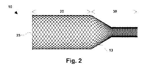
Fig. 2 illustrates the mesh included in the different tubular sections of the
proposed
device having a lower mesh density in the first section than in the second
section.
Fig. 3 schematically illustrates some of the main specifications of the
proposed device.
Fig. 4 is a graph showing Ideal pressure vs. diameter curve of the proposed
device.
CA 03116507 2021-04-14
WO 2020/079082 PCT/EP2019/078088
8
Fig. 5 is a flowchart illustrating a method for extracting a thrombus from a
thrombus site
in a blood vessel of a patient, according to an embodiment of the present
invention.
Fig. 6 is a diagram illustrating an automated thrombectomy apparatus according
to the
present invention.
Fig. 7 schematically illustrates the experimental setup model used in example
2 in
order to evaluate the force in the tip of the catheter.
Detailed Description of Particular Embodiments
Figs. 1 and 2 show particular embodiments of the proposed device for
extraction of
thrombus from a blood vessel. The proposed device includes a segment 10 which
is
self-expandable and defines a distal end 11 and a proximal end 12 and can
adapt its
shape to a surrounding blood vessel from a retracted position in a compressed
state,
for example inside a carrier such a delivery catheter (not shown), to an
extended/expanded position, once coming out of the carrier, to be appositioned
against
the inner wall of a blood vessel to receive and retain a thrombus.
As shown in Fig. 2, the segment 10 comprises a mesh 13 having two sets of
helicoidal
filaments turning respectively in opposite directions and being intertwined.
The mesh
13 in an embodiment can follow a diamond-type structure or a regular
structure. The
density of the mesh 13 defines the elasticity of the segment 10. As detailed
in Table 1
the mesh angle (or braiding angle ([3)) with regard to a longitudinal
direction can be
variable.
The helicoidal filaments can be made of a metal (including metal alloys),
polymers, a
composite including Nitinol or Nitinol/Platinum, or also DFTR (Drawn Filled
Tube),
among other materials having suitable mechanical properties.
As can be seen in the Fig. 1 and 2, the mesh 13 defines two distinct tubular
sections, a
first section 20 and a second section 30. Particularly, the second section 30
comprises
two sub sections, a first sub section 31 and a second sub section 32.
As can be seen in Fig. 2, in this particular embodiment, the end portion of
the first
section 20 at the distal end 11 comprises closed loops 23 facilitating the
expansion of
the segment 10 once it comes out of the cited carrier. Moreover, these closed
loops 23
act as a spring or fixing point by limiting the movement between the
helicoidal filaments
CA 03116507 2021-04-14
WO 2020/079082 PCT/EP2019/078088
9
and thus increasing the outward radial force. The closed loops 23 also provide
a
smooth distal end to reduce possible vessel damage and improve navigability of
the
device within the blood vessel. The rest of the first section 20 creates the
space which
will accommodate the thrombus once it has been aspirated. The first section 20
is
adaptable to the vessel geometry and, because of its configuration (e.g.,
diameter and
braiding angle), provides outward radial forces higher than in the second
section 30 so
that the segment 10 is better appositioned against the inner wall of the
vessel. The
radial forces in the end portions of the first section 20 are particularly
higher than in an
intermediate portion thereof, e.g., because of the spring action of closed
loops 23.
Alternatively, the radial forces in the first section 20 could be uniformly
distributed along
all its generatrix.
The first sub-section 31 (or portion of the second section 30 adjacent to the
first section
20) is cone-shaped or funnel-shaped. Because of its shape, this sub-section 31
has
features enabling it to withstand the blood pressure without collapsing. In
the illustrated
embodiment, the braiding angle (a) changes at the proximal and distal ends of
sub-
section 31 provide radial strength to maintain the conical shape. The braiding
angle (a)
change at the distal end of sub-section 31 also works with the closed loops 23
to
maintain first section 20 in an open position and create the space for the
thrombus. The
covering over sub-section 31 stops the blood flow during the capture and
removal of
the thrombus and protects the captured thrombus during the withdrawal of the
segment
10 to the carrier. This sub-section 31 is also the transition from the larger
diameter of
section 20 to the smaller diameter sub-section 32 for connection to an
aspiration
catheter or a hypotube (not shown).
The second sub-section 32 (or portion of the second section 30 adjacent to
proximal
end 12) has a tubular uniform diameter and provides the connection to the
aspiration
catheter or to the hypotube. In some embodiments, the aspiration catheter is a
PTFE-
lined braided catheter covered by an outer jacket. The aspiration catheter's
braid and
liner extend distally from the outer jacket. A layer of polymer material may
be placed
around the protruding braid and liner, and a mandrel may be placed within the
braid
and liner. Thereafter, the second sub-section 32 of segment 10 may be placed
over
this polymer section, and another layer of polymer may be placed over the mesh
of
subsection 32. This outer layer of polymer material is then melted so that
polymer flows
through the cells of the mesh 13, the mandrel is removed, and a smooth surface
is left
over the entire aspiration catheter. This attachment approach adds structure
and
CA 03116507 2021-04-14
WO 2020/079082 PCT/EP2019/078088
stiffness to the attachment section of the aspiration catheter, so it should
be as short as
possible without compromising the integrity of the attachment of segment 10 to
the
catheter.
Other techniques of connecting segment 10 to an aspiration catheter may be
used, as
5 understood by skilled artisans. For example, in other embodiments, if the
aspiration
catheter is a metal hypotube, the mesh 13 of the sub-section 32 is welded to a
Nitinol
ring. This ring is welded directly to the hypotube. Alternatively, a Stainless-
steel ring
can be glued to the mesh 13 of the sub-section 32. Then, the Stainless-steel
ring is
welded to the hypotube. Another option is to directly mesh the segment 10 over
a
10 perforated ring so that the filaments pass through the holes.
When the segment 10 is compressed inside the carrier, segment 10 elongates to
move
the helicoidal filaments toward a longitudinal alignment so as to reduce the
spring
effect and to facilitate the movement of segment 10 within the carrier by
reducing
friction effects and by increasing pushability. The pushability of the segment
inside the
carrier is related to the navigability of the segment 10 within the arteries.
The mesh angle (13) allows the mesh 13 to be adapted to a curve of the blood
vessel,
avoiding the kinking and creating a free space inside the mesh for
unobstructed
suction.
With reference now to Fig. 3 therein are illustrated some of the main
specifications of
the device according to an embodiment. Table 1 indicates the main
specifications of
the device. Table 2 indicates the measuring method used for calculating such
parameters.
CA 03116507 2021-04-14
WO 2020/079082 PCT/EP2019/078088
11
Table 1. Main specifications of the device
Example Range Big Ref. Small Ref.
OD sec 20 [mm] 6 3.5--6 5.2 Approx. 4.1
OD sec 32 [mm] catheter OD 1--2 1.65 1.65
Shape
L sec 20 [mm] 15 4--40 9 4-8
parameters
a sec 31 [2] 45 15--45 31 20
L sec 32 [mm] 2 1--10 3.5 3
Wire OD [pm] 50 40--60 51 51-58
Braiding Wire number 48 24--48 48 24-36
parameters 13 sec 20 [2] 60 50--65 55 65
13 sec 32 [2] 20 15--50 45 45
Table 1 shows the parameters for particular embodiments. In an embodiment, the
parameters of the device are such indicated in Table 1 for a big blood vessel
("Big
Ref.") of e.g. 4.5 mm diameter, such as the final part of the carotid or the
carotid
siphon. In another embodiment, the parameters of the device are such indicated
in
Table 1 for a small blood vessel ("Small Ref.") of e.g. 2.5 mm diameter, such
as the
Internal Carotid Artery (ICA) or the Middle Cerebral Artery (MCA).
Table 2. Measuring methods used for calculating the different parameters.
Parameter Measuring method
OD sec 20 [mm] The mandrel on which the proposed device is meshed is
measured. It is a solid piece with the same shape as the stent.
The final diameter is determined by measuring the diameter of the
solid piece and adding 4 times the diameter of the helicoidal
filaments/wires.
OD sec 32 [mm] Same as before
L sec 20 [mm] Same as before
a sec 31 [2] Same as before
L sec 32 [mm] Once the proposed device has been meshed, it is placed on a
tool
that determines where the excess length should be cut.
Wire OD [pm] It is measured with a precision measuring instrument.
Wire number Alternative 1: Counting the number of distal loops and
multiplying
by 2
Alternative 2: Counting the number of reels used for meshing
CA 03116507 2021-04-14
WO 2020/079082 PCT/EP2019/078088
12
13 sec 20 [2] Alternative 1: Measuring the number of wire crossings in a
given
length measured in the axial direction.
Alternative 2: If the mandrel is manufactured with grooves so that
during the meshing the wires are inserted inside and the
manufacturing is improved, it is simply measured that the mandrel
is manufactured with the appropriate parameters.
13 sec 32 [2] Same as before
As mentioned, the device may be in two configurations: in a retracted form (or
compressed state) inside the carrier while approaching the thrombus site, and
in an
extended and expanded (deployed) form when there is no interaction with the
carrier or
the blood vessel. The parameters specified herein relate to the device in its
natural
(relaxed) form; i.e. extended and expanded (deployed) position.
The segment 10 may include radiopaque markers made of platinum, tungsten,
barium
derivatives, gold, iridium, among others, at its distal end 11 and/or other
strategic
points within the mesh 13 which allow a physician to know the precise location
of the
device while using fluoroscopy. The radiopaque material can be deposited on
the
helicoidal filaments once manufactured (if the device has a coating, the
material may
also be dispersed on the surface of the coating). Alternative possibilities to
confer
radiopacity to the segment 10 are using helicoidal filaments of different
material and
opacity grade (e.g. Nitinol and Platinum). In a particular embodiment, Nitinol
wires with
a Platinum core are used. Likewise, the delivery catheter may also include
radiopaque
markers.
Moreover, the segment 10 may have a coating, for example covering the first
section
only or covering the whole segment 10. In the embodiments of Figs. 1 and 2,
although not seen, the coating goes from the closed loops 23 to sub-section
32. In one
embodiment, the coating is applied about attachment of segment 10 to an
aspiration
20 catheter by dipping segment 10 into a liquid polymer, the allowing the
polymer to
solidify. Optionally, a mandrel may be disposed inside the mesh 13 of segment
10
when it is dipped into the polymeric coating material. Alternatively, the
coating material
may be sprayed onto the mesh. In other alternative embodiments, the coating
may be
applied before attaching segment 10 to an aspiration catheter. In such
embodiments,
the coating does not reach the proximal end 12 of sub-section 32, but there is
an
uncoated space between the helicoidal filaments, leaving them free to allow
assembly
with the aspiration catheter.
CA 03116507 2021-04-14
WO 2020/079082 PCT/EP2019/078088
13
The coating prevents damage to the arteries, avoiding direct contact with the
helicoidal
filaments. Moreover, the coating provides a watertight compartment so that the
thrombus can be sucked in and protected during removal. In an embodiment, to
apply
the coating, the mesh 13 is attached to the carrier or delivery catheter and
then the
coating is applied.
An interior or exterior glaze can be also applied to the coating to improve
its properties.
By applying a hydrophilic or hydrophobic coating to the exterior surface of
the segment
10, the exterior surface can be more easily displaced into the carrier and
through the
blood vessel by reducing the coefficient of friction. In the same way, by
applying a
treatment in the interior surface of the segment 10 an adhesion effect that
retains the
thrombus once it is inside can be achieved.
The coating is made of an elastic material. In one particular embodiment, the
device
coating is silicone. Alternatively, polyurethanes or other types of plastic
materials can
be used. A blend of polyurethane and silicone may also be employed.
To achieve the double behavior of the coating (lubricious on the exterior
surface of
segment 10 and tacky or rough inside), the coating can be treated by the
addition of a
material as explained, or can have constitutively such features by the
structure of the
mesh itself.
The coating can include holes to avoid collapse of the segment 10. Such holes
may be
formed after the coating has been applied by perforating the coating.
The dimensions of segment 10 depend on the dimensions of the blood vessel in
which
it will be used to capture a thrombus. The dimensions of the sub-sections of
segment
10 and the braid angles of the mesh help segment 10 provide a reduced radially
outward force when compressed into the delivery catheter and sufficient
outward force
when expanded to avoid collapse from the blood pressure. Fig. 4 illustrates a
possible
work curve of one embodiment of the segment 10. Y-axis defines the device
pressure
(mmHg) whereas X-axis defines the diameter of the arteries (mm). The
horizontal
dotted line marks the blood pressure limit. In some embodiments, the diameter
range
of the arteries in which the device of this invention may be used is 2 to 5
mm. The
segment 10 is designed so that it can expand without being blocked by the
artery
working in a standard range of 2 to 5 mm and so that it can cope with a blood
pressure
greater than 200 mmHg. As shown by Fig. 4, this particular embodiment is not
CA 03116507 2021-04-14
WO 2020/079082 PCT/EP2019/078088
14
designed to be compressed to a diameter less than 2 mm. Compression of the
segment 10 within the delivery catheter may result in radially outward forces
high
enough to inhibit advancement of the device within the carrier.
With reference now to Fig. 5, therein it is illustrated an embodiment of a
method for
extracting a thrombus from a thrombus site in a blood vessel of a patient.
According to
this particular embodiment, the method at step 501 comprises advancing a
thrombus
extraction device through a delivery catheter in a delivery configuration to a
thrombus
site within a blood vessel. The thrombus extraction device comprising a mesh
13 of at
least first and second sets of oppositely wound and intertwined helicoidal
filaments
having a first section 20 at a distal end 11 and a second section 30 extending
proximally from a proximal end 12 of the first section 20, the first set of
helicoidal
filaments forming a distally facing first angle with the second set of
helicoidal filaments
in the delivery configuration. At step 502, the method comprises expanding the
first
section 20 of the thrombus extraction device with a first outward radial force
into a
deployment configuration in apposition with an inner wall of the blood vessel
proximate
the thrombus site, the first set of helicoidal filaments forming a second
angle with the
second set of helicoidal filaments in the delivery configuration, the second
angle being
greater than the first angle. Then, at step 503, the method comprises
expanding the
second section 30 of the thrombus extraction device into a conically shaped
deployment configuration with a second outward radial force less than the
first outward
radial force sufficient to stop proximal blood flow, a proximal end of the
second section
having a smaller diameter in the deployment configuration than a distal end of
the
second section 30. Finally, at step 504, the method comprises aspirating a
thrombus
into the thrombus extraction device.
25 Embodiments of the present invention also provide a thrombectomy apparatus
600 for
extraction of thrombus from a blood vessel including the proposed segment 10
of any
of the described embodiments.
Some embodiments of the invention may be automated for use in traditional
(hospital)
and non-traditional (nursing home, assisted care facility) environments which
may allow
30 for greater deployment and usage of the present invention and hasten the
removal of
thrombus, thus significantly improving patient outcomes, as flow may be
restored (e.g.,
to critical areas of the brain) within much shorter times. One such automated
device is
illustrated in W02016/113047.
CA 03116507 2021-04-14
WO 2020/079082 PCT/EP2019/078088
In use, segment 10 and the aspiration catheter or hypotube to which it is
attached are
advanced through a delivery catheter to a thrombus site within a blood vessel
of the
patient. During advancement in the delivery catheter, the segment 10 is in a
delivery
configuration in which the first and second sets of helicoidal filaments form
a first
5 distally facing angle with respect to each other. When segment 10 emerges
from the
delivery catheter, it begins to self-expand to a deployment configuration. In
embodiments in which the mesh forms closed loops at the distal end of segment
10,
the spring action of the closed loops of the helicoidal filaments helps the
first section 20
expand into apposition with the blood vessel proximate to the thrombus site.
In the
10 deployment configuration, the first and second sets of helicoidal filaments
form a
second distally facing angle less than the first angle (i.e., the filaments
are less
longitudinally aligned in the deployment configuration than they were in the
delivery
configuration). Sub-section 31 also self-expands to a conical or funnel shape.
The
distal end of sub-section 31 helps support the proximal end of section 20 in
its
15 deployment configuration.
The coating on the outside of sub-section 31 and section 20 reduce blood flow
to the
thrombus site. The optional holes through the coating permit a small amount of
blood to
pass through the device to avoid collapse of sub-section 31 caused by the
blood
pressure and also by the difference of pressure between the blood pressure
(externally) and the vacuum applied (internally). Once blood flow has been
reduced,
suction may be applied through the catheter or hypotube to the interior spaces
of sub-
section 31 and section 20 to aspirate the thrombus into section 20. Device 10
capturing
the thrombus may then be removed from the patient. In the capture
configuration (i.e.
when the thrombus is inside), the first and second sets of filaments form a
third distally-
facing angle less than the first distally-faced angle (i.e., the filaments
become more
longitudinally aligned) as the device assumes a longer and smaller diameter
shape.
Fig. 6 depicts an example of the thrombectomy apparatus 600 of the present
invention
which allows for the automated maneuvering of the thrombectomy apparatus 600
through a vascular system.
According to this particular example, an automated proximal device 601
provides a
guidance system to deploy the thrombectomy apparatus 600. Moreover, an imaging
device 602 can detect the radiopaque markers included in the segment 10, and
also in
the delivery catheter, and a communications channel 603 can be used to provide
CA 03116507 2021-04-14
WO 2020/079082 PCT/EP2019/078088
16
means to transport the image to a control module 604. The control module 604
is
programmed or configured to allow for guidance of the deployment of the
thrombectomy apparatus 600 and storage of data on a data storage device 605.
The
control module 604 may be a programmable logic controller, a computer, or the
like. In
this particular embodiment the control module 604 is guided by a computer
assisted
controller 606. The communications channel 603 can be Ethernet, WiFi,
Bluetooth, or
the like. The control module 604 is programmed to guide a physician or
technician
operating the thrombectomy apparatus 600 which allows for the thrombectomy
apparatus 600 to be used in non-hospital settings such as nursing homes or
assisted
care living facilities.
By allowing the thrombectomy apparatus 600 to be used "in the field" the time
required
to perform the thrombectomy is greatly reduced significantly improving patient
outcomes. The control may also be via a controller such as those in use in
other
current medical devices. In another embodiment, the system may be controlled
manually.
Following, two particular experimental examples of the thrombectomy apparatus
(also
referred as ANCD device) comprising the Advanced Neurovascular Aspiration
catheter
(also referred as ANA catheter) are detailed.
EXAMPLE 1: In vivo assay: Chronic Evaluation of Performance and Safety of the
ANCD Advanced Flow Restriction System in a Swine Model.
INTRODUCTION:
Endovascular treatment (EVT) is recognized as the most effective treatment for
large
vessel occlusion (LVO) strokes. Highest degree of recanalization in the
shortest time
with the minimum number of attempts have been demonstrated to correlate with
improved clinical outcomes. Although highly effective, failure to reach
complete
recanalization has been reported in about 20% of treated patients. In order to
improve
patient outcomes, different devices and combinations are under development to
increase the first pass complete recanalization rate. The development of such
devices
includes preclinical testing in phantom models simulating the cerebrovascular
human
anatomy, and animal models in which device related vessel injury can be
assessed.
Each simulation model has its own characteristics and therefore it is
recommended that
CA 03116507 2021-04-14
WO 2020/079082 PCT/EP2019/078088
17
any new device or combination will prove its efficacy and safety in different
conditions
before final evaluation in a first in human study.
The Advanced Thrombectomy System (ANCD) is a novel stroke thrombectomy
apparatus that includes the proposed device for extraction of thrombus from a
blood
vessel, a self-expanding radiopaque braid covered by a continuous polymeric
coating,
designed to reduce clot fragmentation and facilitate retrieval by inducing
local flow
restriction and allowing distal aspiration.
The aim of this study was to evaluate the pre-clinical efficacy and safety of
the ANCD,
in a swine model 3 and 30 days following 3 passes, and specifically confirm
that the
use of the novel self-expanding funnel is unrelated to higher vascular injury
in
comparison with a commonly used device, the FlowGateTM Balloon Guide Catheter.
The acute performance and safety on day 0 (performance-usability endpoints,
devices
integrity and angiography) and the safety data (angiography, histology and
health
monitoring) after 3 and 30 days, respectively, were studied.
METHODS:
Description of the ANCD device
The ANCD is a thrombectomy apparatus that is comprised of two coaxial
catheters: a
funnel catheter and a delivery catheter.
The funnel catheter is comprised of highly flexible polymers onto a braided
metallic
structure. It is intended to restrict locally the blood flow during the
intervention. It is
composed of a self-expanding funnel that, when unsheathed, can expand to the
diameter of the blood vessel, adapting to its shape, thereby restricting the
blood flow.
The funnel catheter can provide an effective aspiration that serves as a
complementary
mechanism when combined with retrieval devices. The funnel is designed to have
enough flexibility to adapt to the neurovascular tortuosity. The funnel is
comprised of a
radiopaque braid and a polymeric film.
The delivery catheter is the outermost catheter of the device, which navigates
until
reaching the target vessel. It has a hydrophilic coating to reduce friction
during use and
a radiopaque marker on the distal end for angiographic visualization. The
materials of
CA 03116507 2021-04-14
WO 2020/079082 PCT/EP2019/078088
18
the catheter allow enhanced flexibility in the tip and sufficient stiffness
and pushability
of the proximal portion.
FlowGate TM Balloon Guide Catheter is a common commercial device which offers
proximal flow control and a stable platform to facilitate the insertion and
guidance of an
intravascular catheter. It provides a balance of trackability and support with
a large
lumen and is indicated for use as a conduit for retrieval devices.
Animal Model
All animals were held in quarantine and housed at CBSET (Lexington, MA, USA),
where the study was conducted, a facility accredited by the American
Association for
Accreditation of Laboratory Animal Care, under conditions that met or exceeded
requirements as set forth in the USDA guidelines. Standard veterinary
practices were
performed during quarantine, including physical examinations and clinical
pathology to
determine health status before assignment to the study. A nutritionally
balanced diet
appropriate for the species was offered daily to all animals with water ad
libitum.
Eleven pigs were used in this study (female or castrated male Yorkshire pigs,
weight
39-50 Kg). The swine model was chosen as the experimental species for this
study
because the size and anatomy of the vascular system is clinically relevant for
the
purpose of testing catheter-based medical devices for the treatment of
vascular
disease. Also, swine is an established animal model for vascular studies and
generally
accepted as a scientific standard.
Animals were anesthetized, intubated, and IV catheterized for the
administration of
supportive IV fluids and medications. The surgical procedures were performed
under
aseptic conditions. Physiological parameters were monitored through all the
procedures. The femoral artery was accessed via cutdown approach. A 9 F
introducer
sheath was advanced into the artery and heparin (150 U/kg, IV) was
administered to
prolong Activated Clotting Time (ACT) to approximately 200-350 seconds. ACT
levels
were monitored every 45 minutes during all the procedures, and additional
heparin was
administered as needed to maintain the target ACT. Under fluoroscopic
guidance, an
8F Mach 1 TM guide catheter (CGC: Boston Scientific, Marlborough, MA) was
advanced
through the sheath over a guide wire into the descending aorta and to the
target
arteries. Angiographic images of the vessels were obtained with contrast media
to
identify a suitable location for the treatment site. Angiograms were performed
CA 03116507 2021-04-14
WO 2020/079082 PCT/EP2019/078088
19
throughout the procedure: baseline, after each pass, and prior to necropsy.
The
parameters assessed by angiography (qualitative and quantitative) were: vessel
anatomy, target site, device monitoring, vessel status-injury, vasospasm, and
blood
flow (mTICI scale).
The two devices were used per Instructions for Use (IFU) for the interventions
of the
target vessels:
1- BGC: Balloon Guide Catheter (BGC: 8Fr FlowGate2TM Balloon Guide Catheter
(95
cm); from Stryker Neurovascular, Fremont, CA), and
2- ANCD: ANCD (Anaconda Biomed) through the guide catheter CGC.
Renal, cervical and lingual arteries were targeted. These arteries cover the
diameter
range between 2.2 and 5 mm for ANCD, and 2.7 to 5 mm for the BGC, which
represents the size of the target vessels in the cerebrovasculature (internal
carotid
artery (ICA), middle cerebral artery (MCA)).
ANCD and BGC devices were distributed among target vessels to ensure
assessment
was made in all vascular beds at each time point. Randomization of animals was
not
required for this study as each animal had both ANCD and BGC devices
evaluations.
In order to study the devices in a clinical simulation as a worst case
scenario, three
passes in every study group were assessed in all cases. The potential vascular
injury
caused by the devices (perforation, dissection, thrombosis) and vasospasm was
also
assessed during the procedure by angiography.
Thrombectomy procedures
Intravascular devices were maneuvered under fluoroscopic guidance and
angiographic
images of the vessels were obtained to identify the proper location of the
device.
In intervention 1, the BGC was inflated to arrest flow, as per IFU and usual
practice,
aspiration was applied through the BGC. In intervention 2, the ANCD catheter
system
was advanced close to the target vessel site and the funnel deployed creating
local
flow arrest. In all interventions, aspiration during the thrombectomy
procedure was
performed with a 60 cc syringe (Vaclock; Merit Medical) connected to a three-
way
CA 03116507 2021-04-14
WO 2020/079082 PCT/EP2019/078088
stopcock through either the BGC (intervention 1), or the ANCD funnel catheter
(intervention 2).
The resulting study design is summarized in the following Table 3:
Test/Control Number of Time
Number of Vessels/Treatment Scheme
Device Animals
Point
Renal arteries n=5
ANCD n=8
Cervical or lingual arteries n=3 5 Day
3
FlowGate BGC Renal arteries n=5 n=5
Renal arteries n=6
ANCD n=10 Day
Cervical or lingual arteries n=4 6
2
FlowGate BGC Renal arteries n=6 n=6
5 Table 3. Study design: Testing devices (ANCD and FlowGate BGC), number and
location of vessels, number of animals involved and time point assessments.
Acute performance evaluations on the day of procedure included user interface
and
ability to maneuver the device. The potential vascular injury caused by the
devices was
10 also assessed during the procedure by angiography.
Histopathology
Animals were euthanatized after 3 and 30 days and underwent a comprehensive
necropsy. Treated vessels were dissected and relevant tissues/organs were
collected,
fixed in 10% NBF (Neutral Buffered Formalin) and paraffin embedded and stained
with
15 H&E (hematoxylin and eosin) and Verhoeff's for histomorphologic assessment.
Each
treated vessel was trimmed to yield at least six cross-sections (2 proximal, 2
mid and 2
distal) within the putative area of treatment. For lingual treatments, the
treated vessel
sections were taken from the breadloafed tongue sections and may include
surrounding parenchyma. Additionally, untreated distal sections of the vessel
were
20 obtained within approximately 5 mm of the distal end of the putative
treated area.
Light microscopy was used to determine histomorphological scoring of
parameters that
reflected the degree and extent of the host response/repair process to the
treatment in
target vessels. Histomorphometric markers included: vascular injury, vascular
mural
CA 03116507 2021-04-14
WO 2020/079082 PCT/EP2019/078088
21
compression lesion, inflammation, endothelization, luminal fibrin/thrombus
deposition,
neointima formation, and adventitial fibrosis. Histologic sections of vessels
were also
examined for other microscopic changes including hemorrhage, necrosis, and
type and
relative amounts of inflammatory cell infiltrates. Sections of representative
downstream
tissues were evaluated for any adverse effects associated with treatment,
including
thrombosis, necrosis, inflammation and presence of embolic material. Scoring
values
were calculated for every section and level and reported as an overall mean of
each
vessel, ranking from 0 (no injury) to 3 (highest possible degree of injury) in
all markers
except for endothelization than ranked from 0 (absence of endothelial
covering) to 4
(complete endothelial covering). The pathologist was blinded to the treatment
matrix at
the time of the pathologist read.
RESULTS:
The ANCD navigability and pushability through vessel curvatures was better
compared
with the FlowGate BGC.
The funnel of the ANCD device performed adequately in most cases, with
complete
deployment, accuracy and adaptability to vessel curvatures, and significant
reduction of
the antegrade blood flow.
The ANCD device avoided kinking during the navigability and deployment on the
target
vessel.
The ANCD device was able to complete the 3 passes with correct performance and
maintained its general integrity. Radiopacity, withdrawal of the catheter and
hemostasis
were adequate.
During interventions, the devices could potentially generate clots in situ due
to the
interaction with blood. After the interventions of the present study, no
thrombus was
observed on the catheters or funnels surfaces after removal from the body,
supporting
the adequate thromboresistance of both devices.
No vessel perforation, dissections, or occlusions were observed. Vasospasm was
a
common observation to varying degrees for both test and control devices. This
is a
common observation in the swine model as pigs are prone to vasospasm.
CA 03116507 2021-04-14
WO 2020/079082 PCT/EP2019/078088
22
Histomorphologic markers of vascular injury after 3 and 30 days, respectively,
were
absent to minimal across all groups, and generally improved over time.
Downstream
tissues showed sparse and minimal evidence of embolic material and was not
correlated with necrosis or other evidence of circulatory compromise in the
target
organs. Findings of embolic foreign material was minimal and was similar
across
treatment groups, indicating a shared origin, and was most consistent with
lubricious
coating used on various ancillary products. Other findings, like inflammation,
thrombosis and necrosis were absent to minimal in all groups and time points.
All animals survived to their scheduled time point. Clinical observation of
the animals,
body weight evolution, blood analysis and necropsy did not reveal signs of
clinical
abnormalities related to the ANCD or FlowGate devices for any study animal.
CONCLUSIONS:
= The ANCD device showed an excellent performance, with a complete
deployment
and accuracy, in intravascular interventions, similar to the control device
FlowGate
BGC.
= The ANCD device performed better than the control device, in terms of
navigability,
pushability and adaptability in vessel curvatures. Also, showed a significant
reduction of the antegrade blood flow.
= The ANCD device groups showed minimal vessel injury and similar to
FlowGate
BGC groups, and it improved over time.
= Downstream tissues to treated vessels showed minimal or absence of
lesions,
similar in the ANCD device and FlowGate BGC groups.
= The ANCD device showed a favorable safety profile based on the absence of
systemic and local adverse effects in treated animals.
EXAMPLE 2: Suction force aspiration test of the Advanced Neurovascular
Aspiration
(ANA) catheter
INTRODUCTION:
The endovascular treatment of ischemic stroke involves a wide range of devices
and
techniques. The aspiration thrombectomy technique is intended to restore blood
flow in
patients experiencing acute ischemic stroke due to vessel occlusion by
applying a
suction force to remove the clot. This research is aiming to analyse the
suction-forces
CA 03116507 2021-04-14
WO 2020/079082 PCT/EP2019/078088
23
over the clot of two different catheter designs using experimental analysis.
To study the
suction tip-force, the clot was considered as rigid. For that purpose, the
force
generated by the tip of the ACE68 Reperfusion Catheter (Penumbra Inc,
hereinafter
referred as ACE), and the ANA Advanced Neurovascular Aspiration catheter
(Anaconda Biomed, hereinafter referred as ANA) were evaluated using a tensile
tester
machine and special designed tools that completely cover the tip of the device
in order
to simulate a complete occlusion of the clot.
ANA is a funnel catheter comprised of highly flexible polymers onto a braided
metallic
structure. It is intended to restrict locally the blood flow during the
intervention. It is
composed of a self-expanding funnel that, when unsheathed, can expand to the
diameter of the blood vessel, adapting to its shape, thereby restricting the
blood flow.
The funnel catheter can provide an effective aspiration and it can be used as
a
complementary mechanism when combined with retrieval devices. The funnel is
designed to have enough flexibility to adapt to the neurovascular tortuosity.
The funnel
is comprised of a radiopaque braid and a polymeric film.
ACE is a reperfusion catheter comprised of a coil-winding geometry along 16
transitions. It is intended to create an optimal tracking profile to
facilitate clot extraction
from proximal large vessels with the vacuum power of the Penumbra Pump MAXTM
associated to the catheter.
METHODS:
The test was performed with the purpose of analysing thrombus suction-forces
and
stresses induced by different aspiration devices for the treatment of ischemic
stroke. In
order to evaluate the force in the tip of the catheter the experimental model
shown in
Fig. 6 was defined.
The INSTRON-E0152 tensile tester with 10N load cell was used. A specially
designed
tool was connected to the load cell, tool that was capable of occluding the
inner tip
diameter of the two catheters (units) selected for this study (ANA and ACE).
Once the
tip covered the tool, a negative pressure of 500 mmHg was applied using a
VacMaxi
Pump (APEX) intended for suction catheters. Then a constant speed of 50 mm/min
was applied and the force necessary for the separation of the tool was
evaluated.
CA 03116507 2021-04-14
WO 2020/079082 PCT/EP2019/078088
24
Regarding the design selected for the tooling, it was intended to reduce the
friction
forces produced by the collapse of the tip during the aspiration. This
phenomenon
could have a higher impact in the ANA catheter due to the flexible braided
structured
design of the tip, that could lead to a closer contact with the wall of the
tool. The
authors considered that this fact could have a potential benefit in the
clinical practice
creating a trap for the clot when it is inside the catheter tip.
RESULTS AND CONCLUSIONS:
The results obtained in the test are presented in the following Table.
Device Force (N) Standard deviation Variance coefficient
(%)
ACE 0.11 N 0.04 39%
ANA 0.42 N 0.06 14 %
Table 4. Experimental results
Based on the results it can be stated that the force experimented in the tip
when a rigid
surface is completely occluding the inner diameter, is more than three times
higher in
the ANA than in the ACE catheter. This study represents an approach clinically
relevant of what happens in a scenario in which a hard thrombus (rigid
material) is
completely occluding the tip of the catheter. The features of ANA funnel,
mainly its
bigger and more flexible tip, create an efficient suction trap avoiding the
separation of
the clot from the catheter at higher forces than the ACE device.
Although illustrated and described above with reference to certain specific
embodiments, the present invention is however not intended to be limited to
the details
shown. Rather, various modifications may be made in the details within the
scope and
range of equivalents of the claims.
The scope of the present invention is defined in the following set of claims.