Note: Descriptions are shown in the official language in which they were submitted.
CA 03118296 2021-04-29
WO 2020/092835 PCT/US2019/059255
METHODS AND KITS FOR DETECTING CELLS USING OLIGONUCLEOTIDE
CONJUGATED ANTIBODIES
CROSS-REFERENCE
[0001] This application claims priority to U.S. Provisional Patent Application
No. 62/753,854,
filed October 31, 2018, which is entirely incorporated herein by reference.
BACKGROUND
[0002] Biomarker measurements enable the detection of a variety of biological
states. For some
assays, bulk measurements of a single parameter are sufficient to assess the
disease state of a
given sample; however, these measurements obscure the single cell resolution
data and can
obfuscate the underlying heterogeneity of a biologically relevant specimen.
SUMMARY
[0003] Provided herein are methods comprising: contacting a sample comprising
a plurality of
biological features of interest with a plurality of capture agents, wherein
each capture agent is
capable of binding to a different biological feature of interest, wherein each
capture agent is
conjugated to a different oligonucleotide; fixing the capture agents bound to
biological features
of interest to the sample; contacting each oligonucleotide with a circular
nucleic acid primer,
wherein a segment of the nucleic acid primer is complimentary to the
oligonucleotide, and
wherein each oligonucleotide is contacted with a different nucleic acid
primer; amplifying the
oligonucleotides using the circular nucleic acid primers as a template to
yield amplified
oligonucleotides; contacting each of a subset of the oligonucleotides with a
probe comprising a
label to form a probe-amplified oligonucleotide duplex, wherein each probe can
bind to only one
oligonucleotide; reading the sample to determine the binding pattern for each
of the probes,
inactivating or removing the labels, and repeating the contacting and reading
steps with different
probes that bind to a different subset of oligonucleotides.
[0004] In some embodiments, the sample is a biological sample. In some
embodiments, the
sample is selected from the group consisting of a fresh sample, a frozen
sample, and a chemically
fixed sample. In some embodiments, the sample is a FFPE tissue sample. In some
embodiments,
the sample comprises a cell. In some embodiments, the sample is selected from
the group
consisting of a biological tissue, a biological fluid, and a homogenate. In
some embodiments, the
sample comprises cells. In some embodiments, the cells comprise a rare cell
population. In some
embodiments, the cells comprise cancer cells. In some embodiments, the cell is
selected from the
group consisting of an animal cell, a plant cell, a bacterium, a fungal cell,
or a protist.
1
CA 03118296 2021-04-29
WO 2020/092835 PCT/US2019/059255
[0005] In some embodiments, the sample a human sample or a mouse sample. In
some
embodiments, the sample comprises a pathogen. In some embodiments, the
pathogen is selected
from the group consisting of a bacterial cell, a yeast cell, a bacterial cell,
a virus, a viral vector,
or a prion. In some embodiments, the sample comprises a tumor tissue. In some
embodiments,
the sample comprises healthy tissue. In some embodiments, the sample is
adhered to a slide. In
some embodiments, the biological features comprise proteins. In some
embodiments, the
biological features comprise markers. In some embodiments, at least one of the
markers is a low
level marker. In some embodiments, the biological features comprise a disease
marker. In some
embodiments, the biological features comprise a diagnostic marker. In some
embodiments, the
markers comprise a molecule selected from the group consisting of a
transcription factor, a
signaling molecule, a diffuse extracellular marker, or a cell surface marker.
In some
embodiments, the biological features comprise a mutated protein.
[0006] In some embodiments, the capture agents comprise an antibody. In some
embodiments,
the capture agents comprise an antibody fragment. In some embodiments, the
antibody fragment
is selected from the group consisting of an IgG, an IgM, a polyclonal
antibody, a monoclonal
antibody, a scFv, a nanobody, a Fab, or a diabody
[0007] In some embodiments, each different oligonucleotide is at least 10
nucleotides long. In
some embodiments, each different oligonucleotide is at least 25 nucleotides
long. In some
embodiments, each different oligonucleotide is no more than 100 nucleotides
long.
[0008] In some embodiments, the fixing comprises crosslinking. In some
embodiments, the
crosslinking comprises using formaldehyde.
[0009] In some embodiments, the circular nucleic acid primer is between 6
nucleotides long and
100 nucleotides long. In some embodiments, the segment of the nucleic acid
primer that is
complimentary to the oligonucleotide is between 16 nucleotides long and 18
nucleotides long.
[0010] In some embodiments, the amplifying is performed using a polymerase. In
some
embodiments, the polymerase is Phi29 polymerase. In some embodiments, the
amplifying step
lasts for about 1 hour. In some embodiments, the amplifying step is performed
at about 37 C.
[0011] In some embodiments, each probe comprises a different label than each
other probe. In
some embodiments, the probe-amplified oligonucleotide duplex can have a T. of
at least 15 C.
In some embodiments, the label can be a fluorescent label. In some
embodiments, the fluorescent
label can be selected from the group consisting of Cy3, Cy5, Alexafluor555,
Alexafluor647,
Alexafluor750, POPO-3, TOTO-3, POPRO3, and TOPRO3. In some embodiments, the
fluorescent label can be attached to the probe by a linker.
[0012] In some embodiments, reading the sample comprises fluorescent imaging.
2
CA 03118296 2021-04-29
WO 2020/092835 PCT/US2019/059255
[0013] Provided in this disclosure are kits comprising a plurality of
antibodies, each conjugated
to a unique oligonucleotide; a plurality of primers, each specific to one of
the unique
oligonucleotides; and a plurality of dyes, each specific to one of the unique
oligonucleotides.
[0014] Another aspect of the present disclosure provides a non-transitory
computer readable
medium comprising machine executable code that, upon execution by one or more
computer
processors, implements any of the methods above or elsewhere herein.
[0015] Another aspect of the present disclosure provides a system comprising
one or more
computer processors and computer memory coupled thereto. The computer memory
comprises
machine executable code that, upon execution by the one or more computer
processors,
implements any of the methods above or elsewhere herein.
[0016] Additional aspects and advantages of the present disclosure will become
readily apparent
to those skilled in this art from the following detailed description, wherein
only illustrative
embodiments of the present disclosure are shown and described. As will be
realized, the present
disclosure is capable of other and different embodiments, and its several
details are capable of
modifications in various obvious respects, all without departing from the
disclosure.
Accordingly, the drawings and description are to be regarded as illustrative
in nature, and not as
restrictive.
INCORPORATION BY REFERENCE
[0017] All publications, patents, and patent applications mentioned in this
specification are
herein incorporated by reference to the same extent as if each individual
publication, patent, or
patent application was specifically and individually indicated to be
incorporated by reference.
BRIEF DESCRIPTION OF THE DRAWINGS
[0018] The novel features of the are set forth with particularity in the
appended claims. A better
understanding of the features and advantages of the present invention will be
obtained by
reference to the following detailed description that sets forth illustrative
embodiments, in which
the principles of the invention are utilized, and the accompanying drawings of
which:
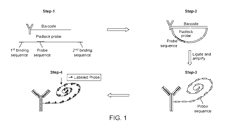
[0019] FIG. 1 illustrates a rolling circle amplification scheme wherein signal
detection is
performed using an extra reporter sequence included in the backbone padlock
probe/circular
nucleic acid primer, in accordance with some embodiments.
[0020] FIG. 2 illustrates a rolling circle amplification scheme wherein signal
detection is
performed using the same segment of the padlock probe that recognizes the
oligonucleotide
("barcode sequence"), in accordance with some embodiments.
3
CA 03118296 2021-04-29
WO 2020/092835 PCT/US2019/059255
[0021] FIG. 3 illustrates a computer system that is programmed or otherwise
configured to
perform or control methods described herein.
[0022] FIG. 4 illustrates human fresh frozen tonsil tissue stained with
oligonucleotide linked
antibodies amplified via RCA and probed with fluorescently labeled probes, in
accordance with
some embodiments. Panel A shows CD45-BX001 (exposure time-50m5) and CD4 -BX021
(exposure time 20m5). Panel B shows a zoomed in portion of panel A. Panel C
shows CD2-
BX002 (exposure time 20m5). Panel D shows a zoomed in portion of panel C.
[0023] FIG. 5 illustrates human fresh frozen paraffin embedded tonsil tissue
stained with
antibodies linked to oligonucleotides, in accordance with some embodiments.
Panels A and B
show CD31-CX001 (exposure time-50m5) and CD3 -CX002 (exposure time 20m5) and.
Panel C
depicts the tissue sample after removal of the labeled probes via de-
hybridization. Panels D and
E present zoomed-in regions of panels A and B, respectively.
[0024] FIG. 6 illustrates example data collected using a 24 marker antibody
panel to stain human
tonsil, in accordance with some embodiments.
DETAILED DESCRIPTION
Overview
[0025] Provided herein are methods, systems, and kits for detecting elements
of a sample, which
can be applied to measure high parameter data with spatial context. Such
measurement can
provide mechanistic understanding of key disease states or therapeutic
modalities. In some cases,
such measurement can enable development of enhanced diagnostic tools.
[0026] A plurality of biological features of interest of a sample can be
detected by employing a
single capture molecule staining step in combination with iterative cycles of
applying a label,
imaging, and removing the label. In some cases, a capture agent can be fixed
to the sample. An
amplification step, such as rolling circle amplification (RCA), can be
employed between the
capture molecule staining step and the iterative cycles, for example to
provide an amplification
of signal.
[0027] Binding capture agents to a sample can allow for the ultimate detection
of elements of the
sample, e.g., biological features of interest. The capture molecule staining
step can comprise
contacting a sample comprising a plurality of biological features of interest
with a plurality of
capture agents, such that each capture agent can be capable of binding a
different biological
feature of interest. In some cases, each capture agent can be conjugated to a
different
oligonucleotide.
[0028] A fixation step can follow the binding of the capture agents, such that
the capture agents
can be fixed to the sample. Such a fixation step can allow for the following
amplification step to
4
CA 03118296 2021-04-29
WO 2020/092835 PCT/US2019/059255
be performed on the tissue surface. In some cases, such a fixation step can
allow for reliable
multiplexing and/or iterative labeling and imaging steps after amplification.
[0029] Oligonucleotides can each be contacted with a circular nucleic acid
primer in preparation
for an amplification step. In some cases, the circular nucleic acid primer can
be non-circular
prior to contacting the oligonucleotide, and be circularized once in contact
with the
oligonucleotide. In some cases, a non-circular nucleic acid can be
circularized via ligation once it
contacts the oligonucleotide. For example, in some cases, a circular nucleic
acid primer can be a
template for an RCA reaction. In some cases, a non-circular nucleic acid
primer can be a
template for RCA after it is circularized to become a circular nucleic acid
primer via ligation.
Such primers can comprise a segment complimentary to a segment of an
oligonucleotide bound
to a capture agent. In some cases, such primers can comprise a probe segment.
When a probe
segment is copied during an amplification step, the copy of the probe segment
can be
complimentary to a nucleic acid probe. In some cases, the probe segment can
have the same
sequence as a nucleic acid probe. In some cases, each oligonucleotide can be
contacted with a
different circular nucleic acid primer.
[0030] After applying circular nucleic acids, an RCA reaction can be performed
to amplify the
sample. The RCA reaction can be performed on the surface of the sample, after
capture agents
are bound, and in some cases after a crosslinking or fixation step. RCA can
provide enhanced
sensitivity compared with a similar method performed without such
amplification, such as an
immunofluorescence method or an immunohistochemistry method. RCA can comprise
for
example isothermal amplification of circular nucleic acid probes bound to the
oligonucleotides,
such that the circular nucleic acid probes act as primers for the RCA
reaction. In some cases,
such as when the circular nucleic acid primer has a probe segment, the RCA
reaction can result
in the creation of multiple binding sites for labeled probes.
[0031] After an RCA reaction, a subset of amplified oligonucleotides can be
contacted with a
probe comprising a label. These probes can be nucleic acid sequences that can
bind to copies of
the probe segment created in each RCA reaction. In some cases, these probes
can be nucleic acid
sequences that can be complimentary to the copies of the probe segment created
in each RCA
reaction. In some cases, each probe can bind to one of the amplified
oligonucleotides in the
subset. In some cases, a different probe sequence is used for each different
amplified
oligonucleotide in the subset. In some cases, each amplified oligonucleotide
in the subset can
bind to one probe sequence. In this way, each biological feature of interest
associated with an
oligonucleotide in the subset of amplified oligonucleotides can be associated
with a different
probe. This can allow for detection of each biological feature of interest.
CA 03118296 2021-04-29
WO 2020/092835 PCT/US2019/059255
[0032] A sample can be read after hybridizing the probes to determine the
binding pattern for
each of the probes. This reading can indicate spatial information about the
biological feature of
interest associated with each of the probes via capture agents. In some cases,
reading a sample
can comprise detecting a label on one or more of the probes. Reading can be
accomplished using
any acceptable method appropriate for the detection of the label. For example,
if a label is a dye
label or a fluorescent label, this can be accomplished by imaging the sample
with a white light or
fluorescent microscope, respectively. As another example, if a label is an
enzyme label, this can
be accomplished by providing the enzyme with a substrate, allowing a reaction
to occur, and
detecting a product of the reaction.
[0033] After reading a sample, a label can be inactivated or removed. In some
cases, a label can
be removed from a probe, while a probe remains on the amplified
oligonucleotide. In some
cases, a probe with its label can be removed from an amplified
oligonucleotide. In some cases, a
label can be inactivated, such that a signal can be no longer detected from
the probe associated
with the label. An inactivation or removal step can allow subsequent rounds of
reading the
sample to determine the binding pattern for a different set of probes.
[0034] Contacting and reading can be repeated with a different set of labeled
probes that can
bind to a different subset of oligonucleotides. In some cases, a subset of
amplified
oligonucleotides comprising amplified oligonucleotides not previously probed
can be each
contacted with a probe comprising a label. In some cases, each amplified
oligonucleotide in the
new subset can bind a different probe than other amplified oligonucleotides in
the new subset. In
some cases, each probe can bind to only one amplified oligonucleotide in the
new subset.
[0035] A reading step can be performed to read the new set of labels to
determine the binding
pattern for the new set of probes. Thus, reading can indicate spatial
information about different
biological features of interest than in the first iteration. In some cases,
the removal or inactivation
of labels, contacting, and reading steps can be repeated. In some cases, the
repeating can
continue until a predetermined number of or all biological features of
interest are detected.
[0036] Methods provided herein can provide amplification strategies that can
provide increased
sensitivity compared with other assays, such as immunohistochemistry or
immunofluorescence.
These methods can allow multiplexing of an assay, and can enable in some cases
the detection of
low-level markers or markers for which available antibodies can be relatively
weak.
[0037] In some methods herein, amplification, the RCA reaction can allow for
amplification of
signal which can increase the stoichiometry of detection molecules relative to
each antibody
molecule. This stoichiometry can be increased by at least 5 times, at least 15
times, at least 20
times, at least 25 times, at least 30 times, at least 40 times, at least 50
times, at least 100 times, at
6
CA 03118296 2021-04-29
WO 2020/092835 PCT/US2019/059255
least 500 times, at least 1000 times, at least 5000 times, or at least 10000
times compared with
other assays.
[0038] In some cases, the methods provided herein can allow detection of
markers that may be
below the detection limit for other detection assays, including some other
assays which do not
possess an amplification step, as well as some other assays which do possess
an amplification
step.
[0039] An example of such a method is illustrated in 4 steps in FIG. 1. In the
first step, an
antibody linked to an oligonucleotide can be bound to a sample (sample not
shown). This
oligonucleotide can comprise a "barcode," or a region capable of binding a
circular nucleic acid
primer. In this illustration, the entire oligonucleotide can serve as the
barcode. In other examples,
a portion of the oligonucleotide that can be less than the entire
oligonucleotide can serve as the
barcode. A circular nucleic acid primer ("padlock probe") can be designed such
that one end can
be the reverse complement to one portion of the barcode (a first binding
sequence) and the other
end can be the complement to the other portion of the barcode (a second
binding sequence).
Between these two ends can be a region having a probe sequence. In the second
step, the circular
nucleic acid primer can hybridize to the oligonucleotide via the first and
second binding
sequences, such that the circular nucleic acid primer takes on a circular
shape. The ends of the
circular nucleic acid primer can be ligated. In the third step, RCA can be
performed, extending
the oligonucleotide, resulting in an amplified oligonucleotide which can
comprise a string of
nucleic acids that can be complementary to the padlock probe in a repeating
fashion. Notably,
the probe sequence can be repeated in this amplified oligonucleotide a
plurality of times. In step
4, the amplified oligonucleotide can be incubated with labeled probes that can
each comprise a
nucleic acid sequence that can be complementary to the probe sequence. Such
probes can be
linked to a detectable label. The labeled probes can hybridize to the probe
sequences on the
amplified oligonucleotide, and can be detected, e.g., by imaging. In some
cases, this type of
method can be called a type 1 RCA method.
[0040] Another example of such a method is illustrated in 4 steps in FIG. 2.
In the first step, an
antibody linked to an oligonucleotide can be bound to a sample (sample not
shown). This
oligonucleotide can comprise a "barcode," or a region that can be capable of
binding a circular
nucleic acid primer. In this illustration, the entire oligonucleotide can
serve as the barcode. In
other examples, a portion of the oligonucleotide that can be less than the
entire oligonucleotide
can serve as the barcode. A circular nucleic acid primer ("padlock probe") can
be designed such
that one end can be the reverse complement to one portion of the barcode (a
first binding
sequence) and the other end can be the complement to the other portion of the
barcode (a second
7
CA 03118296 2021-04-29
WO 2020/092835 PCT/US2019/059255
binding sequence). These two reporter sequences, when the oligonucleotide is
ligated in the next
step to a circular shape, can make up a probe sequence. In the second step,
the circular nucleic
acid primer can hybridize to the oligonucleotide via the first and second
binding sequences such
that the circular nucleic acid can take on a circular shape. The ends of the
circular nucleic acid
primer can be ligated. In the third step, RCA can be performed, extending the
oligonucleotide,
resulting in an amplified oligonucleotide which can comprise a string of
nucleic acids that can be
complementary to the padlock probe in a repeating fashion. Notably, the probe
sequence can be
repeated in this amplified oligonucleotide a plurality of times. In step 4,
the amplified
oligonucleotide can be incubated with labeled probes that can each comprise a
nucleic acid
sequence that can be complementary to the probe sequence. Such probes can be
linked to a
detectable label. The labeled probes can hybridize to the amplified
oligonucleotide, and can be
detected, e.g., by imaging. In some cases, this type of method can be called a
type 2 RCA
method.
Samples
[0041] A sample can be a biological sample. A sample can be fresh, frozen, or
fixed (e.g.,
chemically fixed). A sample can be of animal, plant, bacteria, fungus, or
protist origin. In some
cases, a sample can be that of a human, mouse, rat, cow, pig, sheep, monkey,
rabbit, fruit fly,
frog, nematode or woodchuck. A sample can comprise cells (e.g., isolated
cells, immortalized
cells, primary cells, cultured cells, or cells of a tissue or organism),
biological tissue, biological
fluid, a homogenate, or it can be an unknown sample. In some cases, a sample
can comprise a
pathogen. The pathogen can be cultured or uncultured. A pathogen can be an
infection of a
sample. In some cases, a pathogen can be an infection of a cell, fluid,
tissue, organ, or
microbiome of an organism a sample is collected from. In some cases, a sample
can comprise a
pathogen which is a yeast cell, a bacterial cell, a virus, a viral vector or a
prion.
[0042] A sample can be a tissue section. In some cases, tissue section can
refer to a piece of
tissue that has been obtained from a subject, optionally fixed, sectioned, and
mounted on a planar
surface, e.g., a microscope slide.
[0043] A sample can be a planar sample. In some cases, a sample can be
immobilized on a
surface. In some cases, the surface can be a slide, a plate, a well, a tube, a
membrane, a film, or a
bead. In some cases, a sample can be contacting a slide. A sample contacting a
slide can be
attached to the slide such that the sample is effectively immobilized. This
can be accomplished
for example by fixation or by freezing the sample. Likewise, a sample can be
immobilized on
another type of surface using a same or similar attachment technique.
8
CA 03118296 2021-04-29
WO 2020/092835 PCT/US2019/059255
[0044] In some cases, a sample can be a formalin-fixed paraffin embedded
(FFPE) tissue
section. FFPE can refer to a piece of tissue, e.g., a biopsy that has been
obtained from a subject,
fixed, for example in formalin or formaldehyde (e.g., 3%-5% formalin or
formaldehyde in
phosphate buffered saline) or Bouin solution, embedded in wax, cut into thin
sections, and then
mounted on a microscope slide.
[0045] A sample can be a non-planar sample. A non-planar sample can be a
sample that is not
substantially flat, e.g., a whole or part organ mount (e.g., of a lymph node,
brain, liver, etc.), that
has been made transparent by means of a refractive index matching technique
such as Clear
Lipid-exchanged Acryl amide-hybridized Rigid Imaging-compatible Tissue-
hydrogel
(CLARITY). See, e.g., Roberts et al., J Vis Exp. 2016; (112): 54025. Clearing
agents such as
Benzyl-Alcohol/Benzyl Benzoate (BABB) or Benzyl-ether may be used to render a
specimen
transparent.
[0046] The sample may be fixed using an aldehyde, an alcohol, an oxidizing
agent, a mercurial,
a picrate, or HOPE fixative. In some instances, a sample can be fixed using
acetone,
formaldehyde, formalin, paraformaldehyde, ethanol, or methanol. The sample may
alternatively
be fixed using heat fixation. Fixation may be achieved via immersion or
perfusion.
[0047] In some cases, the biological sample may be frozen. In some cases, the
biological sample
may be frozen at less than 0 C, less than -10 C, less than -20 C, less than -
30 C, less than -40
C, less than -50 C, less than -60 C, less than -70 C, or less than -80 C.
[0048] In some cases, a biological sample can be immobilized in a three-
dimensional form. Said
three-dimensional form can be a frozen block, a paraffin block, or a frozen
liquid. For example, a
biological sample can be a block of frozen animal tissue in an optimal cutting
temperature (OCT)
compound. Such a block of tissue can be frozen or fixed. In some cases, a
block of tissue can be
cut to reveal a surface which can be the surface contacted by the antibody or
antibody fragment.
Sometimes, a block can be sliced such that serial surfaces of the block can be
contacted by the
antibody or antibody fragment. In such cases, data which is three-dimensional
or approximates
three-dimensional data can be acquired.
Biological Features of Interest
[0049] A sample can comprise a biological feature of interest. A biological
feature of interest
can comprise any part of a sample which can be measured using methods
described herein. In
some cases, a biological feature of interest can comprise a part of a sample
that can be indicated
by binding to a capture agent. A biological feature of interest can be a
control feature such as a
housekeeping feature such as for normalization (e.g., actin), a feature which
can identify a part of
9
CA 03118296 2021-04-29
WO 2020/092835 PCT/US2019/059255
a cell (e.g., a protein associated with a nucleus, nuclear membrane,
endoplasmic reticulum,
mitochondria, cell membrane, or other part of the cell), a feature which can
identify a type of cell
(e.g., a cell surface marker or a protein expressed in a particular cell type,
such as an immune cell
or a cancer cell), or another feature of interest. In some cases, a biological
feature of interest can
be a marker of a disease, such as cancer, diabetes, a cardiac disease, a
pulmonary disease, an
autoimmune disease, an inflammatory disease, or another type of disease. In
some cases, a
biological feature of interest can be a marker of injury or a marker that is
present during would
healing. In some cases, a biological feature of interest can be a marker that
can indicate a healthy
cell. In some cases, a biological feature of interest can be a feature of
interest for diagnostic, drug
discovery, research, identification, or optimization purposes. In some cases,
a biological feature
of interest can be an antigen. In some cases, a biological feature of interest
can comprise a cell
wall, a nucleus, cytoplasm, a membrane, keratin, a muscle fiber, collagen,
bone, a protein, a
nucleic acid (e.g., mRNA or genomic DNA, etc), fat, etc. A biological feature
of interest can also
be indicated by immunohistological methods, e.g., using a capture agent that
is linked to an
oligonucleotide.
[0050] A sample can comprise a number of biological features of interest that
can be detected
using the methods herein. In some cases, the multiplexing features of the
method herein (e.g.,
allowing label to be removed while keeping the capture agents intact on the
sample, thus
allowing for several or many iterations of the method on a single sample) can
be used to detect
many biological features of interest. In some cases, at least 2, at least 3,
at least 4, at least 5, at
least 6, at least 7, at least 8, at least 9, at least 10, at least 15, at
least 20, at least 25, at least 30, at
least 35, at least 40, at least 45, at least 50, at least 55, at least 60, at
least 65, at least 70, at least
75, at least 80, at least 85, at least 90, at least 95, or at least 100
biological features of interest can
be detected. In some cases, about 2, about 3, about 4, about 5, about 6, about
7, about 8, about 9,
about 10, about 15, about 20, about 25, about 30, about 35, about 40, about
45, about 50, about
55, about 60, about 65, about 70, about 75, about 80, about 85, about 90,
about 95, or about 100
biological features of interest can be detected. In some cases, more
biological features can be
detected in a sample using the present methods than by using other methods,
such as non-
multiplexed methods, methods wherein a capture agent must be stripped from the
sample,
methods not including fixing or crosslinking a capture agent to a sample,
methods without
amplification, or methods with an amplification method different than RCA.
[0051] A biological feature of interest can comprise a marker. A marker can be
a molecule within
a cell, such as a protein, that can inform on the type, disease status,
pathogenicity, senescence, or
other property of a cell. A marker can in some cases inform a type of cell,
such as a lymph cell, a
CA 03118296 2021-04-29
WO 2020/092835 PCT/US2019/059255
T-cell, a B-cell, a neutrophil, a macrophage, a germ cell, a stem cell, a
neural cell, a cancer cell, a
healthy cell, an aged cell, an infected cell, or a cell belonging to a
particular organ (e.g., a cardiac
cell, a Sertoli cell, a hepatocyte, a dermal cell, a thyroid cell, a lung
cell, an intestinal cell, a
tonsil cell, a muscle cell, a bone cell, a retinal cell such as a rod or a
cone, or a cell of another
organ). In some cases, a marker can be used to identify a pathogen.
[0052] A marker can be a disease marker. A disease marker can be a marker
(e.g., a protein) that
can be altered in shape, activity, quantity, location, or whether or not it is
present or not in a cell
having a given disease state. For example, a disease marker can comprise a
cancer marker (e.g., a
breast cancer marker, a pancreatic cancer marker, a lymphoma marker, a head
and neck cancer
marker, a gastric cancer marker, a testicular cancer marker, a leukemia
marker, a hepatocellular
cancer marker, a lung cancer marker, a melanoma marker, an ovarian cancer
marker, a thyroid
cancer marker, or a marker of another type of cancer), an infectious disease
marker (e.g., a
marker of a disease caused by a pathogen, such as a marker on the pathogen or
a marker of a cell
or tissue infected by the pathogen), or a genetic disease marker.
[0053] A marker can be a diagnostic marker. A diagnostic marker can be for
example a specific
biochemical in the body which has a particular molecular feature that makes it
useful for
detecting a disease, measuring the progress of disease or the effects of
treatment, or for
measuring a process of interest.
[0054] A marker can be a low-level marker, such as a low-level surface marker
Capture Agents
[0055] A capture agent can be a molecule which can bind to a sample. In some
cases, a capture
agent can bind to a biological feature of interest of a sample. In some cases,
a capture agent can
specifically bind to a complementary site on a biological feature in a sample.
Briefly, a
biological feature of interest can be a feature of a sample which can be
detected using a capture
agent using methods described herein. In some cases, a biological feature of
interest can be
bound by the capture agent.
[0056] A capture agent can be a molecule capable of binding a biological
feature. In some cases,
a capture agent can comprise a protein, a peptide, an aptamer, or an
oligonucleotide. In some
cases, a capture agent can comprise an antibody or antigen binding fragment
thereof. In some
instances, an antibody or an antigen-binding fragment thereof can comprise an
isolated antibody
or antigen-binding fragment thereof, a purified antibody or antigen-binding
fragment thereof, a
recombinant antibody or antigen-binding fragment thereof, a modified antibody
or antigen-
11
CA 03118296 2021-04-29
WO 2020/092835 PCT/US2019/059255
binding fragment thereof, or a synthetic antibody or antigen-binding fragment
thereof. It would
be understood that antibodies described herein can be modified as known in the
art.
[0057] A capture agent that is an antibody or antigen binding fragment thereof
can comprise a
variable region. In some cases, the variable region can comprise a part of an
antibody or antigen
binding fragment thereof that can contact or specifically bind bind a sample
to bind with a
biological feature of interest. A variable region can refer to the variable
region of an antibody
light chain, the variable region of an antibody heavy chain, or a combination
of the variable
region of an antibody light chain and the variable region of an antibody light
chain. In some
cases, capture agents which bind different biological features of interest can
comprise variable
regions which are different in amino acid sequence, protein modifications,
three-dimensional
structure, or a combination thereof.
[0058] A capture agent comprising an antibody or antigen binding fragment
thereof can
comprise antibody or antibody fragment can comprise an IgG, an IgM, a
polyclonal antibody, a
monoclonal antibody, a scFv, a nanobody, a Fab, or a diabody. In some cases,
an antibody or
antigen binding fragment thereof can be of mouse, rat, rabbit, human, camelid,
or goat origin. In
some cases, an antibody or antigen binding fragment thereof can be raised
against a human,
mouse, rat, cow, pig, sheep, monkey, rabbit, fruit fly, frog, nematode or
woodchuck antigen. In
some cases, an antibody or antigen binding fragment thereof can be raised
against an animal,
plant, bacteria, fungus, or protist antigen. In some cases, the antibody or
antigen binding
fragment thereof can be raised against a virus, a viral vector, or a prion.
[0059] In some cases, the method may comprise labeling the sample with the
plurality of capture
agents. This step involves contacting the sample (e.g., an FFPE section
mounted on a planar
surface such as a microscope slide) with all of the capture agents, en masse
under conditions by
which the capture agents can bind to biological features of interest in the
sample. Methods for
binding antibodies and aptamers to sites in the sample can be well known.
[0060] A capture agent can be in a buffer. In some cases, a capture agent can
be applied to a
sample in a buffer. A buffer comprising a capture agent can comprise
properties which can allow
the capture agent to be configured or folded in a state in which the capture
agent can bind to a
biological feature of interest. In some cases, a buffer comprising a capture
agent can comprise
properties which can promote binding of the capture agent to a biological
feature of interest. In
some cases, a buffer comprising a capture agent can comprise properties which
can be non-
destructive to the capture agent, non-destructive to an oligonucleotide, non-
destructive to the
sample, or non-destructive to the biological feature of interest.
12
CA 03118296 2021-04-29
WO 2020/092835 PCT/US2019/059255
[0061] A capture agent can have specificity for a biological feature of
interest. In some cases, a
capture agent can have specificity for only one biological feature of
interest. In some cases, a
capture agent can have specificity for a biological feature of interest that
is greater than the
specificity of that capture agent for a different biological feature of
interest. In some cases, a
capture agent can have a specificity for one biological feature of interest
that is so much greater
than its specificity for other biological features of interest that it can be
used to reliably detect the
first biological feature of interest.
[0062] A capture agent can have affinity for an element of the sample. In some
cases, affinity
can refer to how fast or how strong the antibody can bind to an element.
Affinity can sometimes
be described by the dissociation constant (Kd). A capture agent can have a Kd
of no more than
10' M, no more than 10-5M, no more than 10' M, no more than 10-7 M, no more
than 10-8M,
no more than 10-9M, no more than 10-10 NI no more than 10-11M, no more than 10-
12 M, no
more than 10-13 M, or no more than 10-14 M. In some cases, a capture agent can
have a Kd of
about 104M, about 10-5M, about 10' M, about 10' M, about 10-8M, about 10-9M,
about 10-10
M, about 10-11M, about 10-12 M about 1 M, or about i0'4 M.
[0063] A capture agent can bind to a biological feature of interest at a
binding site on a
biological feature of interest. Such a binding site, for example, can be an
epitope. In some cases,
an epitope can be a part of a biological feature of interest. In such a case,
the biological feature of
interest can comprise an antigen. In some cases, an epitope can bind a capture
agent that is an
antibody or antigen binding fragment thereof In such cases, the variable
region of the antibody
or antigen binding fragment thereof can bind the biological feature of
interest at its epitope.
[0064] In some cases, a capture agent can be applied to a sample in excess.
[0065] In some cases, after a capture agent is contacted with the sample, it
can be allowed to
incubate for an amount of time. In some cases, a capture agent can be
incubated on a sample for
at least 30 seconds, at least 1 minute, at least 2 minutes, at least 3
minutes, at least 4 minutes, at
least 5 minutes, at least 5 minutes, at least 10 minutes, at least 15 minutes,
at least 20 minutes, at
least 25 minutes, at least 30 minutes, at least 35 minutes, at least 40
minutes, at least 45 minutes,
at least 50 minutes, at least 55 minutes, at least 60 minutes, at least 1.5
hours, at least 2 hours, at
least 2.5 hours, at least 3 hours, at least 4 hours, at least 5 hours, or at
least 6 hours. In some
cases, a capture agent can be incubated on a sample for no longer than 30
seconds, no longer
than 1 minute, no longer than 2 minutes, no longer than 3 minutes, no longer
than 4 minutes, no
longer than 5 minutes, no longer than 10 minutes, no longer than 15 minutes,
no longer than 20
minutes, no longer than 25 minutes, no longer than 30 minutes, no longer than
35 minutes, no
longer than 40 minutes, no longer than 45 minutes, no longer than 50 minutes,
no longer than 55
13
CA 03118296 2021-04-29
WO 2020/092835 PCT/US2019/059255
minutes, no longer than 60 minutes, no longer for 1.5 hours, no longer than 2
hours, no longer
than 2.5 hours, no longer than 3 hours, no longer than 3.5 hours, no longer
than 4 hours, no
longer than 4.5 hours, no longer than 5 hours, no longer than 5.5 hours, or no
longer than 6
hours. In some cases, a capture agent can be incubated on a sample for between
30 seconds and 6
hours, between 30 seconds and 3 hours, between 30 seconds and 60 minutes,
between 30
seconds and 45 minutes, between 30 seconds and 30 minutes, between 30 seconds
and 15
minutes, between 30 seconds and 5 minutes, between 30 seconds and 1 minute,
between 1
minute and 6 hours, between 1 minute and 3 hours, between 1 minute and 60
minutes, between 1
minute and 45 minutes, between 1 minute and 30 minutes, between 1 minute and
15 minutes,
between 1 minute and 5 minutes, between 5 minutes and 6 hours, between 5
minutes and 3
hours, between 5 minutes and 60 minutes, between 5 minutes and 45 minutes,
between 5 minutes
and 30 minutes, between 5 minutes and 15 minutes, between 15 minutes and 6
hours, between 15
minutes and 3 hours, between 15 minutes and 60 minutes, between 15 minutes and
45 minutes,
between 15 minutes and 30 minutes, between 30 minutes and 6 hours, between 30
minutes and 3
hours, between 30 minutes and 60 minutes, between 30 minutes and 45 minutes,
between 45
minutes and 6 hours, between 45 minutes and 3 hours, between 45 minutes and 60
minutes,
between 60 minutes and 6 hours, or between 60 minutes and 3 hours.
[0066] In some cases, after a sample is contacted with a capture agent, the
capture agent can be
allowed to incubate on the sample at a given temperature. A capture agent can
be incubated on
the sample at about 4 C, about 5 C, about 6 C, about 7 C, about 8 C, about 9
C, about 10 C, about
11 C, about 12 C, about 13 C, about 14 C, about 15 C, about 16 C, about 17
C, about 18 C, about
19 C, about 20 C, about 21 C, about 22 C, about 23 C, about 24 C, about
25 C, about 26 C, about
27 C, about 28 C, about 29 C, about 30 C, about 35 C, about 40 C, about 45
C, about 50 C, or
about 55 C. In some cases, a capture agent can be incubated at a temperature
of at least 4 C, at
least 5 C, at least 6 C, at least 7 C, at least 8 C, at least 9 C, at least 10
C, at least at least
12 C, at least 13 C, at least 14 C, at least 15 C, at least 15 C, at least 16
C, at least 17 C, at least
18 C, at least 19 C, at least 20 C, at least 21 C, at least 22 C, at least 23
C, at least 24 C, at least
25 C, at least 26 C, at least 27 C, at least 28 C, at least 29 C, at least 30
C, at least 35 C, at least
40 C, at least 45 C, at least 50 C, or at least 55 C. In some cases, a capture
agent can be
incubated at a temperature of no more than 4 C, no more than 5 C, no more than
6 C, no more
than 7 C, no more than 8 C, no more than 9 C, no more than 10 C, no more than
11 C, no more
than 12 C, no more than 13 C, no more than 14 C, no more than 15 C, no more
than 16 C, no
more than 17 C, no more than 18 C, no more than 19 C, no more than 20 C, no
more than 21 C,
no more than 22 C, no more than 23 C, no more than 24 C, no more than 25 C, no
more than
14
CA 03118296 2021-04-29
WO 2020/092835 PCT/US2019/059255
26 C, no more than 27 C, no more than 28 C, no more than 29 C, no more than 30
C, no more
than 35 C, no more than 40 C, no more than 45 C, no more than 50 C, or no more
than 55 C. In
some cases, a capture agent can be incubated at at a temperature between 4 C
and 55 C, between
4 C and 50 C, between 4 C and 45 C, between 4 C and 40 C, between 4 C and 35
C, between 4 C
and 30 C, between 4 C and 25 C, between 4 C and 20 C, between 4 C and 15 C,
between 4 C and
C, between 10 C and 55 C, between 10 C and 50 C, between 10 C and 45 C,
between 10 C
and 40 C, between 10 C and 35 C, between 10 C and 30 C, between 10 C and 25 C,
between
10 C and 20 C, between 10 C and 15 C, between 15 C and 55 C, between 15 C and
50 C,
between 15 C and 45 C, between 15 C and 40 C, between 15 C and 35 C, between
15 C and
30 C, between 15 C and 25 C, between 15 C and 20 C, between 20 C and 55 C,
between 20 C
and 50 C, between 20 C and 45 C, between 20 C and 40 C, between 20 C and 35 C,
between
C and 30 C, between 20 C and 25 C, between 25 C and 55 C, between 25 C and 50
C,
between 25 C and 45 C, between 25 C and 40 C, between 25 C and 35 C, between
25 C and
C, between 30 C and 55 C, between 30 C and 50 C, between 30 C and 45 C,
between 30 C
and 40 C, between 30 C and 35 C, between 35 C and 55 C, between 35 C and 50 C,
between
C and 45 C, between 35 C and 40 C, between 40 C and 55 C, between 40 C and 50
C,
between 40 C and 45 C, between 45 C and 55 C, between 45 C and 50 C, or
between 50 C and
55 C.
[0067] In some cases, after a sample is contacted with a capture agent, excess
capture agent can
be washed away. In some cases, a wash step can be performed using a wash
buffer. A wash
buffer can be any buffer than can wash away excess capture agent without
significantly
impacting the sample, bound capture agent, or oligonucleotide bound to capture
agent. In some
cases, a wash buffer can comprise PBS, PBS-T, TBS, TBS-T water, saline, or
Kreb's buffer.
[0068] Excess capture agent can be washed away in one or a plurality of
washes. In some cases,
about 1, about 2, about 3, about 4, about 5, or about 6 washes can be
performed. In some cases,
at least 1, at least 2, at least 3, at least 4, at least 5, or at least 6
washes can be performed. In some
cases, no more than 1, no more than 2, no more than 3, no more than 4, no more
than 5, or no
more than 6 washes can be performed. In some cases, between 1 and 6, between 1
and 5,
between 1 and 4, between 1 and 3, between 1 and 2, between 2 and 6, between 2
and 5, between
2 and 4, between 2 and 3, between 3 and 6, between 3 and 5, between 3 and 4,
between 4 and 6,
between 4 and 5, or between 5 and 6 washes can be performed.
[0069] Each wash can last about 10 seconds, about 15 seconds, about 30
seconds, about 1
minute, about 2 minutes, about 3 minutes, about 4 minutes, about 5 minutes,
about 10 minutes,
or about 15 minutes. Each wash can last at least 10 seconds, at least 15
seconds, at least 30
CA 03118296 2021-04-29
WO 2020/092835 PCT/US2019/059255
seconds, at least 1 minute, at least 2 minutes, at least 3 minutes, at least 4
minutes, at least 5
minutes, at least 10 minutes, or at least 15 minutes. In some cases, a wash
can last for less than
seconds. Each wash can last up to 10 seconds, up to 15 seconds, up to 30
seconds, up to 1
minute, up to 2 minutes, up to 3 minutes, up to 4 minutes, up to 5 minutes, up
to 10 minutes, or
up to 15 minutes. In some cases, a wash can last for more than 15 minutes.
Each wash can be
between 10 seconds and 15 minutes, between 10 seconds and 10 minutes, between
10 seconds
and 5 minutes, between 10 seconds and 1 minute, between 10 seconds and 30
seconds, between
30 seconds and 15 minutes, between 30 seconds and 10 minutes, between 30
seconds and 5
minutes, between 30 seconds and 1 minute, between 1 minute and 15 minutes,
between 1 minute
and 10 minutes, between 1 minute and 5 minutes, between 5 minutes and 15
minutes, between 5
minutes and 10 minutes, or between 10 minutes and 15 minutes.
[0070] Washes can be at a temperature of about 4 C, about 5 C, about 6 C,
about 7 C, about 8 C,
about 9 C, about 10 C, about 11 C, about 12 C, about 13 C, about 14 C, about
15 C, about 16 C,
about 17 C, about 18 C, about 19 C, about 20 C, about 21 C, about 22 C, about
23 C, about 24 C,
about 25 C, about 26 C, about 27 C, about 28 C, about 29 C, about 30 C, about
35 C, about 40 C,
about 45 C, about 50 C, or about 55 C. In some cases, washes can be at a
temperature of at least
4 C, at least 5 C, at least 6 C, at least 7 C, at least 8 C, at least 9 C,
at least 10 C, at least 11 C, at
least 12 C, at least 13 C, at least 14 C, at least 15 C, at least 15 C, at
least 16 C, at least 17 C, at
least 18 C, at least 19 C, at least 20 C, at least 21t, at least 22 C, at
least 23 C, at least 24 C, at
least 25 C, at least 26 C, at least 27 C, at least 28 C, at least 29 C, at
least 30 C, at least 35 C, at
least 40 C, at least 45 C, at least 50 C, or at least 55 C. In some cases,
washes can be at a
temperature of no more than 4 C, no more than 5 C, no more than 6 C, no more
than 7 C, no
more than 8 C, no more than 9 C, no more than 10 C, no more than 11 C, no more
than 12 C, no
more than 13 C, no more than 14 C, no more than 15 C, no more than 16 C, no
more than 17 C,
no more than 18 C, no more than 19 C, no more than 20 C, no more than 21t, no
more than
22 C, no more than 23 C, no more than 24 C, no more than 25 C, no more than 26
C, no more
than 27 C, no more than 28 C, no more than 29 C, no more than 30 C, no more
than 35 C, no
more than 40 C, no more than 45 C, no more than 50 C, or no more than 55 C. In
some cases,
washes can be at a temperature between 4 C and 55 C, between 4 C and 50 C,
between 4 C and
45 C, between 4 C and 40 C, between 4 C and 35 C, between 4 C and 30 C,
between 4 C and
25 C, between 4 C and 20 C, between 4 C and 15 C, between 4 C and 10 C,
between 10 C and
55 C, between 10 C and 50 C, between 10 C and 45 C, between 10 C and 40 C,
between 10 C
and 35 C, between 10 C and 30 C, between 10 C and 25 C, between 10 C and 20 C,
between
10 C and 15 C, between 15 C and 55 C, between 15 C and 50 C, between 15 C and
45 C,
16
CA 03118296 2021-04-29
WO 2020/092835 PCT/US2019/059255
between 15 C and 40 C, between 15 C and 35 C, between 15 C and 30 C, between
15 C and
25 C, between 15 C and 20 C, between 20 C and 55 C, between 20 C and 50 C,
between 20 C
and 45 C, between 20 C and 40 C, between 20 C and 35 C, between 20 C and 30 C,
between
20 C and 25 C, between 25 C and 55 C, between 25 C and 50 C, between 25 C and
45 C,
between 25 C and 40 C, between 25 C and 35 C, between 25 C and 30 C, between
30 C and
55 C, between 30 C and 50 C, between 30 C and 45 C, between 30 C and 40 C,
between 30 C
and 35 C, between 35 C and 55 C, between 35 C and 50 C, between 35 C and 45 C,
between
35 C and 40 C, between 40 C and 55 C, between 40 C and 50 C, between 40 C and
45 C,
between 45 C and 55 C, between 45 C and 50 C, or between 50 C and 55 C.
Oligonucleotides
[0071] An oligonucleotide can be a molecule which can be a chain of
nucleotides.
Oligonucleotides described herein can comprise ribonucleic acids.
Oligonucleotides described
herein can comprise deoxyribonucleic acids. In some cases, oligonucleotides
can be of any
sequence, including a user-specified sequence.
[0072] Sometimes, an oligonucleotide can comprise G, A, T, U, C, or bases that
are capable of
base pairing reliably with a complementary nucleotide. 7-deaza-adenine, 7-
deaza-guanine,
adenine, guanine, cytosine, thymine, uracil, 2-deaza-2-thio-guanosine, 2-thio-
7-deaza-guanosine,
2-thio- adenine, 2-thio- 7-deaza-adenine, isoguanine, 7-deaza-guanine, 5,6-
dihydrouridine, 5,6-
dihydrothymine, xanthine, 7-deaza-xanthine, hypoxanthine, 7-deaza-xanthine,
2,6 diamino-7-
deaza purine, 5- methyl-cytosine, 5-propynyl-uridine, 5-propynyl-cytidine, 2-
thio-thymine or 2-
thio-uridine are examples of such bases, although many others are known. An
oligonucleotide
can comprise an LNA, a PNA, a UNA, or an morpholino oligomer, for example. The
oligonucleotides used herein may contain natural or non- natural nucleotides
or linkages.
[0073] An oligonucleotide can be at least 5, at least 10, at least 15, at
least 20, at least 25, at least
30, at least 35, at least 40, at least 45, at least 50, at least 55, at least
60, at least 65, at least 70, at
least 75, at least 80, at least 85, at least 90, at least 95, or at least 100
nucleotides long. In some
cases, an oligonucleotide can be between 10-30, between 10-50, between 10-70,
between 10-
100, between 20-50, between 20-70, between 20-100, between 30-50, between 30-
70, between
30-100, between 40-70, between 40-100, between 50-70, between 50-100, between
60-70,
between 60-80, between 60-90, or between 60-100 nucleotides in length. In some
cases, an
oligonucleotide can be no more than 5, no more than 10, no more than 15, no
more than 20, no
more than 25, no more than 30, no more than 35, no more than 40, no more than
45, no more
than 50, no more than 55, no more than 60, no more than 65, no more than 70,
no more than 75,
17
CA 03118296 2021-04-29
WO 2020/092835 PCT/US2019/059255
no more than 80, no more than 85, no more than 90, no more than 95, or no more
than 100
nucleotides long.
[0074] In some cases, an oligonucleotide can be wholly single stranded. In
some cases, an
oligonucleotide can be partially double stranded. A partially double stranded
region can be at the
3' end of the oligonucleotide, at the 5' end of the oligonucleotide, or
between the 5' end and 3'
end of the oligonucleotide. In some cases, there may be more than one double
stranded region.
[0075] In some cases, an oligonucleotide can have a secondary structure. In
some cases, an
oligonucleotide can have a tertiary structure. Some oligonucleotides can have
a structure such
that it can fold on itself (e.g. if one region of the oligonucleotide is
complementary to another
region of the oligonucleotide) to produce one or more double stranded regions
comprising a
single strand.
[0076] In some cases, a segment of an oligonucleotide able to bind a circular
nucleic acid primer
can be exposed in a single stranded region of the oligonucleotide or an
unfolded region of the
oligonucleotide. In some cases, a segment of an oligonucleotide able to bind a
circular nucleic
acid primer can be in a double stranded or folded region of the
oligonucleotide, such that upon
melting of the oligonucleotide, such a circular nucleic acid primer can bind.
[0077] In some cases, an oligonucleotide can be conjugated or bound to a
capture agent. In some
cases, an oligonucleotide can be conjugated or bound to a capture agent
directly using any
suitable chemical moiety on the capture agent. In some cases, an
oligonucleotide can be linked to
a capture agent enzymatically, e.g., by ligation. In some cases, an
oligonucleotide can be linked
indirectly to a capture agent, for example via a non-covalent interaction such
as a
biotin/streptavidin interaction or an equivalent thereof, via an aptamer or
secondary antibody, or
via a protein-protein interaction such as a leucine-zipper tag interaction or
the like.
[0078] In some cases, an oligonucleotide can be bound to a capture agent using
click chemistry,
or a similar method. Click chemistry can refer to a class of biocompatible
small molecule
reactions that can allow the joining of molecules, such as an oligonucleotide
and a capture agent.
A click reaction can be a one pot reaction, and in some cases is not disturbed
by water. A click
reaction can generate minimal byproducts, non-harmful byproducts, or no
byproducts. A click
reaction can be driven by a large thermodynamic force. In some cases, a click
reaction can be
driven quickly and/or irreversibly to a high yield of a single reaction
product (e.g.,
oligonucleotide conjugated to capture agent), and can have high reaction
specificity. Click
reactions can include but are not limited to [3+2] cycloadditions, thiol-ene
reactions, Diels-Alder
reactions, inverse electron demand Diels-Alder reactions, [4+1]
cycloadditions, nucleophilic
18
CA 03118296 2021-04-29
WO 2020/092835 PCT/US2019/059255
substitutions, carbonyl-chemistry-like formation of ureas, or addition
reactions to carbon-carbon
double bonds (e.g., dihydroxylation).
[0079] In some cases, one or more segments of such an oligonucleotide can be
complimentary to
one or more segments of nucleic acids of a circular nucleic acid primer. Such
a segment can be
between 3 and 30 nucleic acids long, between 3 and 20 nucleic acids long,
between 3 and 10
nucleic acids long, between 5 and 30 nucleic acids long, between 5 and 20
nucleic acids long,
between 5 and 10 nucleic acids long, between 10 and 30 nucleic acids long,
between 10 and 20
nucleic acids long, or between 20 and 30 nucleic acids long. In some cases,
such a segment can
be at least 3 nucleic acids long, at least 5 nucleic acids long, at least 10
nucleic acids long, at
least 15 nucleic acids long, at least 20 nucleic acids long, at least 25
nucleic acids long, or at least
30 nucleic acids long. In some cases, such a segment can be no more than 3
nucleic acids long,
no more than 5 nucleic acids long, no more than 10 nucleic acids long, no more
than 15 nucleic
acids long, no more than 20 nucleic acids long, no more than 25 nucleic acids
long, or no more
than 30 nucleic acids long.
Fixation and crosslinking.
[0080] Capture agents can be fixed to the sample. In some cases, only capture
agents which are
bound to a biological feature of interest can be fixed to a sample. In some
cases, all capture
agents which are bound to a biological feature of interest can be fixed to a
sample. Fixation of
capture agents to a sample can be performed in some cases after excess (e.g.,
unbound) capture
agent is washed away.
[0081] In some embodiments, the capture agents may be cross-linked or fixed to
the sample,
thereby preventing the capture agent from disassociating during subsequent
steps. In some cases,
cross-linking can prevent a capture agent from dissociating during an RCA
reaction or during
inactivation or removal of one or more labels. Thus, fixation or cross-linking
of the capture agent
to the sample can allow for an RCA reaction to be performed on the sample
(rather than in
solution), and can allow for multiplexing of the assay by permitting multiple
iterations of reading
to be performed, as the labels can be removed or inactivated without
disturbing the capture
agents. This crosslinking step may be done using any amine-to-amine
crosslinker (e.g.
formaldehyde, disuccinimiyllutarate or another reagent of similar action)
although a variety of
other chemistries can be used to cross-link the capture agent to the sample if
desired.
19
CA 03118296 2021-04-29
WO 2020/092835 PCT/US2019/059255
Circular Nucleic Acid Primers
[0082] A circular nucleic acid primer herein can be a nucleic acid molecule
that can be used as a
template for an RCA reaction. In some cases, an oligonucleotide can be
contacted with a circular
nucleic acid primer, and an RCA reaction can be used to lengthen the
oligonucleotide according
to the sequence of the circular nucleic acid primer.
[0083] A circular nucleic acid primer can be a molecule which can be a chain
of nucleotides,
which can be circular. In some cases, a circular nucleic acid primer does not
have an end, such
that if polymerase chain reaction amplification were performed using the
circular nucleic acid
primer, the amplification would not be limited by the length of the primer.
[0084] A nucleic acid primer can be a molecule which can be a chain of
nucleotides that is
circular. Circular can mean that the 3' end of every nucleic acid in the chain
can be connected to
the 5' end of another nucleic acid in the chain, and the 5' end of every
nucleic acid in the chain
can be connected to the 3' end of another nucleic acid in the chain. Circular
nucleic acid primers
described herein can comprise ribonucleic acids. Circular nucleic acid primers
described herein
can comprise deoxyribonucleic acids. In some cases, a circular nucleic acid
primer can be of any
sequence, including a user-specified sequence.
[0085] Sometimes, a circular nucleic acid primer can comprise G, A, T, U, C,
or bases that are
capable of base pairing reliably with a complementary nucleotide. 7-deaza-
adenine, 7-deaza-
guanine, adenine, guanine, cytosine, thymine, uracil, 2-deaza-2-thio-
guanosine, 2-thio-7-deaza-
guanosine, 2-thio- adenine, 2-thio- 7-deaza-adenine, isoguanine, 7-deaza-
guanine, 5,6-
dihydrouridine, 5,6- dihydrothymine, xanthine, 7-deaza-xanthine, hypoxanthine,
7-deaza-
xanthine, 2,6 diamino-7- deaza purine, 5- methyl-cytosine, 5-propynyl-uridine,
5-propynyl-
cytidine, 2-thio-thymine or 2-thio-uridine are examples of such bases,
although many others are
known.
[0086] A circular nucleic acid primer can comprise a segment which can be
complimentary to
one or more segments of an oligonucleotide. Such a segment can be between 3
and 30 nucleic
acids long, between 3 and 20 nucleic acids long, between 3 and 10 nucleic
acids long, between 5
and 30 nucleic acids long, between 5 and 20 nucleic acids long, between 5 and
10 nucleic acids
long, between 10 and 30 nucleic acids long, between 10 and 20 nucleic acids
long, or between 20
and 30 nucleic acids long. In some cases, such a segment can be at least 3
nucleic acids long, at
least 5 nucleic acids long, at least 10 nucleic acids long, at least 15
nucleic acids long, at least 20
nucleic acids long, at least 25 nucleic acids long, or at least 30 nucleic
acids long. In some cases,
such a segment can be no more than 3 nucleic acids long, no more than 5
nucleic acids long, no
more than 10 nucleic acids long, no more than 15 nucleic acids long, no more
than 20 nucleic
CA 03118296 2021-04-29
WO 2020/092835 PCT/US2019/059255
acids long, no more than 25 nucleic acids long, or no more than 30 nucleic
acids long. In some
instances, such a segment can be 15 nucleic acids long,16 nucleic acids long,
17 nucleic acids
long, 18 nucleic acids long, 19 nucleic acids long, or 20 nucleic acids long.
Such a segment can
be between 15 nucleic acids long and 20 nucleic acids long, between 15 nucleic
acids long and
19 nucleic acids long, between 15 nucleic acids long and 18 nucleic acids
long, between 15
nucleic acids long and 17 nucleic acids long, between 15 nucleic acids long
and 16 nucleic acids
long, between 16 nucleic acids long and 20 nucleic acids long, between 16
nucleic acids long
and 19 nucleic acids long, between 16 nucleic acids long and 18 nucleic acids
long, between 16
nucleic acids long and 17 nucleic acids long, between 17 nucleic acids long
and 20 nucleic acids
long, between 17 nucleic acids long and 19 nucleic acids long, between 17
nucleic acids long
and 18 nucleic acids long, between 18 nucleic acids long and 20 nucleic acids
long, between 18
nucleic acids long and 19 nucleic acids long, or between 19 nucleic acids long
and 20 nucleic
acids long.
[0087] A circular nucleic acid primer can comprise a probe segment. A probe
segment can be a
segment which when copied, the copy can be complimentary to a nucleic acid
probe. A probe
segment can be between 3 and 30 nucleic acids long, between 3 and 20 nucleic
acids long,
between 3 and 10 nucleic acids long, between 5 and 30 nucleic acids long,
between 5 and 20
nucleic acids long, between 5 and 10 nucleic acids long, between 10 and 30
nucleic acids long,
between 10 and 20 nucleic acids long, or between 20 and 30 nucleic acids long.
In some cases, a
probe segment can be at least 3 nucleic acids long, at least 5 nucleic acids
long, at least 10
nucleic acids long, at least 15 nucleic acids long, at least 20 nucleic acids
long, at least 25 nucleic
acids long, or at least 30 nucleic acids long. In some cases, a probe segment
can be no more than
3 nucleic acids long, no more than 5 nucleic acids long, no more than 10
nucleic acids long, no
more than 15 nucleic acids long, no more than 20 nucleic acids long, no more
than 25 nucleic
acids long, or no more than 30 nucleic acids long. In some instances, such a
segment can be 15
nucleic acids long,16 nucleic acids long, 17 nucleic acids long, 18 nucleic
acids long, 19 nucleic
acids long, or 20 nucleic acids long. Such a segment can be between 15 nucleic
acids long and
20 nucleic acids long, between 15 nucleic acids long and 19 nucleic acids
long, between 15
nucleic acids long and 18 nucleic acids long, between 15 nucleic acids long
and 17 nucleic acids
long, between 15 nucleic acids long and 16 nucleic acids long, between 16
nucleic acids long
and 20 nucleic acids long, between 16 nucleic acids long and 19 nucleic acids
long, between 16
nucleic acids long and 18 nucleic acids long, between 16 nucleic acids long
and 17 nucleic acids
long, between 17 nucleic acids long and 20 nucleic acids long, between 17
nucleic acids long
and 19 nucleic acids long, between 17 nucleic acids long and 18 nucleic acids
long, between 18
21
CA 03118296 2021-04-29
WO 2020/092835 PCT/US2019/059255
nucleic acids long and 20 nucleic acids long, between 18 nucleic acids long
and 19 nucleic acids
long, or between 19 nucleic acids long and 20 nucleic acids long.
[0088] In some cases, a circular nucleic acid primer can comprise both a probe
segment and a
segment complementary to one or more segments of an oligonucleotide. In some
cases, such a
circular nucleic acid primer can be about 30 nucleic acids long, about 40
nucleic acids long,
about 50 nucleic acids long, about 60 nucleic acids long, about 70 nucleic
acids long, about 80
nucleic acids long, about 90 nucleic acids long, about 100 nucleic acids long,
about 110 nucleic
acids long, or about 120 nucleic acids long. In some cases, such a circular
nucleic acid primer
can be at least 30 nucleic acids long, at least 40 nucleic acids long, at
least 50 nucleic acids long,
at least 60 nucleic acids long, at least 70 nucleic acids long, at least 80
nucleic acids long, at least
90 nucleic acids long, at least 100 nucleic acids long, at least 110 nucleic
acids long, or at least
120 nucleic acids long. In some cases, such a circular nucleic acids primer
can be not more than
30 nucleic acids long, not more than 40 nucleic acids long, not more than 50
nucleic acids long,
not more than 60 nucleic acids long, not more than 70 nucleic acids long, not
more than 80
nucleic acids long, not more than 90 nucleic acids long, not more than 100
nucleic acids long,
not more than 110 nucleic acids long, or not more than 120 nucleic acids long.
In some cases,
such a circular nucleic acid primer can be between 30 nucleic acids long and
120 nucleic acids
long, between 30 nucleic acids long and 110 nucleic acids long, between 30
nucleic acids long
and 100 nucleic acids long, between 30 nucleic acids long and 90 nucleic acids
long, between 30
nucleic acids long and 80 nucleic acids long, between 30 nucleic acids long
and 70 nucleic acids
long, between 30 nucleic acids long and 60 nucleic acids long, between 30
nucleic acids long
and 50 nucleic acids long, between 30 nucleic acids long and 40 nucleic acids
long, between 40
nucleic acids long and 120 nucleic acids long, between 40 nucleic acids long
and 110 nucleic
acids long, between 40 nucleic acids long and 100 nucleic acids long, between
40 nucleic acids
long and 90 nucleic acids long, between 40 nucleic acids long and 80 nucleic
acids long,
between 40 nucleic acids long and 70 nucleic acids long, between 40 nucleic
acids long and 60
nucleic acids long, between 40 nucleic acids long and 50 nucleic acids long,
between 50 nucleic
acids long and 120 nucleic acids long, between 50 nucleic acids long and 110
nucleic acids long,
between 50 nucleic acids long and 100 nucleic acids long, between 50 nucleic
acids long and 90
nucleic acids long, between 50 nucleic acids long and 80 nucleic acids long,
between 50 nucleic
acids long and 70 nucleic acids long, between 50 nucleic acids long and 60
nucleic acids long,
between 60 nucleic acids long and 120 nucleic acids long, between 60 nucleic
acids long and 110
nucleic acids long, between 60 nucleic acids long and 100 nucleic acids long,
between 60 nucleic
acids long and 90 nucleic acids long, between 60 nucleic acids long and 80
nucleic acids long,
22
CA 03118296 2021-04-29
WO 2020/092835 PCT/US2019/059255
between 60 nucleic acids long and 70 nucleic acids long, between 70 nucleic
acids long and 120
nucleic acids long, between 70 nucleic acids long and 110 nucleic acids long,
between 70 nucleic
acids long and 100 nucleic acids long, between 70 nucleic acids long and 90
nucleic acids long,
between 70 nucleic acids long and 80 nucleic acids long, between 80 nucleic
acids long and 120
nucleic acids long, between 80 nucleic acids long and 110 nucleic acids long,
between 80 nucleic
acids long and 100 nucleic acids long, between 80 nucleic acids long and 90
nucleic acids long,
between 90 nucleic acids long and 120 nucleic acids long, between 90 nucleic
acids long and 110
nucleic acids long, between 90 nucleic acids long and 100 nucleic acids long,
between 100
nucleic acids long and 120 nucleic acids long, between 100 nucleic acids long
and 110 nucleic
acids long, or between 110 nucleic acids long and 120 nucleic acids long.
[0089] In some cases, a circular nucleic acid primer can be a padlock primer.
In some cases, a
padlock primer can be a linear oligonucleotide that, upon contact with the
oligonucleotide linked
to the capture agent, can be then fused to form a circular oligonucleotide.
[0090] The amino acids at the 5' end of a padlock probe can be designed to be
complimentary to
the reverse complement of a portion of an oligonucleotide, while the amino
acids at the 3' end of
a padlock probe can be designed to be complimentary to the complement of a
portion of the 3'
end of the oligonucleotide. In this way, when the padlock probe contacts the
oligonucleotide,
hybridization of base pairs can occur such that the padlock probe bound to the
barcode sequence
in a circular fashion.
[0091] A circular nucleic acid probe can be circular prior to contacting an
oligonucleotide.
[0092] A circular nucleic acid probe can be a linear nucleic acid primer that
upon contact with
the oligonucleotide takes on a circular shape. In such cases, the primer can
be complementary to
an oligonucleotide at the 5' end and the 3' end of the circular nucleic acid
probe. In some cases,
at least 3, at least 4, at least 5, at least 6, at least 7, at least 8, at
least 9, at least 10, at least 11, or
at least 12 nucleic acids on the 5' end of the primer can be complimentary to
nucleic acids on an
oligonucleotide. In some cases, at least 3, at least 4, at least 5, at least
6, at least 7, at least 8, at
least 9, at least 10, at least 11, or at least 12 nucleic acids on the 3' end
of the primer can be
complimentary to nucleic acids on an oligonucleotide. In some cases, no more
than 3, no more
than 4, no more than 5, no more than 6, no more than 7, no more than 8, no
more than 9, no more
than 10, no more than 11, or no more than 12 nucleic acids on the 5' end of
the primer can be
complimentary to nucleic acids on an oligonucleotide. In some cases, no more
than 3, no more
than 4, no more than 5, no more than 6, no more than 7, no more than 8, no
more than 9, no more
than 10, no more than 11, or no more than 12 nucleic acids on the 3' end of
the primer can be
complimentary to nucleic acids on an oligonucleotide. In some cases, between 3
and 12, between
23
CA 03118296 2021-04-29
WO 2020/092835 PCT/US2019/059255
3 and 11, between 3 and 10, between 3 and 9, between 3 and 8, between 3 and 7,
between 3 and
6, between 3 and 5, between 3 and 4, between 4 and 12, between 4 and 11,
between 4 and 10,
between 4 and 9, between 4 and 8, between 4 and 7, between 4 and 6, between 4
and 5, between
and 12, between 5 and 11, between 5 and 10, between 5 and 9, between 5 and 8,
between 5 and
7, between 5 and 6, between 6 and 12, between 6 and 11, between 6 and 10,
between 6 and 9,
between 6 and 8, between 6 and 7, between 7 and 12, between 7 and 11, between
7 and 10,
between 7 and 9, between 7 and 8, between 8 and 12, between 8 and 11, between
8 and 10,
between 8 and 9, between 9 and 12, between 9 and 11, between 9 and 10, between
10 and 12,
between 10 and 11, or between 11 and 12 nucleic acids on the 5' end of the
primer can be
complimentary to nucleic acids on the oligonucleotide. In some cases, between
3 and 12,
between 3 and 11, between 3 and 10, between 3 and 9, between 3 and 8, between
3 and 7,
between 3 and 6, between 3 and 5, between 3 and 4, between 4 and 12, between 4
and 11,
between 4 and 10, between 4 and 9, between 4 and 8, between 4 and 7, between 4
and 6, between
4 and 5, between Sand 12, between 5 and 11, between 5 and 10, between 5 and 9,
between Sand
8, between 5 and 7, between 5 and 6, between 6 and 12, between 6 and 11,
between 6 and 10,
between 6 and 9, between 6 and 8, between 6 and 7, between 7 and 12, between 7
and 11,
between 7 and 10, between 7 and 9, between 7 and 8, between 8 and 12, between
8 and 11,
between 8 and 10, between 8 and 9, between 9 and 12, between 9 and 11, between
9 and 10,
between 10 and 12, between 10 and 11, or between 11 and 12 nucleic acids on
the 3' end of the
primer can be complimentary to nucleic acids on an oligonucleotide.
[0093] Oligonucleotides and circular nucleic acid primers can be synthesized
using established
oligonucleotide synthesis methods to afford any desired sequence of
nucleotides. Methods of
synthesizing oligonucleotides are well known in the art. Such methods can
range from standard
enzymatic digestion followed by nucleotide fragment isolation (see for
example, Sambrook, et
al., Molecular Cloning: A Laboratory Manual, Third Edition, Cold Spring
Harbor, N.Y., (2000),
Wu et al, Methods in Gene Biotechnology (CRC Press, New York, N.Y., 1997), and
Recombinant Gene Expression Protocols, in Methods in Molecular Biology, Vol.
62, (Tuan, ed.,
Humana Press, Totowa, N.J., 1997), the disclosures of which are hereby
incorporated by
reference) to purely synthetic methods, for example, by the cyanoethyl
phosphoramidite method
using a Milligen or Beckman System 1Plus DNA synthesizer (for example, Model
8700
automated synthesizer of Milligen-Biosearch, Burlington, Mass. or ABI Model
380B). Synthetic
methods useful for making oligonucleotides are also described by Ikuta et al.,
Ann. Rev.
Biochem. 53:323-356 (1984), (phosphotriester and phosphite-triester methods),
and Narang et
al., Methods Enzymol., 65:610-620 (1980), (phosphotriester method). Protein
nucleic acid
24
CA 03118296 2021-04-29
WO 2020/092835 PCT/US2019/059255
molecules can be made using known methods such as those described by Nielsen
et al.,
Bioconjug. Chem. 5:3-7 (1994).
Amplification
[0094] Amplification can comprise a mechanism for increasing the number of
labels that can
become associated with a biological feature of interest in a sample. In some
methods herein,
amplification can comprise a mechanism for increasing the number of labeled
probes that can
become associated with a biological feature of interest in a sample. In some
cases, amplification
can be accomplished using a mechanism or method to increase the number of
places on an
oligonucleotide that a labeled probe can bind, thus increasing the number of
labeled probes that
can become associated with a biological feature of interest.
[0095] Amplification can occur after the fixation step. In some cases, the
fixation step can allow
the capture agent to be fixed to the sample. In such cases, an oligonucleotide
linked to the
capture agent can be indirectly linked to the sample. In some cases, this
oligonucleotide can be
amplified.
[0096] An amplification reaction can comprise a reaction which can produce one
or more copies
of a DNA template. In some cases, the DNA template can be a circular nucleic
acid primer. The
DNA template can comprise a region which can be complimentary to a region of
the
oligonucleotide conjugated to the capture agent.
[0097] Amplification can comprise an RCA reaction. In an RCA reaction, a
circular nucleic acid
primer can be used as a template for extension of an oligonucleotide. In some
such cases, the
amplification can comprise producing one or more copies of the circular
nucleic acid primer,
which can be connected to an oligonucleotide which is connected to a capture
agent. Herein, the
oligonucleotide to be extended can be an oligonucleotide conjugated to a
capture molecule.
[0098] Components for carrying out an RCA reaction can be added to the sample.
In some cases,
components can be added to the sample after a fixation or crosslinking step.
Components can be
applied to the sample before a circular nucleic acid primer is applied, at the
same time a circular
nucleic acid primer is applied, or after a circular nucleic acid primer is
applied. Components can
be applied individually or as a mixture of components.
[0099] Components can comprise a polymerase. A polymerase can be a DNA
polymerase (e.g.,
to amplify a DNA sequence) or an RNA polymerase (e.g., to amplify an RNA
sequence).
Examples of DNA polymerases can include but are not limited to Phi29, Bst, or
Vent. Examples
of RNA polymerases can include but are not limited to T7 RNA polymerase.
CA 03118296 2021-04-29
WO 2020/092835 PCT/US2019/059255
[0100] Components can comprise a suitable buffer, such as a buffer that is
compatible with the
polymerase. In some cases, a buffer for amplification can comprise Tris-HC1. A
buffer can
comprise about 10 mM, Tris-HC1, about 15 mM Tris-HC1, about 20 mM Tris-HC1,
about 25 mM
Tris-HC1, or about 30 mM Tris-HC1.
[0101] In some cases, a buffer for amplification can comprise magnesium
chloride (MgCl2). A
buffer can comprise at least 1 mM MgCl2, at least 1.5 mM MgCl2, at least 2.0
mM MgCl2, at least
2.5 mM MgCl2, at least 3.0 mM MgCl2, at least 3.5 mM MgCl2, or at least 4.0 mM
MgCl2.
[0102] A buffer for amplification can comprise not more than 1 mM MgCl2, not
more than 1.5
mM MgCl2, not more than 2.0 mM MgCl2, not more than 2.5 mM MgCl2, not more
than 3.0 mM
MgCl2, not more than 3.5 mM MgCl2, or not more than 4.0 mM MgCl2.
[0103] A buffer for amplification can comprise between 1.0 mM MgCl2 and 4.0 mM
MgCl2,
between 1.0 mM MgCl2 and 3.5 mM MgCl2, between 1.0 mM MgCl2 and 3.0 mM MgCl2,
between 1.0 mM MgCl2 and 2.5 mM MgCl2, between 1.0 mM MgCl2 and 2.0 mM MgCl2,
between 1.0 mM MgCl2 and 1.5 mM MgCl2, between 1.5 mM MgCl2 and 4.0 mM MgCl2,
between 1.5 mM MgCl2 and 3.5 mM MgCl2 between 1.5 mM MgCl2 and 3.0 mM MgCl2,
between 1.5 mM MgCl2 and 2.5 mM MgCl2, between 1.5 mM MgCl2 and 2.0 mM MgCl2,
between 2.0 mM MgCl2 and 4.0 mM MgCl2, between 2.0 mM MgCl2 and 3.5 mM MgCl2,
between 2.0 mM MgCl2 and 3.0 mM MgCl2, between 2.0 mM MgCl2 and 2.5 mM MgCl2,
between 2.5 mM MgCl2 and 4.0 mM MgCl2, between 2.5 mM MgCl2 and 3.5 mM MgCl2,
between 2.5 mM MgCl2 and 3.0 mM MgCl2, between 3.0 mM MgCl2 and 4.0 mM MgCl2,
between 3.0 mM MgCl2 and 4.5 mM MgCl2, or between 3.5 mM MgCl2 and 4.0 mM
MgCl2.
[0104] A buffer for amplification can comprise potassium chloride (KC1). A
buffer can comprise
at least 1 mM KC1, at least 5 mM KC1, at least 10 mM KC1, at least 15 mM KC1,
or at least 20
mM KC1.
[0105] A buffer for amplification can comprise not more than 1 mM KC1, not
more than 5 mM
KC1, not more than 10 mM KC1, not more than 15 mM KC1, or not more than 20 mM
KC1.
[0106] A buffer for amplification can comprise about 1 mM KC1, about 5 mM KC1,
about 10
mM KC1, about 15 mM KC1, or about 20 mM KC1.
[0107] A buffer for amplification can comprise between 1 mM KC1 and 20 mM KC1,
between 1
mM KC1 and 15 mM KC1, between 1 mM KC1 and 10 mM KC1, between 1 mM KC1 and 5
mM
KC1, between 5 mM KC1 and 20 mM KC1, between 5 mM KC1 and 15 mM KC1, between 5
mM
KC1 and 10 mM KC1, between 10 mM KC1 and 20 mM KC1, between 10 mM KC1 and 15
mM
KC1, or between 15 mM KC1 and 20 mM KC1.
26
CA 03118296 2021-04-29
WO 2020/092835 PCT/US2019/059255
[0108] In some cases, a buffer for amplification can comprise DMSO. A buffer
can comprise at
least 0.5% v/v DMSO, at least 1% v/v DMSO, at least 2% v/v DMSO, at least 3 %
v/v DMSO, at
least 4 % v/v DMSO, at least 5% v/v DMSO, at least 6% v/v DMSO, at least 7%
v/v DMSO, at
least 8% v/v DMSO, at least 9% v/v DMSO, at least 10% v/v DMSO, at least 15%
v/v DMSO, at
least 20% v/v DMSO, at least 25% v/v DMSO, or at least 30% v/v DMSO.
[0109] A buffer for amplification can comprise not more than 0.5% v/v DMSO,
not more than
1% v/v DMSO, not more than 2% v/v DMSO, not more than 3% v/v DMSO, not more
than 4%
v/v DMSO, not more than 5% v/v DMSO, not more than 6% v/v DMSO, not more than
7% v/v
DMSO, not more than 8% v/v DMSO, not more than 9% v/v DMSO, or not more than
10% v/v
DMSO, not more than 15% v/v DMSO, not more than 20% v/v DMSO, not more than
25% v/v
DMSO, or not more than 30% v/v DMSO.
[0110] A buffer for amplification can comprise about 0.5% v/v DMSO, about 1%
v/v DMSO,
about 2% v/v DMSO, about 3% v/v DMSO, about 4% v/v DMSO, about 5% v/v DMSO,
about
6% v/v DMSO, about 7% v/v DMSO, about 8% v/v DMSO, about 9% v/v DMSO, about
10%
v/v DMSO, about 15% v/v DMSO, about 20% v/v DMSO, about 25% v/v DMSO, or about
30%
v/v DMSO.
[0111] A buffer for amplification can comprise between 0.5% v/v DMSO and 30%
v/v DMSO,
between 0.5% v/v DMSO and 25% v/v DMSO, between 0.5% v/v DMSO and 20% v/v
DMSO,
between 0.5% v/v DMSO and 15% v/v DMSO, between 0.5% v/v DMSO and 10% v/v
DMSO,
between 0.5% v/v DMSO and 9% v/v DMSO, between 0.5% v/v DMSO and 8% v/v DMSO,
between 0.5% v/v DMSO and 7% v/v DMSO, between 0.5% v/v DMSO and 6% v/v DMSO,
between 0.5% v/v DMSO and 5% v/v DMSO, between 0.5% v/v DMSO and 4% v/v DMSO,
between 0.5% v/v DMSO and 3% v/v DMSO, between 0.5% v/v DMSO and 2% v/v DMSO,
between 0.5% v/v DMSO and 1% v/v DMSO, between 1% v/v DMSO and 30% v/v DMSO,
between 1% v/v DMSO and 25% v/v DMSO, between 1% v/v DMSO and 20% v/v DMSO,
between 1% v/v DMSO and 15% v/v DMSO, between 1% v/v DMSO and 10% v/v DMSO,
between 1% v/v DMSO and 9% v/v DMSO, between 1% v/v DMSO and 8% v/v DMSO,
between 1% v/v DMSO and 7% v/v DMSO, between 1% v/v DMSO and 6% v/v DMSO,
between 1% v/v DMSO and 5% v/v DMSO, between 1% v/v DMSO and 4% v/v DMSO,
between 1% v/v DMSO and 3% v/v DMSO, between 1% v/v DMSO and 2% v/v DMSO,
between 2% v/v DMSO and 30% v/v DMSO, between 2% v/v DMSO and 25% v/v DMSO,
between 2% v/v DMSO and 20% v/v DMSO, between 2% v/v DMSO and 15% v/v DMSO,
between 2% v/v DMSO and 10% v/v DMSO, between 2% v/v DMSO and 9% v/v DMSO,
between 2% v/v DMSO and 8% v/v DMSO, between 2% v/v DMSO and 7% v/v DMSO,
27
CA 03118296 2021-04-29
WO 2020/092835 PCT/US2019/059255
between 2% v/v DMSO and 6% v/v DMSO, between 2% v/v DMSO and 5% v/v DMSO,
between 2% v/v DMSO and 4% v/v DMSO, between 2% v/v DMSO and 3% v/v DMSO,
between 3% v/v DMSO and 30% v/v DMSO, between 3% v/v DMSO and 25% v/v DMSO,
between 3% v/v DMSO and 20% v/v DMSO, between 3% v/v DMSO and 15% v/v DMSO,
between 3% v/v DMSO and 10% v/v DMSO, between 3% v/v DMSO and 9% v/v DMSO,
between 3% v/v DMSO and 8% v/v DMSO, between 3% v/v DMSO and 7% v/v DMSO,
between 3% v/v DMSO and 6% v/v DMSO, between 3% v/v DMSO and 5% v/v DMSO,
between 3% v/v DMSO and 4% v/v DMSO, between 4% v/v DMSO and 30% v/v DMSO,
between 4% v/v DMSO and 25% v/v DMSO, between 4% v/v DMSO and 20% v/v DMSO,
between 4% v/v DMSO and 15% v/v DMSO, between 4% v/v DMSO and 10% v/v DMSO,
between 4% v/v DMSO and 9% v/v DMSO, between 4% v/v DMSO and 8% v/v DMSO,
between 4% v/v DMSO and 7% v/v DMSO, between 4% v/v DMSO and 6% v/v DMSO,
between 4% v/v DMSO and 5% v/v DMSO, between 5% v/v DMSO and 30% v/v DMSO,
between 5% v/v DMSO and 25% v/v DMSO, between 5% v/v DMSO and 20% v/v DMSO,
between 5% v/v DMSO and 15% v/v DMSO, between 5% v/v DMSO and 10% v/v DMSO,
between 5% v/v DMSO and 9% v/v DMSO, between 5% v/v DMSO and 8% v/v DMSO,
between 5% v/v DMSO and 7% v/v DMSO, between 5% v/v DMSO and 6% v/v DMSO,
between 6% v/v DMSO and 30% v/v DMSO, between 6% v/v DMSO and 25% v/v DMSO,
between 6% v/v DMSO and 20% v/v DMSO, between 6% v/v DMSO and 15% v/v DMSO,
between 6% v/v DMSO and 10% v/v DMSO, between 6% v/v DMSO and 9% v/v DMSO,
between 6% v/v DMSO and 8% v/v DMSO, between 6% v/v DMSO and 7% v/v DMSO,
between 7% v/v DMSO and 30% v/v DMSO, between 7% v/v DMSO and 25% v/v DMSO,
between 7% v/v DMSO and 20% v/v DMSO, between 7% v/v DMSO and 15% v/v DMSO,
between 7% v/v DMSO and 10% v/v DMSO, between 7% v/v DMSO and 9% v/v DMSO,
between 7% v/v DMSO and 8% v/v DMSO, between 8% v/v DMSO and 30% v/v DMSO,
between 8% v/v DMSO and 25% v/v DMSO, between 8% v/v DMSO and 20% v/v DMSO,
between 8% v/v DMSO and 15% v/v DMSO, between 8% v/v DMSO and 10% v/v DMSO,
between 8% v/v DMSO and 9% v/v DMSO, between 9% v/v DMSO and 30% v/v DMSO,
between 9% v/v DMSO and 25% v/v DMSO, between 9% v/v DMSO and 20% v/v DMSO,
between 9% v/v DMSO and 15% v/v DMSO, between 9% v/v DMSO and 10% v/v DMSO,
between 10% v/v DMSO and 30% v/v DMSO, between 10% v/v DMSO and 25% v/v DMSO,
between 10% v/v DMSO and 20% v/v DMSO, between 10% v/v DMSO and 15% v/v DMSO,
between 15% v/v DMSO and 30% v/v DMSO, between 15% v/v DMSO and 25% v/v DMSO,
28
CA 03118296 2021-04-29
WO 2020/092835 PCT/US2019/059255
between 15% v/v DMSO and 20% v/v DMSO, between 20% v/v DMSO and 30% v/v DMSO,
between 20% v/v DMSO and 25% v/v DMSO, or between 25% v/v DMSO and 30% v/v
DMSO.
[0112] In some cases, a buffer for amplification can have a specified pH. A
buffer for
amplification can have a pH of about 6.8, about 6.9, about 7.0, about 7.1,
about 7.2, about 7.3,
about 7.4, about 7.5, about 7.6, about 7.7, about 7.8, about 7.9, about 8.0,
about 8.1, about 8.2,
about 8.3, about 8.4, about 8.5, about 8.6, about 8.7, about 8.8, about 8.9,
or about 9Ø In some
cases, a buffer can have a pH of between 6.8 and 7.0, between 6.9 and 7.1,
between 7.0 and 7.2,
between 7.1 and 7.3, between 7.2 and 7.4, between 7.3 and 7.5, between 7.4 and
7.6, between 7.5
and 7.7, between 7.6 and 7.8, between 7.7 and 7.9, between 7.8 and 8.0,
between 7.9 and 8.1,
between 8.0 and 8.2, between 8.1 and 8.3, between 8.2 and 8.4, between 8.3 and
8.5, between 8.4
and 8.6, between 8.5 and 8.7, between 8.6 and 8.8, between 8.7 and 8.9,
between 8.8 and 9.0, or
between 8.9 and 9.1.
[0113] Components can comprise a circular nucleic acid primer, as described
herein.
[0114] Components can comprise an oligonucleotide, such as an oligonucleotide
attached to the
capture molecule as described herein.
[0115] Components can comprise one or more nucleotide triphosphates.
Nucleoside
triphosphates can comprise DNA nucleoside triphosphates (dNTPs) or RNA
nucleoside
triphosphates. In some cases, NTP components can comprise adenosine
triphosphate (ATP),
deoxyadenosine triphosphate (dATP), guanosine triphosphate (GTP),
deoxyguanosine
triphosphate (dGTP), cytidine triphosphate (CTP), deoxycytidine triphosphate
(dCTP),
thymidine triphosphate (TTP), deoxythymidine triphosphate (dTTP), uridine
triphosphate (UTP),
and/or deoxyuridine triphosphate (dUTP). In some cases, a mixture of NTP
molecules can be
provided. In some cases, a mixture of NTP molecules can comprise ATP, GTP,
CTP, and UTP.
Other NTP molecules can comprise a tautomer of dATP, a tautomer of dGTP, a
tautomer of
dCTP, a tautomer of dATP, a tautomer of dUTP.
[0116] In some cases, a mixture of NTP molecules can comprise ATP, GTP, CTP,
and TTP. In
some cases, a mixture of NTP molecules can comprise dATP, dGTP, dCTP, and
dUTP. In some
cases, a mixture of NTP molecules can comprise dATP, dGTP, dCTP, and dTTP. In
some cases,
other NTP molecules can be included in the components for an RCA reaction.
[0117] In some cases, components can comprise at least 100 [tM of each of
dATP, dGTP, dCTP,
and dTTP; at least 200 [tM of each of dATP, dGTP, dCTP, and dTTP; at least 300
[tM of each of
dATP, dGTP, dCTP, and dTTP; at least 400 [tM of each of dATP, dGTP, dCTP, and
dTTP; at
least 600 [tM of each of dATP, dGTP, dCTP, and dTTP; at least 600 [tM of each
of dATP,
dGTP, dCTP, and dTTP; or at least 700 [tM of each of dATP, dGTP, dCTP, and
dTTP.
29
CA 03118296 2021-04-29
WO 2020/092835 PCT/US2019/059255
[0118] In some cases, components can comprise not more than 100 [tM of each of
dATP, dGTP,
dCTP, and dTTP; not more than 200 [tM of each of dATP, dGTP, dCTP, and dTTP;
not more
than 300 [tM of each of dATP, dGTP, dCTP, and dTTP; not more than 400 [tM of
each of dATP,
dGTP, dCTP, and dTTP; not more than 500 [tM of each of dATP, dGTP, dCTP, and
dTTP; not
more than 600 [tM of each of dATP, dGTP, dCTP, and dTTP, or not more than 700
[tM of each
of dATP, dGTP, dCTP, and dTTP.
[0119] In some cases, components can comprise between 100 and 700 [tM of each
of dATP,
dGTP, dCTP, and dTTP; between 100 and 600 [tM of each of dATP, dGTP, dCTP, and
dTTP;
between 100 and 500 [tM of each of dATP, dGTP, dCTP, and dTTP; between 100 and
400 [tM
of each of dATP, dGTP, dCTP, and dTTP; between 100 and 300 [tM of each of
dATP, dGTP,
dCTP, and dTTP; between 100 and 200 [tM of each of dATP, dGTP, dCTP, and dTTP;
between
200 and 700 [tM of each of dATP, dGTP, dCTP, and dTTP; between 200 and 600 [tM
of each of
dATP, dGTP, dCTP, and dTTP; between 200 and 500 [tM of each of dATP, dGTP,
dCTP, and
dTTP; between 200 and 400 [tM of each of dATP, dGTP, dCTP, and dTTP; between
200 and
300 [tM of each of dATP, dGTP, dCTP, and dTTP; between 300 and 700 [tM of each
of dATP,
dGTP, dCTP, and dTTP; between 300 and 600 [tM of each of dATP, dGTP, dCTP, and
dTTP;
between 300 and 500 [tM of each of dATP, dGTP, dCTP, and dTTP; between 300 and
400 [tM
of each of dATP, dGTP, dCTP, and dTTP; between 400 and 700 [tM of each of
dATP, dGTP,
dCTP, and dTTP; between 400 and 600 [tM of each of dATP, dGTP, dCTP, and dTTP;
between
400 and 500 [tM of each of dATP, dGTP, dCTP, and dTTP; between 500 and 700 [tM
of each of
dATP, dGTP, dCTP, and dTTP; between 500 and 600 [tM of each of dATP, dGTP,
dCTP, and
dTTP; or between 600 and 700 [tM of each of dATP, dGTP, dCTP, and dTTP.
[0120] An RCA reaction can comprise an elongation step. The elongation can
comprise a single
strand DNA elongation or a single strand RNA elongation. Elongation can
comprise adding
nucleotides to a single oligonucleotide strand (i.e., an oligonucleotide
linked to a capture agent)
according to a template nucleic acid strand (i.e., a circular nucleic acid
primer). Addition of
nucleic acids to the oligonucleotide strand can be mediated by an enzyme, such
as a polymerase.
[0121] An elongation step can comprise incubating an oligonucleotide (e.g., an
oligonucleotide
linked to a capture agent, which is fixed to a sample) with PCR components
(including for
example a polymerase, buffer, a circular nucleic acid primer, and NTPs) at a
temperature at
which the polymerase can add nucleotides to the oligonucleotide. Nucleotides
can be added to
form a long chain of nucleotides appended to an end of the oligonucleotide.
CA 03118296 2021-04-29
WO 2020/092835 PCT/US2019/059255
[0122] An elongation step can occur at a temperature of at least 20 C, at
least 25 C, at least 30 C,
at least 35 C, at least 37 C, at least 40 C, at least 45 C, at least 50 C, at
least 55 C, at least 60 C,
at least 65 C, at least 70 C, or at least 75 C.
[0123] An elongation step can occur at a temperature of not more than 20 C,
not more than 25 C,
not more than 30 C, not more than 35 C, not more than 37 C, not more than 40
C, not more than
45 C, not more than 50 C, not more than 55 C, not more than 60 C, not more
than 65 C, not more
than 70 C, or not more than 75 C.
[0124] An elongation step can occur at a temperature of about 20 C, about 25
C, about 30 C,
about 35 C, about 37 C, about 40 C, about 45 C, about 50 C, about 55 C, about
60 C, about 65 C,
about 70 C, or about 75 C.
[0125] An elongation step can occur at a temperature between 20 C and 75 C,
between 20 C and
70 C, between 20 C and 65 C, between 20 C and 60 C, between 20 C and 55 C,
between 20 C
and 50 C, between 20 C and 45 C, between 20 C and 40 C, between 20 C and 35 C,
between
20 C and 30 C, between 20 C and 25 C, between 25 C and 75 C, between 25 C and
70 C,
between 25 C and 65 C, between 25 C and 60 C, between 25 C and 55 C, between
25 C and
50 C, between 25 C and 45 C, between 25 C and 40 C, between 25 C and 35 C,
between 25 C
and 30 C, between 30 C and 75 C, between 30 C and 70 C, between 30 C and 65 C,
between
30 C and 60 C, between 30 C and 55 C, between 30 C and 50 C, between 30 C and
45 C,
between 30 C and 40 C, between 30 C and 35 C, between 35 C and 75 C, between
35 C and
70 C, between 35 C and 65 C, between 35 C and 60 C, between 35 C and 55 C,
between 35 C
and 50 C, between 35 C and 45 C, between 35 C and 40 C, between 40 C and 75 C,
between
40 C and 70 C, between 40 C and 65 C, between 40 C and 60 C, between 40 C and
55 C,
between 40 C and 50 C, between 40 C and 45 C, between 45 C and 75 C, between
45 C and
70 C, between 45 C and 65 C, between 45 C and 60 C, between 45 C and 55 C,
between 45 C
and 50 C, between 50 C and 75 C, between 50 C and 70 C, between 50 C and 65 C,
between
50 C and 60 C, between 50 C and 55 C, between 55 C and 75 C, between 55 C and
70 C,
between 55 C and 65 C, between 55 C and 60 C, between 60 C and 75 C, between
60 C and
70 C, between 60 C and 65 C, between 65 C and 75 C, between 65 C and 70 C, or
between 70 C
and 75 C.
[0126] An elongation step can last for at least 1 minute, at least 5 minutes,
at least 15 minutes, at
least 30 minutes, at least 45 minutes, at least 1 hour, at least 1.5 hours, at
least 2 hours, at least
2.5 hours, at least 3 hours, at least 3.5 hours, at least 4 hours, at least
4.5 hours, or at least 5
hours.
31
CA 03118296 2021-04-29
WO 2020/092835 PCT/US2019/059255
[0127] An elongation step can last for not more than 1 minute, not more than 5
minutes, not
more than 15 minutes, not more than 30 minutes, not more than 45 minutes, not
more than 1
hour, not more than 1.5 hours, not more than 2 hours, not more than 2.5 hours,
not more than 4
hours, not more than 3.5 hours, not more than 4 hours, not more than 4.5
hours, or not more than
hours.
[0128] An elongation step can last for about 1 minute, about 5 minutes, about
15 minutes, about
30 minutes, about 45 minutes, about 1 hour, about 1.5 hours, about 2 hours,
about 2.5 hours,
about 3 hours, about 3.5 hours, about 4 hours, about 4.5 hours, or about 5
hours.
[0129] An elongation step can last for between 1 minute and 5 hours, between 1
minute and 4.5
hours, between 1 minute and 4 hours, between 1 minute and 3.5 hours, between 1
minute and 3
hours, between 1 minute and 2.5 hours, between 1 minute and 2 hours, between 1
minute and 1.5
hours, between 1 minute and 1 hour, between 1 minute and 45 minutes, between 1
minute and 30
minutes, between 1 minute and 15 minutes, between 1 minute and 5 minutes,
between 5 minutes
and 5 hours, between 5 minutes and 4.5 hours, between 5 minutes and 4 hours,
between 5
minutes and 3.5 hours, between 5 minutes and 3 hours, between 5 minutes and
2.5 hours,
between 5 minutes and 2 hours, between 5 minutes and 1.5 hours, between 5
minutes and 1 hour,
between 5 minutes and 45 minutes, between 5 minutes and 30 minutes, between 5
minutes and
minutes, between 15 minutes and 5 hours, between 15 minutes and 4.5 hours,
between 15
minutes and 4 hours, between 15 minutes and 3.5 hours, between 15 minutes and
3 hours,
between 15 minutes and 2.5 hours, between 15 minutes and 2 hours, between 15
minutes and 1.5
hours, between 15 minutes and 1 hour, between 15 minutes and 45 minutes,
between 15 minutes
and 30 minutes, between 30 minutes and 5 hours, between 30 minutes and 4.5
hours, between 30
minutes and 4 hours, between 30 minutes and 3.5 hours, between 30 minutes and
3 hours,
between 30 minutes and 2.5 hours, between 30 minutes and 2 hours, between 30
minutes and 1.5
hours, between 30 minutes and 1 hour, between 30 minutes and 45 minutes,
between 45 minutes
and 5 hours, between 45 minutes and 4.5 hours, between 45 minutes and 4 hours,
between 45
minutes and 3.5 hours, between 45 minutes and 3 hours, between 45 minutes and
2.5 hours,
between 45 minutes and 2 hours, between 45 minutes and 1.5 hours, between 45
minutes and 1
hour, between 1 hour and 5 hours, between 1 hour and 4.5 hours, between 1 hour
and 4 hours,
between 1 hour and 3.5 hours, between 1 hour and 3 hours, between 1 hour and
2.5 hours,
between 1 hour and 2 hours, between 1 hour and 1.5 hours, between 1.5 hours
and 5 hours,
between 1.5 hours and 4.5 hours, between 1.5 hours and 4 hours, between 1.5
hours and 3.5
hours, between 1.5 hours and 3 hours, between 1.5 hours and 2.5 hours, between
1.5 hours and 2
hours, between 2 hours and 5 hours, between 2 hours and 4.5 hours, between 2
hours and 4
32
CA 03118296 2021-04-29
WO 2020/092835 PCT/US2019/059255
hours, between 2 hours and 3.5 hours, between 2 hours and 3 hours, between 2
hours and 2.5
hours, between 2.5 hours and 5 hours, between 2.5 hours and 4.5 hours, between
2.5 hours and 4
hours, between 2.5 hours and 3.5 hours, between 2.5 hours and 3 hours, between
3 hours and 5
hours, between 3 hours and 4.5 hours, between 3 hours and 4 hours, between 3
hours and 3.5
hours, between 3.5 hours and 5 hours, between 3.5 hours and 4.5 hours, between
3.5 hours and 4
hours, between 4 hours and 5 hours, between 4 hours and 4.5 hours, or between
4.5 hours and 5
hours.
[0130] In some cases, RCA can produce an amplification. In some cases, a
higher amplification
can indicate a higher number of times a template (i.e., circular nucleic acid
primer) has been
copied.
[0131] Amplification can be quantified as the number of labeled probes which
can bind to the
amplified oligonucleotide for detection. For example, 10X amplification can
comprise
amplification that can result in 10 labeled probes binding to one
oligonucleotide for detection. In
some cases, a higher amplification can indicate a higher number of labeled
probes which can
bind to an amplified oligonucleotide. A higher number of labeled probes can
produce a higher
signal which can be detected.
[0132] In some cases, RCA can result in at least 2X amplification, at least 5X
amplification, at
least 10X amplification, at least 20X amplification, at least 30X
amplification, at least 40X
amplification, at least 50X amplification, at least 60X amplification, at
least 70X amplification,
at least 80X amplification, at least 90X amplification, at least 100X
amplification, at least 200X
amplification, at least 300X amplification, at least 400X amplification, at
least 500X
amplification, at least 600X amplification, at least 700X amplification, at
least 800X
amplification, at least 900X amplification, at least 1000X amplification, at
least 5000X
amplification, or at least 10000X amplification.
[0133] In some cases, RCA can result in no more than 2X amplification, no more
than 5X
amplification, no more than 10X amplification, no more than 20X amplification,
no more than
30X amplification, no more than 40X amplification, no more than 50X
amplification, no more
than 60X amplification, no more than 70X amplification, no more than 80X
amplification, no
more than 90X amplification, no more than 100X amplification, no more than
200X
amplification, no more than 300X amplification, no more than 400X
amplification, no more than
500X amplification, no more than 600X amplification, no more than 700X
amplification, no
more than 800X amplification, no more than 900X amplification, no more than
1000X
amplification, no more than 5000X amplification, or no more than 10000X
amplification.
33
CA 03118296 2021-04-29
WO 2020/092835 PCT/US2019/059255
[0134] In some cases, RCA can result in between 2X amplification and 10000X
amplification,
between 2X and 5000X amplification, between 2X amplification and 1000X
amplification,
between 2X amplification and 500X amplification, between 2X amplification and
100X
amplification, between 2X amplification and 50X amplification, between 2X
amplification and
10X amplification, between 10X and 10000X amplification, between 10X and 5000X
amplification, between 10X amplification and 1000X amplification, between 10X
and 500X
amplification, between 10X and 100X amplification, between 10X amplification
and 50X
amplification, between 50X amplification and 10000X amplification, between 50X
amplification
and 5000X amplification, between 50X amplification and 1000X amplification,
between 50X
amplification and 500X amplification, between 50X amplification and 100X
amplification,
between 100X amplification and 10000X amplification, between 100X
amplification and 5000X
amplification, between 100X amplification and 1000X amplification, between
100X
amplification and 500X amplification, between 500X amplification and 10000X
amplification,
between 500X amplification and 5000X amplification, between 500X amplification
and 1000X
amplification, between 1000X amplification and 10000X amplification, between
1000X
amplification and 5000X amplification, or between 5000X amplification and
10000X
amplification.
Probes
[0135] A probe can be a molecule used for the detection of a biological
feature of interest.
[0136] After the sample has been bound to the capture agents and an RCA
reaction
implemented, a method can involve contacting a set of labeled probes with the
sample. In some
cases, such as where the labeled probes are labeled nucleic acid probes, the
contacting can
comprise specifically hybridizing the probes to the sample. Herein, the probes
can be
distinguishably labeled, to produce labeled probe/oligonucleotide duplexes.
[0137] In some cases, at least 1, 2, 3, 4, 5, 6, 7, or 8 probes can be
applied. In some cases, no
more than 1, 2, 3, 4, 5, 6, 7, or 8 probes can be applied. In some cases,
between 1 and 8 probes,
between 1 and 7 probes, between 1 and 6 probes, between 1 and 5 probes,
between 1 and 4
probes, between 1 and 3 probes, between 1 and 2 probes, between 2 and 8
probes, between 2 and
7 probes, between 2 and 6 probes, between 2 and 5 probes, between 2 and 4
probes, between 2
and 3 probes, between 3 and 8 probes, between 3 and 7 probes, between 3 and 6
probes, between
3 and 5 probes, between 3 and 4 probes, between 4 and 8 probes, between 4 and
7 probes,
between 4 and 6 probes, between 4 and 5 probes, between 5 and 8 probes,
between 5 and 7
34
CA 03118296 2021-04-29
WO 2020/092835 PCT/US2019/059255
probes, between 5 and 6 probes, between 6 and 8 probes, between 6 and 7
probes, or between 7
and 8 probes can be applied.
[0138] In some cases, a secondary nucleic acid amplification step, including,
but not limited, to
hybridization chain reaction, branched DNA (bDNA) amplification, etc., can be
performed prior
to applying the labeled probes.
[0139] In some embodiments, a probe may have a calculated melting temperature
(T.) in the
range of 15 C to 70 C (e.g., 20 C-60 C or 35 C-50 C) such that the duplexes
of the
hybridization step have a Tm in the same range. In these embodiments, the T.
may be calculated
using the IDT oligoanalyzer program (available at IDT' s website and described
in Owczarzy et
al., Nucleic Acids Res. 2008 36: W163-9), for example by using the default
settings of 50mM
Na+ and 250nM oligonucleotide.
[0140] A probe can be T.-matched, where the term "T.-matched" can refer to a
sequence that
has a melting temperature within a defined range, e.g., less than 15 C, less
than 10 C or less
than 5 C of a defined temperature. As would be apparent, the probes may be
labeled at the 5'
end, the 3' end or anywhere in between. In some embodiments, a probe can be
specifically
cleavable. For example, a probe can contain a cleavable linker (e.g., a photo-
or chemically-
cleavable linker).
[0141] A T. of a probe-amplified oligonucleotide duplex can be about 10 C,
about 15 C, about
20 C, about 25 C, or about 30 C. A T. of a probe-amplified oligonucleotide
duplex can be at
least 10 C, at least 15 C, at least 20 C, at least 25 C, or at least 30 C. A
T. of a probe-amplified
oligonucleotide duplex can be no more than 10 C, no more than 15 C, no more
than 20 C, no
more than 25 C, or no more than 30 C. A T. of a probe-amplified
oligonucleotide duplex can be
between 10 C and 30 C, between 10 C and 25 C, between 10 C and 20 C, between
10 C and
15 C, between 15 C and 30 C, between 15 C and 25 C, between 15 C and 20 C,
between 20 C
and 30 C, between 20 C and 25 C, or between 25 C and 30 C.
[0142] In some cases, a probe can be incubated on the sample to allow
hybridization to the
sample. A probe can be incubated at a temperature of about 4 C, about 5 C,
about 6 C, about 7 C,
about 8 C, about 9 C, about 10 C, about 11 C, about 12 C, about 13 C, about
14 C, about 15 C,
about 16 C, about 17 C, about 18 C, about 19 C, about 20 C, about 21 C, about
22 C, about 23 C,
about 24 C, about 25 C, about 26 C, about 27 C, about 28 C, about 29 C, about
30 C, about 35 C,
about 40 C, about 45 C, about 50 C, or about 55 C.
[0143] A probe can be incubated on a sample for at least 10 seconds, at least
15 seconds, at least
30 seconds, at least 1 minute, at least 2 minutes, at least 3 minutes, at
least 4 minutes, at least 5
CA 03118296 2021-04-29
WO 2020/092835 PCT/US2019/059255
minutes, at least 10 minutes, or at least 15 minutes. In some cases, a wash
can last for less than
seconds.
[0144] A probe can be incubated on a sample for up to 10 seconds, up to 15
seconds, up to 30
seconds, up to 1 minute, up to 2 minutes, up to 3 minutes, up to 4 minutes, up
to 5 minutes, up to
10 minutes, or up to 15 minutes. In some cases, a wash can last for more than
15 minutes.
[0145] A probe can be incubated on a sample for between 10 seconds and 15
minutes, between
10 seconds and 10 minutes, between 10 seconds and 5 minutes, between 10
seconds and 1
minute, between 10 seconds and 30 seconds, between 30 seconds and 15 minutes,
between 30
seconds and 10 minutes, between 30 seconds and 5 minutes, between 30 seconds
and 1 minute,
between 1 minute and 15 minutes, between 1 minute and 10 minutes, between 1
minute and 5
minutes, between 5 minutes and 15 minutes, between 5 minutes and 10 minutes,
or between 10
minutes and 15 minutes.
Labels
[0146] A label can be a detectable molecule conjugated or linked to a probe.
Labels linked to
probes applied during a single iteration can be distinguishable from each
other, such that those
probes are distinguishably labeled, as described above.
[0147] Distinguishably labeled probes can comprise distinguishable labels.
Such labels can be
distinguished on the basis of excitation wavelength, emission wavelength,
intensity, or some
other property. In some embodiments, a set of labeled probes which are
fluorescently labeled can
comprise probes labeled with one or more distinguishable fluorescent labeled
pairs.
[0148] Suitable distinguishable fluorescent label pairs useful in the subject
methods include Cy3
and Cy5 (Amersham Inc., Piscataway, NJ), Quasar 570 and Quasar 670 (Biosearch
Technology,
Novato CA), Alexafluor555 and Alexafluor647 (Molecular Probes, Eugene, OR),
BODIPY V-
1002 and BODIPY V1005 (Molecular Probes, Eugene, OR), Alexafluor 750, POPO-3
and
TOTO-3 (Molecular Probes, Eugene, OR), and POPRO3 and TOPRO3 (Molecular
Probes,
Eugene, OR). Further suitable distinguishable detectable labels may be found
in Kricka et al.
(Ann Clin Biochem. 39:114-29, 2002), Ried et al. (Proc. Natl. Acad. Sci. 1992:
89: 1388-1392)
and Tanke et al. (Eur. J. Hum. Genet. 1999 7:2-11) and others. In some
embodiments three or
four distinguishable dyes may be used. Specific fluorescent dyes of interest
include: xanthene
dyes, e.g., fluorescein and rhodamine dyes, such as fluorescein isothiocyanate
(FITC), 6
carboxyfluorescein (commonly known by the abbreviations FAM and F), 6 carboxy-
2',4',7',4,7-
hexachlorofluorescein (HEX), 6 carboxy 4', 5' dichloro 2', 7'
dimethoxyfluorescein (JOE or J),
N,N,N',N' tetramethyl 6 carboxyrhodamine (TAMRA or T), 6 carboxy X rhodamine
(ROX or
36
CA 03118296 2021-04-29
WO 2020/092835 PCT/US2019/059255
R), 5 carboxyrhodamine 6G (R6G5 or G5), 6 carboxyrhodamine 6G (R6G6 or G6),
and
rhodamine 110; cyanine dyes, e.g., Cy3, Cy5 and Cy7 dyes; coumarins, e.g.,
umbelliferone;
benzimide dyes, e.g. Hoechst 33258; phenanthridine dyes, e.g., Texas Red;
ethidium dyes;
acridine dyes; carbazole dyes; phenoxazine dyes; porphyrin dyes; polymethine
dyes, e.g.,
BODIPY dyes and quinoline dyes. Specific fluorophores of interest that are
commonly used in
subject applications include: Pyrene, Coumarin, Diethylaminocoumarin, FAM,
Fluorescein
Chlorotriazinyl, Fluorescein, R110, Eosin, JOE, R6G, Tetramethylrhodamine,
TAMRA,
Lissamine, Napthofluorescein, Texas Red, Cy3, and Cy5, etc. As noted above,
within each sub-
set of probes, the fluorophores may be chosen so that they are
distinguishable, i.e., independently
detectable, from one another, meaning that the labels can be independently
detected and
measured, even when the labels are mixed. In other words, the amounts of label
present (e.g., the
amount of fluorescence) for each of the labels are separately determinable,
even when the labels
are co-located (e.g., in the same tube or in the same area of the section).
[0149] The label may be a pro-fluorophore, a secondary activatable
fluorophore, a fluorescent
protein, a visible stain, a polychromatic barcode, a mass tag (e.g., an
isotope or a polymer of a
defined size), a structural tags for label-free detection, a radio sensitive
tag (activated by THz
camera) a radioactive tag or an absorbance tag that only absorbs light at a
specific frequency for
example. In some embodiments, an oligonucleotide may deliver an enzyme that
delivers a
fluorophore or there may be an enzymatic amplification of signal. In some
cases, detectable
signal of a label can be generated in some cases by fluorescence resonance
energy transfer
(FRET), Raman spectroscopy, infrared detection, or magnetic/electrical
detection.
[0150] In some cases, the label can comprise an enzyme. In such cases, the
enzyme can mediate
the deposition of a detectable of substance on a biological sample. In some
cases, this deposition
of a detectable substance can constitute signal amplification. For example, a
label can comprise
horseradish peroxidase or a synthetic enzyme engineered to have properties
similar to those of
horseradish peroxidase, which can mediate tyramide-signal amplification. For
example, an
enzyme can mediate an oxidation-reduction reaction using a substrate such as
hydrogen peroxide
to mediate the deposition of a dye or label, such as a dye or label conjugated
tyramide, to the
surface for detection.
Linkers
[0151] In compositions herein, different molecules can be connected via one or
more linkers. For
example, the capture agent can be attached to the oligonucleotide via a
linker. As another
example, the probe can be attached to the probe via a linker.
37
CA 03118296 2021-04-29
WO 2020/092835 PCT/US2019/059255
[0152] Linkers can comprise a direct bond or an atom such as oxygen or sulfur,
a unit such as
NR1, C(0), C(0)NH, SO, S02, SO2NH or a chain of atoms, such as substituted or
unsubstituted
alkyl, substituted or unsubstituted alkenyl, substituted or unsubstituted
alkynyl, arylalkyl,
arylalkenyl, arylalkynyl, heteroaryl alkyl, heteroarylalkenyl,
heteroarylalkynyl, heterocyclylalkyl,
heterocyclylalkenyl, heterocyclylalkynyl, aryl, heteroaryl, heterocyclyl,
cycloalkyl, cycloalkenyl,
alkylarylalkyl, alkylarylalkenyl, alkylarylalkynyl, alkenylarylalkyl,
alkenylarylalkenyl,
alkenylarylalkynyl, alkynylarylalkyl, alkynylarylalkenyl, alkynylarylalkynyl,
alkylheteroarylalkyl, alkylheteroarylalkenyl, alkylheteroarylalkynyl,
alkenylheteroarylalkyl,
alkenylheteroarylalkenyl, alkenylheteroarylalkynyl, alkynylheteroaryl alkyl,
alkynylheteroarylalkenyl, alkynylheteroarylalkynyl, alkylheterocyclylalkyl,
alkylheterocyclylalkenyl, alkylhererocyclylalkynyl, alkenylheterocyclylalkyl,
alkenylheterocyclylalkenyl, alkenylheterocyclylalkynyl,
alkynylheterocyclylalkyl,
alkynylheterocyclylalkenyl, alkynylheterocyclylalkynyl, alkylaryl,
alkenylaryl, alkynylaryl,
alkylheteroaryl, alkenylheteroaryl, alkynylhereroaryl, where one or more
methylenes can be
interrupted or terminated by 0, S, 5(0), S02, N(R1)2, C(0), cleavable linking
group, substituted
or unsubstituted aryl, substituted or unsubstituted heteroaryl, substituted or
unsubstituted
heterocyclic; where R1 is hydrogen, acyl, aliphatic or substituted aliphatic.
[0153] In some cases, the linker can be a nucleic acid linker. A "nucleic acid
linker" can be a
nucleic acid that connects two parts of a compound, e.g., an affinity molecule
to a label moiety.
A nucleic acid linker can be single-stranded, fully double-stranded, or
partially double-stranded.
A nucleic acid linker can be any length. For example, a nucleic acid linker
can be from 1
nucleotide to about 100 nucleotides in length. When the nucleic acid linker is
double-stranded,
the linker can comprise a double stranded region of about 6 to about 100
consecutive base pairs.
However, the duplex region can be interrupted by one or more single-stranded
regions in one or
both of the strands of the duplex. Further, a double-stranded nucleic acid
linker can comprise a
single-stranded overhang on one or both ends of the double-stranded region.
Moreover, a nucleic
acid linker can comprise one or more nucleic acid modifications described
herein. A nucleic acid
linker can be attached to a compound by a non-nucleic acid linker.
[0154] In some cases, a linker can be a "non-nucleic acid linker" which can be
any linker that is
not a nucleic acid linker.
[0155] A linker can link molecules covalently or non-covalently. Accordingly,
in some
embodiments, the capture agent and the oligonucleotide can be covalently
linked together using a
non-nucleic acid linker. For example, the capture agent and the
oligonucleotide can be covalently
linked together via a linker selected from the group consisting of a bond,
succinimidy1-4-(N-
38
CA 03118296 2021-04-29
WO 2020/092835 PCT/US2019/059255
maleimidomethyl)cyclohexane-l-carboxylate (SMCC) linker, sulfo-SMCC linker,
succinimidy1-
6-hydrazino-nicotinamide (S-HyNic) linker, N-succinimidy1-4-formylbenzamide (S-
4FB) linker,
bis-aryl hydrazone bond (from S-HyNic/S-4FB reaction), zero-length peptide
bond (between -
COOH and -NH2 directly on affinity molecule and nucleic acid), two peptide
bonds on a spacer
(from cross-linking of two -NH2 groups), triazole bond (from "click"
reaction), a
phosphodiester linkage, a phsophothioate linkage, and any combination thereof.
In another
example, the probe and the label can be covalently linked together via a
linker selected from the
group consisting of a bond, succinimidy1-4-(N-maleimidomethyl)cyclohexane-1-
carboxylate
(SMCC) linker, sulfo-SMCC linker, succinimidy1-6-hydrazino-nicotinamide (S-
HyNic) linker,
N-succinimidy1-4-formylbenzamide (S-4FB) linker, bis-aryl hydrazone bond (from
S-HyNic/S-
4FB reaction), zero-length peptide bond (between -COOH and -NH2 directly on
affinity
molecule and nucleic acid), two peptide bonds on a spacer (from cross-linking
of two -NH2
groups), triazole bond (from "click" reaction), a phosphodiester linkage, a
phsophothioate
linkage, and any combination thereof.
Methods
[0156] A biological sample can be procured or prepared prior to or as part of
methods described
herein. Non-limiting examples of biological samples can include tissue, cells,
or organs
[0157] In some cases, a protein blocking agent can be applied to the sample
prior to the
application of the capture agent.
[0158] A capture agent (or a plurality of capture agents) can be incubated on
the sample. The
capture agents can be linked to oligonucleotides, such that each capture agent
is linked to a
different oligonucleotide, as described herein. In some cases, one capture
agent at a time can be
incubated with the sample at the same time. In some cases, 2, 3, 4, 5, 6, 7,
8, or more capture
agents can be incubated with the sample at the same time. In some cases, all
capture agents can
be incubated with the sample at the same time.
[0159] In some cases, a capture agent can be incubated at about 4 C, about 10
C, about 15 C,
about 20 C, about 25 C, about 30 C, about 35 C, about 40 C, or about 45 C. In
some cases, a
capture agent can be incubated at at least 4 C, at least 10 C, at least 15 C,
at least 20 C, at least
25 C, at least 30 C, at least 35 C, at least 40 C, or at least 45 C. In some
cases, a capture agent
can be incubated at not more than 4 C, not more than 10 C, not more than 15 C,
not more than
20 C, not more than 25 C, not more than 30 C, not more than 35 C, not more
than 40 C, or not
more than 45 C. In some cases, a capture agent can be incubated between 4 C
and 45 C, between
4 C and 40 C, between 4 C and 35 C, between 4 C and 30 C, between 4 C and 25
C, between
39
CA 03118296 2021-04-29
WO 2020/092835 PCT/US2019/059255
4 C and 20 C, between 4 C and 15 C, between 4 C and 10 C, between 10 C and 45
C, between
C and 40 C, between 10 C and 35 C, between 10 C and 30 C, between 10 C and 25
C,
between 10 C and 20 C, between 10 C and 15 C, between 15 C and 45 C, between
15 C and
40 C, between 15 C and 35 C, between 15 C and 30 C, between 15 C and 25 C,
between 15 C
and 20 C, between 20 C and 45 C, between 20 C and 40 C, between 20 C and 35 C,
between
C and 30 C, between 20 C and 25 C, between 25 C and 45 C, between 25 C and 40
C,
between 25 C and 35 C, between 25 C and 30 C, between 30 C and 45 C, between
30 C and
40 C, between 30 C and 35 C, between 35 C and 45 C, between 35 C and 40 C, or
between 40 C
and 45 C.
[0160] A capture agent can be incubated for about 30 minutes, about 1 hour,
about 2 hours,
about 3 hours, about 4 hours, about 5 hours, or about 6 hours. In some cases,
a capture agent can
be incubated for at least 30 minutes, at least 1 hour, at least 2 hours, at
least 3 hours, at least 4
hours, at least 5 hours, or at least 6 hours. In some cases, a capture agent
can be incubated for not
more than 30 minutes, not more than 1 hour, not more than 2 hours, not more
than 3 hours, not
more than 4 hours, not more than 5 hours, or not more than 6 hours. In some
cases, a capture
agent can be incubated for between 30 minutes and 6 hours, between 30 minutes
and 5 hours,
between 30 minutes and 4 hours, between 30 minutes and 3 hours, between 30
minutes and 2
hours, between 30 minutes and 1 hour, between 1 hour and 6 hours, between 1
hour and 5 hours,
between 1 hour and 4 hours, between 1 hour and 3 hours, between 1 hour and 2
hours, between 2
hours and 6 hours, between 2 hours and 5 hours, between 2 hours and 4 hours,
between 2 hours
and 3 hours, between 3 hours and 6 hours, between 3 hours and 5 hours, between
3 hours and 4
hours, between 4 hours and 6 hours, between 4 hours and 5 hours, or between 5
hours and 6
hours.
[0161] Following incubation with the capture agent, the sample can be washed
to remove excess
capture agent. Washing can comprise applying a buffer to the sample for an
amount of time
followed by removal of the buffer. In some cases, washing can comprise gentle
agitation, such as
by swirling, shaking, swinging, or rocking the sample. Washing can comprise
applying at least
50 tL, at least 100 tL, at least 500 tL, at least 1 mL, at least 5 mL, at
least 10 mL, at least 20
mL, at least 30 mL, at least 40 mL, or at least 50 mL buffer to the sample.
Washing can comprise
applying no more than 50 tL, no more than 100 tL, no more than 500 tL, no more
than 1 mL,
no more than 5 mL, no more than 10 mL, no more than 20 mL, no more than 30 mL,
no more
than 40 mL, or no more than 50 mL buffer to the sample. In some cases, washing
can comprise
applying between 50 tL, and 50 mL, between 50 tL, and 40 mL, between 50 tL,
and 30 mL,
between 50 tL, and 20 mL, between 50 tL, and 10 mL, between 50 tL, and 5 mL,
between 50
CA 03118296 2021-04-29
WO 2020/092835 PCT/US2019/059255
[tL, and 1 mL, between 50 [tL, and 500 [tL, between 50 [tL, and 100 [tL,
between 100 [tL, and
50 mL, between 100 [tL, and 40 mL, between 100 [tL, and 30 mL, between 100 [tL
and 20 mL,
between 100 [tL and 10 mL, between 100 [tL and 5 mL, between 100 [tL and 1 mL,
between 100
[tL and 500 [tL, between 500 [tL and 50 mL, between 500 [tL and 40 mL, between
500 [tL and
30 mL, between 500 [tL and 20 mL, between 500 [tL and 10 mL, between 500 [tL
and 5 mL,
between 500 [tL and 1 mL, between 1 mL and 50 mL, between 1 mL and 40 mL,
between 1 mL
and 30 mL, between 1 mL and 20 mL, between 1 mL and 10 mL, between 1 mL and 5
mL,
between 5 mL and 50 mL, between 5 mL and 40 mL, between 5 mL and 30 mL,
between 5 mL
and 20 mL, between 5 mL and 10 mL, between 10 mL and 50 mL, between 10 mL and
40 mL,
between 10 mL and 30 mL, between 10 mL and 20 mL, between 20 mL and 50 mL,
between 20
mL and 40 mL, between 20 mL and 30 mL, between 30 mL and 50 mL, between 30 mL
and 40
mL, or between 40 mL and 50 mL buffer to the sample. Wash buffer can be any
acceptable
buffer. In some cases, wash buffer can be for example a same buffer that the
capture agent is in,
or another buffer, such as PBS, PBS-T, TBS, or TBS-T. The washing step can
last for at least 10
seconds, at least 30 seconds, at least 1 minute, at least 2 minutes, at least
3 minutes, at least 4
minutes, at least 5 minutes, at least 10 minutes, or at least 15 minutes. The
washing step can last
for up to 10 seconds, up to 30 seconds, up to 1 minute, up to 2 minutes, up to
3 minutes, up to 4
minutes, up to 5 minutes, up to 10 minutes, or up to 15 minutes. The washing
step can last
between 10 seconds and 15 minutes, between 10 seconds and 10 minutes, between
10 seconds
and 5 minutes, between 10 seconds and 30 seconds, between 30 seconds and 15
minutes,
between 30 seconds and 10 minutes, between 30 seconds and 5 minutes, between
30 seconds and
1 minute, between 1 minute and 15 minutes, between 1 minute and 10 minutes,
between 1
minute and 5 minutes, between 5 minutes and 15 minutes, between 5 minutes and
10 minutes, or
between 1 minutes and 15 minutes. The washing step can be performed 1, 2, 3,
4, 5, or more
times.
[0162] The capture agent can be cross-linked to the sample. Such cross-linking
can prevent the
capture agent from disassociating during subsequent steps. This crosslinking
step may be done
using any amine-to-amine crosslinker (e.g. formaldehyde, paraformaldehyde,
disuccinimiyllutarate, N-hydroxysuccinimide (NETS), or another reagent of
similar action)
although a variety of other chemistries can be used to cross-link the capture
agent to the sample
if desired.
[0163] In some cases, a nucleic acid blocking agent can be applied to the
sample prior to the
application of the circular nucleic acid primer. Any acceptable nucleic acid
blocking agent can
be used in this step, such as salmon sperm DNA or another commercially
available product.
41
CA 03118296 2021-04-29
WO 2020/092835 PCT/US2019/059255
[0164] In some cases, a nucleic acid blocking agent can be incubated at about
4 C, about 10 C,
about 15 C, about 20 C, about 25C, about 30 C, about 35 C, about 40 C, or
about 45 C. In some
cases, a nucleic acid blocking agent can be incubated at at least 4 C, at
least 10 C, at least 15 C,
at least 20 C, at least 25 C, at least 30 C, at least 35 C, at least 40 C, or
at least 45 C. In some
cases, a nucleic acid blocking agent can be incubated at not more than 4 C,
not more than 10 C,
not more than 15 C, not more than 20 C, not more than 25 C, not more than 30
C, not more than
35 C, not more than 40 C, or not more than 45 C. In some cases, a nucleic acid
blocking agent
can be incubated between 4 C and 45 C, between 4 C and 40 C, between 4 C and
35 C, between
4 C and 30 C, between 4 C and 25 C, between 4 C and 20 C, between 4 C and 15
C, between 4 C
and 10 C, between 10 C and 45 C, between 10 C and 40 C, between 10 C and 35 C,
between
C and 30 C, between 10 C and 25 C, between 10 C and 20 C, between 10 C and 15
C,
between 15 C and 45 C, between 15 C and 40 C, between 15 C and 35 C, between
15 C and
30 C, between 15 C and 25 C, between 15 C and 20 C, between 20 C and 45 C,
between 20 C
and 40 C, between 20 C and 35 C, between 20 C and 30 C, between 20 C and 25 C,
between
25 C and 45 C, between 25 C and 40 C, between 25 C and 35 C, between 25 C and
30 C,
between 30 C and 45 C, between 30 C and 40 C, between 30 C and 35 C, between
35 C and
45 C, between 35 C and 40 C, or between 40 C and 45 C.
[0165] In some cases, the blocking step can last for about 10 minutes, about
20 minutes, about
30 minutes, about 40 minutes, about 50 minutes, or about 60 minutes. In some
cases, the
blocking step can last for at least 10 minutes, at least 20 minutes, at least
30 minutes, at least 40
minutes, at least 50 minutes, or at least 60 minutes. In some cases, the
blocking step can last for
not more than 10 minutes, not more than 20 minutes, not more than 30 minutes,
not more than 40
minutes, not more than 50 minutes, or not more than 60 minutes. In some cases,
the blocking
step can last for between 10 minutes and 60 minutes, between 10 minutes and 50
minutes,
between 10 minutes and 40 minutes, between 10 minutes and 30 minutes, between
10 minutes
and 20 minutes, between 20 minutes and 60 minutes, between 20 minutes and 50
minutes,
between 20 minutes and 40 minutes, between 20 minutes and 30 minutes, between
30 minutes
and 60 minutes, between 30 minutes and 50 minutes, between 30 minutes and 40
minutes,
between 40 minutes and 60 minutes, between 40 minutes and 50 minutes, or
between 50 minutes
and 60 minutes.
[0166] The circular nucleic acid primer (e.g., a padlock probe) can be applied
to the sample. The
circular nucleic acid primer can be incubated on the sample such that the
circular nucleic acid
primer can hybridize to the oligonucleotide.
42
CA 03118296 2021-04-29
WO 2020/092835 PCT/US2019/059255
[0167] In some cases, a circular nucleic acid primer can be incubated at about
4 C, about 10 C,
about 15 C, about 20 C, about 25C, about 30 C, about 35 C, about 40 C, or
about 45 C. In some
cases, a circular nucleic acid primer can be incubated at at least 4 C, at
least 10 C, at least 15 C,
at least 20 C, at least 25 C, at least 30 C, at least 35 C, at least 40 C, or
at least 45 C. In some
cases, a circular nucleic acid primer can be incubated at not more than 4 C,
not more than 10 C,
not more than 15 C, not more than 20 C, not more than 25 C, not more than 30
C, not more than
35 C, not more than 40 C, or not more than 45 C. In some cases, a circular
nucleic acid primer
can be incubated between 4 C and 45 C, between 4 C and 40 C, between 4 C and
35 C, between
4 C and 30 C, between 4 C and 25 C, between 4 C and 20 C, between 4 C and 15
C, between 4 C
and 10 C, between 10 C and 45 C, between 10 C and 40 C, between 10 C and 35 C,
between
C and 30 C, between 10 C and 25 C, between 10 C and 20 C, between 10 C and 15
C,
between 15 C and 45 C, between 15 C and 40 C, between 15 C and 35 C, between
15 C and
30 C, between 15 C and 25 C, between 15 C and 20 C, between 20 C and 45 C,
between 20 C
and 40 C, between 20 C and 35 C, between 20 C and 30 C, between 20 C and 25 C,
between
25 C and 45 C, between 25 C and 40 C, between 25 C and 35 C, between 25 C and
30 C,
between 30 C and 45 C, between 30 C and 40 C, between 30 C and 35 C, between
35 C and
45 C, between 35 C and 40 C, or between 40 C and 45 C.
[0168] In some cases, a circular nucleic acid primer can be incubated on the
sample for about 10
minutes, about 20 minutes, about 30 minutes, about 40 minutes, about 50
minutes, or about 60
minutes. In some cases, a circular nucleic acid primer can be incubated on the
sample for at least
10 minutes, at least 20 minutes, at least 30 minutes, at least 40 minutes, at
least 50 minutes, or at
least 60 minutes. In some cases, a circular nucleic acid primer can be
incubated on the sample for
not more than 10 minutes, not more than 20 minutes, not more than 30 minutes,
not more than 40
minutes, not more than 50 minutes, or not more than 60 minutes. In some cases,
a circular
nucleic acid primer can be incubated on the sample for between 10 minutes and
60 minutes,
between 10 minutes and 50 minutes, between 10 minutes and 40 minutes, between
10 minutes
and 30 minutes, between 10 minutes and 20 minutes, between 20 minutes and 60
minutes,
between 20 minutes and 50 minutes, between 20 minutes and 40 minutes, between
20 minutes
and 30 minutes, between 30 minutes and 60 minutes, between 30 minutes and 50
minutes,
between 30 minutes and 40 minutes, between 40 minutes and 60 minutes, between
40 minutes
and 50 minutes, or between 50 minutes and 60 minutes.
[0169] In some cases, after the circular nucleic acid primer is applied to the
sample and
incubated, the sample can be washed, e.g., to remove excess primer. Washing
can be for example
as described above.
43
CA 03118296 2021-04-29
WO 2020/092835 PCT/US2019/059255
[0170] In some cases, the circular nucleic acid probe can be ligated. For
example, in the case,
where the circular nucleic acid probe is linear prior to hybridizing with the
oligonucleotide, it can
be ligated prior to the RCA reaction. Ligation can be performed in a ligation
buffer. Ligase (e.g.,
T4 DNA ligase or other DNA or nucleotide ligase) can be applied to facilitate
the ligation.
Ligation can be performed in some cases at the same time as the blocking step.
In some cases,
ligation can be performed at the same time as the circular nucleic acid primer
is incubated on the
sample for hybridization. In some cases, ligation, hybridization, and blocking
can be performed
at the same time.
[0171] In some cases, a ligase can be incubated at about 4 C, about 10 C,
about 15 C, about
20 C, about 25 C, about 30 C, about 35 C, about 40 C, or about 45 C. In some
cases, a ligase can
be incubated at at least 4 C, at least 10 C, at least 15 C, at least 20 C, at
least 25 C, at least 30 C,
at least 35 C, at least 40 C, or at least 45 C. In some cases, a ligase can be
incubated at not more
than 4 C, not more than 10 C, not more than 15 C, not more than 20 C, not more
than 25 C, not
more than 30 C, not more than 35 C, not more than 40 C, or not more than 45 C.
In some cases, a
ligase can be incubated between 4 C and 45 C, between 4 C and 40 C, between 4
C and 35 C,
between 4 C and 30 C, between 4 C and 25 C, between 4 C and 20 C, between 4 C
and 15 C,
between 4 C and 10 C, between 10 C and 45 C, between 10 C and 40 C, between 10
C and 35 C,
between 10 C and 30 C, between 10 C and 25 C, between 10 C and 20 C, between
10 C and
15 C, between 15 C and 45 C, between 15 C and 40 C, between 15 C and 35 C,
between 15 C
and 30 C, between 15 C and 25 C, between 15 C and 20 C, between 20 C and 45 C,
between
20 C and 40 C, between 20 C and 35 C, between 20 C and 30 C, between 20 C and
25 C,
between 25 C and 45 C, between 25 C and 40 C, between 25 C and 35 C, between
25 C and
30 C, between 30 C and 45 C, between 30 C and 40 C, between 30 C and 35 C,
between 35 C
and 45 C, between 35 C and 40 C, or between 40 C and 45 C.
[0172] In some cases, ligase can be incubated on the sample for about 10
minutes, about 20
minutes, about 30 minutes, about 40 minutes, about 50 minutes, or about 60
minutes. In some
cases, ligase can be incubated on the sample for at least 10 minutes, at least
20 minutes, at least
30 minutes, at least 40 minutes, at least 50 minutes, or at least 60 minutes.
In some cases, a
ligase can be incubated on the sample for not more than 10 minutes, not more
than 20 minutes,
not more than 30 minutes, not more than 40 minutes, not more than 50 minutes,
or not more than
60 minutes. In some cases, ligase can be incubated on the sample for between
10 minutes and 60
minutes, between 10 minutes and 50 minutes, between 10 minutes and 40 minutes,
between 10
minutes and 30 minutes, between 10 minutes and 20 minutes, between 20 minutes
and 60
minutes, between 20 minutes and 50 minutes, between 20 minutes and 40 minutes,
between 20
44
CA 03118296 2021-04-29
WO 2020/092835 PCT/US2019/059255
minutes and 30 minutes, between 30 minutes and 60 minutes, between 30 minutes
and 50
minutes, between 30 minutes and 40 minutes, between 40 minutes and 60 minutes,
between 40
minutes and 50 minutes, or between 50 minutes and 60 minutes.
[0173] The circular nucleic acid primer can be washed (e.g., to remove excess
primer). Washing
can be performed for example as described above.
[0174] An RCA reaction can be performed to elongate the oligonucleotide using
the circular
nucleic acid primer as a template. RCA reaction can comprise incubating the
sample with
reagents for RCA. For example, a sample can be incubated with BSA, a
polymerase (e.g., Phi29
polymerase or another polymerase), dNTPs, and a buffer appropriate for the
chosen polymerase
(e.g., Phi29 polymerase buffer if Phi29 is the selected polymerase).
[0175] The sample can be washed after the RCA reaction (e.g., to remove excess
reagent, such
as excess polymerase or excess dNTPs). Washing can be performed for example as
described
above.
[0176] An RCA reaction can be incubated at about 37 C. In some cases, an RCA
reaction can be
incubated at about 20 C, about 25 C, about 30 C, about 35 C, about 40 C, or
about 45 C. In some
cases, an RCA reaction can be incubated at at least 20 C, at least 25 C, at
least 30 C, at least
35 C, at least 40 C, or at least 45 C. In some cases, an RCA reaction can be
incubated at not
more than 20 C, not more than 25 C, not more than 30 C, not more than 35 C,
not more than
40 C, or not more than 45 C. In some cases, an RCA reaction can be incubated
between 20 C and
45 C, between 20 C and 40 C, between 20 C and 35 C, between 20 C and 30 C,
between 20 C
and 25 C, between 25 C and 45 C, between 25 C and 40 C, between 25 C and 35 C,
between
25 C and 30 C, between 30 C and 45 C, between 30 C and 40 C, between 30 C and
35 C,
between 35 C and 45 C, between 35 C and 40 C, or between 40 C and 45 C.
[0177] Labeled probes can be incubated with the elongated oligonucleotide in
the presence of
DMSO. DMSO can regulate hybridization of the probes to the amplified
oligonucleotide, and in
some cases, DMSO can have the effect of uncompacting or uncoiling the
elongated
oligonucleotide to help to facilitate binding of the probes.
[0178] Probes can be applied to the sample. Application of probes can be
performed for example
by pipetting, wiping, pouring, dropping, or otherwise introducing a solution
containing the
probes to the sample, such that the probes have the opportunity to contact the
elongated
oligonucleotide.
[0179] Probes can be applied to a sample in solution, for example in a buffer
described herein.
Probes can be applied in a volume sufficient to cover the area of the sample
in contact with a
capture agent having an elongated oligonucleotide. In some cases, probes can
be applied in a
CA 03118296 2021-04-29
WO 2020/092835 PCT/US2019/059255
volume sufficient to cover the entire sample. In some cases, at least 5 uL, at
least 10 uL, at least
15 uL, at least 20 uL, at least 30 uL, at least 40 uL, at least 50 uL, at
least 60 uL, at least 70 uL,
at least 80 uL, at least 90 uL, at least 100 uL, at least 200 uL, at least 300
uL, at least 400 uL, or
at least 500 uL, of probes can be applied to a sample. In some cases, no more
than 5 uL, no more
than 10 uL, no more than 15 uL, no more than 20 uL, more than 30 uL, no more
than 40 uL, no
more than 50 uL, no more than 60 uL, no more than 70 uL, no more than 80 uL,
no more than
90 uL, no more than 100 uL, no more than 200 uL, no more than 300 uL, no more
than 400 uL,
no more than 500 uL of probes can be applied to a sample. In some cases,
between 5 uL and
500 uL, between 50 uL and 500 uL, between 100 uL and 500 uL, between 200 uL
and 500 uL,
between 300 uL and 500 uL, between 400 uL and 500 uL, between 5 uL and 400 uL,
between
50 uL and 400 uL, between 100 uL and 400 uL, between 200 uL and 400 uL,
between 300 uL
and 400 uL, between 5 uL and 300 uL, between 50 uL and 300 uL, between 100 uL
and 300
uL, between 200 uL and 300 uL, between 5 uL and 200 uL, between 50 uL and 200
uL,
between 100 uL and 200 uL, between 5 uL and 100 uL, between 50 uL and 100 uL,
or between
uL and 50 uL of probes can be applied to a sample.
[0180] Probes applied to a sample can be in solution, for example in a buffer.
Probes can be
present in the solution at a concentration of at least
[0181] After the probes are applied to the sample such that they are
associated with a biological
feature of interest of the sample via the capture agent and elongated
oligonucleotide, the probes
can be read, or detected, in order to identify and/or quantify the biological
feature of interest. A
plurality of probes can be associated with each biological feature of
interest, thereby amplifying
the signal compared with other methods. Reading can be performed as described
below.
Reading
[0182] A sample can be read to determine the binding pattern for one or more
of the probes. In
some cases, a sample can be read to determine the binding pattern for each of
the probes. The
binding pattern of the probes can indicate spatial information of an
oligonucleotide and
conjugated capture agent, which can in turn indicate spatial information of a
biological feature of
interest.
[0183] After the sample has been washed to remove any labeled probes that have
not hybridized,
the method can comprise reading the sample to obtain an image from which the
binding pattern
for each of the sub-set of probes hybridized in the prior step can be
determined. This step may be
done using any convenient reading method and, in some embodiments, e.g.,
hybridization of the
different probes can be separately read using a fluorescence microscope
equipped with an
46
CA 03118296 2021-04-29
WO 2020/092835 PCT/US2019/059255
appropriate filter for each fluorophore, or by using dual or triple band-pass
filter sets to observe
multiple fluorophores (see, e.g., U.S. Pat. No. 5,776,688).
[0184] In some cases, each biological feature of interest associated with a
label at the same time
another biological feature of interest is associated with a label can be read
during the same
iteration. Labels read during the same iteration can be different. Two labels
can be considered
different if they are distinguishable from each other when detected using the
reading medium.
For example, two fluorescent molecules can be considered different if when
imaged using a
microscope, their signals are differentiable from each other, e.g. by
excitation wavelength,
emission wavelength, intensity, or some other property.
[0185] Each reading can produce an image of the sample showing the pattern of
binding of a
sub-set of probes. In some embodiments, the method may further comprise
analyzing, comparing
or overlaying, at least two of the images. In some embodiments, the method may
further
comprise overlaying all of the images to produce an image showing the pattern
of binding of all
of the capture agents to the sample. The image analysis module used may
transform the signals
from each fluorophore to produce a plurality of false color images. The image
analysis module
may overlay the plurality of false color images (e.g., superimpose the false
colors at each pixel)
to obtain a multiplexed false color image. Multiple images (e.g., unweighted
or weighted) may
be transformed into a single false color, e.g., to represent a biological
feature of interest
characterized by the binding of a specific capture agent. False colors may be
assigned to specific
capture agents or combinations of capture agents, based on manual input from
the user. In certain
aspects, the image may comprise false colors relating only to the intensities
of labels associated
with a feature of interest, such as in the nuclear compartment. The image
analysis module may
further be configured to adjust (e.g., normalize) the intensity and/or
contrast of signal intensities
or false colors, to perform a convolution operation (such as blurring or
sharpening of the
intensities or false colors), or perform any other suitable operations to
enhance the image. The
image analysis module may perform any of the above operations to align pixels
obtained from
successive images and/or to blur or smooth intensities or false colors across
pixels obtained from
successive images.
[0186] In some embodiments, images of the sample may be taken at different
focal planes, in the
z direction. These optical sections can be used to reconstruct a three-
dimensional image of the
sample. Optical sections may be taken using confocal microscopy, although
other methods are
known. The image analysis method may be implemented on a computer. In certain
embodiments, a general-purpose computer can be configured to a functional
arrangement for the
methods and programs disclosed herein. The hardware architecture of such a
computer is well
47
CA 03118296 2021-04-29
WO 2020/092835 PCT/US2019/059255
known by a person skilled in the art, and can comprise hardware components
including one or
more processors (CPU), a random-access memory (RAM), a read-only memory (ROM),
an
internal or external data storage medium (e.g., hard disk drive). A computer
system can also
comprise one or more graphic boards for processing and outputting graphical
information to
display means. The above components can be suitably interconnected via a bus
inside the
computer. The computer can further comprise suitable interfaces for
communicating with
general-purpose external components such as a monitor, keyboard, mouse,
network, etc. In some
embodiments, the computer can be capable of parallel processing or can be part
of a network
configured for parallel or distributive computing to increase the processing
power for the present
methods and programs. In some embodiments, the program code read out from the
storage
medium can be written into a memory provided in an expanded board inserted in
the computer,
or an expanded unit connected to the computer, and a CPU or the like provided
in the expanded
board or expanded unit can actually perform a part or all of the operations
according to the
instructions of the program code, so as to accomplish the functions described
below. In other
embodiments, the method can be performed using a cloud computing system. In
these
embodiments, the data files and the programming can be exported to a cloud
computer, which
runs the program, and returns an output to the user.
Inactivation and Removal
[0187] Labels can be inactivated or removed. Inactivation or removal can allow
for multiplexing
of the method, such that a greater plurality of biological features of
interest can be detected than
without an inactivation or removal step.
[0188] After reading the sample, the method may comprise inactivating or
removing the labels
that are associated with (i.e., hybridized to) the amplified oligonucleotide,
leaving the plurality of
capture agents and their associated amplified oligonucleotides still bound to
the sample. The
labels that are associated the sample may be removed or inactivated by a
variety of methods
including, but not limited to, denaturation (in which case the label and the
probe in its entirety
can be released and can be washed away), by cleaving a linkage in the probe
(in which case the
label and part of the probe can be released and can be washed away), by
cleaving both the probe
and the amplified oligonucleotide to which the probe is hybridized (to release
a fragment that can
be washed away), by cleaving the linkage between the probe and the label (in
which case the
label can be released and can be washed away and can be washed away), by
cleaving the
amplified oligonucleotide such as by using a restriction enzyme (in which case
the amplified
oligonucleotide and the labeled probe can be washed away), or by inactivating
the label itself
48
CA 03118296 2021-04-29
WO 2020/092835 PCT/US2019/059255
(e.g., by breaking a bond in the label, thereby preventing the label from
producing a signal, or by
introducing a quencher to the label to prevent detection of a signal). In
acceptable removal
methods such as the ones provided, the unhybridized amplified oligonucleotides
that are attached
to the other antibodies (e.g., antibodies bound to biological features of
interest not yet detected)
are intact and free to hybridize to the set of labeled probes used in the next
cycle. In some
embodiments, fluorescence may be inactivated by light-based bleaching,
peroxide-based
bleaching, or cleavage of a fluorophore linked to a nucleotide through a
cleavable linker (e.g.,
using TCEP as a cleaving reagent).
[0189] In some embodiments, the removing step is done by removing the
hybridized probes
from the sample by denaturation, leaving the other capture agents (i.e., the
capture agents that are
not hybridized to a probe) and their associated oligonucleotides still bound
to the sample. In
other embodiments, the removing step is not done by removing the hybridized
probes from the
sample by denaturation, leaving the other capture agents (i.e., the capture
agents that are not
hybridized to a probe) and their associated oligonucleotides still bound to
the sample. In these
embodiments, the labels may be removed by cleaving at least one bond in the
probes that are
associated with the sample, or a linker that links the probes to the labels,
thereby releasing the
labels from the probes. This cleavage can be done enzymatically, chemically or
via exposure to
light. Alternatively, the labels can be inactivated by photobleaching or by
chemically altering
the label).
[0190] If removal step is not done by removing the hybridized probes from the
sample by
denaturation, then a variety of chemical-based, enzyme-catalyzed or photo-
induced cleavage
methods may be used. For example, in some embodiments, the probes may contain
a chemically
or photo-cleavable linkage so that they can be fragmented by exposure to a
chemical or light. In
some embodiments, the duplexes (because they are double stranded) may be
cleaved by a
restriction enzyme or a double-stranded DNA specific endonuclease (a
fragmentase), for
example. In some embodiments, the probe may contain a uracil (which can be
cleaved by
USER), or may contain a hairpin that contains a mismatch, which can be cleaved
using a
mismatch-specific endonuclease. In some of these embodiments, after cleavage
the Tm of the
fragment of the probe that contains the label may be insufficiently high to
remain base paired
with the oligonucleotide and, as such, the fragment can disassociate from the
oligonucleotide. In
some embodiments, the probe and the label may be connected by a photo-
cleavable or
chemically-cleavable linker. Cleavage of this linker can release the label
from the sample. In
other embodiments, the probe may be an RNA, and the probe can be degraded
using an RNAse.
In some embodiments, an enzymatically cleavable linkage can be used. For
example, esters can
49
CA 03118296 2021-04-29
WO 2020/092835 PCT/US2019/059255
be cleaved by an esterase and a glycan can be cleaved by a glycase.
Alternatively, the label itself
may be inactivated by modifying the label. In one example, the dye may be
photobleached, but
other methods are known.
[0191] In some embodiments, after reading the sample, the method may comprise
(e) removing
the probes hybridized in step (c) from the sample by denaturation (i.e., by un-
annealing the
labeled probes from the oligonucleotides and washing them away), leaving the
capture agents of
(b) and their associated oligonucleotides still bound to the sample. This step
may be done using
any suitable chemical denaturant, e.g., formamide, DMSO, urea, or a chaotropic
agent (e.g.,
guanidinium chloride or the like), using a toehold release strategy (see,
e.g., Kennedy-Darling,
Chembiochem. 2014 15: 2353-2356), or using heat, base, a topoisomerase or a
single-strand
binding agent (e.g., SSBP). This step can also be achieved through
hybridization of an
oligonucleotide with a greater affinity (e.g. PNA). In some cases, the probes
may by removed by
incubating the sample in 70% to 90% formamide (e.g., 75% to 85% formamide) for
a period of
at least 1 minute (e.g., 1 to 5 mins), followed by a wash. This denaturation
step may be repeated,
if necessary, so that all of the hybridized probes have been removed. As would
be apparent, this
step is not implemented enzymatically, i.e., does not use a nuclease such as a
DNAse or a
restriction enzyme, and does not result in cleavage of any covalent bonds,
e.g., in any of the
probes or oligonucleotides or removal of any of the capture agents from the
sample. In this step,
the strands of the probe/oligonucleotide duplexes are separated from one
another (i.e.,
denatured), and the separated probes, which are now free in solution, are
washed away, leaving
the capture agents and their associated oligonucleotides intact and in place.
[0192] If a cleavable linkage is used (e.g., in the probes or to connect the
probes to the labels,
then the cleavable linker should be capable of being selectively cleaved using
a stimulus (e.g.,
light or a change in its environment) without breakage of bonds in the
oligonucleotides attached
to the antibodies. In some embodiments, the cleavable linkage may be a
disulfide bond, which
can be readily broken using a reducing agent (e.g., -mercaptoethanol or the
like). Suitable
cleavable bonds that may be employed include, but are not limited to, the
following: base-
cleavable sites such as esters, particularly succinates (cleavable by, for
example, ammonia or
trimethylamine), quaternary ammonium salts (cleavable by, for example,
diisopropylamine) and
urethanes (cleavable by aqueous sodium hydroxide); acid-cleavable sites such
as benzyl alcohol
derivatives (cleavable using trifluoroacetic acid), teicoplanin aglycone
(cleavable by
trifluoroacetic acid followed by base), acetals and thioacetals (also
cleavable by trifluoroacetic
acid), thioethers (cleavable, for example, by HF or cresol) and sulfonyls
(cleavable by
trifluoromethane sulfonic acid, trifluoroacetic acid, thioanisole, or the
like); nucleophile-
CA 03118296 2021-04-29
WO 2020/092835 PCT/US2019/059255
cleavable sites such as phthalamide (cleavable by substituted hydrazines),
esters (cleavable by,
for example, aluminum trichloride); and Weinreb amide (cleavable by lithium
aluminum
hydride); and other types of chemically cleavable sites, including
phosphorothioate (cleavable by
silver or mercuric ions) and diisopropyldialkoxysilyl (cleavable by fluoride
ions). Other
cleavable bonds can be apparent to those skilled in the art or are described
in the pertinent
literature and texts (e.g., Brown (1997) Contemporary Organic Synthesis 4(3);
216-237). A
cleavable bond may be cleaved by an enzyme in some embodiments.
[0193] In particular embodiments, a photocleavable ("PC") linker (e.g., a uv-
cleavable linker)
may be employed. Suitable photocleavable linkers for use may include ortho-
nitrobenzyl-based
linkers, phenacyl linkers, alkoxybenzoin linkers, chromium arene complex
linkers, NpSSMpact
linkers and pivaloylglycol linkers, as described in Guillier et al (Chem Rev.
2000 Jun
14;100(6):2091-158). Exemplary linking groups that may be employed in the
subject methods
may be described in Guillier et al, supra and Olejnik et al. (Methods in
Enzymology 1998
291:135-154), and further described in U.S.P.N. 6,027,890; Olejnik et al.
(Proc. Natl. Acad Sci,
92:7590-94); Ogata et al. (Anal. Chem. 2002 74:4702-4708); Bai et al. (Nucl.
Acids Res. 2004
32:535-541); Zhao et al. (Anal. Chem. 2002 74:4259-4268); and Sanford et al.
(Chem Mater.
1998 10:1510-20), and are purchasable from Ambergen (Boston, MA; NHS-PC-LC-
Biotin),
Link Technologies (Bellshill, Scotland), Fisher Scientific (Pittsburgh, PA)
and Calbiochem-
Novabiochem Corp. (La Jolla, CA).
Iterative Methods
[0194] Methods herein can comprise steps that are repeated. In some cases,
this can comprise
repeating steps of the method. This can allow for a greater plurality of
biological features of
interest to be detected than can be accomplished without repeating the steps.
[0195] After removal of the probes, the sample may be hybridized with a
different set of labeled
probes which can bind to a different subset of amplified oligonucleotides
(e.g., a second sub-set
of two to four labeled probes, where the probes are distinguishably labeled),
and the sample may
be re-read to produce an image showing the binding pattern for each of the
most recently
hybridized sub-set of probes. In this manner, in different iterations of
reading the sample,
different biological features of interest can be detected. After the sample
has been read, the
probes may be removed from the sample, e.g., by denaturation or another method
(as described
above), and the hybridization and reading steps may be repeated with another
different set of
distinguishably labeled probes which can bind to another different subset of
amplified
oligonucleotides. In other words, the method may comprise repeating the
hybridization, label
51
CA 03118296 2021-04-29
WO 2020/092835 PCT/US2019/059255
removal or inactivation and reading steps multiple times with a different sub-
set of two to four of
the labeled nucleic acid probes, where the probes in each sub-set are
distinguishably labeled and
each repeat is followed by removal of the probes, e.g., by denaturation or
another method (except
for the final repeat) to produce a plurality of images of the sample, where
each image
corresponds to a sub-set of labeled nucleic acid probes. The
hybridization/reading/label removal
or inactivation steps can be repeated until all of the biological features of
interest have been
analyzed.
[0196] Nucleotide sequences used may be selected in order to minimize
background staining,
either from non-specific adsorption or through binding to endogenous genomic
sequences (RNA
or DNA). Likewise, the hybridization and washing buffers may be designed to
minimize
background staining either from non-specific adsorption or through binding to
endogenous
genomic sequences (RNA or DNA) or through binding to other reporter sequences.
[0197] In addition to the labeling methods described above, the sample may be
stained using a
cytological stain, either before or after performing the method described
above. In these
embodiments, the stain may be, for example, phalloidin, gadodiamide, acridine
orange, bismarck
brown, barmine, Coomassie blue, bresyl violet, brystal violet, DAPI,
hematoxylin, eosin,
ethidium bromide, acid fuchsine, haematoxylin, hoechst stains, iodine,
malachite green, methyl
green, methylene blue, neutral red, Nile blue, Nile red, osmium tetroxide
(formal name: osmium
tetraoxide), rhodamine, safranin, phosphotungstic acid, osmium tetroxide,
ruthenium tetroxide,
ammonium molybdate, cadmium iodide, carbohydrazide, ferric chloride, hexamine,
indium
trichloride, lanthanum nitrate, lead acetate, lead citrate, lead(II) nitrate,
periodic acid,
phosphomolybdic acid, potassium ferricyanide, potassium ferrocyanide,
ruthenium red, silver
nitrate, silver proteinate, sodium chloroaurate, thallium nitrate,
thiosemicarbazide, uranyl acetate,
uranyl nitrate, vanadyl sulfate, or any derivative thereof The stain may be
specific for any
feature of interest, such as a protein or class of proteins, phospholipids,
DNA (e.g., dsDNA,
ssDNA), RNA, an organelle (e.g., cell membrane, mitochondria, endoplasmic
recticulum, golgi
body, nuclear envelope, and so forth), or a compartment of the cell (e.g.,
cytosol, nuclear
fraction, and so forth). The stain may enhance contrast or imaging of
intracellular or extracellular
structures. In some embodiments, the sample may be stained with haematoxylin
and eosin
(H&E).
Kits
[0198] Also provided by this disclosure are kits that contain reagents for
practicing the subject
methods, as described above.
52
CA 03118296 2021-04-29
WO 2020/092835 PCT/US2019/059255
[0199] A kit can comprise two or more capture agents linked to
oligonucleotides. A kit can be
structured such that each different capture agent is linked to a different
oligonucleotide. In some
cases, a capture agent can be pre-linked to an oligonucleotide. In some cases,
a capture agent can
be packaged separately from an oligonucleotide, e.g., wherein the kit
comprises instructions for
linking the capture agents to the oligonucleotides. In some cases, a kit can
comprise one or more
reagents for linking the capture agent to the oligonucleotide.
[0200] A kit can comprise a circular nucleic acid primer. A kit can be
structured such that each
different oligonucleotide corresponds to a different circular nucleic acid
primer. The circular
nucleic acid primer can be joined, or pre-circularized. In some cases, the
circular nucleic acid
primer can be linear (e.g., a padlock probe). In such cases, a kit can
comprise instructions for
circularizing the nucleic acid primer, e.g., after binding to the
oligonucleotide. In some such
cases, a kit can comprise a reagent, such as a ligase, buffer, or other
reagent, for ligating the
circular nucleic acid primer.
[0201] A kit can comprise reagents for crosslinking the capture agents to a
sample. In some
cases, such a kit can comprise paraformaldehyde, formaldehyde, methanol,
ethanol, acetone, a
combination thereof, or another chemical capable of crosslinking the capture
agents to the
sample. In some cases, reagents can be pre-mixed, e.g., as a crosslinking
buffer. In some cases,
reagents can be separate, with instructions for mixing together (e.g., to make
a crosslinking
buffer). In some cases, for example if the reagents are commonly accessible, a
kit may not
provide the reagents, but can provide instructions for making a crosslinking
buffer and using it to
crosslink capture agents to the sample.
[0202] A kit can comprise reagents for carrying out a rolling circle
amplification reaction. Such
reagents can be, for example, BSA, a polymerase (e.g., Phi29 polymerase, or
another polymerase
such as those described herein), dNTPs, and/or reaction buffer appropriate for
the polymerase. In
some cases, the kit can additionally comprise instructions for carrying out
the rolling circle
amplification.
[0203] A kit can comprise probes, each comprising a label. A kit can be
structured such that each
different oligonucleotide can correspond to a different probe. The probe can
be linked to the
label, e.g., by a linker described herein. In some cases, a probe can be pre-
linked to a label. In
some cases, a probe can be packaged separately from a label, e.g., wherein the
kit comprises
instructions for linking the label to the probe.
[0204] A kit can comprise instructions for imaging a sample with the labeled
probes bound. In
some cases, such a kit can comprise instructions and/or reagents which allow
multiplexing of the
53
CA 03118296 2021-04-29
WO 2020/092835 PCT/US2019/059255
imaging protocol. For example, a kit can comprise instructions and/or reagents
for chemically
removing, inactivating, quenching, cleaving, or dehybridizing the probe or
label.
[0205] In addition to above-mentioned components, a kit can further include
instructions for
using the components of the kit to practice the subject methods, i.e.,
instructions for sample
analysis. The instructions for practicing the subject methods are generally
recorded on a suitable
recording medium. For example, the instructions may be printed on a substrate,
such as paper or
plastic, etc. As such, the instructions may be present in the kits as a
package insert, in the
labeling of the container of the kit or components thereof (i.e., associated
with the packaging or
subpackaging), etc. In other embodiments, the instructions are present as an
electronic storage
data file present on a suitable computer readable storage medium, e.g., CD-
ROM, diskette, etc.
In yet other embodiments, the actual instructions are not present in the kit,
but means for
obtaining the instructions from a remote source, e.g., via the internet, are
provided. An example
of this embodiment can be a kit that includes a web address where the
instructions can be viewed
and/or from which the instructions can be downloaded. As with the
instructions, this means for
obtaining the instructions is recorded on a suitable substrate.
Computer systems
[0206] The present disclosure provides computer systems that are programmed
to implement
methods of the disclosure. FIG. 3 shows a computer system 301 that is
programmed or
otherwise configured to perform methods described herein. Computer system 301
can for
example be configured to control delivery of 1) a solution comprising a
plurality of
oligonucleotides conjugated to capture agents as described herein, 2) wash
buffer, 3) a fixing
agent, 4) circular nucleic acid primers, and 5) reagents for executing an RCA
reaction, or any
subset of these components to a sample. The computer system 301 can regulate
various aspects
of methods of the present disclosure, such as, for example, amount of capture
agent delivered,
duration of capture agent incubation, length and timing of washes, amount of
fixing agent
delivered, duration of fixing agent incubation, amount of circular nucleic
acid primer delivered,
hybridization of a circular nucleic acid, a PCR amplification protocol
including timing,
temperature, etc. of cycles, amount of probe delivered, duration of probe
incubation, and/or
detection of probes including controlling imaging equipment and/or software.
The computer
system 301 can be an electronic device of a user or a computer system that is
remotely located
with respect to the electronic device. The electronic device can be a mobile
electronic device.
[0207] The computer system 301 includes a central processing unit (CPU,
also "processor"
and "computer processor" herein) 305, which can be a single core or multi core
processor, or a
54
CA 03118296 2021-04-29
WO 2020/092835 PCT/US2019/059255
plurality of processors for parallel processing. The computer system 301 also
includes memory
or memory location 310 (e.g., random-access memory, read-only memory, flash
memory),
electronic storage unit 315 (e.g., hard disk), communication interface 320
(e.g., network adapter)
for communicating with one or more other systems, and peripheral devices 325,
such as cache,
other memory, data storage and/or electronic display adapters. The memory 310,
storage unit
315, interface 320 and peripheral devices 325 are in communication with the
CPU 305 through a
communication bus (solid lines), such as a motherboard. The storage unit 315
can be a data
storage unit (or data repository) for storing data. The computer system 301
can be operatively
coupled to a computer network ("network") 330 with the aid of the
communication interface
320. The network 330 can be the Internet, an internet and/or extranet, or an
intranet and/or
extranet that is in communication with the Internet. The network 330 in some
cases is a
telecommunication and/or data network. The network 330 can include one or more
computer
servers, which can enable distributed computing, such as cloud computing. The
network 330, in
some cases with the aid of the computer system 301, can implement a peer-to-
peer network,
which may enable devices coupled to the computer system 301 to behave as a
client or a server.
[0208] The CPU 305 can execute a sequence of machine-readable instructions,
which can be
embodied in a program or software. The instructions may be stored in a memory
location, such
as the memory 310. The instructions can be directed to the CPU 305, which can
subsequently
program or otherwise configure the CPU 305 to implement methods of the present
disclosure.
Examples of operations performed by the CPU 305 can include fetch, decode,
execute, and
writeback.
[0209] The CPU 305 can be part of a circuit, such as an integrated circuit.
One or more
other components of the system 301 can be included in the circuit. In some
cases, the circuit is
an application specific integrated circuit (ASIC).
[0210] The storage unit 315 can store files, such as drivers, libraries and
saved programs.
The storage unit 315 can store user data, e.g., user preferences and user
programs. The computer
system 301 in some cases can include one or more additional data storage units
that are external
to the computer system 301, such as located on a remote server that is in
communication with the
computer system 301 through an intranet or the Internet.
[0211] The computer system 301 can communicate with one or more remote
computer
systems through the network 330. For instance, the computer system 301 can
communicate with
a remote computer system of a user. Examples of remote computer systems
include personal
computers (e.g., portable PC), slate or tablet PC's (e.g., Apple iPad,
Samsung Galaxy Tab),
CA 03118296 2021-04-29
WO 2020/092835 PCT/US2019/059255
telephones, Smart phones (e.g., Apple iPhone, Android-enabled device,
Blackberry ), or
personal digital assistants. The user can access the computer system 301 via
the network 330.
[0212] Methods as described herein can be implemented by way of machine
(e.g., computer
processor) executable code stored on an electronic storage location of the
computer system 301,
such as, for example, on the memory 310 or electronic storage unit 315. The
machine executable
or machine readable code can be provided in the form of software. During use,
the code can be
executed by the processor 305. In some cases, the code can be retrieved from
the storage unit
315 and stored on the memory 310 for ready access by the processor 305. In
some situations, the
electronic storage unit 315 can be precluded, and machine-executable
instructions are stored on
memory 310.
[0213] The code can be pre-compiled and configured for use with a machine
having a
processer adapted to execute the code, or can be compiled during runtime. The
code can be
supplied in a programming language that can be selected to enable the code to
execute in a pre-
compiled or as-compiled fashion.
[0214] Aspects of the systems and methods provided herein, such as the
computer system
301, can be embodied in programming. Various aspects of the technology may be
thought of as
"products" or "articles of manufacture" typically in the form of machine (or
processor)
executable code and/or associated data that is carried on or embodied in a
type of machine
readable medium. Machine-executable code can be stored on an electronic
storage unit, such as
memory (e.g., read-only memory, random-access memory, flash memory) or a hard
disk.
"Storage" type media can include any or all of the tangible memory of the
computers, processors
or the like, or associated modules thereof, such as various semiconductor
memories, tape drives,
disk drives and the like, which may provide non-transitory storage at any time
for the software
programming. All or portions of the software may at times be communicated
through the
Internet or various other telecommunication networks. Such communications, for
example, may
enable loading of the software from one computer or processor into another,
for example, from a
management server or host computer into the computer platform of an
application server. Thus,
another type of media that may bear the software elements includes optical,
electrical and
electromagnetic waves, such as used across physical interfaces between local
devices, through
wired and optical landline networks and over various air-links. The physical
elements that carry
such waves, such as wired or wireless links, optical links or the like, also
may be considered as
media bearing the software. As used herein, unless restricted to non-
transitory, tangible
"storage" media, terms such as computer or machine "readable medium" refer to
any medium
that participates in providing instructions to a processor for execution.
56
CA 03118296 2021-04-29
WO 2020/092835 PCT/US2019/059255
[0215] Hence, a machine readable medium, such as computer-executable code,
may take
many forms, including but not limited to, a tangible storage medium, a carrier
wave medium or
physical transmission medium. Non-volatile storage media include, for example,
optical or
magnetic disks, such as any of the storage devices in any computer(s) or the
like, such as may be
used to implement the databases, etc. shown in the drawings. Volatile storage
media include
dynamic memory, such as main memory of such a computer platform. Tangible
transmission
media include coaxial cables; copper wire and fiber optics, including the
wires that comprise a
bus within a computer system. Carrier-wave transmission media may take the
form of electric or
electromagnetic signals, or acoustic or light waves such as those generated
during radio
frequency (RF) and infrared (IR) data communications. Common forms of computer-
readable
media therefore include for example: a floppy disk, a flexible disk, hard
disk, magnetic tape, any
other magnetic medium, a CD-ROM, DVD or DVD-ROM, any other optical medium,
punch
cards paper tape, any other physical storage medium with patterns of holes, a
RAM, a ROM, a
PROM and EPROM, a FLASH-EPROM, any other memory chip or cartridge, a carrier
wave
transporting data or instructions, cables or links transporting such a carrier
wave, or any other
medium from which a computer may read programming code and/or data. Many of
these forms
of computer readable media may be involved in carrying one or more sequences
of one or more
instructions to a processor for execution.
[0216] The computer system 301 can include or be in communication with an
electronic
display 1135 that comprises a user interface (UI) 340 for providing, for
example, instructions for
carrying out methods described herein, user control of various steps of the
methods, or analysis
of images collected. Examples of UI's include, without limitation, a graphical
user interface
(GUI) and web-based user interface.
[0217] Methods and systems of the present disclosure can be implemented by
way of one or
more algorithms. An algorithm can be implemented by way of software upon
execution by the
central processing unit 305. The algorithm can, for example, control methods
described herein,
create oligonucleotide/circular nucleic acid primer/probe sets, or analyze
images and/or data
collected.
Examples
Example 1: Amplification on fresh frozen tissue
[0218] Human fresh frozen tonsil tissue was applied to a slide and fixed with
acetone. The tissue
was hydrated and washed with buffer A (10 mM Tris &.5, 10 mM MgCl2, 150 mM
NaCl, 0.1%
Tritonx100). The tissue sample was then fixed with 1.6% paraformaldehyde, and
again washed
57
CA 03118296 2021-04-29
WO 2020/092835 PCT/US2019/059255
with buffer A. The tissues were incubated with antibodies linked to
oligonucleotides comprising
barcodes (abbreviated BX), denoted CD45-BX001 (specific to CD45), CD4-BX002
(specific to
CD4), and CD2-BX021 (specific to CD2) in buffer A for XX minutes for 3 hours.
Each
oligonucleotide comprised a barcode sequence as presented in Table 1:
Table 1: Barcode sequences
Barcode ID Barcode sequence SEQ. ID.
BX001 5'- CTGATGGTTTAGGACTAC-3' 1
BX002 5'- GATTGGTCCACTAACGTA-3' 2
BX021 5'- CTATTATCATGAGGAGCG-3' 3
[0219] The samples were then washed with buffer A XX times for XX minutes each
to remove
excess antibody, and the antibodies were fixed onto the samples with 1.6% cold
paraformaldehyde, cold methanol, and BS3. Slides were kept at 4 C until the
amplification step.
[0220] Next, the oligonucleotides were contacted with a padlock probe, which
served as the
circular nucleic acid probe. The padlock probe sequences are given in Table 2:
Table 2: Padlock probe sequences
Barcod Padlock probe sequence SEQ.
e ID ID.
BX001 5'- 5
AACCATCAGTACTATCGTAACACATCCATAAGACCATCTGGCA
CGATTAGATGTAGTCCTA-3'
BX002 5'- 6
GGACCAATCGACTCGGCGAGGAGTAGATACTATGGACTGTACA
CATGACTTACGTTAGT-3'
BX021 5'- 7
TGATAATAGTCGATACGAGTGTATAACCCAGGTGATTCGACTT
GACGCAACGTTACGCTCCTCA-3'
[0221] The amino acids at the 5' end of the padlock probes were designed to be
complimentary
to the reverse complement of a portion of the barcode sequence, while the
amino acids at the 3'
end of the padlock probes were designed to be complimentary to the complement
of a portion of
the 3' end of the barcode sequence. In this way, when the padlock probes came
into contact with
the barcode sequences, base pair bonding occurred such that the padlock probe
bound to the
barcode sequence in a circular fashion.
58
CA 03118296 2021-04-29
WO 2020/092835 PCT/US2019/059255
[0222] The padlock probes were incubated on the samples in ligation buffer,
such that the
padlock probes bound to the oligonucleotides were ligated to become circular.
The ligation
buffer additionally comprised T4 DNA ligase (ligating enzyme) and salmon sperm
DNA (for
blocking). The recipe for the ligation buffer can be found in Table 3.
Table 3: Ligation buffer
Ingredient Amount
T4 DNA ligation buffer 1X
Padlock probe 250 nM
T4 DNA ligase 0.05 U/11.L
SSC buffer lx
Salmon sperm DNA 300 ng/ml
[0223] Samples were then washed in PBS to remove DNA ligase, probes, and
salmon sperm
DNA.
[0224] An RCA reaction was then carried out to amplify the oligonucleotides.
The barcodes
were incubated with Phi29 polymerase in amplification buffer for 1 hour at 37
C. The recipe for
the amplification buffer can be found in Table 4.
Table 4: Amplification buffer
Ingredient Amount
BSA 1 mg/ml
Phi29 polymerase 1 U/11.1
dNTPs 250 tM
Phi29 polymerase buffer 1X
[0225] The samples were washed and incubated in 20% DMSO for 10 minutes, and
then
incubated with labeled probes for 5 minutes. The samples were then washed with
20% DMSO,
followed by a wash with 1XAC1, and imaging using a fluorescent microscope.
Sequences of
labeled probes specific to barcodes used are provided in Table 5.
Table 5: Labeled probe sequences
Barcode Reporter Labeled probe sequence SEQ. ID. Fluorophore
ID ID
BX001 RX001 5'- TACGTTAGTGGACCA -3' 8 Alexa750
5'- CGGCGAGGAGTA-3' 9 Alexa750
BX002 RX002 5'- GTAGTCCTAAACCAT -3' 10 ATT0550
5'- ATCGTAACACATCCA-3' 11 Cy5
BX021 RX021 5'- CGCTCCTCATGATAA -3' 12 Cy5
59
CA 03118296 2021-04-29
WO 2020/092835 PCT/US2019/059255
5'- ACGAGTGTATAACCC 13 ATT0550
[0226] The images obtained of the probed human fresh frozen tonsil tissue are
provided in
FIG.4. Panel A shows CD45-BX001 (Exposure time-50m5) and CD4 -BX021 (Exposure
time
20m5). Panel B shows a zoomed in portion of panel A. Panel C shows CD2-BX002
(Exposure
time 20m5). Panel D shows a zoomed in portion of panel C.
Example 2: Oligonucleotide design
[0227] Pipelines were used to create padlock probes for rolling circle
amplification of
oligonucleotides.
[0228] Random 42mer oligonucleotides were generated and filtered for GC
content (36% <
GC% < 55%), T. (66C < T. < 72V), tetramers, and homopolymers. Python and
Biopython were
used for the 42mer generation and filtering.
[0229] 5mer, 6mer, 7mer, and 8mer oligonucleotide count tables of barcodes
were generated
using Perl. Sequences with high counts were filtered out using Kmer.
[0230] The barcodes were blasted against the 42mer oligonucleotides, and the
42mer
oligonucleotides were blasted against the human genome, human transcripts,
human rDNAs, the
mouse genome, mouse transcripts, and mouse rDNAs using NCBI-Blast software.
[0231] At this point, there were 96 in-use barcodes. These 96 barcodes were
split according to
their thermodynamic mid-point to create "sticky end" sequences using Python.
Table 6 shows
sequences that were generated using this approach.
Table 6: Designed oligonucleotides
ID SEQ. Pad-lock SEQ. ID. Barcode SEQ. Reporter Fluoro-phore
ID. probe sequences ID. sequences
sequence
BX 14 5'- 15 5'- 16 5'- Alexa750
001/ AACCAT CTGATG TACGTTAG
RX CAG GTTTAG TGGACCA-
001 AGTAAC GACTAC 3'
CGGGTA -3'
TCTGCCG
CATTATA
TGAACG
GTGTCTA
GGGGTA
GTCCTA-
3'
BX 17 5'- 18 5'- 19 5'- ATT0550
CA 03118296 2021-04-29
WO 2020/092835
PCT/US2019/059255
002/ GGACCA GATTGG GTAGTCCT
RX ATC TCCACT AAACCAT-
002 TGAAATT AACGTA 3'
CATTCGC -3'
AACTCTC
GGCATA
TCGACG
GTACCTT
TG
TACGTTA
GT-3'
BX 20 5'- 21 5'- 22 5'- Cy5
003/ CATCCAC AGGTGG ATCGTAAC
RX CT ATGTGT ACATCCA-
003 CCAGAC TACGAT 3'
TATATGG -3'
CCCGAC
ACATTG
GCGAAC
TAGTTGT
GCAC
ATCGTA
ACA-3'
BX 23 5'- 24 5'- 25 5'- A1exa750
004/ CCTTATT AGAATA ACAATGA
RX CT AGGGCT GCCCTTAT
004 TTAACTA CATTGT- -3'
AATCGC 3'
CGGAGA
AAGCCT
ATTGGA
ATGTGCC
CACG
ACAATG
AGC-3'
BX 26 5'- 27 5'- 28 5'- ATT0550
005/ TAACCCT GAAGGG ACGAGTG
RX TC TTATAC TATAACCC
005 ACACAT ACTCGT- -3'
ATCGATT 3'
TCTGGAC
CGCAATT
AATTGA
CGTGTTG
CC
ACGAGT
GTA-3'
BX 29 5'- 30 5'- 31 5'- Cy5
006/ CCGCTTT ACAAAG ACCGTAA
RX GT CGGTCT GACCGCTT
006 ACTACCC TACGGT -3'
61
CA 03118296 2021-04-29
WO 2020/092835 PCT/US2019/059255
GGTATG -3'
CCAACCT
TAATCG
AGGTAA
CGATCCT
GCA
ACCGTA
AGA-3'
Example 3: Rolling circle amplification on FFPE tissue
[0232] Human FFPE tonsil tissue was prepared on slides and dewaxed and
subjected to a
standard antigen retrieval protocol in Tris/EDTA buffer in preparation for
staining.
[0233] The samples were then incubated with antibodies directed toward CD31
(labeled CX001)
and CD3 (labeled CX002), wherein the antibodies were linked to
oligonucleotides. The
sample/antibody/oligonucleotide complex was incubated with padlock probes
corresponding to
the oligonucleotides for 2 hours at 37 C. The ligation buffer for this step is
the same as that
described in Table 3.
[0234] CX001 and CX002 were incubated with corresponding padlock probes for
ligation at
37 C for 30 minutes. The padlock probes were designed such that after the
rolling circle
amplification step, the labeled probe binds to the same sequence as the
padlock probe.
[0235] Antibodies ligated to padlock probes were then incubated on the tissue
for 2 hours. The
buffer for this step is the same as described in Table 2.
[0236] The tissue samples were washed with PBS, and rolling circle
amplification was executed.
incubated with Phi29 polymerase in amplification buffer for 1 hour at 37 C.
The recipe for the
amplification buffer can be found in Table 4.
[0237] The tissue samples were washed in PBS and incubated with 1.6%
paraformaldehyde,
Cold Me0H and B53 to crosslink the antibodies to the tissue, followed by
another wash in PBS.
Tissue samples were stored at 4 C until imaging.
[0238] Tissue samples were washed and incubated in 20% DMSO for 10 minutes.
Then, the
tissue samples were incubated the labeled probes for 5 minutes, followed by a
wash with 20%
DMSO, a wash with 1XAC1, and imaging using a fluorescent microscope. Reporters
used for
hybridization were AlexaFluor-750-RX001, Atto550-RX002.
[0239] A clear tissue protocol was used to de-hybridize the labeled probes for
removal, and the
tissue samples were again imaged using a fluorescent microscope to verify
removal of the
labeled probes.
[0240] Images of the human FFPE tonsil tissue stained with antibodies linked
to
oligonucleotides from this experiment are shown in FIG. 5. Panels A and B show
CD31-CX001
62
CA 03118296 2021-04-29
WO 2020/092835 PCT/US2019/059255
(exposure time-50ms) and CD3 -CX002 (exposure time 20ms) and. Panel C depicts
the tissue
sample after removal of the labeled probes via de-hybridization. Panels D and
E present zoomed-
in regions of panels A and B, respectively.
Example 4: An RCA-based amplification strategy
[0241] Oligonucleotide-tagged antibodies (e.g., CODEX antibodies) can be
employed in an
RCA based strategy to amplify the antibody signal by 20X as measured through
dye-tagged
oligonucleotide hybridization relative to the standard CODEX technology for
all three
fluorescence channels used (FAM, Cy3 and Cy5).
[0242] Isothermal amplification of cDNA using Phi29 polymerase can be an
efficient method of
creating copies of a repeated oligonucleotide sequence. A repeated sequence
can increase the
signal associated with each CODEX-tagged antibody by creating multiple binding
sites for
CODEX-tagged dyes per antibody conjugation site. Antibodies such as, for
example, anti-human
CD3 (clone: UCHT1), anti-human CD19 (clone: Hfl319) and anti-human CD31
(clone: WM59)
can be used to stain fresh-frozen human tonsil tissues using the standard
CODEX workflow.
Antibody staining can be visualized using a Keyence BZ-X700 inverted
microscope. In some
cases, quantitation can be assessed using ImageJ, and can involve the use of
segmentation-based
algorithms. Staining data from non-amplified hybridization experiments (i.e.
the current CODEX
workflow) can be compared to data generated from RCA-based samples using the
integrated
signal intensity values, which can be generated from segmentation analyses.
[0243] Optimization of RCA conditions for CODEX detection: The current (non-
RCA) CODEX
workflow can comprise: 1) antibody conjugation and panel development, 2)
tissue staining with
CODEX-tagged antibodies and 3) CODEX image acquisition and fluidic cycling.
Amplification
of antibody-conjugated DNA sequences can be efficient and have the least
experimental
complexity when performed after antibody staining and tissue fixation and
prior to CODEX data
acquisition. At this step, all bound CODEX-tagged antibodies can be adhered to
the tissue
specimen through either or both paraformaldehyde and NETS-ester based cross-
linking.
Manipulation of samples to perform the RCA step can thus be performed on the
tissue surface
and not in solution.
[0244] The parameter space associated with RCA methodologies can include: cDNA
and primer
thermodynamic properties, polymerase enzyme: substrate ratios, nucleotide
concentrations, buffer
composition, salt concentration and incubation time and temperature. As a
starting place,
conditions related to enzyme and buffer conditions previously used to amplify
DNA conjugated
to an antibody moiety can be emulated while the oligonucleotide sequence space
is optimized.
63
CA 03118296 2021-04-29
WO 2020/092835 PCT/US2019/059255
There may be three components to the cDNA structure: 1) the binding site (a')
to the antibody-
conjugated oligonucleotide (a), 2) the binding site (b') to the CODEX-tagged
dye (b) and 3) the
sequences between these two binding sites. The oligonucleotide properties for
the CODEX-
tagged dye binding region were carefully worked out for the development of the
existing
CODEX platform. For these sequences, it can be important to have sufficient
length and
associated thermodynamic properties to ensure binding at room temperature
using the optimized
CODEX hybridization buffers and complete removal in the presence of the CODEX
removal
buffers. A set of these sequences can be used as sequences b and b' for the
design of the RCA
compatible oligonucleotides for screening purposes. For the design of
sequences a and a',
different sequence lengths with correspondingly different thermodynamic
properties can be
screened. Optimal sequence properties for these DNA components should result
in efficient and
specific ligation and effective amplification using phi29 polymerase.
Sequences that are too short
can have inefficient or transient binding to the associated CODEX antibody
tags and can
therefore result in lower or no signal enhancement.
[0245] Ligation can be carried out based on previously described conditions.
Reproducible
ligation efficiency, as measured by gel electrophoresis, of greater than 90%
can be deemed
successful. Results from RCA-based CODEX screening experiments using the three
antibody
clones listed above can be quantified and compared to results obtained from a
standard CODEX
workflow. Equivalent exposure times and other associated microscope settings
can be used to
enable direct comparisons of signal sensitivity. A variety of conditions
related to buffer
composition, enzyme:substrate ratios and incubation times can be screened to
determine the
optimal enhancement in signal intensity. An increase in signal intensity of at
least 20X can
result. Screening optimized conditions with antibodies against low-level
markers: The three
antibody clones used to develop the RCA compatible CODEX conditions target
abundant cell
surface markers within human tonsil tissue. These antibodies work well as
positive controls to
ensure the methodology is working. utility of this signal enhancement can be
for the
measurement of less abundant markers. To this end, these optimized conditions
can be applied
towards the detection of a variety of markers that have previously proved
elusive using the
standard CODEX technology. These markers include: GATA-3, T-bet, CD25, pSTAT1
and
RORyT. Purified antibodies for each of these markers can be conjugated to RCA
compatible
CODEX sequences and can be used to stain fresh-frozen human tonsil, lymph node
and
melanoma tissues. It is possible that some of these antibodies can still fail
to reveal relevant
marker staining. This can be due to a variety of reasons, including a lack of
expression within the
test tissues, loss of antigen binding due to antibody conjugation, loss of
antigen binding due to
64
CA 03118296 2021-04-29
WO 2020/092835 PCT/US2019/059255
tissue subjection to acetone (part of the CODEX staining protocol) or a lack
of effective signal
enhancement. Control experiments using the same clones and tissue samples can
be performed
using a standard IF workflow with detection by polymer enhanced secondary
antibodies as a
means of eliminating some of these explanations. These experiments can be
deemed successful if
three of the five markers become detectable by CODEX with associated signal to
noise ratios of
at least 5.
Example 5:Validation of RCA-enhanced methods
[0246] Multiplexed RCA-enhanced methods (e.g., CODEX) can be performed using
tagged
antibodies and associated oligonucleotide reagents to demonstrate the
feasibility of developing a
full panel (e.g., 40+ markers) using iterative cycles of adding and removing
of dye tags. Each
antibody channel can be amplified by a factor of 20X relative to the standard
platform, and there
may be no overlap in signal between samples, as measured by fluorescence
signal and known
staining patterns of associated markers.
[0247] The CODEX technology is based upon a library of sequence orthogonal
probes and
controlling the addition and removal of complementary dye-labeled tags to
reveal subsets of a
large panel of tagged antibodies per cycle. Each probe pair may not interfere
with the signal of
other sequences and does not bind endogenous DNA or RNA components.
Additionally, the
library of CODEX probe pairs can have similar thermodynamic properties such
that binding and
removal of complementary dye tags can be accomplished with the same set of
buffers. To
implement CODEX signal enhancement with RCA for a library of CODEX-tagged
antibodies
one or more of three criteria can be present: 1) design of sequence orthogonal
oligonucleotide
sets, 2) optimization of efficient and reproducible conditions to promote and
dissociate
hybridization pairs and 3) protection of oligonucleotides bound to antibodies
that can not be
subjected to the amplification process The present disclosure describes using
RCA enhancement
to a standard CODEX antibody panel, which are generally between 30-40 markers.
This can be
accomplished through development of conditions to meet each of the
aforementioned criteria on
a smaller panel (e.g., six markers). A larger developmental effort to create a
full library of RCA
CODEX compatible probe sets can be undertaken during the Phase II portion of
this grant
application.
[0248] Each RCA oligonucleotide set can comprise of two regions: the binding
region to the
antibody oligonucleotide tag (a and a') and the binding region to the CODEX-
tagged dye (b and
b'). The properties required for these latter sequences (b and b') should be
equivalent to those
used in the current implementation of the CODEX platform. Optimal sequence
length and
CA 03118296 2021-04-29
WO 2020/092835 PCT/US2019/059255
corresponding melting temperatures can be determined for a and a'. Ten sets of
RCA compatible
CODEX sequences can be designed using a publicly available random DNA sequence
generator
(https://www.random.org/sequences/) based on the thermodynamic property
requirements for a
and b (and by extension a' and b'). Sequence overlap between the different RCA
compatible
CODEX tag sets and overlap with endogenous human RNA and DNA components wcan
be
screened using NCBI PrimerBLAST (www.ncbi.nlm.nih.gov/tools/primer-blast/).
The resultant
group of probe sets can be used to generate a six-marker antibody panel
comprised of the three
antibodies targeting abundant markers used in the screening and optimization
experiments (CD3,
CD19 and CD31) in addition to the three validated low-level markers from the
previous specific
aim. Each CODEX-tagged antibody can be screened individually to optimize the
antibody
staining conditions and associated exposure times. A two cycle RCA-enhanced
CODEX
experiment can be carried out with these six markers revealing half of them on
the first cycle
using the three standard fluorescence channels (FAM, Cy3 and Cy5) and half on
the second
cycle using the same fluorescence dyes. Standard hybridization and probe
removal conditions
based on the current implementation of the CODEX platform can be tested to
determine whether
CODEX-tagged dyes can be effectively removed. This can be measured by imaging
the staining
pattern after CODEX-tag dye hybridization and after removal. The fluorescence
signal where
positive staining was measured can be completely removed and equivalent to
background signal
for successful removal. It is possible that these conditions can be optimized
to accommodate the
difference in DNA structure after RCA compared with the short oligonucleotide
sequence used
in the absence of amplification. A variety of parameters can be screened, if
necessary, to
optimize the removal of bound CODEX-tagged dyes including: DMSO concentration,
salt
concentration and composition and removal buffer incubation time. The
thermodynamic
properties of b and b' can be altered to promote effective CODEX-tag dye
removal. This can be
tested if altering the buffer composition does not result in effective CODEX-
tag dye removal.
Success can be measured against the staining pattern and signal achieved for
each antibody
individually, where signals obtained during the CODEX experiment within 10%
deviation of
these values can be deemed acceptable.
[0249] For some implementations of the RCA-enhanced CODEX experiments, it may
be useful
to amplify some antibody signals but not others. This could be the case when
simultaneously
measuring markers with a large difference in levels of abundance and/or
antibody binding
effectiveness. In these cases, it may not be necessary to amplify the signal
of the very high-level
markers, but can be necessary to increase the signal for the very low-level
markers. Phi29
polymerase has both 5'-*3' polymerization activity and a 3'-*5' exonuclease
activity. To
66
CA 03118296 2021-04-29
WO 2020/092835 PCT/US2019/059255
incorporate the RCA step into the CODEX workflow, it can be necessary to
ensure
oligonucleotides bound to antibody moieties not destined for amplification are
protected from
this potential exonuclease degradation. Sequences used to tag antibodies can
be modified at or
near the 3' end with covalent groups known to inhibit this nuclease activity,
including
phosphorothioate bonds, 2'-0-Methyl-, inverted dT, phosphorylation and PEG
spacers25.
Exonculeoase activity can be measured using a standard CODEX staining
experiment, where the
oligonucleotide bound to tissue bound antibodies would in theory be degraded
upon addition of
phi29 polymerase. The optimal modification can be selected based on prevention
of
oligonucleotide degradation and synthesis cost considerations. These optimized
sequences can be
incorporated into the same two-cycle CODEX experiment described above, with
CD3, CD19
and CD31 containing modifications to prevent degradation by phi29 polymerase.
Fluorescence
intensity for these markers can be compared to samples stained in the absence
of phi29 and
signal intensities within 10% deviation of these values can validate sequence
fidelity during the
RCA process.
[0250] The signal enhancement strategy is extended for the CODEX platform for
use with a full
antibody panel and for the measurement of a variety of marker types. As part
of this effort, a
library of more than 50 RCA compatible CODEX sequences can be designed and
validated.
Correspondingly, multiple antibody clones against transcription factors, cell
signaling proteins
and other potentially low-level markers which are currently below the signal
threshold using the
existing CODEX platform can be screened for use with the RCA-enhanced CODEX
methodology. Additionally, enhanced software analysis tools can be developed
to enable
extraction of meaningful biological data from CODEX data across a variety of
marker types.
Finally, the reagents and associated analysis software for RCA-enhanced CODEX
can be
deployed at two test sites for integrated use with potential and existing
customers to confirm the
utility of the integrated system and provide feedback for the production and
manufacturing of
these pieces.
Example 6: Design and screening of oligonucleotide sequence library
[0251] A library of RCA-enhanced CODEX compatible oligonucleotide sequences
can be
designed and screened for use in a highly multiplex assay (e.g., greater than
50 parameters).
Each oligonucleotide set can be sequence orthogonal and have similar
amplification properties.
In some cases, sequences can result in at least a 20X signal enhancement
relative to the standard
CODEX platform. Antibodies against intracellular targets can be screened for
compatibility with
this platform. Such design can result in some cases in at least 20 validated
clones.
67
CA 03118296 2021-04-29
WO 2020/092835 PCT/US2019/059255
[0252] First, an RCA-enhanced CODEX oligonucleotide library can be generated.
The present
disclosure provides for pre-conjugated CODEX-tagged antibodies with associated
RCA
oligonucleotide components so users can select off-the-shelf antibody panels.
For example, a
library of 50 or more compatible RCA enabled CODEX probe sets can be used.
Additionally,
sequences can be provided in a conjugation kit format such that users can
create their own RCA-
enhanced CODEX antibody panels for more tailored applications. A library size
of 50
compatible oligonucleotide pair sets can be sufficiently large to enable both
routes and produce a
variety of pre-conjugated CODEX antibody products.
[0253] In an illustrative embodiment, the properties of an RCA-enhanced CODEX
oligonucleotide library can be as follows: 1) oligonucleotide antibody bound
(a) and detection
sequences (b) contain sequence that are orthogonal such that there is no
signal overlap between
channels, 2) the thermodynamic properties for each of these sequences can be
similar such that
ligation and polymerization are efficient and reproducible for each set and
the hybridization and
removal of dye probes is accomplished with the same buffer conditions and 3)
each
oligonucleotide set, including the detection and amplification sequences, do
not bind to
endogenous DNA and RNA components across relevant species, including human and
mouse.
The design and generation of a small number of these oligonucleotide sets can
be accomplished
manually; however, for generating a library of this size, a custom software
package can be
developed that screens for these criteria in silico and creates a set of
output sequences amenable
to further screening. An algorithm or software package can generate candidate
oligonucleotide
sequences with the necessary thermodynamic properties, screen these sequences
for overlap both
within the candidate population and against mouse and human transcriptomes and
genomes, and
can eliminate CODEX-tagged dye sequences with known fluorescence interference.
The
resultant candidate sequences can be synthesized and screened for effective
activity in an RCA-
enhanced CODEX experiment.
[0254] The screening and validation process of candidate oligonucleotide sets
can involve
multiple steps. First, the ligation efficiency and polymerization rate can be
measured in isolation
for each set. To do this, each oligonucleotide set can be conjugated to anti-
human CD3 antibody
(clone: UCHT1) and used to stain human fresh-frozen tonsil tissue. The
ligation efficiency can
be determined based on gel electrophoresis, while the polymerization rate can
be measured
relative to a standard CODEX experiment, which can have direct hybridization
to the non-
amplified sequence. Ligation efficiencies of greater than 90% or
polymerization rates resulting in
at least 20-fold signal enhancement can be deemed successful. Next, sequences
can be screened
for signal overlap in relation to amplification sequences, detection
sequences, or both. Samples
68
CA 03118296 2021-04-29
WO 2020/092835 PCT/US2019/059255
of anti-mouse CD45 antibody can be conjugated to corresponding components from
each
oligonucleotide set and used to stain a sample of spleen cells, which can be
isolated from wild-
type mice. Each sample can be combined and spread onto a coverslip for further
processing.
Ligation, amplification and detection through hybridization can be carried
out, for example by
using the CODEX instrument. Each sample can be subjected to iterative addition
and removal of
associated CODEX-tagged dyes. Sequences that show signal overlap can be
attributed to the
amplification sequences, detection sequences, or both. Signal overlap can be
problematic and
may be eliminated. Based on this analysis, the minimal set of overlapping
sequences can be
removed from the candidate library. Finally, the remaining sequences can be
screened for
potential sequence overlap, which can result from association during the
antibody staining step,
where all antibodies can be stained in a single step. Each antibody can be
conjugated to a unique,
previously validated, antibody clone and can be used to stain multiple test
tissues, including
human tonsil, lymph node, and melanoma. Sequence overlap can be determined
based on
comparison of known staining patterns which can be shown for each antibody
clone when
stained independently to the staining that can result from an RCA-enhanced
CODEX
experiment.
[0255] A library of at least 50 RCA-enhanced CODEX compatible oligonucleotide
sets can be
generated. For smaller libraries, including libraries with significantly fewer
than 50
oligonucleotide sets, a similar effort can be undertaken on a set of newly
designed sequences.
The characteristics of oligonucleotides resulting in failure due to each of
these screening efforts
can be evaluated, and the initial oligonucleotide design algorithm can be
updated as needed to
prevent similar failures in other cycles of sequence generation.
[0256] Antibody clones can be validated for the RCA-enhanced CODEX platform.
Over 200
antibodies, which can be anti-mouse and anti-human antibody clones, can be
validated for use
with the CODEX technology on fresh-frozen tissues, and in some cases mostly
target abundant
cell surface markers. RCA-enhanced CODEX can provide increased sensitivity,
and this
increased sensitivity can enable detection of a variety of different marker
types. To this end,
antibody clones that target markers of lower expression. can be screened and
validated. In some
cases, 30-40 additional antibody clones can be screened and validated that
target markers of
lower expression. The standard antibody screening process can involve staining
a variety of test
tissues with the candidate CODEX-tagged antibody, in some cases in the
presence of a directly
dye-conjugated counterstain. The resultant signal pattern can be compared to
the expected
staining pattern of the counterstain as well as to publicly available
databases (e.g.
www.proteinatlas.org/tissue). Reasons for failure to see antibody signal
during these screening
69
CA 03118296 2021-04-29
WO 2020/092835 PCT/US2019/059255
experiments can include loss of antibody signal due to conjugation,
insufficient expression in the
test tissues, or both.
[0257] In some embodiments, if one or more antibody clones is not compatible
with an antibody
conjugation, alternative clones targeting the same antigen can be screened.
For some markers,
expression may be limited in multiple tissue types. To confirm expression in
these instances,
standard IHC assays using a purified primary antibody and polymer conjugated
secondary
antibody can be used. In some cases, up to about 20 antibody clones or more
compatible with
RCA-enhanced CODEX detection for fresh-frozen tissues can be validated.
[0258] Some or all validated antibody panels for the current implementation of
the CODEX
platform can be compatible with analysis of fresh-frozen tissues, formalin-
fixed paraffin-
embedded (FFPE) tissues, or both.
Example 7: Software analysis modules
[0259] Software analysis modules for processing high-parameter RCA-enhanced
CODEX
datasets can contain staining data against intracellular markers.
[0260] The utility of the CODEX platform can be rooted in the data and
associated analysis
tools. For the first time, tissue samples can be analyzed not just with
respect to the marker
expression but additionally in relation to the associated spatial organization
for up to about 40 or
more targets. For the current implementation of the CODEX platform measuring
abundant cell
surface markers and extracting spatial associations between annotated cell
types can be useful.
Technical advancements can enable detection of additional markers by CODEX
potentially
expanding the types of targets that can be measured. An updated analysis
pipeline and associated
software package can enable extraction of meaningful biological data from
these datasets.
[0261] Advanced segmentation-based analysis tools can improve analysis. Tools
and
methodologies for analyzing RCA enabled CODEX data using antibody panels
targeting
transcription factors, signaling molecules and diffuse extracellular proteins
can be utilized. For
transcription factors, signaling molecules, or both, the intracellular
localization can be a critical
parameter with potential biological implications. It can be important to
differentiate between
expression of these markers within the nucleus and within the cytoplasm. A
subcellular
segmentation algorithm can be used map each of these regions and associate the
corresponding
fluorescence signal with this location. The nucleus can be identified using
Hoechst dye. The
cytoplasm can be identified through subtraction of the nucleus relative to the
signal from a cell
surface marker. An additional relevant parameter can be the appearance of
fluorescence signal,
and optionally whether it is punctate or diffuse in nature. This parameter can
be measured
CA 03118296 2021-04-29
WO 2020/092835 PCT/US2019/059255
through analysis of the local fluorescence signal and fitting these patches to
different models
representative of either diffuse or punctate signal. Either or both of the
subcellular localization
and associated nature of the fluorescence signal can be provided in a data
output file, which can
contain the associated expression data for every cell within the tissue
sample, optionally based
upon the current segmentation algorithm or the corresponding spatial
dimensions within the
tissue or both. These cellular features can be used to cluster the cells using
one or more of a
variety of existing algorithms and tools, and in some cases the resultant cell
types can be
annotated. The addition of subcellular localization indicator tools can
classify cells based on the
cell surface marker expression, on their cellular state, or both. In some
cases, classification can
reveal important insights into disease mechanism and therapeutic modes of
action.
[0262] Machine learning algorithms can be used for RCA-enhanced CODEX data
analysis.
Segmentation of tissue data can provide insight into the single-cell
expression profile within this
space. Compensation algorithms can mitigate limitations of this approach due
to proximity of
neighboring cell edges in some cases. In some cases, another segmentation
approach, using use a
combination of classic image analysis and machine learning, can identify and
refine cell patches.
An analysis tool can use these methods to analyze RCA-enhanced CODEX data.
Patches of
fluorescence signal across the parameter space of an RCA-enhanced CODEX
dataset can be
analyzed and used to train a machine learning model to map different
combinations of signals,
corresponding to different marker distributions. In some cases, an open source
machine learning
platform can be used to accomplish the analysis. This step can be followed by
a fluorescence
compensation step. The composition of patches identified in this analysis can
consist of single
cell data or data from neighboring cells or both. The cell borders may or may
not be critical. In
some cases, the signature of signal intensity for marker combinations becomes
the unit of
comparison. For example, patches identified through the machine learning
algorithm consisting
of CD4, CD19, pSTAT1 and Ki67 signal intensities can correspond to a CD4-
helper T cell
residing next to a B cell with signs of proliferation. In some cases, discrete
differentiation
between the signal associated with each individual cell may not be possible.
In some cases,
spatially driven associations between marker combinations can be identified.
In some cases, the
machine learning model can be trained on a subset of their data and then apply
the resultant
model to extract data across the remainder of their datasets. In some cases,
this is easy for the
user.
71
CA 03118296 2021-04-29
WO 2020/092835 PCT/US2019/059255
Example 8: Integration of RCA platform
[0263] The RCA-enhanced CODEX platform can be integrated, including
biochemical reagents
and associated analysis tools. This platform can be deployed to test sites to
test compatibility of
all components, obtain, feedback, or both. A test site can obtain and analyze
data from an RCA-
enhanced CODEX experiment using oligonucleotide-tagged antibodies that are pre-
validated,
developed at the test site, or both.
[0264] In some cases, increased sensitivity of the CODEX platform can lead to
the validation of
additional markers for use with this technology. In some cases, increased
sensitivity of the
CODEX platform can broaden the potential applications that can benefit from
this type of
analysis. Sets of reagents and associated software packages, or kits, can
enable use of RCA-
enhanced CODEX. In some cases, the kits can be used in combination with an
instrument to
analyze a multitude of different sample types. The reagent products can
include pre-conjugated
CODEX antibodies to RCA compatible oligonucleotides, oligonucleotide sequences
that are
amenable to conjugation by the end user for development of custom CODEX
antibody panels, or
both. Corresponding buffer can be used to perform both the RCA steps as well
as the standard
CODEX workflow. RCA-enhanced CODEX data acquisition can take place a CODEX
instrument, which can be coupled a microscope infrastructure, optionally in a
laboratory.
Analysis of the resultant RCA-enhanced CODEX data can be performed using a
toolkit.
[0265] Test sites can be equipped with a CODEX instrument and can receive a
panel of
CODEX-tagged antibodies. The panel can include about 10, 20, 30, 40, or more
CODEX-tagged
antibodies. The panel can include corresponding detection CODEX-tagged dyes,
which can be
optimized for use with mouse tissue applications, human tissue applications,
or both. In some
cases, the panel can include conjugation materials which can be used to create
customized
CODEX antibody panels. Buffers, associated reagents, or both can be provided
based on the
anticipated formulation of the CODEX antibodies. A field application scientist
can be available
to train the personnel on the relevant RCA-enhanced CODEX protocols. Each test
site can
collect at least 1, 5, 10, 20, 30, 40, or more datasets from one or more
tissue types. Datasets can
be collected using the RCA-enhanced CODEX platform. Datasets can be collected
using the pre-
conjugated CODEX-tagged antibody panel A custom set of CODEX-tagged antibodies
for use
with the RCA-enhanced CODEX platform can be designed and created. These
antibodies can be
added to the panel of pre-conjugated antibodies. Additional datasets can be
collected. In some
cases, success can be measured based on the comparison of data generated
independently using
the same tissue specimens. In some cases, different microscopes can be used to
collect the data.
In some cases, direct comparison of fluorescence signal intensity may be
appropriate. In some
72
CA 03118296 2021-04-29
WO 2020/092835 PCT/US2019/059255
cases, comparison of fluorescence signal intensity may not be the best metric.
In some cases,
comparison of fluorescence signal intensity may not be appropriate. In some
cases, signal to
noise ratios can be compared. In some cases, signal to noise ratio values of
independent datasets
within a threshold of each other can be considered successful. In some cases,
signal to noise ratio
values of independent datasets within a threshold of a gold standard value can
be considered
successful. Threshold values can be about 1, 5, 10, 15, 20, 25, or 30%.
[0266] The specificity of the antibody staining can be compared between
samples to detect co-
localization, to detect mutual exclusivity of different markers, optionally
based on their
associated biological function, or both.
Example 9: Analysis of human tonsil tissue
[0267] A fresh-frozen human tonsil tissue sample was stained with a 24-marker
antibody panel
as shown in FIG. 6. A) The nuclear stain was collected during each cycle and
used for drift
compensation. Images B-I show CODEX data collected across eight cycles and
three fluorescent
channels. Signals are listed in the order red, green, and blue, and correspond
to data collected on
the FAM, Cy3 and Cy5 channels, respectively. B) Collagen IV, CD7, Ki67, C)
CD38, CD31,
CD4, D) CD45, CD90, CD19, E) CD15, CD3, CD56, F) CD21, CD34, CD278, G) HLA-DR,
CD22, CD279, H) CD8, CD57, Pan-cytokeratin, I) CD9, podoplanin, CD11 c. An
example of a
segmentation pipeline is shown in panels J-L, where J) shows the zoomed-in
overlay of data
from a single CODEX cycle, K) shows the corresponding nuclear stain and L)
shows the result
of a segmentation algorithm for identifying individual cells. Fluorescence
data for each
segmented cell across all channels and cycles was integrated and listed in an
output table with
the associated spatial dimensions. This data can then be clustered using a
variety of existing tools
and annotated based on known cell types. Various downstream analyses can then
be performed.
M) An example heatmap showing the marker to marker spatial correlation from a
tonsil tissue
sample stained with 42 CODEX-tagged antibodies is shown. N) An example heatmap
correlation
analysis of annotated cell types within follicle regions of a tonsil tissue is
shown.
[0268] While preferred embodiments of the present invention have been shown
and
described herein, it will be obvious to those skilled in the art that such
embodiments are provided
by way of example only. It is not intended that the invention be limited by
the specific examples
provided within the specification. While the invention has been described with
reference to the
aforementioned specification, the descriptions and illustrations of the
embodiments herein are
not meant to be construed in a limiting sense. Numerous variations, changes,
and substitutions
will now occur to those skilled in the art without departing from the
invention. Furthermore, it
73
CA 03118296 2021-04-29
WO 2020/092835 PCT/US2019/059255
shall be understood that all aspects of the invention are not limited to the
specific depictions,
configurations or relative proportions set forth herein which depend upon a
variety of conditions
and variables. It should be understood that various alternatives to the
embodiments of the
invention described herein may be employed in practicing the invention. It is
therefore
contemplated that the invention shall also cover any such alternatives,
modifications, variations
or equivalents. It is intended that the following claims define the scope of
the invention and that
methods and structures within the scope of these claims and their equivalents
be covered thereby.
74