Note: Descriptions are shown in the official language in which they were submitted.
CA 03118424 2021-04-30
WO 2020/092682 PCT/US2019/059017
METHODS AND COMPOSITIONS FOR TREATING HEPATOCELLULAR
CARCINOMA USING ANTISENSE
CROSS REFERENCE TO RELATED APPLICATION
[0001] This application claims priority to U.S. Provisional Application Serial
No. 62/755,064,
filed November 2, 2018, which is incorporated by reference herein in its
entirety for all purposes.
FIELD OF THE INVENTION
[0002] The present disclosure relates to compositions and methods for treating
hepatocellular
carcinoma.
DESCRIPTION OF THE TEXT FILE SUBMITTED ELECTRONICALLY
[0003] The contents of the text file submitted electronically herewith are
incorporated by reference
in their entirety: a computer readable format copy of the Sequence Listing
(filename:
IMVX 010 01W0 SeqList ST25.txt, date recorded Nov. 2, 2019, file size 12,288
bytes).
BACKGROUND
[0004] Primary liver cancer is one of the most common forms of cancer in the
world. There are
two main types of liver cancer; hepatocellular carcinoma (HCC), also known as
malignant
hepatoma, and cholangiocellular carcinoma.
[0005] HCC is now the third leading cause of cancer deaths worldwide, with
over 500,000 people
affected. Treatment options for hepatocellular carcinoma have been limited,
especially in the case
of advanced or recurrent hepatocellular carcinoma. Surgery and radiation
therapy are options for
early stage liver cancer, but not very effective for advanced or recurrent
hepatocellular carcinoma.
Systematic chemotherapies have not been particularly effective, and there are
a very limited
number of drugs available for use.
[0006] Therefore, there is a need in the art to obtain new and improved
treatments for liver cancer,
especially hepatocellular carcinoma.
1
CA 03118424 2021-04-30
WO 2020/092682 PCT/US2019/059017
SUMMARY OF THE INVENTION
[0007] The present disclosure demonstrates that an antisense
oligodeoxynucleotide (AS-ODN)
targeting the insulin-like growth factor receptor-1 (IGF-1R) effectively
stimulates a response in a
subject that treats liver cancer, including hepatocellular carcinoma, when
used in the therapeutic
approaches described herein. In particular aspects, methods are effective for
treating liver cancer
in a patient as part of an autologous cancer cell vaccine alone or,
optionally, along with systemic
administration. In preferred approaches, the methods disclosed herein provide
effective liver
cancer therapy as a monotherapy; i.e. in the absence of chemotherapy and in
the absence of
radiation therapy.
[0008] In embodiments, the present disclosure provides a biodiffusion chamber
for implantation
into a subject suffering from liver cancer, including hepatocellular
carcinoma, the biodiffusion
chamber comprising irradiated tumor cells and irradiated insulin-like growth
factor receptor-1
antisense oligodeoxynucleotide (IGF-1R AS ODN). In embodiments, the tumor
cells are removed
from a resection site of the subject.
[0009] In embodiments, the present disclosure provides a diffusion chamber
comprising irradiated
IGF-1R AS ODN and irradiated, adhesion-enriched, morselized tumor cells;
wherein the
biodiffusion chamber comprises a membrane that is impermeable to the cells and
permeable to the
IGF-1R AS ODN.
[0010] In embodiments, the tumor cells are removed from the resection site
using an endoscopic
device. In further embodiments, the tumor cells are removed from the resection
site using a tissue
morselator. In other embodiments, the tissue morselator comprises a high-speed
reciprocating
inner cannula within a stationary outer cannula. The outer cannula may
comprise a side aperture,
wherein the tumor cells are drawn into the side aperture by electronically
controlled variable
suction. In embodiments, the tissue morselator does not produce heat at the
resection site. In still
further embodiments, the tumor cells are enriched for nestin expression before
they are placed into
the biodiffusion chamber. In some embodiments, implantation of the chamber
inhibits regrowth of
the tumor in the subject. In some embodiments, implantation of the chamber
inhibits regrowth of
the tumor for at least 3 months, at least 6 months, at least 12 months, or at
least 36 months.
[0011] In additional embodiments, the present disclosure provides a method for
preparing a
biodiffusion chamber for implantation into a subject suffering from liver
cancer, including
hepatocellular carcinoma, the method comprising placing tumor cells into the
biodiffusion
2
CA 03118424 2021-04-30
WO 2020/092682 PCT/US2019/059017
chamber in the presence of an IGF-1R AS ODN, and irradiating the biodiffusion
chamber, wherein
the tumor cells are removed from a resection site in the subject using a
tissue morselator that does
not produce heat at the resection site. Typically, multiple chambers are used.
For example, about
chambers, or about 20 chambers. Advantageously, an optimal anti-tumor response
is obtained
when the number of cells in the chamber is about 750,000 to about 1,250,000;
for example about
1,000,000 per chamber where 20 chambers are implanted.
[0012] In some embodiments, the tissue morselator is an endoscopic device. In
further
embodiments, the tissue morselator comprises a high-speed reciprocating inner
cannula within a
stationary outer cannula. In additional embodiments, the outer cannula
comprises a side aperture,
and the tumor cells are drawn into the side aperture by electronically
controlled variable suction.
[0013] In some embodiments, the present disclosure provides a composition
comprising cancer
cells (e.g., hepatocellular carcinoma cells) and antisense (e.g., IGF-1R AS
ODN).
[0014] In embodiments, the present disclosure provides a method of treating a
subject suffering
from liver cancer, including hepatocellular carcinoma, the method comprising
implanting one or
more biodiffusion chambers into the subject, wherein the one or more
biodiffusion chambers
comprise irradiated tumor cells, and irradiated insulin-like growth factor
receptor-1 antisense
oligodeoxynucleotide (IGF-1R AS ODN), wherein the tumor cells are removed from
a resection
site in the subject using a tissue morselator that does not produce heat at
the resection site.
BRIEF DESCRIPTION OF THE DRAWINGS
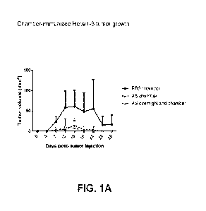
[0015] FIG. 1A-1D shows the result of the chamber immunization experiment
described in
Example 2. FIG. 1A shows tumor volume (mm3) at 4, 7, 12, 15, 19, 22, 26 and 29
days post tumor
cell (Hepal -6) injection. FIG. 1B shows Hepal -6-specific whole IgG levels,
FIG. 1C shows
Hepal -6-specific IgG1 levels, and FIG. 1D shows Hepal -6-specific IgG2A
levels at day 0 and
day 28. The dotted horizontal line represents plate background where
applicable.
DETAILED DESCRIPTION
[0016] The present disclosure relates to compositions and methods for treating
liver cancer,
including hepatocellular carcinoma, using antisense nucleic acids directed
against Insulin-like
Growth Factor-1 Receptor (IGF-1R). The present disclosure also relates to
compositions and
3
CA 03118424 2021-04-30
WO 2020/092682 PCT/US2019/059017
methods for treating liver cancer by treating subjects with at least one
implantable irradiated
biodiffusion chamber (see U.S. Patent No. 6,541,036 and PCT/US2016/026970,
which are
incorporated herein by reference in their entireties) comprising tumor cells
and an antisense nucleic
acid directed against IGF-1R.
Definitions
[0017] All terms not defined herein have their common art-recognized meanings.
[0018] As used herein, terms such as "a," "an," and "the" include singular and
plural referents
unless the context clearly demands otherwise.
[0019] As used herein, the term "about" when preceding a numerical value
indicates the value
plus or minus a range of 10%. For example, "about 100" encompasses 90 and 110.
[0020] As used herein, the term "autologous" means cells or tissues obtained
from the same
individual.
[0021] As used herein, the term "autologous cancer cell vaccine" refers to a
therapeutic produced
in part by isolating tumor cells from an individual and processing these tumor
cells ex vivo. The
cells are then re-administered to the individual from whom the tumor cells
were isolated. In
embodiments, an autologous cancer cell vaccine may comprise additional
components in addition
to the tumor cells, such as a buffer and/or antisense nucleic acids. In
embodiments, "autologous
cancer cell vaccine" may refer to a biodiffusion chamber containing the tumor
cells and one or
more additional components. In certain aspects, the "autologous cancer cell
vaccine" may be a
"fully formulated chamber" also referred to herein as "fully formulated
biodiffusion chamber."
[0022] As used herein, the term "fully formulated chamber" or "fully
formulated biodiffusion
chamber" is a biodiffusion chamber that includes autologous tumor cells and
other cells included
in the tumor microenvironment (TME) that may or may not be treated prior to
encapsulation in the
chamber with a first amount of an IGF-1R AS ODN. The cells are encapsulated
with exogenous
addition of a second amount, for example at least 2 g, at least 4 g, at
least 6 g, at least 8 g, or
at least 10 g, of IGF-1R AS ODN and the chamber is then irradiated with 5 Gy
of gamma-
irradiation.
4
CA 03118424 2021-04-30
WO 2020/092682 PCT/US2019/059017
[0023] As used herein, the term "small molecules" includes nucleic acids,
peptides, proteins, and
other chemicals (such as, for example, cytokines and growth hormones produced
by cells), but
does not include cells, exosomes, or microvesicles.
[0024] The term "targeting IGF-1R expression" or "targets IGF-1R expression"
as used herein
refers to administering an antisense nucleic acid that has a sequence designed
to bind to the IGF-
1R.
[0025] As used herein, the term "systemic administration" refers to achieving
delivery of a
substance throughout the body of a subject. Typical systemic routes of
administration include
parenteral administration, transdermal administration, intraperitoneal
administration, intravenous
administration, subcutaneous administration, and intramuscular administration.
[0026] Other administration routes include oral administration, nasal
administration topical
administration, intraocular administration, buccal administration, sublingual
administration,
vaginal administration, intraheptic, intracardiac, intrapancreatic, by
inhalation, and via an
implanted pump.
Liver Cancer
[0027] There are two main types of liver cancer; hepatocellular carcinoma
(HCC), also known as
malignant hepatoma, and cholangiocellular carcinoma.
[0028] HCC is the most common form of primary liver cancer, and develops
within the
hepatocyte. HCC occurs mostly in men. Symptoms of HCC may include jaundice,
abdominal
pain, unexplained weight loss, an enlarged liver, fatigue, nausea, vomiting,
back pain, itching, and
fever.
[0029] The pathogenesis of HCC is incompletely understood. Much evidence
supports the notion
that DNA damage occurs, resulting in deregulation of DNA methylation,
chromosomal instability,
proto-oncogene activation, and tumor suppressor gene inactivation. RAS
signaling pathways are
activated, and this serves to activate cell proliferation.
[0030] HCC most often occurs in the setting of chronic liver disease, and many
cases, particularly
in economically developed countries, is found in patients with cirrhosis.
Prominent risk factors for
HCC include chronic viral hepatitis B or C and alcohol-related liver disease.
Cirrhosis of any cause
increases the risk of HCC. An emerging threat, therefore, comes from the
obesity epidemic, which
predisposes to nonalcoholic liver disease and cirrhosis. A recent study showed
that the yearly
CA 03118424 2021-04-30
WO 2020/092682 PCT/US2019/059017
cumulative incidence of HCC is 2.6% per year in patients with cirrhosis
secondary to fatty liver
disease, compared to 4.0% per year in patients with Hepatitis C cirrhosis.
[0031] Risk factors for HCC include inherited disorders such as tyrosinemia
and
hemochromatosis, type 2 diabetes, family history, heavy alcohol use, low
immunity, obesity, being
male, smoking, and exposure to arsenic. In less-developed countries,
particularly in tropical and
subtropical climates, aflatoxin exposure is a promoter of HCC. Aflatoxins are
mycotoxins
produced by fungi of the genus Aspergillus, which are commonly present in soil
and as
contaminants of improperly stored nuts, cereals, and other produce.
[0032] In contrast, cholangiocellular carcinoma (CCA) or bile duct cancer
develops in the small
bile ducts within the liver. This type of cancer is more common among women.
According to their
anatomical location, CCAs are commonly classified as intrahepatic and
extrahepatic tumors, the
latter entity being further subdivided into perihilar CCAs, also termed as
Klatskin tumors, and
distal tumors. While a majority of CCAs occur sporadically, established risk
factors include liver
fluke infestation (e.g., Opisthorchis viverrini or Clonorchis sinensis) and
primary sclerosing
cholangitis.
[0033] When a tumor is small and occupies a small part of the liver, that part
of the liver can be
surgically removed (partial hepatectomy). However, many people with liver
cancer have cirrhosis.
This means that a hepatectomy needs to leave behind enough healthy tissue for
the liver to perform
its necessary functions after the procedure. Accordingly, partial hepatectomy
is only considered
for people with otherwise healthy liver function. This procedure is often not
an option when the
cancer has spread to other parts of the liver or other organs in the body.
[0034] Liver transplant is also used to treat liver cancer. However, the
immune system can reject
the new organ, attacking it as a foreign body, and there are limited
opportunities to carry out
transplants. The drugs that suppress the immune system to accommodate a new
liver can also lead
to serious infections and even, on occasion, the spreading of already
metastasized tumors.
[0035] Advanced liver cancer has an extremely low survival rate. Treatments
used to treat cancer
symptoms and slow the growth of a tumor in these cases include ablative
therapy, radiation
therapy, and chemotherapy.
Antisense Molecules
[0036] Antisense molecules are nucleic acids that work by binding to a
targeted complimentary
sequence of mRNA by Watson and Crick base-pairing rules. The translation of
target mRNA is
6
CA 03118424 2021-04-30
WO 2020/092682 PCT/US2019/059017
inhibited by an active and/or passive mechanism when hybridization occurs
between the
complementary helices. In the passive mechanism, hybridization between the
mRNA and
exogenous nucleotide sequence leads to duplex formation that prevents the
ribosomal complex
from reading the message. In the active mechanism, hybridization promotes the
binding of
RnaseH, which destroys the RNA but leaves the antisense intact to hybridize
with another
complementary mRNA target. Either or both mechanisms inhibit translation of a
protein
contributing to or sustaining a malignant phenotype. As therapeutic agents,
antisense molecules
are far more selective and as a result, more effective and less toxic than
conventional drugs.
[0037] The methods and compositions disclosed herein involve the use of
antisense molecules for
treating cancer. Typically, the antisense molecule is an antisense
oligodeoxynucleotide (AS-
ODN). In some embodiments, the antisense molecule comprises a modified
phosphate backbone.
In certain aspects, the phosphate backbone modification renders the antisense
more resistant to
nuclease degradation. In certain embodiments, the modification is a locked
antisense. In other
embodiments, the modification is a phosphorothioate linkage. In certain
aspects, the antisense
contains one or more phosphorothioate linkages. In certain embodiments, the
phosphorothioate
linkages stabilize the antisense molecule by conferring nuclease resistance,
thereby increasing its
half-life. In some embodiments, the antisense may be partially
phosphorothioate-linked. For
example, up to about 1%, up to about 3%, up to about 5%, up to about 10%, up
to about 20%, up
to about 30%, up to about 40%, up to about 50% up to about 60%, up to about
70%, up to about
80%, up to about 90%, up to about 95%, or up to about 99% of the antisense may
be
phosphorothioate-linked. In some embodiments, the antisense is fully
phosphorothioate-linked.
In other embodiments, phosphorothioate linkages may alternate with
phosphodiester linkages. In
certain embodiments, the antisense has at least one terminal phosphorothioate
monophosphate.
[0038] In some embodiments, the antisense molecule comprises one or more CpG
motifs. In other
embodiments, the antisense molecule does not comprise a CpG motif. In certain
aspects, the one
or more CpG motifs are methylated. In other aspects, the one or more CpG
motifs are
unmethylated. In certain embodiments, the one or more unmethylated CpG motifs
elicit an innate
immune response when the antisense molecule is administered to a subject. In
some aspects, the
innate immune response is mediated by binding of the unmethylated CpG-
containing antisense
molecule to Toll like Receptors (TLR).
7
CA 03118424 2021-04-30
WO 2020/092682 PCT/US2019/059017
[0039] In certain embodiments, the antisense molecule comprises at least one
terminal
modification or "cap". The cap may be a 5' and/or a 3' -cap structure. The
terms "cap" or "end-
cap" include chemical modifications at either terminus of the oligonucleotide
(with respect to
terminal ribonucleotides), and including modifications at the linkage between
the last two
nucleotides on the 5' end and the last two nucleotides on the 3' end. The cap
structure may increase
resistance of the antisense molecule to exonucleases without compromising
molecular interactions
with the target sequence or cellular machinery. Such modifications may be
selected on the basis
of their increased potency in vitro or in vivo. The cap can be present at the
5' -terminus (5'-cap) or
at the 3' -terminus (3' -cap) or can be present on both ends. In certain
embodiments, the 5'- and/or
3' -cap is independently selected from phosphorothioate monophosphate, abasic
residue (moiety),
phosphorothioate linkage, 4'-thio nucleotide, carbocyclic nucleotide,
phosphorodithioate linkage,
inverted nucleotide or inverted abasic moiety (2'-3' or 3'-3'),
phosphorodithioate monophosphate,
and methylphosphonate moiety. The phosphorothioate or phosphorodithioate
linkage(s), when
part of a cap structure, are generally positioned between the two terminal
nucleotides on the 5' end
and the two terminal nucleotides on the 3' end.
[0040] In preferred embodiments, the antisense molecule targets the expression
of Insulin like
Growth Factor 1 Receptor (IGF-1R). IGF-1R is a tyrosine kinase cell surface
receptor that shares
70% homology with the insulin receptor. When activated by its ligands (IGF-I,
IGF-II and
insulin), it regulates broad cellular functions including proliferation,
transformation and cell
survival. The IGF-1R is not an absolute requirement for normal growth, but it
is essential for
growth in anchorage-independent conditions that may occur in malignant
tissues. A review of the
role of IGF-1R in tumors is provided in Baserga et al., Vitamins and Hormones,
53:65-98 (1997),
which is incorporated herein by reference in its entirety.
[0041] In certain embodiments, the antisense molecule is an oligonucleotide
directed against DNA
or RNA of a growth factor or growth factor receptor, such as, for example, IGF-
1R.
[0042] In certain embodiments, the antisense is a deoxynucleotide directed
against IGF-1R (IGF-
1R AS ODN). The full length coding sequence of IGF-1R is provided as SEQ ID
NO:15 (see, for
example, PCT/U52016/26970, which is incorporated herein by reference in its
entirety).
[0043] In certain embodiments, the antisense molecule comprises nucleotide
sequences
complementary to the IGF-1R signal sequence, comprising either RNA or DNA. The
signal
sequence of IGF-1R is a 30 amino acid sequence. In other embodiments, the
antisense molecule
8
CA 03118424 2021-04-30
WO 2020/092682 PCT/US2019/059017
comprises nucleotide sequences complementary to portions of the IGF-1R signal
sequence,
comprising either RNA or DNA. In some embodiments, the antisense molecule
comprises
nucleotide sequences complementary to codons 1-309 of IGF-1R, comprising
either RNA or DNA.
In other embodiments, the antisense molecule comprises nucleotide sequences
complementary to
portions of codons 1-309 of IGF-1R, comprising either RNA or DNA.
[0044] In certain embodiments, the IGF-1R AS ODN is at least about 5
nucleotides, at least about
nucleotides, at least about 15 nucleotides, at least about 20 nucleotides, at
least about 25
nucleotides, at least about 30 nucleotides, at least about 35 nucleotides, at
least about 40
nucleotides, at least about 45 nucleotides, or at least about 50 nucleotides
in length. In some
embodiments, the IGF-1R AS ODN is from about 15 nucleotides to about 22
nucleotides in length.
In certain aspects, the IGF-1R AS ODN is about 18 nucleotides in length.
[0045] In certain embodiments, the IGF-1R AS ODN forms a secondary structure
at 18 C, but
does not form a secondary structure at about 37 C. In other embodiments, the
IGF-1R AS ODN
does not form a secondary structure at about 18 C or at about 37 C. In yet
other embodiments, the
IGF-1R AS ODN does not form a secondary structure at any temperature. In other
embodiments,
the IGF-1R AS ODN does not form a secondary structure at 37 C. In particular
embodiments, the
secondary structure is a hairpin loop structure.
[0046] In some aspects, the IGF-1R AS ODN comprises the nucleotide sequence of
SEQ ID NO:1,
or a fragment thereof. In certain embodiments, the IGF-1R AS ODN may have at
least about 70%,
at least about 75%, at least about 80%, at least about 85%, at least about
90%, at least about 95%,
at least about 96%, at least about 98%, or 100% identity to SEQ ID NO: 1, or a
fragment thereof
In some embodiments, the IGF-1R AS ODN comprises one or more phosphorothioate
linkages
(see, e.g., SEQ ID NO: 16).
[0047] In certain aspects, the IGF-1R AS ODN consists of SEQ ID NO: 1. NOBEL
is an 18-mer
oligodeoxynucleotide with a phosphorothioate backbone, and a sequence
complimentary to codons
2 through 7 in the IGF -1R gene. As such, NOBEL is an antisense
oligonucleotide directed against
IGF-1R (IGF-1R AS ODN). The NOBEL sequence, derived as the complimentary
sequence of
the IGF-1R gene at the 5' end, is:
5' -TCCTCCGGAGCCAGACTT- 3' (SEQ ID NO: 1).
[0048] NOBEL has a stable shelf life and is resistant to nuclease degradation
due to its
phosphorothioate backbone. Administration of NOBEL can be provided in any of
the standard
9
CA 03118424 2021-04-30
WO 2020/092682 PCT/US2019/059017
methods associated with introduction of oligodeoxynucleotides known to one of
ordinary skill
in the art. Advantageously, the AS ODNs disclosed herein, including NOBEL, may
be
administered with little/no toxicity. Even levels of about 2g/kg (scaled)
based on mice tests (40 i.tg
in the tail vain) did not reveal toxicity issues. NOBEL can be manufactured
according to ordinary
procedures known to one of ordinary skill in the art.
[0049] The antisense molecule, for example the NOBEL sequence of SEQ ID NO: 1,
may also
comprise one or more p-ethoxy backbone modifications as disclosed in U.S.
Patent No. 9,744,187,
which is incorporated by reference herein in its entirety. In some
embodiments, the nucleic acid
backbone of the antisense molecule comprises at least one p-ethoxy backbone
linkage. For
example, up to about 1%, up to about 3%, up to about 5%, up to about 10%, up
to about 20%, up
to about 30%, up to about 40%, up to about 50%, up to about 60%, up to about
70%, up to about
80%, up to about 90%, up to about 95%, or up to about 99% of the antisense
molecule may be p-
ethoxy-linked. The remainder of the linkages may be phosphodiester linkages or
phosphorothioate
linkages or a combination thereof. In a preferred embodiment 50% to 80% of the
phosphate
backbone linkages in each oligonucleotide are p-ethoxy backbone linkages,
wherein 20% to 50%
of the phosphate backbone linkages in each oligonucleotide are phosphodiester
backbone linkages.
[0050] Various IGF-1R antisense sequences are bioactive in some or all of the
multi-modality
effects of the NOBEL sequence. The 18-mer NOBEL sequence has both IGF-1R
receptor
downregulation activity as well as TLR agonist activity, and further
experimentation in mice
suggests that both activities are necessary for in vivo anti-tumor immune
activity. While the AS
ODN molecule has anti-tumor activity, the complimentary sense sequence does
not, despite also
having a CpG motif.
[0051] In certain embodiments, the sequence of the antisense is selected from
the group consisting
of SEQ ID NOS 1-14, as shown in Table 1. In some embodiments, the antisense
has 90% sequence
identity to one or more of SEQ ID NOS 1-14. In some embodiments, the antisense
has 80%
sequence identity to one or more of SEQ ID NOS 1-14. In some embodiments, the
antisense has
70% sequence identity to one or more of SEQ ID NOS 1-14.
TABLE 1: Additional downstream sequences for IGF-1R AS ODN Formulation
Sequences with ACGA Motif Corresponds to IGF-1R SEQ ID NO:
Codons
5'-TCCTCCGGAGCCAGACTT-3' 2-7 1
CA 03118424 2021-04-30
WO 2020/092682 PCT/US2019/059017
Sequences with ACGA Motif Corresponds to IGF-1R SEQ ID NO:
Codons
5'-TTCTCCACTCGTCGGCC-3' 26-32 2
5' -ACAGGCCGTGTCGTTGTC-3 ' 242-248 3
5' -GCACTCGCCGTCGTGGAT-3 ' 297-303 4
5' -CGGATATGGTCGTTCTCC-3' 589-595 5
5'- TCTCAGCCTCGTGGTTGC-3' 806-812 6
5'-TTGCGGCCTCGTTCACTG-3' 1,033-1,039 7
5' -AAGCTTCGTTGAGAAACT-3' 1,042-1,048 8
5' -GGACTTGCTCGTTGGACA-3' 1,215-1,221 9
5' -GGCTGTCTCTCGTCGAAG-3 ' 1,339-1,345 10
5'-CAGATTTCTCCACTCGTCGG-3' 27-34 11
5' -CCGGAGCCAGACTTCAT-3' 1-6 12
5'-CTGCTCCTCCTCTAGGATGA-3' 407-413 13
5'-CCCTCCTCCGGAGCC-3' 4-8 14
[0052] In certain embodiments, the IGF-1R AS ODN comprises the nucleotide
sequence of any
one of SEQ ID NOs:1-14, or fragments thereof In certain embodiments, the IGF-
1R AS ODN
may have at least about 70%, at least about 75%, at least about 80%, at least
about 85%, at least
about 90%, at least about 95%, at least about 96%, at least about 98%, or 100%
identity to any one
of SEQ ID NOs: 1-14, or fragments thereof.
[0053] In some embodiments, the antisense molecule downregulates the
expression of genes
downstream of IGF-1R pathway in a cell. In certain aspects, the downstream
gene is hexokinase
(Hex II). In some embodiments, the antisense molecule downregulates the
expression of
housekeeping genes in the cell. In some aspects, the housekeeping gene is L13.
[0054] In certain aspects, the IGF-1R AS ODN is chemically synthesized. In
certain
embodiments, the IGF-1R AS ODN is manufactured by solid phase organic
synthesis. In some
aspects, the synthesis of the IGF-1R AS ODN is carried out in a synthesizer
equipped with a closed
chemical column reactor using flow-through technology. In some embodiments,
each synthesis
cycle sequence on the solid support consists of multiple steps, which are
carried out sequentially
until the full-length IGF-1R AS ODN is obtained. In certain embodiments, the
IGF-1R AS ODN
is stored in a liquid form. In other embodiments, the IGF-1R AS ODN is
lyophilized prior to storing.
11
CA 03118424 2021-04-30
WO 2020/092682 PCT/US2019/059017
In some embodiments, the lyophilized IGF-1R AS ODN is dissolved in water prior
to use. In other
embodiments, the lyophilized IGF-1R AS ODN is dissolved in an organic solvent
prior to use. In
yet other embodiment, the lyophilized IGF-1R AS ODN is formulated into a
pharmaceutical
composition. In some aspects the pharmaceutical composition is a liquid
pharmaceutical
composition. In other aspects, the pharmaceutical composition is a solid
pharmaceutical
composition. Additional antisense nucleic acids are also described in U.S.
Publication No.
2017/0056430, which is incorporated herein by reference in its entirety.
Autologous Cancer Cell Vaccine
Introduction
[0055] Immunotherapy is currently used to target hematologic malignancies with
one common
cellular antigen. Unfortunately, solid tumors are far more complex,
representing epigenetic
progression of genetic changes to a malignant state with an unidentifiable
number of tumor-
specific targets. Even more challenging, within a WHO diagnostic cancer group
there exists
marked variations in tumor phenotypes. An autologous cell vaccine would
encompass all such
variations and all such targets and represent an ideal subject-specific
immunotherapy for solid
tumor cancers. An autologous cancer cell vaccine however, cannot be derived
from primary
cell cultures because serial passages alter the tumor phenotype thus
diminishing the array of
tumor-specific antigens. This would also require impossible lot-release
qualification at each
passage. The present disclosure eliminates these concerns by plating freshly
resected, morselized
tumor cells and reimplanting them within 24 hours as a depot antigen. In
certain aspects, the
excellent results achieved herein are obtained by ensuring that an appropriate
number of cells
are present in the chamber(s), among other specifics described herein.
[0056] Previous studies have designed autologous cell vaccine through the use
of antigen
presenting cells, instead of autologous tumor cells. In this paradigm, a
subject's monocytes are
collected from a pre-treatment plasma leukopheresis and differentiated into
autologous dendritic
cells (DC) ex vivo. The dendritic cells are then presented with the subject's
tumor crude lysate
inducing DC activation/maturation, and at a later time point, the matured
dendritic cells, now
cross-primed with tumor antigens are injected in the subject as a DC vaccine.
Ex vivo
differentiation, however, is missing a number of key stimulatory components
only occurring in
vivo. In addition, differentiation of DCs from hematopoietic precursors
requires extensive in
12
CA 03118424 2021-04-30
WO 2020/092682 PCT/US2019/059017
vitro manipulations with labor-intensive cell processing in expensive
facilities. The present
disclosure obviates these concerns by providing an endogenous DC maturation
process and an
immunomodulatory and immunostimulatory antisense oligodeoxynucleotide (AS-ODN)
that
promotes the development of an appropriate immune response. More specifically,
the present
disclosure provides a biodiffusion chamber comprising dispersed tumor cells
derived from the
patient and irradiated antisense molecules, which is implanted into the
patient for therapeutically
effective time. Without being bound by any theory, it is thought that the
combination of irradiated
tumor cells, antisense, and biodiffusion chamber act in concert to simulate
the local immune
response, and enhance the response by reducing or eliminating M2 cells,
preventing dampening
of the immune system.
[0057] Thus, the present disclosure shows that an irradiated, implantable
biodiffusion chamber
comprising freshly resected tumor cells and IGF-1R AS ODN safely serves as an
effective,
subject-specific autologous cell vaccine for liver cancer immunotherapy. As
such, the use of
the claimed implantable biodiffusion chamber to mount an immune response that
selectively
targets tumor cells in a subject provides a new and significant approach for
the treatment of liver
cancer, especially hepatocellular carcinoma.
Biodiffusion chamber
[0058] A representative diffusion chamber comprises a chamber barrel having
two ends, a first
end and a second end. In embodiments, the biodiffusion chamber is a small ring
capped on either
side by a porous, cell-impermeable membrane, such as the Duropore membrane
manufactured by
Millipore Corporation. Optionally, one of the ends may be closed off as part
of the chamber body
leaving only one end open to be sealed using the porous membrane. The
membranes can be made
of plastic, teflon, polyester, or any inert material which is strong, flexible
and able to withstand
chemical treatments. The chamber can be made of any substance, such as and not
limited to plastic,
teflon, lucite, titanium, Plexiglass or any inert material which is non-toxic
to and well tolerated by
humans. In addition, the chambers should be able to survive sterilization. In
some aspects, the
diffusion chambers are sterilized with ethylene oxide prior to use. Other
suitable chambers are
described in U.S. Prov. No. 62/621,295, filed January 24, 2018, U.S. Patent
No. 6,541,036,
PCT/U516/26970, and U.S. Patent No. 5,714,170, which are each incorporated
herein by reference
in their entirety.
13
CA 03118424 2021-04-30
WO 2020/092682 PCT/US2019/059017
[0059] In certain embodiments, the membrane allows passage of small molecules
but does not
allow passage of cells (i.e., the cells cannot leave or enter the chamber). In
some aspects, the
diameter of the pores of the membrane allows nucleic acids and other chemicals
(such as, for
example, cytokines produced by cells) to diffuse out of the chamber, does not
allow passage of
cells between the chamber and the subject in which it is implanted. The
biodiffusion chambers
useful in the present disclosure include any chamber which does not allow
passage of cells between
the chamber and the subject in which it is implanted, provided however, that
the chamber permits
interchange and passage of factors between the chamber and the subject. Thus,
in certain aspects,
the pore size has a cut-off that prevent passage of materials that are greater
than 100 m3 in volume
into and out of the chamber. In some embodiments, the pores of the membrane
have a diameter
of about 0.25 [tm or smaller. For example, the pores may have a diameter of
about 0.1 [tm. In
particular aspects, the pores range in diameter from 0.1 [tm to 0.25 [tm. See
also, Lange, et al., J.
Immunol., 1994, 153, 205-211 and Lanza, et al., Transplantation, 1994, 57,
1371-1375, each of
which is incorporated herein by reference in their entireties. This pore
diameter prevents the
passage of cells in or out of the chamber. In certain embodiments, diffusion
chambers are
constructed from 14 mm Lucite rings with 0.1 [tm pore-sized hydrophilic
Durapore membranes
(Millipore, Bedford, Mass.).
[0060] In certain embodiments, a biodiffusion chamber comprises a membrane
that allows the
IGF-1R AS ODN to diffuse out of the chamber. In some embodiments, about 50% of
the IGF-1R
AS ODN diffuses out of the chamber in about 12 hours, about 60% of the IGF-1R
AS ODN
diffuses out of the chamber in about 24 hours, about 80% of the IGF-1R AS ODN
diffuses out of
the chamber in about 48 hours, and/or about 100% of the IGF-1R AS ODN diffuses
out of the
chamber in about 50 hours.
[0061] In an exemplary approach, to assemble the biodiffusion chamber, a first
porous membrane
is attached to one side of a first diffusion chamber, using glue and pressure
to create a tight seal.
A second porous membrane is similarly attached to a second diffusion chamber
ring. The
membranes can be secured in position with rubber gaskets which may also
provide a tighter seal.
The diffusion chamber rings are left overnight (minimum 8 hours) to dry. Then,
the first diffusion
chamber ring and the second diffusion chamber ring are attached to one another
using glue and
left overnight (minimum 8 hours) to dry. In a preferred embodiment, the first
chamber ring and
second chamber ring joining process comprises using 2 dichloroethane as a
solvent to facilitate
14
CA 03118424 2021-04-30
WO 2020/092682 PCT/US2019/059017
adhesion between the two rings. In an alternative approach, the chamber may
have only one side
that contains a porous membrane.
[0062] On the barrel portion of the chamber, one or more openings (e.g. ports)
are provided which
can be covered by a cap which is accessed from outside of the subject's body
once the chamber is
implanted, thus allowing the diffusion chamber to be refilled. The openings
allow for multiple
and sequential sampling of the contents, without contamination and without
harming the subject,
therefore significantly reducing the number of implantation procedures
performed on the subject.
Before implantation into the patient, the one or more openings may be sealed
with bone wax, a
port plug or cap made from, for example, PMMA. The cap can be a screw-on type
of self-sealing
rubber and fitted to the opening. In some configurations, the diffusion
chamber may contain two
or more injection openings or ports. Sampling of the chamber contents can be
performed by
accessing the opening by removing the cap on the outside of the subject's body
and inserting an
ordinary needle and syringe. In some embodiments, the chamber may further
include a removal
device. Such a device facilitates removal of the chamber from the patient.
[0063] In embodiments, the chamber serves as an antigen depot designed so that
tumor antigens
diffuse out of the chamber for the purpose of promoting a therapeutic host
immune response.
Exogenous IGF-1R AS ODN and ex vivo irradiation promote a pro-inflammatory
response. This
formulation is associated with clinical and radiographic improvements,
prolonged survival on
protocol, and represents a novel autologous cell vaccine that includes an
exogenous active
pharmaceutical ingredient (API) and radiation that we interpret as inducing or
enhancing tumor
immunity effect. Furthermore the addition of low concentration of the IGF-1R
AS ODN is critical
to a pro-inflammatory response.
[0064] In certain embodiments the disclosure provides a biodiffusion chamber
for implantation
into a subject suffering from cancer comprising: (a) tumor cells; and (b) an
effective amount of an
antisense molecule. In other embodiments is provided a method for treating
cancer in a subject
comprising: (a) obtaining a biodiffusion chamber comprising tumor cells and an
effective amount
of an antisense nucleic acid; (b) irradiating the biodiffusion chamber and
contents; and (c)
implanting the irradiated biodiffusion chamber into the subject for a
therapeutically effective time.
[0065] In certain embodiments, the IGF-1R AS ODN is present in the
biodiffusion chamber in an
amount ranging from about 0.5 i.tg to about 10 pg. In certain aspects, the IGF-
1R AS ODN is
present in an amount ranging from about 1 i.tg to about 5 i.tg per chamber, or
from about 2 i.tg to 4
CA 03118424 2021-04-30
WO 2020/092682 PCT/US2019/059017
[tg per chamber. In certain embodiments, the IGF-1R AS ODN is present in the
biodiffusion
chamber in an amount ranging from about 0.5 [tg to about 10 [tg. In certain
aspects, the IGF-1R
AS ODN is present in an amount ranging from about 1 [tg to about 5 [tg per
chamber, or from
about 2 [tg to about 4 [tg per chamber. For example, the IGF-1R may be present
in an amount of
about 1, about 1.5, about 2, about 2.5, about 3, about 3.5, about 4, about
4.5, or about 5 [tg per
chamber. In specific aspects, the IGF-1R AS ODN is present in an amount of
about 2 [tg per
chamber. In specific aspects, the IGF-1R AS ODN is present in an amount of
about 4 [tg per
chamber. In certain embodiments, the IGF-1R AS ODN is present in the
biodiffusion chamber in
an amount ranging from about 10 [tg to about 500 [tg. In certain aspects, the
IGF-1R AS ODN is
present in an amount ranging from about 40 [tg to about 400 [tg per chamber,
from about 40 to
about 70 [tg per chamber, or from about 100 [tg to about 200 [tg per chamber.
In specific aspects,
the IGF-1R AS ODN is present in an amount of about 100 [tg per chamber. In
specific aspects,
the IGF-1R AS ODN is present in an amount of about 200 [tg per chamber. In
certain aspects, the
IGF-1R AS ODN is present in an amount ranging from about 1 mg to about 100 mg
per chamber,
or from about 2 mg to about 10 mg per chamber. In specific aspects, the IGF-1R
AS ODN is
present in an amount of about 2 mg per chamber. In specific aspects, the IGF-
1R AS ODN is
present in an amount of about 4 mg per chamber. Without being bound by any
theory it is thought
that these levels promote an enhanced Thl response in a subject, while
avoiding an M2
immunostimulatory response in the subject.
[0066] In certain embodiments, the tumor cells are not treated with an IGF-1R
AS ODN prior to
encapsulation in the chamber. Typically, however, the tumor cells are treated
with an IGF-1R AS
ODN prior to encapsulation in the chamber. The time for treating the cells pre-
encapsulation may
vary. For example, the tumor cells may be treated ex vivo with an IGF-1R AS
ODN immediately
before encapsulation, for up to about 4 hours, for up to about 6 hours, for up
to about 8 hours, for
up to about 12 hours or for up to about 18 hours. Typically, the tumor tissue
may be treated ex
vivo for about 12 hours to about 18 hours pre-encapsulation. Conveniently, the
cells may be
encapsulated after a pre-treatment lasting up to overnight. Without being
bound by theory, it is
thought that the pre-encapsulation treatment plays a desirable role in
stimulating production of
tumor antigen.
[0067] The amount of IGF-1R AS ODN used for the pre-encapsulation treatment
may be in a
range of about 1 mg to 8 mg per million cells; for example, about 2 mg to
about 6 mg per million
16
CA 03118424 2021-04-30
WO 2020/092682 PCT/US2019/059017
cells, about 3 mg to about 5 mg per million cells. Typically the amount of IGF-
1R AS ODN used
for treatment prior to encapsulation is about 4 mg per million cells.
[0068] In some embodiments, the IGF-1R AS ODN for ex vivo treatment of the
tumor cells is
used at a concentration ranging from about at least 2 mg/ml to at least about
5 mg/ml. In certain
aspects, the IGF-1R AS ODN is used at a concentration of at least 4 mg/ml. In
specific
embodiments, the IGF-1R AS ODN is used at a concentration of 4 mg/ml.
[0069] In certain embodiments, the IGF-1R AS ODN used to treat tumor cells ex
vivo and the
IGF-1R AS ODN present in the chamber are the same. In other embodiments, the
IGF-1R AS
ODN used to treat tumor cells ex vivo and the IGF-1R AS ODN present in the
chamber are
different. In certain embodiments, the IGF-1R AS ODN used to treat tumor cells
ex vivo is at least
about 5 nucleotides, at least about 10 nucleotides, at least about 15
nucleotides, at least about 20
nucleotides, at least about 25 nucleotides, at least about 30 nucleotides, at
least about 35
nucleotides, at least about 40 nucleotides, at least about 45 nucleotides, or
at least about 50
nucleotides in length. In some embodiments, the IGF-1R AS ODN used to treat
tumor cells ex
vivo is from about 15 nucleotides to about 22 nucleotides in length. In
certain aspects, the IGF-
1R AS ODN used to treat tumor cells is about 18 nucleotides in length.
[0070] In certain embodiments, the IGF-1R AS ODN used to treat tumor cells ex
vivo forms a
secondary structure at 18 C, but does not form a secondary structure at about
37 C. In other
embodiments, the IGF-1R AS ODN used to treat tumor cells does not form a
secondary structure
at about 18 C or at about 37 C. In yet other embodiments, the IGF-1R AS ODN
used to treat
tumor cells ex vivo does not form a secondary structure at any temperature. In
other embodiments,
the IGF-1R AS ODN used to treat tumor cells does not form a secondary
structure at 37 C. In
particular embodiments, the secondary structure is a hairpin loop structure.
[0071] In some aspects, the IGF-1R AS ODN used to treat tumor cells comprises
the nucleotide
sequence of SEQ ID NO:1, or a fragment thereof. In certain embodiments, the
IGF-1R AS ODN
used to treat tumor cells may have at least about 70%, at least about 75%, at
least about 80%, at
least about 85%, at least about 90%, at least about 95%, at least about 96%,
at least about 98%, or
100% identity to SEQ ID NO: 1, or a fragment thereof In certain aspects, the
IGF-1R AS ODN
used to treat tumor cells is SEQ ID NO: 1.
[0072] After the tumor cells are treated with the AS-ODN for a period of time,
the AS-ODN is
removed and fresh AS-ODN is added to the chamber, which is then irradiated
prior to implantation
17
CA 03118424 2021-04-30
WO 2020/092682 PCT/US2019/059017
into a subject. In certain aspects, the biodiffusion chamber is treated with
gamma irradiation at an
amount of about 1 Gy, about 2 Gy, about 4 Gy, about 5 Gy, about 6 Gy, about 10
Gy, or up to
about 15 Gy. In certain aspects, the dose of radiation is not more than about
5 Gy. In other aspects,
the dose of radiation is at least about 5 Gy. In some aspects, the dose of
radiation is 5 Gy. In
certain embodiments, the biodiffusion chamber may be irradiated at least once,
at least twice, at
least three times, at least four times, or at least five times. In some
embodiments, the chamber is
irradiated less than about 24 hours prior to implantation into a subject. In
other embodiments,
chamber is irradiated about 24 hours prior to implantation into the subject.
In yet other
embodiments, the chamber is irradiated at least about 24 hours prior to
implantation into the
subject. In still other embodiments, the chamber is irradiated not more than
about 48 hours prior
to implantation into the subject. In yet other embodiments, the chamber is
irradiated at least about
48 hours prior to implantation into the subject.
[0073] While the tumor cells are typically killed prior to implantation; for
example by radiation,
the cells need not be killed and indeed it may be advantageous to maintain the
cells in an alive
state to promote release of antigen. Thus, in certain embodiments, the cells
may not be irradiated
prior to implantation. For safety purposes, however, it is desirable to
prevent release of live tumor
cells into the subject.
[0074] Tumor cells can be placed in a diffusion chamber in varying numbers. In
certain
embodiments, about 1 x 10⁴ to about 5 x 10⁶ tumor cells are placed in each
diffusion chamber. In
other embodiments, about 1 x 10⁵ to about 1.5 x 10⁶ tumor cells are placed in
the diffusion
chamber. In yet other embodiments, about 5 x 10⁵ to 1 x 10⁶ tumor cells are
placed in the chamber.
with a subject can be used. We have discovered that the number of tumor cells
can impact the
subjects' anti-tumor response and that an appropriate range should be selected
to increase the
chance to obtain the desired results. In some embodiments, the anti-tumor
immune response is
optimal in a range of about 750,000 to about 1,250,000 cells in a chamber,
with a peak at about 1
million cells/chamber. Multiple chamber containing irradiated tumor cells are
administered and
to maintain the optimal immune the response the number of cells/chamber is
preferably maintained
within the range. Preferably, the tumor cells are intact and not autolyzed or
otherwise damaged as
described herein.
[0075] In certain embodiments, it may be preferable to maintain the ratio of
cells to AS ODN in a
chamber. Thus, in certain aspects a chambers may contain about 2 jig of AS ODN
and between
18
CA 03118424 2021-04-30
WO 2020/092682 PCT/US2019/059017
750,000 and 1,250,000 cells; for example 1,000,000 cells. The ratio of cells
to AS ODN may thus
be in a range from about 3.75 x 10⁵ to about 6.25 x 10⁵ per g AS ODN; for
example, about 5.0 x
105 cells per g. Thus, in a typical patient receiving 20 chambers the total
dose of AS ODN is
about 40 g.
[0076] Typically, administration will be in a chamber as described herein;
however, in certain
aspects, the irradiated cells and IGF-1R AS ODN may be co-administered to the
subject without
being contained physically together in the chamber or another container. In
certain methods using
this approach, the irradiated cells IGF-1R AS ODN thus disperse, diffuse, or
are metabolized in
the body limited by the physiology of the subject. Thus, in certain aspects,
e.g. the tumors cells
for use may be prepared as described herein for the chamber and administered
with the IGF-1R
AS ODN but the administration may be not contained within a physical
container. Such
administration is typically intramuscular.
Tumor Tissue Preparation for Chamber
[0077] Tumor cells for use in the autologous vaccination are surgically
removed from the subject.
In embodiments, the tumor cells are removed from the patient using a tissue
morselator. The
extraction device preferably combines a high-speed reciprocating inner cannula
within a stationary
outer cannula and electronically controlled variable suction. The outer
cannula has a diameter of
1.1 mm, 1.9 mm, 2.5 mm, or 3.0 mm, and a length of 10 cm, 13 cm, or 25 cm. The
instrument
also relies on a side-mouth cutting and aspiration aperture located 0.6 mm
from the blunt desiccator
end. The combination of gentle forward pressure of the aperture into the
tissue to be removed and
suction draws the desired tissue into the side aperture, allowing for
controlled and precise tissue
resection through the reciprocal cutting action of the inner cannula. A key
feature is the absence
of a rotation blade; this avoids drawing unintended tissue into the aperture.
An example of a
suitable device is the Myriad tissue aspirator (NICO Corporation
Indianapolis, IN), a
minimally invasive surgical system which may be used for the removal of soft
tissues with direct,
microscopic, or endoscopic visualization. The shaved tissue is suctioned,
gathered in to a
collection chamber, and is collected in a sterile tissue trap. During
collection of the tissue in the
sterile tissue trap, blood is removed from the preparation. Preferably, the
sterile trap contains a
collection dish at the bottom of the trap and a stem that provides access to
the trap. The trap
19
CA 03118424 2021-04-30
WO 2020/092682 PCT/US2019/059017
structure may also contain an inner ladle-shaped structure that is removable
from the trap to
facilitate tissue removal from the trap.
[0078] Preferably, the morselator generates no heat at the resection site or
along its shaft, and
requires no ultrasonic energy for tissue removal. Thus, in particular
embodiments, the tumor tissue
is morselized tumor tissue (i.e. tumor shaved tissue obtained by side-mouth
cutting in the absence
of heat, and optionally in the absence of ultrasonic treatment).
Advantageously, the aspirator-
extract and morselized tissue has higher viability than tissue removed by
other methods. It is
believed that the extraction process maintains higher tumor cell viability in
part due to restricting
exposure of the tumor cells to high temperatures during removal. For example,
the methods herein
do not expose tumor cells to above 25 C during removal. Thus, the cells are
not exposed to
temperatures above body temperature, i.e., about 37 C.
[0079] In some embodiments, the tumor cells are viable when they are removed
from the patient,
for example, using a tissue morcellator. In some embodiments, removal of the
cells from the
patient using a tissue morcellator does not kill the cells. In some
embodiments, at least 60%, at
least 60%, at least 70%, at least 80%, at least 90%, at least 95%, at least
98, or at least 99% of the
tumor cells removed from the patient using a tissue morcellator are viable.
[0080] The amount of tumor tissue obtained from the subject may vary.
Preferably, the amount
is at least 1, at least 2, at least 3 grams or at least 4 grams of wet tumor
tissue is obtained from the
patient. The tissue is removed from the sterile tissue trap and disaggregated
by pipetting with a
sterile pipette to break up large tissue fragments. The disaggregated cell
suspension is then placed
onto sterile tissue culture plates in serum-containing media, and incubated in
a tissue culture
incubator. This plating step serves to enrich the desired functional cells by
adherence, and also
helps to remove debris from the preparation. Thus, the tumor cells used in
treatments described
herein preferably consist essentially of, or consist of, adherent cells from
the tumor tissue.
[0081] After a predetermined incubation time (e.g., 6, 12, 24, or 48 hours),
the cells are removed
from the plates. The cells may be removed by scraping, by chemical methods
(e.g. EDTA) or by
enzymatic treatment (e.g. trypsin). The cells are placed into one or more
diffusion chambers. In
some embodiments, the cells are split between 2, 3, 4, 5, 6, 7, 8, 9, 10, 11,
12, 13, 14, 15, 16, 17,
18, 19, 20, 21, 22, 23, 24, 25, 26, 27, 28, 29, 30 or more diffusion chambers.
Often, 20 chambers
are used. In some embodiments, each diffusion chamber contains an equal number
of cells. In
some embodiments, a first diffusion chamber contains more cells than a second
chamber.
CA 03118424 2021-04-30
WO 2020/092682 PCT/US2019/059017
[0082] In some embodiments, the cells are sorted before being placed in the
chamber. In some
embodiments, the cells are enriched by selecting for one or more cellular
markers before being
placed in the chamber. The selection may be performed, for example, using
beads or by cell sorting
techniques known to those of skill in the art. In some embodiments, the cells
placed into the
chamber are enriched for one or more markers.
[0083] In some embodiments, implantation of the biodiffusion chamber for a
therapeutically
effective time reduces or eliminates return of the cancer in the subject. In
certain aspects,
implantation of the biodiffusion chamber causes a reduction of tumor volume
associated with the
cancer in the subject. In yet other embodiments, implantation of the
biodiffusion chamber for a
therapeutically effective time induces elimination of the tumor in the
subject. In some
embodiments, implantation of the chamber inhibits regrowth of the tumor for at
least 3 months, at
least 6 months, at least 12 months, at least 36 months, or indefinitely.
[0084] The biodiffusion chamber can be implanted in a subject in the following
non-limiting ways:
subcutaneously, intraperitoneally, and intracranially. In certain embodiments,
the diffusion
chamber(s) is implanted into an acceptor site of the body having good
lymphatic drainage and/or
vascular supply such as the rectus sheath. In other embodiments, a refillable
chamber can be
employed such that the diffusion chamber can be re-used for treatments and
emptied following
treatments. In certain aspects, a plurality of diffusion chambers, preferably
between 5 and 20, can
be used in a single subject.
[0085] In certain embodiments, at least about 1, at least about 2, at least
about 3, at least about 4,
at least about 5, at least about 10, at least about 15, at least about 20, at
least about 25, at least
about 30, at least about 35, at least about 40, at least about 45, or at least
about 50 chambers are
implanted into the subject. In some embodiments, 10-20 chambers are implanted
into the subject.
Preferably, about 20 chambers are implanted into the subject. In certain
embodiments, the tumor
cells are divided equally among each chamber.
[0086] Typically, the chamber is removed after period of time. For example,
the chamber may be
implanted in the subject for about 24 hours, about 48 hours, about 72 hours,
or about 96 hours.
Implantation for about 48 hours is associated with beneficial therapeutic
outcomes. Accordingly,
the preferred time of implantation is about 48 hours. In certain embodiments,
the vaccination
procedure is performed one time per patient. In other embodiments, the
vaccination procedure is
performed multiple times per patient. In embodiments, the vaccination
procedure is performed
21
CA 03118424 2021-04-30
WO 2020/092682 PCT/US2019/059017
two times, three times, four times, five times, six times, seven times, or
eight times in a single
patient. In embodiments, the vaccination is repeated every 7, 14, or 28 days,
or every 1, 3, or 6
months for a given period of time. In further embodiments, the vaccination
procedure is repeated
periodically until the patient is free of cancer.
[0087] Without being bound by any theory, it is thought that implantation of
the biodiffusion
chamber causes elimination or reduction of M2 cells at or near the
implantation site such that an
immune response against tumor antigens diffusing out from the chamber is
achieved. In certain
aspects, elimination or reduction of M2 cells at the implantation site leads
to enhanced presentation
of autologous tumor antigens by antigen-presenting cells (APC) to CD4 T cells
leading to
production of interferon-gamma (IFNy) and the induction of type 1 tumor
immunity. In certain
aspects, the production of IFNy by tumor antigen-specific CD4 T cells and the
anti-M2 effects of
IGF-1R AS ODN drive type 1 anti-tumor immunity and the loss of anti-
inflammatory M2 cells
from the circulation and tumor microenvironment indirectly interfering with
tumor growth. In
some aspects, the production of IFNy by tumor antigen-specific CD4 T cells and
the anti-M2
effects of IGF-1R AS ODN unleashes effector-mediated damage to the tumor cells
and tumor
microenvironment (M2 cells) and initiates the longer process of programming
memory T cells
recognizing tumor antigens. In certain embodiments, the anti-tumor adaptive
immune response
sustains continued tumor regression.
[0088] Optionally, the cells introduced into the chamber may be enriched for
certain cell types.
Nestin a, cytoskeleton-associated class VI intermediate filament (IF) protein,
has traditionally been
noted for its importance as a neural stem cell marker. We have discovered that
in certain tumor
samples, cells positive for nestin (nestin+ cells) are enriched compared to
benign tissue, and that
this associated corresponds to improved therapeutic response. Thus, in certain
aspects, a subject's
tumor can be biopsied to assess the degree of nestin expression, and
therefore, in certain aspects,
the chamber cells are enriched Nestin-positive ("+") cells compared to benign
tissue. Without
being bound by theory, it is thought that nestin provides a marker associated
with antigens suitable
useful in producing an anti-tumor immune response. Accordingly, the cells
implanted into the
chamber may be enriched for nestin+ cells compared to the tumor cell
population as a whole when
extracted from the subject.
Compositions
22
CA 03118424 2021-04-30
WO 2020/092682 PCT/US2019/059017
[0089] In some embodiments, the present disclosure provides a composition
comprising cancer
cells (e.g., hepatocellular carcinoma cells) and antisense (e.g., IGF-1R AS
ODN). In some
embodiments, the composition comprises morselized cancer cells (e.g.,
hepatocellular carcinoma
cells) and antisense (e.g., IGF-1R AS ODN). In some embodiments, the
composition comprises
morselized cancer cells (e.g., hepatocellular carcinoma cells) and antisense
(e.g., IGF-1R AS
ODN), wherein one or both of the cancer cells and antisense is irradiated. In
some embodiments,
the composition comprises morselized, irradiated cancer cells (e.g.,
hepatocellular carcinoma
cells) and irradiated antisense (e.g., IGF-1R AS ODN).
[0090] In some embodiments, the composition comprises about 1x10⁴, about
5x10⁴, about 1x10⁵,
about 5x10⁵, about 1x10⁶, about 5x10⁶, about 1x10⁷, about 5x10⁷, about 1x10⁸,
about 5x10⁸, about
1x10⁹, about 5x10⁹, about lx101 , about 5x101 cancer cells. In some
embodiments, the
composition comprises about 1 pg, about 2 pg, about 3 pg, about 4 pg, about 5
pg, about 6 pg,
about 7 pg, about 8 pg, about 9 pg, about 10 pg, about 20 pg, about 30 pg,
about 40 pg, about 50
pg, about 60 pg, about 70 pg, about 80 pg, about 90 pg, about 100 pg, about
250 pg, about 500
pg, about 750 pg, about 1 mg, about 2 mg, about 3 mg, about 4 mg, about 5 mg,
about 6 mg, about
7 mg, about 8 mg, about 9 mg, or about 10 mg antisense (e.g., IGF-1R AS-ODN).
In some
embodiments, the composition comprises about 1 pg/ml, about 2 pg/ml, about 3
pg/ml, about 4
pg/ml, about 5 pg/ml, about 6 pg/ml, about 7 pg/ml, about 8 pg/ml, about 9
pg/ml, about 10 pg/ml,
about 20 pg/ml, about 30 pg/ml, about 40 pg/ml, about 50 pg/ml, about 60
pg/ml, about 70 pg/ml,
about 80 pg/ml, about 90 pg/ml, about 100 pg/ml, about 250 pg/ml, about 500
pg/ml, about 750
i.tg/ml, or about 1 mg/mL antisense (e.g., IFG-1R AS-ODN).
[0091] In some embodiments, the compositions comprise cancer cells (e.g.,
hepatocellular
carcinoma cells) and antisense (e.g., IGF-1R AS ODN), and further comprise a
pharmaceutically
acceptable carrier, buffer, stabilizer, or excipient.
[0092] Also provided are biodiffusion chambers comprising the compositions of
the disclosure.
Systemic Administration
[0093] As an alternative to, or supplement to, implantation of the chambers,
IGF-1R AS ODN
may be administered systemically. Thus, in embodiments, the IGF-1R AS ODN is
provided in a
pharmaceutical composition for systemic administration. In addition to the IGF-
1R AS ODN, the
pharmaceutical composition may comprise, for example, saline (0.9% sodium
chloride). The
23
CA 03118424 2021-04-30
WO 2020/092682 PCT/US2019/059017
composition may comprise phospholipids. In some aspects, the phospholipids are
uncharged or
have a neutral charge at physiologic pH. In some aspects, the phospholipids
are neutral
phospholipids. In certain aspects, the neutral phospholipids are
phosphatidylcholines. In certain
aspects, the neutral phospholipids are dioleoylphosphatidyl choline (DOPC). In
some aspects, the
phospholipids are essentially free of cholesterol.
[0094] In some aspects, the phospholipids and oligonucleotides are present at
a molar ratio of from
about 5:1 to about 100:1, or any ratio derivable therein. In various aspects,
the phospholipids and
oligonucleotides are present at a molar ratio of about 5:1, 10:1, 15:1, 20:1,
25:1, 30:1, 35:1, 40:1,
45:1, 50:1, 55:1, 60:1, 65:1, 70:1, 75:1, 80:1, 85:1, 90:1, 95:1, or 100:1. In
some aspects, the
oligonucleotides and phospholipids form an oligonucleotide-lipid complex, such
as, for example,
a liposome complex. In some aspects, at least 75%, 76%, 77%, 78%, 79%, 80%,
81%, 82%, 83%,
84%, 85%, 86%, 87%, 88%, 89%, 90%, 91%, 92%, 93%, 94%, 95%, 96%, 97%, 98%, or
99% of
the liposomes are less than 5 microns in diameter. In various aspects, the
composition further
comprises at least one surfactant, such as, for example, polysorbate 20. In
some aspects, at least
about 5% of the total liposomal antisense drug product consists of surfactant
and at least about
90% of the liposomes are less than 5 microns in diameter. In some aspects, at
least about 15% of
the total liposomal antisense drug product consists of surfactant and at least
about 90% of the
liposomes are less than 3 microns in diameter. In some aspects, the population
of oligonucleotides
are incorporated in the population of liposomes.
[0095] In some aspects the pharmaceutical composition is a liquid
pharmaceutical composition.
In other aspects, the pharmaceutical composition is a solid pharmaceutical
composition.
[0096] Dosages for systemic administration of the antisense in human subjects
may be about 0.025
g/kg, about 0.05 g/kg, about 0.1 g/kg, about 0.15 g/kg, or about 0.2 g/kg. In
certain embodiments,
the dosage for systemic administration may be from 0.025 g/kg to 0.2 g/kg. In
some embodiments,
the dosage is about 0.2 g/kg. In other embodiments, the dosage is from 0.004
g/kg to 0.01 g/kg.
In other embodiments, the dosage is less than 0.01 g/kg. In further
embodiments, the dosage is
not between 0.01 g/kg to 0.2 g/kg. In certain aspects, the antisense is
supplied as a lyophilized
powder and re-suspended prior to administration. When resuspended the
concentration of the
antisense may be about 50 mg/ml, about 100 mg/ml, about 200 mg/ml, about 500
mg/ml, about
1000 mg/ml, or a range between those amounts.
24
CA 03118424 2021-04-30
WO 2020/092682 PCT/US2019/059017
[0097] In certain embodiments, the AS ODN may be administered systemically pre-
operatively;
for example prior to surgery to reduce tumor burden. For example, the AS ODN
may be
administered up to 24 hours, up to 36 hours, up to 48 hours or up to 72 hours
before surgery. In
particular aspects, the pharmaceutical composition may be administered about
48 to about 72 hours
before surgery. Typically, in such circumstances, the administration is by
intravenous bolus.
Combination Therapies
[0098] Historically, cancer therapy has involved treating subjects with
radiation, with
chemotherapy, or both. Such approaches have well-documented challenges.
Advantageously,
however, the chamber implantation methods disclosed herein may be used to
treat a subject having
cancer as a monotherapy. Thus it is preferable that the methods disclosed
herein do not include
chemotherapy or radiation therapy. Notwithstanding the excellent effect
achieved by monotherapy
approaches herein, however, it may be beneficial under certain circumstances
to combine the
chamber methods with other therapies; for example, radiation therapy. In
certain embodiments,
the radiation therapy includes, but is not limited to, internal source
radiation therapy, external beam
radiation therapy, and systemic radioisotope radiation therapy. In certain
aspects, the radiation
therapy is external beam radiation therapy. In some embodiments, the external
beam radiation
therapy includes, but is not limited to, gamma radiation therapy, X-ray
therapy, intensity
modulated radiation therapy (INIRT), and image-guided radiation therapy
(IGRT). In certain
embodiments, the external beam radiation therapy is gamma radiation therapy.
Radiation may be
administered before chamber implantation or after implantation; for example,
as a salvage therapy.
Typically, such salvage therapy approaches are not implemented until the
cancer is determined to
have returned.
[0099] Thus, in certain combination approaches, both the chamber methods, and
the systemic
methods and compositions, described herein may be used in the same subject,
alone or in
combination with radiation or chemotherapy. In the combination approaches
described herein, the
chamber implantation is preferably used as a first-line therapy. Using the
chamber implantation
first is desirable because the subject's immune system can be inhibited by
other therapies, reducing
the therapeutic benefit of the chamber implantation.
[00100] Optionally, systemic administration may be performed prior to
chamber
implantation. Such an approach can be used to enhance the subjects immune
system, as a priming
CA 03118424 2021-04-30
WO 2020/092682 PCT/US2019/059017
approach. The priming approach may be especially advantageous where prior
therapy has resulted
in the subject having a compromised immune system.
[00101] When systemic administration is used in combination, the AS ODN
may be
systemically administered at least 2 weeks, at least 1 week, at least 3 days,
or at least 1 day prior
to treatment of the patient using an autologous cancer cell vaccine. In other
embodiments, the AS
ODN may be systemically administered at least 1 day, at least 3 days, at least
1 week, or at least 2
weeks following treatment of the patient using an autologous cancer cell
vaccine; i.e. the chamber.
[00102] Optionally, the subject may be revaccinated with chambers using
the methods
described here subsequent to the first vaccination. A second or further
additional vaccination may
use tumor cells taken from the subject during the tissue removal and stored.
Optionally, the second
or further additional vaccination may use fresh tumor tissue removed from the
subject and treated
as described herein. Any tumor remaining in the subject may express the same
antigens and thus
act as a depot, providing for re-stimulation. However, recurring tumors may
develop new antigens
and thus provide additional options to stimulate an anti-tumor response. A
subsequent vaccination
may be after the first treatment is complete and the tumor has recurred or if
the subject has not
responded to the first treatment
Subjects for Treatment with the IGF-1R AS ODN
[00103] Suitable subjects are animals with cancer; typically, the subject
is a human. While
liver cancers, benefit particularly from this methods disclosed herein, the
methods apply to cancer
generally. Accordingly, the disclosure provides methods of treating cancers,
including those
selected from the group consisting of: hepatocellular carcinoma and
cholangiocellular carcinoma.
In specific embodiments, the liver cancer is hepatocellular carcinoma. In some
embodiments, the
liver cancer is metastatic hepatocellular carcinoma. In some embodiments, the
liver cancer is a
recurrent hepatocellular carcinoma. In certain embodiments, the subject who is
a candidate for
treatment is suffering from WHO grade II, WHO grade III, or WHO grade IV
tumor. For example,
in certain embodiments, the tumor is selected from a grade I hepatocellular
carcinoma, a grade II
hepatocellular carcinoma, a grade III hepatocellular carcinoma, and a grade IV
hepatocellular
carcinoma. In certain embodiments, the tumor is selected from a stage I
hepatocellular carcinoma,
a stage II hepatocellular carcinoma, a stage III hepatocellular carcinoma, and
a stage IV
hepatocellular carcinoma.
26
CA 03118424 2021-04-30
WO 2020/092682 PCT/US2019/059017
[00104] The subject is preferably one who has not been previously treated
with any
therapeutic approaches that are immunosuppressive. In particular aspects,
eligible subjects are
over 18 years of age and have a Karnofsky score of 60 or above.
[00105] Optionally, a subject who is a candidate for treatment may be
identified by
performing a tumor biopsy on the subject. In some embodiments, tumors from the
subject are
assayed for the presence of monocytes. In certain aspects, the monocytes
include, but are not
limited to, CD1 lb+, CD14+, CD15+, CD23+, CD64+, CD68+, CD163+, CD204+, or
CD206+
monocytes. The presence of monocytes in the tumors may be assayed using
immunohistochemistry. In certain embodiments, a subject who is a candidate for
treatment shows
CD163+ M2 cells greater than about 10%, about 15%, about 20%, about 25%, about
30%, about
35%, about 40%, about 45%, or about 50% of the subjects total peripheral blood
mononuclear
cells (PBMCs). In certain aspects, the subject shows CD163+ M2 cells greater
than about 20% of
the subject's total PBMCs.
[0100] In yet other embodiments, a subject who is a candidate for treatment is
identified by the
presence of one or more cytokines in the serum of the subject. These cytokines
include, without
limitation, CXCL5, CXCL6, and CXCL7, IL6, IL7, IL8, IL10, IL11, IFN-y, and HSP-
70.
[0101] In yet other embodiments, a subject who is a candidate for treatment is
identified by the
presence of one or more growth factors in the serum of the subject. These
growth factors include,
without limitation, FGF-2, G-CSF, GM-CSF, and M-CSF.
[0102] In some embodiments, a subject who is a candidate for treatment with
the biodiffusion
chamber is identified by measuring the levels of a specific set of cytokines.
In some embodiments,
the subject has elevated levels of these cytokines in comparison to a healthy
subject. As used
herein, the term "healthy subject" refers to a subject not suffering from
cancer or any other disease
and not in need of treatment with the biodiffusion chamber.
[0103] In particular embodiments, the cytokines may be added to the chamber to
augment the anti-
tumor immune response. For example, the cytokines added to the chamber may be
selected from
the group consisting of CCL19, CCL20, CCL21, and CXCL12, and combinations
thereof.
[0104] In certain embodiments, the circulating CD14+ monocytes have an
elevated level of
CD163 in comparison to a healthy subject. In some aspects, the levels of CD163
on the circulating
CD14+ monocytes are elevated by at least about 2 fold, at least about 3 fold,
at least about 4 fold,
at least about 5 fold, at least about 10 fold, at least about 20 fold, at
least about 30 fold, at least
27
CA 03118424 2021-04-30
WO 2020/092682 PCT/US2019/059017
about 40 fold, at least about 50 fold, at least about 60 fold, at least about
70 fold, at least about 80
fold, at least about 90 fold, or at least about 100 fold in comparison to a
healthy subject. In
particular embodiments, the levels of CD163 on the circulating CD14+ monocytes
are elevated by
about 2 fold in comparison to a healthy subject.
[0105] In other embodiments, a subject who is a candidate for treatment has
serum that polarizes
undifferentiated monocytes towards M2 cells. In certain aspects, incubation of
the subject's sera
with undifferentiated monocytes induces the expression of one or more cell
surface markers on the
monocytes including, but not limited to, CD1 lb, CD14, CD15, CD23, CD64, CD68,
CD163,
CD204, and/or CD206. In other aspects, incubation of the subject's sera with
undifferentiated
monocytes elevates the expression of one or more cell surface markers on the
monocytes in
comparison to monocytes not incubated with the subject's sera. In certain
aspects, the cell surface
markers include, but are not limited to, CD1 lb, CD14, CD15, CD23, CD64, CD68,
CD163,
CD204, and/or CD206. In some aspects, the levels of one or more surface
markers are elevated
by at least about 1.3 fold, at least about 1.5 fold, at least about 1.8 fold,
at least about 2 fold, at
least about 3 fold, at least about 4 fold, at least about 5 fold, at least
about 10 fold, at least about
20 fold, at least about 30 fold, at least about 40 fold, at least about 50
fold, at least about 60 fold,
at least about 70 fold, at least about 80 fold, at least about 90 fold, or at
least about 100 fold in
comparison to undifferentiated monocytes not incubated with the subject's
sera. In particular
embodiments, the levels of one or more surface markers are elevated by about 2
fold in comparison
to undifferentiated monocytes not incubated with the subject's sera. Monocytes
polarized by a
subject's sera may be measured using FAC S.
Target Cells
[0106] Without being bound by theory it is thought that the AS ODN reduces the
subjects M2 cells
and/or inhibits polarization of cells into M2 cells by downregulating IGF-1R
expression. In some
embodiments, IGF-1R expression in M2 cells is downregulated by at least about
1%, at least about
2%, at least about 5%, at least about 10%, at least about 20%, at least about
30%, at least about
40%, at least about 50%, at least about 60%, at least about 70%, at least
about 80%, at least about
90%, or at least about 95% in comparison to cells not treated with the
antisense. IGF-1R
expression in M2 cells may be measured by quantitative RT-PCR.
[0107] In some embodiments, IGF-1R expression in M2 cells remains
downregulated in the
subject for at least about 1 day, at least about 2 days, at least about 3
days, at least about 4 days, at
28
CA 03118424 2021-04-30
WO 2020/092682 PCT/US2019/059017
least about 5 days, at least about 6 days, at least about 7 days, at least
about 8 days, at least about
9 days, at least about 10 days, at least about 11 days, at least about 12
days, at least about 13 days,
at least about 14 days, at least about 3 weeks, at least about 4 weeks, at
least about 5 weeks, or at
least about 6 weeks after receiving one dose of the antisense.
[0108] In some aspects, the downregulation of expression of IGF-1R in M2 cells
causes a selective
reduction of M2 cells in a subject in comparison to cells not expressing IGF-
1R. In certain
embodiments, M2 cells in a subject are reduced by at least about 2%, at least
about 5%, at least
about 10%, at least about 20%, at least about 30%, at least about 40%, at
least about 50%, at least
about 60%, at least about 70%, at least about 80%, at least about 90%, or at
least about 95% in
comparison to a subject not treated with the antisense. In other embodiments,
the M2 cell
population is eliminated. For example, after implantation of the biodiffusion
chamber, the M2 cell
population may be about 1%, about 2%, about 5%, or about 10% of the population
before
implantation of the biodiffusion chamber. M2 cells in a subject may be
measured using FACS. In
certain aspects, after treatment the M2 cells are eliminated; i.e.,
undetectable by FACS. In other
aspects, the decrease in M2 cells may be measured using a proxy assay; for
example, serum from
the subject may be obtained before and after treatment to assess its ability
to polarize M2 cells.
Following treatment with methods disclosed herein, the ability of the serum to
polarize M2 cells
is reduced by about 80% to about 100%, about 20% to about 60%, or about 10% to
about 50%.
[0109] In some embodiments, targeting the expression of IGF-1R in M2 cells
causes the M2 cells
to undergo cell death. In certain embodiments, the cell death is necrosis. In
other embodiments,
the cell death is apoptosis. Apoptosis, for purposes of this disclosure, is
defined as programmed
cell death and includes, but is not limited to, regression of primary and
metastatic tumors.
Apoptosis is a programmed cell death which is a widespread phenomenon that
plays a crucial role
in the myriad of physiological and pathological processes. Necrosis, in
contrast, is an accidental
cell death which is the cell's response to a variety of harmful conditions and
toxic substances. In
yet other embodiments, targeting the expression of IGF-1R in M2 cells causes
the M2 cells to
undergo cell cycle arrest.
Kits
[0110] Preparation of a completed chamber requires multiple components and
multiple steps. In
another aspects of the disclosure kits containing components for practicing
the methods disclosed
herein are provided. In certain aspects, the kits comprise the chamber body,
which may be present
29
CA 03118424 2021-04-30
WO 2020/092682 PCT/US2019/059017
in one portion or in two halves. Items to seal the chamber may also be
included including one or
more membranes, glues and solvents (e.g., an alcohol, or 2 dichloroethane).
Optionally, the
membrane may by sonically welded onto the chamber to create a seal. The kits
include the
antisense ODN. Optionally, the ODN may be divided into two portions. A first
portion to treat the
cells after surgical removal from the subject, and a second portion to combine
with the cells when
introduced into the subject. Other optional kit items include media for
culturing the cells, and
antibiotics for preventing bacterial growth in the media.
[0111] Optionally, chambers in the kit may be pre-connected (e.g by suture) to
each other using
an eyelet or other device attached to the chamber and adapted to receive the
connecting material.
Advantageously, by pre-connecting multiple chambers, the desired number of
chambers may be
readily introduced and removed by the surgeon.
EXAMPLE S
Example 1
Preparation of Biodiffusion Chambers
[0112] Tumor tissue is surgically removed from hepatocellular carcinoma
patients using a tissue
aspirator (NICO Myriad ) and placed into sterile tissue traps. The sterile
tissue traps are then
transferred to a designated BSL-2 facility, where the tumor tissue is
processed and placed into
biodiffusion chambers.
[0113] The biodiffusion chambers contain autologous tumor cells removed at
surgery. Prior to
being added to the biodiffusion chambers, the cells are pretreated overnight
(approx. 12-18 hours)
with a first amount (4 mg/ml) of an 18-mer IGF-1R AS ODN with the sequence 5'-
TCCTCCGGAGCCAGACTT- 3' (NOBEL) Based on data showing that the AS ODN has
immunomodulatory properties, a second amount (2 g) of exogenous NOBEL
antisense is added
to the chambers (C-v), and the chambers are subsequently irradiated. The ratio
of tumor cells to
IGF-1R AS ODN in the chamber is in a range from about 3.75 x 10⁵: 1 j_tg to
about 6.25 x 10⁵: 1
Example 2
Vaccination Induces an Anti-Tumor Response in a Mouse Hepatocellular Carcinoma
Model
CA 03118424 2021-04-30
WO 2020/092682 PCT/US2019/059017
[0114] The biodiffusion chambers described herein were tested in a mouse
hepatocellular
carcinoma model, to see if an anti-tumor response is induced in vivo. In these
experiments, Hepal-
6 cells were cultured overnight alone or with 4 mg IGF-1R AS ODN per one
million cells. Cells
were harvested the following day and one million cells were added to each
chamber containing
NOBEL (2 pg). Chambers containing PBS alone, Hepal-6 cells, or Hepal-6 cells
incubated with
NOBEL overnight and with additional NOBEL in chamber, were surgically
implanted in the flank
of mice for 48 hours and then removed. On day 35 animals were challenged in
the opposite flank
with 106 Hepal-6 cells implanted subcutaneously.
[0115] Fig. 1A shows that animals with chamber immunizations had minimal tumor
growth,
which regressed completely, whereas mock-treated animals grew sizable tumors.
Hepal-6-specific
IgG (Fig. 1B), Hepal-6-specific IgG1 (Fig. 1C), and Hepal-6-specific IgG2A
(Fig. 1D) was
measured from sera taken 28 days post chamber immunization and reflects a
strong Th2 bias in
animals exposed to Hepal-6 cells. Dotted horizontal line represents plate
background where
applicable. Therefore, mice immunized with Hepal-6 cells and NOBEL in chambers
had a lower
incidence of tumor formation and these tumors were smaller and regressed
faster than surgical
control mice that had empty chambers implanted. Immunized mice also produced
Hepal-6-
specific antibody, confirming that a specific anti-tumor response was induced
by chamber
vaccination.
[0116] Taken together, these data demonstrate that the biodiffusion chambers
disclosed herein
may be used to induce anti-tumor immune responses against hepatocellular
cancers.
Example 3
NOBEL Does not Cause Cell Death in Hepal-6 Cells Grown in Culture
[0117] Experiments were also performed to test whether NOBEL causes cell death
in Hepal-6
cells grown in culture. In these experiments, Hepal-6 cells were cultured in a
24 well plate with
105 cells per well. NOBEL was added in media to each well and titrated at
varying doses in
triplicate. After 24 hour incubation with NOBEL (0.5 pg, 1 pg, 2 pg, 4 pg, 40
pg, 400 tg and 4
mg), the plate was centrifuged to pellet any nonadherent, presumably dead
cells. Media was
removed and cells were trypsinized then resuspended in media. The cells were
then transferred to
wells of a 96 well-V bottom plate and washed in PBS. Live/dead staining was
performed using
31
CA 03118424 2021-04-30
WO 2020/092682 PCT/US2019/059017
Zombie Green viability stain in PBS. Cells were then washed, fixed and flow
cytometry was
performed to determine viability.
[0118] No cell death was observed in NOBEL-treated Hepal -6 cells grown in
culture, compared
to the no-treatment control.
Example 4
[0119] Vaccination with Autologous Tumor Cells and IGF-1R AS ODN in Patients
with
Hepatocellular Carcinoma
[0120] Hepatocellular carcinoma patients are identified for treatment before,
after, or concurrently
with treatment using standard therapy. Each patient may meet the following
criteria: age > 18, a
Karnofsky performance score of 60 or better, and no co-morbidities that would
preclude treatment.
[0121] Biodiffusion chambers are prepared as in Example 1. The day following
surgery to remove
tumor tissue, 2-20 irradiated biodiffusion chambers are implanted into the
rectus sheath of the
patients. After 24-48 hours, the chambers are removed. Optionally, an
antisense nucleotide
against IGFR-1 is systemically administered concurrently.
[0122] Clinical efficacy is monitored over time using radiological
assessments. Serial imaging
assessments may be performed on Philips 1.5T and 3T MRI scanners or a GE 1.5T
MRI scanner.
Routine anatomic MM features may be rated. Physiologic MM techniques of
dynamic
susceptibility weighted (DSC) MR perfusion and 15-direction diffusion tensor
imaging (DTI) may
also be utilized. MR perfusion and DTI post processing may be performed on
Nordic Ice
workstation (v.2.3.14). rCBV may be calculated in relation to contralateral
normal white matter.
Averaged diffusion coefficient (mean diffusivity) can be calculated from the
DTI data.
[0123] Treatment may be repeated as necessary to inhibit and/or eliminate
tumor growth.
INCORPORATION BY REFERENCE
[0124] All patents and publications referenced herein are hereby incorporated
by reference in their
entireties.
32
CA 03118424 2021-04-30
WO 2020/092682 PCT/US2019/059017
NUMBERED EMBODIMENTS OF THE INVENTION
[0001] Notwithstanding the appended claims, the disclosure sets forth the
following numbered
embodiments:
1. A method for preparing a biodiffusion chamber for implantation into a
subject having liver
cancer, the method comprising:
(a) encapsulating tumor cells obtained from the subject into the
biodiffusion chamber
in the presence of an IGF-1R AS ODN;
wherein the tumor cells are obtained from the subject using a tissue
morselator, and
(b) irradiating the biodiffusion chamber.
2. The method of embodiment 1, wherein the liver cancer is selected from
hepatocellular
carcinoma (HCC) and cholangiocellular carcinoma.
3. The method of embodiment 2, wherein the liver cancer is HCC.
4. The method of embodiment 2, wherein the liver cancer is
cholangiocellular carcinoma.
5. The method of any one of embodiments 1-4, wherein the tumor cells are
dispersed before
adding to the chamber.
6. The method of any one of embodiments 1-5, wherein the cells are not
exposed to
temperatures above body temperature during removal from the subject.
7. The method of any one of embodiments 1-6, wherein the cells are not
exposed to
temperatures above 37 C during removal from the subject.
8. The method of any one of embodiments 1-7, wherein the tissue morselator
comprises a
sterile trap.
33
CA 03118424 2021-04-30
WO 2020/092682 PCT/US2019/059017
9. The method of any one of embodiments 1-8, wherein the tissue morselator
comprises a
high-speed reciprocating inner cannula within a stationary outer cannula.
10. The method of any one of embodiments 1 to 9, wherein the outer cannula
comprises a side
aperture, and further wherein the tumor cells are drawn into the side aperture
by
electronically controlled variable suction.
11. The method of any one of embodiments 1 to 10, wherein the tumor cells
are enriched for
nestin expression before they are placed into the biodiffusion chamber.
12. The method of any one of embodiments 1 to 11, wherein the tumor cells
in the chamber
are enriched for adherent cells compared to the tumor cells obtained from the
subject.
13. The method of embodiment 12, wherein the tumor cells consist
essentially of adherent
cells.
14. The method of any one of embodiments 1 to 13, wherein the cells are
treated with IGF-1R
AS ODN before encapsulation into the chamber.
15. The method of embodiment 14, wherein the treatment with IGF-1R AS ODN
prior to
encapsulation is for up to about 18 hours.
16. The method of embodiment 14, wherein the treatment with IGF-1R AS ODN
prior to
encapsulation is for about 12 hours to about 18 hours.
17. The method of embodiment 1, wherein the IGF-1R AS ODN has the sequence
of SEQ ID
NO:l.
18. A method of treating a subject having liver cancer, comprising
implanting two or more
biodiffusion chambers according to embodiment 1 into the subject.
34
CA 03118424 2021-04-30
WO 2020/092682 PCT/US2019/059017
19. The method of embodiment 18, wherein about 10 to about 30 biodiffusion
chambers are
implanted into the subject.
20. The method of embodiment 19, wherein about 10 to about 20 biodiffusion
chambers are
implanted into the subject.
21. The method of any one of embodiments 18 to 20, wherein the diffusion
chambers are
implanted into the subject for 48 hours.
22. The method of any one of embodiments 18 to 21, wherein the liver cancer
is a
hepatocellular carcinoma.
23. The method of embodiment 22, wherein the hepatocellular carcinoma is
selected from
stage I, stage II, stage III, and stage IV hepatocellular carcinoma.
24. The method of embodiment 28, wherein the hepatocellular carcinoma is a
stage IV
hepatocellular carcinoma.
25. The method of any one of embodiments 18 to 24, wherein the method is
performed without
chemotherapy, without radiation therapy, or without both.
26. The method of embodiment 18, further comprising a second implantation
of chambers
subsequent to the first implantation.
27. The method of embodiment 26, wherein the second implantation uses
chambers
comprising tumor cells obtained from the subject at the same time as the cells
of the
chambers used in the first implantation.
28. The method of embodiment 26, wherein the second implantation uses tumor
cells obtained
from the subject after the first implantation is complete and the tumor has
recurred or not
responded to the first implantation.
CA 03118424 2021-04-30
WO 2020/092682 PCT/US2019/059017
29. A method of vaccinating a subject having a liver cancer comprising:
(i) obtaining morselized tumor tissue from the subject;
(ii) collecting the moreslized tissue in a sterile trap;
(iii) harvesting adherent cells from the moreslized tissue;
(iv) encapsulating the harvested cells in a biodiffusion chamber along with
insulin-
like growth factor receptor-1 antisense oligodeoxynucleotide (IGF-1R AS ODN)
having
the sequence of SEQ ID NO: 1;
(v) irradiating the chamber, and
(vi) implanting the chamber in the subject,
wherein an immune response against the liver cancer is obtained.
30. The method of embodiment 29, wherein the liver cancer is selected from
hepatocellular
carcinoma (HCC) and cholangiocellular carcinoma.
31. The method of embodiment 30, wherein the liver cancer is HCC.
32. The method of embodiment 30, wherein the liver cancer is
cholangiocellular carcinoma.
33. The method of any one of embodiments 29 to 32, comprising the step of
treating the
adherent cells with IGF-1R AS ODN for up to 18 hours prior to encapsulation.
34. The method of any one of embodiments 29 to 33, wherein the subject is
vaccinated with
20 chambers for about 48 hours.
35. The method of any one of embodiments 29 to 34, wherein the tumor cells
are not exposed
to temperatures above body temperature.
36. The method of embodiment 31, wherein the HCC is selected from a stage
I, stage II, stage
III, and stage IV HCC.
36
CA 03118424 2021-04-30
WO 2020/092682 PCT/US2019/059017
37. The method of embodiment 36, wherein the HC is a stage IV HCC.
38. A biodiffusion chamber for implantation into a subject having liver
cancer, the biodiffusion
chamber comprising:
(a) irradiated tumor cells,
wherein the tumor cells comprise adherent cells obtained from the subject's
tumor tissue;
wherein the tumor cells are pre-incubated with insulin-like growth factor
receptor-1 antisense oligodeoxynucleotide (IGF-1R AS ODN) prior to
encapsulation within the chamber; and
(b) irradiated IGF-1R AS ODN wherein the IGF-1R AS ODN has the sequence
of SEQ ID NO:l.
39. The biodiffusion chamber of embodiment 38, wherein the liver cancer is
selected from
hepatocellular carcinoma (HCC) and cholangiocellular carcinoma.
40. The biodiffusion chamber of embodiment 39, wherein the liver cancer is
HCC.
41. The biodiffusion chamber of embodiment 39, wherein the liver cancer is
cholangiocellular
carcinoma.
42. The biodiffusion chamber of any one of embodiments 38 to 41, wherein
the tumor cells in
the chamber are enriched for Nestin-positive cells compared to the tumor
tissue obtained
from the subject.
43. The biodiffusion chamber of any one of embodiments 38 to 42, wherein
the tumor cells are
obtained from the subject using a tissue morselator.
44. The biodiffusion chamber of embodiment 43, wherein the tissue
morselator comprises a
high-speed reciprocating inner cannula within a stationary outer cannula.
37
CA 03118424 2021-04-30
WO 2020/092682 PCT/US2019/059017
45. The biodiffusion chamber of embodiment 43, wherein the tissue
morselator does
not produce heat when the tumor tissue is obtained from the subject.
38