Note: Descriptions are shown in the official language in which they were submitted.
CA 03119452 2021-05-10
WO 2020/102321 PCT/US2019/061140
-1-
T CELLS WITH IMPROVED MITOCHONDRIAL FUNCTION
CROSS REFERENCE TO RELATED APPLICATIONS
[0001] This application claims the priority benefit of U.S.
Provisional Application No.
62/760,392, filed November 13, 2018, which application is incorporated herein
by reference.
FIELD OF THE INVENTION
[0002] The present disclosure relates generally to methods for
manufacturing T cells
and compositions concerning the same. The present disclosure also relates to
methods for
adoptively transferring T cells to treat an immune-related disease or
condition, and
compositions comprising the same.
BACKGROUND
[0003] Adoptive cell immunotherapy is an emerging strategy to treat a
variety of
immune-related diseases and conditions, and involves administering immune
system derived
cells with the goal of improving immune functionality and characteristics.
Adoptive T cell
immunotherapy typically requires extracting T cells from a subject, modifying
and/or
expanding the cells ex vivo, and then introducing the modified and/or expanded
T cells into a
patient. The application of adoptive cell immunotherapy has been constrained
by the ability
to isolate, differentiate, modify, and/or expand functional T cells having
desired phenotypes
and characteristics ex vivo. Therefore, the transition of adoptive T cell
immunotherapy from a
promising experimental regimen to an established standard of care treatment
relies largely on
the development of safe, efficient, robust, and cost-effective cell
manufacturing protocols. A
T cell manufacturing protocol having general applicability is particularly
desirable because
there are many types of T cell populations, such as inducible regulatory T
cells, chimeric
antigen receptor-expressing T cells, tumor infiltrating lymphocytes, T cell
receptor modified
and virus specific effector T cells, which are suitable for use in adoptive T
cell
immunotherapy.
CA 03119452 2021-05-10
WO 2020/102321 PCT/US2019/061140
-2-
[0004] The present background discusses inducible regulatory T cells
as
representative example of therapeutic T cells and the need for improved
methods of producing
therapeutic T cells suitable for use in adoptive cell immunotherapy. There are
other types of
therapeutic T cells that are suitable for use in adoptive cell immunotherapy,
such as chimeric
antigen receptor-expressing T cells (CAR-Ts), tumor infiltrating lymphocytes
(TIL), and virus
specific effector T cells, and these other types of therapeutic T cells can
also benefit from
improved methods of manufacturing therapeutic T cells and are included in the
presently
disclosed methods and compositions.
[0005] The immune system is finely tuned to efficiently target a
broad array of diverse
pathogens and keep cancer cells in check, while avoiding reactions against
self. To control
autoimmunity, humans, similar to all mammals, have developed a number of
suppressor cell
populations. Among these, regulatory T cells (Tregs) have emerged as the major
cell subset
maintaining tolerance, with the ability to potently suppress the activation
and effector function
of other immune cells, including CD4+ and CD8+ T cells, B cells, NK cells,
macrophages,
and dendritic cells.
[0006] Regulatory T cells encompass various subsets of CD4+ and CD8+
cells. In
general, these subsets are classified according to their site of development
and/or the
cytokines they produce. One subset of regulatory T cells develops in the
thymus (natural
regulatory T cells or "nTreg") while a different subset develops in the
periphery when naïve
CD4+ T cells encounter antigen and differentiate into inducible regulatory T
cells ("iTregs")
in the presence of TGF-f3, IL-10 and IL-2. Both regulatory T cell populations
control naïve
and ongoing immune responses through a number of independent pathways ranging
from
direct cell-cell interactions to indirect suppression mediated by soluble
cytokines (e.g. IL-10,
IL-35 and TGF-f3), and metabolic controls. The consequence of these activities
is to reduce
effector T cell function and promote immune regulation and tolerance.
[0007] Since regulatory T cells control pathogenic self-reactive
cells, they have
therapeutic potential for treating autoimmune diseases as well as suppressing
inflammatory
conditions (e.g. immune rejection in stem cell, tissue, and organ
transplantation, as well as
CA 03119452 2021-05-10
WO 2020/102321 PCT/US2019/061140
-3-
adverse graft vs. host disease). Among the regulatory T cell subsets, CD4 CD25
Foxp3+
iTregs offer a promising immunomodulatory treatment strategy due to their role
in preventing
autoimmunity and enhancing tolerance. The low number of nTregs in human
peripheral blood
as well as the low proliferative potential of nTregs remain significant
challenges to broader
clinical applications in adoptive T cell therapy and make them less desirable
than iTregs.
[0008] Inducible Treg (iTreg) can reestablish tolerance in settings
where nTreg are
decreased or defective. However, clinical implementation of their potent
immune regulatory
activity by collection, manufacturing, and dosing quantity and frequency of
autologous (self)
and allogeneic (other) iTreg in vivo administration has proven challenging.
More specifically,
experience to date with autologous iTregs has been challenged with the
difficulty to expand
from the small numbers that can generally be isolated from the peripheral
blood, and their
functional properties decrease during ex vivo expansion. Moreover, the
instability of
expression of Forkhead box P3 (FOXP3, a.k.a. FoxP3 and Foxp3) transcription
factor that is
important for iTreg differentiation and function has to date posed a
significant barrier to iTreg
clinical application.
[0009] FOXP3 is a member of the forkhead/winged-helix family of DNA
binding
transcription factors and is the master regulator for the development and
maintenance of
regulatory CD4+25high Treg. Deletion or mutation of the FOXP3 gene in either
mice or in
humans can result in severe autoimmune disease, attributable to Treg
deficiency. Activated
protein 1 (AP-1), Nuclear factor of activated T-cells 1 (NFAT1), Nuclear
factor-KB (NF-kB),
Small mothers against decapentaplegic 2 (smad2), smad3, and signal transducer
and activator
of transcription 5 (STAT5) all have been identified as regulators of FOXP3
mRNA
expression. In addition, stable FOXP3 expression is associated with epigenetic
regulatory
control in mice.
[0010] The regulation of FOXP3 expression in human CD4+ T cells is not
fully
elucidated. Human FOXP3 is expressed by activated CD4+ and CD8+ T cells as a
possible
negative feedback loop on cytokine production. In addition, human CD4+ T cells
have two
splice variants of FOXP3 mRNA, while there is only one version in mice. Both
human
CA 03119452 2021-05-10
WO 2020/102321 PCT/US2019/061140
-4-
FOXP3 splice variants are co-expressed and no known functional difference has
been
determined.
[0011] Some prior methods have produced autologous iTregs ex vivo by
isolating
peripheral blood mononuclear cells from blood, stimulating the peripheral
blood mononuclear
cell population with an antigen to produce iTregs, and recovering and
expanding the iTregs.
The clinical efficacy of these cells, when transferred to a patient, is
hampered by the
acquisition of terminal effector differentiation and exhaustion features
during expansion ex
vivo, thus preventing their function and persistence in vivo. Specifically,
large scale ex vivo
T cell expansion and effector differentiation can lead to not only robust
antigen-specific
cytolysis but also to terminal effector differentiation and poor capacity to
further expand and
persist in vivo. Accumulating evidence suggests that optimal therapeutic
effects are achieved
when ex vivo generated T cells maintain features associated with early naïve
phenotype.
Hence, a compromise must be sought to ensure efficient antigen priming while
limiting T cell
differentiation during the ex vivo culture expansion period.
[0012] Therefore, new methods are needed for producing, ex vivo, non-
exhausted
iTregs that maintain an immature phenotype. New methods are also needed for
treating an
inflammatory or an autoimmune condition (e.g. autoimmune diseases, transplant
rejection,
and graft vs. host disease). New iTreg compositions expanded ex vivo in such
manner to
render sufficient numbers to expectedly have in vivo therapeutic effect whilst
maintaining an
immature phenotype and lacking exhaustion features are also needed.
[0013] More generally, new methods are needed for producing, ex vivo,
therapeutic T
cells having suitable characteristics (e.g. immature phenotypes, lack of
exhaustion features,
etc.). New methods are also needed for treating immune-related diseases or
conditions with
adoptively transferred therapeutic T cells. New therapeutic T cell
compositions comprising
therapeutic T cells manufactured and/or expanded ex vivo, and which have in
vivo therapeutic
effect whilst maintaining suitable characteristics (e.g. immature phenotypes,
lack of
exhaustion features, etc.), are also needed.
CA 03119452 2021-05-10
WO 2020/102321 PCT/US2019/061140
-5-
SUMMARY OF THE INVENTION
[0014] In embodiments of the invention, methods for manufacturing T
cells are
provided comprising inducing tunneling nanotube (TNT) transfer of mitochondria
from
adjacent cells. In embodiments of the invention, the adjacent cells can be a
mesenchymal
stromal cell (MSC) feeder layer. In embodiments of the invention,
mitochondrial transfer can
be increased by inducing TNT formation between cells of interest, including T
cells. In other
embodiments, mitochondrial transfer can be decreased by inhibiting TNT
formation between
cells of interest, including cancerous cells.
[0015] Methods for producing inducible regulatory T cells from blood
are provided.
.. In embodiments, blood may be sourced from umbilical cord or adult
phlebotomy or pheresis,
for example. In certain embodiments, the methods for producing inducible
regulatory T cells
from blood includes: providing blood; isolating naïve CD4+ T cells from the
blood; inducing
the naïve CD4+ T cells to differentiate into a first composition comprising
iTregs; separating
the iTregs from the first composition to form a substantially purified iTreg
composition; and
expanding the purified iTreg composition over a mesenchymal stromal cell (MSC)
feeder
layer to form an expanded iTreg composition with sustained FoxP3 expression
and
suppressive function in inflammatory conditions. In embodiments, the MSCs are
induced to
form TNT to facilitate mitochondrial transfer to proliferating T cells with
sustained FoxP3
expression and suppressive function in inflammatory conditions during ex vivo
expansion. In
embodiments, the iTregs express CD4+, CD25+, and FoxP3+ proteins. In some
embodiments
the purified iTreg composition is expanded by increasing BACH2 transcriptional
regulation of
FoxP3 expression.
[0016] Methods for treating an inflammatory or an autoimmune
condition (e.g.
autoimmune diseases, transplant rejection, and graft vs. host disease) using
blood derived
inducible regulatory T cells expanded over mesenchymal stromal cells with
induced TNT
formation are also provided. In certain embodiments, the methods for treating
an
inflammatory or an autoimmune condition in a subject in need thereof includes:
administering
CA 03119452 2021-05-10
WO 2020/102321 PCT/US2019/061140
-6-
to the subject a composition comprising a therapeutically effective dose of
blood derived
iTregs expanded over mesenchymal stromal cells providing mitochondrial
transfer.
[0017] Compositions comprising umbilical cord blood or adult blood
derived
inducible regulatory T cells expanded over mesenchymal stromal cells with
enhanced
mitochondrial transferring TNT activity are also provided.
[0018] Methods for producing therapeutic T cells from umbilical cord
blood or adult
blood are provided. In certain embodiments, the methods for producing
therapeutic T cells
from umbilical cord blood or adult blood include: providing umbilical cord
blood or adult
blood; isolating naïve CD4+ T cells from the umbilical cord blood or adult
blood; and
manufacturing a therapeutic T cell composition from the isolated naïve CD4+ T
cells. In
certain embodiments, the manufacturing step comprises culturing the
therapeutic T cell
composition, or a precursor thereto, over a mesenchymal stromal cell (MSC)
feeder layer.
[0019] In certain embodiments, the methods and manufacturing steps
comprise
inducing BACH2 transcriptional regulation to increase expression of FoxP3, by
methods such
as, but not limited to, gene transduction via lentiviral transduction or
electroporation.
[0020] Methods for treating an immune-related disease or condition
are also provided.
In certain embodiments, the methods for treating an immune-related disease or
condition in a
subject in need thereof include: administering to the subject a composition
comprising a
therapeutically effective dose of a blood derived therapeutic T cell
composition, wherein the
blood derived therapeutic T cell composition or a precursor thereto was
cultured over a
mesenchymal stromal cell (MSC) feeder layer with enhanced mitochondrial
transferring TNT
activity.
[0021] Compositions are provided comprising umbilical cord or adult
blood derived
therapeutic T cells, wherein the umbilical cord or adult blood derived
therapeutic T cells or a
precursor thereto were cultured over a mesenchymal stromal cell (MSC) feeder
layer with
induced TNT activity. Methods and compositions are also provided for inducing
mitochondrial transfer, such as for the treatment of neurological diseases.
Methods and
CA 03119452 2021-05-10
WO 2020/102321 PCT/US2019/061140
-7-
compositions are further provided for inhibiting mitochondrial transfer, such
as for the
treatment of cancerous tissues.
[0022] Methods for increasing available ATP in a cell are provided.
In certain
embodiments, the methods for increasing ATP in a cell include administering an
effective
amount of an agent which promotes mitochondrial transfer between a first cell
type and a
second proliferating cell.
[0023] Methods for decreasing available ATP in a cell are provided.
In certain
embodiments, the methods for decreasing ATP in a cell include administering an
effective
amount of an agent which prevents/decreases mitochondrial transfer from a
first cell to a
second proliferating cell.
[0024] Methods for treating diseases characterized by either high or
low ATP are
provided. In certain embodiments, methods for treating diseases characterized
by either high
or low ATP, include administering an effective amount of an agent that
promotes or prevents
mitochondrial transfer between cells.
BRIEF DESCRIPTION OF THE DRAWINGS
[0025] Figures 1A-1C show MSC mitochondria are transferred into
proliferating
iTregs during IL-2 driven ex vivo expansion.
[0026] Figure 2 shows UCB iTreg uptake of mitochondria from BM-MSC
occurs via
tunneling nanotubes during IL-2 driven ex vivo expansion.
[0027] Figure 3 shows UCB iTreg uptake of mitochondria from BM-MSC via
tunneling nanotubes during IL-2 driven ex vivo expansion.
[0028] Figure 4 shows UCB iTreg uptake of mitochondria from BM-MSC
via
tunneling nanotubes during IL-2 driven ex vivo expansion.
[0029] Figures 5A-5B show quantification of UCB iTreg uptake of
mitochondria from
MSC during IL-2 driven ex vivo expansion.
CA 03119452 2021-05-10
WO 2020/102321 PCT/US2019/061140
-8-
[0030] Figure 6 shows that Cytochalasin B blocks mitochondria
transfer from BM-
MSC into iTregs during IL-2 driven ex vivo expansion.
[0031] Figures 7A-7B show that Cytochalasin B blocks mitochondria
transfer from
MSC into UCB iTregs during IL-2 driven ex vivo expansion.
[0032] Figures 8A-8B show Cytochalasin B treatment significantly
inhibits
mitochondria transfer to proliferating iTeg during IL-2 driven ex vivo
expansion.
[0033] Figures 9A-9B show that ROS inhibitor does not significantly
reduce
mitochondria uptake by proliferating UCB iTreg during IL-2 driven ex vivo
expansion.
[0034] Figures 10A-10B show iTregs receiving mitochondria in MSC
platform
culture have greatly enhanced ROS levels.
[0035] Figures 11A-11B show mitochondrial membrane potential is
enhanced in iTreg
IL-2 driven ex vivo expansion conditions over MSC.
[0036] Figures 12A-12B show that mitochondrial membrane potential is
enhanced in
iTreg expanded ex vivo in IL-2 over MSC.
[0037] Figure 13 shows iTregs ATP were enhanced in ex vivo expansion
conditions
over a BM MSC platform.
[0038] Figures 14A-14H and 15A-15D show that the CD39/CD73 pathway
drives
MSC mitochondrial transfer into proliferating iTreg.
[0039] Figures 16A-16B show that CD39/CD73 pharmacological inhibitors
block
transfer of MSC mitochondria into UCB iTregs during IL-2 driven ex vivo
expansion.
[0040] Figures 17A-17I and 18A-18G show that MSC co-culture with
iTregs
ameliorates xenogeneic GVHD and allogeneic GVHD in humanized mouse model.
[0041] Figures 19A-19B show that dysfunctional mitochondria do not
transfer into
iTregs.
CA 03119452 2021-05-10
WO 2020/102321 PCT/US2019/061140
-9-
DETAILED DESCRIPTION
[0042] In embodiments of the invention, methods for expanding T cells
are provided
comprising inducing tunneling nanotube (TNT) transfer of mitochondria from
adjacent cells.
In embodiments of the invention, the adjacent cells can be a mesenchymal
stromal cell (MSC)
.. feeder layer. In embodiments of the invention, mitochondrial transfer can
be increased by
inducing TNT formation between cells of interest, including T cells. In other
embodiments,
mitochondrial transfer can be decreased by inhibiting TNT formation between
cells of
interest, including cancerous cells.
[0043] TNT formation and mitochondrial transfer can be induced by
compounds such
.. as M-Sec, also known as tumor necrosis factor-a-induced protein, actin
polymerization
factors including the Rho GTPases family Racl and Cdc42, and their downstream
effectors
WAVE and WASP, and by the expression of the leukocyte specific transcript 1
(LST1)
protein in HeLa and HEK cell lines, as described in DuPont et al., Front.
Immunol., 25
January 2018 (http s ://doi. org/10.3389/fimmu .2018.00043). TNT and
mitochondrial transfer
can also be induced by compounds such as doxorubicin and other anthracycline
analogs and
other agents that cause cellular stress responses, as described in Desir et
al, Scientific Reports,
volume 8, Article number: 9484 (2018). TNT and mitochondrial transfer can be
inhibited by
Cytochalasin B, and nucleoside analogs, such as cytarabine (cytosine
arabinoside, AraC), as
described in Omsland et al., Scientific Reports, volume 8, Article number:
11118 (2018).
.. Furthermore, Cytochalasin D is cell permeable and an actin inhibitor.
Cytocalasin D can
cause significant reduction in TNT formation, as shown in Saenz-de-Santa-Maria
et al.,
Oncotarget, 2017. See also Hanna et al. Scientific Reports (2017); Keller et
al. Invest
Ophthalmol Vis Sci. (2017).
[0044] Treg express apyrases (CD39) and ecto-5'-nucleotidase (CD73)
that promote
mitochondrial transfer. CD39/CD73 may be upregulated by using type 1 IFNs,
TNFa, IL-lb,
prostaglandin (PG) E2, TGF-f3, agonists of the wnt signaling pathway, E2F-1,
CREB, Sp 1,
HIF1-a, 5tat3, and hypoxia. See Beavis et al., Trends in Immunology (2012);
Bao et al., Int'l
J. of Molecular Med (2012); Regateiro et al., Eur. J. Immunol (2011);
Synnestvedt et al., J.
CA 03119452 2021-05-10
WO 2020/102321 PCT/US2019/061140
-10-
Clin Invest (2002); Eltzschig et al., J of Exp. Med (2003); Eltzschig et al.,
Blood (2009); and
Chalmin et al., Immunity (2012). Gfi-1 represses CD39/CD73 expression, as
described in
Chalmin, Immunity (2012). CD39/CD73 may also be inhibited using blocking
antibodies or
pharmacological inhibitors such as POM1 (a E-NTPDases inhibitor), and
Adenosine 5'-(a,f3-
.. methylene)diphosphate.
[0045] Methods for producing inducible regulatory T cells from blood
are provided.
In embodiments, blood may be obtained from umbilical cord or adult phlebotomy
or pheresis,
for example. In certain embodiments, the methods for producing inducible
regulatory T cells
from blood includes: providing blood; isolating naïve CD4+ T cells from the
blood; inducing
the naïve CD4+ T cells to differentiate into a first composition comprising
iTregs; separating
the iTregs from the first composition to form a substantially purified iTreg
composition; and
expanding the purified iTreg composition over a mesenchymal stromal cell (MSC)
feeder
layer to form an expanded iTreg composition with sustained FoxP3 expression
and
suppressive function in inflammatory conditions. In embodiments, the MSC are
induced to
form TNT to facilitate mitochondrial transfer.
[0046] Methods for treating an inflammatory or an autoimmune
condition (e.g.
autoimmune diseases, transplant rejection, and graft vs. host disease) using
blood derived
inducible regulatory T cells expanded over mesenchymal stromal cells with
induced TNT
formation are also provided. In certain embodiments, the methods for treating
an
inflammatory or an autoimmune condition in a subject in need thereof includes:
administering
to the subject a composition comprising a therapeutically effective dose of
blood derived
iTregs expanded over mesenchymal stromal cells that provide mitochondrial
transfer.
[0047] Compositions comprising umbilical cord blood or adult blood
derived
inducible regulatory T cells expanded over mesenchymal stromal cells with
enhanced
mitochondrial transferring TNT activity are also provided. In some
embodiments, the
umbilical cord blood iTregs have been differentiated by inducing BACH2
transcriptional
regulation of FoxP3 expression.
CA 03119452 2021-05-10
WO 2020/102321 PCT/US2019/061140
-11-
[0048] Methods for producing therapeutic T cells from umbilical cord
blood or adult
blood are provided. In certain embodiments, the methods for producing
therapeutic T cells
from umbilical cord blood or adult blood include: providing umbilical cord
blood or adult
blood; isolating naïve CD4+ T cells from the umbilical cord blood or adult
blood; and
manufacturing a therapeutic T cell composition from the isolated naïve CD4+ T
cells. In
certain embodiments, the manufacturing step comprises culturing the
therapeutic T cell
composition, or a precursor thereto, over a mesenchymal stromal cell (MSC)
feeder layer.
[0049] In certain embodiments, the methods and manufacturing steps
comprise
inducing BACH2 transcriptional regulation to increase expression of FoxP3, by
methods such
as, but not limited to, gene transduction via lentiviral transduction or
electroporation.
[0050] Methods for treating an immune-related disease or condition
are also provided.
In certain embodiments, the methods for treating an immune-related disease or
condition in a
subject in need thereof include: administering to the subject a composition
comprising a
therapeutically effective dose of a blood derived T cell composition, wherein
the blood
derived therapeutic T cell composition or a precursor thereto was cultured
over a
mesenchymal stromal cell (MSC) feeder layer with enhanced mitochondrial
transferring TNT
activity.
[0051] Compositions are provided comprising umbilical cord or adult
blood derived
therapeutic T cells, wherein the umbilical cord or adult blood derived T cells
or a precursor
thereto were cultured over a mesenchymal stromal cell (MSC) feeder layer with
induced TNT
activity. Methods and compositions are also provided for inhibiting
mitochondrial transfer,
such for the treatment of cancerous tissues.
[0052] Methods for increasing available ATP in a cell are provided.
In certain
embodiments, the methods for increasing ATP in a cell include administering an
effective
amount of an agent which promotes mitochondrial transfer between a first cell
and a second
proliferating cell. In some embodiments, mitochondrial transfer may be
promoted with
hypoxia. In certain embodiments, mitochondrial transfer is increased by
promoting the
formation of TNT. In certain embodiments, mitochondrial transfer is promoted
through the
CA 03119452 2021-05-10
WO 2020/102321 PCT/US2019/061140
-12-
upregulation of CD39 and/or CD73. In some embodiments, mitochondrial transfer
may be
promoted using: type 1 IFNs, TNFa, IL-lb, prostaglandin (PG) E2, TGF-f3,
agonists of the
wnt signaling pathway, E2F-1, CREB, Sp 1, HIF1-a, a Stat3, or any combination
thereof. In
some embodiments mitochondrial transfer is promoted using: M-Sec, an actin
polymerization
factor including in the Rho GTPases family Racl and Cdc42, or their downstream
effectors
WAVE and WASP, leukocyte specific transcript 1 (LST1), doxorubicin or another
anthracycline analog, or another agent that causes cellular stress responses.
[0053]
Certain diseases are characterized by low ATP. For example, injured neurons
may uptake mitochondria from surrounding cells, and promotion of this process
may be
beneficial to neural repair. Therefore, in some embodiments, methods for
increasing ATP in a
cell may be used in the treatment of diseases including, but not limited to,
neurological
diseases, immune diseases, or allergic diseases. In other embodiments, ATP may
be increased
in cells, such as T cells, in culture expansion conditions, to attain
sufficient therapeutic cell
doses before administering them to a subject. In some embodiments, an agent
that affects
mitochondrial transfer is administered directly to a subject. In some
embodiments the cell
type that provides the mitochondria is MSC.
[0054]
Methods for decreasing available ATP in a cell are provided. In certain
embodiments, the methods for decreasing ATP in a cell include administering an
effective
amount of an agent which prevents mitochondrial transfer between a first cell
and a second
proliferating cell. In certain embodiments, mitochondrial transfer is
decreased by preventing
the formation of TNT. In certain embodiments, an actin inhibitor is
administered. In certain
embodiments, cytochalasin B, cytochalasin D, or a nucleoside analog, such as
cytarabine is
administered.
In certain embodiments, mitochondrial transfer is decreased through
downregulation of the CD39 and/or CD73 signaling pathways. In certain
embodiments,
CD39 and/or CD73 are downregulated using surface blocking antibodies. In
certain
embodiments, Gfi-1, E-NTPDases inhibitor, or Adenosine 5'-(a,f3-
methylene)diphosphate is
administered.
CA 03119452 2021-05-10
WO 2020/102321 PCT/US2019/061140
-13-
[0055] Certain diseases are characterized by high ATP. For example,
cancerous cells
may uptake mitochondria from surrounding cells to promote cancerous growth.
Therefore, in
some embodiments, methods for decreasing ATP in a cell may be used in the
treatment of
diseases including, but not limited to, cancer.
[0056] Overall, a number of different diseases or conditions may be treated
through
the promotion or prevention of mitochondrial transfer between cells. In some
embodiments
cells may be grown in culture and then introduced to a subject. In other
embodiments, agents
that promote or prevent mitochondrial transfer between cells may be administer
directly to a
subject.
[0057] When introducing elements of the present invention or the preferred
embodiment(s) thereof, the articles "a", "an", "the" and "said" are intended
to mean that there
are one or more of the elements. The terms "comprising", "including" and
"having" are
intended to be inclusive and mean that there may be additional elements other
than the listed
elements.
[0058] It is understood that aspects and embodiments of the invention
described
herein include "consisting" and/or "consisting essentially of' aspects and
embodiments.
[0059] Throughout this disclosure, various aspects of this invention
are presented in a
range format. It should be understood that the description in range format is
merely for
convenience and brevity and should not be construed as an inflexible
limitation on the scope
of the invention. Accordingly, the description of a range should be considered
to have
specifically disclosed all the possible sub-ranges as well as individual
numerical values within
that range. For example, description of a range such as from 1 to 6 should be
considered to
have specifically disclosed sub-ranges such as from 1 to 3, from 1 to 4, from
1 to 5, from 2 to
4, from 2 to 6, from 3 to 6 etc., as well as individual numbers within that
range, for example,
1,2, 3,4, 5, and 6. This applies regardless of the breadth of the range.
[0060] As used herein, "about" will be understood by persons of
ordinary skill in the
art and will vary to some extent depending upon the context in which it is
used. If there are
CA 03119452 2021-05-10
WO 2020/102321 PCT/US2019/061140
-14-
uses of the term which are not clear to persons of ordinary skill in the art,
given the context in
which it is used, "about" will mean up to plus or minus 10% of the particular
term.
[0061] As used herein, the term "aberrant immune response" refers to
inappropriately
regulated immune responses that lead to patient symptoms. Aberrant immune
responses can
include the failure of a subject's immune system to distinguish self from non-
self (e.g.
autoimmunity), the failure to respond appropriately to foreign antigens,
hyperimmune
responses to foreign antigens (e.g. allergic disorders), and undesired immune
responses to
foreign antigens (e.g. immune rejections of cell, tissue, and organ
transplants, and graft vs.
host disease).
[0062] As used herein, the term "antigen" embraces any molecule capable
of
generating an immune response. In the context of autoimmune disorders, the
antigen is a self-
antigen.
[0063] As used herein, "immune response" embraces a subject's
response to foreign
or self antigens. The term includes cell mediated, humoral, and inflammatory
responses.
[0064] As used herein, "inappropriately regulated" embraces the state of
being
inappropriately induced, inappropriately suppressed, non-responsiveness,
undesired induction,
undesired suppression, and/or undesired non-responsiveness.
[0065] As used herein, "patient" or "subject" means a human or animal
subject to be
treated.
[0066] As used herein, "proliferation" or "expansion" refers to the ability
of a cell or
population of cells to increase in number.
[0067] As used herein, a composition containing a "purified cell
population" or
"purified cell composition" means that at least 30%, 50%, 60%, typically at
least 70%, and
more preferably 80%, 90%, 95%, 98%, 99%, or more of the cells in the
composition are of
the identified type.
CA 03119452 2021-05-10
WO 2020/102321 PCT/US2019/061140
-15-
[0068] As used herein, the term "regulatory T cell" embraces T cells
that express the
CD4 CD25 FoxP3+ phenotype.
[0069] As used herein, "substantially purified," "substantially
separated from" or
"substantially separating" refers to the characteristic of a population of
first substances being
removed from the proximity of a population of second substances, wherein the
population of
first substances is not necessarily devoid of the second substance, and the
population of
second substances is not necessarily devoid of the first substance. However, a
population of
first substances that is "substantially purified" or "substantially separated
from" a population
of second substances has a measurably lower content of second substances as
compared to the
non-separated mixture of first and second substances. In one aspect, at least
30%, 50%, 60%,
70%, 80%, 90%, 95%, 98%, 99%, or more of the second substance is removed from
the first
substance.
[0070] The terms "suppression," "inhibition" and "prevention" are
used herein in
accordance with accepted definitions. "Suppression" results when an ongoing
immune
response is blocked or significantly reduced as compared with the level of
immune response
that results absent treatment (e.g., by the iTreg cells disclosed herein).
Similarly, "inhibition"
refers to blocking the occurrence of an immune response or significantly
reducing such
response as compared with the level of immune response that results absent
treatment (e.g., by
the iTreg cells disclosed herein). When administered prophylactically, such
blockage may be
complete so that no targeted immune response occurs, and completely blocking
the immune
response before onset is typically referred to as a "prevention."
[0071] As used herein, "therapeutically effective" refers to an
amount of cells that is
sufficient to treat or ameliorate, or in some manner reduce the symptoms
associated with a
disease such as an aberrant immune response. When used with reference to a
method, the
method is sufficiently effective to treat or ameliorate, or in some manner
reduce the symptoms
associated with a disease such as an aberrant immune response. For example, an
effective
amount in reference to a disease is that amount which is sufficient to block
or prevent its
onset; or if disease pathology has begun, to palliate, ameliorate, stabilize,
reverse or slow
CA 03119452 2021-05-10
WO 2020/102321 PCT/US2019/061140
-16-
progression of the disease, or otherwise reduce pathological consequences of
the disease. In
any case, an effective amount may be given in single or divided doses.
[0072] As used herein, the term "treatment" embraces at least an
amelioration of the
symptoms associated with the aberrant immune response in the patient, where
amelioration is
used in a broad sense to refer to at least a reduction in the magnitude of a
parameter, e.g. a
symptom associated with the condition being treated. As such, "treatment" also
includes
situations where the disease, disorder, or pathological condition, or at least
symptoms
associated therewith, are completely inhibited (e.g. prevented from happening)
or stopped
(e.g. terminated) such that the patient no longer suffers from the condition,
or at least the
symptoms that characterize the condition.
[0073] Methods are provided for generating iTregs. In embodiments,
the methods
comprise one or more of the following steps: providing umbilical cord blood;
isolating naïve
CD4+ T cells from the umbilical blood; inducing the naïve CD4+ T cells to
differentiate into
a first composition comprising iTregs; separating the iTregs from the first
composition to
form a substantially purified iTreg composition; and expanding the purified
iTreg
composition over a mesenchymal stromal cell (MSC) feeder layer to form an
expanded iTreg
composition.
[0074] In some embodiments, umbilical cord blood can originate from a
variety of
animal sources including, for example, humans. Thus, some embodiments can
include
providing human umbilical cord blood.
[0075] In some embodiments, naïve CD4+ T cells are separated/isolated
from
umbilical cord blood. In some embodiments, naïve CD4+ T cells are
substantially separated
from other cells in umbilical cord blood to form a purified naïve CD4+ T cell
composition.
Methods for separating/purifying naïve CD4+ T cells from blood are well known
in the art.
Exemplary techniques can include Ficoll-Paque density gradient separation to
isolate viable
mononuclear cells from blood using a simple centrifugation procedure, and
affinity separation
to separate naïve CD4+ T cells from the mononuclear cells. Exemplary affinity
separation
techniques can include, for example, magnetic separation (e.g. antibody-coated
magnetic
CA 03119452 2021-05-10
WO 2020/102321 PCT/US2019/061140
-17-
beads) and fluorescence-activated cell sorting. In one non-limiting example,
mononuclear
cells can be obtained from umbilical cord blood by gradient density separation
using Ficoll.
Non-desired cells (i.e. non CD4+ T cells) from the mononuclear cell fraction
can be labeled
with biotinylated anti-CD45R0 antibodies and magnetically separated/depleted
using
magnetically assisted cell sorting ("MACS"), leaving behind an
enriched/purified population
of naïve CD4+ T cells. In some embodiments, at least 75%, 85%, 86%, 87%, 88%,
89%,
90%, 91%, 92%, 93%, 94%, 95%, 96%, 97%, 98%, 99%, or more of the cells of the
resulting
composition are naïve CD4+ T cells. In some embodiments, the purity of naïve
CD4+ T cells
is equal to or greater than 75%, 85%, 86%, 87%, 88%, 89%, 90%, 91%, 92%, 93%,
94%,
95%, 96%, 97%, 98%, 99%, or more.
[0076]
In some embodiments, the purified population of naïve CD4+ T cells are
induced to render a first composition comprising iTregs. The naïve T cells can
be stimulated
to render iTregs using methods well known in the art. One exemplary technique
for
stimulating naïve CD4+ T cells to render iTregs includes culturing naïve CD4+
T cells with
Dynabeads (anti-CD3, anti-CD28) at a 1:1 ratio in IL-2 (100 U/ml) and TGF-01
(5 ng/ml).
Activated CD4+ T cells can be harvested and washed after a suitable period of
time such as,
for example, 96 hours of these stimulation methods.
[0077]
In some embodiments, iTregs are separated/isolated from the first composition
comprising iTregs to form a substantially purified iTreg composition. In some
embodiments,
iTregs are substantially separated from other cells in the first composition
comprising iTregs
to form a substantially purified iTreg composition.
Methods for
separating/purifying/enriching iTregs are well known in the art. Exemplary
techniques can
include affinity separation methods such as magnetic cell sorting (e.g.
antibody-coated
magnetic beads) and fluorescence-activated cell sorting to separate iTregs
from other cells. In
one non-limiting example, iTregs are purified using magnetic separation kits.
In some
embodiments, at least 75%, 85%, 86%, 87%, 88%, 89%, 90%, 91%, 92%, 93%, 94%,
95%,
96%, 97%, 98%, 99%, or more of the cells of the substantially purified iTreg
composition are
iTregs. In some embodiments, the purity of iTregs is equal to or greater than
75%, 85%,
86%, 87%, 88%, 89%, 90%, 91%, 92%, 93%, 94%, 95%, 96%, 97%, 98%, 99%, or more.
In
CA 03119452 2021-05-10
WO 2020/102321 PCT/US2019/061140
-18-
some embodiments, at least 75%, 85%, 86%, 87%, 88%, 89%, 90%, 91%, 92%, 93%,
94%,
95%, 96%, 97%, 98%, 99%, or more of the cells of the substantially purified
iTreg
composition are CD4 CD25 Foxp3 .
[0078] In some embodiments, the purified iTreg composition is
expanded over a
mesenchymal stromal cell (MSC) feeder layer to form an expanded iTreg
composition. Thus,
in embodiments, the purified iTreg composition is expanded to produce a larger
population of
iTregs. The expansion step can use culture techniques and conditions well
known in the art.
In certain embodiments, the iTregs are expanded by maintaining the cells in
culture for about
1 day to about 3 months. In further embodiments, the iTregs are expanded in
culture for about
2 days to about 2 months, for about 4 days to about 1 month, for about 5 days
to about 20
days, for about 6 days to about 15 days, for about 7 days to about 10 days,
and for about 8
days to about 9 days. The mesenchymal stromal cells (MSC) can be derived from
any suitable
source (e.g. bone marrow, adipose tissue, placental tissue, umbilical cord
blood, umbilical
cord tissue).
[0079] In some embodiments, the cultured iTregs are expanded at least 2-
fold, at least
3-fold, 4, 5, 6, 7, 8, 9, 10, 50, 100, 200, 300, 500, or at least 800-fold. In
some embodiments,
compositions comprising the expanded iTregs contain a clinically relevant
number or
population of iTreg cells. In some embodiments, compositions include about
103, about 104,
about 105 cells, about 106 cells, about 107 cells, about 108 cells, about 109
cells, about 1010
cells or more. In some embodiments, the number of cells present in the
composition will
depend upon the ultimate use for which the composition is intended, e.g., the
disease or state
or condition, patient condition (e.g., size, weight, health, etc.), and other
health-related
parameters that a skilled artisan would readily understand. In addition, in
some embodiments,
the clinically relevant number of cells can be apportioned into multiple
infusions that
cumulatively equal or exceed the desired administration, e.g., 109 or 1010
cells.
[0080] In embodiments, transcription factor 'broad complex-Tramtrack-
Bric-a-brac
domain (BTB) and Cap'n'collar (CNC) homology 1, basic leucine zipper
transcription factor
2' (BACH2) is combined with an ex vivo culture of UCB-derived iTregs to
enhance iTreg
CA 03119452 2021-05-10
WO 2020/102321 PCT/US2019/061140
-19-
generation by regulation of Foxp3 expression and the suppressive function of
UCB-derived
iTregs.
[0081] The substantially purified iTregs can be used immediately. The
substantially
purified iTregs can also be frozen at liquid nitrogen temperatures and stored
for long periods
of time, being thawed and capable of being used. The cells may be stored, for
example, in
DMSO and/or FCS, in combination with medium, glucose, etc.
[0082] Methods are provided for treating an inflammatory or an
autoimmune
condition in a subject in need thereof. In embodiments, the methods comprise
administering
to the subject a composition comprising a therapeutically effective dose of
umbilical cord
blood derived iTregs expanded over mesenchymal stromal cells.
[0083] In some embodiments, the compositions of the present
disclosure comprising
umbilical cord blood derived iTregs expanded over mesenchymal stromal cells
are useful for
suppression of immune function in a patient. For example, autologous cells may
be isolated,
expanded and cultured in vitro as described herein, and subsequently
administered back to the
same patient. In some embodiments, such treatment is useful, for example, to
down-regulate
harmful T cell responses to self and foreign antigens, and/or to induce long
term tolerance.
[0084] In some embodiments, a therapeutically effective amount of a
composition
comprising umbilical cord blood derived iTregs expanded over mesenchymal
stromal cells
can be administered to the subject with a pharmaceutically acceptable carrier.
Administration
routes may include any suitable means, including, but not limited to,
intravascularly
(intravenously or intra-arterially). In some embodiments, a preferred
administration route is
by IV infusion. In some embodiments, the particular mode of administration
selected will
depend upon the particular treatment, disease state or condition of the
patient, the nature or
administration route of other drugs or therapeutics administered to the
subject, etc.
[0085] In some embodiments, about 105-1011 cells can be administered in a
volume of
a 5 ml to 1 liter, 50 ml to 250 ml, 50 ml to 150, and typically 100 ml. In
some embodiments,
the volume will depend upon the disorder treated, the route of administration,
the patient's
CA 03119452 2021-05-10
WO 2020/102321 PCT/US2019/061140
-20-
condition, disease state, etc. The cells can be administered in a single dose
or in several doses
over selected time intervals, e.g., to titrate the dose.
[0086] In one aspect, the compositions and methods disclosed herein
are directed to
modulating an aberrant immune response in a subject, such as an autoimmune
disorder or an
allergy, by administering the umbilical cord blood derived iTregs expanded
over
mesenchymal stromal cells with increased mitochondrial transfer as disclosed
herein. In some
embodiments, the subject is suffering from an autoimmune disorder or an
allergic response,
and the umbilical cord blood derived iTregs expanded over mesenchymal stromal
cells are
used to treat the autoimmune disorder or allergic disorder. In some
embodiments, the subject
is a human afflicted with an autoimmune disorder or allergic disorder.
[0087] The umbilical cord blood derived iTregs expanded over
mesenchymal stromal
cells disclosed herein can be used to treat, alleviate or ameliorate the
symptoms of or suppress
a wide variety of autoimmune disorders. In some embodiments, the autoimmune
disorders
including, but are not limited to, Addison's disease, Alopecia universalis,
ankylosing
spondylitisis, antiphospholipid antibody syndrome, aplastic anemia, asthma,
autoimmune
hepatitis autoimmune infertility, autoimmune thyroiditis, autoimmune
neutropenia, Behcet's
disease, bullous pemphigoid, Chagas' disease, cirrhosis, Coeliac disease,
colitis, Crohn's
disease, Chronic fatigue syndrome, chronic active hepatitis, dense deposit
disease, discoid
lupus, degenerative heart disease, dermatitis, insulin-dependent diabetes
mellitus,
dysautonomia, endometriosis, glomerulonephritis, Goodpasture's disease,
Graves' disease,
graft versus host disease (GVHD), graft rejection in a recipient following
solid organ (e.g.,
heart, liver, kidney, lung), tissue, bone marrow, or stem cell
transplantation, Graves' disease,
Guillain-Barre syndrome, Hashimoto's disease, hemolytic anemia, Hidradenitis
suppurativa,
idiopathic thrombocytopenia purpura, inflammatory bowel disease ("IBD"),
insulin dependent
diabetes mellitus, interstitial cystitis, mixed connective tissue disease,
multiple sclerosis
("MS"), myasthenia gravis, neuromyotonia, opsoclonus myoclonus syndrome, optic
neuritis,
Ord's thyroiditis, pemphigus vulgaris, pernicious anemia, polyarthritis,
polymyositis, primary
biliary cirrhosis, psoriasis, Reiter's syndrome, rheumatoid arthritis ("RA"),
sarcoidosis,
scleroderma, Sjogren's syndrome, systemic lupus erythematosus, Takayasu's
arteritis,
CA 03119452 2021-05-10
WO 2020/102321 PCT/US2019/061140
-21-
temporal arteritis, thrombocytopenia purpura, ulcerative colitis, vitiligo,
vulvodynia, warm
autoimmune hemolytic anemia, or Wegener's granulomatosis.
[0088] Additionally or alternatively, in some embodiments, the
umbilical cord blood
derived iTregs expanded over mesenchymal stromal cells disclosed herein can be
used to
treat, alleviate or ameliorate the symptoms of or suppress a wide variety of
immune related
diseases or conditions. In some embodiments, the immune related disease or
condition
includes, without limitation, allergic conjunctivitis, allergic rhinitis,
allergic contact
dermatitis, anaphylactoid purpura, asthma, erythema elevatum diutinum,
erythema
marginatum, erythema multiforme, allergic granulomatosis, granuloma annulare,
granlocytopenia, hypersensitivity pneumonitis, keratitis, nephrotic syndrome,
overlap
syndrome, pigeon breeder's disease, pollinosis, idiopathic polyneuritis,
urticaria, uveitis,
juvenile dermatomyositis, acute disseminated encephalomyelitis (adem),
Addison's disease,
agammaglobulinemia, alopecia areata, amyotrophic lateral sclerosis, ankylosing
spondylitis,
antiphospholipid syndrome, antisynthetase syndrome, atopic allergy, atopic
dermatitis,
autoimmune aplastic anemia, autoimmune cardiomyopathy, autoimmune enteropathy,
autoimmune hemolytic anemia, autoimmune hepatitis, autoimmune inner ear
disease,
autoimmune lymphoproliferative syndrome, autoimmune peripheral neuropathy,
autoimmune
pancreatitis, autoimmune polyendocrine syndrome, autoimmune progesterone
dermatitis,
autoimmune thrombocytopenic purpura, autoimmune urticaria, autoimmune uveitis,
Balo
.. disease/Balo concentric sclerosis, Behget's disease, Berger's disease,
Bickerstaffs encephalitis,
Blau syndrome, bullous pemphigoid, cancer, Castleman's disease, celiac
disease, Chagas
disease, chronic inflammatory demyelinating polyneuropathy, chronic recurrent
multifocal
osteomyelitis, chronic obstructive pulmonary disease, Churg-Strauss syndrome,
cicatricial
pemphigoid, Cogan syndrome, cold agglutinin disease, complement component 2
deficiency,
contact dermatitis, cranial arteritis, crest syndrome, Crohn's disease,
Cushing's Syndrome,
cutaneous leukocytoclastic angiitis, Dego's disease, Dercum's disease,
dermatitis
herpetiformis, dermatomyositis, diabetes mellitus type 1, diffuse cutaneous
systemic sclerosis,
Dres sler's syndrome, drug-induced lupus, discoid lupus erythematosus ,
eczema,
endometriosis, enthesitis-related arthritis, eosinophilic fasciitis,
eosinophilic gastroenteritis,
epidermolysis bullosa acquisita, erythema nodosum, erythroblastosis fetalis,
essential mixed
CA 03119452 2021-05-10
WO 2020/102321 PCT/US2019/061140
-22-
cryoglobulinemia, Evan's syndrome, fibrodysplasia ossificans progressiva,
fibrosing alveolitis
(idiopathic pulmonary fibrosis), gastritis, gastrointestinal pemphigoid,
glomerulonephritis,
Goodpasture's syndrome, Graves' disease, Guillain-Barre syndrome (GBS),
Hashimoto's
encephalopathy, Hashimoto's thyroiditis, Henoch-Schonlein purpura, herpes
gestationis
(gestational pemphigoid), hidradenitis suppurativa, Hughes-Stovin syndrome,
hypogammaglobulinemia, idiopathic inflammatory demyelinating diseases,
idiopathic
pulmonary fibrosis, idiopathic thrombocytopenic purpura (autoimmune
thrombocytopenic
purpura), IgA nephropathy, inclusion body myositis, chronic inflammatory
demyelinating
polyneuropathy, interstitial cystitis, juvenile idiopathic arthritis (juvenile
rheumatoid arthritis),
Kawasaki's disease, Lambert-Eaton myasthenic syndrome, leukocytoclastic
vasculitis, lichen
planus, lichen sclerosus, linear IgA disease (lad), Lou Gehrig's disease
(Amyotrophic lateral
sclerosis), lupoid hepatitis (autoimmune hepatitis), lupus erythematosus,
Majeed syndrome,
Meniere's disease, microscopic polyangiitis, Miller-Fisher syndrome (Guillain-
Barre
Syndrome), mixed connective tissue disease, morphea, Mucha-Habermann disease
(Pityriasis
lichenoides et varioliformis acuta), multiple sclerosis, myasthenia gravis,
myositis,
narcolepsy, neuromyelitis optica (devic's disease), neuromyotonia, occular
cicatricial
pemphigoid, opsoclonus myoclonus syndrome, Ord's thyroiditis, palindromic
rheumatism,
pandas (pediatric autoimmune neuropsychiatric disorders associated with
streptococcus),
paraneoplastic cerebellar degeneration, paroxysmal nocturnal hemoglobinuria
(pnh), Parry
Romberg syndrome, Parsonage-Turner syndrome, pars planitis, pemphigus
vulgaris,
pernicious anaemia, perivenous encephalomyelitis, poems syndrome,
polyarteritis nodosa,
polymyalgia rheumatica, polymyositis, primary biliary cirrhosis, primary
sclerosing
cholangitis, progressive inflammatory neuropathy, psoriasis, psoriatic
arthritis, pyoderma
gangrenosum, pure red cell aplasia, Rasmussen's encephalitis, Raynaud
phenomenon,
relapsing polychondritis, Reiter's syndrome, restless leg syndrome,
retroperitoneal fibrosis,
rheumatoid arthritis, rheumatic fever, sarcoidosis, schizophrenia, Schmidt
syndrome,
Schnitzler syndrome, scleritis, scleroderma, serum sickness, Sjogren's
syndrome,
spondyloarthropathy, Still's disease (Juvenile Rheumatoid Arthritis), stiff
person syndrome,
subacute bacterial endocarditis (sbe), Susac's syndrome, Sweet's syndrome,
Sydenham chorea
see PANDAS, sympathetic ophthalmia, systemic lupus erythematosis, Takayasu's
arteritis,
CA 03119452 2021-05-10
WO 2020/102321 PCT/US2019/061140
-23-
temporal arteritis (giant cell arteritis), thrombocytopenia, Tolosa-Hunt
syndrome, transverse
myelitis, ulcerative colitis, undifferentiated connective tissue disease,
undifferentiated
spondyloarthropathy, urticarial vasculitis, vasculitis, vitiligo, wegener's
granulomatosis, graft
versus host disease (GVHD).
[0089] In some embodiments, the umbilical cord blood derived iTregs
expanded over
mesenchymal stromal cells disclosed herein can be used to treat, alleviate or
ameliorate the
symptoms of or suppress a wide variety of allergic disorders including, but
not limited to,
allergic conjunctivitis, allergic rhinitis, allergic contact dermatitis,
alopecia universalis,
anaphylactoid purpura, asthma, atopic dermatitis, dermatitis herpetiformis,
erythema elevatum
diutinum, erythema marginatum, erythema multiforme; erythema nodosum, allergic
granulomatosis, granuloma annulare, granlocytopenia, hypersensitivity
pncumonitis, keratitis,
neplirotic syndrome, overlap syndrome, pigeon breeder's disease, pollinosis,
idiopathic
polyneuritis, urticaria, uveitis, juvenile dermatomyositisitis, and vitiligo.
[0090] In some embodiments, the umbilical cord blood derived iTregs
expanded over
mesenchymal stromal cells with induced TNT formation disclosed herein can be
introduced
into the subject to treat or modulate an autoimmune disorder or allergic
disorder. For
example, the subject may be afflicted with a disease characterized by having
an ongoing or
recurring autoimmune reaction or allergic reaction. In some embodiments, the
modulating
comprises inhibiting the autoimmune reaction or allergic reaction.
[0091] In some embodiments, umbilical cord blood derived iTregs expanded
over
mesenchymal stromal cells disclosed herein can be administered to a subject
for
immunotherapy, such as, for example, in tumor surveillance, immunosuppression
of cancers
such as solid tumor cancers (e.g., lung cancer), and the suppression of in
vivo alloresponses
and autoimmune responses, including but not limited to, graft versus host
disease (GVHD).
[0092] The subject methods find use in the treatment of a variety of
different
conditions and transplant situations in which the modulation of an aberrant
immune response
in a patient is desired. By way of example, but not by way of limitation, in
the case of cellular,
tissue, or organ transplantation, a composition comprising umbilical cord
blood derived
CA 03119452 2021-05-10
WO 2020/102321 PCT/US2019/061140
-24-
iTregs expanded over mesenchymal stromal cells as disclosed herein may be
administered
during the time of surgery to prevent graft rejection in an organ transplant
patient. To keep
the cells at the site until completion of the surgical procedure, in some
embodiments, it is
convenient to administer the cells in a pharmaceutically acceptable carrier,
such as an
artificial gel, or in clotted plasma, or by utilizing other controlled release
mechanism known
in the art.
[0093] Manipulation of TNT and mitochondrial transfer may be used in
the treatment
of any number of diseases. For example, by inducing mitochondrial transfer
iTregs with
improved number and function are produced; these iTregs may be used in the
treatment of
many diseases such as autoimmune disorders or allergic disorders. In some
embodiments, the
disclosed methods can be used to either promote or inhibit mitochondrial
transfer to non-T
cell types. Intercellular mitochondrial transfer by MSC has been previously
described in
neuronal injury and cancer models (Babenko et al., 2015). For example,
proliferating acute
leukemia blasts in the marrow microenvironment have been shown to take
mitochondria from
MSC (Marlein et al., 2017). Therefore, mitochondrial transfer inhibition may
be used to treat
cancer. Conversely, the promotion of mitochondrial transfer may be used to
treat
neurological diseases for example.
EXAMPLES
Summary
[0094] Ex vivo expansion in standard media/IL-2 conditions was observed
over a
MSC platform which significantly improved UCB iTreg number, phenotype, and
function,
compared to standard media/IL-2 suspension cultures alone.
[0095] To determine potential mechanisms underlying improved iTreg
number and
function during 3 week IL-2 driven ex vivo expansion over MSC, experiments
were
performed to determine whether mitochondria may be transferred from MSC to
proliferating
UCB iTregs during IL-2 driven ex vivo expansion. Experiments demonstrated
transfer of
CA 03119452 2021-05-10
WO 2020/102321 PCT/US2019/061140
-25-
MSC mitochondria to proliferating iTregs via tunneling nanotubules (TNT)
during IL-2
driven ex vivo expansion.
[0096] Use of a human bone marrow mesenchymal stromal cells (hBM-MSC)
platform significantly enhanced the number of iTreg during IL-2 driven 21 day
ex vivo
expansion vs. standard suspension culture condition (MSC platform: 80.2 x 10⁶
vs.
IL2/media: 39.3 x 10⁶, n=6; p<0.01). Also, the number of iTreg expressing a
naive phenotype
(CD4 CD45RA and CD4 CD62L+ ) were significantly increased (CD45RA ; MSC
platform:
74.4 1.6 x 10⁶ vs. IL2/media: 45.9 2.9 x 10⁶, n=6, p<0.001; CD62L+; MSC
platform: 79.1
1.3 x 10⁶ vs. IL2/media: 54.5 2.1 x 10⁶, n=6, p<0.001), as well as stability
of Foxp3
expression (IL-2/media: 88.2 1.7% vs. MSC platform: 96.2 1.1%, n=7;
p<0.05). In
addition, iTreg suppressive function was noted to be more potent during 21 day
IL-2 driven
ex vivo expansion compared to standard IL-2/media culture condition (MSC
platform: 79%
vs. media: 35% inhibition of T cell proliferation in 10:1 ratio, n=6; p<0.01).
iTreg expanded
over a hBM-MSC platform exhibited higher surface CD25, CTLA-4, and ICOS MFI
.. expression (CD25; MSC platform: 1410 vs. Media: 774; p<0.001, CTLA-4; MSC
platform:
1084 vs. Media: 318; p<0.001, ICOS; MSC platform: 4386 vs. Media: 2641,
p<0.01, n=6).
Notably, hBM-MSC enhancement of iTreg ex vivo expansion requires direct cell-
cell contact,
as Foxp3 expression in iTreg was not enhanced by hBM-MSC conditioned media
(CM:73.4
6.8% vs. MSC platform: 96.2 1.0%, p<0.001; and IL2/media: 88.8 1.6% vs.
MSC
platform: 96.2 1.0%, p<0.01) nor in a trans-well culture experiments
(Transwell: 83.4
2.5% vs. IL2/media: 88.8 1.6%; and Transwell: 83.4 2.5% vs. MSC platform:
96.2
1.0%, p<0.01).
[0097] Optical sectioning microscopy and flow cytometry revealed that
hBM-MSC
supports iTreg number and function via direct contact-dependent mitochondrial
transfer.
Cytochalasin B treatment blocked mitochondrial transfer, suggesting that
tunneling nanotubes
(TNT) facilitate mitochondrial transfer from hBM-MSC to iTreg during IL-2
driven ex vivo
expansion (Mock: 2208 122.1 vs. Cyto B: 923.8 89 MFI, n=6, p<0.0001).
Moreover, the
quantity of ATP (n=6; p<0.01) mitochondrial potential of iTreg (MSC platform:
9010 224.5
CA 03119452 2021-05-10
WO 2020/102321 PCT/US2019/061140
-26-
vs. media: 7316 122.7 MFI, n=6; p<0.01) were significantly enhanced in iTreg
during IL-2
driven ex vivo expansion over a hBM-MSC platform. Taken together, hBM-MSC
significantly improves the number, maturation, and function of iTreg during 21
day IL-2
driven ex vivo expansion. One key mechanism of action of hBM-MSC underlying
these
.. favorable effects on iTreg during ex vivo expansion was identified to be
mitochondrial
transfer via TNT. Notably, this invention identifies a novel role of hBM-MSC
to overcome
current limitations in IL-2/media suspension culture conditions including T
cell senescence,
and loss of Foxp3 expression.
[0098] After it was observed that MSC mitochondrial transfer relies
on TNT rather
than via episomal transfer (Sinclair et al., 2013; Vignais et al., 2017), it
was determined it was
driven by mitochondrial metabolic function (CD39/CD73 signaling) in
proliferating iTreg
during short-term (21 day) IL-2 ex vivo expansion. Enhanced expression of
BACH2, SENP3
was noted in iTregs co-cultured with MSCs which promoted Foxp3 stability in
iTregs
expanded in this condition. Mitochondrial metabolic function (CD39/CD73
signaling) in
proliferating iTreg was also noted to induce MSC mitochondria Rho-GPTase 1)
Miro 1
expression. Miro-1 serves to attach mitochondria to the KLF 5 kinesin motor
protein to
ensure concerted mitochondrial transport (Chang et al., 2011; Quintero et al.,
2009).
[0099] Together, these studies provide insight into cellular and
molecular mechanisms
that drive MSC mitochondrial transfer to proliferating iTreg that serve to
maintain robust
Foxp3 expression and suppressive function despite adverse inflammatory milieu
in vitro and
in vivo.
Detailed Description of Examples
Summary of experimental methods
[00100] Foxp3+ iTregs induction: Magnetic bead enriched UCB CD4+ T
cells
(Miltenyi Biotech, Auburn, CA) were stimulated with dynabeads (CD2/3/28) at a
concentration 5 x 10⁵ cells/ml in IL-2 (100 U/ml) and TGF-P (5 ng/ml).
CA 03119452 2021-05-10
WO 2020/102321 PCT/US2019/061140
-27-
[00101] Expansion iTregs: iTregs were collected after 4 days
differentiation in TGF-f3,
and set up for ex vivo expansion. Cells were stained with CellTrace Far Red
and 5 x 10⁵
cells/ml seeded with 100U/m1 IL-2 added.
[00102] Dye staining: BM-MSCs were resuspended in complete media at 2
x 106
cells/ml. Cells were incubated 45 min at 37 C with 200 nM MitoTracker Green
FM. iTregs
were also resuspended at 2 x 10e6 cells/ml. Cells were incubated 20 min at 37
C with 5 uM
CellTrace Far Red. BM-MSC 5 x 10e5 cells/ml were seeded into 6 well plates
(9.4cm2).
Analysis: FACS Fortessa.
[00103] Figure 1A-1C show BM-MSC were pre-stained with MitoTracker
green FM
.. for 30 min and then cultured with iTregs. After 24 h co-culture iTregs were
analyzed using
MitoTracker green MFI by flow cytometry. CellTrace and CD4+ iTreg cells were
gated
(98.2%) and analyzed for MitoTracker+ iTreg cells. Media condition,
MitoTracker+ iTregs
expanded in media/IL-2 alone (Media) were 0.557% and in MSC platform expansion
condition rendered 28.8% are MitoTracker+ iTregs. These results demonstrate
that
mitochondria are transferred from BM-MSC into iTregs during IL-2 driven ex
vivo culture.
Data shown from three different experiments SD (n=6). ****, P<0.0001.
[00104] UCB iTregs uptake mitochondria from MSC during IL-2 driven ex
vivo
expansion. Day 0-4 Foxp3+ iTregs induction: UCB CD4+ T cells were stimulated
with
dynabeads (CD2/3/28) at 5 x 10⁵ cells/ml in IL-2 (100 U/ml) and TGF-P (5
ng/ml).
Expansion iTregs: iTreg cells were collected after 4 days differentiation in
TGF-f3 + 2 days
rest and used for co-culture over BM-MSC monolayer. Dye labeled iTregs were
seeded at 5 x
105 cells/ml with media/100 U/ml IL-2 added. Dye staining: BM-MSCs were
resuspended in
complete media at 2 x 10⁶ cells/ml. Cells were stained with CFSE or PKH and
immediately
BM-MSCs were incubated 45 min at 37 C with MitoTracker Red FM (500 nM) or
MitoTracker green FM (200 nM). iTregs were also resuspended at 2 x 10⁶
cells/ml. iTregs
were incubated 30 min at 37 C with lug Hoechst. BM-MSC: 5 x 10⁵ cells/ml.
Microscopy
image analysis was performed using ZEN 2012 software (Carl Zeiss).
CA 03119452 2021-05-10
WO 2020/102321 PCT/US2019/061140
-28-
[00105] Figure 2 shows UCB iTreg uptake of mitochondria from BM-MSC
occurs via
tunneling nanotubes during IL-2 driven ex vivo expansion. After 24 hours, UCB
iTregs
(Hoechst) were analyzed using MitoTracker Red FM by confocal microscopy. BM-
MSC
were pre-stained with CFSE and MitoTracker Red. Analysis of recorded images
was
performed using Zen 2012 (Carl Zeiss) software. This data supports that UCB
iTregs receive
BM-MSC mitochondria via TNT direct contact. Images are representative from 4
different
experiments (n=6).
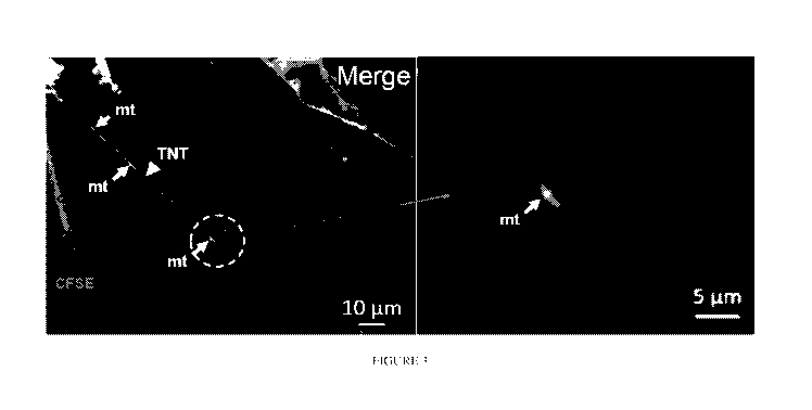
[00106] Figure 3 shows at higher magnification that after 24 h, UCB
iTregs were
analyzed using MitoTracker Red FM by confocal microscopy. BM-MSC were pre-
stained
with CFSE and MitoTracker Red FM and then cultured for 24 h. Analysis of
recorded images
was performed using Zen 2012 (Carl Zeiss) software. iTregs are observed
immediately
adjacent to MSC TNT which contains mitochondria (red arrow). This data
supports that UCB
iTregs take up BM-MSC mitochondria via TNT direct contact. Images are
representative from
4 different experiments (n=6).
[00107] Figure 4 shows experimental methods are described in slide 18. BM-
MSC
were pre-stained with CFSE and MitoTracker Red FM and then cultured with iTreg
for 24 h.
After 24 h, UCB iTregs were analyzed using MitoTracker Red FM by confocal
microscopy.
Analysis of recorded images was performed using Zen 2012 (Carl Zeiss)
software. These
images support that UCB iTregs take up MSC mitochondria via TNT direct
contact. Images
are representative from 4 different experiments (n=6).
[00108] Figures 5A-5B show that BM-MSC were pre-stained with CFSE and
MitoTracker Red FM and then cultured with iTreg for 24 h. After 24 h, UCB
Tregs were
analyzed using MitoTracker Red FM by confocal microscopy. Analysis of recorded
images
was performed using Zen 2012 (Carl Zeiss) software. Here, the image shows that
the majority
of iTreg are MitoTracker positive, indicating uptake of MSC mitochondria.
Results were
normalized by iTreg cultured in media/IL-2 alone. Data are representative of
three
independent experiments SD (n=6). **, P<0.01.
CA 03119452 2021-05-10
WO 2020/102321 PCT/US2019/061140
-29-
[00109] Figure 6 depicts use of Cytochalasin B, an F-actin-
depolymerizing agent
known to abolish TNT formation. To test whether mitochondrial transfer occurs
via
Tunneling NanoTubule (TNT), BM-MSC were treated with Cytochalasin B dissolved
in
DMSO and compared with DMSO alone for mock control. MSC were pre-stained with
MitoTracker green FM for 30 min and then cultured with iTreg (MSC Platform)
including
mock DMSO control (Mock control) and Cytochalasin B (350 nM) treated BM-MSC
(Cytochalasin B) for 24 h, and compared with iTreg alone (Treg alone). After
24 h iTregs
were analyzed using MitoTracker green MFI by flow cytometry. Flow data are
representative
from three different experiments (n=6).
[00110] Figures 7A-7B show that mitochondrial transfer occurs via Tunneling
NanoTubule (TNT). BM-MSC were treated with Cytochalasin B and compared with
mock
control (Co-culture). MSC were pre-stained with MitoTracker green FM for 30
min and then
cultured with iTreg (Co-culture; Red) and compared with iTreg alone (Treg
alone; shaded)
and Cytochalasin B (350 nM) treated BM-MSC (Cyto B tx; blue) for 24 h. After
24 h co-
culture with BM-MSC treated with Cytochalasin B, iTregs were analyzed using
MitoTracker
green MFI by flow cytometry.
[00111] Figures 8A-8B show that BM-MSC were pre-stained with CFSE and
MitoTracker Red FM and then cultured with UCB iTregs with and without
Cytochalasin B
(350 nM) for 24 h. After 24 h, UCB iTregs (Hoechst) were analyzed using
MitoTracker Red
FM by confocal microscopy. Analysis of recorded images was performed using
ImageJ or
Zen 2012 (Carl Zeiss) software. Arrows show BM- MSC mitochondria within iTreg.
Image
data shows significantly reduced number of MitoTracker+ iTregs detected. Data
shown from
three different experiments SD (n=6). ****, P<0.0001. These data demonstrate
that
Cytochalasin B treatment significantly blocks mitochondrial transfer from MSC
to UCB
iTregs during IL-2 driven ex vivo expansion. Together, these results support
that TNT is a
critical conduit for mitochondrial transfer from BM-MSC to UCB iTregs during
IL-2 driven
ex vivo expansion.
CA 03119452 2021-05-10
WO 2020/102321 PCT/US2019/061140
-30-
[00112] Figures 9A-9B show that ROS inhibitor: antioxidant N-
acetylcysteine (NAC)
200uM was added to BM-MSC + iTreg culture for 24-36 h. Data are representative
of two
independent experiments SD (n=6). **, P<0.01. Image was take at 24-36 h
incubation.
Analysis of recorded images was performed using Zen 2012 (Carl Zeiss)
software. Arrows
show BM-MSC mitochondria in iTreg. Image results demonstrate that addition of
this ROS
inhibitor minimally reduced mitochondria uptake by Treg, as there are
MitoTracker+ iTregs
detected. These image results support that mitochondrial transfer from BM-MSC
into Tregs
is not dependent on ROS mediated mechanism of action. *P<0.05.
[00113] Figures 10A-10B show iTregs in MSC platform culture have
greatly enhanced
ROS levels.
[00114] Studies were designed to identify the mechanisms underlying
the observation
that IL-2 driven ex vivo expansion of iTreg over MSC significantly enhances
the number and
function of iTregs. Cell expansion: 2-5 x 10⁵ cells were sub-cultured with ex-
vivo media with
added IL- 2 (100 U/ml). Media was changed every 2-3 days. UCB iTreg cells were
harvested at
day 21- 23 from media/IL-2 alone vs media/IL-2 over a MSC platform.
Tetramethylrhodamine
(TMRM): Methyl ester is a cell-permeant dye. It accumulates in healthy active
mitochondria
membrane. Cells were stained with Image-iTTM TMRM (invitrogen) following
manufacturers
instruction. Cells were analyzed by FACS and confocal microscopy.
[00115] Figures 11A-11B show TGF-f3 induced UCB iTreg were harvested
during IL-2
driven expansion in either media/IL-2 alone (media) v. media/IL-2 over BM- MSC
(BM-
MSC) and surface stained with CD4-APC. Cells were resuspended in media at
concentration
2 x 10e6 cells/ml. Cells were incubated 30 min at 37 C with
Tetramethylrhodamine, methyl
ester (TMRM) (20 nM). Cells were washed with buffer and analyzed. Data shown
from two
different experiments. **, P<0.01. This data demonstrates that iTregs expanded
ex vivo in IL-
2/media over a MSC monolayer have increased mitochondrial membrane potential.
These
results support that MSC mitochondria enhance iTregs function by enhanced
mitochondrial
activity after transfer.
CA 03119452 2021-05-10
WO 2020/102321 PCT/US2019/061140
-31-
[00116] Figures 12A-12B show iTregs ex vivo expanded over BM-MSC have
increased
mitochondria membrane potential. These results support that BM-MSC
mitochondria
enhance iTregs function by increased mitochondria activity after transfer into
iTregs.
Analysis of recorded images was performed using ImageJ or Zen 2012 (Carl
Zeiss) software.
Data shown from two different experiments. ***, P<0.01.
[00117] Figure 13 shows iTreg's ATP were enhanced in BM MSC platform.
[00118] Additional experiments were conducted to determine the
mechanisms that
drive MSC mitochondrial transfer into proliferating iTregs. Treg express
apyrases (CD39)
and ecto-5'-nucleotidase (CD73) which have been shown to contribute to their
inhibitory
function by generating adenosine (Alam et al., 2009; Kerkela et al., 2016).
Also, it has been
shown that CD73-generated adenosine induces cortical actin polymerization via
adenosine Al
receptor (A1R) induction of a Rho GTPase CDC42-dependent conformational change
of the
actin-related proteins 2 and 3 (ARP2/3) actin polymerization complex member N-
WASP
(Bowser et al., 2016). To test whether CD73 contributes to drive MSC
mitochondrial
transfer, CD73 blocking Ab was added to MSC co-culture and iTreg mitochondrial
mass was
measured.
[00119] Figure 14B shows that after CD 73 blocking, iTreg
mitochondrial mass was
significantly diminished.
[00120] Figures 14A-14H and 15A-15D show that the CD39/CD73 pathway
drives
MSC mitochondrial (mt) transfer into proliferating iTreg. MSCs were transduced
with mt-
GFP lentivirus to generate stable mt-GFP+ MSCs (Figure 15A). Mt-GFP+ iTregs
were
detected and were significantly increased during 21 day co-cultured with mt-
GFP lentivirus
transduced MSCs (Figures 15B-15C). Mt-GFP+ iTregs were significantly decreased
with
surface CD73 blocking or inhibition of TNT formation (Figure 14C). As CD39 and
CD73
calibrate purinergic signals delivered to immune cells through the conversion
of ADP/ATP to
AMP and AMP to adenosine, respectively. (Allard et al., 2017; Antonioli et
al., 2013).
Experiments were conducted to inhibit each pathway and noted significantly
reduced mt-
GFP+ iTregs (Figures 14D and 15D).
CA 03119452 2021-05-10
WO 2020/102321 PCT/US2019/061140
-32-
[00121] As Miro 1 has been shown to be a key regulator in
mitochondrial intracellular
transport (Liu et al., 2012), experiments were designed to determine its role,
if any, in MSC
mitochondrial transfer to iTreg via TNT. Miro 1 expression was measured in MSC
after co-
culture with iTregs. iTreg co-cultivation was associated with significantly
enhanced
expression of Miro 1 in MSC including both protein and RNA levels (Figure
14E). Further,
the enhanced MSC expression of Mirol was inhibited by CD39 inhibition (Figure
14F).
[00122] BACH2 has been shown to maintain the stability and function of
murine Treg
(Kim et al., 2014; Roychoudhuri et al., 2013). BACH2 was previously identified
as highly
expressed in human iTreg and plays a key role in iTreg stability (Do et al.,
2018). Additional
studies have identified a pathway by which SENP3 modulates the SUMOylation of
BACH2
to control iTreg stability in response to changes in environmental conditions,
particularly
intracellular ROS (Yu et al., 2018). It was postulated that mitochondrial
transfer from MSC
may modulate BACH2/SENP3 expression and/or function, given the previous
observations
of MSC mitochondrial transfer exerting a positive effect on iTreg Foxp3
stability and
suppressive function (Jang et al., 2015; Watanabe-Matsui et al., 2011; Yu et
al., 2018). iTreg
BACH2 and SENP3 protein expression was measured, and MSC co-culture and
standard IL-
2/media conditions during short-term (day 14 and 21) in vitro culture were
compared. iTreg
Foxp3, BACH2 and SENP3 expression was notably significantly increased in MSC
co-culture
conditions (Figure 14G). The effect of MSC mitochondrial transfer on iTreg
BACH2 and
SENP3 protein levels after cytochalasin B treatment was examined next. Levels
of iTreg
BACH2 protein expression were dramatically decreased (Figure 14H).
Cytochalasin B
treatment had no significant effect on iTreg SENP3 protein expression,
possibly due to
SENP3 activation and/or function in this setting (Figure 14H). Overall, this
data suggests that
MSC mitochondrial transfer is associated with enhanced BACH2 expression and
due to its
regulation of Foxp3, positive benefit of enhanced and stable Foxp3 expression.
[00123] Figures 16A-16B show that CD39/CD73 pharmacological inhibitors
block
transfer of MSC mitochondria into UCB iTregs during IL-2 driven ex vivo
expansion. To test
whether mitochondrial transfer is mediated by CD39 and CD73 signals,
pharmacological
inhibitors were added during IL-2 driven expansion. Differentiated Foxp3 +
iTreg cells were
CA 03119452 2021-05-10
WO 2020/102321 PCT/US2019/061140
-33-
added to mt-GFP lentiviral transduced MSC platform (24 well plate) at 5 x 10⁵
cells/ml with
IL-2 (100U/m1) for 72hrs. To block CD39 and CD73 signal, 50 uM CD39 inhibitor
(P0M1, a
E-NTPDases inhibitor; sigma) and 100 uM CD73 inhibitor (Adenosine 5'-(a,f3-
methylene)diphosphate; sigma) or together were added into culture. iTregs were
collected at
72 hrs after culture. iTregs were surface stained with anti-human CD4 APC
antibody for
Flow analysis. Stained cells were analyze with FACS Fortessa . Data are from 3-
4 individual
experiments (n = 5-14). **, p<0.01; ***, p<0.001. These data demonstrate that
treatment of
CD39/CD73 inhibitor significantly blocks mitochondrial transfer from BM-MSC to
UCB
iTregs during IL-2 driven ex vivo expansion. Collectively, these results
suggest that
mitochondrial transfer from BM-MSC to UCB iTregs during IL-2 driven ex vivo
expansion is
mediated by the ectoenzymes CD39 and CD73 pathway.
[00124] Figures 17A-17I and 18A-18G show that MSC co-culture with
iTregs
ameliorates xenogeneic GVHD and allogeneic GVHD in humanized mouse model.
Inhibition
of MSC mitochondrial transfer into iTregs resulted in significantly reduced
suppressive
function vs. control (Figure 17A). For assessment of the in vivo functions of
iTreg in MSC
co-culture during iTreg ex vivo expansion in IL-2/media, iTregs were
adoptively transferred
into GVHD induced NSG mice. This xenogeneic model of GVHD was used in which
iTregs
culture expanded short-term (21 days) were injected 7 days after adult human
PBMC iv
injection to induce GVHD. Treatment groups were blinded to technicians
performing GVHD
(Ehx et al., 2018) and survival assessments (Sonntag et al., 2015).
[00125] Mice treated with MSC co-culture iTregs demonstrated
significantly improved
survival, stable weight, and lower GVHD clinical scores (Figure 17B). Foxp3
expression and
Foxp3+ CD4 T cells in harvested spleen were significantly higher in mice
treated with MSC
co-cultured iTregs at 2 weeks after GVHD induction (Figures 17C and 18A). IFN7
producing CD8+ and CD4+ T cells were dramatically reduced in spleen cells
harvested from
mice treated with MSC co-cultured iTregs (Figure 18B). Consistent with
cytokine staining,
serum and ex vivo levels of pro-inflammatory cytokine IFN7 and TNFa were
significantly
reduced in animals treated with MSC co-cultured iTregs (Figures 17D and 17E).
CA 03119452 2021-05-10
WO 2020/102321 PCT/US2019/061140
-34-
[00126] To determine whether MSC co-culture expanded iTregs have a
therapeutic
effect in autoimmune disease, a chronic GVHD model using humanized mice which
is similar
with autoimmune phenotype was used (Sonntag et al., 2015). Human CD45+
engraftment
was measured in mice which received CD34+ cells at 24 weeks (Figure 18C). MSC
co-
.. culture expanded iTreg received mice had slightly decreased body weight
loss than the other
group (Figure 17C) but survival was improved (Figure 17F). Foxp3+ Treg cells
were
significantly enhanced in MSC co-culture expanded iTreg transferred mice
(Figure 18D). It
was observed that IFN7 producing CD8+ and CD4+ T cells were significantly
reduced in
MSC co-culture expanded iTreg treated mice (Figure 17G). To explore the effect
of MSC
.. mitochondrial transfer on iTregs in vivo, MSC co-culture iTregs with added
CD39 inhibitor
were generated. CD39 inhibitor treatment reduced Foxp3+ iTreg percentage in
adult PBL-
induced GVHD (Figure 17H). IFNI, producing CD8+ and CD4+ T cells were
significantly
increased in CD39 inhibitor treated iTreg treated mice (Figures 171 and 18E).
The level of
serum and production of IFN7 and TNFa were significantly increased in CD39
inhibitor iTreg
treated mice compared to the control (Figures 18F and 18G). Collectively,
these results
reveal that the suppressive iTregs were maintained by MSC mitochondrial
transfer during IL-
2 driven ex vivo expansion, and further that these iTregs can protect
autoimmune disease in
vivo.
[00127] Figures 19A-19B show that dysfunctional mitochondria do not
transfer into
iTregs. To test whether apoptotic MSCs transfer mitochondria into
proliferating iTregs, 3 x
105 MSCs were incubated with mt-GFP lentiviral and seeded into 24 well plate.
MSCs were
incubated 30 minutes at 37 degrees and cells were washed with media. Foxp3+
iTreg cells
were added into mt-GFP lentiviral transduced mock or rotenone treated MSC
platform (24
well plate) at 5 x 10⁵ cells/ml with IL-2 (100 Um') in culture for 72 hrs.
iTregs were surface
.. stained with anti-human CD4 APC antibody for Flow analysis. Stained cells
were analyzed
with FACS Fortessa . Data are from two different experiments. N=5-6, ***
p<0.001.
[00128] Intrinsic defects in Tregs as observed in autoimmune disease
patients may
hamper the success of autologous nTreg therapies (Kumar et al., 2006; Valencia
et al., 2006;
Viglietta et al., 2004). Previous studies have shown that ex vivo-expanded,
partially HLA-
CA 03119452 2021-05-10
WO 2020/102321 PCT/US2019/061140
-35-
matched nTregs from allogeneic UCB are well tolerated in humans (Brunstein et
al., 2011).
A major obstacle to the clinical implementation of UCB iTregs is the
instability of iTreg
Foxp3 expression and loss of suppressor function upon transfer to an
inflammatory
environment (Hippen et al., 2011). The results reported in this invention
demonstrate that
BM-MSC co-culture during IL-2 driven ex vivo expansion provides new
physiological
condition to sustain Foxp3 expression and thereby iTreg suppressive function
in inflammatory
milieus in vitro and in vivo.
[00129] MSC co-culture was observed to be associated with
significantly upregulated
expression of CD25, CTLA-4, and ICOS, while expression of LAG-3 and TIM-3,
which may
contribute to exhaustion of activated T cells, was decreased. Higher
proportions of CD62L+
and CD45RA iTreg cells after short-term (21 day) MSC co-culture was observed.
Inflammatory environments are attributed to the rapid loss of Foxp3 expression
and functional
activity of iTregs (Koenecke et al., 2009). The data show a significantly
enhanced
suppressive function, including inhibition of effector cell activation and
maintained Foxp3
stability in MSC co-culture expanded iTregs exposed to inflammatory conditions
in vitro and
in vivo. It will be important to examine the epigenetic modification of the
Treg-specific
demethylated region in the Foxp3 gene to gain further insights into the
mechanisms of BM-
MSC-enhanced iTreg stability (Someya et al., 2017).
[00130] Although prior studies point to MSC enhancement of Treg
function to be
primarily via paracrine mechanisms (Del Fattore et al., 2015), this invention
identifies that
MSC support of iTreg Foxp3 expression and sustained suppressive function
requires direct
cell-cell contact. Further, this invention includes the surprising finding
that a key mechanism
involves mitochondrial transfer by MSC. As the cellular and molecular
mechanisms driving
MSC mitochondrial transfer to proliferating cells have not been previously
been elucidated,
this invention further identifies that iTreg CD39/CD73 signaling drives MSC
mitochondrial
transfer and further that mitochondrial transfer results in augmented iTreg
BACH2 and
SENP3 expression. BACH2 regulates human UCB iTreg development via direct
transcriptional activity at the Foxp3 promoter (Do et al., 2018). SENP3 is a
SUMO specific
protease that serves to maintain Treg stability (Yu et al., 2018).
CA 03119452 2021-05-10
WO 2020/102321 PCT/US2019/061140
-36-
[00131] CD39 and CD73 play together strategic roles in immune
responses (Allard et
al., 2017; Antonioli et al., 2013). CD39 and CD73 degrade extracellular ATP to
yield AMP
and anti-inflammatory adenosine (Deaglio et al., 2007). CD73-/- mice show
enhanced
antitumor immunity (Stagg et al., 2011) and worse gastritis compared to
functional CD73
controls, and adoptive injection of WT Tregs reverses these immune responses
(Alam et al.,
2009). This invention and others identify (Ehrentraut et al., 2013; Kobie et
al., 2006) that
CD73 signaling on Tregs is critical to maintain Treg suppressive function. As
mitochondrial
transfer to Tregs has not been previously examined, this invention identifies
that CD39 and
CD73 signaling on proliferating iTreg drives MSC mitochondria transfer into
iTregs during
IL-2 driven ex vivo expansion.
[00132] Miro 1 has been shown to regulate intercellular mitochondrial
transfer and
enhance injured cell recovery (Ahmad et al., 2014). In a previous study
injured astrocytes
induced increased levels of Miro 1 expression in MSC and this was correlated
with
mitochondrial transfer (Babenko et al., 2018). In the present invention, data
show that
proliferating iTregs induced Miro 1 expression on BM-MSC during co-cultivation
ex vivo.
Further, MSC mitochondrial transfer occurs via TNT.
[00133] Mitochondria are an important source of ROS (Turrens, 2003).
Interestingly,
ROS can regulate cell cycle and function in signaling (Byun et al., 2008;
Schieke et al., 2008)
and are critical for cancer cell tumorigenicity (Weinberg et al., 2010). This
is particularly
.. interesting in light of the finding that ROS is a major driving force for
mitochondrial transfer
via TNTs from bone marrow stromal cells to leukemic blasts (Marlein et al.,
2017). Further
work has demonstrated that mitochondrial metabolism also plays a critical role
in T cell
activation. This invention identifies for the first time the role of CD39/73
expression on
proliferating iTreg that drives the transfer of MSC mitochondria. The
observations in this
invention strongly indicate that increased mitochondria quantity in iTregs is
derived from
MSCs via TNT transfer but intriguingly, metabolic signaling of the
proliferating cells rather
than ROS expression appears to be a critical mechanism driving this process.
CA 03119452 2021-05-10
WO 2020/102321 PCT/US2019/061140
-37-
[00134] Regardless of cell type, mitochondria are now recognized to
play roles that
extend well beyond the production of energy. A central idea emerging from many
studies is
that T cells undergo changes in cellular metabolism during activation (Akkaya
et al., 2018).
In contrast to quiescent T cells whose metabolic requirements mainly encompass
only cellular
trafficking and housekeeping functions, actively proliferating cells must
generate additional
ATP for functions including the generation of intermediates required in
various biosynthesis
pathways and signaling molecules for anabolic metabolism. Future work is
indicated vaulting
from this invention to identify how signaling mechanisms can be modulated to
enhance
mitochondrial transfer. Importantly, this invention supports that
mitochondrial transfer is a
key mechanism of MSC supportive function for proliferating cells. Importantly,
iTreg
mitochondria membrane potential is boosted by co-culture with MSCs. Second,
pharmacological inhibition of mitochondrial transfer nearly completely negates
the benefit of
MSCs on iTreg Foxp3 expression and suppressive function.
[00135] In summary, these surprising results highlight the importance
of mitochondrial
.. functions in maintaining iTreg FoxP3 expression and suppressive function in
inflammatory
conditions in vitro and in vivo. Although, several factors can modulate iTreg
stability and
functions in different disease models, these results provide that the
mitochondrial BACH2
pathway is a prominent mechanism of MSC-enhanced iTreg stability and function,
and
CD39/73 signaling on proliferating cells drives MSC-mediated mitochondrial
transfer.
[00136] These and additional embodiments of the invention will be apparent
to one
skilled in the art upon a review of the present disclosure, which is not
intended to be limiting
to the scope of the claims. The references cited are hereby incorporated by
reference herein in
their entireties.
REFERENCES
Ahmad, T., Mukherjee, S., Pattnaik, B., Kumar, M., Singh, S., Kumar, M.,
Rehman, R.,
Tiwari, B.K., Jha, K.A., Barhanpurkar, A.P., et al. (2014). Mirol regulates
intercellular
mitochondrial transport & enhances mesenchymal stem cell rescue efficacy. EMBO
J 33,
994-1010.
CA 03119452 2021-05-10
WO 2020/102321 PCT/US2019/061140
-38-
Akkaya, B., Roesler, A.S., Miozzo, P., Theall, B.P., Al Souz, J., Smelkinson,
M.G., Kabat, J.,
Traba, J., Sack, M.N., Brzostowski, J.A., et al. (2018). Increased
Mitochondrial Biogenesis
and Reactive Oxygen Species Production Accompany Prolonged CD4(+) T Cell
Activation. J
Immunol 201, 3294-3306.
.. Alam, M.S., Kurtz, C.C., Rowlett, R.M., Reuter, B.K., Wiznerowicz, E., Das,
S., Linden, J.,
Crowe, S.E., and Ernst, P.B. (2009). CD73 is expressed by human regulatory T
helper cells
and suppresses proinflammatory cytokine production and Helicobacter felis-
induced gastritis
in mice. J Infect Dis 199, 494-504.
Allard, B., Longhi, M.S., Robson, S.C., and Stagg, J. (2017). The
ectonucleotidases CD39
.. and CD73: Novel checkpoint inhibitor targets. Immunol Rev 276, 121-144.
Antonioli, L., Pacher, P., Vizi, E.S., and Hasko, G. (2013). CD39 and CD73 in
immunity and
inflammation. Trends Mol Med 19, 355-367.
Babenko, V.A., Silachev, D.N., Popkov, V.A., Zorova, L.D., Pevzner, I.B.,
Plotnikov, E.Y.,
Sukhikh, G.T., and Zorov, D.B. (2018). Mirol Enhances Mitochondria Transfer
from
Multipotent Mesenchymal Stem Cells (MMSC) to Neural Cells and Improves the
Efficacy of
Cell Recovery. Molecules 23.
Babenko, V.A., Silachev, D.N., Zorova, L.D., Pevzner, I.B., Khutornenko, A.A.,
Plotnikov,
E.Y., Sukhikh, G.T., and Zorov, D.B. (2015). Improving the Post-Stroke
Therapeutic Potency
of Mesenchymal Multipotent Stromal Cells by Cocultivation With Cortical
Neurons: The
Role of Crosstalk Between Cells. Stem Cells Transl Med 4, 1011-1020.
Baecher-Allan, C., Brown, J.A., Freeman, G.J., and Hafler, D.A. (2001).
CD4+CD25high
regulatory cells in human peripheral blood. J Immunol 167, 1245-1253.
Beres, A., Komorowski, R., Mihara, M., and Drobyski, W.R. (2011). Instability
of Foxp3
expression limits the ability of induced regulatory T cells to mitigate graft
versus host disease.
Clin Cancer Res 17, 3969-3983.
CA 03119452 2021-05-10
WO 2020/102321 PCT/US2019/061140
-39-
Bluestone, J.A., Buckner, J.H., Fitch, M., Gitelman, S.E., Gupta, S.,
Hellerstein, M.K.,
Herold, K.C., Lares, A., Lee, M.R., Li, K., et al. (2015a). Type 1 diabetes
immunotherapy
using polyclonal regulatory T cells. Sci Transl Med 7, 315ra189.
Bluestone, J.A., Trotta, E., and Xu, D. (2015b). The therapeutic potential of
regulatory T cells
for the treatment of autoimmune disease. Expert Opin Ther Targets 19, 1091-
1103.
Bowser, J.L., Blackburn, M.R., Shipley, G.L., Molina, J.G., Dunner, K., Jr.,
and Broaddus,
R.R. (2016). Loss of CD73-mediated actin polymerization promotes endometrial
tumor
progression. J Clin Invest 126, 220-238.
Brunstein, C.G., Miller, J.S., Cao, Q., McKenna, D.H., Hippen, K.L.,
Curtsinger, J., Defor, T.,
Levine, B.L., June, C.H., Rubinstein, P., et al. (2011). Infusion of ex vivo
expanded T
regulatory cells in adults transplanted with umbilical cord blood: safety
profile and detection
kinetics. Blood 117, 1061-1070.
Byun, H.O., Kim, H.Y., Lim, J.J., Seo, Y.H., and Yoon, G. (2008).
Mitochondrial dysfunction
by complex II inhibition delays overall cell cycle progression via reactive
oxygen species
production. Journal of cellular biochemistry 104, 1747-1759.
Chang, K.T., Niescier, R.F., and Min, K.T. (2011). Mitochondrial matrix Ca2+
as an intrinsic
signal regulating mitochondrial motility in axons. Proc Natl Acad Sci U S A
108, 15456-
15461.
Chen, W., Jin, W., Hardegen, N., Lei, K.J., Li, L., Marinos, N., McGrady, G.,
and Wahl, S.M.
(2003). Conversion of peripheral CD4+CD25- naive T cells to CD4+CD25+
regulatory T
cells by TGF-beta induction of transcription factor Foxp3. The Journal of
experimental
medicine 198, 1875-1886.
Davidson, T.S., DiPaolo, R.J., Andersson, J., and Shevach, E.M. (2007).
Cutting Edge: IL-2 is
essential for TGF-beta-mediated induction of Foxp3+ T regulatory cells.
Journal of
immunology 178, 4022-4026.
CA 03119452 2021-05-10
WO 2020/102321 PCT/US2019/061140
-40-
Deaglio, S., Dwyer, K.M., Gao, W., Friedman, D., Usheva, A., Erat, A., Chen,
J.F., Enjyoji,
K., Linden, J., Oukka, M., et al. (2007). Adenosine generation catalyzed by
CD39 and CD73
expressed on regulatory T cells mediates immune suppression. J Exp Med 204,
1257-1265.
Del Fattore, A., Luciano, R., Pascucci, L., Goffredo, B.M., Giorda, E.,
Scapaticci, M.,
Fierabracci, A., and Muraca, M. (2015). Immunoregulatory Effects of
Mesenchymal Stem
Cell-Derived Extracellular Vesicles on T Lymphocytes. Cell Transplant 24, 2615-
2627.
Do, J.S., Zhong, F., Huang, A.Y., Van't Hof, W.J., Finney, M., and Laughlin,
M.J. (2018).
Foxp3 expression in induced T regulatory cells derived from human umbilical
cord blood vs.
adult peripheral blood. Bone Marrow Transplant 53, 1568-1577.
Du, Y.M., Zhuansun, Y.X., Chen, R., Lin, L., Lin, Y., and Li, J.G. (2018).
Mesenchymal stem
cell exosomes promote immunosuppression of regulatory T cells in asthma. Exp
Cell Res 363,
114-120.
Ehrentraut, H., Clambey, E.T., McNamee, E.N., Brodsky, K.S., Ehrentraut, S.F.,
Poth, J.M.,
Riegel, A.K., Westrich, J.A., Colgan, S.P., and Eltzschig, H.K. (2013). CD73+
regulatory T
cells contribute to adenosine-mediated resolution of acute lung injury. FASEB
J 27, 2207-
2219.
Ehx, G., Somja, J., Warnatz, H.J., Ritacco, C., Hannon, M., Delens, L.,
Fransolet, G.,
Delvenne, P., Muller, J., Beguin, Y., et al. (2018). Xenogeneic Graft-Versus-
Host Disease in
Humanized NSG and NSG-HLA-A2/HHD Mice. Front Immunol 9, 1943.
Goodman, W.A., Levine, A.D., Massari, J.V., Sugiyama, H., McCormick, T.S., and
Cooper,
K.D. (2009). IL-6 signaling in psoriasis prevents immune suppression by
regulatory T cells. J
Immunol 183, 3170-3176.
Han, H., Hu, J., Yan, Q., Zhu, J., Zhu, Z., Chen, Y., Sun, J., and Zhang, R.
(2016). Bone
marrow-derived mesenchymal stem cells rescue injured H9c2 cells via
transferring intact
mitochondria through tunneling nanotubes in an in vitro simulated
ischemia/reperfusion
model. Molecular medicine reports 13, 1517-1524.
CA 03119452 2021-05-10
WO 2020/102321 PCT/US2019/061140
-41-
Hayakawa, K., Esposito, E., Wang, X., Terasaki, Y., Liu, Y., Xing, C., Ji, X.,
and Lo, E.H.
(2016). Transfer of mitochondria from astrocytes to neurons after stroke.
Nature 535, 551-
555.
Hippen, K.L., Merkel, S.C., Schirm, D.K., Nelson, C., Tennis, N.C., Riley,
J.L., June, C.H.,
Miller, J.S., Wagner, J.E., and Blazar, B.R. (2011). Generation and large-
scale expansion of
human inducible regulatory T cells that suppress graft-versus-host disease. Am
J Transplant
11, 1148-1157.
Huehn, J., Polansky, J.K., and Hamann, A. (2009). Epigenetic control of FOXP3
expression:
the key to a stable regulatory T-cell lineage? Nat Rev Immunol 9, 83-89.
Jang, K.J., Mano, H., Aoki, K., Hayashi, T., Muto, A., Nambu, Y., Takahashi,
K., Itoh, K.,
Taketani, S., Nutt, S.L., et al. (2015). Mitochondrial function provides
instructive signals for
activation-induced B-cell fates. Nat Commun 6, 6750.
Kanamori, M., Nakatsukasa, H., Okada, M., Lu, Q., and Yoshimura, A. (2016).
Induced
Regulatory T Cells: Their Development, Stability, and Applications. Trends in
immunology
37, 803-811.
Kasakovski, D., Xu, L., and Li, Y. (2018). T cell senescence and CAR-T cell
exhaustion in
hematological malignancies. J Hematol Oncol 11, 91.
Kerkela, E., Laitinen, A., Rabina, J., Valkonen, S., Takatalo, M., Larjo, A.,
Veijola, J.,
Lampinen, M., Siljander, P., Lehenkari, P., et al. (2016). Adenosinergic
Immunosuppression
by Human Mesenchymal Stromal Cells Requires Co-Operation with T cells. Stem
Cells 34,
781-790.
Kim, E.H., Gasper, D.J., Lee, S.H., Plisch, E.H., Svaren, J., and Suresh, M.
(2014). Bach2
regulates homeostasis of Foxp3+ regulatory T cells and protects against fatal
lung disease in
mice. J Immunol 192, 985-995.
CA 03119452 2021-05-10
WO 2020/102321 PCT/US2019/061140
-42-
Kobie, J.J., Shah, P.R., Yang, L., Rebhahn, J.A., Fowell, D.J., and Mosmann,
T.R. (2006). T
regulatory and primed uncommitted CD4 T cells express CD73, which suppresses
effector
CD4 T cells by converting 5'-adenosine monophosphate to adenosine. J Immunol
177, 6780-
6786.
Koenecke, C., Czeloth, N., Bubke, A., Schmitz, S., Kissenpfennig, A.,
Malissen, B., Huehn,
J., Ganser, A., Forster, R., and Prinz, I. (2009). Alloantigen-specific de
novo-induced Foxp3+
Treg revert in vivo and do not protect from experimental GVHD. Eur J Immunol
39, 3091-
3096.
Kumar, M., Putzki, N., Limmroth, V., Remus, R., Lindemann, M., Knop, D.,
Mueller, N.,
Hardt, C., Kreuzfelder, E., and Grosse-Wilde, H. (2006). CD4+CD25+FoxP3+ T
lymphocytes
fail to suppress myelin basic protein-induced proliferation in patients with
multiple sclerosis. J
Neuroimmunol 180, 178-184.
Lange, C., Scholl, M., Melms, A., and Bischof, F. (2011). CD62L(high) Treg
cells with
superior immunosuppressive properties accumulate within the CNS during
remissions of
EAE. Brain, behavior, and immunity 25, 120-126.
Le Texier, L., Lineburg, K.E., Cao, B., McDonald-Hyman, C., Leveque-El
Mouttie, L.,
Nicholls, J., Melino, M., Nalkurthi, B.C., Alexander, K.A., Teal, B., et al.
(2016). Autophagy-
dependent regulatory T cells are critical for the control of graft-versus-host
disease. JCI
Insight 1, e86850.
Lee, S., Park, K., Kim, J., Min, H., and Seong, R.H. (2018). Foxp3 expression
in induced
regulatory T cells is stabilized by C/EBP in inflammatory environments. EMBO
Rep 19.
Leprat, P., Ratinaud, M.H., and Julien, R. (1990). A new method for testing
cell ageing using
two mitochondria specific fluorescent probes. Mech Ageing Dev 52, 149-167.
Liu, S., Sawada, T., Lee, S., Yu, W., Silverio, G., Alapatt, P., Millan, I.,
Shen, A., Saxton, W.,
Kanao, T., et al. (2012). Parkinson's disease-associated kinase PINK1
regulates Miro protein
level and axonal transport of mitochondria. PLoS Genet 8, e1002537.
CA 03119452 2021-05-10
WO 2020/102321 PCT/US2019/061140
-43-
Long, S.A., Cerosaletti, K., Bollyky, P.L., Tatum, M., Shilling, H., Zhang,
S., Zhang, Z.Y.,
Pihoker, C., Sanda, S., Greenbaum, C., and Buckner, J.H. (2010). Defects in IL-
2R signaling
contribute to diminished maintenance of FOXP3 expression in CD4(+)CD25(+)
regulatory T-
cells of type 1 diabetic subjects. Diabetes 59, 407-415.
Lu, S.Y., Liu, K.Y., Liu, D.H., Xu, L.P., and Huang, X.J. (2011). High
frequencies of
CD62L(+) naive regulatory T cells in allografts are associated with a low risk
of acute graft-
versus-host disease following unmanipulated allogeneic haematopoietic stem
cell
transplantation. Clinical and experimental immunology 165, 264-277.
Marlein, C.R., Zaitseva, L., Piddock, R.E., Robinson, S.D., Edwards, D.R.,
Shafat, M.S.,
Zhou, Z., Lawes, M., Bowles, K.M., and Rushworth, S.A. (2017). NADPH oxidase-2
derived
superoxide drives mitochondrial transfer from bone marrow stromal cells to
leukemic blasts.
Blood 130, 1649-1660.
Melief, S.M., Schrama, E., Brugman, M.H., Tiemessen, M.M., Hoogduijn, M.J.,
Fibbe, W.E.,
and Roelofs, H. (2013). Multipotent stromal cells induce human regulatory T
cells through a
novel pathway involving skewing of monocytes toward anti-inflammatory
macrophages.
Stem Cells 31, 1980-1991.
Quintero, 0.A., DiVito, M.M., Adikes, R.C., Kortan, M.B., Case, L.B., Lier,
A.J., Panaretos,
N.S., Slater, S.Q., Rengarajan, M., Feliu, M., and Cheney, R.E. (2009). Human
Myo 19 is a
novel myosin that associates with mitochondria. Curr Biol 19, 2008-2013.
Rauch, K.S., Hils, M., Lupar, E., Minguet, S., Sigvardsson, M., Rottenberg,
M.E., Izcue, A.,
Schachtrup, C., and Schachtrup, K. (2016). Id3 Maintains Foxp3 Expression in
Regulatory T
Cells by Controlling a Transcriptional Network of E47, Spi-B, and 50053. Cell
Rep 17,
2827-2836.
Rojewski, M.T., Weber, B.M., and Schrezenmeier, H. (2008). Phenotypic
Characterization of
Mesenchymal Stem Cells from Various Tissues. Transfus Med Hemother 35, 168-
184.
CA 03119452 2021-05-10
WO 2020/102321 PCT/US2019/061140
-44-
Roychoudhuri, R., Eil, R.L., Clever, D., Klebanoff, C.A., Sukumar, M., Grant,
F.M., Yu, Z.,
Mehta, G., Liu, H., Jin, P., et al. (2016). The transcription factor BACH2
promotes tumor
immunosuppression. J Clin Invest 126, 599-604.
Roychoudhuri, R., Hirahara, K., Mousavi, K., Clever, D., Klebanoff, C.A.,
Bonelli, M.,
Sciume, G., Zare, H., Vahedi, G., Dema, B., et al. (2013). BACH2 represses
effector
programs to stabilize T(reg)-mediated immune homeostasis. Nature 498, 506-510.
Schieke, S.M., McCoy, J.P., Jr., and Finkel, T. (2008). Coordination of
mitochondrial
bioenergetics with G1 phase cell cycle progression. Cell cycle 7, 1782-1787.
Schmitt, E.G., and Williams, C.B. (2013). Generation and function of induced
regulatory T
cells. Frontiers in immunology 4, 152.
Sinclair, K., Yerkovich, S.T., and Chambers, D.C. (2013). Mesenchymal stem
cells and the
lung. Respirology 18, 397-411.
Someya, K., Nakatsukasa, H., Ito, M., Kondo, T., Tateda, K.I., Akanuma, T.,
Koya, I.,
Sanosaka, T., Kohyama, J., Tsukada, Y.I., et al. (2017). Improvement of Foxp3
stability
through CNS2 demethylation by TET enzyme induction and activation. Int Immunol
29, 365-
375.
Sonntag, K., Eckert, F., Welker, C., Muller, H., Muller, F., Zips, D., Sipos,
B., Klein, R.,
Blank, G., Feuchtinger, T., et al. (2015). Chronic graft-versus-host-disease
in CD34(+)-
humanized NSG mice is associated with human susceptibility HLA haplotypes for
autoimmune disease. J Autoimmun 62, 55-66.
Stagg, J., Divisekera, U., Duret, H., Sparwasser, T., Teng, M.W., Darcy, P.K.,
and Smyth,
M.J. (2011). CD73-deficient mice have increased antitumor immunity and are
resistant to
experimental metastasis. Cancer Res 71, 2892-2900.
Tasso, R., Ilengo, C., Quarto, R., Cancedda, R., Caspi, R.R., and Pennesi, G.
(2012).
Mesenchymal stem cells induce functionally active T-regulatory lymphocytes in
a paracrine
CA 03119452 2021-05-10
WO 2020/102321 PCT/US2019/061140
-45-
fashion and ameliorate experimental autoimmune uveitis. Investigative
ophthalmology &
visual science 53, 786-793.
Tone, Y., Furuuchi, K., Kojima, Y., Tykocinski, M.L., Greene, M.I., and Tone,
M. (2008).
Smad3 and NFAT cooperate to induce Foxp3 expression through its enhancer. Nat
Immunol
.. 9, 194-202.
Trzonkowski, P., Szarynska, M., Mysliwska, J., and Mysliwski, A. (2009). Ex
vivo expansion
of CD4(+)CD25(+) T regulatory cells for immunosuppressive therapy. Cytometry A
75, 175-
188.
Turrens, J.F. (2003). Mitochondrial formation of reactive oxygen species. The
Journal of
physiology 552, 335-344.
Valencia, X., Stephens, G., Goldbach-Mansky, R., Wilson, M., Shevach, E.M.,
and Lipsky,
P.E. (2006). TNF downmodulates the function of human CD4+CD25hi T-regulatory
cells.
Blood 108, 253-261.
van Leeuwen, M.A., du Pre, M.F., van Wanrooij, R.L., de Ruiter, L.F.,
Raatgeep, H.R.,
Lindenbergh-Kortleve, D.J., Mulder, C.J., de Ridder, L., Escher, J.C., and
Samsom, J.N.
(2013). Changes in natural Foxp3(+)Treg but
not muc os ally-imprinted
CD62L(neg)CD38(+)Foxp3(+)Treg in the circulation of celiac disease patients.
PloS one 8,
e68432.
Viglietta, V., Baecher-Allan, C., Weiner, H.L., and Hafler, D.A. (2004). Loss
of functional
suppression by CD4+CD25+ regulatory T cells in patients with multiple
sclerosis. J Exp Med
199, 971-979.
Vignais, M.L., Caicedo, A., Brondello, J.M., and Jorgensen, C. (2017). Cell
Connections by
Tunneling Nanotubes: Effects of Mitochondrial Trafficking on Target Cell
Metabolism,
Homeostasis, and Response to Therapy. Stem Cells Int 2017, 6917941.
CA 03119452 2021-05-10
WO 2020/102321 PCT/US2019/061140
-46-
Wang, J., Liu, X., Qiu, Y., Shi, Y., Cai, J., Wang, B., Wei, X., Ke, Q., Sui,
X., Wang, Y., et
al. (2018). Cell adhesion-mediated mitochondria transfer contributes to
mesenchymal stem
cell-induced chemoresistance on T cell acute lymphoblastic leukemia cells. J
Hematol Oncol
11, 11.
Watanabe-Matsui, M., Muto, A., Matsui, T., Itoh-Nakadai, A., Nakajima, 0.,
Murayama, K.,
Yamamoto, M., Ikeda-Saito, M., and Igarashi, K. (2011). Heme regulates B-cell
differentiation, antibody class switch, and heme oxygenase-1 expression in B
cells as a ligand
of Bach2. Blood 117, 5438-5448.
Weinberg, F., Hamanaka, R., Wheaton, W.W., Weinberg, S., Joseph, J., Lopez,
M.,
Kalyanaraman, B., Mutlu, G.M., Budinger, G.R., and Chandel, N.S. (2010).
Mitochondrial
metabolism and ROS generation are essential for Kras-mediated tumorigenicity.
Proceedings
of the National Academy of Sciences of the United States of America 107, 8788-
8793.
Xu, Y., Melo-Cardenas, J., Zhang, Y., Gau, I., Wei, J., Montauti, E., Zhang,
Y., Gao, B., Jin,
H., Sun, Z., et al. (2019). The E3 ligase Hrdl stabilizes Tregs by
antagonizing inflammatory
cytokine-induced ER stress response. JCI Insight 4.
Yu, X., Lao, Y., Teng, X.L., Li, S., Zhou, Y., Wang, F., Guo, X., Deng, S.,
Chang, Y., Wu,
X., et al. (2018). SENP3 maintains the stability and function of regulatory T
cells via BACH2
deSUMOylation. Nature communications 9, 3157.
Zheng, J., Chan, P.L., Liu, Y., Qin, G., Xiang, Z., Lam, K.T., Lewis, D.B.,
Lau, Y.L., and
Tu, W. (2013). ICOS regulates the generation and function of human CD4+ Treg
in a CTLA-
4 dependent manner. PLoS One 8, e82203.
Zorn, E., Nelson, E.A., Mohseni, M., Porcheray, F., Kim, H., Litsa, D.,
Bellucci, R.,
Raderschall, E., Canning, C., Soiffer, R.J., et al. (2006). IL-2 regulates
FOXP3 expression in
human CD4+CD25+ regulatory T cells through a STAT-dependent mechanism and
induces
the expansion of these cells in vivo. Blood 108, 1571-1579.
CA 03119452 2021-05-10
WO 2020/102321 PCT/US2019/061140
-47-
Bao, R., Shui, X., et al. (2016). Adenosine and the adenosine A2A receptor
agonist,
CGS21680, upregulate CD39 and CD73 expression through E2F-1 and CREB in
regulatory T
cells isolated from septic mice. Intl J. of Molecular Med., 38, 969-975,
DOT:
10.3892/ijmm.2016.2679.
Beavis, P.A., Stagg, J., Darcy, P.K., and Smyth, M.J. (2012). Trends in
Immunology, Vol. 33,
No. 5.
Chalmin, F., Mignot, G., Bruchard, M., et al. (2012). Immunity 36, 362-373,
DOT
10.1016/j.immuni.2011.12.019.
Eltzschig, H.K., Kohler, D., Eckle, T., et al. (2009). Central role of Spl-
regulated CD39 in
hypoxia/ischemia protection. Blood, 113, 224-232.
Eltzschig, H.K., Ibla, J.C., Furuta, G.T., et al. (2003). Coordinated Adenine
Nucleotide
Phosphohydrolysis and Nucleoside Signaling in Posthypoxic Endothelium: Role of
Ectonucleotidases and Adenosine A2B Receptors. J of Exp. Med., Vol. 198, No.
5, 783-796
http://doi.org/10.1084/jem.20030891.
Hanna S.J., McCoy-Simandle K., Miskolci V., et al. (2017). The Role of Rho-
GTPases and
actin polymerization during Macrophage Tunneling Nanotube Biogenesis.
Scientific Reports,
7: 8547, DOI:10.1038/s41598-017-08950-7 1.
Keller, K.E., Bradley J.M., Sun Y.Y., et al. (2017). Tunneling Nanotubes are
Novel Cellular
Structures That Communicate Signals Between Trabecular Meshwork Cells. Invest
Ophthalmol Vis Sci. 2017;58:5298-5307. DOI:10.1167/iovs.17-22732.
Regateiro, F.S., Howie, D., Nolan, K.F., et al. (2011). Generation of anti-
inflammatory
adenosine by leukocytes is regulated by TGF-f3. Eur. J. Immunol. 2011. 41:
2955-2965, DOT
10.1002/eji.201141512.
CA 03119452 2021-05-10
WO 2020/102321 PCT/US2019/061140
-48-
Saenz-de-Santa-Maria, I., Bernardo-Castilieira, C., Enciso, E., et al. (2017).
Control of long-
distance cell-to-cell communication and autophagosome transfer in squamous
cell carcinoma
via tunneling nanotubes. Oncotarget, Vol. 8, No. 13, 20939-20960.
Synnestvedt, K., et al. (2002). Ecto-5'-nucleotidase (CD73) regulation by
hypoxia-inducible
factor-1 mediates permeability changes in intestinal epithelia. J. Clin
Invest. 110(7), 993-
1002, https://doi.org/10.1172/JCI15337.