Note: Descriptions are shown in the official language in which they were submitted.
CA 03119482 2021-05-10
WO 2020/106743 PCT/US2019/062248
Methods of Generating Mycelia! Scaffolds and Applications Thereof
This is a Non-Provisional Patent Application and claims the benefit of
Provisional
Patent Application 62/769,789 filed November 20, 2018.
This invention relates to methods of generating mycelial scaffolds. More
particularly, this invention relates to methods of generating biocompatible
and
biodegradable mycelial scaffolds.
Background
As is known, filamentous fungi are comprised of cross-linked networks of
filamentous cells called hyphae, which expand via polarized tip extension and
branch
formation (increasing the number of growing tips), which is equivalent to cell
division in
animals and plants. See Griffin D, Timberlake W, Cheney J., Regulation of
macromolecular synthesis, colony development and specific growth rate of
Achlya
bisexualis during balanced growth. Journal of General Microbiology 80, 381-
388.(1974).
Hyphal tip extension can display a number of tropisms (positive or negative)
including
gravitropisms, autotropisms, and galvanotropisms, of which modification is
adequate to
affect meaningful organizational and morphological variety in the fungal
thallus
(mycelium) and fruiting bodies (mushrooms) See Moore, Fungal Morphogenesis.
Cambridge University Press. Cambridge, UK. (1998).
Filamentous fungi are defined by their phenotypic plasticity and may produce a
secondary mycelium which, based on the "fuzzy logic" of differentiation as a
function of
differential expression of discrete "subroutines" rather than defined pathways
(See,
Moore, Tolerance of Imprecision in Fungal Morphogenesis. Proceedings of the
Fourth
1
CA 03119482 2021-05-10
WO 2020/106743 PCT/US2019/062248
Conference on the Genetics and Cellular Biology of Basidiomycetes, 13-19), can
express
variable degrees of differentiation spanning from complex reproductive
structures
(mushrooms) to a completely undifferentiated vegetative mycelium expressing a
variety
of network morphologies varying in cell density, branching/crosslinking
frequency, cell
diameter distribution, cellular agglomeration, structural anisotropy, and
volume fraction.
As described in USSN 16/190,585, filed November 14, 2018, one known method
of growing a biopolymer material employs incubation of a growth media
comprised of
nutritive substrate and a fungus in containers that are placed in a closed
incubation
chamber with air flows passed over each container while the chamber is
maintained with
a predetermined environment of humidity, temperature, carbon dioxide and
oxygen.
As described in USSN 16/519,384, a panel of biopolymer material as described
in
USSN 16/190,585, may be modified to generate a material with a custom texture,
flavor,
and nutritional profile for use as a foodstuff or a tissue scaffold. The
method involves
tailoring the density, morphology, and composition of the undifferentiated
fungal material
during growth and/or the use of post-processes, to improve mouth-feel and/or
affinity
toward flavors, fats, cellular cultures, or the like.
In one embodiment, the growth conditions in the incubation chamber are altered
to yield a well-aligned macromolecular structure, resembling meat, which can
then be
amended with flavorings and other additives including, but not limited to,
proteins, fats,
flavors, aromatics, heme molecules, micronutrients, and colorants.
As is known, cell-based meat technologies generally employ perfusion
bioreactor
systems consisting of suspension reactor units for beef myocyte propagation,
dialysis,
oxygenation, pumps for media cycling between reactor units and media feeding,
and
2
CA 03119482 2021-05-10
WO 2020/106743 PCT/US2019/062248
scaffold bioreactor units for producing agglomerated cell masses with or
without
mechanical actuation of the agglomerated cellular mass. W02018011805A9
(Nahmias),
JP6111510B1 (Yi) and Byrd, Clean meat's path to your dinner plate, The Good
Food
Institute. Website Accessed 11/14/18, https://www.gfi.org/clean-meats-path-to-
commercialization.
As is also known, tissue cultivation and engineering for biomedical
applications
focused on production or repair of damaged organs typically require
cultivation of given
cells on scaffolds of particular mechanical, porosity, biocompatibility and
biodegradability
characteristics.
It is an object of the invention to leverage the phenotypic plasticity of
filamentous
fungi to produce fungal scaffold materials with specifically targeted network
morphologies.
It is another object of the invention to produce mycelium scaffolds for
implementation in perfusion bioreactor systems for cell-based meat
technologies.
It is another object of the invention to provide mycelium scaffolds that
provide an
optimized fibrous, complex substrate for adhesion, propagation, and
agglomeration of
mammalian cells in suspended or submerged culture.
It is an object of the described invention to produce biocompatible and
biodegradable mycelium scaffolds with unique plasticity of manufacture,
allowing for
porosity and structure to be uniquely tunable for biomedical applications.
Brief Description of the Invention
Briefly, the invention provides a method of generating a mycelial scaffold
comprising the steps of inoculating a filamentous organism into a medium
containing
nutrition for cultivation and growth of the organism and incubating the
inoculated medium
3
CA 03119482 2021-05-10
WO 2020/106743 PCT/US2019/062248
in a defined environment for a time sufficient for the growth of a mycological
biopolymer
growth from the medium without producing a stipe, cap or spore therein. The
defined
environment is typically with a temperature of from 85 F to 95 F and a carbon
dioxide
content of from 3% to 7% of the environment. The method is characterized in
that the
fungus is a. biocompatible species and in removing the growth of mycological
biopolymer
from the medium as a one piece self-contained scaffold, for example, in the
form of a
billet.
The methods described within can be used to modify a three-dimensional
mycelial
matrix, as described in "Mycological Biopolymers Grown in Void Space Tooling"
(US
20150033620 A), to create a custom, mass-produced, non-animal scaffold as a
stand-
alone material, or as a structural scaffold for cultivation of a non-
filamentous secondary
cell-type.
The methods allow for the production of large, inert, tissue billets that can
be further
modified to generate a material with custom texture, flavor, and nutritional
profile for use
in biomedical applications or as a foodstuff. The methods involve tailoring
the density,
morphology, and composition of the fungal hyphal matrix during growth and/or
the use of
post-processes.
One embodiment of this involves altering incubation conditions to yield a well-
aligned macromolecular structure, resembling meat, which can then be amended
with
flavorings and other additives (including, but not limited to, proteins, fats,
flavors,
aromatics, heme molecules, micronutrients, and colorants).
A second embodiment involves the deposition of flavorings and other additives
during the growth process, either through liquid or solid deposition, or
through natural
4
CA 03119482 2021-05-10
WO 2020/106743 PCT/US2019/062248
cellular uptake (bio- adsorption) (e.g., increasing mineral content in growth
media, to
increase final content in tissue).
A third embodiment involves the removal of unwanted residues (e.g., malodors,
enzymes that affect shelf-stability, etc.) through either post-processing, or
the altering of
incubation conditions.
A fourth embodiment involves the tuning of incubation, synthetic biology,
and/or
post-process conditions to yield a tissue that, texturally, resembles animal
meat (e.g.,
increasing alignment and decreasing growth density via temperature and airflow
controls
and/or mechanically, enzymatically, or chemically altering the structure of
the tissue).
A fifth embodiment involves using this latter tissue (whole, or washed of any
interfering residues) as a three-dimensional matrix in which non-fungal tissue
cells can
be supported and cultured, allowing for the in vitro production of tissue for
meat
consumption, or biomedical applications. This tissue can be engineered, using
growth
conditions, post-processing, or synthetic biology to increase the affinity for
desired cell
growth (e.g., increasing or decreasing porosity, increasing or decreasing
mycelial
diameter, deacetylation of the chitin, enhanced cell adhesion sites, or
improving yield by
generating more limiting nutrients and the like).
These and other objects and advantages of the invention will become more
apparent from the following detailed description, taken with the accompanying
drawings
wherein:
Figure 1 illustrates a photomicrograph of a vegetative mycelium comprised of
an
isotropic matrix of discrete hyphae during growth;
CA 03119482 2021-05-10
WO 2020/106743 PCT/US2019/062248
Figure 2 illustrates a photomicrograph of a modified isotropic matrix with
increased
strand thickness and increased network fractional anisotropy in accordance
with the
invention;
Figure 3 illustrates a photomicrograph of a modified isotropic matrix with
increased
strand thickness and without increased network fractional anisotropy in
accordance with
the invention;
Figure 4 illustrates a photomicrograph of a modified isotropic matrix with an
expressed ellipsoidal morphology in accordance with the invention; and
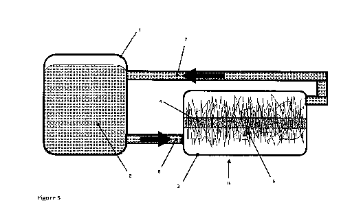
Figure 5 illustrates a flow diagram of an apparatus employing a myocyte
suspension reactor unit with a filamentous scaffold tray unit for attaching
mycocytes to a
hyphal scaffold within the tray unit.
Example Methods
[001] Static Submerged-Submerged Cultivation for Production of
Composite Cellular Masses
1. Filamentous organism inoculum is introduced into a bioreactor vessel
containing a liquid medium prepared with appropriate asepsis and nutrition for
cultivation
of the given filamentous species, and may or may not contain a solid substrate
or surface
to support filamentous growth, creating a first inoculated media. An example
liquid
medium appropriate for Laetiporus spp. would be 20g/L malt extract with 2g/L
peptone.
The media may be filter sterilized via a 0.2um filter or pressure sterilized
at 15psi for 45
minutes.
2. The first inoculated media is incubated under conditions selected to
affect
a specific three-dimensional filamentous network morphology. A generic example
for
6
CA 03119482 2021-05-10
WO 2020/106743 PCT/US2019/062248
Laetiporus spp. would be static incubation at 27 C for 15 days. If a solid
substrate or
surface is included in the vessel, the three-dimensional filamentous network
will develop
with attachment to the surface, if not the filamentous network will develop
within the
volume of the vessel. A suitable substrate would have pore sizes >1 um, such
that
hyphae can penetrate the substrate.
3. After development of the three-dimensional filamentous network has
concluded, the culture media within the vessel is replaced with chemistry
designed to
decellularize the hyphal matrix, retaining the structural wall matrix of the
fungal cells while
removing all components with the potential to interfere in non-filamentous
cell growth,
creating a decellularized filamentous scaffold. The chemistry employed is an
immersion
in a solvent, particularly a 75% ethanol solution for a period greater than 1
hour. The
solvent and effluent are then rinsed away with deionized water.
4. After decellularization, the decellularization chemistry is replaced
with an
appropriate liquid medium for cultivation of a given cell line of non-
filamentous organism,
and inoculum of the non-filamentous organism introduced into the vessel
creating a
second inoculated media.
5. The second inoculated media is incubated under conditions appropriate to
support metabolism and growth of the given line of non-filamentous organism
within the
filamentous scaffold (e.g. typical conditions for cultivating myocytes),
populating the inter-
cellular regions of the filamentous scaffold and attaching to the surface of
the
decellularized filamentous cells.
6. Once the inter-cellular regions of the filamentous scaffold are
determined to
be adequately populated with the non-filamentous organism, creating a
composite cellular
7
CA 03119482 2021-05-10
WO 2020/106743 PCT/US2019/062248
mass, the composite cellular mass is extracted from the bioreactor vessel and
passaged
to post-processing.
[002]
Static Solid State-Submerged (SSSS) Cultivation for Production of
Composite Cellular Masses
1. Solid substrate is prepared with appropriate asepsis and supplemental
nutrition to support metabolism and growth of a given filamentous organism,
filamentous
organism inoculum introduced to the prepared substrate creating an inoculated
substrate,
and the inoculated substrate loaded into the bioreactor vessel. An example
substrate for
Laetiporus spp. would be hardwood chips supplemented with 20% wheat bran,
which is
pressure sterilized at 15psi for 1 hour.
2. The inoculated substrate is incubated under conditions specifically
selected
to affect expression of a specific three-dimensional filamentous network
morphology,
which occurs external to the solid substrate mass creating a cohesive
filamentous
network which may be isolated from the solid substrate mass. Such incubation
conditions
are described in USSN 16/190,585.
3. Example 001 steps 3-6.
[003] Stirred Submerged-Submerged Cultivation for Production of
Composite Cellular Masses
1.
Filamentous organism inoculum is introduced into a bioreactor vessel
containing a liquid medium prepared with appropriate asepsis and nutrition (as
per
Example 1) for cultivation of the given filamentous organism, creating a first
inoculated
media. The rate of addition of the filamentous organism inoculum is adjusted
to target
specific resultant filamentous pellet sizes optimized for downstream texture
and cell
adhesion to support growth, and media preparation and inoculation are
performed to
8
CA 03119482 2021-05-10
WO 2020/106743 PCT/US2019/062248
target an optimal media viscosity of 150 centipoises for maintenance of
dissolved oxygen
for filamentous organism cultivation.
A generic example of the rate of addition would be an 8% inoculation rate
(vol/vol
cell suspension inoculum to liquid medium) with the cell suspension prepared
to at least
75% turbidity at OD590nm. The inoculum rate that was reduced to practice was
an
aliquot of 5x10⁴ cells that were resuspended in 25pL of fresh culture medium
and were
seeded onto scaffolds that had been immersed in medium and then compressed to
expel the liquid.
2. Stirred incubation of the inoculated media is performed with conditions
and
stir rates selected to affect expression of a specific three-dimensional
filamentous pellet
morphology. The stirring is such as to maintain pellets opposed to breaking
matts into
pellets. The inoculum are individual fragments that further pelletize under
stirred
incubation conditions.
3. Example 001 steps 3-6
[004] Stirred Submerged-Drip Cultivation for Production of Shaped
Filamentous Structures
1. Example 003 steps 1-2
2. Application of inoculated media to surface of preformed shape
representative of final desired product by sterile drip-application over the
course of a
number of days until a well formed mycelial sheet is grown on the surface of
the shape
3. Extraction of mycelial sheet from shape surface with retention of shape
as
either a 2-D shell or a thicker 3-D tissue mat.
[005] Submerged Co-Cultivation of Filamentous and Non-Filamentous
Organisms for Production of Composite Cellular Masses
9
CA 03119482 2021-05-10
WO 2020/106743 PCT/US2019/062248
1. Examples 001 and 003 in which a media is prepared that is appropriate
for
cultivation of both the filamentous and non-filamentous organisms, and
inoculum of each
organism is introduced to the media simultaneously. Such a media could include
potato
dextrose broth, which supports both a filamentous fungus and a single celled
bacterium.
2. Examples 001 and 003, in which incubation is performed with conditions
appropriate for the cultivation of both filamentous and non-filamentous
organisms, for
example, at temperatures between 27 C and 37 C, the upper threshold being
appropriate
for mammalian tissue culture and bacteria.
[006] SSSS Cultivation of Cellular Structure with Controlled Morphology
Method [002] is followed.
After step 2. The following steps occur:
1. Moisture, signaling compounds such as hormones, minerals, and other
molecules
are directly deposited with micrometer precision on a grid (x,y) over the
surface
of the growth medium. While this embodiment contemplates the use of a
printhead (much like a 3D printer), deposition method may be via spray, air
conveyance, or any other method which allows precise deposition of material
across the surface of a planar growth part. Molecules that both enhance
growth,
modify growth, and retard growth are contemplated. The addition of other
living
cells at this stage is contemplated and may either provide further in-situ
molecule
or signaling synthesis (e.g. time-delayed molecule synthesis post deposition)
and/or become embedded into the growth of the part.
2. The rate of the deposition can be calibrated to match the growth rate of
the
organism in the y direction. Ideally, the entire surface of the part can be
treated
CA 03119482 2021-05-10
WO 2020/106743 PCT/US2019/062248
prior to additional upward tissue expansion (e.g. entire surface treatment can
occur prior to a cell division of one hyphal length). The rate of deposition
can also
be arbitrarily slow so as to only allow one pass during an entire growth
cycle.
Deposition rate is selected based on the ultimate feature resolution desired
and
will often sit between these two extremes.
3. During each cycle, n, of hyphal growth in the z axis (measured in microns
or
hyphal lengths), the print head passes over the surface of the part in an x,y
grid.
Each x,y cell receives a precise dose of liquid which influences the tissue
morphology & metabolism. Influenced tissue morphology and metabolism
includes, but is not limited to, hyphal branching rate, cell wall thickness,
types of
hyphal tissue created, types of proteins and compounds excreted during hyphal
growth, and direction of hyphal extension. Grid spacing can be selected at a
minimum to match one hyphal unit (e.g. microns by microns cell size) or
upwards
to relatively large divisions (e.g. 1 mmxl mm resolution). Resolution is
selected
based on the precision required for the grown tissue. Fine features, such as
as
scaffolding for capillaries, may require a very high level of resolution,
where-in
bulk features (creating a zone of higher density tissue in a structural
element) may
require relatively low resolution of deposition control.
4. Step 3 is repeated until the entire desired pattern (x,y,z envelope) is
imprinted
upon the grown tissue or until the tissue reaches its maximum hyphal extension
limit.
11
CA 03119482 2021-05-10
WO 2020/106743 PCT/US2019/062248
5. The use of an x,y axis is used to describe the printing process and this
embodiment contemplates the print head would move in linear fashion from an
origin of (0,0) to (x,y)
6. The tissue is extracted from the reactor and can be further post processed
or used
as is. Potential applications include patterning of hyphal tissue to match
existing
organ types for cellular scaffolding (e.g. lung, liver, kidney, and the like),
and
patterning of hyphal tissue to create pre-determined macro-geometric
structures
(e.g , a honeycomb pattern with areas of low density of mycelium as per Figure
1
and thicker hexagonal walls of higher density of mycelium as per Figure 3).
The
principle of this approach is to selectively control regions of mycelium
growth to
present varying densities across a surface in a predicted manner.
[007] Organisms
1. Examples 001-005, in which the filamentous organism is a saprobic fungus
of the phylum Basidiomycota, Ascomycota, Zygomycota, Chytridiomycota, or
Glomeromycota.
2. Example 001-005, in which the filamentous organism is a fungus that
produces a monomitic, dimitic, or trimitic mycelium. Also, dimorphic organisms
that
initially present as an individual yeast cell and are then induced to go
filamentous may
be used, an example of which is Aureobasidium pullulans.
3. Example 001-005, in which the fungus is one of an edible species and is
generally considered safe for human consumption.
4. Examples 001-005, in which the filamentous organism is a fungus which
produces one or more cellular structures such as generative hyphae, binding
hyphae,
12
CA 03119482 2021-05-10
WO 2020/106743 PCT/US2019/062248
coralloid binding hyphae, skeletal hyphae, pseudoparenchyma, pseudocarp,
intercalary
blastogenic cells, acropetal blastogenic cells, cell swelling, terminal
conidiation,
intercallary conidiation, oidiation, arthrosporulation, stroma, perithecia,
conidiogenic cells,
conidiophores, rhizoids, or rhizomorphs.
5. Examples 001-005, in which the filamentous organism is a fungus of genus
Pleurotus, Ganoderma, Polyporus, Grifola, Lentinus, Lentinula, Trametes,
Herecium,
Agrocybe, Armillaria, Agaricus, Stropharia, Schizophyllum, Laetiporus,
Lepista,
Hypomyces, Inonotus, Pycnoporus, Fomes, Fomitopsis, Daedaleopsis, Piptoporus,
lschnoderma, Phellinus, Phaeolus, Sparassis, Tyromyces, Laricifomes, Panellus,
Rhizopus, Phlebia, Phanerochaete, Dichomitus, Ceriporiopsis, Lepiota, Stereum,
Trichoderma, Xylaria, Cordyceps, Hymenochaete, Hypsizygus, Flammulina,
Coprinopsis,
Coprinus, Morchella, Clitocybe, Cerioporus, Volvariella, Tremella, Calvatia,
or Fistulina.
6. Examples 001-005, in which the non-filamentous organism is a cell of a
chordate organism and may be mammal, fish, bird, reptile, or amphibian.
7. Examples 001-005, in which the non-filamentous organism is a plant cell.
8. Examples 001-005, in which the non-filamentous organism is a non-chordate
and may be a mollusk or arthropod cell.
9. Examples 001-005, in which the non-filamentous organism is a myocyte,
neuron, neuroglial cell, lung cell, fibroblast, chondrocyte, endothelial cell,
osteocyte,
osteoblast, adipocyte, or stem cell.
10. Examples 001-005, in which the non-filamentous organism is a bacterium,
yeast, algae, filamentous fungus, nucleic acid based lifeforms (virus,
bacteriophage) or a
mycoplasma.
13
CA 03119482 2021-05-10
WO 2020/106743 PCT/US2019/062248
11. Examples 001-005, in which the non-filamentous organism is a cell of a
coral
or shell structure.
[008] Cultivation Paradigm Variations
Any of the below can be employed with Examples 001-005:
1. Examples 001, 002, and 003 where incubation of both the first and second
inoculated media occurs in a single batch in which all media components are
expended
within the incubation phase without further adjustment.
2. Examples of 001 and 003 (both first and second inoculated media) and 002
(second inoculated media), where incubation is performed using a fed-batch
paradigm, in
which nutrients (carbon, nitrogen, minerals, and pH adjustment) are
periodically fed into
the inoculated media, with spent media proportionally removed, based on active
or periodic
monitoring of set threshold conditions for the given nutrient concentrations.
3. Examples of 001 and 003 (both first and second inoculated media) and 002
(second inoculated media), where incubation is performed using a continuous
feed
paradigm, in which nutrients (carbon, nitrogen, minerals, and pH) are
continuously
adjusted based on a continuous monitoring of set conditions for the given
nutrient
concentrations.
4. Example 002, where solid-state cultivation of the filamentous organism
occurs in a tray vessel which is incubated in a secondary vessel which
provides controlled
gas exchange and content, relative humidity, and temperature. In this
paradigm, the three-
dimensional extra-particle filamentous matrix extends from the top surface of
the solid
substrate from the tray.
14
CA 03119482 2021-05-10
WO 2020/106743 PCT/US2019/062248
5. Example 002, where solid-state cultivation of the filamentous organism
occurs in an actively aerated packed-bed bioreactor vessel in which input air
is conditioned
to specific CO2, humidity, and temperature and passes through the solid-
substrate matrix.
In this paradigm, a void space remains in the vessel within which the three-
dimensional extra-particle filamentous matrix develops..
6. Example 002, where the three-dimensional filamentous matrix is isolated
from the solid substrate matrix prior to decellularization.
7. Example 002, where the three-dimensional filamentous matrix is not
isolated from the solid substrate matrix prior to decellularization, and the
composite
cellular mass is isolated from the solid substrate matrix at the conclusion of
cultivation.
8. Examples 001, 003, and 005 in which the filamentous and non-filamentous
organisms are cultivated in separate vessels (A and B, respectively) in
parallel, and in
which the non-filamentous cells are passaged from vessel A to vessel B,
filtered through
the filamentous organism network of vessel B, depositing non-filamentous cells
throughout
the filamentous cell network. Non-filamentous cells which passage completely
through
vessel B are reclaimed and passaged back to vessel A or vessel B. Flow of non-
filamentous cells from vessel A to vessel B may be periodic or continuous, and
may occur
during or after filamentous organism network development in vessel B.
9. Examples 001-005, in which the filamentous organism scaffold is fully or
selectively filled with a secondary biocompatible material such as agarose or
gelatin gels.
These gels do not provide inherent vasculature or structure, but do provide
another lever
of control for surface area and porosity, serve as a secondary cross-linking
agent, assist
in modulating the modulus of elasticity selectively within the filamentous
scaffold, aid in
CA 03119482 2021-05-10
WO 2020/106743 PCT/US2019/062248
initiating/directing cellular differentiation of adhered cell, as well as
potentially bolster water
uptake and retention.
10. Examples 001-005, in which the filamentous organism scaffold is fully
or
selectively imbibed with growth factors for the non-filamentous organism. The
growth
factors may be perfuse within the filamentous scaffold (naturally diffusing),
encapsulated
within a time-release device, or through the use of synthetic biology to
express said
compounds constitutively or through inducible DNA controlling sequences and
mechanisms (i.e, temporal, thermal, availability feedback loops, etc.).
11. Example 001, in which the filamentous organism scaffold develops
attached
to, or is otherwise attached to, a solid support connected to a mechanical
actuation device
by one or more faces of the three-dimensional filamentous scaffold. During
Example 001,
steps 4-6, the filamentous organism scaffold is mechanically actuated during
non-
filamentous organism propagation within the filamentous scaffold, stimulating
differentiation and propagation.
[009] Modulation of Cultivation Conditions to Affect Different Three-
Dimensional Fungal Scaffold Morphologies
1. Examples 001-005 in which the filamentous organism is a saprobic fungus,
for
example a Laetiporus species. The Laetiporus species is selected and
cultivated under
conditions favorable to producing a vegetative mycelium comprised of an
isotropic matrix
of discrete hyphae (Figure 001). For Laetiporus spp. an example would be
incubation at
27 C for 15 days via the media and paradigms described in examples 001-005.
2. The isotropic matrix of 1 may be modified to express galvanotropism and
hyphal
agglomeration increasing the average strand thickness with (Figure 002), or
without
16
CA 03119482 2021-05-10
WO 2020/106743 PCT/US2019/062248
(Figure 003), increased network fractional anisotropy, as well as express an
ellipsoidal
morphology (Figure 004) by any combination of increasing incubation
temperature, e.g.
to 37 C, increasing CO2 content, e.g. to greater than 2%, addition of volatile
organic
compounds or paramorphogens, e.g. terpene, decreasing gas exchange rate,
increasing
supplementation of starch or other simple carbohydrates, increasing
supplementation of
fatty acids, adjusting the nitrogen supplement, for example, by supplementing
with
peptone, or supplementation with surfactants (such as Tween 80).
3. The isotropic matrix of 1 may have network crosslinking (the combined
effect
of branching, anastomosis, and hyphal entanglement) and/or cell volume density
decreased by any combination of increasing incubation temperature, increasing
CO2
content, addition of volatile organic compounds or paramorphogens, decreasing
gas
exchange rate, decreasing starch or other simple carbohydrates, fatty acids or
nitrogen
supplementation, modifying supplementation of calcium, or supplementation with
surfactants.
4. The isotropic matrix of 1 may have network crosslinking and/or cell volume
density increased by any combination of decreasing incubation temperature;
decreasing
CO2 content, for example, decreasing to 17 -22 C; increasing gas exchange
rate, for
example, increasing the gas exchange rate such that CO2 is maintained at
atmospheric
levels; increasing starch or other simple carbohydrate supplementation;
supplementing
with recalcitrant carbohydrates, such as cellulose; and modifying
supplementation of
calcium.
[010] Propagation of Myocytes on a Filamentous Fungal Scaffold as an
Alternative Meat
17
CA 03119482 2021-05-10
WO 2020/106743 PCT/US2019/062248
1. Per Examples 001-005 and 008-009 in which the filamentous organism is an
edible fungal species per Example 007, such as a Laetiporus species.
2. Per Examples 001-005 and 008-009 in which the non-filamentous organism is
a chordate myocyte of a bovine, avian (such as chicken), or fish cell line.
[011] Production of Ground Meat Product Modifying Texture by
Adjustment of Filamentous Scaffold Pellet Size
1. The process of Example 003 in which the filamentous organism is an
edible
fungal species (such as a Laetiporus spp.) which produces a floccose pellet
morphology,
and the non-filamentous organism is a cow (beef) myocyte.
2. Example 003 in which the inoculation rate of the Laetiporus species into
the
media is adjusted to target a specific textural quality of the resultant
composite tissue mass.
For instance, to target a coarse texture the inoculation rate would be
decreased resulting
in a larger pellet size, and ultimately a larger beef myocyte pellet. For
example, the addition
rate of Example 3 (8% v/v) may be reduced to 2%, or alternatively the 75%
turbidity
inoculum may be diluted.
Alternatively, to create a fine texture, the inoculation rate would be
increased
resulting in a smaller pellet size, and ultimately a smaller beef myocyte
pellet. For example,
the addition rate of Example 3 (8% v/v) may be increased to 16%, or
alternatively the 75%
turbidity inoculum may be concentrated to a higher cell density.
3. The resultant Laetiporus-beef myocyte composite tissue mass is applied
as
a ground beef replacement with "grind", or texture, dictated by the tissue
pellet size per 1
and 2.
4. steps 1-3 with alternative myocyte lines as per Example 007, steps 5-7.
18
CA 03119482 2021-05-10
WO 2020/106743 PCT/US2019/062248
[012] Example 011 with Additional Filamentous Organism Regrowth
into Specific Geometries
1. Example 011, except the fungal scaffold is not decellularized prior to
cultivation of the beef myocyte, thus maintaining the viability of the fungal
scaffold fraction.
2. After extraction of the fungus-beef myocyte composite mass, the mass is
cast into molds of a defined geometry, for example a patty.
3. The molded fungus-beef myocyte composite mass is incubated under
conditions appropriate for continued growth of the fungal fraction, leading to
the discrete
pellets binding together through filamentous extension into a cohesive mass of
the given
geometry. The final fungus-beef myocyte form is employed as a food product.
[013] Production of Alternative Protein Matrix
1. Example 002, in which the filamentous organism is an edible
species, such
as Laetiporus, with the hyphal scaffold being aseptically extracted from the
reactor or
solid state substrate after step 2 and used, with or without further
modification, as a food
product.
[014] Modifications of Alternative Protein Matrix
1. Example 010-013, where the harvested tissue is forced to express excess
exocellular-mucilage through immersion in water, alteration of media
nutrition, for
example, supplementation with simple sugars and/or environmental conditions,
for example, increasing the temperature to 37 C.
2. Example 010-013, where autolysis is induced in the living scaffold, to
yield a more
tender texture. For example, temperature induced autolysis maybe induced by
19
CA 03119482 2021-05-10
WO 2020/106743 PCT/US2019/062248
increasing the incubation temperature to 40 C for a short period at the
conclusion of the
incubation cycle (2-48 hours).
3. Example 010-013, where additional enzymes (e.g, chitinase,
transglutaminases,
proteases, glucanases, or the like) are applied to the extracted scaffold, or
expressed (via synthetic biology) to modify the texture of the structure.
4. Example 010-013, where a secondary organism producing enzymes of interest
is
co-cultured upon the scaffold to produce in-vivo modification of the scaffold
texture and structure. For instance, a mycoparasite, such as Trichoderma spp.
or
Mucor spp., which produce proteases, glucinases, and chitinases may be used
as the secondary organism.
5. Example 010-013, where the harvested tissue is subjected to a corrosive
compound (e.g., 1M HCI, 0.8M acetic acid [white vinegar], or the like), with
or
without heat, to alter the texture or porosity of the resultant structure.
6. Example 010-013, where the harvested tissue is subjected to a strong base,
with
or without heat, to remove acetyl groups from chitin, and/or alter the texture
or
porosity of the scaffold. An example of a method for chitin extraction is
described
at http://www.iglobaljournal.com/wp-content/uploads/2015/07/6.-
Krishnaveni-
Ragunathan-IGJPS-2015.pdf.
7. Example 010-013, where the harvested tissue is subjected to a known solvent
for
chitin (e.g., CaCl2 saturated methanol, ionic liquids, or the like), to alter
texture or
modify porosity
CA 03119482 2021-05-10
WO 2020/106743 PCT/US2019/062248
8. Example 010-013, where the harvested tissue is subjected to mechanical
degradation to alter the natural texture, porosity, or density of the tissue
(e.g.,
perforation, cutting, rolling, pressing, or the like)
9. Example 010-013, where the natural flavor compounds in the edible species
are
overexpressed through nutrition, synthetic biology, or environmental
conditions
(e.g., benzaldehyde in Pleurotus, phenylacetaldehyde in Suillus, anisaldehyde
in
Trametes, or the like)
10. Example 010-013, where another organism is cultivated upon the resulting
matrix, where said organism produces desired flavoring compounds. (e.g.,
diacetyl [buttery] from lactic acid bacteria, pyrazine [roasted] and
glutamates
[meaty] from Corynebacterium glutamicum, or the like)
11. Example 010-013, where commercially available flavorings, fats, colors,
heme,
thickeners, sweeteners, acids, or the like are infused into the tissue
scaffold to
create a food product.
12. Example 010-013, where compounds are added to tissue or media during
growth, to alter the end product's flavor, texture, or color (e.g., addition
of
glutamate in media, atomization of colorant with misters, atomization of
natural
flavor extracts, addition of forskolin to media to induce hyphal branching and
alter
finished texture, and the like)
13. Example 010-013, where the resulting tissue is fortified with vitamins and
minerals to boost nutritional value, and/or replicate that of meat.
14. Example 010-013, where the growth media is amended, to vary the final
nutritional profile of the tissue (e.g., addition of amino acids to increase
fatty acid
21
CA 03119482 2021-05-10
WO 2020/106743 PCT/US2019/062248
concentration, mineral atomization or addition, vitamin supplementation,
proteins,
and the like)
15. Example 010-013, in which the hyphal scaffold is imparted with a defined
grain
by selection of fungal species or cultivation conditions that result in
galvanotropism and hyphal agglomeration per Examples 007 & 009.
16. Example 010-013, in which the hyphal scaffold is imparted with a more
delicate
and fracturable texture by selection of fungal species or cultivation
conditions that
result in intercallary and/or terminal conidiation per Examples 007 & 009.
17. Example 010-013, in which the hyphal scaffold is imparted with a more
delicate
and fracturable texture by selection of fungal species or cultivation
conditions that
result in conidiation per Examples 007 & 009.
18. Example 010-013, in which the hyphal scaffold is imparted with a more
delicate
texture by selection of fungal species with a monomitic, dimitic, or otherwise
a
hyphal morphology free of structural or skeletal hyphae per Examples 007 &
009.
19. Example 010-013, in which the hyphal scaffold is imparted with a uniform
texture
by selection of fungal species or cultivation conditions that result in an
isotropic
hyphal morphology per Examples 007 & 009.
20. Example 010-013, in which the hyphal scaffold is imparted with a tough or
chewy
texture by selection of fungal species with a trimitic, dimitic, or otherwise
a hyphal
morphology that includes structural or skeletal hyphae per Examples 007 & 009.
21. Example 010-013, in which the hyphal scaffold is imparted with a reduced
cohesiveness and/or cohesiveness of mass by selection of fungal species or
22
CA 03119482 2021-05-10
WO 2020/106743 PCT/US2019/062248
cultivation conditions that result in blastogenesis or pseudoparenchyma per
Example 007 & 009.
22. Example 010-013, in which the hyphal scaffold is imparted with a greater
density
by modification of cultivation conditions to increase hyphal branching,
anastomosis, and/or entanglement per Examples 007 & 009. Alternatively, the
hyphal scaffold may be imparted with a reduced density by modification of
cultivation conditions to decrease hyphal branching, anastomosis, and/or
entanglement per Examples 007 & 009.
[015] Embodiment: Bovine meat
As per methods 9 and 10, in which the filamentous scaffold is a saprophytic
fungus
of the genus Laetiporus grown in conditions described therein, where the
secondary non-
filamentous organism is comprised of myoblasts of the genus Bos, creating a
three-
dimensional edible fungal scaffold, imbibed with propagated bovine meat cells,
to be used
as a food product.
[016] Embodiment: Seafood
As per method 005, in which the filamentous scaffold is a saprophytic fungus
of
the genus Rhizopus grown in conditions described therein, where the non-
filamentous
organism is a myoblast of the phylum Mollusca, creating a three-dimensional
edible
fungal scaffold, imbibed with propagated mollusk meat cells, to be used as a
food product
or structural material.
[017] Embodiment: Neutral alternative protein
As per method 013, in which a solid billet of vegetative hyphae of the genus
Herecium is extracted without any inoculation with non-filamentous organisms.
This
23
CA 03119482 2021-05-10
WO 2020/106743 PCT/US2019/062248
scaffold is post- processed per 014, with an application of chitinase from
papaya extract
to improve texture, then heated in 1 molar acetic acid to further modify
texture. The
resultant tissue is then imbued with vegetable fat, marinated in autolyzed
yeast, smoke
flavor, tomato extract, spices, and fortified with minerals and vitamins.
Then, the tissue is
cooked until crispy, to produce a non-animal bacon-like product.
[018] Embodiment: Flavored alternative protein
As per method 002 in which a solid billet of vegetative hyphae of the genus
Flammulina is grown with added glutamate in media to impart umami and
essential
dietary minerals and to fortify the resulting tissue. After initial growth,
the filamentous
scaffold is then inoculated with lactic acid bacteria or yeast to produce
diacetyl in situ,
lending a butter-like flavor and aroma. The tissue is then harvested, imbued
with
vegetable fats and proteins, and cooked. Resulting in a food item, with
natural flavoring
and meat-like texture and properties.
[019] Embodiment: Lung
As per method 006 in which the filamentous scaffold is a saprophytic fungus of
the
genus Ganoderma grown in conditions described therein and the secondary non-
filamentous organism is bronchiolar epithelium cells. The filamentous scaffold
is grown
under conditions described in 009, in which agglomerative galvanotropic growth
is
elected, to mimic the vascular nature of alveoli, allowing the secondary cells
to form a
structured three-dimensional mass of tissue.
[020] Embodiment: Brain, using rhizomorph to support axon growth
As per methods 002 and 006 in which the filamentous scaffold is a saprophytic
fungus of the genus Armillaria grown in conditions described therein,
selecting growth
24
CA 03119482 2021-05-10
WO 2020/106743 PCT/US2019/062248
parameters that express rhizomorphic growth, a highly anisotropic,
galvanotropic, cord-
like morphology. These cord-like structures are then inoculated with a
secondary non-
filamentous organism such as mammalian neural stem cells, to support axon-like
cell
growth, along a naturally-structured scaffold.
[021] Embodiment: Beauty applicator
As per method 003, in which a solid billet of vegetative hyphae of the genus
Laetiporus is incubated under day/night light cycles and increased air
exchange, which
elicit the expression of exogenous pigmentation of the hyphal scaffold. This
scaffold is
then post processed as per 014, with the impregnation of beneficial fatty
acids, such as
lauric acid, to improve application smoothness and foam rigidity, resulting in
a makeup
applicator like foam with naturally produced pigments that can be applied to
the skin.
[022] Embodiment: Disposable Paint Brushes
As per method 013, in which a solid billet of vegetative hyphae of the genus
Ganoderma is extracted without any inoculation with non-filamentous organisms
and post
processed as per 014 with a 10% hydrogen peroxide soak to exfoliate the tissue
and
increase porosity/absorptive capacity, resulting in a biodegradable foam
billet that can be
used to replace traditional polymeric foam brushes.
[023] Embodiment: Sensing
As per method 002, in which the filamentous scaffold is a saprophytic fungus
of
the genus Rhizopus grown in conditions described therein, where the secondary
non-
filamentous organism is comprised of electroactive bacteria, such as the genus
Shewanella, and wired to a current collector and a voltmeter, for monitoring
of water
contamination of sewage, runoff, and/or pollutants.
CA 03119482 2021-05-10
WO 2020/106743 PCT/US2019/062248
[024] Embodiment: Waste Water Treatment
As per method 002, in which the filamentous scaffold is a saprophytic fungus
of
the genus Ganoderma that is grown in conditions described therein, where the
secondary
non-filamentous organism is comprised of a hybrid culture of Cyanobacteria,
for oxygen
production, and Betaproteobacteria for organic treatment, resulting in a
biodegradable
cassette that can be used and/or produced in-field for treatment of latrines,
disaster relief,
or the like.
[025] Embodiment: Antibiotic Sponge
As per method 002, in which the scaffold is comprised of a saprophytic fungus
of
the genus Trametes (with or without drug resistance) that is grown in the
conditions
described therein (with or without antibiotics), where the panel is either
then sterilized,
and imbibed with antibiotics, or inoculated and incubated with an antibiotic
producing
organism, then sterilized and packaged. This biodegradable 3-D scaffold can
then be
adjusted to size and used for implantation, for internal antibiotic treatment
of cavity
wounds, or use as a biodegradable temporary wound dressing for trauma or
disaster
relief.
[026] Embodiment: Absorption/dispersal
1. Method 002 is followed, followed by imbibement of desired antibiotic for
medical
treatment
2. Tissue is then rendered flat by cold compression to form an essentially 2-D
shape
3. Flattened tissue is desiccated to preserve tissue quality
4. Imbued 2-D tissue is used in small space insertion.
26
CA 03119482 2021-05-10
WO 2020/106743 PCT/US2019/062248
5. Expansion within the space beyond the small insertion space is affected
with
specific design as imparted by lattice memory where 2-D flat sheets can fit
through
small holes/incisions, rehydrate, and expand to original morphology to fill
the
largely inaccessible space
6. Expanded tissue fulfills role as interior diffusive scaffold for
antibiotics for internal
surgery
[027] Embodiment: Biodegradable wound dressing for damaged tree limbs
1. Method 002 is followed
2. Resultant tissue is rendered vitally inert through heat application
3. Tissue is imbued with antifungal and antibiotic treatments specific to
injured tree
specie
4. Tissue is applied to wound surface for an indeterminate amount of time,
until the
tissue mat is degraded or overgrown
[028] Embodiment: An Implantable Fungal Scaffold with Semiconducting
Properties
1. Example 001 steps 1-2, Example 002 steps 1-2, or Example 003 steps 1-2, in
which the filamentous organism is Schizophyllum commune or Morchella spp, and
is a
strain of which produces indigotin.
2. MgSO4, 7H20 is supplemented at a rate of 0.1-1% (mass/volume) into the
culture
media of Examples 001-003.
3. Incubation occurs under environmental conditions appropriate for supporting
metabolism and growth of the selected fungal strain, during which
27
CA 03119482 2021-05-10
WO 2020/106743 PCT/US2019/062248
biosynthesis of exogenous indigotin occurs, resulting in indigotin deposition
on the
exterior of the fungal hyphae. In this case, the extent of indigotin
biosynthesis and
exogenous deposition may be modified by the MgSO4, 7H20 supplementation rate
per
step 2.
4. The resultant three-dimensional hyphal scaffold, with exogenous indigotin
or
melanin coating of the hyphal cells, is isolated for downstream use as an
implantable,
biocompatible, semi-conducting material.
5. The semi-conducting hyphal scaffold of step 4 is passaged to Examples 001-
005 steps 3 forward.
[029] Embodiment: Example 028 in which Exogenous lndigotin is
Deposited onto a Secondary Surface
1. Example 028 steps 1-3 in which an additional cell-type or material co-
occupies the culture medium with an indigotin producing fungal strain.
2. Step 1, in which the additional cell-type is the non-filamentous species
of
Examples 001-005.
3. Step 1, in which the additional material co-occupying the culture medium
is
an organic substrate.
4. Step 1, in which the additional material co-occupying the culture medium
is
an inorganic substrate.
[030] Embodiment: Implementation of Static Submerged Filamentous
Fungus Scaffolding Reactor Unit in a Perfusion Reactor System to
Produce an Alternative Meat Product
Method [001] is followed.
28
CA 03119482 2021-05-10
WO 2020/106743 PCT/US2019/062248
During step 1, the fungus selected is one of an edible species, for example
Laetiporus spp., and specifically, Laetiporus sulphureus, which is inoculated
into a vessel
containing a culture medium comprised of corn steep solids, glucose, potassium
phosphate, magnesium sulfate, and pH adjusted to between 5.5-6.5. The vessel
is
designed such as to allow flow of media through the vessel, and is implemented
as a
scaffold tray unit within a perfusion bioreactor system in which a suspension
bioreactor
for beef myocytes feeds directly to the scaffold tray unit in which the
filamentous fungus
is to be cultivated. The vessel further contains a sparger and diffuser in the
center of the
scaffold tray vessel volume, running the length of the scaffold tray vessel.
During step 2, incubation of the Laetiporus spp. inoculated media occurs
without
flow from the beef myocyte suspension reactor unit under static conditions
with dissolved
oxygen levels maintained by an filtered air feed through the sparger and
diffuser, allowing
for a contiguous hyphal network to develop within the scaffold tray vessel,
which further
grows into the sparger and diffuser, anchoring the contiguous hyphal network
in place.
Scaffold tray bioreactor operation may be performed as a batch, fed-batch, or
continuous-
feed process. During this stage the dissolved oxygen levels, light exposure,
temperature,
and media components may be modified according to Method [009].
Step 3 is followed.
During step 4, the decellularization chemistry is replaced with fetal bovine
serum
containing growth factors for the beef myocytes, and flow of beef myocytes
from the
suspension bioreactor unit to the filamentous fungus scaffold tray reactor
unit is initiated.
The media may be further supplemented with polylactic acid, polycaprolactone,
or
29
CA 03119482 2021-05-10
WO 2020/106743 PCT/US2019/062248
polyglycolic acid to assist with adhesion of beef myocytes to the
decellularized
filamentous fungal cells (hyphae).
Referring to Figure 005, during steps 5 and 6, incubation occurs with either
continuous or periodic flow of fetal bovine serum and suspended beef myocytes
(6, 7)
between the filamentous scaffold tray unit (3) and the myocyte suspension
reactor unit
(1), during which myocytes (2) attach to the hyphal scaffold (5) within the
scaffold tray
reactor unit (3) anchored to the sparger and diffuser (4) and replicate,
resulting in
agglomerations of myocytes within the inter-hyphal volume of the filamentous
network,
creating a composite cellular mass of hyphae and myocytes. This composite
cellular
mass is then extracted for post-processing as an alternative meat.
[031] Embodiment: Implementation of a Static Solid State - Submerged
Filamentous Fungus Scaffolding Reactor Unit in a Perfusion
Reactor System to Produce an Alternative Meat Product
Method [002] is followed.
During step 1, a solid substrate is prepared with corn stover, starch, cereal
grains,
and is inoculated with an edible fungal species such as Laetiporus spp., and
specifically,
Laetiporus sulphureus, The prepared substrate is filled into a Type I tray
bioreactor
system, such as described in Mitchell et al. (Eds) Solid-State Fermentation
Bioreactors,
Springer-Verlag Berlin Heidelberg (2010), and loaded into an incubation vessel
with
temperature, light, carbon dioxide, oxygen, relative humidity, and vapor
deposition
control.
During step 2, incubation conditions are maintained at 5% carbon dioxide and
near
100% relative humidity. Additionally, Method [006] may be followed during this
stage to
effect specific heterogeneous morphologies. A negatively gravitropic extra-
particle fungal
CA 03119482 2021-05-10
WO 2020/106743 PCT/US2019/062248
hyphal matrix develops from the inoculated substrate, which is further
modified during
growth via modulation of light, oxygen, carbon dioxide, relative humidity, or
vapor
deposition rate per Method [009]. The extra-particle hyphal matrix develops
into a
contiguous mass, which is isolated from the solid substrate for post-
processing.
Method [001] step 3 is followed.
The decellularized hyphal scaffold is transferred to a scaffold tray vessel
within a
perfusion bioreactor system. Steps 4-6 of Embodiment 030 are followed.
[032] Embodiment: Implementation of Submerged Co-Cultivation of a
Filamentous Fungal Matrix and Beef Myocytes in a Perfusion
Bioreactor System for Production of an Alternative Meat Product
Method [001] is performed according to the modifications of Method [004].
During Method [001] step 1, a culture medium is prepared and inoculated within
a
tray vessel reactor implemented in a perfusion bioreactor per Embodiment 030.
During Method [001] step 2, incubation of the Laetiporus spp within the
scaffold
tray vessel occurs according to Embodiment 030 until filamentous growth of
Laetiporus
spp has been established and has become anchored in the sparger and diffuser.
According to Method [005] step 1, flow from the beef myocyte suspension
reactor
per Embodiment 030 is initiated through the developing fungal scaffold within
the scaffold
tray vessel. At this point, the media is comprised of nutrients supportive of
both
propagation of Laetiporus spp and the beef myocytes, and may include corn
steep solids,
glucose, potassium phosphate, magnesium sulphate, fetal bovine serum, beef
myocyte
growth factors, polylactic acid, polycaprolactone, or polyglycolic acid, and
pH adjusted to
between 5-7. According to Method [004] step 2 both P.ostreatus and beef
myocytes
31
CA 03119482 2021-05-10
WO 2020/106743 PCT/US2019/062248
develop in parallel, producing a composite cellular mass according to
Embodiment 030
steps 5 and 6.
[033] Embodiment: Use of imbued tissue to produce crystal structure
Deposition
1. Method [002] is followed to produce a mycelial tissue sheet.
2. Tissue sheet is compressed to flatten and evacuate residual moisture from
intercellular pores
3. Tissue is then subjected to heavily mineralized liquid and allowed to
absorb said
liquid to full saturation
4. Tissue is deposited in location specified as mineral deposition zone
5. Tissue desiccates ambiently and acts as a time release of mineralized
residue
previously held within intercellular pore spaces
[034] Embodiment: Production of Vasculature
Method [001] is performed according to Embodiment 030, where the filamentous
organism is a rhizomorphic strain of Armillaria gallica, and the non-
filamentous organism
is comprised of any combination of endothelial cells, myocytes, and
fibroblasts
During steps 1 and 2 A.gallica fills the volume of the scaffold tray
bioreactor unit
with a matrix of rhizomorphs ranging from <1mm to 5mm in diameter.
During steps 4-6 endothelial cells, myocytes, and/or fibroblasts attach to and
propagate along the surface of the rhizomorphs, forming a cohesive outer
cellular layer
or sleeve.
32
CA 03119482 2021-05-10
WO 2020/106743 PCT/US2019/062248
During post-processing, a sleeve of endothelial cells, myocytes, and/or
fibroblasts
are isolated from the underlying A.gallica rhizomorph by any combination of
chemical
lysis or mechanical separation.
[035] Embodiment: Grown Tools
Examples 001-005, in which the filamentous scaffold is grown into
predetermined
shapes, such as small hand tools (hammer). The scaffold is co-cultured with
non-
filamentous cells (i.e., yeast, bacteria, and he like), which adhere and
deposit polymers,
metals, keratin, calcite, or spider silk onto the scaffold matrix, thus
providing enhanced
mechanical strength, and structural stability. The synthesis and deposition of
compounds
can the enhanced through strain engineering.
[036] Embodiment: Alternative to Mammalian Meat (Yeast)
Examples 001-005, in which the filamentous scaffold is co-cultured with yeast
cells
which are allowed to adhere to either decellularized or intact cellular
scaffolds. Yeast will
be cultivated in co-culture or independently (fermenter B, Figure 5), and used
as an
alternative to mammalian cells to circumvent cellular cultivation with
expensive bovine
serums, and surface attachment requirements.
1. Yeast or the filamentous organism can be genetically engineered to enhance
binding affinity to the scaffolds (i.e., chitin, hydrophobin binding motifs).
2. Yeast can be engineered to express meat based flavors and properties (i.e.
heme, fats, pigments). The expression of these compounds can be constitutive
throughout cultivation/assembly, or induced at desired times during the
cellular
assembly process for optimized expression and impact.
33
CA 03119482 2021-05-10
WO 2020/106743 PCT/US2019/062248
[037] Embodiment: Engineered Fungal Edible Meats
Examples 001-005, which the filamentous scaffold organism is genetically
engineered to possess desired characteristics of natural meat flavor, color,
texture, and
smells (i.e., heme, fats, pigments).
Examples of how the organism can be genetically engineered include methods of
up-regulating existing genes to enhance the composition of glutamic acid
within the fungal
tissue to provide a more umami flavor profile, or to do the same for
pigmentation pathways
such as melanin induction. Further, the organism can be engineered to "knock-
out" or
eliminate specific genes that lead the differentiation of the mycelium into a
mushroom
thus amending or limiting texture changes. Finally, the organism can be
engineered to
introduce a promoter and gene cassette for a molecule from another organism,
such as
heme.
[038] Embodiment: Therapeutic Delivery
Examples 001-005, in which the filamentous scaffold organism and/or co-
cultured
non-filamentous cells are used to deliver therapeutics to implanted tissues
(i.e. dermal,
subcutaneous, intramuscular, and the like). In this embodiment, the
therapeutic is
produced by the non-filamentous cells and encapsulated within the filamentous
scaffold.
The release of the therapeutic can be related to concentration differentials
between the
scaffold and the surrounding tissues (e.g., Fickian or Non-Fickian Diffusion).
The
therapeutic can also be released to surrounding tissues as the scaffold is
degraded or
incorporated into said tissues.
1 Filamentous organism can be genetically engineered to express or have cell
surface binding/release affinity for the delivered therapeutic.
34
CA 03119482 2021-05-10
WO 2020/106743 PCT/US2019/062248
2. Non-filamentous organisms can be genetically engineered to express or have
binding/release affinity for the delivered therapeutic.
3. Therapeutic can be released by constitutive compound synthesis, or a
temporal
base degradation release profile (therapeutic binding affinity)
4. Both (1-2) cells can be engineered to detect the titer of the therapeutic
in the
implanted tissue or extracellular matrix, thus regulating the synthesis or
release of the
therapeutic.
[039] Embodiment: Self-Protective Scaffold (Sense-Response)
Examples 001-005, in which the filamentous scaffold organism and/or co-
cultured
non-filamentous cells are genetically programmed to sense microbial
contaminants and
pathogens (E.coli, Staph).
In this embodiment, non-filamentous strains (i.e., bacteria, yeast) are
genetically
engineered to contain multiple sensors integrated into the genome that respond
to signals
associated with microbial contaminants such as bacteria and fungi that
represent human
health threats, or are detrimental to the structural integrity of the
filamentous scaffold
matrix. Multiple sensors and specificity will be achieved through the
integration of these
sensors via genetic logic gates in order to positively identify the strain.
Engineered non-filamentous organisms would be co-cultured with the scaffold
and
maintained as living cells to provide an active immunity against infection.
These co-
cultured strains will respond to particular patterns of quorum molecules
associated to the
contaminants, along with other indicators, and use a classifier circuit to
select the correct
antibiotic/antifungal to produce.
CA 03119482 2021-05-10
WO 2020/106743 PCT/US2019/062248
1. Food safety - enable foodstuffs to identify the presence of pathogenic
microbes
(i.e., E.coli, salmonella, Clostridium, etc. ), and initiate a response by
expressing
and secreting species specific antibiotics to suppress or kill said
contaminants.
2. Implantation - enable the living cellular scaffold matrix to sense the
presence of
problematic microbial contaminants (i.e., Staphylococcus aureus , etc.) and
initiate a response to suppress or kill invading microbes before and after
surgical
implantation.
[040] Embodiment: Living Scaffold Utilities
Examples 001-005 and [007] Organisms, Enable filamentous and non-filamentous
cells to express limiting nutrients need for successful cultivation and
surgical implantation
scaffold viability.
1. Position microbes in co-cultivation microbiome that are natural growth
promoting
organisms and target for enrichment.
2. Genetically engineer [007] microbes to promote enhanced system wide growth
in
the cultivation / scaffold assembly process,
3. Genetically engineer [007] microbes to support scaffold health and
sustainability
once implanted as a medical device.
[041] Embodiment: Tissue Generated / Scaffold Removal
Examples 001-005, in which the filamentous scaffold organism is used to
support
the adhesion and differentiation of co-cultivated cells (i.e., myoblasts) to
establish
functional tissue forms i.e., medical devices, foodstuffs, and the like.
1. Scaffold remains part of the formed tissue throughout the intended
usable life
36
CA 03119482 2021-05-10
WO 2020/106743 PCT/US2019/062248
of the tissue (i.e., medical implantation, foodstuff), and continues to
provide
structural support or fosters viability and/or growth of attached cells.
2. Scaffold is "removed" from the final tissue form. The filamentous
scaffold is
degraded in vitro or in situ using enzymes, compounds, pH, thermochemical
applications, and the like.
3. Scaffold degrading enzymes, compounds, small molecules, or other
Substrates can be introduced during the formation of the final tissue
(Le.,within the fermentor) by being fed into the reactor from an external
source, or expressing said degrading agents from co-cultured cells
(adhered or free floating).
4. Degrading agents could also be produced by microbes in a secondary
reactor. Agents could be isolated and purified, or used within the cell
suspension, then transferred to the final tissue to remove or degrade the
filamentous scaffold matrix leaving behind "pure tissue" i.e., myoblasts, and
the like.
The invention thus provides methods of generating mycelial scaffolds that
leverage
the phenotypic plasticity of filamentous fungi to produce fungal scaffold
materials with
specifically targeted network morphologies.
The invention also provides mycelium scaffolds for implementation in perfusion
bioreactor systems for cell-based meat technologies.
The invention also provides mycelium scaffolds that provide an optimized
fibrous,
complex substrate for adhesion, propagation, and agglomeration of mammalian
cells in
suspended or submerged culture.
37
CA 03119482 2021-05-10
WO 2020/106743 PCT/US2019/062248
The invention also provides methods to produce biocompatible and biodegradable
mycelium scaffolds with unique plasticity of manufacture, allowing for
porosity and
structure to be uniquely tunable for biomedical applications.
38