Note: Descriptions are shown in the official language in which they were submitted.
CA 03120186 2021-05-14
WO 2020/102609
PCT/US2019/061575
COMPOSITIONS AND METHODS FOR THE CYTOPLASMIC DELIVERY OF
ANTIBODIES AND OTHER PROTEINS
GOVERNMENT INTEREST STATEMENT
[0001] This invention was supported by Grant Number F30 CA221385-01 awarded by
the
National Institutes of Health (NIH). The United States government has certain
rights in the
invention.
FIELD OF THE INVENTION
[0002] The invention relates to compositions and methods for the cytoplasmic
delivery of
antibodies and other proteins. Specifically, provided herein are compositions
having an anionic
to polypeptide and a cationic transfection agent for facilitating the
cytoplasmic delivery of an
antibody or a protein.
BACKGROUND OF THE INVENTION
[0003] Many intracellular therapeutic targets are not susceptible to small
molecule drugs,
because they lack natural ligands or even ligand binding sites. Currently,
most approved small-
molecule therapeutics target proteins such as enzymes and receptors that
contain small, but
critical, binding pockets amenable to modulation by small molecule drugs, but
many potential
therapeutic targets lack such pockets.
[0004] Moreover, even if a small molecule drug can bind a desired target, it
may not effectively
inhibit protein function. Small molecule drugs capable of disrupting
interactions between two
proteins have been particularly difficult to identify. Furthermore, even where
a small molecule
drug is identified, it must be able to reach its target site with good
pharmacokinetic properties
and minimal off-target toxicity. These stringent requirements have led to long
development
times and only a very small fraction of small molecule drugs have been
successfully translated
into the clinic, despite decades of research and countless high-throughput
screens.
[0005] Therapeutic monoclonal antibodies have had considerable success as
cancer
therapeutics, but their inability to cross cell membranes has restricted their
targets to secreted
or membrane-associated antigens.
[0006] Currently, there are two broad approaches for cytoplasmic antibody
delivery: 1) cell-
penetrating peptide (CPP) based and 2) protein transfection-based methods.
CPPs are short,
poly-cationic peptides that can induce endocytic cellular uptake of not only
themselves, but also
cargo conjugated to them. Although CPPs are highly effective at delivering
large cargos into
the endosome-lysosome system, a vanishingly small fraction of cargos actually
escape into the
cytosol, where most therapeutically relevant targets are. Because of this
endosome escape
1
CA 03120186 2021-05-14
WO 2020/102609
PCT/US2019/061575
problem, CPPs have been limited to delivering enzymes and other proteins
capable of greatly
amplifying their effects. Since it is likely that stoichiometric amounts of
inhibitory antibodies
need to be delivered relative to their targets for a sustained biological
effect, tremendous
advances in CPP-mediated endosome escape must be made before they become
viable for
cytoplasmic antibody delivery.
[0007] Like nucleic acid transfection, in protein transfection, cargo proteins
are encapsulated
into lipid or polymer nanoparticles that can induce endocytic uptake and then
destabilize the
endosome membrane to allow for cytoplasmic release of cargo proteins. This
takes advantage
of advances in nucleic acid delivery that have led to lipid and polymer
formulations much better
at endosomal escape than CPPs. Although many protein transfection systems have
been
developed, they should be evaluated cautiously ¨ most were developed using
fluorescently
labeled model proteins, which does not allow one to easily distinguish between
proteins bound
to the cell surface, proteins stuck in endosomes, and proteins delivered to
the cytoplasm,
dramatically increasing the potential for false positives. In fact, evaluation
of 4 commercially
available protein transfection systems for antibody delivery using a very
stringent Cre-
recombinase based cytoplasmic delivery reporter system revealed that none were
able to deliver
to > 6% of cells, indicating that these systems suffer from poor efficacy,
poor reproducibility,
or likely both. Finally, none of the systems described thus far have managed
to deliver
antibodies cytoplasmically in vivo, likely due to poor serum stability.
However, recent progress
in delivering Cas9 proteins, which are as large as antibodies, with lipid
nanoparticles for
genome editing in vivo indicates that protein transfection is a viable
strategy for cytoplasmic
antibody delivery. Making therapeutic cytoplasmically delivered antibodies a
reality, though,
requires 1) a reproducible method to efficiently encapsulate a large variety
of antibodies into
stable lipid or polymer nanoparticle formulations, 2) stringent testing of
formulations using a
cell reporter assay that only detects cytoplasmic protein delivery, and 3)
demonstration that
cytoplasmically delivered antibodies can inhibit therapeutically relevant
targets in both cell
culture and model organisms.
[0008] If antibodies could efficiently be delivered into the cytosol of living
cells, it would
significantly increase the number of possible druggable targets. Antibodies
can be developed to
bind nearly any exposed protein epitope, with high specificity and affinity.
There are a countless
number of therapeutic possibilities that could be pursued if antibodies could
effectively be
delivered into cells, from inhibiting protein function, to driving proteins
interactions, to
targeting intracellular proteins for degradation. Not surprisingly, numerous
attempts have been
made to deliver antibodies into cells, but a robust and efficient approach has
yet to be identified.
2
CA 03120186 2021-05-14
WO 2020/102609
PCT/US2019/061575
[0009] Accordingly, there exists a need to develop a modular approach to
efficiently deliver
antibodies and other proteins into the cytoplasm of living cells.
SUMMARY OF THE INVENTION
[000101In one aspect, provided herein are compositions comprising: an antibody
or other
protein; an anionic polypeptide, an anionic polymer or an anionic nucleic
acid; and a cationic
transfection agent, wherein the presence of said anionic polypeptide, anionic
polymer or anionic
nucleic acid and said cationic transfection agent in said composition
facilitate cytoplasmic
delivery of said antibody or other protein.
[000111In one aspect, provided herein are compositions comprising: an antibody
or other
to protein; an anionic polypeptide; and a cationic transfection agent,
wherein the anionic
polypeptide comprises a plurality of negatively charged amino acid residues,
and wherein the
presence of said anionic polypeptide and said cationic transfection agent in
said composition
facilitate cytoplasmic delivery of said antibody or other protein. In one
embodiment, said
antibody is operably linked to an antibody binding domain (AbBD). In some
embodiments, said
antibody binding domain is operably linked to a photoreactive amino acid
group, for example,
benzoylphenylalanine (BPA) resulting in a photoreactive antibody binding
domain (pAbBD).
In one example, at least 20% of residues in said anionic polypeptide are
negatively charged
amino acid residues (e.g., aspartic acid residues, glutamic acid residues,
unnatural amino acids,
or combinations thereof). In another example, the cationic transfection agent
is an ionizable
lipid, lipid-like, and/or polymeric particle. In another example, the particle
is a nanoparticle.
[00012] In another aspect, provided herein are compositions comprising: an
antibody or other
protein; an anionic polypeptide; and an agent that induces protein
degradation, wherein said
anionic polypeptide comprises a plurality of negatively charged amino acid
residues. In some
embodiments, a composition described herein includes an agent that modifies
the function of a
target protein; an agent that induces nuclear, cytoplasmic, membrane, or
membrane-associated
proteins to be sorted into subcellular compartments; or a combination thereof.
[000131In another aspect, provided herein are conjugates comprising an
antibody binding
domain (AbBD) operably linked, ligated or fused to an anionic polypeptide
comprising a
plurality of negatively charged amino acid residues. In some embodiments, the
antibody
binding domain is operably linked to a photoreactive amino acid group, for
example,
benzoylphenylalanine (BPA) resulting in a photoreactive antibody binding
domain (pAbBD).
In one example, at least 20% of residues in said anionic polypeptide are
negatively charged
amino acid residues (e.g., aspartic acid residues, glutamic acid residues,
unnatural amino acids,
or combinations thereof).
3
CA 03120186 2021-05-14
WO 2020/102609
PCT/US2019/061575
[00014] In one aspect, provided herein are conjugates comprising: a protein
operably linked,
ligated, conjugated or fused to an anionic nucleic acid. In some embodiments,
the protein is a
single chain protein. In certain embodiments, the protein is operably linked
to the anionic
nucleic acid. In some embodiments, the single chain protein is a single chain
protein, as
.. described herein. In various embodiments, the anionic nucleic acid is
operably linked, ligated,
conjugated or fused to an antibody. In particular embodiments, the anionic
nucleic acid is
operably linked, ligated, conjugated or fused to an AbBD.
[00015] In another aspect, provided herein are methods of delivering an
antibody or other protein
to cell cytoplasm in a subject, comprising: providing a composition described
herein; and
to administering said composition to said subject.
[00016] In one aspect, the invention provides a method of delivering an method
of delivering
an antibody or other protein to cytoplasm of a cell in a subject, comprising:
providing a
composition described herein; and administering said composition to said
subject, wherein the
composition comprises: the antibody or the other protein; an anionic nucleic
acid; and a cationic
transfection agent. In some embodiments, the protein is a single chain
protein. In some
embodiments, the nucleic acid comprises a plurality of negatively charged
residues, i.e., is an
anionic nucleic acid. In various embodiments, the anionic nucleic acid is
operably linked,
ligated, conjugated or fused to an antibody. In particular embodiments, the
anionic nucleic acid
is operably linked, ligated, conjugated or fused to an AbBD.
[00017] In another aspect, the invention provides a method of treating a
disease or disorder in a
subject, comprising: delivering a composition described herein to cell
cytoplasm in the subject.
[00018] In another aspect, the invention provides a method of manufacturing a
composition for
a cytoplasmic delivery, comprising: covalently linking, ligating, or fusing an
antibody or other
protein with an anionic polypeptide in order to prepare a conjugate; and
mixing or complexing
a cationic transfection agent with said conjugate.
[00019] In one aspect, the invention provides a method of manufacturing a
composition for a
cytoplasmic delivery, comprising: covalently linking, ligating, or fusing a
protein to an nucleic
acid in order to prepare a conjugate; and mixing or complexing a cationic
transfection agent
with said conjugate. In some embodiments, the nucleic acid comprises a
plurality of negatively
.. charged residues, i.e., is an anionic nucleic acid. In an embodiment, the
protein is a single chain
protein. In some embodiments, the single chain protein is a single chain
protein, as described
herein. In various embodiments, the anionic nucleic acid is operably linked,
ligated, conjugated
or fused to an antibody. In particular embodiments, the anionic nucleic acid
is operably linked,
ligated, conjugated or fused to an AbBD.
4
CA 03120186 2021-05-14
WO 2020/102609
PCT/US2019/061575
[00020] In another aspect, the antibody or protein is further labeled with an
imaging agent, drug,
and/or toxin.
[00021] In one aspect, the invention provides compositions comprising: a
protein; an nucleic
acid; and a cationic transfection agent, wherein the nucleic acid comprises a
plurality of
negatively charged residues, and wherein the presence of the anionic nucleic
acid and the
cationic transfection agent in the composition facilitate cytoplasmic delivery
of said antibody
or other protein. In another aspect, the protein is single chain protein. In
an embodiment, the
single chain protein is further labeled with an imaging agent, drug, and/or
toxin. In various
embodiments, the anionic nucleic acid is operably linked, ligated, conjugated
or fused to an
to antibody. In particular embodiments, the anionic nucleic acid is
operably linked, ligated,
conjugated or fused to an AbBD.
[00022] In one aspect, provided herein are methods for sensitizing a tumor
cell to a
chemotherapeutic agent, the method comprising administering to cytoplasm of
the tumor cell:
(i) a conjugate comprising an antibody binding domain (AbBD) operably linked,
ligated or
fused to an anionic polypeptide comprising a plurality of negatively charged
amino acid
residues or (ii) a cell recombinantly expressing the conjugate of (i).
[00023] In another aspect, provided herein are methods for sensitizing a tumor
cell to a
chemotherapeutic agent, the method comprising administering to cytoplasm of
the tumor cell:
(i) a conjugate comprising a protein operably linked, ligated, conjugated or
fused to a nucleic
acid, wherein the nucleic acid comprises a plurality of negatively charged
residues, i.e., is an
anionic nucleic acid, and wherein presence of the anionic nucleic acid and the
cationic
transfection agent in the composition facilitate cytoplasmic delivery of said
antibody or other
protein, or (ii) a cell recombinantly expressing the conjugate of (i). In an
embodiment, the
protein is single chain protein. In an embodiment, the anionic nucleic acid is
operably linked,
ligated, conjugated or fused to an antibody. In particular embodiments, the
anionic nucleic acid
is operably linked, ligated, conjugated or fused to an AbBD.
[00024] In one aspect, provided herein are methods for decreasing or
inhibiting growth of a
tumor cell, the method comprising administering to cytoplasm of the tumor cell
in a subject in
need thereof a composition comprising an antibody or other protein; an anionic
polypeptide;
and a cationic transfection agent, wherein said anionic polypeptide comprises
a plurality of
negatively charged amino acid residues, and wherein the presence of said
anionic polypeptide
and said cationic transfection agent in said composition facilitate
cytoplasmic delivery of said
antibody or other protein.
[00025] In another aspect, provided herein are methods for decreasing or
inhibiting growth of a
tumor cell, the method comprising administering to cytoplasm of the tumor cell
in a subject in
5
CA 03120186 2021-05-14
WO 2020/102609
PCT/US2019/061575
need thereof a composition comprising: a protein; an anionic nucleic acid; and
a cationic
transfection agent, wherein presence of the anionic nucleic acid and the
cationic transfection
agent in the composition facilitate cytoplasmic delivery of the protein. In an
embodiment, the
protein is a single chain protein. In an embodiment, the protein is operably
linked to the anionic
nucleic acid. In various embodiments, the anionic nucleic acid is operably
linked, ligated,
conjugated or fused to an antibody. In particular embodiments, the anionic
nucleic acid is
operably linked, ligated, conjugated or fused to an AbBD.
[00026] In one aspect, provided herein are methods for inhibiting NF-kB
transcription and/or
reducing RelA nuclear translocation a cancer cell, the method comprising
administering to
to cytoplasm of the cancer cell in a subject in need thereof a composition
comprising an antibody
or other protein; an anionic polypeptide; and a cationic transfection agent,
wherein said anionic
polypeptide comprises a plurality of negatively charged amino acid residues,
and wherein the
presence of said anionic polypeptide and said cationic transfection agent in
said composition
facilitate cytoplasmic delivery of said antibody or other protein.
[00027] In another aspect, provided herein are methods for inhibiting NF-kB
transcription
and/or reducing RelA nuclear translocation a cancer cell, the method
comprising administering
to cytoplasm of the cancer cell in a subject in need thereof a composition
comprising a protein;
an anionic nucleic acid; and a cationic transfection agent, wherein presence
of the anionic
nucleic acid and the cationic transfection agent in the composition facilitate
cytoplasmic
delivery of the single chain protein. In an embodiment, the protein is a
single chain protein. In
an embodiment, the protein is operably linked to the anionic nucleic acid. In
some
embodiments, the anionic nucleic acid is operably linked, ligated, conjugated
or fused to an
antibody. In particular embodiments, the anionic nucleic acid is operably
linked, ligated,
conjugated or fused to an AbBD.
[00028] Other features and advantages of this invention will become apparent
from the
following detailed description, examples, and figures. It should be
understood, however, that
the detailed description and the specific examples while indicating preferred
embodiments are
given by way of illustration only, since various changes and modifications
within the spirit and
scope of the invention will become apparent to those skilled in the art from
this detailed
description.
BRIEF DESCRIPTION OF THE DRAWINGS
[00029] The following drawings form part of the present specification and are
included to further
demonstrate certain aspects of this disclosure, the inventions of which can be
better understood
by reference to one or more of these drawings in combination with the detailed
description of
6
CA 03120186 2021-05-14
WO 2020/102609
PCT/US2019/061575
specific embodiments presented herein. The patent or application file contains
at least one
drawing executed in color. Copies of this patent or patent application
publication with color
drawing(s) will be provided by the Office upon request and payment of the
necessary fee.
[00030] Figures 1A-1B respectively show a schematic depicting light activated
site-specific
conjugation of IgG with pAbBD and reducing SDS-PAGE gels of various human IgG
subclasses alone or after photocrosslinking with a pAbBD. Fig. 1A shows
irradiation with non-
damaging long-wavelength UV light allows for covalent attachment of pAbBD with
attached
cargo (green star). The photoreactive amino acid (e.g., BPA) is represented by
a yellow circle.
In Fig. 1B the reducing SDS-PAGE gels of various human IgG subclasses alone or
after
to photocrosslinking with a pAbBD, nearly 100% crosslinking is achieved.
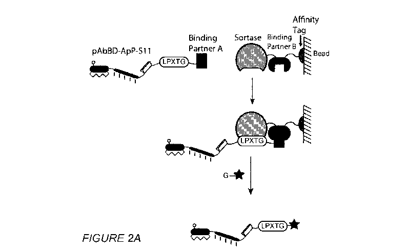
[00031] Figures 2A-2C respectively show a schematic of proximity-based sortase
ligation
(PBSL), capture of the expressed recombinant protein and the efficiency of
PBSL. Fig. 2A
shows two binding partners are used to bring the sortase recognition motif
(LPXTG) into close
proximity with sortase, to increase the ligation efficiency with a peptide
that possesses an N-
terminal glycine. The peptide can be labeled with any chemical moiety, e.g.,
imaging agent,
drug, hapten, etc. (red star). Fig. 2B shows that when SpyCatcher and SpyTag
are employed as
binding domains, ¨80% of the expressed recombinant protein can be captured.
Fig. 2C shows
the efficiency of ligation is >95% in the PBSL system and is completed in 4-6
hours.
[00032] Figures 3A-3B respectively show formation of IgG-ApP cationic lipid
complexes and
cytoplasmic delivery of the IgG-ApP cationic lipid complexes and detection by
fluorescence of
splitGFP complementation. Fig. 3A shows negatively charged IgG-ApP conjugates
can be
complexed with cationic lipids. Fig. 3B shows IgG-ApP lipid complexes are
taken up into
reporter cell lines expressing splitGFP(1-10) in the cytoplasm. The lipids
allow escape of IgG-
ApP into the cytoplasm. In the cytoplasm, splitGFP complementation occurs
between
splitGFP(1-10) and the splitGFP Sll peptide resulting in turn-on splitGFP
fluorescence.
[00033] Figures 4A-4D show in vitro splitGFP complementation and fluorescence
of pAbBD-
S 1 1 ((Figs. 4A-4B) and Ritux-(pAbBD-S11)2 (Figs. 4C-4D). Time course of
400pm01
splitGFP(i-10) incubated with (Fig. 4A) pAbBD-S11 or (Fig. 4C) Ritux-(pAbBD-S
11)2 at
37 C shows turn-on fluorescence that plateau within 6 hours. Fluorescence is
linearly associated
with the amount of (Fig. 4B) pAbBD-S11 or (Fig. 4D) Ritux-(pAbBD-S11)2 added
at all time
points. As expected, Ritux-(pAbBD-S11)2 fluorescence is approximately twice
that of pAbBD-
Sll since ¨2 pAbBD-S11 are crosslinked to each Rituximab molecule. Data are
means SEM
of n=3 biological replicates.
[00034] Figures 5A-5B show splitGFP complementation by flow cytometry and
fluorescence
microscopy, respectively, in HEK293T splitGFP(1-10) cells after delivery of
pAbBD-S11 or
7
CA 03120186 2021-05-14
WO 2020/102609
PCT/US2019/061575
Ritux-(pAbBD-S11)2 into the cytoplasm. pAbBD-S11 or Ritux-(pAbBD-S11)2 were
delivered
by electroporation into the cytoplasm of HEK293T cells stably expressing
splitGFP(1-10).
After 6 hours, the cells were processed for (Fig. 5A) flow cytometry, which
shows a 33.5-fold
increase in median fluorescence with 40 M pAbBD-S11 and 39.35-fold increase
with 7.5 M
Ritux-(pAbBD-S11)2, and (Fig. 5B) fluorescence microscopy, which shows diffuse
splitGFP
fluorescence. For Ritux-(pAbBD-S11)2, there is nuclear depletion of splitGFP
fluorescence
because Ritux-(pAbBD-S11)2 conjugates are too large to passively cross the
nuclear pore
complex.
[000351 Figure 6 shows pAbBD-Dx/Ex-S11 SDS-PAGE. SDS-PAGE of pAbBD-S11 and
to pAbBD-Dx/Ex-S11 containing 10, 15, 20, 25, or 30 repeats of aspartic
acid (D) or glutamic acid
(E) used in delivery studies.
[00036] Figure 7 shows pAbBD-Dx-S11 delivery with Lipofectamine 2000. 80,000
HEK293T
splitGFP(1-10) cells were seeded onto each well of a 48 well plate in 180 L
media at 37 C for
12-16 hours. Lipid nanoparticles were formed by incubating 2 L Lipofectamine
2000 with the
indicated protein (500nM final concentration in well) in OptiMEM (20 L final
volume, pH 7.4)
at 25 C for 10 minutes. The lipid nanoparticles were then added to the cells
and incubated for
6 hours at 37 C before live-cell fluorescence microscopy. 50 g/mL Hoechst
33342 was added
30 minutes prior to microscopy. Top panel is the splitGFP channel, which shows
cytoplasmic
delivery. Middle panel is the Hoechst channel, which shows all cell nuclei.
Bottom panel is the
splitGFP and Hoechst channel merged. Fluorescence microscopy shows greater
cytoplasmic
delivery (diffuse splitGFP fluorescence) with increasing aspartic acid (D)
repeat length until a
maximum at 20 repeats followed by a decrease with longer repeats.
[00037] Figure 8 shows pAbBD-Ex-S11 delivery with Lipofectamine 2000. 80,000
HEK293T
splitGFP(1-10) cells were seeded onto each well of a 48 well plate in 180 L
media at 37 C for
12-16 hours. Lipid nanoparticles were formed by incubating 2 L Lipofectamine
2000 with the
indicated protein (500nM final concentration in well) in OptiMEM (20111 final
volume, pH 7.4)
at 25 C for 10min. The lipid nanoparticles were then added to the cells and
incubated for 6
hours at 37 C before live-cell fluorescence microscopy. 50 g/mL Hoechst 33342
was added 30
minutes prior to microscopy. Top panel is the splitGFP channel, which shows
cytoplasmic
delivery. Middle panel is the Hoechst channel, which shows all cell nuclei.
Bottom panel is the
splitGFP and Hoechst channel merged. Fluorescence microscopy shows greater
cytoplasmic
delivery (diffuse splitGFP fluorescence) with increasing glutamic acid (E)
repeat length.
[00038] Figure 9 shows pAbBD-Dx-S11 delivery with Lipofectamine RNAiMax.
80,000
HEK293T splitGFP(1-10) cells were seeded onto each well of a 48 well plate in
180 L media
at 37 C for 12-16 hours. Lipid nanoparticles were formed by incubating 2 L
Lipofectamine
8
CA 03120186 2021-05-14
WO 2020/102609
PCT/US2019/061575
RNAiMax with the indicated protein (500nM final concentration in well) in
OptiMEM (20 L
final volume, pH 7.4) at 25 C for 10min. The lipid nanoparticles were then
added to the cells
and incubated for 6 hours at 37 C before live-cell fluorescence microscopy. 50
g/mL Hoechst
33342 was added 30 minutes prior to microscopy. Top panel is the splitGFP
channel, which
shows cytoplasmic delivery. Middle panel is the Hoechst channel, which shows
all cell nuclei.
Bottom panel is the splitGFP and Hoechst channel merged. Fluorescence
microscopy shows
greater cytoplasmic delivery (diffuse splitGFP fluorescence) with increasing
aspartic acid (D)
repeat length until a maximum at 25 repeats and then a small decrease at D30.
[00039] Figure 10 shows pAbBD-Dio/Eio delivery with Lipofectamine 2000. 80,000
HEK293T
to splitGFP(1-10) cells were seeded onto each well of a 48 well plate in
180 L media at 37 C for
12-16 hours. Lipid nanoparticles were formed by incubating the indicated
amount of cationic
lipid with pAbBD-Dio/Eio-S11 (500nM final concentration in well) in OptiMEM
(20 L final
volume, pH 7.4) at 25 C for 10min. The lipid nanoparticles were then added to
the cells and
incubated for 6 hours at 37 C before determining the amount of splitGFP
complementation by
flow cytometry or viability by LDH assay. Negative controls undergo the same
procedure, but
with 500nM pAbBD-S11 protein. The left panel shows a representative flow
cytometry
histogram of splitGFP fluorescence. For each cationic lipid amount used, the
fold-increase in
median splitGFP fluorescence as well as the percentage of splitGFP positive
cells are indicated.
The middle panel shows that as more cationic lipids are used during particle
formation, more
protein is cytoplasmically delivered as quantified by the fold-increase in
median splitGFP
fluorescence over the negative control. The right panel shows the relationship
between the
amount of cationic lipids used and viability as well as the percent of cells
gated as splitGFP
positive. Dashed line indicates 90% of the cell population. Data are means
SEM of n=4
biological replicates.
[00040] Figure 11 shows pAbBD-Dis/Eis delivery with Lipofectamine 2000. 80,000
HEK293T
splitGFP(1-10) cells were seeded onto each well of a 48 well plate in 180 L
media at 37 C for
12-16 hours. Lipid nanoparticles were formed by incubating the indicated
amount of cationic
lipid with pAbBD-Di5/E15-S11 (500nM final concentration in well) in OptiMEM
(20 L final
volume, pH 7.4) at 25 C for 10min. The lipid nanoparticles were then added to
the cells and
incubated for 6 hours at 37 C before determining the amount of splitGFP
complementation by
flow cytometry or viability by LDH assay. Negative controls undergo the same
procedure, but
with 500nM pAbBD-S11 protein. The left panel shows a representative flow
cytometry
histogram of splitGFP fluorescence. For each amount of cationic lipid used,
the fold-increase
in median splitGFP fluorescence as well as the percentage of splitGFP positive
cells are
indicated. The middle panel shows that as more cationic lipids are used during
particle
9
CA 03120186 2021-05-14
WO 2020/102609
PCT/US2019/061575
formation, more protein is cytoplasmically delivered as quantified by the fold-
increase in
median splitGFP fluorescence over the negative control. The right panel shows
the relationship
between the amount of cationic lipids used and viability as well as the
percent of cells gated as
splitGFP positive. Dashed line indicates 90% of the cell population. Data are
means SEM of
n=4 biological replicates.
[00041] Figure 12 shows pAbBD-D20/E20 delivery with Lipofectamine 2000. 80,000
HEK293T
splitGFP(1-10) cells were seeded onto each well of a 48 well plate in 180 L
media at 37 C for
12-16 hours. Lipid nanoparticles were formed by incubating the indicated
amount of cationic
lipid with pAbBD-D20/E20-S11 (500nM final concentration in well) in OptiMEM
(20 L final
to volume, pH 7.4) at 25 C for 10min. The lipid nanoparticles were then
added to the cells and
incubated for 6 hours at 37 C before determining the amount of splitGFP
complementation by
flow cytometry or viability by LDH assay. Negative controls undergo the same
procedure, but
with 500nM pAbBD-S11 protein. The left panel shows a representative flow
cytometry
histogram of splitGFP fluorescence. For each amount of cationic lipid used,
the fold-increase
in median splitGFP fluorescence as well as the percentage of splitGFP positive
cells are
indicated. The middle panel shows that as more cationic lipids are used during
particle
formation, more protein is cytoplasmically delivered as quantified by the fold-
increase in
median splitGFP fluorescence over the negative control. The right panel shows
the relationship
between the amount of cationic lipids used and viability as well as the
percent of cells gated as
splitGFP positive. Dashed line indicates 90% of the cell population. Data are
means SEM of
n=4 biological replicates.
[00042] Figure 13 shows pAbBD-D25/E25 delivery with Lipofectamine 2000. 80,000
HEK293T
splitGFP(1-10) cells were seeded onto each well of a 48 well plate in 180 L
media at 37 C for
12-16 hours. Lipid nanoparticles were formed by incubating the indicated
amount of cationic
lipid with pAbBD-D25/E25-S11 (500nM final concentration in well) in OptiMEM
(20 L final
volume, pH 7.4) at 25 C for 10min. The lipid nanoparticles were then added to
the cells and
incubated for 6 hours at 37 C before determining the amount of splitGFP
complementation by
flow cytometry or viability by LDH assay. Negative controls undergo the same
procedure, but
with 500nM pAbBD-S11 protein. The left panel shows a representative flow
cytometry
histogram of splitGFP fluorescence. For each amount of cationic lipid used,
the fold-increase
in median splitGFP fluorescence as well as the percentage of splitGFP positive
cells are
indicated. The middle panel shows that as more cationic lipids are used during
particle
formation, more protein is cytoplasmically delivered as quantified by the fold-
increase in
median splitGFP fluorescence over the negative control. The right panel shows
the relationship
between the cationic lipid amounts used and viability, as well as the percent
of cells gated as
CA 03120186 2021-05-14
WO 2020/102609
PCT/US2019/061575
splitGFP positive. Dashed line indicates 90% of the cell population. Data are
means SEM of
n=4 biological replicates.
[00043] Figure 14 shows pAbBD-D30/E30 delivery with Lipofectamine 2000. 80,000
HEK293T
splitGFP(1-10) cells were seeded onto each well of a 48 well plate in 180 L
media at 37 C for
12-16 hours. Lipid nanoparticles were formed by incubating the indicated
amount of cationic
lipid with pAbBD-D30/E30-S11 (500nM final concentration in well) in OptiMEM
(20 L final
volume, pH 7.4) at 25 C for 10min. The lipid nanoparticles were then added to
the cells and
incubated for 6 hours at 37 C before determining the amount of splitGFP
complementation by
flow cytometry or viability by LDH assay. Negative controls undergo the same
procedure, but
to with 500nM pAbBD-S11 protein. The left panel shows a representative flow
cytometry
histogram of splitGFP fluorescence. For each cationic lipid amount used, the
fold-increase in
median splitGFP fluorescence, as well as the percentage of splitGFP positive
cells are indicated.
The middle panel shows that as more cationic lipids are used during particle
formation, more
protein is cytoplasmically delivered as quantified by the fold-increase in
median splitGFP
fluorescence over the negative control. The right panel shows the relationship
between the
cationic lipid amounts used and viability, as well as the percent of cells
gated as splitGFP
positive. Dashed line indicates 90% of the cell population. Data are means
SEM of n=4
biological replicates.
[00044] Figure 15 shows pAbBD-Dx/E-S11 delivery with Lipofectamine 2000. 500nM
pAbBD-Dx/E-S11 with 5, 10, 15, 20, 25, or 30 repeats of aspartic acid (D) or
glutamic acid (E)
was delivered into HEK293T splitGFP(1-10) cells as previously described with
1pL or 2pL
Lipofectamine 2000. Under these conditions, cell viability remained > 90%.
Flow cytometry
analysis was then used to determine the fold-increase in median splitGFP
fluorescence, as well
as the percent of cells gated splitGFP positive. For poly-aspartic acid
repeats, cytoplasmic
delivery increased with repeat length until D20, after which there is a
significant decrease in
delivery efficiency. For poly-glutamic acid repeats, cytoplasmic delivery
increased with repeat
length. Poly-aspartic acid and poly-glutamic acid repeats achieved similar
maximal delivery
efficiencies at repeat lengths of D20 and E30, respectively.
[00045] Figures 16A-16B respectively show Rituximab-(pAbBD-Dx/Ex-S11)2
conjugate
preparation with either repeats of aspartic acid or glutamic acid. Preparation
of Rituximab-
(pAbBD-Dx/Ex-S 11)2 conjugates in which the pAbBD has 10, 15, 20, 25, or 30
repeats of (Fig.
16A) aspartic acid (D) or (Fig. 16B) glutamic acid (E). For each protein,
pAbBD was added to
Ritux. at a 2:1 ratio prior to crosslinking (Pre-CL). Post-CL shows the
protein following 4 hours
of irradiation with 365nm light at 4 C. Final shows the IgG-pAbBD conjugate
following
11
CA 03120186 2021-05-14
WO 2020/102609
PCT/US2019/061575
processing (washing with PBS to remove uncrosslinked pAbBD and concentrating)
just prior
to storage in -80 C.
[00046] Figure 17 shows Ritux-(pAbBD-Dx/Ex-S11)2 SDS-PAGE. SDS-PAGE of Ritux-
(pAbBD-S11)2 and Ritux-(pAbBD-Dx/Ex-S11)2 containing 10, 15, 20, 25, or 30
repeats of
aspartic acid (D) or glutamic acid (E) used in delivery studies.
[00047] Figure 18 shows Rituximab-(pAbBD-Dx/Ex-S11)2 Native Gel. Equimolar
amounts of
Rituximab and indicated Rituximab-(pAbBD-Dx/Ex-S11)2 conjugates were run on a
Tris-
Acetate 3-8% gradient gel under non-reducing and native conditions at 150V for
2 hours.
Afterwards, the gel was stained with SimplyBlue Coomassie G-250 stain.
Rituximab, which
a) has a theoretical net charge of +18 did not migrate into the gel. All
Rituximab-(pAbBD-Dx/Ex-
S11)2 conjugates, however, were able to migrate down the gel. Increasing poly-
aspartic acid or
poly-glutamic acid repeat length increased the migration distance, suggesting
a concordant
decrease in the net charge of the corresponding Rituximab-(pAbBD-Dx/Ex-S11)2
conjugate.
[00048] Figure 19 shows Ritux-(pAbBD-Dx-S11)2 delivery with Lipofectamine
2000. 80,000
HEK293T splitGFP(1-10) cells were seeded onto each well of a 48 well plate in
1801.it media
at 37 C for 12-16 hours. Lipid nanoparticles were formed by incubating 21.it
Lipofectamine
2000 with the indicated protein (500nM final concentration in well) in OptiMEM
(201.it final
volume, pH 7.4) at 25 C for 10min. The lipid nanoparticles were then added to
the cells and
incubated for 6 hours at 37 C before live-cell fluorescence microscopy.
501.tg/mL Hoechst
33342 was added 30 minutes prior to microscopy. Top panel is the splitGFP
channel, which
shows cytoplasmic delivery. Middle panel is the Hoechst channel, which shows
all cell nuclei.
Bottom panel is the splitGFP and Hoechst channel merged. Fluorescence
microscopy shows
greater cytoplasmic delivery (diffuse splitGFP fluorescence with nuclear
depletion; occasional
puncta) with increasing aspartic acid (D) repeat length until a maximum at 25
repeats and then
a small decrease at D30.
[00049] Figure 20 shows Ritux-(pAbBD-Ex-S11)2 delivery with Lipofectamine
2000. 80,000
HEK293T splitGFP(1-10) cells were seeded onto each well of a 48 well plate in
1801.it media
at 37 C for 12-16 hours. Lipid nanoparticles were formed by incubating 21.it
Lipofectamine
2000 with the indicated protein (500nM final concentration in well) in OptiMEM
(201.it final
volume, pH 7.4) at 25 C for 10min. The lipid nanoparticles were then added to
the cells and
incubated for 6 hours at 37 C before live-cell fluorescence microscopy.
501.tg/mL Hoechst
33342 was added 30 minutes prior to microscopy. Top panel is the splitGFP
channel, which
shows cytoplasmic delivery. Middle panel is the Hoechst channel, which shows
all cell nuclei.
Bottom panel is the splitGFP and Hoechst channel merged. Fluorescence
microscopy shows
12
CA 03120186 2021-05-14
WO 2020/102609
PCT/US2019/061575
greater cytoplasmic delivery (diffuse splitGFP fluorescence with nuclear
depletion; occasional
puncta) with increasing glutamic acid (E) repeat length until a plateau
beginning at 20 repeats.
[00050] Figure 21 shows Ritux-(pAbBD-Dx-S11)2 delivery with Lipofectamine
RNAiMax.
80,000 HEK293T splitGFP(1-10) cells were seeded onto each well of a 48 well
plate in 180 L
media at 37 C for 12-16 hours. Lipid nanoparticles were formed by incubating 2
L
Lipofectamine RNAiMax with the indicated protein (500nM final concentration in
well) in
OptiMEM (20 L final volume, pH 7.4) at 25 C for 10min. The lipid nanoparticles
were then
added to the cells and incubated for 6 hours at 37 C before live-cell
fluorescence microscopy.
50 g/mL Hoechst 33342 was added 30 minutes prior to microscopy. Top panel is
the splitGFP
channel, which shows cytoplasmic delivery. Middle panel is the Hoechst
channel, which shows
all cell nuclei. Bottom panel is the splitGFP and Hoechst channel merged.
Fluorescence
microscopy shows greater cytoplasmic delivery (diffuse splitGFP fluorescence
with nuclear
depletion; occasional puncta) with increasing aspartic acid (D) repeat length.
[00051] Figure 22 shows Ritux-(pAbBD-Ex-S11)2 delivery with Lipofectamine
RNAiMax.
80,000 HEK293T splitGFP(1-10) cells were seeded onto each well of a 48 well
plate in 180 L
media at 37 C for 12-16 hours. Lipid nanoparticles were formed by incubating 2
L
Lipofectamine RNAiMax with the indicated protein (500nM final concentration in
well) in
OptiMEM (20 L final volume, pH 7.4) at 25 C for 10min. The lipid nanoparticles
were then
added to the cells and incubated for 6 hours at 37 C before live-cell
fluorescence microscopy.
50 g/mL Hoechst 33342 was added 30 minutes prior to microscopy. Top panel is
the splitGFP
channel, which shows cytoplasmic delivery. Middle panel is the Hoechst
channel, which shows
all cell nuclei. Bottom panel is the splitGFP and Hoechst channel merged.
Fluorescence
microscopy shows greater cytoplasmic delivery (diffuse splitGFP fluorescence
with nuclear
depletion; occasional puncta) with increasing glutamic acid (E) repeat length.
[00052] Figure 23 shows Ritux-(pAbBD-Dio/Eio-S11)2 delivery with Lipofectamine
2000.
80,000 HEK293T splitGFP(1-10) cells were seeded onto each well of a 48 well
plate in 180 L
media at 37 C for 12-16 hours. Lipid nanoparticles were formed by incubating
the indicated
amount of cationic lipid with Ritux-(pAbBD-Dio/Eio-S11)2 (500nM final
concentration in well)
in OptiMEM (20 L final volume, pH 7.4) at 25 C for 10min. The lipid
nanoparticles were then
added to the cells and incubated for 6 hours at 37 C before determining the
amount of splitGFP
complementation by flow cytometry or viability by LDH assay. Negative controls
undergo the
same procedure, but with 500nM Ritux-(pAbBD-S11)2 protein. The left panel
shows a
representative flow cytometry histogram of splitGFP fluorescence. For each
cationic lipid
amount used, the fold-increase in median splitGFP fluorescence, as well as the
percentage of
splitGFP positive cells are indicated. The middle panel shows that as more
cationic lipids are
13
CA 03120186 2021-05-14
WO 2020/102609
PCT/US2019/061575
used during particle formation, more protein is cytoplasmically delivered as
quantified by the
fold-increase in median splitGFP fluorescence over the negative control. The
right panel shows
the relationship between the cationic lipid amounts used and viability as well
as the percent of
cells gated as splitGFP positive. Dashed line indicates 90% of the cell
population. Data are
means SEM of n=4 biological replicates.
[000531Figure 24 shows Ritux-(pAbBD-Dis/E15-S11)2 delivery with Lipofectamine
2000.
80,000 HEK293T splitGFP(1-10) cells were seeded onto each well of a 48 well
plate in 180 L
media at 37 C for 12-16 hours. Lipid nanoparticles were formed by incubating
the indicated
cationic lipid amount with Ritux-(pAbBD-Dis/E15-S11)2 (500nM final
concentration in well) in
to OptiMEM (20 L final volume, pH 7.4) at 25 C for 10min. The lipid
nanoparticles were then
added to the cells and incubated for 6 hours at 37 C before determining the
amount of splitGFP
complementation by flow cytometry or viability by LDH assay. Negative controls
undergo the
same procedure, but with 500nM Ritux-(pAbBD-S11)2 protein. The left panel
shows a
representative flow cytometry histogram of splitGFP fluorescence. For each
cationic lipid
amount used, the fold-increase in median splitGFP fluorescence, as well as the
percentage of
splitGFP positive cells are indicated. The middle panel shows that as more
cationic lipids are
used during particle formation, more protein is cytoplasmically delivered as
quantified by the
fold-increase in median splitGFP fluorescence over the negative control. The
right panel shows
the relationship between the cationic lipid amounts used and viability, as
well as the percent of
cells gated as splitGFP positive. Dashed line indicates 90% of the cell
population. Data are
means SEM of n=4 biological replicates.
[000541Figure 25 shows Ritux-(pAbBD-D20/E20-S11)2 delivery with Lipofectamine
2000.
80,000 HEK293T splitGFP(1-10) cells were seeded onto each well of a 48 well
plate in 180 L
media at 37 C for 12-16 hours. Lipid nanoparticles were formed by incubating
the indicated
cationic lipid amount with Ritux-(pAbBD-D20/E20-S11)2 (500nM final
concentration in well) in
OptiMEM (20 L final volume, pH 7.4) at 25 C for 10min. The lipid nanoparticles
were then
added to the cells and incubated for 6 hours at 37 C before determining the
amount of splitGFP
complementation by flow cytometry or viability by LDH assay. Negative controls
undergo the
same procedure, but with 500nM Ritux-(pAbBD-S11)2 protein. The left panel
shows a
representative flow cytometry histogram of splitGFP fluorescence. For each
cationic lipid
amount used, the fold-increase in median splitGFP fluorescence as well as the
percentage of
splitGFP positive cells are indicated. The middle panel shows that as more
cationic lipids are
used during particle formation, more protein is cytoplasmically delivered as
quantified by the
fold-increase in median splitGFP fluorescence over the negative control. The
right panel shows
the relationship between the cationic lipid amounts used and viability, as
well as the percent of
14
CA 03120186 2021-05-14
WO 2020/102609
PCT/US2019/061575
cells gated as splitGFP positive. Dashed line indicates 90% of the cell
population. Data are
means SEM of n=4 biological replicates.
[000551Figure 26 shows Ritux-(pAbBD-D25/E25-S11)2 delivery with Lipofectamine
2000.
80,000 HEK293T splitGFP(1-10) cells were seeded onto each well of a 48 well
plate in 180 L
media at 37 C for 12-16 hours. Lipid nanoparticles were formed by incubating
the indicated
amount of cationic lipid with Ritux-(pAbBD-D25/E25-S11)2 (500nM final
concentration in well)
in OptiMEM (20 L final volume, pH 7.4) at 25 C for 10min. The lipid
nanoparticles were then
added to the cells and incubated for 6 hours at 37 C before determining the
amount of splitGFP
complementation by flow cytometry or viability by LDH assay. Negative controls
undergo the
to same procedure, but with 500nM Ritux-(pAbBD-S11)2 protein. The left panel
shows a
representative flow cytometry histogram of splitGFP fluorescence. For each
cationic lipid
amount used, the fold-increase in median splitGFP fluorescence as well as the
percentage of
splitGFP positive cells are indicated. The middle panel shows that as more
cationic lipids are
used during particle formation, more protein is cytoplasmically delivered as
quantified by the
fold-increase in median splitGFP fluorescence over the negative control. The
right panel shows
the relationship between the cationic lipid amounts used and viability, as
well as the percent of
cells gated as splitGFP positive. Dashed line indicates 90% of the cell
population. Data are
means SEM of n=4 biological replicates.
[000561Figure 27 shows Ritux-(pAbBD-D30/E30-S11)2 delivery with Lipofectamine
2000.
80,000 HEK293T splitGFP(1-10) cells were seeded onto each well of a 48 well
plate in 180 L
media at 37 C for 12-16 hours. Lipid nanoparticles were formed by incubating
the indicated
amount of cationic lipid with Ritux-(pAbBD-D30/E30-S11)2 (500nM final
concentration in well)
in OptiMEM (20 L final volume, pH 7.4) at 25 C for 10min. The lipid
nanoparticles were then
added to the cells and incubated for 6 hours at 37 C before determining the
amount of splitGFP
complementation by flow cytometry or viability by LDH assay. Negative controls
undergo the
same procedure, but with 500nM Ritux-(pAbBD-S11)2 protein. The left panel
shows a
representative flow cytometry histogram of splitGFP fluorescence. For each
cationic lipid
amount used, the fold-increase in median splitGFP fluorescence as well as the
percentage of
splitGFP positive cells are indicated. The middle panel shows that as more
cationic lipids are
used during particle formation, more protein is cytoplasmically delivered as
quantified by the
fold-increase in median splitGFP fluorescence over the negative control. The
right panel shows
the relationship between the cationic lipid amounts used and viability as well
as the percent of
cells gated as splitGFP positive. Dashed line indicates 90% of the cell
population. Data are
means SEM of n=4 biological replicates.
CA 03120186 2021-05-14
WO 2020/102609
PCT/US2019/061575
[00057] Figure 28 shows Ritux-(pAbBD-Dx/Ex-S11)2 delivery with Lipofectamine
2000.
500nM Ritux-(pAbBD-Dx/Ex-S11)2 with 5, 10, 15, 20, 25, or 30 repeats of
aspartic acid (D) or
glutamic acid (E) was delivered into HEK293T splitGFP(1-10) cells as
previously described
with 1 pL or 2pL Lipofectamine 2000. Flow cytometry analysis was then used to
determine the
fold-increase in median splitGFP fluorescence, as well as the percent of cells
gated splitGFP
positive. For poly-aspartic acid repeats, cytoplasmic delivery increased with
repeat length until
D25, after which there is a slight decrease at D30. For poly-glutamic acid
repeats, cytoplasmic
delivery increased with repeat length until it hits a plateau at D20. Poly-
aspartic acid and poly-
glutamic acid repeats achieved similar maximal delivery efficiencies at repeat
lengths of D25
and E25, respectively.
[00058] Figure 29 shows Ritux-(pAbBD-Dio/Eio-S11)2 delivery with Lipofectamine
RNAiMax.
80,000 HEK293T splitGFP(1-10) cells were seeded onto each well of a 48 well
plate in 180 L
media at 37 C for 12-16 hours. Lipid nanoparticles were formed by incubating
the indicated
amount of cationic lipid with Ritux-(pAbBD-D10/Em-S11)2 (500nM final
concentration in well)
.. in OptiMEM (20 L final volume, pH 7.4) at 25 C for 10min. The lipid
nanoparticles were then
added to the cells and incubated for 6 hours at 37 C before determining the
amount of splitGFP
complementation by flow cytometry or viability by LDH assay. Negative controls
undergo the
same procedure, but with 500nM Ritux-(pAbBD-S11)2 protein. The left panel
shows a
representative flow cytometry histogram of splitGFP fluorescence. For each
amount of cationic
lipid used, the fold-increase in median splitGFP fluorescence as well as the
percentage of
splitGFP positive cells are indicated. The middle panel shows that as more
cationic lipids are
used during particle formation, more protein is cytoplasmically delivered as
quantified by the
fold-increase in median splitGFP fluorescence over the negative control. The
right panel shows
the relationship between the amount of cationic lipids used and viability as
well as the percent
.. of cells gated as splitGFP positive. Dashed line indicates 90% of the cell
population. Data are
means SEM of n=4 biological replicates.
[00059] Figure 30 shows Ritux-(pAbBD-Dis/E15-S11)2 delivery with Lipofectamine
RNAiMax.
80,000 HEK293T splitGFP(1-10) cells were seeded onto each well of a 48 well
plate in 180 L
media at 37 C for 12-16 hours. Lipid nanoparticles were formed by incubating
the indicated
amount of cationic lipid with Ritux-(pAbBD-Di5/E15-S11)2 (500nM final
concentration in well)
in OptiMEM (20 L final volume, pH 7.4) at 25 C for 10min. The lipid
nanoparticles were then
added to the cells and incubated for 6 hours at 37 C before determining the
amount of splitGFP
complementation by flow cytometry or viability by LDH assay. Negative controls
undergo the
same procedure, but with 500nM Ritux-(pAbBD-S11)2 protein. The left panel
shows a
representative flow cytometry histogram of splitGFP fluorescence. For each
cationic lipid
16
CA 03120186 2021-05-14
WO 2020/102609
PCT/US2019/061575
amount used, the fold-increase in median splitGFP fluorescence, as well as the
percentage of
splitGFP positive cells are indicated. The middle panel shows that as more
cationic lipids are
used during particle formation, more protein is cytoplasmically delivered as
quantified by the
fold-increase in median splitGFP fluorescence over the negative control. The
right panel shows
the relationship between the cationic lipid amounts used and viability, as
well as the percent of
cells gated as splitGFP positive. Dashed line indicates 90% of the cell
population. Data are
means SEM of n=4 biological replicates.
[00060] Figure 31 shows Ritux-(pAbBD-D20/E20-S11)2 delivery with Lipofectamine
RNAiMax.
80,000 HEK293T splitGFP(1-10) cells were seeded onto each well of a 48 well
plate in 180 L
to media at 37 C for 12-16 hours. Lipid nanoparticles were formed by
incubating the indicated
amount of cationic lipid with Ritux-(pAbBD-D20/E20-S11)2 (500nM final
concentration in well)
in OptiMEM (20 L final volume, pH 7.4) at 25 C for 10min. The lipid
nanoparticles were then
added to the cells and incubated for 6 hours at 37 C before determining the
amount of splitGFP
complementation by flow cytometry or viability by LDH assay. Negative controls
undergo the
same procedure, but with 500nM Ritux-(pAbBD-S11)2 protein. The left panel
shows a
representative flow cytometry histogram of splitGFP fluorescence. For each
cationic lipid
amount used, the fold-increase in median splitGFP fluorescence, as well as the
percentage of
splitGFP positive cells are indicated. The middle panel shows that as more
cationic lipids are
used during particle formation, more protein is cytoplasmically delivered as
quantified by the
fold-increase in median splitGFP fluorescence over the negative control. The
right panel shows
the relationship between the cationic lipid amounts used and viability, as
well as the percent of
cells gated as splitGFP positive. Dashed line indicates 90% of the cell
population. Data are
means SEM of n=4 biological replicates.
[00061] Figure 32 shows Ritux-(pAbBD-D25/E25-S11)2 delivery with Lipofectamine
RNAiMax.
80,000 HEK293T splitGFP(1-10) cells were seeded onto each well of a 48 well
plate in 180 L
media at 37 C for 12-16 hours. Lipid nanoparticles were formed by incubating
the indicated
amount of cationic lipid with Ritux-(pAbBD-D25/E25-S11)2 (500nM final
concentration in well)
in OptiMEM (20 L final volume, pH 7.4) at 25 C for 10min. The lipid
nanoparticles were then
added to the cells and incubated for 6 hours at 37 C before determining the
amount of splitGFP
complementation by flow cytometry or viability by LDH assay. Negative controls
undergo the
same procedure, but with 500nM Ritux-(pAbBD-S11)2 protein. The left panel
shows a
representative flow cytometry histogram of splitGFP fluorescence. For each
cationic lipid
amount used, the fold-increase in median splitGFP fluorescence, as well as the
percentage of
splitGFP positive cells are indicated. The middle panel shows that as more
cationic lipids are
used during particle formation, more protein is cytoplasmically delivered as
quantified by the
17
CA 03120186 2021-05-14
WO 2020/102609
PCT/US2019/061575
fold-increase in median splitGFP fluorescence over the negative control. The
right panel shows
the relationship between the cationic lipid amounts used and viability, as
well as the percent of
cells gated as splitGFP positive. Dashed line indicates 90% of the cell
population. Data are
means SEM of n=4 biological replicates.
[00062] Figure 33 shows Ritux-(pAbBD-D30/E30-S11)2 delivery with Lipofectamine
RNAiMax.
80,000 HEK293T splitGFP(1-10) cells were seeded onto each well of a 48 well
plate in 180 L
media at 37 C for 12-16 hours. Lipid nanoparticles were formed by incubating
the indicated
amount of cationic lipid with Ritux-(pAbBD-D30/E30-S11)2 (500nM final
concentration in well)
in OptiMEM (20 L final volume, pH 7.4) at 25 C for 10min. The lipid
nanoparticles were then
to added to the cells and incubated for 6 hours at 37 C before determining
the amount of splitGFP
complementation by flow cytometry or viability by LDH assay. Negative controls
undergo the
same procedure, but with 500nM Ritux-(pAbBD-S11)2 protein. The left panel
shows a
representative flow cytometry histogram of splitGFP fluorescence. For each
cationic lipid
amount used, the fold-increase in median splitGFP fluorescence as well as the
percentage of
splitGFP positive cells are indicated. The middle panel shows that as more
cationic lipids are
used during particle formation, more protein is cytoplasmically delivered as
quantified by the
fold-increase in median splitGFP fluorescence over the negative control. The
right panel shows
the relationship between the cationic lipid amounts used and viability, as
well as the percent of
cells gated as splitGFP positive. Dashed line indicates 90% of the cell
population. Data are
means SEM of n=4 biological replicates.
[00063] Figure 34 shows Ritux-(pAbBD-Dx/Ex-S11)2 delivery with Lipofectamine
RNAiMax.
500nM Ritux-(pAbBD-Dx/Ex-S11)2 with 5, 10, 15, 20, 25, or 30 repeats of
aspartic acid (D) or
glutamic acid (E) was delivered into HEK293T splitGFP(1-10) cells as
previously described
with 1 pL or 2pL Lipofectamine RNAiMax. Under these conditions, cell viability
remained >
90%. Flow cytometry analysis was then used to determine the fold-increase in
median splitGFP
fluorescence as well as the percent of cells gated splitGFP positive. For both
poly-aspartic acid
and poly-glutamic acid repeats, cytoplasmic delivery increased with repeat
length. There were
no significant differences in delivery efficiency between poly-aspartic acid
and poly-glutamic
acid repeats. Maximal delivery with Lipofectamine RNAiMax was lower than that
of with
Lipofectamine 2000.
[00064] Figure 35 shows Ritux-(pAbBD-D25/E25-S11)2 delivery with Lipofectamine
3000.
80,000 HEK293T splitGFP(1-10) cells were seeded onto each well of a 48 well
plate in 180 L
media at 37 C for 12-16 hours. Lipid nanoparticles were formed by incubating
the indicated
amount of cationic lipid with Ritux-(pAbBD-D25/E25-S11)2 (500nM final
concentration in well)
in OptiMEM (20 L final volume, pH 7.4) at 25 C for 10min. The lipid
nanoparticles were then
18
CA 03120186 2021-05-14
WO 2020/102609
PCT/US2019/061575
added to the cells and incubated for 6 hours at 37 C before determining the
amount of splitGFP
complementation by flow cytometry or viability by LDH assay. Negative controls
undergo the
same procedure, but with 500nM Ritux-(pAbBD-S11)2 protein. The left panel
shows a
representative flow cytometry histogram of splitGFP fluorescence. For each
cationic lipid
amount used, the fold-increase in median splitGFP fluorescence, as well as the
percentage of
splitGFP positive cells are indicated. The middle panel shows that as more
cationic lipids are
used during particle formation, more protein is cytoplasmically delivered as
quantified by the
fold-increase in median splitGFP fluorescence over the negative control. The
right panel shows
the relationship between the cationic lipid amounts used and viability, as
well as the percent of
to cells gated as splitGFP positive. Dashed line indicates 90% of the cell
population. Data are
means SEM of n=3 biological replicates.
[00065] Figures 36A-361 show cytoplasmic IgG delivery in A549 and HT1080
splitGFP(1-10)
cells. Figs. 36A-36C show 500 nM Ritux-(pAbBD-D25-S11)2 (Figs. 36A, 36C),
Ritux-
(pAbBD-E25-S11)2 (Figs. 36B, 36C), or Ritux-(pAbBD-S11)2 (negative control)
was
complexed with 1 to 3 ul of Lipo 2000 and added to A549 splitGFP(1-10) cells
for 6 h.
Afterwards, splitGFP fluorescence was determined by flow cytometry (Figs. 36A,
36B) or live-
cell fluorescence microscopy (Fig. 36C). For flow cytometry, the left panel
shows a
representative histogram of splitGFP fluorescence. Flow cytometry data were
quantified as the
percent of cells splitGFP-positive (middle panel) and the fold-increase in
median splitGFP
fluorescence over negative control (right panel). The dotted line indicates
either 90% of the cell
population (middle panel) or no increase in fluorescence (right panel).
Viability was determined
with the LDH assay. Figs. 36D-36F show the same cytoplasmic IgG delivery as
for Fig. 36A-
36C, but with Lipo RNAiMax. Fig. 36G-361) show the same cytoplasmic IgG
delivery as for
Figs. 36A-36C, but with Lipo RNAiMax and HT1080 splitGFP(1-10) cells. Data are
means +
s.e.m., n=4, **p<0.01 ***p<0.001 (one-sided one sample t-test of log-ratios).
[00066] Figures 37A-37B show MRP1 Calcein and Doxorubicin Export Assays. Fig.
37A
shows that in the calcein export assay, cells will first be incubated with
calcein-AM, a non-
fluorescent membrane permeable calcein analog. Intracellular esterases cleave
calcein-AM to
calcein, which is not only fluorescent, but also accumulates intracellularly
since it is membrane
impermeable. Cells with high MRP1 activity will rapidly export calcein whereas
MRP1
inhibition with MK571, a small molecule inhibitor, or QCRL-3 will result in
calcein
fluorescence retention. Fig. 37B shows MRP1 overexpressing cells are resistant
to doxorubicin,
a MRP1 substrates, but their doxorubicin sensitivity will be restored upon
MRP1 inhibition.
[00067] Figure 38 shows cytoplasmic QCRL3 can inhibit endogenous MRP1 in
HEK293T cells.
Left panel: QCRL3-(pAbBD-S11)2 and mIgG2a-(pAbBD-S11)2 (isotype control) were
directly
19
CA 03120186 2021-05-14
WO 2020/102609
PCT/US2019/061575
delivered cytoplasmically into HEK293T splitGFP(1-10) cells via
electroporation. Afterwards,
the cells were incubated at 37 C for 6 hours to allow for splitGFP
complementation and then
analyzed by flow cytometry. Electroporating with increasing concentrations of
both IgGs
resulted in increasing cytoplasmic delivery as quantified by fold-increase in
median splitGFP
fluorescence. Right panel: QCRL3-(pAbBD-S11)2 and mIgG2a-(pAbBD-S11)2 were
directly
delivered cytoplasmically into HEK293T cells via electroporation. Then cells
were incubated
at 37 C for 6 hours and then loaded with 0.1pM calcein-AM at 37 C for 30
minutes.
Afterwards, fresh media was added to the cells and they were allowed to export
calcein at 37 C
for 12 hours. Flow cytometry shows that cytoplasmic delivery of QCRL3-(pAbBD-
S11)2 via
to .. electroporation, but not mIgG2a-(pAbBD-S11)2, causes HEK293T cells to
retain calcein relative
to mock-electroporated HEK293T cells due to QCRL3-mediated inhibition of
endogenously
expressed MRP1. Data are mean SEM of n=3 biological replicates.
[000681Figures 39A-39C show cytoplasmic QCRL3 delivery inhibits MRP1 calcein-
export.
Fig. 39A shows representative flow cytometry histograms of calcein
fluorescence after 16 h of
export in calcein-loaded HT1080 cells treated with 20 pM MK571, 500 nM QCRL3-
(pAbBD-
D25-S11)2, 500 nM cytosolically delivered mIgG2a-(pAbBD-D25-S11)2, or 500 nM
cytosolically delivered QCRL3-(pAbBD-D25-S11)2. Fig. 39B shows calcein-efflux
assay
quantification across HEK293T, HT1080, and A549 cell lines. Only QCRL3
delivery and
MK571 treatment resulted in calcein fluorescence retention. Data are mean +
s.e.m., n=4,
***p<0.001 (one-sided one sample t-test of log-ratios). Fig. 39C is the
cytoplasmic delivery
same as for Fig. 39A, but in calcein-loaded A549 cells treated with cytosolic
delivery of 500nM
QCRL3 with or without photocrosslinking to pAbBD-D25-S11. Calcein fluorescence
retention
is only seen with photocrosslinked QCRL3, indicating that photocrosslinking is
necessary for
delivery.
[00069] Figures 40A-40B show cytoplasmically delivered QCRL3 sensitizes A549
cells to
doxorubicin (Fig. 40A) and vincristine (Fig. 40B). 70,000 A549 were seeded
onto each well of
a 24 well plate in 360111 of media at 37 C for 12-16 hours. Lipid
nanoparticles were formed by
incubating 4111 Lipofectamine 2000 with mIgG2a-(pAbBD-D25-S11)2 or QCRL3-
(pAbBD-D25-
S11)2 (500nM final concentration in well) in OptiMEM (40111 final volume, pH
7.4) at 25 C for
10min. The lipid nanoparticles were then added to the cells and incubated for
6 hours at 37 C.
Then, the cells were trypsinized and 5,000 cells (doxorubicin) or 2,500 cells
(vincristine) were
reseeded onto each well of a 96 well plate in 100111 media with the indicated
concentration of
doxorubicin or vincristine. After 48 hours for doxorubicin or 72 hours for
vincristine, cell
viability was determined by the MTT assay. For A549 cells only and 20 M MK571
treatment
conditions, 5,000 (doxorubicin) or 2,500 (vincristine) A549 cells were seeded
into each well of
CA 03120186 2021-05-14
WO 2020/102609
PCT/US2019/061575
a 96-well flat bottom tissue culture plate containing serial dilutions of
either doxorubicin or
vincristine in 100 ul media with or without 20 uM MK571. Data are mean +
s.e.m, n=4.
[00070] Figure 41 shows Ritux-(pAbBD-D25/E25-S11)2 delivery with Lipofectamine
CRISPRMax. 80,000 HEK293T splitGFP(1-10) cells were seeded onto each well of a
48 well
plate in 180 L media at 37 C for 12-16 hours. Lipid nanoparticles were formed
by incubating
the indicated amount of cationic lipid with Ritux-(pAbBD-D25/E25-S11)2 (500nM
final
concentration in well) in OptiMEM (20 L final volume, pH 7.4) at 25 C for
10min. The lipid
nanoparticles were then added to the cells and incubated for 6 hours at 37 C
before determining
the amount of splitGFP complementation by flow cytometry or viability by LDH
assay.
to .. Negative controls undergo the same procedure, but with 500nM Ritux-
(pAbBD-S11)2 protein.
The left panel shows a representative flow cytometry histogram of splitGFP
fluorescence. For
each cationic lipid amount used, the fold-increase in median splitGFP
fluorescence, as well as
the percentage of splitGFP positive cells are indicated. The middle panel
shows that as more
cationic lipids are used during particle formation, more protein is
cytoplasmically delivered as
quantified by the fold-increase in median splitGFP fluorescence over the
negative control. The
right panel shows the relationship between the cationic lipid amounts used and
viability, as well
as the percent of cells gated as splitGFP positive. Dashed line indicates
either no increase in
fluorescence (middle panels) or 90% of the cell population (right panels).
Data are means
SEM of n=4 biological replicates.
[00071] Figure 42 shows Ritux-(pAbBD-D25/E25-S11)2 delivery with Lipofectamine
MessengerMax. 80,000 HEK293T splitGFP(1-10) cells were seeded onto each well
of a 48
well plate in 180 L media at 37 C for 12-16 hours. Lipid nanoparticles were
formed by
incubating the indicated amount of cationic lipid with Ritux-(pAbBD-D25/E25-
S11)2 (500nM
final concentration in well) in OptiMEM (20 L final volume, pH 7.4) at 25 C
for 10min. The
lipid nanoparticles were then added to the cells and incubated for 6 hours at
37 C before
determining the amount of splitGFP complementation by flow cytometry or
viability by LDH
assay. Negative controls undergo the same procedure, but with 500nM Ritux-
(pAbBD-S11)2
protein. The left panel shows a representative flow cytometry histogram of
splitGFP
fluorescence. For each cationic lipid amount used, the fold-increase in median
splitGFP
fluorescence, as well as the percentage of splitGFP positive cells are
indicated. The middle
panel shows that as more cationic lipids are used during particle formation,
more protein is
cytoplasmically delivered as quantified by the fold-increase in median
splitGFP fluorescence
over the negative control. The right panel shows the relationship between the
cationic lipid
amounts used and viability, as well as the percent of cells gated as splitGFP
positive. Dashed
21
CA 03120186 2021-05-14
WO 2020/102609
PCT/US2019/061575
line indicates either no increase in fluorescence (middle panels) or 90% of
the cell population
(right panels). Data are means SEM of n=4 biological replicates.
[00072] Figures 43A-43B show delivery scope for IgGs of different species and
isotypes.
35,000 A549 splitGFP(1-10) cells were seeded onto each well of a 48 well plate
in 180 L media
at 37 C for 12-16 hours. Lipid nanoparticles were formed by incubating 2111
Lipofectamine
2000 with IgG-(pAbBD-D25-S11)2 (500nM final concentration in well) in OptiMEM
(20 L
final volume, pH 7.4) at 25 C for 10min. Human IgG1 (hIgG1), hIgG2, mouse
IgG2a
(mIgG2a), mIgG2b, mIgG3, rat IgG2c (rIgG2c), and rabbit IgG (rabIgG) were
tested for
compatibility with the cytoplasmic delivery strategy. The lipid nanoparticles
were then added
to to the cells and incubated for 6 hours at 37 C before determining the
amount of splitGFP
complementation by flow cytometry or viability by LDH assay. Negative controls
undergo the
same procedure, but with 500nM IgG-(pAbBD-S11)2 protein. Flow cytometry data
were
quantified as the (Fig. 43A) percentage of cells splitGFP-positive and the
(Fig. 43B) fold-
increase in median splitGFP fluorescence over negative control. The dashed
line indicates no
increase in fluorescence. Data are means SEM of n=4 biological replicates.
**p<0.01,
***p<0.001.
[00073] Figure 44 shows the relationship between Ritux-(pAbBD-D25-S11)2
delivery
concentration and delivery efficiency. 35,000 A549 splitGFP(1-10) cells were
seeded onto each
well of a 48 well plate in 180 L media at 37 C for 12-16 hours. Lipid
nanoparticles were formed
by incubating 2111 Lipofectamine 2000 with the indicated concentration of
Ritux-(pAbBD-D25-
S11)2 in OptiMEM (20 L final volume, pH 7.4) at 25 C for 10min. The lipid
nanoparticles
were then added to the cells and incubated for 6 hours at 37 C before
determining the amount
of splitGFP complementation by flow cytometry or viability by LDH assay.
Negative controls
undergo the same procedure, but with 500nM Ritux-(pAbBD-S11)2 protein. The
left panel
shows a representative flow cytometry histogram of splitGFP fluorescence. Flow
cytometry
data were quantified as the percent of cells splitGFP-positive (middle panel)
and the fold-
increase in median splitGFP fluorescence over negative control (right panel).
The dotted line
indicates either 90% of the cell population (middle panel) or no increase in
fluorescence (right
panel). Dashed line indicates either no increase in fluorescence (middle
panels) or 90% of the
cell population (right panels). Data are means SEM of n=4 biological
replicates. **p<0.01,
***p<0.001.
[00074] Figure 45 shows the relationship between Ritux-(pAbBD-E25-S11)2
delivery
concentration and delivery efficiency. 35,000 A549 splitGFP(1-10) cells were
seeded onto each
well of a 48 well plate in 180 L media at 37 C for 12-16 hours. Lipid
nanoparticles were formed
by incubating 2111 Lipofectamine 2000 with the indicated concentration of
Ritux-(pAbBD-E25-
22
CA 03120186 2021-05-14
WO 2020/102609
PCT/US2019/061575
S11)2 in OptiMEM (20 L final volume, pH 7.4) at 25 C for 10min. The lipid
nanoparticles
were then added to the cells and incubated for 6 hours at 37 C before
determining the amount
of splitGFP complementation by flow cytometry or viability by LDH assay.
Negative controls
undergo the same procedure, but with 500nM Ritux-(pAbBD-S11)2 protein. The
left panel
shows a representative flow cytometry histogram of splitGFP fluorescence. Flow
cytometry
data were quantified as the percent of cells splitGFP-positive (middle panel)
and the fold-
increase in median splitGFP fluorescence over negative control (right panel).
The dotted line
indicates either 90% of the cell population (middle panel) or no increase in
fluorescence (right
panel). Dashed line indicates either no increase in fluorescence (middle
panels) or 90% of the
cell population (right panels). Data are means SEM of n=4 biological
replicates. **p<0.01,
***p<0.001.
[00075] Figures 46A-46D show cytosolic anti-RelA IgG delivery inhibits NF-03.
Fig. 46A
shows a schematic of NFKB inhibition. Anti-RelA IgGs inhibit NFKB
transcriptional activity
by preventing its nuclear translocation following TNFa stimulation. Figs. 46B-
46C show
representative immunofluorescence images (Fig. 46B) and quantification (Fig.
46C) of RelA
nuclear translocation following delivery of the indicated 150 nM IgG-(pAbBD-
D25-S11)2
antibody and TNFa treatment. Only delivery of anti-RelA IgGs reduced RelA
nuclear
translocation. Data are mean + s.e.m, n=3, ***p<0.001 (one-way ANOVA). In Fig.
46D A549
cells were transiently transfected with a NFKB-driven firefly luciferase
reporter plasmid. NFKB
transcriptional activity was detected by luminescence following delivery of
the indicated 150
nM IgG-(pAbBD-D25-S11)2 antibody and TNFa treatment. Only delivery of anti-
RelA IgGs
inhibited NFKB transcriptional activity. Data are mean + s.e.m, n=3, *p<0.05
**p<0.01
***p<0.001 (one-way ANOVA).
[00076] Figures 47A-47E show RelA immunofluorescence quantification and are
related to
Figs 46A-46D. Figs. 47A-47E show representative immunofluorescence images of
A549 cells
with or without TNFa stimulation are shown without protein delivery (Fig. 47A)
or with 150
nM mIgG3-(pAbBD-D25-S11)2 (anti-RelA NLS isotype control) (Fig. 47B), anti-
RelA NLS
IgG-(pAbBD-D25-S11)2 (Fig. 47C), rabIgG-(pAbBD-D25-S11)2 (anti-RelA C-term
isotype
control) (Fig. 47D), or anti-RelA C-term IgG-(pAbBD-D25-S11)2 (Fig. 47E)
delivered with 2
ul Lipo RNAiMax. CellProfiler was used for automated image analysis. The DAPI
channel was
used for nuclear segmentation whereas the CellMask Red channel was used for
cellular
segmentation. For each cell, the nuclear RelA fluorescence intensity was
normalized to the
cellular RelA fluorescence intensity. At least 5 image sets were taken for
each biological
replicate. Histograms of normalized nuclear RelA fluorescence are shown for
one biological
23
CA 03120186 2021-05-14
WO 2020/102609
PCT/US2019/061575
replicate for each delivery condition. 50% nuclear RelA was used as a cutoff
for denoting a cell
as having nuclear RelA.
[00077] Figure 48 shows cytoplasmic delivery of proteins besides pAbBD and
IgGs. Lipid
nanoparticles formed with Lipofectamine 2000 and 500nM anti-Taq affibody, anti-
GFP
nanobody, DARPinK27, or Omomyc with either no ApP or the indicated ApP in
OptiMEM,
were added to A549 splitGFP(1-10) cells and incubated for 6 hours at 37 C
before determining
the amount of splitGFP complementation by flow cytometry.
[00078] Figure 49 shows cytoplasmic delivery of DARPinK27 can inhibit KRas-
G12C
signaling in A549 cells.
[00079] Figures 50A-50B respectively show a schematic of a pAbBD-S11 fusion
protein
conjugated to an oligonucleotide and flow cytometry histograms of splitGFP
fluorescence after
incubation of A549 splitGFP(1-10) cells with lipid nanoparticles formed by
incubating 2p1
Lipofectamine 2000 with 500nM of the following pAbBD-S11-oligo conjugates:
pAbBD-S11-
oligo, pAbBD-D25-S11, or pAbBD-E25-S11 compared to histograms of Lipo only and
pAbBD- S 11 only.
[00080] Figure 51 shows live fluorescence microscopy photos of A549 splitGFP(1-
10) cells
incubated with lipid nanoparticles formed by incubating 2p1 Lipofectamine 2000
with pAbBD-
S 11-oligonucleotide 500nM pAbBD-S11-oligo, pAbBD-D25 -S 11, or pAbBD-E25-S 11
in
OptiMEM at 25 C for 10min. Lipo only and pAbBD-S11 labeled at the c-terminus
with a
DBCO, complexed with Lipo, were used as negative controls.
[00081] Figures 52A-52B respectively show a light activated site-specific
conjugate of an IgG
with a pAbBD-S11 fusion protein conjugated to an oligonucleotide, and
representative flow
cytometry histograms of splitGFP fluorescence after incubation of A549
splitGFP(1-10) cells
with lipid nanoparticles formed by incubating 2p1 Lipofectamine 2000 with
500nM Ritux-
(pAbB D-S 11 -oligo)2, Ritux-(pAbBD-D25 -S 11)2, or Ritux-(pAbBD-E25 -S 11)2
in OptiMEM.
Negative controls underwent the same procedure, but with 500nM Ritux-(pAbBD-
S11)2protein
or Ritux only.
[00082] Figure 53 shows live fluorescence microscopy photos of A549 splitGFP(1-
10) cells
after incubation with lipid nanoparticles formed by incubating the following
conjugates
complexed with lipofectamine 2000: Ritux-(pAbBD-S11-DBC0)2, Ritux-(pAbBD-S11-
oligo)2, Ritux-(pAbBD-D25-S11)2, or Ritux-(pAbBD-E25-S11)2 Lipo only and Ritux
only
were used as negative controls.
24
CA 03120186 2021-05-14
WO 2020/102609
PCT/US2019/061575
DETAILED DESCRIPTION OF THE INVENTION
[00083] The subject matter here may be understood more readily by reference to
the following
detailed description which forms part of this disclosure. It is to be
understood that this invention
is not limited to the specific products, methods, conditions or parameters
described or shown
here, and that the terminology used here is for the purpose of describing
certain embodiments
by way of example only and is not intended to be limiting of the claimed
invention.
[00084] Unless otherwise defined herein, scientific and technical terms used
in connection with
ths application shall have the meanings that are commonly understood by those
of ordinary skill
in the art. Further, unless otherwise required by context, singular terms
shall include pluralities
to and plural terms shall include the singular.
[00085] As employed above and throughout the disclosure, the following terms
and
abbreviations, unless otherwise indicated, shall be understood to have the
following meanings.
[00086] In this disclosure the singular forms "a," "an," and "the" include the
plural reference,
and reference to a particular numerical value includes at least that
particular value, unless the
context clearly indicates otherwise. Thus, for example, a reference to "a
compound" is a
reference to one or more of such compounds and equivalents thereof known to
those skilled in
the art, and so forth. The term "plurality", as used herein, means more than
one. When a range
of values is expressed, another embodiment includes from the one particular
and/or to the other
particular value. Similarly, when values are expressed as approximations, by
use of the
antecedent "about," it is understood that the particular value forms another
embodiment. All
ranges are inclusive and combinable.
[00087] Embodiments of the invention provide compositions and methods for
cytoplasmic
delivery of antibodies and other proteins. Specifically, provided herein are
compositions having
an anionic polypeptide, an anionic polymer, or an anionic nucleic acid and a
cationic
transfection agent, facilitating the cytoplasmic delivery of an antibody or a
protein.
[00088] In one aspect, the antibody or other protein to be cytoplasmically
delivered is fused to
the anionic polypeptide, an anionic polymer, or an anionic nucleic acid. In
another aspect, the
anionic polypeptide, anionic polymer, or anionic nucleic acid is chemically
conjugated to the
antibody or other protein to be cytoplasmically delivered. In another aspect,
the anionic
polypeptide, anionic polymer, or anionic nucleic acid is enzymatically
conjugated or ligated to
the antibody or other protein to be cytoplasmically delivered. In another
aspect there is a linker
between the antibody or other protein to be cytoplasmically delivered and the
anionic
polypeptide, anionic polymer, or anionic nucleic acid.
[00089] Surprisingly and unexpectedly, the inventors of this application
developed a novel, one-
step bioconjugation strategy that allows the site-specific and covalent
attachment of anionic
CA 03120186 2021-05-14
WO 2020/102609
PCT/US2019/061575
polypeptides (ApP) to an antibody. The inventors of this application have
shown, using
common transfection agents (e.g., Lipofectamine 2000) that antibodies can be
delivered into
cells with a transfection efficiency of >60% at sub-micromolar concentrations
and with minimal
cytotoxicity. This is significantly more efficient than any non-mechanical
technique that has
been reported to date. The modular nature of this approach not only allows for
any `off-the-
shelf' antibody to be easily swapped into compositions of the invention, but
also preserves the
binding affinity of the antibody variable region.
[00090] In one aspect, provided herein are compositions comprising: an
antibody or other
protein; an anionic polypeptide, an anionic polymer or an anionic nucleic
acid; and a cationic
to transfection agent. In some embodiments, the compositions comprise: an
antibody; an anionic
polypeptide; and a cationic transfection agent.
[00091] The term "anionic polypeptide" refers to a polypeptide that has an
anionic or a negative
charge at physiologic pH. The anionic polypeptide may include a plurality of
negatively charged
amino acid residues or unnatural amino acid residues. The term "anionic
polymer" refers to a
polymer that has an anionic or a negative charge at physiologic pH. The term
"anionic nucleic
acid" refers to a nucleic acid that has an anionic or a negative charge at
physiologic pH.
[00092] In some embodiments, the anionic polypeptide may include a plurality
of "repeats" of
negatively charged amino acid residues. For example, at least 20% of residues
in the anionic
polypeptide are repeats of negatively charged amino acid residues. In some
embodiments, the
anionic polypeptide may have a net charge of less than -5, less than -10, less
than -20, less than
-30, less than -40, less than -50, less than -100, less than -200, less than -
300, less than -400, or
less than -500.
[00093] Examples of negatively charged amino acid residues are well known in
the art. In one
embodiment, the negatively charged amino acid residue is aspartic acid. In a
particular
embodiment, the anionic polypeptide comprises a plurality of aspartic acid
residues.
[00094] In some embodiments, the negatively charged amino acid residue is
glutamic acid. In
some embodiments, the anionic polypeptide comprises a plurality of glutamic
acid residues.
[0009511n some embodiments, the negatively charged amino acid residue is an
unnatural amino
acid. For example, the anionic polypeptide comprises a plurality of negatively
charged
unnatural amino acid residues.
[00096] In some embodiments, the negatively charged amino acid is a glutamic
acid, aspartic
acid, or negatively charged unnatural amino acid. In some embodiments, the
anionic
polypeptide comprises a plurality of glutamic acid, aspartic acid, and
negatively charged
unnatural amino acid residues. In some embodiments, the anionic polypeptide
comprises
glutamic acid, aspartic acid residues, and negatively charged unnatural amino
acids.
26
CA 03120186 2021-05-14
WO 2020/102609
PCT/US2019/061575
[00097] The number of repeats of the negatively charged amino acid residues
range from about
2 to about 50, about 10 to about 40, about 20 to about 30, or about 25 to
about 30.
[00098] In another aspect, the invention provides compositions comprising: a
protein; an
anionic nucleic acid; and a cationic transfection agent, wherein presence of
the anionic nucleic
acid and the cationic transfection agent in the composition facilitate
cytoplasmic delivery of the
protein. In some embodiments, the anionic nucleic acid is used in the
composition as an
alternative to an anionic polypeptide. In particular embodiments, the protein
is operably linked
to the anionic nucleic acid. In certain embodiments, the protein is a single
chain protein. In
some embodiments, the protein is operably linked to or comprises a
photoreactive amino acid
to group. In an embodiment, the photoreactive amino acid is
benzoylphenylalanine (BPA). In
some embodiments, the antibody binding domain is operably linked to or
comprises a
photoreactive amino acid group. In some embodiments, the anionic nucleic acid
is operably
linked, ligated, conjugated or fused to an antibody. In certain embodiments,
the anionic nucleic
acid is operably linked, ligated, conjugated or fused to an AbBD.
[00099] In another embodiment, the cationic transfection agent is a nano-
carrier. In an
embodiment, the cationic transfection agent is an ionizable carrier. In
certain embodiments, the
ionizable carrier includes an ionizable-lipid, polymer, or combination
thereof. In some
embodiments, the ionizable carrier is an ionizable lipid-like nanoparticle. In
a particular
embodiment, the compositions comprising: a protein; an anionic nucleic acid;
and a cationic
transfection agent further comprise an agent that induces protein degradation
of a target protein.
In some embodiments, the antibody binds to the target protein. In some
embodiments, the
anionic nucleic acid is operably linked, ligated, conjugated or fused to an
antibody. In particular
embodiments, the anionic nucleic acid is operably linked, ligated, conjugated
or fused to an
AbB D.
[000100] In various embodiments, the agent comprises a domain for targeted
degradation.
In some embodiments, the protein is a single chain protein. In certain
embodiments, the
compositions further comprise an agent that modifies the function of a target
protein. In various
embodiments, the compositions further comprise an agent that induces nuclear,
cytoplasmic,
membrane or membrane-associated proteins to be sorted into compartments where
they are
inactive or degraded. In particular embodiments, the single chain protein is a
single chain
antibody, a single chain antigen-binding fragment (scFab) or a single chain Fv
(scFv). In an
embodiment, the single chain protein is a single chain targeting ligand. In
some embodiments,
the single chain targeting ligand is an affibody, a nanobody, an antibody
mimetic or a peptide.
In certain embodiments, the affibody is an anti-Taq affibody. In various
embodiments, the
nanobody is an anti-GFP nanobody. In particular embodiments, the antibody
mimetic is a
27
CA 03120186 2021-05-14
WO 2020/102609
PCT/US2019/061575
genetically engineered designed ankyrin repeat protein (DARPin). In some
embodiments, the
peptide comprises Omomycin.
[000101]
Cationic transfection agents are well known in the art. Any suitable cationic
transfection agent known in the art can be used. In one embodiment, the
cationic transfection
agent is an ionizable carrier, for example, an ionizable-lipid, polymer, or
lipid-like molecule.
[000102] In
one embodiment, the cationic transfection agent is a cationic lipid. The term
"cationic lipid" refers to a lipid which has a cationic, or positive charge at
physiologic pH.
Cationic lipids can take a variety of forms including, but not limited to,
liposomes or micelles.
Cationic lipids useful for certain aspects of this disclosure are known in the
art, and, generally
to comprise both polar and non-polar domains, and bind to polyanions.
Cationic lipids have been
used in the art to deliver molecules to cells (see, e.g., U.S. Pat. Nos.
5,855,910; 5,851,548;
5,830,430; 5,780,053; 5,767,099; 8,569,256; 8,691,750; 8,748,667; 8,758,810;
8,759, 104;
8,771 ,728; Lewis et al. 1996. Proc. Natl. Acad. Sci. 93:3176; Hope et al.
1998. Molecular
Membrane Biology, 15:1, each of which is incorporated by reference herein in
its entirety).
[000103] Examples of cationic lipids include, but are not limited to,
Lipofectin (a
combination of DOTMA and DOPE), Lipofectase, LIPOFECTAMINE (e.g.,
LIPOFECTAMINE 2000, LIPOFECT AMINE 3000, LIPOFECTAMINE RNAiMAX,
LIPOFECTAMINE LTX, LIPOFECTAMINE MessengerMAXTm), LIPOFECTAMINE
CRISPRMaxTm Cas9 Transfection Reagent, Invivofectamine, SAINT-RED (Synvolux
Therapeutics, Groningen Netherlands), DOPE, Cytofectin (Gilead Sciences,
Foster City, CA),
and Eufectins (JBL, San Luis Obispo, CA). Exemplary cationic liposomes can be
made from
N-P-(2,3-dioleoloxy)-propyll-N,N,N-trimethylammonium chloride (DOTMA), N- [1-
(2,3-
dioleoloxy)-propyll-N,N,N-trimethylammonium methylsulfate (DOTAP), 3P- fl\T-
(N' ,N' -
dimethylaminoethane)carbamoyllcholesterol (DC-Choi), 2,3
, -dioleyloxy-N-
[2(sperminecarboxamido)ethyll-N,N-dimethyl-l-propanaminium trifluoroacetate
(DOSPA),
1,2-dimyristyloxypropy1-3-dimethyl-hydroxyethyl ammonium
bromide; and
dimethyldioctadecylammonium bromide (DDAB).
[000104] In
other embodiments, the cationic transfection agent is a cationic polymer. The
term "cationic polymer," as used herein, refers to a polymer having a net
positive charge.
Cationic polymers are well known in the art, and include those described in
Samal et al.,
Cationic polymers and their therapeutic potential. Chem Soc Rev. 2012 Nov
7;41(21):7147-94;
in published U.S. patent applications U52014/0141487, U52014/0141094,
U52014/0044793,
U52014/0018404, U52014/0005269, and U52013/0344117; and in U.S. Pat. Nos.
8,709,466;
8,728,526; 8,759,103; and 8,790,664; the entire contents of each are
incorporated herein by
reference. Exemplary cationic polymers include, but are not limited to,
polyallylamine (PAH);
28
CA 03120186 2021-05-14
WO 2020/102609
PCT/US2019/061575
polyethyleneimine (PEI); poly(L-lysine) (PLL); poly(L-arginine) (PLA);
polyvinylamine
homo- or copolymer; a poly(vinylbenzyl-tri-Ci-C4-alkylammonium salt);
polyethylenimine,
polyamidoamine (PAMAM) starburst dendrimers, In vivo-jetPEI, TransIT-QR, a
polymer of
an
aliphatic or araliphatic dihalide and an aliphatic N,N,N' ,N' -tetra-Ci-C4-
alkyl-
alkylenediamine; a poly(vinylpyridin) or poly(vinylpyridinium salt); a
poly(N,N-diallyl-N,N-
di-Ci-C4-alkyl-ammoniumhalide); a homo- or co-polymer of a quaternized di-Ci-
C4-alkyl-
aminoethyl acrylate or methacrylate; POLYQUADTM; and a polyaminoamide.
[000105]
Suitable cationic lipids, lipid-like materials and cationic polymers are
disclosed
herein, and additional suitable lipids and lipid-like materials are known in
the art (see, e.g.,
those described in Akinc et al., Nature Biotechnology 26, 561-569 (2008), the
entire contents
of which is incorporated herein by reference).
[000106] In
one embodiment, the cationic transfection agent is a nano-carrier. Any
suitable nano-carrier known in the art can be used. In one embodiment, the
cationic transfection
agent is an ionizable lipid-like nanoparticle. For example, the ionizable
lipid comprises a
polyamine core structure reacted with an alkyl epoxide.
[000107] In
some embodiments that nano-carrier is pegylated or coated with a material
that increases solubility, increases biocompatibility, reduces opsonization,
and/or extends
circulation time. The nanoparticle may be further modified with a targeting
ligand that confers
specificity for a cell surface receptor.
[000108] In some embodiments, compositions of the invention include an
agent that
induces protein degradation.
[000109]
Agents that can induce protein degradation may be a protein, polypeptide,
small
molecule, or nucleic acid that can induce intracellular degradation of a
protein or protein
complexes that the agent is associated with, either covalently linked or non-
covalently bound
to. Proteins and polypeptides that can induce target protein degradation
include, but are not
limited to, degrons, destabilizing domains such as FKBP L106P (Banaszynski et
al. 2006. Cell.
126:995) or ecDHFR destabilizing domains (Iwamoto et al. 2010. Chem. Biol.
17:981), protein
domains or fragments that can recruit the proteasome to target proteins such
as fragments of
omithine decarboxylase (Renicke et al. 2013. Chem. Biol. 20:619), proteases or
other enzymes
that can expose degradation signals in target proteins, proteins or peptides
that are recognition
sequences for protein degradation machinery, and ubiquitin ligation-associated
proteins.
Ubiquitin ligation-associated proteins refer to the entire or fragments of El
ubiquitin-activating
enzymes, E3 ubiquitin-conjugating enzymes, E3 ubiquitin-protein ligases, or
other proteins that
associate with El, E2, or E3 enzymes. Examples of ubiquitin ligation-
associated proteins that
can induce targeted protein degradation includes, but are not limited to, the
promiscuous E3
29
CA 03120186 2021-05-14
WO 2020/102609
PCT/US2019/061575
ligase CHIP (Portnoff et al. 2014. J. Biol. Chem. 289:7844), the IAA17 degron
paired with the
TIR1 protein (Nishimura et al. 2009. Nat. Methods. 6:917), Slmb as well as its
F-box domain
fragment (Caussinus et al. 2012. Nat. Struct. Mol. Biol. 19:117), the E3
ubiquitin ligase adaptor
SPOP (Shin et al. 2015. Sci. Rep. 5:14269), and the VHL protein as well as its
fragments
(Fulcher et al. 2016. Open Biol. 6:160255).
[000110]
Nucleic acids that can induce target protein degradation include, but are not
limited to, aptamers that can recruit the proteasome or ubiquitin ligation-
associated proteins to
target proteins.
[000111]
Small molecules that can induce target protein degradation include, but are
not
to limited to, molecules that work via hydrophobic tagging such as molecules
containing an
adamantyl group (Neklesa et al. 2011. Nat. Chem. Biol. 7:538) or Boc3Arg
groups (Long et al.
2012. Chem. Biol. 19:629), as well as molecules that can recruit target
proteins to E3 ligases
such as proteolysis targeting chimera (PROTACs, Sakamoto et al. 2001. Proc.
Natl. Acad. Sci.
U.S.A. 98:8554). Small molecule PROTACs include, but are not limited to,
molecules
containing nutlin-3a and nutlin derivatives that recruit target proteins to
MDM2 (Schneekloth
et al. 2008. Bioorg. Med. Chem. Lett. 18:5904), molecules containing bestatin
and bestatin
derivatives that recruit target proteins to IAP1 (Itoh et al. 2010. J. Am.
Chem. Soc. 132:5820),
molecules that bind and recruit target proteins to the VHL E3 Ligase (Buckley
et al. 2012. J.
Am. Chem. Soc. 134:4465), and molecules containing pthalimides and pthalimide
derivatives
which recruit target proteins to the cereblon E3 ligase (Lu et al. 2015. Chem.
Biol. 22:755).
[000112] The
agents may have their degradative capability engineered to be under control
of light, small molecules, or temperature.
[000113] In
one embodiment, the compositions of the invention include an agent that
modifies the function of a target protein.
[000114] Agents that can modify target protein function by localization may
include a
protein, polypeptide, small molecule, or nucleic acid that can modify target
protein function
through its recruitment to a specific subcellular compartment or by inducing
target protein
aggregation. Target proteins may be recruited to compartments in which they
are active in order
to augment their function or may be recruited to compartments in which they
are inactive in
order to decrease target protein function. Subcellular compartments include
the nucleus,
lysosome, mitochondria, cytoplasm, plasma membrane, as well as any other
membrane-bound
or membrane-less organelle. Target proteins may be recruited to each other or
to aggregation-
prone proteins in order to induce target protein aggregation and modify its
function.
[000115] For
example, compositions of the invention may include an agent, such as a
protein, polypeptide, small molecule, or nucleic acid, that induces nuclear,
cytoplasmic,
CA 03120186 2021-05-14
WO 2020/102609
PCT/US2019/061575
membrane, or membrane-associated target proteins to be sorted into
compartments where they
are inactive or degraded.
[000116] In
one embodiment, the protein to be cytoplasmically delivered can be an affinity
protein or a therapeutic protein. In some embodiments, the protein is an
enzyme, transcription
factor, nuclease, nucleic acid binding protein, genome editing protein, or
Cas9. In some
embodiments, the protein is a protein-based drug or toxin. In some
embodiments, the protein is
an antibody. In other embodiments, the protein is an artificial affinity
protein, such as an
affibody, Affitin, Carbohydrate binding module, DARPin, knottin, monobody,
nanobody, or
other scaffold known in the art. In a particular embodiment, the affibody is
an anti-Taq affibody.
to In another embodiment, the anti-Taq affibody inhibits Tag polymerase
activity. In another
embodiment, the nanobody is an anti-GFP nanobody. In certain embodiments, the
DARPin is
the antibody mimetic DARPinK27. In another embodiment, DARPinK27, inhibits
KRas
activity. In some embodiments, the protein to be cytoplasmically delivered is
Omomyc, which
is a mini-protein derived from the basic helix-loop-helix (bHLH) domain of
Myc. Omomyc is
Myc/Max oncogene inhibitor having mutations in the leucine zipper to improve
dimerization.
[000117] The
term "antibody" is used herein in the broadest sense and specifically covers
monoclonal antibodies, polyclonal antibodies, dimers, multimers, multispecific
antibodies (e.g.,
bispecific antibodies), and antibody fragments, so long as they exhibit the
desired biological
activity. Antibodies may be murine, human, humanized, chimeric, or derived
from other
species. An antibody is a protein generated by the immune system that is able
to recognize and
bind to a specific antigen. A target antigen generally has numerous binding
sites, also called
epitopes, recognized by CDRs on multiple antibodies. Each antibody that
specifically binds to
a different epitope has a different structure. Thus, an antigen may have more
than one
corresponding antibody. An antibody includes a full-length immunoglobulin or
an
immunologically active portion of a full-length immunoglobulin, i.e., a
molecule that contains
an antigen binding site that immunospecifically binds an antigen of a target
of interest or part
thereof, such targets include, but are not limited to, cancer cell or cells
that produce autoimmune
antibodies associated with an autoimmune disease.
[000118] In
another aspect, a composition of the invention comprises an antibody-binding
domain (AbBD) operably linked to a photoreactive amino acid (e.g.,
benzoylphenylalanine),
creating a photoreactive antibody binding domain (pAbBD), wherein said domain
is operably
linked to an antibody or a fragment thereof. "Antibody fragments" comprise a
portion of a full
length antibody, generally the antigen binding or variable region thereof.
Examples of antibody
fragments include Fab, Fab', F(ab1)2, and Fv fragments; diabodies; linear
antibodies; fragments
produced by a Fab expression library, anti-idiotypic (anti-Id) antibodies, CDR
(complementary
31
CA 03120186 2021-05-14
WO 2020/102609
PCT/US2019/061575
determining region), and epitope-binding fragments of the above which
immunospecifically
bind to cancer cell antigens, viral antigens or microbial antigens, single-
chain antibody
molecules; and multispecific antibodies formed from antibody fragments.
[000119] In
some embodiments, a small antibody-binding domain (AbBD) is used. In
other embodiments, the AbBD is engineered to contain a photoreactive unnatural
amino acid in
its Fc-binding site. The introduction of a photoreactive amino acid allows for
formation of a
covalent linkage between a photoreactive antibody-binding domain (pAbBD)-
anionic
polypeptide (ApP) fusion protein and an antibody.
[000120] In
another aspect, the anionic polypeptide is fused to the AbBD or pAbBD. In
to another
aspect, the anionic polypeptide, anionic polymer, or anionic nucleic acid is
chemically
conjugated to the AbBD or pAbBD. In another aspect, the anionic polypeptide,
anionic
polymer, or anionic nucleic acid is enzymatically conjugated or ligated to the
AbBD or pAbBD.
In another aspect, there is a linker between the anionic polypeptide, anionic
polymer, or anionic
nucleic acid and the AbBD or pAbBD.
[000121] The AbBD or pAbBD is able to bind to both heavy chains of an
antibody, thereby
creating highly negatively charged antibodies that can be efficiently packaged
with cationic
lipids, polymers, and or lipid-like materials and delivered into cells using
similar approaches to
that are used for gene delivery.
[000122] In
some embodiments, the AbBD is engineered to contain a chemical moiety
that allows for proximity-induced antibody conjugation (e.g. reactive halide,
aryl ketone,
Michael acceptor, aryl isothiocyanate, aryl carbamate side chains, or other
moeities known in
the art), enabling site-specific covalent bond formation without UV or
chemical treatment.
[000123] In
another aspect, provided herein is an AbBD comprising a protein, such as a
Protein G HTB 1 domain, Protein Z domain, Protein A, Protein G, Protein L,
Protein LG, Protein
LA, Protein A/G, or an Fc-binding peptide, such as Fc-III, Fc-III-4C, APAR,
PAM, FcBP-2,
RRGW, KHRFNKD, or sub-domains thereof having at least one amino acid or amino
acid
modifications that are adapted to specifically bind and cros slink to an
immunoglobulin. In
another aspect, provided herein is a conjugate molecule or an adapter
comprising a first
antibody binding domain (AbBD) fused to a second antibody binding domains
(AbBD),
wherein the first AbBD has one or more amino acids or amino acid modifications
that are
adapted to specifically bind and crosslink to a first immunoglobulin and
wherein the second
AbBD has one or more amino acids or amino acid modifications that are adapted
to specifically
bind and crosslink to a second immunoglobulin. In a particular embodiment, the
immunoglobulin is IgG.
32
CA 03120186 2021-05-14
WO 2020/102609
PCT/US2019/061575
[000124] For
example, the antibody binding domain (AbBD) crosslinks to an
immunoglobulin Fc region. In another example, the antibody binding domain
(AbBD)
crosslinks to an immunoglobulin Fab region.
[000125] As
used herein, the term "Fc domain" encompasses the constant region of an
immunoglobulin molecule. The Fc region of an antibody interacts with a number
of Fc receptors
and ligands, imparting an array of important functional capabilities referred
to as effector
functions, as described herein. For IgG the Fc region comprises Ig domains CH2
and CH3. An
important family of Fc receptors for the IgG isotype are the Fc gamma
receptors (FcyRs). These
receptors mediate communication between antibodies and the cellular arm of the
immune
to system.
[000126] As
used herein, the term "Fab domain" encompasses the region of an antibody
that binds to antigens. The Fab region is composed of one constant and one
variable domain of
each of the heavy and the light chains.
[000127] As
used herein, the term "immunoglobulin G" or "IgG" refers to a polypeptide
belonging to the class of antibodies that are substantially encoded by a
recognized
immunoglobulin gamma gene. In humans this class comprises IgGI , IgG2, IgG3,
and IgG4. In
mice this class comprises IgGI , IgG2a, IgG2b, IgG3. As used herein, the term
"modified
immunoglobulin G" refers to a molecule that is derived from an antibody of the
"G" class. For
example, the antibody is a protein consisting of one or more polypeptides
substantially encoded
by all or part of the recognized immunoglobulin genes. The recognized
immunoglobulin genes,
for example in humans, include the kappa (K) lambda (2\,) and heavy chain
genetic loci, which
together comprise the myriad variable region genes, and the constant region
genes mu (p) delta
(6), gamma (y), sigma (G) and alpha (a) which encode the IgM, IgD, IgG, IgE,
and IgA isotypes
or classes, respectively. The term "antibody" is meant to include full-length
antibodies, and
may refer to a natural antibody from any organism, an engineered antibody, or
an antibody
generated recombinantly for experimental, therapeutic, diagnostic or other
purposes.
Furthermore, full-length antibodies comprise conjugates as described and
exemplified herein.
Antibodies can be antagonists, agonists, neutralizing, inhibitory, or
stimulatory. Specifically
included within the definition of "antibody" are full-length antibodies
described and
exemplified herein. By "full length antibody" herein is meant the structure
that constitutes the
natural biological form of an antibody, including variable and constant
regions.
[000128] The
"variable region" of an antibody contains the antigen binding determinants
of the molecule, and thus determines the specificity of an antibody for its
target antigen. The
variable region is so named because it is the most distinct in sequence from
other antibodies
within the same isotype. The majority of sequence variability occurs in the
complementarity
33
CA 03120186 2021-05-14
WO 2020/102609
PCT/US2019/061575
determining regions (CDRs). There are 6 CDRs total, three each per heavy and
light chain,
designated VH CDR1, VH CDR2, VH CDR3, VL CDR1, VL CDR2, and VL CDR3. The
variable region outside of the CDRs is referred to as the framework (FR)
region. Although not
as diverse as the CDRs, sequence variability does occur in the FR region
between different
antibodies. Overall, this characteristic architecture of antibodies provides a
stable scaffold (the
FR region) upon which substantial antigen binding diversity (the CDRs) can be
explored by the
immune system to obtain specificity for a broad array of antigens.
[000129] In
addition, antibodies may exist in a variety of other forms including, for
example, Fv, Fab, and (Fab' )2, as well as bi-functional (i.e. bi-specific)
hybrid antibodies (e.g.,
to Lanzavecchia et al., Eur. J. Immunol. 17, 105 (1987)) and in single
chains (e.g., Huston et al.,
Proc. Natl. Acad. Sci. U.S.A., 85, 5879-5883 (1988) and Bird et al., Science,
242, 423-426
(1988), which are incorporated herein by reference). (See, generally, Hood et
al.,
"Immunology", Benjamin, N.Y., 2nd ed. (1984), and Hunkapiller and Hood,
Nature, 323, 15-16
(1986)).
[000130]The term "epitope" refers to a region of an antigen that binds to the
antibody or antigen-
binding fragment. It is the region of an antigen recognized by a first
antibody, where the binding
of the first antibody to the region prevents binding of a second antibody or
other bivalent
molecule to the region. The region encompasses a particular core sequence or
sequences
selectively recognized by a class of antibodies. In general, epitopes are
comprised by local
surface structures that can be formed by contiguous or noncontiguous amino
acid sequences.
[000131] As used herein, the terms "selectively recognizes", "selectively
bind" or "selectively
recognized" mean that binding of the antibody, antigen-binding fragment or
other bivalent
molecule to an epitope is at least 2-fold greater, preferably 2-5 fold
greater, and most preferably
more than 5-fold greater than the binding of the molecule to an unrelated
epitope or than the
binding of an antibody, antigen-binding fragment or other bivalent molecule to
the epitope, as
determined by techniques known in the art and described herein, such as, for
example, ELISA
or cold displacement assays.
[0001321As used herein, the term "antibody" encompasses the structure that
constitutes the
natural biological form of an antibody. In most mammals, including humans, and
mice, this
form is a tetramer and consists of two identical pairs of two immunoglobulin
chains, each pair
having one light and one heavy chain, each light chain comprising
immunoglobulin domains
VL and CL, and each heavy chain comprising immunoglobulin domains VH, Cyl,
Cy2, and Cy3.
In each pair, the light and heavy chain variable regions (VL and VH) are
together responsible for
binding to an antigen, and the constant regions (CL, Cyl, Cy2, and Cy3,
particularly Cy2, and
Cy3) are responsible for antibody effector functions. In some mammals, for
example in camels
34
CA 03120186 2021-05-14
WO 2020/102609
PCT/US2019/061575
and llamas, full-length antibodies may consist of only two heavy chains, each
heavy chain
comprising immunoglobulin domains VH, Cy2, and Cy3. By "immunoglobulin (Ig)"
is meant a
protein having one or more polypeptides substantially encoded by
immunoglobulin genes.
Immunoglobulins include, but are not limited to, antibodies. Immunoglobulins
may have a
number of structural forms, including but not limited to full-length
antibodies, antibody
fragments, and individual immunoglobulin domains including but not limited to
VH, Cy I, Cy2,
Cy3, VL, and CL.
[0001331Depending on the amino acid sequence of the constant domain of their
heavy chains,
intact antibodies can be assigned to different "classes." There are five-major
classes (isotypes)
to of
intact antibodies: IgA, IgD, IgE, IgG, and IgM, and several of these may be
further divided
into "subclasses", e.g., IgGI , IgG2, IgG3, IgG4, IgA, and IgA2. The heavy-
chain constant
domains that correspond to the different classes of antibodies are called
alpha, delta, epsilon,
gamma, and mu, respectively. The subunit structures and three-dimensional
configurations of
different classes of immunoglobulins are well known to one skilled in the art.
[000134] In one embodiment, the term "antibody" or "antigen-binding fragment"
respectively
refer to intact molecules as well as functional fragments thereof, such as
Fab, a scFv-Fc bivalent
molecule, F(ab')2, and Fv that are capable of specifically interacting with a
desired target. In
some embodiments, the antigen-binding fragments comprise:
(1) Fab, the fragment which contains a monovalent antigen-binding fragment of
an
antibody molecule, which can be produced by digestion of whole antibody with
the enzyme
papain to yield an intact light chain and a portion of one heavy chain;
(2) Fab', the fragment of an antibody molecule that can be obtained by
treating whole
antibody with pepsin, followed by reduction, to yield an intact light chain
and a portion of the
heavy chain; two Fab' fragments are obtained per antibody molecule;
(3) (Fab' )2, the fragment of the antibody that can be obtained by treating
whole antibody
with the enzyme pepsin without subsequent reduction; F(ab')2 is a dimer of two
Fab' fragments
held together by two disulfide bonds;
(4) Fv, a genetically engineered fragment containing the variable region of
the light
chain and the variable region of the heavy chain expressed as two chains; and
(5) Single chain antibody ("SCA"), a genetically engineered molecule
containing the
variable region of the light chain and the variable region of the heavy chain,
linked by a suitable
polypeptide linker as a genetically fused single chain molecule.
(6) scFv-Fc, is produced in one embodiment, by fusing single-chain Fv (scFv)
with a
hinge region from an immunoglobulin (Ig) such as an IgG, and Fc regions.
CA 03120186 2021-05-14
WO 2020/102609
PCT/US2019/061575
[000135] In some embodiments, an antibody provided herein is a monoclonal
antibody. In some
embodiments, the antigen-binding fragment provided herein is a single chain Fv
(scFv), a
diabody, a tandem scFv, a scFv-Fc bivalent molecule, an Fab, Fab', Fv, F(ab')2
or an antigen
binding scaffold (e.g., affibody, monobody, anticalin, DARPin, Knottin, etc.).
[000136] As used herein, the terms "binds" or "binding" or grammatical
equivalents, refer to
compositions having affinity for each other. "Specific binding" is where the
binding is selective
between two molecules. A particular example of specific binding is that which
occurs between
an antibody and an antigen. Typically, specific binding can be distinguished
from non-specific
when the dissociation constant (KD) is less than about 1x10"5 M or less than
about 1x10⁶M or
to 1x10⁷ M. Specific binding can be detected, for example, by ELISA,
immunoprecipitation,
coprecipitation, with or without chemical crosslinking, two-hybrid assays and
the like.
Appropriate controls can be used to distinguish between "specific" and "non-
specific" binding.
[000137] In some embodiments, an antibody or antigen-binding fragment provided
herein
comprises a modification. For example, the modification minimizes
conformational changes
during the shift from displayed to secreted forms of the antibody or antigen-
binding fragment.
It is to be understood by a skilled artisan that the modification can be a
modification known in
the art to impart a functional property that would not otherwise be present if
it were not for the
presence of the modification. Encompassed are antibodies which are
differentially modified
during or after translation, e.g., by glycosylation, acetylation,
phosphorylation, amidation,
derivatization by known protecting/blocking groups, proteolytic cleavage,
linkage to an
antibody molecule or other cellular ligand, etc. Any of numerous chemical
modifications may
be carried out by known techniques including, but not limited, to specific
chemical cleavage by
cyanogen bromide, trypsin, chymotrypsin, papain, V8 protease, NaBH4,
acetylation,
formylation, oxidation, reduction, metabolic synthesis in the presence of
tunicamycin, etc.
[000138] In some embodiments, the modification is a N-terminus modification.
In some
embodiments, the modification is a C-terminus modification. In some
embodiments, the
modification is an N-terminus biotinylation. In some embodiments, the
modification is an C-
terminus biotinylation. In some embodiments, the secretable form of the
antibody or antigen-
binding fragment comprises an N-terminal modification that allows binding to
an
Immunoglobulin (Ig) hinge region. some embodiments, the Ig hinge region is
from but is not
limited to, an IgA hinge region. In some embodiments, the secretable form of
the antibody or
antigen-binding fragment comprises an N-terminal modification that allows
binding to an
enzymatically biotinylatable site. In some embodiments, the secretable form of
the antibody or
antigen-binding fragment comprises an C-terminal modification that allows
binding to an
enzymatically biotinylatable site. In some embodiments, biotinylation of said
site functionilizes
36
CA 03120186 2021-05-14
WO 2020/102609
PCT/US2019/061575
the site to bind to any surface coated with streptavidin, avidin, avidin-
derived moieties, or a
secondary reagent.
[000139] It will be appreciated that the term "modification" can encompass an
amino acid
modification, such as an amino acid substitution, insertion, and/or deletion
in a polypeptide
sequence.
[000140] In one embodiment, a variety of radioactive isotopes are available
for the production
of radioconjugate antibodies and can be of use in the methods and compositions
provided
herein. Examples include, but are not limited to, At211, 1131, 1125, y90,
Re"6, Re 1", sm153, Bi212,
P32, and radioactive isotopes of Lu.
to [000141] In some embodiments, enzymatically active toxin or fragments
thereof that can be
used in the compositions and methods provided herein include, but are not
limited to, diphtheria
A chain, nonbinding active fragments of diphtheria toxin, exotoxin A chain
(from Pseudomonas
aeruginosa), ricin A chain, abrin A chain, modeccin A chain, alpha-sarcin,
Aleurites fordii
proteins, dianthin proteins, Phytolaca americana proteins (PAPI, PAPII, and
PAP-S),
momordica charantia inhibitor, curcin, crotin, sapaonaria officinalis
inhibitor, gelonin,
mitogellin, restrictocin, phenomycin, enomycin, and the tricothecenes.
[000142] A chemotherapeutic or other cytotoxic agent may be conjugated to the
antibody or
protein described herein. In one aspect, the chemotherapeutic or cytotoxic
agent may be a
prodrug. The term "prodrug" refers to a precursor or derivative form of a
pharmaceutically
active substance that is less cytotoxic to tumor cells compared to the parent
drug and is capable
of being enzymatically activated or converted into the more active parent
form. See, for example
Wilman, 1986, Biochemical Society Transactions, 615th Meeting Belfast, 14:375-
382; and
Stella et al., "Prodrugs: A Chemical Approach to Targeted Drug Delivery,"
Directed Drug
Delivery, Borchardt et al., (ed.): 247-267, Humana Press, 1985. The prodrugs
that may find
use with the compositions and methods as provided herein include but are not
limited to
phosphate-containing prodrugs, thiophosphate-containing prodrugs, sulfate-
containing
prodrugs, peptide-containing prodrugs, D-amino acid-modified prodrugs,
glycosylated
prodrugs, beta-lactam- containing prodrugs, optionally substituted phenoxy ac
etamide-
containing prodrugs or optionally substituted phenylacetamide-containing
prodrugs, 5-
fluorocytosine and other 5-fluorouridine prodrugs which can be converted into
the more active
cytotoxic free drug. Examples of cytotoxic drugs that can be derivatized into
a prodrug form
for use with the antibodies and Fc fusions of the compositions and methods as
provided herein
include but are not limited to any of the aforementioned chemotherapeutic.
37
CA 03120186 2021-05-14
WO 2020/102609
PCT/US2019/061575
[000143] In one embodiment, a combination of a recombinant protein with the
biological active
agents specified above, i.e., a cytokine, an enzyme, a chemokine, a
radioisotope, an
enzymatically active toxin, or a chemotherapeutic agent can be used.
[000144] In one embodiment, a variety of other therapeutic agents may find use
for
administration with the antibodies and conjugates of the compositions and
methods provided
herein. In one embodiment, the conjugate comprising an antibody is
administered with an anti-
angiogenic agent. As used herein, the term "anti-angiogenic agent" refers to a
compound that
blocks, or interferes to some degree, the development of blood vessels. The
anti-angiogenic
factor may, for instance, be a small molecule or a protein, for example an
antibody, Fc fusion,
to or cytokine, that binds to a growth factor or growth factor receptor
involved in promoting
angiogenesis. In an alternate embodiment, the composition is administered with
a therapeutic
agent that induces or enhances adaptive immune response. In an alternate
embodiment, the
conjugate is administered with a tyrosine kinase inhibitor. The term "tyrosine
kinase inhibitor"
refers to a molecule that inhibits to some extent tyrosine kinase activity of
a tyrosine kinase as
known in the art.
[000145] In one embodiment, the compositions provided herein may be used for
various
therapeutic or diagnostic purposes. In one embodiment, the conjugates are
administered to a
subject to treat an antibody-related disorder. In another embodiment, the
conjugate proteins are
administered to a subject with an inflammatory disease. In another embodiment,
the conjugate
proteins are administered to a subject with an auto-immune disease. In another
embodiment,
the conjugate proteins are administered to a subject with a neurological
disorder. In another
embodiment, the conjugate proteins are administered to a subject to treat a
tumor or a cancer.
A "subject" for the purposes of the compositions and methods provided herein
includes humans
and other animals, preferably mammals and most preferably humans. Thus the
conjugates
provided herein have both human therapy and veterinary applications. In
another embodiment
the subject is a mammal, and in yet another embodiment the subject is human.
By "condition"
or "disease" herein are meant a disorder that may be ameliorated by the
administration of a
pharmaceutical composition comprising the conjugate of the compositions and
methods
provided herein. Antibody related disorders include but are not limited to
autoimmune diseases,
immunological diseases, infectious diseases, inflammatory diseases,
neurological diseases, and
oncological and neoplastic diseases including cancer.
[000146] In some embodiments, an antibody or protein of the invention may be
labeled or
conjugated with an imaging agent. Imaging agents may include a radionuclide,
fluorescent dye,
magnetic resonance contrast agent, CT contrast agent, or any other agent
capable of providing
contrast on an acquired image.
38
CA 03120186 2021-05-14
WO 2020/102609
PCT/US2019/061575
[000147] In another embodiment, provided herein are nucleic acid constructs
encoding the
fusion proteins or conjugate components provided herein. The term "nucleic
acid" refers to
polynucleotide or to oligonucleotides such as deoxyribonucleic acid (DNA),
and, where
appropriate, ribonucleic acid (RNA) or mimetic thereof. The term should also
be understood to
include, as equivalents, analogs of either RNA or DNA made from nucleotide
analogs, and, as
applicable to the embodiment being described, single (sense or antisense) and
double-stranded
polynucleotides. This term includes oligonucleotides composed of naturally
occurring
nucleobases, sugars and covalent internucleoside (backbone) linkages as well
as
oligonucleotides having non-naturally-occurring portions, which function
similarly. Such
to modified or substituted oligonucleotides are often preferred over native
forms because of
desirable properties such as, for example, enhanced cellular uptake, enhanced
affinity for
nucleic acid target and increased stability in the presence of nucleases.
[000148] In one embodiment, provided herein are primers used for amplification
and
construction of the vectors and nucleic acids provided herein. It is to be
understood by a skilled
artisan that other primers can be used or designed to arrive at the vectors,
nucleic acids and
conjugates provided herein.
[000149] In one embodiment, provided herein is a vector comprising the nucleic
acid encoding
for the fusion protein or conjugate components provided herein. In another
embodiment, the
vector comprises nucleic acid encoding the recombinant protein, polypeptides,
peptides,
antibodies, and recombinant fusions provided herein.
[000150] In another embodiment, the nucleic acid can be expressed in a variety
of different
systems, in vitro and in vivo, according to the desired purpose. For example,
a nucleic acid can
be inserted into an expression vector, introduced into a desired host, and
cultured under
conditions effective to achieve expression of a polypeptide coded for by the
nucleic acid.
.. Effective conditions include any culture conditions which are suitable for
achieving production
of the polypeptide by the host cell, including effective temperatures, pH,
medusa, additives to
the media in which the host cell is cultured (e.g., additives which amplify or
induce expression
such as butyrate, or methotrexate if the coding nucleic acid is adjacent to a
dhfr gene),
cycloheximide, cell densities, culture dishes, etc. In another embodiment, a
nucleic acid can be
introduced into the cell by any effective method including, e.g., naked DNA,
calcium phosphate
precipitation, electroporation, injection, DEAE-Dextran mediated transfection,
fusion with
liposomes, association with agents which enhance its uptake into cells, viral
transfection. A cell
into which the nucleic acid provided herein has been introduced is a
transformed host cell. The
nucleic acid can be extrachromosomal or integrated into a chromosome(s) of the
host cell. It
can be stable or transient. An expression vector is selected for its
compatibility with the host
39
CA 03120186 2021-05-14
WO 2020/102609
PCT/US2019/061575
cell. Host cells include, mammalian cells (e.g., COS-7, CV1, BHK, CHO, HeLa,
LTK, NIH
3T3, 293, PAE, human, human fibroblast, human primary tumor cells, testes
cells), insect cells,
such as Sf9 (S. frugipeda) and Drosophila, bacteria, such as E. coli,
Streptococcus, bacillus,
yeast, such as S. cerevisiae (e.g., cdc mutants, cdc25, cell cycle and
division mutants, such as
ATCC Nos. 42563, 46572, 46573, 44822, 44823, 46590, 46605, 42414, 44824,
42029, 44825,
44826, 42413, 200626, 28199, 200238, 74155, 44827, 74154, 74099, 201204,
48894, 42564,
201487, 48893, 28199, 38598, 201391, 201392), fungal cells, plant cells,
embryonic stem cells
(e.g., mammalian, such as mouse or human), fibroblasts, muscle cells, neuronal
cells, etc.
Expression control sequences are similarly selected for host compatibility and
a desired
to .. purpose, e.g., high copy number, high amounts, induction, amplification,
controlled expression.
Other sequences which can be employed include enhancers such as from 5V40,
CMV, RSV,
inducible promoters, cell-type specific elements, or sequences which allow
selective or specific
cell expression. Promoters that can be used to drive its expression, include,
e.g., the endogenous
promoter, promoters of other genes in the cell signal transduction pathway,
MMTV, 5V40, trp,
lac, tac, or T7 promoters for bacterial hosts; or alpha factor, alcohol
oxidase, or PGH promoters
for yeast.
[000151] In one embodiment, reporter genes may be incorporated into expression
constructs to
facilitate identification of transcribed products. For example, reporter genes
used are selected
from 0-galactosidase, chloramphenicol acetyl transferase, luciferase or a
fluorescent protein.
[000152] In one embodiment, the conjugates are purified or isolated after
expression. Proteins
may be isolated or purified in a variety of ways known to those skilled in the
art. Standard
purification methods include chromatographic techniques, including ion
exchange,
hydrophobic interaction, affinity, sizing or gel filtration, and reversed-
phase, carried out at
atmospheric pressure or at high pressure using systems such as FPLC and HPLC.
Purification
methods also include electrophoretic, immunological, precipitation, dialysis,
and
chromatofocusing techniques. Ultrafiltration and diafiltration techniques, in
conjunction with
protein concentration, are also useful. As is well known in the art, a variety
of natural proteins
bind Fc and antibodies, and these proteins can find use in the present
invention for purification
of conjugates. For example, the bacterial proteins A and G bind to the Fc
region. Likewise, the
bacterial protein L binds to the Fab region of some antibodies, as of course
does the antibody's
target antigen. Purification can often be enabled by a particular fusion
partner. For example,
proteins may be purified using glutathione resin if a GST fusion is employed,
Ni+2 affinity
chromatography if a His-tag is employed, or immobilized anti-flag antibody if
a flag-tag is used.
The degree of purification necessary will vary depending on the screen or use
of the conjugates.
In some instances no purification is necessary. For example in one embodiment,
if the
CA 03120186 2021-05-14
WO 2020/102609
PCT/US2019/061575
conjugates are secreted, screening may take place directly from the media. As
is well known in
the art, some methods of selection do not involve purification of proteins.
Thus, for example, if
a library of conjugates is made into a phage display library, protein
purification may not be
performed.
[000153] The term "about" or "approximately" means within an acceptable error
range for the
particular value as determined by one of ordinary skill in the art, which will
depend in part on
how the value is measured or determined, i.e., the limitations of the
measurement system. For
example, "about" can mean within 1 or more than 1 standard deviations, per
practice in the art.
Alternatively, when referring to a measurable value such as an amount, a
temporal duration, a
to concentration, and the like, may encompass variations of 20% or 10%,
more preferably 5%,
even more preferably 1%, and still more preferably 0.1% from the specified
value, as such
variations are appropriate to perform the disclosed methods.
[000154] Described herein are techniques for the rapid production of antibody
conjugates using
full-length IgG. These techniques generally do not require any genetic
manipulation of the IgG.
Any off the shelf IgGs can be used to make the antibodies.
[000155] IgGs are site-specifically modified using photoreactive antibody
binding domains.
Antibody binding domains (AbBDs) include Protein A, Protein G, Protein L,
Protein LG,
Protein LA, Protein A/G, CD4 and their subdomains, e.g., B1 domain of Protein
G, engineered
subdomains, e.g., Protein Z, HTB1, or an Fc-binding peptide, such as Fc-III,
Fc-III-4C, APAR,
PAM, FcBP-2, RRGW, KHRFNKD, or sub-domains thereof.
[000156] The term "Protein Z," as used here, refers to the Z domain based on
the B domain of
Staphylococcal aureus Protein A. The amino acid sequence of wild-type Protein
Z is:
VDNKFNKEQQNAFYEILHLPNLNEEQRNAFIQS LKDDPS QS ANLLAEAKKLNDAQAP
KMRM (SEQ ID NO: 1). Photoreactive Protein Z includes those where an amino
acid in protein
Z has been replaced with benzoylphenylalanine (BPA), such as F13BPA and F5BPA
(see
underlined amino acids in bold in SEQ ID NO: 1). Examples of other BPA-
containing mutants
of Protein Z include, for example, but are not limited to, Q32BPA, K35BPA,
N28BPA,
N23BPA, and L17BPA. Examples of Protein Z variants or mutants include, F5I,
such as F5I
K35BPA. The Protein Z amino acid sequence may also include homologous,
variant, and
fragment sequences having Z domain function. In some embodiments, the Protein
Z amino acid
sequence may include an amino acid sequence which is 60, 65, 70, 75, 80, 85,
90, 95, or 99%
identity to the sequence set forth in SEQ ID NO: 22.
[000157] The term "Protein G," as used herein, refers to a B1 domain based of
Streptococcal
Protein G. Preferably, the Protein G is a hypothermophilic variant of a B1
domain based of
Streptococcal Protein G. The amino acid sequence of Protein G preferably is:
41
CA 03120186 2021-05-14
WO 2020/102609
PCT/US2019/061575
MTFKLIINGKTLKGEITIEAVDAAEAEKIFIjQYANDYGIDGEWTYDDATKTFTVTE
(SEQ ID NO: 2). Nine Protein G variants were successfully designed and
expressed, each
having an Fc-facing amino acid substituted by BPA: V21, A24, K28, 129, K31,
Q32, D40, E42,
W42 (see underlined amino acids in bold in SEQ ID NO: 2). Two variants, A24BPA
and
K28BPA, allowed ¨100% of all human IgG subtypes to be labeled. The Protein G
amino acid
sequence may also include homologous, variant, and fragment sequences having
B1 domain
function. In some embodiments, the Protein G amino acid sequence may include
an amino acid
sequence which is 60, 65, 70, 75, 80, 85, 90, 95, or 99% identity to the
sequence set forth in
SEQ ID NO: 2.
to [000158] In some embodiments, one or more photoreactive groups, e.g.,
benzophenone, are
introduced into the AbBDs. These can be incorporated into the AbBDs during
translation (e.g.,
benzoylphenylalanine, BPA) using non-natural amino acid incorporation,
synthetically during
peptide synthesis, or the AbBDs can be post-modified with a photocrosslinker
(e.g., 4-(N-
Maleimido)benzophenone). In this case, a cysteine is introduced into the AbBD
at the location
where a benzophenone is desired. BPA as a photoreactive crosslinker has
several favorable
properties. Specifically, BPA's benzophenone group can be activated by long
wavelength UV
light (365nm), which is not harmful to antibodies or other proteins. In
addition, even after being
UV excited to its triplet state, benzophenone can relax back to its unreactive
ground state if
there are no abstractable hydrogen atoms in close proximity. This allows
photoreactive proteins
to be produced and handled in ambient light conditions with low risk of
photobleaching.
However, other photoreactive crosslinkers can also be used, including those
that possess aryl
azides, diazirines, or other photoreactive moieties known in the art.
[000159] There are many techniques, known in the art for linking molecules. A
variety of linkers
be used in the compositions and methods provided herein to generate conjugates
or fusions.
.. [000160] The term "linker", "linker sequence", "spacer", "tethering
sequence" or grammatical
equivalents thereof refer to a molecule or group of molecules (such as a
monomer or polymer)
that connects two molecules and often serves to place the two molecules in a
preferred
configuration. A number of strategies may be used to covalently link molecules
together. These
include, but are not limited to polypeptide linkages between N- and C-terminus
of proteins or
protein domains, linkage via disulfide bonds, and linkage via chemical cross-
linking reagents.
In one aspect of this embodiment, the linker is a peptide bond, generated by
recombinant
techniques or peptide synthesis. In another embodiment the linker is a
cysteine linker. In yet
another embodiment it is a multi-cysteine linker. Choosing a suitable linker
for a specific case
where two polypeptide chains are to be connected depends on various
parameters, including but
not limited to the nature of the two polypeptide chains (e.g., whether they
naturally
42
CA 03120186 2021-05-14
WO 2020/102609
PCT/US2019/061575
oligomerize), the distance between the N- and the C-termini to be connected if
known, and/or
the stability of the linker towards proteolysis and oxidation. Furthermore,
the linker may contain
amino acid residues that provide flexibility. Thus, the linker peptide may
predominantly include
the following amino acid residues: Gly, Ser, Ala, or Thr. The linker peptide
should have a length
that is adequate to link two molecules in such a way that they assume the
correct conformation
relative to one another so that they retain the desired activity. Suitable
lengths for this purpose
include at least one and not more than 30 amino acid residues. In one
embodiment, the linker is
from about 1 to 30 amino acids in length. In another embodiment, the linker is
from about 1 to
amino acids in length. In addition, the amino acid residues selected for
inclusion in the linker
to peptide should exhibit properties that do not interfere significantly
with the activity of the
polypeptide. Thus, the linker peptide on the whole should not exhibit a charge
that would be
inconsistent with the activity of the polypeptide, or interfere with internal
folding, or form bonds
or other interactions with amino acid residues in one or more of the monomers
that would
seriously impede the binding of receptor monomer domains. Useful linkers
include glycine-
15 serine polymers, glycine-alanine polymers, alanine-serine polymers, and
other flexible linkers
such as the tether for the shaker potassium channel, and a large variety of
other flexible linkers,
as will be appreciated by those in the art. Suitable linkers may also be
identified by screening
databases of known three-dimensional structures for naturally occurring motifs
that can bridge
the gap between two polypeptide chains. In one embodiment, the linker is not
immunogenic
when administered in a human subject. Thus, linkers may be chosen such that
they have low
immunogenicity or are thought to have low immunogenicity. Another way of
obtaining a
suitable linker is by optimizing a simple linker, e.g., (Gly4Ser)õ, through
random mutagenesis.
Alternatively, once a suitable polypeptide linker is defined, additional
linker polypeptides can
be created to select amino acids that more optimally interact with the domains
being linked.
Other types of linkers that may be used in the compositions and methods
provided herein
include artificial polypeptide linkers and inteins. In another embodiment,
disulfide bonds are
designed to link the two molecules. In another embodiment, linkers are
chemical cross-linking
agents. For example, a variety of bifunctional protein coupling agents may be
used, including
but not limited to N-succinimidy1-3-(2-pyridyldithiol) propionate (SPDP),
succinimidy1-4-(N-
maleimidomethyl) cyclohexane-1-carboxylate, iminothiolane (IT), bifunctional
derivatives of
imidoesters (such as dimethyl adipimidate HCL), active esters (such as
disuccinimidyl
suberate), aldehydes (such as glutareldehyde), bis-azido compounds (such as
bis(p-
azidobenzoyl) hexanediamine), bis-diazonium derivatives (such as bis-(p-
diazoniumbenzoy1)-
ethylenediamine), diisocyanates (such as toluene 2,6-diisocyanate), and bis-
active fluorine
compounds (such as 1,5-difluoro-2,4-dinitrobenzene). In another embodiment,
chemical linkers
43
CA 03120186 2021-05-14
WO 2020/102609
PCT/US2019/061575
may enable chelation of an isotope. For example, Carbon-14-labeled 1-
isothiocyanatobenzy1-
3-methyldiethylene triaminepentaacetic acid (MX-DTPA) is an exemplary
chelating agent for
conjugation of radionucleotide to the antibody. In another embodiment, the
linker may be
cleavable. For example, an acid-labile linker, peptidase-sensitive linker,
dimethyl linker or
disulfide-containing linker (Chari et al., 1992, Cancer Research 52: 127-131)
may be used.
Alternatively, a variety of nonproteinaceous polymers, including but not
limited to polyethylene
glycol (PEG), polypropylene glycol, polyoxyalkylenes, or copolymers of
polyethylene glycol
and polypropylene glycol, may find use as linkers, that is may find use to
link the components
of the conjugates of the compositions and methods provided herein. In another
embodiment, a
to cleavable linker may facilitate release of the cytotoxic drug in the
cell.
[000161] In one aspect, the invention provides biological linking modules.
These are fused in
frame with an antibody or protein to be cytoplasmically delivered, the AbBDs,
and/or anionic
polypeptides (ApP) at the N- or C-terminus. In some embodiments, the AbBDs,
are fused in
frame with an anionic polypeptides (ApP) at the N- or C-terminus.
[000162] SpyCatcher and SpyTag. AbBD or ApP can be fused to SpyCatcher,
SpyTag, or a
combination thereof. See Zakeri et al., "Peptide tag forming a rapid covalent
bond to a protein,
through engineering a bacterial adhesin" PNAS (2012) vol. 109 no. 12, pgs.
E690¨E697, doi:
10.1073/pnas.1115485109, which is hereby incorporated by reference in its
entirety.
[000163] Split inteins (or other intein-based systems). AbBD or ApP can be
fused to the split
intein.
[000164] Heterodimeric proteins that have an affinity for each other (e.g., c-
fos and c-jun,
leucine zippers, peptide velcro, etc.) can also be used.
[000165] Dock-and-lock. This system involves two docking proteins, which can
be fused to the
AbBDs or ApPs. These proteins bring together the two molecules. Then a third
peptide is used
to covalently link the two docking proteins together.
[000166] Sortase. Sortase substrates (e.g., LPXTG and an N-terminal glycine)
can be fused to
the AbBDs or ApPs.
[000167] In another aspect, provided herein are chemical linking modules. The
antibody or
protein to be cytoplasmically delivered, AbBDs or ApPs are modified at their N-
or C-terminus
with various chemical moieties that can be used to link them together or to
other proteins,
peptides, nucleic acids, polymers, or small molecules (including but not
limited to drugs or
imaging agents).
[000168] Click chemistries. The antibody or protein to be cytoplasmically
delivered, AbBD or
ApP can be modified with an azide, an alkyne or constrained alkyne (e.g.,
ADIBO or DBCO).
Other popular click chemistries exist (e.g., tetrazine and TCO). Click
chemistries can be
44
CA 03120186 2021-05-14
WO 2020/102609
PCT/US2019/061575
incorporated using various techniques, e.g., intein-mediated expressed protein
ligation, sortase,
sortase-tag expressed protein ligation, non-natural amino acid incorporation,
maleimide
chemistry, carbodiimide chemistry, NHS chemistry, aldehyde chemistry,
chemoenzymatic
approaches (e.g., lipoic acid ligase, formylglycine), etc.
[000169] In one aspect, the invention provides oligonucleotides. Click
chemistries or
conventional chemistries are used to attach oligonucleotides (e.g.,
complementary
oligonucleotides) to the antibody or protein to be cytoplasmically delivered,
AbBDs or ApPs.
[000170] In one example, AbBDs with complementary linking modules (e.g.,
SpyCatcher and
SpyTag) are covalently linked to IgG upon exposure to long UV light (typically
long
to wavelength UV light). The two complementary AbBD-IgG conjugates are then
mixed together
to form the bispecific antibody.
[000171] In other embodiments, a single construct with two photoreactive AbBDs
fused
together are used to make bispecific antibodies. For example, photoreactive
AbBDs with unique
specificity for different IgG isotypes are fused. Therefore, if it is
desirable to link together two
IgGs with two distinct subclasses, it is not necessary to use a linking
module; rather AbBDs
that are directly fused together can be used.
[000172] Similarly, in other embodiments, IgG homodimers are prepared using
AbBDs that are
fused together and do not require a linking module.
[000173] In
one aspect, provided herein is a method for sensitizing a tumor cell to a
chemotherapeutic agent, the method comprising administering to cytoplasm of
the tumor cell:
(i) a conjugate comprising an antibody binding domain (AbBD) operably linked,
ligated or
fused to an anionic polypeptide comprising a plurality of negatively charged
amino acid
residues or (ii) a cell recombinantly expressing the conjugate of (i). In some
embodiments, the
antibody binding domain is operably linked to or comprises a photoreactive
amino acid group.
In a particular embodiment, the photoreactive amino acid is
benzoylphenylalanine. In another
embodiment, at least 20% of residues in the anionic polypeptide are negatively
charged amino
acid or unnatural amino acid residues. In certain embodiments, the negatively
charged amino
acid residues are aspartic acid residues, glutamic acid residues, or a
combination of aspartic
acid residues and glutamic acid residues. In some embodiments, the number of
the plurality of
amino acid residues ranges from about 2 to about 50, from about 10 to about
40, from about 20
to about 30, or from about 25 to about 30. In some embodiments, the conjugate
comprises an
anionic nucleic acid as an alternative to an anionic polypeptide. In some
embodiments, the
anionic nucleic acid is operably linked, ligated, conjugated or fused to an
antibody. In particular
embodiments, the anionic nucleic acid is operably linked, ligated, conjugated
or fused to an
AbBD.
CA 03120186 2021-05-14
WO 2020/102609
PCT/US2019/061575
[000174] In an embodiment, the conjugate or the cell recombinantly expressing
the conjugate
further comprises or is mixed with or complexed with a cationic transfection
agent. In another
embodiment, the conjugate or the cell recombinantly expressing the conjugate
further
comprises an agent that modifies the function of a target protein. In some
embodiments, the
conjugate or the cell recombinantly expressing the conjugate further comprises
an agent that
induces nuclear, cytoplasmic, membrane or membrane-associated proteins to be
sorted into
compartments where they are inactive or degraded. In another embodiment, the
conjugate or
the cell recombinantly expressing the conjugate further comprises an agent
that induces a
protein degradation. In an embodiment, the agent that induces the protein
degradation
to comprises a domain for targeted degradation. In some embodiments, the
AbBD comprises an
AbBD of anti-human multidrug resistance-associated protein 1 (MRP1) monoclonal
antibody
QCRL3. In an embodiment, the AbBD of anti-human MRP1 binds to conformation-
dependent
internal epitope of human MRP1, and the epitope comprising amino acids 617-932
of human
MRP1. In another embodiment of the herein provided method, the
chemotherapeutic agent is
doxorubicin or vincristine. In certain embodiments, the tumor cell is a
multidrug resistant
(MDR) tumor cell. In other embodiments, the MDR tumor cell is a human non-P-
glycoprotein
MDR tumor cell.
[000175] In
another aspect, provided herein is a method for sensitizing a tumor cell to a
chemotherapeutic agent, the method comprising administering to cytoplasm of
the tumor cell:
(i) a conjugate comprising protein operably linked, ligated or conjugated to
an anionic nucleic
acid or (ii) a cell recombinantly expressing the conjugate of (i). In some
embodiments, the
protein is operably linked or conjugated to the anionic nucleic acid. In an
embodiment, the
protein is a single chain protein, as described herein. In some embodiments,
the anionic nucleic
acid is operably linked, ligated, conjugated or fused to an antibody. In
particular embodiments,
the anionic nucleic acid is operably linked, ligated, conjugated or fused to
an AbBD. In an
embodiment, the antibody binding domain is operably linked to or comprises a
photoreactive
amino acid group. In a particular embodiment, the photoreactive amino acid is
benzoylphenylalanine. In another embodiment, at least 20% of residues in the
anionic
polypeptide are negatively charged amino acid or unnatural amino acid
residues. In certain
embodiments, the negatively charged amino acid residues are aspartic acid
residues, glutamic
acid residues, or a combination of aspartic acid residues and glutamic acid
residues. In some
embodiments, the number of the plurality of amino acid residues ranges from
about 2 to about
50, from about 10 to about 40, from about 20 to about 30, or from about 25 to
about 30. In an
embodiment, the conjugate or the cell recombinantly expressing the conjugate
further
comprises or is mixed with or complexed with a cationic transfection agent. In
another
46
CA 03120186 2021-05-14
WO 2020/102609
PCT/US2019/061575
embodiment, the conjugate or the cell recombinantly expressing the conjugate
further
comprises an agent that modifies the function of a target protein. In some
embodiments, the
conjugate or the cell recombinantly expressing the conjugate further comprises
an agent that
induces nuclear, cytoplasmic, membrane or membrane-associated proteins to be
sorted into
compartments where they are inactive or degraded. In another embodiment, the
conjugate or
the cell recombinantly expressing the conjugate further comprises an agent
that induces a
protein degradation. In an embodiment, the agent that induces the protein
degradation
comprises a domain for targeted degradation. In some embodiments, the AbBD
comprises an
AbBD of anti-human multidrug resistance-associated protein 1 (MRP1) monoclonal
antibody
to QCRL3. In an embodiment, the AbBD of anti-human MRP1 binds to
conformation-dependent
internal epitope of human MRP1, and the epitope comprising amino acids 617-932
of human
MRP1. In another embodiment of the herein provided method, the
chemotherapeutic agent is
doxorubicin or vincristine. In certain embodiments, the tumor cell is a
multidrug resistant
(MDR) tumor cell. In other embodiments, the MDR tumor cell is a human non-P-
glycoprotein
MDR tumor cell.
[000176] In
another aspect, provided herein is a method for decreasing or inhibiting
growth of a tumor cell, the method comprising administering to cytoplasm of
the tumor cell in
a subject in need thereof a composition comprising an antibody or other
protein; an anionic
polypeptide; and a cationic transfection agent, wherein the anionic
polypeptide comprises a
plurality of negatively charged amino acid residues, and wherein the presence
of the anionic
polypeptide and the cationic transfection agent in the composition facilitate
cytoplasmic
delivery of the antibody or other protein. In an embodiment, the protein is a
therapeutic protein.
In another embodiment, the protein is a protein-based drug or toxin. In some
embodiments, the
protein is an artificial affinity protein. In certain embodiments, the
antibody is a bispecific
antibody. In certain embodiments, the antibody is an immunoglobulin G (IgG) or
a fragment
thereof. In particular embodiments, the anionic polypeptide is operably linked
to the antibody
or other protein. In an embodiment, the polypeptide is fused to the antibody
or other protein.
In some embodiments, the antibody is operably linked to an antibody binding
domain (AbBD),
wherein the AbBD is operably linked or fused to the anionic polypeptide. In
specific
embodiments, the antibody binding domain is operably linked to or comprises a
photoreactive
amino acid group. In particular embodiments, the photoreactive amino acid is
benzoylphenylalanine (BPA). In some embodiments, at least 20% of residues in
the anionic
polypeptide are negatively charged amino acid or unnatural amino acid
residues. In another
embodiment, the negatively charged amino acid residues are aspartic acid
residues, glutamic
acid residues, or a combination of aspartic acid residues and glutamic acid
residues. In still
47
CA 03120186 2021-05-14
WO 2020/102609
PCT/US2019/061575
another embodiment, the number of the plurality of amino acid residues ranges
from about 2 to
about 50, from about 10 to about 40, from about 20 to about 30, or from about
25 to about 30.
In certain embodiments, the AbBD is operably linked or fused to an anionic
nucleic acid instead
of/as an alternative to the anionic polypeptide. In some embodiments, the
cationic transfection
agent is a nano-carrier. In some embodiments, the anionic nucleic acid is
operably linked,
ligated, conjugated or fused to an antibody. In particular embodiments, the
anionic nucleic acid
is operably linked, ligated, conjugated or fused to an AbBD.
[000177] In particular embodiments, the cationic transfection agent is an
ionizable carrier. In
certain embodiments, the ionizable carrier includes an ionizable-lipid,
polymer, or combination
to thereof. In an embodiment, the ionizable carrier is an ionizable lipid-
like nanoparticle. In an
embodiment, the composition comprising an antibody or other protein; an
anionic polypeptide;
and a cationic transfection agent further comprises an agent that induces
protein degradation.
In some embodiments, the agent that induces protein degradation comprises a
domain for
targeted degradation. In another embodiment, the composition further comprises
an agent that
modifies the function of a target protein. In some embodiments, the
composition further
comprises an agent that induces nuclear, cytoplasmic, membrane or membrane-
associated
proteins to be sorted into compartments where they are inactive or degraded.
In particular
embodiments, the other protein comprises genetically engineered designed
ankyrin repeat
proteins (DARPins). In specific embodiments, the other protein comprises
Omomycin. In
another embodiment, the AbBD comprises an AbBD of anti-human multidrug
resistance-
associated protein 1 (MRP1) monoclonal antibody QCRL3. In some embodiments,
the tumor
cell is a multidrug resistant (MDR) tumor cell. In particular embodiments, the
MDR tumor cell
is a human non-P-glycoprotein MDR tumor cell.
[000178] In another aspect, provided herein is a method for inhibiting NF-kB
transcription
and/or reducing RelA nuclear translocation a cancer cell, the method
comprising administering
to cytoplasm of the cancer cell in a subject in need thereof a composition
comprising an
antibody or other protein; an anionic polypeptide; and a cationic transfection
agent, wherein the
anionic polypeptide comprises a plurality of negatively charged amino acid
residues, and
wherein the presence of the anionic polypeptide and the cationic transfection
agent in the
composition facilitate cytoplasmic delivery of the antibody or other protein.
In an embodiment,
the protein is a therapeutic protein. In some embodiments, the protein is a
protein-based drug
or toxin. In certain embodiments, the protein is an artificial affinity
protein. In other
embodiments, the antibody is a bispecific antibody. In specific embodiments,
the antibody is
an immunoglobulin G (IgG) or a fragment thereof. In particular embodiments,
the anionic
polypeptide is operably linked to the antibody or other protein. In some
embodiments, the
48
CA 03120186 2021-05-14
WO 2020/102609
PCT/US2019/061575
anionic polypeptide is fused to the antibody or other protein. In another
embodiment, the
antibody is operably linked to an antibody binding domain (AbBD), wherein the
AbBD is
operably linked or fused to the anionic polypeptide. In some embodiments, the
AbBD is
operably linked or fused to an anionic nucleic acid instead of/as an
alternative to the anionic
polypeptide. In particular embodiments, the antibody binding domain is
operably linked to or
comprises a photoreactive amino acid group. In specific embodiments, the
photoreactive amino
acid is benzoylphenylalanine (BPA). In another embodiment, at least 20% of
residues in the
anionic polypeptide are negatively charged amino acid or unnatural amino acid
residues. In
some embodiment, the negatively charged amino acid residues are aspartic acid
residues,
to glutamic acid residues, or a combination of aspartic acid residues and
glutamic acid residues.
In an embodiment, the number of the plurality of amino acid residues ranges
from about 2 to
about 50, from about 10 to about 40, from about 20 to about 30, or from about
25 to about 30.
In a particular embodiment, the cationic transfection agent is a nano-carrier.
In an embodiment,
the cationic transfection agent is an ionizable carrier. In another
embodiment, the ionizable
carrier includes an ionizable-lipid, polymer, or combination thereof. In some
embodiments, the
ionizable carrier is an ionizable lipid-like nanoparticle. In a particular
embodiment, the
composition comprising an antibody or other protein further comprises an agent
that induces
protein degradation. In another embodiment of the composition, the agent
comprises a domain
for targeted degradation. In still another embodiment, the composition further
comprises an
agent that modifies the function of a target protein. In another embodiment,
further comprising
an agent that induces nuclear, cytoplasmic, membrane or membrane-associated
proteins to be
sorted into compartments where they are inactive or degraded. In specific
embodiments, the
AbBD comprises an AbBD of anti-RelA antibody, wherein the antibody is an IgG.
[000179] It will be appreciated that the methods provided herein can also be
used to make
antibody-protein and antibody-enzyme conjugates, as well as other types of
antibody-
conjugates. In these cases, the second linking module is placed on the protein
or enzyme that is
to be linked to the IgG or AbBD-IgG conjugate, which contains the other half
of the linking
module, e.g., to make IgG-affibody conjugates.
[000180] Also provided herein are nucleic acids and vectors that encode the
conjugates
described herein. Further provided herein are cells that express the
conjugates described herein.
[000181] In another aspect, the invention also provides pharmaceutical
compositions.
[000182] Pharmaceutical compositions are contemplated wherein fusion conjugate
or adopter
of the compositions and methods provided herein and one or more
therapeutically active agents
are formulated. Formulations of the conjugates of the compositions and methods
provided
herein are prepared for storage by mixing said antibody or Fc fusion having
the desired degree
49
CA 03120186 2021-05-14
WO 2020/102609
PCT/US2019/061575
of purity with optional pharmaceutically acceptable carriers, excipients or
stabilizers, in the
form of lyophilized formulations or aqueous solutions. Acceptable carriers,
excipients, or
stabilizers are nontoxic to recipients at the dosages and concentrations
employed, and include
buffers such as phosphate, citrate, acetate, and other organic acids;
antioxidants including
ascorbic acid and methionine; preservatives (such as octadecyldimethylbenzyl
ammonium
chloride; hexamethonium chloride; benzalkonium chloride, benzethonium
chloride; phenol,
butyl orbenzyl alcohol; alkyl parabens such as methyl or propyl paraben;
catechol; resorcinol;
cyclohexanol; 3-pentanol; and m-cresol); low molecular weight (less than about
10 residues)
polypeptides; proteins, such as serum albumin, gelatin, or immunoglobulins;
hydrophilic
to polymers such as polyvinylpyrrolidone; amino acids such as glycine,
glutamine, asparagine,
histidine, arginine, or lysine; monosaccharides, disaccharides, and other
carbohydrates
including glucose, mannose, or dextrins; chelating agents such as EDTA; sugars
such as
sucrose, mannitol, trehalose or sorbitol; sweeteners and other flavoring
agents; fillers such as
microcrystalline cellulose, lactose, corn and other starches; binding agents;
additives; coloring
agents; salt-forming counter-ions such as sodium; metal complexes (e.g., Zn-
protein
complexes); and/or non-ionic surfactants or polyethylene glycol (PEG). In
another
embodiment, the pharmaceutical composition that comprises the conjugate of the
compositions
and methods provided herein is in a water-soluble form, such as being present
as
pharmaceutically acceptable salts, which is meant to include both acid and
base addition salts.
"Pharmaceutically acceptable acid addition salt" refers to those salts that
retain the biological
effectiveness of the free bases and that are not biologically or otherwise
undesirable, formed
with inorganic acids such as hydrochloric acid, hydrobromic acid, sulfuric
acid, nitric acid,
phosphoric acid and the like, and organic acids such as acetic acid, propionic
acid, glycolic acid,
pyruvic acid, oxalic acid, maleic acid, malonic acid, succinic acid, fumaric
acid, tartaric acid,
citric acid, benzoic acid, cinnamic acid, mandelic acid, methanesulfonic acid,
ethanesulfonic
acid, p-toluenesulfonic acid, salicylic acid and the like. "Pharmaceutically
acceptable base
addition salts" include those derived from inorganic bases such as sodium,
potassium, lithium,
ammonium, calcium, magnesium, iron, zinc, copper, manganese, aluminum salts
and the like.
Particularly preferred are the ammonium, potassium, sodium, calcium, and
magnesium salts.
Salts derived from pharmaceutically acceptable organic non-toxic bases include
salts of
primary, secondary, and tertiary amines, substituted amines including
naturally occurring
substituted amines, cyclic amines and basic ion exchange resins, such as
isopropylamine,
trimethylamine, diethylamine, triethylamine, tripropylamine, and ethanolamine.
The
formulations to be used for in vivo administration are preferably sterile.
This is readily
accomplished by filtration through sterile filtration membranes or other
methods.
CA 03120186 2021-05-14
WO 2020/102609
PCT/US2019/061575
[000183] The conjugate molecules disclosed herein may also be formulated as
immunoliposomes. A liposome is a small vesicle comprising various types of
lipids,
phospholipids and/or surfactant that is useful for delivery of a therapeutic
agent to a mammal.
Liposomes containing the conjugates are prepared by methods known in the art,
such as
described in Epstein et al., 1985, Proc Nat'l Acad Sci USA, 82:3688; Hwang et
al., 1980, Proc
Nat'l Acad Sci USA, 77:4030; U.S. Pat. No. 4,485,045; U.S. Pat. No. 4,544,545;
and PCT WO
97/38731. Liposomes with enhanced circulation time are disclosed in U.S. Pat.
No. 5,013,556.
The components of the liposome are commonly arranged in a bilayer formation,
similar to the
lipid arrangement of biological membranes. Particularly useful liposomes can
be generated by
the reverse phase evaporation method with a lipid composition comprising
phosphatidylcholine,
cholesterol and PEG-derivatized phosphatidylethanolamine (PEG-PE). Liposomes
are extruded
through filters of defined pore size to yield liposomes with the desired
diameter. A
chemotherapeutic agent or other therapeutically active agent is optionally
contained within the
liposome (Gabizon et al., 1989, J National Cancer Inst 81:1484).
[000184] The conjugate molecules provided herein may also be entrapped in
microcapsules
prepared by methods including but not limited to coacervation techniques,
interfacial
polymerization (for example using hydroxymethylcellulose or gelatin-
microcapsules, or poly-
(methylmethacylate) microcapsules), colloidal drug delivery systems (for
example, liposomes,
albumin microspheres, microemulsions, nano-particles and nanocapsules), and
macroemulsions. Such techniques are disclosed in Remington's Pharmaceutical
Sciences 16th
edition, Osol, A. Ed., 1980. Sustained-release preparations may be prepared.
Suitable examples
of sustained-release preparations include semipermeable matrices of solid
hydrophobic
polymer, which matrices are in the form of shaped articles, e.g., films, or
microcapsules.
Examples of sustained-release matrices include polyesters, hydrogels (for
example poly(2-
hydroxyethyl-methacrylate), or poly(vinylalcohol)), polylactides (U.S. Pat.
No. 3,773,919),
copolymers of L-glutamic acid and gamma ethyl-L-glutamate, non-degradable
ethylene-vinyl
acetate, degradable lactic acid-glycolic acid copolymers (which are injectable
microspheres
composed of lactic acid-glycolic acid copolymer and leuprolide acetate), and
poly-D-(-)-3-
hydroxybutyric acid) which is a microsphere-based delivery system composed of
the desired
bioactive molecule incorporated into a matrix of poly-DL-lactide-co-glycolide
(PLG).
[000185] The conjugate molecules may also be linked to nanoparticle surfaces
using the linking
methods provided herein. In one embodiment, the nanoparticles can be used for
imaging or
therapeutic purposes.
[000186] Administration of the pharmaceutical composition comprising the
conjugates
provided herein, preferably in the form of a sterile aqueous solution, may be
done in a variety
51
CA 03120186 2021-05-14
WO 2020/102609
PCT/US2019/061575
of ways, including, but not limited to, orally, subcutaneously, intravenously,
intranasally,
intraotically, transdermally, topically (e.g., gels, salves, lotions, creams,
etc.), intraperitoneally,
intramuscularly, intrapulmonary, intratumoral, vaginally, parenterally,
rectally, or
intraocularly. As is known in the art, the pharmaceutical composition may be
formulated
accordingly depending upon the manner of introduction.
[000187] According to one aspect, provided herein is a method of delivering an
antibody or
other protein to cell cytoplasm in a subject, comprising: providing a herein
provided
composition of the invention; and administering said composition to said
subject.
[000188] In
another aspect, provided herein is a method of delivering a protein to
to cytoplasm of a cell in a subject, comprising: providing a herein
provided composition of the
invention; and administering said composition to said subject, wherein the
composition
comprises: a protein; an anionic nucleic acid; and a cationic transfection
agent. In an
embodiment, the protein is a single chain protein, as described herein. In
various embodiments,
the anionic nucleic acid is operably linked, ligated, conjugated or fused to
an antibody. In
particular embodiments, the anionic nucleic acid is operably linked, ligated,
conjugated or fused
to an AbBD.
[000189]
According to another aspect, provided herein is a method of treating a disease
or
disorder in a subject, comprising: delivering a composition of the invention
to cell cytoplasm
in the subject. According to yet another aspect, provided herein is a method
for manufacturing
a composition for a cytoplasmic delivery, comprising: covalently linking,
ligating, or fusing an
antibody or other protein with an anionic polypeptide in order to prepare a
conjugate; and
mixing or complexing a cationic transfection agent with said conjugate. In one
aspect, provided
herein is a method for manufacturing a composition for a cytoplasmic delivery,
comprising:
covalently linking, ligating, or fusing a protein to an anionic nucleic acid;
and mixing or
complexing a cationic transfection agent with the conjugate. In some
embodiments, the protein
is a s single chain protein, as described herein. In various embodiments, the
anionic nucleic acid
is operably linked, ligated, conjugated or fused to an antibody. In particular
embodiments, the
anionic nucleic acid is operably linked, ligated, conjugated or fused to an
AbBD.
[000190] In
another aspect, the invention provides a method of treating a disease or
disorder in a subject, comprising: delivering a composition described herein
to cell cytoplasm
in the subject.
[000191] In
another aspect, the invention provides a method of manufacturing a
composition for a cytoplasmic delivery, comprising: covalently linking,
ligating, or fusing an
antibody or other protein with an anionic polypeptide in order to prepare a
conjugate; and
mixing or complexing a cationic transfection agent with said conjugate.
52
CA 03120186 2021-05-14
WO 2020/102609
PCT/US2019/061575
[000192] In
one aspect, the invention provides a method of manufacturing a composition
for a cytoplasmic delivery, comprising: covalently linking, ligating, or
fusing a protein to an
anionic nucleic acid in order to prepare a conjugate; and mixing or complexing
a cationic
transfection agent with the conjugate. In an embodiment, the protein is a
single chain protein.
In some embodiments, the anionic nucleic acid is operably linked, ligated,
conjugated or fused
to an antibody. In particular embodiments, the anionic nucleic acid is
operably linked, ligated,
conjugated or fused to an AbBD.
[000193] The term "subject" refers to a mammal, including a human in need of
therapy for, or
susceptible to, a condition or its sequelae. The subject may include dogs,
cats, pigs, cows, sheep,
to goats, horses, rats, and mice and humans. The term "subject" does not
exclude an individual
that is normal in all respects.
[000194] The following examples are presented in order to more fully
illustrate the preferred
embodiments of the invention. They should in no way be construed, however, as
limiting the
broad scope of the invention.
EXAMPLES
EXAMPLE 1
Cytoplasmic Delivery of Antibodies
[000195] Photoreactive antibody-binding domains (pAbBDs): Since antibodies are
unable to
cross cell membranes, their delivery into cells will most certainly require
some sort of
modification, which would likely either have to be introduced as a fusion
protein or via a post-
translational chemical conjugation. While made-to-order genes, advanced
expression systems,
and new high efficiency cloning techniques can simplify and accelerate the
production of fusion
proteins, the need for genetic engineering and expression of complex proteins
severely limits
the throughput of this approach. It also requires information on the sequence
of the IgG variable
region or other targeting domain (if another scaffold is used), which is
rarely available.
Moreover, fusions with IgG or antibody fragments may exhibit a loss of
specificity,
aggregation, and heterogeneity, potentially requiring months or more to
optimize. Because of
the time needed to make fusion proteins, it will also likely be necessary to
pre-validate any
antibody that is to be used, perhaps by microinjection or electroporation, to
ensure that the
antibody effectively inhibits the desired pathway, prior to committing to a
lengthy genetic
engineering process. Unfortunately, these mechanical delivery approaches are
not widely
available and have their own limitations, e.g., low viability, low efficiency.
This creates a barrier
for the widespread study of antibodies against intracellular targets, even if
a methodology for
cytoplasmic delivery was identified.
53
CA 03120186 2021-05-14
WO 2020/102609
PCT/US2019/061575
[000196] Clearly, a chemically-based approach to modifying antibodies post-
translationally that
does not require antibody engineering and cloning would offer significant
advantages in regards
to increasing the throughput in which antibodies can be tested against
intracellular targets. In
particular, it would open up the possibility of using any 'off-the-shelf ,
commercially available
.. antibody, with no need of prior knowledge of the IgG sequence or target
epitope. Moreover, the
time required to prepare the antibody conjugates would be reduced from months
to hours and
pre-validation with mechanical delivery methods would no longer be required.
[000197] Recently, a rapid and site-specific bioconjugation technique was
developed that
allows for the efficient attachment of small molecules, polypeptides,
proteins, or enzymes to
to full-length IgG. This technique uses a small antibody-binding domain
that is engineered to
contain a photoreactive unnatural amino acid (benzoyl-phenylalanine, BPA) in
its Fc-binding
site (Fig. 1). The photoreactive antibody-binding domain (pAbBD) is created
from a small (-6.5
kl)), thermally stable domain of Protein G (HTB1). The introduction of a
photoreactive amino
acid allows for the formation of a covalent linkage between the pAbBD and IgG.
The pAbBD
is capable of photocrosslinking to both heavy chains of IgG from a wide range
of hosts (e.g.,
human, mouse rat, rabbit, goat, etc.) and subclasses. The pAbBD can be fused
with nearly any
desired biomolecule, thus allowing for the attachment of the fused protein to
IgG. The pAbBD
fusions can be grown in large yields in bacterial expression systems using
standard techniques.
Binding of the pAbBD to Fc sites of IgG does not interfere with normal IgG
binding affinity.
Therefore, light-activated site-specific conjugation with pAbBDs represents a
highly modular
and universal approach to making IgG conjugates for cytoplasmic delivery and
enables nearly
any 'off-the-shelf IgG to be easily swapped into this system without the need
for genetic
engineering.
[000198] Anionic polypeptide-IgG conjugates: If antibodies could be
efficiently delivered into
the cytosol of living cells, it would significantly increase the number of
possible druggable
targets. Antibodies can be developed to bind nearly any exposed protein
epitope, with high
specificity and affinity. There are a countless number of therapeutic
possibilities that could be
pursued if antibodies could be effectively delivered into cells, from
inhibiting protein function,
to driving proteins interactions, to tagging proteins for proteasomal
degradation. Not
surprisingly, numerous attempts have been made to deliver antibodies into
cells, but a robust
and efficient approach has yet to be identified.
[000199] It was discovered that IgGs that are labeled with highly anionic
polypeptides (ApPs)
can be complexed with a variety of commercially available cationic lipids that
were originally
designed for gene delivery (e.g., Lipofectamine2000, Lipofectamine3000,
RNAiMax) (Figs.
19-35). These complexes can then be used to efficiently deliver the IgG into
the cytoplasm of
54
CA 03120186 2021-05-14
WO 2020/102609
PCT/US2019/061575
living cells. Nearly any IgG can be site-specifically and efficiently labeled
with ApPs using
pAbBD-ApP fusion proteins. The pAbBD-ApP is fused with the spitGFP Sll peptide
to enable
cytoplasmic delivery of the IgG-ApP conjugates to be monitored in cells
engineered to express
splitGFP(1-10). Upon successful cytosolic delivery, splitGFP complementation
occurs between
the splitGFP Sll peptide and the splitGFP(1-10), resulting in turn-on splitGFP
fluorescence.
No splitGFP fluorescence is observed if IgG conjugates with the Sll peptide
are extracellular
or within endosomal/lysosomal compartments.
[000200] Preparation of anionic polypeptide-IgG conjugates (IgG-ApPs): This
approach to
antibody delivery requires IgG to be complexed with cationic transfection
agents. This is
to accomplished through the attachment of ApPs composed of long repeats of
aspartic acid (D),
glutamic acid (E) or combinations thereof. The coding sequences for the ApPs
are cloned
downstream of the pAbBD, so they can be easily and site-specifically
conjugated to any IgG of
choice. The pAbBD-ApPs fusion proteins were prepared with 0, 10, 15, 20, 25,
and 30 aspartic
acid or glutamic acid repeats. In addition, the splitGFP Sll peptide was fused
downstream of
the ApPs to allow successful cytoplasmic antibody delivery to be easily
detected by turn-on
splitGFP fluorescence, in cells engineered to express the complementary
splitGFP(1-10). To
prepare the antibody conjugates, the pAbBD-ApP-S11 fusion proteins were simply
mixed with
the desired IgG and photocrosslinked for 4 hrs using non-damaging far-UV light
(365nm). An
anti-CD20 antibody (Rituximab) was used to validate this approach, unless
noted otherwise,
since it is not expected to bind to any intracellular or extracellular targets
in the engineered cell
lines. This allows us to purely study cytoplasmic delivery, without any
complicating factors that
could be associated with binding. Evaluation of IgG-ApP conjugates by SDS-PAGE
(Fig. 16-
18) confirmed that >95% of the heavy chains were covalently linked to the
pAbBD-ApP-S11
fusion proteins, i.e. two pAbBD-ApP-S11 per IgG (one per heavy chain). Free
pAbBD-ApP is
easily removed by filtration (100 kDa MWCO, Millipore), due to the low
molecular weight of
the pAbBD-ApP-S11 fusion proteins compared with IgG.
[000201] Cytoplasmic delivery of IgG-ApP conjugates: Once IgG-ApPs were
prepared, they
were complexed with Lipofectamine 2000 according to the manufacturer's
protocol. The
complex was then added to HEK293T splitGFP(1-10) cells at a final antibody
concentration of
500 nM for 6 hrs. The cells were then washed and analyzed by fluorescence
microscopy or flow
cytometry. Turn-on splitGFP fluorescence increased as the length of the ApP
was increased up
to 25 aspartic residues and then seemed to decrease with longer chain lengths
(Fig. 28). When
glutamic acid was used, turn-on splitGFP fluorescence increased as the length
of the ApP was
increased up to 25 residues (Fig. 28). The splitGFP fluorescence was generally
confined to the
cytosol (Figs. 19-22), with the nucleus appearing darker, presumably due to
the inability of the
CA 03120186 2021-05-14
WO 2020/102609
PCT/US2019/061575
large IgG to passively cross nuclear pore complexes. This was not evident when
the pAbBD-
ApP-S11 fusion proteins (without IgG) were delivered into cells. The much
smaller fusion
proteins were able to diffuse into the nucleus and turn-on splitGFP
fluorescence was evident
throughout the cell, including the nucleus.
[000202] When ApPs had >20 anionic amino acid residues, >60% of the cells were
found to be
positive for splitGFP fluorescence with a median fluorescence that could be
more than 15 times
higher than the negative cell population (Fig. 28), which consist of cells
that undergo the same
procedure but with IgG conjugated to pAbBD-S11 fusion proteins without ApPs.
Technical and
biological quadruplicates were acquired for all studies. Interestingly, the
transfection efficiency
to was marginally lower with the pAbBD-ApP-S11 fusion proteins (without
IgG), with up to ¨50-
55% of cells being positive for splitGFP fluorescence and a ¨4-fold increase
in median
fluorescence (Fig 15).
[000203] Similar to when Lipofectamine 2000 is used for gene delivery,
transfection efficiency
and cell viability are both dependent on the amount of the Lipofectamine 2000
reagent that is
utilized (Fig. 23-27). At a fixed concentration of IgG-ApP (500 nM), it was
found that as the
amount of Lipofectamine 2000 reagent was increased from 1 to 2 p,L, the median
splitGFP
fluorescence in HEK293T splitGFP(1-10) cells more than doubled; however, a
further increase
in Lipofectamine 2000 did not always lead to an improvement in median
fluorescence and
sometimes decreased. The percent of the cell population that was positive for
splitGFP
fluorescence generally increased with the amount of Lipofectamine 2000, but 2
p,L of the
reagent was still close to the optimum with a transfection efficiency of ¨65%
with ApP lengths
of 20 more anionic residues. In general, cell viability was inversely
correlated with
concentration of Lipofectamine 2000; however, at 2 p,L the cell viability was
still >90% in many
cases.
[000204] In addition to delivering antibodies into the cytoplasm of HEK293T
splitGFP(1-10)
cells, it was also demonstrated that cationic lipids could be used to deliver
IgG-ApP conjugates
into the cytoplasm of A549-splitGFP(1-10) cells and HT1080-splitGFP(1-10)
cells (Fig. 36).
Greater than 50% transfection efficiency was achieved in both cell types,
pointing to the
generalizability of this approach.
[000205] Antibody delivery was not limited to the use of Lipofectamine 2000,
but was also
achieved with RNAiMaxand Lipofectamine 3000 (Fig. 21, 22, 29-35). RNAiMax and
Lipofectamine 3000 proved to be slightly less efficient with the conditions
tested. Nonetheless,
these results again highlight the generalizability of this approach and show
that cationic lipid
formulations that are more appropriate for systemic delivery can also be
utilized for cytoplasmic
antibody delivery.
56
CA 03120186 2021-05-14
WO 2020/102609
PCT/US2019/061575
[000206] Antibody-mediated inhibition of MRP1: To demonstrate that antibodies
in the
cytoplasm are functionally active and not inactivated by the reducing
intracellular environment,
a calcein export assay was performed (Fig. 37A). In this assay, cells were
first incubated with
calcein-AM, a non-fluorescent membrane permeable calcein analog. Intracellular
esterases
cleave calcein-AM to calcein, which is not only fluorescent, but also
accumulates intracellularly
since it is membrane impermeable. Cells with high MRP1 activity will rapidly
export calcein
yielding cells with low fluorescence; however, cytoplasmic delivery of the
anti-MRP1 antibody
will inhibit calcein export and will result in higher fluorescence due to
calcein retention.
QCRL3, an anti-MRP1 antibody was used that has previously been shown to
inhibit MRP1
to activity. QCRL3 (500 nM) was cytoplasmically delivered into HEK293T cells
with
Lipopectamine 2000. The cells were then incubated with calcein-AM for 30
minutes and then
allowed to export calcein for 16 h. Cellular fluorescence was analyzed by flow
cytometry at this
time (Fig. 39). For comparison, analogous studies were performed with an
isotype matched
antibody (mIgG2a). The negative control consisted of cells that were not
treated with IgG-lipid
complexes. It was found that intracellular QCRL3-ApP-S11 conjugates were able
to inhibit
calcein export, leading to a statistically significant increase in median
cellular fluorescence
compared to cells treated with mIgG2a. This suggests that cytoplasmically
delivered antibodies
are functionally active and can be delivered in sufficient quantities to
inhibit normal cellular
activity.
EXAMPLE 2
IgG Conjugates for Maximum Cytoplasmic Delivery
[000207] The efficiency of cytoplasmic delivery with IgG-ApP-S11 conjugates,
with 10, 15, 20,
25, and 30 aspartic acid or glutamic acid residues, was tested using three
different transfection
agents and three different cell lines. While these findings provide strong
evidence of
cytoplasmic delivery, there are still many additional variables that can be
explored. Most
notably, one can explore the dependence of cytoplasmic delivery on incubation
time and the
concentration of IgG-ApP-S11. All studies were performed with 500nM IgG (final
concentration). In these studies, transfection efficiency was found to
generally increase with the
number of glutamic acid residues in the ApP. One can prepare additional ApPs
with 35 or more
residues to see if transfection efficiency can be increased further. One can
also explore whether
mixtures of glutamic acid and aspartic acid residues perform any better than
homopolypeptides
or if the placement of uncharged amino acids between the glutamic acid or
aspartic acide
improves cytoplasmic delivery. Finally, one can continue and try additional
transfection agents
to see if even higher delivery efficiencies can be achieved. Once optimized
conditions for
57
CA 03120186 2021-05-14
WO 2020/102609
PCT/US2019/061575
cytoplasmic antibody delivery have been realized, one can explore two distinct
applications. In
the first application, one can evaluate the ability of anti-MRP1 antibodies to
inhibit calcein
export and in a more biologically relevant assay, export of the
chemotherapeutic doxorubicin
(Dox). In particular, on can test whether antibody-mediated MRP1 inhibition
can sensitize cells
to Dox-triggered cell death. In a second demonstration of intracellular
targeting, one can use an
anti-Ras antibody to inhibit Ras-dependent signaling.
[000208] Preparation of pAbBD-ApP-S11 fusion proteins: Additional pAbBD-ApP-
S11 fusion
proteins can be prepared with 35 and 40 aspartic acid residues. Even longer
polypeptides can
be prepared if an upward trend in transfection efficiency continues to be
seen. All fusion
to proteins can be expressed in a Proximity-Based Sortase-mediated Ligation
(PBSL) system (Fig.
2). PBSL provides us with the flexibility to site-specifically label the C-
terminus of the pAbBD-
ApP-S11 fusion proteins with a fluorescent dye (i.e. Cy5, which is optically
distinct from GFP)
so that cellular uptake and cytosolic delivery can be monitored independently.
Specifically,
complementation of the splitGFP Sll peptide with splitGFP(1-10) enables
cytosolic delivery
to be monitored, while the fluorescently labeled pAbBD-ApP-S11 fusion protein
can help
monitor total cell uptake. Even when no label is introduced, PBSL still allows
proteins to be
isolated with significantly higher purity than conventional purification
systems due to the very
mild, sortase-mediated elution conditions (i.e. calcium and triglycine). All
of the pAbBD-ApP-
Sll fusion proteins created to date are already produced using this PBSL
system.
[000209] Antibody Conjugations: The pAbBD-ApP-S11 fusion proteins can be
crosslinked to
IgG, as previously described. The reaction products can be analyzed on a
reducing and non-
reducing PAGE to confirm specific labeling of the heavy chains. Unconjugated
pAbBD-ApP-
S 1 1 fusion proteins can be removed using ultrafiltration spin columns (100
kDa MWCO,
Millipore). Filtration can be conducted using Protein A/G elution buffer, to
ensure only
covalently bound pAbBD-ApP-S11 remains in the retentate. After washing,
samples will be
returned to PBS, pH 7.4. Purity of the resulting IgG-ApP-S11 conjugates can be
evaluated by
PAGE, FPLC, and mass spectrometry.
[000210] Optimization of cytoplasmic antibody delivery: HEK293T splitGFP(1-
10), A549
splitGFP(1-10), and HT1080 split GFP(1-10) cells can be seeded onto a 48 well
plate and
incubated overnight. The IgG-ApP-S11 can be complexed with cationic lipid from
various
commercial vendors (e.g., Lipofectamine 2000, Lipofectamine 3000, RNAiMax,
CRISPRMax,
FuGENE, ViaFect, etc.) according to the manufacturer's instructions. A range
of
IgG:transfection reagent ratios can be tested. Rituximab can be used as a
model IgG for all
optimization experiments. The lipid-IgG complexes can be added to the cells at
a final
concentration between 5 nM and 1 p,M IgG and incubated for 1 to 24 hours at 37
C before
58
CA 03120186 2021-05-14
WO 2020/102609
PCT/US2019/061575
determining the amount of splitGFP complementation by flow cytometry and
fluorescence
microscopy. Cy5 fluorescence can also be analyzed to assess total cellular
uptake and
intracellular distribution. Co-localization of Cy5 and GFP can be evaluated
using ImageJ.
Parallel studies can be performed to assess viability by LDH assay, for each
of the experimental
conditions tested. Negative control cells can undergo the same procedure, but
with IgG
conjugated to pAbBD-S11 protein, i.e. no ApP.
[000211] Characterization of antibody-mediated MRP1 inhibition via calcein
assay: Anti-
MRP1 antibodies (QCRL3; hybridoma acquired from ATCC) or isotype control
antibodies
(mIgG2a) can be delivered into HEK293T cells using optimal conditions, as
determined from
to the above experiments (i.e. maximum delivery based on splitGFP
fluorescence and >90% cell
viability). The HEK293T cells can be loaded with calcein-AM at 37 C for 30
minutes, after the
IgG-lipid complex is washed from the cells. After the calcein-AM is removed
and fresh media
is added, the cells can be allowed to export calcein for 1 to 48 hours.
Negative control cells can
be treated with no IgG-lipid complex. Cells can be analyzed for fluorescence
by flow cytometry
and fluorescence microscopy, as a function of export time. If the median
fluorescence of cells
receiving QCRL3 is still elevated after 48 hrs, compared to cells treated with
isotype control,
one can test longer export times until no difference is observed to better
understand the
timeframe of inhibition. In addition to HEK293T cells, one can also test
calcein export in A549,
HT1080, and HEK293T cells that have been engineered to over express MRP1. Cell
clones can
be identified that express various levels of MRP1.
[000212] Antibody-mediated inhibition of drug export: Cell export assays can
be performed
with the chemotherapeutic drug doxorubicin (Dox) using the same procedure as
described
above with calcein; however, in addition to analysis of Dox fluorescence, cell
viability can also
be quantified via an MTT assay as a function of QCRL3 and mIgG2a
concentration. Dose
response curves and EC50 values can be determined using four-parameter curve
fitting. Since
Dox is a known substrate for MRP1, the EC50 will likely be lower for cells
treated with QCRL3.
[000213] Characterization of antibody-mediated inhibition of Ras: The anti-Ras
inhibitory
antibody Y13-259, a non-inhibitory anti-Ras antibody Y13-238, and an isotype
matched control
IgG can be used to study the inhibition of Ras. Notably Y13-259 is a pan-Ras
inhibitor that has
previously been validated to inhibit Ras following microinjection. The
hybridomas for both
Y13-259 and Y13-238 are available from ATCC. Ras inhibition can be assessed by
immunofluorescence staining for active phospho-Erk (ppErk) in A549 and HT1080
cells, which
harbor activating mutations in K-Ras and N-Ras, respectively. A549 and HT1080
cells
expressing splitGFP(1-10) can be seeded in 96-well plates and allowed to
adhere overnight.
IgG-ApP-S11 conjugates can be complexed with cationic lipids and delivered to
cells under the
59
CA 03120186 2021-05-14
WO 2020/102609
PCT/US2019/061575
optimized conditions determined above. After cytoplasmic delivery of IgG-ApP-
S11, cells can
be incubated in serum-free culture medium to remove external Ras-Erk pathway
stimuli. After
24 h serum starvation, cells can be fixed, permeabilized, immunostained for
active ppErk
(pThr202/Tyr204), and probed with a secondary antibody conjugated to Alexa594.
Cells can be
imaged and analyzed for GFP and Alexa594. Efficacy of Ras inhibition can be
assessed by
examining single-cell distributions of ppErk intensity in cells that are GFP-
positive. If GFP
fluorescence is lost during processing, GFP-positive cells can be isolated by
FACS and ppErk
levels detected by Western Blot. Serum stimulation and Mek inhibition (30min,
lOpM U0126)
can be used as positive and negative controls, respectively. If cells do not
survive 24 h of
to combined serum starvation and Ras inhibition, shorter starvation times
can be tested. If Ras
silencing is observed, results can be further validated by probing for Ras-
dependent
transcription targets (eg. Cyclin D1), proliferation (EdU incorporation,
Ki67), and apoptosis
(TUNEL, Annexin V).
[000214] Statistics: Statistical significance between groups/time points can
be determined by
analysis of variance (ANOVA) or a Student's t-test where appropriate. A p <
0.05 can be
considered statistically significant.
EXAMPLE 3
IgG Conjugates That Target Intracellular Proteins for Degradation
[000215] While using antibodies to directly inhibit intracellular protein
interactions or interfere
with normal functionality can be an effective therapeutic strategy, the
ability to target proteins
for degradation could prove to be a more universal and modular approach, since
inhibition
would be independent of the binding epitope. Essentially any antibody with
affinity for the
target protein could be utilized. To target proteins for degradation, one can
test two different
destabilizing domains (DDs), FKBP12 and ecDH1-R. These particular systems were
selected
because it has been shown that these DDs can be stabilized upon the
administration of a small
molecule. Degradation is only induced in the absence of this small molecule.
This is
advantageous because it will allow initiation of target protein degradation
only after delivery
has plateaued, which will allow the study of the degradation kinetics and the
duration of
degradation, independent of delivery. To monitor cytoplasmic delivery, IgG' s
will be labeled
with pAbBDs that have been fused with the DDs as well as the ApP and Sll
peptide. Constructs
can be tested with the DD fused adjacent to the pAbBD at either the N- or C-
terminus. To study
the kinetics and duration of target protein degradation, one can remove the
Sll peptide and
study the degradation of GFP via flow cytometry and fluorescence microscopy,
using an anti-
GFP-DD-ApP conjugate. IgG-DD-ApP conjugates can also be evaluated in their
ability to
CA 03120186 2021-05-14
WO 2020/102609
PCT/US2019/061575
degrade MRP1 and Ras, using both inhibitory and non-inhibitory antibodies.
These antibodies
will allow evaluation to determine if there is any added benefit of using an
inhibitory antibody
in combination with a DD.
[000216] Preparation of pAbBD-DD-ApP fusion proteins and IgG-conjugates:
Fusion proteins
composed of a pAbBD, DD, ApP, and Sll peptide can be created. The DD can be
fused adjacent
to the pAbBD at either the N- or C-terminus. The ApP and Sll peptide can be
fused at the C-
terminus of the pAbBD-DD construct. Two different DDs can be tested, FKBP12 (-
12kDa)
and ecDHFR (-18kDa). Identical fusions proteins can be prepared without the
Sll peptide. The
pAbBD-DD fusion proteins can be crosslinked to IgG, as described above.
to [000217] Optimization of antibody delivery: Optimization of cytoplasmic
antibody delivery can
be carried out as described herein, but with IgG conjugates that also include
the DD domain
(IgG-DD-ApP-S11). This will allow determination of whether the inclusion of
the DD affects
the efficiency of cytoplasmic delivery. A mouse IgG1 antibody can be used in
these
experiments, to match the anti-GFP antibody that can be used to study the
kinetics of protein
degradation. All cells can be continuously treated with either Shldl or
trimethoprim to stabilize
FKBP12 and ecDHFR, respectively, and prevent the degradation of the IgG-DD-ApP-
S11
conjugates. The time at which cytoplasmic antibody delivery peaks, after
treatment with IgG-
cationic lipid complexes, can be determined and used for subsequent
degradation studies.
Analogous studies can be performed to assess viability by an LDH assay, for
each of the
experimental conditions tested. Negative control cells can undergo the same
procedure, but with
IgG conjugated to pAbBD-S11 protein, i.e. no ApP.
[000218] Evaluation of GFP degradation kinetics and duration: IgG-DD-ApP
conjugates
(without the S 1 1 peptide) can be prepared with an anti-GFP antibody (GFP-G1;
hybridoma
available from DSHB) and can be delivered into HEK293T-GFP, A549-GFP, and
HT1080-
GFP cells, based on conditions previously determined to be optimal. Notably,
these cells can
be engineered to stably express full-length GFP, not splitGFP(1-10). All cells
can be
continuously treated with either Shldl or trimethoprim to stabilize FKBP12 and
ecDHFR,
respectively, during IgG delivery. At a time when cytoplasmic IgG levels are
found to plateau,
based on studies described above, the media can be replaced with fresh media
that does not
contain Shldl or trimethoprim. GFP fluorescence can be analyzed by flow
cytometry and
fluorescence microscopy before and after removal of the Shldl or trimethoprim,
until GFP
fluorescence returns to pre-treatment levels. This will allow study of the
degradation kinetics
and the duration of degradation, independent of delivery. Control studies may
include cells
treated with isotype control antibodies (mIgG1) and untreated cells. Analogous
studies can be
performed without Shldl and trimethoprim.
61
CA 03120186 2021-05-14
WO 2020/102609
PCT/US2019/061575
[000219] Optimization and characterization of antibody-mediated degradation of
MRP1-GFP:
Non-inhibitory anti-GFP antibodies or isotype control antibodies (mIgG1) can
be conjugated to
pAbBD-DD-ApP fusion proteins and delivered into HEK293T cells that have been
engineered
to overexpress MRP1-GFP. Calcein export assays can be conducted as described
above, except
calcein blue-AM can be used, to avoid spectral overlap with GFP. Cells can be
analyzed for
fluorescence by flow cytometry and fluorescence microscopy, as a function of
export time until
no difference in calcein and GFP fluorescence is observed, between treated and
untreated cells.
Analogous studies can be performed with inhibitory anti-MRP1 antibodies
(QCRL3) and
isotype control antibodies (mIgG2a).
to [000220] Antibody-mediated degradation of drug export: Cell export
assays can be performed
with the chemotherapeutic doxorubicin (Dox) using the same procedure as
described above
with Calcein blue-AM, except instead of fluorescence cell viability can be
quantified via MTT
assay as a function of antibody concentration. Dose response curves and EC50
values can be
determined using four-parameter curve fitting.
[000221] Characterization of antibody-mediated degradation of Ras: Ras assays
can be
performed using the same procedure as described above, except with IgG-DD-ApP
conjugates.
Both an inhibitory anti-Ras antibody (Y13-259) and a non-inhibitory anti-Ras
antibody (Y13-
238) can be tested to see if there is any added benefit of using an inhibitory
antibody in
combination with a DD. Isotype control mIgG2a can be used as a negative
control.
EXAMPLE 4
Lipid-like Nanoformulations for Targeted Delivery of Antibodies into Cells
[000222] Cytoplasmic delivery of antibodies into cells can be studied in
living subjects. In
particular, one can create a library of nanoparticle (NP) formulations
comprised of a novel
ionizable lipid-based material that was previously utilized for in vivo
nucleic acid delivery, C12-
200. This material consists of a chemically-modified polyamine (200) reacted
with an epoxide-
terminated carbon tail (C12). The resulting branched, amine-rich ionizable
material can facilitate
efficient complexation with the IgG-ApP conjugates under acidic formulation
conditions.
Modification of polyamines with alkyl chains affords lipid-like properties,
promoting NP
formation through hydrophobic aggregation in aqueous conditions. Because of
their lipid-like
properties, polyethylene glycol (PEG)-lipids can be anchored into the surface
of NPs, which
acts to enhance NP serum stability and improve biodistribution in vivo. One
can develop
nanoparticles optimized for antibody delivery by creating a library of
formulations that vary in
terms of the amount and ratios of (i) phospholipid ¨ in addition to the
ionizable lipid material ¨
which provides structure to the NP bilayer and can assist in endosomal escape,
(ii) cholesterol,
62
CA 03120186 2021-05-14
WO 2020/102609
PCT/US2019/061575
which enhances NP stability and promotes membrane fusion, and (iii) lipid-
anchored PEG,
which reduces NP aggregation and enhances biodistribution. The efficiency of
cytoplasmic
antibody delivery and the efficacy of Ras inhibition can be evaluated in both
culture and in
tumor bearing mice. The method of Ras inhibition, i.e. either direct target
inhibition or target
degradation, can be selected based on what was found to be the most potent
approach.
[000223] Synthesis and characterization of lipid-like NPs: C12-200 can be
synthesized by
reacting C12 epoxide-terminated lipids tails with a polyamine core (termed
200) at a 3:1 molar
ratio at 90 C in 100% ethanol for 48-72 hours. This material can be
characterized by flash and
thin layer chromatography, using matrix-assisted laser desorption ionization
time of flight
to (MALDI-TOF) and 11-1-NMR spectroscopy. Upon confirmation of synthesizing
the correct
chemical structure, C12-200 can be combined with three excipients
(phospholipid, cholesterol,
and lipid-anchored PEG) and mixed with antibodies in a microfluidic device,
used to induce
chaotic mixing of the alcoholic lipid solution with aqueous antibody solution
to produce NPs
and promote entrapment of the IgG-ApP conjugates. To optimize NPs for antibody
delivery,
one can develop a library of NP formulations where one varies (i) the C12-
200:antibody weight
ratio, (ii) the phospholipid identity (including DSPC, DOPE, DOPC), and (iii)
the molar
composition of the four-component NP formulation (ionizable lipid,
phospholipid, cholesterol,
lipid-anchored PEG) using Design of Experiment optimization methodologies
previously
described. The pAbBD fusion protein to be utilized, for antibody conjugations,
can be selected
based on findings discussed above. In particular, one can select the approach,
i.e. either direct
target inhibition or target degradation, that leads to the most potent
inhibition of normal Ras
function. NP structure can be characterized by transmission electron
microscopy, and NP size
can be determined using dynamic light scattering (DLS). Antibody concentration
can be
measured using a Cy5 label on the antibody. Lipid-like NPs have previously
been shown to
deliver a range of nucleic acids to tumors.
[000224] Evaluation of cytoplasmic antibody delivery: Optimization of
cytoplasmic antibody
delivery and cytotoxicity assays can be carried out as described above.
[000225] Characterization of antibody-mediated inhibition/degradation of Ras:
Ras assays can
be performed using the same procedure as described above.
[000226] Preparation of pAbBD-ApP-HiBit NanoLuc fusion proteins: To enable
cytoplasmic
delivery of antibodies to be monitored in living subjects, one can fuse HiBiT
to the c-terminus
of the pAbBD-ApP construct, in place of the Sll peptide. HiBiT is an 11-amino
acid peptide
that binds tightly to LgBiT (KD = 0.7 nM) to spontaneously form a bright,
bioluminescent
enzyme, NanoLuc (Promega). IgG conjugates can otherwise be prepared in an
identical manner.
HEK293T, A549, and HT1080 cells can be engineered to express the LgBiT enzyme
to enable
63
CA 03120186 2021-05-14
WO 2020/102609
PCT/US2019/061575
cytoplasmic delivery to be monitored by bioluminescence. This system can be
validated in
culture by comparing the kinetics of cytoplasmic delivery to analogous studies
with the
splitGFP system.
[000227] Bioluminescent analysis of cytoplasmic antibody delivery in tumor-
bearing mice: A
dose-ranging study can be performed using a 4-log range of IgG-NPs to
determine the
approximate dose needed to achieve cytoplasmic delivery, based on
bioluminescent
measurements. The IgG-NPs can be prepared using a non-targeted antibody (i.e.
Rituximab)
and pAbBD-ApP-HiBiT constructs without a DD domain. A549-LgBiT cells can be
subcutaneously implanted into ¨6-week old nude mice (n=50). Once tumors reach
¨5mm, mice
to can be randomly placed into 5 IgG dose groups (PBS-only, 0.05 mg/kg, 0.5
mg/kg, 5 mg/kg,
and 50 mg/kg; n=10 mice/group). The IgG-NPs can be delivered i.v. over the
course of 5 days
(one injection per day). Mice can be evaluated daily for bioluminescence,
weight, activity, well-
being, and overall survival. Tumor growth can be measured with a caliper and
tumor volume
calculated using an ellipsoid formula. Bioluminescence can be monitored for at
least 1-week
after the first injection and until no bioluminescence in the tumor can be
detected, compared
with surrounding muscle. The mice can be sacrificed at this time and the
blood, tumor, lungs,
liver, spleen, bladder, heart, kidneys, and brain can be harvested. A standard
curve with IgG-
HiBiT conjugates in the presence of LgBit can be established in vitro and used
to estimate the
percent injected IgG dose that is cytoplasmically delivered in vivo.
Additional animals (n=10),
that received the highest dose, can be sacrificed 24 h after the final
injection, so that the
accumulation and distribution of IgG in the tumors can be assessed by
immunostaining.
[000228] Harvested tissues can be examined by a veterinary pathologist (Penn
Comparative
Pathology Core) blinded to the treatment groups, to assess for potential
effects of the NPs on
organ morphology and function. Whole blood analysis can also be performed
including white
blood cell count, hemoglobin, platelet count, neutrophils, lymphocytes,
monocytes, blood urea
nitrogen, alkaline phosphatase, aspartate aminotransferase, alanine
aminotransferase and
creatine phosphokinase. Tumors can be sectioned and immunostained for the
presence of
Rituximab using anti-human antibodies.
[000229] Targeting Ras in tumor-bearing mice: Approximately 6-week old nude
mice (n=36)
can have A549 cells implanted subcutaneously. Once tumors reach ¨5mm, mice can
be further
sub-divided into 3 groups (PBS-only, Inhibitory IgG-NPs, non-inhibitory IgG-
NPs; n=12
mice/group). The IgG-NPs can be delivered i.v. over the course of 5 days (one
injection per
day). The IgG conjugates utilized can be based on the formulation from
Examples 2 or 3 that
leads to the most potent inhibition of normal Ras function. Mice can be
evaluated daily for
bioluminescence, weight, activity, and well-being, and overall survival (out
to 30 days post-
64
CA 03120186 2021-05-14
WO 2020/102609
PCT/US2019/061575
treatment). Tumor growth can be measured with a caliper and tumor volume can
be calculated
using an ellipsoid formula. A log-rank analysis can be performed on data in
Kaplan-Meier
curves (generated from survival data) to identify statistical significance
(p<0.05) between
groups. At time of sacrifice, the blood, mammary glands, lungs, liver, spleen,
bladder, heart,
kidneys, and brain can be harvested and analyzed by the Penn Comparative
Pathology Core as
described above. Additional animals (n=36) that receive PBS-only, Inhibitory
IgG-NPs, or non-
inhibitory IgG-NPs (n=12 mice/group) can be sacrificed 24 h after the final
injection, so that
the Ras inhibition can be assessed by immunostaining. Specifically, all tumors
can be sectioned
and immunostained for the presence of ppErk.
EXAMPLE 5
Cytosolic anti-RelA I2G Delivery Inhibits NFKB Transcriptional Activity
[000230] The RelA (also known as p65) gene encodes the human protein
transcription factor
p65 (also known as nuclear factor NF-kappa-B p65 subunit). RelA is a member of
the NF-KB
transcription factor complex. NF-kB-mediated signaling has roles in
inflammatory and immune
responses; abnormal NF-kB activity has also been associated with cancers,
including solid
tumors and hematologic cancers, such as acute and chronic leukemias,
lymphomas, multiple
myeloma and myelodysplastic syndromes, and promoting tumor growth. A549 is a
cancer cell
line, specifcally an adenocarcinomic human alveolar basal epithelial cell
line, which is used to
study lung cancer and to testanti-cancer drugs in vitro and in vivo via
xenografting. Anti-RelA
antibodies, such as anti- RelA IgGs inhibit NF-kB transcriptional activity by
preventing its
nuclear translocation following TNFa stimulation, as shown in the schematic of
Fig. 46A. 150
nM of the following antibody conjugates were delivered to the cytosol of A549
cells: 150 nM
IgG-(pAbBD -D25 -S 11)2 antibody, mIg G3 -(pAbB D-D25 -S 11)2 (anti-RelA NLS
is otype
control), anti-RelA NLS IgG-(pAbBD-D25-S 11)2, rabIgG-(pAbBD-D25-S 11)2 (anti-
RelA C-
term isotype control) or anti-RelA C-term IgG-(pAbBD-D25-S 11)2. Figs. 46B-46C
show
representative immunofluorescence images (Fig. 46B) and quantification (Fig.
46C) of RelA
nuclear translocation following delivery of the indicated 150 nM IgG-(pAbBD-
D25-S 11)2
antibody and TNFa treatment. Only cytosolic delivery of the anti-RelA IgGs
reduced RelA
nuclear translocation. Data are mean + s.e.m, n=3, ***p<0.001 (one-way ANOVA).
[000231] Fig. 46D shows A549 cells that were transiently transfected with a
NFKB-driven
firefly luciferase reporter plasmid. NFKB transcriptional activity was
detected by luminescence
following delivery of the indicated 150 nM IgG-(pAbBD-D25-S 11)2 antibody and
TNFa
treatment. Only delivery of anti-RelA IgGs inhibited NFKB transcriptional
activity. Data are
.. mean + s.e.m, n=3, *p<0.05 **p<0.01 ***p<0.001 (one-way ANOVA).
CA 03120186 2021-05-14
WO 2020/102609
PCT/US2019/061575
[000232] Figs. 47A-47E relate to Figs. 46 and show RelA immunofluorescence
quantification
and are related to Figs 46A-46D. Figs. 47A-47E show representative
immunofluorescence
images of A549 cells with or without TNFa stimulation are shown without
protein delivery
(Fig. 47A) or with 150 nM mIgG3-(pAbBD-D25-S11)2 (anti-RelA NLS isotype
control) (Fig.
47B), anti-RelA NLS IgG-(pAbBD-D25-S11)2 (Fig. 47C), rabIgG-(pAbBD-D25-S11)2
(anti-
RelA C-term isotype control) (Fig. 47D), or anti-RelA C-term IgG-(pAbBD-D25-
S11)2 (Fig.
47E) delivered with 2 ul Lipo RNAiMax. CellProfiler was used for automated
image analysis.
The DAPI channel was used for nuclear segmentation whereas the CellMask Red
channel was
used for cellular segmentation. For each cell, the nuclear RelA fluorescence
intensity was
to normalized to the cellular RelA fluorescence intensity. At least 5 image
sets were taken for each
biological replicate. Histograms of normalized nuclear RelA fluorescence are
shown for one
biological replicate for each delivery condition. 50% nuclear RelA was used as
a cutoff for
denoting a cell as having nuclear RelA.
EXAMPLE 6
Cytoplasmic Delivery of Proteins Other than pAbBD and IgGs: Single
Chain Targeting Ligands
[000233] 35,000 A549 splitGFP(1-10) cells were seeded onto each well of a 48
well plate in
180 L media at 37 C for 12-16 hours. Lipid nanoparticles were formed by
incubating 2111
Lipofectamine 2000 with 500nM anti-Taq affibody, anti-GFP nanobody, DARPinK27,
or
Omomyc with either no ApP or the indicated ApP in OptiMEM (20 L final volume,
pH 7.4) at
C for 10min., as indicated in Fig. 48. The lipid nanoparticles were then added
to the cells
and incubated for 6 hours at 37 C before determining the amount of splitGFP
complementation
by flow cytometry. Negative controls undergo the same procedure, but with
500nM pAbBD-
S11 protein. Representative flow cytometry histograms of splitGFP fluorescence
are shown in
25 Fig. 48. For each protein, the percent of cells splitGFP-positive and
the fold-increase in median
splitGFP fluorescence over negative control (pAbBD-S11) are indicated in Fig.
48.
EXAMPLE 7
Cytoplasmic Delivery of DARPinK27 Can Inhibit KRas-G12C Signaling in A549
Cells
[000234] DARPinK27 is a synthetically designed protein capable of binding to
and inhibiting
KRas activity. 500nM DARPinK27-D3o-S11 and a negative control DARPinK27n3-D3o-
S11
were either co-incubated or cytosolically delivered into A549 cells with Lipo
2000. 4 hours
following delivery, KRAs signaling activity was determined by stimulating A549
cells with
10Ong/mL hEGF for 30 mm. and then western blotting for phosphorylated ERK.
100nM
66
CA 03120186 2021-05-14
WO 2020/102609
PCT/US2019/061575
Trametinib treatment was used as a positive control. a-Tubulin was used as a
loading control,
as shown in Fig. 49. As expected, treating cells with Trametinib, a MEK
inhibitor, abolished
ERK phosphorylation, but cytoplasmically delivering DARPinK27n3, which is
incapable of
binding to KRas, or incubating cells with either the control or active
DARPinK27 did not affect
pERK levels. Cytoplasmic delivery of 500nM DARPinK27-D30-S11 reduced pERK
levels by
¨65%, indicating successful inhibition of KRas-G12S signaling.
EXAMPLE 8
pAbBD-S11-oligonucleotide Conjugate Delivery into A549 SplitGFP(1-10) Cells
[000235] A pAbBD-S11 fusion protein was conjugated to an anionic nucleic acid
(an
to oligonucleotide), as shown in the schematic of Fig. 50A. The pAbBD-S11-
oligo conjugate was
complexed with lipofectamine 2000 and delivered into A549 splitGFP(1-10)
cells. Lipid
nanoparticles were formed by incubating 2111 Lipofectamine 2000 with 500nM
pAbBD-S11-
oligo, pAbBD-D25-S11, or pAbBD-E25-S11 in OptiMEM (20 L final volume, pH 7.4)
at 25 C
for 10min. The lipid nanoparticles were then added to the cells and incubated
for 6 hours at
37 C before determining the amount of splitGFP complementation by flow
cytometry. Negative
controls undergo the same procedure, but with 500nM pAbBD-S11 protein or lipid
only.
Representative flow cytometry histograms of splitGFP fluorescence are shown in
Fig. 50B. For
each protein, the percent of cells splitGFP-positive are indicated in Fig.
50B.
EXAMPLE 9
pAbBD-S11-oligonucleotide Cell Delivery with Lipofectamine 2000
[000236] Lipid nanoparticles were formed by incubating 2111 Lipofectamine 2000
("lipo") with
500nM pAbBD-S11-oligo, pAbBD-D25-S11, or pAbBD-E25-S11 in OptiMEM (20 L final
volume, pH 7.4) at 25 C for 10min. Lipo only and pAbBD-S11 labeled at the c-
terminus with
a DBCO ("pAbBD-S11- DBCO"), complexed with Lipo, were used as negative
controls. The
lipid nanoparticles were added to the cells and incubated for 6 hours at 37 C
before live cell
fluorescence microscopy. 50 ug/mL Hoechst 33342 was added 30 minutes prior to
microscopy.
In Fig. 51, the top channel is the Hoechst channel, the middle panel is the
splitGFP channel,
and the bottom panel is the split GFP and Hoechst channel merged.
67
CA 03120186 2021-05-14
WO 2020/102609
PCT/US2019/061575
EXAMPLE 10
Delivery of Light Activated Site-specific Conjugate of IgG with a pAbBD-S11
Fusion
Protein Conjugated to an Oligonucleotide
[000237] A light activated site-specific conjugate of IgG with a pAbBD-S11
fusion protein
conjugated to an anionic nucleic acid (an oligonucleotide) is shown in the
schematic of Fig.
52A. Rituximab (Ritux) was used as a model IgG to conjugate to the fusion
protein pAbBD-
S11-oligo)2 to make the conjugate Ritux-(pAbBD-S11-oligo)2 Ritux-(pAbBD-S 11-
oligo)2 was
complexed with lipofectamine 2000 and delivered into A549 splitGFP(1-10)
cells. Lipid
nanoparticles were formed by incubating 2111 Lipofectamine 2000 with 500nM
Ritux-(pAbBD-
to S11-oligo)2, Ritux-(pAbBD-D25-S11)2, or Ritux-(pAbBD-E25-S11)2 in
OptiMEM (20 L final
volume, pH 7.4) at 25 C for 10min. The lipid nanoparticles were then added to
the cells and
incubated for 6 hours at 37 C before determining the amount of splitGFP
complementation by
flow cytometry. Negative controls undergo the same procedure, but with 500nM
Ritux-
(pAbBD-S11)2 protein or Ritux only. Representative flow cytometry histograms
of splitGFP
fluorescence are shown in Fig. 52B. For each protein, the percent of cells
splitGFP-positive are
indicated.
EXAMPLE 11
Cell Delivery of Ritux - pAbBD-S11-oligonucleotide Conjugates with
Lipofectamine 2000
[000238] A schematic depicting the light activated site-specific conjugation
of IgG with a
pAbBD-S11 fusion protein conjugated to an anionic nucleic acid (an
oligonucleotide) is shown
in Fig. 52A. Ritux-(pAbBD-S11-oligo)2 was complexed with lipofectamine 2000
and delivered
into A549 splitGFP(1-10) cells. Lipid nanoparticles were formed by incubating
2111
Lipofectamine 2000 with 500nM Ritux-(pAbBD-S11-oligo)2, Ritux-(pAbBD-D25-
S11)2, or
Ritux-(pAbBD-E25-S11)2 in OptiMEM (20 L final volume, pH 7.4) at 25 C for 10
mm. The
lipid nanoparticles were then added to the cells and incubated for 6 hours at
37 C before live
cell fluorescence microscopy, shown in Fig. 53. 50 ug/mL Hoechst 33342 was
added 30
minutes prior to microscopy. Fig. 53 shows live fluorescence microscopy photos
of A549
splitGFP(1-10) cells after incubation with lipid nanoparticles formed by
incubating the
following conjugates complexed with lipofectamine 2000: Ritux-(pAbBD-S 11 -
DBC0)2 , Ritux-(pAbBD-S11-oligo)2, Ritux-(pAbBD-D25-S11)2, or Ritux-(pAbBD-E25-
S11)2 Lipo only and Ritux only were used as negative controls. In Fig. 53, the
top channel is
the Hoechst channel, the middle panel is the splitGFP channel and the bottom
panel is the split
GFP and Hoechst channel merged.
68
CA 03120186 2021-05-14
WO 2020/102609
PCT/US2019/061575
[000239] Having described preferred embodiments of the invention with
reference to the
accompanying drawings, it is to be understood that the invention is not
limited to the precise
embodiments, and that various changes and modifications may be effected
therein by those
skilled in the art without departing from the scope or spirit of the invention
as defined in the
appended claims.
69