Note: Descriptions are shown in the official language in which they were submitted.
CA 03120394 2021-05-18
WO 2020/113338
PCT/CA2019/051751
1
METHOD OF REDUCING NEURONAL MICROTUBULE BINDING PROTEIN TAU (TAU) LEVELS
CROSS REFERENCE TO RELATED APPLICATIONS
This application is a PCT application Serial No PCT/CA2019/* filed on December
5, 2019 and published in
English under PCT Article 21(2), which itself claims benefit of U.S.
provisional application Serial No.
62/775,520, filed on December 5, 2018. All documents above are incorporated
herein in their entirety by
reference.
STATEMENT REGARDING FEDERALLY SPONSORED RESEARCH OR DEVELOPMENT
N.A.
FIELD OF THE INVENTION
The present invention relates to a method of reducing neuronal microtubule
binding protein Tau (Tau) levels.
More specifically, the present invention is concerned with such a method using
an agent that increases a long
Numb isoform expression and/or activity in a subject having a pathology caused
by elevated levels of Tau
such as a tauopathy (e.g., Alzheimer's disease), or a Tau-associated optic
neuropathy (e.g., glaucoma).
REFERENCE TO SEQUENCE LISTING
Pursuant to 37 C.F.R. 1.821(c), a sequence listing is submitted herewith as an
ASCII compliant text file named
Sequence listing 12810-692_5T25, that was created on December 3, 2019 and
having a size of 112 kilobytes.
The content of the aforementioned file named Sequence listing 12810-692_5T25
is hereby incorporated by
reference in its entirety.
BACKGROUND OF THE INVENTION
Tauopathy
Tauopathies are a class of neurodegenerative diseases characterized by the
accumulation of toxic forms
(often misfolded) of the microtubule-binding protein Tau. The spectrum of
diseases associated with pathologic
Tau accumulation is large and includes Alzheimer's disease, Pick disease,
progressive supranuclear palsy,
corticobasal degeneration, argyrophilic grain disease, globular glial
tauopathies, primary age-related
tauopathy, neurofibrillary tangle dementia, chronic traumatic encephalopathy,
and age-related tau
astrogliopathy. Various clinical symptoms are associated with tauopathies,
such as frontotemporal dementia,
corticobasal syndrome, Richardson syndrome, parkinsonism, pure akinesia with
gait freezing and, less
frequently, motor neuron symptoms or cerebellar ataxia.
Alzheimer's disease
Alzheimer's disease (AD) is the most common form of dementia, affecting
millions of people worldwide.
CA 03120394 2021-05-18
WO 2020/113338
PCT/CA2019/051751
2
Neuronal cell (neurons) death, synaptic loss, amyloid plaques, and
neurofibrillary tangles (NFTs) are the main
neuropathological features of the disease. In recent years, increasing
evidence have suggested that the
intraneuronal accumulation of the microtubule binding protein Tau (Tau) is an
important toxic insult leading to
neurodegeneration in AD, suggesting that deficits in the pathways that remove
pathological forms of Tau in
neurons might play a key part in AD. And conversely, mechanisms that could
promote the degradation of Tau
in neurons might constitute an interesting therapeutic approach.
Tau is a highly soluble microtubule-binding protein that stabilizes axonal
microtubules. In AD, however, toxic
species of Tau accumulate in neurons to form insoluble fibrillar structures
called neurofibrillary tangles (NFTs),
which are defining hallmarks of the AD brain. In recent years, the conceptual
framework of AD pathogenesis
has evolved to suggest that the soluble pathological forms of Tau might be the
toxic entities leading to
neurodegeneration, rather than the NFTs, largely because synaptic loss and
microglia activation appear
before any NFTs can be detected (de Calignon et al., 2012; Lasagna-Reeves et
al., 2012; Yoshiyama et al.,
2007). While many studies have proposed a role for the various modified forms
of Tau such as
hyperphosphorylation, acetylation, ubiquitination, or truncation in AD
pathogenesis, it remains unclear which
exact form(s) actually compromise(s) neuronal function (Chesser et al., 2013).
Reducing Tau levels in neurons
attenuates neuronal dysfunction in mouse model of AD (Ittner et al., 2010;
Roberson et al., 2011; Roberson
et al., 2007), and the extent of Tau accumulation correlates with cognitive
decline in human patients (Guillozet
et al., 2003). Recent studies have also shown that lowering the levels of Tau
improve cognitive function in
mouse models of tauopathy (Lasagna-Reeves et al., 2016; Myeku et al., 2016).
It is therefore likely that deficits
in pathways that selectively remove pathological forms of Tau could play a
pivotal role in AD. Consequently,
a better understanding of the degradation pathways regulating Tau levels in
neurons is an important step
towards the development of therapies.
Tau-associated Optic neuropathies
Certain optic neuropathies are associated with an elevation of Tau (Tau-
associated optic neuropathies).
Various studies have shown that optic neuropathy, retinal ganglion cell (RGC)
loss, and visual impairment are
clinical features of patients with AD (Parnell et al., 2012; Sivak, 2013),
showing that AD leads to both brain
and retinal pathologies. Interestingly, R-amyloid (AR) deposits in AD mouse
models overexpressing mutant
human APP and presenilin 1 lead to retinal degeneration (Ning et al., 2008;
Perez et al., 2009), and apoptosis
of RGCs in animal models of glaucoma is associated with increased production
of AR (McKinnon, 2003).
Interestingly, RGC degeneration in glaucoma models can be reversed by
inhibition of AR formation and
aggregation (Guo et al., 2007). Pathogenic Tau can also trigger retinal
degeneration, as elevated
phosphorylated Tau is observed in the optic nerve of glaucoma patients (Gupta
et al., 2008), and Tau
overexpression in RGCs triggers cell death (Bull et al., 2012; Gasparini et
al., 2011). Together, these results
CA 03120394 2021-05-18
WO 2020/113338
PCT/CA2019/051751
3
indicate that retinal neurons are susceptible to AR- and Tau-mediated
neurodegeneration, much like brain
neurons in AD. The retina is the only part of the CNS that can be directly
examined using simple non-invasive
methods even in unanesthetized subjects, it is easily accessible for in vivo
cellular or genetic manipulations,
and it is not essential for survival, making it a prime model to study
mechanisms of neurodegeneration.
The present description refers to a number of documents, the content of which
is herein incorporated by
reference in their entirety.
SUMMARY OF THE INVENTION
More specifically, in accordance with the present invention, there are
provided the following items and items':
Item 1. A method of reducing neuronal microtubule binding protein Tau (Tau)
levels, promoting neuronal
Tau degradation and/or promoting neuronal survival, in a subject in need
thereof comprising contacting the
subject's neurons with an effective amount of an agent that increases a long
phosphotyrosine-binding (PTB)
Numb isoform expression and/or activity.
Item 2. The method of item 1, wherein the long PTB Numb isoform is Numb-72 or
Numb-66.
Item 3. The method of item 1, wherein the long PTB Numb isoform is Numb-72.
Item 4. The method of any one of items 1 to 3, wherein the neurons are retinal
neurons.
Item 5. The method of any one of items 1 to 4, wherein the subject has a
tauopathy or a Tau-associated
optic neuropathy.
Item 6. The method of item 6, wherein the subject has a Tau-associated optic
neuropathy.
Item 7. The method of any one of items 1 to 6, wherein the reducing is
performed by administration of the
long PTB Numb isoform in a gene delivery vector.
Item 8. The method of item 7, wherein the gene delivery vector is a viral
vector.
Item 9. The method of item 8, wherein the viral vector is an adeno-associated
vector (AAV),
Item 10. A method for stratifying a subject having a pathological condition
associated with toxic intraneuronal
Tau accumulation, comprising detecting a long phosphotyrosine-binding (PTB)
Numb isoform expression
and/or activity in the subject's neurons, wherein said detecting enables the
stratification of the subject,
preferably wherein when a reduced long PTB Numb isoform expression and/or
activity is detected as
compared to a reference long PTB Numb isoform expression and/or activity, the
subject is included in a clinical
trial for an agent that increases the long PTB Numb isoform expression and/or
activity.
Item 11. The method of item 10, wherein the pathological condition associated
with intraneuronal Tau
accumulation is a tauopathy or a Tau-associated optic neuropathy.
CA 03120394 2021-05-18
WO 2020/113338
PCT/CA2019/051751
4
Item 12. The method of item 11, wherein the tauopathy is Alzheimer's disease.
Item 13. A composition comprising (a) an agent that increases neuronal long
phosphotyrosine-binding (PTB)
Numb isoform expression and/or activity; and (b) (i) a pharmaceutically
acceptable carrier; (ii) at least one
further therapeutic agent; or (iii) a combination of (i) and (ii).
Item 14. The composition of item 13, comprising at least one further
therapeutic agent.
Item 15. The composition of item 14, wherein the at least one further
therapeutic agent comprises an
acetylcholinesterase inhibitor.
Item 16. A kit or package comprising (a) an agent that increases neuronal long
phosphotyrosine-binding
(PTB) Numb isoform expression and/or activity; and (b) (i) instructions to use
the agent to treat a pathological
condition associated with intraneuronal Tau accumulation; (ii) at least one
further therapeutic agent; or (iii) a
combination of (i) and (ii).
Item 17. The kit or package of item 16, comprising at least one further
therapeutic agent.
Item 18. The kit or package of item 17, wherein the at least one further
therapeutic agent comprises an
acetylcholinesterase inhibitor.
Item 19. A kit or package comprising (a) a reagent for determining a long
phosphotyrosine-binding (PTB)
Numb isoform expression and/or activity; and (b) (i) a reagent for determining
Tau expression and/or activity;
(ii) instructions for the prognosis and/or diagnosis of pathological condition
associated with intraneuronal Tau
accumulation; or (iii) a combination of (i) and (ii).
Item' 1. A method of reducing neuronal microtubule binding protein Tau (Tau)
levels, promoting neuronal
Tau degradation and/or promoting neuronal survival, in a subject in need
thereof comprising contacting the
subject's neurons with an effective amount of an agent that increases a long
phosphotyrosine-binding (PTB)
Numb isoform expression and/or activity, whereby neural Tau levels is reduced
in the presence of the agent,
the neuronal Tau degradation is promoted and/or the neuronal survival is
promoted as compared to in the
absence thereof.
Item' 2. The method of item' 1, wherein the long PTB Numb isoform is Numb-72
or Numb-66.
Item' 3. The method of item' 1, wherein the long PTB Numb isoform is Numb-72.
Item' 4. The method of any one of item's 1 to 3, wherein the neurons are
retinal neurons.
Item' 5. The method of any one of item's 1 to 4, wherein the subject has a
tauopathy or a Tau-associated
optic neuropathy.
Item' 6. The method of item' 5, wherein the subject has a Tau-associated optic
neuropathy.
CA 03120394 2021-05-18
WO 2020/113338
PCT/CA2019/051751
Item' 7. The method of any one of item's 1 to 3, wherein the neurons are
motoneurons.
Item' 8. The method of item' 7, wherein the subject has a paralysis.
Item' 9. The method of any one of item's 1 to 8, wherein the reducing is
performed by administration of the
long PTB Numb isoform in a gene delivery vector.
Item' 10. The method of item' 9, wherein the gene delivery vector is a viral
vector.
Item' 11. The method of item'10, wherein the viral vector is an adeno-
associated vector (AAV).
Item' 12. The method of item' 11, wherein the AAV of serotype 2.
Item' 13. A method for stratifying a subject having a pathological condition
associated with toxic intraneuronal
Tau accumulation, comprising detecting a long phosphotyrosine-binding (PTB)
Numb isoform expression
and/or activity in the subject's neurons, wherein said detecting enables the
stratification of the subject,
preferably wherein when a reduced long PTB Numb isoform expression and/or
activity is detected as
compared to a reference long PTB Numb isoform expression and/or activity, the
subject is included in a clinical
trial for an agent that increases the long PTB Numb isoform expression and/or
activity.
Item' 14. The method of item' 13, wherein the pathological condition
associated with intraneuronal Tau
accumulation is a tauopathy, a Tau-associated optic neuropathy or a motor
deficit.
Item' 15. The method of item' 14, wherein the tauopathy is Alzheimer's
disease.
Item' 16. A composition comprising (a) an agent that increases neuronal long
phosphotyrosine-binding (PTB)
Numb isoform expression and/or activity; and (b) (i) a pharmaceutically
acceptable carrier; (ii) at least one
further therapeutic agent; or (iii) a combination of (i) and (ii).
Item' 17. The composition of item' 16, comprising at least one further
therapeutic agent.
Item' 18. The composition of item' 17, wherein the at least one further
therapeutic agent comprises an
acetylcholinesterase inhibitor.
Item' 19. A kit or package comprising (a) an agent that increases neuronal
long phosphotyrosine-binding
(PTB) Numb isoform expression and/or activity; and (b) (i) instructions to use
the agent to treat a pathological
condition associated with intraneuronal Tau accumulation; (ii) at least one
further therapeutic agent; or (iii) a
combination of (i) and (ii).
Item' 20. The kit or package of item' 19, comprising at least one further
therapeutic agent.
Item' 21. The kit or package of item' 20, wherein the at least one further
therapeutic agent comprises an
acetylcholinesterase inhibitor.
CA 03120394 2021-05-18
WO 2020/113338
PCT/CA2019/051751
6
Item' 22. A kit or package comprising (a) a reagent for determining a long
phosphotyrosine-binding (PTB)
Numb isoform expression and/or activity; and (b) (i) a reagent for determining
Tau expression and/or activity;
(ii) instructions for the prognosis and/or diagnosis of pathological condition
associated with intraneuronal Tau
accumulation; or (iii) a combination of (i) and (ii).
There is also provided a use of an agent that increases a long phosphotyrosine-
binding (PTB) Numb isoform
expression and/or activity, for reducing neuronal microtubule binding protein
Tau (Tau) levels, promoting
neuronal Tau degradation and/or promoting neuronal survival, in a subject in
need thereof.
There is also provided a use of an agent that increases a long phosphotyrosine-
binding (PTB) Numb isoform
expression and/or activity, for the preparation of a medicament for reducing
neuronal microtubule binding
protein Tau (Tau) levels, promoting neuronal Tau degradation and/or promoting
neuronal survival, in a subject
in need thereof.
There is also provided an agent that increases a long phosphotyrosine-binding
(PTB) Numb isoform
expression and/or activity, for use in the reduction of neuronal microtubule
binding protein Tau (Tau) levels,
promotion of neuronal Tau degradation and/or promotion of neuronal survival,
in a subject in need thereof.
Other objects, advantages and features of the present invention will become
more apparent upon reading of
the following non-restrictive description of specific embodiments thereof,
given by way of example only with
reference to the accompanying drawings.
BRIEF DESCRIPTION OF THE DRAWINGS
In the appended drawings:
FIGs. 1A-B: Numb is highly expressed in retinal ganglion cells. FIG. 1A: Numb
immunostaining (visible in
ganglion cell layer (GCL)) in adult retinal section at 5-month-old, showing
Numb expression in retinal ganglion
cells (RGCs), indicated by arrows. Dapi (4',6-diamidino-2-phenylindole), is a
fluorescent stain that binds
strongly to adenine¨thymine rich regions in DNA and is used here as a marker
of cell nuclei. FIG. 1B: Numb
immunostaining (visible in GCL layer) in primary retinal cell culture prepared
from postnatal day 8 (P8) retina
and cultured for 14 days. Neurofilament 165 (NF165 visible as string) is a
specific marker of RGCs
neurofilament, showing that Numb is expressed in cell body and neurites of the
RGCs. INL: inner nuclear
layer. GCL: ganglion cell layer.
FIGs. 2A-D: Numb is essential for long-term survival of RGCs. FIG. 2A. A
diagram illustrating the breeding
scheme to generate Numb cK0 (conditional knock-out) in RGCs using the Cre/loxP
system. The Islet1-Cre
mouse line (Srinivas et al., BMC Dev. Biol., 2001) is crossed with a mouse
line in which exon 1 of the numb
gene is flanked by loxP sites (Wilson et al., 2007). The animals are produced
on a Numb-like (NbL) null
CA 03120394 2021-05-18
WO 2020/113338
PCT/CA2019/051751
7
background to avoid compensation of Numb inactivation by NbL. The Rosa-
TdTomato mouse is used as a
Ore reporter. FIG. 2B. Recombination pattern in the retina using the Islet1-
Cre mouse at different stages of
retina development. TdTomato was detected on retinal section in developing
RGCs from E14.5 (arrowheads),
and bipolar cells in the adult retina (asterisk). FIGs. 20-F. lmmunostaining
for CHX10, a transcription factor
specifically expressed in bipolar cells, Pax6, a transcription factor
expressed in amacrine cells and Brn3b, a
transcription factor specifically expressed in RGCs, was performed on retinal
section at 5-month-old in controls
(Islet Cre+; Numb fl/+ are shown on the images)) and cK0 (Islet Cre+; Numb NM
mice. The fl/+ refer to one
floxed allele and one wildtype allele of Numb: these animals are Numb
heterozygotes when Ore is present
and used as controls, whereas the MI have both alleles of Numb floxed: they
are Numb homozygotes
knockout when Ore is present (cK0). For all the markers the numbers of
positive cells were counted on a
200um stretch of retina. The number of bipolar, amacrine and RGCs were
unchanged at 5-month-old. Mean
SEM, n= 4 animals/genotype/time point. Anova test n.s: not significant. FIGs.
2G-J. lmmunostaining for
CHX10, Pax6, and Brn3b was performed on retinal section at 20-month-old in
controls (Islet Cre+; Numb fl/+
are shown on these images) and cK0 (Islet Cre+; Numb NM mice. For all the
markers the numbers of positive
cells were counted on a 200um stretch of retina. The number of bipolar and
amacrine cells were unchanged
at 20-month-old, indicating that the lost of Numb does not affect bipolar and
amacrine cells survival at long
term, whereas a 50% loss of RGCs in 20-month-old mice was observed, indicating
that Numb function is
essential for long-term survival of RGCs. Mean SEM, n= 4
animals/genotype/time point. Anova test n.s: not
significant, **p0.01. FIG. 2K. lmmunostaining for Brn3b, was performed on
retinal flat mounts at 5-month-
old (FIG. 2K, left panels), at 8-months-old (FIG. 2K, middle panels) and 20-
month-old (FIG. 2K, right panels)
in controls (Ore-, Numb fl/fl; and Islet Cre+, Numb fl/+) and cK0 (Islet Cre+;
Numb NM mice. Images were
taken in the ganglion cell layer (GCL). FIG. 2L. Quantification of the number
of Brn3b+RGC per mm2 in control
and cK0 mice at 5, 8 and 20 months old. A 50% loss of RGCs in 20-month-old
mice was observed, indicating
that Numb function is essential for long-term neuronal survival. Mean SEM,
n= 5 animals/genotype/time
point. Anova test n.s: not significant, * p0.05, **p0.01.
FIGs. 3A-G: Numb is essential to maintain axonal homeostasis in vivo and in
culture. FIG. 3A, left panels (Dil
In vivo incorporation). Dil diffusion into RGCs axons of control and cK0 in
retinal flat mount at 5-month-old.
An increase of bleb, a marker of neurodegeneration, was observed in cK0,
arrowheads point to blebbing.
FIG. 3A, right panels (AAVEGFP2 injections). AAVEGFP (adeno-associated viral
vector enhanced green
fluorescent protein) type 2 injection in control and cK0 retina for 7 days at
5-month-old. Brn3b staining was
performed on retinal flat mount, green fluorescent protein (GFP) reflects the
AAVEGFP injection, blebs
(arrowheads) can be observed in vivo. FIG. 3B. Primary retinal cultures were
performed at P8 and analysed
14 days later. By using a combination of TdTomato report and NF165
immunostaining, neuronal morphology
was analysed in control and cK0 RGCs. An increase of blebs (arrowheads) was
observed in cK0 RGCs.
CA 03120394 2021-05-18
WO 2020/113338
PCT/CA2019/051751
8
FIGs. 30-F. Primary retinal cultures were performed at P8 and analysed 14 days
later. Quantification of axon
length, branching number and relative number of axonal blebs in control (Islet
Cre+; Numb fl/+) and cK0 (Islet
Cre+; Numb NM RGC neurites. Control numbers were normalized to 1. Error bars
represent SEM, ** p 0.001;
n.s. non-significant Student's t test. This experiment indicates that before
any cell loss could be detected,
axonal blebbing was increased in Numb cK0 RGCs both in vitro and in vivo. FIG.
3G. Primary retinal cultures
were performed at P8 and analysed 14 days later. lmmunostaining for phosphoTau
(AT8 antibody) on cK0
RGCs TdTomato+. The second, third and last panels of FIG. 3G constitute a
magnification of the boxed area
of FIG. 3G, left panel. Axonal blebs contained phosphorylated Tau
(arrowheads), suggesting a connection
between the loss of Numb and the formation of toxic forms of Tau.
FIGs. 4A-G: Tau levels are increased in Numb cK0 RGCs and Tau overexpression
in RGCs phenocopies
Numb inactivation. FIG. 4A. Detection of Tau levels by western blot in optic
nerve extracts from 5-month-old
control and cK0 mice. GAPDH level was used to normalise. Acetylated Tubulin
shows that microtubule
integrity was not changed in Numb cDKO. FIG. 4B. Quantification of relative
levels of Tau. Error bar represents
SEM, * p 0.05; Student's t test, n=7 CTL and 7 cK0. FIG. 40. Detection of
monomers and oligomers form
of Tau levels using T22, tau oligomeric antibody, by western blot in optic
nerve extracts from adult 5-month-
old of control and cK0 mice, confirmed the appearance of Tau monomeric at
50KDa and oligomeric complex
form of Tau above 50kDa in absence of Numb. FIG. 4D. Quantification of
relative levels of monomers and
oligomers of Tau. Error bar represents SEM, *p 0.05; Student's t test, n=7 CTL
and 8 cK0. FIG. 4E. Primary
retinal cultures were performed at P8 and cells were transfected using AmaxaTM
electroporator and analysed
14 days later. Cells GFP and NF165 positive were analysed in control and cK0
cells. Images represent an hi-
magnification view of RGC axons stained for NF165. Arrowheads indicate
blebbing. FIG. 4F. Quantification
of relative number of axonal blebs after GFP or Tau::GFP transfection in RGCs.
Error bars represent SEM, *
p 0.05; Student's t test. FIG. 4G. Quantification of relative number of axonal
blebs after GFP, Tau::GFP or
3 human mutated forms of Tau, transfection in RGCs using AmaxaTM
electroporator. Error bars represent
SEM, * p 0.05; Anova test.
FIGs. 5A-C: Down regulation of Tau in Numb cK0 RGCs rescues survival to
control levels after NMDA-
mediated excitotoxicity. FIG. 5A. lmmunostaining for Brn3b on retina flat
mounts in control and cK0 5-month-
old mice 3 days after injection of saline (left panels, FIG. 5A) or NMDA
(10mM; right panels FIG. 5A). Images
were taken in the ganglion cell layer. FIGs. 5B and C. Quantification of the
number of Brn3b+ RGCs per mm2
in control and cK0 mice 3 days after intravitreal injection of saline (FIG.
5B) or NMDA (FIG. 5C). Error bars
represent SEM, * p 0.05, n.s. non- significant; Student's t test. FIG. 5D.
Analysis of Tau levels by western
blot in retinal extract 24h, 48h or 72h after injection of siRNA against Tau
in the eye. Tau levels show a
significant reduction after 72h. FIGs. 5E. lmmunostaining for Brn3b on retina
flat mount 72h (3 days) after
CA 03120394 2021-05-18
WO 2020/113338
PCT/CA2019/051751
9
injection of sublethal doses of NMDA (10nM) in the eye with scramble siRNA
(middle panel) or with siTAU
(right panel) in 5-month-old control and cK0 mice. Images were taken in the
ganglion cell layer. FIG. 5F.
Quantification of number of Brn3b RGC per mm2 in control and cK0 mice 72h
after injection. Error bars
represent SEM, n.s is not significant, * p 0.05; Anova test.
FIGs. 6A-I: lsoform Numb-72 interacts with Tau and regulates Tau level. FIG.
6A. Schematic of 4 Numb
isoforms, differing with the presence or the absence of an insertion
(arrowhead) in RIB domain arrowhead,
and the presence or the absence of an insertion in the proline-rich region
(PRR). FIG. 6B.
Coimmunoprecipitation of Tau and 4 isoforms of Numb in HEK293 cells. Each Numb
isoform was transfected
in HEK293T cells together with a flag-tagged version of Tau and
immunoprecipitation was perform 24 hours
later with a flag antibody and blotted for Numb. This experiment revealed that
Tau interacts with the 4 isoforms
of Numb. FIG. 6C. Detection of Tau::GFP level in presence of 4 isoforms of
Numb by western blot in HEK293.
All the Numb isoforms were transfected with a Tau::GFP fusion protein in
HEK293T cells and the levels of
Tau::GFP was analysed by western blot 48 hours after transfection. FIG. 6D.
Schematic representation of
DNA construct of a human-medulloblastoma-derived cell line (DAOY) expressing
human Tau fused with
EGFP (Lasagna-Reeves et al., 2016). Additionally, this cell line expresses
DsRed upstream of an internal
ribosomal entry site (IRES), which is translated independently of the Tau-GFP
protein. DsRed-IRES-
Tau::EGFP expressing cells were used to assess the abundance of Tau by
monitoring the Tau-GFP to DsRed
fluorescence ratio. FIG. 6E. DsRed and Tau::EGF expression in Daoy cells,
transfected with Myc only
construct. FIGs. 6F-J. Fluorescence-activated cell sorting (FACs) analysis of
the 4 Numb isoforms affected 3
days after transfection on ratio DsRed versus DsRed and GFP-positives cells
compared to the control (Myc-
Tag). FIG. 6K Quantification represents fold change for 5 experiments, -F/-
SEM, **p 0.001; n.s. non-
significant Anova 1-way test.
FIGs. 7A-B: Reduction of Tau levels by Numb72 does not appear to require the
proteasome or lysosome
pathways. FIG.7A Stable inducible cell line HEK293 that express a Tau::GFP
fusion protein was transfected
with Control Myc or Numb72::Myc, and the Tau::GFP expression was activated by
adding Doxycycline 6h
later. The cells were fixed 48h later and stained for Myc. FIG. 7B. The mean
intensity ( SEM) of intracellular
Tau::GFP fluorescence in transfected cells was quantified. Cells were treated
with vehicle, MG132 (a
proteasome inhibitor, 25uM) or Chloroquine (a lysosome inhibitor, 25uM) for 4h
before fixation and Myc
staining. Quantification represents mean intensity -F/- SEM, *p 0.01; Anova 1-
way test.
FIGs. 8A-B: Autophagy does not appear to be altered by Numb in optic nerves.
FIG.8A. Detection of LC3
levels, a marker of autophagy, by western blot in optic nerve extracts from 5-
month-old control and cK0
mice. A cytosolic form of LC3 (LC3-I) was conjugated to
phosphatidylethanolamine to form LC3-
phosphatidylethanolamine conjugate (LC3-II), was recruited to autophagosomal
membranes. Lysosomal
CA 03120394 2021-05-18
WO 2020/113338
PCT/CA2019/051751
turnover of the autophagosomal marker LC3-1I therefore reflects autophagic
activity. FIG. 8B The ratio
LC311/LC3I was measured to evaluate autophagy.. Quantification of LC3 levels
error bar represents SEM, n.s
non-significant; Student's t test, n=3 CTL and 5 cK0.
FIGs. 9A-B: Numb72 stimulates secretion of the monomeric form of Tau in the
extracellular media in cell lines,
but not oligomeric (toxic) Tau. FIG. 9A. Dot blot of monomeric Tau levels
(5A6) and oligomer of Tau (T22) in
the media of HEK293T stable inducible cells line expressing Tau transfected
with either GFP (Control) or
Numb72. Tau expression was activated by adding Doxycycline 6h after
transfection and the media collected
24h later for Dot-blot. FIG. 9B. Quantification of extracellular Tau levels;
data are mean SEM, n.s non-
significant; *p < 0.05. Student's t test, n = 6 for Tau5A6 and n= 4 for T22.
FIGs. 10A-H: Numb72 reduces blebbing in AD mouse model RGCs. FIG.10A. Primary
retinal cultures were
performed at P8 and cells were transfected using an AmaxaTM electroporator and
analysed 14 days later.
Cells GFP and NF165 positive were analysed in B6129J mouse line (Control) and
triple transgenic mice
(3xTGAD) expressing three mutations associated with familial Alzheimer's
disease (APP Swedish, MAPT
P301L, and PSEN1 M146V) after transfection of GFP or Numb-72 IRES::GFP. An
increase in number of blebs
(indicated by arrowheads), was observed in transgenic neuron compare to
control but reversed when Numb-
72 was overexpressed. FIGs. 10B-D. Quantification of relative number of axonal
blebs (FIG. 10B); axon
(neurites) length (FIG. 10C) and branching number (FIG. 10D) in Control
(B6129J) and 3xTGAD RGC axons
was performed. Control numbers were normalized to 1. Error bars represent SEM,
** p 0.001; n.s. non-
significant; Anova 2 way for blebs and Student's t test for length and
branching. FIG. 10E Primary retinal
culture was performed at P8 and cells were transfected using an AmaxaTM
electroporator and analysed 14
days later. Cells GFP and NF165 positive were analysed in C57b6 mouse line
(Control) and P301S Tau
mutant (TauP301S) (model of tauopathy) after transfection of GFP or Numb-72
IRES::GFP. An increase in
the number of blebs was observed in transgenic neuron compared to control but
reversed when Numb-72
was overexpressed. FIGs. 10F-H. Quantification of relative number of axonal
blebs (FIG. 10F); axon (neurites)
length (FIG. 10G) and branching number (FIG. 7H) in Control (C57b6) (Control,
FIGs. 10F-H) and TauP301S
(FIGs. 10F-H) RGC axons was performed. Control numbers were normalized to 1.
Error bars represent SEM,
* p0.05; n.s. non-significant Anova 2 way for blebs and Student's t test for
length and branching.
FIGs.11A-C. Numb72 reduces RGC death in vivo in AD mouse model (3xTGAD). FIG.
11A. lmmunostaining
for Brn3b (labelling RGCs) of retina flat mounts from wild type mice, 7 weeks
after intravitreal injections of
AAVGFP (adeno-associated viral vector enhanced green fluorescent protein) type
2 in 5 months-old animals
(top panel). lmmunostaining for Numb and Brn3b of retina flat mounts from wild
type mice, 7 weeks after
intravitreal injections of AAVNumb72 type 2 in 5 months-old animals (bottom
panel). FIG. 11B:
lmmunostaining for Brn3b of retina flat mounts from B6129J mouse line
(Control) and triple transgenic mice
CA 03120394 2021-05-18
WO 2020/113338
PCT/CA2019/051751
11
(3xTGAD), 7 weeks after intravitreal injections of AAVGFP type 2 or AAVNumb72
type 2 in 5 months-old
animals. Three days (72h) prior to sacrifice, all animals received an
intravitreal injection of sublethal doses of
NMDA (10nM). Images were taken in the ganglion cell layer. FIG. 11C.
Quantification of number of Brn3b
RGC per mm2 in control and 3xTGAD after AAVGFP + NMDA or AAVNumb72 + NMDA
injection. Error bars
represent SEM, n.s = not significant, **p 0.01; Anova 2-way test.
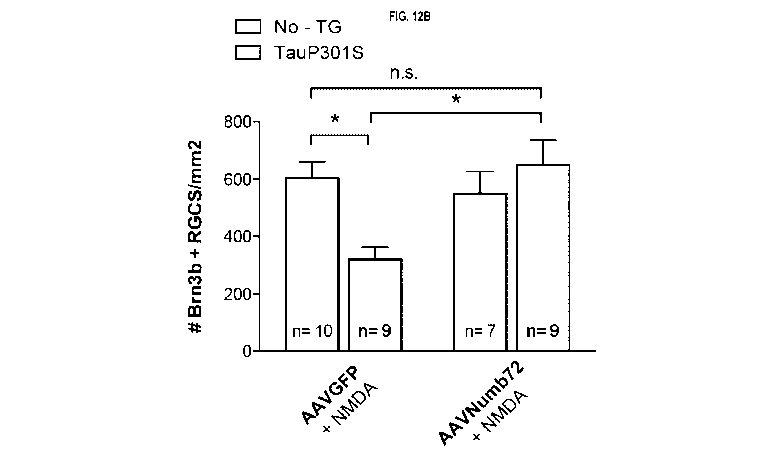
FIGs 12A-B: Numb72 reduces RGCs cell death in vivo in tauopathy mouse model.
FIG. 12A. lmmunostaining
for Brn3b of retina flat mounts from C57b6 mouse line (Control) and transgenic
mutant human Tau mouse
line (TauP301S), 7 weeks after intravitreal injections of AAVGFP type 2 or
AAVNumb72 type 2 in 5 months-
old animals. Three days (72h) prior to sacrifice, all animals received an
intravitreal injection of sublethal doses
of NMDA (10nM). Images were taken in the ganglion cell layer. FIG. 12B.
Quantification of number of Brn3b
RGC per mm2 in control and TauP301S after AAVGFP + NMDA or AAVNumb72 + NMDA
injection. Error bars
represent SEM, n.s = not significant, *p 0.05; Anova 2-way test.
FIGs. 13A-B: The absence of Numb in TauP301S mouse increases RGC death. FIG.
13A: Diagram of mouse
crossing, cK0 mice for Numb are crossed with transgenic mutant human Tau mouse
line (TauP301S), a
mouse model of tauopathy. FIG. 13B: lmmunostaining for Brn3b of retina flat
mounts at 8-month-old from
Ilset1Cre, Numbflox, TauP301S mouse line. 3 different controls were used
(Islet Cre Negative (Neg), Numb
fl/fl, TauP301S transgene negative (TauP301S Tg¨) top left panel; Islet Cre+
(IsletCre), Numb fl/fl, TauP301S
Tg-, top right panel; and IsletCre Neg (Neg), Numb fl/fl, TauP301S transgene
positive (TauP301S Tg+),
bottom left panel; and one cKO/TauP301S transgene positive (Islet Cre+
(IsletCre), Numb fl/fl, TauP301S
Tg+), bottom right panel. FIG. 13C: Quantification of the number of Brn3b+ RGC
per mm2 in controls and
cKO/Tg+ mice at 8 months old. The loss of Numb in a P301S transgenic Tau
background leads to a
significantly more important loss of RGCs than Tau transgenic alone or Numb
cK0 alone, supporting the idea
that Numb has a protective effect on neuronal survival in this model. Mean
SEM, n= 5 animals/genotype.
Anova one-way test, * p0.05, **p0.01, ****p0.0001.
FIGs. 14A-B. The absence of Numb in TauP301S transgenic mice accelerates
lumbar paralysis. Because
Islet-1-Cre is active in motoneurons of the spinal cord, where Numb is also
expressed, the impact of loss of
Numb in these motoneurons on motor deficits was assessed in the Numb
cKO/TauP301S double mutant
mice. FIG. 14A Top images: Representative picture of 260 days-old transgenic
TauP301S mouse (not
paralysed) next to a representative picture of an Islet Cre+; Numb fl/fl,
TauP301S at the same age (showed
obvious signs of paralysis). Arrow points to spinal cord defects in the lumbar
region (top picture). FIG. 14A:
Bottom images: Representative pictures of: a TauP301S mouse with a normal hind-
limb reflex at 260 days
when suspended by the tail and an Islet Cre+; Numb fl/fl, TauP301S mouse with
a complete absence of
extension reflex in both hindlimbs. FIG. 14B Graph depicting the time of
paralysis onset in the lumbar region
CA 03120394 2021-05-18
WO 2020/113338
PCT/CA2019/051751
12
in TauP301S and Islet Cre+; Numb fl/fl, TauP301S mice, Matel-Cox test p=0.02,
n=21 TauP301S and n=8
Islet Cre+; Numb fl/fl, TauP301S.
FIGs. 15A-B: FIG. 15A: Diagram of the construct used to generate a Cre-
inducible Numb72 transgenic mouse
line. FIG 15B: Flat mounts of retinas stained for GFP and Numb 4 weeks after
intravitreal injection of an
AAVCRE vector. GFP and Numb are overexpressed in infected cells. Images were
taken in the ganglion cell
layer (GCL) at 40X and 63X.
FIGs. 16A-B: human Numb1 (Numb-72) amino acid sequence (SEQ ID NO: 1) (FIG.
16A); and human Numb1
nucleic acid sequence (SEQ ID NO: 2) (FIG. 16B).
FIGs. 17A-D: human Numb2 (Numb-66) amino acid sequence (SEQ ID NO: 3); and
human Numb1 nucleic
acid sequence (SEQ ID NO: 5) (FIGs. 17A and C-D); and the RIB Numb domain with
bolded and underlined
exon 3 encoded domain (FIG. 17B) (SEQ ID NO: 4).
FIG. 18A-D: amino acid sequences of Numb3 (Numb-71) (SEQ ID NO: 6), Numb4
(Numb-65) (SEQ ID NO:
7), Numb7 (SEQ ID NO:8 and Numb8 (SEQ ID NO: 9).
FIGs. 19A-D: FIGs. 19A-B: Alignment of amino acid sequences of human Numb
isoforms 1-4, 7 and 8 (SEQ
ID NOs: 1, 3 and 6-9); FIG. 19C a consensus sequence thereof (SEQ ID NO: 10);
and FIG.19D: consensus
of human Numb1 and Numb2 (SEQ ID NO: 11).
FIGs. 20A-C: amino acid sequences for human polypyrimidine tract binding
protein 1 (PTBP1) isoforms 1
(SEQ ID NO: 12), 2 (SEQ ID NO: 13), and 3 (SEQ ID NO: 14).
FIGs. 21A-C: amino acid sequences for human serine and arginine rich splicing
factor 1 (ASF/5F2) isoforms
1 (SEQ ID NO: 15), 2 (SEQ ID NO: 16), and 3 (SEQ ID NO: 17).
FIGs. 22A-H: amino acid sequences for human Tau isoforms 1 (SEQ ID NO: 18), 2
(SEQ ID NO: 19), 3 (SEQ
ID NO: 20), 4 (SEQ ID NO: 21), 5 (SEQ ID NO: 22), 6 (SEQ ID NO: 23), 7 (SEQ ID
NO: 24), and 8 (SEQ ID
NO: 25).
FIGs. 23A-D: amino acid sequences for human RNA-binding motif protein 4 (RBM4)
isoform 1 (SEQ ID NO:
26), 2 (SEQ ID NO: 27), 3 (SEQ ID NO: 28), and 4 (SEQ ID NO: 29).
DESCRIPTION OF ILLUSTRATIVE EMBODIMENTS
Definitions
Numb is an endocytic adaptor protein containing a proline rich region (PRR)
that can be short (e.g., 65 or
66kDa) or long (71 or 72kDa) (called herein "Numb-65, Numb-66, Numb-71 and
Numb-72, respectively) and
a phosphotyrosine-binding (PTB) domain that can be short (Numb-65 and Numb-71)
or long (Numb-66 and
CA 03120394 2021-05-18
WO 2020/113338
PCT/CA2019/051751
13
Numb-72) depending on the isoform (Dho et al., 1999; Karaczyn et al., 2010).
In vertebrates, four protein
isoforms of Numb are produced through alternative splicing (AS) of two
cassette exons, namely exons 3 (E3)
and 9 (E9). AS of E9 produces E9-included (p72/p71) and -excluded (p66/p65)
protein products, whereas AS
of E3 produces E3-included (p72/p66) and excluded (p71/p65) proteins.
Expression of Numb isoforms is
developmentally regulated, with E9-included products usually expressed in
proliferating progenitors, whereas
E9-excluded isoforms are dominantly expressed in postmitotic differentiated
cells. In humans, the 65kDA
(Numb-65), 66kDa (Numb-66), 71kDa (Numb-71) and 72kDa (Numb-72) correspond
respectively to the
isoforms 4 (e.g., accession number NP_001005745.1 or AAD54282.1, 592aa), 2
(e.g., accession number
NP_001307043.1 or NP_001005744.1 or AAD54280.1, 603aa), 3 (e.g., accession
number NP_003735.3 or
AAD54281.1, 640aa) and 1 (e.g., accession number NP_001005743.1 or AAD54279.1,
651aa) of Numb.
Human Numb isoforms also include isoforms 7 (accession no. ABY89092.1, 456aa)
and 8 (accession no.
ABY89093.1, 445aa). Without being so limited, an illustrative amino acid
sequence of human Numb1 (Numb-
72) is depicted in FIG. 16A and the corresponding illustrative nucleotide
sequence of human Numb1 (Numb-
72) is depicted in FIG. 16B (NM_001005743.1).
Alternative splicing, or differential splicing, is a regulated process during
gene expression that results in a
single gene coding for multiple proteins. In this process, particular exons of
a gene may be included within or
excluded from the final processed messenger RNA (mRNA) produced from that
gene. Consequently, the
proteins translated from alternatively spliced mRNAs will contain differences
in their amino acid sequences
and, often, in their biological functions. There are numerous modes of
alternative splicing observed, of which
the most common is exon skipping. In this mode, a particular exon may be
included in mRNAs under some
conditions or in particular tissues and omitted from the mRNA in others. The
production of alternatively spliced
mRNAs is regulated by a system of trans-acting proteins that bind to cis-
acting sites on the primary transcript
itself. Trans-acting proteins include splicing activators that promote the
usage of a particular splice site, and
splicing repressors that reduce the usage of a particular site. There are two
major types of cis-acting RNA
sequence elements present in pre-mRNAs and they have corresponding trans-
acting RNA-binding proteins.
Splicing silencers are sites to which splicing repressor proteins bind,
reducing the probability that a nearby
site will be used as a splice junction. These can be located in the intron
itself (intronic splicing silencers, ISS)
or in a neighboring exon (exonic splicing silencers, ESS). They vary in
sequence, as well as in the types of
proteins that bind to them. The majority of splicing repressors are
heterogeneous nuclear ribonucleoproteins
(hnRNPs) such as hnRNPA1 and polypyrimidine tract binding protein (PTB).
Splicing enhancers are sites to
which splicing activator proteins bind, increasing the probability that a
nearby site will be used as a splice
junction. These also may occur in the intron (intronic splicing enhancers,
ISE) or exon (exonic splicing
enhancers, ESE). Most of the splicing activator proteins that bind to ISEs and
ESEs are members of the
Serine/Arginine (SR) protein family. Such proteins contain RNA recognition
motifs and arginine and serine-
CA 03120394 2021-05-18
WO 2020/113338
PCT/CA2019/051751
14
rich (RS) domains.
Antisense Oligonucleotides (ASO, AON) can be used to modulate alternative
splicing. ASOs are short
oligonucleotides, typically 15-25 bases in length, which are the reverse
complement sequence of a specific
RNA transcript target region. ASOs function by forming Watson-Crick base-pairs
with the target RNA. ASO
binding to a target RNA sterically blocks access of splicing factors to the
RNA sequence at the target site.
ASOs targeted to a splicing enhancer or silencer can prevent binding of
transacting regulatory splicing factors
at the target site and effectively block or promote splicing. The sequence
specificity of ASOs allows them to
bind precisely to endogenous RNAs and, importantly, their fidelity allows
targeting of distinct RNA isoforms.
In addition to their specificity, ASOs have many other features that make them
an ideal therapeutic tool. For
example, ASOs are relatively non-invasive in that they do not alter the genome
directly and improvements in
chemistries have been developed to improve the utility of ASOs as a
therapeutic drug.
Some RNA binding proteins can block or promote the inclusion of specific exons
by binding the same
sequence at different regions of the pre-mRNA. For example, Rbfox protein can
function as an activator and
a repressor of alternative splicing depending on its binding location on pre-
mRNA relative to the regulated
exon. For instance, Rbfox proteins enhance exon inclusion by binding to the
(U)GCAUG element that lies
downstream of the alternative Numb E9, whereas they repress inclusion by
binding to the same element
upstream of the alternative Numb E9 (Kim et al., 2013). An ASO targeting the
upstream intronic (UGCAUG)
site of Numb E9 is expected to have the effect of promoting exon 9 inclusion.
As used herein the term "long RIB Numb isoform" refers to a Numb isoform
comprising a RIB form including
the sequence ERKFFKGFFGK (SEQ ID NO: 30) encoded by exon 3 (see e.g., FIGs.
16A, 17A and 17B). Of
note, this fragment is identical in human and mice orthologs. Without being so
limited, long RIB Numb
isoforms includes Numb-66 and Numb-72.
Long RIB Numb isoform gene or nucleic acid (such as Numb-72 and Numb-66 gene
or nucleic acid) refers
to nucleic acid (e.g., genomic DNA, cDNA, RNA) encoding a long PTB Numb
isoform polypeptide. The
description of the various aspects and embodiments of the invention is
provided with reference to exemplary
long PTB Numb isoform nucleic acid sequences and amino acid sequence (e.g., as
shown in FIGs. 16A-B
and 17A and C). Such reference is meant to be exemplary only and the various
aspects and embodiments of
the invention are also directed to other long PTB Numb isoform nucleic acids
and polypeptides (also referred
to long PTB Numb isoform gene expression products), such as long PTB Numb
isoform nucleic acid or
polypeptide mutants/variants, long PTB Numb isoform variants from species to
species or subject to subject.
Consensuses derived from the alignments of certain Numb variants are also
encompassed by the present
invention (see e.g., SEQ ID NOs: 10-11). In specific embodiments of the
consensus, each X in the consensus
sequence is defined as being any amino acid, or absent when this position is
absent in one or more of Numb
CA 03120394 2021-05-18
WO 2020/113338
PCT/CA2019/051751
homo sapiens isoforms, variants or orthologues. In specific embodiment of the
consensus, each X in the
consensus sequences is defined as being any amino acid that constitutes a
conserved or semi-conserved
substitution of any of the amino acid in the corresponding position in the
orthologues presented in the
alignment, or absent when this position is absent in one or more of the
orthologues presented in the alignment.
Conservative substitutions are denoted by the symbol ":" and semi-conservative
substitutions are denoted by
the symbol ".". In another embodiment, each X refers to any amino acid
belonging to the same class as any
of the amino acid residues in the corresponding position in the orthologues
presented in the alignment, or
absent when this position is absent in one or more of the orthologues
presented in the alignment. In another
embodiment, each X refers to any amino acid in the corresponding position of
the orthologues presented in
the alignment, or absent when this position is absent in one or more of the
orthologues presented in the
alignment. The Table below indicates which amino acid belongs to each amino
acid class.
Class Name of the amino acids
Aliphatic Glycine, Alanine, Valine, Leucine, lsoleucine
Hydroxyl or Sulfur/Selenium-containing Serine, Cysteine, Selenocysteine,
Threonine, Methionine
Cyclic Proline
Aromatic Phenylalanine, Tyrosine, Tryptophan
Basic Histidine, Lysine, Arginine
Acidic and their Amide Aspartate, Glutamate, Asparagine, Glutamine
As used herein the term "Tau", unless more specifically identified, refers to
all forms of tau including toxic
forms of Tau (e.g., phosphorylated tau, and oligomeric tau; without being so
limited, the phosphorylated form
is believed to lead to oligomeric tau).
Protein expression
As used herein the terms "long PTB Numb isoform level" (e.g., "Numb-72
expression level"; "Numb-72
expression", "Numb-66 expression level"; "Numb-66 expression"), or "Tau
expression level" or "Tau
expression", refer to the measurement in a cell or a tissue of a long PTB Numb
isoform level or Tau gene
product, respectively. Long PTB Numb isoform levels and TAU expression levels
could be evaluated at the
polypeptide and/or nucleic acid levels (e.g., DNA or RNA) using any standard
methods known in the art. The
nucleic acid sequence of a nucleic acid molecule in a sample can be detected
by any suitable method or
technique of measuring or detecting gene sequence or expression. Such methods
include, but are not limited
to, polymerase chain reaction (PCR), reverse transcriptase-PCR (RT-PCR), in
situ PCR, SAGE, quantitative
CA 03120394 2021-05-18
WO 2020/113338
PCT/CA2019/051751
16
PCR (q-PCR), in situ hybridization, Southern blot, Northern blot, sequence
analysis, microarray analysis,
detection of a reporter gene, or other DNA/RNA hybridization platforms. For
RNA expression, preferred
methods include, but are not limited to: extraction of cellular mRNA and
Northern blotting using labeled probes
that hybridize to transcripts encoding all or part of one or more of the genes
of this invention; amplification of
mRNA expressed from one or more of the genes of this invention using gene-
specific primers, polymerase
chain reaction (PCR), quantitative PCR (q-PCR), and reverse transcriptase-
polymerase chain reaction (RI-
PCR), followed by quantitative detection of the product by any of a variety of
means; extraction of total RNA
from the cells, which is then labeled and used to probe cDNAs or
oligonucleotides encoding all or part of the
genes of this invention, arrayed on any of a variety of surfaces; in situ
hybridization; and detection of a reporter
gene.
In the context of this invention, "hybridization" means hydrogen bonding
between complementary nucleoside
or nucleotide bases. The terms "specifically hybridizable" and "complementary"
are the terms which are used
to indicate a sufficient degree of complementarity or precise pairing such
that stable and specific binding
occurs between the oligonucleotide and the DNA or RNA target. It is understood
in the art that the sequence
of an antisense compound need not be 100% complementary to that of its target
nucleic acid to be specifically
hybridizable. An antisense compound (e.g., ASO) is specifically hybridizable
when binding of the compound
to the target DNA or RNA molecule interferes with the normal function of the
target DNA or RNA to cause
e.g., a loss of utility or affect splicing, and there is a sufficient degree
of complementarity to avoid non-specific
binding of the antisense compound (e.g., ASO) to non-target sequences under
conditions in which specific
binding is desired, i.e., under physiological conditions in the case of in
vivo assays or therapeutic treatment,
and in the case of in vitro assays, under conditions in which the assays are
performed. Such conditions may
comprise, for example, 400 mM NaCI, 40 mM PIPES pH 6.4, 1 mM EDTA, at 50 to 70
C for 12 to 16 hours,
followed by washing. The skilled person will be able to determine the set of
conditions most appropriate for a
test of complementarity of two sequences in accordance with the ultimate
application of the hybridized
nucleotides.
Methods to measure protein expression levels of selected genes of this
invention, include, but are not limited
to: western blot, tissue microarray, immunoblot, enzyme-linked immunosorbent
assay (ELISA),
radioimmunoassay (RIA), immunoprecipitation, surface plasmon resonance,
chemiluminescence, fluorescent
polarization, phosphorescence, immunohistochemical analysis, matrix-assisted
laser desorption/ionization
time-of-flight (MALDI-TOF) mass spectrometry, microcytometry, microscopy,
fluorescence activated cell
sorting (FAGS), flow cytometry, and assays based on a property of the protein
including but not limited to DNA
binding, ligand binding, or interaction with other protein partners. In a
further embodiment, the long RIB Numb
isoform level and/or Tau expression level is measured by immunohistochemical
staining, and the percentage
CA 03120394 2021-05-18
WO 2020/113338
PCT/CA2019/051751
17
and/or the intensity of immunostaining of immunoreactive cells in the sample
is determined.
In an embodiment, the level of a long RIB Numb isoform and/or Tau polypeptide
is determined using an anti-
long RIB Numb isoform or an anti-Tau antibody. By "long RIB Numb isoform
antibody" and "anti-long RIB
Numb isoform" or "Tau antibody" and "anti-Tau", in the present context is
meant to refer to an antibody capable
of detecting (i.e. binding to) a long RIB Numb isoform protein or a long RIB
Numb isoform protein fragment
(e.g., the RIB fragment ERKFFKGFFGK (SEQ ID NO: 30)) or a Tau protein or a Tau
protein fragment,
respectively.
Without being limited, long RIB Numb isoform antibodies (which can be used for
detection) include those
listed in Table I below, Tau antibodies include those listed in Table II
below. Other antibodies can be found
on the BiocompareTM webpage.
Table I: Examples of available long PTB Numb isoform antibodies
Company Name/catalog Type
number
Millipore Sigma 07-144 Rabbit polyclonal Numb-
72
Developmental and Stem Cell Rabbit polyclonal Numb-
Institute of West China Second 72
University Hospital
Table II: Examples of available Tau antibodies.
Company Name/catalog number Type
Millipore Sigma MAB2241 monoclonal
BosterBio M00097 monoclonal
Atlas Antibodies HPA048895 monoclonal
LifeSpan Biosciences LS-B1223 polyclonal
Methods for normalizing the level of expression of a gene are well known in
the art. For example, the
expression level of a gene of the present invention can be normalized on the
basis of the relative ratio of the
mRNA level of this gene to the mRNA level of a housekeeping gene, or the
relative ratio of the protein level
CA 03120394 2021-05-18
WO 2020/113338
PCT/CA2019/051751
18
of the protein encoded by this gene to the protein level of the housekeeping
protein, so that variations in the
sample extraction efficiency among cells or tissues are reduced in the
evaluation of the gene expression level.
A "housekeeping gene" is a gene the expression of which is substantially the
same from sample to sample or
from tissue to tissue, or one that is relatively refractory to change in
response to external stimuli. A
housekeeping gene can be any RNA molecule other than that encoded by the gene
of interest that will allow
normalization of sample RNA or any other marker that can be used to normalize
for the amount of total RNA
added to each reaction. For example, the GAPDH gene, the G6PD gene, the actin
gene, ribosomal RNA,
3664 RNA, PGK1, RPLPO, or the like, may be used as a housekeeping gene.
Methods for calibrating the level of expression of a gene are well known in
the art. For example, the expression
of a gene can be calibrated using reference samples, which are commercially
available. Examples of
reference samples include but are not limited to: StratageneTM QPCR Human
Reference Total RNA,
Clontech -Hy' Universal Reference Total RNA, and XpressRefTM Universal
Reference Total RNA.
In an embodiment, the above-mentioned methods comprise determining the level
of a long PTB Numb isoform
and/or Tau protein and/or nucleic acid (e.g., nucleic acids or encoded
proteins as shown in in FIGs. 16A-B,
17A and C, 18C-D, 19A-D. and 22A-H) in the sample. In another embodiment, the
above-mentioned method
comprises determining the level of a long PTB Numb isoform and/or Tau
polypeptide (e.g., polypeptides as
shown in FIGs. 16A-B, FIGs. 17A and C, 18C-D, 19A-D and 22A-H) in the sample.
Nucleic acids and host cells
The present invention also relates to nucleic acids comprising nucleotide
sequences encoding the above-
mentioned agent (e.g., a long PTB Numb isoform). The nucleic acid may be codon-
optimized. The nucleic
acid can be a DNA or an RNA. The nucleic acid sequence can be deduced by the
skilled artisan on the basis
of the disclosed amino acid sequences.
The present invention also encompasses vectors (e.g., plasmids, viral vector)
comprising the above-
mentioned nucleic acids. The vectors can be of any type suitable, e.g., for
expression of said polypeptides or
propagation of genes encoding said polypeptides in a particular organism. The
organism may be of eukaryotic
or prokaryotic origin. The specific choice of vector depends on the host
organism and is known to a person
skilled in the art. In an embodiment, the vector comprises transcriptional
regulatory sequences (e.g., a GAG
promoter) or a promoter operably-linked (see definition of "operably-linked"
above) to a nucleic acid
comprising a sequence encoding one or more of the above-mentioned agents
(e.g., a long PTB Numb isoform)
of the invention. A first nucleic acid sequence is "operably-linked" with a
second nucleic acid sequence when
the first nucleic acid sequence is placed in a functional relationship with
the second nucleic acid sequence.
For instance, a promoter (e.g., GAG) is operably-linked to a coding sequence
if the promoter affects the
CA 03120394 2021-05-18
WO 2020/113338
PCT/CA2019/051751
19
transcription or expression of the coding sequence. Generally, operably-linked
DNA sequences are
contiguous and, where necessary to join two protein coding regions, in reading
frame. However, since for
example enhancers generally function when separated from the promoters by
several kilobases and intronic
sequences may be of variable lengths, some polynucleotide elements may be
operably-linked but not
contiguous.
"Transcriptional regulatory sequences" or "transcriptional regulatory
elements" are generic terms that refer to
DNA sequences, such as initiation and termination signals, enhancers, and
promoters, splicing signals,
polyadenylation signals, etc., which induce or control transcription of
protein coding sequences with which
they are operably-linked. For instance, the GAG promoter is a strong non-
specific synthetic promoter
frequently used to drive high levels of gene expression in mammalian
expression vectors containing (A)
the cytomegalovirus (CMV) early enhancer element, (B) the promoter, the first
exon and the first intron of
chicken beta-actin gene, and (C) the splice acceptor of the rabbit beta-globin
gene.
A recombinant expression vector comprising a nucleic acid sequence of the
present invention may be
introduced into a cell, e.g., a host cell (such as a neuron), which may
include a living cell capable of expressing
the protein coding region from the defined recombinant expression vector.
Accordingly, the present invention
also relates to cells (host cells) comprising the nucleic acid and/or vector
as described above. The suitable
host cell may be any cell of eukaryotic or prokaryotic (bacterial) origin that
is suitable, e.g., for expression of
or propagation of genes/nucleic acids encoding said above-mentioned agents
(e.g., a long PTB Numb
isoform). The eukaryotic cell line may be of mammalian, of yeast, or
invertebrate origin. The specific choice
of cell line is known to a person skilled in the art. Choice of bacterial
strains will depend on the task at hand
and is commonly known to a person skilled in the art. The terms "host cell"
and "recombinant host cell" are
used interchangeably herein. Such terms refer not only to the particular
subject cell, but also to the progeny
or potential progeny of such a cell. Because certain modifications may occur
in succeeding generations due
to either mutation or environmental influences, such progeny may not, in fact,
be identical to the parent cell,
but are still included within the scope of the term as used herein.
Vectors can be introduced into cells via conventional transformation or
transfection techniques. The terms
"transformation" and "transfection" refer to techniques for introducing
foreign nucleic acid into a host cell (such
as a neuron), including calcium phosphate or calcium chloride co-
precipitation, DEAE-dextran-mediated
transfection, lipofection, electroporation, microinjection and viral-mediated
transfection. Suitable methods for
transforming or transfecting host cells can for example be found in Sambrook
et al. (supra), Sambrook and
Russell (supra) and other laboratory manuals. Methods for introducing nucleic
acids into mammalian cells in
vivo are also known and may be used to deliver the vector DNA of the invention
to a subject for gene therapy.
The above-mentioned nucleic acid or vector may be delivered to cells in vivo
(to induce the expression of the
CA 03120394 2021-05-18
WO 2020/113338
PCT/CA2019/051751
above-mentioned agents (e.g., a long PTB Numb isoform) using methods well
known in the art such as direct
injection of DNA, receptor-mediated DNA uptake, viral-mediated transfection or
non-viral transfection and lipid
based transfection, all of which may involve the use of gene therapy vectors.
Direct injection has been used
to introduce naked DNA into cells in vivo. A delivery apparatus (e.g., a "gene
gun") for injecting DNA into cells
in vivo may be used. Such an apparatus may be commercially available (e.g.,
from BioRad). Naked DNA may
also be introduced into cells by complexing the DNA to a cation, such as
polylysine, which is coupled to a
ligand for a cell-surface receptor. Binding of the DNA-ligand complex to the
receptor may facilitate uptake of
the DNA by receptor-mediated endocytosis. A DNA-ligand complex linked to
adenovirus capsids which disrupt
endosomes, thereby releasing material into the cytoplasm, may be used to avoid
degradation of the complex
by intracellular lysosomes.
Defective retroviruses are well characterized for use as gene therapy vectors
(for a review see Miller, A. D.
(1990) Blood 76:271). Protocols for producing recombinant retroviruses and for
infecting cells in vitro or in
vivo with such viruses can be found in Current Protocols in Molecular Biology,
Ausubel, F. M. et al. (eds.)
Greene Publishing Associates, (1989), Sections 9.10-9.14 and other standard
laboratory manuals. Examples
of suitable retroviruses include pLJ, pZIP, pWE and pEM which are well known
to those skilled in the art.
Examples of suitable packaging virus lines include psiCrip, psiCre, psi2 and
psiAm. Retroviruses have been
used to introduce a variety of genes into many different cell types, including
epithelial cells, endothelial cells,
lymphocytes, myoblasts, hepatocytes, and bone marrow cells, in vitro and/or in
vivo.
For use as a gene therapy vector, the genome of an adenovirus may be
manipulated so that it encodes and
expresses a nucleic acid of the invention (e.g., a nucleic acid encoding one
of the above-mentioned agents
(e.g., a long PTB Numb isoform)), but is inactivated in terms of its ability
to replicate in a normal lytic viral life
cycle. Suitable adenoviral vectors derived from the adenovirus strain Ad type
5 dI324 or other strains of
adenovirus (e.g., Ad2, Ad3, Ad7 etc.) are well known to those skilled in the
art. Recombinant adenoviruses
are advantageous in that they do not require dividing cells to be effective
gene delivery vehicles (e.g., viral
vectors) and can be used to infect a wide variety of cell types, including
neurons, RGCs, airway epithelium,
endothelial cells, hepatocytes, and muscle cells.
Adeno-associated virus (AAV) may be used as a gene delivery vector for
delivery of DNA for gene therapy
purposes (e.g., adeno-associated viral (AAV) vector expressing a long PTB Numb
isoform). AAV is a naturally
occurring defective virus that requires another virus, such as an adenovirus
or a herpes virus, as a helper
virus for efficient replication and a productive life cycle. AAV may be used
to integrate DNA into non-dividing
cells. Lentiviral gene therapy vectors may also be adapted for use in the
invention. Alphavirus vectors such
as Semliki Forest virus-based vectors and Sindbis virus-based vectors and the
like can also be used.
Delivery
CA 03120394 2021-05-18
WO 2020/113338
PCT/CA2019/051751
21
As seen herein, the long PTB Numb isoform of the instant disclosure can be
delivered to target cells through
the use of a nucleic acid encoding the isoform (e.g., viral vector). It can
also be directly delivered as a purified
(recombinant) protein. In particular, nanoparticles or peptide-based
technologies can be used. In particular,
for intraocular delivery, and without being so limited, membrane
permeabilizing amphiphilic peptide of Feldan
Therapeutics could be used.
Long PTB Numb isoform activity
As used herein the terms "long PTB Numb isoform activity" and "long PTB Numb
isoform function" (such as
"Numb-72 activity" and "Numb-72 function", "Numb-66 activity" and "Numb-66
function") are used
interchangeably and refer to detectable (direct or indirect) enzymatic,
biochemical or cellular activity
attributable to a long PTB Numb isoform (e.g., increasing neuronal survival
(see e.g., Examples 3, 6 and 13),
preventing neurodegeneration (e.g., Example 14), including in stress condition
such as aging (Examples 3, 6
and 13) and/or excitotoxicity (see e.g., Examples 6 and 13), preventing motor
deficit (e.g., Example 15),
decreasing intracellular (neuronal) Tau (e.g., oligomeric) levels (e.g., in
RGCs) (see e.g., Examples 4 and 8),
reducing Tau (e.g., pTau)-containing axonal blebbing (see e.g., Examples 3 and
5), interaction with Tau (see
e.g., Example 7), stimulating secretion of monomeric Tau (see e.g., Example
11), and promoting intraneuronal
Tau degradation (e.g., in brain tissue) (see e.g., Example 12). Long PTB Numb
isoforms activity could also
be indirectly measured by evaluating the level of expression of the long PTB
Numb isoform, or a fragment
thereof, in cells as well as in biological samples (e.g., tissue, organ,
fluid).
Modulation of long PTB Numb isoform expression or activity
The modulation of long PTB Numb isoform expression and/or activity (e.g., Numb-
72 and/or Numb-66
expression and/or activity) could be achieved directly or indirectly by
various mechanisms, which among
others could act at the level of (i) transcription, for example by activating
promoter or enhancer elements and
thereby increasing their messenger RNA expression (e.g., by cytokine
stimulation, etc.), (ii) splicing, for
example by inhibiting expression or activity of a splicing regulator that
promotes Numb exon 3 exclusion, or
by activating a splicing regulator that enhance exon 3 inclusion, (iii)
translation, (iv) post-translational
modifications, e.g., glycosylation, sulfation, phosphorylation, ubiquitination
(e.g., polyubiquitinylation and
proteasomal degradation), (v) cellular localization (e.g., cytoplasmic versus
nuclear localization), and (vi)
protein-protein interaction. These regulatory processes occur through
different molecular interactions that
could be modulated using a variety of compounds or modulators.
In the context of the present invention, a "compound" is a molecule such as,
without being so limited, a dsRNA
(e.g., siRNA), antisense molecule (ASO), protein, peptide, small molecule,
antibody, etc.
Agent that increases long PTB Numb isoform expression and/or activity
CA 03120394 2021-05-18
WO 2020/113338
PCT/CA2019/051751
22
As used herein an "agent that increases a long PTB Numb isoform expression
and/or activity" (e.g., "agent
that increases Numb-72 or Numb-66 expression and/or activity"), such as agents
that promote a long PTB
Numb isoform expression and/or activity in neurons, refers to any compound or
composition that directly or
indirectly increases at least one long PTB Numb isoform expression and/or
activity (e.g., Numb-72 or Numb-
66 expression and/or activity). It includes molecules such as, without being
so limited, nucleic acids encoding
a long PTB Numb isoform expression product (such as human Numb-72 (Numb1)
nucleic acid (see e.g.,
NM_001005743.1 shown in FIG. 16B) or human Numb-66 (Numb2) nucleic acid (e.g.,
NM_001005744.1
shown in FIG. 170)); a long PTB Numb isoform polypeptide or a fragment thereof
having a long PTB Numb
isoform activity, such as human Numb-72 (e.g., NP_001005743.1 shown in FIG.
16A) or a fragment thereof
having Numb-72 activity; or human Numb-66 polypeptide (see e.g.,
NP_001307043.1 shown in FIG. 17A) or
a fragment thereof having Numb-66 activity; a long PTB Numb polypeptide of
sequence SEQ ID NO: 10 or
11; an agent which increases the level of a long PTB Numb isoform (e.g.,
nucleic acid encoding for Numb-66
or Numb-72, e.g., FIG. 16B and 170) by acting on splicing; an agent promoting
the Numb exon 3 inclusion by
activating an RNA binding protein that enhances exon 3 inclusion such as RNA-
binding motif protein 4 (RBM4)
(see e.g.. FIGs. 23A-D and SEQ ID NOs: 26-29) that promotes inclusion of exon
3 of Numb but excludes exon
9 (Tarn et al. 2016); an agent such as an antisense oligonucleotide (ASO) that
blocks the recognition of
sequences required for Numb exon 3 exclusion; an agent such as a siRNA
targeting a gene coding for a
splicing regulator promoting the exclusion of Numb exon 3; an agent promoting
the Numb exon 9 inclusion by
activating an RNA binding protein that enhances exon 9 inclusion such as but
not limited to RNA-binding
protein 6 (RBM6) (e.g., P78332-1 (isoform 1); P78332-3 (isoform 2) and P78332-
3 (isoform 3)); RNA-binding
protein 5 (RBM5) (e.g., P52756-1 (isoform 1), P52756-2 (isoform 2), P52756-3
(isoform 3), P52756-4 (isoform
4), and P52756-5 (isoform 5)); PTBP1 (Rajendran et al., 2016) (see
illustrative human amino acid sequences
in FIGs. 20A-C and SEQ ID NOs: 12-14); Mitogen-activated protein kinase
(MAPK)/extracellular signal-
regulated kinase (ERK) (Rajendran et al., 2016), agonists to MAPK/ERK (such as
Honokiol (Zhai et al. 2005),
CHPG sodium salt (Tao Chen et al. 2012), LM22B-10 (Yang T, et al.
Neuropharmacology. 2016)); an agent
such as an antisense oligonucleotide (ASO) that blocks the recognition of
sequences required for Numb exon
9 exclusion, such as an ASO targeting upstream intronic UGCAUG (see above); an
agent such as a siRNA
targeting a gene coding for a splicing regulator promoting the exclusion of
Numb exon 9, such as an siRNA
targeting upstream intronic UGCAUG; an exon 9 splicing factor inhibitor, such
as a serine and arginine rich
splicing factor 1 (ASF/5F2) inhibitor (see e.g., FIGs. 21A-C for ASF/5F2
isoforms 1-3 sequences); a
polypyrimidine Tract Binding Protein 1 (PTBP1) inhibitor (see e.g., FIGs. 20A-
C for PTB1 isoforms 1-3
sequences); an RNA binding motif protein 6 (RBM6) inhibitor; an RNA binding
motif protein (RBM10) inhibitor;
or an RNA-binding FOX3 (RBFOX3) inhibitor.
More particularly, agents of the present invention include nucleic acid
encoding Numb-72 or Numb-66 and
CA 03120394 2021-05-18
WO 2020/113338
PCT/CA2019/051751
23
agents which increase the level of RNAs encoding for Numb-66 or Numb-72
isoforms by modulating splicing
to increase Numb exon 3 inclusion; small RNA molecules (e.g., antisense
oligonucleotides (AOS) and
siRNAs); peptides; small molecules; antibodies, etc. Candidate compounds are
tested using a variety of
methods and assays.
Agents that increase a long RIB Numb isoform expression and/or activity (e.g.,
Numb-72 and/or Numb-66
expression and/or activity) can be used to target (e.g., tau expressing)
neurons (e.g., RGCs, brain neurons,
spinal cord, motoneurons) using e.g., viral vectors (e.g., adenoviruses,
lentivirus, AAVs (see Example 13))
or other gene/protein delivery and thereby force a long RIB Numb isoform
expression (e.g., Numb-72
expression) on the neurons. Such neurons may thereafter benefit from
treatments described herein.
Cell targets of the agents of the present invention are neurons. Without being
so limited, such cells include
neurons of the central nervous system such as brain neurons, retinal neurons
(RGCs) and spinal cord
neurons.
Screening assays
Given the correlation between long RIB Numb isoforms expression/activity
(e.g., Numb-72 and/or Numb-66
expression/activity) on intraneuronal Tau levels or degradation, compounds
which are capable of increasing
a long RIB Numb isoform (e.g., Numb-72 and/or Numb-66 expression and/or
activity) may be used for the
prevention and/or treatment of a pathological condition associated with
intraneuronal Tau accumulation.
Screening for agents that increase long RIB Numb isoforms expression and/or
activity
Therefore, the invention further relates to screening methods using a long RIB
Numb isoform positive cells
for the identification and characterization of compounds capable of increasing
a long RIB Numb isoform
activity and/or expression which may be used for the prevention and/or
treatment of a pathological condition
associated with intraneuronal Tau accumulation.
The present invention also provides a method (e.g., an in vitro method) for
determining whether a test
compound is useful for the prevention and/or treatment of a pathological
condition associated with
intraneuronal Tau accumulation, said method comprising: (a) contacting said
test compound with a (neuronal)
cell expressing a long PTB Numb isoform and Tau; and (b) determining the
intraneuronal Tau levels,
degradation and/or neuronal survival, in the presence or absence of said test
compound (and eventually in
the presence of NMDA); wherein a decrease in the Tau levels and/or degradation
and/or an increase in the
Tau survival in the presence of said test compound relative to the absence
thereof is indicative that said test
compound may be used for the prevention and/or treatment of a pathological
condition associated with
intraneuronal Tau accumulation (e.g., tauopathy and/or Tau-associated optic
neuropathy).
The present invention also provides a method (e.g., an in vitro method) for
determining whether a test
CA 03120394 2021-05-18
WO 2020/113338
PCT/CA2019/051751
24
compound is useful for the prevention and/or treatment of a pathological
condition associated with
intraneuronal Tau accumulation, said method comprising: (a) contacting said
test compound with a (neuronal)
cell expressing a long RIB Numb isoform (e.g., Numb-72); and (b) determining
the a long RIB Numb isoform
levels in the presence or absence of said test compound; wherein an increase
in the long RIB Numb isoform
levels in the presence of said test compound relative to the absence thereof
is indicative that said test
compound may be used for the prevention and/or treatment of a pathological
condition associated with
intraneuronal Tau accumulation (e.g., tauopathy and/or Tau-associated optic
neuropathy).
The present invention also provides a method (e.g., an in vivo method in an
animal model) for determining
whether a test vector (e.g., AAV vector) expressing a long RIB Numb isoform
(e.g., Numb-72) or a fragment
thereof having a long RIB Numb isoform activity (e.g., Numb-72 activity) is
useful for the prevention and/or
treatment of a pathological condition associated with intraneuronal Tau
accumulation, said method
comprising: (a) expressing said long PTB Numb isoform or fragment thereof in a
(neuronal) cell expressing
Tau; and (b) determining the intraneuronal Tau levels, degradation and/or
neuronal survival, in the presence
or absence of said long PTB Numb isoform or fragment thereof; wherein a
decrease in the Tau levels and/or
degradation and/or an increase in the neuronal survival in the presence of
said long PTB Numb isoform or
fragment thereof relative to the absence thereof is indicative that said test
viral vector expressing said long
PTB Numb isoform or fragment thereof may be used for the prevention and/or
treatment of a pathological
condition associated with intraneuronal Tau accumulation (e.g., tauopathy
and/or Tau-associated optic
neuropathy). (see e.g., Example 13)
The present invention also provides a method (e.g., an in vitro method) for
determining whether a test
compound is useful for the prevention and/or treatment of a pathological
condition associated with
intraneuronal Tau accumulation, said method comprising: (a) contacting said
test compound with a long PTB
Numb isoform polypeptide, or a fragment thereof having a long PTB Numb isoform
activity; and (b)
determining the expression and/or activity of the long PTB Numb isoform
polypeptide or fragment thereof, in
the presence or absence of said test compound; wherein an increase in the
expression and/or activity of the
long PTB Numb isoform in the presence of said test compound relative to the
absence thereof is indicative
that said test compound may be used for the prevention and/or treatment of a
pathological condition
associated with intraneuronal Tau accumulation.
The present invention also provides a method (e.g., an in vitro method) for
determining whether a test
compound is useful for the prevention and/or treatment of a pathological
condition associated with
intraneuronal Tau accumulation (e.g., tauopathy, Tau-associated optic
neuropathy), said method comprising:
(a) contacting said test compound with a cell comprising a first nucleic acid
comprising a transcriptionally
regulatory element normally associated with a long PTB Numb isoform gene,
operably linked to a second
CA 03120394 2021-05-18
WO 2020/113338
PCT/CA2019/051751
nucleic acid comprising a reporter gene encoding a reporter protein; and (b)
determining whether the reporter
gene expression and/or reporter protein activity is increased in the presence
of said test compound; wherein
said increase in reporter gene expression and/or reporter protein activity is
indicative that said test compound
may be used for prevention and/or treatment of a pathological condition
associated with intraneuronal Tau
accumulation.
The present invention also provides a method (e.g., an in vitro method) for
identifying an agent (e.g., an agent
that promotes the Numb exon 3 inclusion) is useful for the prevention and/or
treatment of a pathological
condition associated with intraneuronal Tau accumulation (e.g., tauopathy, Tau-
associated optic neuropathy),
said method comprising: (a) contacting said test compound with a cell
comprising a first nucleic acid
comprising Numb exon 3, its upstream and downstream flanking introns, and
constitutive exons 2 and 4,
wherein the Numb exon 3 is operably linked to a second nucleic acid comprising
a reporter gene encoding a
reporter protein (e.g., fluorescent reporter), so that the presence of exon 3
is revealed by the reporter protein;
and (b) determining whether the reporter gene expression and/or reporter
protein activity is increased (e.g.,
fluorescence) in the presence of said test compound; wherein said increase in
reporter gene expression
and/or reporter protein activity is indicative that said test compound may be
used for prevention and/or
treatment of a pathological condition associated with intraneuronal Tau
accumulation.
The above-mentioned methods may be employed either with a single test compound
or a plurality or library
(e.g., a combinatorial library) of test compounds. In the latter case,
synergistic effects provided by
combinations of compounds may also be identified and characterized. The above-
mentioned compounds may
be used for prevention and/or treatment of a pathological condition associated
with intraneuronal Tau
accumulation or may be used as lead compounds for the development and testing
of additional compounds
having improved specificity, efficacy and/or pharmacological (e.g.,
pharmacokinetic) properties. In an
embodiment, the compound may be a prodrug which is altered into its active
form at the appropriate site of
action, (e.g., neurons). In certain embodiments, one or a plurality of the
steps of the screening/testing methods
of the invention may be automated.
Such assay systems may comprise a variety of means to enable and optimize
useful assay conditions. Such
means may include but are not limited to: suitable buffer solutions, for
example, for the control of pH and ionic
strength and to provide any necessary components for optimal long PTB Numb
isoform (e.g., Numb-72)
activity and stability, temperature control means for long PTB Numb isoform
activity and or stability, and
detection means to enable the detection of a long PTB Numb isoform activity
reaction product. A variety of
such detection means may be used, including but not limited to one or a
combination of the following:
radiolabeling (e.g., 32P, 140, 3w,
) antibody-based detection, fluorescence, chemiluminescence, spectroscopic
methods (e.g., generation of a product with altered spectroscopic properties),
various reporter enzymes or
CA 03120394 2021-05-18
WO 2020/113338
PCT/CA2019/051751
26
proteins (e.g., horseradish peroxidase, green fluorescent protein), specific
binding reagents (e.g.,
biotin/(strept)avidin), and others.
The assay may be carried out in vitro utilizing a source of long PTB Numb
isoform (e.g., Numb-72) which may
comprise naturally an isolated or recombinantly produced long PTB Numb
isoform, in preparations ranging
from crude to pure. Recombinant long PTB Numb isoform (e.g., Numb-72) may be
produced in a number of
prokaryotic or eukaryotic expression systems, which are well known in the art.
Such assays may be performed
in an array format.
As noted above, the invention further relates to methods for the
identification and characterization of
compounds capable of modulating long PTB Numb isoform (e.g., Numb-72) gene
expression. Such a method
may comprise assaying long PTB Numb isoform (e.g., Numb-72) gene expression in
the presence versus the
absence of a test compound. Such gene expression may be measured by detection
of the corresponding
RNA or protein, or via the use of a suitable reporter construct comprising one
or more transcriptional regulatory
element(s) normally associated with a long PTB Numb isoform (e.g., Numb-72)
gene, operably-linked to a
reporter gene.
See above for definitions of "operably-linked" and "Transcriptional regulatory
element". The expression of a
reporter gene may be measured on the transcriptional or translational level,
e.g., by the amount of RNA or
protein produced. RNA may be detected by for example Northern analysis or by
the reverse transcriptase-
polymerase chain reaction (RT-PCR) method (see for example Sambrook et al.
(1989) Molecular Cloning: A
Laboratory Manual (2nd edition), Cold Spring Harbor Laboratory Press, Cold
Spring Harbor, New York, USA).
Protein levels may be detected either directly using affinity reagents (e.g.,
an antibody or fragment thereof (for
methods, see for example Harlow, E. and Lane, D (1988) Antibodies : A
Laboratory Manual, Cold Spring
Harbor Laboratory Press, Cold Spring Harbor, NY); a ligand which binds the
protein) or by other properties
(e.g., fluorescence in the case of green fluorescent protein) or by
measurement of the protein's activity, which
may entail enzymatic activity to produce a detectable product (e.g., with
altered spectroscopic properties) or
a detectable phenotype (e.g., alterations in cell growth/function). Suitable
reporter genes include but are not
limited to chloramphenicol acetyltransferase, beta-D galactosidase,
luciferase, or green fluorescent protein
(GFP or EGFP).
Long PTB Numb isoform (e.g., Numb-72) expression levels could be determined
using any standard methods
known in the art. Non-limiting examples of such methods include western blot,
immunoblot, enzyme-linked
immunosorbent assay (ELISA), radioimmunoassay (RIA), immunoprecipitation,
immunocytochemistry,
immunohistochemistry, as well as methods to determine mRNA levels such as RT-
PCR and northern analysis,
real-time PCR, PCR, in situ hybridization and so on.
CA 03120394 2021-05-18
WO 2020/113338
PCT/CA2019/051751
27
In another aspect, the present invention provides an agent that increases long
PTB Numb isoform (e.g.,
Numb-72) expression or activity identified by the above-noted screening
method.
Pathological condition associated with intraneuronal Tau accumulation
As used herein, the term "pathological condition associated with intraneuronal
Tau accumulation" refers to
tauopathies and Tau-associated optic neuropathies.
As used herein the term "tauopathy" refers to neurodegenerative diseases
associated with the pathological
aggregation of tau protein in neurofibrillary or gliofibrillary tangles in the
human brain and eyes. Without being
so limited, tauopathies include Alzheimer's disease, primary age-related
tauopathy (PART), chronic traumatic
encephalopathy, including dementia pugilistica, progressive supranuclear
palsy, corticobasal degeneration,
frontotemporal dementia, parkinsonism linked to chromosome 17, Lytico-Bodig
disease (Parkinson-dementia
complex of Guam), ganglioglioma, gangliocytoma, meningioangiomatosis,
postencephalitic parkinsonism,
subacute sclerosing panencephalitis, lead encephalopathy, tuberous sclerosis,
pantothenate kinase-
associated neurodegeneration, and lipofuscinosis.
As used herein the term "Tau-associated optic neuropathy" refers to diseases
characterized by damages in
the optic nerve associated to an elevated level of tau which then causes
degeneration of retinal ganglion cells
such as but no limited to glaucoma (Chiasseu, 2016), optic neuritis
(Frederiksen et al. 2012) and compressive
optic neuropathy (Oku et al. 2019).
As used herein the term "motor deficit" refers to motor nerves degeneration
leading to paralysis and/or
muscles atrophy.
Treatment and prevention
The terms "treat/treating/treatment" and "prevent/preventing/prevention" as
used herein, refers to eliciting the
desired biological response, i.e., a therapeutic and prophylactic effect,
respectively. In accordance with the
subject invention, the therapeutic effect comprises one or more of a
decrease/reduction in the severity of the
pathological condition associated with intraneuronal Tau accumulation (e.g.,
tauopathy (e.g., reduced memory
loss), Tau-associated optic neuropathy (reduced visual impairment), motor
deficit, a decrease/reduction in at
least one symptom or disease-related effect (e.g., a reduction of
intraneuronal Tau levels, an increase of
intraneuronal Tau degradation, an increase of neuron survival), an
amelioration of at least one symptom or
disease-related effect, and an increased survival time of the affected host
animal, following administration of
the at least one agent that increases a long RIB Numb isoform expression or
activity, or of a composition
comprising the agent. In accordance with the invention, a prophylactic effect
may comprise a complete or
partial avoidance/inhibition of the pathological condition associated with
intraneuronal Tau accumulation, and
an increased survival time of the affected host animal, following
administration of the at least one agent that
CA 03120394 2021-05-18
WO 2020/113338
PCT/CA2019/051751
28
increases a long RIB Numb isoform expression or activity or of a composition
comprising the agent.
As such, a "therapeutically effective" or "prophylactically effective" amount
of agent affecting a long RIB Numb
isoform expression and/or activity (e.g., Tau intraneuronal levels or
degradation or neuron survival), or a
combination of such agents, may be administered to an animal, in the context
of the methods of treatment
and prevention, respectively, described herein.
Types of samples from the subject and of control samples
As used herein, the term "organism" refers to a living thing which, in at
least some form, is capable of
responding to stimuli, reproduction, growth or development, or maintenance of
homeostasis as a stable whole
(e.g., an animal). The organism may be composed of many cells which may be
grouped into specialized
tissues or organs.
"Sample" or "biological sample" refers to any solid or liquid sample isolated
from a live being. In a particular
embodiment, it refers to any solid (e.g., tissue sample) or liquid sample
isolated from an animal (e.g., human),
such as a biopsy material (e.g., solid tissue sample), blood (e.g., plasma,
serum or whole blood), saliva,
synovial fluid, urine, amniotic fluid and cerebrospinal fluid. Such sample may
be, for example, fresh, fixed
(e.g., formalin-, alcohol- or acetone-fixed), paraffin-embedded or frozen
prior to analysis of a long RIB Numb
isoform expression level or Tau expression level. In an embodiment, the above-
mentioned sample is obtained
from a subject having a pathological condition associated with intraneuronal
Tau accumulation.
As used herein, the term "tissue" or "tissue sample" refers to a group of
cells, not necessarily identical, but
from the same origin, that together carry out a specific function. A tissue is
a cellular organizational level
intermediate between cells and a complete organism. Organs are formed by the
functional grouping together
of multiple tissues. Examples of tissues include neuronal, retinal and nervous
tissues. Other examples of
biological tissues include blood cells populations (e.g., B or T lymphocytes
populations), breast, skin, lung or
colon tissues.
Similarly, the expression "reference gene expression and/or activity of a
gene" refers to the expression and/or
activity of that gene used as a control for the measure performed in a sample
from a subject. "Reference gene
sample" as used herein refers to a sample comprising a reference expression
and/or activity of a gene.
More particularly, the expression "reference long RIB Numb isoform expression
and/or activity" and
"reference Tau expression and/or activity" refers to the long PTB Numb isoform
and Tau expression and/or
activity, respectively, used as a control for the measure performed in a
sample from a subject. "Reference
long PTB Numb isoform sample" and "reference Tau sample" as used herein refer
to a sample comprising a
"reference long PTB Numb isoform expression and/or activity" and "reference
Tau expression and/or activity",
respectively.
CA 03120394 2021-05-18
WO 2020/113338
PCT/CA2019/051751
29
Depending on the type of assay performed, the reference long RIB Numb isoform
(e.g., Numb-72) expression
and/or activity and reference Tau expression can be selected from an
established standard, a corresponding
long RIB Numb isoform and Tau expression and/or activity, respectively,
determined in the subject (in a
sample from the subject) at an earlier time; a corresponding long RIB Numb
isoform and Tau expression
and/or activity, respectively, determined in one or more control subject(s)
known to not being predisposed to
a pathological condition associated with intraneuronal Tau accumulation, known
to not having a pathological
condition associated with intraneuronal Tau accumulation (in specific
embodiments, a tauopathy such as AD;
or a Tau-associated optic neuropathy) or known to have a good prognosis; or
determined in one or more
control subject(s) known to have a predisposition to a pathological condition
associated with intraneuronal
Tau accumulation, known to have a pathological condition associated with
intraneuronal Tau accumulation or
known to have a poor prognosis. In another embodiment, the reference long RIB
Numb isoform expression
and/or activity and reference Tau expression and/or activity is the average or
median value obtained following
determination of long RIB Numb isoform and Tau expression or activity,
respectively, in a plurality of samples
(e.g., samples obtained from several healthy subjects or samples obtained from
several subjects having a
pathological condition associated with intraneuronal Tau accumulation (e.g.,
tauopathy or Tau-associated
optic neuropathy)).
"Corresponding normal tissue" or "corresponding tissue" as used herein refers
to a reference sample obtained
from the same tissue as that obtained from a subject. Corresponding tissues
between organisms (e.g., human
subjects) are thus tissues derived from the same origin (e.g., two B
lymphocyte populations).
Measurement of a long PTB Numb isoform, and Tau in a sample
The present invention encompasses methods comprising detecting the presence of
long RIB Numb isoform
(e.g., Numb-72) and optionally Tau activity and/or expression in a subject
sample. In a specific embodiment,
the present invention encompasses detecting the presence of long RIB Numb
isoform and optionally Tau
activity and/or expression in a subject sample. In another specific
embodiment, the present invention
encompasses detecting the presence of long RIB Numb isoform activity and/or
expression in a subject
sample.
In another embodiment, the present invention encompasses methods comprising
determining whether a long
PTB Numb isoform and optionally Tau expression and/or activity in a subject
sample is substantially similar
to that of a reference expression and/or activity. In a specific embodiment,
the present invention encompasses
determining whether a long PTB Numb isoform activity and/or expression in a
subject sample is substantially
similar to that of a reference expression and/or activity.
In another embodiment, the present invention encompasses methods comprising
determining whether a long
CA 03120394 2021-05-18
WO 2020/113338
PCT/CA2019/051751
RIB Numb isoform activity and/or expression in a subject sample is higher than
a reference expression and/or
activity.
In cases where the reference long RIB Numb isoform sample and reference Tau
samples are from the subject
at an earlier time; from subject(s) known not to being predisposed to a
pathological condition associated with
intraneuronal Tau accumulation, known not to have an pathological condition
associated with intraneuronal
Tau accumulation, or known to have a good prognosis, (1) a decreased long RIB
Numb isoform (e.g., Numb-
72); and optionally (i) increased Tau expression and/or activity, respectively
in the sample from the subject
relative to the reference long RIB Numb isoform and Tau expression and/or
activity, respectively, is indicative
that the subject would likely benefit from an agent that increases long RIB
Numb isoform expression or
activity, while a comparable or lower expression or activity in a sample from
the subject relative to the
reference expression and/or activity is indicative that the subject would
likely not benefit from an agent that
increase long RIB Numb isoform expression or activity.
In cases where the reference long RIB Numb isoform sample is from subject(s)
known to have a pathological
condition associated with intraneuronal Tau accumulation, known to have a
pathological condition associated
with intraneuronal Tau accumulation or known to have a poor prognosis, (1) a
comparable or a decreased
long RIB Numb isoform and eventually (i) a comparable or an increased Tau
expression and/or activity,
respectively in the sample from the subject relative to the reference long RIB
Numb isoform and reference
Tau expression and/or activity, respectively, is indicative that the subject
would likely benefit from an agent
that increases long RIB Numb isoform expression or activity, while a higher
long RIB Numb isoform
expression or activity in a sample from the subject relative to the reference
expression and/or activity is
indicative that the subject would likely not benefit from an agent that
increases long RIB Numb isoform
expression or activity.
As used herein, a "higher" or "increased" level of expression and/or activity
of a long RIB Numb isoform (e.g.,
Numb-72 and/or Numb-66 expression and/or activity) refers to levels of
expression or activity in a sample (i.e.
sample from the subject) which exceeds with statistical significance that in
the reference sample (e.g., an
average corresponding level of expression or activity a healthy subject or of
a population of healthy subjects,
or when available, the normal counterpart of the affected or pathological
tissue) measured through direct (e.g.,
Anti-long RIB Numb isoform antibody, Anti-Tau antibody, quantitative PCR) or
indirect methods. The
increased level of expression and/or activity refers to level of expression
and/or activity in a sample (i.e.
sample from the subject) which is at least 10% higher, in another embodiment
at least 15% higher, in another
embodiment at least 20% higher, in another embodiment at least 25%, in another
embodiment at least 30%
higher, in a further embodiment at least 40% higher; in a further embodiment
at least 50% higher, in a further
embodiment at least 60% higher, in a further embodiment of at least 70%
higher, in a further embodiment of
CA 03120394 2021-05-18
WO 2020/113338
PCT/CA2019/051751
31
at least 80% higher, in a further embodiment of at least 90% higher, in a
further embodiment at least 100%
higher (i.e. 2-fold), in a further embodiment at least 200% higher (i.e. 3-
fold), in a further embodiment at least
300% higher (i.e. 4-fold), relative to the reference expression and/or
activity.
As used herein, a "substantially similar level" refers to a difference in the
level of expression or activity between
the level determined in a first sample (e.g., sample from the subject) and the
reference expression and/or
activity which is less than about 10 %; in a further embodiment, 5% or less,
in a further embodiment, 2% or
less.
Methods for measuring a long RIB Numb isoform and Tau expression and/or
activity are well known. See in
particular under title "Protein expression" above and Examples herein.
Subjects stratification methods
The methods of the present invention may also be used for classifying or
stratifying a subject into subgroups
based on a long RIB Numb isoform and/or Tau expression and/or activity
enabling a better characterization
of the subject disease and a better selection of treatment. It may further be
used to determine whether a
subject should be included in a clinical trial testing an agent that increases
a long RIB Numb isoform
expression or activity, depending on the subgroup to which the subject
belongs. If a subject belongs to the
subgroup of subjects having Tau positive neurons, he would likely be a good
candidate for inclusion in a
clinical trial testing an agent that increases a long RIB Numb isoform
expression or activity (i.e. the subject is
likely responsive to such an agent).
In one aspect, the present invention provides a method for stratifying a
subject, said method comprising: (a)
detecting/determining the expression and/or activity of a long PTB Numb
isoform in a sample from the subject,
and optionally (b) comparing said expression and/or activity to a reference
expression and/or activity; and (c)
stratifying said subject based on said detection and/or said comparison in a
subgroup. In a specific
embodiment, the method further comprises detecting/determining the expression
and/or activity of Tau.
The invention provides a method for stratifying a subject based on the
expression and/or activity of such
biomarkers as determined in a tissue sample (e.g., a biopsy) from the subject
using the assays/methods
described herein.
Combination of therapies
In an embodiment, the above-mentioned prevention/treatment comprises the
use/administration of more than
one (i.e. a combination of) therapies (e.g., active/therapeutic agent (e.g.,
an agent capable of preventing
and/or treating a pathological condition associated with intraneuronal Tau
accumulation)). The combination
of prophylactic/therapeutic agents and/or compositions of the present
invention may be administered or co-
administered (e.g., consecutively, simultaneously, at different times) in any
conventional dosage form. Co-
CA 03120394 2021-05-18
WO 2020/113338
PCT/CA2019/051751
32
administration in the context of the present invention refers to the
administration of more than one prophylactic
or therapeutic agent in the course of a coordinated treatment to achieve an
improved clinical outcome. Such
co-administration may also be coextensive, that is, occurring during
overlapping periods of time. For example,
a first agent may be administered to a subject before, concomitantly, before
and after, or after a second active
agent is administered. The agents may in an embodiment be combined/formulated
in a single composition
and thus administered at the same time. In an embodiment, the one or more
active agent(s) of the present
invention may be used/administered in combination with one or more agent(s)
currently used to prevent or
treat the pathological condition associated with intraneuronal Tau
accumulation (e.g., an acetylcholinesterase
inhibitor). Acetylcholinesterase inhibitors inhibit the acetylcholinesterase
enzyme from breaking down
acetylcholine, thereby increasing both the level and duration of action of the
neurotransmitter acetylcholine.
They are known to treat certain tauopathies and Tau-associated optic
neuropathies. Known
acetylcholinesterase inhibitor include Acotiamide, Aldicarb, Alpha-Pinene,
Ambenonium, Bendiocarb,
Bufencarb, Cadusafos, Caffeine, Carbamates, Carbaryl, Carbendazim,
Carbetamide, Carbofuran,
Carbosulfan, Chlorbufam, Chloropropham, Chlorpyrifos, Coumarins, Cyclosarin,
Demecarium, Diazinon,
Dichlorvos, Diisopropyl fluorophosphate, Dimethoate, Donepezil, Echothiophate,
Edrophonium, Ethiofencarb,
Formetanate, Galantamine, Huperzine A, Lactucopicrin, Ladostigil, Malathion,
Methiocarb, Methomyl,
Metrifonate, Neostigmine, Onchidal, Organophos, Oxamyl, Parathion,
Phenanthrene derivatives,
Phenmedipham, Physostigmine, Pinmicarb, Piperidines, Pirimicarb, Propamocarb,
Propham, Propoxur,
Pyridostigmine, Rivastigmine, Rosmarinic acid, Sarin, Soman, Tabun, Tacrine,
also known as
tetrahydroaminoacridine (THA'), Ungeremine, VE, VG, VM, and VX.
In one embodiment, the prevention and/or treatment of a pathological condition
associated with intraneuronal
Tau accumulation with an agent that increase a long PTB Numb isoform
expression or activity is combined
with at least one other active agent known to prevent and/or treat that
pathological condition (e.g.,
acetylcholinesterase inhibitor).
Dosage
The amount of the agent or pharmaceutical composition which is effective in
the prevention and/or treatment
of a particular disease, disorder or condition (e.g., pathological condition
associated with intraneuronal Tau
accumulation) will depend on the nature and severity of the disease, the
chosen prophylactic/therapeutic
regimen (i.e., compound, DNA construct, protein, cells), systemic
administration versus localized delivery, the
target site of action, the patient's body weight, the patient's general
health, the patient's sex, special diets
followed by the patient, concurrent medications being used (drug interaction),
the administration route, time
of administration, and other factors that will be recognized and will be
ascertainable with routine
experimentation by those skilled in the art. The dosage will be adapted by the
clinician in accordance with
CA 03120394 2021-05-18
WO 2020/113338
PCT/CA2019/051751
33
conventional factors such as the extent of the disease and different
parameters from the patient. Typically,
0.001 to 1000 mg/kg of body weight/ of subject per day will be administered to
the subject. In an embodiment,
a daily dose range of about 0.01 mg/kg to about 500 mg/kg, in a further
embodiment of about 0.1 mg/kg to
about 200 mg/kg, in a further embodiment of about 1 mg/kg to about 100 mg/kg,
in a further embodiment of
about 10 mg/kg to about 50 mg/kg, may be used. The dose administered to a
subject, in the context of the
present invention should be sufficient to produce a beneficial prophylactic
and/or therapeutic response in the
patient over time. The size of the dose will also be determined by the
existence, nature, and extent of any
adverse side-effects that accompany the administration. Effective doses may be
extrapolated from dose
response curves derived from in vitro or animal model test systems. For
example, in order to obtain an
effective mg/kg dose for humans based on data generated from rat studies, the
effective mg/kg dosage in rat
may be divided by six.
Adjustment of dose of agents of the present invention
In one embodiment of the present invention, the dose of the at least one agent
administered to increase a
long PTB Numb isoform expression and/or activity, is adjusted to the level of
the long PTB Numb isoform in
the sample (e.g., neuronal tissue).
In another aspect, the present invention provides a method for adjusting a
treatment, for example the dose of
an agent, to administer to a subject. Such method comprising: (a) determining
the expression and/or activity
of a long PTB Numb isoform (and/or Tau) in a sample from said subject; (b)
comparing said expression and/or
activity to a reference expression and/or activity of the long PTB Numb
isoform (and/or Tau), determined in a
biological sample obtained from said subject at an earlier time (e.g., at the
start of treatment); wherein an
increase in said long PTB Numb isoform expression and/or activity relative in
the sample compared to the
expression and/or activity of the long PTB Numb isoform (and/or a decrease in
said Tau intraneuronal levels
in the sample compared to the expression and/or activity of Tau intraneuronal
levels) determined in the
biological sample obtained from said subject at an earlier time (at the start
of treatment) is indicative that the
dose of the at least one administered agent is appropriate whereas a similar
level or a decrease of a long PTB
Numb isoform expression and/or activity (and/or an increase in said Tau
intraneuronal levels) over time is
indicative that the dose of the at least one agent administered to the subject
should be increased.
Pharmaceutical composition
The invention also provides a pharmaceutical composition (medicament)
comprising at least one agent of the
invention (e.g., a Numb-72) (alone or in combination with another agent- see
combined treatment above), and
a pharmaceutically acceptable carrier (e.g. diluent, solvent, excipient, salt
or adjuvant). Such carriers include,
for example, saline, buffered saline, dextrose, water, glycerol, ethanol, and
combinations thereof. In a specific
CA 03120394 2021-05-18
WO 2020/113338
PCT/CA2019/051751
34
embodiment, the pharmaceutically acceptable carrier is appropriate for
targeting neurons. The pharmaceutical
composition may be adapted for the desired route of administration (e.g.,
intra-ocular (e.g., intravitreal), oral,
sublingual, nasal, parenteral, intravenous, intramuscular, intra-peritoneal,
aerosol). In some embodiments,
gene therapy is utilized to deliver therapeutic molecules (e.g., a long PTB
Numb isoform) to the subject. See
also section Nucleic acids and hosts above.
Kit or package
The present invention also provides a kit or package comprising the above-
mentioned agent or
pharmaceutical compositions. Such kit may further comprise, for example,
instructions for the prevention
and/or treatment of pathological condition associated with intraneuronal Tau
accumulation (e.g., tauopathy or
Tau-associated optic neuropathy), containers, devices for administering the
agent/composition, etc.
The present invention also provides a kit or package comprising a reagent
useful for determining a long PTB
Numb isoform (e.g., Numb-72) and/or Tau expression and/or activity (e.g., a
ligand that specifically binds to
any long PTB Numb isoform and/or Tau polypeptide such as an anti- long PTB
Numb isoform or anti-Tau
antibody, or a ligand that specifically binds a long PTB Numb isoform and/or
Tau nucleic acid such as an
oligonucleotide). Such kit may further comprise, for example, instructions for
the prognosis and/or diagnosis
of the pathology, control samples, containers, reagents useful for performing
the methods (e.g., buffers,
enzymes), etc.
As used herein the term "subject" is meant to refer to any animal, such as a
mammal including human, mice,
rat, dog, cat, pig, cow, monkey, horse, etc. In a particular embodiment, it
refers to a human.
A "subject in need thereof" or a "patient" in the context of the present
invention is intended to include any
subject that will benefit or that is likely to benefit from the increase in
the expression and/or activity of a long
PTB Numb isoform or decrease of the intraneuronal levels of Tau. In an
embodiment, the subject in need
thereof is a subject diagnosed as having a pathological condition associated
with intraneuronal Tau
accumulation.
As used herein, the term "a" or "the" means "at least one".
Although the present invention has been described hereinabove by way of
specific embodiments thereof, it
can be modified, without departing from the spirit and nature of the subject
invention as defined in the
appended claims.
The present invention is illustrated in further details by the following non-
limiting examples.
EXAMPLE 1 : Materials and Methods
Animals
CA 03120394 2021-05-18
WO 2020/113338
PCT/CA2019/051751
All animal work was carried in accordance with the Canadian Council on Animal
Care guidelines. The Islet1-
Cre mouse line (Srinivas et al., 2001) and numb floxed (Wilson et al., 2007)
mouse lines were used to generate
cK0. Both heterozygotes and Cre-negative animals were used as controls in this
study and referred to as
"control" throughout the text and figures. Female triple transgenic (3xTgAD)
homozygote mice harbouring the
APPswE, PS1m146v and Taup3o1 transgenes and associated controls (B6129) were
imported from Jackson
laboratory. Transgenic TauP301S mice (PS19) expressing human Tau-P301S (1N4R),
associated controls
(C5766) were imported from Jackson laboratory. We generated Islet1-Cre,
Numbflox/flox, TauP301S by
crossing the 2 line together. TauP301S allele is heterozygote in all the
lines.
Histology, immunohistochemistry, and electron microscopy
Para formaldehyde (PFA) was used as a fixative for all histology and
immunohistochemistry. Eyes were
enucleated and fixed by immersion in freshly prepared 4% paraformaldehyde in
phosphate-buffered saline
solution (PBS) for overnight (0/N) at 4 C, cryoprotected in sucrose 20%
overnight, and cryosectioned at
14um. For retina flat mounts, mice were enucleated after euthanasia, and eyes
fixed for 2 h in 4% PFA before
PBS wash (3 times 10 minutes) and dissected to isolate neural retina from the
eye cup. Sections and flat
mounts were pre-incubated for 1 h in blocking and permeabilization solution
(1% BSA in 0.4% Triton) and
then incubated overnight at 4 C with the primary antibodies. Primary
antibodies used in this study: Brn3b,
Chx10, Pax6, Numb, NF165, AT8.
RGCs culture
Retinal tissues were isolated from eyeballs of postnatal day 7 (P7) mice, cut
in small pieces and incubated in
PBS containing 5 mg/ml of papain, 0.24 mg/ml of L-cysteine, 0.5 mmo1/1 of
EDTA, and 10 U/ml of DNase I for
2x3 min. The reaction was stopped by adding Lo-Ovo solution in PBS, and 10U/m1
of DNase I. The retinal
cells were mechanically dissociated by gentle pipetting and collected as a
suspension. Procedures were
conducted at room temperature in a laminar flow hood. After centrifugation at
1000rpm for 11min, cells were
resuspended in RGCs media and plate on glass coverslips at 50000
cells/coverslips on a 24 wells plate and
incubated at 37 C; 8% CO2 for 2 weeks, half media was changed every 3 days.
The glass coverslips were
incubated the day before with poly-D-lysine (PDL bug/m1) for lh and laminin
for the entire night. RGCs media
is Neurobasal medium supplement with Sato solution, B27 supplement for Primary
Neurons (Invitrogen
cat#17504044), Penicillin/Streptavidin, Sodium Pyruvate, GlutaMAXTIvI, N-
Acetyl-Cysteine (NAC), T3 (Triiodo-
Thyronine), Insulin and growth factor, BDNF and CNTF.
Trans fections and constructs
Daoy, and COS-7 cells were transfected using Lipofectamine or Jet Prime with
the following constructs:
Rab5::DsRed (Addgene plasmid Plasmid #13050), Myc, Numb65::Myc, Numb66::Myc,
Numb71::Myc,
CA 03120394 2021-05-18
WO 2020/113338
PCT/CA2019/051751
36
Numb72::Myc. HEK293T cells were transfected with Jet Prime using the following
constructs Tau::Flag,
Tau::GFP, Numb65, Numb66, Numb71 and Numb72.
Quantification
RGCs survival was assessed by counting Brn3b-positive cells on retina flat
mounts the within four square
(212um x 212um) areas around the optic nerve disc, and averaging this number
per retina and calculate with
the area by multiplied by the total area of square. Cell type quantification
in retinal section were made by
averaging the total number of positive cells for specific markers in a 200pm
region of the central and peripheral
retina on 4 different retinal sections per animal.
Statistical analysis
Data analysis and statistics were performed using Prism 6 by two-way analysis
of variance, one-way analysis
of variance (ANOVA) followed by a Bonferroni or Tukey post hoc tests, or by a
Student's t-test as indicated in
the legends.
Intraocular injection
Saline, NMDA, siTau and AAVs were injected in adult eyes according to a
modified procedure previously
described (Matsuda and Cepko, 2004). The titer of the AAV vectors used was
5.50E+13vg/m1 for GFP and
1.63E+14 vg/ml for Numb72 and the serotype was AAV2. The procedures for
construction and purification of
adenoviral vectors and intraocular injections were described previously
(Flannery et al., 1997). The volume of
the injection was maintained at 2 pl per eye in the vitreous. Animals were
injected with AAV-Numb72 into one
eye and received a control injection of vehicle or AAV-GFP into the contra-
lateral eye. Eyes were collected 7
weeks after intra-vitreous injections for AAV and after 3 days for NMDA,
Saline or siTau injections. After
sacrifice, eyes were fixed and neural retina isolated for immunostaining on
flat mounts, as described above.
Protein extraction, immunoblotting, and immunoprecipitation
Cells were harvested, and lysed in a NP-40 buffer (50 mm TRIS, pH 8.0, 150 mm
NaCI, 1.0% NP-40 with
Complete Protease Inhibitor Cocktail (Roche)). For immunoblotting, 40pg of
protein samples for in vivo blot
and 1Oug for in vitro blot were loaded on a 10% acrylamide SDS-PAGE gels for
separation by electrophoresis
migration and then transferred onto PVDF membranes using transblot machine
(Millipore). The membranes
were blocked with 5% milk in Tris-Buffered saline solution with Teen (TBST,
Tris-HCL concentration, NaCI
concentration, pH 8.0, 0.1% TweenT19. lmmunoblotting with the primary antibody
was performed at 4 C
overnight in 0.5% dry milk in TBST. The primary antibody was detected with an
HRP-conjugated goat anti-
rabbit (1:10,000; Jackson lmmunoresearch) in 0.5% dry milk in TBST. HRP
activity on the membrane was
visualized with the ECL kit (GE Life Science).
CA 03120394 2021-05-18
WO 2020/113338
PCT/CA2019/051751
37
For immunoprecipitation (IP), Dynabeads Magnetic Beads (Dynabeads Protein G,
Invitrogen) were used
according to manufacturer's specification. Briefly, 40 pl of beads were
incubated with primary antibody for 1 h
at 4 C. 1 mg of cell lysate was incubated with the bead-antibody mixture in
1ph Buffer (50 mm Tris pH 8.0,
150 mm NaCI, 5 mm EDTA, 0.1% NP-40) overnight at 4 C. The beads were separated
using a magnet
(MagnaBind, Pierce) and washed in 1ph Buffer. The beads were then boiled in 2x
Laemmli buffer at 95 C for
min and the supernatant was used for immunoblotting as described above.
EXAMPLE 2: Numb is highly expressed in retinal ganglion cells
The inventors have found that Numb is expressed in the retina, and more
strongly in retinal ganglion cells
(RGCs), both in vivo and in cultured primary cells (FIGs. 1A-B). More
particularly, FIG. 1A shows Numb
immunostaining (shown in ganglion cell line (GCL)) in adult retinal section at
5-month-old, showing that Numb
is expressed in retinal ganglion cells. FIG. 1B shows Numb immunostaining
(shown as dots and/or boxed in
the FIG.) in primary retinal cell culture prepared from P8 retina and cultured
for 14 days. Neurofilament 165
(NF165 shown as line) is a specific marker of RGCs neurofilament, showing that
Numb is expressed in cell
body and neurites of the RGCs. Arrow indicates Numb presence in neurites.
EXAMPLE 3: Numb is essential for long-term neuron survival, and to maintain
axonal homeostasis in vivo
and in culture
The inventors generated a conditional knockout (cK0) mouse of Numb in RGCs
using the Cre/loxP system.
To do this, they crossed the Islet1-Cre mouse line (Srinivas et al., BMC Dev.
Biol., 2001) with a mouse line in
which exon 1 of the numb gene is flanked by loxP sites (Wilson et al., J.
Immunol., 2007). These mice were
also null for Numblike, a Numb homolog known to compensate for Numb loss of
function and contained a
Rosa-TdTomato reporter of Cre activity (FIG. 2A). Examination of the TdTomato
reporter expression in the
retina showed Cre-mediated recombination in RGCs as early as embryonic day
14.5 (E14.5) and bipolar cells
in the adult retina (FIG. 2B).
The retinal sections were immunostaining at 5-month-old and 20-month-old mice
in controls (Islet Cre-F; Numb
fl/-F shown on the images) and cK0 (Islet Cre-F; Numb NM mice for CHX10, a
transcription factor specifically
expressed in bipolar cells, Pax6, a transcription factor expressed in amacrine
cells and Brn3b, a transcription
factor specifically expressed in RGCs. The fl/-F refer to one floxed allele
and one wildtype allele of Numb:
these animals are Numb heterozygotes when Cre is present and used as controls,
whereas the fl/fl have both
alleles of Numb floxed: they are Numb homozygotes knockout when Cre is present
(cK0). For all the markers
the numbers of positive cells were counted on a 200um stretch of retina.
The number of bipolar, amacrine and RGCs were unchanged at 5-month-old (FIGs.
2C-F). The number of
bipolar and amacrine cells were unchanged at 20-month-old (FIGs. 2G-1),
indicating that the loss of Numb
CA 03120394 2021-05-18
WO 2020/113338
PCT/CA2019/051751
38
does not affect bipolar and amacrine cells survival at long term, whereas
around 50% loss of RGCs in 20-
month-old mice and around 25% loss of RGCs in 8-month-old mice was observed,
indicating that Numb
function is essential for long-term survival of RGCs (FIGs.2G, J-L).
Thus, Numb is required to prevent neurodegeneration in stress conditions such
as aging (FIGs. 2G and J-L).
FIGs. 2G and J-L indeed show that absence of Numb in the cK0 leads to neuronal
cell loss in older animals
only (20 month-old and 8-month old and not 5 month-old) therefore indicating
that Numb protects neurons
from age-induced neurodegeneration.
Importantly, before any cell loss could be detected, the inventors observed
increased axonal blebbing in Numb
cK0 RGCs both in vitro and in vivo (FIGs. 3A-B (cKO: lower panels, arrows
point to blebbing) and FIGs. 3E
and F), but no change in neurite length (FIG. 30) or branching (FIG. 3D),
suggesting that altered axonal
integrity might increase susceptibility to cellular stress. They also observed
that the axonal blebs contained
phosphorylated Tau (FIG. 3G), suggesting a connection between the loss of Numb
and the formation of toxic
forms of Tau.
EXAMPLE 4: Tau levels are increased in Numb cK0 optic nerves
In total protein extracts of optic nerve samples (containing axons of RGCs)
from control and cK0 mice, the
inventors found that the total levels of Tau were sharply increased in Numb
cK0 (FIGs. 4A-B). Similarly, the
levels of oligomeric forms of Tau, which are toxic in neurons, were increased
in Numb cK0 optic nerves
compared to controls (FIGs. 40-D). These results indicate that Numb is
required to maintain a proper balance
of total and oligomeric Tau protein levels.
EXAMPLE 5: Tau overexpression in RGCs leads to axonal blebbing, as observed in
the Numb cK0
The inventors overexpressed Tau fused to GFP (Tau::GFP) (FIGs. 4E-F) or the
various Tau mutants
associated with tauopathies (TauP301S, TauR406W, TauV337M) (FIG. 4G) in
primary mouse RGC cultures
and found that they all lead to axonal blebbing (FIGs. 4E-G).
EXAMPLE 6: Numb is required to prevent neurodegeneration in stress conditions
N-methyl-D-aspartate (NMDA) receptors overactivation is linked to
neurodegeneration/excitotoxicity. To study
the susceptibility of neurons to excitotoxicity in the presence or absence of
Numb, the inventors injected
sublethal doses of NMDA or a control saline solution in the eyes of Numb cK0
and control mice and studied
RGC survival 3 days later. Whereas saline injections did not affect the number
of RGCs, they found that
NMDA injections led to a two-fold reduction of RGC numbers in Numb cK0 mice
compared to control (FIGs.
5A-C).
Thus, as indicated in Example 2 above, Numb is required to prevent
neurodegeneration in stress conditions
CA 03120394 2021-05-18
WO 2020/113338
PCT/CA2019/051751
39
such as age-induced neurodegeneration (FIGs. 2G and J-L), and, as shown in
this Example, such as
excitotoxicity (FIGs. 5A-C).
To determine whether the elevated Tau levels in Numb cK0 were responsible for
the increased susceptibility
to NMDA-mediated neurodegeneration, the inventors co-injected NMDA with an
siRNA targeting Tau. They
found that reducing Tau levels with the siRNA was sufficient to rescue RGC
numbers to those of control-
injected eyes (FIGs. 5D-F).
EXAMPLE 7: Numb interacts with Tau
To study a possible physical interaction between Numb and Tau, the inventors
expressed each isoform of
Numb in HEK293T cells together with a flag-tagged version of Tau and 24 hours
later they immunoprecipitated
with a flag antibody and blotted for Numb. They found that all isoforms of
Numb co-immunoprecipitated with
Tau:Flag, showing that the proteins form a complex (FIGs. 6A-B).
EXAMPLE 8: Numb decreases Tau levels in an isoform-specific manner
To determine whether increasing the levels of Numb could affect the levels of
Tau, the inventors co-expressed
all isoforms of Numb together with a Tau::Venus fusion protein in HEK293T
cells and analyzed the levels of
Tau: :Venus by western blot 48 hours after transfection. While Numb-65 and
Numb-71 did not affect Tau levels,
Numb-72 and Numb-66 significantly reduced the levels of Tau in this assay
(FIGs. 6B-C).
Each Numb isoform was then transfected in a human-medulloblastoma-derived cell
line (DAOY) expressing
human Tau fused with GFP (Lasagna-Reeves et al., 2016). Additionally, this
cell line expresses DsRed
upstream of an internal ribosomal entry site (IRES), which is translated
independently of the Tau-GFP protein,
allowing to distinguish between effects of Numb on Tau protein levels from
effects on transgene transcription
(FIGs. 6D-E). Three days after transfection, the cells were collected and the
levels of Tau-GFP over DsRed
were analyzed by flow cytometry. Expression of Numb-72 increased the
proportion of cells with low levels of
Tau-GFP, compared to controls in this assay (FIGs. 6F-K). These results
indicate that in this assay
overexpression of Numb-72 reduces the levels of intracellular Tau in human
cells.
Together, these results suggest that while Numb-72 is more consistent at doing
so in multiple contexts and
different cell lines than Numb-66, both Numb-66 and Numb-72 (which both have a
long PTB) can reduce Tau
levels.
EXAMPLE 9: Reduction of Tau levels by Numb72 does not appear to require the
proteasome or lysosome
pathways
The potential role of the proteasome and of the lysosome on the ability of
Numb to reduce Tau levels was
assessed. A stable inducible cell line (HEK293) expressing a Tau::GFP fusion
protein was transfected with
CA 03120394 2021-05-18
WO 2020/113338
PCT/CA2019/051751
Numb72 or a control empty vector. The intracellular Tau::GFP levels was
compared when the cells were
treated with a proteasome inhibitor (MG132), a lysosome inhibitor
(Chloroquine) or a control (vehicle).
Numb72 induced a decrease of Tau::GFP levels, and neither the proteasome
inhibitor nor the lysosome
inhibitor abolished the ability of Numb72 to reduce Tau::GFP levels,
suggesting that the proteasome or
lysosome activity is not required for Numb function. (FIGs. 7A-B).
EXAMPLE 10: Autophagy does not appear to be altered in Numb KO mouse optic
nerves
The inventors sought to assess the impact of Numb on autophagy in optic
nerves. To that effect, the level of
an autophagy marker (LC3) was compared in optic nerve extracts from 5-month-
old control mice versus that
in cK0 mice. A cytosolic form of LC3 (LC3-1) was conjugated to
phosphatidylethanolamine to form LC3-
phosphatidylethanolamine conjugate (LC3-II), and recruited to autophagosomal
membranes. The ratio
LC311/LC3I was then measured to evaluate autophagy. The ratio was unchanged
between CTL and cK0,
suggesting that the absence of Numb does not affect autophagy in this assay.
(FIGs. 8A-B).
EXAMPLE 11: Numb72 stimulates secretion of the monomeric form of Tau in the
extracellular media in cell
lines, but not oligomeric (toxic) Tau
The impact of Numb on the secretion of monomeric and oligomeric Tau was
assessed. A cell line (HEK293T)
expressing Tau was transfected with either GFP (Control) or Numb72, and the
monomeric Tau (5A6) and
oligomeric Tau (T22) levels were assessed in the cell media by dot blot assay
on the collected culture medium.
FIGs. 9A-B show that Numb72 stimulates the secretion of monomeric Tau but not
of toxic (oligomeric) Tau.
This data suggest that Numb regulates the amount of Tau monomer present in the
cell by stimulating its
secretion in the extracellular space. Decreasing the levels of Tau monomers in
the cells could indirectly lead
to reduced formation of the toxic Tau oligomers.
EXAMPLE 12: Numb-72 prevents the accumulation of axonal blebs in mouse models
of tauopathy such as
Alzheimer's disease
To determine the neuroprotective effects of Numb-72, the inventors prepared
primary RGC neuron cultures
from control mice, triple transgenic mice (3xTGAD) expressing three mutations
associated with familial
Alzheimer's disease (APP Swedish, MAPT P301L, and PSEN1 M146V), or a P301S Tau
mutant (model of
tauopathy), and then expressed GFP or Numb-72 in these neurons by plasmid
transfection. Two weeks later,
the length of neurites, the number of branches, and the number of axonal blebs
were counted. While neurite
length (FIGs. 10C and G) and the number of branches (FIGs. 10D and H) was not
changed in any condition,
the inventors observed a significant increase in the number of axonal blebs in
GFP-transfected neurons of
the 3xTGAD (FIGs. 10A-B, GFP) and P301S mice (FIGs. 10E-F, GFP), but this
number was reduced back to
control levels upon expression of Numb-72 (FIGs. 10A-B and 10E-F, Numb72).
These results indicate that
CA 03120394 2021-05-18
WO 2020/113338
PCT/CA2019/051751
41
expression of Numb-72 is neuroprotective in mouse models of AD and tauopathy
in vitro.
EXAMPLE 13: Long PTB Numb isoforms prevents axonal blebbing and neurons loss
in vivo in tauopathy
mouse model
To test neuroprotective activity of long PTB Numb-72 isoform in vivo, the
inventors used an adeno-associated
viral vector enhanced (AAV) vector to express a control protein (GFP (AAVGFP))
or the long PTB Numb-72
isoform (AAVNumb72) in neurons of 3xTGAD and TauP301S mice. The AAV serotype 2
vectors, which were
shown to preferentially infect RGCs (Pang et al., 2008; Reid et al., 2017),
were intravitreally injected in control,
and 3xTGAD and TauP301S mice at 5 months of age. Since in this mouse model
neurodegeneration is only
detectable after 8 months, a low dose of NMDA was injected to accelerate
neurodegeneration by causing
excitotoxicity. Three days prior to sacrifice, all animals therefore received
an intravitreal injection of sublethal
doses of NMDA. RGC survival was assessed. Numb-72 expression reduced RGC death
in 3xTGAD (FIGs.
11B-C) and TauP301S (FIGs. 12A-B) mice compared to control. These results show
that expression of Numb-
72 has neuroprotective effects on NMDA-mediated excitotoxicity in these animal
models.
EXAMPLE 14: Loss of Numb accelerates neurodegeneration in mouse model of
tauopathy
The Tau P301S mouse models display neurodegeneration but only at late stages
(9-12 months), suggesting
that some other mechanisms allow for neuronal survival in this model for many
months before the mutations
cause neurons loss. To test whether Numb expressed in this disease mouse model
is neuroprotective, the
inventors crossed the Tau P301S with the Numb cK0 mice (FIG. 13A). At 8-month-
old a more severe
phenotype was observed in the TauP301S model (IsletCre, Numb fl/fl, TauP301S
Tg-F) when Numb was
deleted, suggesting that Numb loss accelerates neurodegeneration in this model
(FIGs. 13B-C).
EXAMPLE 15: The absence of Numb in TauP301S transgenic mice accelerates lumbar
paralysis
The inventors also observed an acceleration in motoneuron lumbar degeneration
leading to posterior paralysis
(motor deficit caused by degeneration of motor nerves) in the TauP301S mouse
model when Numb was
absent (FIGs. 14A-B). This effect also indicated the protective effect of Numb
in neurodegeneration.
EXAMPLE 16: Incidence of Numb-72 in other types of neurons in vivo
The inventors also designed and developed a transgenic inducible mouse line
overexpressing the Numb-72
isoform in a Cre dependent manner (FIG. 15A-B). In this context, the incidence
of Numb-72 will be analyzed
in other types of neuron, in particular in the hippocampus where Tau tangles
are abundant in AD by using a
specific Cre to drive Numb-72 overexpression.
EXAMPLE 17: Cell-based assay for identifying small molecules that increase the
levels of the long PTB Numb
isoforms isoform in cells
CA 03120394 2021-05-18
WO 2020/113338
PCT/CA2019/051751
42
The inventors generate a stable cell line expressing a Numb mini-gene, in
which the genomic fragment
corresponding to a long PTB Numb isoform (e.g., Numb-72) exon 3, its upstream
and downstream flanking
introns, and constitutive exons 2 and 4 are inserted. The construct is built
such that presence of exon 3 is
revealed by a fluorescent reporter (producing a fusion protein), whereas the
splicing out of exon 3 extinguishes
fluorescence (e.g., reconstitution of a functional GFP fusion protein in the
presence of exon 3). The cell-based
assay is used to screen libraries of small molecules to identify those that
promote the inclusion of exon 3 (long
PTB). The cell-based assay is used for screening libraries of antisense
oligonucleotides (AS0s), from 15-25
bases in length, derived from the genomic fragment corresponding to Numb exon
3, its upstream and
downstream flanking introns.
EXAMPLE 18: Demonstration that a small molecule or an ASO promoting the
generation of the long Numb
isoform is neuroprotective in vitro and in vivo
The small molecule(s) and/or ASOs identified in the screen assay above are
tested for their neuroprotective
effects on AD and tauopathy model neurons in culture and for their ability to
reduce accumulation of Tau and
prevent axonal blebbing. Molecules with positive effects are then be selected
for in vivo studies in which they
are injected systemically or directly in the eyes of animal models of AD and
tauopathy.
The scope of the claims should not be limited by the preferred embodiments set
forth in the examples but
should be given the broadest interpretation consistent with the description as
a whole.
CA 03120394 2021-05-18
WO 2020/113338
PCT/CA2019/051751
43
REFERENCES
Bull, N.D., Guidi, A., Goedert, M., Martin, K.R., and Spillantini, M.G.
(2012). Reduced axonal transport and
increased excitotoxic retinal ganglion cell degeneration in mice transgenic
for human mutant P301S tau.
PLoS One 7, e34724.
Chen T et al (2012) The selective mGluR5 agonist CHPG protects against
traumatic brain injury in vitro and
in vivo via ERK and Akt pathway. Int J Mol Med 29(4) : 630-6.
Chesser, AS., Pritchard, S.M., and Johnson, G.V. (2013). Tau clearance
mechanisms and their possible role
in the pathogenesis of Alzheimer disease. Front Neurol 4, 122.
Chiasseu et al., (2016). Tau Accumulation, Altered Phosphorylation, and
Missorting Promote
Neurodegeneration in Glaucoma.J Neurosci. 2016 May 25;36(21):5785-98.
de Calignon, A., Polydoro, M., Suarez-Calvet, M., William, C., Adamowicz,
D.H., Kopeikina, K.J., Pitstick, R.,
Sahara, N., Ashe, K.H., Carlson, G.A., et al. (2012). Propagation of tau
pathology in a model of early
Alzheimer's disease. Neuron 73, 685-697.
Dho, SE., French, M.B., Woods, S.A., and McGlade, C.J. (1999).
Characterization of four mammalian numb
protein isoforms. Identification of cytoplasmic and membrane-associated
variants of the phosphotyrosine
binding domain. J Biol Chem 274, 33097-33104.
Flannery et al. (1997) Efficient photoreceptor-targeted gene expression in
vivo by recombinant adeno-
associated virus Proc Natl Acad Sci U S A. 94(13): 6916-6921.
Frederiksen et al. (2012), Tau protein: a possible prognostic factor in optic
neuritis and multiple sclerosis. Mult
Soler. 2012 May;18(5):592-9.
Gasparini, L., Crowther, R.A., Martin, K.R., Berg, N., Coleman, M., Goedert,
M., and Spillantini, M.G. (2011).
Tau inclusions in retinal ganglion cells of human P301S tau transgenic mice:
effects on axonal viability.
Neurobiol Aging 32, 419-433.
Guillozet, A.L., Weintraub, S., Mash, D.C., and Mesulam, M. M. (2003).
Neurofibrillary tangles, amyloid, and
memory in aging and mild cognitive impairment. Arch Neurol 60, 729-736.
Guo, L., Salt, T.E., Luong, V., Wood, N., Cheung, W., Maass, A., Ferrari, G.,
Russo-Marie, F., Sillito, A.M.,
Cheetham, M.E., et al. (2007). Targeting amyloid-beta in glaucoma treatment.
Proc Natl Acad Sci U S A
104, 13444-13449.
Gupta, N., Fong, J., Ang, L.C., and Yucel, Y.H. (2008). Retinal tau pathology
in human glaucomas. Can J
Ophthalmol 43, 53-60.
lttner, L.M., Ke, Y.D., Delerue, F., Bi, M., Gladbach, A., van Eersel, J.,
Wolfing, H., Chieng, B.C., Christie,
M.J., Napier, I.A., et al. (2010). Dendritic function of tau mediates amyloid-
beta toxicity in Alzheimer's
disease mouse models. Cell 142, 387-397.
CA 03120394 2021-05-18
WO 2020/113338
PCT/CA2019/051751
44
Karaczyn, A., Bani-Yaghoub, M., Tremblay, R., Kubu, C., Cowling, R., Adams,
T.L., Prudovsky, I., Spicer, D.,
Friesel, R., Vary, C., et al. (2010). Two novel human NUMB isoforms provide a
potential link between
development and cancer. Neural Dev 5, 31.
Kim et al., (2013) Rbfox3-regulated alternative splicing of Numb promotes
neuronal differentiation during
development. J. Cell Biol., 200 (4): 443.
Lasagna-Reeves, C.A., Castillo-Carranza, D.L., Sengupta, U., Sarmiento, J.,
Troncoso, J., Jackson, G.R.,
and Kayed, R. (2012). Identification of oligomers at early stages of tau
aggregation in Alzheimer's disease.
FASEB J 26, 1946-1959.
Lasagna-Reeves, C.A., de Haro, M., Hao, S., Park, J., Rousseaux, M.W., Al-
Ramahi, I., Jafar-Nejad, P.,
Vilanova-Velez, L., See, L., De Maio, A., et al. (2016). Reduction of Nuak1
Decreases Tau and Reverses
Phenotypes in a Tauopathy Mouse Model. Neuron 92, 407-418.
Matsuda T and Cepko CL., (2004) Electroporation and RNA interference in the
rodent retina in vivo and in
vitro. Proc Natl Acad Sci U S A. Jan 6;101(1):16-22. Epub 2003 Nov 5.McKinnon,
S.J. (2003). Glaucoma:
ocular Alzheimer's disease? Front Biosci 8, s1140-1156.
Myeku, N., Clelland, C.L., Emrani, S., Kukushkin, N.V., Yu, W.H., Goldberg,
A.L., and Duff, K.E. (2016). Tau-
driven 26S proteasome impairment and cognitive dysfunction can be prevented
early in disease by
activating cAMP-PKA signaling. Nat Med 22, 46-53.
Ning, A., Cui, J., To, E., Ashe, K.H., and Matsubara, J. (2008). Amyloid-beta
deposits lead to retinal
degeneration in a mouse model of Alzheimer disease. Invest Ophthalmol Vis Sci
49, 5136-5143.
Oku et al. (2019). Tau Is Involved in Death of Retinal Ganglion Cells of Rats
from Optic Nerve Crush.Invest
Ophthalmol Vis Sci 60(6):2380-2387
Pang et al., (2008). Comparative analysis of in vivo and in vitro AAV vector
transduction in the neonatal mouse
retina: Effects of serotype and site of administration. Vision Research 48(3):
377-385.
Parnell, M., Guo, L., Abdi, M., and Cordeiro, M.F. (2012). Ocular
manifestations of Alzheimer's disease in
animal models. Int J Alzheimers Dis 2012, 786494.
Perez, SE., Lumayag, S., Kovacs, B., Mufson, E.J., and Xu, S. (2009). Beta-
amyloid deposition and functional
impairment in the retina of the APPswe/PS1DeltaE9 transgenic mouse model of
Alzheimer's disease.
Invest Ophthalmol Vis Sci 50, 793-800.
Rajendran, D., Zhang, Y., Berry, D.M., and McGlade, C.J. (2016). Regulation of
Numb isoform expression by
activated ERK signaling. Oncogene 35, 5202-5213.
Reid et al., (2017) Improvement of Photoreceptor Targeting via Intravitreal
Delivery in Mouse and Human
Retina Using Combinatory rAAV2 Capsid Mutant Vectors. Invest Ophthalmol Vis
Sci. 58(14): 6429-6439.
Roberson, E.D., Halabisky, B., Yoo, J.W., Yao, J., Chin, J., Yan, F., Wu, T.,
Hamto, P., Devidze, N., Yu, G.Q.,
et al. (2011). Amyloid-beta/Fyn-induced synaptic, network, and cognitive
impairments depend on tau
CA 03120394 2021-05-18
WO 2020/113338
PCT/CA2019/051751
levels in multiple mouse models of Alzheimer's disease. J Neurosci 31, 700-
711.
Roberson, E.D., Scearce-Levie, K., Palop, J.J., Yan, F., Cheng, I.H., Wu, T.,
Gerstein, H., Yu, G.Q., and
Mucke, L. (2007). Reducing endogenous tau ameliorates amyloid beta-induced
deficits in an Alzheimer's
disease mouse model. Science 316, 750-754.
Sivak, J.M. (2013). The aging eye: common degenerative mechanisms between the
Alzheimer's brain and
retinal disease. Invest Ophthalmol Vis Sci 54, 871-880.
Srinivas et al. (2001). Cre reporter strains produced by targeted insertion of
EYFP and ECFP into the R05A26
locus. BMC Developmental Biology 1(4).
Wilson et al. (2007) Normal hemopoiesis and lymphopoiesis in the combined
absence of numb and numblike.
J lmmunol. Jun 1;178(11):6746-51.
Yang et al. (2016). A small molecule TrkB/TrkC neurotrophin receptor co-
activator with distinctive effects on
neuronal survival and process outgrowth. Neuropharmacology. 110(Pt A):343-361.
Yoshiyama, Y., Higuchi, M., Zhang, B., Huang, S.M., lwata, N., Saido, T.C.,
Maeda, J., Suhara, T.,
Trojanowski, J.Q., and Lee, V.M. (2007). Synapse loss and microglial
activation precede tangles in a
P301S tauopathy mouse model. Neuron 53, 337-351.
Zhai et al. (2005). Honokiol-induced neurite outgrowth promotion depends on
activation of extracellular signal-
regulated kinases (ERK1/2). Eur J Pharmacol. 1;516(2):112-7.