Note: Descriptions are shown in the official language in which they were submitted.
CA 03121902 2021-06-02
WO 2020/117742 PCT/US2019/064140
STEREOCOMPLEXES FOR THE DELIVERY OF ANTI-CANCER AGENTS
CROSS REFERENCE TO RELATED APPLICATIONS
This application claims priority upon U.S. provisional application Serial Nos.
62/775,076 filed on December 4, 2018 and 62/893,863 filed August 30, 2019.
These applications are hereby incorporated by reference in their entirety.
BACKGROUND
Delivery of hydrophobic drugs to the appropriate tissues in the body has long
been a challenge for medical researchers, who must maximize biocompatibility
while minimizing toxicity. An ideal delivery vehicle would avoid premature
release
of its cargo, thereby delivering a larger dose of the drug to the effective
site. Further,
it is highly desirable to avoid affecting non-target tissue in order to
maximize
treatment of the target area as well as to avoid systemic effects. This is of
particular
concern in cancer research, where many anti-cancer chemotherapeutic agents are
hydrophobic and can have toxic side effects. Chemotherapeutic agents,
especially
those with low molecular weights, can enter all cell types via random
diffusion,
which both decreases their availability at tumor sites and leads to systemic
side
effects. Random diffusion may further result in rapid cellular uptake rather
than
extended therapeutic effect. Finally, filtration by the kidneys can rapidly
remove
drugs from the bloodstream.
Furthermore, personalized cancer treatment is increasingly becoming
possible. Using such an approach, a chemotherapeutic agent or combination of
chemotherapeutic agents can be selected to treat a subject's specific tumor(s)
more
effectively than a general course of chemotherapy. Ideally, the
chemotherapeutic
agents could be selected based on tests such as biopsies, cell culture, and
susceptibility assays rather than conducting expensive genetic tumor profiles.
Further, in some instances, it may be clinically desirable to treat a subject
who has cancer with more than one chemotherapeutic agent simultaneously.
However, individual chemotherapeutic agents often display toxic side effects
and
combined side effects of two or more chemotherapeutic agents may prove to be
intolerable for subjects.
1
CA 03121902 2021-06-02
WO 2020/117742 PCT/US2019/064140
Currently, polymeric drug conjugates are receiving a great deal of attention
for their desirable properties in treating various forms of cancer, including
low
toxicity and localized delivery. Many polymeric drug conjugates have been
successfully tested, but tumor cells often develop resistance to therapy with
single
drugs. Although combination therapies using polymeric drug conjugates have
been
developed, most of these have yet to be extensively tested in vivo.
What is needed is a method for treating cancer or reducing tumor size in a
subject that minimizes toxicity and is biocompatible, that offers targeted
delivery of
anti-cancer agents via polymeric drug conjugates or similar means to tumor
cells
without adversely affecting surrounding tissue, that exhibits controlled,
sustained
release rates for the anti-cancer agents, and that allows for synergistic
combination
of two or more anti-cancer agents without a concomitant increase in side
effects.
Ideally, the method could also be customized for individual patients.
SUMMARY
Disclosed herein are stereocomplexes for the delivery of one or more anti-
cancer agents. The stereocomplexes exhibit low toxicity and are biodegradable
while also providing for controlled release of one or more anti-cancer agents
at
tumor sites. The stereocomplexes can be designed such that the anti-cancer
agents operate synergistically and may optionally include additional targeting
groups and functionalities. The stereocomplexes disclosed herein can be
combined with pharmaceutically-acceptable carriers and/or excipients to form
pharmaceutical compositions. By varying the amount of each anti-cancer agent
in
the stereocomplex, specific types of tumors and cancer cell lines can be
treated.
The advantages of the materials, methods, and devices described herein will
be set forth in part in the description that follows, or may be learned by
practice of
the aspects described below. The advantages described below will be realized
and
attained by means of the elements and combinations particularly pointed out in
the
appended claims. It is to be understood that both the foregoing general
description
and the following detailed description are exemplary and explanatory only and
are
not restrictive.
BRIEF DESCRIPTION OF THE DRAWINGS
2
CA 03121902 2021-06-02
WO 2020/117742
PCT/US2019/064140
The accompanying drawings, which are incorporated in and constitute a part
of this specification, illustrate several aspects described below.
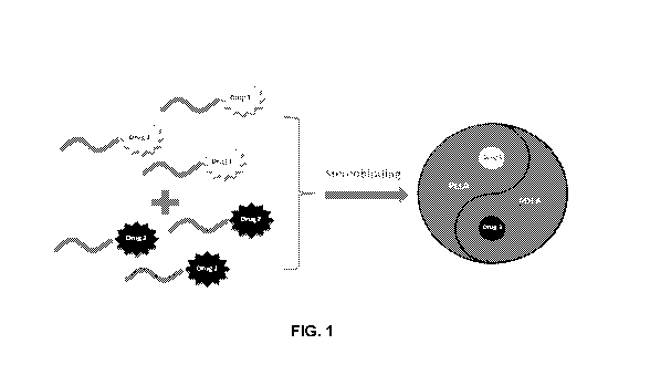
Figure 1 shows a schematic illustration of the stereocomplexes disclosed
herein comprising two different drugs based on the stereocomplexation between
PLLA and PDLA.
Figure 2 shows a schematic illustration of polymer conjugated drugs (PCD)
for stereocomplexation. In panel (a), hydrophilic elements project into
solution and
hydrophobic elements are located at the particle core of the stereocomplexes.
In
panel (b), examples of anti-cancer drugs conjugated with hydrophobic parts
with
cleavable linkers are shown. In one example form, mertansine (DM1) is
conjugated
using a disulfide bond while docetaxel (DTX) is conjugated using a hydrazone
bond
(linker 1), a disulfide bond (linker 2), or an ester bond (linker 3).
Figure 3 shows the structures of cRGD-PEG-PDLA, FA-PEG-PLLA, and
methyl-a-glucose-PEG-PDLA.
Figure 4 shows a synthetic scheme for producing mPEG-PDLA-SS-DM1.
Figure 5 shows 1H NMR of mPEG-PDLA-SS-DM1 in DMSO-d6. Peaks that
are lettered correspond to the same letters on the inset structure; a: -CH- of
PDLA;
b and c: -CH3 of DM1.
Figure 6 shows a synthetic scheme for producing rriPEG-PLLA-hydrazone-
DTX.
Figure 7 shows 1H NMR of mPEG-PLLA-hydrazone-DTX in DMSO-c16.
Peaks that are lettered correspond to the same letters on the inset structure;
a: ¨
NH- of DTX-hydrazone-OH; e: -CH- of PLLA.
Figure 8 shows 1H NMR of rnPEG-PLLA-ester-DTX in CDCI3. Peaks that
are lettered correspond to the same letters on the inset structure; c and d: -
CH3 of
DTX; e: -CH- of PLLA.
Figure 9 shows a synthetic scheme for producing mPEG-PLLA-SS-DTX.
Figure 10 shows 111 NMR of cRGD-amide-PEG-PDLA in DMSO-cis. Peaks
that are lettered correspond to the same letters on the inset structure; a:
=CH- of
cRGD; b: -CH2- of cRGD; c: -CH- of PDLA.
CA 03121902 2021-06-02
WO 2020/117742 PCT/US2019/064140
Figure 11 shows 1H NMR of folate-amide-PEG-PLLA in DMSO-c16. Peaks
that are lettered correspond to the same letters on the inset structure; a:
=CH- of
folate; b: -CH2- of folate; c: -CH- of PDLA.
Figure 12 shows 1H NMR of methyl-a-glucose-PEG-PDLA in CDCI3. Peaks
that are lettered correspond to the same letters on the inset structure; a: -
CH- of
PDLA; b: -CH2- of PEG; c: -CH3 of glucose.
Figure 13 shows the combination index (CI, displayed on the vertical axis) of
free DM1 and DTX in different cell lines at different ratios of DM1 to DTX. CI
is
used for quantitative synergy determination of two-drug combinations.
Synergism
is represented by CI < 1, an additive effect occurs when CI = 1, and
antagonism
occurs when CI > 1. Cell lines include A549 (adenocarcinomic human alveolar
basal epithelia cells; red circles), NCI-H460 (non-small cell lung cancer
cells; black
squares), MiA PaCa-2 (pancreatic cancer cells, blue triangles), SGC-7901
(gastric
cancer cells, teal triangles), and Hep3B2.1-7 (liver cancer cells, pink
triangles).
Fig. 14A shows particle sizes of prodrug mPEG-PDLA-SS-DM1(D-DM1) and
mPEG-PLLA-hydrazone-DTX(L-DTX). Fig. 14B shows the particle size of the
complex formation produced by dialysis (left panel) and after freeze-drying
and
reconstitution (right panel). Fig. 14C shows the particle size of the complex
formation produced by using rotary evaporation (left panel) and after freeze-
drying
and reconstitution (right panel).
Figs 15A-15B show DSC results evaluating melting temperature (TO of the
various formulations. Fig. 15A shows free DM1 powder (triangles), mPEG-PDLA-
SS-DM1 lyophilized powder (circles), and mPEG-PDLA lyophilized powder
(squares). Fig. 15B shows mPEG-PLLA-hydrazone-DTX lyophilized powder
(squares), mPEG-PDLA-SS-DM1 lyophilized powder (circles), and a complex
lyophilized powder formed from previously mentioned two polymers (triangles).
Figs 16A-16B show release of drugs from complexes over time. Fig. 16A
shows the release of docetaxel over time from the complex at pH 7.4 (squares)
and
pH 5.5 (circles); this difference is due to the pH sensitivity of the
hydrazone linker.
Fig. 16B shows the release of DM1 over time from the prodrug D-DM1 formulation
and complex at pH 7.4 with and without glutathione (GSH). The complex provides
4
CA 03121902 2021-06-02
WO 2020/117742 PCT/US2019/064140
a much slower release than the prodrug with GSH (circles and squares,
respectively), while the DM1 conjugation with a redox sensitive disulfide
linker
prohibits the premature release of DM1 without GSH (complex represented by the
inverted triangles, prodrug by the triangles overlapping with the inverted
triangles).
Figure 17A shows the tolerance of the various formulations in mice without
tumors with injections given at days 1, 8, 15, and 22. Figure 17B shows the
body
weight change of mice without tumors in three weeks after complex injections
at
3.6mg/kg DM1 once/week for three injections, at 5mg/kg DI1/11 once/two weeks
for
two injections and at 7mg/kg DM1 only one injection, respectively.
Figs. 18A-18D show the in vivo antitumor efficacy of the complex in
subcutaneous BGC-823 (gastric) tumor model via intravenous. Human gastric
cancer cell suspensions were injected subcutaneously on the backs of mice to
establish the tumor model. When tumor volume reached approximately 60 mm3,
groups of tumor-bearing mice (n = 5) were injected with the through the tail
vein on
days indicated by the arrows in panel (a) with stereocomplex (i.e., days 1, 8,
and
15 at a dosage of 4 mg/kg DM1 and 36 mg/kg DTX per injection). Fig. 18A shows
tumor size measurements for a control group (squares) and treatment groups
(circles). No significant body weight loss was observed for the treatment
group or
the control group (Fig. 18B). Significant tumor reduction was achieved for the
group
administered the complex (excised tumors are pictured in Fig. 18C), with a
greater
total reduction in tumor weight achieved in the stereocorriplex treatment
group (Fig.
18D).
Figs. 19A-19B show the in vivo antitumor efficacy of the complex in
subcutaneous MIA PaCa-2 (pancreatic) tumor model via intravenous injection.
Cell
suspensions were injected subcutaneously on the backs of mice to establish the
tumor model. When tumor volume reached approximately 140 mrri3, groups of
tumor bearing mice (n = 5) were injected with the complex through the tail
vein once
every two weeks. After two injections (i.e., days 1, and 14 at a dosage of 5
mg/kg
DM1 and 40 mg/kg DTX per injection), one mouse was tumor-free on the 24th day,
and in total three mice had no tumors by the 38th day. Fig. 19A shows tumor
size
change for the group treated with complex (circles) versus the control group
0
CA 03121902 2021-06-02
WO 2020/117742 PCT/US2019/064140
(squares). Fig. 19B shows control mice (top row of photos) and treated mice
(bottom row of photos) on the 29th day of the trial.
Figs. 20A-20B shows a comparison between complex and prodrug for in vivo
antitumor efficacy and toxicity in a subcutaneous MIA PaCa-2 when the
treatment
is delivered via intravenous injection.
Cell suspensions were injected
subcutaneously on the backs of mice to establish the tumor model. When tumor
volume reached approximately 140 mm3, groups of tumor bearing mice (n = 5)
were
injected with the complex through the tail vein. After four injections of D-
DM1
prodrug and two injections for the complex, approximately the same antitumor
effects were observed (Fig. 20A, circles and triangles, respectively), but the
prodrug
treatment induced mouse death and obvious body weight decreases (Fig. 20B,
circles), while the complex treatment croup showed a body weight increase
during
the treatment period (Fig. 20B, triangles).
Figs. 21A-21D show a comparison between complex and control (no
complex administration) in vivo antitumor efficacy and toxicity in a
subcutaneous
Hep 3B2.1-7 (liver) tumor model when the treatment is delivered via
intravenous
injection. Cell suspensions were injected subcutaneously on the backs of mice
to
establish the tumor model. When tumor volume reached approximately 130 mm3,
groups of tumor bearing mice (n = 5) were injected with the complex through
the
tail vein. Fig. 21A shows that tumor volume continued to grow for the control
group
(squares) but was reduced in the complex treatment group (circles). Fig. 21B
shows that body weight for the complex treatment group remained approximately
the same throughout the trial (circles) but significantly decreased for the
control
(squares). Fig. 21C shows excised tumors from the control croup (top row) and
complex group (bottom row) and Fig. 21D shows a comparison of tumor weight
between the control group (left bar) and complex group (right bar).
Figs. 22A-22D show tumor size changes for complex treatment group versus
a control group in a subcutaneous HT-29 (colon) tumor model. Cell suspensions
were injected subcutaneously on the backs of mice to establish the tumor model
and when tumors were approximately 100 mm3, groups of tumor bearing mice (n =
5) were injected with the compositions through the tail vein. Fig. 22A shows
that
tumor volume increased significantly more with untreated mice (squares), while
6
CA 03121902 2021-06-02
WO 2020/117742 PCT/US2019/064140
mice treated with the complex (circles) exhibited lower final tumor volumes
(with
arrows indicating injection dates). Fig. 22B shows the change of body weight
for
the control group and treatment group. Fig. 22C shows excised tumors for the
control group (top row) and the stereocomplex treatment group (bottom row).
Fig.
22D shows the tumor weight comparison of the control group (left) and
stereocomplex treatment group (right).
Fig. 23 shows a comparison between complex and prodrug (D-DM1) for in
vivo antitumor efficacy in a subcutaneous CNE (nasopharynaeal) tumor model
when the treatment is delivered via intravenous injection. Cell suspensions
were
injected subcutaneously on the backs of mice to establish the tumor model.
When
tumor volume reached approximately 100 mm3, groups of tumor bearing mice (n =
5) were injected with the complex through the tail vein. As shown, with the
equivalent DM1 dose, tumor volume continued to grow for the prodrug group
(squares) but complex treatment showed good tumor growth inhibition efficacy
(circles).
Figs. 24A-24B show the in vivo antitumor efficacy of the complex in
subcutaneous NCI-H526 (small-cell lung cancer) tumor model via intravenous
injection. Cell suspensions were injected subcutaneously on the backs of mice
to
establish the tumor model. When tumor volume reached approximately 100 mm3,
groups of tumor bearing mice (n = 5) were injected with the complex through
the
tail vein once every week. After three injections, one mouse was tumor-free on
the
18th day,
and all mice had no tumors by the 32nd day. Fig. 24A shows tumor size
change for the group treated with complex (circles) versus the control group
(squares). Fig. 24B shows control mice (top row of photos) and treated mice
(bottom row of photos) on the 18th day of the trial.
Figs. 25A-25D show the in vivo antitumor efficacy of the complex in
subcutaneous NCI-H1975 (non-small cell lung cancer) tumor model via
intravenous
injection. Cell suspensions were injected subcutaneously on the backs of mice
to
establish the tumor model and when tumors were approximately 130 mm3, groups
of tumor bearing mice (n = 5) were injected with the compositions through the
tail
vein. Fig. 25A shows that tumor volume increased significantly more with
untreated
mice (squares), while mice treated with the complex (circles) exhibited lower
final
7
CA 03121902 2021-06-02
WO 2020/117742 PCT/US2019/064140
tumor volumes after only one injection (with arrows indicating injection
dates). Fig.
25B shows the change of body weight for the control group and treatment group.
Fig. 25C shows excised tumors for the control group (top row) and the
stereocomplex treatment group (bottom row). Fig. 25D shows the tumor weight
comparison of the control group (left) and stereocomplex treatment group
(right).
Figs. 26A-26D show the in vivo antitumor efficacy of the complex in
subcutaneous MDA-MB-231 (triple negative breast cancer) tumor model. Cell
suspensions were injected subcutaneously on the backs of mice to establish the
tumor model and when tumors were approximately 100 mm3, groups of tumor
bearing mice (n = 6) were injected with the compositions through the tail
vein. Fig.
26A shows that tumor volume increased significantly more with untreated mice
(squares), while mice treated with the complex (circles) exhibited lower final
tumor
volumes after only one injection. Fig. 26B shows the change of body weight for
the
control group and the treatment group. Fig. 26C shows excised tumors for the
control group (top row) and stereocomplex treatment group (bottom row),
notably,
one mouse was tumor-free from the 231.d day. Fig. 26D shows the tumor weight
comparison of the control group (left) and stereocomplex treatment group
(right).
Figs. 27A-27B show the in vivo antitumor efficacy of the complex in
subcutaneous MX-1 (breast) tumor model via intravenous injection.
Cell
suspensions were injected subcutaneously on the backs of mice to establish the
tumor model. When tumor volume reached approximately 530 mm3, groups of
tumor bearing mice (n = 5) were injected with the complex through the tail
vein.
After only one injection, tumor size decreased continuously in the next 20
days, as
shown in Fig.27A, which demonstrated the efficacy of complex even in large
tumors.
No body weight loss was observed for this treatment (Fig. 27B).
Figs. 28A-28D show the in vivo antitumor efficacy of the complex in
subcutaneous MCF-7 (breast) tumor model. Cell suspensions were injected
subcutaneously on the backs of mice to establish the tumor model and when
tumors
were approximately 100 mm", groups of tumor bearing mice (n = 8) were injected
with the compositions through the tail vein. Fig. 28A shows that tumor volume
increased significantly more with untreated mice (squares), while mice treated
with
the complex (circles) exhibited lower final tumor volumes after two injections
(with
8
CA 03121902 2021-06-02
WO 2020/117742 PCT/US2019/064140
arrows indicating injection dates). Fig. 28B shows the change of body weight
for
the control group and treatment group. Fig. 28C shows excised tumors for the
control group (top row) and stereocomplex treatment group (bottom row),
notably,
one mouse was tumor-free from the 25th day, and three mice were tumor-free at
the
end of the test. Fig. 28D shows the tumor weight comparison of the control
group
(left) and stereocomplex treatment group (right).
Figs. 29A-29D show the in vivo antitumor efficacy of the complex in
subcutaneous RT112 (bladder) tumor model. Cell suspensions were injected
subcutaneously on the backs of mice to establish the tumor model and when
tumors
were approximately 100 mm3, groups of tumor bearing mice (n = 5) were injected
with the compositions through the tail vein. Fig. 29A shows that tumor volume
increased significantly more with untreated mice (squares), while mice treated
with
the complex (circles) exhibited lower final tumor volumes after three
injections (with
arrows indicating injection dates). Fig. 29B shows the change of body weight
for
the control group and treatment group. Fig. 29C shows excised tumors for the
control group (top row) and the stereocomplex treatment group (bottom row).
Fig.
29D shows the tumor weight comparison of the control group (left) and
stereocomplex treatment group (right).
Figs. 30A-30D show the in vivo antitumor efficacy of the complex in
subcutaneous T.T (esophagus) tumor model. Cell suspensions were injected
subcutaneously on the backs of mice to establish the tumor model and when
tumors
were approximately 110 mm3, groups of tumor bearing mice (n = 5) were injected
with the compositions through the tail vein. Fig. 30A shows that tumor volume
increased significantly more with untreated mice (squares), while mice treated
with
the complex (circles) exhibited lower final tumor volumes after three
injections (with
arrows indicating injection dates). Fig. 30B shows the change of body weight
for
the control group and treatment group. Fig. 30C shows excised tumors for the
control group (top row) and stereocomplex treatment group (bottom row),
notably,
one mouse was tumor-free from the 27th day. Fig. 30D shows the tumor weight
comparison of the control group (left) and the stereocomplex treatment group
(right).
9
CA 03121902 2021-06-02
WO 2020/117742 PCT/US2019/064140
Figs. 31A-31D show the in vivo antitumor efficacy of the complex in
subcutaneous U251 (glioblastoma) tumor model. Cell suspensions were injected
subcutaneously on the backs of mice to establish the tumor model and when
tumors
were approximately 150 mm3, groups of tumor bearing mice (n = 5) were injected
with the compositions through the tail vein. Fig. 31A shows that tumor volume
increased significantly more with untreated mice (squares), while mice treated
with
the complex (circles) exhibited lower final tumor volumes after two injections
(with
arrows indicating injection dates). Fig. 31B shows the change of body weight
for
the control group and treatment group. Fig. 31C shows excised tumors for the
.. control group (top row) and the stereocomplex treatment group (bottom row).
Fig.
31D shows the tumor weight comparison of the control group (left) and
stereocomplex treatment group (right).
Figs. 32A-32D show the in vivo antitumor efficacy of the complex in
subcutaneous Caki-1(kidney) tumor model. Cell suspensions were injected
subcutaneously on the backs of mice to establish the tumor model and when
tumors
were approximately 170 mm3, groups of tumor bearing mice (n = 5) were injected
with the compositions through the tail vein. Fig. 32A shows that tumor volume
increased significantly more with untreated mice (squares), while mice treated
with
the complex (circles) exhibited lower final tumor volumes after three
injections (with
.. arrows indicating injection dates). Fig. 32B shows the change of body
weight for
the control group and treatment group. Fig. 32C shows excised tumors for the
control group (top row) and the stereocomplex treatment group (bottom row).
Fig.
32D shows the tumor weight comparison of the control group (left) and
stereocomplex treatment group (right).
Figs. 33A-33D show the in vivo antitumor efficacy of the complex in
subcutaneous NCI-H522 (Non-small cell lung cancer) tumor model. Cell
suspensions were injected subcutaneously on the backs of mice to establish the
tumor model and when tumors were approximately 130 mm3, groups of tumor
bearing mice (n = 5) were injected with the compositions through the tail
vein. Fig.
33A shows that tumor volume increased significantly more with untreated mice
(squares), while mice treated with the complex (circles) exhibited lower final
tumor
volumes after two injections (with arrows indicating injection dates). Fig.
33B shows
CA 03121902 2021-06-02
WO 2020/117742 PCT/US2019/064140
the change of body weight for the control group and treatment group. Fig. 33C
shows excised tumors for the control group (top row) and stereocomplex
treatment
group (bottom row), notably; three mice were tumor-free at the end of the
test. Fig.
33D shows the tumor weight comparison of the control group (left) and
stereocomplex treatment group (right).
Figs. 34A-34D show the in vivo antitumor efficacy of the complex in
subcutaneous NCI-H226 (Non-small cell lung cancer) tumor model.
Cell
suspensions were injected subcutaneously on the backs of mice to establish the
tumor model and when tumors were approximately 120 mm3, groups of tumor
bearing mice (n = 4) were injected with the compositions through the tail
vein. Fig.
34A shows that tumor volume increased significantly more with untreated mice
(squares), while mice treated with the complex (circles) exhibited lower final
tumor
volumes after two injections (with arrows indicating injection dates). Fig.
34B shows
the change of body weight for the control group and treatment group. Fig. 34C
shows excised tumors for the control group (top row) and the stereocomplex
treatment group (bottom row). Fig. 34D shows the tumor weight comparison of
the
control group (left) and stereocomplex treatment group (right).
Figs. 35A-35D show the in vivo antitumor efficacy of the complex in
subcutaneous Ovcar-3 (Ovarian) tumor model. Cell suspensions were injected
subcutaneously on the backs of mice to establish the tumor model and when
tumors
were approximately 150 mm3, groups of tumor bearing mice (n = 5) were injected
with the compositions through the tail vein. Fig. 35A shows that tumor volume
increased significantly more with untreated mice (squares), while mice treated
with
the complex (circles) exhibited lower final tumor volumes after only one
injection.
Fig. 35B shows the change of body weight for the control group and treatment
group. Fig. 35C shows excised tumors for the control group (top row) and the
stereocomplex treatment group (bottom row). Fig. 35D shows tumor weight
comparison of the control group (left) and stereocomplex treatment group
(right).
Figs. 36A-36D show the in vivo antitumor efficacy of the complex in
subcutaneous PC-3 (prostate) tumor model. Cell suspensions were injected
subcutaneously on the backs of mice to establish the tumor model and when
tumors
were approximately 130 mm3, groups of tumor bearing mice (n = 5) were injected
11
CA 03121902 2021-06-02
WO 2020/117742 PCT/US2019/064140
with the compositions through the tail vein. Fig. 36A shows that tumor volume
increased significantly more with untreated mice (squares), while mice treated
with
the complex (circles) exhibited lower final tumor volumes after three
injections (with
arrows indicating injection dates). Fig. 36B shows the change of body weight
for
the control group and treatment group. Fig. 36C shows excised tumors for the
control group (top row) and the stereocomplex treatment group (bottom row).
Fig.
36D shows the tumor weight comparison of the control group (left) and
stereocomplex treatment group (right).
Figs. 37A-37B show the in vivo antitumor efficacy of the complex in
subcutaneous Raji (lymphoma) tumor model via intravenous injection. Cell
suspensions were injected subcutaneously on the backs of mice to establish the
tumor model. When tumor volume reached approximately 130 mm3, groups of
tumor bearing mice (n = 4) were injected with the complex through the tail
vein.
After only one injection, three mice were tumor-free on the 15111 day, and all
mice
had no tumors from the 22nd day. Fig. 37A shows tumor size change for the
group
treated with complex (circles) versus the control group (squares). Fig. 37B
shows
the photos of the control mice (top row) and the treated mice (bottom row) on
the
25th day of the trial.
Figs. 38A-38B show the blood parameters in nude mice after a single iv.
injection of the complex. Mice (4 animals per group) were injected with the
complex
at single i.v. at the dose of 5mg/kg DM1 and 32.5mg/kg DTX, and then were
killed
on days 3, 7 and 14. Blood samples were collected and analyzed for the
following
general parameters: white blood cell count (WBC); red blood cell count (RBC);
hemoglobin concentration (HGB) and platelet count (PLT). As compared with the
control (without injection) labeled as day0. RBC and HGB showed no statistical
difference in the whole test. Even lower WBC and PLT were observed on day 3,
they were all recovered on day 7 and kept normal on day 14.
Fig. 39 shows the clinical chemistry in nude mice after a single iv. injection
of the complex formulation. Mice (4 animals per group) were injected with the
complex at single iv. at the dose of 5mg/kg DM1 and 32.5ma/kg DTX, and then
were killed on days 3, 7 and 14. Blood samples were collected and analyzed for
the
following parameters: alanine aminotransferase (ALT); aspartate
aminotransferase
12
CA 03121902 2021-06-02
WO 2020/117742 PCT/US2019/064140
(AST); alkaline phosphatase (ALP), creatinine (CREA) and urea (UREA). As
compared with control (without injection) labeled as day0. ALT and AST were
elevated after injection, but recovered on day 14. There is no obvious
difference in
UREA and CREA, which means no nephrotoxicity at all.
Figs. 40 and 41 show the histopathological analysis of organs for the
complex treatment group (Complex) versus the untreated group (Control) and
prodrug treatment group (D-DM1) in a CNE (nasopharyngeal) tumor model. Cell
suspensions were injected subcutaneously on the backs of mice to establish the
tumor model and when tumors were approximately 100 mm3, groups of tumor
bearing mice (n = 5) were injected with the compositions through the tail vein
weekly
for 4 consecutive weeks at doses of 4mg/kg DM1 for D-DM1 group and 4mg/kg
DM1 with 26mg/kg DTX for complex group, respectively. After harvesting hearts,
kidneys, spleens, lungs and livers, sections were stained with hematoxylin and
eosin for observation. Compared with control and D-DM1 treatment, complex
treatment did not induce any damages to organs.
Figs. 42A-42B show the in vivo antitumor efficacy of the complex containing
glucose in subcutaneous Raji (lymphoma) tumor model via intravenous injection.
Cell suspensions were injected subcutaneously on the backs of mice to
establish
the tumor model. When tumor volume reached approximately 130 mm3, groups of
tumor bearing mice (n = 4) were injected with the complex through the tail
vein.
After only one injection, three mice were tumor-free on the 15th day, and all
mice
had no tumors from the 18th day. Fig. 42A shows tumor size change for the
group
treated with complex (circles) versus the control group (squares). Fig. 42B
shows
the photos of the control mice (top row) and the complex containing glucose
treated
mice (bottom row) on the 25th day of the trial.
Fig. 43 shows the PET/CT images of patient 1 before and after treatment
with the stereocomplex. By the comparison, the intensity of the subcarinal
lymph
node was reduced significantly caused due to treatment.
Fig. 44 shows the sagittal MR imaging of patient 3 before and after treatment
with the stereocomplex. Before treatment, sagittal MR of the spine showed
multiple
large irregular shape masses occupying most of the spinal canal from L1 to Si,
and
13
CA 03121902 2021-06-02
WO 2020/117742 PCT/US2019/064140
the CSF space was minimally visible. After treatment, MR revealed marked
decreased tumor masses in spinal canal between L1 to Si. Only residual small
masses behind L4 and L5 were noted, where CSF space and cauda equina nerve
fibers can be easily identified
Fig. 45 shows the PET/CT images of patient 4 before and after treatment
with the stereocomplex. Tumor size decreased due to treatment.
Fig. 46 shows the PET/CT image of patient 4 before and after treatment with
the stereocomplex. The intensity of the uptake of mediastinal, hilar and
abdominal
aorta lymph node was reduced.
Fig. 47 shows the PET/CT images of patient 4 before and after treatment
with the stereocomplex. Before treatment, the tumor was found to invade the
parietal pleural; however, after treatment, the tumor and parietal pleural
were found
to be completely separated
DETAILED DESCRIPTION
Before the present materials, articles, and/or methods are disclosed and
described, it is to be understood that the aspects described below are not
limited to
specific compounds, synthetic methods, or uses, as such may, of course, vary.
It
is also to be understood that the terminology used herein is for the purpose
of
describing particular aspects only and is not intended to be limiting.
In the specification and in the claims that follow, reference will be made to
a
number of terms that shall be defined to have the following meanings:
It must be noted that, as used in the specification and the appended claims,
the singular forms "a," "an," and "the" include plural referents unless the
context
clearly dictates otherwise. Thus, for example, reference to "an anti-cancer
agent"
includes mixtures of two or more such anti-cancer agents, and the like.
"Optional" or "optionally" means that the subsequently described event or
circumstance can or cannot occur, and that the description includes instances
where the event or circumstance occurs and instances where it does not. For
example, the compositions described herein may optionally contain one or more
targeting groups, where the targeting group may or may not be present.
14
CA 03121902 2021-06-02
WO 2020/117742 PCT/US2019/064140
As used herein, the term "about" is used to provide flexibility to a numerical
range endpoint by providing that a given value may be "a little above" or "a
little
below" the endpoint without affecting the desired result. For purposes of the
present
disclosure, "about" refers to a range extending from 10% below the numerical
value
to 10% above the numerical value. For example, if the numerical value is 10,
"about
10" means between 9 and 11 inclusive of the endpoints 9 and 11.
Throughout this specification, unless the context dictates otherwise, the word
"comprise," or variations such as "comprises" or "comprising," will be
understood to
imply the inclusion of a stated integer or step or group of integers or steps,
but not
the exclusion of any other integer or step or group of integers or steps. It
is also
contemplated that the term "comprises" and variations thereof can be replaced
with
other transitional phrases such as "consisting of" and "consisting essentially
of."
"Admixing" or "admixture" refers to a combination of two components
together when there is no chemical reaction or physical interaction. The terms
"admixing" and "admixture" can also include the chemical interaction or
physical
interaction among any of the components described herein upon mixing to
produce
the composition. The components can be admixed alone, in water, in another
solvent, or in a combination of solvents.
The term "solid tumor" as defined herein is an abnormal mass of tissue that
.. usually does not contain cysts or liquid areas. Solid tumors may be benign
(not
cancer), or malignant (cancer). Different types of solid tumors are named for
the
type of cells that form them. Examples of solid tumors are sarcomas,
carcinomas,
and lymphomas.
The term "subject" as defined herein is any organism in need of cancer
.. treatment and/or prevention. In one aspect, the subject is a mammal
including, but
not limited to, humans, domesticated animals (e.g., dogs, cats, horses),
livestock
(e.g., cows, pigs), and wild animals.
The term "treat" as used herein is defined as maintaining or reducing the
symptoms of a pre-existing condition. For example, the compositions described
herein are used to treat cancer.
CA 03121902 2021-06-02
WO 2020/117742 PCT/US2019/064140
The term "prevent" as used herein is defined as eliminating or reducing the
likelihood of occurrence of one or more symptoms of a disease or disorder. For
example, the compositions described herein can be used to prevent the regrowth
of tumor cells or reduce the rate of regrowth of tumor cells.
The term "inhibit" as used herein is the ability of the compounds described
herein to completely eliminate an activity or reduce the activity when
compared to
the same activity in the absence of the compound. For example, the
compositions
described herein can be used to inhibit the growth and/or spread of cancer in
the
body of a subject.
"Biodegradable" materials are capable of being decomposed by bacteria,
fungi, or other organisms, or by enzymes in the body of a subject.
"Biocompatible" materials are materials that perform their desired functions
without eliciting harmful or deleterious changes to the subject in which they
are
implanted or to which they are applied, either locally or systemically. In one
aspect,
the compositions disclosed herein are biocompatible.
As used herein, "toxicity" refers to harmful effects a substance has on an
organism such as a human or mammal, or on cells within that organism. A
compound or composition with high toxicity would be unsuitable for use as a
medical treatment, while a compound or composition with low toxicity would be
acceptable for use as a medical treatment. In one aspect, the compounds and
compositions disclosed herein exhibit low toxicity.
The term "alkyl group" as used herein is a branched or unbranched saturated
hydrocarbon group of 1 to 25 carbon atoms, such as methyl, ethyl, n-propyl,
isopropyl, n-butyl, isobutyl, t-butyl, pentyl, hexyl, heptyl, octyl, decyl,
tetradecyl,
hexadecyl, eicosyl, tetracosyl, and the like. Examples of longer chain alkyl
groups
include, but are not limited to, an oleate group or a palm itate group. A
"lower alkyl"
group is an alkyl group containing from one to six carbon atoms.
The term "aryl group" as used herein is any carbon-based aromatic group
including, but not limited to, benzene, naphthalene, etc. The term "aryl
group" also
includes "heteroaryl group," which is defined as an aromatic group that has at
least
one heteroatom incorporated within the ring of the aromatic group. Examples of
16
CA 03121902 2021-06-02
WO 2020/117742 PCT/US2019/064140
heteroatoms include, but are not limited to, nitrogen, oxygen, sulfur, and
phosphorus. The aryl group can be substituted or unsubstituted. The aryl
group, if
substituted, can be substituted with one or more groups including, but not
limited
to, alkyl, alkynyl, alkenyl, aryl, halide, nitro, amino, ester, ketone,
aldehyde,
hydroxyl, carboxylic acid, or alkoxy.
The term "alkoxy group" as used herein is defined as RO-, where R is an
alkyl group or aryl group defined herein.
The term "halogenated group" is any organic group such as, for example, an
alkyl group or aryl group that possesses at least one halogen (F, Cl, Br, I).
References in the specification and concluding claims to parts by weight, of
a particular element in a composition or article, denote the weight
relationship
between the element or component and any other elements or components in the
composition or article for which a part by weight is expressed. Thus, in a
compound
containing 2 parts by weight of component X and 5 parts by weight of component
Y, X and Y are present at a weight ratio of 2:5, and are present in such a
ratio
regardless of whether additional components are contained in the compound. A
weight percent of a component, unless specifically stated to the contrary, is
based
on the total weight of the formulation or composition in which the component
is
included.
As used herein, a plurality of items, structural elements, compositional
elements, and/or materials may be presented in a common list for convenience.
However, these lists should be construed as though each member of the list is
individually identified as a separate and unique member. Thus, no individual
member of any such list should be construed as a de facto equivalent of any
other
member of the same list based solely on its presentation in a common group,
without indications to the contrary.
Concentrations, amounts, and other numerical data may be expressed or
presented herein in a range format. It is to be understood that such a range
format
is used merely for convenience and brevity and thus should be interpreted
flexibly
to include not only the numerical values explicitly recited as the limits of
the range,
but also to include all the individual numerical values or sub-ranges
encompassed
17
CA 03121902 2021-06-02
WO 2020/117742 PCT/US2019/064140
within that range as if each numerical value and sub-range was explicitly
recited.
As an illustration, a numerical range of "about 1 to about 5" should be
interpreted to
include not only the explicitly recited values of about 1 to about 5, but also
to include
individual values and sub-ranges within the indicated range. Thus, included in
this
numerical ramie are individual values such as 2, 3, and 4, sub-ranges such as
from
1-3, from 2-4, from 3-5, etc., as well as 1, 2, 3, 4, and 5, individually. The
same
principle applies to ranges reciting only one numerical value as a minimum or
a
maximum. Furthermore, such an interpretation should apply regardless of the
breadth of the range or the characteristics being described.
Disclosed are materials and components that can be used for, can be used
in conjunction with, can be used in preparation for, or are products of the
disclosed
compositions and methods. These and other materials are disclosed herein, and
it
is understood that when combinations, subsets, interactions, groups, etc., of
these
materials are disclosed, that while specific reference to each various
individual and
collective combination and permutation of these compounds may not be
explicitly
disclosed, each is specifically contemplated and described herein. For
example, if
an anti-cancer agent is disclosed and discussed and a number of different
linkers
are discussed, each and every combination of anti-cancer agent and linker that
is
possible is specifically contemplated unless specifically indicated to the
contrary.
For example, if a class of molecules A, B, and C are disclosed, as well as a
class
of molecules D, E, and F, and an example combination of A + D is disclosed,
then
even if each is not individually recited, each is individually and
collectively
contemplated. Thus, in this example, each of the combinations A + E, A + F, B
+
D, B + E, B + F, C + D, C + E, and C + F is specifically contemplated and
should be
considered from disclosure of A, B. and C; D. E, and F; and the example
combination of A + D. Likewise, any subset or combination of these is also
specifically contemplated and disclosed. Thus, for example, the sub-group of A
+
E, B + F, and C + E is specifically contemplated and should be considered from
disclosure of A, B, and C; D, E, and F, and the example combination of A + D.
This
concept applies to all aspects of the disclosure including, but not limited
to, steps in
methods of making and using the disclosed compositions. Thus, if there are a
variety of additional steps that can be performed with any specific embodiment
or
18
CA 03121902 2021-06-02
WO 2020/117742 PCT/US2019/064140
combination of embodiments of the disclosed methods, each such combination is
specifically contemplated and should be considered disclosed.
Components in the Stereocomplexes
The stereocomplexes described herein are useful in delivering one or more
anti-cancer agents to a subject. In one aspect, the stereocomplexes are
composed
at least two components, each component having a hydrophilic group, an
isotactic
polylactic acid moiety, a linker, and an anti-cancer agent.
In one aspect, disclosed herein are stereocomplexes having the following
components:
)(1-y1-L1_21 (I)
X2-Y2-L2-Z2 (II)
wherein X1 and X2 are hydrophilic groups; Y1 and Y2 are PDLA or PLLA; L1
and L2 are cleavable linkers, Z1 is an anti-cancer agent, Z2 is an anti-cancer
agent
or imaging agent, wherein when Z2 is an anti-cancer agent, Z1 and Z2 are
different
anti-cancer agents, and wherein (1) when Y1 is PDLA then Y2 is PLLA, and when
Y1 is PLLA then Y2 is PDLA and (2) the ratio of the total number of D-lactic
acid
units in the stereocomplex to the total number of L-lactic acid units in the
stereocomplex is from 0.9:1.1 to 1.1:0.9.
Not wishing to be bound by theory, stereocomplexes form when the PDLA
and PLLA units present in components (I) and (II) form an extensive 3-
dimensional
network driven by hydrogen bonding. The stereocomplexes described herein have
enhanced properties such as tensile strength, Young's modulus, and elongation
at
break compared to either component (I) or component (II) alone.
The
stereocomplexes have high stability and high resistance to hydrolytic
degradation,
which eliminates the premature release of drugs (i.e., before the
stereocomplexes
reach their target tissue) and increases the circulation time of the
stereocomplexes
in the blood.
Fig. 1 shows a schematic illustration of an exemplary stereocomplex with two
different drugs based on the stereocomplexation between PDLA and PLLA. Fig. 2
shows a schematic illustration of exemplary polymer conjugated drugs for
19
CA 03121902 2021-06-02
WO 2020/117742 PCT/US2019/064140
stereocomplexation. Without wishing to be bound by theory, hydrophilic
elements
project into solution while hydrophobic elements cluster at the particle core
(Fig.
2A). Examples anti-cancer drugs conjugated to hydrophobic parts with cleavable
linkers are provided in Fig. 2B. Referring to Fig. 2B, mertansine (DM1) is
linked to
a carrier with a disulfide bond (D-DM1) and docetaxel (DTX) is linked to a
carrier
with a hydrazone bond, ester bond, or disulfide bond (L-DTX).
Each component used to prepare the stereocomplexes and methods for
making and using thereof are described in detail herein.
a, Hydrophilic Group
The components used to produce the stereocomplexes disclosed herein
include a hydrophilic group. In one aspect, X1 and X2 in components (I) and
(II) are
different hydrophilic groups. In an alternative aspect, X1 and X2 are the same
hydrophilic group. In still another aspect, Xi and X2 are each a polyalkylene
glycol.
"Polyalkylene glycol" as used herein refers to a condensation polymer of
ethylene oxide or propylene oxide and water. Polyalkylene glycols are
typically
colorless liquids with high molecular weights and are soluble in water as well
as
some organic solvents. In one aspect, the hydrophilic group in the
stereocomplexes
disclosed herein is a polyalkylene glycol. In another aspect, the polyalkylene
glycol
is polyethylene glycol and/or polypropylene glycol.
In another aspect, the
polyalkylene glycol is monornethoxy polyethylene glycol. The generic structure
of
a polyalkylene glycol is as follows:
R --(y1R3
1
2
Substitutions in selected polyalkylene glycols are provided in Table 1:
Table 1: Polyalkylene Glycols
Compound Name R1 R2 R3
Polyethylene glycol H H OH
Polypropylene glycol H CH3 OH
Monomethoxy polyethylene CH3 H OH
glycol
CA 03121902 2021-06-02
WO 2020/117742 PCT/US2019/064140
In a further aspect, polyalkylene glycols are of low enough molecular weight
that the chemical nature of the end groups (usually, but not always;
hydroxyls) still
affects the performance of the polymers.
In addition to being hydrophilic,
polyalkylene glycols can modify the viscosity of the stereocomplexes disclosed
herein and may aid in the formation of emulsions. In another aspect,
polyalkylene
glycols are biocompatible and/or biodegradable. In still another aspect, the
polyalkylene glycols and/or other hydrophilic groups used herein are non-
toxic.
In one aspect, when X' and X2 in components (I) and (II) are polyalkylene
glycols; X1 and X2 have molecular weights of from about 1000 Da to about 5,000
Da, or from 1,500 Da to 4,500 Da, or from 2,000 Da to 4,000 Da, or have
molecular
weights of about 1,000 Da, 1,500 Da, 2000,
Da, 2,500 Da, 3,000 Da, 3,500 Da,
4;000 Da, 4,500 Da, or about 5,000 Da, where any value can be a lower and
upper
end-point of a range (e.g.; 2;000 Da to 4,000 Da).
In another aspect; X1 and X2 in components (I) and (II) of the
stereocomplexes disclosed herein are each monomethoxy polyethylene glycol
having molecular weights of from about 1,000 to about 5;000 Da, or from 1,500
Da
to 4,500 Da, or from 2,000 Da to 4,000 Da, or have molecular weights of about
1,000 Da, 1,500 Da; 2,000 Da, 2,500 Da, 3;000 Da; 3,500 Da, 4,000 Da, 4;500
Da,
or about 5;000 Da, where any value can be a lower and upper end-point of a
range
(e.g.; 2;000 Da to 4,000 Da). In one aspect; X' and X2 in components (I) and
(II) of
the stereocomplexes disclosed herein are each monomethoxy polyethylene glycol
having the same molecular weight.
b. PDLA/PLLA
Polylactic acid is a polyester derived from lactic acid. The polyester is
composed of lactic acid units depicted in the structure below, where m
indicates the
number of lactic acid units. The lactic acid unit has one chiral center;
indicated by
the asterisk (*) in the structure below, where m is the number of lactic acid
units:
21
CA 03121902 2021-06-02
WO 2020/117742 PCT/US2019/064140
CH3
Polylactic acid polymerization can begin from 0 or L lactic acid or a mixture
thereof, or lactide, a cyclic diester. Properties of polylactic acid can be
fine-tuned
by controlling the ratio of o to L enantiomers used in the polymerization, and
polylactic acid polymers can also be synthesized using starting materials that
are
only D or only L rather than a mixture of the two. Polylactic acid prepared
from only
D starting materials is referred to as poly-o-lactide (PDLA) (i.e., only
composed of
o-lactic acid units); conversely, polylactic acid prepared from only L
starting
materials is poly- L-Iactide (or PLLA) (i.e., only composed of L-lactic acid
units).
As used herein, a 0-lactic acid unit or an L-lactic acid unit refers to the
monomer units within the polylactic acid polymers described herein, wherein a
D-
lactic acid unit is derived from the 0-lactic acid or o-lactide starting
material, and an
L-lactic acid unit is derived from the L-lactic acid or L-lactide starting
material, as
shown in Table 2:
Table 2: Starting Materials for PDLA and PLLA
0 0 ____________
YOH \)*(OH
OH
L-lactic acid 0-lactic acid
Oy0i 00
==='''`µ
0
L-lactide D-lactide
The polylactic acid in components (I) and (II) is represented by Y1 and Y2,
where when Y1 is PDLA then Y2 is PLLA or, in the alternative, when Y1 is PLLA
then
Y2 is PDLA.
22
CA 03121902 2021-06-02
WO 2020/117742 PCT/US2019/064140
In one aspect, the ratio of the total number of 0-lactic acid units in the
stereocomplex to the number of L-lactic acid units in the stereocomplex is
from
0.9:1.1 to 1.1:0.9. In another aspect, the ratio of the total number of 0-
lactic acid
units to the number of L-lactic acid units is 0.9:1.1, 0.95:1.05, 1:1,
1.05:0.95, or
1.1:0.9. In one aspect, the ratio of the total number of 0-lactic acid units
to the
number of L-lactic acid units approaches 1:1. Put another way, in some
aspects,
the total number of 0-lactic acid units and L-lactic acid units in the
stereocomplexes
are approximately equal.
PDLA and PLLA are present in components (I) and (II); however, as will be
discussed in greater detail below, additional components can be used to
prepare
the stereocomplexes herein that include PDLA or PLLA. These components add
to the total number of 0-lactic acid units or L-lactic acid units present in
the
stereocomplex.
In one aspect, PDLA and PLLA present in components (I) and (II) has a
molecular weight of from about 700 Da to about 5,000 Da, or about 750 Da to
4000
Da, or about 1,000 Da to about 3,000 Da. Further in this aspect, PDLA and PLLA
has a molecular weight of about 700 Da, 750 Da, 800 Da, 900 Da, 1,000 Da,
1,250
Da, 1,500 Da, 2,000 Da, 2,500 Da, 3,000 Da, 3,500 Da, 4,000 Da, 4,500 Da, or
5,000 Da, where any value can be a lower and upper end-point of a range (e.g.,
1,000 Da to 3,000 Da). In another aspect, PDLA and PLLA present in components
(I) and (II) have equal molecular weights or approximately equal molecular
weights.
In another aspect, the number of 0-lactic acid units present in PDLA and L-
lactic acid units present in PLLA in components (I) and (II) is from 10, 15,
20, 25,
30, 35, 40, 45, 50, 55, 60, 65, 70, 75, 80, 85, 90, 95, or 100, where any
value can
.. be a lower and upper end-point of a range (e.g., 10 to 60). In another
aspect, PDLA
and PLLA present in components (I) and (II) have the same number of 0-lactic
acid
units and L-lactic acid units, respectively.
The hydrophilic group in components (I) and (II) is covalently bonded to
PDLA or PLLA. In one aspect, when the hydrophilic group is monomethoxy
.. polyethylene glycol, the terminal hydroxyl group can react with the
terminal carboxyl
23
CA 03121902 2021-06-02
WO 2020/117742 PCT/US2019/064140
group of PDLA or PLLA to form a new ester. Exemplary methods for bonding the
hydrophilic group to PDLA or PLLA are provided in the Examples.
c. Cleavable Linker
In one aspect, the stereocomplexes disclosed herein include a cleavable
linker. The cleavable linker includes at least one cleavable group so that
upon
cleavage the anti-cancer-agent is released. Cleavage of the cleavable group
can
occur by, for example, enzymatically, hydrolytically, or with a change in pH.
In one aspect, L1 and L2 present in components (I) and (I) are different
cleavable linkers. Further in this aspect, L' and L2 can exhibit different
cleavage
rates (e.g., hydrolytic, enzymatic, pH) due to the presence of different
cleavable
groups and can thus release their linked anti-cancer agents at different,
controlled
rates. In one aspect, the selection of the cleavable linker and rate of
hydrolytic
degradation of the linker can reduce the required concentration of the anti-
cancer
agents or can enhance the synergistic effects of the anti-cancer agents
present in
the stereocomplex. Thus, L1 and L2 can be selected to achieve a sequential or
concomitant release of two different anti-cancer agents. In an alternative
aspect,
L1 and L2 are the same cleavable linker.
In one aspect, L1 and L2 independently includes a cleavable group including,
but not limited to, a disulfide group, an ester group, a hydrazone group, an
acetal
group, an imine group, a 13-thiopropionate group, an amide group, or any
combination thereof. The cleavable linker molecule can include one or more of
these groups. The cleavable linker can also include additional functional
groups so
that the cleavable linker can be covalently bonded to PDLA or PLLA. In one
aspect,
the cleavable linker includes a functional group that can react with a
terminal
hydroxyl group of PDLA or PLLA to produce a new covalent bond. For example,
the cleavable linker can include a carboxyl group (e.g., carboxylic acid,
ester,
anhydride) that reacts with the hydroxyl group of PDLA or PLLA. Exemplary
methods for bonding the cleavable linker to PDLA or PLLA are provided in the
Examples and figures herein.
Disulfide group
24
CA 03121902 2021-06-02
WO 2020/117742 PCT/US2019/064140
As used herein, a disulfide group is a functional group with the structure (-S-
S-). Upon cleavage of the disulfide group, the anti-cancer agent is released.
In one
aspect, L1 or L2 or both L1 and L2 of the stereocomplexes disclosed herein
include
a disulfide group. Not wishing to be bind by theory, disulfide groups are
glutathione
redox sensitive. Glutathione (GSH) in cancer cells is implicated in the
regulation of
carcinogenic mechanisms sensitivity against cytotoxic drugs, ionizing
radiations,
and some cytokines; DNA synthesis; and cell proliferation and death. GSH can
cleave disulfide bonds in order to release the anti-cancer agent present in
the
stereocomplex to produce the corresponding thiol. Representative linkers with
a
disulfide group are provided herein.
Ester group
As used herein, an ester group is a functional group with the following
structure
In one aspect, L1 or L2 or both L1 and L2 of the stereocomplexes disclosed
herein include an ester group. Upon cleavage of the ester group, the anti-
cancer
agent is released. Exemplary methods for preparing and using a cleavable
linker
with an ester group are provided in the Examples and figures herein.
Hydrazone group
As used herein, a hydrazone group is a functional group with the following
structure, where R and R' can be the same or different:
R'
N ,s
In one aspect, Ll or L2 or both Ll and L2 of the stereocomplexes disclosed
herein include a hydrazone group. Upon cleavage of the hydrazone group, the
anti-
CA 03121902 2021-06-02
WO 2020/117742 PCT/US2019/064140
cancer agent is released. Exemplary methods for preparing and using a
cleavable
linker with a hydrazone group are provided in the Examples and figures herein.
!Icel.& group
As used herein, an acetal group is a functional group with the following
.. structure, where R, R', and R" can be the same or different:
OR'
in one aspect. L1 or L2 or both L1 and L2 of the stereocomplexes disclosed
herein include an acetal group. Upon cleavage of the acetal group, the anti-
cancer
agent is released.
/mine group
As used herein, an imine group is a functional group with the following
structure:
r\J"-?-5.:
R/IA
In one aspect. L1 or L2 or both L1 and L2 of the stereocomplexes disclosed
herein include an imine group. Upon cleavage of the imine group, the anti-
cancer
agent is released.
/1-thiopropionate group
As used herein, a 13-thiopropionate group is a functional group with the
following structure, where R and R' can be the same or different:
R'\\b/\/\
26
CA 03121902 2021-06-02
WO 2020/117742 PCT/US2019/064140
In one aspect, L1 or L2 or both L' and L2 of the stereocomplexes disclosed
herein include a p-thiopropionate group. Upon cleavage of the 13-
thiopropionate
group, the anti-cancer agent is released.
Amide group
As used herein, an amide group is a functional group with the following
structure:
uttt,"-Nt;1(
In one aspect, Li or L2 or both Li and L2 of the stereocomplexes disclosed
herein include an amide group. Upon cleavage of the amide group, the anti-
cancer
agent is released
d. Anti-Cancer Agent
In one aspect, the stereocomplexes described herein include two or more
anti-cancer agents. As used herein, an "anti-cancer agent" is a compound used
to
kill cancer cells in the body of a subject, to slow the growth of cancer in a
subject,
to keep cancer from spreading in a subject, or to prevent the return of a
tumor that
has been surgically removed. Anti-cancer agents may operate by a variety of
methods including, but not limited to, by alkylatina DNA (which can interfere
with
coiling and recognition by DNA replication enzymes), by interfering with the
production of DNA, by interfering with the production of proteins in cancer
cells, by
preventing cancer cells from dividing, or by slowing the growth of a cancer
that
depends on hormones. The anti-cancer agent is covalently bonded to the
cleavable
linker.
The relative amount of each anti-cancer agents present in the stereocorriplex
can be varied to achieve additive and/or synergistic therapeutic effects with
specific
types of cancer. This feature of the stereocomplexes is described in greater
detail
below.
In one aspect, the anti-cancer agent is paclitaxel, doxorubicin, gemcitabine,
cisplatin, methotrexate, 5-fluorouricil, betulinic acid, amphotericin B,
diazepam,
27
CA 03121902 2021-06-02
WO 2020/117742 PCT/US2019/064140
nystatin, propofol, testosterone, docetaxel, a maytansinoid, a PD-1 inhibitor,
a PD-
L1 inhibitor, a protein kinase inhibitor, a P-glycoprotein inhibitor, an
autophage
inhibitor, a PARP inhibitor, an aromatase inhibitor, a monoclonal antibody, a
photosensitizer, a radiosensitizer, an interleukin, an antiandrogen, or any
combination thereof. In one aspect, when the anti-cancer agent is a
maytansinoid,
it can be ansamitocin, mertansine (DM1), ravtansine, or another maytansinoid.
In
a further aspect, an anti-cancer agent can fall into multiple of the above
categories
at the same time.
For example, an aromatase inhibitor can also be an
antiandrogen, or a PD-1 inhibitor can also be a monoclonal antibody.
In one aspect, the anti-cancer agent is a PD-1 inhibitor or a PD-L1 inhibitor.
PD-1 inhibitors and PD-L1 inhibitors are immune checkpoint inhibitors that
inhibit
the association of programmed death-ligand 1 (PD-L1) with programmed cell
death
protein 1 (PD-1). This protein-ligand interaction is involved in the
suppression of
the immune system in certain types of cancer. In one aspect, the compositions
disclosed herein include PD-1 and/or PD-L1 inhibitors. In a further aspect,
the PD-
1 inhibitor can be pembrolizumab, nivolumab, pidilizumab, AMP-224, AMP-514, or
PDR001. In a still further aspect, the PD-L1 inhibitor can be atezolizumab,
avelurriab, durvalumab, or BM30936559. Without wishing to be bound by theory,
when PD-L1 on a cancer cell interacts with PD-1 on a T-cell, T-cell function
signals
are reduced, thereby preventing the immune system from attacking the tumor
cell.
Thus, blocking of this interaction allows the immune system to target the
tumor cell.
In one aspect, advanced melanoma, non-small cell lung cancer, renal cell
carcinoma, bladder, cancer, Hodgkin's lymphoma, and other cancers can be
treated by PD-1 and PD-L1 inhibitors.
In one aspect, the anti-cancer agent is a monoclonal antibody. In
monoclonal antibody therapy, monoclonal antibodies bind monospecifically to
target cells and/or proteins, stimulating a subject's immune system to attack
those
cells. In some aspects, monoclonal antibody therapy is used in conjunction
with
radiotherapy. In one aspect, the compositions disclosed herein include
monoclonal
antibodies. Monoclonal antibodies may be murine (suffix -orriab), chimeric
(suffix -
ximab), humanized (suffiz -zumab); or human (suffix -umab). In one aspect, the
monoclonal antibody is ramucirumab, 3F8, 8H9, Abagovomab, Abituzumab,
28
CA 03121902 2021-06-02
WO 2020/117742 PCT/US2019/064140
Adalimumab, Afutuzumab, Alacizumab pegol, Amatuximab. Anatumomab
mafenatox, Andecaliximab, Aneturnab ravtansine, Apolizumab, Arcitumomab,
Ascrinvacumab, Atezolizumab, Avelumab, Azintuxizumab vedotin, Bavituximab,
BCD-100, Belantamab mafodotin, Belimumab, Bemarituzumab, Besilesomab,
Bevacizumab, Bivatuzumab mertansine, Brentuximab vedotin, Brontictuzumab,
Cabiralizumab, Camidaniumab tesirine, Camrelizumab. Cantuzumab mertansine,
Cantuzumab ravtansine, Carotuximab, Cantumaxornab, cBR96-doxorubicin
immunoconjugate, Cemiplimab, Cergutuzumab amunaleukin, Cetrelimab,
Cetuximab, Cibisatamab, Citatuzurnab bogatox, Cixutumumab, Clivatuzumab
tetraxetan, Codrituzumab, Cofetuzumab pelidotin, Coltuximab ravtansine,
Conatumumab, Cusatuzumab, Dacetuzumab, Dalotuzurnab, Daraturnumab,
Demcizumab, Denintuzumab mafodotin, Depatuxizumab mafodotin, Derlotuximab
biotin, Detumomab, Dinutuximab, Drozitumab, DS-8201, Duligotuzumab,
Durvalumab, Dusitgitumalo, Duvortuxizumalo, Ecromeximab, Edrecolomab,
Elgemtumab, Elotuzumab, Emactuzumab, Emibetuzumab, Enapotomab vedotin,
Enavatuzumab, Enfortumab vedotin, Enoblituzumab, Ensituximab, Epratuzumab,
Ertumaxomab, Etaracizumab, Faricimab, Farletuzumab, FBTA05, Ficlatuzumab,
Figitumumab, Flanvotumab, Flotetuzumab, Futuximab, Galiximab, Gancotamab,
Ganitumab, Gatipotozumab, Gemtuzumab ozogamicin, Girentuximab,
Glembatumumab vedotin, IBI308, Ibritumomab tiuxetan, Icrucumab, Iladatuzurnab
vedotin, IMAB362, Imalumab, Imgatuzumab, Indatuximab ravtansine, Indusatumab
vedotin, INebilizumab, INtetumurnab, piIimumab, Iratumumab, Isatuximab,
Istiratumab, Labetuzumab, Lacnotuzumab, Ladiratuzumab vedotin, Lenzilumab,
Lexatumumab, Lifastuzumab vedotin, Loncastuximab tesirine, Losatuxizumab
vedotin, Lilotornab satetraxetan, Lintuzumab, Lirilumab, Lorvotuzumab
mertansine,
Lucatumumab, Lumihximab, Lumretuzumab, MABp1, Mapatumumab,
Margetuximab, Matuzumab, Milatuzumab, Mirvetuximab soravtansine,
Mitumomab, Modotuximab, Mogamulizumab, Monalizumab, Mosunetuzumab,
Moxetumomab pasudotox, Nacoiomab tafenatox, Naptumomab estafenatox,
Narnatumab, Navicixizumab, Naxitamab, Necitumumab, Nesvacumab,
Nirnotuzurnab, Nivolumab, Nofetumornab merpentan, Obinutuzumab,
Ocaratuzumab, Ofatumumab, Olaratumab, Oleclumab, Onartuzumab,
29
CA 03121902 2021-06-02
WO 2020/117742 PCT/US2019/064140
Ontuxizumab, Oportuzumab monatox, ORegovomalo, Otlertuzumab,
Pamrevlurnab, Panitumumab, Pankomab, Parsatuzurnab, Pasotuxizumab,
Patritumab, PDR001, Pembrolizumab, Pemtumomalo, Pertuzumab, Pidilizumab,
Pinatuzumab vedotin, Polatuzumab vedotin, Pritumumab, Racotumomab,
Radretumab, Ramucirumab, Rilotumumab, Rituxiarnab, Robatumumab,
Rosmantuzumab, Rovalpituzumab tesirine, Sacituzumab govitecan, Samalizumab,
Samrotamab vedotin, Seribantumab, Sibrotuzumab, SGN-CD19A, Siltuximab,
Sirtratumab vedotin, Sofituzumab vedotin, Solitomab, Spartalizumab, Tabalumab,
Tacatuzumab tetraextan, Tapitumumab paptox, Tarexturnab, Tavolimab,
Telisotuzumab vedotin, Tenatumomab, Tepotidimalo, Tetulomab, TGN1412,
Tigatuzumab, Tim igutuzurnab, Tiragotumab, Tislezlizurnab, Tisoturnab vedotin,
TNX-650, Tomuzutuximalo, Tovetumab, Trastuzumab, Trastuzumab emtansine,
TRBS07, Tremelimumab, Tucotuzumab celmoleukin, Ublituximab, Ulocuplumab,
URelumab, Utomilumab, Vadastuximab talirine, Vandortuzumab vedotin,
Vantictumab, Vanucizumab, Varisacumab, Varlilumab, Veltuzumab, Vesencumab,
Volociximab, Von lerolizumab, Vorsetuzumab mafodotin, Votumumab, XMAB-5574,
Zalutumumab, Zatuximab, Zenocutuzumab, Zolbetuximab, or tositumomab. In
another aspect, monoclonal antibodies can be used to treat advanced
malignancies
and lymphomas such as non-Hodgkins lymphoma as well as neuroblastoma,
sarcoma, metastatic brain cancers, ovarian cancer, prostate cancer, breast
cancers
including triple-negative breast cancer, lymphoma, non-small cell lung
carcinoma,
gastric cancer, gastroesophageal junction adenocarcinorna, hematological
cancers, melanoma, squamous cell carcinoma, Hodgkin's lymphoma, anaplastic
large-cell lymphoma, pancreatic cancer, acute lymphoblastic leukemia, acute
myeloid leukemia, hepatocellular carcinoma, colorectal cancer, angiosarcoma,
head and neck cancer, ovarian cancer, solid tumors, multiple myeloma,
glioblastoma, testicular cancer, B-cell malignancies, urotnelial cancer,
chronic
lymphocytic leukemia, adenocortical carcinoma, acute myelogenous leukemia,
clear cell renal cell carcinoma, chronic myelornonocytic leukemia, juvenile
myelomonocytic leukemia, small cell lung carcinoma, hairy cell leukemia, renal
cell
carcinoma, nasopharyngeal cancer, glioma, chronic lymphatic leukemia, diffuse
large B-cell lymphoma, and other cancers.
CA 03121902 2021-06-02
WO 2020/117742 PCT/US2019/064140
In one aspect, the anti-cancer agent is a photosensitizer. Photosensitizers
are used in conjunction with light and molecular oxygen to elicit cell death.
In one
aspect, the compositions disclosed herein include photosensitizers. Without
wishing to be bound by theory, first a photosensitizer is administered in the
absence
of light until the photosensitizer reaches a critical concentration in the
tissue to be
treated. Following this, the photosensitizer is activated by exposure to light
at a
level sufficient to activate the photosensitizer while minimizing damage to
nearby
healthy tissue. In a further aspect, malignant cancers of the head and neck,
lung,
bladder, and skin (including Kaposi's sarcoma and cutaneous non-melanoma skin
cancer), metastatic breast cancer, cancers of the gastrointestinal tract, and
bladder
cancer may be particularly susceptible to photosensitizers. In one aspect, the
photosensitizer can be a porphyrin, a chlorine, or a dye. In another aspect,
the
photosensitizer is 5-aminolevulinic acid (Levulan), silicon phthalocyanine Pc
4,
naphthalocyanines, metallo-naphthalocyanines, tin (IV) purpurins, copper
octaethylbenzochlorin, zinc (II) purpurins, m-tetrahydroxyphenylchlorin, mono-
L-
aspartyl chlorine e6, Allumera, Photofrin, Visudyne (Verteporfin), Foscan,
Met's/ix,
Hexvix, Cysview, Laserphyrin, Antrin, Photochlor, Photosens, Photrex,
Purlytin,
Lutex, Lurnacan, Cevira, Visonac, BF-200 ALA, Amphinez, azadipyrromethenes,
zinc phthalocyanine, or another photosensitizer.
In one aspect, the anti-cancer agent is a protein kinase inhibitor. Protein
kinase inhibitors block the action of one or more protein kinases. Protein
kinases
may be overexpressed in certain types of cancer. In some aspects, the
compositions disclosed herein include one or more protein kinase inhibitors.
In a
further aspect, the protein kinase inhibitor can be afatanib, axitinib,
bosutinib,
cetuximab, cobimetinib, crizotinib, cabozanitinib, dasatinib, entrectinib,
erlotinib,
fostamatinib, gefitinib, ibrutinib, imatinib, lapatinib, lenvatinib,
mubritinib, nilotinib,
pazopanib, pegaptanib, ruxolitinib, sorafenib, sunitinib, 3U6656, vandetanib,
vemurafenib, or another protein kinase inhibitor. In some aspects, protein
kinase
inhibitors are particularly useful against non-small cell lung cancer, renal
cell
carcinoma, chronic myoleginous leukemia, advanced melanoma, metastatic
medullary thyroid cancer, neruoblastoma, colorectal cancer, breast cancer,
thyroid
31
CA 03121902 2021-06-02
WO 2020/117742 PCT/US2019/064140
cancer, renal cancer, myelofibrosis, renal cell carcinoma, or gastrointestinal
stromal
tumors.
In one aspect, the anti-cancer agent can be a p-glycoprotein inhibitor. P-
glycoproteins are promiscuous drug efflux pumps and can reduce bioavailability
of
drugs at tumor sites. Not wishing to be bound by theory, p-glycoprotein
inhibitors
can enhance the intracellular accumulation of anti-cancer agents. In one
aspect,
this can be accomplished by binding to p-alycoprotein transporters, inhibiting
transmembrane transport of anti-cancer agents. Inhibition of transmembrane
transport may result in increased intracellular concentrations of anti-cancer
agent,
which ultimately can enhance its cytotoxicity. In a further aspect, the p-
glycoprotein
inhibitor is verapamil, cyclosporine, tamoxifen, a calmodulin antagonist,
dexverapamil, dexniguldipine, vaispodar (PSC 833), biricodar (VX-710),
tariquidar
(XR9576), zosuquidar (LY335979), laniquidar (R101933), elacridar (GF120918),
timcodar (VX-853), taxifolin, naringenin, diosmin, guercetin, diltiazem,
bepridil,
nicardipine, nifedipine, felodipine, isradipine, trifluoperazine,
clopenthixol,
trifluopromazine, flupenthixol, emopamil, gallopamil, Roll -2933, am iodarone,
clarithromycin, colchicines, erythromycin, lansoprazole, omeprazole, another
proton-pump inhibitor, paroxefine, sertraline, guinidine, or any combination
thereof.
In one aspect, p-glycoprotein inhibitors are particularly effective at
treating drug-
resistant cancers, including as part of a combination therapy.
In one aspect, the anti-cancer agent is an autophagy inhibitor. Autophagy,
as used herein, is a mechanism of intracellular degradation dependent upon
lysosomes. Autophagy involves multiple proteins, including some protein
kinases.
Autophagy inhibitors can target early stages of autophagy (i.e., pathways
involved
.. in initial steps of the core autophagy machinery) or can target later
stages (i.e., the
functions of lysosomes). In one aspect, the compositions disclosed herein
include
one or more autophagy inhibitors. In a further aspect, the autophagy inhibitor
can
be 3-methyladenine, wortmannin, LY294002, PT210, GSK-2126548, spautin-1,
SAR405, compound 31, VPS34-IN1, PIK-Ill, compound 6, MRT68921, SBI-
0206965, pepstafin A, E64d, bafilomycin Al clomiprarnine, iucanthone,
chloroguine, hydroxychloroguine, Lys05, ARN5187, compound 30, or another
autophagy inhibitor. In a further aspect, autophagy inhibitors may be useful
for
32
CA 03121902 2021-06-02
WO 2020/117742 PCT/US2019/064140
treating non-small cell lung cancer, chronic myeloid leukemia, metastatic
prostate
cancer, castrate refractory prostate cancer, metastatic colorectal cancer,
breast
cancer, brain metastases, relapsed and refractory multiple myeloma,
glioblastoma
multiform, and other cancers.
In one aspect, the anti-cancer agent is a radiosensitizer. Radiosensitizers
make tumor cells more sensitive to radiation therapy.
In one aspect, the
compositions disclosed herein include one or more radiosensitizers. In one
aspect,
the radiosensitizer is a fluoropyrimidine, gemcitabine, a platinum analog such
as
cisplatin, NBTXR3, Nimoral, trans sodium crocetinate, NVX-108, misonidazole,
metronidazole, tirapazamine, or another radiosensitizer. Without wishing to be
bound by theory, radiosensitizers interfere with the regulation of cell cycle
checkpoints in tumor cells, especially those with DNA damage caused by
radiation
therapy. Some radiosensitizers may crosslink DNA strands, exacerbating DNA
damage caused by radiation therapy. In one aspect, radiosensitizers may be
particularly useful for soft tissue sarcoma of the extremities and trunk wall,
hepatocellular carcinoma, prostate cancer, squamous cell cancer of the oral
cavity,
squamous cell carcinoma of the head and neck, and glioblastoma.
In one aspect, the anti-cancer agent is a PARP inhibitor. PARP inhibitors
act against the enzyme poly ADP ribose polymerase. In one aspect, the
compositions disclosed herein include one or more PARP inhibitors. Without
wishing to be bound by theory, PARP inhibitors block PARP activity, preventing
the
repair of DNA damage, and may also localize PARP proteins at sites of DNA
damage, which blocks DNA replication and is thus cytotoxic. In one aspect,
PARP
inhibitors are effective against recurrent platinum-sensitive ovarian cancer,
tumors
with BRCA1, BRCA2, or PALB2 mutations, PTEN-defective tumors (e.g., certain
prostate cancers), fast-growing tumors that are low in oxygen, epithelial
ovarian
cancer, fallopian tube cancer, primary peritoneal cancer, squamous cell lung
cancer, hematological malignancies, advanced or recurrent solid tumors, non-
small
cell lung cancer, triple-negative breast cancer, colorectal cancer, metastatic
breast
and ovarian cancer, and metastatic melanoma. In one aspect, the PARP inhibitor
is MK-4827 (also known as niraparib), rucaparib, iniparib, talazoparib,
olaparib,
33
CA 03121902 2021-06-02
WO 2020/117742 PCT/US2019/064140
veliparib, CEP 9722, E7016, BGB2-290, 3-aminobenzamide, or another PARP
inhibitor.
In one aspect, the anti-cancer agent is an interleukin. Interleukins are
cytokines, or signal molecules, typically expressed by white blood cells. In
some
aspects, externally synthesized interleukins can be used as cancer treatments.
In
one aspect, the compositions disclosed herein include one or more
interleukins. In
a further aspect, the interleukin can be PROLEUKIN (also known as IL-2 and
aldesleukin) or another interleukin. Without wishing to be bound by theory,
interleukins may aid in encouraging the growth of killer T cells and other
immune
cells, thereby enhancing the function of a subject's immune system as it
relates to
emerging tumor cells. In another aspect, interleukins may be effective against
kidney cancers and melanoma.
In one aspect, the anti-cancer agent is a mTOR inhibitor. mTOR inhibitors
are drugs that inhibit the mechanistic target of rapamycin.
mTOR is a
serine/threonine-specific protein kinase and is important for regulation of
metabolism, growth, and cell proliferation. In one aspect, the compositions
disclosed herein include one or more mTOR inhibitors. In a further aspect, the
mTOR inhibitor can be rapamycin, sirolimus, temsirolimus, everolimus,
ridaforolimus, deforolimus, dactolisib, sapanisertib, AZD8055, AZD2014, or
another
mTOR inhibitor. Without wishing to be bound by theory, mTOR inhibitors act
against T-cell proliferation and proliferative responses induced by various
cytokines, including processes related to tumor angiogenesis. In one aspect,
certain mTOR inhibitors may be primarily effective against tumors with
specific
genetic determinants or mutations. mTOR inhibitors may be particularly useful
against renal cell carcinoma, subependymal giant cell astrocytoma, progressive
neuroendocrine tumors of pancreatic origin, or advanced breast cancer. In
another
aspect, mTOR inhibitors can be used as monotherapy for disease stabilization
or
as part of combination therapy for many cancer types.
In one aspect, the anti-cancer agent is an aromatase inhibitor. Aromatase
inhibitors are useful in the treatment and prevention of breast and ovarian
cancers,
especially in postmenopausal women, high-risk women, and women with hormone-
sensitive tumors. In one aspect, the compositions disclosed herein include one
or
34
CA 03121902 2021-06-02
WO 2020/117742 PCT/US2019/064140
more aromatase inhibitors. Without wishing to be bound by theory, aromatase
inhibitors block the conversion of various precursors, including
androstenedione
and testosterone. In one aspect, the aromatase inhibitor is an irreversible
steroidal
inhibitor, which can act by forming a permanent bond with the aromatase
enzyme.
In another aspect, the aromatase inhibitor is a nonsteroidal inhibitor, which
reversibly competes with substrates for the aromatase enzyme. In still another
aspect, the specific mechanism of action of the aromatase inhibitor may be
unknown. In one aspect, the aromatase inhibitor can be aminoglutethimide,
testolactone, anastrozole, letrozole, exernestane, vorozole, formestane,
fadrozole,
1,4,6-androstatrien-3,17-dione, 4-androstene,3,6,17-trione, or another
aromatase
inhibitor.
In one aspect, the anti-cancer agent is an antiandrogen. Antiandrogens, or
androgen synthesis inhibitors, prevent the biosynthesis of androgen hormones.
In
one aspect, the compositions disclosed herein include one or more
antiandrogens.
Without wishing to be bound by theory, antiandrogens can act at a variety of
different steps in the androgen synthesis pathway including, but not limited
to,
inhibiting the conversion of cholesterol into a steroid hormone precursor, or
inhibiting the conversion of pregnane steroids into androgens. In one aspect,
the
antiandrogen can be am inoglutethim ide (which also acts as an aromatase
inhibitor),
ketoconazole, abiraterone acetate, seviteronel, or another antiandrogen.
e. Imaging Agent
The stereocomplexes disclosed herein may include one or more imaging
agents. As used herein, "imaging agent" refers to a compound or composition
that
enhances contrast, visibility, or another property during a medical imaging
procedure such as, for example, an X-ray, computed tomography and single
photon
emission computed tomography, an ultrasound, an IVIRI (magnetic resonance
imaging), a nuclear medical procedure (including positron emission tomography
and related techniques), optical imaging, near infrared imaging, angiography,
venography, endoscopy, voiding cystourethrogaphy, hysterosalpindogram,
intravenous urography, or another medical imaging procedure. Imaging agents
are
generally non-toxic and stable in vivo. Ideal imaging agents should rapidly
clear
from the bloodstream and bind to and accumulate in specific target tissues.
CA 03121902 2021-06-02
WO 2020/117742 PCT/US2019/064140
In one aspect, Z2 in component (II) is an imaging agent, where the imaging
agent is covalently bonded to component (II). In another aspect, the
stereocomplex
is composed of the following components:
),(1-r_c_Z1 (I)
X2-Y2-L2-Z2 (II)
wherein X1 and X2 are hydrophilic groups; Y1 and Y2 are PDLA or PLLA; L1
and L2 are cleavable linkers, 11 is an anti-cancer agent, Z2 is an imaging
agent, and
wherein (1) when Y1 is PDLA then Y2 is PLLA, and when Y1 is PLLA then Y2 is
PDLA and (2) the ratio of the total number of D-lactic acid units in the
stereocomplex
to the total number of L-lactic acid units in the stereocomplex is from 09:1.1
to
1 . 1 :0.9.
In another aspect, the stereocomplex is composed of the following
components:
..y1-L1-z1 (1)
)(2.:y2...L2-z2 (II)
X5-Y5-L5-Z5 (IX)
wherein X1, X2, and X5 are hydrophilic groups; Y1, Y2, and Y5 are PDLA or
PLLA; L1, L2, and L5 are cleavable linkers, Z1 and Z5 are different anti-
cancer agents,
Z2 is an imaging agent, and wherein the ratio of the total number of D-lactic
acid
units in the stereocomplex to the total number of L-lactic acid units in the
stereocomplex is from 0.9:1.1 to 1.1:0.9.
In one aspect, the imaging agent can be a radiopharmaceutical such as 11C-
L-methyl-methionine, 18F-fluorodeoxyglucose, 18F-sodium fluoride, 18F
fluorochoilne, 18F desmethoxyfallypride, 67Ga-Ga3+, 68Ga-dotatoc, ""Ga-PSMA,
111In-diethylenetriarninepentaacetic acid, 111 In-lekuocytes, 111 In-
platelets,
penetreotide, 1111n-octreotide, 123I-iodide, 123I-o-
iodohippurate, 1231-m-
iodobenzylguanidine, 1231-FP-CIT, 1251-fibrinogen, 131I-
i0dide, 131 km_
iodobenzylguanidine, 8/ Krrn-gas, 81 Krm-aqueous solution, 13N-ammonia, 150-
water,
75Se-selenorcholesterol, 75Se-seleno-25-homo-tauro-cholate, 121.1-T1+, 133Xe-
gas,
133Xe in isotonic sodium chloride solution, 99Tcm-pertechnetate, 99Tcm-human
36
CA 03121902 2021-06-02
WO 2020/117742 PCT/US2019/064140
albumin including macroaggregates or microspheres, 99Tcm phosphonates and/or
phosphates, 99Tcm-diethylenetriaminepentaacetic acid, 99Tcm-dimercaptosuccinic
acid, 99Tcm-colloid, 99Tcm-hepatic iminodiacetic acid, 99Tcm whole red blood
cells,
99Tcm-mercaptoacetyltriglycine, 99Tcm exametazime including exametazime
labeled
leucocytes, 99Tcm sesta-methoxy isobutyl isonitrile, 99Tcm IMMU-MN3 murine
Fab'-
SH antigranulocyte monoclonal antibody fragments, 99Tcm-technegas, 99Tcm human
irrimunoglobulin, 99Tcm-tetrofosrn in, 99Tcm-ethyl cysteinate dirrier, or
another
radiopharmaceutical In still another aspect, the radiopharmaceutical is a
metal ion
accompanied by a chelating agent.
In another aspect, the imaging agent can be a radiocontrast agent. In a
further aspect, the imaging agent can be an iodinated contrast agent and can
be
ionic, such as, for example, diatrizoate, metrizoate, lothalamate, or
ioxaglate, or can
be non-ionic such as, for example, iopamidol, iohexol, ioxilan, iopromide,
iodixanol,
ioversol, or can be another iodinated contrast agent. In a further aspect, the
imaging agent can be based on barium sulfate or can be a gadolinium-based
contrast agent such as, for example, gadoterate, gadodiamide, gadobenate,
gadopentetate, gadoteridol, gadofosveset, gadoversetamide, gadoxetate,
gadobutrol, or another gadolinium chelating agent.
In yet another aspect, the imaging agent can be an optical imaging agent
useful for fluorescence, chromoendoscopy, or another optical imaging
technique.
In a further aspect, the imaging agent can be methylene blue, indigo carmine,
or
another nonspecific dye. In an alternative aspect, the imaging agent can be a
fluorophore such as, for example, fluorescein isothiocyanate, indocyanine
green,
rosamine, BODIPY (boron-dipyrromethane) derivatives, chalcone, xanthone,
oxazole yellow, thiazole orange, fluorescein, luciferin, Texas red, squaraine,
a
porphyrine, a phthalocyanine, a polymethine cyanine dye (e.g., Cy3, Cy5,
Cy5.5,
Cy7), an Alexa fluor, or a precursor molecule (e.g., 5-aminlevulinic acid) for
a
fluorescent metabolite (e.g., protoporphyrin X). In one aspect, the
fluorophore can
be a metal chelating agent.
A "quantum dot," as referred to herein, is a nanoparticle made from a
semiconductor material. Quantum dots have properties that differ from larger
semiconductor particles and materials; these properties are tunable with size
and
37
CA 03121902 2021-06-02
WO 2020/117742 PCT/US2019/064140
shape of the particles. Quantum dots may be useful in medical imaging. In one
aspect, the imaging agents useful herein can include quantum dots which may be
uncoated or which can be coated or encapsulated with a polymer or hydrociel.
In
one aspect, quantum dots have high extinction coefficients and are useful in
fluorescence-based imaging techniques.
Additional Components
In addition to components (I) and (n), additional components can be used to
produce the stereocomplexes. In one aspect, the stereocomplexes disclosed
herein can include one or more additional components to perform functions such
as maintain stereocomplex formation with different anti-cancer agent ratios,
or to
add one or more additional anti-cancer agents (aside from Z1 and Z2) to the
stereocomplexes, to target a specific cell or tissue type, or to perform
another
function.
a, Anti-Cancer Agent Ratio Modifiers
In one aspect, provided herein are stereocomplexes further having the
additional component (VII):
X3-Y3 (VII)
wherein X3 is a hydrophilic group as described previously and Y3 is PDLA or
PLLA.
In certain aspects, if a 1:1 ratio of two different anti-cancer agents (Z1 and
.. Z2) is required, then equimolar amounts of components (I) and (II) can be
used.
However, in certain aspects, it is desirable to vary the relative amount of Z1
and Z2.
The desired ratio of one anti-cancer agent to a second anti-cancer agent
depends
on the type of cancer being treated in the subject.
The inclusion of component (VII) allows for modification of the molar ratio of
anti-cancer agents present in the stereocomplex while maintaining the optimal
ratio
of D-lactic acid units and L-lactic acid units for stereocomplex formation. As
an
example, the scheme below is provided to demonstrate how to produce a
stereocomplex Z1: Z2 ratio of 2:1.
X1-PDLA-L1-Z1 (I) (1 molar equivalent)
X2-PLLA-L2-Z2 (II) (0.5 molar equivalent)
38
CA 03121902 2021-06-02
WO 2020/117742 PCT/US2019/064140
X3-PLLA (VII) (0.5 molar equivalent)
In this example, components (I), (II), and (VII) are admixed, where the
number of D-lactic acid units in component (I) is equal to or approximately
equal to
the sum of the L-lactic acid units present in components (II) and (VII). Thus,
by
varying the amount of component (VII) that is added with a reduction in
component
(II), it is possible to vary the relative amount of Z1 and Z2 present in the
stereocomplex and still balance the total number D- and L-lactic acid units in
order
to produce the stereocomplex (i.e., where the ratio of the total number of D-
lactic
acid units in the stereocomplex to the total number of L-lactic acid units in
the
stereocomplex is from 0.9:1.1 to 1.1:0.9).
In one aspect, PDLA or PLLA present in component (VII) has a molecular
weight of from about 700 Da to about 5,000 Da, or about 750 Da to 4000 Da, or
about 1,000 Da to about 3,000 Da. Further in this aspect, PDLA and PLLA has a
molecular weight of about 700 Da, 750 Da, 800 Da, 900 Da, 1,000 Da, 1,250 Da,
1,500 Da, 2,000 Da, 2,500 Da, 3,000 Da, 3,500 Da, 4,000 Da, 4,500 Da, or 5,000
Da, where any value can be a lower and upper end-point of a range (e.g., 1,000
Da
to 3,000 Da). In another aspect, PDLA or PLLA present in component (VII) has
approximately the same molecular weight as that of PDLA and PLLA present in
components (I) and (II).
In another aspect, the number of D-lactic acid units present in PDLA or L-
lactic acid units present in PLLA in component (VII) is from 10, 15, 20, 25,
30, 35,
40, 45, 50, 55, 60, 65, 70, 75, 80, 85, 90, 95, or 100, where any value can be
a
lower and upper end-point of a range (e.g., 10 to 60). In another aspect, PDLA
or
PLLA present in component (VII) has the same number of D-lactic acid units and
L-
lactic acid units as that of PDLA and PLLA present in components (I) and (II).
In one aspect, X3 in component (VII) is a polyalkylene glycol having
molecular weight of from about 1,000 Da to about 5,000 Da, or from 1,500 Da to
4,500 Da, or from 2,000 Da to 4,000 Da, or have molecular weights of about
1,000
Da, 1,500 Da, 2,000 Da, 2,500 Da, 3,000 Da, 3,500 Da, 4,000 Da, 4,500 Da, or
about 5,000 Da, where any value can be a lower and upper end-point of a range
(e.g., 2,000 Da to 4,000 Da).
39
CA 03121902 2021-06-02
WO 2020/117742 PCT/US2019/064140
In another aspect, X3 in component (VII) is monomethoxy polyethylene glycol
having a molecular weight of from about 1,000 to about 5,000 Da, or from 1,500
Da
to 4,500 Da, or from 2,000 Da to 4,000 Da, or have molecular weights of about
1,000 Da, 1,500 Da, 2,000 Da, 2,500 Da, 3,000 Da, 3,500 Da, 4,000 Da, 4,500
Da,
or about 5,000 Da, where any value can be a lower and upper end-point of a
range
(e.g., 2,000 Da to 4,000 Da). In one aspect, X1, X2, and X3 in components (I),
(II),
and (VII) of the stereocorriplexes disclosed herein are each monornethoxy
polyethylene glycol having the same molecular weight.
b. Targeting group
In one aspect, the stereocomplexes disclosed herein can also include a
component (VIII):
TA-X4-Y4 (VIII)
wherein X4 is a hydrophilic group as discussed previously; Y4 is PDLA or
PLLA; and TA is a targeting agent or targeting group. In a further aspect, the
stereocomplexes include two or more components (VIII) with different targeting
groups. The targeting group TA is covalently bonded to the hydrophilic group
X4.
In one aspect, X4 can be a polyalkylene glycol having a molecular weight of
from about 1,000 Da to about 5,000 Da. Further in this aspect, the molecular
weight
of X4 is greater than the molecular weight of X1 and X2 in components (I) and
(II),
respectively.
In one aspect, X4 in component (VIII) is a polyalkylene glycol having
molecular weight of from about 1,000 Da to about 5,000 Da, or from 1,500 Da to
4,500 Da, or from 2,000 Da to 4,000 Da, or have molecular weights of about
1,000
Da, 1,500 Da, 2,000 Da, 2,500 Da, 3,000 Da, 3,500 Da, 4,000 Da, 4,500 Da, or
about 5,000 Da, where any value can be a lower and upper end-point of a range
(e.g., 2,000 Da to 4,000 Da).
In another aspect, X4 in component (VIII) is polyethylene glycol having a
molecular weight of from about 1,000 to about 5,000 Da of from about 1,000 Da
to
about 5,000 Da, or from 1,500 Da to 4,500 Da, or from 2,000 Da to 4,000 Da, or
have molecular weights of about 1,000 Da, 1,500 Da, 2000, Da, 2,500 Da,
3,000
CA 03121902 2021-06-02
WO 2020/117742 PCT/US2019/064140
Da, 3,500 Da, 4,000 Da, 4,500 Da, or about 5,000 Da, where any value can be a
lower and upper end-point of a range (e.g., 2,000 Da to 4,000 Da).
In one aspect, PDLA or PLLA present in component (VIII) has a molecular
weight of from about 700 Da to about 5,000 Da, or about 750 Da to 4000 Da, or
about 1,000 Da to about 3,000 Da. Further in this aspect, PDLA and PLLA has a
molecular weight of about 700 Da, 750 Da, 800 Da, 900 Da, 1,000 Da, 1,250 Da,
1,500 Da, 2,000 Da, 2,500 Da, 3,000 Da, 3,500 Da, 4,000 Da, 4,500 Da, or 5,000
Da, where any value can be a lower and upper end-point of a range (e.g., 1,000
Da
to 3,000 Da). In another aspect, PDLA or PLLA present in component (VIII) has
approximately the same molecular weight as that of PDLA and PLLA present in
components (I) and (II).
In another aspect, the number of o-lactic acid units present in PDLA or L-
lactic acid units present in PLLA in component (VIII) is from 10, 15, 20, 25,
30, 35,
40, 45, 50, 55, 60, 65, 70, 75, 80, 85, 90, 95, or 100, where any value can be
a
lower and upper end-point of a range (e.g., 10 to 60). In another aspect, PDLA
or
PLLA present in component (VII) has the same number of o-lactic acid units and
L.--
lactic acid units as that of PDLA and PLLA present in components (I) and (II).
The use of a targeting group in compound (VIII) with the stereocomplexes
described herein can better localize the anti-cancer agents to specific sites
in the
body or specific tissue types. Further in these aspects, the targeting group
improves the specificity of the stereocomplexes to cancer cells. In a further
aspect,
such targeting reduces the systemic side effects of the anti-cancer agent. In
one
aspect, the targeting group can be an antibody, antibody fragment, aptamer,
peptide, oligosaccharide or other carbohydrate, a lectin, or a similar
molecule. As
.. targeting groups, structures complementary to cell surface antigens or
receptors
can be used. In one aspect, the targeting group is an antibody, an antibody
fragment, a saccharide, an epitope-binding peptide, or an aptamer. In a
further
aspect, the targeting group can be a monosaccharide, disaccharide,
oligosaccharide or a methacryloylated saccharide unit; an antibody such as IgG
(rat
immunoglobulin) or antibody fragment; a protein such as transferrin or
melanocyte-
stimulating hormone (MSH); or a peptide. In another aspect, the targeting
group
can be galactosam ine, galactose, glucose, glucosam ine, mannosam me,
41
CA 03121902 2021-06-02
WO 2020/117742 PCT/US2019/064140
fucosylamine, lactose, a folate derivative, a hormone (e.g., MSH, secretin),
an
opiate, a monoclonal antibody, or polyclonal antibodies. In one aspect, the
targeting
group can be Fab' from the OV-TL16 antibody specific to CD47 (expressed on the
majority of ovarian carcinoma cells) or an antibody towards prostate specific
membrane antigen (PSMA).
In one aspect, the targeting group can be a peptide such as, for example,
arginylglycylaspartic acid (RGD), which is a specific sequence recognized by
integrins. As used herein, integrins are proteins that function to attach the
cytoskeleton to the extracellular matrix (ECM) and that sense whether this
adhesion
has occurred. Further in this aspect, integrins are involved in cell adherence
to the
ECM, in apoptosis and its prevention, tissue regeneration, and other processes
that
are relevant to cancer cell proliferation. In still another aspect, integrins
are
overexpressed on tumor cells and tumor vasculature. In one aspect, an RGD
targeting group as used herein will help deliver higher concentrations of the
stereocomplexes disclosed herein to tumor tissues while minimizing interaction
with
nearby healthy cells. In a further aspect, the RGD targeting group can be
linear or
cyclic (i.e., cRGD).
In another aspect, the targeting group can be folic acid or folate. Further in
this aspect, folate has a high affinity for the folate receptor, which
captures ligands
and concentrates them in the cytosol using an endocytosis mechanism. In one
aspect, the folate receptor is overexpressed on the surface of malignant
cancer
cells and activated macrophages. In some aspects, activated macrophages are
found in inflamed tissues and tissues with extensive symptoms of disease.
Further
in this aspect, using a folate ligand as a targeting group may help localize
the
stereocomplexes disclosed herein near tumors or other areas of diseased
tissue.
In one aspect, component VIII has the structure
ri;Ha
TA 6 *G. Loakmb oio e-o - H
"3 4 js
(xv)
wherein n3 is from 45 to 90;
42
CA 03121902 2021-06-02
WO 2020/117742 PCT/US2019/064140
M3 is from 15 to 60; and
the stereochemistry at C" is R or S.
In one aspect, the targeting group (TA) in structure XV can be an
unsubstituted or substituted sugar. Examples of sugars useful herein include,
but
are not limited to, glucose, ribose, galactose, mannose, fructose, fuculose,
glucosamine, or fucoidan. In one aspect, the targeting group in structure XV
is
glucose or substituted glucose. In another aspect, the targeting group in
structure
XV is glucose substituted with one or more alkyl groups as defined herein,
where
one or more the hydroxyl protons of glucose can be substituted with an alkyl
group.
.. In another aspect, the targeting group in structure XV is glucose
substituted with
one or more methyl, ethyl, or propyl groups. In another aspect, the targeting
group
in structure XV is glucose substituted with one methyl group. In another
aspect, the
targeting group in structure XV is glucose where the Cl hydroxyl proton is
substituted with a methyl group. In one aspect, the targeting group in
structure XV
is methyl-a-glucose or methyl-glucose. In another aspect, the targeting group
in
structure XV is methyl-a-glucose, where the methylated glucose moiety is
covalently bonded to the carbonyl group in structure XV at the C6 hydroxyl
position.
This is depicted in Figure 3.
Not wishing to be bound by theory, according to the Warburg effect, cancer
cells require more glucose for faster proliferation. In some aspects, the
transportation of glucose is supported by the GLUT family and SGLT family.
SGLT
transporters are seen in both early and late phases of tumor growth, whereas
upregulated GLUT transporters are usually seen in the late phase of tumor
development. Further in this aspect, using a sugar such as, for example,
glucose
or substituted glucose as a targeting group can increase the uptake and
infiltration
of the stereocomplexes described herein near or within the tumor, which
ultimately
results in an improvement of therapeutic efficacy.
In one aspect, the targeting group is a ligand for a cell-surface receptor on
a
cancer cell or a cell such as, for example, an endothelial cell, that is part
of the
.. vasculature of a solid tumor. In one aspect, biorecognition of a targeting
group at
the cell surface results in enhanced uptake of the stereocomplexes through
receptor-mediated endocytosis, pinocytosis, or another selective mechanism. In
a
43
CA 03121902 2021-06-02
WO 2020/117742 PCT/US2019/064140
further aspect, this increased uptake results in an improvement in therapeutic
efficacy.
In another aspect, subcellular targeting to specific organelles can be
achieved through the use of specific targeting agents. In a further aspect,
mitochondria can be targeted using positively-charged triphenylphosphoniurri
ions
linked to the stereocomplexes disclosed herein as previously described. In a
related aspect, nuclear targeting may be achieved through the use of steroid
hormones as targeting groups. Examples of component (VIII) are provided in
Fig.
3.
The inclusion of component (VIII) allows for modification of the molar ratio
of
anti-cancer agents present in the stereocomplex while maintaining the optimal
ratio
of D-lactic acid units and L-lactic acid units for stereocomplex formation. As
an
example, the scheme below is provided to demonstrate how to produce a
stereocomplex Z1: Z2 ratio of 2:1 using component (VIII), where the total
number of
D-lactic acid units is equal to the total number of L-lactic acid units.
X1-PDLA-L1-Z1 (I) (1 molar equivalent)
X2-PLLA-L2-Z2 (II) (0.5 molar equivalent)
TA-X4-PLLA (VIII) (0.5 molar equivalent)
In this example, components (I), (II), and (VIII) are admixed, where the
number of D-lactic acid units in component (I) is equal to or approximately
equal to
the sum of the L-lactic acid units present in components (II) and (VIII).
Thus, by
varying the amount of component (VIII) that is added with a reduction in
component
(II), it is possible to vary the relative amount of Z1 and Z2 present in the
stereocomplex and still balance the total number D- and L-lactic acid units in
order
to produce the stereocomplex (i.e., where the ratio of the total number of D-
lactic
acid units in the stereocomplex to the total number of L-lactic acid units in
the
stereocomplex is from 0.9:1.1 to 1.1:0.9).
In other aspects, component (VII) can be added to components (I), (II), and
(VIII) to produce the stereocomplex. As an example, the scheme below is
provided
to demonstrate how to produce a stereocomplex Z1: Z2 ratio of 2:1 using
44
CA 03121902 2021-06-02
WO 2020/117742 PCT/US2019/064140
components (VII) and (VIII); where the total number of D-lactic acid units is
equal
to the total number of L-lactic acid units.
X1-PDLA-Lal-Z1 (I) (1 molar equivalent)
X2-PLLA-L2-Z2 (II) (0.5 molar equivalent)
X3-PLLA (VII) (0.25 molar equivalent)
TA-X4-PLLA (VIII) (0.25 molar equivalent)
c. Additional Anti-Cancer Agents
In another aspect; the stereocomplexes disclosed herein can include one or
more components having formula (IX):
X5-Y5-L5-Z5 (IX)
wherein X5 is a hydrophilic group as described previously; Y5 is PDLA or
PLLA; L5 is a cleavable linker; and each Z5 is an anti-cancer agent as
described
herein, where Z5 is different from Z1 and Z2.
In one aspect, X5 can be a polyalkylene glycol having a molecular weight of
from about 1;000 Da to about 5;000 Da. In another aspect, X5 in component (IX)
is
a polyalkylene glycol having molecular weight of from about 1000 Da to about
5;000 Da, or from 1,500 Da to 4,500 Da, or from 2000,
Da to 4,000 Da, or have
molecular weights of about 1,000 Da; 1,500 Da, 2,000 Da, 2500 Da, 3,000 Da,
3,500 Da, 4,000 Da, 4;500 Da; or about 5,000 Da, where any value can be a
lower
and upper end-point of a range (e.g., 2,000 Da to 4,000 Da).
In another aspect; X5 in component (IX) is polyethylene glycol having a
molecular weight of from about 1,000 to about 5,000 Da, or from 1,500 Da to
4,500
Da, or from 2,000 Da to 4,000 Da, or have molecular weights of about 1,000 Da,
1,500 Da, 2,000 Da, 2;500 Da; 3;000 Da, 3,500 Da, 4,000 Da, 4,500 Da, or about
5,000 Da, where any value can be a lower and upper end-point of a range (e.g.,
2000 Da to 4;000 Da).
In one aspect; PDLA or PLLA present in component (IX) has a molecular
weight of from about 700 Da to about 5,000 Da, or about 750 Da to 4000 Da, or
about 1;000 Da to about 3,000 Da. Further in this aspect, PDLA and PLLA has a
CA 03121902 2021-06-02
WO 2020/117742 PCT/US2019/064140
molecular weight of about 700 Da, 750 Da, 800 Da, 900 Da, 1,000 Da, 1,250 Da,
1,500 Da, 2,000 Da, 2,500 Da, 3,000 Da, 3,500 Da, 4,000 Da, 4,500 Da, or 5,000
Da, where any value can be a lower and upper end-point of a range (e.g., 1,000
Da
to 3,000 Da). In another aspect, PDLA or PLLA present in component (IX) has
approximately the same molecular weight as that of PDLA and PLLA present in
components (I) and (II).
In another aspect, the number of b-lactic acid units present in PDLA or L.--
lactic acid units present in PLLA in component (IX) is from 10, 15, 20, 25,
30, 35,
40, 45, 50, 55, 60, 65, 70, 75, 80, 85, 90, 95, or 100, where any value can be
a
lower and upper end-point of a range (e.g., 10 to 60). In another aspect, PDLA
or
PLLA present in component (IX) has the same number of b-lactic acid units and
L.--
lactic acid units as that of PDLA and PLLA present in components (I) and (II).
In one aspect, L5 includes a cleavable group as described herein including,
but not limited to, a disulfide group, an ester group, a hydrazone group, an
acetal
group, an imine group, aI3-thiopropionate group, or an amide group. The
cleavable
linker L5 can include one or more of these groups. The cleavable linker L5 can
also
include additional functional groups so that the cleavable linker can be
covalently
bonded to PDLA or PLLA.
In some aspects, in this manner, by incorporating one or more components
of formula (VIII) into the stereocomplexes, additional anti-cancer agents can
be
administered to a subject. In one aspect, three, four, or more anti-cancer
agents
can be incorporated into the stereocomplexes described herein.
The inclusion of component (IX) allows for modification of the molar ratio of
anti-cancer agents present in the stereocomplex while maintaining the optimal
ratio
of o-lactic acid units and L-lactic acid units for stereocomplex formation. As
an
example, the scheme below is provided to demonstrate how to produce a
stereocomplex Z1:Z2: Z5 ratio of 2:1:1 using component (IX), where the total
number
of D-lactic acid units is equal to the total number of L-lactic acid units.
X1-PDLA-L1-Z1 (I) (1 molar equivalent)
X2-PLLA-L2-Z2 (II) (0.5 molar equivalent)
46
CA 03121902 2021-06-02
WO 2020/117742 PCT/US2019/064140
X5-PLLA-L5-Z5 (IX) (0.5 molar equivalent)
In this example, components (I), (II), and (IX) are admixed, where the number
of D-lactic acid units in component (I) is equal to or approximately equal to
the sum
of the L-lactic acid units present in components (II) and (VIII). Thus, by
varying the
amount of component (IX) that is added with a reduction in component (II), it
is
possible to vary the relative amount of 11, Z2, and Z5 present in the
stereocomplex
and still balance the total number D- and L-lactic acid units in order to
produce the
stereocomplex (i.e., where the ratio of the total number of D-lactic acid
units in the
stereocomplex to the total number of L-lactic acid units in the stereocomplex
is from
0.9:1.1 to 1.1:0.9).
In other aspects, components (VII) and/or (VIII) can be added to components
(I), (II), and (IX) to produce the stereocomplex.
d. Adjuvants
In one aspect, one or more adjuvants can be incorporated in the
stereocomplexes described herein. For example, the adjuvant can be admixed
with
components I and II as described herein to produce a stereocomplex with
adjuvant.
In one aspect, the adjuvant targets stromal cells. As used herein, "stromal
cells" are the connective tissue cells in any organ and collectively form the
stroma.
In a further aspect, the interaction of stromal cells and cancer cells plays a
role in
cancer progression. In still a further aspect, stromal cells may release
growth
factors that promote cell division, or can provide an extracellular matrix
that
supports tumor cells. In a further aspect, stroma-rupturing agents can be used
in
the compositions disclosed herein. In one aspect, the stroma-rupturing agent
can
be an angiotensin receptor blocker such as, for example, losartan, azilsartan,
candesartan, eprosartan, irbesartan, olmesartan, telmisartan, valsartan, or a
combination thereof. In another aspect, the stroma-rupturing agent can be a
flavonoid such as, for example, luteolin, quercetin, genistein, catechin,
cyaniding,
naringenin, delphinidin, malvidin, petunidin, peonidin, pelargonidin,
gallocatechin,
catechin-3-gallate, epicatechin, epigallocatechin, daidzein, glycetein, equol,
kaempherol, myricetin, eriodictyol, hesperitin, taxifolin, or a combination
thereof.
47
CA 03121902 2021-06-02
WO 2020/117742 PCT/US2019/064140
In another aspect, the adjuvant can target fibrosis and/or cancer-assisted
fibrosis. In a further aspect, fibrosis is a component of the
microenvironnient of a
tumor and can significantly affect behavior of the cancer. In a further
aspect, fibrosis
is characterized by infiltration and proliferation of multipotent stromal
cells (i.e.,
mesenchyrnal cells) in the interstitial space. In a further aspect, anti-
fibrosis agents
can be used in the compositions disclosed herein. In one aspect, the anti-
fibrosis
agent can be a pyridine such as, for example, pirfenidone, mirnosine;
ciclopirox,
diodone, bemegride, deferiprone, or a combination thereof. In another aspect,
the
anti-fibrosis agent can be N-acetylcysteine, etanrecept, bosentan, sildenafil,
nintedanib, colchicine, or a combination thereof.
In another aspect, the adjuvant can be an aromatase inhibitors (anastrozole,
letrozole, exemestane), estrogen blocker (tamoxifen, toremifene, fulvestrant,
fulvestrant), blockers of ovarian function (goserelin, Ieuprolide),
gonadotropin-
releasing hormone agonists (buserelin, histrelin, leuprorelin; triptorelin,
nafarelin),
estrogen modulators (toremifene citrate), proaestin therapeutic (megestrol
acetate),
LHRH aaonists (firmagon), androgen-reducing agents (abiraterone,
ketoconazole),
anti-androgens (flutamide, bicalutamide, nilutamide, enzalutamide,
apalutamide,
darolutamide), and the like. In some aspects, these therapies can be used as
adjuvants in the methods disclosed herein.
In yet another aspect, inimunotherapies can be used as adjuvants in
conjunction with the methods disclosed herein. In one aspect, these can
include
immune-suppressing agents including corticosteroids (hydrocortisone),
methotrexate, and interferons (e.g., interferon a-2A, a-2b, a-n3, 13-1a, 13-
1b, y-lb,
and the like).
e. Exemplary Components and Stereocomplexes
In one aspect, disclosed herein are stereocomplexes where component (I)
has the following features: X1 is monomethoxy polyethylene glycol having a
molecular weight of from about 2,000 Da to about 4,000 Da, a number of L-
lactic
acid units or D-lactic acid units of from about 15 to about 60, L1 includes a
disulfide
group, and Z1 is mertansine (DM1). Further in this aspect, the components can
be
abbreviated as L-s-s-DMi and/or D-s-s-DM1 to further specify polymer
stereochemistry, linker group, and anti-cancer agent.
48
CA 03121902 2021-06-02
WO 2020/117742 PCT/US2019/064140
In another aspect, disclosed herein are stereocomplexes where component
(I) has the structure (Ill):
0
9-1141-1
CH 0,/ \ Ho
õ
H3c --- 0 cH2cH201 ................ !c- &' 0 .. c (cH2) s-
4,1 I . 6 t
H
d
a
(III)
wherein n1 is from 45 to 90;
m1 is from 15 to 60;
o is from 1 to 4; and
the stereochemistry at Ca is R or S.
In a further aspect, o in formula (III) is 2.
In another aspect, disclosed herein are stereocomplexes where component
(II) has the following features: X2 is monomethoxy polyethylene glycol having
a
molecular weight of from about 2,000 Da to about 4,000 Da, a number of L-
lactic
acid units or 0-lactic acid units from about 15 to about 60, L2 includes an
ester,
hydrazone, or disulfide group, and Z2 is docetaxel. In any of these aspects,
docetaxel may be abbreviated as DTX (e.g., L-s-s-DTX would refer to a
stereocomplex component with PLLA, a disulfide linkage, and docetaxel).
In another aspect, disclosed herein are stereocomplexes where component
(II) has the structure (IV):
OHO
HO .L0-,
,
CH3 0 0
I a t ="s0/)-µs-s 0H9 0
113C -0-1-CH204204-4C-C-0 --C¨(CH2)--C ..... 0 -
in2 P NH
0 H 0 0
0 0 k
(IV)
wherein n2 is from 45 to 90;
49
CA 03121902 2021-06-02
WO 2020/117742
PCT/US2019/064140
M2 is from 15 to 60;
p is from 0 to 7; and
the stereochemistry at Ca is R or S.
In one aspect, p in formula (IV) is 2.
In still another aspect, disclosed herein are stereocomplexes where
component (II) has the structure (V):
OHO
CH o 0=-=17
Hac.¨o-f 04,c14,0 04¨c cott c N =CH ¨CH 4CH2+-
'ff2 4 ;"113 P &f3 P8 \--Nti
1-<
(V)
wherein n2 is from 45 to 90;
m2 is from 15 to 60;
each p is independently from 0 to 7;
q is from 1 to 7; and
the stereochemistry at Ca is R or S.
In a further aspect, each p is 2, q is 3.
In still another aspect, disclosed herein are stereocomplexes where
component (II) has the structure (VI):
HO
0 =
.11
/
CH 3 0 \
\01-49 b
H3c-0.4cH2c1-40¨c-1-0-1-C -4,C1-12tS-S-4CH3)--C-0--(
3ttell )-= NH \iS"S"1
H 0 0 /
4====ta
,
;) 0
(VI)
wherein n2 is from 45 to 90;
m2 is from 15 to 60;
each p is independently from 0 to 7;
CA 03121902 2021-06-02
WO 2020/117742 PCT/US2019/064140
the stereochemistry at Ca is R or S.
In another aspect, in structure (VI), each p is 2.
f. Pharmaceutical Compositions
The stereocomplexes described herein can be combined with at least one
pharmaceutically-acceptable carrier to produce a pharmaceutical composition.
The
pharmaceutical compositions can be prepared using techniques known in the art.
In one aspect, the pharmaceutical composition is prepared by admixing the
stereocomplexes with a pharmaceutically-acceptable carrier.
Pharmaceutically-acceptable carriers are known to those skilled in the art.
These most typically would be standard carriers for administration to humans
and/or
other mammals, including solutions such as sterile water, saline, and buffered
solutions at physiological pH.
Molecules intended for pharmaceutical delivery may be formulated in a
pharmaceutical composition. Pharmaceutical compositions may include carriers,
thickeners, diluents, buffers, preservatives, surface active agents, and the
like, in
addition to the stereocomplexes described herein. Pharmaceutical compositions
may also include one or more additional active ingredients such as
antimicrobial
agents, anti-inflammatory agents, anesthetics, and the like.
The pharmaceutical composition may be administered in a number of ways
depending on whether local or systemic treatment is desired, and on the area
to be
treated. Administration may be parenterally, orally, subcutaneously,
intralesionally,
intraperitoneally, intravenously, or intramuscularly.
Preparations for administration include sterile aqueous or non-aqueous
solutions, suspensions, and emulsions. Examples of non-aqueous carrier include
alcoholic/aqueous solutions, emulsions, or suspensions, including saline and
buffered media. Parenteral vehicles, if needed for collateral use of the
disclosed
compositions and methods, include sodium chloride solution, Ringer's dextrose,
dextrose and sodium chloride, lactated Ringers, or fixed oils. Intravenous
vehicles,
if needed for collateral use of the disclosed compositions and methods,
include fluid
and nutrient replenishers, electrolyte replenishers (such as those based on
Ringer's
51
CA 03121902 2021-06-02
WO 2020/117742 PCT/US2019/064140
dextrose), and the like. Preservatives and other additives may also be present
such
as, for example, antimicrobials, antioxidants, chelating agents, inert gases,
and the
like.
In one aspect, provided herein is a pharmaceutical composition containing
the stereocomplexes described herein and a pharmaceutically acceptable carrier
or excipient.
Preparation and Characterization of the Stereocomplexes
In any of the above aspects, solutions of PLLA- and PDLA-conjugated
polymers and anti-cancer and/or imaging agents, dissolved in compatible
organic
solvents can be mixed together with stirring and then replaced the solvents
with
buffer to prepare the stereocomplexes described herein. As with the precursor
components, in some aspects, particle size of the stereocomplexes can be
characterized using dynamic light scattering.
In a further aspect, melting
temperature of the crystalline anti-cancer agents, prepared prodrugs, and
stereocomplexes can be characterized using a technique such as, for example,
differential scanning calorimetry.
Non-limiting methods for producing the
stereocomplexes is provided in the Examples.
In one aspect, the stereocomplexes herein are nanoparticles. In a further
aspect, the stereocomplexes have average diameters of from 50 to 500 nm, or
from
100 to 400 nm, or from 100 to 200 nm. In a still further aspect, the diameters
of the
nanoparticles can be measured using dynamic light scattering (DLS),
transmission
electron microscopy (TEM), scanning electron microscopy (SEM), atomic force
microscopy (AFM), photon correlation spectroscopy (PCS), x-ray diffraction
(XRD),
or other methods.
Applications of the Stereocomplexes
The stereocomplexes described herein are effective in delivering one,
preferably two or more anti-cancer agents to a subject using a single delivery
device. The selection of the cleavable linker can be varied for each component
in
the stereocomplex in order to deliver each anti-cancer agent at a specific
rate (e.g.,
immediate release, delayed release, controlled release). Depending upon the
type
of cancer to be treated, the selection of the anti-cancer agents and cleavable
linkers
52
CA 03121902 2021-06-02
WO 2020/117742 PCT/US2019/064140
can be fine-tuned to maximize the efficiency of the stereocomplex to treat
cancer.
As will be demonstrated below, the stereocomplexes permit the safe delivery of
two
anti-cancer agents while minimizing unwanted side-effects associated with co-
administration of the agents. Moreover, the stereocomplexes permit the
delivery of
the anti-cancer agents such that the agents synergistically affect one
another.
In one aspect, when the stereocomplexes are nanoparticles, they have
improved tumor targeting by using the "enhanced permeability and retention
(EPR)
effect. As used herein, the "enhanced permeability and retention (EPR) effect"
refers to the tendency of nanoparticles to accumulate in tumor tissue more so
than
.. in healthy tissue. In one aspect, the stereocomplexes disclosed herein, due
to their
average particle size, tend to accumulate in or near cancer cells; in the
absence of
or in addition to any specific cellular targeting.
In another aspect, the stereocomplexes have an increased resistance to
hydrolytic degradation. Not wishing to be bound by theory, due to the
hydrophilic
nature of the stereocomplexes, the hydrophilic linker can form a thick and
dynamic
hydration shell around the nanoparticles that can prevent the absorption of
serum
proteins on the surface of the nanoparticles. Additionally, the hydrophilic
linkers of
the stereocomplexes can reduce opsonization and clearance by the mononuclear
phagocytic system (MPS), which can extend blood circulation time.
In another aspect, provided herein is a method for treating cancer in a
subject, the method involving the step of administering the stereocomplexes
disclosed herein to the subject. In a further aspect, the cancer can be
pancreatic
cancer, non-small cell lung cancer, small cell lung cancer, ovary cancer,
nasopharyngeal cancer, breast cancer, ovarian cancer, prostate cancer, colon
cancer, gastric adenocarcinoma, head cancer, neck cancer, brain cancer, oral
cancer, pharynx cancer, thyroid cancer, esophagus cancer, gall bladder cancer,
liver cancer, rectum cancer, kidney cancer, uterine cancer, bladder cancer,
testis
cancer, lymphoma, myeloma, melanoma, leukemia, or a nonspecified solid tumors.
In an alternative aspect, provided herein is a method for reducing the size of
a tumor in a subject, the method involving the step of administering the
stereocomplexes disclosed herein to the subject.
In one aspect, the
stereocomplexes can reduce the weight or volume of an existing tumor from 10%
53
CA 03121902 2021-06-02
WO 2020/117742 PCT/US2019/064140
to 100% when compared to a control (i.e., no treatment with stereocomplex). In
another aspect, the stereocomplexes can reduce the weight or volume of an
existing tumor by 10%, 15%, 20%, 25%, 30%, 35%, 40%, 45%, 50%, 55%, 60%,
65%, 70%, 75%, 80%, 85%, 90%, 95%, or 100%, where any value can be a lower
and upper end-point of a range (e.g., 30% to 70%, 50% to 90%, etc.). In one
aspect,
the stereocomplexes can eliminate the tumor such that the tumor no longer
exists
and does not return (i.e., remission). In another aspect, the stereocomplexes
can
prevent the growth of an existing tumor (i.e., suppression).
In one aspect, the stereocomplexes disclosed herein, alone or combined
with pharmaceutically-acceptable carriers or excipients to form pharmaceutical
compositions, can be administered to a subject in need of cancer treatment via
intravenous injection. In one aspect, the stereocomplex can be administered to
the
subject at least once a week, at least two times per week, or at least three
times
per week. In other aspects, the stereocomplex can be administered every two
weeks, three weeks, four weeks, six weeks, or eight weeks.
In another aspect, different populations of stereocomplex can be
administered to the subject. For example, a first population of stereocomplex
composed of components (I) and (II) with certain ratio of anti-cancer agents
(e.g.,
Z1: Z2 at 2:1) can be prepared and administered, then a second population
(e.g.,
Z1: Z2 at 1:1) can be subsequently administered. Alternatively, the second
population can include a component with a different anti-cancer agent
combination
(Z1: Z5 or Z2: Z5).
In one aspect, the molar ratio of the anti-cancer agents 11 to Z2 is from 10:1
to 1:10, or is about 10:1, 9:1, 8:1, 7:1, 6:1, 5:1, 4:1, 3:1, 2:1, 1:1, 1:2,
1:3, 1:4, 1:5,
1:6, 1:7, 1:8, 1:9, or 1:10, where any value can be a lower and upper end-
point of a
range (e.g., 5:1 to 1:5).
In one further aspect, Zi in component (I) is mertansine and Z2 in component
(I) is docetaxel. In another aspect, the stereocomplex has a molar ratio of
mertansine to docetaxel of from about 4:1 to about 1:10, or having a ratio of
4:1,
3:1, 2:1, 1:1, 1:2, 1:3, 1:4, 1:5, 1:6, 1:7, 1:8, 1:9, or 1:10, where any
value can be a
lower and upper end-point of a range (e.g., 5:1 to 1:5). In one aspect, the
ratio of
mertansine to docetaxel is from about 1:6 to about 1:10.
54
CA 03121902 2021-06-02
WO 2020/117742 PCT/US2019/064140
In another aspect, when the stereocomplex includes mertansine and
docetaxel as the anti-cancer agents, the molar ratio of each agent can be
varied
depending upon the cancer to be treated. Below is a table providing molar
ratios of
components described herein to produce stereocomplexes for treating various
types of cancer (cell line in parenthesis; components (III), (IV), and (V)
defined
above).
Table 3
Cancer Molar Ratio of Components (III), (IV), and (V)
Gastric (SGC-7901) III: IV 1:1 to 1:14
Gastric (SGC-7901) III: V 4:1 to 1:14
Lung (NCI-H460) III: V 1:6 to 1:14
Breast (MCF-7) III: IV 4:1 to 1:1 and 1:8 to 1:14
Breast (MCF-7) III: V 1:4 to 1:6
Lung (A549) III: IV 1:4 to 1:12
Pancreatic (MIAPaCa-2) III: IV 4:1 to 1:14
In one aspect, the dosage of DM1 administered to the subject in
stereocomplex is from about 2 mg/kg to about 5 mg/kg of body weight per single
administration, or about 2, about 2.5, about 3, about 3.5, about 4, about 4.5,
or
about 5 mg/kg, where any value can be a lower and upper end-point of a range
(e.g., about 2.5 mg/kg to about 4 mg/kg, about 3.5 ma/kg to about 4.5 mg/kg,
etc.).
In another aspect, the dosage of docetaxel administered to the subject in
stereocomplex is from about 12 mg/kg to about 50 mg/kg of body weight per
single
administration, or about 12, about 15, about 20, about 25, about 30, about 35,
about
40, about 45, or about 50 ma/kg, where any value can be a lower and upper end-
point of a range (e.g., about 15 mg/kg to about 40 mg/kg, about 25 to about 35
mg/kg, etc.).
In one aspect, the single unit dosage of DM1 administered to the subject in
stereocomplex is from about 0.5 mg/m2 to about 15 mg/m2, where the unit mg/m2
is the body surface area calculated on height and weight. In another aspect,
the
single unit dosage of DM1 administered to the subject in stereocomplex is
about
0.5 mg/m2, 1 mg/m2, 1.5 mg/m2, 2 mg/m2, 2.5 mg/m2, 3 mg/m2, 3.5 mg/m2, 4
mg/m2,
CA 03121902 2021-06-02
WO 2020/117742 PCT/US2019/064140
4.5 mg/m2, 5 mg/m2, 5.5 mg/m2, 6 mg/m2, 6.5 mg/m2, 7 mg/m2, 7.5 mg/m2, 8
mg/m2,
8.5 mg/m2, 9 mg/m2, 9.5 mg/m2,10 mg/m2, 10.5 mg/m2, 11 mg/m2, 11.5 mg/m2, 12
mg/m2, 12.5 mg/m2, 13 mg/m2, 13.5 mg/m2, 14 mg/m2, 14.5 mg/m2, 15 mg/m2,
where any value can be a lower and upper end-point of a range (e.g., about 1
mg/m2
to about 6 mg/m2, about 3 mg/m2 to about 5 mg/m2, etc.).
In one aspect, the single unit dosage of docetaxel administered to the subject
in stereocomplex is from about 3 mg/m2 to about 135 mg/m2, where the unit
mg/m2
is the body surface area calculated on height and weight. In another aspect,
the
single unit dosage of docetaxel administered to the subject in stereocomplex
is
about 3 mg/m2, 5 mg/m2, 10 mg/m2, 15 mg/m2, 20 mg/m2, 25 mg/m2, 30 mg/m2, 35
mg/m2, 40 mg/m2, 45 mg/m2, 50 mg/m2, 55 mg/m2, 60 mg/m2, 65 mg/m2, 70 mg/m2,
75 mg/m2, 80 mg/m2, 85 mg/m2, 90 mg/m2, 95 mg/m2, 100 mg/m2, 105 mg/m2, 110
mg/m2, 115 mg/m2, 120 mg/m2, 125 mg/m2, 130 mg/m2, 135 mg/m2, where any
value can be a lower and upper end-point of a range (e.g., about 5 mg/m2 to
about
70 mg/m2, about 20 mg/m2 to about 60 mg/m2, etc.).
In one aspect, chemotherapy with the stereocomplexes disclosed herein can
be used in combination with one or more other treatment strategies including,
but
not limited to, surgical excision of all or part of the tumor or affected
organ or tissue,
radiotherapy, high intensity focused ultrasound, magnetic hyperthermia,
photothermal therapy, immunotherapy, or a combination thereof.
Aspects
The following listing of exemplary aspects supports and is supported by the
disclosure provided herein.
Aspect 1: A stereocomplex comprising the components
k_r_c_z1 (I)
)(22,(2-L2-z2 (
wherein
each X1 and X2 is a hydrophilic group;
each Y1 and Y2 is PDLA or PLLA;
each L1 and L2 is a cleavable linker:
56
CA 03121902 2021-06-02
WO 2020/117742 PCT/US2019/064140
Z1 is an anti-cancer agent,
Z2 is an anti-cancer agent or imaging agent, wherein when Z2 is an
anti-cancer agent, Z1 and Z2 are different anti-cancer agents; and
wherein (1) when Y1 is PDLA then Y2 is PLLA, and when Y1 is PLLA
then Y2 is PDLA and (2) the ratio of the total number of D-lactic acid
units in the stereocomplex to the total number of L-lactic acid units in
the stereocomplex is from 0.9:1.1 to 1.1:0.9.
Aspect 2: The stereocomplex of Aspect 1, wherein X1 and X2 are different
hydrophilic groups.
Aspect 3: The stereocomplex of Aspect 1, wherein X' and X2 are the same
hydrophilic group.
Aspect 4: The stereocomplex of Aspect 1, wherein X' and X2 are each a
polyalkylene glycol.
Aspect 5: The stereocomplex of Aspect 1, wherein X1 and X2 are each a
polyalkylene glycol having a molecular weight from 1,000 Da to 5,000 Da.
Aspect 6: The stereocomplex of Aspect 1, wherein X' and X2 are each a
polyethylene glycol having a molecular weight from 1,000 Da to 5,000 Da.
Aspect 7: The stereocomplex in any one of Aspect 1, wherein X' and X2 are each
a monomethoxy polyethylene glycol having a molecular weight from 1,000 Da to
5,000 Da.
Aspect 8: The stereocomplex in any one of Aspects 1-7 wherein PDLA and PLLA
has a molecular weight from 700 Da to 5,000 Da.
Aspect 9: The stereocomplex in any one of Aspects 1-8, wherein L1 and L2 are
different linkers.
Aspect 10: The stereocomplex in any one of Aspects 1-8, wherein L1 and L2 are
the same linker.
Aspect 11: The stereocomplex in any one of Aspects 1-8, wherein L1 and L2 are
independently containing a disulfide group, an ester group, a hydrazone group,
an
acetal group, an imine group, a p-thiopropionate group, or an ;amide group.
Aspect 12: The stereocomplex in any one of Aspects 1-11, wherein Z1 and Z2 are
independently paclitaxel, doxorubicin, aemcitabine, cisplatin, methotrexate, 5-
fluorouracil, betulinic acid, amphotericin B, diazepam, nystatin, propofol,
57
CA 03121902 2021-06-02
WO 2020/117742 PCT/US2019/064140
testosterone, estrogen, prednisolone, prednisone, 2,3 mercaptopropanol,
progesterone, docetaxel, a maytansinoid, a PD-1 inhibitor, a PD-L1 inhibitor,
a
protein kinase inhibitor, a P-glycoprotein inhibitor, an autophage inhibitor,
a PARP
inhibitor, an aromatase inhibitor, a monoclonal antibody, a photosensitizer, a
radiosensitizer, an interleukin, an antiandrogen, or any combination thereof.
Aspect 13: The stereocomplex of Aspect 12, wherein the maytansinoid is
ansamitocin, mertansine (DM1) or ravtansine.
Aspect 14: The stereocomplex in any one of Aspects 1-13, wherein the molar
ratio
of Z1 to Z2 is from 10:1 to 1:10.
Aspect 15: The stereocomplex in any one of Aspects 1-14, wherein Z1 is
mertansine and Z2 is docetaxel.
Aspect 16: The stereocomplex in any one of Aspects 1-15, wherein for component
I, X1 is monomethoxy polyethylene glycol having a molecular weight from 2,000
Da
to 4,000 Da, the number of L-lactic acid units or D-lactic acid units is from
15 to 60,
Ll comprises a disulfide group, and Z1 is mertansine (DM1).
Aspect 17: The stereocomplex of Aspect 16, wherein component I has the
following
structure:
0
NH
7
CH3 ri/ f
1,
H3C [ CH2CH201---ie 8 I 2 c --------------- (CH )
0 4 -F11
O
a.-
(HI)
wherein n1 is from 45 to 90;
m1 is from 15 to 60;
o is from 1 to 4; and
the stereochemistry at C8 is R or S.
Aspect 18: The stereocomplex of Aspect 17, wherein o is 2.
Aspect 19: The stereocomplex in any one of Aspects 16-18, wherein for
component
X2 is monomethoxy polyethylene glycol having a molecular weight from 2,000 Da
58
CA 03121902 2021-06-02
WO 2020/117742 PCT/US2019/064140
to 4,000 Da, the number of L-lactic acid units or D-lactic acid units is from
15 to 60,
L2 comprises an ester, hydrazone or disulfide group, and Z2 is docetaxel.
Aspect 20: The stereocomplex of Aspect 19, wherein the molar ratio of
mertansine
to docetaxel is from 4:1 to 1:10.
Aspect 21: The stereocomplex of Aspect 19, wherein component U has the
following structure:
0H0
HO
..!
/ ¨ k
0 /
CH3
r a b
H3c¨o-icH2cti2o c ¨c ¨04¨c ¨ (CH2)¨C-0...<
t.t2 t 11
M 20 P 1 \
a H 0 0
<is
(IV)
wherein n2 is from 45 to 90;
m2 is from 15 to 60;
p is from 0 to 7; and
the stereochemistry at Ca is R or S.
Aspect 22: The stereocomplex of Aspect 21, wherein p is 2.
Aspect 23: The stereocomplex of Aspect 19, wherein component U has the
following structure:
HQ)--)%11 )Th
cH3 9 k--ko
14,0-04.04204201 .. !c= $5'-0! c+0142-y=c---0 -(-C}12)--a --
Nall CH --- CH -iCti2i-C b
3nz 8 H
01)¨ek
(V)
wherein n2 is from 45 to 90;
m2 is from 15 to 60;
each p is independently from 0 to 7;
q is from 1 to 7; and
the stereochemistry at Ca is R or S.
59
CA 03121902 2021-06-02
WO 2020/117742 PCT/US2019/064140
Aspect 24: The stereocomplex of Aspect 23, wherein each p is 2, and q is 3.
Aspect 25: The stereocomplex of Aspect 19, wherein component II has the
following structure
dio
HO \
0
ii3C"`'04CH2C14204""+C4ILO*C`"iCii24="4"'"S'"fCH2)"'c ---- 0 154,
>relit llP
0 H 0
(VI)
wherein n2 is from 45 to 90;
m2 is from 15 to 60;
each p is independently from 0 to 7;
the stereochemistry at Ca is R or S.
.. Aspect 26: The stereocomplex of Aspect 25, wherein each p is 2.
Aspect 27: The stereocomplex in any one of Aspects 1-26, wherein the
stereocomplex further comprises component VII
X3-Y3 (VII)
I )
wherein
X3 is a hydrophilic group; and
Y3 is PDLA or PLLA.
Aspect 28: The stereocomplex of Aspect 27, wherein X3 is a polyalkylene glycol
having a molecular weight from 1,000 Da to 5,000 Da.
Aspect 29: The stereocomplex of Aspect 27, wherein X3 is a polyethylene glycol
having a molecular weight from 1,000 Da to 5,000 Da.
Aspect 30: The stereocomplex of Aspect 27, wherein X3 is monomethoxy
polyethylene glycol having a molecular weight from 1,000 Da to 5,000 Da.
Aspect 31: The stereocomplex of Aspect 27, wherein X3 is monomethoxy
polyethylene glycol having a molecular weight from 2,000 Da to 4,000 Da, and
the
number of L-lactic acid units or D-lactic acid units present in PDLA or PLLA
is from
15 to 60.
CA 03121902 2021-06-02
WO 2020/117742 PCT/US2019/064140
Aspect 32: The stereocomplex of Aspects 1-31, wherein the stereocomplex
further
comprises component VIII
TA-X4-Y4 (VIII)
wherein
X4 is a hydrophilic group;
Y4 is PDLA or PLLA; and
TA is a targeting group.
Aspect 33: The stereocomplex of Aspect 32, wherein X4 is a polyalkylene glycol
having a molecular weight from 1;000 Da to 5;000 Da, wherein the molecular
weight
of X4 is greater than the molecular weight of X1 and X2.
Aspect 34: The stereocomplex of Aspect 32; wherein X4 is a polyethylene glycol
having a molecular weight from 1,000 Da to 5,000 Da; wherein the molecular
weight
of X4 is greater than the molecular weight of X1 and X2.
Aspect 35: The stereocomplex of Aspect 32, wherein X4 is polyethylene glycol
having a molecular weight from 2,000 Da to 4,000 Da; and the number of L-
lactic
acid units or D-lactic acid units present in PDLA or PLLA is from 15 to 60.
Aspect 36: The stereocomplex of Aspect 32, wherein TA is a ligand.
Aspect 37: The stereocomplex of Aspect 32, wherein the component VIII has the
structure
0 CI-13
1 a
TA C H2C-4-0C1.420424-04C¨C ¨0-1¨H
pia = I I ma
0 11
(XV)
wherein n3 is from 45 to 90;
m3 is from 15 to 60; and
95 the stereochemistry at Ca is R or S.
Aspect 38: The stereocomplex of Aspect 32, wherein TA is an unsubstituted or
substituted sugar.
Aspect 39: The stereocomplex of Aspect 38, wherein the sugar is ribose,
galactose,
mannose, fructose; fuculose, glucosamine, or fucoidan.
61
CA 03121902 2021-06-02
WO 2020/117742 PCT/US2019/064140
Aspect 40: The stereocomplex of Aspect 32, wherein TA is glucose or
substituted
glucose.
Aspect 41: The stereocomplex of Aspect 40, wherein TA is alkyl substituted
glucose.
Aspect 42: The stereocomplex of Aspect 40, wherein TA is methyl-a-glucose or
methyl-p-glucose.
Aspect 43: The stereocomplex in any one of Aspects 1-42, wherein the
stereocomplex further comprises one or more components of formula IX
X5-Y5-0-Z5 (IX)
wherein
X5 is a hydrophc group;
Y5 is PDLA or PLLA:
L5 is a cleavable linker and
Z5 is an anti-cancer agent, wherein Z5 is different from Z1 and Z2.
Aspect 44: The stereocomplex in any one of Aspects 1-44, wherein Z2 is an
imaging
agent and wherein the imaging agent comprises a radiopharrnaceutical, a
radiocontrast agent, an optical imaging agent or precursor thereof, a quantum
dot,
or a combination thereof.
Aspect 45: The stereocomplex of Aspect 44, wherein the radiopharmaceutical
comprises "C-L-methyl-methionine, 18F-fluorodeoxyglucose, 18F-sodium fluoride,
18F fluorochoilne, 18F desrnethoxyfallypride, 87Ga-Ga3+, 68Ga-dotatoc, 88Ga-
PSMA,
111In-diethylenetriaminepentaacetic acid, 1111n-lekuocytes, "'In-platelets,
111in_
penetreotide, 111In-octreotide, 123I-iodide, 123I-o-
iodohippurate, 123I-M-
iodobenzylguanidine, 1231-FP-CIT, 125I-fibrinogen, 131I-
iodide, 1311_m_
iodobenzylguanidine, 811<rm-gas, 81E04'1-aqueous solution, 13N-ammonia, 150-
water,
75Se-selenorcholesterol, 75Se-seleno-25-homo-tauro-cholate, 120T1-T1+, 133Xe-
gas,
133Xe in isotonic sodium chloride solution, 99Tcm-pertechnetate, 99Ten-human
albumin including macroaggregates or microspheres, 99Tcm phosphonates and/or
phosphates, 99Tcm-diethylenetriaminepentaacetic acid, 99Tcm-dimercaptosuccinic
acid, 99Tcm-colloid, 99Tcm-hepatic iminodiacetic acid, 99Tcm whole red blood
cells,
62
CA 03121902 2021-06-02
WO 2020/117742 PCT/US2019/064140
99Tcm-mercaptoacetyltriglycine, 99Tcm exametazime including exametazime
labeled
leucocytes, 99Tcm sesta-rnethoxy isobutyl isonitrile, 99Tcm IMMU-MN3 murine
Fab'-
SH antigranulocyte monoclonal antibody fragments, 99Tcm-technegas, 99Tcm human
immunoglobulin, 99Tcm-tetrofosmin, 99Tcm-ethyl cysteinate dimer, or another
radiopharmaceutical.
Aspect 46: The stereocomplex of Aspect 44, wherein the radiocontrast agent
comprises diatrizoate, metrizoate, iothalamate, ioxaglate, iopamidol, iohexol,
ioxilan, iopromide, iodixanol, ioversol, another iodinated contrast agent,
barium
sulfate, gadoterate, gadodiamide, gadobenate, gadopentetate, gadoteridol,
gadofosveset, gadoversetamide, gadoxetate, gadobutrol, or another gadolinium
chelating agent.
Aspect 47: The stereocomplex of Aspect 44, wherein the optical imaging agent
or
precursor thereof comprises methylene blue, indigo carmine, another
nonspecific
dye, fluorescein isothiocyanate, indocyanine green, rosamine, BODIPY (boron-
dipyrromethane) derivatives, chalcone, xanthone, oxazole yellow, thiazole
orange,
fluorescein, luciferin, Texas red, squaraine, a porphyrine, a phthalocyanine,
a
polymethine cyanine dye including Cy3, Cy5, Cy5.5, or Cy7, an Alexa fluor, 5-
aminolevulinic acid, a metal chelating agent, or another optical imaging
agent.
Aspect 48: The stereocomplex in any one of Aspects 1-47, wherein the
stereocomplex further comprises an adjuvant.
Aspect 49: The stereocomplex of Aspect 48, wherein the adjuvant comprises a
strorna-rupturing agent, an anti-fibrosis agent, an aromatase inhibitor,
immune-
suppressing agent, an estrogen blocker, a gonadotropin-releasing hormone
agonist, an estrogen modulator, a progestin therapeutic, a LHRH agonist, an
androgen-reducing agent, an anti-androgen, an immune-suppressing agent, or any
combination thereof.
Aspect 50: The stereocomplex of Aspect 48, wherein the adjuvant comprises a
stroma-rupturing agent, wherein the stroma-rupturing agent comprises losartan,
azilsartan, candesartan, eprosartan, irbesartan, olmesartan, telmisartan,
valsartan,
luteolin, quercetin, genistein, catechin, cyaniding, naringenin, delphinidin,
malvidin,
petunidin, peonidin, pelargonidin, gallocatechin, catechin-3-gallate,
epicatechin,
63
CA 03121902 2021-06-02
WO 2020/117742 PCT/US2019/064140
epigallocatechin, daidzein, glycetein, equol, kaempherol, myricetin,
eriodictyol,
hesperitin, taxifolin, or any combination thereof.
Aspect 51: The stereocomplex of Aspect 48, wherein the adjuvant comprises an
anti-fibrosis agent, wherein the anti-fibrosis agent comprises pirfenidone,
mimosine,
ciclopirox, diodone, bemegride, deferiprone, etanrecept, bosentan, sildenafil,
nintedanib, colchicine, or a combination thereof.
Aspect 52: The stereocomplex of Aspect 1, wherein component has the following
structure:
5)-141'
CH 0
non HO
H3C CH2CH30-1 a 0 .. C .. (C11-12)0 S -
H
.1sis I I 1
0 0
(III)
wherein n1 is from 45 to 90;
m1 is from 15 to 60;
o is from 1 to 4; and
the stereochemistry at Ca is R or S; and
component It has the following structure:
ONQ
HO \
citia K-1=43
o cii2m0 0
1....c.C1-0-c¨o-ie1424-8¨NH¨N :::=CH-ci-tiC1-1121-c-0-- 4j ".( - 674
'n2L8 4 Wg P8 643 P8 )-44t1 .. r\\
6
/222
\\ u
(V)
wherein n2 is from 45 to 90;
m2 is from 15 to 60;
each p is independently from 0 to 7;
q is from 1 to 7; and
the stereochemistry at Ca is R or S.
64
CA 03121902 2021-06-02
WO 2020/117742 PCT/US2019/064140
wherein the ratio of the total number of D-lactic acid units in the
stereocomplex to the total number of L-lactic acid units in the
stereocomplex is from 0.9:1.1 to 1.1:0.9.
Aspect 53: The stereocomplex of Aspect 52, wherein o is 2; each p is 2; and q
is
r
J 3.
Aspect 54: The stereocomplex of Aspects 1-53, wherein the stereocomplex has an
average diameter from 50 nm to 200 nm.
Aspect 55: A pharmaceutical composition comprising the stereocomplex in any
one
of Aspects 1-54 and a pharmaceutically acceptable carrier.
Aspect 56: A method for treating cancer in a subject comprising administering
to
the subject the stereocomplex in any one of Aspects 1-54.
Aspect 57: The method of Aspect 50, wherein the cancer is pancreatic cancer,
non-
small cell lung cancer, small cell lung cancer, ovary cancer, nasopharyngeal
cancer,
breast cancer, ovarian cancer, prostate cancer, colon cancer, gastric
adenocarcinoma, head cancer, neck cancer, brain cancer, oral cancer, pharynx
cancer, thyroid cancer, esophagus cancer, gall bladder cancer, liver cancer,
rectum
cancer, kidney cancer, uterine cancer, bladder cancer, testis cancer,
lymphoma,
myeloma, melanoma, leukemia, or a nonspecified solid tumor.
Aspect 58: A method for reducing a tumor in a subject comprising administering
to
the subject the stereocomplex in any one of Aspects 1-54.
Aspect 59: The method in any one of Aspects 56-58, wherein the stereocomplex
is administered to the subject by intravenous injection.
Aspect 60: The method in any one of Aspects 56-59, wherein component I has the
following structure:
0
,
9 0/-
0 "
ihr [cH2cH20i .............. tc µtr
4 mitg 6 I /
(III)
wherein n1 is from 45 to 90;
CA 03121902 2021-06-02
WO 2020/117742 PCT/US2019/064140
m1 is from 15 to 60;
o is from 1 to 4; and
the stereochemistry at Ca is R or S; and
component H has the following structure:
KO ir Th
C143
1.
H:1-0 = C142C1'420 .. C = C .0 +C142
04012-i-6¨NH 81-3\
8 4 ntii p 8 '4 aHs
== Q
k%,1
d
(V)
wherein n2 is from 45 to 90;
m2 is from 15 to 60;
each p is independently from 0 to 7;
q is from Ito 7; and
the stereochemistry at Ca is R or S.
wherein the ratio of the total number of D-lactic acid units in the
stereocomplex to the total number of L-lactic acid units in the
stereocomplex is from 0.9:1.1 to 1.1:0.9.
Aspect 61: The method of Aspect 60, wherein o is 2; each p is 2; and q is 3.
EXAMPLES
The following examples are put forth so as to provide those of ordinary skill
in the art with a complete disclosure and description of how the compounds,
compositions, and methods described and claimed herein are made and evaluated,
and are intended to be purely exemplary and are not intended to limit the
scope of
what the inventors regard as their invention. Efforts have been made to ensure
accuracy with respect to numbers (e.g., amounts, temperature, etc.) but some
errors and deviations should be accounted for. Unless indicated otherwise,
parts
are parts by weight, temperature is in C or is at ambient temperature, and
pressure
is at or near atmospheric. Numerous variations and combinations of reaction
conditions (e.g., component concentrations, desired solvents, solvent
mixtures,
temperatures, pressures, and other reaction ranges and conditions) can be used
to
66
CA 03121902 2021-06-02
WO 2020/117742 PCT/US2019/064140
optimize the product purity and yield obtained from the described process.
Only
reasonable and routine experimentation will be required to optimize such
process
conditions.
Example 1: Synthesis of Polymer Conjugated Drugs
Synthesis of mPEG-PD/LLA:
mPEG-PD/LLA copolymer was synthesized by ring-opening polymerization
with mPEG-OH as initiator. Briefly, in a flame-dried and nitrogen-purged
flask,
distilled mPEG (Mn=2000) and recrystallized D/L-lactide were added under N2
stream. After stannous octoate (in toluene) and toluene were added to the
flask
sequentially, the sealed flask was maintained at 120 C for 24 h. The
synthesized
polymer was recovered by precipitation in ice-cooled diethyl ether. The
resultant
precipitate was filtered and dried under vacuum at room temperature and yield
was
calculated to be 90%.
Synthesis of DMI-SS-COOH:
Mertansine (DM1) and 3-(pyridin-2-yldisulfanyl)propanoic acid were
dissolved in N, N-dimethylacetamide (DM1, 3-(pyridin-2-yldisulfanyl)propanoic
acid
stoichiometric molar ratio: 1:2), followed by addition of acetic acid
(10pL/mL. of
reaction solution). After stirred at 35 C for 24 hours under a nitrogen
atmosphere,
the reaction solution was cooled to room temperature, and then dialyzed
against
deionized water. After lyophilization, the resulting product was obtained and
used
in the next step without further purification and yield was calculated to be
88%. A
schematic for the synthesis is shown in Fig. 4.
Synthesis of mPEG-PDLA-SS-DM1:
mPEG-PDLA copolymer, DM1-SS-COOH. DOC, and DMAP were dissolved
in dry dichloromethane and cooled with an ice bath (mPEG-PDLA: DM1-SS-COOH:
DCC: DMAP stoichiometric molar ratio: 1: 1: 2: 2). The reaction was stirred at
0 C
for 48 hours under a nitrogen atmosphere, followed by filtered and
concentrated
under reduced pressure. The DM1 conjugated mPEG-PDLA was recovered by
precipitation in cold diethyl ether and dried under vacuum. To remove free DM1-
SS-COOH, gel permeation chromatography (GPC) with THE as mobile phase was
used and yield was calculated to be 64%. A schematic for the synthesis is
shown
in Fig. 4. Fig. 5 shows 1H NMR of the purified product.
67
CA 03121902 2021-06-02
WO 2020/117742 PCT/US2019/064140
Synthesis of DTX-LEV:
Docetaxel (DTX) was esterified on 21-hydroxyl of DTX with LEV to afford the
respective ester derivate. Briefly, EDC-HCI and LEV were dissolved in
dichloromethane under stirring at 4 C for 30 min. Then dichloromethane
solution of
DTX and DMAP was added to the reaction (DTX:
DMAP: LEV
stoichiometric molar ratio: 1: 2: 2: 2). The reaction was kept stirring at 4 C
under
nitrogen atmosphere overnight. After being washed with 0.05 N HCI twice and
sat.
NaCI once, the organic phase was dried over anhydrous Na2SO4 and concentrated
under reduced pressure to give the product with a 77% yield. A schematic for
synthesis is shown in Fig. 6.
Synthesis of DTX-hydrazone-OH:
The hydrazone contained derivative of DTX was achieved by the reaction of
DTX-LEV and 4-hydroxybutanehydrazide. Briefly, DTX-LEV and 4-
hydroxybutanehydrazide were dissolved in anhydrous methanol under stirring at
45 C (DTX-LEV: 4-hydroxybutanehydrazide stoichiometric molar ratio: 1:10). The
reaction was performed for 2 hours after addition of acetic acid(10pL/mL of
reaction
solution). Then the reaction solution was cooled to room temperature, and
washed
with saturated NaHCO3 to remove acetic acid and unreacted 4-
hydroxybutanehydrazide, followed by extracting with acetyl acetate, drying
over
anhydrous NaSO4, and concentrating under reduced pressure to give the crude
product, which was purified with silica gel column chromatography using
CH2Cl2:
Me0H(90:10) as mobile phase with a yield of 72%. A schematic for synthesis is
shown in Fig. 6.
Synthesis of mPEG-PLLA-COOH:
Succinic anhydride, DMAP and m PEG-PLLA were dissolved in
dichloromethane followed by addition of TEA (mPEG-PLLA: succinic anhydride:
DMAP: TEA stoichiometric molar ratio: 1: 2: 2: 2). After carried out at room
temperature for 24 hours, the reaction solution was washed with 0.1M HCI and
Di-
water twice, respectively, to remove DMAP and unreacted succinic anhydride,
then
dried over anhydrous Na2SO4 and concentrated under reduced pressure. By
precipitation in cold diethyl ether, the resulting mPEG-PLLA-COOH was
retrieved
with a yield of 78%. A schematic for synthesis is shown in Fig. 6.
68
CA 03121902 2021-06-02
WO 2020/117742 PCT/US2019/064140
Synthesis of mPEG-PLLA-hydrazone-D7X:
Distilled mPEG-PLLA-COOH, DTX-hydrazone-OH, DCC, and DMAP were
dissolved in dry dichloromethane and cooled with ice bath (mPEG-PLLA-COOH:
DTX-hydrazone-OH: DCC: DMAP stoichiometric molar ratio: 1: 1.2: 2: 2). The
reaction was stirred at 0 C for 48 hours under nitrogen atmosphere, then
filtered
and concentrated under reduced pressure. The DTX conjugated mPEG-PLLA was
recovered by precipitation in cold diethyl ether and dried under vacuum. The
final
product was purified by preparative del permeation chromatography with THF as
mobile phase. The yield was calculated to be 60%. A schematic for synthesis is
shown in Fig. 6. Fig. 7 shows 1H NMR of the purified product.
Synthesis of mPEG-PLLA-ester-D7X:
Distilled mPEG-PLLA-COOH (previously described), DTX, DCC, and DMAP
were dissolved in dry dichloromethane and cooled with an ice bath (mPEG-PLLA-
COOH: DTX: DCC: DMAP stoichiometric molar ratio: 1: 2: 2: 2). The reaction was
stirred at 0 C for 48 hours under a nitrogen atmosphere, then filtered and
concentrated under reduced pressure. The DTX conjugated mPEG-PLLA was
recovered by precipitation in cold diethyl ether and dried under vacuum. To
remove
free DTX, GPC with THF as mobile phase was used. The yield was calculated to
be 42%. Fig. 8 shows 1H NMR of the purified product.
Synthesis of DTX-SS-Pyridine:
DTX was esterified on the 2'-hydroxyl of DTX with 3-(pyridin-2-
yldisulfanyl)propanoic acid to afford the respective ester derivate. Briefly,
DTX, 3-
(pyridin-2-yldisulfanyl)propanoic acid, CMPI and DMAP were dissolved in
anhydrous CH2Cl2 (DTX: 3-(pyridin-2-yldisulfanyl)propanoic acid: CMPI: DMAP
stoichiometric molar ratio: 1: 1: 1.2 :2.4). The reaction mixture was stirred
at 40 C
for 1 hour. The resulting reaction solution was concentrated under reduced
pressure to give the crude product, which was purified with silica gel column
chromatography using CH2Cl2: acetyl acetate (50:50) as mobile phase with a
yield
of 80%. A schematic for synthesis is shown in Fig. 9.
Synthesis of DTX-SS-COOH:
DTX-SS-Pyridine and 3-mercaptopropanoic acid were dissolved in N,N-
dimethylacetamide (DTX-SS-Pyridine, 3-mercaptopropanoic acid stoichiometric
69
CA 03121902 2021-06-02
WO 2020/117742 PCT/US2019/064140
molar ratio: 1:1.1), followed by addition of acetic acid (10pUmL of reaction
solution).
After stirring at 35 C for 24 hours under nitrogen atmosphere, the reaction
solution
was cooled to room temperature and dialyzed against deionized water. After
lyophilization, the resulting product was obtained and used in the next step
without
further purification. The yield was estimated to be approximately 75%. A
schematic
for synthesis is shown in Fig. 9.
Synthesis of mPEG-PLLA-SS-D TX:
Distilled mPEG-PLLA copolymer, DTX-SS-COOH, DCC, and DMAP were
dissolved in dry dichloromethane and cooled with an ice bath (mPEG-PLLA: DTX-
SS-000H: DOC: DMAP stoichiometric molar ratio: 1: 1.2: 2.4: 2.4). The reaction
was stirred at 0 C to r.t. for 48 hours under nitrogen atmosphere, followed by
filtration and concentration under reduced pressure. The resulting mPEG-PLLA-
SS-
DTX conjugate was recovered by precipitation in cold diethyl ether and dried
under
vacuum. To remove free DTX-SS-000H, GPC with THE as mobile phase was
used. A schematic for synthesis is shown in Fig. 9.
Synthesis of HOOC-PEG-PDLA:
HOOC-PEG-PDLA copolymer was synthesized by ring-opening
polymerization with COOH-PEG-OH as initiator. Briefly, in a flame-dried and
nitrogen-purged flask, distilled H000-PEG (Mn=3500) and recrystallized D-
lactide
were added under N2 stream. After stannous octoate (in toluene) and toluene
were
added to the flask sequentially, the sealed flask was maintained at 120 C for
24
hours. The synthesized polymer was recovered by precipitation in ice-cooled
diethyl
ether. The resultant precipitate was filtered and dried at room temperature
under
vacuum for a yield of 88%.
Synthesis of cRGD-amide-PEG-PDLA:
H000-PEG-PDLA was dissolved in DMF and activated with HBTU for 1
hour under stirring at room temperature, followed by addition of a DMF
solution of
cRGD and DIEA (H000-PEG-PLLA: cRGD: HBTU: DIEA stoichiometric molar ratio:
1: 1 . 1 : 3: 3). After being kept at room temperature for 24 hours under
nitrogen
atmosphere, the reaction was filtered and recovered by precipitation in ice-
cooled
diethyl ether. The resultant precipitate was re-dissolved in DMF and dialyzed
CA 03121902 2021-06-02
WO 2020/117742 PCT/US2019/064140
against water. After lyophilization, the resulting cRGD-amide-PEG-PDLA was
obtained with a yield of 75%. Fig. 10 shows 1H NMR of the purified product.
Synthesis of tvialeimide-PEG-PDLA:
Maleimide-PEG-PDLA copolymer was synthesized by ring-opening
polymerization with maleimide-PEG-OH as initiator. Briefly, in a flame-dried
and
nitrogen-purged flask, distilled maleimide-PEG (Mn=3500) and recrystallized D-
lactide were added under N2 stream. After stannous octoate (in toluene) and
toluene were added to the flask sequentially, the sealed flask was maintained
at
120 C for 24 hours. The synthesized polymer was recovered by precipitation in
ice-
cooled diethyl ether. The resultant precipitate was filtered and dried at room
temperature under vacuum. The yield was calculated to be 67%.
Synthesis of cRGD-S-PEG-PDLA:
Maleimide-PEG-PDLA and cRGDfc were dissolved in DMF (Maleimide-
PEG-PDLA: cRGDfc stoichiometric molar ratio: 1: 1.2). The reaction mixture was
stirred at room temperature under nitrogen atmosphere for overnight. The final
mixture was dialyzed against deionized water. After lyophilization, the
resulting
cRGD-Maleimide-PEG-PDLA was obtained.
Synthesis of Folate-AIH2:
Folate was dissolved in DMSO followed by addition of NHS and DCC. After
activated for 6 hours at 50 C under nitrogen atmosphere in dark, the DMSO
solution
of ethane-1,2-diamine and pyridine was added to the reaction mixture (Folate:
ethane-1,2-diamine: NHS: DCC: pyridine stoichiometric molar ratio: 1: 2: 2: 2:
1).
Then the reaction was allowed to proceed at room temperature for 24 hours. The
mixture was filtered, precipitated in ACN, and placed at 4 C overnight before
being
centrifuged (4000 rpm, 5min). The solid was washed twice with ethanol and
dried
under vacuum, then used in the next step without further purification. The
yield was
estimated to be 58%.
Synthesis of Folate-amide-PEG-PLIDLA:
Distilled HOOC-PEG-PUDLA (previously described), NHS. and EDC=HCI
were dissolved in DMSO. The reaction mixture was stirred at room temperature
under a nitrogen atmosphere overnight. Then, DMSO solution of folate-NH2 was
added to the mixture followed by keeping at room temperature in dark for 24
hours
71
CA 03121902 2021-06-02
WO 2020/117742 PCT/US2019/064140
(HOOC-PEG-PL/DLA: folate-NH2: NHS: EDC-FICI: pyridine stoichiometric molar
ratio: 1: 2: 1.5: 1.5). The final mixture was dialyzed against DIVISO and
water,
sequentially. After lyophilization, the resulting Folate-amide-PEG-PUDLA was
obtained with a yield of 53%. Fig. 11 shows 1H NMR of the purified product.
__ Synthesis of Giu-PEG-PDLA:
HOOC-PEG-PDLA, methyl a-D-glucopyranoside, and lipase 435 were
suspended in acetonitrile. The mixture was homogenized for 5 days under 50
degree. The enzyme was filtered off, subsequently, the solvent was evaporated.
The residue was dissolved in CH2Cl2, followed by washing with di-water. The
__ organic phase was dried over anhydrous Na2SO4 and concentrated. The
resulting
Glu-PEG-PDLA conjugate was recovered by precipitation in cold diethyl ether
with
a yield of 80%. Fig. 12 shows 1H NMR of the purified product.
Example 2: Preparation of the Stereocomplex
D-D1141 Formulation Preparation
mPEG-PDLA-SS-DM1 was dissolved in 0.5mL DMF and 0.5 mL DMSO at a
concentration of 20 mg/mL. The solution was added into Di-PBS dropwise. After
stirring 1 hour at room temperature, the mixture was transferred to a dialysis
membrane (cutoff 3.5K) to remove solvent by dialysis against PBS for two days.
After filtering with a 450 nm filter, the size of the D-DM1 formulation was
__ characterized by dynamic light scattering (Zetasizer from Malvern
Instruments,
Malvern, UK). The concentration of DM1 was tested by HPLC with the help of
DTT.
Briefly, 100 jAl D-DM1 solution was lyophilized to powder. Then, lmL DMF
solution
containing 40mM DTT was used to dissolve the powder and sonicated for 30 mins.
The content of DM1 was evaluated using RP-HPLC system with UV detection at
__ 254 nm using a mixture of acetonitrile and water (v/v, 60/40) as mobile
phase. The
standard curve of DM1 was given by Y=14.51448X-15.43867(Y=peak area; X=DM1
concentration; r2=0.99709; 1-50ua/m1)
L-DTX Formulation Preparation
mPEG-PLLA-Hydrazone-DTX was dissolved in 1 mL DMSO at a final
__ concentration of 30 mg/mL. The DMSO solution was added into Di-PBS
dropvvise.
After stirring 1 hour at room temperature, the mixture was transferred to a
dialysis
membrane (cutoff 3.5K) to remove solvent by dialysis against PBS for two days.
72
CA 03121902 2021-06-02
WO 2020/117742 PCT/US2019/064140
After filtering with a 450 nm filter, the size of the L-DTX formulation was
characterized by dynamic light scattering (Zetasizer from Malvern Instruments,
Malvern, UK). The concentration of DTX was tested by HPLC after alkaline
degradation. Briefly, L-DTX sample solution (80 pt), 6M NaOH (200 pL) and
water
(220 1.1) were added to 15ml centrifuge tube in sequence. The mixture was
incubated in water bath at 60 C overnight. Next, 6M formic acid (250 pL) was
added
and the volume of solution was adjusted to 3 mL with water. The solution
containing
benzoic acid, which was derived from the degradation of DTX in alkaline
solution,
was used for HPCL. The mobile phase was ammonium acetate (20 mM) and
methanol in 90:10 ratio. The column effluent was detected at 230nm. Benzoic
acid
standard dissolved in methanol was used to prepare a calibration curve. The
mass
conversion ratio of DTX: benzoic acid is 6.62:1. The calibration curve of
benzoic
acid is Y=5.8601X-3.9858 (Y=peak area, X=DTX concentration:r2=0.99871;6.62-
132.44/mL)
Complex Formulation Preparation
mPEG-PLLA-hydrazone-DTX was dissolved in 2 mL THE at the
concentration of 17 mmol. rnPEG-PDLA-S-S-DM1 with or without mPEG-PDLA
was dissolved in 2 mL DIME at the concentration of 2.3 mmol for mPEG-PDLA-SS-
DM1 and 13.5mmol for mPEG-PDLA, respectively. After mixing of THE solution
and DMF solution, the mixture was stirred at room temperature for 4 h and then
was
added into Di-PBS dropwise. After stirring 1 hour at room temperature in a
fume
hood to evaporate THE as much as possible, the mixture was transferred to
dialysis
membrane (cutoff 3.5K) to remove solvent by dialysis against PBS for two days.
After filtering with a 450 nm filter, the size of the complex was
characterized by
dynamic light scattering (Zetasizer from Malvern Instruments, Malvern, UK).
The
methods used for concentration of DM1 and DTX in complex were the same as the
ones used in the prodrua formulations. Reconstitution of freeze-dried powder
from
the solution of the complex was successful without the help of any
lyoprotectant,
and the concentration of DTX and DM1 in complex formulation is 6.63 mmol and
0.95 mmol, respectively.
In a second procedure, mPEG-PLLA-hydrazone-DTX was dissolved in 2 mL
THE at the concentration of 17 mmol. mPEG-PDLA-S-S-DM1 with or without
73
CA 03121902 2021-06-02
WO 2020/117742 PCT/US2019/064140
mPEG-PDLA was dissolved in 2 mL acetonitrile at the concentration of 2.3 mmol
for rriPEG-PDLA-DM1 and 10.5 mmol for mPEG-PDLA, respectively. After mixing
of THE solution and acetonitrile solution, the mixture was stirred at room
temperature for 4 h and then was added into Di-PBS dropwise. After stirring 1
hour
at room temperature, the organic solvents were rotary evaporated under vacuum.
After frozen-dry and reconstitution, the concentration of DTX and DM1 in
complex
is 7.79 mmol and 1.08 mmol, respectively.
Complex Containing Glucose Formulation Preparation
mPEG-PLLA-hydrazone-DTX was dissolved in 2 mL THE at the
concentration of 17 mmol. mPEG-PDLA-S-S-DM1 and Glu-PEG-PDLA were
dissolved in 2 mL DMF at the concentration of 2.3 mrnol for rriPEG-PDLA-DM1
and
25 mmol for Glu-PEG-PDLA, respectively. After mixing of THE solution and DMF
solution, the mixture was stirred at room temperature for 4 h and then was
added
into Di-PBS dropwise. After stirring 1 hour at room temperature and rotary
evaporation under vacuum at room temperature to remove solvent as much as
possible, the mixture was transferred to dialysis membrane (cutoff 3.5K) to
remove
the residual organic solvents by dialysis against PBS for two days. After
filtering
with a 450 nm filter, the size of the complex was characterized by dynamic
light
scattering (Zetasizer from Malvern Instruments, Malvern, UK). Reconstitution
of
freeze-dried powder from the solution of the complex containing glucose was
successful without the help of any lyoprotectant, and the concentration of DTX
and
DM1 in complex formulation is 5.37 mmol and 0.745 rnrnol, respectively.
Example 3: In Vitro Release Test
The release of poly-DTX was performed by dialysis in phosphate buffer
saline (pH 7.4 and 5.5, containing 0.2%wlv polysorbate 80). Briefly, 1 mL poly-
DTX
formulation with the concentration of docetaxel adjusted to 3mg/mL with PBS
was
placed in a dialysis bag (MWC0=3.5k Da), which was sealed. After being
immediately immersed in 10mL release medium, the sample was incubated at
37 C. At scheduled time intervals (4, 8, 24, 48h), 1mL of the external release
medium was taken out and replenished with an equal volume of fresh medium.
The cumulative release of DTX was measured indirectly by quantifying the
content of benzoic acid (one stable final degradation product of DTX) by HPLC.
In
74
CA 03121902 2021-06-02
WO 2020/117742 PCT/US2019/064140
brief, the solution withdrawn from release medium was lyophilized, dissolved
in 6M
NaOH (0.25m L) and incubated at 60 C in a water bath overnight. Finally, 6M
formic
acid (0.25mL) was added and the mixture was filtrated through a 0.45pm PTFE
filter
for HPLC detection. The mobile phase consisted of ammonium acetate (20mM) and
methanol in 90:10 ratio. The column effluent was detected at 230nm. A benzoic
acid standard dissolved in methanol was used to prepare a calibration curve.
The
mass conversion ratio of DTX: benzoic acid is 6.62:1. The calibration curve of
benzoic acid was calculated to have the equation Y=4.92334X-3.53882 (Y=peak
area, X=DTX concentration, r2=0.99976) for concentrations ranging from 6.62-
132.4 pgirn L.
The release of poly-DM1 was performed by dialysis in phosphate buffered
saline (pH 7.4, containing 0.2%w/v polysorbate 80) with or without 10 mM
glutathione (GSH). Briefly, 1 rnL of the poly-DM1 formulation with the
concentration
of DM1 adjusted to 0.5mg/mL by PBS was placed in a dialysis bag (MWC0=3.5k
Da); which was sealed. After being immediately immersed in 10m L release
medium,
the sample was incubated at 37 C. At scheduled time intervals (4, 8, 24, 48h),
1m L
of the external release medium was taken out and replenished with an equal
volume
of fresh medium. All released samples were freeze-dried and the amount of
released DM1 was determined using HPLC measurements as described
.. previously.
Example 4: Combination Index of DM1 and DTX
Combination index was calculated for free DM1 and DTX in different cell lines
at different ratios of DM1 to DTX. As used herein, "combination index" or "CI"
refers
quantitative determination of drug combinations. Results are categorized as
synergism (condition Cl < 1), additive effect (condition CI = 1), and
antagonism
(condition CI > 1). Tests were conducted in Human adenocarcinomic alveolar
basal
epithelial cells (A549), non-small cell lung cancer cells (NCI-H460),
pancreatic
cancer cells (MiA PaCa-2), gastric cancer cells (SGC-7901), and liver cancer
cells
(Hep3B2.1-7). Combination index results for these five cell lines are shown in
Fig.
13, with different drug ratios on the horizontal axis and combination index on
the
vertical axis.
Example 5: Dynamic Light Scattering to Determine Particle Size
CA 03121902 2021-06-02
WO 2020/117742 PCT/US2019/064140
Particle sizes for the stereocomplexes and individual component molecules
were determined using dynamic light scattering. Fig. 14A shows particle sizes
of
prodrug mPEG-PDLA-SS-DM1 and mPEG-PLLA-hydrazone-DTX. Fig. 14B shows
the particle size of the complex formation produced by dialysis (left panel)
and after
freeze-drying and reconstitution (right panel). The size of the complex is
about 80
nm. Fig. 140 shows the particle size of the complex formation produced by
using
rotary evaporation (left panel) and after freeze-drying and reconstitution
(right
panel). The size of the complex is about 50 nm.
Example 6: Differential Scanning Calorimetry to Determine Melting
Temperature
Melting temperatures of prodrugs, precursor molecules and the
stereocomplexes were determined using differential scanning calorimetry (DSC).
DSC results are shown in Figs. 15A-15B. Fig. 15A shows DSC profiles of free
DM1
powder (blue line), m PEG-PDLA lyophilized powder (black line), and mPEG-PDLA-
DM1 prodrug (red line). DM1 has a melting temperature of 177 C, while the PDLA
has a melting temperature of about 120 C. Fig. 15B shows melting temperature
profiles for prodrugs mPEG-PLLA-DTX (black line), mPEG-PDLA-DM1 (red line),
and the stereocomplex formed between the two (blue line). A new melting
temperature of 186 C appears in the DSC profile for the stereocomplex,
indicating
a strong interaction between PDLA and PLLA during stereocomplexation.
Example 7: Release of DTX and DM1 over Time
Release of DTX from the stereocomplex over time was measured at pH 7.4
(squares) and 5.5 (circles); results are presented in Fig. 16A. Conjugation of
DTX
with the pH-sensitive hydrazone linker provides a faster release of DTX at pH
5.5
than at a pH of 7.4, which is closer to neutral.
Release of DM1 from the stereocomplex over time, with and without
glutathione (GSH) was measured at pH 7.4; results are presented in Fig. 16B.
DM1
is released from the isolated prodrug most quickly in the presence of GSH
(squares)
and more slowly from the stereocomplex (circles). In the absence of
glutathione,
DM1 is essentially not released from either the isolated prodrug or the
complex
(triangles and inverted triangles at 0% cumulative release). Thus, conjugation
of
76
CA 03121902 2021-06-02
WO 2020/117742 PCT/US2019/064140
DM1 with a redox-sensitive disulfide linker prohibits premature release of DM1
without GSH.
Example 8: Tolerance of Formulations in Mice without Tumors
Healthy mice (n = 5 for each treatment group) were injected on days 1, 8,
15, and 22 of a 28-day trial in the tail vein with either the DM1-containing
prodrug
at 4 mg/kg, the DM1-containing prodrug plus mPEG-PLLA at 4 mg/kg, or the
stereocomplex with 4 mg/kg DM1 and 27 mg/kg DTX per injection and body weight
was monitored to determine tolerance of the treatments. Body weight increased
slightly, on average, with the DM1-containing prodrug, though two mice showed
tail
swelling and ulceration on days 8-14 (squares). One mouse in the prodrug plus
PLLA treatment group died on day 4 and treatment of this group was stopped
(circles). Body weight decreased slightly, on average, with stereocomplex
treatment, but stayed under a 5% change. No tail swelling or ulceration was
observed in the stereocomplex treatment group (triangles). Results are
presented
in Fig. 17A. Healthy mice (n=5 for each treatment group) were injected with
complex at 3.6mg/kg DM1 once/week for total three injections, at 5mg/kg DM1
once/two weeks for total two injections and at 7mg/kg DM1 only once,
respectively.
No obvious body weight lose were observed in all treatment croup in a 21-day
trail,
as shown in Fig. 17B.
Example 9: Tumor Size for Treatment and Control Groups
Human gastric cancer cell suspensions (BGC-823) were injected
subcutaneously on the backs of mice to establish the tumor model. When tumor
volume reached approximately 60 mm3, groups of tumor-bearing mice (n = 5) were
injected with the through the tail vein on days indicated by the arrows in
panel (a)
with stereocomplex(i.e., days 1, 8, and 15 at a dosage of 4 mg/kg DM1 and 36
mg/kg DTX per injection). Fig. 18A shows tumor size measurements for a control
group (squares) and treatment groups (circles). No significant body weight
loss
was observed for the treatment group or the control group (Fig. 18B).
Significant
tumor reduction was achieved for the complex group (excised tumors are
pictured
in Fig. 18C), with a greater total reduction in tumor weight achieved in the
stereocomplex treatment group (Fig. 18D).
Example 10: Antitumor Efficacy and Toxicity in a Pancreatic Tumor Model
77
CA 03121902 2021-06-02
WO 2020/117742 PCT/US2019/064140
in vivo antitumor efficacy of the complex was assessed in a subcutaneous
MiA PaCa-2 pancreatic tumor model. MiA PaCa-2 cell suspensions were injected
subcutaneously on the backs of mice to establish the tumor model. When tumor
volume reached approximately 140 mm3, groups of tumor bearing mice (n = 5)
were
injected through the tail vein on day 1 and day 14 of an approximately 40-day
trial.
After 24 days, one treated mouse was tumor-free, and three total mice were
tumor-
free after 38 days. After tumor regression, no relapses were observed during
the
testing period. Tumor size change is presented in Fig. 19A and Fig. 19B shows
photographs of the mice in the control group (top row) and treatment group
(bottom
row) on 291h day.
Example 11: Antitumor Efficacy Comparison in a Pancreatic Tumor Model
Antitumor efficacy and toxicity for the stereocomplex and DM1-containing
prodrug were assessed in a subcutaneous MiaPaCa-2 pancreatic tumor model.
MiA PaCa-2 cell suspensions were injected subcutaneously on the backs of mice
to establish the tumor model. When tumor volume reached approximately 140
mm3, groups of tumor-bearing mice (n = 5) were injected with treatment through
the
tail vein. After four injections for the DM1-containing prodrug (D-DM1) and
two
injections for the stereocomplex, almost the same antitumor effect was
observed.
However, the D-DM1 treatment induced mouse death and obvious body weight
decrease and this treatment group was ended after the 4th injection for humane
reasons. Conversely, the stereocomplex treatment group shows a body weight
increase during the whole period, indicating general safety of the treatment.
Results
are presented in Figs. 20A-20B. Fig. 20A shows tumor volume for control group
(squares), D-DM1 prodrug group (circles), and stereocomplex treatment group
(triangles). Fig. 20B shows body weight for control group (squares), D-DM1
prodrug
group (circles), and stereocomplex treatment group (triangles).
Example 12: Antitumor Efficacy and Toxicity in a Liver Tumor Model
Antitumor efficacy and toxicity for the stereocomplex was assessed in a
subcutaneous Hep 362.1-7 liver tumor model. Hep 3B2.1-7 cell suspensions were
injected subcutaneously on the backs of mice to establish the tumor model.
When
tumor volume reached approximately 130 rnm3, groups of tumor-bearing mice (n =
5) were injected with a treatment through the tail vein. In this model, the
control
78
CA 03121902 2021-06-02
WO 2020/117742 PCT/US2019/064140
group's body weight decreased with tumor size increase. Body weight for the
stereocomplex treatment group remained normal for the entire test period.
After
treatment, one mouse from the stereocomplex treatment group was tumor free and
the average tumor weight of the stereocomplex treatment group (n = 5) was only
4.5% of that of the control group (n = 3), where two mice died before the end
of the
trial due to the formation of very large tumors.
Results are presented in Figs. 21A-21D. Fig. 21A shows tumor volume for
control group (squares) and stereocomplex treatment group (circles), arrows
indicate injection dates for treatment group at a dosage of 4 mg/kg DM1 and 28
.. mg/kg DTX per injection. Fig. 21B shows body weight for control group
(sgaures)
and stereocomplex treatment group (circles). Fig. 21C shows excised tumors for
the control group (top row) and stereocomplex treatment group (bottom row).
Fig.
21D shows tumor weight comparison of the control group (left) and
stereocomplex
treatment group (right).
Example 13: Antitumor Efficacy and Toxicity in a Colon Tumor Model
Antitumor efficacy and toxicity for the stereocomplex was assessed in a
subcutaneous HT-29 colon tumor model. Cell suspensions were injected
subcutaneously on the backs of mice to establish the tumor model and when
tumors
were approximately 100 mm3, groups of tumor bearing mice (n = 5) were injected
with the compositions through the tail vein. Fig. 22A shows that tumor volume
increased significantly more with untreated mice (squares), while mice treated
with
the complex (circles) exhibited lower final tumor volumes (with arrows
indicating
injection dates at a dosage of 4 mg/kg DM1 and 32 mg/kg DTX per injection).
Fig.
22B shows the change of body weight for control group and treatment group.
Fig.
22C shows excised tumors for the control group (top row) and stereocomplex
treatment group (bottom row). Fig. 22D shows tumor weight comparison of the
control group (left) and stereocomplex treatment group (right).
Example 14: Antitumor Efficacy Comparison in a Nasopharyngeal Tumor
Model
Antitumor efficacy and toxicity for the stereocomplex and DM1-containing
prodrug were assessed in a subcutaneous CNE nasopharyngeal tumor model when
79
CA 03121902 2021-06-02
WO 2020/117742 PCT/US2019/064140
the treatment is delivered via intravenous injection. Cell suspensions were
injected
subcutaneously on the backs of mice to establish the tumor model. When tumor
volume reached approximately 100 mm3; groups of tumor bearing mice (n = 5)
were
injected with the complex through the tail vein. Fig. 23 shows that tumor
volume
increased significantly with D-DM1 prodrug treatment group after four
injections at
4 mg/kg DM1 once per week, while mice treated with the complex exhibited lower
final tumor volume after four injections at 4 mg/kg DM1 and 26 mg/kg DTX once
per
week.
Example 15: Antitumor Efficacy and Toxicity in a Small Cell Lung Tumor
Model
In vivo antitumor efficacy of the complex was assessed in a subcutaneous
NCI-H526 small cell lung tumor model. NCI-H526 cell suspensions were injected
subcutaneously on the backs of mice to establish the tumor model. When tumor
volume reached approximately 100 mm3, groups of tumor bearing mice (n = 5)
were
injected through the tail vein once every week. After three injections at 3.8
mg/kg
DM1 and 32mg/kg DTX once per week, one mouse was tumor-free on the 18th day,
and all mice had no tumors from the 32nd day. Fig. 24A shows tumor size change
for the group treated with complex (red line) versus the control group (black
line).
Fig. 24B shows control mice (top row of photos) and treated mice (bottom row
of
photos) on the 18th day of the trial.
Example 16: Antitumor Efficacy and Toxicity in a Non-Small Cell Lung Tumor
Model
In vivo antitumor efficacy of the complex was assessed in a subcutaneous
NCI-H1975 non-small cell lung tumor model. Cell suspensions were injected
subcutaneously on the backs of mice to establish the tumor model and when
tumors
were approximately 130 mm3, groups of tumor bearing mice (n = 5) were injected
with the compositions through the tail vein. Fig. 25A shows that tumor volume
increased significantly more with untreated mice (black line), while mice
treated with
the complex (red line) exhibited lower final tumor volumes after only one
injection
at 5mg/kg DM1 and 33 mg/kg DTX. Fig. 25B shows the change of body weight for
control group and treatment group. Fig. 250 shows excised tumors for the
control
group (top row) and stereocomplex treatment group (bottom row), notably, one
CA 03121902 2021-06-02
WO 2020/117742 PCT/US2019/064140
mouse had no tumor at the end of the trial in the treatment group. Fig. 25D
shows
tumor weight comparison of the control group (left) and stereocomplex
treatment
group (right).
Example 17: Antitumor Efficacy and Toxicity in a Triple-Negative Breast
Tumor Model
In vivo antitumor efficacy of the complex was assessed in a subcutaneous
MDA-MB-231 triple negative breast tumor model. Cell suspensions were injected
subcutaneously on the backs of mice to establish the tumor model and when
tumors
were approximately 100 mm3, groups of tumor bearing mice (n = 6) were injected
with the compositions through the tail vein. Fig. 26A shows that tumor volume
increased significantly more with untreated mice (squares), while mice treated
with
the complex (circles) exhibited lower final tumor volumes after only one
injection at
2.5mg/kg DIV11 and 18 mg/kg DTX. Fig. 26B shows the change of body weight for
control group and treatment group. Fig. 26C shows excised tumors for the
control
group (top row) and stereocomplex treatment group (bottom row), notably, one
mouse was tumor-free from the 231.d day to the end of the trial. Fig. 26D
shows the
tumor weight comparison of the control group (left) and stereocomplex
treatment
group (right).
Example 18: Antitumor Efficacy and Toxicity in a Breast Tumor Model
In vivo antitumor efficacy of the complex in large tumors was assessed in
a subcutaneous MX-1 breast tumor model. Cell suspensions were injected
subcutaneously on the backs of mice to establish the tumor model. When tumor
volume reached approximately 530 mm3, groups of tumor bearing mice (n = 5)
were
injected with the complex through the tail vein. After only one injection at
6mg/kg
DM1 and 42 mg/kg DTX, tumor size decreased continuously in the next 20 days,
as shown in Fig. 27A, which demonstrated the efficacy of complex even in large
tumors. No body weight loss was observed for this treatment (Fig. 27B).
Example 19: Antitumor Efficacy and Toxicity in a Breast Tumor Model
In vivo antitumor efficacy of the complex was assessed in a subcutaneous
MCF-7 breast tumor model. Cell suspensions were injected subcutaneously on the
backs of mice to establish the tumor model and when tumors were approximately
100 mm3, groups of tumor bearing mice (n = 8) were injected with the
compositions
81
CA 03121902 2021-06-02
WO 2020/117742 PCT/US2019/064140
through the tail vein. Fig. 28A shows the tumor volume change for the
untreated
group(black line) and the treatment one with the complex (red line) after two
injections (with arrows indicating injection dates at 5mg/kg DM1 and 30mg/kg
DTX
each injection). Fig. 28B shows no difference of body weight change between
the
control group and treatment group. Fig. 28C shows excised tumors for the
control
group (top row) and stereocomplex treatment group (bottom row), notably, one
mouse was tumor-free from the 25th day, and three mice were tumor-free at the
end
of the test. Fig. 28D shows the tumor weight comparison of the control group
(left)
and stereocomplex treatment group (right).
Example 20: Antitumor Efficacy and Toxicity in a Bladder Tumor Model
In vivo antitumor efficacy of the complex was assessed in a subcutaneous
RT112 bladder tumor model. Cell suspensions were injected subcutaneously on
the backs of mice to establish the tumor model and when tumors were
approximately 100 mm3, groups of tumor bearing mice (n = 5) were injected with
the compositions through the tail vein. Fig. 29A shows that tumor volume
increased
significantly more with untreated mice (squares), while mice treated with the
complex (circles) exhibited lower final tumor volumes after three injections
at 3.6
mg/kg DM1 and 30 mg/kg DTX once per week. Fig. 29B shows the change of body
weight for the control group and treatment group. Fig. 29C shows excised
tumors
for the control group (top row) and the stereocomplex treatment group (bottom
row).
Fig. 29D shows the tumor weight comparison of the control group (left) and
stereocomplex treatment group (right).
Example 21: Antitumor Efficacy and Toxicity in an Esophagus Tumor Model
In vivo antitumor efficacy of the complex was assessed in a subcutaneous
T.T esophagus tumor model. Cell suspensions were injected subcutaneously on
the backs of mice to establish the tumor model and when tumors were
approximately 110 mm3, groups of tumor bearing mice (n = 5) were injected with
the compositions through the tail vein. Fig. 30A shows that tumor volume
increased
significantly with untreated mice (squares), while tumor shrinked quickly with
complex treated mice after three injections at 4 mg/kg DM1 and 40 mg/kg DTX
once
per week. Fig. 308 shows the change of body weight for the control group and
treatment group. Fig. 30C shows excised tumors for the control group (top row)
82
CA 03121902 2021-06-02
WO 2020/117742 PCT/US2019/064140
and stereocomplex treatment group (bottom row), notably, one mouse was tumor-
free from the 27th day. Fig. 30D shows the tumor weight comparison of the
control
group (left) and the stereocomplex treatment group (right).
Example 22: Antitumor Efficacy and Toxicity in a Glioblastoma Tumor Model
In vivo antitumor efficacy of the complex was assessed in a subcutaneous
U251 globlastoma tumor model. Cell suspensions were injected subcutaneously
on the backs of mice to establish the tumor model and when tumors were
approximately 150 mm3, groups of tumor bearing mice (n = 5) were injected with
the compositions through the tail vein. Fig. 31A shows that tumor volume
increased
significantly with untreated mice (squares), while mice treated with the
complex
(circles) exhibited lower final tumor volumes after two injections at 3 mg/kg
DM1
and 30 mg/kg DTX once per week. Fig. 31B shows the change of body weight for
the control group and treatment group. Fig. 31C shows excised tumors for the
control group (top row) and the stereocomplex treatment group (bottom row).
Fig.
31D shows the tumor weight comparison of the control group (left) and
stereocomplex treatment group (right).
Example 23: Antitumor Efficacy and Toxicity in a Kidney Tumor Model
In vivo antitumor efficacy of the complex was assessed in a subcutaneous
Caki-1 kidney tumor model. Cell suspensions were injected subcutaneously on
the
backs of mice to establish the tumor model and when tumors were approximately
170 mm3, groups of tumor bearing mice (n = 5) were injected with the
compositions
through the tail vein. Fig. 32A shows that tumor volume increased
significantly more
with untreated mice (squares), while mice treated with the complex (circles)
exhibited lower final tumor volumes after three injections at 3.2 mg/kg DM1
and 32
mg/kg DTX once per week. Fig. 32B shows the change of body weight for the
control group and treatment group. Fig. 32C shows excised tumors for the
control
group (top row) and the stereocomplex treatment group (bottom row). Fig. 32D
shows the tumor weight comparison of the control group (left) and
stereocomplex
treatment group (right).
Example 24: Antitumor Efficacy and Toxicity in a Non-Small Cell Lung Tumor
Model
83
CA 03121902 2021-06-02
WO 2020/117742 PCT/US2019/064140
In vivo antitumor efficacy of the complex was assessed in a subcutaneous
NCI-H522 non-small cell lung tumor model. Cell suspensions were injected
subcutaneously on the backs of mice to establish the tumor model and when
tumors
were approximately 130 mm3, groups of tumor bearing mice (n = 5) were injected
with the compositions through the tail vein. Fig. 33A shows that tumor volume
increased significantly with untreated mice (squares), while the tumor volume
fell
sharply with complex treated mice after two injections at 5 mg/kg DM1 and 50
mg/kg
DTX once per week. Fig. 33B shows the change of body weight for the control
group and treatment group. Fig. 33C shows excised tumors for the control group
(top row) and stereocomplex treatment group (bottom row), notably, three mice
were tumor-free at the end of the test. Fig. 33D shows the tumor weight
comparison
of the control group (left) and stereocomplex treatment group (right).
Example 25: Antitumor Efficacy and Toxicity in a Non-Small Cell Lung Tumor
Mod&
In vivo antitumor efficacy of the complex was assessed in a subcutaneous
NCI-H226 non-small cell lung tumor model. Cell suspensions were injected
subcutaneously on the backs of mice to establish the tumor model and when
tumors
were approximately 120 mm3, groups of tumor bearing mice (n = 4) were injected
with the compositions through the tail vein. Fig. 34A shows that tumor volume
increased significantly more with untreated mice (squares), while mice treated
with
the complex (circles) exhibited lower final tumor volumes after two injections
at 4
mg/kg DM1 and 32 mg/kg DTX once per two weeks. Fig. 34B shows the change
of body weight for the control group and treatment group. Fig. 34C shows
excised
tumors for the control group (top row) and the stereocomplex treatment group
(bottom row). Fig. 34D shows the tumor weight comparison of the control group
(left) and stereocomplex treatment group (right).
Example 26: Antitumor Efficacy and Toxicity in a Ovarian Tumor Model
In vivo antitumor efficacy of the complex was assessed in a subcutaneous
ovcar-3 ovarian tumor model. Cell suspensions were injected subcutaneously on
the backs of mice to establish the tumor model and when tumors were
approximately 150 mm3, groups of tumor bearing mice (n = 5) were injected with
the compositions through the tail vein. Fig. 35A shows that tumor volume
increased
84
CA 03121902 2021-06-02
WO 2020/117742 PCT/US2019/064140
significantly with untreated mice (squares), while mice treated with the
complex
(circles) exhibited small final tumor volumes after only one injection at 6
mg/kg DM1
and 39 mg/kg DTX. Fig. 35B shows the change of body weight for the control
group
and treatment group. Fig. 35C shows excised tumors for the control group (top
row)
and the stereocomplex treatment group (bottom row). Fig. 35D shows tumor
weight
comparison of the control group (left) and stereocomplex treatment group
(right).
Example 27: Antitumor Efficacy and Toxicity in a Prostate Tumor Model
in vivo antitumor efficacy of the complex was assessed in a subcutaneous
PC-3 prostate tumor model. Cell suspensions were injected subcutaneously on
the
.. backs of mice to establish the tumor model and when tumors were
approximately
130 mm3, groups of tumor bearing mice (n = 5) were injected with the
compositions
through the tail vein. Fig. 36A shows that tumor volume increased
significantly with
untreated mice (squares), while mice treated with the complex (circles)
exhibited
small tumor volumes after three injections at 3.8 mg/kg DM1 and 38 mg/kg DTX
once per week. Fig. 36B shows that the body weight of mice for the control
group
decreased, while the body weight kept normal in the treatment group. Fig. 36C
shows excised tumors for the control group (top row) and the stereocomplex
treatment group (bottom row). Fig. 36D shows the tumor weight comparison of
the
control group (left) and stereocomplex treatment group (right).
.. Example 28: Antitumor Efficacy and Toxicity in a Lymphoma Tumor Model
In vivo antitumor efficacy of the complex was assessed in a subcutaneous
Raji lymphoma tumor model via intravenous injection. Cell suspensions were
injected subcutaneously on the backs of mice to establish the tumor model.
When
tumor volume reached approximately 130 mm3, groups of tumor bearing mice (n =
4) were injected with the complex through the tail vein. After only one
injection at 5
mg/kg DM1 and 40 mg/kg DTX, three mice were tumor-free on the 15th day, and
all
mice had no tumors from the 22nd day. Fig. 37A shows tumor size change for the
group treated with complex (circles) versus the control group (squares). Fig.
37B
shows the photos of the control mice (top row) and the treated mice (bottom
row)
on the 251h day of the trial.
Example 29: Blood Parameters and Clinical Chemistry Test After Single
Injection of Complex
CA 03121902 2021-06-02
WO 2020/117742 PCT/US2019/064140
Mice (4 animals per group) were injected with complex at a single iv. dose
of 5mg/kg DM1 and 32.5mg/kg DTX ,and then were killed on days 3, 7 and 14.
Blood samples were collected and analyzed for the following general
parameters:
white blood cell count (WBC); red blood cell count (RBC); hemoglobin
concentration
(HGB) and platelet count (PLT). As compared with control (without injection)
labeled
as day0, RBC and HGB showed no statistical difference in all tries. Even lower
number of WBC and PLT were observed on day 3, they were all recovered on day
7 and kept normal on day 14, as shown in Fig. 38.
Fig. 39 shows the clinical chemistry in nude mice after a single iv. injection
of complex. Mice (4 animals per group) were injected with complex at a single
i.V.
dose of 5mg/ka DM1 and 32.5mg/kg DTX ,and then were killed on days 3, 7 and
14. Blood samples were collected and analyzed for the following parameters:
alanine aminotransferase (ALT); aspartate aminotransferase (AST); alkaline
phosphatase (ALP), creatitine (CREA) and urea (UREA). As compared with control
(without injection) labeled as day0, ALT and AST were elevated after
injection, but
returned to normal on day 14. There is no obvious difference in UREA and CREA,
which means no nephrotoxicity at all.
Example 30: Histopathological Analysis of Organs after Multiple Injections of
Complex
CNE (nasopharyngeal) tumor cell suspensions were injected
subcutaneously on the backs of mice to establish the tumor model and when
tumors
were approximately 100 mm3, groups of tumor bearing mice (n = 5) were injected
with the compositions through the tail vein weekly for 4 consecutive weeks at
doses
of 4rng/kg DM1 for D-DM1 group and 4ma/kg DI1/11 with 26mg/kg DTX for complex
group. After harvesting hearts, kidneys, spleens, lungs and livers, sections
were
stained with hematoxylin and eosin for observation. Compared with control and
D-
DM1 treatment, complex treatment did not induce any damages to organs, as
shown in Figs. 40 and 41.
Example 31: Antitumor Efficacy and Toxicity in a Lymphoma Tumor Model for
Complex with Glucose
In vivo antitumor efficacy of the complex containing glucose was assessed
in a subcutaneous Raji lymphoma tumor model via intravenous injection. Cell
86
CA 03121902 2021-06-02
WO 2020/117742 PCT/US2019/064140
suspensions were injected subcutaneously on the backs of mice to establish the
tumor model. When tumor volume reached approximately 130 mm3, groups of
tumor bearing mice (n = 4) were injected with the complex through the tail
vein.
After only one injection at 5 mg/kg DM1 and 40 mg/kg DTX, three mice were
tumor-
free on the 15th day, and all mice had no tumors from the 18th day. Fig. 42A
shows
tumor size change for the group treated with complex (red line) versus the
control
group (black line). Fig. 42B shows the photos of the control mice (top row)
and the
complex containing glucose treated mice (bottom row) on the 25th day of the
trial.
Example 32: Patient Studies
Stereocomplex Preparation and Administration
mPEG-PLLA-hydrazone-DTX was dissolved in 2 mt.. THF, and mPEG-
PDLA-S-S-DM1 with rnPEG-PDLA was dissolved in 2 rnL acetonitrile. The two
solutions were mixed with one another, and the mixture was stirred at room
temperature for 4 h and then was added into Di-PBS dropwise. After stirring 1
hour
at room temperature, the organic solvents were rotary evaporated under vacuum.
After evaporation, the stereocomplex was freeze-dried, the powder was
reconstituted with water and filtered with a 200 nm filter for sterilization.
The weight
percentage of DM1 is about 0.8% and the weight percentage of DTX is about 6%,
which means the weight ratio of DTX to DM1 is from 7 to 9.
The aqueous solution of stereocomplex was mixed with saline (500 mL) for
intravenous injection. The stereocomplex was administered intravenously to
each
patient for approximately one hour. The stereocomplex was administered about
every two weeks after the first treatment. The amount of DM1 and DTX
administered to each patient below varied. The total amount of DM1 was
measured, where the unit mg/m2 is the body surface area calculated on height
and
weight. As noted below, patient 1 was administered 4 mg/m2 DM1. Thus, if the
patient has a body surface area (BSA) of 1.2 m2, the total DM1 that was
administered is 4.8 mg. As provided above, the weight percentage of DM1 in the
stereocomplex is about 0.8 wt% of the stereocomplex, which means the total
weight
of the stereocomplex administered to the subject is about 600 mg (4.8/0.8).
Patient 1
87
CA 03121902 2021-06-02
WO 2020/117742 PCT/US2019/064140
The first patient is a 71 year old man with squamous cell lung cancer. From
the PET-CT study, AJCC cancer staging, the lung cancer was classified as stage
3
with hypermetabolic subcarinal node at the right lower lobe. After four
treatments
(4 mg DM1/m2 per treatment) based on the PET-CT study, the intensity of the
subcarinal lymph node was significantly reduced as shown in Fig.43. The
patient
underwent a right total lobectomy. The 38 adjacent lymph nodes that were
removed
were normal without evidence of tumor on the pathology report, and the cancer
was
pathologically classified as stage 1. After the surgery, no further treatments
were
conducted, and the person was normal after a four months follow-up based on
PET-
CT scan.
Patient 2
The second patient is a 70 year old man with pancreatic cancer. At the
beginning of the study, the size of the pancreatic head mass was 3.68cm x
3.77cm
x 4.26cm as confirmed by MR imaging (axial and coronal plans), and the biopsy
revealed adenocarcinoma of pancreas. After treatment with the stereocomplex (4
mg DM1/m2), the size of the pancreatic mass was 3.19cm x 3.27cm confirmed by
contrast CT (axial plans), which is a 25% decrease in cross section area.
After five
treatments with the stereocomplex (4 ma DM1/m2 per treatment), the person
underwent Whipple procedure. The surgical specimen revealed a 2cm x 1.5cm
pancreatic head tumor, which means roughly an 80% decrease in cross section
area compared to the beginning MR imaging.
Patient 3
The third patient is 6 year old boy with diffuse intrinsic pontine glioma
(DIPG).
At the beginning, the boy was treated with 30 daily times hyperfractionated
radiation
therapy at a total dose is 54 Gray (Gy). After that leptomeningeal metastasis
was
found by MR, and grade 4 diffuse intrinsic pontine glioma was confirmed by
stereotactic biopsy. Before treatment with the stereocomplex, sagittal MR of
the
spine showed multiple large irregular shape masses occupying most of the
spinal
canal from L1 to Si. and the CSF space was minimally visible. After five
treatments
with the stereocomplex (10 mg DM1/m2 per treatment), MR revealed marked
decreased tumor masses in spinal canal between L1 to Si, which is about a 93%
88
CA 03121902 2021-06-02
WO 2020/117742 PCT/US2019/064140
decrease of tumor volume in this area. Only residual small masses behind L4
and
L5 were noted, where CSF space and cauda equina nerve fibers can be easily
identified (Fig. 44).
Patient 4
The fourth patient is a 70 year old man with non-small cell lung cancer. At
the
beginning of the study, the size of the tumor was 3.3 cm x 2.9 cm as confirmed
by
PET/CT. Shortly after the first treatment, the size of the tumor increased to
3.5 cm
x 2.8 cm as confirmed PET/CT. After two treatments with stereocomplex (12 mg
DIV11/m2 per treatment), the PET/CT tested verified that the long diameter of
tumor
decreased from 3.5 cm to 2.2 cm in 15 days as shown in Fig. 45. The uptake of
mediastinal, hilar and abdominal aorta lymph node changed to normal (Fig. 46).
Noteworthy, before treatment, the tumor was found to invade the parietal
pleural;
however, after treatment, tumor size shrank, and the tumor and parietal
pleural were
found to be completely separated (Fig. 47).
Throughout this publication, various publications are referenced. The
disclosures of these publications in their entireties are hereby incorporated
by
reference into this application in order to more fully describe the methods,
compositions, and compounds herein.
Various modification and variations can be made to the materials, methods,
and articles described herein. Other aspects of the materials, methods, and
articles
described herein will be apparent from consideration of the specification and
practice of the materials, methods, and articles disclosed herein. It is
intended that
the specification and examples be considered as exemplary.
89