Note: Descriptions are shown in the official language in which they were submitted.
CA 03122342 2021-06-07
WO 2020/132747 PCT/CA2019/051906
COMPOSITION AND METHOD FOR SEGREGATING
EXTRACELLULAR DNA IN BLOOD
CROSS-REFERENCE TO RELATED APPLICATIONS
[0001] This application claims priority to United States provisional patent
application
U562/784,592 filed 24 December 2018, which is hereby incorporated by reference
herein in its
entirety.
FIELD OF THE INVENTION
[0002] The present invention pertains to methods and compositions suitable
for stabilizing
extracellular DNA in a cell-containing sample, in particular, a blood sample.
In particular, a
method and composition is provided for maintaining a separation between
extracellular DNA
and intracellular DNA in blood for extended periods of time at ambient
temperature.
BACKGROUND
[0003] Blood contains a very large number of circulating cells, only a
fraction of which
(white blood cells) contain intracellular DNA. In addition, very small amounts
of DNA are found
in the plasma, the extracellular liquid component of blood. In blood (and
other bodily fluids)
from normal and diseased individuals, there exists tiny amounts of
extracellular DNA, which is
also referred to as "cell-free DNA" (et DNA) or "cell-free fetal DNA"(effDNA)
"or "circulating
tumor DNA" (ctDNA) or "liquid biopsy". In cases of pregnancy, this
extracellular DNA derives
from cells from the developing fetus that have entered the maternal
circulation; in the case of
malignancy, the extracellular DNA can originate from circulating lysed cancer
cells. In the case
of pregnancy, by analysing this DNA considerable genetic information about the
developing
fetus can be obtained, often as early as 8-10 weeks in pregnancy. In
particular, extracellular
DNA from the fetus circulating in maternal blood can be used to identify its
sex, to diagnose
chromosomal abnormalities, and to monitor pregnancy-associated complications.
In cases of
1
CA 03122342 2021-06-07
WO 2020/132747 PCT/CA2019/051906
malignancy or disease, early detection of recurrence after therapy is
possible. In particular, the
presence of certain extracellular DNA in many medical conditions,
malignancies, and infectious
disease is of interest for screening, diagnosis, prognosis, surveillance for
disease progression,
identifying potential therapeutic targets, and for monitoring treatment
response. Analysis of
extracellular DNA can also provide information on the presence and
concentration of said
extracellular DNA derived from cells from damaged or diseased tissues or
organs.
[0004] Academic research and commercial efforts have focused on extraction,
purification,
stabilization and genetic analysis of the extracellular DNA present in blood
for the purposes of
diagnostics such as cancer diagnostics and prenatal diagnostics. Improvements
in non-invasive
prenatal genetic tests based on DNA from the fetus in maternal blood can lead
to significant
health and commercial benefits. One principal benefit of cffDNA diagnostics is
that a non-
invasive blood test can potentially decrease the requirement for
amniocentesis, an invasive
procedure that carries an approximately 1% risk of inducing abortion.
Commercial tests based
on circulating DNA from the fetus are currently available for detecting
aneuploidy, such as
trisomy of chromosome 21, which causes Down Syndrome. Rapid improvements in
cancer
diagnostics and testing for genetic diseases at earlier times during pregnancy
will also benefit
from improved technology in sample collection.
[0005] Blood contains about 30-60 pg DNA/mL. The amount of extracellular
DNA is
typically very tiny (<20 ng/mL). Therefore, any "artifactual" contamination by
intracellular DNA
derived from circulating white blood cells as a result of mishandling of blood
samples can
make an analysis of the extracellular DNA more difficult. In the case of
samples from pregnant
women, fetal DNA typically comprises <10% of the total (maternal + fetal)
extracellular DNA,
although sometimes it can be as high as 30%. If an anticoagulated blood sample
is held at
room temperature more than 1-2 days, intracellular DNA is released from white
blood cells
because of apoptosis or necrosis and the fetal DNA becomes increasingly more
dilute, making
genetic analysis more difficult. In particular, even a small contamination of
maternal DNA in the
final nucleic acid sample will raise the level of background and make genetic
tests of the fetus
2
CA 03122342 2021-06-07
WO 2020/132747 PCT/CA2019/051906
more difficult and complex. Maintaining the highest possible proportion of DNA
from the fetus
and lowest possible amount of DNA from lysing somatic cells is a highly
desirable feature of
any collection and purification system.
[0006] Different strategies are used to maintain the separation between
extracellular DNA
and intracellular DNA in freshly collected blood samples. In one method in
common use for
collecting blood samples for the recovery of extracellular DNA, a freshly
collected blood
sample is collected in an evacuated tube with a pierceable rubber stopper,
such as a BD
Vacutainer tube, containing an anticoagulant such as EDTA in dry form. Once a
full tube of
blood is introduced and the DNA is dissolved, the final concentration of EDTA
is approximately
mM. Higher concentrations of EDTA are generally avoided because they can
interfere with
analysis of plasma analytes such as calcium, magnesium and other di- and
trivalent metal ions.
The liquid phase of blood, i.e., the plasma, is then separated from the
cellular phase containing
red and white blood cells by low-speed centrifugation to remove all cells as
soon as possible
after collection of the blood sample. Centrifuging such blood samples causes
white blood cells
to collect as a layer between the plasma and the red blood cells. This layer
of white blood cells
is called the "buffy coat" and consists of nucleated cells. To recover the
plasma, which contains
the extracellular DNA of interest, the plasma must be very carefully removed
by aspiration so
as to avoid contaminating it with any of the white blood cells in the buffy
coat. Any white blood
cells aspirated accidentally into the plasma may lyse as a result of the
handling and release
their intracellular DNA. Once the plasma is removed, any of a variety of
methods can be used
to purify and concentrate the extracellular DNA that is contained therein.
Although effective in
recovering extracellular DNA, this method requires near immediate
centrifugation to remove
plasma and therefore limits the point of collection to a location close to a
laboratory where the
plasma can be removed and stored frozen or processed immediately. With time
during storage
and transportation of blood within a blood tube, white blood cells
spontaneously lyse,
releasing intracellular materials such as DNA, thereby contaminating the
plasma with
3
CA 03122342 2021-06-07
WO 2020/132747 PCT/CA2019/051906
intracellular DNA, in particular, genomic DNA. This process is further
accelerated at ambient or
elevated temperatures.
[0007] Several blood collection tubes designed for cell free DNA are
commercially
available from different manufacturers. Examples of manufacturers and tubes
are StreckTM
(cfDNA BCT), Roche Diagnostics (Cell-Free DNA Collection Tube), and QiagenTM
(PAXgeneTM
Blood ccfDNA Tube). In all cases, venous blood is collected directly into
tubes containing a
stabilizer or preservative; each manufacturer uses a different stabilizer to
stabilize nucleated
blood cells in blood and slow down the process of cellular DNA release into
plasma. After the
contents of the tube are mixed with the stabilizer, the tube is stored at room
temperature until
plasma is prepared and DNA is extracted. A number of recent studies have
compared these
tubes to one another and to plasma collected in EDTA tubes without a
preservative. It has
been found that these blood tubes (i.e., without a thixotropic gel) have
limitations; the period
of time that the samples stored in these tubes at room temperature remain
stable is limited to
approximately one week, after which performance deteriorates.
[0008] W02017201612 provides an example of a differential precipitation
method for
preserving cell free DNA without the use of the thixotropic gel. It employs
the principle initially
described by Lis and Schleif (Nucleic Acids Research, 2: 383-389, 1975). These
authors teach
that exposing DNA to a combination of polyethylene glycol and sodium chloride
at different
concentrations can differentially precipitate different size classes of DNA.
Thus, very high
molecular weight DNA that is released from cells by high concentrations of
sodium chloride are
immediately precipitated by the presence of polyethylene glycol. Cell free
DNA, which has a
much lower molecular weight, is not precipitated under these conditions,
allowing these two
size classes of DNA to be separated.
[0009] Tubes designed for collecting plasma contain a special gel that
creates a barrier
between blood cells and plasma after centrifugation. In particular, a
thixotropic gel material is
disposed in the container which has a specific gravity intermediate between
the specific
gravities of the heavier (blood cells) and lighter (plasma) phases. During
centrifugation, the
4
CA 03122342 2021-06-07
WO 2020/132747 PCT/CA2019/051906
thixotropic gel used in these tubes lodges between the lower packed blood
cells and the
upper plasma layer. The position of the gel after centrifugation is influenced
by many
characteristics of the gel, such as its specific gravity, yield stress and
viscosity. It can also be
affected by temperature, centrifugation speed, acceleration and deceleration,
storage. It is also
affected by factors related to the blood collected, such as heparin therapy,
low hematocrit,
elevated plasma protein, high lipid content and other factors that influence
plasma specific
gravity. The type of polymer which is used to construct the gel can also
affect its viscosity,
density, and other physical properties.
[0010] United States patent US4816168 to Carrol et al. describes a method
for separation
of mononuclear cells (lymphocytes and monocytes) from granulocytes and
erythrocytes in a
whole blood sample using a thixotropic gel-like substance while maintaining
viability of the
mononuclear cells. This was intended to be an improvement upon the method
described in
US4190535 to Luderer et al., which in turn was intended to be an improvement
upon
conventional buoyant density centrifugation utilizing Ficoll-Paquee, a liquid
having a density of
1.077 g/cc. Luderer et al. describe the use of a thixotropic gel-like material
having a density
between 1.065 - 1.077 g/cc capable of separating mononuclear cells from
granulocytes and
erythrocytes following centrifugation. The limitation in this method
identified by Carrol et al. is
that granulocytes in blood appear to swell with time at ambient temperature,
leading to a
lowering of their buoyant density. Since separation between mononuclear cells
and
granulocytes is based upon buoyant density, the swollen, less dense
granulocytes develop a
similar density to the mononuclear cell layer, interfering with their
separation, a highly
undesirable feature. Carrol et al. show that freshly drawn blood, treated as
described by
Luderer et al., produces quantitative recovery of mononuclear cells at
purities of 85% or higher.
However, on storage at ambient temperature for 1-2 hours following the blood
draw, the
recovered mononuclear cell layer after centrifugation becomes significantly
contaminated with
granulocytes. Carrol et al. seeks to prevent this undesirable effect and
teaches that the density
of the thixotropic gel can affect the yield of cells, from a low of 15-20%
(gel density = 1.055
CA 03122342 2021-06-07
WO 2020/132747 PCT/CA2019/051906
g/cc) to a high of 70-80% (gel density = 1.08 g/cc), where higher yield
inevitably results in
lower purity. Given the desire of Carrol et al. to improve purity of the
mononuclear cells, Carrol
et al. generally teaches that diluting anticoagulated blood with a diluent, in
a ratio of 3:1 by
volume, can improve the purity without describing the affect on yield. It is
suggested that the
diluent prevents or reverses swelling of granulocytes, which in turn raises
their buoyant density
away from the buoyant density of mononuclear cells, thereby increasing the
purity of the latter.
Several diluents (having a requirement for being essentially chemically
compatible with blood
cells) are described in general detail, including: a hypertonic sodium
chloride solution; iso- or
hypertonic solutions of metrizamide, an iodinated organic compounds having a
lipophilic
substituent; or a mammalian cell culture medium. Carrol et al. also teaches
that gels with a
range of densities (1.063-1.075 g/cc) in combination with a diluent maintain
the percentage of
mononuclear cells above the gel barrier for periods up to 24 hours. However,
there is no
disclosure in Carrol et al. of the nature or concentration of chemicals that
will free plasma from
essentially all blood cells, particularly since Carrol et al. is directed to
isolation and separation
of mononuclear cells from other cell types.
[0011] Becton Dickinson subsequently released a commercial product to
separate
mononuclear cells from other white blood cells and red blood cells (BD
Vacutainer CPTTm
Mononuclear Cell Preparation Tube - Sodium Citrate). The product differs
appreciably from
Carrol et al. in that it contains an isotonic concentration of an
anticoagulant (sodium citrate or
sodium heparin) located above a polyester gel. Below the gel is a known dense
mixture of a
synthetic saccharide and sodium diatrizoate (FicollTM HypaqueTm). The latter
is routinely used to
separate mononuclear cells without the use of a barrier gel.
[0012] There remains a need for a composition, device, and method for
establishing and
maintaining over time separation and segregation of intracellular and
extracellular DNA in
blood and for prevention of contamination of extracellular DNA in extracted
blood products. In
particular, there is a need for a composition that prevents cellular DNA
release into plasma but
still permits recovery of extracellular DNA from the plasma fraction in blood.
6
CA 03122342 2021-06-07
WO 2020/132747 PCT/CA2019/051906
[0013] This background information is provided for the purpose of making
known
information believed by the applicant to be of possible relevance to the
present invention. No
admission is necessarily intended, nor should be construed, that any of the
preceding
information constitutes prior art against the present invention.
SUMMARY OF THE INVENTION
[0014] An object of the present invention is to provide a device, method,
and
compositions suitable for stabilizing extracellular DNA in a cell-containing
sample, in particular,
a blood sample. In particular, a method and composition is provided for
segregating
extracellular DNA separate from intracellular DNA in blood.
[0015] In an aspect there is provided a composition for segregating
extracellular DNA in
blood, the composition comprising: a thixotropic barrier gel; and a
stabilizing agent in aqueous
solution at a concentration of at least 400 mM, wherein the stabilizing agent
in aqueous
solution is in a ratio of 1 part stabilizing agent in aqueous solution to at
least 6 parts blood, by
volume, and wherein when the composition is mixed with whole blood and
centrifuged,
plasma is separated from the packed cell layer by the thixotropic barrier gel
and the blood
cells are separated away from the plasma.
[0016] In an embodiment of the composition, the stabilizing agent is a
polyol.
[0017] In another embodiment of the composition, the polyol is sucrose,
lactose,
trehalose, melibiose, mannitol, inositol, or a combination thereof.
[0018] In another embodiment of the composition, the stabilizing agent is
an ionic
stabilizing agent.
[0019] In another embodiment of the composition, the ionic stabilizing
agent is selected
from a potassium salt of EDTA, a potassium salt of CDTA, a sodium salt of
EDTA, a sodium salt
of CDTA, sodium citrate, sodium chloride, and potassium chloride, and/or a
combination
thereof.
[0020] In another embodiment of the composition, the aqueous solution has a
pH of
between 4.0 and 10Ø
7
CA 03122342 2021-06-07
WO 2020/132747 PCT/CA2019/051906
[0021] In another embodiment, the density of the thixotropic barrier gel is
between about
1.045 and 1.060.
[0022] In another embodiment, the concentration of stabilizing agent in the
aqueous
solution is from about 400 mM to 2000 mM.
[0023] In another embodiment, the molecular weight of the stabilizing agent
is less than
500.
[0024] In another aspect there is provided a use of a composition for
segregating blood
cells from plasma and isolating extracellular DNA in blood, the composition
comprising a
thixotropic barrier gel, an aqueous fluid, and a stabilizing agent at a
concentration of at least
400 mM in the aqueous fluid.
[0025] In another embodiment, when mixed with blood the volume of the
composition
used is less than 14.3% of the total volume of combined blood and composition.
[0026] In another aspect there is provided a device for segregating
extracellular DNA in
blood, the device comprising: a centrifuge tube having a composition
comprising: a thixotropic
barrier gel; and a stabilizing agent in aqueous solution at a concentration of
at least 400 mM,
wherein the stabilizing agent in aqueous solution is in a ratio of 1 part
stabilizing agent in
aqueous solution to at least 6 parts blood, by volume, and wherein when the
composition is
mixed with whole blood and centrifuged, plasma is separated from the packed
cell layer by the
thixotropic barrier gel and the blood cells are separated away from the plasma
[0027] In another aspect there is provided a method for segregating
extracellular DNA in
blood, the method comprising: combining blood with a composition comprising a
thixotropic
barrier gel and a stabilizing agent in aqueous solution at a concentration of
at least 400 mM,
the stabilizing agent in aqueous solution in a ratio of 1 part to at least 6
parts of the volume of
blood; centrifuging the blood into a plasma layer, a gel layer, and a cellular
layer; and isolating
the extracellular DNA from the plasma layer.
8
CA 03122342 2021-06-07
WO 2020/132747 PCT/CA2019/051906
[0028] In an embodiment, the method further comprises storing the
centrifuged blood for
more than 1 day at ambient temperature prior to isolating the extracellular
DNA from the
plasma layer.
[0029] In an embodiment, the method further comprises storing the
centrifuged blood for
more than 1 week at ambient temperature prior to isolating the extracellular
DNA from the
plasma layer.
[0030] In an embodiment, the method further comprises storing the
centrifuged blood for
more than 2 weeks at ambient temperature prior to isolating the extracellular
DNA from the
plasma layer.
[0031] In an embodiment, the method further comprises storing the
centrifuged blood for
more than 3 weeks at ambient temperature prior to isolating the extracellular
DNA from the
plasma layer.
[0032] In another embodiment of the method, the blood and composition are
mixed and
centrifuged within 4 hours of the time of collection.
[0033] In another embodiment of the method, the blood and the composition
are mixed,
maintained at a temperature of between 0 C and 10 C, and wherein the blood is
centrifuged
within 5 days of the time of collection.
[0034] In an embodiment of the method, the plasma layer is substantially
free of cells.
BRIEF DESCRIPTION OF THE FIGURES
[0035] For a better understanding of the present invention, as well as
other aspects and
further features thereof, reference is made to the following description which
is to be used in
conjunction with the accompanying drawings, where:
[0036] Figure 1A graphically depicts the amount of extracellular total DNA
after 21 days;
[0037] Figure 1B graphically depict the amount of extracellular fetal DNA
after 21 days;
[0038] Figure 2A graphically depicts the amount of total DNA in a plasma
blood fraction
of a sample treated with two different stabilizing agents over 21 days;
9
CA 03122342 2021-06-07
WO 2020/132747 PCT/CA2019/051906
[0039] Figure 2B graphically depicts the amount of fetal DNA in a plasma
blood fraction of
a sample treated with two different stabilizing agents over 21 days;
[0040] Figure 3 graphically depicts the amount of total extracellular DNA
in plasma
prepared from blood from a single donor;
[0041] Figures 4A and 4B graphically depict the amount of extracellular
total DNA and
fetal DNA, respectively, in plasma prepared from blood from a pregnant donor;
[0042] Figure 5 graphically depicts the amount of total DNA in a plasma
blood fraction of
a single sample treated with two stabilizing agents, alone or in combination,
over 21 days;
[0043] Figure 6 graphically depicts the amount of total DNA in a plasma
blood fraction of
a sample treated with a stabilizing agent after 21 days;
[0044] Figure 7 graphically depicts the amount of total DNA in a plasma
blood fraction of
a sample treated with a stabilizing agent after 7 days;
[0045] Figure 8 graphically depicts the amount of total DNA in a plasma
blood fraction of
samples treated with one of three different stabilizing agents after 7 days;
[0046] Figures 9A and 9B graphically depicts the amount of extracellular
total DNA (9A)
and fetal DNA (9B) in plasma prepared from blood from a pregnant donor;
[0047] Figures 10A, 10B, 10C, and 10D graphically depict the amount of
extracellular total
DNA in plasma prepared from blood from 4 donors;
[0048] Figure 11 graphically depicts the amount of extracellular total DNA
in plasma
prepared from blood from one donors treated with three different stabilizing
agents; and
[0049] Figure 12 graphically depicts the amount of extracellular total DNA
in plasma
prepared from blood from one donor treated with one of two different
stabilizing agents and
Held at 0 C, 4 C or 10 C for 5 days prior to centrifugation.
DETAILED DESCRIPTION OF THE INVENTION
[0050] Unless defined otherwise, all technical and scientific terms used
herein have the
same meaning as commonly understood by one of ordinary skill in the art to
which this
invention belongs.
CA 03122342 2021-06-07
WO 2020/132747 PCT/CA2019/051906
[0051] As used in the specification and claims, the singular forms "a",
"an" and "the"
include plural references unless the context clearly dictates otherwise.
[0052] The term "comprising" as used herein will be understood to mean that
the list
following is non-exhaustive and may or may not include any other additional
suitable items, for
example one or more further feature(s), component(s), ingredient(s) and/or
element(s) as
appropriate.
[0053] As used herein, the term "thixotropic gel" refers to a gel-like
substance that is thick
or viscous under static conditions but will flow and become less viscous when
shaken, agitated,
sheared or otherwise stressed. The function of the barrier gel is to keep the
dense solution
separate from the anticoagulant solution and the blood until the tube is
centrifuged.
Components of the thixotropic gel may include, for example, polyesters,
denatured collagens,
polypropylenes, polysiloxanes such as dimethylpolysiloxane and
ethyltriethoxysilane, and
Hydrocarbon gel-like materials such as polybutene, and combinations thereof.
The thixotropic
gel, also referred to herein as a barrier gel, has a density in the
approximate range of 1.045 to
1.060, and is chemically inert to blood constituents. The thixotropic gel also
has a thixotropic
index greater than 1 and up to 10, and exhibits sufficient viscosity such that
at centrifugal
forces below 1800g it will flow and form the desired barrier between the
plasma and blood
cells.
[0054] As used herein, the term "polyol" refers to a hydrocarbon compound
having more
than one hydroxyl alcohol group, such as, for example, carbohydrates. Polyols
particularly
useful in the present invention include sugars, particularly disaccharide
sugars.
[0055] Herein is described device, composition, and method suitable for
stabilizing
extracellular DNA in a cell-containing sample, in particular, a blood sample.
In particular, a
method and composition is provided for separating and segregating
extracellular DNA from
intracellular DNA in blood, and for a prolonged period of time. The presently
described
invention is capable of establishing and maintaining separation between
intracellular and
11
CA 03122342 2021-06-07
WO 2020/132747 PCT/CA2019/051906
extracellular DNA in blood over time by means of physical barrier; this
results in a delay or
prevention of contamination of extracellular DNA by intracellular DNA released
from lysed cells
in extracted blood products. In particular, the composition is capable of
separating and
segregating intracellular DNA that may be released from whole cells over a
prolonged period
of time while still permitting facile recovery of extracellular DNA from the
plasma fraction in
blood. It has been found that a solution of concentrated stabilizing agent at
or above 0.4 M in
combination with a thixotropic barrier gel can be added to blood and held for
several minutes
before mixing while still maintaining the integrity of the membrane of
nucleated blood cells
sufficiently such that intracellular DNA does not leak from cells. By mixing
such a composition
at not more than 0.15 volumes of concentrated solution to 1 volume of whole
blood, blood can
be treated without damage to the integrity of the membrane of blood cells to
segregate
extracellular cfDNA. Once a blood tube containing a thixotropic gel,
stabilizing agent and
blood is centrifuged, the extracellular nucleic acids are protected from
contamination by
cellular genomic DNA (gDNA) because the cells are separated by a physical
barrier that is
interposed between the cell free plasma and the packed cell layer.
Furthermore, by separating
cells from plasma, nucleases released as a result of cell lysis will have only
limited access to
cell-free nucleic acids in the plasma.
[0056] The present invention approaches the problem of stabilization of
extracellular DNA
differently than other of blood collection tubes for cell free DNA. In
particular, the presently
described composition takes advantage of the fact that blood collection tubes
containing a
thixotropic separator/barrier gel are commonly used in clinical practice and
are familiar to
phlebotomists. For clinical purposes, the presence of a barrier gel in a blood
collection tube is
used to simplify the recovery of plasma or serum from the cellular components
or clotted
blood, allowing measurement of analytes such as glucose, electrolytes etc.
Simplifying the
workflow in a clinical testing lab is very important because of the large
number of tubes that
are routinely processed. For routine clinical tests of simple analytes,
contamination of plasma
by small numbers of cells is of no consequence. The chemical composition of
the barrier gel
12
CA 03122342 2021-06-07
WO 2020/132747 PCT/CA2019/051906
that separates plasma from packed cells is of consequence only insofar as it
might leach
materials that could interfere with routine clinical tests. Thus, the present
invention requires
both a barrier gel and a preserving agent to effectively eliminate all, or
nearly all, cellular
components from the plasma without causing damage to cell membranes that could
cause
leakage of nucleic acids from the cells. Since analytes (other than nucleic
acids) are not being
measured, the choice of preserving agent can be broad. In developing the
present
composition, it has surprisingly been found that very concentrated/hypertonic
solutions (0.4 M
to 2 M) of certain low molecular weight chemical compounds could be added to
samples of
blood, where mixing might be delayed for several minutes. One would anticipate
that blood
cells located at the interface between the concentrated solution and blood
before mixing
would become disrupted by the osmotic shock, leading to release of
intracellular DNA into the
plasma; only once the concentrated solution becomes properly mixed with plasma
in the
blood, the more dilute/less hypertonic solution would be expected to be
compatible with cell
stability. However, as will be shown in the Examples, this is not the case;
very little release of
intracellular DNA occurs. The significance of this observation is that
standard blood tubes have
a fixed volume and it is highly desirable to maximize the amount of plasma
that can be
recovered from a single tube. Introducing the smallest possible volume of
stabilizing agent into
the blood tube therefore requires it to be highly concentrated to achieve the
desired final
concentration.
[0057] During centrifugation, the plasma moves to the top of the tube, the
cellular
components move to the bottom of the tube, and the barrier gel moves to an
intermediate
position between the two, forming a stable physical barrier that separates the
liquid portion
(plasma) from the cellular portion (red and white blood cells). This works
because the gel has a
density that is intermediate between the density of the plasma and the packed
blood cells.
Once blood is introduced into the tube and the tube is centrifuged (typically
at 2,000g for 10
minutes), the thixotropic gel liquefies and moves to its isopycnic position
above the cellular
component and below the plasma component, thereby separating the two
components. Once
13
CA 03122342 2021-06-07
WO 2020/132747 PCT/CA2019/051906
the gel reaches its isopycnic position and stops migrating, the gel once again
becomes solid.
This process is usually carried out at the point of blood collection within 1-
2 hours of collection.
In combination with the presently described composition comprising a barrier
gel and
stabilizing agent, this process separates the extracellular DNA in plasma from
all blood cells,
which are the source of intracellular DNA.
[0058] Recognizing that whole blood is made up from approximately 58%
liquid plasma
and 42% packed cells means that when blood is diluted with a stabilizing
agent, it is primarily
the plasma that is diluted; a small amount of the stabilizing agent may enter
the cells. For
simplicity, the total volume of blood will be referred to herein. However, it
is understood that,
upon dilution, primarily the plasma fraction of the blood will be diluted by
the addition of
stabilizing agent.
[0059] The present invention uses buoyant density centrifugation, also
referred to as
isopycnic centrifugation or equilibrium density-gradient centrifugation, to
separate the cells in
blood from their plasma solution by their difference in density. In
particular, by combining
whole blood with stabilizing agent solution and a thixotropic gel, the blood
cells can be
separated from plasma to keep intercellular DNA from contaminating the cfDNA
in plasma. In
addition, it has been found that the present compositions are capable of
maintaining this
separation for an extended period of time.
[0060] Without being bound by theory, it is believed that the stabilizing
agent acts to
eliminate cells above the barrier gel by increasing the density and the
tonicity of the plasma, or
a combination of the two, without causing damage to cell membranes that could
result in
leakage of nucleic acids into the plasma. Assuming no change in the buoyant
density of cells,
denser plasma will allow more dense white blood cells to float in the plasma,
leading to the
undesirable effect of a higher number of white blood cells above the barrier
gel. To overcome
this tendency, cells must be exposed to a very hypertonic/hyperosmotic medium,
which will
cause the cells to expel water and shrink. This causes the buoyant density of
the cells to
increase because intracellular proteins and nucleic acids have a much higher
density (1.2 and
14
CA 03122342 2021-06-07
WO 2020/132747 PCT/CA2019/051906
1.7 g/cc, respectively) than water (density of 1.00), which has been expelled
from the cells. An
intact cell membrane is freely permeable to water. For cell shrinkage to
occur, the integrity of
the cell membrane must be sufficiently preserved to be able to exclude the
chemicals added to
produce the hyperosmotic condition. In the equilibrium state, the
intracellular and the
extracellular osmotic pressures are equal. Since the cell membrane is fragile
and yet a very
hypertonic stabilizing agent is required, it is necessary to restrict the
choice of chemicals to
ones that do the least damage to the cell membrane. The thixotropic gel is not
considered to
be significantly affected by the hypertonic medium. Rather, the increase in
buoyant density of
the plasma slows the upward migration of the gel during centrifugation,
allowing more time for
cells to migrate downward to a position below the gel before it reaches its
isopycnic position
and becomes solid. Individual aspects of this scheme are well known to those
skilled in the art.
However, because of this complex interplay of multiple factors, the choice and
concentration of
chemicals used in the stabilizing agent could not be predicted and needed to
be determined
empirically.
[0061] The density of the thixotropic gel can be measured by preparing a
series of salt
solutions whose densities are carefully established using a pycnometer.
Different salt solutions
are added to a set of tubes containing the gel in question and then
centrifuged at fairly high
speed. The addition of salt solutions denser than the gel will cause the gel
to rise to the top
during centrifugation. Conversely, if lower density solutions are added, the
gel will remain in
place. The density of the thixotropic gel can thereby the estimated.
[0062] The present composition is suitable for recovery of extracellular
DNA from the
plasma fraction of blood because it does not, or only limitedly, precipitates
extracellular DNA,
thereby leaving it freely accessible in the plasma fraction. Intracellular DNA
is believed to be
trapped below the barrier gel, leaving the extracellular DNA free in solution
and separated
from the intracellular DNA. The present composition comprising a barrier gel
and stabilizing
agent can be introduced into a blood collection tube prior to collection,
during collection, or
shortly after collection. Upon mixing and centrifugation of blood with the
present composition,
CA 03122342 2021-06-07
WO 2020/132747 PCT/CA2019/051906
the extracellular DNA remain soluble in the liquid phase or plasma fraction,
which can be used
as a source of extracellular DNA.
[0063] The present compositions inhibit release of intracellular DNA into
the plasma
fraction of blood, thereby minimizing contamination of extracellular DNA by
intracellular DNA.
Stabilizing the cellular portion of the blood using the presently described
composition occurs
without significantly affecting the amount of extracellular DNA. Further, once
the blood sample
is exposed to the presently described composition and centrifuged briefly, the
separated
blood sample can be stored at below ambient, at ambient, or at above ambient
temperatures
for a prolonged period of time without significant increase in the amount of
DNA in the
plasma, as a result of the intracellular DNA in the sample being stabilized
from release over
time. Using the present composition, extracellular DNA isolated from
stabilized samples
comprises significantly less contamination with intracellular DNA, in
particular nuclear DNA,
compared to extracellular DNA that is isolated from unstabilized samples. The
present
compositions for separating intracellular from extracellular DNA can therefore
improve
reliability of diagnostic analyses of extracellular DNA due to stabilization
of concentration and
integrity of the extracellular DNA in the sample. Preservation of the
extracellular DNA can thus
improve the accuracy of diagnostic tests and provides below ambient, ambient,
and above
ambient temperature storage and transportation of stable extracellular DNA
samples in blood.
[0064] Blood samples are commonly collected in blood collection tubes
containing EDTA
to prevent clotting the blood. EDTA chelates calcium, magnesium, and other
metal ions,
thereby inhibiting the blood coagulation cascade. The amount of EDTA in a
standard
anticoagulant tube is on the order of 5 millimolar when mixed with the
appropriate amount of
blood for the size of the collection tube and is calculated to be only
slightly greater than the
amount of calcium in the blood so as to remove calcium ion from the clotting
cascade.
Notably, some formulations of the stabilizing agent are chelators and can
therefore act as
anticoagulants. By including a stabilizing agent prior to blood collection and
prior to the
presently described separation and centrifugation, cellular DNA is stabilized
and prevented
16
CA 03122342 2021-06-07
WO 2020/132747 PCT/CA2019/051906
from entering the plasma portion of the blood sample. Without being bound by
theory, it is
hypothesized that the stabilizing agent affects any of the cell size, density,
or shape, and/or
affects the rate of upward migration of the barrier gel such that the vast
majority of the cells are
transported below the plasma layer and below the barrier gel layer after
centrifugation.
[0065] Preferably, thixotropic barrier gels for use in the present
composition have a
specific gravity intermediate to the plasma and packed blood cell phases to be
separated after
treatment with the stabilizing agent and are chemically inert with respect to
the blood
components. Additionally, preferable thixotropic barrier gels are essentially
non-flowable or
semi-rigid when at rest subsequent to centrifugation to allow for reasonable
travel and storage.
In one example, the thixotropic gel comprises a polyester. Many types of
separator gels are
known in the art; they are present in commercially available blood tubes where
separation of
plasma and the cellular fraction of blood or where separation of serum and
clotted material is
desired. The usage of gel barriers has provided a large benefit in collecting,
processing, and
storage of the specimen in the primary tube. Separator gels are capable of
providing barrier
properties because of the way they respond to applied forces. After blood is
drawn into the
evacuated gel tube, and once centrifugation begins, the g-force applied to red
blood cells
forces the gel at the bottom of the tube to move upward; the gel viscosity
also decreases,
enabling it to move or flow upwards to its isopycnic position. Once the gel
reaches its
isopycnic position and movement ceases, the gel becomes an immobile barrier
between the
supernatant and the cells. When first introduced, separator tubes contained a
silicone gel, but
these were unstable after sterilization. Gels are generally comprised of more
than one
component. They may consist of a primary organic phase, referred to as a
resin, an inorganic
powder, and a network stabilizing agent. The inorganic phase is needed to
adjust the density
of the gel so that it is between the density of the serum or plasma and the
cells. To render the
organic and inorganic phases compatible, a chemical stabilizing agent must be
added as
another component to the gel. Due to the composite nature of gels, the shelf
life of gel tubes
is finite. One major manufacturer of a thixotropic gel is Nippon Paint (USA)
Inc., which makes
17
CA 03122342 2021-06-07
WO 2020/132747 PCT/CA2019/051906
the product PS Gel . This product is described as an acrylic polymer and a
suppressed filler
that is thixotropic and water insoluble and retains gel-like properties.
[0066] Lin eta! (Laboratory Medicine vol. 32, page 588, 2001) describe the
movement of
cells and the gel barrier during centrifugation in blood collection tubes. In
a Becton Dickinson
PST tube for separating plasma from cells, the gel moves to the interface of
plasma and cells
shortly after centrifugation begins. The gel moves through the blood in
discrete portions until it
reaches the interface. This results in fast barrier formation but entrapment
of many cells above
and in the gel. Lower-density and smaller cells (platelets and some white
blood cells) can
remain in the plasma after centrifugation. Fatas et al. (Clinical Chemistry
vol. 54, page 771,
2008) report on the density of the gel in BD PST II tubes. Using different
concentrations of
dextran in saline, they observed that the density of the gel was between 1.038
and 1.045.
[0067] Stabilizing agents for use in the present composition can be non-
ionic or ionic.
Non-ionic stabilizing agents dissolve in aqueous solution without producing
ions. Non-limiting
examples of non-ionic stabilizing agents include polyols, and preferable
polyols are sucrose,
trehalose, lactose, and melibiose, inositol and mannitol. An ionic stabilizing
agent is one which
forms anions and cations upon dissolution in aqueous solution. Non-limiting
examples of ionic
stabilizing agents include salts such as, for example, potassium chloride,
sodium chloride,
potassium ethylenediamine tetraacetic acid (KEDTA), and potassium
cyclohexanediamine
tetraacetic acid (KCDTA), sodium CDTA, sodium citrate, sodium EDTA, and sodium
oxamate.
Compositions may also include more than one stabilizing agent, which is a
combination of
more than one ionic stabilizing agent, more than one non-ionic stabilizing
agent, or a
combination of at least one ionic and at least one non-ionic stabilizing
agent. Stabilizing agents
useful in the present compositions preferably have a molecular weight of less
than 500 Da. The
concentration of stabilizing agent should preferably be in the range of 0.4
molar to 2 molar and
should be added in an amount that is no greater than 15% of the volume of the
blood. It is
understood that the concentration of stabilizing agent should be compatible
with functioning
of the separator gel such that the separator gel retains its desired
properties in the
18
CA 03122342 2021-06-07
WO 2020/132747 PCT/CA2019/051906
composition. The pH of the aqueous solution can be anywhere between pH 4.0 and
pH 10.0,
and is preferably between pH 6.5 to pH 7Ø
[0068] The present compositions do not generally damage membranes of
nucleated cells
such that leakage of intracellular DNA from damaged cells into the plasma
fraction is
prevented from occurring for a prolonged period of time. The extracellular DNA
in the plasma
fraction of the blood treated with the present compositions has been found to
be stable at
room temperature for up to 21 days or longer with little or no increase in the
concentration of
extracellular DNA. The ratio of volume of the cell-containing biological
sample to the
stabilizing solution can be, for example, between about 8:1 and 20:1.
Preferably, the volume of
treated blood to stabilizing solution is about 10:1. The ratio of volume of
the cell-containing
biological sample to the thixotropic gel can be, for example, between about
7:1 and 20:1.
Preferably, the volume of treated blood to thixotropic gel is about 10:1.
[0069] In one example method for removing nucleated white blood cells from
plasma in
blood, four basic process steps are involved: (1) a water-insoluble,
thixotropic gel-like
substance that is chemically inert to blood components and exhibits a specific
gravity between
about 1.040-1.08 g/cc is placed into a blood collection tube; (2) a
stabilizing agent in solution
is introduced into the tube; (3) blood is introduced into the tube; (4) the
gel-stabilizing agent-
blood sample is mixed and centrifuged for a sufficient length of time to cause
the gel-like
substance to form a barrier between the blood cells (erythrocytes, platelets,
granulocytes,
lymphocytes, and monocytes) and the plasma; and, thereafter, (5) the blood
cells are
sequestered below the plasma.
[0070] Experimental Details
[0071] Unless otherwise specified, the following techniques were used.
Using standard
phlebotomy techniques, venous blood samples (approximately 25 mL) were
collected from
donors who provided informed consent as specified by the local research ethics
board. Some
of the blood donors were pregnant women known to be carrying a male fetus.
Blood was
19
CA 03122342 2021-06-07
WO 2020/132747 PCT/CA2019/051906
collected in BD VacutainerTM Blood Collection Tubes with K2EDTA as an
anticoagulant or
Greiner Bio-One VACUETTE' Blood Collection Tubes with K2EDTA as an
anticoagulant.
Collected blood was then pooled into a single 50 mL polypropylene tube and 3.5
mL aliquots
were distributed into opened 5-6 mL plasma collection tubes containing a
barrier gel (either
Greiner K2EDTA Sep, #456058P, 5 mL or Intervac EDTA K2 with separating gel,
#EG2602, 6
mL) from which the EDTA had been removed by rinsing with water. Stabilizing
agents were
added (about 9% v/v of the combined volume of blood + stabilizing agent) and
the tubes were
capped and mixed by inversion within 3-4 minutes of combining the blood with
the stabilizing
agent. Within 0.1-4 hours of collection, the tubes were centrifuged at 2000g
for 10 minutes at
22 C in an Eppendorf model 5810R clinical centrifuge with a swinging bucket
rotor.
[0072] An initial aliquot (0.35 mL) of plasma was recovered following
centrifugation and
other aliquots taken at the times indicated in the Examples during subsequent
storage of the
tube at room temperature for up to 3 weeks. Aliquots were centrifuged at
13,000g for 5
minutes to remove any residual cells or cell fragments. Supernatants were
removed and stored
at -30 C until DNA was purified and analysed.
[0073] Two types of experimental controls were used. "NG-controls"
contained neither
gel nor stabilizing agent. They were used to assess the benefit of the
complete composition
(gel plus stabilizing agent) compared to anticoagulant alone (5 mM EDTA).
"Controls"
contained a gel but no stabilizing agent. They were used to assess the benefit
of the gel
compared to gel plus stabilizing agent.
[0074] Purification of DNA
[0075] DNA was purified using a manual method to ensure high recovery.
Aliquots of the
plasma were mixed with guanidine hydrochloride; Proteinase K was added and
samples were
incubated at 60 C for 30 minutes. Samples were then subjected to a phenol
extraction
procedure followed by two alcohol precipitation steps. The final pellet was
dissolved in a dilute
buffer solution in preparation for analysis by qPCR.
CA 03122342 2021-06-07
WO 2020/132747 PCT/CA2019/051906
[0076] Quantitation of DNA
[0077] DNA extracted from the plasma was measured by qPCR. DNA was
quantified by
quantitative qPCR using a Bio-Rad CFX 96 96-well instrument. The qPCR kit was
SensiFASTTm
SYBRTM No-ROX Kit Cat #: Bio-98002 from Bioline (FroggaBio Inc., Toronto,
Ontario, Canada).
Total human DNA was quantified using primers directed at the thymidylate
synthase gene
(TYMS, Gene ID: 7298) located on autosome 18. Male-specific DNA was quantified
using Y-
chromosome specific primers directed at testis specific protein, Y-linked 1
gene (TSPY, Gene
ID: 7258). Their respective primers are:
TS143 (Forward: GCCCTCTGCCAGTTCTA; Reverse: TTCAGGCCCGTGATGT)
TSPY1_N3 (Forward: GGGCCAATGTTGTATCCTTC; Reverse: CCATCGGTCACTTACACTTC)
[0078] Cycling conditions were 95 C (10") / 60 C (20") / (72 C (40") for 42
cycles.
Standard curves were included in each qPCR run. Data were generated using Bio-
Rad CFX
Manager 3.1 software. Ct values were calculated by the CFX Manager software.
Quantity of
DNA was estimated by referencing standard curves included in each run using
highly purified
male donor blood DNA that had been quantified by absorbance. Results are
typically
presented as nanograms per millilitre (ng/mL) plasma.
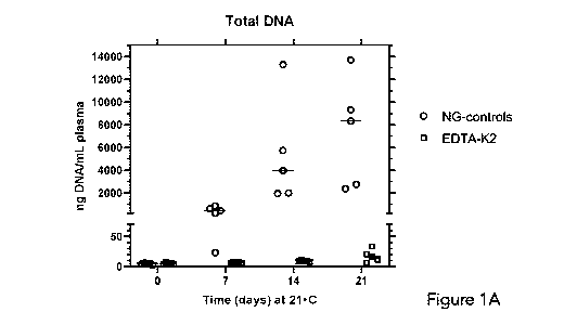
[0079] Figures 1A and 1B graphically depict the amount of extracellular
total DNA and
extracellular fetal DNA, respectively, in plasma prepared from blood from 5
pregnant donors
(pD316, pD317, pD318, pD319, pD320). General experimental details are as
described in the
above section Experimental Details. To demonstrate the utility of the
composition described
Herein, a comparison was made between blood samples maintained for different
periods of
time at 21 C either in a tube containing no gel (NG-control) or in a tube
containing the present
composition (gel plus 0.1 volume of stabilizing agent to 1 volume of blood.
The stabilizing
agent was 0.5 M potassium EDTA at pH 6.8. The results are presented as a
'scatter dot plot',
with the horizontal line representing the median. As also observed by numerous
authors (e.g.,
21
CA 03122342 2021-06-07
WO 2020/132747
PCT/CA2019/051906
Norton et al., J. Clin. Lab. Anal. 27:305, 2013), it is evident from these
results (Figure 1A, Total
DNA) that NG (no gel) control tubes containing only the anticoagulant, 5 mM
EDTA, are
unable to prevent the large release (about 1000 times of the initial value) of
intracellular DNA
into the plasma that occurs with time. Considering that the amount of total
DNA in 1 mL of
blood is approximately 30,000 nanograms, a large proportion of the
intracellular DNA from
lysed cells has been released by day 21. In striking contrast, the amount of
total DNA in blood
exposed to the present composition is largely unchanged until day 14 and
increased only
slightly (about 3-fold) by day 21. At day 0, the amount of total DNA is
equivalent in both types
of tubes, i.e., containing 5 mM EDTA or the present composition. It is
possible to measure
DNA that is unambiguously extracellular DNA in origin in the case where the
blood donors are
pregnant women and fetal DNA can be detected in the plasma. The level of fetal
DNA
compared to total DNA acts as an internal control, since it is expected to
neither increase or
decrease with time in a perfectly stabilized sample. It is noted, however,
that the amount of
fetal DNA in fresh plasma may vary 10-fold or more from pregnant donor to
donor. Figure 1B
shows that the median amount of fetal DNA did not vary appreciably in samples
held at 21 C
for up to 21 days in tubes containing either 5 mM EDTA or in the present
composition,
indicating that the large increase in total DNA is indeed arising from
maternal blood cells.
[0080]
Figures 2A and 2B graphically depict the amount of extracellular total DNA and
extracellular fetal DNA, respectively, in plasma prepared from blood from a
pregnant donor
(pD218). General experimental details are as described in the section
Experimental Details. To
demonstrate the utility of different stabilizing agents, a comparison was made
between blood
samples maintained for different periods of time at 21 C either in a tube
containing a gel only
(Control) or in a tube containing a composition (gel plus 0.1 volume of
stabilizing agent to 1
volume of blood). The stabilizing agent was either 0.9 molar potassium
chloride (KCI) or 1.0
molar sucrose (Sucrose). The results are presented as a bar graph, where the
error bars
represent the range of 2 aliquots from the same plasma sample. Note that in
Control tubes,
the amount of Total DNA continued to increase with time, reaching about 20
times the initial
22
CA 03122342 2021-06-07
WO 2020/132747 PCT/CA2019/051906
level at day 7 and 60 times the initial level at day 21. This is far less than
the approximately
1000-fold increase in the amount of total DNA in NG-control samples,
indicating that the gel
plays an important part in the composition. In both stabilizing agent-treated
samples, the
increase was less than 2 times, showing stabilization of the sample by the
stabilizing agents
described herein. During the same time, the amount of fetal DNA in the samples
remained
substantially unchanged, both in the Control samples and in the samples
treated additionally
with stabilizing agents. Error bars indicate the range of duplicate analyses
of a single sample.
These data demonstrate that the presence of a thixotropic gel alone (Control)
removes a large
fraction but not all the cells above the gel. In the case of tubes
additionally containing a
stabilizing agent, there was very little increase in DNA with time, indicating
the vast majority of
cells had been removed by the barrier gel. The slow release of DNA with time
in both Control
and test samples suggests that cells are attached to the gel surface, and that
with time they
undergo either lyse or apoptosis, releasing their DNA. We have direct evidence
showing that
slow release of DNA in Control tubes is indeed due to cells attached to the
surface of the gel.
At day 0, following centrifugation, all the plasma was carefully removed from
Control tubes.
The tubes were cut about 1 cm above the surface of the gel. The gel surface
was first rinsed
gently with saline, then washed vigorously with a solution containing a strong
detergent,
sodium dodecyl sulphate. DNA in the gently rinsed and vigorously washed
fractions was
analysed. It was found that there was almost no total DNA in the gently rinsed
fraction but a
relatively large amount of total DNA in the vigorously washed fraction.
Interestingly, there was
very little fetal DNA in either of these fractions, indicating the total DNA
was need coming from
loosely attached or gel-embedded cells.
[00811 Figure 3 graphically depicts the amount of extracellular total DNA
in plasma
prepared from blood from a single donor (pD236). Aliquots of the blood were
distributed into
blood tubes containing a thixotropic gel (Control) or tubes containing a
composition (gel plus
0.1 volume of stabilizing agent to 1 volume of blood) (EDTA-K2). The
stabilizing agent was 0.5
M potassium EDTA at pH 6.8. In both cases, tubes were mixed shortly before
centrifugation as
23
CA 03122342 2021-06-07
WO 2020/132747 PCT/CA2019/051906
described in Experimental Details. Note that in the Control samples, the
amount of DNA
continued to increase with time, reaching over 200 times the initial level at
day 14. In the
stabilizing agent-treated samples, there was no discernible increase in DNA,
showing
stabilization of the sample by the stabilizing agents described herein. These
data demonstrate
that the presence of a thixotropic gel alone (Control) is insufficient to
remove all the cells above
the gel. In the case of tubes additionally containing a stabilizing agent,
there was very little
increase in DNA with time, indicating the vast majority of cells had been
removed by the
barrier gel.
[0082] Figures 4A and 4B graphically depict the amount of extracellular
total DNA and
fetal DNA, respectively, in plasma prepared from blood from a pregnant donor
(pD247).
Aliquots of the blood were distributed into blood tubes containing a
thixotropic gel (Control)
or tubes containing a composition (gel plus 0.1 volume of stabilizing agent to
1 volume of
blood) (EDTA-K2). The stabilizing agent was 0.5 M potassium EDTA at pH 6.8.
The tubes were
then held at ambient temperature for 14 days. Note that in Control samples the
amount of
total DNA continued to increase with time, reaching about 5 times the initial
level by day 14. In
the stabilizing agent-treated samples, there was no discernible increase in
total DNA, showing
stabilization of the sample by the stabilizing agents described herein. Two
separate initial
aliquots of blood behaved similarly. During the same time, the amount of fetal
DNA in the
samples decreased less than 2-fold.
[0083] Figure 5 graphically depicts the amount of extracellular total DNA
in plasma
prepared from blood from a single donor (pD251). Aliquots of the blood were
distributed into
blood tubes containing a thixotropic gel (Control) or tubes containing a
composition (gel plus
0.1 volume of various stabilizing agents to 1 volume of blood) (EDTA-K2,
Sucrose or EDT+Suc).
The stabilizing agents were, respectively, in 0.5 M of potassium EDTA at pH
6.8., 1 M sucrose,
or a mixture of the latter 2 chemicals in a 1:1 ratio by volume. The tubes
were then held at
ambient temperature for a period up to 21 days. Note that in Control samples,
the amount of
DNA continued to increase with time, reaching about 10 times the initial level
at day 14 and 30
24
CA 03122342 2021-06-07
WO 2020/132747 PCT/CA2019/051906
times the initial level at day 21. In the stabilizing agent-treated samples,
there is no discernible
change in the amount of cell free DNA in any of the samples. Furthermore,
there is indication
that a mixture of EDTA-K2 and Sucrose can be also stabilizing up to 14 days.
[0084] Figure 6 graphically depicts the amount of extracellular total DNA
in plasma
prepared from blood from donor pD261. Aliquots of the blood were distributed
into blood
tubes containing a thixotropic gel (Control) or tubes containing a composition
(gel plus 0.1
volume of stabilizing agent to 1 volume of blood) (CDTA-K2). The stabilizing
agent was 0.5 M
potassium CDTA (cyclohexane diamine tetraacetate) at pH 6.8. The tubes were
then held at
ambient temperature for 7 days. Note that, in Control samples, the amount of
total DNA
continued to increase with time, reaching about 4 times the initial level by
day 7. In the
stabilizing agent-treated samples, there was only a negligible increase in
total DNA, showing
stabilization of the sample by this stabilizing agent for up to 7 days. This
experiment
demonstrates that the potassium salt of another ionic compound can stabilize
cell free DNA in
plasma.
[0085] Figure 7 graphically depicts the amount of extracellular total DNA
in plasma
prepared from blood from a single donor (pD264) treated with one of 3
different stabilizing
agents after 7 days. Aliquots of the blood were distributed into blood tubes
containing a
thixotropic gel (Control) or tubes containing a composition (gel plus 0.1
volume of various
stabilizing agents to 1 volume of blood) (Trehalose, Mannitol or Sucrose). The
stabilizing agents
were, respectively, 1.0 M trehalose, 1.0 M mannitol or 1.0 M sucrose. The
tubes were then held
at ambient temperature for 7 days. Note that in Control samples, the amount of
DNA
continued to increase with time, reaching 50-100 times the initial level at
day 7. In the
stabilizing agent-treated samples, there is about 2-4 times increase in the
amount of cell free
DNA in all of the samples. This provides evidence that low molecular weight
polyols, as well as
sucrose, can also be stabilizing up to 7 days.
[0086] Figure 8 graphically depicts the amount of extracellular total DNA
in plasma
prepared from blood from a single donor (pD266) treated with one of 3
different stabilizing
CA 03122342 2021-06-07
WO 2020/132747 PCT/CA2019/051906
agents after 7 days. Aliquots of the blood were distributed into blood tubes
containing a
thixotropic gel (Control) or tubes containing a composition (gel plus 0.1
volume of various
stabilizing agents to 1 volume of blood) (Sucrose, Inositol or Mannitol). The
stabilizing agents
were, respectively, 1.0 M sucrose, 1.0 M inositol or 1.0 M mannitol. The tubes
were then held
at ambient temperature for 7 days. Note that in Control samples, the amount of
DNA
continued to increase with time, reaching 10 times the initial level at day 7.
In the stabilizing
agent-treated samples, there was no increase in the amount of cell free DNA in
any of the
samples. This provides further evidence that low molecular weight polyols,
including inositol
and mannitol, can also serve as stabilizing agents up to 7 days.
[0087] Figures 9A and 9B graphically depicts the amount of extracellular
total DNA (9A)
and fetal DNA (9B) in plasma prepared from blood from a pregnant donor (pD379)
treated with
a composition and held at 4 C for 1, 2, 3 or 6 days prior to centrifugation.
Aliquots of the
blood were distributed into a blood tube containing no thixotropic gel (NG-
control) or tubes
containing a composition (gel plus 0.1 volume of a stabilizing agent to 1
volume of blood)
(EDTA-K2). The stabilizing agent was 0.5 M dipotassium EDTA. The NG-control
tube was
centrifuged immediately to prepare cell-free plasma. The remaining tubes were
then held at
4 C temperature for 1-6 days, as indicated, then centrifuged and an aliquot of
the plasma
removed. Samples were then maintained at ambient temperature. At the indicated
time post-
centrifugation, the tubes were centrifuged and aliquots of the plasma removed.
Other details
are described in Experimental Details. This demonstrates that the mixing of a
solution of low
molecular weight (<500 Da) stabilizing agent with blood does not cause damage
to the
membrane of white blood cells that can result in leakage of intracellular DNA
into the plasma
fraction. This lack of damage is shown to be maintained for up to 6 days
provided the sample
is maintained at 4 C. The level of fetal DNA is essentially the same in the
control and all
treated samples.
[0088] Figures 10A, 10B, 10C, and 10D graphically depicts the amount of
extracellular
total DNA in plasma prepared from blood from 4 donors (pD382, pD383, pD390 and
pD391)
26
CA 03122342 2021-06-07
WO 2020/132747 PCT/CA2019/051906
treated with one of 4 different stabilizing agents and held at 4 C for 3 to 5
days prior to
centrifugation. Aliquots of the blood were distributed into blood tubes
containing a thixotropic
gel (Control) or tubes containing a composition (gel plus 0.1 volume of
various concentrated
stabilizing agents to 1 volume of blood) (SUC, LAC, TREH and MELD. The
concentrated
stabilizing agents were, respectively, 1.0 M sucrose, 1.0 M lactose, 1.0 M
trehalose or 1.0 M
melibiose. The Control tube was centrifuged immediately to prepare cell-free
plasma. The
remaining tubes were then held at 4 C temperature for 3-5 days, as indicated.
Following that,
samples were maintained at ambient temperature. At the indicated time post-
centrifugation,
the tubes were centrifuged and aliquots of the plasma removed. Other details
are described in
Experimental Details. The experiments depicted in Figures 10A-D demonstrate
that the mixing
of a 1.0 M solution of low molecular weight (<500 Da) polyol with blood (at a
ratio of 0.1 part
concentrated solution to 1 part of blood, by volume) does not cause damage to
the membrane
of white blood cells that results in significant leakage of intracellular DNA
into the plasma
fraction. Surprisingly, this lack of damage is maintained for up to 5 days
provided the sample is
maintained at 4 C. However, if such samples are maintained at ambient
temperatures even in
the presence of stabilizing agent, extensive damage the membrane of cells
occurs resulting in
leakage of a large amount of intracellular DNA into the plasma fraction (data
not shown). The
utility of these findings is that it allows sufficient time for samples to be
collected at one
location and stored or transported at refrigerator temperatures to a second
location where they
can then be centrifuged. Furthermore, after centrifugation, the samples can be
stored at
ambient temperature for periods of 7-14 days with little increase in the
amount of intracellular
DNA in the plasma fraction.
[0089] Figure 11 graphically depicts the amount of extracellular total DNA
in plasma
prepared from blood from one donors (pD394) treated with one of 3 different
stabilizing
agents and held at 4 C for 5 days prior to centrifugation. Aliquots of the
blood were
distributed into blood tubes containing a thixotropic gel (Control) or tubes
containing a
composition (gel plus 0.1 volume of various stabilizing agents to 1 volume of
blood). The
27
CA 03122342 2021-06-07
WO 2020/132747 PCT/CA2019/051906
stabilizing agents were the following mixtures: 0.375 M dipotassium EDTA/0.25
M sucrose
(75E/25S); 0.250 M dipotassium EDTA/0.5 M sucrose (50E/50S); 0.125 M
dipotassium
EDTA/0.75 M sucrose (25E/755). The Control tube was centrifuged immediately to
prepare
cell-free plasma. The remaining tubes were then held at 4 C temperature for 5
days. Following
that, samples were maintained at ambient temperature. At the indicated time
post-
centrifugation, the tubes were centrifuged and aliquots of the plasma removed.
Other details
are described in Experimental Details. It is evident from the results depicted
in Figure 11 that
essentially all nucleated blood cells survive the immediate shock of contact
and mixing with
concentrated chemicals in 3 different stabilizing agents and subsequent
incubation of the
plasma-diluted stabilizing agents for 5 days at 4 C, then centrifuged.
Essentially no difference
in the amount of released DNA in the plasma fraction of the Control (processed
shortly after
blood collection) and any of the 3 compositions is seen. When subsequently
incubated at
ambient temperature for up to 21 days post-centrifugation, some differences
between the 3
stabilizing agents is seen, with 75E/25S showing greatest stability. The
utility of these findings
is that the composition allows sufficient time for samples to be collected at
one location and
stored or transported at refrigerator temperatures to a second location where
they can then be
centrifuged.
[0090] Figure 12 graphically depicts the amount of extracellular total DNA
in plasma
prepared from blood from one donor (pD399), treated with one of two different
stabilizing
agents and held at 0 C, 4 C or 10 C for 5 days prior to centrifugation.
Aliquots of the blood
were distributed into blood tubes containing a thixotropic gel (Control) or
tubes containing a
composition (gel plus 0.1 volume of various stabilizing agents to 1 volume of
blood). The
stabilizing agents were 1.0 M sucrose (SUC) or a mixture containing 0.85 M
sucrose and 0.075
M dipotassium EDTA (855/15E). The Control tube was centrifuged immediately to
prepare cell-
free plasma. The remaining tubes were then held at 0 C, 4 C or 10 C for 5
days. After 5 days,
the tubes were centrifuged and aliquots of the plasma removed and used to
determine the
amount of extracellular DNA. Other details are described in Experimental
Details. It is evident
28
CA 03122342 2021-06-07
WO 2020/132747 PCT/CA2019/051906
from these results that essentially all nucleated blood cells survive the
immediate shock of
contact and mixing with concentrated chemicals in 2 different stabilizing
agents for 5 days at 00
and 4 C. A small amount of leakage of intracellular DNA appears to occur at 10
C. The utility
of these findings is that the composition allows sufficient time for samples
to be collected at
one location and stored or transported at wet ice or refrigerator temperatures
to a second
location where they can then be centrifuged.
[00911 All publications, patents and patent applications mentioned in this
specification are
indicative of the level of skill of those skilled in the art to which this
invention pertains and are
Herein incorporated by reference. The invention being thus described, it will
be obvious that
the same may be varied in many ways. Such variations are not to be regarded as
a departure
from the scope of the invention, and all such modifications as would be
obvious to one skilled
in the art are intended to be included within the scope of the following
claims.
29