Note: Descriptions are shown in the official language in which they were submitted.
CA 03122384 2021-06-03
WO 2020/118257
PCT/US2019/065075
TUBE-CONTAINING MEDICAL DEVICES
WITH BIOACTIVE LUMINAL WIRE
BACKGROUND
Clinicians struggle to manage catheter related blood stream infections
(CRBSIs)
and luminal occlusions on a daily basis. The occurrence rate of CRBSIs post
implementation of maximum barrier precautions and other insertion site
protection
measures (regular site cleaning, antimicrobial dressings/discs, etc.) has been
reduced,
but is still in the range of 0.5-2 catheter days per 1,000 catheters, which is
based on the
number of infections (x) per 1,000 days of insertion, leading to mortality and
morbidity. It
also causes an economic burden to the healthcare system. CRBSIs can be an
issue
with long term catheters, where the intraluminal route of infection dominates.
Likewise,
about 20% of all catheter lumens occlude during implanted life. The
predominant cause
of catheter occlusions is thrombosis and/or related factors. When lumens
occlude,
functionality of the catheter becomes limited, as the lumen can no longer be
used
effectively or at all to aspirate and/or infuse fluids therethrough.
Furthermore, drug
eluting coatings on devices of various sizes can deplete quickly due to
limited
availability of the reservoir of drug and non-eluting passive coatings can
biofoul, thus
restricting the antimicrobial, anti-thrombogenic, and/or thrombolytic effects.
For
example, there are devices that can deliver bioactive agent of some type into
the lumen
of a catheter and/or into the body of the subject for a period of a few
seconds, a few
minutes, a few hours, or even a few days or more. However, some of these
catheters
and longer term catheters, on the other hand, can be harder to manage, and in
many
cases, catheters are removed and fully replaced to deal with infection,
thrombotic
occlusion, etc., putting the subject/patient at further risk. Likewise,
replaceable lock
1
CA 03122384 2021-06-03
WO 2020/118257
PCT/US2019/065075
solutions have been devised to address longer term catheter complications, but
are
slow to be developed and marketed due to lengthy clinical trials. In the case
of
thrombolytic locks, Cathflo from Genentech (USA), which is a tissue
plasminogen
activator is available on the market and is effective, but a single fluidic
dose is currently
well over $100, which is quite expensive considering the considerably lower
cost of the
catheter hardware itself.
BRIEF DESCRIPTION OF THE DRAWINGS
FIG. 1 illustrates an example of a catheter assembly including a luminal wire
that
can be replaceable in the lumen of a catheter to provide, for example, long-
term
effectiveness by providing antimicrobial, anti-thrombogenic, thrombolytic,
and/or other
properties to a lumen of the catheter in accordance with the present
disclosure.
FIG. 2 illustrates an example catheter with a Y-junction and multiple hubs
having
a luminal wire positioned therein that can be replaceable and can be effective
for
providing antimicrobial, anti-thrombogenic, or thrombolytic, and/or other
properties to a
lumen of the catheter in accordance with the present disclosure.
FIG. 3 is a graph illustrating the antimicrobial effectiveness of various
types of
luminal wires, including unmodified metal luminal wire and copper-plated
luminal wire,
.. compared to a commercial product in accordance with the present disclosure.
FIGS. 4-5 illustrate a schematic example catheter assembly or kit used in
generating data for examples of the present disclosure.
FIG. 6 illustrates a flow system used to simulate blood flow in the human body
in
generating data for examples of the present disclosure.
FIGS. 7-9 are graphs illustrating lumen patency assessments that were
conducted in accordance with examples of the present disclosure.
FIG. 10 illustrates a schematic example catheter assembly or kit used in
generating data for examples of the present disclosure.
FIGS. 11-14 provide data for multiple antimicrobial studies related to the use
of
copper wire inserts in accordance with examples of the present disclosure.
2
CA 03122384 2021-06-03
WO 2020/118257
PCT/US2019/065075
FIGS. 15-16 provide data related to the health and safety of blood when
exposed
to the copper luminal wires in accordance with examples of the present
disclosure.
DETAILED DESCRIPTION
The present disclosure is drawn to tube-containing medical devices with
replaceable bioactive luminal wires, bioactive luminal wires for insertion
into a lumen of
a tube-containing medical device, and methods of making and using tube-
containing
medical devices with bioactive luminal wires. These devices, systems, and
methods can
be effective for preventing or reducing intraluminal infection and/or luminal
thrombotic
occlusion in catheters and other elongated tubular structures of medical
devices
susceptible to infection, e.g., feeding tubes, tracheal tubes, shunts,
drainage tubes, e.g.,
external ventricular drain (EVD), etc. These systems, devices, and methods can
provide
for not only short term solutions to the introduction of infection into the
lumen of tube-
containing medical devices and connectors, but can also exhibit long term
effectiveness. To illustrate by way of specific example, one can consider
catheters
generally. Thus, examples herein drawn specifically to catheters should be
read to
include other similar tubing examples where the medical device is not a
catheter per se,
e.g., a shunt, drainage tube, trachea, feeding tube, etc. For example, a shunt
effectively
has two ports, one at each end that redirects fluids within the body from one
location to
another. In this instance, a hub may be placed outside of the body for
insertion of a
luminal wire in one or both directions toward the shunt openings, etc. With
that
understanding, for simplicity in explanation here and elsewhere in the
specification,
catheters are described in greater detail providing a proxy example of one of
the
multiple types of medical devices that can be used, and such disclosure is
directly
relevant and supports other tube-containing medical devices.
With specific reference to catheters, this type of medical device typically
includes
a hub outside of the body of the patient that can be accessed by a medical
professionals or that can be self-accessed by the patient. The hub can be a
connector
of any type, as defined herein. Catheters can also include a port, which is
the structure
3
CA 03122384 2021-06-03
WO 2020/118257
PCT/US2019/065075
that connects the catheter to the body, e.g., to a vein, to a bladder, or to
any other body
structure the catheter is designed to be inserted. For example, hubs and ports
can have
an elongated lumen therebetween, and these three structures, e.g., the hub,
the
elongated tubing defining a lumen, and the port, are often used to access or
draw fluids
from the body, to introduce locking fluids into the lumen of the catheter, or
to introduce
therapeutic fluids into the body through the catheter. With respect to longer
term
antimicrobial, anti-thrombogenic, and/or thrombolytic activity within the
catheter lumen,
or for general catheter maintenance for even shorter term use, a separate
bioactive
luminal wire device can be configured to be introduced, or can be pre-
positioned within,
the lumen of a catheter, and can be replaceable by removing the luminal wire
and
replacing it with the same type or different type of luminal wire.
Thus, in accordance with an example of the present disclosure, a catheter
assembly can include a catheter including an access opening positionable
exterior with
respect to a subject, a fluid communication opening positionable within the
subject to
fluidically communicate with a body fluid of the subject, and an elongated
lumen
therebetween. The catheter assembly can also include a luminal wire inserted
in the
elongated lumen, wherein the luminal wire has a length that is at least 5% of
a length of
the elongated lumen, and/or has an x-y cross-sectional diameter sufficiently
smaller
than an x-y cross-sectional diameter of the elongated lumen to allow insertion
and
retraction of wire, and furthermore, may also be configured to provide fluid
flow
therebetween in instances where there may be a desire to flow fluid thereby.
The
luminal wire can be bioactive, e.g., antimicrobial, anti-thrombogenic,
thrombolytic, or
can be used to deliver a bioactive agent such as a drug or other therapeutic
compound.
In one example, if the wire length is longer than 100% of the luminal length
(or in some
cases even if shorter), there can be a mechanism or other control system in
place to
prevent the luminal wire from extending past the fluid communication opening,
or
alternatively indicate to the user where to stop inserting the luminal wire,
for example.
The luminal wire can also be replaceable with a new luminal wire by simply
removing
the luminal wire and inserting a replacement luminal wire. In one example, a
fluid lock
solution can be loaded in the elongated lumen along with the luminal wire.
4
CA 03122384 2021-06-03
WO 2020/118257
PCT/US2019/065075
In another example, a catheter kit can include a catheter including an access
opening positionable exterior with respect to a subject, a fluid communication
opening
positionable within the subject to fluidically communicate with a body fluid
of the subject,
and an elongated lumen therebetween. The catheter kit can also include a
luminal wire
insertable within the elongated lumen, wherein the luminal wire has a length
that is at
least 5% of a length of the elongated lumen, and/or has an x-y cross-sectional
diameter
sufficiently smaller than an x-y cross-sectional diameter of the elongated
lumen to allow
insertion and retraction of wire, and furthermore, in some examples, provide
fluid flow
therebetween in instances where there may be a desire to flow fluid thereby.
As a note,
.. space for providing fluid flow can typically be less than that practical
for removing and
inserting luminal wire, but this is not always the case. In one example, if
the wire length
is longer than 100% of the luminal length (or in some cases even if shorter),
there can
be a mechanism or other control system in place to prevent the luminal wire
from
extending past the fluid communication opening, or alternatively indicate to
the user
where to stop inserting the luminal wire, for example. The luminal wire can
also be
replaceable with a new luminal wire by simply removing the luminal wire and
inserting a
replacement luminal wire. In some types of medical tubing or catheters, the
fluid
communication opening may be on a side wall of the catheter. In such
instances, in one
example, the luminal wire can be inserted through the lumen of the catheter to
the tip,
.. past the side wall opening, without luminal wire exiting the catheter and
entering the
patient. Regardless of the arrangement, the luminal wire can be inserted into
the lumen
of the catheter, in one example, to a length that provides antimicrobial, anti-
thrombogenic, and/or thrombolytic properties, and can reside as an indwelling
sub-
device within the tubing of the catheter or other tubular medical device, at
any portion or
all of the lumen of the catheter, while preventing the luminal wire from
entering the body
of the subject outside of the protection of the walls of the tubing.
In another example, a method of manufacturing a catheter assembly or kit can
include obtaining a catheter including an access opening positionable exterior
with
respect to a subject, a fluid communication opening positionable within the
subject to
fluidically communicate with a body fluid of the subject, and an elongated
lumen
5
CA 03122384 2021-06-03
WO 2020/118257
PCT/US2019/065075
therebetween. The method can further include establishing a length of a
luminal wire
that is positionable within at least 5% the elongated lumen, wherein the
luminal wire has
an x-y cross-sectional diameter sufficiently smaller than an x-y cross-
sectional diameter
of the elongated lumen to allow removal and/or insertion of a luminal wire.
The luminal
wire in this example is bioactive. The method can also include sizing the
luminal wire to
correspond with the length. Sizing can be shortened by cutting, lengthened by
stretching in the z-direction of the wire, forming luminal wire at the length
so that
shortening or lengthening is not required. With this method, the length can be
such that
the luminal wire is not long enough or otherwise not insertable into the
elongated lumen
far enough to extend beyond the fluid communication opening. The catheter
assembly
or kit can further be used to prepare the catheter assembly by inserting the
luminal wire
in the elongated lumen. The catheter kit can include, for example, multiple
interchangeable luminal wires of the same length. Furthermore, the multiple
interchangeable luminal wires include a first luminal wire pre-inserted in the
elongated
lumen, and a second luminal wire as part of the catheter assembly or kit as a
replacement luminal wire.
In another example, a method of protecting the lumen of a catheter can include
inserting a fluid communication opening of a catheter within a body of a
subject, the
catheter further including an access opening exterior to the body to access an
elongated lumen of the catheter in fluid communication with the fluid
communication
opening. The method further includes sliding a luminal wire within the
elongated lumen,
wherein the luminal wire has a length that is within 50% to 100% of the
elongated
lumen, and has an x-y cross-sectional diameter sufficiently smaller than an x-
y cross-
sectional diameter of the elongated lumen to allow fluid flow therebetween,
and wherein
the luminal wire is bioactive. In one example, the step of sliding the luminal
wire within
the elongated lumen occurs prior to inserting the fluid communication opening
of the
catheter within the body of the subject. In another example, the step of
sliding the
luminal wire within the elongated lumen occurs after inserting the fluid
communication
opening of the catheter within the body of the subject. In one specific
example, the
method can include applying an electrical potential to the luminal wire, which
may be
6
CA 03122384 2021-06-03
WO 2020/118257
PCT/US2019/065075
used to provide any of a number of bioactive effects, including improving or
enhancing
the antimicrobial properties of the luminal wire, such as to improve longevity
or to
improve antimicrobial efficacy compared to when there is no electrical
potential applied.
In addition to the examples described above, e.g., the tube-containing medical
device assemblies, the tube-containing medical device kits, the methods of
manufacturing, and methods of protecting the lumen of a catheter, certain
features will
be discussed in greater detail below. It is noted that when discussing the
catheter
assembly, the catheter kit, or any of the methods herein, these relative
details can be
considered applicable to the other examples, whether or not they are
explicitly
discussed in the context of that example. Thus, for example, in discussing a
luminal
wire related to the catheter assembly, such disclosure is also relevant to and
directly
supported in the context of the catheter kit and/or methods described herein,
and vice
versa, etc.
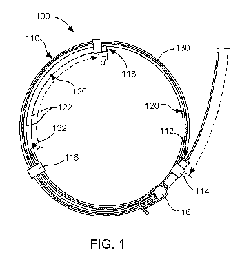
As a point of initial orientation, FIGS. 1 and 2 are provided to show several
features of a catheter assembly 100. These FIGS. are also relevant to catheter
kits and
methods herein, but are shown in the context of the catheter assembly for
convenience
and to show the spatial relationship of the luminal wire and the catheter in
connection to
one another during or after assembly. Thus, in these FIGS., the catheter
assembly
includes a catheter 110 and a luminal wire 130. The catheter in this example
includes
an access opening 112 defined by a hub 114. The hub can be integrated as part
of the
catheter or can be assembleable with the tubing. In this particular example,
there is also
a clamp 116 associated with the catheter near the hub. The catheter also
includes a
fluid communication opening 118, which in some examples can be defined by or
fluidly
associated with a port (not shown), e.g., needle port. If present, however,
the port may
be integrated with the tubing or can be assembleable with the tubing, for
example. The
port can be, for example, merely a harder polymeric portion of the tubing
suitable for
insertion of the catheter through the skin and into a vein or artery of a
subject (or some
other fluidic channel), or can be some other device other than a needle port.
Between
the access opening and the fluid communication opening is an elongated lumen
120
defined by catheter walls 122 (or the tubing). In one example. The catheter
can be
7
CA 03122384 2021-06-03
WO 2020/118257
PCT/US2019/065075
branched, as shown generally at 124 in FIG. 2, with multiple hubs 114A, 114B
at a Y-
junction, for example. The luminal wire 130 shown in FIG. 1 is sized so that
when fully
inserted, a distal tip 132 of the wire will be positioned just short of the
catheter port 118,
shown in this example as distance "d." In this specific instance, the luminal
wire can be
about 99% to as long as the elongated lumen 120, though other lengths can be
used
with other distances short of the fluid communication opening (or port) for
example. In
further detail, the tube-containing medical device can include an insert hub
opening (or
wire insertion/removal opening) for inserting or removing the luminal wire
from the
tubing that is separate from a fluidic hub used for introducing or removing
fluid from the
tubing. This can provide the benefit of allowing for insertion and/or removal
of the
luminal wire without occupying or using up a hub opening that may be used for
a
different reason, or it may be that there is reason to have a separate
insertion hub
opening that is independent of convenience, e.g., adding an insertion hub to a
shunt so
that the multiple port ends can be accessed for inserting and/or removing
luminal wire in
either direction or both directions.
Alternatively, the wire could be attached to a cap (not shown) which provides
the
same function of preventing the luminal wire from exiting the fluid
communication
opening at the distal tip if the design of the catheter is such that the wire
could exit
through an end opening or even side openings in some examples. Thus, the
luminal
wire can be inserted (attached at the proximal end to the cap) and secured
such that the
wire is not long enough to pass through the vascular insertion end, or fluid
communication opening as previously described, when capped off. Alternatively,
an
atraumatic tip can also be used for the luminal wire to provide that no damage
to the
vessel wall or lungs occurs if the wire is accidentally inserted beyond an
opening at or
near a distal tip (fluid communication opening) of the catheter.
Alternatively, a ball tip
(which some needles include) could likewise be implemented here.
Alternatively, the
luminal wire can be equipped with a polymer bit fabricated at its distal tip
for additional
safety as well.
Turning now to further detail regarding the catheter assemblies, catheter
kits, and
methods, the luminal wire can be made of a material or coated with a material
that is
8
CA 03122384 2021-06-03
WO 2020/118257
PCT/US2019/065075
antimicrobial, anti-thrombogenic, and/or provides thrombolytic activity, so
that an outer
surface of the luminal wire contacts fluid as it flows through the catheter in
either
direction. Thus, a cross-sectional x-y dimension of the luminal wire is
smaller than a
luminal surface of the catheter so that the luminal wire can be removed and
inserted, for
example, and in cases where there may be fluid flow through the catheter or
other
tubular medical device, the wire can be configured such that fluid can flow
between the
tubing wall and the luminal wire. Furthermore, if or when the luminal wire
loses its
therapeutic effect (antimicrobial, anti-thrombogenic, and/or thrombolytic,
etc.), the
luminal wire can be replaced, for example, while the catheter remains fluidly
coupled to
the patient, e.g., the catheter does not need to be removed or in some
instances even
disturbed as an old or spent luminal wire is removed and replaced with a fresh
luminal
wire insert. By including a replaceable luminal wire, this can eliminate a
reservoir effect
of bioactive agent, as the luminal wire can be designed to last for a
predetermined
period of time, e.g., from 1 hour to 12 months, from 1 day to 4 months, from 2
days to 2
months, from 3 days to 1 month, from 1 day to 1 week, etc., and then replaced
easily
without removing the catheter from the body of the subject. Thus, if a
bioactive agent is
loaded on a wire substrate, then when the bioactive agent is spent or
released, the
luminal wire can be replaced with a fresh luminal wire with more bioactive
agent or
another bioactive agent or with a luminal wire that does not have a separate
bioactive
agent thereon. This can continue until the life of the catheter is finished,
providing a
modular mechanism to treat intraluminal microbial colonization and thrombotic
occlusions.
The term "luminal wire" herein includes any rigid or semi-rigid device with an
antimicrobial, anti-thrombogenic, and/or thrombolytic surface that is
elongated in the z-
dimension and having a cross-sectional area or size in the x-y-dimension thin
or small
enough to fit within the lumen of a catheter while retaining enough space
between the
outer surface of the luminal wire and the surface of the catheter lumen to
allow for
insertion or removal of the luminal wire, and allow for fluid flow in
instances where there
may be fluid flow. The term "luminal wire" is thus defined to include a number
of
structures that are x-y-dimensionally thin and z-dimensionally elongated, such
as metal
9
CA 03122384 2021-06-03
WO 2020/118257
PCT/US2019/065075
wires, metal alloy wires, multiple wires braided or twisted together, coated
wires, tubular
wire, multilayered wire, e.g., multiple material in an annular arrangement,
composited
wire, e.g., multiple wires composited or bound together, etc. For example, a
tubular or
annular wire arrangement can include a hollow elongated core (or even discrete
core
pockets present at specific locations, periodically placed, or both) with a
metal or metal
alloy shell, which can include pores or includes an orifice at an end of the
luminal wire
that may release bioactive agent loaded within the hollow core. The luminal
wire can be
flexible and/or malleable so that it can bend or otherwise conform with
bending and/or
twisting of the catheter tubing, but may be more laterally (x-y-dimension)
rigid than the
catheter tubing, less laterally rigid than the catheter tubing, or about the
same degree of
lateral rigidity as the catheter tubing. The bending can be such that the
luminal wire
conforms to the new shapes and retains that shape, or the bending can be
resilient so
that after bending or flexing, the luminal wire returns or partially returns
to the original
orientation prior to bending or flexing with the catheter.
With respect to the luminal wire length, a number of lengths can be used. For
example, the luminal wire can have a length that is at least 5% of a length of
the
elongated lumen, at least 5% of a length of the elongated lumen, at least 10%
of a
length of the elongated lumen, at least 20% of a length of the elongated
lumen, at least
35% of a length of the elongated lumen, or at least 50% of a length of the
elongated
lumen, and/or can have an x-y cross-sectional diameter sufficiently smaller
than an x-y
cross-sectional diameter of the elongated lumen to allow insertion and
retraction of wire.
In one example, the space between the luminal wire and the lumen walls can
provide
for allowing fluid flow therebetween in instances where there may be a desire
to flow
fluid thereby. Luminal wire length may or may not alternatively or
additionally be limited,
for example, by the length of the luminal cavity of the catheter (which can
include the
catheter tubing defining an elongated catheter lumen, hubs, ports, etc.) or
catheter
system (which can include other devices, tubing, etc., that may be connectable
to the
catheter). In other words, whatever portion that should be protected by the
luminal wire,
the wire can be inserted therein to remain for carrying out its function,
e.g., to protect
from infection and/or thrombotic occlusion. In some instances, the luminal
wire length
CA 03122384 2021-06-03
WO 2020/118257
PCT/US2019/065075
can terminate at about the distal tip of the catheter within the body of the
subject (at or
near the port), and in other instances, a distal tip of the luminal wire
length can
terminate short of a fluid communication opening within the body of the
subject, e.g., the
port or other opening near the distal end of the catheter. For example, the
luminal wire
length can be inserted into the catheter through a hub and slid into the
catheter until the
luminal wire is no longer exposed at the hub end of the catheter, or the wire
can include
a marking on the luminal wire so that the user (medical practitioner) knows
where to
stop inserting the wire where in one example, a clamp could be used to prevent
further
insertion. In another example, a mechanical stopper could be associated with
the
luminal wire to prevent insertion of the luminal wire beyond a certain fixed
location, e.g.,
stopped by the architecture or shape of the hub. In these three examples, the
luminal
wire can be slid into the catheter and stopped a safe distance prior to
insertion of the
wire into the body of the subject beyond the protective sheath of the catheter
walls. For
example, when fully inserted, a distal tip of the luminal wire can be
configured to stop
just short of a port or other opening of the catheter where fluids communicate
between
the body of the patient and the lumen of the catheter. If not inserting all
the way to the
end of the catheter at the distal end, suitable distances where the distal tip
of the
luminal wire can be positioned just short of a fluid communication opening can
be from
from 0.1 cm to 55 cm, 0.1 cm to 50 cm, from 0.1 cm to 35 cm, 0.1 cm to 20 cm,
from 0.1
cm to 10 cm, 0.1 cm to 5 cm, 0.5 cm to 50 cm, 0.5 cm to 35 cm, 0.5 cm to 20
cm, from
0.5 cm to 10 cm, 0.5 cm to 5 cm, from 1 cm to 20 cm, from 1 cm to 10 cm, or
from 1 cm
to 5 cm short of the fluid communication opening. By stopping insertion of the
luminal
wire short of an opening of the catheter (or other tubular medical device with
the body of
the subject, the luminal wire does not enter the body of the subject outside
of the lumen
of the catheter, with some margin of safety. In catheters with a fluid
communication
opening along a side wall of the tubing, the luminal wire can go beyond the
fluid
communication opening without entering the body of the subject, as there may
still be
tubing between the fluid communication opening and the distal tip of the
catheter within
the body of the subject. Either way, the luminal wire can be inserted in a way
that
remains within the lumen of the catheter/tubing so that the wire cannot cause
any
11
CA 03122384 2021-06-03
WO 2020/118257
PCT/US2019/065075
damage to tissue within the body of the subject, as it is protected by the
walls of the
tubing.
In accordance with this, in one example, the luminal wire can extend all the
way
through the elongated lumen of the catheter, or through 5% to 100%, 10% to
100%,
20% to 100%, 20% to 99.9%, 20% to 99%, 20% to 95%, 20% to 75%, 35% to 100%,
35% to 99.9%, 35% to 99%, 35% to 95%, 35% to 75%, 50% to 100%, 50% to 99.9%,
50% to 99%, 50% to 95%, 50% to 75%, 65% to 100%, 65% to 99.9%, 65% to 99%,
65% to 95%, 65% to 75%, etc., of the elongated lumen, for example. These
values are
based on how much of the elongated lumen includes luminal wire, not how long
the
.. luminal wire per se is, as in some examples, the luminal wire can extend
out beyond the
hub (outside or through the hub), into or through the hub connector, into
another tube or
device, etc., for example. Thus, the luminal wire may be (in total length),
for example,
from 5% to 300%, from 10% to 300%, from 5% to 200%, from 10% to 200%, from 5%
to
100% from 10% to 100%, from 5% to 99.9%, from 10% to 99.9%, from 5% to 95%,
from
10% to 95%, from 20% to 300%, 20% to 300%, 20% to 150%, 20% to 100%, 20% to
99.9%, 20% to 99%, 20% to 95%, 20% to 75%, 35% to 200%, 35% to 150%, 35% to
100%, 35% to 99.9%, 35% to 99%, 35% to 95%, 35% to 75%, 50% to 200%, 50% to
150%, 50% to 100%, 50% to 99.9%, 50% to 99%, 50% to 95%, 50% to 75%, 65% to
200%, 65% to 150%, 65% to 100%, 65% to 99.9%, 65% to 99%, 65% to 95%, 65% to
75%, etc., of the length of the elongated lumen. In one example, if the wire
length is
longer than 100% of the luminal length (or in some cases even if shorter),
there can be
a mechanism or other control system in place to prevent the luminal wire from
extending
past the fluid communication opening, or alternatively indicate to the user
where to stop
inserting the luminal wire, for example. If the fluid communication opening is
on a side
wall within the body of the patient (or to be placed within the body), then
the luminal wire
can be inserted beyond the opening, as the distal tip without a luminal
opening can act
to stop the luminal wire from being inserted further into the body of the
subject. In other
examples, the luminal wire can terminate at one end at about the access
opening of the
hub, e.g., within about 5 cm or about 10 cm on either side of the port
opening. At the
port, or at an opening along the lumen of the catheter that will be or is
inserted within a
12
CA 03122384 2021-06-03
WO 2020/118257
PCT/US2019/065075
body of a subject, the wire can terminate at the port or other opening, e.g.,
within 20 cm,
within 10 cm, or within 5 cm short of the port or other opening as described
above.
The term "elongated" when referring to the z-dimension of the luminal wire can
be defined as having z-dimension length to x-y-dimension cross-sectional
diameter ratio
of at least 50:1, at least 100:1, at least 200:1, or at least 500:1. Example
ratio ranges
can be from 50:1 to 3,000:1, from 50:1 to 2,000:1, from 50:1 to 1,000:1, from
50:1 to
500:1, from 100:1 to 3,000:1, from 100:1 to 2,000:1, from 100:1 to 1,000:1,
from 100:1
to 500:1, from 200:1 to 3,000:1, from 200:1 to 2,000:1, from 200:1 to 1,000:1,
from
200:1 to 500:1, from 500:1 to 3,000:1, from 500:1 to 2,000:1, or from 500:1 to
1,000:1.
For example, using the American Wire Gauge (AWG) standard which relates
specifically to wire diameter of round/solid wire, a 16 gauge luminal wire
that is about 5
feet long may have an aspect ratio of about 1:1,200 or so. An 18 gauge luminal
wire
that is three feet long may have an aspect ratio of about 1:900 or so. A 12
gauge
luminal wire that is about 1 foot long may have an aspect ratio of about 1:150
or so. A
20 gauge luminal wire that is about 6 feet long may have an aspect ratio of
about
1:2,250 or so. A table of wire gauges and diameters in inches and millimeters
is
provided in Table 1 below, but this list or the use of the AWG system is not
considered
limiting, but rather merely exemplary. Additionally, Table 1 provides relative
cross-
sectional areas in the x-y-dimension, which would be relevant to luminal wires
(including
the wire-like structures defined as "luminal wires" herein) that may or may
not have a
circular aspect ratio.
Table 1
Gauge (AWG) Diameter (inch) Area (inch2) Diameter (mm)
Area (mm2)
6 0.1620 0.0206 4.115 13.3
7 0.1443 0.0163 3.665 10.5
8 0.1285 0.0130 3.264 8.37
9 0.1144 0.0103 2.906 6.63
10 0.1019 0.0082 2.588 5.26
11 0.0907 0.0065 2.305 4.17
12 0.0808 0.0051 2.053 3.31
13 0.0720 0.0041 1.828 2.62
14 0.0641 0.0032 1.628 2.08
15 0.0571 0.0026 1.450 1.65
13
CA 03122384 2021-06-03
WO 2020/118257
PCT/US2019/065075
16 0.0508 0.0020 1.291
1.31
17 0.0453 0.0016 1.150
1.04
18 0.0403 0.0013 1.024
0.823
19 0.0359 0.0010 0.912
0.653
20 0.0320 0.0008 0.812
0.518
21 0.0285 0.0006 0.723
0.410
22 0.0253 0.0005 0.644
0.326
23 0.0226 0.0004 0.573
0.258
24 0.0201 0.0003 0.511
0.205
In further detail, the cross-sectional area of the luminal wire and the cross-
sectional area of the elongated lumen of the tubing, e.g., catheter tubing,
can also be
established. For example, the luminal wire can have an x-y-dimension cross-
sectional
.. area and the elongated lumen (space defined by inner surface of tubing
wall(s)) has an
x-y-dimension cross-sectional area. Thus, the x-y-dimension cross-sectional
area of the
luminal wire can occupy an average of 7% to 90% of the x-y-dimension cross-
sectional
area elongated lumen when the luminal wire is fully inserted in the elongated
lumen.
Alternatively, the x-y-dimension cross-sectional area of the luminal wire can
occupy an
.. average of 25% to 85%, 35% to 75%, 30% to 75%, or from 50% to 90% of the x-
y-
dimension cross-sectional area elongated lumen when the luminal wire is fully
inserted
in the elongated lumen.
Regarding the luminal wire material that can provide antimicrobial, anti-
thrombogenic, and/or thrombolytic activity within the lumen of the catheter,
the luminal
wire itself can be of a material that provides one or more of these properties
without
modification, or the luminal wire can comprise a wire substrate that is coated
or
composited with an antimicrobial agent, an anti-thrombogenic agent, or a
thrombolytic
agent. Examples of materials that can be used for the luminal wire (coated or
uncoated)
include metals or metal alloys, such as copper, silver, zinc, gold, tin,
alloys thereof. The
term "alloys" includes copper, silver, zinc, and/or gold together, but can
also include
alloys of one or more of these metals with any other metal(s) or non-metal(s)
that may
provide a therapeutic or other practical property. For example, as copper
oxidizes,
copper can be alloyed with another metal, such as tin, zinc, gold, silver,
etc., to slow or
prevent oxidation. As copper is a good metal for providing antimicrobial
properties, the
14
CA 03122384 2021-06-03
WO 2020/118257
PCT/US2019/065075
alloy can include a substantial portion of copper and a lesser proportion of
other metals
(or non-metals) that may be included to slow oxidation, but may not contribute
to the
antimicrobial properties of the luminal wire to the extent that copper can
contribute.
Thus, if a copper alloy is used, the copper can be present in the alloy at
from 50 wt% to
99 wt%, from 50 wt% to 95 wt%, from 50 wt% to 90 wt%, from 50 wt% to 80 wt%,
55
wt% to 99 wt%, from 55 wt% to 95 wt%, from 55 wt% to 90 wt%, from 55 wt% to 80
wt%, 60 wt% to 99 wt%, from 60 wt% to 95 wt%, from 60 wt% to 90 wt%, from 60
wt%
to 80 wt%, or from 55 wt% to 70 wt%.
With more specific reference to copper, in addition to the cationic nature of
copper ions (Cu2+) that cause them to bind or become attracted to negatively
charged
protective cell components and obliterate or otherwise disrupt or damage the
cell
membrane or wall, e.g., bacteria or other microbes, copper can also kill
microbes by
contact with the elemental metal (rather than by ions that slowly diffuse into
solution
over time). Thus, copper can act as a contact killer of various microbial
organisms.
Thus, copper in particular can be a good material for use as the luminal wire
per se (or
as the wire substrate to be coated with a bioactive agent) of the present
disclosure,
either as an uncoated luminal wire or as a coated or partially coated wire
substrate,
because it can kill pathogens continuously upon surface contact therewith to
actively
reduce microbial colonization. This can occur by virtue of the charge density
around the
interface where the microbe contacts the copper, which may cause membrane
damage,
nucleic acid damage, and/or generation of a reactive oxygen species that may
be
detrimental to the microbe. Thus, copper ions do not necessarily need to
diffuse or
leach out from the surface of the luminal wire and into the fluid to kill
microbes in order
to have a good antimicrobial effect. Instead, the native elemental or alloyed
copper
surface can possess active antimicrobial properties that can prevent
colonization, and
furthermore, the diffusion of ions out into the fluid can enhance further the
antimicrobial
effect. Because of the contact killing nature of copper in particular, this
allows elemental
or alloyed copper luminal wire to last long-term without depletion or
consumption of the
killing effectiveness of the luminal wire. In still further detail, an
additional benefit of
using a luminal wire with contact killing properties, e.g., copper, is that it
may not
CA 03122384 2021-06-03
WO 2020/118257
PCT/US2019/065075
expose the subject to undesired high dosages of copper. Furthermore, the
divalent
nature of copper can be particularly effective at killing microbes, compared
to
monovalent metals, for example.
With respect to metal or metal alloy luminal wires (or metal or metal alloy
coatings thereof), apart from the capacity of metal or metal alloy material,
such as
copper, copper alloy, etc., to effect antimicrobial activity, the wire also
can be activated
by passing a small electrical potential along or across the luminal wire to
produce a
more potent effect in terms of distance of efficacy and strength of
antimicrobial
performance.
As mentioned, the luminal wire can be wire substrate modified with an
antimicrobial, anti-thrombogenic, and/or thrombolytic compound that may be
composited with, coated on, chemically attached to, etc., a surface of the
wire substrate,
for example. The coating, if applied, can be included along the entire length
of the wire
substrate at a surface thereof, along 75% to less than 100% of the wire
substrate
length, along 50% to less than 75% of the wire substrate length, along 25% to
less than
50% of the wire substrate length, or along 0.1 A to less than 25% of the wire
substrate
length. For example, an antimicrobial copper (or other) wire substrate can
include a
heparin (or other anti-thrombogenic) coating along a length of the wire
substrate at or
near a distal tip of the wire substrate, which may also be at or near a distal
tip opening
or other opening of the catheter positioned or positionable within the body of
the
subject. To illustrate, heparin can be coated from the distal tip of the wire
substrate up
to about 20 cm from the distal tip, up to about 10 cm from the distal tip, or
up to about 5
cm from the distal tip, for example.
Thus, if the luminal wire is a coated or otherwise modified wire substrate
with
associated antimicrobial agent, anti-thrombogenic agent, thrombolytic agent,
drug, or
other bioactive agent not inherent in the metal itself, the "luminal wire" can
be defined to
include a wire substrate, e.g., the metal or alloy of any wire-like
configuration such as
solid wire, braided wires, twisted wires, composited wires, etc., and the
bioactive agent
can be associated physically or chemically with the wire substrate to be
released
therefrom into a catheter fluid or released by fluid passing through the
catheter. The
16
CA 03122384 2021-06-03
WO 2020/118257
PCT/US2019/065075
bioactive agent can be an antimicrobial agent such as an antibiotic compound,
an
antifungal compound, an antiviral compound, an anti-thrombogenic compound, a
thrombolytic compound, etc., for example. This bioactive agent can thus be a
separate
component than the material of the wire substrate that may also be
antimicrobial, anti-
thrombogenic, thrombolytic, etc. If the bioactive agent is an antibiotic,
antifungal, or
antiviral compound, the bioactive agent associated with the wire substrate can
include,
for example, vancomycin, clindamycin, rifampin, minocycline, amoxicillin,
tetracycline,
chlorhexidine, iodine, silver, copper, zinc, gold, curcumin and its
derivatives,
gentamycin, cephalosporin, etc., or a combination thereof. If the bioactive
agent is an
anti-thrombogenic compound, the bioactive agent associated with the wire
substrate
can include, for example, heparin, direct thrombin inhibitors, Factor Xa
inhibitors,
aspirin, EDTA, citrate, platelet glycoprotein lib inhibitors, platelet
glycoprotein IIla
inhibitors, antiplatelet agents, direct P2YR inhibitors, Nitric oxide and
precursors, etc., or
a combination thereof. If the bioactive agent is a thrombolytic compound, the
bioactive
agent associated with the wire substrate can include, for example, a tissue
plasminogen
activator, urokinase, streptokinase, plasmin, etc.
The application of the bioactive agent(s) can be part of a matrix that
includes, for
example, polymers, sugars, carriers, excipients, or binders, etc. Based on
dosing or
purpose of the bioactive agent, from a portion of the wire substrate to the
entire length
of the wire substrate can be physically or chemically associated therewith. In
one
example, the bioactive agent can be coated on a surface of the wire substrate
as part of
a coating composition or matrix that may adhere to a surface of wire substrate
for
immediate release, or which may be released over a time frame that is somewhat
predictable. If the wire is designed to be both antimicrobial and include a
coating for
release of a bioactive agent, for example, then more immediate release or
partial
coating (leaving some wire substrate portions exposed) from the wire substrate
may
desirable in some circumstances.
In further detail, if the luminal wire is a coated wire substrate, in one
example, the
wire substrate can be of a material other than a metal or a metal alloy, and
the coating
can be a metal or a metal alloy that is electrolytically plated thereon. For
example,
17
CA 03122384 2021-06-03
WO 2020/118257
PCT/US2019/065075
rather than a metal or metal alloy wire substrate, the wire could include a
flexible plastic
wire substrate and a metal or metal alloy coating applied thereon by
electroplating, for
example. Or the opposite could be the case, where a metal wire substrate is
joined,
composited, or coated with a plastic material, such as toward a distal end of
the wire
substrate. These arrangements could take advantage of the antimicrobial
properties of
a metal wire or wire coating, such as copper, and the other plastic material
(wire
substrate or coating) could take advantage of the non-conductive and non-
thrombogenic nature of polymers (relative to metals), and/or the plastics or
other
polymeric materials could be included as they sometimes can be more easily
coated
with other compounds, e.g., bioactive agents.
Alternatively, the wire substrate could be a nitinol wire substrate coated
with a
metal or metal alloy applied by electroplating. Nitinol is an alloy of nickel
and titanium
with about equivalent atomic percentages of nickel and titanium. Nitinol 60,
for example,
includes about 60 wt% nickel and about 40 wt% titanium; nitinol 55 includes
about 55
wt% nickel and about 45 wt% titanium; and so forth. Most medical grade nitinol
wire is
about equal atomic parts nickel and titanium, but can be varied for providing
different
properties. Different grades of nitinol wire can be obtained, such as nitinol
#1, nitinol #2,
nitinol #3, nitinol #4, nitinol #5, nitinol #6, nitinol #7, nitinol #8, and
nitinol #9, for
example (available from Fort Wayne Metals ¨ USA). Notably, other providers of
nitinol
wire can also source this material. Of these nitinol wires mentioned above
from Fort
Wayne Metals, nitinol #1 and nitinol #4-#9 meet the chemistry requirements set
forth by
ASTM F2063 for use in surgical implants, and may even have a higher purity
than how
this ASTM defines "medical grade" purity. That stated, since the nitinol wires
of the
present disclosure would be coated, or partially coated, with another
bioactive material,
such as copper or another bioactive metal or metal alloy coating, and as the
wire would
remain sheathed by the catheter while in use, the medical grade properties may
not be
needed for use in accordance with the present disclosure. Thus, nitinol #2,
nitinol #3, or
other non-medical grade nitinol wire could be used if coated, e.g., electro-
plated, or
otherwise associated with an antimicrobial metal or metal alloy and/or with an
antimicrobial, anti-thrombogenic, or thrombolytic compound. In still further
detail, nitinol
18
CA 03122384 2021-06-03
WO 2020/118257
PCT/US2019/065075
wire can be heat-treated as a super-elastic wire, or can be more malleable.
When heat
treated, the nitinol wire can allow for strain of up to 8% without permanent
kinking at
body temperature. When the wire is coated with another metal or metal alloy,
the rigidity
and/or strain may be modified from this value. Nitinol can be used for
guidewires, and
as a guidewire, and the guidewire can be the wire substrate that can be coated
with a
bioactive material, such as a bioactive metal or alloy or other compound to
provide a
bioactive effect as described herein. Likewise, the nitinol may be simply a
wire substrate
that is not configured as a guide wire, but is merely a wire substrate to
receive a
bioactive coating thereon. Nickel and titanium are not particularly bioactive
in and of
themselves, but with a copper or copper alloy coating, for example, their wire
substrate
functionality or other properties may be leveraged to include a bioactive
function, e.g.,
anti-infective or antimicrobial properties.
In further detail, the luminal wires that include a wire substrate that is
modified
with a bioactive compound, such as copper or copper alloy, can be a metal, a
metal
alloy, e.g., nitinol, or a non-metal (or combination metal/alloy and non-
metal). Thus, the
wire substrate can be merely a wire with no secondary function other than to
support
the bioactive compound, or can have a secondary function like a guidewire that
is
associated with the bioactive compound, e.g., guidewire electroplated with an
antimicrobial metal, such as copper or a copper alloy. Thus, a medical
professional
could insert a catheter into a subject using a guidewire within the catheter,
and then
rather than remove the guidewire, if coated or constructed from an
antimicrobial metal
or coated with a bioactive agent, for example, the guidewire could remain in
the catheter
for a period of hours, days, weeks, months, etc., thus providing
antimicrobial, anti-
thrombogenic, and/or thrombolytic properties to the lumen of the catheter
after set in
place by the guidewire. Alternatively, in addition to coating wire substrates
of any
material by electroplating, alternative coating processes that can be used
include
sputter coating, dip coating, spray coating, or the like. Thus, for example,
in the case of
nitinol, which has mechanical properties medical practitioners are accustomed
to and
which are already FDA approved, an advantage of coating such a material with
copper
or other antimicrobial, anti-thrombogenic, and/or thrombolytic material allows
for the
19
CA 03122384 2021-06-03
WO 2020/118257
PCT/US2019/065075
leveraging of technology already approved for use while adding the additional
benefit(s)
described herein.
In another example, regarding the assemblies, kits, methods, and other
examples herein, a length of the luminal wire can be tapered, e.g., along a
portion of the
length of the luminal wire or along the full length of the luminal wire. In
another
example, the luminal wire can include a proximal end and a distal tip, wherein
the
luminal wire includes both an antimicrobial agent and anti-thrombogenic agent,
wherein
the antimicrobial agent is nearer to the proximal end and the anti-
thrombogenic agent is
nearer the distal tip. In yet another example, the bioactive agent is
activatable by a lock
solution in use.
In further detail, there are other additional advantages of using the wires
inside
the lumen of a catheter as described herein over the use of antimicrobial/anti-
thrombogenic solutions to provide antimicrobial and/or anti-thrombogenic
properties.
First, by adding a wire to an existing catheter design, there may be no reason
to
redesign an existing catheter that is in common use. By designing a wire of
appropriate
length and diameter, the wire can simply be slid into the catheter from a
catheter
opening or hub, stopping just short of an area where the wire might otherwise
exit an
opening within a body of a subject, e.g., at the port or other opening that
may be
present on a side wall of the catheter. With a wire that does not diffuse
significant
amount of ionic metal, there could be designs with minimal body fluid
exposure, as
contact killing could occur mostly within the lumen of the catheter. If
bioactive agents
are to be released into the body, those bioactive agent delivery profiles
could be
controlled based on loading levels, loading locations, coating thicknesses,
etc. If the
coatings are for intraluminal protection only, they may diffuse out into the
lumen of the
catheter for protecting the catheter, and to avoid introducing those compounds
into the
body fluids within the subject, the catheter could be aspirated out before
subsequent
diffusion of fluidic bioactive agents into the body. Furthermore, the wires
can be
designed for guiding catheters and/or for imaging, for example, and have the
added
effect of providing antimicrobial, anti-thrombogenic, and/or thrombolytic
properties of the
guiding and/or imaging device.
CA 03122384 2021-06-03
WO 2020/118257
PCT/US2019/065075
11 is to be noted that, as used in this specification and the appended claims,
the
singular forms "a," "an," and "the" include plural referents unless the
context clearly
dictates otherwise.
As used herein, the term "about" is used to provide flexibility to a numerical
range
endpoint by providing that a given value may be "a little above" or "a little
below" the
endpoint. The degree of flexibility of this term can be dictated by the
particular variable
and would be within the knowledge of those skilled in the art to determine
based on
experience and the description herein.
The term "catheter" is used herein to refer generally to devices used to
provide
fluid access to internal body spaces of a subject, either for infusion of a
fluid or
withdrawal of a body fluid, or both. This includes transcutaneous access as
well as
access through ducts, tracts, or other passages. These access devices include,
without
limitation, vascular catheters, venous catheters, arterial catheters, feeding
tubes,
injection catheters, perfusion catheters, urinary catheters, and shunts, e.g.,
ventriculoperitoneal (VP) shunts, ventriculoatrial (VA) shunts,
lumboperitoneal (LP)
shunts, etc. A catheter typically includes a proximal end with an opening,
e.g., at a hub
(to fluidly connect to other devices), and a distal end, e.g., at a port,
which is inserted in
the body of a subject for fluid communication with the subject at an opening
within the
body (at the port or elsewhere within the body). On the other hand, there are
many
different types of valves, fittings, junctions, connectors, chambers, fluid
delivery devices,
bags, syringes, fluid metering devices, other tubing, fittings, medical
devices, inserts
(other than the wire insert), etc., or the like may be attached to the
catheter at the hub
(or even at the port to supplement the functionality of the port), for
example, and thus, a
"catheter system" includes any system that includes a catheter connected to
another
"device(s)," typically at the port or hub. For example, catheters can be
connectable to
other structures or devices by luer connector, barbed fitting, pressure
fittings, etc., or
other connectable structure.
The term "wire" includes any elongated structure that can be placed along a
partial or full length of tubing, such as catheter tubing. Thus, wires can be
defined to
include indwelling bioactive inserts that are elongated in the z-direction,
for example, but
21
CA 03122384 2021-06-03
WO 2020/118257
PCT/US2019/065075
do not have any specific other dimensional requirements with respect to cross-
sectional
shape (in the direction of the x- and y-axes). The cross-section shape can be,
for
example, circular, oval, triangular, square, rhomboidal, trapezoidal, other
polygonal
shapes, e.g., with 5-16 sides, U-channeled, T-shaped, annular (with an open
center),
etc. Furthermore, the surface of the wire, in some examples, can be textured
to provide
more surface area for enhancing bioactive function(s), e.g., more surface area
for
contact killing, e.g., textures can be patterns, roughed surface, ridged,
grooved,
stepped, etc. Furthermore, along the length of the wire, the wire can be a
cross-
sectional shape along the entire length, can be tapered at one end or the
other or both,
can be thinner at areas between multiple ends, etc. The wire(s) of the present
disclosure can be inserted in catheters per se, or can be inserted in catheter
systems,
but when inserted in a catheter system, the wire is at least within a lumen of
the
catheter, but may pass through or partially through other structures, such as
fittings, etc.
Wires can be included in catheters as manufactured, or can be inserted after
the
catheter is in place within a subject, or can be removed and replaced after
the catheter
is in place within the subject. The wire can be inserted into an opening at a
proximal end
defined as the end of the catheter where the user or medical professional
accesses the
catheter. When the wire is inserted, it can be inserted in a direction where a
tip of the
wire is advanced towards a distal end of the catheter defined as the end of
the catheter
that is found with a body of a subject or patient for communicating fluids
between the
subject and the lumen of the catheter when the catheter is in use.
The term "diameter" refers to a distance across the x-y cross-sectional
dimension
of a wire with a circular cross-sectional shape. For wires of shapes with x-y
cross-
sectional shapes that are not circular, the "diameter" can be calculated by
determining
the area of the x-y cross-section and reshaping the area to a circular
dimension, thus
providing a "diameter" dimension that is based on an area of the calculated
circular
cross-section.
Sizes, amounts, and other numerical data may be expressed or presented herein
in a range format. It is to be understood that such a range format is used
merely for
convenience and brevity and thus should be interpreted flexibly to include not
only the
22
CA 03122384 2021-06-03
WO 2020/118257
PCT/US2019/065075
numerical values explicitly recited as the limits of the range, but also to
include all the
individual numerical values or sub-ranges encompassed within that range as if
each
numerical value and sub-range is explicitly recited. As an illustration, a
numerical range
of "about 1.0 to 2.0 percent" should be interpreted to include not only the
explicitly
recited values of about 1.0 percent to about 2.0 percent, but also include
individual
values and sub-ranges within the indicated range. Thus, included in this
numerical
range are individual values such as 1.1, 1.3, and 1.5, and sub-ranges such as
from 1.3
to 1.7, 1.0 to 1.5, and from 1.4 to 1.9, etc. This same principle applies to
ranges reciting
only one numerical value. Furthermore, such an interpretation should apply
regardless
of the breadth of the range or the characteristics being described.
Percentages herein are wt% unless stated otherwise or the context dictates
otherwise.
As used herein, a plurality of items, structural elements, compositional
elements,
and/or materials may be presented in a common list for convenience. However,
these
lists should be construed as though each member of the list is individually
identified as a
separate and unique member. Thus, no individual member of such list should be
construed as a de facto equivalent of any other member of the same list solely
based on
their presentation in a common group without indications to the contrary.
It is also noted that any of the device features described herein and/or shown
in
the FIGS. can be combined together in any manner that is not specifically
shown or
described. For example, it is not the purpose of the present disclosure to put
together
every possible combination of features in the drawings or description, but
rather
describe fully the combination of catheters or catheter systems of various
types to be
combined with luminal wires of various types.
EXAMPLES
The following illustrates examples of the present disclosure. However, it is
to be
understood that the following are merely illustrative of the application of
the principles of
the present disclosure. Numerous modifications and alternative devices,
methods, and
23
CA 03122384 2021-06-03
WO 2020/118257
PCT/US2019/065075
systems may be devised without departing from the spirit and scope of the
present
disclosure. The appended claims are intended to cover such modifications and
arrangements.
Example 1 ¨ 3-day Antimicrobial Performance Study of Copper Wire, Copper-
Plated Nitinol, and Arrowg+ard Blue HD
An antimicrobial study was conducted to validate the antimicrobial activity of
copper wire and copper-plated nitinol wire (nickel titanium alloy wire, e.g.
Nitinol 55,
Nitinol 60, etc.), when inserted within the lumen of a 14F hemodialysis
catheter. These
catheters with luminal wire inserts were compared for antimicrobial
effectiveness
relative to a commercially available antimicrobial hemodialysis catheter
product
(ARROWg+ard Blue HD), which is a technology that impregnates the catheter
substrate including the inner lumen of the hemodialysis catheter with
chlorhexidine
acetate and silver sulfadiazine, both of which are known to be effective
antimicrobial
compounds. An uncoated hemodialysis catheter was also included in the study as
a
negative control.
More specifically, for this study, an 18 gauge (AGW) copper wire (100 wt%
copper) was inserted into the lumen of a 14F hemodialysis catheter along the
entire
length thereof. Likewise, a copper plated Nitinol wire (copper plated by
electroplating)
was inserted into the lumen of another 14F hemodialysis catheter. In addition
to the
catheters with the luminal wire inserts and the comparative ARROWg+ard Blue
HD
catheter (from Arrow International Inc., Reading, PA, USA), a fourth control
catheter
was also inoculated that was not chemically or structurally modified in any
way, e.g., no
anti-microbial impregnation, no luminal wires, no antimicrobial fluids, etc.
All four
catheters were exposed to a single saline lock for 2 days, and then to a
microbial
inoculum for 24 hours at 37 C. The microbial species used for the study was
Staphylococcus aureus prepared in a 5% TSB solution for inoculation. After the
24h
incubation, microbial counts on the control hemodialysis catheter was
determined, and
the LogR reduction for the three other catheters was determined based on a
comparison to the control. Both biofilm (adherent to luminal wall of the
catheter) and
24
CA 03122384 2021-06-03
WO 2020/118257
PCT/US2019/065075
planktonic (free floating in lumen) recoveries are represented in the graph
provided in
FIG. 3.
As can be seen in the data, the log reduction compared to a Control catheter
using luminal wire in accordance with the present disclosure outperformed
ARROWg+ard Blue HD significantly with respect to both biofilm and planktonic
growth.
Example 2 ¨ Preparation of Anti-thrombogenic Luminal Wire for Insertion in
Catheter
Tubing
A full length (the length of the catheter) nitinol (nickel titanium alloy)
wire with
heparin coating was prepared with the distal tip (10 cm) of the nitinol wire
dipped in a
heparin solution (HBAC ¨ heparin-benzalkonium chloride) having 2,500 units of
activity
(I.U.) per mL, and then dried for a period of 2 hours. This process was
repeated 5 times.
Thus, the nitinol wire retained its original alloy structure along its length,
and the 10 cm
of the distal-most tip of the nitinol wire was coated with 5 coatings of
heparin.
Example 3 ¨ Lumen Patency Assessment
Three separate studies were carried out to determine if the heparin coated
luminal wire prepared in accordance with Example 2 provided better lumen
patency
when inserted in a 7F central venous catheter than a Control catheter (no wire
insert or
lumen wall coatings). The studies were conducted using a catheter device and
insert
similar to that shown in FIGS. 4 and 5, and for two of the three studies
(Assessment 2
and 3), the blood used in the study was subjected to additional challenges
using the
flow system shown in FIG. 6 to more closely simulate the fluidics and
conditions that
may occur with a real patient.
FIGS. 4 and 5 (and FIG. 10) schematically show a device (not to scale) similar
to
that shown and described in FIGS. 1 and 2, with a catheter assembly 100
including a
catheter 110 and a luminal wire 130 (heparin-coated nitinol in FIGS. 4 and 5,
and
copper wire in FIG. 10). The catheter in this example includes an access
opening 112
(or access openings at or near the tip), a pair of hubs 114A, 114B with
associated
CA 03122384 2021-06-03
WO 2020/118257
PCT/US2019/065075
access clamps 116. The luminal wire can be attached to the hub to prevent the
distal tip
from being inserted beyond the vascular insertion end/opening of the catheter,
as
shown by distance "x." Alternatively, the wire could be attached to a cap
which provides
the same function, where the wire is inserted and the cap secured such that
the wire is
not long enough to pass through the vascular insertion end, or fluid
communication
opening as previously described, when capped off. With the cap arrangement,
blood
can be drawn such as with a syringe 140, and then the wire inserted and the
end
capped off, for example. In this particular example, the distal tip portion of
the luminal
wire is coated with heparin 132, which when blood 142 is drawn into the lumen
of the
catheter, the heparin can diffuse into the blood and provide its anti-
thrombogenic
function.
Regarding FIG. 6, a simulated fluidic device 150 included a fluidic loop of
tubing
160, which can be used to circulate saline, blood, or other fluids using a
roller pump
170, for example. In examples of the present disclosure, the heparin-coated
luminal
wire was evaluated in a catheter against a Control catheter without an insert
therein.
The circulating fluid 180 for this study is kept at about body temperature
using a 37 C
water bath.
Assessment 1 was carried out using the catheter 110 and luminal wire insert
130
(coated with heparin) of FIGS. 4 and 5, and Assessments 2 and 3 were carried
out
using the catheter and insert of FIGS. 4 and 5 as well as the flow system 150
shown in
FIG. 6.
For Assessment 1, blood 142 was drawn into the lumen of the catheter as show
in FIG. 5. One catheter included the heparin-coated insert and a Control
catheter did not
contain the heparin insert (or any other anti-thrombogenic technologies). Once
the
Control catheter had completely occluded, the infusion pressure using an
infusion
system was measured from both catheters. Lower infusion pressure compared to
the
clogged Control catheter indicates that the lumen of the catheter with the
heparin insert
remains patent. FIG. 7 illustrates the data collected from this assessment. As
can be
seen, the catheter with the heparin coated wire remained patent at the time
that the
Control catheter became clogged.
26
CA 03122384 2021-06-03
WO 2020/118257
PCT/US2019/065075
For Assessment 2, a similar evaluation was conducted, but prior to drawing the
blood into the two catheters (one with the heparin coated wire insert and a
Control
catheter with an insert), the blood was circulated for -2 hour in the flow
system followed
by drawing the blood into the lumens of the respective catheters. This
protocol is more
similar to circulating blood in a human subject, e.g., the way thrombosis
initiates on a
catheter is closer to clinical conditions under human blood circulation. The
data
collected for this study is provided in FIG. 8, and again, the catheter with
the heparin-
coated insert remained patent.
For Assessment 3, the same protocol of Assessment 2 was carried out, except
prior to the -2 hour of blood circulation, the tubing of the flow system was
circulated with
saline for 3 days with the catheter containing heparin-coated wire in place
and uncoated
Control catheter. This was done to see if the saline would cause the heparin
to leach
out, rendering the insert ineffective during the subsequent blood challenge.
The data
collected for this study is shown in FIG. 9. Again, the catheter with the
heparin-coated
wire insert remained patent when the Control catheter became clogged.
Example 4 - Antimicrobial Performance of Copper Wire Luminal Inserts
Six separate studies were conducted using various copper wire luminal inserts
with 14F hemodialysis catheters compared to various control or comparative
devices
and/or fluid challenges. The study was conducted using a catheter 110 and
luminal wire
130 insert as shown schematically in FIG. 10, which is similar to that shown
in FIGS. 1,
2, 4, and 5, except the luminal wire in these examples are made of copper
instead of
Nitinol or a nitinol wire plating with copper on the outer diameter.
In further detail, for these studies, the following general procedures were
followed
where applicable. Various lengths (5 cm, 10 cm, 15 cm, 20 cm in one example)
of 18
gauge (AGW) copper wire (100 wt% copper) were inserted into the lumen of a 14F
hemodialysis catheter (uncoated HD catheters). Control catheters (uncoated
hemodialysis catheters) as well as a comparative ARROWg+ard Blue HD catheter
(from Arrow International Inc., Reading, PA, USA), which is a commercial
product
containing chlorhexidine and silver sulphadiazine coating on the inside and
outside,
27
CA 03122384 2021-06-03
WO 2020/118257
PCT/US2019/065075
were also evaluated for comparison purposes. In several of these studies, the
catheters
were exposed to a single saline lock for 2 days, and then to a microbial
inoculum for 24
hours at 37 C. The microbial species used for the study was Staphylococcus
aureus
prepared in a 5% TSB solution for inoculation. After the 24 hour incubation,
microbial
counts from the different catheters was determined, and the Log reduction for
the
catheter containing the copper insert and Arrowg+ard Blue HD was determined
based
on a comparison to the Control catheter Both biofilm (adherent to luminal wall
of the
catheter) and planktonic (free floating in lumen) recoveries were collected.
FIG. 11 demonstrates the antimicrobial activity of a very short piece of
copper
wire (5 cm) compared to the Arrowg+ard Blue HD technology, with the Logi
microbial
growth data also provided for the Control catheter (no insert, no coating,
hemodialysis
catheter). As can be seen in the data collected, the bacterial count with just
5 cm of
copper wire had a significantly reduced microbial count compared to the
Control
catheter, and was comparable or better than the comparative Arrowg+arg Blue HD
commercial antimicrobial catheter with respect to the microbial count on the
luminal wall
count and the lock solution.
FIG. 12 illustrates a comparison of the antimicrobial activity for various
lengths of
copper wire, e.g., 5 cm, 10 cm, 15 cm, and 20 cm, relative to the Control
catheter. As
can be seen, as the length of the copper wire is increased, the antimicrobial
effect also
increased substantially, with the exception that the 15 cm copper wire
exhibited better
antimicrobial activity than the 20 cm copper wire with respect to microbial
recoveries in
the lock solution. Nevertheless, the trend of greater lengths of copper wire
relative to
antimicrobial activity is generally corollary.
FIG. 13 illustrates the results of a 3-day antimicrobial study similar to that
shown
and described in Example 1 (FIG. 3). In this instance, however, a copper wire
insert of
20 cm in length was evaluated against a Control catheter and the Arrow+ard
Blue HD
catheter. As can be seen, the copper wire insert outperformed the Arrow+ard
Blue HD
catheter, exhibit about a 7-log reduction at the luminal wall and the close to
that in the
lock solution. As a point of reference, a 7-log reduction is a 99.99999%
reduction in
microbial count relative to the Control catheter. Notably, the data used for
this particular
28
CA 03122384 2021-06-03
WO 2020/118257
PCT/US2019/065075
graph is from the same study shown and described with respect to Example 1 and
FIG.
3, but is presented as microbial growth instead of log reduction so that it
can be easily
compared to the data collected and present in FIG. 14 (the 17-day study).
FIG. 14 illustrates the results of a 17-day antimicrobial study. In this
instance, a
copper wire insert of 20 cm in length was evaluated against a Control catheter
and the
Arrow+ard Blue HD catheter. As can be seen, the copper wire insert
outperformed the
Arrow+ard Blue HD catheter, exhibit about a 9-log reduction in the lock
solution. The
luminal wall log reduction was not as impressive but was still significantly
better than the
performance of the Arrow+ard Blue HD catheter.
To evaluate the safety and biological factors related to exposing human blood
to
a 20 cm copper wire in a hemodialysis catheter, the studies shown in FIGS. 15
and 16
were carried out to verify that the copper wire would have no impact on blood
cell count
and blood clotting times, which are two metrics used to evaluate healthy
blood.
As can be seen in FIG. 15, there was virtually no difference in white blood
count,
red blood count, or platelet count when comparing a control blood sample, a
luminal
blood sample without any inserts or coatings present, and a luminal blood
sample with
the 20 cm copper wire therein. FIG. 16 illustrates that the clotting times are
also virtually
unaffected compared to the respective controls as described above, thus
indicating that
contact with copper wire does not impact the function of the blood.
While the forgoing example and description is illustrative of the principles
of the
present technology in one or more particular applications, it will be apparent
to those of
ordinary skill in the art that numerous modifications in form, usage and
details of
implementation can be made without the exercise of inventive faculty, and
without
departing from the principles and concepts of this technology. Accordingly, it
is not
intended that the technology be unduly limited.
29