Note: Descriptions are shown in the official language in which they were submitted.
CA 031530 2021--138
WO 2020/127667 PCT/EP2019/086213
- 1 -
MEDICAL FASTENING DEVICE FOR THE FASTENING OF GRAFTS
Object of the Invention
The present invention relates to the field of traumatology.
In particular, the present invention relates to a medical
fastening device for the fastening of at least one graft in at
least one bone tunnel. This invention is intended for the
reconstruction of connective tissue, such as tendons and
ligaments, of the knee joint or other parts of the body.
Background of the Invention
In the field of traumatology, one of the most common
injuries consists of tears in the ligaments of joints. A
ligament is a band of longitudinal connective tissue configured
for joining bones to one another within a joint.
One of the most common tears in sports medicine is tearing
of the anterior cruciate ligament (ACL) of the knee. A person
whose ACL has sustained a complete tear presents both an
anteroposterior instability and a rotational instability that
prevents playing sports again.
A preferred option today for repairing a torn or injured
ACL is the use of fibrous connective tissue obtained from the
actual patient, mainly autografts of the semitendinosus and
gracilis tendons.
In any event, to repair a damaged ACL, the graft is
inserted in tibial and femoral bone tunnels. In particular, a
currently widespread practice consists of suspending the bent
end of the graft in the femoral bone tunnel or femoral bone
tunnels by means of cortical buttons and using interference
screws, with or without a plug/sleeve, for fastening the free
ends in the tibial bone tunnel.
This practice requires having a sufficient volume of graft,
so autografts of both the semitendinosus and gracilis tendons
are normally used.
The semitendinosus-gracilis tendons have a mean length of
around 260 mm, so when two grafts are provided and folded once,
a graft length and thickness which are sufficient for the repair
CA 03122530 2021-06-08
WO 2020/127667 PCT/EP2019/086213
- 2 -
are obtained.
There are different studies which demonstrate that there
are fewer sequelae for the patient when only the semitendinosus
is used, without removing the gracilis tendon. The following are
examples of said studies:
Ardern CL, Webster KE, Taylor NF, Feller JA. (2010)
"Hamstring strength recovery after hamstring tendon harvest for
anterior cruciate ligament reconstruction: a comparison between
graft types", and Segawa H, Omori G, Koga Y, Kameo T, Iida S,
Tanaka M. (2002) "Rotational muscle strength of the limb after
anterior cruciate ligament reconstruction using semitendinosus
and gracilis tendon".
For this reason, a second practice has become common in
this type of repair, said practice consisting of joining the
free ends of a single semitendinosus graft, which is folded up
in order to configure a graft of three or four branches joined
by sutures. This provides a sufficient volume and allows using
cortical buttons for suspension of the semitendinosus autograft
both in the femoral bone tunnel and in the tibial bone tunnel.
However, there are studies demonstrating that the use of
knots, sutures, and cortical buttons can cause the repair to
fail due to graft tension loss, both by sliding in the knots and
sutures and by the movement of the cortical button, which may
even make its way into the bone tunnel. The following are
examples of said studies:
Barrow AE, Pilia M, Guda T et al. (2014) "Femoral
suspension devices for anterior cruciate
ligament
reconstruction: do adjustable loops lengthen?" and Johnson JS,
Smith SD, Laprade CM et al. (2014) "A biomechanical comparison
of femoral cortical suspension devices for soft tissue anterior
cruciate ligament reconstruction under high loads".
The interference screw is a fastening device which directly
restrains the graft against the internal wall of the bone tunnel
located in the porous trabecular bone. Therefore, the screw is
located longitudinally contiguous to the graft, restraining it
against the wall of the bone tunnel. A screwing guide which goes
CA 03122530 2021-06-08
WO 2020/127667 PCT/EP2019/086213
- 3 -
through the interference screw and a screwdriver are used for
this fastening.
Figure 1A shows an interference screw (600) which, like
most interference screws, comprises two contiguous longitudinal
through conduits (601, 602) with a smaller diameter. In
particular, the distal conduit (601), which has the smallest
diameter of the two, is intended for introducing the screwing
guide. The proximal conduit (602), which has the largest
diameter of the two, is configured with internal polyhedral
faces for engaging the screwdriver, with no other use for such
conduits being contemplated. Figure 1A shows, in the section of
the screw, both conduits (601, 602). Interference screws (600)
are fastening devices designed for restraining the graft against
the internal wall of the bone tunnel. The internal conduits are
secondary elements for guiding and screwing in the screw in the
bone tunnel.
A risk associated with the use of an interference screw is
that the trabecular bone has a porosity of up to 90%. Therefore,
due to the inconsistency of the porous bone of the tibia, these
interference screws may be subject to migrations and not
correctly retain the graft inside the tunnel, with the
subsequent failure of the repair.
Another complication associated with interference screws is
that they are screwed directly onto the graft, risking damage
thereto, and that they do not allow accurately controlling the
tension with which the graft is fastened. Furthermore, this
device may cause the widening of the bone tunnel, which slows
down and hinders correct osseointegration of the graft.
In this sense, in the current state of the art, there is a
need to improve the performance of current fastening devices in
relation to ultimate tensile strength, graft tension loss, the
possibility of migration of the device into the bone tunnel,
efficacy of the fastening of a single semitendinosus graft
without the need to extract the gracilis tendon, and resistance
to sliding of the graft, in the case of interference screws.
To solve the described problems associated with the use of
CA 03122530 2021-06-08
WO 2020/127667 PCT/EP2019/086213
- 4 -
interference screws and cortical buttons, various patents
disclosing fastening devices for ACL repair have been developed.
However, the complications associated with these devices
continue to condition the mechanical properties of the joint
after reconstruction, with the interference screw and cortical
button still being the most used devices. Several of said
patents are identified below.
For the purpose of mitigating the risk of damaging the
graft, US 6517579 B1 describes a technical solution consisting
of the fastening of grafts by means of a screw which is
introduced in a plug/sleeve with external longitudinal channels
in which the grafts are positioned, such that when the screw is
introduced in the longitudinal central conduit existing in the
plug, the grafts are restrained between the external face of the
plug and the wall of the bone tunnel.
This type of technical solution prevents the graft from
coming into contact with the screw while screwing it in, which
does in fact occur with interference screws without a
plug/sleeve. However, this technical solution suffers the
primary problem associated with interference screws: when the
grafts are restrained between the external face of the plug and
the actual wall of the bone tunnel, due to the inconsistency of
the porous bone of the walls of the tibial bone tunnel, the
device does not provide sufficient retaining force, and the
graft may slide into the tibial bone tunnel.
Patent document U520160100936 describes a screw that is
screwed coaxial to a ferrule housing the graft. Figures 1C and
1D illustrate the device of said patent application. As can be
seen, the screw is screwed directly onto the graft which, as in
the case of the interference screw, may damage it.
Patent document WO 2006/091278 Al describes a screw which
is introduced in the bone tunnel and restrains the graft against
the internal wall of the tunnel, while at the same time a second
screw, with a washer, which enters perpendicular to the cortical
bone and is screwed into the angled face of the first screw,
restrains the graft a second time.
CA 03122530 2021-06-08
WO 2020/127667 PCT/EP2019/086213
- 5 -
In this configuration, the reinforcing screw must separate
and necessarily pass between the fascicles of the graft to
perform fastening, therefore:
- the area being formed on the actual angled face of the
screw decreases due to the space occupied by the actual
reinforcing screw, and accordingly, the fastening surface
is very small, and
- the use of this device involves fitting the graft between
the washer and a limited and open tightening area,
resulting in a possible incomplete fastening and/or
possible damage to the graft.
Patent document US 2006052787 Al proposes a solution
similar to the one proposed in WO 2006/091278 Al, but the screw
is replaced with a non-threaded restraining element. Figures 1E
and 1F illustrate the device of said patent application. As can
be seen, the graft (10) necessarily passes between the external
face of the restraining element (801) and the wall of the bone
tunnel of the bone, to then bend towards the angled face of the
restraining element, with the screw (802), with the washer,
pressing the graft (10) against said element.
Patent document WO 2015/169978 Al describes a fastening for
grafts with a longitudinal conduit and a screwing element, and a
fastening passage exclusively in the proximal upper rim of the
ferrule. Figures 2A to 2E illustrate the device of said patent
application. In practice, this device has displayed the
following limitations:
- Although the proximal upper rim provides the graft with a
pronounced retaining bend, it provides a limited
restraining passage which leaves part of the graft
unrestrained, reducing the resistance to sliding of the
restrained graft, and for this reason the device has to be
resized so as to restrain the entire graft.
- The use of direct mechanical restraining devices
positioned on the cortical bone, of the type described in
international patent application WO 2015/169978 Al, does
CA 03122530 2021-06-08
WO 2020/127667 PCT/EP2019/086213
- 6 -
not allow ACL repair with a single semitendinosus graft,
since in order to achieve sufficient thickness, it is
folded with a 3-strand or 4-strand configuration, which is
too short for a cortical fastening.
The patent application document US-Al 2013/304099 describes
a fixation device for securing a transplant in a bone tunnel.
The patent application document US-Al 2008/051795 describes
a tissue fixation device preferably used to secure a ligament or
graft within a prepared bone tunnel.
Therefore, there is a need to provide a solution which
allows fastening at least one graft inside the bone tunnel,
improving the performance of current devices, providing the
necessary rigidity and force, and solving the described
problems.
Description of the Invention
The present invention proposes a solution to the preceding
problems by means of a medical fastening device for fastening a
graft according to claim 1, and a system for inserting a medical
fastening device according to claim 14. Preferred embodiments of
the invention are defined in the dependent claims.
In a first inventive aspect, there is provided a medical
fastening device for the fastening of a graft, wherein the
medical fastening device is suitable for being inserted in a
bone tunnel of a bone, and wherein said medical fastening device
comprises
- a first fastening element,
- a second fastening element, comprising:
o a first through conduit with a first end and a second
end, wherein said first through conduit is extended
along a first longitudinal axis, said first through
conduit is configured for receiving the graft at its
first end and for housing the graft inside same and
at its second end,
o a second fastening conduit with a first end and a
second end, wherein said second fastening conduit is
extended along a second longitudinal axis,
CA 03122530 2021-06-08
WO 2020/127667 PCT/EP2019/086213
- 7 -
wherein the first through conduit and the second fastening
conduit are arranged inside the second fastening element
such that the first longitudinal axis and the second
longitudinal axis define an angle, a, with one another other
than 0
wherein the second end of the first through conduit and the
second end of the second fastening conduit are joined
defining an annular proximal surface,
such that when the first fastening element is housed and
fastened in the fastening conduit, an annular fastening passage
is defined between the first fastening element and the annular
proximal surface.
In one embodiment, the first longitudinal axis coincides
with the axial axis of symmetry of the first through conduit. In
another embodiment compatible with any of the preceding
embodiments, the second fastening conduit is configured for
housing and fastening the first fastening element. In another
embodiment compatible with any of the preceding embodiments, the
second fastening conduit is extended along the second
longitudinal axis.
Throughout this document, graft will be understood as:
- any type of autograft from the actual patient,
- any type of allograft or xenograft from a donor, or
- any type of manufactured graft, flexible material, or
synthetic implant.
Throughout this document, the first end of each element of
the medical fastening device will be understood to mean the
distal end of said element, i.e., the end of the element
farthest away from the surgeon at the time of being inserted in
the bone tunnel. On the other hand, the second end of each
element of the medical fastening device will be understood to
mean the proximal end of said element, i.e., the end of the
element closest to the surgeon at the time of being inserted in
the bone tunnel. On the other hand, it will also be understood
that both the first through conduit and the second fastening
CA 03122530 2021-06-08
WO 2020/127667 PCT/EP2019/086213
- 8 -
conduit are hollow.
In that sense, a proximal element or the proximal end is,
respectively, an element or the area located in the closest
gripping position of the second fastening element for the use
thereof by a surgeon. Therefore, when the second fastening
element is introduced in a bone tunnel, the proximal end is the
area closest to the surgeon and farthest away from the bone
tunnel. A distal element or the distal area is, respectively, an
element or the area located in the position that is the farthest
away for use thereof by any surgeon, and therefore the closest
to the bone tunnel. Accordingly, when the second fastening
element is introduced in a bone tunnel, the distal area is the
area farthest away from the surgeon and the closest to the bone
tunnel.
The first through conduit of the second fastening element
is configured for receiving the graft through one of the ends of
the through conduit and leading said graft therethrough until at
least one of the ends of the graft is located in the inner part
or face of the annular proximal surface.
The annular proximal surface is located at the second end
of the medical fastening device. In particular, the annular
proximal surface is a rim which joins the second end of the
first through conduit and the second end of the second fastening
conduit. Advantageously, the annular proximal surface guides the
graft, such that the latter parts from the first longitudinal
axis with a retaining bend. As a result of said rim, the
retaining bend can be formed in any part of the perimeter of the
rim, reinforcing the fastening.
The inner face of the annular proximal surface is part of
the annular fastening passage and advantageously allows the
fastening of several grafts with a fastening passage that is
larger than the one in the mentioned elements of the state of
the art. Therefore, the device of the present invention is
configured for fastening one or more grafts.
It must be understood that the annular fastening passage is
annular because the arrangement of the annular proximal surface
CA 03122530 2021-06-08
WO 2020/127667 PCT/EP2019/086213
- 9 -
continuously prolongs the perimeter defining the junction of the
second end of the first through conduit and the second end of
the second fastening conduit.
The inner face of the surface of the annular fastening
passage is the fastening surface. The fastening surface is the
surface fastening the graft, such that the graft is housed in
the annular fastening passage and settled on the surfaces making
up the fastening surface.
Therefore, the annular fastening passage is the space or
volume that is defined between the first fastening element and
the inner face of the annular proximal surface of the second end
of the second fastening element, when the first fastening
element is housed in the second fastening conduit, such that
when the graft is housed in the annular fastening passage, and
in turn settled between the surfaces making up the fastening
passage, it allows fastening and immobilizing the graft. The
path of the graft can be linear, curved, zigzag, bent, or be of
any other shape indicated in the medical literature.
Advantageously, said annular fastening passage assures that
fastening is complete and performed in a simpler and more
reliable manner compared with the devices of the state of the
art.
In one embodiment, the annular proximal surface is
prolonged along the entire annular perimeter, defining a
proximal cavity.
The proximal cavity is located at the second end or
proximal end of the medical fastening device, and, like any
cavity, it comprises a bottom and an outer opening defining said
volume. Therefore, the bottom and the opening of the cavity are
separated from one another by a given distance.
The bottom of the proximal cavity defines the junction
between the second end of the first through conduit and the
second end of the second fastening conduit. In a particular
embodiment, the annular proximal surface and the proximal cavity
are located around the second longitudinal axis of the second
fastening conduit intended for housing the first fastening
CA 03122530 2021-06-08
WO 2020/127667 PCT/EP2019/086213
- 10 -
element.
The annular proximal surface comprises two surfaces: an
outer face intended for being in contact with the bone material
and an inner face intended for being in contact with the graft.
The proximal cavity can be considered a prolongation of the
first through conduit and the second fastening conduit, such
that it emerges from the second fastening element like a
protrusion parting from the first and second longitudinal axes.
Therefore, in this embodiment, the annular fastening
passage is the space or volume that is defined between the first
fastening element and the annular proximal surface, when the
first fastening element is housed in the second fastening
conduit, such that it allows fastening the at least one graft.
Advantageously, this embodiment allows the proximal cavity
to be part of the annular fastening passage, substantially and
advantageously increasing the fastening surface. Accordingly,
the fastening surface and the annular fastening passage of the
fastening device of the present invention reinforce the
fastening and the possibilities of the graft resulting in a
successful repair. This new configuration provides the graft
with an annular fastening surface sufficient for accommodating
it as it expands when it is restrained.
This allows increasing the fastening force in the at least
one graft and obtaining optimal fastening, thus solving the
problems observed in the state of the art. This differs from
what occurs with the fastening devices on the angled proximal
end of an interference screw, in which since a very small or
reduced fastening surface is available due to the area occupied
by the screw itself, the fastening that is achieved may be
incomplete.
Advantageously, the annular fastening passage of the
present invention provides complete fastening of the graft,
which prevents part of the material of the graft from coming out
of the annular fastening passage as the graft is being
restrained, thereby solving a problem observed in the state of
the art. This differs from what occurs with other fastening
CA 03122530 2021-06-08
WO 2020/127667 PCT/EP2019/086213
- 11 -
devices, in which the fastening is sometimes incomplete as the
fastening surface is very small or reduced.
In a particular embodiment, the second fastening element is
a fastening ferrule.
In a particular embodiment, the second fastening element
comprises a distal appendage with a first end and a second end.
In another particular embodiment, the second end of the
distal appendage is connected to the first end of the first
through conduit, the distal appendage projecting from the first
end of the first through conduit. Preferably, the distal
appendage is connected to the first end of the first through
conduit.
In a particular embodiment, the second end of the distal
appendage is additionally housed in the first through conduit
dividing said first through conduit into two sections, and the
first end projects from the first end of the first through
conduit.
In another additional embodiment, the first through conduit
has axial symmetry. On the other hand, the distal appendage is
located on a first plane perpendicular to the entrance section
of the first end of the first through conduit. This position
shall be considered throughout the document as the distal
appendage being in the vertical position.
In another embodiment, the distal appendage is located on a
second plane perpendicular to the entrance section of the first
end of the first through conduit, which in turn is also
perpendicular to the first plane perpendicular to the entrance
section of the first end of the first through conduit defined in
the preceding embodiment. This position shall be considered
throughout the document as the distal appendage being in the
horizontal position.
The distal appendage allows keeping the branches of the at
least one graft separated. Advantageously, these embodiments
allow making it possible, in anatomical repairs of the cruciate
ligaments by means of a single tibial bone tunnel, to provide
the intra-articular segment of the branches of the graft with
CA 03122530 2021-06-08
WO 2020/127667 PCT/EP2019/086213
- 12 -
the precise degrees of twisting that the original ACL has in the
specific flexion-extension position of the knee in which it is
repaired. It must be understood that the intra-articular segment
refers to the segment of the graft between both insertions,
tibial and femoral, in the intra-articular cavity. It therefore
corresponds to the entire part of the graft joining both bones,
tibial and femoral, and located outside the bone tunnels, i.e.,
where the ACL is originally located.
In ACL repairs by means of a single tibial bone tunnel, the
distal appendage advantageously allows leading into the tibial
bone tunnel the two main branches of the ACL, the anteromedial
(AM) branch and posterolateral (PL) branch, to the positions
corresponding to both branches of the original insertions.
Accordingly, the original anatomy of the ACL is advantageously
restored.
Advantageously, in one or more particular embodiments, in
which the distal appendage keeps the branches of the graft
separated a certain distance, it allows more anatomical ACL
repairs, in which this separation allows restoring the ample
areas of insertion of the original ACL, both in the tibia and in
the femur, without needing to use double tunnel techniques in
the tibia and/or femur.
In one embodiment, the first end of the distal appendage
additionally comprises a hole, said hole being intended for the
suspension of a bent end of the graft, such that when the first
fastening element is housed and fastened in the second fastening
conduit of the second fastening element, the medical fastening
device allows fastening at least one free end of the graft while
simultaneously at the same time the hole allows maintaining the
suspension of the at least one bent end of the graft.
The distal appendage with a hole is a suspension element
configured for receiving the bent end of the graft, since it
allows suspending the graft when it is introduced through the
hole. This configuration allows the free ends of the graft to
come back around so they can be fastened. In cruciate ligament
repairs by means of a single graft of sufficient length, this
CA 03122530 2021-06-08
WO 2020/127667 PCT/EP2019/086213
- 13 -
advantageously allows the use of the 3-strand or 4-strand graft
configuration without using sutures or knots. Preventing the use
of sutures and knots allows both free ends of the graft to be
fastened by the device in a direct mechanical manner.
Advantageously, this configuration reduces the size of the
device since even though the intra-articular segment of the
graft is in a 3-strand or 4-strand configuration, the device
only has to fasten its two free ends once they have come back
around from the path through the intra-articular region, from
the tibia to the femur and, back around, from the femur to the
tibia.
Therefore, in the 3-strand configuration, with a single
tibial bone tunnel, the graft is configured with two loops, a
tibial loop and a femoral loop, which are suspended from
respective holes of the respective tibial and femoral fastening
devices, with the free end of the graft coming from the tibial
hole being fastened by the femoral fastening device and the free
end of the graft coming from the femoral hole being fastened by
the tibial fastening device.
In one embodiment, the distal appendage is preferably a
strip, cord, or band, wherein the first end of the distal
appendage is connected to the second fastening element, and
wherein the second end of the distal appendage is a free end,
such that when the first fastening element is housed and
fastened in the fastening conduit, additionally the second end
of the distal appendage is housed in the fastening passage,
configuring an adjustable loop, with the adjustable loop being
intended for the suspension of a bent end of the graft.
Advantageously, unlike cortical buttons this adjustable
loop does not require knots and can be revised, such that where
necessary, the first fastening element can be loosened to change
the tension of the graft the bent end of which is suspended from
the adjustable loop.
In a particular manner, the distal appendage in the form of
a strip, cord, or band of the preceding embodiment is
intertwined or interwoven.
CA 03122530 2021-06-08
WO 2020/127667 PCT/EP2019/086213
- 14 -
In a particular embodiment, the distal appendage is
preferably a strip, cord, or band, in which the first end and
the second end of the distal appendage are connected to the
second fastening element, configuring a hole, the hole being
intended for the suspension of a bent end of the graft, such
that when the first fastening element is housed and fastened in
the second fastening conduit of the second fastening element,
the medical fastening device allows fastening at least one free
end of the graft while simultaneously at the same time the hole
maintains the suspension of the at least one bent end of the
graft. Since both ends of the distal appendage are connected,
the distal appendage is fastened and non-adjustable.
In a particular manner, the distal appendage in the form of
a strip, cord, or band of the preceding embodiment is
intertwined or interwoven.
In a particular embodiment, the first fastening element is
a screw with a screw shaft and a head, and a washer, and wherein
- the head of the screw comprises a circular step concentric
to the shaft of the screw, and
- the washer comprises an inner circular step with a
reciprocal shape with respect to the circular step
concentric to the shaft of the screw, wherein said inner
circular step is located in the area of the washer for
receiving the head of the screw.
The inner surface of the washer is the inner face or
section which is intended for housing the screw and it is where
said circular step is located. On the other hand, the outer
surface of the washer is the outer face of the washer intended
for coming into contact with the graft and/or with the annular
proximal surface.
Advantageously, this embodiment prevents the washer from
being dragged to a position that is not coaxial with the shaft
of the screw, which prevents the rotational movement of the
screw from being transmitted to the washer, facilitating the
insertion and fastening in the medical fastening device of the
present invention.
CA 03122530 2021-06-08
WO 2020/127667 PCT/EP2019/086213
- 15 -
In a particular embodiment, the first fastening element
comprises
- a screw with a circular head and a threading on its shank,
and
- a circular washer configured for being housed in the head
of the screw, and
wherein the second fastening conduit comprises a threading
complementary to the threading of the screw configured for
fastening the screw to the second conduit.
The washer comprises an orifice and an inner surface
extending from the orifice to an outer edge where said surface
ends. The orifice is configured for receiving the shank of the
screw and the inner surface of the washer is configured for
housing the head of the screw. In the embodiments comprising a
washer, the fastening surface is configured between the outer
surface of the washer and the inner face of the annular proximal
surface.
In these embodiments, as a result of the annular proximal
surface being configured around the threaded shaft of the screw,
since it completely surrounds the entire opening of the proximal
cavity, the fastening device of the invention allows applying
greater compressive force without dragging the graft, and
without the fastening screw going through the graft during
fastening. Accordingly, the present invention prevents the screw
from necessarily going through and deteriorating the graft as
occurs with current devices by means of a screw with a washer.
Advantageously, the graft is placed between a wide sector
of the external face of the washer, and the inner face of the
annular proximal surface. The device of the present invention
provides a higher fastening stiffness than that which was
provided by means of screw and washer devices of the state of
the art.
In a particular embodiment, the second fastening element
comprises a movable junction area movable between the first
through conduit and the second fastening conduit, and wherein
said movable junction area is configured for:
CA 03122530 2021-06-08
WO 2020/127667 PCT/EP2019/086213
- 16 -
- moving to a first position inside the second fastening
conduit, and
- moving to a second position inside the first through
conduit.
Junction area must be understood as the area where the
edges of the second end of the first through conduit and the
second end of the second fastening conduit are joined.
On one hand, the first position leaves room for housing the
graft. On the other hand, the second position protects the
graft, by preventing it from being pinched and/or perforated by
the first fastening element while it is being inserted in the
second fastening conduit.
In a particular embodiment, the second end of the second
fastening element additionally comprises at least one flange
located on an axis perpendicular to the second longitudinal
axis. Preferably, the at least one flange is in the form of an
annular retaining lobe.
The at least one flange is configured for abutting with the
cortical bone which demarcates the external upper part of the
inlet opening into the bone tunnel. Advantageously, this
embodiment prevents the movement of the medical fastening device
of the present invention in the bone tunnel.
In a particular embodiment, the angle (a) is comprised
between 30 and 60 .
In a preferred embodiment, the second end of the second
fastening element comprises two flanges located on an axis
perpendicular to the second longitudinal axis.
In a particular embodiment with an adjustable loop, the
second fastening element comprises a strip or band additionally
comprising grooves that are reciprocal to grooves existing in
the first fastening element, preferably a screw without a
washer. Advantageously, this embodiment reinforces the fastening
of the adjustable loop configuring the device.
In one embodiment, the second fastening element is one-
piece.
In one embodiment, the elements of the medical fastening
CA 03122530 2021-06-08
WO 2020/127667 PCT/EP2019/086213
- 17 -
device are manufactured from a semicrystalline and biocompatible
thermoplastic polymer material.
In a particular embodiment, the first fastening element is
manufactured from titanium, and the second fastening element is
manufactured from polyether ether ketone.
In a particular embodiment, the second fastening element is
two-piece, with a first part being manufactured from polyether
ether ketone and to which there is coupled by means of clipping
and/or ultrasounds, a second part comprising an interwoven or
intertwined cord or band.
In another particular embodiment, the second fastening
element is two-piece, manufactured from polyether ether ketone
and with a titanium ring or half-ring.
In a particular embodiment, the second fastening element
comprises at least one longitudinal or helicoidal rib on its
outer surface.
Advantageously, in the embodiments in which the second
fastening element comprises at least one longitudinal rib on its
outer surface, the turning of the second fastening element
inside the bone tunnel as the fastening screw is being screwed
in is prevented.
In a particular embodiment, the annular proximal surface
comprises at least one slot or groove on its inner face. In
another embodiment, the annular proximal surface comprises at
least one rib or projection on its inner face. In another
embodiment, the annular proximal surface comprises at least one
groove and at least one rib on its inner face.
Advantageously, in the embodiments in which the inner face
of the fastening surface comprises at least one groove and/or
rib, the fastening on the graft is reinforced.
In a second inventive aspect, the invention provides a
system for inserting a medical fastening device, comprising
- a medical fastening device according to any of the
embodiments of the first inventive aspect,
- an inserter, comprising
o a first end with a reciprocal shape with respect to
CA 03122530 2021-06-08
WO 2020/127667 PCT/EP2019/086213
- 18 -
the shape of the second fastening element and
configured for engaging the second end of the second
fastening element,
o a handle located at a second end of the inserter, and
- a coupling screw.
In particular, the inserter system of the present invention
allows providing a fast and efficient system for implanting a
medical fastening device in a patient.
In a further example, there is provided a medical fastening
device for the fastening of a graft, wherein the medical
fastening device is suitable for being inserted in a bone tunnel
of a bone, and wherein said medical fastening device comprises
- a first fastening element,
- a second fastening element, comprising:
o a fastening conduit, intended for housing the first
fastening element configuring a fastening passage for
fastening either against the bone or else against the
device itself, and
o a distal appendage configuring a suspension element,
preferably hole or loop, intended for the suspension
of at least one bent end of the graft.
It must be understood that the definition of graft
indicated in the first inventive aspect applies mutatis mutandis
in all the embodiments of the third inventive aspect.
The hole or loop is configured for receiving and suspending
a bent end of the graft. This configuration allows the free ends
of the graft to come back around so that they can be fastened.
This advantageously allows using in cruciate ligament repairs by
means of a single graft of sufficient length, a 3-strand or 4-
strand graft configuration without the use of sutures or knots.
Preventing the use of sutures and knots allows both free ends of
the graft to be fastened by the device in a direct mechanical
manner.
Furthermore, this configuration advantageously reduces the
size of the device since when the intra-articular segment has a
CA 03122530 2021-06-08
WO 2020/127667 PCT/EP2019/086213
- 19 -
3-strand graft configuration, the graft is configured with two
loops, a tibial loop and another femoral loop, which are
suspended from respective holes or loops of the respective
tibial and femoral fastening devices, with the free end of the
graft coming out from the tibial hole or loop being fastened by
the femoral fastening device and the free end of the graft
coming from the hole or loop located in the femoral bone tunnel
being fastened by the tibial fastening device.
Similarly, in the 4-strand configuration, the device only
has to fasten its two free ends once they have come back from
the path through the intra-articular region, from the tibia to
the femur and, back around, from the femur to the tibia.
In a particular embodiment, the distal appendage is
preferably a strip, cord, or band and comprises a first end and
a second end, wherein the first end of the distal appendage is
connected to the second fastening element, and wherein the
second end of the distal appendage is a free end, such that when
the first fastening element is housed and fastened in the
fastening conduit, additionally the second end of the distal
appendage is housed in the fastening passage, configuring an
adjustable loop, with the adjustable loop being intended for the
suspension of a bent end of the graft.
In a particular manner, the distal appendage in the form of
a strip, cord, or band of the preceding embodiment is
intertwined or interwoven.
Advantageously, in one or more particular embodiments in
which the hole or loop keeps the branches of the bent end of the
suspended graft separated a certain distance, allowing more
anatomical ACL repairs, in which this separation allows
restoring the ample areas of insertion of the original ACL, both
in the tibia and in the femur, without needing to use double
tunnel techniques in the tibia and/or femur.
In a particular embodiment, the distal appendage is
preferably a strip, cord, or band and comprises a first end and
a second end, wherein the first end and the second end of the
distal appendage are connected to the second fastening element,
CA 03122530 2021-06-08
WO 2020/127667 PCT/EP2019/086213
- 20 -
configuring a hole or loop, the hole or loop being intended for
the suspension of a bent end of the graft, such that when the
first fastening element is housed and fastened in the fastening
conduit, the medical fastening device allows fastening at least
one free end of the graft in the fastening passage, while
simultaneously at the same time the hole or loop allows
maintaining the suspension of the at least one bent end of the
graft.
In a particular manner, the distal appendage in the form of
a strip, cord, or band of the preceding embodiment is
intertwined or interwoven.
All the features of the embodiments described for the first
inventive aspect apply to this third inventive aspect, with the
exception of those exclusive or incompatible combinations.
In the same manner, all the features described in this
specification (including the claims, description, and drawings)
can generally be combined in any combination, with the exception
of combinations of such mutually exclusive features.
Description of Drawings
These and other features and advantages of the invention
will become more apparent from the following detailed
description of preferred embodiments given solely by way of non-
limiting illustrative examples in reference to the attached
drawings.
Figures 1A to 1E show several graft retaining systems of
the state of the art.
Figures 2A to 2E show a graft fastening system of the state
of the art where the retention is performed in the upper part of
the inside of the ferrule.
Figures 3A to 3D show several exploded views of the medical
fastening device of the first inventive aspect.
Figures 4A to 4C show several views of the medical
fastening device of the first inventive aspect, when the first
fastening element is housed in the second fastening element.
Figures 5A and 5B show several views of the medical
fastening device of the first inventive aspect with the graft
CA 03122530 2021-06-08
WO 2020/127667 PCT/EP2019/086213
- 21 -
being fastened.
Figures 6A to 6D show several views of the medical
fastening device of the first inventive aspect with a distal
appendage configuring an adjustable loop.
Figures 7A to 7C show several views of the medical
fastening device of the first inventive aspect with a distal
appendage configuring a fastened loop.
Figures 8A to 8C show several views of the medical
fastening device of the first inventive aspect with a distal
appendage configuring a half-ring.
Figures 9A to 9D show several views of the medical
fastening device of the first inventive aspect with a horizontal
distal appendage.
Figures 10A to 10D show several views of the medical
fastening device of the first inventive aspect with a vertical
distal appendage.
Figures 11A to 11E show several views of the system for
inserting a medical fastening device according to the first and
second inventive aspects.
Figures 12A to 12D show the steps for using a medical
fastening device according to the first inventive aspect, with
an adjustable loop in the femur, and a vertical appendage
without a hole, during right knee ACL repair.
Figures 13A to 13D show a schematic view of a system of
devices of the first inventive aspect as a whole, which allow
the 3-strand configuration of a single graft.
Figures 14A to 14C show a schematic view of a system of
devices of the first inventive aspect as a whole, which allow
the 4-strand configuration of a single graft.
Figures 15A to 16D show several views of the device of the
third inventive aspect.
Figures 17A to 17D show several views of the medical
fastening device of the first inventive aspect with two flanges
located at the second end of the second fastening element.
Figures 18A to 18D show some steps of a surgical method for
restoring a damaged anterior cruciate ligament (ACL) by means of
CA 03122530 2021-06-08
WO 2020/127667 PCT/EP2019/086213
- 22 -
a first and a second fastening devices of the invention.
Detailed Description of the Invention
The present invention describes two alternative graft
fastening devices. Preferably, in the embodiments described in
this section the second fastening element can be one-piece,
manufactured from a semicrystalline and biocompatible
thermoplastic polymer material; the first fastening element may
comprise a screw made of a titanium alloy and a washer made of a
semicrystalline and biocompatible thermoplastic polymer
material, or of any other material described in the state of the
art. Additionally, the outer surface of the second fastening
element may comprise grooves which favor osseointegration of the
device by increasing the contact surface with the bone.
All possible geometries, all possible materials, all
possible methods of manufacture, and all possible surface
treatments to be found in the medical literature are
contemplated in other embodiments for the second fastening
element and for the first fastening element.
The first fastening element being one-piece is likewise
contemplated in other embodiments.
In the repair of the anterior cruciate ligament (ACL) of
the knee by means of grafts, a femoral bone tunnel is normally
used in which the bent end of the graft is suspended by means of
fastening devices described herein, with loop, adjustable loop,
hole, or suspension element. On the other hand, one or more
fastening devices described herein are used in one or two tibial
tunnels having a diameter of between 6 mm and 12 mm for
fastening the other end of the graft. Therefore, this range is
the preferred size envisaged for the second fastening elements
of the medical fastening devices intended for being used in the
embodiments shown.
This must not limit the applications of the present
invention, which works efficiently in the repair of any ligament
present in a joint, with suitable dimensions in each case.
First fastening device
CA 03122530 2021-06-08
WO 2020/127667 PCT/EP2019/086213
- 23 -
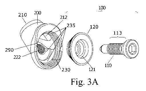
Figures 3A to 14D show embodiments of the first medical
fastening device (1). In particular, the medical fastening
device (1) comprises a first fastening element (100) and a
second fastening element (200). It must be observed that in any
of the described examples, the second fastening element (200)
can be a fastening ferrule.
First fastening device: first fastening element (100)
An embodiment of a first fastening element (100) according
to the first invention can be observed in Figures 3A to 3D. In
this example, the first fastening element is a fastening screw
(110) with a head provided with an Allen-type actuation area and
a threading (113) that is reciprocal with respect to the
threading (223) of the second conduit (220).
Furthermore, the first fastening element (100) comprises a
washer (120) surrounding the head of the screw when in use.
Figures 3A to 3D show a view of the screw (110) and the
washer (120). In particular, the head of the screw is reciprocal
with respect to the shape of the inner surface of the washer
(120). In this embodiment, the screw (110) comprises a circular
step (111), in a plane perpendicular to the shaft of the screw.
In turn, there is provided in a reciprocal manner in the washer
(120) an inner circular step (121) opposite that of the screw.
Advantageously, this example prevents the engagement of the
washer with the screw from seizing up, which allows keeping the
washer aligned with the screw and optimizes the transmission of
force between both.
First fastening device: first embodiment of the second fastening
element (200): Figures 3A to 5B
Figures 3A to 5B show a first embodiment of the first
medical fastening device (1). In particular, the second
fastening element (200) comprises a first through conduit (210)
which is extended along a first longitudinal axis (215). As can
be seen in the section view of Figures 3C and 3D, the second
fastening element (200) further comprises a first end (211) and
a second end (212).
In relation to the first through conduit (210) of the
CA 03122530 2021-06-08
WO 2020/127667 PCT/EP2019/086213
- 24 -
second fastening element (200), Figures 3A and 3B show that said
through conduit (210) further comprises a first end (211) and a
second end (212). In particular, the first through conduit (210)
is configured for housing a graft (10) as will be shown below in
the description of the method of use indicated in Figures 5A to
5B, 14A to 14D. In particular, the first end (211) is intended
for receiving the graft (10), the second end (212) is intended
for being part of the fastening surface of the annular fastening
passage (300) as shown in Figures 5A and 5B.
The configuration of the distal end of the first conduit
(210) of the second fastening element (200) can be circular,
elliptical, rectangular, trilobular, half-annular, or correspond
to any other geometric shape known in the state of the art,
without there necessarily having to be a correspondence between
the configuration of the distal end and of the proximal end.
Furthermore, as can be seen in Figures 3A to 3D, the second
fastening element (200) comprises a second fastening conduit
(220) which is extended along a second longitudinal axis (225).
The second fastening conduit (220) further comprises a first end
(221) and a second end (222).
The second fastening conduit (220) is configured for
housing and fastening the first fastening element (100) as can
be seen in Figures 3A to 5B. To that end, the second fastening
conduit (220) can have several forms on its inner face according
to the form of the type of the first fastening element (100).
For example, in the section view of Figure 3C, the inner face of
the second fastening conduit (220) has the form of threading
complementary to the threads of a screw. In another embodiment,
the inner face of the second conduit (220) is in the form of a
clip or leaf spring complementary to the outer surface of the
first fastening element (100). In another embodiment, the inner
face of the second fastening conduit (220) has a smooth form for
a first self-tapping fastening element (100).
As can be seen in the example of the section view of Figure
3C, the first through conduit (210) and the second fastening
conduit (220) are arranged inside the second fastening element
CA 03122530 2021-06-08
WO 2020/127667 PCT/EP2019/086213
- 25 -
(200) such that the first longitudinal axis (215) and the second
longitudinal axis (225) define an angle (a) with one another
other than 0, preferably an angle between 0 and 90 .
Preferably, the angle (a) is comprised between 30 and 60 . Even
more preferably, in these embodiments the angle (a) can be 45 .
In particular, Figure 3A shows that the second fastening
element (200) comprises an annular proximal surface (230) which
defines a proximal cavity (235) at the second end (212) of the
second fastening element (200). Furthermore, the second end
(212) of the first through conduit (210) and the second end
(222) of the second fastening conduit (220) are joined defining
a junction (290) at the bottom of the proximal cavity (235). The
junction (290), the second end (212) of the first through
conduit (210), and the second end (222) of the second fastening
conduit (220) are part of the bottom of the proximal cavity
(235) of the second end (212) of the second fastening element
(200) extending to the opening of said proximal cavity (235).
Furthermore, it can be seen in Figure 4B that the junction
(290) is movable in this embodiment.
As can be seen in the embodiment of Figure 3D, there is a
lower stop (236) on the annular proximal surface (230) of the
second fastening element (200). Therefore, the annular proximal
surface (230) comprises a lower stop (236). The combination of
the annular proximal surface (230) and stop (236) allows there
to be a position of maximum tightness, in which a sector of the
contour of the first fastening element (100) is supported on the
lower stop (236), which advantageously increases the rigidity of
the fastening provided by the device.
The stop (236) allows the fastening of the graft to a
predetermined thickness, providing rigidity to the fastening.
The annular configuration of the annular proximal surface (230)
guarantees enough space for expansion of the graft (10) from the
starting thickness to this predetermined thickness which
corresponds with the position of maximum tightness of the
fastening device (1).
As can be seen in the embodiment of Figures 3A to 5B, the
CA 03122530 2021-06-08
WO 2020/127667 PCT/EP2019/086213
- 26 -
second fastening element comprises a flange (231) which
advantageously prevents migration of the device (1) into the
bone tunnel.
Finally, Figures 4A to 5B show an embodiment with the first
fastening element (100) housed and fastened in the second
fastening conduit (220) of the first embodiment of the second
fastening element (200). It can be seen how the annular
fastening passage (300) is formed between the inner face of the
annular proximal surface (230) and the outer surface of the
washer (120).
Figures 5A and 5B show views of the upper part as a whole
and the lower part in section, respectively, of the medical
fastening device with at least one graft (10) restrained
therein. In particular, the annular fastening passage (300)
created by the medical fastening device (1) of the present
invention can clearly be seen in Figure 5B. Said annular
fastening passage (300) is annular because the arrangement of
the annular proximal surface (230) generates the continuous and
annular surface of the proximal cavity (235). This allows the
annular proximal surface (230) to be part of the annular
fastening passage (300), which allows fastening several grafts
with a larger fastening passage than in the elements mentioned
of the state of the art. Furthermore, the shape of the annular
fastening passage (300) provides greater structural consistency
to the fastening.
If Figures 5A and 5B are compared with Figures 1A to 1F,
the differences of the medical fastening device (1) of the
present invention with respect to other devices (600, 700, 800)
of the state of the art can clearly be seen. The interference
screw (600) of Figures 1A and 1B performs fastening against the
bone, where it is a very weak fastening due to the porosity of
said bone (900). Furthermore, it can be seen that said
interference screw (600) can damage the ligament, even further
weakening the fastening.
The screw (700) of Figures 1C and 1D is coaxially screwed
into the ferrule housing the graft, directly onto the graft,
CA 03122530 2021-06-08
WO 2020/127667 PCT/EP2019/086213
- 27 -
which, like the interference screw, may damage said graft.
Furthermore, in the case of use in the tibia, the largest part
of the head of the screw is housed inside the tibia bone which,
due to the high porosity of this bone, weakens the resistance to
movement of the device.
The restraining element (800) of Figures 1E to 1F is
introduced into the bone tunnel and restrains the graft against
the internal wall of the tunnel, while at the same time the
screw (802), with the washer, which enters perpendicular to the
cortical bone and is screwed into the angled face of the
restraining element, restrains the graft a second time. This
configuration locates the fastening area around the screw with
the washer, so the screw must necessarily separate and pass
between the bundles of the graft to proceed to the fastening
thereof, which, like in the two prior art examples, may damage
the graft, in addition to causing other problems associated with
these devices that have been described in the background of the
invention.
In contrast, the first medical fastening device (1)
prevents damaging the ligament with the screw (110) due to the
fact that the first conduit (210) and the second conduit (220)
are arranged inside the second fastening element (200) such that
the first longitudinal axis (215) and the second longitudinal
axis (225) define an angle (a) with one another other than 0.
Furthermore, like in the first medical fastening device (1) the
ligament is housed inside same, with the aid of the annular
proximal surface (230), a larger fastening passage (300) is
obtained and fastening against porous bone (900) is prevented,
providing a stronger and more durable fastening.
The configuration of the fastening passage exclusively in
the upper proximal area of the ferrule can be seen in Figure 2E.
If Figure 2E is compared with Figures 5A and 5B, Figure 2E
provides a limited fastening passage which leaves part of the
graft (10) unrestrained, which reduces both the stability of the
fastening and the ultimate tensile strength offered by the
device, unless the device is oversized, which is not an
CA 03122530 2021-06-08
WO 2020/127667 PCT/EP2019/086213
- 28 -
acceptable solution.
First fastening device: second embodiment of the second
fastening element (200): distal appendage (240) configuring an
adjustable loop (245); Figures 6A to 6D
Figures 6A to 6D show a second embodiment of the first
medical fastening device (1) of the invention. As can be seen,
the second embodiment is similar to the first embodiment, with
the exception that the second embodiment further comprises a
distal appendage (240) configuring an adjustable loop (245).
As can be seen in Figure 6A, the distal appendage (240)
comprises a strip, cord, or band, with a first end (241) joined
to the first end of the second fastening element (200) and a
second end (242) with a first fastening-free position. In a
second position, the second end (242) can be configured such
that said end (242) goes back to the second fastening element
(200) configuring an adjustable loop (245) which is fastened in
the desired position by means of the first fastening element
(100), as shown in Figures 6B, 6C, and 6D.
In particular, as shown in Figures 6B to 6D, once the first
fastening element (100) is housed and fastened in the second
fastening conduit (220) of the second fastening element (200),
the free end (242) is also fastened in said fastening passage
(300).
As can be seen in Figure 6C, the dotted line shows an
example of the different positions the adjustable loop (245) can
take; therefore, it can be considered that the distal appendage
(240) is flexible and can define more than one final position in
this embodiment.
The length of the adjustable loop (245) is fastened by
means of the first fastening element (100), the adjustable loop
(245) being intended for the suspension of a bent end of the
graft (10) in the bone tunnel or bone canal. Said fastening is
adjustable from a first position without tension, to a final
position with ideal tension of the suspended graft.
First fastening device: third embodiment of the second fastening
element (200): distal appendage (250) configuring a fastened
CA 03122530 2021-06-08
WO 2020/127667 PCT/EP2019/086213
- 29 -
loop; Figures 7A to 7C
Figures 7A to 7C show a third embodiment of the first
medical fastening device (1) of the invention. As can be seen,
the third embodiment is similar to the first embodiment, with
the exception that the third embodiment further comprises a
distal appendage (250) with hole (253) configuring a non-
adjustable loop. It can be called a fastened or non-adjustable
loop because both ends (251, 252) of the appendage (250) are
fastened to the second fastening element (200), and therefore
the area of suspension, i.e., the size of the hole (253), is
constant. In one embodiment, the distal appendage (250) is
flexible. In the examples of Figures 7A to 7C, the distal
appendage (250) is in the vertical position.
As can be seen in this particular embodiment, the distal
appendage (250) comprises a strip, cord, or band, with both ends
(251, 252) joined to the second fastening element (200), such
that a closed or non-adjustable loop intended for the suspension
of a bent end of the graft in the bone tunnel is configured.
First fastening device: fourth embodiment of the second
fastening element (200): distal appendage (260, 270) with a hole
for suspension; Figures 8A to 9D
Figures 8A to 8C, show a fourth embodiment of the first
medical fastening device (1) of the invention. As can be seen,
the fourth embodiment is similar to the first embodiment, except
that the fourth embodiment further comprises a distal appendage
(260). In these examples, the distal appendage (260) is a half-
ring defining a hole (263), intended for the suspension of a
bent end of the graft in the bone tunnel.
In the particular embodiment of Figures 9A to 9D, the
second fastening element (200) does not comprise a flange (231),
and instead it has a frustoconical external contour, which
advantageously retains the device at the inlet of the bone
tunnel (10), preventing it from penetrating the bone tunnel
(10), with said second fastening element (200) being similar to
that shown in the first embodiment of the first invention in
terms of the remaining aspects.
CA 03122530 2021-06-08
WO 2020/127667 PCT/EP2019/086213
- 30 -
As can be seen in the examples of Figures 9C and 9D, the
distal appendage (270) comprises a first end (271) and a second
end (272) located at the first end of the second fastening
element (200). Furthermore, the distal appendage (270) comprises
a hole (273) intended for the suspension of a bent end of the
graft in the bone tunnel. More particularly, the hole (273) is
located at the first the end (271) of the distal appendage
(270).
In other embodiments, the second fastening element (200) of
Figures 9A to 9D may comprise a flange (231) like the one shown
in the examples of Figures 8A to 8D.
In view of Figures 8A to 9D, it can be considered that the
distal appendage (260, 270) is in the horizontal position.
First fastening device: fifth embodiment of the second fastening
element (200): distal appendage (280) with a hole (281) for
suspension; Figures 10A to 10D
Figures 10A to 10D show a fifth embodiment of the first
medical fastening device (1) of the invention. As can be seen,
the fifth embodiment is similar to the first embodiment, with
the exception that the fifth example further comprises a distal
appendage (280) located at the first end of the second fastening
element (200). In these examples, the distal appendage (280)
comprises a first end (281), a second end (282), and a hole
(283) intended for the suspension of a bent end of the graft in
the bone tunnel. Furthermore, the hole (283) is located at the
first the end (281) of the distal appendage (280).
In view of Figures 10A to 10D, it can be considered that
the distal appendage (280) is in vertical position.
In a particular manner, the second end (282) is housed in
the first through conduit (210) dividing the first through
conduit (210) into two sections. In other words, the first
through conduit (210) is divided into a first section (213) and
a second section (214) by the second end (282) of the distal
appendage (280).
On the other hand, Figures 10A, 10B, and 10D show that the
second end (282) of the distal appendage (280) is part of the
CA 03122530 2021-06-08
WO 2020/127667 PCT/EP2019/086213
- 31 -
bottom of the proximal cavity (235) and of the junction between
the second end (212) of the first through conduit (210) and the
second end (222) of the second fastening conduit (220).
First fastening device: sixth embodiment of the second fastening
element (200): distal appendage (290) with a hole (293) for
suspension; Figures 17A to 17D
Figures 17A to 17D show a sixth embodiment of the first
medical fastening device (1) of the invention. As it can be
seen, the sixth embodiment is similar to the first embodiment,
with the exception that the second end (222) of the second
fastening conduit (220) in the sixth example further comprises
two flanges located opposite on an axis perpendicular to the
second longitudinal axis (225). Another difference with the
first embodiment is that the sixth example comprises a distal
appendage (290) located at the first end of the second fastening
element (200).
In the example, the two flanges (231) are in the form of an
annular retaining lobe configured for abutting with the cortical
bone which demarcates the external upper part of the inlet
opening into the bone tunnel.
Advantageously, the flanges (231) retains the device at the
inlet of the bone tunnel (10), preventing it from penetrating
the bone tunnel (10).
In the example, the distal appendage (290) comprises a hole
(293) intended for the suspension of a bent end of the graft in
the bone tunnel.
First fastening device: system (400) for inserting a medical
fastening device (1): Figures 11A to 11E
Figures 11A to 11E show an embodiment of a system (400) for
inserting a medical fastening device (1) according to any of the
embodiments shown above. The system (400) comprises the medical
fastening device (1) of the present invention, a coupling screw
(440), and an inserter (450) with a first end (410) configured
for engaging the second end of the second fastening element
(200) and a handle (430) located at a second end (420) of the
inserter (450).
CA 03122530 2021-06-08
WO 2020/127667 PCT/EP2019/086213
- 32 -
Advantageously, the use of an inserter (450) with coupling
screw (440) allows handling the device through the handle (430)
and applying the necessary force for the insertion of the device
(1) in the bone tunnel (11). This prevents the device (1) from
uncoupling from the inserter (450), as can be seen in greater
detail in Figures 11A and 11E.
First fastening device: example of use of a medical fastening
device, with an adjustable loop in the femur and rigid vertical
distal appendage in the tibia: Figures 12A to 12D
Figures 12A to 12D show the repair of an anterior cruciate
ligament (ACL) of a right knee (15) using two of the first
medical fastening devices (1) of the invention. The first
medical fastening device (1) is similar to that described in the
example of Figures 6A to 6C and the second medical device (1)
comprises a distal appendage in the vertical position, without a
hole.
Advantageously, this distal appendage without a hole allows
maintaining the twisting of the intra-articular segment of the
graft branches without said twisting reaching the intra-tunnel
segment, which is important for restoring the anatomy and
biomechanics of twisting of the original ACL, as can be seen in
the embodiment of Figures 12A to 12D.
Figure 12A shows the free ends of the grafts (10)
projecting from the bone tunnel (12) made in the tibia. The bent
ends of the grafts (10) are suspended from the adjustable loop
(245) of the first medical fastening device (1).
Advantageously, the adjustable loop (245) allows making a
femoral bone tunnel (11), or a femoral bone canal, and pulling
on the bent end of the graft to fit it in, which further allows
checking and adjusting the tension at which the graft (10) is
fastened.
Figure 12B shows the bents ends of the grafts once they are
fitted in, with the adjustable loop being restrained by the
femoral fastening device. The free ends of the grafts (10) still
project from the bone tunnel (12) made in the tibia.
Figure 12C shows the twisting of the intra-articular
CA 03122530 2021-06-08
WO 2020/127667 PCT/EP2019/086213
- 33 -
segment of the graft (10) which mimics the anatomical twisting
(16) characteristic of the original ACL, i.e., counter-clockwise
direction, in the ACL of the right knee which is shown.
Advantageously, the second medical device (1) with a distal
appendage and without a hole shown in Figure 12C allows keeping
the branches of the graft (10) separated, such that the
anatomical twisting imparted to the intra-articular segment of
the graft does not uncontrollably propagate into the bone
tunnel. As can be seen, an inserter system (400) is used for
handling the device.
Figure 12D shows the fastening device (1) with the first
threaded fastening element (100) once the graft (10) has been
tensed at the tension necessary for recovering the stability in
the knee joint.
First fastening device: example of use of the first medical
fastening device (1) of the invention: 3-strand configuration of
a graft: Figures 13A to 13D
Figure 13A shows a diagram of the joint use of two first
medical devices (1) of the invention, in the repair of the ACL
of the right knee, in which said use comprises:
- providing a graft (10) which is configured in three
branches, with two bent ends and two free ends.
- providing two medical fastening devices (1) with a distal
appendage (250, 260, 270, 280) with a hole, intended for
being introduced in respective first and second bone
tunnels (11, 12). The first fastening element (100) and
second fastening element (200) assembly is depicted as a
rectangle in Figure 13A. The hole (253, 263, 273, 283) is
depicted as a circle in Figure 13A.
- configuring, as shown in Figures 13B to 13D, the two
medical fastening devices (1) such that it allows
fastening a first free end and suspending a first bent end
of the graft (10) by means of the first medical device (1)
inserted in the first bone tunnel (11). On the other hand,
fastening a second free end and suspending a second bent
CA 03122530 2021-06-08
WO 2020/127667 PCT/EP2019/086213
- 34 -
end of the graft (10) by means of the second medical
device (1) inserted in the second bone tunnel.
Figures 13B to 13D show the use of two medical fastening
devices (1) such as those shown in Figures 9A to 9D for the 3-
strand configuration of a single graft, according to the diagram
of Figure 13A.
First fastening device: example of use of the medical fastening
device (1) of the first invention: 4-strand configuration of a
graft: Figures 14A to 14C
Figure 14A shows the use diagram of two first medical
devices (1) of the invention in the repair of the ACL of the
right knee, in which said use comprises:
- providing a graft (10) with a 4-strand configuration in
four branches, with three bent ends and two free ends.
- providing two medical fastening devices (1), a first
device with a vertical distal appendage with a hole (280),
of Figures 10A to 10D, intended for being introduced in a
first bone tunnel (11), and a second device with an
adjustable loop, intended for being introduced in a second
bone tunnel (12). The first fastening element (100) and
second fastening element (200) assembly is depicted as a
rectangle in Figure 14A. The suspension element, i.e.,
hole or adjustable loop, is depicted as a circle in Figure
14A.
- configuring, as shown in Figures 14B and 14C, the two
medical fastening devices (1) such that they allow
fastening both free ends and simultaneously suspending a
first bent end of the graft (10), by means of the first
medical device (1) inserted in the first bone tunnel (11).
On the other hand, by means of the second medical device
(1) inserted in the second bone tunnel (12), suspending,
in an adjustable manner, a second and a third bent end of
the graft (10).
Second fastening device
Figures 15A to 16D show embodiments of the second medical
CA 03122530 2021-06-08
WO 2020/127667 PCT/EP2019/086213
- 35 -
fastening device (500) of the invention. In particular, the
medical fastening device (500) comprises a first fastening
element (510) and a second fastening element (520) comprising a
fastening conduit (523) and a suspension element. In this second
fastening device (500), once the first fastening element (510)
is fastened in the fastening conduit (523) of the second
fastening element (520) and introduced in the bone tunnel, a
fastening passage is produced between the second fastening
element (520) and the bone tunnel which fastens the graft in
said bone tunnel, or between the first fastening element (510)
and the second fastening element (520) which fastens the graft
to the device itself.
Second fastening device: first fastening element (510)
An embodiment of a first fastening element (510) according
to the second fastening device can be observed in Figures 15A to
16D. In the examples of Figures 15A, 15C, and 15D, the first
fastening element (510) is a screw the head of which has the
same diameter as its shank (514). In the example of Figure 15B,
the diameter of the head of the screw is greater than the
diameter of its shank (514). The head of the screw (511) is
provided with an Allen-type actuation area (512) and its shank
(514) comprises a threading.
Second fastening device: first embodiment of the second
fastening element (520): Figures 15A to 15C and 16A to 16D
Figures 15A to 15C and 16A to 16D show a first embodiment
of the second medical fastening device (500) of the invention.
To clarify in the example of Figures 15A to 15C and 16A to 16D,
it has been indicated that the second fastening element (520)
has a first end (521) and a second end (522); furthermore, the
second fastening element (520) comprises a fastening conduit
(523) located at its second end (522) intended for housing the
first fastening element (510).
As can be seen, the second fastening element (520)
comprises a distal appendage (530) configuring a suspension
element, with a first end (531) and a second end (532) connected
to the first end (521) of the second fastening element (520),
CA 03122530 2021-06-08
WO 2020/127667 PCT/EP2019/086213
- 36 -
such that a non-adjustable hole or closed loop (533) intended
for the suspension of a bent end of the graft in the bone tunnel
is configured.
Advantageously, the devices of Figures 15A to 15C and 16A
to 16D allow reproducing the diagrams of Figure 13A, 3-strand
configuration of the graft, and Figure 14A, 4-strand
configuration of the graft, by means of methods of use similar
to those described in the first invention.
Second fastening device: second embodiment of the second
fastening element (520) configuring an adjustable loop; Figure
15D
Figure 15D shows a second embodiment of the second medical
fastening device (500) of the invention. As can be seen, the
second embodiment is similar to the first embodiment, with the
exception that the distal appendage (530) is configured as an
adjustable loop (533).
As can be seen in Figure 15D, in this particular embodiment
the distal appendage (530) comprises a strip, cord, or band,
with a first end (531) connected to the first end (521) of the
second fastening element (520) and a second fastening-free end
(532). The second end (532) can be configured such that it goes
back to the second fastening element (520) configuring an
adjustable loop (533) as shown in Figure 15D.
The length of the adjustable loop is fastened by means of
the first fastening element (510), with the adjustable loop
(533) being intended for the suspension of a bent end of the
graft (10) in the bone tunnel or bone canal. Said fastening is
adjustable from a first position without tension, to a final
position with ideal tension of the suspended graft.
Surgical method: restauration of a damaged anterior cruciate
ligament (ACL) by means of a first and a second fastening
devices (1); Figures 18A-18D.
In figures 18A-18D an example of a surgical method for restoring
a damaged anterior cruciate ligament (ACL) is shown. With this
CA 03122530 2021-06-08
WO 2020/127667 PCT/EP2019/086213
- 37 -
surgical method, the restauration of the ACL is carried out by
means of a first and a second fastening devices (1) with
suspension holes (273).
Advantageously, providing a device (1) with a suspension
hole (273) for each of the bone tunnels - tibial and femoral
bone tunnels - allows suspending a first graft (10) in one of
the bone tunnels and suspending a second graft (10) in the
opposite bone tunnel to fasten, in both cases, the free ends of
each graft (10) with the device (1) placed in the opposite side
where the graft (10) is suspended, as shown in figure 18B, 18C
and 18D.
The surgical method comprises the following steps:
1) performing a femoral bone tunnel, with intra-articular
entrance of the femoral bone tunnel in the insertion site
of the original ACL,
2) performing a tibial bone tunnel, with intra-articular
entrance of the tibial bone tunnel in the insertion site
of the original ACL,
3) suspending, from the suspension hole (273) of the first
fastening device (1), the bent end of a first graft (10),
4) introducing the free ends of the first graft (10) through
the extra-articular entrance of the femoral bone tunnel
until reaching, throughout the intra-articular cavity of
the knee joint, the intra-articular entrance of the tibial
bone tunnel until the free ends of the first graft (10)
protrude from the extra-articular entrance of the tibial
bone tunnel,
5) suspending, from the suspension hole (273) of the second
fastening device (1), the bent end of a second graft (10),
6) introducing the free ends of the second graft (10) through
the extra-articular entrance of the tibial bone tunnel
until reaching, throughout the intra-articular cavity of
the knee joint, the intra-articular entrance of the
femoral bone tunnel until the free ends of the second
CA 03122530 2021-06-08
WO 2020/127667 PCT/EP2019/086213
- 38 -
graft (10) protrude from the extra-articular entrance of
the femoral bone tunnel,
being the free ends of the first and second grafts (1) placed in
the extra-articular tibial and femoral entrances, so that each
of the free ends can be fastened by the device (1) placed in the
opposite side where the graft (10) is suspended, as shown in
figure 18C and 18D.
This configuration, additionally, allows twisting both grafts so
that the intra-articular segment of the grafts is configured
with the anatomical twist that is characteristic of the
biomechanics of the original ACL, as it is shown in figure 18C
and 18D.