Note: Descriptions are shown in the official language in which they were submitted.
CA 03122551 2021-06-08
WO 2020/124052 PCT/US2019/066404
INSTRUMENTS, GUIDES AND RELATED METHODS FOR
TOTAL ANKLE REPLACEMENT
CROSS REFERENCE TO RELATED APPLICATIONS
[0001] This application perfects and claims priority benefit of U.S.
Provisional Patent
Application No. 62/779,092, filed December 13, 2018, and entitled Instruments,
Guides and
Related Methods for Total Ankle Replacement, and U.S. Provisional Patent
Application No.
62/898,854, filed September 11, 2019, and entitled "Distractors Having
Attachable Paddles,
Impaction Devices, And Methods For Use In Total Ankle Replacement," which are
hereby
incorporated herein by reference in their entireties.
TECHNICAL FIELD
[0002] The present disclosure relates generally to general, podiatric, and
orthopaedic
surgery related to joint deformities. More specifically, but not exclusively,
the present
disclosure relates to instruments, guides, systems and related methods for
maintaining,
correcting and/or repairing a joint deformity and/or injury.
BACKGROUND OF THE INVENTION
[0003] Total ankle replacement (TAR), or ankle arthroplasty, is a surgical
procedure to
replace deformed and/or damaged articular surfaces of the human ankle joint
with a
prosthetic joint. TAR is becoming the treatment of choice for patients with a
deformed
and/or injured/damaged ankle joint, replacing the conventional use of
arthrodesis (i.e. fusion
of the ankle bones). One of the main advantages of TAR compared with ankle
arthrodesis is
preservation of functional range of motion (ROM), which is sacrificed in ankle
fusion.
Improved ROM allows patients to better perform activities of daily living and
possibly regain
athletic activities
[0004] Many types of total ankle prostheses have been developed, such as
the cylindric-
type ankle replacement prosthesis, the spherical-type ankle replacement
prosthesis, and the
sliding cylindric-type ankle replacement prosthesis. These and other typical
total ankle
replacement (TAR) prosthesis comprise a tibial prosthesis component, a talus
prosthesis
component, and a tibial bearing or insert component positioned between the
tibial and talus
prosthesis components. In these types of TAR prostheses, the tibial component
is implanted
on/in a tibia, the talus component is implanted on/in a talus, and the tibial
insert is fixed to the
tibial component and articulates with the talus component to form a
replacement ankle joint.
1
CA 03122551 2021-06-08
WO 2020/124052 PCT/US2019/066404
[0005] The proper size and position/orientation/alignment of the tibial
component of a
TAR prosthesis with respect to the distal end of a tibia and the corresponding
ankle joint, the
proper size and position/orientation of the talus component of the TAR
prosthesis with
respect to the proximal end of a talus and the corresponding ankle joint, and
the proper size
and position/orientation/alignment of the tibial insert of the TAR prosthesis
with respect to
the tibial component, the talus component and the corresponding ankle joint,
are all important
to achieving a stable replacement ankle joint and a replacement ankle joint
that provides for
full articulation/motion (e.g., achieving a range of motion of typical
"healthy" ankle joints).
For example, proper sizing and position/orientation of the tibial prosthesis,
the talus
prosthesis and the tibial insert of a TAR prosthesis with respect to an ankle
joint of a
particular patient can prevent overstuffing or understuffing of the
replacement ankle joint
(and thereby provide full articulation/motion) and can ensure proper coverage
of the tibial
prosthesis on the tibial and the talus prosthesis on the talus. As another
example, the
position/orientation/alignment of the tibial prosthesis, the talus prosthesis
and the tibial insert
with respect to the mechanical axis of an ankle joint of a particular patient
(e.g., the
mechanical axis of the tibia) can ensure the mechanical forces of the
replacement ankle joint
are properly distributed and full and properly-oriented range of motion is
achieved.
[0006] Total ankle replacement instrumentation, guides, systems and methods
that
facilitate the selection of a properly sized tibial prosthesis, talus
prosthesis, and tibial insert of
a total ankle replacement implant system for an ankle joint of a particular
patient are thereby
desirable. Further, total ankle replacement instrumentation, guides, systems
and methods that
facilitate implantation of the tibial prosthesis in/on a tibia, and
implantation of the talus
prosthesis in/on a talus, of a total ankle replacement implant system in
proper positions and
orientations (and thereby the proper position and orientation of the
corresponding tibial
insert) for an ankle joint of a particular patient are thereby also desirable.
SUMMARY OF THE INVENTION
[0007] The present disclosure is directed toward instruments, guides,
systems and related
methods for total ankle replacement prostheses. The instruments, guides,
systems and related
methods may facilitate preparation of a tibia and/or talus of a patient for
implantation of a
total ankle prosthesis therein. The instruments, guides, systems and related
methods may also
facilitate selection of a particular size of a tibial component, a talus
component and/or a tibial
insert of the total ankle prosthesis that suits the patient. The instruments,
guides, systems and
related methods include a tibial trial component, a talar/talus trial
component and tibial insert
2
CA 03122551 2021-06-08
WO 2020/124052 PCT/US2019/066404
trial component that replicate one or more aspects of the tibial component,
the talus
component and the tibial insert, respectively, of the total ankle prosthesis.
[0008] In one aspect of the present application, a total ankle replacement
(TAR) trial and
bone preparation guide system comprising a tibial trial and bone preparation
first component
and a talar trial and bone preparation second component is disclosed. The
tibial trial and
bone preparation first component comprises a base portion and an arm portion.
The base
portion comprises a first side with a first tibial engagement surface
configured to engage a
resected distal tibia and at least one bone aperture formation guide through
hole extending
from the first tibial engagement surface to a second tibial insert engagement
side, and the arm
portion extends proximally from an anterior portion of the base portion and is
configured to
engage an anterior side of the resected distal tibia. The talar trial and bone
preparation
second component comprises a first talar engagement surface, a posterior trial
articulation
surface, an anterior window, a posterior cut slot and a plurality of pin
apertures. The first
talar engagement surface is positioned on a distal side of the second
component and is
configured to engage a portion of a resected talus. The posterior trial
articulation surface is
positioned on a proximal side of the second component and is anteriorly-
posteriorly arcuately
convex. The anterior window extends through the second component between the
proximal
side and the distal side thereof. The posterior cut slot extends through the
second component
between the proximal side and the distal side thereof that is angled
posteriorly as in extends
from the proximal side to the distal side. The plurality of pin apertures
extend through the
second component between the proximal side and the distal side.
[0009] In some embodiments, the resected distal tibia and the resected
talus for
implantation therein and therebetween, a TAR prosthesis comprising a tibial
component
comprising a second tibial engagement surface and at least one bone engagement
projection,
a tibial insert configured to removably couple with the tibial component and
comprising a
second tibial articulation surface, and a talar component comprising a second
talar
engagement surface and a talar articulation surface that articulates with the
second tibial
articulation surface.
[0010] In some embodiments, the system further comprises a tibial trial
insert comprising
a distal side with a first talar trial engagement surface that is configured
to engage the
posterior trial articulation surface of the second component, and a proximal
side configured to
removably couple with the second tibial insert engagement side of the first
component. In
some embodiments, the second tibial insert engagement side of the first
component
comprises a coupling slot, and wherein the tibial trial insert comprises a
coupling projection
3
CA 03122551 2021-06-08
WO 2020/124052 PCT/US2019/066404
configured to removably mate within the coupling slot. In some embodiments,
the coupling
slot is a dove-tail shaped slot, and wherein the coupling projection is a dove-
tail shaped
projection configured to mate with the dove-tail shaped slot. In some
embodiments, the
coupling slot and the coupling projection extend and are elongated anteriorly-
posteriorly. In
some embodiments, at least one through hole of the at least one bone aperture
formation
guide through hole is positioned within the coupling slot.
[0011] In some embodiments, the configuration of the first talar trial
engagement surface
corresponds to at least a portion of the second tibial articulation surface.
In some
embodiments, the first talar trial engagement surface is arcuately concave
anteriorly-
posteriorly. In some embodiments, the first talar trial engagement surface
comprises an
anterior rail portion that extends medially-laterally and defines an
engagement surface that is
flat or convex anteriorly-posteriorly, and a posterior rail portion that
extends medially-
laterally and defines an engagement surface that is flat or convex anteriorly-
posteriorly. In
some embodiments, the first talar trial engagement surface further comprises a
strut portion
that extends anteriorly-posteriorly, and wherein the posterior trial
articulation surface of the
second component comprises a strut slot that extends anteriorly-posteriorly
and is configured
to accept the strut portion therein.
[0012] In some embodiments, at least a portion of the first tibial
engagement surface
corresponds in size and shape to at least a portion of the second tibial
engagement surface.
[0013] In some embodiments, the first tibial engagement surface is convex
medially-
laterally.
[0014] In some embodiments, the first side of the base portion of the first
component
includes at least one reference slot extending medially-laterally through at
least a portion of
the first tibial engagement surface. In some embodiments, the at least one
reference slot
comprises at least one of: a center reference slot positioned in the medial-
lateral center of the
base portion and/or corresponding to the medial-lateral center of the tibial
component; a bone
aperture formation reference slot extending through at least a portion of the
at least one bone
aperture formation guide through hole; an anterior reference slot positioned
in an anterior end
portion of the base portion corresponding to an anterior end of the tibial
component; and an
posterior reference slot positioned in a posterior end portion of the base
portion
corresponding to a posterior end of the tibial component.
[0015] In some embodiments, the arm portion of the first component
comprises a
plurality of pin through holes extending therethrough anteriorly-posteriorly.
In some
embodiments, the arm portion of the first component comprises a medial wing
and a lateral
4
CA 03122551 2021-06-08
WO 2020/124052 PCT/US2019/066404
wing, the medial and lateral wings each comprising at least one pin through
hole of the
plurality of pin through holes, and wherein the at least one pin through hole
of the medial and
lateral wings converge medially-laterally as they extend posteriorly. In some
embodiments,
the plurality of pin through holes comprise at least one pair of aligned pin
through holes that
are medially-laterally spaced.
[0016] In some embodiments, the arm portion of the first component
comprises a
positioning mechanism that is configured to engage the anterior side of the
resected distal
tibia and adjust the anterior-posterior position of the base portion of the
first component
relative to the resected distal tibia. In some embodiments, the positioning
mechanism
comprises at least one adjustment screw threadably coupled with the arm
portion.
[0017] In some embodiments, the first talar engagement surface is planar
and is
configured to engage a planar portion of the resected talus. In some
embodiments, the first
talar engagement surface comprises medial and lateral side edges that are
exposed at medial
and lateral sides of the second component. In some embodiments, the distal
side of the
second component further comprises a medially-laterally extending center
reference slot
extending through the first talar engagement surface corresponding to the
medial-lateral
center of the talar component, the medially-laterally extending center
reference slot being
exposed at medial and lateral sides of the second component.
[0018] In some embodiments, the posterior cut slot is exposed at medial and
lateral sides
of the second component at the distal side of the second component. In some
embodiments,
the posterior cut slot defines a posterior end of the medially-laterally
extending center
reference slot.
[0019] In some embodiments, the distal side of the second component further
comprises a
medially-laterally extending anterior reference slot, the medially-laterally
extending anterior
reference slot being exposed at medial and lateral sides of the second
component and
corresponding to the position and orientation of an anterior-posterior pathway
of the posterior
trial articulation surface at the distal side of the second component.
[0020] In some embodiments, the system further comprises at least one
anterior cut guide
configured to engage the proximal side of the second component and extend at
least partially
over the anterior window, the at least one anterior cut guide comprising a
bone cutting guide
through hole configured to mate with at least one cutting implement to form an
anterior
chamfer on the resected talus. In some embodiments, the distal side of the
second component
further comprises a medially-laterally extending anterior cut reference slot,
the medially-
laterally extending anterior cut reference slot being exposed at medial and
lateral sides of the
CA 03122551 2021-06-08
WO 2020/124052 PCT/US2019/066404
second component and corresponding to the position and orientation of the
anterior chamfer
on the resected talus.
[0021] In some embodiments, the posterior trial articulation surface
corresponds in size
and shape to at least a portion of the talar articulation surface of the talar
component. In
some embodiments, the posterior cut slot is positioned anteriorly-posteriorly
between at least
a portion of the posterior trial articulation surface and the anterior window.
In some
embodiments, the posterior cut slot is configured to accept a cutting blade
therethrough to
form a posterior chamfer on the resected talus.
[0022] In some embodiments, the second component further comprises a
plurality of pin
through holes extending therethrough between the proximal and distal sides
thereof In some
embodiments, the plurality of pin through holes of the second component
comprise at least
one pair of through holes that are medially-laterally spaced and converge
medially-laterally
as they extend from the proximal side to the and distal side of the second
component. In
some embodiments, the plurality of pin through holes of the second component
comprise at
least one pair of aligned pin through holes that are medially-laterally
spaced.
[0023] In some embodiments, the second component further comprises a handle
that
forms an anterior end of the second component. In some embodiments, the second
component further comprises a socket that forms an anterior end of the second
component.
In some embodiments, the system further comprises a distractor, and wherein
the socket is
configured to couple with a first arm of the distractor. In some embodiments,
the distractor
further comprises a second arm with a paddle coupled thereto, and wherein the
paddle is
configured to engage with the second tibial insert engagement side of the base
portion of the
first component.
[0024] In some embodiments, the system further comprises a chamfer checker
instrument, the chamfer checker instrument comprising at least one third talar
engagement
surface configured to engage the resected talus, a fourth talar engagement
surface extending
from the at least one third talar engagement surface configured to engage an
anterior chamfer
of the resected talus formed via the anterior window of the second component,
and a fifth
talar engagement surface extending from the at least one third talar
engagement surface
configured to engage a posterior chamfer of the resected talus formed via the
cut slot of the
second component. In some embodiments, the at least one third talar engagement
surface
comprises a medially-laterally extending center reference slot corresponding
to the medial-
lateral center of the talar component, the center reference slot of the at
least one third talar
engagement surface being exposed at medial and lateral sides of the chamfer
checker. In
6
CA 03122551 2021-06-08
WO 2020/124052 PCT/US2019/066404
some embodiments, the chamfer checker further comprises first and second
medially-laterally
extending through hole positioned proximally of the at least one third talar
engagement
surface, the first medially-laterally extending through hole being tangent to
a reference line
extending along the fourth talar engagement surface and the second medially-
laterally
extending through hole being tangent to a reference line extending along the
fifth talar
engagement surface.
[0025] In some embodiments, the system further comprises a talar chamfer
trial
comprising: a fifth talar engagement surface on a distal side of the talar
chamfer trial
configured to engage the resected talus; a second posterior trial articulation
surface on a
proximal side of the talar chamfer trial that comprises an anteriorly-
posteriorly and medially-
laterally arcuately convex portion; at least one bone aperture formation guide
through hole;
and a plurality of pin through holes extending between the proximal and distal
sides thereof
In some embodiments, at least a portion of the second posterior trial
articulation surface
corresponds in size and shape to at least a portion of the second tibial
engagement surface. In
some embodiments, the fifth talar engagement surface comprises a first planar
surface for
engaging a planar surface of the resected talus, a second planar surface
extending anteriorly
from the first planar surface on a distal angle configured to engage an
anterior chamfer
surface of the resected talus formed via the anterior window of the second
component, and a
third planar surface extending posteriorly from the first planar surface on a
distal angle
configured to engage a posterior chamfer of the resected talus formed via the
cut slot of the
second component. In some embodiments, the at least one bone aperture
formation guide
through hole of the talar chamfer trial comprises an elongated slot at an
anterior end portion
of the talar chamfer trial that extends through the second planar surface. In
some
embodiments, the system further comprises a bone aperture formation guide
configured to
couple to the anterior end portion of the talar chamfer trial, the bone
aperture formation guide
comprising a second elongated slot that extends over the elongated slot when
coupled to the
anterior end portion, the second elongated slot configured to accept a bone
cutting instrument
therethrough to form an elongated aperture in the anterior chamfer surface of
the resected
talus formed via the anterior window of the second component. In some
embodiments, the at
least one bone aperture formation guide through hole comprises a pair of
medially-laterally
spaced drill guide through holes that extend through the second posterior
trial articulation
surface to the distal side of the talar chamfer trial.
[0026] In another aspect of the present application, a method comprising
trialing a total
ankle replacement (TAR) prosthesis, preparing a resected tibia of the ankle
joint, and
7
CA 03122551 2021-06-08
WO 2020/124052 PCT/US2019/066404
preparing the resected talus of the ankle joint. The trialing the TAR
prosthesis in the ankle
joint comprises trialing a TAR prosthesis that includes a tibial component, a
tibial insert with
a tibial articulation surface, and a talar component with a talar articulation
surface via a total
ankle replacement (TAR) guide system. The TAR guide system comprises a tibial
trial and
bone preparation first component and a talar trial and bone preparation second
component is
disclosed. The tibial trial and bone preparation first component comprises a
base portion and
an arm portion. The base portion comprises a first side with a first tibial
engagement surface
configured to engage a resected distal tibia and at least one bone aperture
formation guide
through hole extending from the first tibial engagement surface to a second
tibial insert
engagement side, and the arm portion extends proximally from an anterior
portion of the base
portion and is configured to engage an anterior side of the resected distal
tibia. The talar trial
and bone preparation second component comprises a first talar engagement
surface, a
posterior trial articulation surface, an anterior window, a posterior cut slot
and a plurality of
pin apertures. The first talar engagement surface is positioned on a distal
side of the second
component and is configured to engage a portion of a resected talus. The
posterior trial
articulation surface is positioned on a proximal side of the second component
and is
anteriorly-posteriorly arcuately convex. The anterior window extends through
the second
component between the proximal side and the distal side thereof The posterior
cut slot
extends through the second component between the proximal side and the distal
side thereof
that is angled posteriorly as in extends from the proximal side to the distal
side. The plurality
of pin apertures extend through the second component between the proximal side
and the
distal side. The preparing the resected tibia of the ankle joint comprises
preparing the
resected tibia of the ankle joint for the implantation of at least one
projection of an
engagement side of the tibial component therein, comprising coupling the arm
portion of the
first component to the anterior side of the resected distal tibia with the
first tibial engagement
surface of the first component engaged with the resected distal tibia, and
passing at least one
projection of a bone aperture formation instrument though the at least one
bone aperture
formation guide through hole of the base portion of the first component and
into the resected
distal tibia to form at least one aperture in the resected distal tibia that
is configured to accept
the at least one projection of the tibial component therein. The preparing the
resected talus of
the ankle joint comprises preparing the resected talus of the ankle joint for
coupling with an
engagement side of the talar component, comprising: coupling the second
component to the
resected distal talus such that the first talar engagement surface of the
second component
engage a surface portion of the resected talus; passing a bone cutting
instrument through the
8
CA 03122551 2021-06-08
WO 2020/124052 PCT/US2019/066404
anterior window of the second component to form an anterior chamfer surface on
the resected
talus; and passing a bone cutting blade through the posterior cut slot the
second component to
form a posterior chamfer surface on the resected talus.
In some embodiments, the system further comprises a talar chamfer trial
comprising: a
second talar engagement surface on a distal side of the talar chamfer trial
configured to
engage the surface portion, the anterior chamfer and the posterior chamfer of
the resected
talus; a second posterior trial articulation surface on a proximal side of the
talar chamfer trial
that comprises an anteriorly-posteriorly and medially-laterally arcuately
convex portion; at
least one bone aperture formation guide through hole; and a plurality of pin
through holes
extending between the proximal and distal sides thereof In some embodiments,
the method
further comprises preparing the resected talus of the ankle joint for the
implantation of at
least one projection of the engagement side of the talar component therein,
comprising:
coupling the second talar engagement surface in engagement with the surface
portion, the
anterior chamfer and the posterior chamfer of the resected talus by passing a
plurality of pin
through the plurality of pin through holes and into the resected talus; and
passing at least one
projection of a bone aperture formation instrument though the at least one
bone aperture
formation guide through hole of the talar chamfer trial and into the resected
talus to form at
least one aperture in the resected talus that is configured to accept the at
least one projection
of the talar component therein.
[0027] In another aspect of the present application, a total ankle
replacement (TAR)
guide system for a TAR prosthesis including a tibial component, a tibial
insert with a tibial
articulation surface, and a talar component with a talar articulation surface
and a talar
engagement side opposing the articulation surface is disclosed. The TAR guide
system
comprises a tibial trial component, a tibial trial insert, and a talar trial
component. The tibial
trial component comprises a base portion and an arm portion extending
proximally from an
anterior portion of the base portion. The tibial trial insert comprises an
anterior rail portion
that extends in a medial-lateral direction and defines an engagement surface
that is flat or
convex in the anterior-posterior direction, a posterior rail portion that
extends in a medial-
lateral direction and defines an engagement surface that is flat or convex in
the anterior-
posterior direction, and a strut portion that extends in an anterior-posterior
direction. The
anterior and posterior rail portions approximate anterior and posterior
portions of the tibial
articulation surface of the tibial insert. The talar trial component comprises
a trial articulation
surface that approximates the talar articulation surface of the talar
component, a posterior cut
9
CA 03122551 2021-06-08
WO 2020/124052 PCT/US2019/066404
slot extending through the articulation surface in the medial-lateral
direction, a strut slot
extending through the articulation surface in the anterior-posterior
direction, an anterior
window positioned anterior to the posterior cut slot. The tibial trial insert
and the tibial trial
are coupled together. The anterior and posterior rail portions are engaged
with the trial
articulation surface, and the strut portion is positioned within the strut
slot.
[0028] In some embodiments, the talar trial component further comprises a
pair of
apertures extending therethrough that converge or diverge.
[0029] In another aspect of the present application a method is disclosed.
The method
comprises trialing a total ankle replacement (TAR) prosthesis in an ankle
joint that includes a
tibial component, a tibial insert with a tibial articulation surface, and a
talar component with a
talar articulation surface via a total ankle replacement (TAR) guide system.
The TAR guide
system comprises a tibial trial component, a tibial trial insert, and a talar
trial component.
The tibial trial component comprises a base portion and an arm portion
extending proximally
from an anterior portion of the base portion. The tibial trial insert
comprises an anterior rail
portion that extends in a medial-lateral direction and defines an engagement
surface that is
flat or convex in the anterior-posterior direction, a posterior rail portion
that extends in a
medial-lateral direction and defines an engagement surface that is flat or
convex in the
anterior-posterior direction, and a strut portion that extends in an anterior-
posterior direction.
The anterior and posterior rail portions approximate anterior and posterior
portions of the
tibial articulation surface of the tibial insert. The talar trial component
comprises a trial
articulation surface that approximates the talar articulation surface of the
talar component, a
posterior cut slot extending through the articulation surface in the medial-
lateral direction, a
strut slot extending through the articulation surface in the anterior-
posterior direction, an
anterior window positioned anterior to the posterior cut slot. The tibial
trial insert and the
tibial trial are coupled together. The anterior and posterior rail portions
are engaged with the
trial articulation surface, and the strut portion is positioned within the
strut slot. The method
further comprises preparing a talus bone of the ankle joint for the
implantation of the talar
component therein by resecting a posterior portion of the talus bone via a cut
slot of the talar
CA 03122551 2021-06-08
WO 2020/124052 PCT/US2019/066404
trial component, and by resecting an anterior portion of the talus bone via an
anterior window
of the talar trial component.
[0030] In some embodiments, the talar trial component further comprises a
pair of
apertures extending therethrough that converge or diverge.
[0031] These and other objects, features and advantages of this disclosure
will become
apparent from the following detailed description of the various aspects of the
disclosure taken
in conjunction with the accompanying drawings.
BRIEF DESCRIPTION OF THE DRAWINGS
[0032] The accompanying drawings, which are incorporated in and constitute
a part of
the specification, illustrate embodiments of the disclosure and together with
the detailed
description herein, serve to explain the principles of the disclosure. It is
emphasized that, in
accordance with the standard practice in the industry, various features are
not drawn to scale.
In fact, the dimensions of the various features may be arbitrarily increased
or reduced for
clarity of discussion. The drawings are only for purposes of illustrating
preferred
embodiments and are not to be construed as limiting the disclosure.
[0033] FIG. 1A illustrates a side view of an exemplary embodiment of a
total ankle
replacement (TAR) prosthesis formed of tibial component implanted on the
tibia, a talar
component implanted on the talus, and a tibial insert coupled to the tibial
component and
articulating on the talar component, in accordance with an aspect of the
present disclosure;
[0034] FIG. 1B illustrates a perspective view of the exemplary TAR
prosthesis of FIG.
1A, in accordance with an aspect of the present disclosure;
[0035] FIG. 2 illustrates an elevational perspective view of a total ankle
replacement
(TAR) trial and guide system including a tibial trial guide, a tibial trial
insert and a talar trial
guide for facilitating selection of a TAR prosthesis and preparation of a
tibia and talus
therefore, in accordance with an aspect of the present disclosure;
[0036] FIG. 3 illustrates a bottom perspective view of the TAR trial and
guide system of
FIG. 2, in accordance with an aspect of the present disclosure;
[0037] FIG. 4 illustrates a medial/lateral side view of the TAR trial and
guide system of
FIG. 2, in accordance with an aspect of the present disclosure;
[0038] FIG. 5 illustrates an anterior view of the TAR trial and guide
system of FIG. 2, in
accordance with an aspect of the present disclosure;
[0039] FIG. 6 illustrates a posterior view of the TAR trial and guide
system of FIG. 2, in
accordance with an aspect of the present disclosure;
11
CA 03122551 2021-06-08
WO 2020/124052 PCT/US2019/066404
[0040] FIG. 7 illustrates a proximal view of the TAR trial and guide system
of FIG. 2, in
accordance with an aspect of the present disclosure;
[0041] FIG. 8 illustrates a distal view of the TAR trial and guide system
of FIG. 2, in
accordance with an aspect of the present disclosure;
[0042] FIG. 9 illustrates an elevational perspective view of the tibial
trial guide of the
TAR trial and guide system of FIG. 2, in accordance with an aspect of the
present disclosure;
[0043] FIG. 10 illustrates another elevational perspective view of the
tibial trial guide of
FIG. 9, in accordance with an aspect of the present disclosure;
[0044] FIG. 11 illustrates a bottom perspective view of the tibial trial
guide of FIG. 9, in
accordance with an aspect of the present disclosure;
[0045] FIG. 12 illustrates a bottom perspective view of the tibial trial
guide of FIG. 9, in
accordance with an aspect of the present disclosure;
[0046] FIG. 13 illustrates a bottom view of the tibial trial guide of FIG.
9, in accordance
with an aspect of the present disclosure;
[0047] FIG. 14 illustrates a medial side view of the tibial trial guide of
FIG. 9, in
accordance with an aspect of the present disclosure;
[0048] FIG. 15 illustrates a lateral side view of the tibial trial guide of
FIG. 9, in
accordance with an aspect of the present disclosure;
[0049] FIG. 16 illustrates a posterior perspective view of the tibial trial
guide of FIG. 9,
in accordance with an aspect of the present disclosure;
[0050] FIG. 17 illustrates an anterior view of the tibial trial guide of
FIG. 9, in
accordance with an aspect of the present disclosure;
[0051] FIG. 18 illustrates a posterior view of the tibial trial guide of
FIG. 9, in
accordance with an aspect of the present disclosure;
[0052] FIG. 19 illustrates an elevational anterior perspective view of the
tibial trial insert
of the TAR trial and guide system of FIG. 2, in accordance with an aspect of
the present
disclosure;
[0053] FIG. 20 illustrates an elevational posterior perspective view of the
tibial trial
insert of FIG. 19, in accordance with an aspect of the present disclosure;
[0054] FIG. 21 illustrates a bottom anterior perspective view of the tibial
trial insert of
FIG. 19, in accordance with an aspect of the present disclosure;
[0055] FIG. 22 illustrates a bottom posterior perspective view of the
tibial trial insert of
FIG. 19, in accordance with an aspect of the present disclosure;
12
CA 03122551 2021-06-08
WO 2020/124052 PCT/US2019/066404
[0056] FIG. 23 illustrates a proximal view of the tibial trial insert of
FIG. 19, in
accordance with an aspect of the present disclosure;
[0057] FIG. 24 illustrates a distal view of the tibial trial insert of FIG.
19, in accordance
with an aspect of the present disclosure;
[0058] FIG. 25 illustrates a medial side view of the tibial trial insert of
FIG. 19, in
accordance with an aspect of the present disclosure;
[0059] FIG. 26 illustrates a lateral side view of the tibial trial insert
of FIG. 19, in
accordance with an aspect of the present disclosure;
[0060] FIG. 27 illustrates an anterior view of the tibial trial insert of
FIG. 19, in
accordance with an aspect of the present disclosure;
[0061] FIG. 28 illustrates a posterior side view of the tibial trial insert
of FIG. 19, in
accordance with an aspect of the present disclosure;
[0062] FIG. 29 illustrates an elevational anterior perspective view of the
talar trial guide
of the TAR trial and guide system of FIG. 2, in accordance with an aspect of
the present
disclosure;
[0063] FIG. 30 illustrates an elevational posterior perspective view of the
talar trial guide
of FIG. 29, in accordance with an aspect of the present disclosure;
[0064] FIG. 31 illustrates a lateral side view of the talar trial guide of
FIG. 29, in
accordance with an aspect of the present disclosure;
[0065] FIG. 32 illustrates an anterior view of the talar trial guide of
FIG. 29, in
accordance with an aspect of the present disclosure;
[0066] FIG. 33 illustrates a medial side view of the talar trial guide of
FIG. 29, in
accordance with an aspect of the present disclosure;
[0067] FIG. 34 illustrates a posterior view of the talar trial guide of
FIG. 29, in
accordance with an aspect of the present disclosure;
[0068] FIG. 35 illustrates a distal view of the talar trial guide of FIG.
29, in accordance
with an aspect of the present disclosure;
[0069] FIG. 36 illustrates a proximal view of the talar trial guide of FIG.
29, in
accordance with an aspect of the present disclosure;
[0070] FIG. 37 illustrates a posterior elevational perspective view of the
talus component
of the TAR prosthesis of FIGS. 1A and 2B, in accordance with an aspect of the
present
disclosure;
[0071] FIG. 38 illustrates an anterior bottom perspective view of the talus
component of
FIG. 37, in accordance with an aspect of the present disclosure;
13
CA 03122551 2021-06-08
WO 2020/124052 PCT/US2019/066404
[0072] FIG. 39 illustrates a lateral side view of the talus component of
FIG. 37, in
accordance with an aspect of the present disclosure;
[0073] FIG. 40 illustrates a proximal view of the talus component of FIG.
37, in
accordance with an aspect of the present disclosure;
[0074] FIG. 41 illustrates a distal view of the talus component of FIG. 37,
in accordance
with an aspect of the present disclosure;
[0075] FIG. 42 illustrates a posterior elevational perspective view of the
talus component
of FIG. 37 overlaid with the talar trial guide of FIG. 29, in accordance with
an aspect of the
present disclosure;
[0076] FIG. 43 illustrates a lateral side view of the talus component of
FIG. 37 overlaid
with the talar trial guide of FIG. 29, in accordance with an aspect of the
present disclosure;
[0077] FIG. 44 illustrates an elevational posterior perspective view of the
TAR prosthesis
of FIGS. 1A and 2B forming an ankle joint between a tibia and talus, in
accordance with an
aspect of the present disclosure;
[0078] FIG. 45 illustrates a lateral side view of the TAR prosthesis of
FIGS. 1A and 2B
forming an ankle joint between a tibia and talus, in accordance with an aspect
of the present
disclosure;
[0079] FIG. 46 illustrates an anterior view of the tibial trial guide of
FIG. 9 positioned on
a resected distal tibia, in accordance with an aspect of the present
disclosure;
[0080] FIG. 47 illustrates an anterior elevational perspective view of the
talar trial guide
of FIG. 29 positioned on a talus, in accordance with an aspect of the present
disclosure;
[0081] FIG. 48 illustrates an anterior elevational perspective view of the
talar trial guide
of FIG. 29 and the tibial trial insert of FIG. 19 positioned on a talus, in
accordance with an
aspect of the present disclosure;
[0082] FIG. 49 illustrates a medial perspective exploded view of the talar
trial guide of
FIG. 29, the tibial trial insert of FIG. 19 and the talus component of FIG.
37, in accordance
with an aspect of the present disclosure;
[0083] FIG. 50 illustrates a medial perspective view of the talar trial
guide of FIG. 29
engaged with the tibial trial insert of FIG. 19, and the talus component of
FIG. 37 overlaid on
the tibial trial insert of FIG. 19, in accordance with an aspect of the
present disclosure;
[0084] FIG. 51 illustrates a posterior elevational perspective view of the
talar trial guide
of FIG. 29 engaged with the tibial trial insert of FIG. 19, in accordance with
an aspect of the
present disclosure;
14
CA 03122551 2021-06-08
WO 2020/124052 PCT/US2019/066404
[0085] FIG. 52 illustrates an anterior elevational perspective view of the
talar trial guide
of FIG. 29 engaged with the tibial trial insert of FIG. 19, in accordance with
an aspect of the
present disclosure;
[0086] FIG. 53 illustrates a medial side view of the talar trial guide of
FIG. 29 engaged
with the tibial trial insert of FIG. 19, in accordance with an aspect of the
present disclosure;
[0087] FIG. 54 illustrates a distal view of the tibial trial insert of FIG.
19, in accordance
with an aspect of the present disclosure;
[0088] FIG. 55 illustrates a series of cross-sectional views of the talar
trial guide of FIG.
29 engaged with the tibial trial insert of FIG. 19 along a range of motion
therebetween, in
accordance with an aspect of the present disclosure;
[0089] FIG. 56 illustrates a series of additional cross-sectional views of
the talar trial
guide of FIG. 29 engaged with the tibial trial insert of FIG. 19 along a range
of motion
therebetween, in accordance with an aspect of the present disclosure;
[0090] FIG. 57 illustrates an anterior elevation perspective view of the
tibial trial guide
of FIG. 9 engaged with a plurality of pin members and the tibial trial insert
of FIG. 19, in
accordance with an aspect of the present disclosure;
[0091] FIG. 58 illustrates a medial side view of the tibial trial guide of
FIG. 9 engaged
with a plurality of pin members and the tibial trial insert of FIG. 19, in
accordance with an
aspect of the present disclosure;
[0092] FIG. 59 illustrates an anterior view of the tibial trial guide of
FIG. 9 engaged with
a plurality of pin members and the tibial trial insert of FIG. 19, in
accordance with an aspect
of the present disclosure;
[0093] FIG. 60 illustrates an anterior elevation perspective view of the
tibial trial guide
of FIG. 9 engaged with the tibial trial insert of FIG. 19, and the talar trial
guide of FIG. 29
engaged with a plurality of pin members and the tibial trial insert of FIG.
19, in accordance
with an aspect of the present disclosure;
[0094] FIG. 61 illustrates an anterior elevation view of the talar trial
guide of FIG. 29
engaged with a plurality of pin members, in accordance with an aspect of the
present
disclosure;
[0095] FIG. 62 illustrates a medial elevation perspective view of the talar
trial guide of
FIG. 29 engaged with a plurality of pin members and a bone removal guide, in
accordance
with an aspect of the present disclosure;
CA 03122551 2021-06-08
WO 2020/124052 PCT/US2019/066404
[0096] FIG. 63 illustrates a proximal view of the talar trial guide of FIG.
29 engaged
with the plurality of pin members and the bone removal guide of FIG. 62, in
accordance with
an aspect of the present disclosure;
[0097] FIG. 64 illustrates a medial view of the talar trial guide of FIG.
29 engaged with
the plurality of pin members and the bone removal guide of FIG. 62, in
accordance with an
aspect of the present disclosure;
[0098] FIG. 65 illustrates a posterior view of the talar trial guide of
FIG. 29 engaged with
the plurality of pin members and the bone removal guide of FIG. 62, in
accordance with an
aspect of the present disclosure;
[0099] FIG. 66 illustrates a medial view of the talar trial guide of FIG.
29 engaged with
the plurality of pin members and the bone removal guide of FIG. 62, and a bone
aperture
formation instrument engaged with the bone removal guide of FIG. 62, in
accordance with an
aspect of the present disclosure;
[0100] FIG. 67 illustrates a medial view of the talar trial guide of FIG.
29 engaged with a
bone cutting blade, a plurality of pin members and a bone cutting blade
support member, and
the tibial trial insert of FIG. 19 overlaid on the talar trial guide of FIG.
29, in accordance with
an aspect of the present disclosure;
[0101] FIG. 68 illustrates a medial elevational perspective view of the
talar trial guide of
FIG. 29 engaged with a bone cutting blade, a plurality of pin members and a
bone cutting
blade support member, and the tibial trial insert of FIG. 19 overlaid on the
talar trial guide of
FIG. 29, in accordance with an aspect of the present disclosure;
[0102] FIG. 69 illustrates an anterior view of the talar trial guide of
FIG. 29 engaged
with a bone cutting blade, a plurality of pin members and a bone cutting blade
support
member, and the tibial trial insert of FIG. 19 overlaid on the talar trial
guide of FIG. 29, in
accordance with an aspect of the present disclosure;
[0103] FIG. 70 illustrates a medial elevation perspective view of the talar
trial guide of
FIG. 29 positioned on a resected talus, in accordance with an aspect of the
present disclosure;
[0104] FIG. 71 illustrates a medial side view of the talar trial guide and
the resected talus
of FIG. 71, in accordance with an aspect of the present disclosure;
[0105] FIG. 72 illustrates a medial elevation perspective view of the talar
trial guide of
FIG. 29 positioned on a resected and chamfered talus, in accordance with an
aspect of the
present disclosure;
[0106] FIG. 73 illustrates a medial side view of the talar trial guide and
the resected and
chamfered talus of FIG. 72, in accordance with an aspect of the present
disclosure;
16
CA 03122551 2021-06-08
WO 2020/124052 PCT/US2019/066404
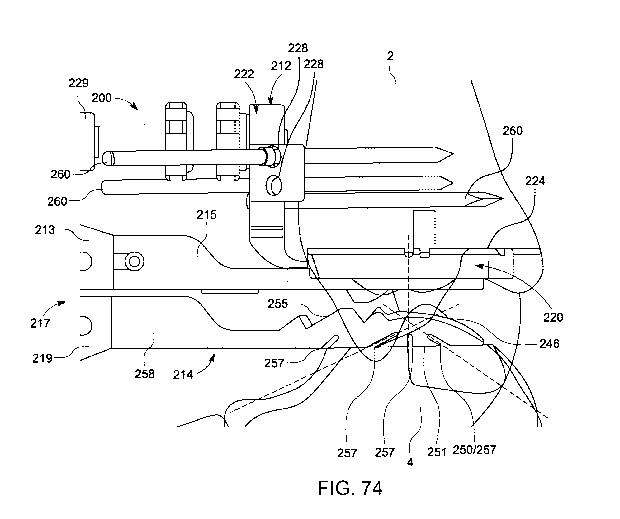
[0107] FIG. 74 illustrates a medial side view of another TAR trial and
guide system
including a tibial trial guide, a first talar trial guide and a distractor for
facilitating selection of
a TAR prosthesis and preparation of a tibia and talus therefore, in accordance
with an aspect
of the present disclosure;
[0108] FIG. 75 illustrates an anterior view of the TAR trial and guide
system of FIG. 74,
in accordance with an aspect of the present disclosure;
[0109] FIG. 76 illustrates an elevational anterior perspective view of the
tibial trial guide
and the distractor of the TAR trial and guide system of FIG. 74, in accordance
with an aspect
of the present disclosure;
[0110] FIG. 77 illustrates a distal medial perspective view of the tibial
trial guide and the
distractor of FIG. 76, in accordance with an aspect of the present disclosure;
[0111] FIG. 78 illustrates an elevational posterior perspective view of the
tibial trial
guide of the TAR trial and guide system of FIG. 74, in accordance with an
aspect of the
present disclosure;
[0112] FIG. 79 illustrates a distal posterior perspective view of the
tibial trial guide of
FIG. 78, in accordance with an aspect of the present disclosure;
[0113] FIG. 80 illustrates a proximal view of the tibial trial guide of
FIG. 78, in
accordance with an aspect of the present disclosure;
[0114] FIG. 81 illustrates a distal perspective view of the tibial trial
guide of FIG. 78, in
accordance with an aspect of the present disclosure;
[0115] FIG. 82 illustrates a medial view of the tibial trial guide of FIG.
78, in accordance
with an aspect of the present disclosure;
[0116] FIG. 83 illustrates an anterior view of the tibial trial guide of
FIG. 78, in
accordance with an aspect of the present disclosure;
[0117] FIG. 84 illustrates a posterior view of the tibial trial guide of
FIG. 78, in
accordance with an aspect of the present disclosure;
[0118] FIG. 85 illustrates a distal elevational perspective view of the
first talar trial guide
and the distractor of FIG. 76, in accordance with an aspect of the present
disclosure;
[0119] FIG. 86 illustrates a distal medial perspective view of the tibial
trial guide and the
distractor of FIG. 85, in accordance with an aspect of the present disclosure;
[0120] FIG. 87 illustrates an elevational posterior perspective view of the
first talar trial
guide of the TAR trial and guide system of FIG. 74, in accordance with an
aspect of the
present disclosure;
17
CA 03122551 2021-06-08
WO 2020/124052 PCT/US2019/066404
[0121] FIG. 88 illustrates a distal posterior perspective view of the first
talar trial guide
of FIG. 87, in accordance with an aspect of the present disclosure;
[0122] FIG. 89 illustrates a proximal view of the first talar trial guide
of FIG. 87, in
accordance with an aspect of the present disclosure;
[0123] FIG. 90 illustrates a distal view of the first talar trial guide of
FIG. 87, in
accordance with an aspect of the present disclosure;
[0124] FIG. 91 illustrates a medial side view of the first talar trial
guide of FIG. 87, in
accordance with an aspect of the present disclosure;
[0125] FIG. 92 illustrates an anterior view of the first talar trial guide
of FIG. 87, in
accordance with an aspect of the present disclosure;
[0126] FIG. 93 illustrates posterior view of the first talar trial guide of
FIG. 87, in
accordance with an aspect of the present disclosure;
[0127] FIG. 94 illustrates a medial side view of the first talar trial
guide and the distractor
of FIG. 76 on a resected talus, and a bone cutting blade cutting a posterior
chamfer on the
resected talus via the first talar trial guide, in accordance with an aspect
of the present
disclosure;
[0128] FIG. 95 illustrates an elevational anterior perspective view of the
first talar trial
guide of the TAR trial and guide system of FIG. 74 and a bone cutting blade
cutting
positioned within a cut slot thereof;
[0129] FIG. 96 illustrates an elevational anterior view of the first talar
trial guide of the
TAR trial and guide system of FIG. 74 on a resected talus, and a bone removal
guide and
bone cutting instrument cutting an anterior chamfer on the resected talus via
the first talar
trial guide, in accordance with an aspect of the present disclosure;
[0130] FIG. 97 illustrates an elevational anterior perspective view of the
first talar trial
guide of the TAR trial and guide system of FIG. 74 and a bone removal guide
and bone
cutting instrument engaged therewith, in accordance with an aspect of the
present disclosure;
[0131] FIG. 98 illustrates a medial side view of the first talar trial
guide, the bone
removal guide and the bone cutting instrument of FIG. 97, in accordance with
an aspect of
the present disclosure;
[0132] FIG. 99 illustrates an elevational anterior view of the first talar
trial guide of the
TAR trial and guide system of FIG. 74 on a resected talus, and another bone
removal guide
and bone cutting instrument further cutting an anterior chamfer on the
resected talus via the
first talar trial guide, in accordance with an aspect of the present
disclosure;
18
CA 03122551 2021-06-08
WO 2020/124052 PCT/US2019/066404
[0133] FIG. 100 illustrates the tibial trial guide of the TAR trial and
guide system of FIG.
74 engaged with a resected distal tibia and a chamfer checker instrument of
the TAR trial and
guide system engaged with a resected and chamfered talus, in accordance with
an aspect of
the present disclosure;
[0134] FIG. 101 illustrates an elevational anterior perspective view of the
chamfer
checker instrument of FIG. 100, in accordance with an aspect of the present
disclosure;
[0135] FIG. 102 illustrates an distal posterior perspective view of the
chamfer checker
instrument of FIG. 100, in accordance with an aspect of the present
disclosure;
[0136] FIG. 103 illustrates a medial side view of the chamfer checker
instrument of FIG.
100, in accordance with an aspect of the present disclosure;
[0137] FIG. 104 illustrates an elevational anterior perspective view of
another chamfer
checker instrument for a TAR trial and guide system, in accordance with an
aspect of the
present disclosure;
[0138] FIG. 105 illustrates a distal posterior perspective view of the
chamfer checker
instrument of FIG. 104, in accordance with an aspect of the present
disclosure;
[0139] FIG. 106 illustrates a medial side view of the chamfer checker
instrument of FIG.
104, in accordance with an aspect of the present disclosure;
[0140] FIG. 107 illustrates a distal medial side perspective view of the
tibial trial guide
of FIG. 78 engaged with a tibial trial insert and a second talar trial guide
of the TAR trial and
guide system, in accordance with an aspect of the present disclosure;
[0141] FIG. 108 illustrates a medial side view of the tibial trial guide,
the tibial trial
insert and the second talar trial guide of FIG. 107, in accordance with an
aspect of the present
disclosure;
[0142] FIG. 109 illustrates an anterior view of the tibial trial guide, the
tibial trial insert
and the second talar trial guide of FIG. 107, in accordance with an aspect of
the present
disclosure;
[0143] FIG. 110 illustrates a medial side view of the tibial trial insert
and the second talar
trial guide of FIG. 107, in accordance with an aspect of the present
disclosure;
[0144] FIG. 111 illustrates an anterior view of the tibial trial insert and
the second talar
trial guide of FIG. 107, in accordance with an aspect of the present
disclosure;
[0145] FIG. 112 illustrates an elevational anterior perspective view of the
second talar
trial guide of FIG. 107, in accordance with an aspect of the present
disclosure;
[0146] FIG. 113 illustrates a distal posterior perspective view of the
second talar trial
guide of FIG. 112, in accordance with an aspect of the present disclosure;
19
CA 03122551 2021-06-08
WO 2020/124052 PCT/US2019/066404
[0147] FIG. 114 illustrates a lateral side view of the second talar trial
guide of FIG. 112,
in accordance with an aspect of the present disclosure;
[0148] FIG. 115 illustrates an elevational anterior view of the second
talar trial guide of
FIG. 112, in accordance with an aspect of the present disclosure;
[0149] FIG. 116 illustrates an elevational anterior perspective view of the
tibial trial
insert of FIG. 107, in accordance with an aspect of the present disclosure;
[0150] FIG. 117 illustrates a distal anterior perspective view of the
tibial trial insert of
FIG. 116, in accordance with an aspect of the present disclosure;
[0151] FIG. 118 illustrates a distal view of the tibial trial insert of
FIG. 116, in
accordance with an aspect of the present disclosure;
[0152] FIG. 119 illustrates a medial side view of the tibial trial insert
of FIG. 116, in
accordance with an aspect of the present disclosure;
[0153] FIG. 120 illustrates an anterior view of the second talar trial
guide of FIG. 112
coupled to a resected and chamfered talus, in accordance with an aspect of the
present
disclosure;
[0154] FIG. 121 illustrates an elevational anterior perspective view of the
second talar
trial guide of FIG. 112 and a bone removal guide and bone cutting instrument
engaged
therewith, in accordance with an aspect of the present disclosure;
[0155] FIG. 122 illustrates an elevational posterior perspective view of
the second talar
trial guide, the bone removal guide and the bone cutting instrument of FIG.
121, in
accordance with an aspect of the present disclosure;
[0156] FIG. 123 illustrates an anterior perspective view of the second
talar trial guide,
the bone removal guide and the bone cutting instrument of FIG. 121, in
accordance with an
aspect of the present disclosure;
[0157] FIG. 124 illustrates a proximal view of the second talar trial
guide, the bone
removal guide and the bone cutting instrument of FIG. 121 forming an aperture
in a resected
talus, in accordance with an aspect of the present disclosure;
[0158] FIG. 125 illustrates a anterior view of the second talar trial
guide, the bone
removal guide and the bone cutting instrument of FIG. 121 forming an aperture
in a resected
talus, in accordance with an aspect of the present disclosure
[0159] FIG. 126 illustrates an elevational anterior perspective view of a
third talar trial
guide and a bone aperture formation instrument engaged therewith for a TAR
trial and guide
system, in accordance with an aspect of the present disclosure;
CA 03122551 2021-06-08
WO 2020/124052 PCT/US2019/066404
[0160] FIG. 127 illustrates an elevational posterior perspective view of
the third talar
trial guide and the bone aperture formation instrument of FIG. 126, in
accordance with an
aspect of the present disclosure;
[0161] FIG. 128 illustrates a medial side view of the third talar trial
guide and the bone
aperture formation instrument of FIG. 126, in accordance with an aspect of the
present
disclosure;
[0162] FIG. 129 illustrates an elevational anterior perspective view of the
third talar trial
guide of FIG. 126, in accordance with an aspect of the present disclosure;
[0163] FIG. 130 illustrates an distal anterior perspective view of the
third talar trial guide
of FIG. 129, in accordance with an aspect of the present disclosure;
[0164] FIG. 131 illustrates a proximal view of the third talar trial guide
of FIG. 129, in
accordance with an aspect of the present disclosure;
[0165] FIG. 132 illustrates a distal view of the third talar trial guide of
FIG. 129, in
accordance with an aspect of the present disclosure; and
[0166] FIG. 133 illustrates a medial side view of the third talar trial
guide of FIG. 129, in
accordance with an aspect of the present disclosure.
DETAILED DESCRIPTION FOR CARRYING OUT THE INVENTION
[0167] Generally stated, disclosed herein are instruments, guides, systems
and related
methods for total ankle replacement prostheses. The instruments, guides,
systems and related
methods may facilitate preparation of a tibia and/or talus of a patient for
implantation of a
total ankle replacement prosthesis therein. The instruments, guides, systems
and related
methods may also facilitate selection of a particular size of a tibial
component, a talus
component and/or a tibial insert of the total replacement ankle prosthesis
that suits the
patient. The instruments, guides, systems and related methods include a tibial
trial
component, a talar/talus trial component and tibial insert trial component
that replicate one or
more aspects of the tibial component, the talus component and the tibial
insert, respectively,
of the total ankle prosthesis. The talar trial component may include an
articulation surface
that articulates with convex articulation surface of the tibial insert trial
component. The talar
trial component may also include slots that facilitate resection of the
patient's talus for the
implantation of the talus component therein.
[0168] In this detailed description and the following claims, the words
proximal, distal,
anterior or plantar, posterior or dorsal, medial, lateral, superior and
inferior are defined by
their standard usage for indicating a particular part or portion of a bone,
joint (or any other
21
CA 03122551 2021-06-08
WO 2020/124052 PCT/US2019/066404
anatomical structure) or implant according to the relative disposition of the
natural bone, joint
(or any other anatomical structure) or directional terms of reference. For
example,
"proximal" means the portion of a device or instrument nearest the torso,
while "distal"
indicates the portion of the device or instrument farthest from the torso. As
for directional
terms, "anterior" is a direction towards the front side of the body,
"posterior" means a
direction towards the back side of the body, "medial" means towards the
midline of the body,
"lateral" is a direction towards the sides or away from the midline of the
body, "superior"
means a direction above and "inferior" means a direction below another object
or structure.
Further, specifically in regards to the foot and/or ankle, the term "dorsal"
refers to the top of
the foot and the term "plantar" refers the bottom of the foot.
[0169] Similarly, positions or directions may be used herein with reference
to anatomical
structures or surfaces. For example, as the current instruments, guides,
systems and related
methods (and components thereof) are described herein with reference to use
with the bones
of the ankle, the bones of the foot, ankle and lower leg may be used to
describe the surfaces,
positions, directions or orientations of the instruments, guides, systems and
related methods
(and components thereof). Further, the instruments, guides, systems and
related methods,
and the aspects, components, features and the like thereof, disclosed herein
may be described
with respect to one side of the body (e.g., the left for right ankle) for
brevity purposes.
However, as the human body is relatively symmetrical or mirrored about a line
of symmetry
(midline), it is hereby expressly contemplated that the instruments, guides,
systems and
related methods, and the aspects, components, features and the like thereof,
described and/or
illustrated herein may be changed, varied, modified, reconfigured or otherwise
altered for use
or association with another side of the body for a same or similar purpose
without departing
from the spirit and scope of the disclosure. For example, the instruments,
guides, systems
and related methods, and the aspects, components, features and the like
thereof, described
herein with respect to the right ankle of a patient may be mirrored so that
they likewise
function with the left ankle of the patient. Further, the instruments, guides,
systems and
related methods, and the aspects, components, features and the like thereof,
disclosed herein
are described with respect to the ankle for brevity purposes, but it should be
understood that
the instruments, guides, systems and related methods (and components thereof)
may be used
with other joints of a human body (or other mammalian body) having similar
structures.
[0170] Referring to the drawings, wherein like reference numerals are used
to indicate
like or analogous components throughout the several views, and with particular
reference to
FIG. 1, there is illustrated an example of a total ankle replacement (TAR)
prosthesis 10. The
22
CA 03122551 2021-06-08
WO 2020/124052 PCT/US2019/066404
TAR prosthesis 10 includes a tibial prosthesis component or implant 12, a
talar (or talus)
prosthesis component or implant 14, and a tibial bearing or insert component
16 positioned
between the tibial and talar components 12, 14. The tibial component 12 of the
TAR
prosthesis 10 engages the distal end of a tibia 2 of a patient and may be
implanted partially
therein, as shown in FIG. 1. In some examples, the distal end of the tibia 2
may be resected,
and the tibial component 12 may engage at least the resected portion of the
distal end of the
tibia 2 and/or at least one post or other projection of the tibial component
12 may be
implanted into the resected portion of the tibia 2. The talar/talus component
14 of the TAR
prosthesis 10 engages the proximal portion of a talus 4 of the patient and may
be implanted
partially therein, as shown in FIG. 1. In some examples, the proximal portion
or end of the
talus 4 may be resected, and the talar component 14 may engage at least the
resected portion
of the proximal portion of the talus 4 and/or at least one post or other
projection of the talar
component 14 may be implanted into the resected portion of the talus 4.
[0171] As shown in FIG. 1, the tibial insert 16 of the TAR prosthesis 10
couples to the
tibial component 12 and engages the talar component 14. The tibial insert 16
may fixedly or
removably couple with the tibial component 12 such that the tibial insert 16
is positioned at
least partially between the tibial component 12 and the talar component 14.
Specifically, as
discussed further below, the tibial insert 16 and the talar component 14 each
include at least
one articular surface that engage and articulate with each other. The
articular surface of the
tibial insert 16 may be concave and is defined by at least one first radius,
and the articular
surface of the talar component 14 may be convex and is defined by at least one
second radius.
The articular surfaces of the tibial insert 16 and the talar component 14 may
correspond or
match (e.g., the at least one first and second radii thereof may be the same
or substantially
similar). The tibial insert 16 and the talar component 14 may articulate with
each other via
sliding/gliding motion over the articular surfaces thereof
[0172] Turning to FIGS. 2-36, a total ankle replacement (TAR) guide,
instrumentation
and/or system 100 (and related methods) that facilitates use of a TAR
prosthesis, such as
TAR prosthesis 10 of FIG. 1, to replace an ankle joint of a patient is shown.
The TAR guide
100 can facilitate the selection of a properly sized tibial component 12,
talar component 14
and/or tibial insert 16 of the TAR prosthesis 10 based on the
size/configuration of the ankle
joint of the particular patient. For example, as described further below, the
TAR guide 100
may be positioned between the tibia and talus (potentially at least partially
resected) of the
patient, and the ankle joint formed thereby potentially articulated, to ensure
the particular
tibial component 12, talar component 14 and/or tibial insert 16 achieve a
stable replacement
23
CA 03122551 2021-06-08
WO 2020/124052 PCT/US2019/066404
ankle joint (e.g., the TAR prosthesis 10 sufficiently distributes the forces
acting through the
joint) that provides for full articulation/motion of the joint (e.g., the TAR
prosthesis 10 does
not overstuff or understuff the ankle joint). Further, the TAR guide 100 can
facilitate
implantation of the tibial component 12 in/on the distal end tibia of the
patient, and/or
implantation of the proximal aspect of the talar component 14 in/on the talus
of the patient, in
proper positions and orientations (and thereby the proper position and
orientation of the
corresponding tibial insert 16) for the ankle joint of the particular patient.
For example, as
described further below, the TAR guide 100 may be positioned between the tibia
and talus
(potentially at least partially resected) of the patient, and the ankle joint
formed thereby
potentially articulated, and the TAR guide 100 utilized to resect or otherwise
remove one or
more portions of the tibia and/or talus for engagement and implantation of the
tibial
component 12 in/on the tibia, and/or the talar component 14 in/on the talus.
[0173] As shown in FIGS. 2-18, the TAR guide 100 includes a tibial trial
component 112,
a talar/talus trial component 114 and a tibial trial insert 116. The tibial
trial component 112
of the TAR guide 100 may correspond, in at least one aspect, to the tibial
component 12 of
the TAR prosthesis 10. The talar trial component 114 of the TAR guide 100 may
correspond,
in at least one aspect, to the talar component 14 of the TAR prosthesis 100.
The tibia trial
insert 116 of the TAR guide 100 may correspond, in at least one aspect, to the
tibia insert 16
of the TAR prosthesis 100. For example, the proximal-distal thickness, the
medial-lateral
width and/or the anterior-posterior size/dimension, shape and/or orientation
of at least one
aspect of the tibial trial component 112, the talar trial component 114 and
the tibial trial insert
116 may correspond (e.g., match or closely approximate) to that of the tibial
component 12,
the talar component 14 and the tibial trial insert 16, respectively. As noted
above, differently
sized tibial components 12, talar components 14 and tibial trial inserts 16
may be utilized
based on a particular patient. As such, the tibial trial component 112, the
talar trial
component 114 and the tibial trial insert 116 may be configured or provided in
differing sizes
that correspond to the differently sized tibial components 12, talar
components 14 and tibial
trial inserts 16, respectively. For example, a plurality of tibial trial
components 112, talar
trial components 114 and tibial trial inserts 116 may be configured or
provided with differing
anterior-posterior lengths, medial-lateral widths and/or proximal-distal
thicknesses thereof.
Based on the trialing of one or more tibial trial component 112, one or more
talar trial
component 114 and/or one or more tibial trial insert 116, a particular size of
the tibial trial
component 112, talar trial component 114 and/or tibial trial insert 116 (and
thereby
corresponding tibial component 12, talar component 14 and tibial trial insert
16) may be
24
CA 03122551 2021-06-08
WO 2020/124052 PCT/US2019/066404
selected based on the particular patient/ankle that best suits the
patient/ankle (and utilized to
prepare the tibia and/or talus to implantation of the tibial trial component
112 and talar trial
component 114, respectively, therein/thereon). The tibial trial component 112,
the talar trial
component 114 and the tibial trial insert 116 may comprise a radio radiopaque
material such
that at least a portion of the components are visible under fluoroscopy or
other imaging in
situ.
[0174] The tibial trial component 112 of the TAR prosthesis 100 is
configured to be
coupled to a distal tibia (e.g., a resected portion thereof) and be utilized
as a sizing and
orientation trial instrument, and/or a punch/drill/cut guide to the distal
tibia, for one or more
corresponding tibial components 12. As shown in FIGS. 2-18, the tibial trial
component 112
may comprise a base portion 120 and an arm or wing portion 122. The proximal-
distal
thickness, the medial-lateral width and/or the anterior-posterior
size/dimension, shape and/or
orientation of the base portion 120 of the tibial trial component 112 may
correspond (e.g.,
match or closely approximate) to that of the tibial component 12 of the TAR
prosthesis 10.
The base portion 120 includes a proximal bone engagement surface or side 124
configured to
engage/abut the distal tibia (potentially resected) of a patient. In some
embodiments, the
proximal bone engagement surface 124 of the base portion 120 is convex (e.g.,
arcuately
convex) along the medial-lateral direction, as shown in FIG. 18. In some other
embodiments
(not shown), the proximal bone engagement surface 124 of the base portion 120
is flat/planar
along the medial-lateral direction, as shown in FIG. 18.
[0175] The base portion 120 includes at least one through hole or aperture
130 that
extends through the base portion 120 along the proximal-distal direction from
the proximal
bone engagement surface 124 to a distal insert engagement surface or side 132
that opposes
the proximal bone engagement surface 124, as shown in FIGS. 2, 7 and 9-13.
[0176] In some embodiments, the base portion 120 includes a plurality of
through holes
130. The at least one through hole 130 is configured as a guide hole for a
bone removal
and/or aperture formation instrument (e.g., a sharp tipped trocar, drill,
punch, etc.) to remove
portions of the distal tibia to accommodate at least one peg of a
corresponding tibial
component 12 therein. The at least one through hole 130 may thereby correspond
to the
position/location (and potentially size and/or orientation) of at least one
implantable post of a
corresponding tibial component 12. For example, in some embodiments, the
system 100 may
include a distractor that forces at least one projection/pin through the at
least one through
hole 130 and into the resected distal tibia 2 to form an aperture in the bone
that is configured
to accept or mate with at least one peg of a corresponding tibial component
12, such as that
CA 03122551 2021-06-08
WO 2020/124052 PCT/US2019/066404
disclosed in U.S. Provisional Patent Application No. 62/898,854 filed
September 11, 2018
and entitled Distractors Having Attachable Paddles, Impaction Devices, and
Methods for Use
in Total Ankle Replacement (Attorney Docket No. 3645.159P1) and/or
International PCT
Patent Application filed on December 13, 2019, and entitled Distractors Having
Attachable
Paddles, Impaction Devices, and Methods for Use in Total Ankle Replacement
(Attorney
Docket No. 3645.159AW0), which are hereby incorporated herein by reference in
their
entireties. As another example, in some embodiments, include an impactor tool
(in addition
to, or instead of, the distractor) that forces at least one projection/pin
through the at least one
through hole 130 and into the resected distal tibia 2 to form an aperture in
the bone that is
configured to accept or mate with at least one peg of a corresponding tibial
component 12,
such as that disclosed in U.S. Provisional Patent Application No. 62/898,854
filed September
11, 2018 and entitled Distractors Having Attachable Paddles, Impaction
Devices, and
Methods for Use in Total Ankle Replacement (Attorney Docket No. 3645.159P1)
and/or
International PCT Patent Application filed on December 13, 2019, and entitled
Distractors
Having Attachable Paddles, Impaction Devices, and Methods for Use in Total
Ankle
Replacement (Attorney Docket No. 3645.159AW0.
[0177] It is noted that differing tibial trial components 112 may
correspond to differing
corresponding tibial components 12 (e.g., differing sized components), and
thereby may
include differing numbers, locations and/or configurations of the at least one
through hole
130 to correspond to the at least one implantable peg of a respective
corresponding tibial trial
component 112. As shown in FIGS. 2, 7 and 9-13, the illustrative embodiment
includes four
through holes 130 comprising an anterior and medial through hole 130, an
anterior and lateral
through hole 130, a posterior and medial through hole 130, and a posterior and
lateral through
hole 130.
[0178] The proximal bone engagement surface 124 may include at least one
slot or
indentation 125 extending therein, such as at least one slot that is elongated
along the medial
lateral direction, as shown in FIGS. 7, 10, 12, 14 and 15. The at least one
slot 125 may
extend at least through the proximal apex or highest surface portion of the
bone engagement
surface 124 such that the at least one slot 125 (i.e., the edges thereof) is
visible when viewed
along the medial-lateral direction (e.g., visible under fluoroscopy or other
imaging in situ), as
shown in FIGS. 14 and 15. In this way, the at least one slot 125 may be
utilized to identify
portions or aspects of the base portion 120 that may not be visible, or may be
difficult to
decipher when viewed at least along the medial-lateral direction (e.g., under
fluoroscopy or
other imaging in situ). In some embodiments, the base portion 120 may include
a plurality of
26
CA 03122551 2021-06-08
WO 2020/124052 PCT/US2019/066404
slots 125 in the proximal bone engagement surface 124. For example, the
illustrative
embodiment includes an anterior slot 125 and a posterior slot 125 that passes
through or
corresponds to the anterior through holes 125 and the posterior through holes
125,
respectively. As another example, the illustrative embodiment also includes a
central slot
125 that passes through or corresponds to the center of the base portion 120
(and thereby the
corresponding tibial component 12) along the anterior-posterior direction,
which may be
utilized to align the base portion 120 to the long and/or mechanical axis of
the tibia along the
anterior-posterior direction. Still further, the illustrative embodiment also
includes at least
one posterior end slot 125 that passes through a posterior end portion of the
base portion 120
that corresponds to at least one posterior end of at least one first
"standard" corresponding
tibial component 12 and/or tibial insert 16. The posterior end of the base
portion 120 may
correspond to the posterior end of at least one second "long" corresponding
tibial component
12 and/or tibial insert 16. The at least one posterior end slot 125 and the
posterior end of the
base portion 120 may thereby be utilized to correctly position the base
portion 120 and/or the
tibial component 12 relative to the tibia 2 (e.g., align a center thereof with
an axis of the tibia
2 along the anterior-posterior direction) and determine an appropriately size
(e.g., "standard"
or "long") of the tibial component 12 and/or tibial insert 16 to be used with
the particular
tibia 2 (e.g., a tibial component 12 that extends over the maximum area of the
tibia 2 to
distribute the forces acting through the joint).
[0179] As shown in FIGS. 2-18, the arm portion 122 of the tibial component
112 extends
proximally from the anterior end of the base portion 120. The proximal end of
the arm
portion 122 may be wider in the medial-lateral direction than the distal end
thereof. The arm
portion 122 includes an adjustment screw 129 threadably extending through an
adjustment
aperture 127 along the anterior-posterior direction, as shown in FIGS. 2-18.
The position of
the adjustment screw 129 relative to the arm portion 122 along the anterior-
posterior may
adjusted by rotation of the adjustment screw 129. The anterior-posterior
position/location of
the adjustment screw 129 relative to the arm portion 122 may be adjusted with
the posterior
tip of the adjustment screw 129 contacting the anterior face of the tibia 2
proximal to the
resected portion thereof (e.g., the anterior crown of the tibia 2), as shown
in FIG. 45. In this
way, the anterior-posterior position/location of the base portion 120 on the
distal tibia 2 can
be adjusted via anterior-posterior adjustment (e.g., via rotation) of the
adjustment screw 129.
In some embodiments, the adjustment screw 129 may include a nut or other
mechanism that
selectively locks the anterior-posterior position of the adjustment screw 129
in at least one
direction along the anterior-posterior direction. The adjustment screw 129 and
adjustment
27
CA 03122551 2021-06-08
WO 2020/124052 PCT/US2019/066404
aperture 127 may be aligned with, or positioned proximate to, the medial-
lateral midline of
the base portion 120.
[0180] As shown in FIGS. 5, 6, 10, 11, 17 and 18, the arm portion 122 of
the tibial
component 112 may include a plurality of pin apertures 126, 128 extending
therethrough
along the anterior-posterior direction. The pin apertures 126, 128 may be
configured to
accept a pin, k-wire or other bone fixation member therethrough and into the
tibia 2. For
example, the illustrative embodiment of the arm portion 122 includes at least
a pair of first
pin apertures 126 that are aligned with each other and the anterior-posterior
direction (i.e.,
extend normal to the coronal plane (and parallel to the sagittal plane)), as
shown in FIGS. 5,
6, 10, 11, 17 and 18. As also shown in FIGS. 5, 6, 10, 11, 17 and 18, the
illustrative
embodiment of the arm portion 122 includes at least a pair of second pin
apertures 128 that
converge (or diverge) as the extend posteriorly (i.e., are angled with respect
to the sagittal
plane). The pair of first pin apertures 126, and/or the pair of second pin
apertures 128, may
each include a pin aperture positioned on a medial side of the medial-lateral
midline of the
base portion 120, and a pin aperture positioned on a lateral side of the
medial-lateral midline
of the base portion 120. The first and second pin apertures 126, 128 may be
configured to
house pins or other fixation members extending therethrough and into the tibia
2.
[0181] The first pin apertures 126 may facilitate insertion of first pins
or other fixation
members therethrough and into the tibia 2 with the proximal bone engagement
surface 124 of
the base portion 120 engaged with the distal end (e.g., resected) of the tibia
2 between the
tibia 2 and the talus 4 (see FIG. 46). The adjustment screw 129 can then be
adjusted to
translate the tibial component 112 over the first pins along the anterior-
posterior to adjust the
anterior-posterior position of the base portion 120 on the distal tibia 2
(e.g., to align the center
thereof, potentially indicated by a slot 125) with the axis of the tibia 2.
The first pins
extending through the first pin apertures 126 may thereby fix the tibial
component 112 along
the medial-lateral and proximal-distal directions while allowing adjustment of
the tibial
component 112 (particularly the base portion 120 thereof) along the anterior-
posterior
direction along the first pins via the adjustment screw 129. It is noted that
the adjustment
screw 129 (which may be fixed via a nut or other mechanism) may also prevent
the tibial
component 112 from translating posteriorly toward the tibia 2. Once the base
portion 120 is
positioned in a desirable location (e.g., the center thereof aligned with the
anatomical and/or
mechanical axis of the tibia 2), the second pins or other fixation members may
be inserted
through the second pin apertures 128 and the tibia 2 to lock the anterior-
posterior position of
the tibial component 112 (and particularly the base portion 120 thereof).
28
CA 03122551 2021-06-08
WO 2020/124052 PCT/US2019/066404
[0182] The distal insert side 132 of the base portion 120 includes a distal
recessed portion
133, as shown in FIGS. 11, 13 and 17. The recessed portion 133 of the distal
insert side 132
engages and couples with the tibial trial insert 116, as shown in FIGS. 3-6.
As shown in
FIGS. 11 and 17, sides of the recessed portion 133 may include an undercut or
otherwise be
angled toward (or away) the periphery of the base portion 120 as they extend
proximally to a
planar proximal end surface to form a sliding dovetail socket or female
portion. The recessed
portion 133 (and thereby the socket/female portion formed thereby) may be open
on one side
thereof For example, in the illustrative embodiment the recessed portion 133
(and thereby
the socket/female portion formed thereby) is open at the anterior end of the
base portion 120
distal to the arm portion 122. The proximal end surface of the recessed
portion 133 may
include a slot or indentation 134, as shown in FIGS. 11 and 14.
[0183] The tibial trial insert 116 includes a proximal projection or male
portion 135 on a
proximal side thereof, as shown in FIGS. 19, 20, 22, 23 and 25-28. The
projection 135 of the
tibial trial insert 116 is configured to mate with the recessed portion 133 of
the distal insert
side 132 of the base portion 120, as shown in FIGS. 3-6. For example, the
sides of the
projection 135 may include an undercut or otherwise be angled away (or toward)
the
periphery of the tibial trial insert 116 as they extend proximally to a planar
proximal end
surface to form a sliding dovetail male portion. The projection 135 of the
tibial trial insert
116 may include a boss 136, as shown in FIGS. 19, 20, 22, 23 and 25-28. The
boss 136 is
configured to mate within the slot or indentation 134.
[0184] The proximal projection 135 of the tibial trial insert 116 (e.g., a
sliding dovetail
male portion) may engage the recessed portion 133 of the base portion 120
(e.g., a sliding
dovetail socket/female portion), as shown in FIGS. 3-6. The proximal
projection 135 may
slidingly engage the recessed portion 133 along the anterior-posterior
direction, and access
the recessed portion 133 via the anterior opening thereof The configuration of
the proximal
projection 135 and the recessed portion 133 may couple or fix the tibial trial
insert 116 and
the base portion 120 of the tibial trial component 112 along the medial-
lateral and proximal-
distal directions. Alternatively, the base portion 120 may include the
projection 135, and the
tibial trial insert 116 may include the recessed portion 133, to couple the
base portion 120 and
the trial insert 116 together.
[0185] The tibial trial insert 116 and the base portion 120 may be fixed or
coupled along
the anterior-posterior direction via the slot 134 within the recessed portion
133 of the base
portion 120 and the boss 136 of the projection 135 of the tibial trial insert
116. The tibial trial
insert 116 may slide posteriorly within the recessed portion 133 (e.g., in
situ) until the boss
29
CA 03122551 2021-06-08
WO 2020/124052 PCT/US2019/066404
136 is seated within the slot 134 to selectively fix the tibial trial insert
116 and the base
portion 120 of the tibial trial component 112 along the medial-lateral,
proximal-distal and
anterior-posterior directions. It is also noted that the posterior end of the
projection 135 of
the tibial trial insert 116 may engage the posterior end of the recessed
portion 133 of the base
portion 120.
[0186] The relative position of the tibial trial insert 116 on the base
portion 120 along the
anterior-posterior direction may be thereby established by the anterior-
posterior location of
the boss 136 and/or the posterior end of the projection 135. As shown in FIGS.
4, 19-23, 25
and 26, at least one of the medial and lateral sides of the tibial trial
insert 116 may include a
marker or identifier portion 137 that provides a visual (and potentially
tactile) identification
of the center of the tibial trial insert 116 along the anterior-posterior
direction. In some
embodiments, the marker 137 may extend through the width of the tibial trial
insert 116
between the medial and lateral sides of the tibial trial insert 116. In some
other embodiments,
one marker 137 may be positioned on the medial side, and another marker 137
may be
positioned on the lateral side, of the tibial trial insert 116. The marker 137
may comprise a
radio radiopaque material such the marker visible under fluoroscopy or other
imaging in situ.
Other portions of the tibial trial insert 116 may comprise a material that is
less visible under
fluoroscopy or other imaging than that of the material forming the marker 137.
For example,
the portions of the tibial trial insert 116 may be formed of polypheylsulfone
(e.g., Radel
(PPSU)), and the marker 137 may be formed of a metal material.
[0187] The marker 137 may be utilized to ensure the center of the tibial
trial insert 116
(and thereby the tibial insert 16 corresponding thereto) is aligned with the
anatomical and/or
or mechanical axis of the tibia 2. The marker 137 of the tibial trial insert
116 may also be
utilized to align the tibial trial insert 116 (and thereby the tibial insert
16 corresponding
thereto) appropriately with the mechanical axis of the talus 4. For example,
differing tibial
trial inserts 116 may include bosses 136 and/or posterior ends of the
projection 135
positioned in different anterior-posterior locations (as compared to other
portions of the tibial
trial inserts 116). For example, a "long" tibial trial insert 116
(corresponding to a "long"
tibial insert 16) may be longer in the anterior-posterior direction than a
"short/standard" tibial
trial insert 116 (corresponding to a "short/standard" tibial insert 16). The
boss 136 and/or
posterior end of the projection 135 of the "long" tibial trial insert 116 may
positioned further
posteriorly (and the anterior-posterior center and marker 137 thereby
positioned further
anteriorly) than that of the "short/standard" tibial trial insert 116. A
"long" or
"short/standard" tibial trial insert 116 may thereby be selected to ensure the
tibial trial insert
CA 03122551 2021-06-08
WO 2020/124052 PCT/US2019/066404
116 is aligned with the anatomical and/or or mechanical axis of the tibia 2 in
the anterior-
posterior direction (and the talus 4 is appropriately aligned with the axis of
the talus 4 for a
particular patient, as the tibial trial insert 116 may effectively position,
at least in part, the
talar trial component 114, and thereby the corresponding talar component 14
and talus 4)
(i.e., a particular tibia 2 and talus 4).
[0188] As shown in FIGS. 4, 19-22, 24-26 and 53-56, the distal side of the
tibial trial
insert 116 opposing the projection 135 includes at least one posterior rail
portion 142 and at
least one anterior rail portion 144 that are each extended along the medial-
lateral direction
and spaced from each other along the anterior-posterior direction. The at
least one posterior
rail portion 142 and the at least one anterior rail portion 144 provide
sliding/gliding
articulation over an articulation surface 146 of the talar trial component
114, and thereby
about a point or axis of rotation of the talar trial component 114 (and
thereby a point or axis
of a corresponding talar component 14) and/or the talar 4, as shown in FIGS.
2, 4, 53 and 56.
In some embodiments, the at least one posterior rail portion 142 and the at
least one anterior
rail portion 144 provide sliding/gliding articulation over the articulation
surface 146 of the
talar trial component 114 about an axis of rotation of the talar trial
component 114 and/or the
talar 4 that extends along the medial-lateral direction (e.g., provides
sagittal rotation of the
talar trial component 114 over the talar trial component 114), as shown in
FIG. 56.
[0189] As shown in FIGS. 21, 22, 24 and 54, the tibial trial insert 116 may
comprise a
pair of anterior rail portions 144 that are positioned on respective medial
and lateral sides of a
strut portion 140 that is extended in the anterior-portion direction to the at
least one posterior
rail portion 142. In some other embodiments, the tibial trial insert 116 may
comprise a single
anterior rail portion 144 that is not segmented by the strut portion 140. The
strut portion 140
may extend further distally than the distal apex of the engagement surface 145
of the anterior
rail portions 144, as shown in FIGS. 25 and 26. As also shown in FIGS. 25 and
26, the
engagement surface 145 of the anterior rail portions 144, which engage and
articulate on the
articulation surface 146 of the talar trial component 114 as shown in FIG. 56,
may be flat or
convex (i.e., not concave). In some embodiments, the engagement surface 143 of
the
posterior rail portion 142, which engage and articulate on the articulation
surface 146 of the
talar trial component 114 as shown in FIG. 56, may be flat or convex (i.e.,
not concave). In
some embodiments, both the engagement surface 143 of the posterior rail
portion 142 and the
engagement surface 145 of the anterior rail portions 144 are arcuately convex
with the
engagement surface 143 of the posterior rail portion 142 being defined by a
larger radius than
a radius defining the engagement surface 145 of the anterior rail portions
144, as shown in
31
CA 03122551 2021-06-08
WO 2020/124052
PCT/US2019/066404
FIG. 56. In some examples, the engagement surface 143 of the posterior rail
portion 142 is
defined by at least a first radius within the range of 3 mm to 10 mm, or more
preferably
within the range of 3 mm to 5 mm. In some examples, the engagement surfaces
145 of the
anterior rail portions 144 are arcuately convex and defined by at least one
second radius
within the range of 50 mm to 150 mm, or more preferably within the range of
100 mm to 110
mm. It is noted that the posterior portion of the articulation surface 146 of
the talar trial
component 114 may be interrupted by a cut slot 150, as shown in FIGS. 29, 30,
35, 36, 47,
49-52, 55 and 56 and described further below. If the engagement surface 143 of
the posterior
rail portion 142 is arcuately convex, the engagement surface 143 may be
defined by a radius
that is at least twice the length of the anterior-posterior length of the cut
slot 150, such as at
least 2.5, 3, 3.5, 4, 4.5 or 5 times the width, so that the anterior rail
portion 145 can articulate
over the cut slot 150 without binding or extending within the cut slot 150
such that the
articulation is prevented or unsatisfactorily disrupted, as shown in FIG. 56.
If the
engagement surface 143 of the posterior rail portion 142 is flat (i.e.,
planar), the anterior-
posterior width of the engagement surface 143 of the posterior rail portion
142 may be wider
that the anterior-posterior length of the cut slot 150, such as at least 1.5
times the length, so
that the posterior rail portion 142 can articulate over the cut slot 150
without binding or
extending within the cut slot 150 such that the articulation is prevented or
unsatisfactorily
disrupted.
[0190] As
shown in FIG. 53, the anterior rail portion 144 and the posterior rail
portions
142 may be spaced along the anterior-posterior direction such that a portion
of the height H1
of the articulation surface 146 of the talar trial component 114 extends above
(e.g.,
proximally) the points of contact between the engagement surfaces 143, 145 and
the
articulation surface 146 that allows the tibial trial insert 116 to be
inserted between the tibial
trial component 112 and the talar trial component 114 in situ and prevents the
tibial trial
insert 116 from "squeezing" or otherwise being compressed out from the tibial
trial
component 112 and the talar trial component 114 in situ. In such embodiments,
the posterior
rail portion 142 and the anterior rail portions 144 may be spaced along the
anterior-posterior
direction such that 2 mm of height H1 of the articulation surface 146 of the
talar trial
component 114 extends above (e.g., proximally) the points of contact between
the
engagement surfaces 143, 145 and the articulation surface 146, as shown in
FIG. 53. It is
noted that differently sized talar trial components 114 (corresponding to
differently sized trial
components 14) may include articulation surfaces 146 defined by differing
radii, and
therefore differently sized tibial trial inserts 116 (corresponding to
differently sized trial
32
CA 03122551 2021-06-08
WO 2020/124052 PCT/US2019/066404
components 14) may include differently configured posterior and anterior rail
portions 142,
144 (e.g., differently spaced along the anterior-posterior direction) such
that the proper height
H1 of the articulation surface 146 (i.e., 2 mm) extends above (e.g.,
proximally) the points of
contact between the engagement surfaces 143, 145 posterior and anterior rail
portions 142,
144 and the articulation surface 146.
[0191] As also shown in FIGS. 4, 19-22, 24-26, 54 and 55, the strut portion
140 is
extended along the anterior-portion direction. As shown in FIG. 49, 50 and 55,
the strut
portion 140 seats within a strut slot 148 in the articulation surface 146 of
the talar trial
component 114 that corresponds to the medial-lateral width, and is at least as
deep as the strut
140 on the proximal-distal direction, of the talar trial component 114 to lock
relative
motion/rotation of the tibial trial insert 116 and the talar trial component
114 about the
inversion-eversion (I-E) direction (i.e., lock inversion-eversion rotation)
over the articulation
range of motion. The strut portion 140 and strut slot 148 may thereby also fix
the tibial trial
insert 116 and the talar trial component 114 along the medial-lateral
direction. As shown in
FIGS. 21, 22, 24, 54 and 55, the strut portion 140 may extend from/past the
anterior rail
portions 144 to an anterior portion of the posterior rail portion 142 along
the anterior-
posterior direction.
[0192] As shown in FIGS. 29-36, the posterior end portion of the proximal
side of the
talar trial component 114 may include the articulation surface 146, the strut
slot 148 and the
cut slot 150. As noted above, the articular surface 146 of the tibial trial
insert 116 may be
convex (in the anterior-posterior direction), and the posterior and anterior
rails 142, 144 of
the tibial trial insert 116 may engage and articulate thereover via a
sliding/gliding motion.
The articular surface 146 may be arcuately convex along the anterior-posterior
direction, and
defined by at least one radius extending from a point or axis of rotation of
the corresponding
talar component 14 and/or the talus 4. In some embodiments, the articular
surface 146 may
be flat/linear in the medial-lateral direction. The at least one radius of the
articular surface
146 may thereby corresponds, approximate or generalize the articular surface
46 of the
corresponding talar component 14. For example, the articular surface 146 at
least one radius
within the range of 13 mm to 37 mm, or 18 mm to 22 mm, or 23 mm to 27 mm. In
some
embodiments, as shown in FIGS. 29-36, the articular surface 146 of the tibial
trial insert 116
is defined by a single radius, such as a single radius within the range of 13
mm to 37 mm, or
18 mm to 22 mm, or 23 mm to 27 mm. It is noted that differently sized talar
trial components
114 (corresponding to differently sized trial components 14) may include
articulation
surfaces 146 defined by differing radii. For example, differently sized talar
trial components
33
CA 03122551 2021-06-08
WO 2020/124052 PCT/US2019/066404
114 may include differing anterior-posterior lengths, medial-lateral widths
and/or proximal-
distal thicknesses. Such differently sized talar trial components 114 may also
include
articulation surfaces 146 defined by differing radii, such as radii that
differ by 1/2-2 mm, or
by 1 mm, for example (e.g., each within the range of 13 mm to 37 mm, or 18 mm
to 22 mm,
or 23 mm to 27 mm). In some embodiments, some differently sized talar trial
components
114 may include articulation surfaces 146 defined by the same radius.
[0193] As shown in FIGS. 29, 30, 35 and 36, the cut slot 150 may extend
through the
talar trial component 114 from the proximal side to the distal side thereof.
The cut slot 150
may be angled posteriorly as is extends through the talar trial component 114
from the
proximal side to the distal side thereof. As explained further below, when a
talar engagement
surface portion 151 of the posterior end portion of the distal side of the
talar trial component
114 engages a resected surface of a portion of a talus 4, the cut slot 150 may
be angled
posteriorly as is extends through the talar trial component 114 from the
proximal side to the
distal side thereof such that the cut slot 150 can be utilized as a cut guide
for the removal of a
posterior portion of the talus 4 that extends (and therefore is angled)
distally and posteriorly
from the resected proximal surface of the talus 4, as shown in FIGS. 44, 45
and 70-73 (when
the talar trial component 114 is pinned to the talus 4). The cut slot 150 may
extend through
an anterior portion of the articulation surface 146 such that the articulation
surface 146
includes a posterior portion on the posterior side of the cut slot 150 and an
anterior portion on
the anterior side of the cut slot 150.
[0194] As also shown in FIGS. 29, 30, 35 and 36, the strut slot 148 of the
talar trial
component 114 may extend along the anterior-posterior direction, and
potentially through the
entirety of the anterior-posterior length of the articulation surface 146. The
strut slot 148 may
thereby house the strut 140 of the tibial trial insert 116 therein over the
entire range of motion
(e.g., along the anterior-posterior direction) between the talar trial
component 114 and the
tibial trial insert 116 to lock relative I-E rotation over the entire range of
motion. The strut
slot 148 may also extend across the cut slot 150 such that the strut slot 148
includes a
posterior portion on the posterior side of the cut slot 150 and an anterior
portion on the
anterior side of the cut slot 150. The anterior portion of the strut slot 148
may be utilized by
one or more cut guide to resect/chamfer anterior and/or posterior aspects of
the talus 4, as
described further below. The talar trial component 114 may include at least
three pin
apertures 152, 153 extending therethrough along the proximal-distal direction,
as shown in
FIGS. 29, 30, 35 and 36. The pin apertures 152, 153 may be configured to
accept a pin, k-
wire or other bone fixation member therethrough and into a talus 4. For
example, the
34
CA 03122551 2021-06-08
WO 2020/124052 PCT/US2019/066404
illustrative embodiment the talar trial component 114 includes at least a pair
of first pin
apertures 152 that converge (or diverge) as the extend distally (i.e., are
angled with respect to
the sagittal plane), as shown in FIGS. 35 and 36. As also shown in FIGS. 35
and 36, the
illustrative embodiment of the talar trial component 114 includes at least one
second pin
aperture 153 that is positioned anteriorly from the first pin apertures 152.
In some
embodiments, the talar trial component 114 may include at least a pair of
second pin
apertures 153 that are positioned anteriorly from the first pin apertures 152.
In such
embodiments, second pin apertures 153 may be aligned with each other (i.e.,
extend parallel
on all planes). The pair of first pin apertures 152, and/or the pair of second
pin apertures 153
(if provided), may each include a pin aperture positioned on a medial side of
the medial-
lateral midline of the talar trial component 114, and a pin aperture
positioned on the lateral
side of the medial-lateral midline of the talar trial component 114. The first
and second pin
apertures 152, 153 may be angled posteriorly as they extend through the talar
trial component
114 from the proximal side thereto to the distal side thereof
[0195] The first pin apertures 152 may facilitate insertion of first pins
or other fixation
members therethrough and into the talus 4 with the distal bone engagement
surface 151 of the
talar trial component 114 engaged with the proximal end (e.g., resected) of
the talus 2 and
positioned between the tibia 2 and the talus 4 (see FIGS. 60-69) with the
talar trial component
114 positioned in a desirable location (e.g., the center thereof aligned with
the anatomical
and/or mechanical axis of the tibia 2 and/or talus 4), to fix or lock the
talar trial component
114 to the resected talus 4 (e.g., along the medial-lateral, proximal-distal
and anterior-
posterior directions). The at least one second aperture 153 may also
facilitate insertion of at
least one second pin or other fixation member therethrough and into the talus
4 for a third
point of fixation of the talar trial component 114 to the talus 4. Such at
least one second pin
extending through the at least one second aperture 153 may also be utilized as
a reference pin
for further processing/resecting/cutting the talus 4, such as after removal of
the talar trial
component 114 from the talus 4 (e.g., a chamfer cutting jig or other cut guide
may utilize the
at least one second pin for processing/resecting/cutting the talus 4). In some
embodiments,
the first pin apertures 152 and the at least one second pin aperture 153 may
be non-threaded
or threaded (e.g., for threaded engagement with a bushing or the like). It is
noted that the
talar trial component 114 and the talus 4 may need to be placed into plantar
flexion to access
the first pin apertures 152 and/or the second pin apertures 153 in situ (i.e.,
to translate pins
therethrough) (as the tibial component 112, the tibial insert trial 116 and/or
anatomical
structure(s) of the patient may block access to the first pin apertures 152
and/or the second
CA 03122551 2021-06-08
WO 2020/124052 PCT/US2019/066404
pin apertures 153 when the talar trial component 114 and the patient's foot is
not in plantar
flexion.
[0196] As shown in FIGS. 29, 30, 35 and 36, the talar trial component 114
may include
an anterior window or aperture 154 extending therethrough from the proximal
side to the
distal side thereof. In some embodiments, at least a portion of the anterior
window 154 may
be positioned between the first pin apertures 152, and the second pin
apertures 153, if
provided, along the medial-lateral direction. In some embodiments, at least a
portion of the
anterior window 154 may be positioned between the first pin apertures 152 and
the at least
one second pin aperture 153. As explained further below, when the talar
engagement surface
portion 151 of the posterior end portion of the distal side of the talar trial
component 114
engages a resected surface of a portion of the talus 4, the anterior window
154 may be
positioned over the anterior side of the talus 4 such that the anterior window
154 can be
utilized with a cut guide for the removal of an anterior portion of the talus
4 that extends (and
therefore is angled) distally and anteriorly from the resected surface of the
talus 4, as shown
in FIGS. 44, 45, 47, 48 and 70-73 (when the talar trial component 114 is
pinned to the talus
4).
[0197] As also shown in FIGS. 29, 30, 35 and 36, the talar trial component
114 may
include at least one anterior cut guide support surface 155 (which may be
recessed from the
proximal surface of the talar trial component 114). The anterior cut guide
support surface
155 may be angled anteriorly as it extends partially through the talar trial
component 114
from the proximal side toward the distal side thereof. The anterior cut guide
support surface
155 is positioned anteriorly of at least a portion of the anterior window 154,
as shown in
FIGS. 29, 30, 35 and 36. The anterior cut guide support surface 155 may be
configured to
accept and support an anterior cut guide over the anterior window 154. The
anterior cut
guide support surface 155 may be configured to prevent movement of a cut guide
positioned
thereon (as explained further below) relative to the talar trial component 114
along the
anterior-posterior direction and the distal direction (and potentially along
the medial-lateral
direction). The anterior end portion of the talar trial component 114 may
include a manually
engageable handle portion 156 that may be utilized to manipulate the talar
trial component
114 and the talus 4 (i.e., the patients foot), such as plantar and dorsal
flexion.
[0198] As shown in FIGS. 29, 30, 32, 33, 35, 36, 43, 45, 51, 52, 53, 71 and
73, the distal
side of the talar trial component 114 may include at least one reference slot
or indentation
157 extending therein from at least one of the medial and lateral sides
thereof, such as at least
one reference slot 157 that is elongated along the medial lateral direction.
The at least one
36
CA 03122551 2021-06-08
WO 2020/124052 PCT/US2019/066404
reference slot 157 may be visible at least when the talar trial component 114
is viewed along
the medial-lateral direction (e.g., visible under fluoroscopy or other imaging
in situ), as
shown in FIGS. 31, 43, 45, 71 and 73. In this way, the at least one slot 157
may be utilized to
identify portions or aspects of the talar trial component 114 (and/or a
corresponding talar
component 14) that may not be visible, or may be difficult to decipher, when
the talar trial
component 114 is viewed at least along the medial-lateral direction (e.g.,
under fluoroscopy
or other imaging in situ). In some embodiments, the talar trial component 114
may include a
plurality of reference slots 157 in the distal side. For example, the talar
trial component 114
may include a posterior reference slot 157 that corresponds to the location of
one or more
posterior support pegs 19 of a corresponding talar component 14, which may be
utilized to
align the talar trial component 114 so that the one or more pegs 19 are
properly located in the
talus 4. In some embodiments, as shown in FIGS. 38, 39 and 41, the talar trial
component
114 may include an anterior reference slot 157 that corresponds to the
position of an anterior
portion of the articulation surface 46 of the corresponding talar component
14, which may be
utilized as a reference for alignment of the talar trial component 114 so that
the anterior
resection of the talus 4 and the corresponding talar component 14 is properly
located (see
FIGS. 38, 39 and 41). As another example, the illustrative embodiment also
includes a
posterior angled reference slot 157 that is aligned and corresponds with the
posterior cut slot
150, which may be utilized as a reference for alignment of the talar trial
component 114 so
that the posterior resection of the talus 4 and corresponding portion 16 of a
corresponding
talar component 14 is properly located (see FIGS. 38, 39 and 41). Still
further, the illustrative
embodiment also includes at least one articulation reference slot 157 that
corresponds to the
center (e.g., of the radius or articulation axis of) the articulation surface
146 of the talar trial
component 114 (and potentially thereby that of a corresponding tibial
component 14) along
the anterior-posterior direction, which may be utilized to align articulation
surface 146 of the
talar trial component 114 (and thereby that of the correspond tibial component
14) to the
mechanical axis of the tibia 2 and/or talus 4 along the anterior-posterior
direction. As yet
another example, the illustrative embodiment also includes a posterior angled
reference slot
157 that is aligned and corresponds with an anterior resection of the talus 4
via an anterior cut
guide 170 and the anterior window 154, which may be utilized as a reference
for alignment of
the talar trial component 114 so that the anterior resection of the talus 4
and corresponding
portion 13 of a corresponding talar component 14 is properly located (see
FIGS. 38, 39 and
41).
37
CA 03122551 2021-06-08
WO 2020/124052 PCT/US2019/066404
[0199] As shown in FIGS. 37-43, 49 and 50, the articulation surface 146 of
the talar trial
component 114 may correspond to a talar component 14 that includes an
articulation surface
46 that contacts and articulates (via sliding/gliding motion) with that of a
corresponding tibial
insert 16 (see FIG. 1). The articulation surface 46 of the talar component 14
may be defined
by a plurality of radii. The articulation surface 146 of the talar trial
component 114 may
approximate or substantially correspond to the articulation surface 46 of the
talar component
14. For example, the articulation surface 146 of the talar trial component 114
may be defined
by a radius extending from an axis of rotation that corresponds to or matches
that of at least
one portion of the articulation surface 46 of the talar component 14. As
another example, the
articulation surface 146 of the talar trial component 114 may be defined by a
radius extending
from an axis of rotation that corresponds to an average or general
approximation of the radii
of the articulation surface 46 of the talar component 14. The articulation
surface 146 of the
talar trial component 114 may thereby provide a close approximation of the
articulation
surface 46 of the corresponding talar component 14 such that the size and
range of motion of
the corresponding talar component 14 be tested or trialed via the articulation
surface 146 of
the talar trial component 114 (and the tibial trial insert 114).
[0200] As shown in FIGS. 38, 39, 41, the distal side of a talar component
14 may be
comprised of a plurality of planar surfaces that engage a talus 4. For
example, a talar
component 14 may include a planar posterior surface 13, a planar central
surface 15 and a
planar anterior surface 17, as shown in FIGS. 38, 39, 41. The planar posterior
surface 13 may
extend posteriorly and distally from the planar central surface 15, and the
planar anterior
surface 17 may extend anteriorly and distally from the planar central surface
15. The planar
anterior surface 17 may include one or more support pegs 19 extending distally
therefrom for
implantation into the talus 4.
[0201] The talar engagement surface portion 151 of the distal side of the
talar trial
component 114 may correspond to at least a portion of the planar central
surface 15 of the
talar component 14. In some embodiments, the size and shape of the talar
engagement
surface portion 151 of the distal side of the talar trial component 114
matches that of the
planar central surface 15 of the talar component 14. It is noted that a talus
4 may be resected
to form a planar central surface 15' corresponding to the planar central
surface 15 of the talar
component 14 via a guide system not disclosed herein, as shown in FIGS. 45, 48
and 70-73.
[0202] As shown in FIGS. 45, 48 and 70-73, the talar engagement surface
portion 151 of
the distal side of the talar trial component 114 may be positioned on (i.e.,
engage or abut) the
resected planar central surface 15' of the talus 4 and utilized to further
resect the talus 4 to
38
CA 03122551 2021-06-08
WO 2020/124052 PCT/US2019/066404
include a chamfered planar posterior surface 13' that corresponds to the
planar posterior
surface 13 of the talar component 14 (via the posterior cut slit 150) and a
chamfered planar
anterior surface 17' that corresponds to the planar anterior surface 17 of the
talar component
14 (via an anterior cut guide 170 and the anterior window 154), as explained
further below.
[0203] As shown in FIGS. 44 and 45 and discussed above, the TAR guide 100
may be
utilized with a resected ankle joint of a patient (i.e., between a resected
distal tibia 2 and a
resected proximal talus 4) to facilitate the selection of a properly sized
tibial component 12,
talar component 14 and/or tibial insert 16 of a TAR prosthesis 10 based on the
size/configuration of the ankle joint of the particular patient, as well as
facilitate implantation
of the tibial component 12 in/on the tibia 2 and/or implantation of the talar
component 14
in/on the talus 4, in proper positions and orientations (and thereby the
proper position and
orientation of the corresponding tibial insert 16) for the particular ankle
joint.
[0204] As shown in FIGS. 44-56, the engagement surface 124 of the base
portion 120 of
the tibial trial component 112 may be positioned on the resected portion of
the distal tibia 2
and adjusted into a correct or preferable position (and/or swapped for a
differing sized tibial
trial component 112), which may be tactically and/or visually (directly or
under fluoroscopy
or other imaging) determined, as described above. For example, the peripheral
edges of the
tibial trial component 112 (including the reference slots 125) may be utilized
to position the
tibial trial component 112 with respect to the tibia 4. Once properly
positioned, a plurality of
pins 160 may be driven through the pin apertures 126, 128 of the arm portion
122 of the tibial
trial component 112 and into the tibia 2 to fix the tibial trial component 112
to the tibia 2, as
shown in FIGS. 57-59. In some embodiments, the plurality of through holes 130
may be
utilized to with a cutting instrument (e.g., a sharp tipped trocar) to remove
portions of the
distal tibia 2 for implantation of at least one peg of the corresponding
tibial component 12
therein.
[0205] With the tibial trial component 112 coupled to the tibia 2, the
engagement surface
portion 151 of the posterior end portion of the distal side of the talar trial
component 114 may
be positioned in the resected surface portion 15' of the talus 4, as shown in
FIGS. 44, 45, 47,
48 and 70-73. The size of the tibial trial component 112 may be inspected to
determine if the
tibial trial component 112 (and thereby the corresponding talar component 14)
provides
proper coverage of the talus 4 and is properly positioned, and the anterior
and posterior
resected surface portions or chamfers 13', 17' formed via cut slot 150 and the
window 154 of
the tibial trial component 112 would be properly positioned, as described
above. Once a
proper size of the talar trial component 114 is chosen and properly positioned
on the resected
39
CA 03122551 2021-06-08
WO 2020/124052 PCT/US2019/066404
talus 4, the tibial trial insert 116 may be inserted between the tibial trial
component 112 and
the talar trial component 114, as shown in FIGS. 44, 45, 60 and 61, as
described above. For
example, the tibial trial insert 116 may be inserted into the recess 133 of
the base portion 120
of the tibial trial component 112, as shown in FIGS. 44, 45, 57-61, and the
posterior and
anterior articulation rails 142, 144 engaged with the articulation surface 146
of the talar trial
component 114, as shown in FIGS. 44, 45, 48-53, 55, 56 and 60. The tibial
trial insert 116
and the patient's foot may then be dorsiflexed and plantar flexed to test or
trial the
articulation afforded by the TAR guide system 100 (and thereby the
corresponding TAR
prosthesis 10), as shown in FIGS. 55 and 56 and described above. Once the
proper
articulation and soft-tissue balancing provided by the TAR prosthesis 100 is
determined or
achieved (e.g., via selection of differing sized tibial trial inserts 116
(e.g., thicknesses
thereof), and the tibial trial insert 116 is properly positioned, a plurality
of pins 162 may be
driven through the pin apertures 152, 153 of the talar trial component 114 and
into the talus 2
to fix the tibial trial component 112 to the tibia 2, as shown in FIGS. 60-69.
As noted above,
the patient's foot may need to be plantar flexed to access the pin apertures
152, 153.
[0206] With the tibial trial component 112 fixed to the tibia 2 via the
plurality of pins 162
driven through the pin apertures 152, 153 and into the talus 2, the tibial
trial component 112
and the tibial trial insert 116 may be removed from the ankle joint. The talar
trial component
114 may then be utilized to form the anterior and posterior resected surface
portions or
chamfers 17', 13' via the anterior window 154 and the cut slot 150,
respectively. For
example, as shown in FIGS. 62-66, an anterior cut guide 170 may be engaged
with, or
coupled to, the tibial trial insert 116 via the anterior cut guide support
surface 155 and/or the
strut slot 148 such that it extends over the anterior window 154, and thereby
over the anterior
portion of the talus 4. For example, the anterior cut guide 170 may include an
anterior
support portion that seats/engages within/on the anterior cut guide support
surface 155,
and/or a posterior projection that seats within the strut slot 148. The
anterior cut guide 170
may include one or more apertures 172 extending therethrough along the
proximal-distal
direction that are aligned over the anterior window 154 and the anterior
portion of the talus 4,
as shown in FIGS. 62-65. In some embodiments, the anterior cut guide 170
includes a
plurality of apertures 172, as shown in FIGS. 62-65. In alternative
embodiments, the anterior
cut guide 170 includes a single aperture 172 (not shown).
[0207] The cut guide 170 may include an exposed proximal guide surface 174
that is be
planar and angled at an orientation that matches the orientation of the planar
anterior surface
17 of the talar component 14, and thereby the desired chamfered planar
anterior surface 17'
CA 03122551 2021-06-08
WO 2020/124052 PCT/US2019/066404
corresponding thereto. As shown in FIGS. 66 and 70-73, a cutting end of a
cutting
implement 176 may be passed through the at least one aperture 172 of the cut
guide 170, and
a guide surface of the cutting implement 176 (e.g., provided by a shoulder
and/or bushing
thereof, for example) engaged with the guide surface 174, to cut/chamfer the
anterior portion
of the talus 4 and form the chamfered planar anterior surface 17' (see FIGS.
70-73). The
cutting implement 176 may be configured to form a planar cut surface of the
anterior portion
of the talus 4. In some embodiments, the cutting implement 176 may be a one-
piece
instrument. It is noted that the cutting implement 176 be moved or translated
within the at
least one aperture 172 of the cut guide 170, and/or more than one cut guide
170 with differing
positioned apertures 172 may be utilized, to fully cut the anterior portion of
the talus 4 and
form the chamfered planar anterior surface 17'. Further, multiple cutting
implements 176
with one or more one cut guides 170 may be utilized to fully cut the anterior
portion of the
talus 4 and form the chamfered planar anterior surface 17'.
[0208] With the tibial trial component 112 and the tibial trial insert 116
removed from the
ankle joint and the tibial trial component 112 fixed to the tibia 2 via the
plurality of pins 162
driven through the pin apertures 152, 153 and into the talus 2, the tibial
trial insert 116 may
also be utilized to form the posterior resected surface portion or chamfer 13'
via the posterior
cut slot 150 and at least one cutting implement 180. For example, as shown in
FIGS. 67-69, a
posterior cutting guide 182 may be coupled to the tibial trial insert 116 via
the cut anterior cut
guide support surface 155 and/or the strut slot 148 (and/or an anterior
aperture) such that it
extends adjacent (e.g., anteriorly) or over the cut slot 150. In some
embodiments, the
posterior cutting guide 182 may be engaged with, or coupled to, the tibial
trial insert 116 via
the anterior cut guide support surface 155 and/or the strut slot 148 such that
it extends to the
posterior cut slot 150. For example, the posterior cutting guide 182 may
include an anterior
support portion that seats/engages within/on the anterior cut guide support
surface 155,
and/or a posterior projection that seats within the strut slot 148. In some
embodiments, the
posterior cutting guide 182 may extend over and cover/block off the anterior
window 154.
[0209] The posterior cut guide 182 may include an exposed guide surface 184
that is
planar and aligned with the cut slot 150 (e.g., angled at an orientation that
matches angle of
the cut slot 150), and thereby aligned with the planar posterior surface 13 of
the talar
component 14 and the desired chamfered planar posterior surface 13'
corresponding thereto.
In one alternative embodiment, the posterior cut guide 182 may include an
angled slot, as
opposed to the planar exposed guide surface 184, that is configured to accept
the at least one
cutting implement 180 therethrough that is angled at an orientation that
matches the
41
CA 03122551 2021-06-08
WO 2020/124052 PCT/US2019/066404
orientation of the planar posterior surface 13 of the talar component 14. As
shown in FIGS.
67-69, the cutting implement 180 (e.g., a saw blade) may be passed through the
posterior cut
slot 150 of the talar trial component 114, and engaged or rested on the guide
surface 184 of
the posterior cut guide 182, and operated to cut/chamfer the posterior portion
of the talus 4 to
form the chamfered planar posterior surface 13' (see FIGS. 70-73).
[0210] As shown in FIGS. 70-73, the talar trial component 114 of the TAR
guide 100
may thereby be utilized to both trial a corresponding talar component 14 of a
TAR prosthesis
and to form anterior and posterior resected surface portions or chamfers 17',
13' (via the
anterior window 154 and the anterior cut guide 170, and via the cut slot 150
and posterior cut
guide 182, respectively) on the proximal talus 4 for implantation of the talar
component 14
thereon/therein.
[0211] FIGS. 74-133 illustrate components of another TAR trial and bone
preparation
guide system 200, in accordance with the present disclosure, that is
configured to trial, and
prepare a resected distal tibia and a resected talus for implantation therein
and therebetween,
a TAR prosthesis comprising a tibial component comprising a tibial engagement
surface with
at least one bone engagement projection/peg, a tibial insert configured to
removably couple
with the tibial component and comprising a tibial articulation surface, and a
talar component
comprising a talar engagement surface with at least one bone engagement
projection/peg/fin
and a talar articulation surface that articulates with the tibial articulation
surface of the tibial
insert. At least some component of the TAR trial and bone preparation guide
system 200 of
FIGS. 74-133 are similar to that of the TAR trial and bone preparation guide
system 100 of
FIGS. 2-73, and therefore like reference numerals preceded with" 2" are used
to indicate
like components, portions, aspects, features and functions, and the
description above directed
thereto (including any alternative embodiments thereof) equally applies to the
TAR trial and
bone preparation guide system 200 and is not repeated hereinbelow only for
brevity sake.
[0212] As shown in FIGS. 74, 76, 77, 85, 86 and 94, the system 200 may
include a
distractor 217. The distractor 217 may include a first arm 215 and a second
arm 219 that are
configured to engage the base portion 220 of the tibial trial guide 212 and
the anterior end
258 of the first talar trial guide 214, respectively. For example, the first
arm 215 may include
a tool (e.g., at least one paddle) that presses (directly or indirectly)
against the distal side of
the base portion 220 of the tibial trial guide 212 and/or forces a projection
through bone
aperture formation hole(s) 230 thereof (and into the resected distal tibia 2),
as shown in FIGS.
74, 76 and 77. As another example, the second arm 219 may couple with a socket
anterior
end portion 258 of the of the first talar trial guide 214, as shown in FIGS.
74, 85 and 86. The
42
CA 03122551 2021-06-08
WO 2020/124052
PCT/US2019/066404
distractor 217 may be configured to be manually manipulated to effectuate
movement of the
first arm 215 and the second arm 258 together and apart anteriorly-
posteriorly. The distractor
217 may thereby be utilized to form one or more aperture in the resected
distal tibia 2,
distract the ankle joint, and/or facilitate engagement/coupling of the tibial
trial guide 212 with
the resected distal tibia 2 and/or the first talar trial guide 214 with the
resected talus 4, for
example. In some embodiments, the distractor 217 may be a distractor disclosed
in U.S.
Provisional Patent Application No. 62/898,854 filed September 11, 2018 and
entitled
Distractors Having Attachable Paddles, Impaction Devices, and Methods for Use
in Total
Ankle Replacement (Attorney Docket No. 3645.159P1) and/or International PCT
Patent
Application filed on December 13, 2019, and entitled Distractors Having
Attachable Paddles,
Impaction Devices, and Methods for Use in Total Ankle Replacement (Attorney
Docket No.
3645.159AW0).
[0213] As
shown in FIGS. 78-84, the tibial trial guide 212 of the system 200 may differ
from the tibial trial guide 112 with respect to the configuration of the
second pin apertures
228. As shown in FIGS. 78-84, the medial and lateral wings of the arm portion
222 may each
include a pair of second pin apertures 228 that are proximally-distally
spaced. It is noted that
in some embodiments, a proximal second pin aperture 228 and a distal second
pin aperture
228 of the medial and lateral (or lateral and medial) wing portions may be
utilized via pin
members 260 (e.g., shoulder pins) to couple the tibial trial guide 212 to the
resected distal
tibia 2.
[0214] As also shown in FIGS. 78-84, the tibial trial guide 212 may differ
from the
tibial trial guide 112 with respect to the configuration of the first pin
apertures 226. As
shown in FIGS. 78-84, the arm portion 222 may include a pair of medially-
laterally spaced
proximally-distally elongated or oblong first pin apertures 228 and a pair of
medially-laterally
spaced circular first pin apertures 228. The pair of medially-laterally spaced
proximally-
distally elongated or oblong first pin apertures 228 may converge medially-
laterally as they
extend posteriorly. The pair of medially-laterally spaced proximally-distally
elongated or
oblong first pin apertures 228 may be configured to accept pin members 260
(e.g., threaded
shoulder pins) therethrough and into the resected distal tibia 2 to couple the
tibial trial guide
212 to the resected distal tibia 2. The pair of medially-laterally spaced
circular first pin
apertures 228 may be aligned and configured to accept pin members 260 (e.g.,
smooth
Steinmann pins) therethrough and into the resected distal tibia 2 to couple
the tibial trial
guide 212 to the resected distal tibia 2. It is noted that in some embodiments
only the medial
43
CA 03122551 2021-06-08
WO 2020/124052 PCT/US2019/066404
first pin aperture 228 of the pair of medially-laterally spaced circular first
pin apertures 228
may be utilized with a pin member 260.
[0215] As further shown in FIGS. 78-84, the tibial trial guide 212 may
differ from the
tibial trial guide 112 with respect to the configuration of the anterior-
posterior adjustment
screw mechanism 229. As shown in FIGS. 78-84, the adjustment screw mechanism
229 may
include a double nut configuration to lock the anterior-posterior position of
the adjustment
screw mechanism 229 within the arm portion 222. As also shown in FIGS. 78-84,
the
adjustment screw mechanism 229 may include an anti-removal member or feature
that
prevents the adjustment screw 229 from disengaging from the arm portion 222.
[0216] As further shown in FIGS. 78-84, the tibial trial guide 212 may
differ from the
tibial trial guide 112 with respect to the configuration of the at least one
through hole 230 of
the base portion 220. As shown in FIGS. 78-84, the base portion 220 only
includes a pair of
medially-laterally spaced (anteriorly-posteriorly aligned) through holes 230
between the
anterior-posterior center and posterior end of the base portion 220. Further,
the recessed
portion 233 of the distal insert side 232 of the base portion 220 may be void
of a slot or
indentation, as shown in FIGS. 79 and 81.
[0217] As shown in FIGS. 87-95, the first talar trial guide 214 of the
system 200 may
differ from the talar trial guide 114 with respect to the configuration of the
articulation
surface 246. As shown in FIGS. 87-95, the articulation surface 246 comprises a
smooth
anteriorly-posteriorly arcuately convex surface that extends medially-
laterally at least across
the medial-lateral centerline of the first talar trial guide 214. In this way,
the articulation
surface 246 is thereby void of a anteriorly-posteriorly extending slot (or
projection). In some
embodiments, the articulation surface 246 may be defined by a single radius,
as shown in
FIG. 91. In some other embodiments, the articulation surface 246 may be
defined by a
plurality of differing radii. In some embodiments, the articulation surface
246 may be flat or
linear medially-laterally.
[0218] It is noted that although the articulation surface 246 may
correspond or
approximate to the articulation surface of the 46 of the tibial insert 14, a
tibial trail insert 216
(see FIGS. 116-119) may not be inserted between the tibial trial guide 112 and
the first talar
trial guide 214 and trialed, as discussed above with respect to system 100.
Rather, the
articulation surface 246 (and the reference lies/slots 257) may only be
visually utilized to
trial/examine the size, position an and orientation of the tibial insert 14.
However, in some
other embodiments the tibial trail insert 216 (see FIGS. 116-119) may be
inserted between
44
CA 03122551 2021-06-08
WO 2020/124052 PCT/US2019/066404
the tibial trial guide 112 and the first talar trial guide 214 and trialed, as
discussed above with
respect to system 100.
[0219] As discussed above and also shown in FIGS. 87-95, the first talar
trial guide 214
of the system 200 may differ from the talar trial guide 114 with respect to
the configuration
of the anterior end portion 258. As shown in FIGS. 87-95, the anterior end
portion 258 is
configured as a socket (or projection) that mates with (i.e., accepts therein)
a tool or an arm of
the distractor 217. As shown in FIGS. 86 and 88, the anterior end portion 258
may include a
socket with a key-hole shaped slot or opening configured to removably couple
the anterior
end portion 258 to a tool or an arm of the distractor 217.
[0220] As shown in FIGS. 87-95, in some embodiments the multi-aperture bone
removal
guide 270 that is configured to couple to the support surface(s) 255 of the
first talar trial
guide 214 may include an elongated handle portion 275 that may be manually
stabilized
during use to keep the bone removal guide 270 firmly seated on the support
surface(s) 255 of
the first talar trial guide 214. Similarly, as shown in FIG. 99, the single-
aperture bone
removal guide 270' may include an elongated handle portion 275 that may be
manually
stabilized during use to keep the bone removal guide 270' firmly seated on the
support
surface(s) 255 of the first talar trial guide 214.
[0221] With reference to FIGS. 100-103, in some embodiments the system 200
may
include a chamfer checker tool or instrument 231 that is configured to aide in
ensuring the
resected planar central surface 15', the chamfered planar posterior surface
13' (formed via the
cut clot 250 of the first talar trial guide) and the chamfered planar anterior
surface 17'
(formed via the anterior window 254 of the first talar trial guide 214) of the
resected talus 4
properly correspond (or match-up) with the planar central surface 15, the
planar posterior
surface 13 and the planar anterior surface 17, respectively, of the talar
component 14 (see,
FIGS. 38, 39 and 41). As shown in FIGS. 100-103, the chamfer checker tool 231
includes a
handle portion 138 and an end portion 239. As shown in FIGS. 100-103, the end
portion 239
of the chamfer checker tool 231 includes a planar central surface 241, a
chamfered planar
posterior surface 249 extending from the posterior end of the central surface
241, and a
chamfered planar anterior surface 247 extending from the anterior end of the
central surface
241 that correspond (or match) the planar central surface 15, the planar
posterior surface 13
and the planar anterior surface 17, respectively, of the talar component 14
(see, FIGS. 38, 39
and 41).
[0222] As shown in FIGS. 100-103, the end portion 239 of the chamfer
checker tool 231
further includes a center reference slot 257 that extends medially-laterally
through the
CA 03122551 2021-06-08
WO 2020/124052 PCT/US2019/066404
anterior-posterior center of the central surface 241, and thereby correspond
to the anterior-
posterior center of the central surface 241, the talar component 14 (and
potentially the axis
and/or anterior-posterior center of the talus 4 and/or tibia 2). The center
reference slot 257
can thereby be utilized to visually inspect the anterior-posterior position of
the talar
component 14 when implanted on the talus 4.
[0223] The end portion 239 of the chamfer checker tool 231 may further
include a pair of
reference apertures 256, as shown in FIGS. 100-103. The reference apertures
256 may
extend medially-laterally through the end portion 239 proximally of the
central surface 241.
The reference apertures 256 may be arranged such that a first reference
aperture 256 is
positioned tangent to a reference line extending along the chamfered planar
posterior surface
249, and a second reference aperture 256 is positioned tangent to a reference
line extending
along the chamfered planar anterior surface 247, as shown in FIG. 103. The
reference
apertures 256 can thereby be utilized to visually inspect the position and
orientation of the
talar component 14 when implanted on the talus 4.
[0224] FIGS. 104-106 illustrate another chamfer checker tool or instrument
331 that may
be included in or utilized with the system 200. The chamfer checker tool 331
is similar to the
chamfer checker tool 231 of FIGS. 100-103, and therefore like reference
numerals preceded
with "3" as opposed to "2" are used to indicate like components, portions,
aspects, features
and functions, and the description above directed thereto (including any
alternative
embodiments thereof) equally applies to the chamfer checker tool 331 and is
not repeated
hereinbelow only for brevity sake.
[0225] As shown in FIGS. 104-106, chamfer checker tool or instrument 331
differs from
chamfer checker tool or instrument 331 in that it includes an anterior end
portion 339A with a
first central surface 241A and the chamfered planar anterior surface 247, and
a posterior end
portion 339B that with a second central surface 241B and the chamfered planar
posterior
surface 249. The handle portion 338 extends between the anterior end portion
339A and the
posterior end portion 339B. The anterior end portion 339A can thereby be
utilized to
inspect/test the arrangement and configuration of the resected planar central
surface 15' and
the chamfered planar anterior surface 17', and the posterior end portion 339B
can thereby be
utilized to inspect/test the arrangement and configuration of the resected
planar central
surface 15' and the chamfered planar posterior surface 13'.
[0226] With reference to FIGS. 107-109, in some embodiments the system 200
may
include a second talar trial guide 290 that is utilized with the tibial trial
guide 212 and a tibial
trial insert 216 after the resected talus 4 is chamfered via the first tibial
trial guide 214 (i.e.,
46
CA 03122551 2021-06-08
WO 2020/124052 PCT/US2019/066404
after formation and inspection/trialing of the resected planar central surface
15', the
chamfered planar posterior surface and the chamfered planar anterior surface
17' of the
resected talus 4, as described above). As described above, the proximal
projection 235 on the
proximal side of the tibial trial insert 216 may be mated with the distal
recessed portion or
slot 233 of the distal insert side 232 of the base portion 220 of the tibial
trial guide 212 to
couple the tibial trial insert 216 to the tibial trial guide 212 within the
ankle joint between the
resected distal tibia 2 and the resected and chamfered talus 4 (the tibial
trial guide 212 maybe
previously coupled to the resected distal tibia 2 as described above).
[0227] The second talar trial guide 290 may also be positioned within the
ankle joint
between the resected distal tibia 2 and the resected and chamfered talus 4
such that the second
talar trial guide 290 engages the resected and chamfered talus 4 and the
tibial trial insert 216.
As shown in FIGS. 110-119, the articulation surface 291 of the second talar
trial guide 290
may engage and articulate with the articulation surface 243 of the tibial
trial insert 216 to trial
the tibial trial insert 216 (i.e., the tibial insert 16 corresponding
thereto).
[0228] As described above, the configuration of the tibial trial insert 216
may
correspond/match or closely approximate that of the tibial insert 16 in a
mirrored relationship.
For example, as shown in FIGS 116-120, the articulation surface 243 of the
tibial trial insert
216 may include medial and lateral surface portions that are medially-
laterally and anteriorly-
posteriorly arcuately concave, and an anteriorly-posteriorly arcuately concave
and medially-
laterally convex medial surface portion that extends medially-laterally
between the medial
and lateral surface portions. The articulation surface 46 of the tibial insert
16 may thereby be
correspondingly or approximately likewise configured (in a proximally-distally
mirrored
relationship).
[0229] To allow the tibial trial insert 216 (and thereby the tibial insert
16) to be trialed in
the ankle joint prior to the aperture formation in the resected and chamfered
talus 4 that
accept the bone engagement projection(s) 19 of the tibial insert 16), the
articulation surface
291 of the second talar trial guide 290 may also correspond/match or closely
approximate that
of the tibial insert 16 and/or that of the talar implant 14. For example, as
shown in FIGS.
111-113 and 115 the articulation surface 291 of the second talar trial guide
290 may include
medial and lateral surface portions that are medially-laterally and anteriorly-
posteriorly
arcuately convex, and an anteriorly-posteriorly arcuately convex and medially-
laterally
concave medial surface portion 292 that extends medially-laterally between the
medial and
lateral surface portions. The articulation surface 46 of the tibial insert 16
may thereby be
correspondingly or approximately likewise configured (in a proximally-distally
mirrored
47
CA 03122551 2021-06-08
WO 2020/124052 PCT/US2019/066404
relationship). The articulation surface 291 of the second talar trial guide
290 and the
articulation surface 243 of the tibial trial insert 216 may thereby mate and
articulate with each
other in the same or substantially similar fashion as the talar implant 14 and
the tibial insert
16 of the TAR prosthesis that they correspond to.
[0230] As shown in FIGS. 113 and 114, the distal engagement side 296 of the
second
talar trial guide 290 may include a plurality of planar surface portions that
correspond to
surfaces of the talar engagement side of the talar component 14. For example,
the distal
engagement side 296 of the second talar trial guide 290 may include a planar
central surface
portion 293, a planar posterior surface 296 and a planar anterior surface 294
that correspond
to the planar central surface 15, the planar posterior surface 13 and the
planar anterior surface
17, respectively, of the talar component 14, as shown in FIGS. 113 and 114. As
also shown
in FIGS. 113 and 114, the distal engagement side 296 of the second talar trial
guide 290 may
include a center reference slot 295 that extends medially-laterally through
the planar central
surface portion 293, and thereby corresponds to the anterior-posterior center
of the planar
central surface portion 293, the second talar trial guide 290 and/or the
corresponding talar
component 14 (and potentially the axis and/or anterior-posterior center of the
talus 4 and/or
tibia 2). The center reference slot 295 can thereby be utilized to visually
inspect the anterior-
posterior position of the second talar trial guide 290 when positioned on the
talus 4, as shown
in FIGS. 120, 124 and 125.
[0231] The second talar trial guide 290 may also include a plurality of pin
apertures 297
extending proximally-distally through the second talar trial guide 290. For
example, as
shown in FIGS. 112, 113, 115 and 120-125, the second talar trial guide 290 may
include at
least a pair of anterior pin apertures 297 that extend through an anterior end
portion thereof
from the articulation surface 291 to the planar anterior surface 294. As shown
in FIGS. 122
and 123, in some embodiments the pair of anterior pin apertures 297 may be
configured to
accept pin members 288 (e.g., smooth and/or shoulder pins) therethrough and
into the
anterior chamfer portion 17' of the resected talus 4 to couple the second
talar trial guide 290
to the resected distal tibia 2, as shown in FIGS. 120, 124 and 125. As shown
in FIGS. 122
and 123, in some embodiments the pair of anterior pin apertures 297 (and thus
the pin
members 288 extending therethrough) may converge medially-laterally as they
extend
distally from the second talar trial guide 290.
[0232] As also shown in FIGS. 112, 113, 115 and 120-125, the anterior
portion of the
second talar trial guide 290 may also include a guide recess 299, at least one
guide aperture
298 and at least one bone aperture formation guide through hole 201. The
recess 299, at least
48
CA 03122551 2021-06-08
WO 2020/124052 PCT/US2019/066404
one guide aperture 298 and/or the bone aperture formation guide through hole
201 may be
positioned medially-laterally between the pair of anterior pin apertures 297.
As shown in
FIGS. 112 and 115, in some embodiments the anterior portion of the second
talar trial guide
290 may also include a pair of medially-laterally spaced guide apertures 298
that are
positioned, at least partially, within the guide recess 299. In some such
embodiments, the at
least one bone aperture formation guide through hole 201 may be positioned
medially-
laterally between the pair of guide apertures 298.
[0233] As also shown in FIGS. 112 and 115, in some embodiments the at least
one bone
aperture formation guide through hole 201 may comprise one or more through
holes that are
anteriorly-posteriorly elongated or oblong. The position, size and orientation
of the at least
bone aperture formation guide through hole 201 may correspond to that of the
at least one
bone engagement projection 19 of the corresponding talar component 14. For
example, the
corresponding talar component 14 may include a single fin projection 15 that
is anteriorly-
posteriorly elongated or oblong, and the at least one bone aperture formation
guide through
hole 201 may comprise a corresponding single anteriorly-posteriorly elongated
or oblong
aperture. In some other embodiments, the corresponding talar component 14 may
include a
plurality of cylindrical/circular or irregular shaped projections 15, and the
at least one bone
aperture formation guide through hole 201 may comprise a plurality of
corresponding
cylindrical/circular or irregular shaped projections 15.
[0234] With reference to FIGS. 121-125, the system 200 may include a second
talar trial
guide bone aperture formation guide 287 configured to mate with the recess 299
and the at
least one guide aperture 298 of the second talar trial guide 290 and a bone
aperture formation
instrument (e.g., a reamer) to form at least one aperture in the resected
talus 4 (such as in the
anterior chamfer portion 17' thereof) that corresponds to the bone engagement
projection(s)
15 of the talar engagement side (e.g., such as the planar anterior surface 17
thereof) of the
talar component 14.
[0235] As shown in FIGS. 121-125, the second talar trial guide bone
aperture formation
guide 287 may include a handle portion 283 and a head portion 289 that
includes at least one
second bone aperture formation guide through hole 201B. The head portion 289
is
configured to mate/next within the guide recess 299, and includes at least one
guide
projection that is configured to extend into the at least one guide aperture
298, such that the at
least one second bone aperture formation guide through hole 201B is aligned
with or at least
overlaps the at least one bone aperture formation guide through hole 201 of
the second talar
trial guide 290. In some embodiments, the head portion 289 may include at
least one
49
CA 03122551 2021-06-08
WO 2020/124052 PCT/US2019/066404
threaded guide projection that is configured to threadably couple with a
threaded aperture of
the at least one guide aperture 298. In some embodiments, the head portion 289
may include
at least one non-threaded guide projection that is configured to couple with a
non-threaded
aperture of the at least one guide aperture 298.
[0236] As shown in FIGS. 123-125, the at least one second bone aperture
formation
guide through hole 201B is configured to accept a bone aperture formation
instrument 276
therein such that the bone aperture formation instrument 276 (e.g., a reamer)
forms at least
one aperture in the resected talus 4 (such as in the anterior chamfer portion
17' thereof) that
corresponds to the bone engagement projection(s) 15 of the talar engagement
side (e.g., such
as the planar anterior surface 17 thereof) of the talar component 14. In some
embodiments,
the at least one second bone aperture formation guide through hole 201B may be
configured
to allow the bone aperture formation instrument 276 to translate anteriorly-
posteriorly therein
(and in the at least one bone aperture formation guide through hole 201 of the
second talar
trial guide 290) to form an anteriorly-posteriorly elongated or oblong
aperture in the resected
talus 4 (such as in the anterior chamfer portion 17' thereof) that corresponds
to at least one
bone engagement fin 15 of the talar engagement side (e.g., such as the planar
anterior surface
17) of the talar component 14.
[0237] In some embodiments, the system 200 may include a third talar trial
guide 400
that is configured to trial a tibial trial insert 16 and form at least one
aperture in a resected
talus 4 that does not include the chamfered anterior and posterior surfaces
(e.g., include a
planar or flat proximal resected surface) for the implantation/engagement of a
corresponding
talar implant that includes a planar or flat talar engagement surface (not
shown), as shown in
FIGS. 126-133. As shown in FIGS. 128, 130, 132 and 133, the third talar trial
guide 400 may
include a planar distal talar bone engagement surface portion 418 that is
configured to
engage/abut the planar resected talus. As shown in FIGS. 128, 130, 132 and
133, the third
talar trial guide 400 may also include a medially-laterally extending center
reference slot 420
that represents the anterior-posterior center of a tibial trial insert
articulation surface 404 of
the third talar trial guide 400 and/or the corresponding talar implant.
[0238] As shown in FIGS. 126-133, the third talar trial guide 400 may
include an anterior
end portion 416 configured as a socket (or projection) that is configured to
couple with an
arm of the distractor 217. In some other embodiments, the anterior end portion
416 of the
third talar trial guide 400 may be configured as a manually engageable handle.
CA 03122551 2021-06-08
WO 2020/124052 PCT/US2019/066404
[0239] As shown in FIGS. 126-133, the third talar trial guide 400 may
include a pair of
medially-laterally spaced anterior first pin apertures 414 that extend through
the third talar
trial guide 400 proximally-distally. The pair of first pin apertures 414 may
be configured to
accept pin members 424 (e.g., smooth pins) therethrough and into the flat
resected talus 4. In
some embodiments, the pair of first pin apertures 414 (and thereby the pin
members 424
extending therethrough) may be aligned and/or oriented vertically, as shown in
FIGS. 127
and 128.
[0240] As also shown in FIGS. 126-133, the third talar trial guide 400 may
include a pair
of medially-laterally spaced anterior second pin apertures 412 that extend
through the third
talar trial guide 400 proximally-distally. The pair of second pin apertures
412 may be
configured to accept pin members 422 (e.g., threaded shoulder pins)
therethrough and into the
flat resected talus 4. In some embodiments, the pair of second pin apertures
412 (and thereby
the pin members 422 extending therethrough) may be angled posteriorly and/or
converge
medially-laterally as they extend distally, as shown in FIGS. 127 and 128.
[0241] In some embodiments, the third talar trial guide 400 may include a
window
aperture 410 that extends through the third talar trial guide 400 proximally-
distally, as shown
in FIGS. 126-133. In some embodiments, the window 410 may be positioned, at
least
partially, medially-laterally between the pair of first pin apertures 414
and/or between the pair
of second pin apertures 412. In some embodiments, the window 410 may be
positioned, at
least partially, anteriorly-posteriorly between the anterior end portion 416
and the tibial trial
insert articulation surface 404 of the third talar trial guide 400.
[0242] To allow the tibial trial insert 216 (and thereby the tibial insert
16) to be trialed in
the ankle joint prior to the aperture formation in the flat resected talus 4
that accept the bone
engagement projection(s) 19 of the tibial insert 16), the articulation surface
404 of the third
talar trial guide 400 may correspond/match or closely approximate that of the
tibial insert 16
and/or that of the talar implant. For example, as shown in FIGS. 129 and 131,
the articulation
surface 404 of the third talar trial guide 400 may include medial and lateral
surface portions
that are medially-laterally and anteriorly-posteriorly arcuately convex, and
an anteriorly-
posteriorly arcuately convex and medially-laterally concave medial surface
portion 406 that
extends medially-laterally between the medial and lateral surface portions.
The articulation
surface 46 of the tibial insert 16 may thereby be correspondingly or
approximately likewise
configured (in a proximally-distally mirrored relationship). The articulation
surface 404 of
the third talar trial guide 400 and the articulation surface 243 of the tibial
trial insert 216 may
51
CA 03122551 2021-06-08
WO 2020/124052 PCT/US2019/066404
thereby mate and articulate with each other in the same or substantially
similar fashion as the
talar implant and the tibial insert 16 of the TAR prosthesis that they
correspond to.
[0243] As shown in FIGS. 126-133, the third talar trial guide 400 may
include at least
one bone aperture formation guide through hole 408 that extends through the
third talar trial
guide 400 proximally-distally. In some embodiments, the at least one bone
aperture
formation guide through hole 408 comprises a pair of medially-laterally spaced
bone aperture
formation guide through holes 408, which may be angled posteriorly as then
extend distally,
as shown in FIGS. 126-133. In some embodiments, the at least one bone aperture
formation
guide through hole 408 may extend through an anterior portion of the
articulation surface
243, as shown in FIGS. 126-133.
[0244] As shown in FIGS. 126-128, the at least one bone aperture formation
guide
through hole 408 is configured to accept a bone aperture formation instrument
430 (e.g., a
reamer) therein such that the bone aperture formation instrument 430 forms at
least one
aperture in the resected talus (such as in an anterior portion thereof) that
corresponds to the
bone engagement projection(s) of the talar engagement side of the talar
component that
corresponds to the articulation surface 404 of the third talar trial guide
400.
[0245] The above disclosure describes a portion of a total ankle
replacement (TAR)
procedure and aspects, devices and systems used in that procedure. Additional
understanding
of the TAR procedure and the aspects, devices and systems may be found in U.S.
Provisional
Patent Application No. 62/779,436, filed December 13, 2018, and entitled Joint
Replacement
Systems and Methods of Use and Assembly (Attorney Docket No. 3645.138P1),
International
PCT Patent Application No. PCT/U52019/029009, filed April 24, 2019, and
entitled Implants
and Methods of Use and Assembly (Attorney Docket No. 3645.139AW0), U.S.
Provisional
Patent Application No. 62/779,092, filed December 13, 2018, and entitled
Instruments,
Guides and Related Methods for Total Ankle Replacement (Attorney Docket No.
3645.144P), the International PCT Patent Application filed on December 13,
2019, and
entitled Instruments, Guides and Related Methods for Total Ankle Replacement
(Attorney
Docket No. 3645.144AW0), U.S. Provisional Patent Application No. 62/890,611,
filed
August 22, 2019, and entitled Patient Specific Instruments and Methods of Use
(Attorney
Docket No. 3645.152P), International PCT Patent Application No.
PCT/U52019/066336,
filed December 13, 2019, and entitled Patient Specific Instruments and Methods
of Use
(Attorney Docket No. 3645.152AW0), U.S. Provisional Patent Application No.
62/899,703,
filed September 12, 2019, and entitled Joint Replacement Alignment Guides,
Systems and
Methods of Use and Assembly (Attorney Docket No. 3645.155P1), the
International PCT
52
CA 03122551 2021-06-08
WO 2020/124052
PCT/US2019/066404
Patent Application filed on December 13, 2019, and entitled Joint Replacement
Alignment
Guides, Systems and Methods of Use and Assembly (Attorney Docket No.
3645.155AW0),
U.S. Provisional Patent Application No. 62/899,655, filed September 12, 2019,
and entitled
Alignment Instruments and Methods for Use in Total Ankle Replacement (Attorney
Docket
No. 3645.156P1), International PCT Patent Application No. PCT/U52019/066149,
filed
December 13, 2019, and entitled Alignment Instruments and Methods for Use in
Total Ankle
Replacement (Attorney Docket No. 3645.156AW0), U.S. Provisional Patent
Application No.
62/899,740, filed September 12, 2019, and entitled Joint Replacement Alignment
Guides,
Systems and Methods of Use and Assembly (Attorney Docket No. 3645.157P1),
International
PCT Patent Application No. PCT/U52019/066393, filed December 13, 2019, and
entitled
Joint Replacement Alignment Guides, Systems and Methods of Use and Assembly
(Attorney
Docket No. 3645.157AW0), U.S. Provisional Patent Application No. 62/898,615,
filed
September 11, 2019, and entitled Resection Guides, Sweeping Reamers, and
Methods for Use
in Total Ankle Replacement (Attorney Docket No. 3645.158P1), International PCT
Patent
Application No. PCT/U52019/064948, filed December 6, 2019, and entitled
Resection
Guides, Sweeping Reamers, and Methods for Use in Total Ankle Replacement
(Attorney
Docket No. 3645.158AW0), U.S. Provisional Patent Application No. 62/898,854,
filed
September 11, 2019, and entitled Distractors Having Attachable Paddles,
Impaction Devices,
and Methods for Use in Total Ankle Replacement (Attorney Docket No.
3645.159P1),
International PCT Patent Application No. PCT/U52019/066398, filed December 13,
2019,
and entitled Distractors Having Attachable Paddles, Impaction Devices, and
Methods for Use
in Total Ankle Replacement (Attorney Docket No. 3645.159AW0), U.S. Provisional
Patent
Application No. 62/899,646, filed September 12, 2019, and entitled Trial
Insert Assembly
(Attorney Docket No. 3645.160P1), International PCT Patent Application No.
PCT/U52019/065025, filed December 6, 2019, and entitled Trial Insert Assembly
(Attorney
Docket No. 3645.160AW0), U.S. Provisional Patent Application No. 62/899,460,
filed
September 12, 2019, and entitled Total Ankle Replacement Surgical Method
(Attorney
Docket No. 3645.161P1), and the International PCT Patent Application filed on
December
13, 2019, and entitled Total Ankle Replacement Surgical Method (Attorney
Docket No.
3645.161AW0), which are each hereby incorporated herein by reference in their
entireties.
[0246] As
may be recognized by those of ordinary skill in the art based on the teachings
herein, numerous changes and modifications may be made to the above-described
and other
embodiments of the present disclosure without departing from the scope of the
disclosure.
The components of the instruments, guides, systems and related methods as
disclosed in the
53
CA 03122551 2021-06-08
WO 2020/124052 PCT/US2019/066404
specification, including the accompanying abstract and drawings, may be
replaced by
alternative component(s) or feature(s), such as those disclosed in another
embodiment, which
serve the same, equivalent or similar purpose as known by those skilled in the
art to achieve
the same, equivalent or similar results by such alternative component(s) or
feature(s) to
provide a similar function for the intended purpose. In addition, the
instruments, guides,
systems and related methods (and components thereof) may include more or fewer
components or features than the embodiments as described and illustrated
herein.
Accordingly, this detailed description of the currently-preferred embodiments
is to be taken
in an illustrative, as opposed to limiting of the disclosure.
[0247] The terminology used herein is for the purpose of describing
particular
embodiments only and is not intended to be limiting of the disclosure. As used
herein, the
singular forms "a", "an" and "the" are intended to include the plural forms as
well, unless the
context clearly indicates otherwise. It will be further understood that the
terms "comprise"
(and any form of comprise, such as "comprises" and "comprising"), "have" (and
any form of
have, such as "has", and "having"), "include" (and any form of include, such
as "includes"
and "including"), and "contain" (and any form of contain, such as "contains"
and
"containing") are open-ended linking verbs. As a result, a method or device
that
"comprises," "has," "includes," or "contains" one or more steps or elements
possesses those
one or more steps or elements, but is not limited to possessing only those one
or more steps
or elements. Likewise, a step of a method or an element of a device that
"comprises," "has,"
"includes," or "contains" one or more features possesses those one or more
features, but is
not limited to possessing only those one or more features. Furthermore, a
device or structure
that is configured in a certain way is configured in at least that way, but
may also be
configured in ways that are not listed.
[0248] The disclosure has been described with reference to the preferred
embodiments. It
will be understood that the architectural and operational embodiments
described herein are
exemplary of a plurality of possible arrangements to provide the same general
features,
characteristics, and general system operation. Modifications and alterations
will occur to
others upon a reading and understanding of the preceding detailed description.
It is intended
that the disclosure be construed as including all such modifications and
alterations.
54