Note: Descriptions are shown in the official language in which they were submitted.
CA 03124269 2021-06-18
WO 2020/124271
PCT/CA2019/051895
SYSTEMS AND METHODS FOR MICROCOLONY GROWTH AND MICROBIAL
CELL CHARACTERIZATION
CROSS-REFERENCE TO RELATED APPLICATION
This application claims priority to U.S. Provisional Patent Application No.
62/784,234, titled "SYSTEMS AND METHODS FOR PERFORMING AND
MONITORING RAPID MICROBIAL COLONY GROWTH" and filed on December 21,
2018, the entire contents of which is incorporated herein by reference, and to
U.S.
Provisional Patent Application No. 62/928,935, titled "SYSTEMS AND METHODS
FOR MICROCOLONY GROWTH AND MICROBIAL CELL CHARACTERIZATION"
and filed on October 31, 2019, the entire contents of which is incorporated
herein by
reference.
BACKGROUND
The present disclosure relates to the growth, detection and characterization
of
microbial cells. More particularly, the present disclosure relates to
microcolony
growth and characterization and antimicrobial susceptibility testing.
Identifying causative organisms of microbial infection and determining their
antimicrobial susceptibility profile is the main goal of diagnostic routing in
clinical
microbiology laboratories. As a common practice, this task is currently
performed by
drawing patient blood into culture bottles containing antibiotic absorbing
agents,
incubating the bottle in an environment that promotes growth of the blood
microbial
cell content, performing Gram stain to classify bacterial cells in terms of
cell wall
characteristic and morphology, sub-culturing the cells on solid phase growth
media
such as agar plates to obtain pure microbial colonies, partially or fully
identifying the
microbial cells, suspending the colony content in a media in a manner by which
the
cell concentration falls in a desired range, incubating aliquots of the cell-
suspension
in contact with different doses of selected antimicrobials in appropriate
medium, and
determining the minimum inhibitory concentration (MIC) from the growth
profiles of
the cell aliquots. The major shortcomings of this diagnostic routing are long
time to
result (of order of few days) and the possibility of preferential growth in
the case of
polymicrobial samples.
SUMMARY
An integrated fluidic device is employed to perform microbial cell separation,
in situ microcolony growth, and optional identification and antimicrobial
susceptibility
testing. While the integrated fluidic device is maintained in a closed state,
microbial
1
CA 03124269 2021-06-18
WO 2020/124271
PCT/CA2019/051895
cell separation is performed to provide a microbial cell suspension that is
contacted
with a solid phase growth medium. A liquid component of the suspension is
removed,
thereby retaining microbial cells on the growth medium for incubation, growth,
and
subsequent harvesting and characterization. In some embodiments, antimicrobial
susceptibility testing is performed by contacting growth media with a solid
support
having an antimicrobial agent provided thereon, such that the antimicrobial
agent
diffuses into a subregion of the growth medium that is accessible through an
aperture
surrounded, at least in part, by the solid support. Microbial cells retained
on the
surface of the subregion may be assessed for growth or inhibition in the
presence of
the antimicrobial agent.
Accordingly, in a first aspect, there is provided a method of processing a
sample containing microbial cells, the method comprising:
introducing the sample into an integrated fluidic device, the integrated
fluidic device comprising a sample processing module and a growth module;
while maintaining the integrated fluidic device in a closed state to
prevent ingress of external microbial cells:
processing the sample within the sample processing module to
separate the microbial cells from the sample and obtain a microbial cell
suspension,
the microbial cell suspension comprising the microbial cells suspended within
a
liquid;
transporting the microbial cell suspension from the sample
processing module to the growth module such that the microbial cell suspension
contacts a solid phase growth medium residing within the growth module, the
solid
phase growth medium being configured to promote microbial cell growth;
removing at least a portion of the liquid from the microbial cell
suspension such that at least one microbial cell is retained on a surface of
the solid
phase growth medium; and
incubating at least the growth module under conditions
suitable for promoting colony growth.
In some implementations of the method, at least a portion of the liquid is
removed by absorption of the at least a portion of the liquid by the solid
phase growth
medium.
In some implementations of the method, the liquid is a first liquid and the
solid
phase growth medium is a gel-based medium, the method further comprising,
prior to
contacting the microbial cell suspension with the solid phase growth medium,
subjecting the integrated fluidic device to a centrifugal force to remove a
second
liquid from the solid phase growth medium, thereby obtaining a partially
dehydrated
2
CA 03124269 2021-06-18
WO 2020/124271
PCT/CA2019/051895
solid phase growth medium, such that when the microbial cell suspension is
contacted with the partially dehydrated solid phase growth medium, the at
least a
portion of the first liquid is removed via absorption by the partially
dehydrated solid
phase growth medium. The centrifugal force may range between 1,000g and
10,000g. The sample may be processed within the sample processing module to
separate the microbial cells from the sample according to a centrifugal-based
separation method, and wherein the centrifugal force is applied to the solid
phase
growth medium during the centrifugal-based separation method. The centrifugal
force
may be applied in a direction that is less than 30 degrees from a surface
normal
associated with the surface of the solid phase growth medium. The centrifugal
force
may be applied in a direction that is perpendicular to the surface of the
solid phase
growth medium.
In some implementations of the method, the surface contacting the microbial
cell suspension is a first surface, the solid phase growth medium further
comprises a
second surface opposing the first surface, the centrifugal force being applied
such
that the second liquid is removed from a region proximal to the second
surface.
In some implementations of the method, the surface contacting the microbial
cell suspension is a first surface, the solid phase growth medium further
comprises a
second surface opposing the first surface, the centrifugal force being applied
such
that a first region of the solid phase growth medium that is proximal to the
first
surface is more dehydrated than a second region of the solid phase growth
medium
that is proximal to the second surface.
In some implementations of the method, the second liquid is absorbed by an
absorbent material in flow communication with the solid phase growth medium.
In some implementations of the method, a porous membrane resides
between the solid phase growth medium and the absorbent material.
In some implementations of the method, the centrifugal force is a first
centrifugal force and the method further comprises, after having contacted the
partially dehydrated solid phase growth medium with the microbial cell
suspension,
subjecting the integrated fluidic device to a second centrifugal force to
promote
absorption of the at least a portion of the liquid from the microbial cell
suspension by
the partially dehydrated solid phase growth medium and retention of the at
least one
microbial cell on the surface. The second centrifugal force may range between
500g
and 4000g.
In some implementations of the method, the solid phase growth medium is
configured to passively absorb the at least a portion of the liquid. The solid
phase
growth medium may comprise a porous network and resides in at least a
partially
3
CA 03124269 2021-06-18
WO 2020/124271
PCT/CA2019/051895
dehydrated state prior to contact with the microbial cell suspension. The
solid phase
growth medium may be provided as a partially dehydrated hydrogel.
In some implementations of the method, at least a portion of the liquid is
evaporatively removed through a gas-permeable membrane.
In some implementations of the method, at least a portion of the liquid is
evaporatively removed via active circulation of air.
In some implementations of the method, the microbial cells are separated via
a separation method selected from the group consisting of filtration,
immunomagnetic
separation and microfluidic separation.
In some implementations of the method, at least one microbial cell retained
on the surface of the solid phase growth medium is a Streptococcus pneumoniae
microbial cell and wherein colony growth associated with the Streptococcus
pneumoniae microbial cell is achieved in an absence of control of a carbon
dioxide
environment within the growth module.
In some implementations of the method, the sample is a whole blood sample
and the microbial cells are separated from the sample in the sample processing
module by: (i) mixing the whole blood sample and a blood lysis reagent, the
blood
lysis reagent comprising saponin, sodium polyanethole sulfonate and an
alkaline
buffer, to obtain a mixture having a concentration of saponin between 0.75 and
60
mg/ml, a concentration of sodium polyanethole sulfonate between 0.35 and 50
mg/ml
and a pH between 7.8 and 10; and (ii) separating microbial cells from the
mixture.
In some implementations, the method further comprises: (i) detecting a
presence of a colony on the solid phase growth medium, the colony having a
diameter of less than 100 microns; and (ii) harvesting microbial cells from
the colony.
In some implementations of the method, detecting the presence of the colony
on the solid phase growth medium comprises: (i) obtaining a first image of the
solid
phase growth medium; (ii) obtaining a second image of the solid phase growth
medium, wherein the second image is obtained after a time delay during
incubation
of the growth module; (iii) registering the first image to the second image
using
surface artefacts present in the image; (iv) performing image subtraction
on
the registered first and second images to remove surface artefacts from the
second
image, thereby obtaining a subtracted image; and (v) processing the subtracted
image to identify a location of the colony. At least a subset of the surface
artefacts
may be inhomogeneities in the surface of the solid phase growth medium, and/or
at
least a subset of the surface artefacts may be lysis debris particles residing
on the
surface of the solid phase growth medium, the lysis debris particles having
been
generated by lysis of the sample with the sample processing module, where the
lysis
4
CA 03124269 2021-06-18
WO 2020/124271
PCT/CA2019/051895
debris particles may be blood lysis debris particles, and where a mean
particle
diameter of the blood lysis debris particles may be less than 10 microns. The
sample
processing module may be configured such that an areal fraction of coverage of
the
solid phase growth medium by the lysis debris particles is less than 20
percent, 50
percent or 90 percent.
In some implementations, the method further comprises employing the
harvested microbial cells to perform antimicrobial susceptibility testing.
Prior to
harvesting the microbial cells from the colony, the method may include
interrogating
the colony, without compromising a viability of the colony, to classify the
microbial
cells as belonging to a microbial cell class selected from a set of microbial
classes.
The selected microbial cell class may be determined, at least in part, based
on a
measured growth rate of the colony. The selected microbial cell class of the
microbial
cells may be selected from the set of microbial cell classes comprising
bacterial cells
and fungal cells. The selected microbial cell class of the microbial cells may
be
selected from the set of microbial cell classes consisting of bacterial cells
and fungal
cells. The selected microbial cell class of the microbial cells may be
selected from the
set of microbial cell classes comprising gram positive bacterial cells, gram
negative
bacterial cells, and fungal cells. The selected microbial cell class of the
microbial
cells may be selected from the set of microbial cell classes consisting of
gram
positive bacterial cells, gram negative bacterial cells, and fungal cells.
The antimicrobial susceptibility testing may be performed using one or more
antibiotics, wherein the one or more antibiotics are selected according to the
selected
microbial cell class. In some implementations the method further comprises
employing the selected microbial cell class to determine when the colony is
expected
to contain a sufficient quantity of microbial cells to perform antimicrobial
susceptibility
testing; wherein the harvested microbial cells are harvested after a
determination is
made that the colony contains a sufficient quantity of microbial cells. The
determination that the colony contains the sufficient quantity of microbial
cells may be
made based on the selected microbial cell class and an optically detected
colony size
measure associated with a size of the colony. The determination of when the
colony
contains the sufficient quantity of microbial cells may be based on a pre-
determined
relationship between the selected microbial cell class and the colony size
measure.
The determination of when the colony contains the sufficient quantity of
microbial
cells may be based on a pre-determined relationship between the selected
microbial
cell class and a growth time duration. The determination of when the colony
contains
the sufficient quantity of microbial cells may be based, at least in part, on
a measured
growth rate of the colony. The determination that the colony contains the
sufficient
5
CA 03124269 2021-06-18
WO 2020/124271
PCT/CA2019/051895
quantity of microbial cells may be further made based on an optically detected
colony
size measure associated with a size of the colony.
The solid phase growth media may be a chromogenic growth media, and
wherein the selected microbial cell class is determined based on a detected
spectral
feature of the colony, and the spectral feature may be detected via Raman
microscopy, via Fourier transform infrared spectroscopy microscopy, or via
fluorescence microscopy.
In some implementations of the method, interrogating the colony to determine
the selected microbial cell class comprises: (i) directing an optical beam
onto the
colony; (ii) obtaining an image scattered light from the colony; and (iii)
processing
the image to determine the selected microbial cell class.
In some implementations of the method, microbial cells are harvested from
the colony prior to the colony being detectable by the naked eye. In some
implementations of the method, microbial cells are harvested from the colony
when
the colony has a diameter between 70 microns and 100 microns. In some
implementations of the method, microbial cells are harvested from the colony
prior to
the colony reaching a diameter of 100 microns. In some implementations of the
method, microbial cells are harvested from the colony prior to the colony
reaching a
diameter of 50 microns. In some implementations of the method, microbial cells
are
harvested from the colony prior to the colony reaching a diameter of 70
microns.
In some implementations of the method, the colony is a first colony, the
microbial cells harvested from the first colony are first microbial cells, and
the method
further comprises: (i) detecting a presence of a second colony on the solid
phase
growth medium; and (ii) harvesting second microbial cells from the second
colony. In
some implementations of the method, the antimicrobial susceptibility testing
is
performed using microbial cells harvested from both the first colony and the
second
colony.
In some implementations, the method further comprises, prior to performing
the antimicrobial susceptibility testing, interrogating the first colony and
the second
colony to determine a presence or absence of a phenotypic correspondence
between
the first colony and the second colony. The presence or absence of the
phenotypic
correspondence between the first colony and the second colony may be
determined
by comparing first optical signals detected from the first colony with second
optical
signals detected from the second colony. The presence or absence of the
phenotypic
correspondence between the first colony and the second colony may be
determined
by comparing a first optical image of the first colony with a second optical
image of
the second colony.
6
CA 03124269 2021-06-18
WO 2020/124271
PCT/CA2019/051895
In some implementations of the method, the selected microbial cell class is a
first selected microbial cell class associated with a first type of the first
microbial cells
within the first colony, and wherein the presence or absence of the phenotypic
correspondence between the first colony and the second colony may be
determined
by: (i) interrogating the second colony, without compromising a viability of
the second
colony, to determine a second selected microbial cell class associated with a
second
type of the second microbial cells within the second colony, wherein the
second
selected microbial cell class is selected from the set of microbial cell
classes; and (ii)
determining whether or not the first microbial cell class is the same as the
second
microbial cell class.
In some implementations of the method, the first microbial cell class is
associated with a first species of the first microbial cells of the first
colony, and
wherein the second microbial cell class is associated with a second species of
the
second microbial cells of the second colony, and wherein a presence of the
phenotypic correspondence may be established when the first species is
determined
to be the same as the second species.
The antimicrobial susceptibility testing may be performed using microbial
cells
from both the first microbial cells and the second microbial cells after
having
determined the presence of the phenotypic correspondence between the first
colony
and the second colony.
The phenotypic correspondence may be determined to be absent between
the first microbial cells and the second microbial cells, and antimicrobial
susceptibility
testing may be performed separately using the first microbial cells and the
second
microbial cells to determine separate antimicrobial susceptibility measures
for the
first microbial cells and the second microbial cells.
In some implementations of the method, the selected microbial cell class is a
preliminary selected microbial cell class, and the preliminary selected
microbial cell
class is determined according to a first classification method, and wherein
the set of
microbial cell classes is a first set of microbial cell classes, the method
may further
comprise, after having determined the phenotypic correspondence between the
first
colony and the second colony: interrogating the second microbial cells
harvested
from the second colony to determine a supplementary microbial cell class
associated
with the type of the second microbial cells, wherein the supplementary
microbial cell
class is selected from a second set of microbial cell classes, wherein the
supplementary microbial cell class is determined according to a second
classification
method. The second set of microbial cell classes may include a greater number
of
microbial cell classes than the first set of microbial cell classes. The
supplementary
7
CA 03124269 2021-06-18
WO 2020/124271
PCT/CA2019/051895
microbial cell class may be absent from the first set of microbial cell
classes. The
supplementary microbial cell class may be a species-level microbial cell
class. The
first set of microbial cell classes may be absent of species-level microbial
cell
classes, and wherein the second set of microbial cell classes may comprise a
plurality of species-level microbial cell classes. The second classification
method
may be capable of determining a given microbial cell class with greater
confidence
than the first classification method. The supplementary microbial cell class
may be
determined using matrix assisted laser desorption/ionization mass
spectrometry,
Raman detection and/or Fourier transform infrared spectroscopy.
In some implementations of the method, the second microbial cells from the
second colony are harvested after harvesting the first microbial cells from
the first
colony, and wherein the second colony may be incubated for a longer time
duration
than the first colony, such that the second colony, when harvested, is larger
than the
first colony, when harvested.
In some implementations, the method further comprises determining when
the second colony is expected to contain a sufficient quantity of microbial
cells to
facilitate the determination of the supplementary microbial cell class by the
second
classification method; wherein the second microbial cells are harvested from
the
second colony after a determination is made that the second colony contains
the
sufficient quantity of microbial cells. The determination that the second
colony
contains a sufficient number of microbial cells may be made after having
initiated the
antimicrobial susceptibility testing on the first microbial cells from the
first colony, and
wherein the determination of the supplementary microbial cell class associated
with
the second microbial cells is made prior to the completion of the
antimicrobial
susceptibility testing. The second colony may be incubated to facilitate
further colony
growth after the first microbial cells are harvested and before the second
microbial
cells are harvested.
In some implementations, the method further comprises reporting the
supplementary microbial cell class associated with the second microbial cells
and a
minimum inhibitory concentration associated with the first microbial cells.
In some implementations of the method, the solid phase growth medium is a
first solid phase growth medium and the microbial cell suspension is a first
microbial
suspension, and wherein the antimicrobial susceptibility testing is performed
by: (i)
resuspending the harvested microbial cells, thereby obtaining a second
microbial cell
suspension; (ii) dispensing the second microbial cell suspension onto
additional solid
phase growth media at a plurality of locations, each location having a
different local
antibiotic concentration; and (iii) monitoring the plurality of locations to
infer an
8
CA 03124269 2021-06-18
WO 2020/124271
PCT/CA2019/051895
antimicrobial susceptibility of the microbial cells. The additional solid
phase growth
media may have a hydrophobic layer provided thereon and with plurality of
apertures
formed in the hydrophobic layer, wherein each aperture is formed over a
respective
location, and wherein the liquid is dispensed at each location through a
respective
aperture.
In some implementations of the method, the solid phase growth medium is a
first solid phase growth medium and the microbial cell suspension is a first
microbial
suspension, and wherein the antimicrobial susceptibility testing may be
performed
by: (i) resuspending the harvested microbial cells, thereby obtaining a second
microbial cell suspension; (ii) providing a solid support that at least
partially
surrounds an aperture, the solid support comprising a contact surface, wherein
a
chemical agent is provided on the contact surface and/or impregnated beneath
the
contact surface; (iii) contacting a second phase growth medium with the
contact
surface of the solid support such that a subregion of the second solid phase
growth
medium is accessible through the aperture, and such that at least a portion of
the
chemical agent diffuses inwardly into the subregion; (iv) depositing a volume
of the
second microbial cell suspension onto a surface of the subregion, such that
microbial
cells within the second microbial cell suspension are retained on the surface
of the
subregion; (v) incubating the second solid phase growth medium over a time
duration
that is sufficiently long to permit exposure of the retained microbial cells
to the
chemical agent; and (vi) detecting a presence or absence of microbial cell
growth
within the subregion.
In another aspect, there is provided a method of processing a sample
suspected of containing microbial cells, the method comprising:
contacting a suspension of viable microbial cells with a solid phase
growth medium under conditions suitable for promoting growth of the viable
microbial
cells;
detecting a presence of a colony on the solid phase growth medium,
the colony having a diameter of less than 100 microns;
optically interrogating the colony to identify a microbial cell class
associated with the colony;
employing the microbial cell class to determine when the colony is
expected to contain a sufficient quantity of microbial cells to perform
antimicrobial
susceptibility testing;
after the colony has grown to contain the sufficient quantity of
microbial cells for antimicrobial susceptibility testing, harvesting microbial
cells from
the colony; and
9
CA 03124269 2021-06-18
WO 2020/124271
PCT/CA2019/051895
employing the harvested microbial cells to perform antimicrobial
susceptibility testing.
In some implementations of the method, the colony is a first colony, the
microbial cells harvested from the first colony are first microbial cells, and
the method
further comprises: (i) detecting a presence of a second colony on the solid
phase
growth medium; and (ii) harvesting second microbial cells from the second
colony.
The antimicrobial susceptibility testing may be performed using microbial
cells
harvested from both the first colony and the second colony.
In some implementations, the method further comprises, prior to performing
the antimicrobial susceptibility testing, interrogating the first colony and
the second
colony to determine a presence or absence of a phenotypic correspondence
between
the first colony and the second colony. The presence or absence of the
phenotypic
correspondence between the first colony and the second colony may be
determined
by comparing first optical signals detected from the first colony with second
optical
signals detected from the second colony. The presence or absence of the
phenotypic
correspondence between the first colony and the second colony may be
determined
by comparing a first optical image of the first colony with a second optical
image of
the second colony.
In some implementations of the method, the selected microbial cell class is a
first selected microbial cell class associated with a first type of the first
microbial cells
within the first colony, and wherein the presence or absence of the phenotypic
correspondence between the first colony and the second colony may be
determined
by: (i) interrogating the second colony, without compromising a viability of
the second
colony, to determine a second selected microbial cell class associated with a
second
type of the second microbial cells within the second colony, wherein the
second
selected microbial cell class is selected from the set of microbial cell
classes; and (ii)
determining whether or not the first microbial cell class is the same as the
second
microbial cell class. The first microbial cell class may be associated with a
first
species of the first microbial cells of the first colony, and wherein the
second
microbial cell class is associated with a second species of the second
microbial cells
of the second colony, and wherein a presence of the phenotypic correspondence
may be established when the first species is determined to be the same as the
second species.
In some implementations of the method, the antimicrobial susceptibility
testing is performed using microbial cells from both the first microbial cells
and the
second microbial cells after having determined the phenotypic correspondence
between the first colony and the second colony.
CA 03124269 2021-06-18
WO 2020/124271
PCT/CA2019/051895
In some implementations of the method, the phenotypic correspondence is
determined to be absent between the first microbial cells and the second
microbial
cells, and antimicrobial susceptibility testing is performed separately using
the first
microbial cells and the second microbial cells to determine separate
antimicrobial
susceptibility measures for the first microbial cells and the second microbial
cells.
In some implementations of the method, the selected microbial cell class is a
preliminary selected microbial cell class, and wherein the preliminary
selected
microbial cell class is determined according to a first classification method,
and
wherein the set of microbial cell classes is a first set of microbial cell
classes, the
method further comprising, after having determined the correspondence between
the
first colony and the second colony: interrogating the second microbial cells
harvested
from the second colony to determine a supplementary microbial cell class
associated
with the type of the second microbial cells, wherein the supplementary
microbial cell
class is selected from a second set of microbial cell classes, wherein the
supplementary microbial cell class is determined according to a second
classification
method. The second set of microbial cell classes may include a greater number
of
microbial cell classes than the first set of microbial cell classes. The
supplementary
microbial cell class may be absent from the first set of microbial cell
classes. The
supplementary microbial cell class may be a species-level microbial cell
class. The
first set of microbial cell classes may be absent of species-level microbial
cell
classes, and wherein the second set of microbial cell classes comprises a
plurality of
species-level microbial cell classes. The second classification method may be
capable of determining a given microbial cell class with greater confidence
than the
first classification method. The supplementary microbial cell class may be
determined
using matrix assisted laser desorption/ionization mass spectrometry. The
supplementary microbial cell class may be determined using Raman detection
and/or
Fourier transform infrared spectroscopy.
In some implementations of the method, the second microbial cells from the
second colony are harvested after harvesting the first microbial cells from
the first
colony, and wherein the second colony is incubated for a longer time duration
than
the first colony, such that the second colony, when harvested, is larger than
the first
colony, when harvested.
In some implementations, the method further comprises: determining when
the second colony is expected to contain a sufficient quantity of microbial
cells to
facilitate the determination of the supplementary microbial cell class by the
second
classification method; wherein the second microbial cells are harvested from
the
11
CA 03124269 2021-06-18
WO 2020/124271
PCT/CA2019/051895
second colony after a determination is made that the second colony contains
the
sufficient quantity of microbial cells. The determination that the second
colony
contains a sufficient number of microbial cells may be made after having
initiated the
antimicrobial susceptibility testing on the first microbial cells from the
first colony, and
wherein the determination of the supplementary microbial cell class associated
with
the second microbial cells is made prior to the completion of the
antimicrobial
susceptibility testing. The second colony may be incubated to facilitate
further colony
growth after the first microbial cells are harvested and before the second
microbial
cells are harvested.
In some implementations, the method further comprises reporting the
supplementary microbial cell class associated with the second microbial cells
and a
minimum inhibitory concentration associated with the first microbial cells.
In some implementations of the method, the suspension of viable microbial
cells is obtained from a whole blood sample.
In another aspect, there is provided an integrated fluidic device for
separating
and growing viable microbial cells, the integrated fluidic device comprising:
a separation region configured to facilitate separation of microbial
cells from a sample under suitable actuation of the integrated fluidic device;
and
a colony growth region comprising a solid phase growth medium,
wherein the colony growth region is configured to receive, under suitable
actuation of
the integrated fluidic device, separated microbial cells from an output of the
separation region, such that the separated microbial cells are contacted with
the solid
phase growth medium, while maintaining an internal flow path of the integrated
fluidic
device in a closed state, thereby preventing ingress of external microbial
cells.
In some implementations of the device, the colony growth region may be
configured to facilitate monitoring of growth of the separated microbial cells
residing
on the solid phase growth medium during incubation under conditions suitable
for
promoting growth of the separated microbial cells. The solid phase growth
medium
may be configured to passively absorb a liquid in which the separated
microbial cells
are delivered from the separation region.
The solid phase growth medium may comprise a porous network and is provided is
in a partially-hydrated state. The solid phase growth medium may be provided
as a
partially hydrated hydrogel.
In some implementations of the device, the colony growth region is
detachably removable from a remainder of the integrated fluidic device.
In another aspect, there is provided a method of determining an effect of a
chemical agent on growth of microbial cells, the method comprising:
12
CA 03124269 2021-06-18
WO 2020/124271
PCT/CA2019/051895
providing a microbial cell suspension containing the microbial cells;
providing a solid support that at least partially surrounds an
aperture, the solid support comprising a contact surface, wherein a chemical
agent is provided on the contact surface and/or impregnated beneath the
contact
surface;
contacting a solid phase growth medium with the contact surface of
the solid support such that a subregion of the solid phase growth medium is
accessible through the aperture, and such that at least a portion of the
chemical
agent diffuses inwardly into the subregion;
depositing a volume of the microbial cell suspension onto a surface of
the subregion, such that microbial cells within the microbial cell suspension
are
retained on the surface of the subregion;
incubating the solid phase growth medium over a time duration that is
sufficiently long to permit exposure of the retained microbial cells to the
chemical
agent; and
detecting a presence or absence of microbial cell growth within the
subregion.
In some implementations of the method, the contact surface may comprise a
planar contact surface, and wherein the solid support is contacted with the
solid
phase growth medium such that the planar contact surface contacts a surface of
the
solid phase growth medium and at least partially surrounds the subregion, and
such
that a portion of the chemical agent diffuses from the planar contact surface
into the
subregion. The solid support may fully surround the aperture. The solid
support may
further comprise a flashing feature residing adjacent to the aperture, the
flashing
feature being configured such that when the planar contact surface is
contacted with
the solid phase growth medium, the flashing feature is submerged beneath the
surface of the solid phase growth medium, thereby preventing or reducing
ingress of
the microbial cell suspension between the contact surface and the surface of
the
solid phase growth medium. The flashing feature may be configured to penetrate
the
solid phase growth medium to a depth of less than 250 microns. The flashing
feature
may be configured to penetrate the solid phase growth medium to a depth of
less
than 100 microns.
In some implementations of the method, at least a portion of the solid support
may have an annular shape.
In some implementations of the method, the solid support may comprise a
lateral confinement component located further from the aperture than the
planar
13
CA 03124269 2021-06-18
WO 2020/124271
PCT/CA2019/051895
contact surface, the lateral confinement component being configured such that
when
the planar contact surface is contacted with the solid phase growth medium,
the
lateral confinement component is submerged within the solid phase growth
medium.
The lateral confinement component may fully surround the aperture.
In some implementations of the method, the contact surface may comprise a
lateral contact surface located further from the aperture than the planar
contact
surface, the lateral contact surface being configured such that when the
planar
contact surface is contacted with the solid phase growth medium, the lateral
contact
surface is submerged within the solid phase growth medium with the lateral
contact
surface facing toward the subregion, such that chemical agent diffuses from
both the
planar contact surface and the lateral contact surface into the subregion. The
lateral
contact surface may fully surround the aperture. The lateral contact surface
may be
configured such that when the planar contact surface is contacted with the
solid
phase growth medium, the lateral contact surface is inserted into the solid
phase
growth medium to a depth exceeding 1 mm. The lateral contact surface may be
configured such that when the planar contact surface is contacted with the
solid
phase growth medium, the lateral contact surface is inserted into the solid
phase
growth medium to a depth exceeding 2 mm.
In some implementations of the method, the solid support may comprise a
tubular component, and wherein at least a distal surface region of an inner
surface of
the tubular component is coated with and/or impregnated with the chemical
agent,
and wherein the tubular component is contacted with the solid phase growth
medium
such that at least a portion of the distal surface region is submerged within
the solid
phase growth medium, and such that the chemical agent diffuses inwardly within
the
subregion of the solid phase growth medium that resides within a lumen of the
tubular component. The tubular component may be inserted into the solid phase
growth medium such that a proximal portion of the tubular component extends
outwardly from the solid phase growth medium, and wherein the volume of the
microbial cell suspension is dispensed into the proximal portion of the
tubular
component. The tubular component may be inserted such that a distal end of the
tubular component contacts a support surface that supports the solid phase
growth
medium, thereby enclosing the subregion and confining diffusion of the
chemical
agent within the tubular component. The support surface may comprise one or
more
mating features provided therein or thereon, the one or more mating features
being
configured to contact the distal end of the tubular component. The one or more
mating features may comprise one or both of a projection and a recess. The one
or
more mating features may fully surround the distal end of the tubular
component. The
14
CA 03124269 2021-06-18
WO 2020/124271
PCT/CA2019/051895
tubular component may be a cylindrical component. A wall thickness of a distal
portion of the tubular component may be less than 500 microns.
In some implementations of the method, the chemical agent is uniformly
distributed on the contact surface.
In some implementations of the method, the chemical agent is provided at a
plurality of separated regions on the contact surface.
In some implementations of the method, one or more of an area density and a
subsurface density of the chemical agent spatially varies along the contact
surface.
The chemical agent may be provided on the contact surface according to a
gradient
in one or more of the local area density and the subsurface density. The
gradient
may be provided such that the one or more of the local area density and the
subsurface density of the chemical agent is lowest in a surface region that is
closest
to the aperture.
In some implementations of the method, the chemical agent may be provided
on the contact surface with a suitable quantity and a suitable spatial
distribution such
that a concentration of the chemical agent immediately below a central portion
of the
surface of the subregion varies by less than 10% between one hour and three
hours
after contacting the contact surface with the solid phase growth medium.
In some implementations of the method, the chemical agent may be provided
on the contact surface with a suitable quantity and a suitable spatial
distribution such
that a concentration of the chemical agent immediately below a central portion
of the
surface of the subregion varies by less than 5% between one hour and three
hours
after contacting the contact surface with the solid phase growth medium.
In some implementations of the method, the chemical agent may be provided
on the contact surface with a suitable quantity and a suitable spatial
distribution such
that a concentration of the chemical agent immediately below a central portion
of the
surface of the subregion varies by less than 10% between two hours and four
hours
after contacting the contact surface with the solid phase growth medium.
In some implementations of the method, the chemical agent may be provided
on the contact surface with a suitable quantity and a suitable spatial
distribution such
that a concentration of the chemical agent immediately below a central portion
of the
surface of the subregion may vary by less than 5% between two hours and four
hours after contacting the contact surface with the solid phase growth medium.
In some implementations of the method, the solid phase growth medium may
be contacted with the contact surface such that a concentration of the
chemical agent
immediately below a central portion of the surface of the subregion reaches a
maximum concentration within 30 minutes of contact between the solid phase
growth
CA 03124269 2021-06-18
WO 2020/124271
PCT/CA2019/051895
medium and the contact surface.
In some implementations of the method, the solid support may comprise a
hydrophobic upper surface configured to facilitate retention of the volume of
the
microbial cell suspension over the subregion. The hydrophobic upper surface
may be
beveled toward the aperture to assist in retention of the volume of the
microbial cell
suspension over the subregion.
In some implementations of the method, a minimum width of the aperture
may be less than 5 mm, less than 2 mm, or less than 1 mm.
In some implementations of the method, the number of microbial cells within
the volume of the microbial cell suspension deposited onto the surface of the
subregion may be less than 50, less than 20, or less than 10.
In some implementations of the method, the volume of the microbial cell
suspension deposited onto the surface of the subregion may be less than 5
microliters or less than 2 microliters.
In some implementations of the method, the solid phase growth medium is
retained within a microwell, and wherein a volume of the solid phase growth
medium
may be less than 100 microliters or less than 50 microliters.
In some implementations of the method, a thickness of the solid phase growth
medium may be less than 2 mm or less than 1 mm.
In some implementations of the method, the chemical agent may be an
antimicrobial agent.
In some implementations of the method, the microbial cell suspension may be
obtained by processing a whole blood sample in an absence of blood culture.
In some implementations of the method, the microbial cell suspension may be
obtained from a blood culture bottle in an absence of performing subculture.
The
microbial cell suspension may be obtained by diluting a blood culture sample.
In some implementations of the method, detecting the presence or absence
of microbial cell growth within the subregion may be performed by obtaining
one or
more images of the surface of the subregion and processing the one or more
image.
In some implementations, the method further comprises: (i) providing one or
more additional solid supports, each additional solid support at least
partially
surrounding a respective additional aperture, each additional solid support
comprising a respective additional contact surface, wherein each additional
contact
surface has a different amount of the chemical agent provided thereon and/or
impregnated therebeneath; (ii) contacting the solid phase growth medium with
each
additional contact surface such that additional subregions of the solid phase
growth
medium are accessible through the respective additional apertures, and such
that at
16
CA 03124269 2021-06-18
WO 2020/124271
PCT/CA2019/051895
least a portion of the chemical agent diffuses inwardly into each respective
additional
subregions from each respective additional contact surface; (iii) depositing
additional
volumes of the microbial cell suspension onto a respective surface of each
additional
subregion, such that microbial cells within the microbial cell suspension are
retained
on the respective surfaces of the additional subregions; and (iv) after
incubating the
solid phase growth medium, detecting a presence or absence of microbial cell
growth
within each subregion.
In some implementations of the method, may further comprise determining a
minimum inhibitory concentration of the chemical agent based on the assessment
of
the presence or absence of microbial cell growth within the subregions. The
minimum
inhibitory concentration may be determined according to an estimated
concentration
or concentration range of the chemical agent below the surface of each
subregion
during incubation of the solid phase growth medium. The solid support and the
additional solid supports may be mechanically coupled and form an array of
solid
supports. The solid phase growth medium may be supported by a solid phase
growth
medium support structure, the support structure comprising a plurality of
microwells,
each microwell comprising a respective volume of the solid phase growth
medium,
and wherein the array of solid supports is contacted with the solid phase
growth
medium such that each contact surface of the array of solid supports is
contacted
with a different respective volume of the solid phase growth medium in a
different
respective nnicrowell. The array of solid supports and the solid phase growth
medium
support structure may comprise a keyed feature that facilitates alignment
between
the respective contact surfaces and the respective microwells. The keyed
feature
may facilitate alignment of one or more of a lateral position and a depth of
each
contact surface relative to the respective microwells.
In another aspect, there is provided a method of determining an effect of a
chemical agent on growth of microbial cells, the method comprising:
providing a microbial cell suspension containing the microbial cells;
contacting a solid phase growth medium with the chemical agent at
one or more contact regions that at least partially surround and reside
adjacent to a
subregion of the solid phase growth medium, such that at least a portion of
the
chemical agent diffuses into the subregion from the one or more contact
regions,
wherein the one or more contact regions are provided such that a spatial
extent of
the subregion, when measured in at least one direction parallel to a surface
of the
solid phase growth medium, is less than 5 mm;
depositing a volume of the microbial cell suspension onto a surface of
the subregion, such that microbial cells within the microbial cell suspension
are
17
CA 03124269 2021-06-18
WO 2020/124271
PCT/CA2019/051895
retained on the surface of the subregion;
incubating the solid phase growth medium over a time duration that is
sufficiently long to permit exposure of the retained microbial cells to the
chemical
agent; and
detecting a presence or absence of microbial cell growth within the
subregion.
In another aspect, there is provided a method of introducing a chemical agent
into a solid phase growth medium, the method comprising:
providing a solid support that at least partially surrounds an
aperture, the solid support comprising a contact surface, wherein a chemical
agent is provided on the contact surface and/or impregnated beneath the
contact
surface;
contacting the solid phase growth medium with the contact surface of
the solid support such that a subregion of the solid phase growth medium is
accessible through the aperture, and such that at least a portion of the
chemical
agent diffuses inwardly into the subregion.
In another aspect, there is provided a device for assessing an effect of a
chemical agent on microbial cells, the device comprising:
a solid support at least partially surrounding an aperture, the solid
support comprising a contact surface having the chemical agent provided
thereon
and/or impregnated thereunder, such that after contact of the contact surface
of the
solid support with a solid phase growth medium, the chemical agent diffuses
inwardly, at least in part, from the contact surface into a subregion of the
solid phase
growth medium that is accessible through the aperture, thereby permitting
exposure
of microbial cells to the antimicrobial agent when a microbial cell suspension
containing the microbial cells is inoculated onto the subregion.
In some implementations of the device, the contact surface may comprise a
planar contact surface. The solid support may fully surround the aperture. The
solid
support may further comprise a flashing feature residing adjacent to the
aperture, the
flashing feature being configured such that when the planar contact surface is
contacted with the solid phase growth medium, the flashing feature is
submerged
beneath the surface of the solid phase growth medium, thereby preventing or
reducing ingress of the microbial cell suspension between the contact surface
and
the surface of the solid phase growth medium. The flashing feature may be
configured to penetrate the solid phase growth medium to a depth of less than
250
microns when the planar contact surface contacts the surface of the solid
phase
growth medium. The flashing feature may be configured to penetrate the solid
phase
18
CA 03124269 2021-06-18
WO 2020/124271
PCT/CA2019/051895
growth medium to a depth of less than 100 microns when the planar contact
surface
contacts the surface of the solid phase growth medium.
In some implementations of the device, at least a portion of the solid support
has an annular shape.
In some implementations of the device, the solid support may comprise a
lateral confinement component located further from the aperture than the
planar
contact surface, the lateral confinement component being configured such that
when
the planar contact surface is contacted with the solid phase growth medium,
the
lateral confinement component is submerged within the solid phase growth
medium.
The lateral confinement component may fully surround the aperture.
In some implementations of the device, the contact surface may comprise a
lateral contact surface located further from the aperture than the planar
contact
surface, the lateral contact surface being configured such that when the
planar
contact surface is contacted with the solid phase growth medium, the lateral
contact
surface is submerged within the solid phase growth medium with the lateral
contact
surface facing toward the subregion, such that chemical agent diffuses from
both the
planar contact surface and the lateral contact surface into the subregion. The
lateral
contact surface may fully surround the aperture. The lateral contact surface
may be
configured such that when the planar contact surface is contacted with the
solid
phase growth medium, the lateral contact surface is inserted into the solid
phase
growth medium to a depth exceeding 1 mm. The lateral contact surface may be
configured such that when the planar contact surface is contacted with the
solid
phase growth medium, the lateral contact surface is inserted into the solid
phase
growth medium to a depth exceeding 2 mm.
In some implementations of the device, the solid support comprises a tubular
component, and wherein at least a distal surface region of an inner surface of
the
tubular component is coated with and/or impregnated with the chemical agent,
such
that when at least a portion of the distal surface region is submerged within
the solid
phase growth medium, the chemical agent diffuses inwardly within the subregion
of
the solid phase growth medium that resides within a lumen of the tubular
component.
The tubular component may be inserted into the solid phase growth medium such
that a proximal portion of the tubular component extends outwardly from the
solid
phase growth medium, and wherein the volume of the microbial cell suspension
is
dispensed into the proximal portion of the tubular component. The tubular
component
may be inserted such that a distal end of the tubular component contacts a
support
surface that supports the solid phase growth medium, thereby enclosing the
subregion and confining diffusion of the chemical agent within the tubular
component.
19
CA 03124269 2021-06-18
WO 2020/124271
PCT/CA2019/051895
The tubular component may be a cylindrical component. A wall thickness of a
distal
portion of the tubular component may be less than 500 microns.
In some implementations of the device, the chemical agent may be uniformly
distributed on the contact surface.
In some implementations of the device, the chemical agent may be provided
at a plurality of separated regions on the contact surface.
In some implementations of the device, one or more of a local area density
and the subsurface density of the chemical agent spatially varies along the
contact
surface. The chemical agent may be provided on the contact surface according
to a
gradient in one or more of the local area density and the subsurface density.
The
area density gradient may be provided such that the one or more of the local
area
density and the subsurface density of the chemical agent is lowest in a
surface
region that is closest to the aperture.
In some implementations of the device, the solid support may comprise a
hydrophobic upper surface. The hydrophobic upper surface may be beveled toward
the aperture to assist in retention of the volume of the microbial cell
suspension over
the subregion.
In some implementations of the device, a minimum width of the aperture may
be less than 5 mm, less than 2 mm, or less than 1 mm.
In some implementations of the device, the chemical agent is an antimicrobial
agent.
In some implementations, the device further comprises one or more additional
solid supports, each additional solid support at least partially surrounding a
respective additional aperture, each additional solid support comprising a
respective
additional contact surface, wherein each additional contact surface has a
different
amount of the chemical agent provided thereon and/or impregnated therebeneath.
The solid support and the additional solid supports may be mechanically
coupled and
form an array of solid supports.
In another aspect, there is provided a kit comprising: (i) a device as
described
above, further comprises one or more additional solid supports, each
additional solid
support at least partially surrounding a respective additional aperture, each
additional
solid support comprising a respective additional contact surface, wherein each
additional contact surface has a different amount of the chemical agent
provided
thereon and/or impregnated therebeneath; and (ii) a solid phase growth medium
support structure, the support structure comprising a plurality of microwells,
each
microwell comprising a respective volume of the solid phase growth medium, the
solid phase growth medium support structure being configured to be contactable
with
CA 03124269 2021-06-18
WO 2020/124271
PCT/CA2019/051895
the array of solid supports, each contact surface of the array of solid
supports is
contacted with a different respective volume of the solid phase growth medium
in a
different respective microwell. One or more of the array of solid supports and
the
solid phase growth medium support structure may comprise a keyed feature that
facilitates alignment between the respective contact surfaces and the
respective
microwells. The keyed feature may facilitate alignment of one or more of a
lateral
position and a depth of each contact surface relative to the respective
microwells.
A further understanding of the functional and advantageous aspects of the
disclosure can be realized by reference to the following detailed description
and
drawings.
BRIEF DESCRIPTION OF THE DRAWINGS
Embodiments will now be described, by way of example only, with reference
to the drawings, in which:
FIG. 1 schematically illustrates two example functional modules of an
example integrated fluidic cartridge intended for the separation of microbial
cells from
a sample and the subsequent seeding of the separated microbial cells onto a
solid
phase growth media in a closed cartridge configuration.
FIGS. 2A and 2B show top and side views, respectively, of an example
growth module of an integrated sample processing and growth fluidic cartridge
for
receiving and seeding a microbial cell suspension for the subsequent growth of
microcolonies.
FIG. 3A illustrates a section of a blood agar plate imaged by an upright
reflected-illumination (epi) bright-field (BF) metallurgical microscope with
5x infinite
plan objective. One pL of microbial cell suspension, obtained from whole blood
which
was treated by selectively lysing with a blood lysis reagent composed of
saponin and
sodium polyanethole sulfonate (SPS), followed by two centrifugal wash cycles,
was
dispensed on the plate and allowed to air dry before obtaining the microscopic
image. The region over which the sample had spread is indicated by 312.
FIG. 3B illustrates a section of a blood agar plate imaged by an upright
reflected-illumination (epi) bright-field (BF) metallurgical microscope with
5x infinite
plan objective. One pL of microbial cell suspension, obtained from whole blood
which
was treated by selectively lysing with an alkaline blood lysis reagent
including
saponin, SPS, Triton-X100, and carbonate-bicarbonate buffer, followed by 2
wash
cycles, was dispensed on the plate and allowed to air dry before taking the
microscopic image.
FIG. 3C illustrates the blood debris size distribution obtained using the
blood
21
CA 03124269 2021-06-18
WO 2020/124271
PCT/CA2019/051895
lysis reagent employed when processing the sample according to the method
described with reference to FIG. 3B. One pL of microbial cell suspension,
obtained
from whole blood, which was treated by selectively lysing with an alkaline
blood lysis
reagent including saponin, SPS, Triton-X100, and carbonate-bicarbonate buffer
followed by 2 or 4 wash cycles was dispensed on the plate and was allowed to
air dry
before taking microscopic image by 10x infinite plan objective. The image was
analyzed for particle size distribution and the histogram of the particle size
distributions was plotted for both 2 wash cycle (left) and 4 wash cycle
(right).
FIGS. 4A-C schematically illustrate an example growth module that is
optionally detachable from the integrated fluidic cartridge for separate
incubation and
monitoring after microbial cell seeding (under suitable environmental
conditions for
growth of microbial cell microcolonies).
FIGS. 5A-D schematically illustrate an example centrifugal method for
contacting the microbial suspension of a sample on the gel-based solid phase
growth
medium within a growth chamber of the growth module. As the gel is centrifuged
in
shown in FIG. 5A and subjected to a centrifugal force, a portion of its liquid
(e.g.
water) component is forced outward (relative to the centrifugation axis) and
such that
after centrifugation, as shown in FIG. 5B, the gel surface is partially
dewatered. In
FIG. 5C, the microbial cell suspension is contacted with the gel surface and
its liquid
component is absorbed by the dewatered gel surface (e.g. optionally assisted
gravitationally or via further centrifugation), thereby retaining the
microbial cells on
the gel surface, as shown in FIG. 5D.
FIG. 5E plots the fractional water loss of gels of various compositions after
centrifugation. Each gel was placed on a membrane with pore size of 0.45 # m
and
centrifugated for 8 minutes at 3200g.
FIG. 5F plots the fractional water loss of gels of various compositions after
centrifugation. Each gel was placed on a membrane with pore size of 5nm and
centrifugated for 8 minutes at 3200g.
FIG. 5G plots the dewatering (water loss) and rehydration (water gain) levels
of gels with different compositions after centrifugation on a membrane with
pore size
of 5nm and centrifugated for 8 minutes at 3200g and followed by 20 minutes of
soaking in water.
FIG. 5H plots the level of partial-dehydration of various gels through
evaporation followed by rehydration via soaking in water.
FIG. 51 schematically illustrates one example embodiment for removing the
centrifugally exuded liquid from the gel through an enforced membrane, during
the
steps shown in FIGS. 5A and 5B.
22
CA 03124269 2021-06-18
WO 2020/124271
PCT/CA2019/051895
FIG. 5J schematically illustrates another example embodiment for removing
the centrifugally exuded liquid from the gel, through a channel, during the
steps
shown in FIGS. 5A and 5B.
FIG. 5K schematically illustrates a growth chamber for testing an
implementation of the embodiment presented in FIG. 5J.
FIG. 5L shows a photo of an experimental realization of the growth chamber
of FIG. 5K at different time points after pouring the gel.
FIG. 5M shows a photo of an experimental realization of the growth chamber
of FIG. 5K at different time points after centrifugation for 8 minutes at
3200g and
dispensing 100 ii L of dye solution at 4 spots and allowing the liquid to
settle for 5
minutes.
FIG. 6 illustrates a section of mini-culture regions (MCRs) formed on agar
plates after dispensing of 1 pL of microbial cell suspension obtained by
centrifugally
separating a whole blood sample spiked with Proteus mirabilis (PM), imaged by
a
bright-field (BF) metallurgical microscope with 5x infinite plan objectives at
time
points of 0 hour, 2 hours, 3 hours, and 4 hours following incubation. The
arrows
indicate some of the PM microcolonies which can be visually discerned relative
to the
blood lysis debris.
FIG. 7 illustrates example steps for differentiating microbial colonies on the
MCRs of FIG. 6 from the blood lysis debris via time-lapse image analysis.
Imaging
data acquired at different time points (0, 2, 3 and 4 hours after seeding) was
spatially
aligned (registered) with respect to 0 hour image, followed by a subtraction
of the 0
hour image. Intensity features present within the 0 hour image were classified
as
background (blood lysis debris) while intensity features appearing in the
subtracted
images were classified as foreground microcolonies).
FIG. 8A plots the number of colony-forming units (CFU) of PM bacterial cells
recovered after the centrifugal separation and subsequent seeding onto agar of
microbial cells from a spiked whole blood sample (as employed in the
experiment of
FIG. 6 at different time points following seeding the final cell suspension
and
incubating at 37 C for 4 hours.
FIG. 8B plots the number of CFU of Staphylococcus epidermidis bacterial
cells recovered after the centrifugal separation and subsequent seeding onto
agar of
microbial cells from a spiked whole blood sample at different time points
following
seeding the final cell suspension and incubating at 37 C for 4 hours.
FIG. 8C plots the number of CFU of Pseudomonas aeruginosa bacterial cell
recovered after the centrifugal separation and subsequent seeding onto agar of
23
CA 03124269 2021-06-18
WO 2020/124271
PCT/CA2019/051895
microbial cells from a spiked whole blood sample at different time points
following
seeding the final cell suspension and incubating at 37 C for 4 hours.
FIG. 80 plots the number of CFU of Escherichia coli bacterial cell recovered
after the centrifugal separation and subsequent seeding onto agar of microbial
cells
from a spiked whole blood sample at different time points following seeding
the final
cell suspension and incubating at 37 C for 6 hours.
FIG. 9A is a table presenting the measured growth parameters of seeded
ATCC strains of Gram-positive bacteria, recovered from spiked blood sample via
centrifugal separation and subsequent seeding onto agar. The lag time before
growth, growth rate, estimated time to positivity and the average time
required for the
number of cells in a microcolony to reach 104 and 105 CFU are presented for
seeded
cells growth vs reference growth inside blood culture bottles (liquid
culture).
FIG. 9B is a table presenting the measured growth parameters of seeded
clinical isolates of Gram-positive bacteria, recovered from spiked blood
sample via
centrifugal separation and subsequent seeding onto agar. The lag time before
growth, growth rate, estimated time to positivity and the average time
required for the
number of cells in a microcolony to reach 104 and 105 CFU are presented for
seeded
cells growth vs reference growth inside blood culture bottles (liquid
culture).
FIG. 9C is a table presenting the measured growth parameters of additional
seeded clinical isolates of Gram-positive bacteria, recovered from spiked
blood
sample via centrifugal separation and subsequent seeding onto agar. The lag
time
before growth, growth rate, estimated time to positivity and the average time
required
for the number of cells in a microcolony to reach 104 and 105 CFU are
presented for
seeded cells growth vs reference growth inside blood culture bottles (liquid
culture)..
FIG. 90 is a table presenting the measured growth parameters of seeded
ATCC strains of Gram-negative bacteria, recovered from spiked blood sample via
centrifugal separation and subsequent seeding onto agar. The lag time before
growth, growth rate, estimated time to positivity and the average time
required for the
number of cells in a microcolony to reach 104 and 105 CFU are presented for
seeded
cells growth vs reference growth inside blood culture bottles (liquid
culture). FIG. 9E
is a table presenting the measured growth parameters of seeded clinical
isolates of
Gram-negative bacteria, recovered from spiked blood sample via centrifugal
separation and subsequent seeding onto agar. The lag time before growth,
growth
rate, estimated time to positivity and the average time required for the
number of
cells in a microcolony to reach 104 and 105 CFU are presented for seeded cells
growth vs reference growth inside blood culture bottles (liquid culture).
24
CA 03124269 2021-06-18
WO 2020/124271
PCT/CA2019/051895
FIG. 9F is a table presenting the measured growth parameters of additional
seeded clinical isolates of Gram-negative bacteria, recovered from spiked
blood
sample via centrifugal separation and subsequent seeding onto agar. The lag
time
before growth, growth rate, estimated time to positivity and the average time
required
for the number of cells in a microcolony to reach 104 and 105 CFU are
presented for
seeded cells growth vs reference growth inside blood culture bottles (liquid
culture).
FIG. 9G is a table presenting the measured growth parameters of seeded
ATCC strains of fungal cells, recovered from spiked blood sample via
centrifugal
separation and subsequent seeding onto agar. The lag time before growth,
growth
rate, estimated time to positivity and the average time required for the
number of
cells in a microcolony to reach 104 and 105 CFU are presented for seeded cells
growth vs reference growth inside blood culture bottles (liquid culture).
FIG. 9H is a table presenting the measured growth parameters of seeded
clinical isolates of fungi, recovered from spiked blood sample via centrifugal
separation and subsequent seeding onto agar. The lag time before growth,
growth
rate, estimated time to positivity and the average time required for the
number of
cells in a microcolony to reach 104 and 105 CFU are presented for seeded cells
growth vs reference growth inside blood culture bottles (liquid culture).
FIG. 10 illustrates the determination, via optical microscopy, of the
positivity of
a spiked blood sample for Candida albicans cells (visible inside ovals) after
separation from whole blood sample and incubating for 4 hours.
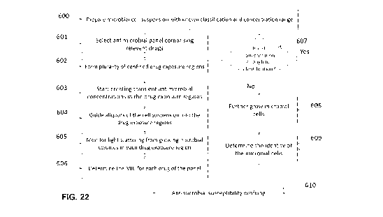
FIG. 11A provides a flow chart illustrating an example method for performing
rapid antimicrobial susceptibility testing (AST) on microcolonies.
FIG. 11B illustrates the average diameter versus microcolony cell content plot
for E. coli. The plot has been fitted with a power law trendline for enabling
the
estimation of average microcolony diameters, for example, at 103 and 105 cell
content levels.
FIG. 11C plots the average microcolony diameters at 103 and 105 cell content
levels for various pathogenic gram-positive bacteria prevalent in blood stream
infection.
FIG. 11D illustrates the average microcolony diameters at 103 and 105 cell
content levels for various pathogenic gram-negative bacteria prevalent in
blood
stream infection.
FIG. 12 schematic of a system for performing automated centrifugation and
washing with an integrated fluidic processing cartridge.
FIGS. 13A to 13E illustrate an example integrated fluidic processing cartridge
configured for extraction of a sample directly from a collection tube,
subsequent
CA 03124269 2021-06-18
WO 2020/124271
PCT/CA2019/051895
centrifugation and washing, to obtain a concentrated and purified suspension
of
microbial cells.
FIG 14 provides a flow chart illustrating an example method for performing
automated centrifugation and washing.
FIG. 15A shows a schematic of an example system for incubating the growth
chamber, monitoring growth of microbial cells for the detection of viable
microbial
microcolonies and classifying the microcolony cells as belonging to a given
microbial
cell class. Objective lenses with low magnification are employed to increase
the
observed area and can increase temporal resolution for time-lapse imaging.
FIG. 15B shows a schematic of another example system for incubating the
growth chamber, monitoring growth of microbial cells for the detection of
viable
microbial microcolonies.
FIG. 15C illustrates the comparison of E. coli microcolonies/colonies detected
on a solid phase growth media 4 hours (right) and 20 hours (left) after sample
seeding and incubation at 35 C. The image on the right is the result of mosaic
stitching of 448 aligned/registered microscopic images taken by 5x brightfield
objective in an automated system similar to the one schematically presented in
FIG.
15B. The image on the left was taken by a conventional camera.
FIG. 15D illustrates the 18 microcolonies identified on the plate shown in
FIG.
15C, as detected during 4 hours of incubation via time-lapse image processing.
As
can be seen in the figure, 15 out of 18 microcolonies, shown by white circles,
were
detected at 3 hours, and one microcolony (microcolony 7) was detected at 2
hours.
FIG. 15E shows the morphology of Streptococcus pneumoniae colonies
respectively grown in the presence (up-left) and absence (up-right) of a CO2
pack for
overnight. The image is taken with 5X brightfield objective. A typical
microcolony of
Streptococcus pneumoniae formed after 4 hours of incubation in the absence of
CO2
pack is presented at the bottom-left corner. The image has been taken by 10X
brightfield objective. The zoomed image of the microcolony, presented in
bottom right
corner, is more similar to the overnight colony formed at the presence of CO2
pack.
FIG. 16 is a photograph of a blood agar gel on which Staphylococcus aureus
cells have been streaked. Commercial paper disks (HardydiskTm), respectively,
impregnated with Oxacillin 1 pg/mL, Tetracycline 30 pg/mL and Norfloxacin 10
pg/mL, have been placed on the solid phase growth media and photo has been
taken after overnight incubation. The measure r1 shows visible absence of
microbial
lane up to a distance from the center and represents zone of inhibition.
Beyond this
distance, sparse lane is observed up to a distance r2, beyond which the lane
is full.
FIGS. 17A and 17B schematically compare the diffusion of an antimicrobial
26
CA 03124269 2021-06-18
WO 2020/124271
PCT/CA2019/051895
agent from an impregnated disk which has been placed on the gel surface. The
cases of conventional disk diffusion AST is shown in FIG. 17A, while an
example
annular disk diffusion AST embodiment is shown in FIG. 17B. The arrows
represent
the directions of diffusion of the antimicrobial agent that are respectively
relevant for
disk diffusion antimicrobial susceptibility testing and annular-disk diffusion
antimicrobial susceptibility testing.
FIG. 18A schematically illustrates an example annular disk diffusion device (a
"LD-AST unit"), indicating the parameters r1, rad and h.
FIGS. 18B-18I illustrate example embodiments of solid support structures for
performing lateral diffusion AST.
FIG. 18J shows a set of images demonstrating capability of a guiding ring, cut
from a 100pm thick Thermoplastic polyurethane (TPU) sheet, to localize and
concentrate microbial cells within the inner aperture of the ring, when the
ring is
placed on a gel surface and a microbial cell suspension is dispensed over the
aperture.
FIGS. 19A-19E schematically represents the assembly of annular disk
diffusion "LD-AST units" into an array. The annular disks impregnated with an
antimicrobial at different concentration levels are supplied with interaction
rings and
assembled in a strip (FIG. 19A). A complementary strip (FIG. 19B) includes
assembly
of sealed microwells having agar gels. During the assay two components are
attached and aliquots of the sample are dispensed inside the interaction rings
(FIG.
19C).
FIG. 20A plots the simulated concentration on the surface of the gel across a
centerline passing through an annular diffusion disk (the disk is indicated by
the black
strip). The gel thickness is taken to be h=4 mm and its radius is r2=14 mm.
The
cross-section of the annular disk, having the inner radius ri=1.5 mm and the
outer
radius r0d=3 mm, is illustrated by thick line.
FIG. 20B plots the evolution of the simulated concentration on the surface of
the gel across a centerline passing through the annular disk, indicated by the
black
strip. The gel thickness is taken to be h=2 mm and its radius r2=14 mm.
FIG. 20C plots the evolution of the simulated concentration on the surface of
the gel across a centerline passing through the annular disk, indicated by the
black
strip. The gel thickness is taken to be h=4 mm and its radius r2=5 mm.
FIG. 20D plots the evolution of the simulated concentration on the surface of
the gel across a line passing through the annular disk, indicated by the black
strip.
The gel thickness is taken to be h=2 mm and its diameter r2=5 mm.
FIG. 20E plots an example impregnated concentration profile on annular disk,
27
CA 03124269 2021-06-18
WO 2020/124271
PCT/CA2019/051895
referred to below as duall
FIG. 20F plots the evolution of the simulated concentration on the surface of
the gel across a centerline passing through the annular disk according to the
impregnated concentration profile shown in FIG. 20E. The gel thickness is
taken to
be h=2mm and its radius r2=5 mm.
FIG. 20G plots an example impregnated concentration profile on annular disk,
referred to below as dua12.
FIG. 20H plots the evolution of the simulated concentration on the surface of
the gel across a centerline passing through the annular disk according to the
impregnated concentration profile shown in FIG. 20G. The gel thickness is
taken to
be h=2 mm and its radius r2=5 mm.
FIG. 21A represents the simulated temporal behavior of drug concentration at
the center of the region of interest for the LD-AST units of FIGS. 20A to 20H.
The
plots labeled with (r2=14 mm, h=4 mm), (r2=14 mm, h=2 mm), (r2=5 mm, h=4 mm),
(r2=5 mm, h=2 mm), (r2=5 mm, h=2 dual1) and (r2=5 mm, h=2 dua12), respectively
correspond to FIGS. 20A, 20B, 20C, 20D, 20F, and 20G. r ¨ horizontal dimension
of
gel; h ¨ gel thickness.
FIG. 21B plots the relative change in the concentration at the center of the
region of interest for the curves of FIG. 21A between during the period of
reaching
max concentration at ¨0.5h and 4 h after placing the annular disk on the gel.
The
plots labeled with (r2=14 mm, h=4 mm), (r2=14 mm, h=2 mm), (r2=5 mm, h=4 mm),
(r2=5 mm, h=2 mm), (r2=5 mm, h=2 dual1) and (r2=5 mm, h=2 dua12), respectively
correspond to FIGS. 20A, 20B, 20C, 20D, 20F, and 20G.
FIG. 21C presents the qualitative and quantitative concentration profile of a
dye solution deposited on an annular disk according to the method of Example
9B.
FIG. 22 represents a flowchart for rapidly performing annular-disk diffusion
AST.
FIG. 23A is an overhead photograph of a strip for running LD-AST at 8 drug
concentration levels. The photo corresponds to the case of thin strips, i.e.
50 pL of
gel in each microwell.
FIG. 23B is a photograph showing a side view of the strip shown in FIG. 23A.
The photo corresponds to the case of thin strips, i.e. 50 pL of gel in each
microwell.
FIGS. 24A-24C show overhead images of the regions of interest (ROls) of the
strip shown in FIGS. 23A and 23B (low gel volume of ¨ 50 uL; gel thickness of
1mm) after incubating for 3 hours, 4 hours, and overnight incubation,
respectively.
The antibiotic is Norfloxacin and the microbial cell is Escherichia coli.
FIGS. 25A-25C show overhead images of the ROls of the mid-thick (gel
28
CA 03124269 2021-06-18
WO 2020/124271
PCT/CA2019/051895
volume ¨ 150 ju L; gel thickness ¨ 3 mm) strip after incubating for 3 hours, 4
hours,
and overnight incubation, respectively. The antibiotic is Norfloxacin and the
microbial
cell is Escherichia coli.
FIGS. 26A-26C show overhead images of the ROls of the thick (gel volume =
350 pL; gel thickness ¨ 7 mm) strip after incubating for 3 hours, 4 hours, and
overnight incubation, respectively. The antibiotic is Norfloxacin and the
microbial cell
is Escherichia coli.
FIG. 27A shows overhead images of the ROls of the "thin strip" type LO-AST
(shown in FIGS. 23A and 23B) after incubating for 3 and 4 hours. The
antibiotic is
Vancomycin and the microbial cell is Staphylococcus aureus.
FIG. 27B shows overhead images of the ROls of the "mid-thick strip" type LD-
AST after incubating for 3 and 4 hours. The antibiotic is Vancomycin and the
microbial cell is Staphylococcus aureus.
FIG. 28 shows overhead images of the ROls of the "mid-thick strip" type LD-
AST after incubating for 4 hours following the inoculation. The antibiotic is
Vancomycin and the microbial cell is Staphylococcus aureus.
FIG. 29 shows overhead images of the regions of interest (ROls) of the "thin
strip" type LD-AST after incubating for 3 hours 4 hours and overnight while
testing
the susceptibility of Amphotoricin B against Candida albicans.
FIG. 30 show overhead images of the regions of interest (ROls) of the "thin
strip" type LD-AST after incubating for 3 hours and 4 hours, respectively, for
clean
cell suspension (top) and the positive blood culture diluted by 1000-fold
(bottom).
The antibiotic is Oxacillin and the microbial cell is Methiciffin-resistant
Staphylococcus
aureus (MRSA 111 with not strong resistance).
FIG. 31A shows the growth pattern of MRSA-110 in the wells of a commercial
broth microdilution AST plate. The indicated concentrations are in pg/mL. The
abbreviations are as the following: CHL = Chloramphenicol, DAP = Daptomycin,
GEN = Gentamicin, LZD = Linezolid , RIF = Rifampin, SXT = Trimethoprim /
Sulfannethoxazole , SYN = Quinupristin / Dalfopristin, TET = Tetracycline, ERY
=
Erythromycin, OXA+ = Oxacillin + 2% NaCI , AMP = Ampicillin, PEN = Penicillin,
VAN = Vancomycin, LEVO = Levofloxacin, TGC = Tigecycline, MXF = Moxifloxacin,
CLI = Clindamycin, STR = Streptomycin, CIP = Ciprofloxacin, NIT =
Nitrofurantoin,
DT1 = D Test 1 , DT2 = D Test 2, FOXS = Cefoxitin screen , NEG = Negative
control
and POS = Positive control.
FIG. 31B shows the growth pattern of MRSA-110 in the wells of a 96 well
microplate where the LD-AST assay is performed. The abbreviations are the same
as FIG. 31A.
29
CA 03124269 2021-06-18
WO 2020/124271
PCT/CA2019/051895
FIG. 32 shows a comparison between the results of LD-AST and standard
broth microdilution AST for selected drug-bug combinations. The abbreviations
for
the microbial cells are the following: Staphylococcus aureus (SA), Methicillin
-resistant
Staphylococcus aureus (MRSA), Acinetobacter baumannii (AB), Escherichia coli
(EC), Pseudomonas aeruginosa (PA), Proteus mirabilis (PM), Klebsiella
pneumoniae
(KP), carbapenem-resistant Enterobacteriaceae Klebsiella pneumoniae (CRE).
DETAILED DESCRIPTION
Various embodiments and aspects of the disclosure will be described with
reference to details discussed below. The following description and drawings
are
illustrative of the disclosure and are not to be construed as limiting the
disclosure.
Numerous specific details are described to provide a thorough understanding of
various embodiments of the present disclosure. However, in certain instances,
well-
known or conventional details are not described in order to provide a concise
discussion of embodiments of the present disclosure.
As used herein, the terms "comprise" and "comprising" are to be construed as
being inclusive and open ended, and not exclusive. Specifically, when used in
the
specification and claims, the terms "comprise" and "comprising" and variations
thereof mean the specified features, steps or components are included. These
terms
are not to be interpreted to exclude the presence of other features, steps or
components.
As used herein, the term "exemplary" means "serving as an example,
instance, or illustration," and should not be construed as preferred or
advantageous
over other configurations disclosed herein.
As used herein, the terms "about" and "approximately" are meant to cover
variations that may exist in the upper and lower limits of the ranges of
values, such
as variations in properties, parameters, and dimensions. Unless otherwise
specified,
the terms "about" and "approximately" mean plus or minus 25 percent or less.
It is to be understood that unless otherwise specified, any specified range or
group is as a shorthand way of referring to each and every member of a range
or
group individually, as well as each and every possible sub-range or sub -group
encompassed therein and similarly with respect to any sub-ranges or sub-groups
therein. Unless otherwise specified, the present disclosure relates to and
explicitly
incorporates each and every specific member and combination of sub-ranges or
sub-
groups.
As used herein, the term "on the order of", when used in conjunction with a
quantity or parameter, refers to a range spanning approximately one tenth to
ten
CA 03124269 2021-06-18
WO 2020/124271
PCT/CA2019/051895
times the stated quantity or parameter.
Unless defined otherwise, all technical and scientific terms used herein are
intended to have the same meaning as commonly understood to one of ordinary
skill
in the art. Unless otherwise indicated, such as through context, as used
herein, the
following terms are intended to have the following meanings:
As used herein, the phrase "intact cell" refers to a microbial cell containing
nucleic acids, proteins or intracellular contents of interest, where the
microbial cell is
separable via a separation method such as, but not limited to, centrifugal
separation,
filtration, microfluidic separation, or immunomagnetic separation.
As used herein, the phrase "sample" refers to a liquid or suspension that
contains, may contain, or is suspected of containing one or more microbial
cells.
Non-limiting examples of samples include body fluids such as urine, lymph
fluid,
cerebrospinal fluid, blood (e.g. whole blood, blood culture, and plasma),
sputum and
saliva. Other examples of samples include homogenized tissue suspensions,
including, but not limited to, stool, homogenized suspensions of muscle
tissue, brain
tissue and liver tissue. A sample may be processed or unprocessed and may
optionally include one or more reagents or growth media. In the case of a
blood
culture sample (a sample containing growth media and whole blood), the blood
culture sample may be a blood culture sample having been deemed positive for
the
presence of microbial cells via a detection modality (e.g. via an automated
blood
culture system), a mid-culture blood culture sample for which the presence of
microbial cells is suspected based on measurements made via one or more mid-
culture detection modalities, or mid-culture blood culture sample for which no
initial
detection results are available.
As used herein, the phrase "blood cells" refers to mammalian cells present in
blood, including, but not limited to, red blood cells (erythrocytes), white
blood cells
(leukocytes) and blood platelets (thrombocytes).
As used herein, the phrase "blood sample" refers to any sample comprising
one or more blood cells. Non-limiting examples of blood samples include whole
blood
samples, blood culture samples, buffy coat samples and platelet samples.
As used herein, the phrase "whole blood" or "whole blood sample" refers to
mammalian blood comprising blood plasma and blood cells. "Whole blood" or "a
whole blood sample" may include one or more reagents, such as anticoagulation
reagents. For example, whole blood may be collected in a sample bottle that
may
include one or more reagents such as, but not limited to, anticoagulants
including
SPS (sodium polyanethole sulfonate), EDTA (ethylenediaminetetraacetic acid),
sodium citrate and heparin.
31
CA 03124269 2021-06-18
WO 2020/124271
PCT/CA2019/051895
As used herein, the phrase "selective lysis" refers to a blood lysis reagent
or
lysis process whereby the fraction of microbial cells that remain viable
following lysis
exceeds the fraction of eukaryotic cells that remain viable following lysis,
where the
eukaryotic cells are associated with the subject from which the sample was
collected.
As used herein, the phrases "microbial cell" and "micro-organism" comprises
bacteria (e.g. Gram-positive and Gram-negative bacteria, as well as bacterial
spores)
and unicellular fungi (such as yeast and molds).
As used herein, the phrase "eukaryotic cell" refers to cells originating from
an
eukaryotic organism excluding fungi, such as animals, in particular animals
containing blood, comprising invertebrate animals such as crustaceans and
vertebrates. As used herein, "vertebrates" comprise both cold-blooded animals
(fish,
reptiles, amphibians) and warm-blooded animals (birds and mammals).
As used herein, the phrase "effective buffer concentration", when used with
reference to a mixture formed by mixing a volume of a sample with a volume of
a
blood lysis reagent, where the blood lysis reagent includes a buffer system,
refers to
the product of the buffer concentration of the blood lysis reagent and a ratio
formed
by dividing the volume of the blood lysis reagent by the sum of the volume of
the
blood lysis reagent and the volume of the sample. The effective buffer
concentration
represents the contribution of the blood lysis reagent to the buffer system in
the final
mixture (i.e. the dilution factor applied to the buffer concentration of the
blood lysis
reagent) and may be different than the actual buffer concentration in the
final mixture
due to buffering components present in the sample.
As used herein, the phrase "separation process" refers to a process suitable
for separating and optionally concentrating microbial cells. Non-limiting
examples of
separation processes include centrifugation, filtration, immunomagnetic
separation
and microfluidic separation.
As used herein, the phrase "cell suspension" refers to an aqueous medium
that contains microbial cells.
As used herein, the terms "colony" and "microcolony" refer to a multiplicity
or
population of microorganisms that lie in close proximity to each other, that
lie on a
surface, and that are the clonal descendants, by in situ replication, of a
single
ancestral microorganism. In general, a "colony" is visible to the human eye
and is
typically greater than about 50 lam, 60 m, 80 4m, or 100 m, in diameter.
However,
as used herein, unless otherwise stated, the term "colony" is meant to include
both
colonies having a diameter of 100 gri or more, and the term "microcolony" is
meant
to refer to a colony having a diameter less than 100 m.
32
CA 03124269 2021-06-18
WO 2020/124271
PCT/CA2019/051895
Various example embodiments of the present disclosure address the
aforementioned shortcomings with conventional approaches to microbial growth
and
antimicrobial susceptibility testing (AST). As explained in detail below, many
of the
example embodiments of the present disclosure employ an integrated fluidic
cartridge to facilitate the separation of microbial cells and the subsequent
growth of
microbial colonies in situ within the integrated fluidic cartridge. In many
example
embodiments, the microbial cells are separated and contacted with a solid
phase
growth media for subsequent colony growth while maintaining at least a portion
of the
integrated fluidic cartridge in a closed configuration.
Referring now to FIG. 1, an example integrated fluidic cartridge (device) for
performing microbial colony growth is schematically illustrated. A sample 20
may be
introduced into the integrated fluidic cartridge 10, which includes a cell
separation
module (a portion or region of the integrated fluidic cartridge) 30 and a
colony growth
module (another portion or region of the integrated fluidic cartridge) 40. The
integrated fluidic cartridge 10 facilitates the separation of microbial cells
from other
components of a sample, such as eukaryotic cells (e.g. host cells from a host
subject) such that the separated microbial cells are separated in an intact
and viable
form (capable of cell division). The separated microbial cells may be
provided, an
optionally concentrated, in a liquid medium (e.g. saline or another buffer
suitable for
maintaining microbial cell viability), thereby providing a microbial cell
suspension.
The cell suspension is subsequently introduced, e.g. via one or more fluidic
conduits
35, into the colony growth module 40 and contacted therein with (e.g. seeded
on) a
solid phase growth media, examples of which are described in detail below. The
colony growth module 40 is configured to permit the monitoring (e.g. via
optical or
electrical modalities) of colonies that grow on the solid phase growth media.
The
integrated fluidic cartridge 10 may subsequently be incubated at an
appropriate
temperature and environment for promoting growth (e.g. 35-38 C) while
permitting
monitoring of the growth of microbial colonies.
As noted above, the microbial cells may be separated and contacted with a
solid phase growth media for subsequent colony growth while maintaining at
least a
portion of the integrated fluidic cartridge in a closed configuration. It will
be
understood that the phrases "closed" and "closed state" refer to a capability
or
configuration of an internal region of the integrated fluidic cartridge to be
at least
temporarily brought into a state that prohibits the ingress (entrance or
introduction) of
external microbial cells within an interior region of the integrated fluidic
device,
thereby avoiding contamination of microbial cells that are separated from the
sample
and grown in colonies within the colony growth module. An inner region of an
33
CA 03124269 2021-06-18
WO 2020/124271
PCT/CA2019/051895
integrated fluidic cartridge may be brought into a closed state by actuation
of suitably
located valves.
In some example embodiments, an internal region of an integrated fluidic
cartridge may be in a closed state while permitting gas communication of the
internal
region with an external gas source or external environment, for example,
through a
filter that prohibits that passage of microbial cells. One internal region of
a fluidic
cartridge may be configured in a closed state while other internal regions of
the fluidic
cartridge reside in a non-closed state. As explained in some example
embodiments
below, at least a portion of an internal region of an integrated fluidic
cartridge that
resides in a closed state may be opened to provide external access to
microbial cells
(e.g. microbial colonies) residing therein, for example, after sufficient
microbial
growth for further testing has been detected.
It will be understood that the integrated fluidic cartridge 10 may be
configured
such that the cell separation module performs separation of viable microbial
cells,
prior to the contact of the viable microbial cells with the solid phase growth
media,
according to a manual method, a semi-automated method, or a fully-automated
method, such that the resulting microbial suspension may be transported to the
colony growth module while maintaining the microbial cell suspension in a
closed
environment within the integrated fluidic cartridge.
For example, in some example embodiment, the microbial cells may be
separated via an automated lysis-centrifugation process, using an integrated
fluidic
cartridge such as the cartridge shown in FIGS. 13A-13E (described in detail
below as
providing an example of the cell separation module of an integrated fluidic
device) or
a variation thereof, such that the suspension automatically to contact the
solid phase
growth media. In an alternative example implementation, the process may be
fully
automated within a closed integrated cartridge, which may be beneficial in
avoiding
the introduction of contaminants when transferring the separated microbial
cells to
the solid phase growth media. In another example embodiment, the microbial
cells
may be separated using a filter, where the integrated fluidic cartridge
includes a
plunger that is either manually or robotically actuated.
It will be understood that although the example integrated cartridge shown in
FIGS. 13A-13E employs automated lysis-centrifugation for performing separation
of
viable microbial cells, such an integrated cartridge may be modified to
include an
alternative separation modality, such as, but not limited to, filtration,
immunomagnetic
separation, or other separation modalities including, but not limited to, cell
sorting
techniques, such as flow cytometric cell sorting, electrical cell sorting or
microfluidic
cell sorting. Furthermore, while some of the example embodiments disclosed
herein
34
CA 03124269 2021-06-18
WO 2020/124271
PCT/CA2019/051895
involve the separation of viable microbial cells from a blood sample, it will
be
understood that a wide variety of sample types may be employed, as per the
definition of the phase "sample" provided above.
The colony growth module includes a growth chamber that facilitates both the
growth and monitoring of microbial colonies on the solid phase growth media.
An
example embodiment of such a growth module with an internal growth chamber is
illustrated in FIGS. 2A-2B. Referring first to FIG. 2A (plan view) and 2B
(section view
A-A), the colony growth module 180 includes a growth chamber 100 with a lower
inner wall 105 having a layer of solid phase growth media 110 formed thereon,
an
upper wall 120, a conduit 101 fluidically connected, for example, to the cell
separation module via path 161 (or 35 in FIG. 1). The growth chamber 100 may
also
be fluid ically connected via conduit 150 to an air vent 130 or such an air
vent may be
formed in upper wall 120 of the chamber.
In some example embodiments, the upper wall 120 may contain a gas
permeable section for allowing air exchange between the chamber and the
external
environment during the microbial cell growth. The gas permeable section may
be, for
example, a gas permeable membrane. An example of a suitable gas permeable
membrane includes, but is not limited to, a polyurethane membrane. These
example
membranes enable a sufficient rate of gas exchange with ambient environment to
facilitate cell growth on the solid phase, while sustaining a contaminant-free
environment within the chamber.
The solid phase growth media 110 is suitable for providing the microbial cells
with an appropriate source of growth media. Non-limiting examples of solid
phase
growth media include conventional agar, gelatin, guar gum, Xanthan gum, having
suitable growth nutrients. In some example implementations, the solid phase
growth
media may be chromogenic according to the type of microbial cell. In some
embodiments chromogenic or fluorogenic substrate may be added to the agar
media
for identifying the microorganism by specific or non-specific staining of the
colonies,
as described, for example, in European Patent Application No. EP1088896A2.
As explained in further detail below, the solid phase growth media may be in
a dry or partially dry format such that that a liquid component from the cell
suspension is absorbed (at least partially) upon contact.
It will be understood that the microbial cell suspension can be introduced
into
the growth chamber according to a number of different example implementations.
For
example, the cell suspension may be flowed into the chamber and over the solid
phase growth media by, for example, positive pressure from an upstream
location or
negative pressure from a downstream location (example via path 150 of FIG. 2B)
and
CA 03124269 2021-06-18
WO 2020/124271
PCT/CA2019/051895
via the actuation of appropriately placed valves and pumps. In some example
implementations, physical structures such as barriers that impede or reduce
fluid flow
may be provided within the growth chamber in order to facilitate a suitable
spreading
of the cell suspension to distribute the microbial cells over the surface of
the solid
phase growth media.
It will be understood that in some example implementations involving the
growth of a small number of colonies and/or the growth of colonies to a
limited size,
the growth chamber may be closed in the absence of a gas-permeable region. For
example, in some of the example embodiments described below, a sufficient
volume
of required atmosphere such as ambient air or 5% CO2 may be enclosed within
the
colony chamber for microbial respiration in the absence of a gas-permeable
region.
In alternative example embodiments, the growth chamber may be in gas
communication, through a filter that serves as a barrier to external microbial
cells,
with an oxygen source (e.g. the external environment) to facilitate the
provision of
continuous or intermitted oxygen to the chamber. For example, the growth
chamber
may be in gas communication, through an intermediate filter that prohibits the
entry
of external microbial cells, with an external port that is vented to the
atmosphere or to
a pressurized or pneumatic gas source. The gas communication with the external
oxygen source may be controlled via an intermediate valve.
In one example embodiment, the upper wall 120 may include an optically
transparent or transmissive region (e.g. a window) through which the growth of
colonies can be inspected (e.g. visually or via an imaging device such as a
camera).
In another embodiment, the upper wall 120 is in the form of a separable lid,
which
can be occasionally removed for imaging or visually interrogating the gel
surface for
the growth of microbial colonies.
The growth chamber 100 may optionally include one or more indicators that
are capable of detecting metabolic activity of the growing colonies via the
colorimetric
sensor detection of volatile organic compounds produced by the microorganism.
Non-limiting examples of suitable indicators has been disclosed in US Patent
Publication No. 20150099694. Such indicators may permit the identification of
the
cells in the colony with some level of taxonomic granularity (e.g. Gram
status, family,
genus, species, strain). In one example embodiment, the one or more indicators
may
provide information similar to that of a Gram-stain test, permitting the
selection of a
suitable antibiotic susceptibility testing panel in a non-destructive manner
without
perturbing or sacrificing any of growing colonies.
In some example embodiments, the colonies may be identified in a non-
destructive manner by providing the solid phase growth media with indicators.
Non-
36
CA 03124269 2021-06-18
WO 2020/124271
PCT/CA2019/051895
limiting examples of suitable indicators include chromogenic or fluorogenic
substrates, biochemical dyes, pH indicators, as described, for example, in
United
States Patent Publication No. 2012/0295299A1.
In some example implementations, the gas permeable membrane or another
portion of the structure enclosing the growth chamber may be formed from or
may
include a transparent material (such as, for example, polyurethane) and may
extend
over the spatial region associated with the growth media, as illustrated in
the
example embodiment shown in FIG. 2B, thereby permitting the observation of
colony
growth. The observation can be performed, for example, visually or using an
optical
detection (e.g. imaging) system, such as phase-contrast or dark-field
microscopy.
Alternatively, the colonies may be illuminated from backside by the collimated
beam
of a monochromatic light source, such as laser, and the optical scattering
pattern
may be inspected or processed to perform microbial cell identification or to
determine
a class of microbial cells of a given colony, as described further below.
It will be understood that different detection modalities will have different
limits
of detection for colony growth on solid phase growth media. For example, in
the
direct microscopic monitoring method, the limit of detection in terms of
number of
cells per colony can be in the order of 103. For example, Yoshiakiet al.
[Colony
fingerprint for discrimination of microbial species based on lensless imaging
of
microcolonies." PloS one 12.4 (2017): e0174723] were able to detect
microcolonies
with diameters in 10-500 pm range.
An important factor which may limit the detection of microcolonies is the size
and density of surface artefacts (the background) that are observable via
microscopy
after contacting the treated sample with the solid phase growth medium, where
such
surface artefacts are not representative of microbial cells or microbial cell
colonies.
Two example types of such surface artefacts that contribute to a background
include
(i) surface inhomogeneities of the gel surface and (i) lysis debris particles
that remain
after the lysis of the sample, such as lysis debris particles resulting from
the digestion
of blood cells, which persist in the sample after centrifugal washing.
The surface density of the first type of artefact, namely gel surface
inhomogeneities, can be reduced through controlled fabrication of the gel. An
example non-limiting method for preparing gels with a low density of surface
inhomogeneities is described Example 6 below.
The size and amount of the second type of the artefacts, namely lysis debris
particles may vary from one whole blood sample to another and has been found
to
depend on the composition of the blood lysis reagent (BLR).
International Patent Application No. PCT/0A2013/000992, titled
37
CA 03124269 2021-06-18
WO 2020/124271
PCT/CA2019/051895
"APPARATUS AND METHOD FOR EXTRACTING MICROBIAL CELLS" discloses a
number of different blood lysis reagent compositions that may be employed for
the
digestion of blood components prior to centrifugation. As noted above, the
presence
of the blood lysis reagent causes the selective lysis of blood cells. In one
example
implementation taught by International Patent Application No.
PCT/CA2013/000992,
the blood lysis reagent may be an aqueous liquid including saponin and sodium
polyanetholesulfonate (a sodium salt of polyanetholesulfonic acid, known as
SPS),
and a blood lysis reagent having such a composition is henceforth referred to
as a
"type 1" blood lysis reagent. The blood lysis reagent may also include an
antifoaming
agent, such as poly (propylene glycol) (PPG, e.g. with a molecular weight of
approximately 2000). International Patent Application No. PCT/CA2013/000992
teaches example concentration ranges of saponin and SPS for a type 1 blood
lysis
reagent, upon mixing whole blood and the blood lysis reagent, of approximately
1.5
to 80 mg/mL and 0.5 to 20 mg/mL, respectively.
As taught in International Patent Application No. PCT/0A2013/000992, SPS
is an anti-coagulant and anti-phagocytosis agent and is known to inhibit
antimicrobial
agents (Sullivan, N. M., Sutter, V. L., & Finegold, S. M. (1975). Practical
aerobic
membrane filtration blood culture technique: development of procedure. Journal
of
clinical microbiology, 1(1), 30-36). The mechanism by which SPS assists in
blood cell
lysis is not well understood. Without intended to be limited by theory, it is
believed
that SPS may offer some level of protection to the microorganisms during blood
cell
lysis, reduce the incidence of entrapment of bacteria in cell debris, and/or
reduce the
quantity of lysis debris components that may otherwise be present in the
sediment.
In another example implementation of a blood lysis reagent composition
taught by International Patent Application No. PCT/CA2013/000992, a blood
lysis
reagent may be an aqueous liquid including Triton X-100 and SPS in a buffer
having
a pH ranging from 9 to 11, and a blood lysis reagent having such a composition
is
henceforth referred to as a "type 2" blood lysis reagent. The blood lysis
reagent may
also include an antifoaming agent, such as poly (propylene glycol) (PPG, e.g.
with a
molecular weight of approximately 2000). International Patent Application No.
PCT/CA2013/000992 teaches example concentration ranges of Triton X-100 and
SPS for a type 2 blood lysis reagent, upon mixing whole blood and the blood
lysis
reagent, of approximately 0.5 to 1.5% w/v and 5 to 10 mg/mL, respectively.
As noted above, the type 1 blood lysis reagent composition described above
was found to be suitable for manual and semi-automated separation and
concentration of microbial cells from whole blood as per the teachings of
International
Patent Application No. PCT/CA2013/000992. However, when adapting the reagent
38
CA 03124269 2021-06-18
WO 2020/124271
PCT/CA2019/051895
formulations disclosed in International Patent Application No.
PCT/CA2013/000992
to the automated separation and concentration, and subsequent identification,
of
microbial cells from whole blood as per the automated methods of International
Patent Application No. PCT/0A2015/050449, titled "Apparatus, System and Method
for Performing Automated Centrifugal Separation", filed on May 19, 2015, which
is
hereby incorporated by reference in its entirety, the present inventors found
that the
type 1 blood lysis reagent composition was most suitable for cases in which
the
quantity of whole blood was less than approximately 1 ml.
Another example blood lysis reagent for achieving a low surface density of
lysis debris particles is described in International Patent Application No.
PCT/0A2019/050716, titled METHODS AND COMPOSITIONS FOR THE
SELECTIVE LYSIS OF BLOOD CELLS AND SEPARATION OF MICROBIAL
CELLS", and filed on May 24, 2019, which is incorporated herein by reference
in its
entity, and which describes example blood lysis reagent compositions and
method
for preserving microbial cell viability and lowering the sample viscosity to
the levels
that the fluidic movement operations through narrow channels on the cartridge
could
be performed without intolerable impediment. This blood lysis reagent
composition,
henceforth referred to as a type 3 blood lysis reagent, may be provided
containing
saponin, SPS, an alkaline buffer and optionally a non-ionic surfactant.
In one example embodiment, the type 3 blood lysis reagent may have a
composition such that after the blood lysis reagent is mixed with the sample,
the
concentration of saponin lies between 3 and 60 mg/ml, the concentration of SPS
lies
between 1.5 and 50 mg/ml, the concentration of non-ionic surfactant lies
within 0-3%
w/v and the pH lies within a range of 7.8-10. In some example embodiments, the
buffer concentration may be selected such that the effective buffer
concentration lies
in the range of 10-300 mM. It will be understood that a suitable concentration
range
for a given components of a blood lysis reagent can be determined, for a given
set of
conditions, by experimentally investigating the effect of changes in
concentration of
the given component on one or more performance metrics, such as, but not
limited
to, blood lysis efficiency, quantity of residual blood cell debris, microbial
cell
intactness and microbial cell viability. A type 3 blood lysis reagent may be
provided
as two or more reagents that can be stored separately and mixed prior to use,
such
that the saponin component of the blood lysis reagent is stored in an acidic
environment that is separated from the alkaline component of the blood lysis
reagent.
In one example implementation, one or more of the reagents that are mixed to
form
the final blood lysis reagent may be stored in a solid phase.
Accordingly, in some example implementations, a sample may be processed
39
CA 03124269 2021-06-18
WO 2020/124271
PCT/CA2019/051895
by a type 3 lysis reagent comprising saponin, sodium polyanethole sulfonate
(SPS),
a non-ionic surfactant (such as, but not limited to, TritonTm X-100), a buffer
(e.g. a
carbonate-bicarbonate buffer), with an alkaline pH. The blood lysis reagent
may also
include an antifoaming agent such as SE-15 (e.g. a 10% emulsion w/v of active
silicone polymer and non-ionic emulsifiers).
A blood lysis reagent without a non-ionic surfactant and a carbonate-
bicarbonate buffer is known to be benign to microbial cells. However, as
illustrated
below, in the case of processing whole blood samples, the size of the blood
debris
transferred to the final cell suspension according to the use of such a blood
lysis
reagent has been found to give rise to an elevated level of surface artefacts
(background) that can impede microcolony detection and characterization. In
contrast, addition of moderate amounts of non-ionic surfactant (e.g. TritonTm
X-100)
and carbonate-bicarbonate buffer, along with saponin and SPS, while not
significantly impacting the cell viability, can be beneficial in significantly
reducing the
surface artefacts arising from lysis debris.
In some example implementations, a whole blood sample with a volume up to
10mL may be mixed with a type-3 blood lysis reagent such that the
concentration of
saponin in the final mixture ranges between 10-30 mg/ml (or, in some example
implementations, 3-60 mg/ml), the concentration of SPS in the final mixture
may
range between 5-50 mg/ml (or, in some example implementations, 1.5-50 mg/ml),
the
effective buffer concentration lies in the range of 10-300 nnM, the
concentration of
non-ionic surfactant lies within 0-3% w/v (or, in some example
implementations, 0-
1% w/v), the pH of the final mixture may range between 7.8-10 (or, in some
example
implementations, 8.2-9.5), and the concentration of the antifoaming agent
emulsion
lies within 0.005 to 0.5% (v/w).
In order to illustrate the dependence of the surface artefact density on the
composition of the blood lysis reagent, 4 mL of whole blood sample was
processed
according to the method of Example 5, both with 2 washing cycles using a blood
lysis
reagent having a composition as the following: 35 mg/mL saponin, 20 mg/mL SPS
(BLR1) and (ii) 35 mg/mL saponin, 20 mg/mL SPS, 0.3% w/v TritonTm X-100, and
50
mM carbonate-bicarbonate buffer, with a pH of 10 (BLR2). After exposure of the
sample to the respective blood lysis reagents and centrifugal separation, 1 pL
of
each final microbial cell suspension were pipetted on a spot-on agar gel
plate. The
microbial cell suspension samples spread to circular areas with diameters of
about 5
mm and air dried in about three minutes. These areas, which herein are labeled
as
mini-culture region (MCR) were imaged by a microscope equipped to a 5x
objective
and was presented in FIGS. 3A and 3B. In these figures the MCR, unused agar
plate
CA 03124269 2021-06-18
WO 2020/124271
PCT/CA2019/051895
surface and the boundary between two regions are respectively indicated by
310,
312, and 311. As it is observed the inclusion of Triton X-100 and carbonate-
bicarbonate buffer significantly reduces the background (density of surface
artefacts).
The present inventors found that this background level could not be
significantly reduced further by increasing washing cycles. For example, this
was
demonstrated by treating a 4 mL whole blood sample using BLR2 having a
formulation as described above with 2 or 4 subsequent centrifugal wash cycles.
A 1
pL of the resulting microbial cell suspension was dispensed on the agar plate
and
allowed to spread and air dry. The pictures of the resulting MCRs were
recorded with
a 10x microscopic objective, and were analyzed for the size distribution of
debris at
the end of 2 and 4 wash cycles. The particles were located via intensity-based
adaptive auto-threshold methods and were fitted with ellipses. More precisely,
image
segmentation was performed and a label was assigned to every connected group
of
pixels in an image such that pixels with the same label share certain
intensity
characteristics. The histogram of the measured major particle diameter (major
axis of
a fitted ellipse) distribution is presented in FIG. 3C. As it is observed, the
distribution
plots are qualitatively similar, despite the fact that the sample is diluted
by a factor of
400-fold between 2 and 4 washes.
These results suggest that the size of the debris particles is strongly
influenced by the composition of the blood lysis reagent when processing blood
samples for subsequent direct colony growth. However, the requirement for the
microbial cell viability, particularly in the case of Gram-negative bacteria,
limits the
ability to achieve smaller debris size by increasing the digestion capacity of
the blood
lysis reagent. This limitation can be tolerated as long as the sizes and the
density of
the debris does not impede the detection of microbial cell growth at
microcolony
level. One example criterion in this respect is that after spreading the final
cell
suspension on the cell growth chamber the lysis debris artefacts should not
cover the
surface of the solid phase growth medium with an areal fraction exceeding 90%,
or
preferably not exceeding 50%, or more preferably not exceeding 20%. The
present
inventors have found that this criterion can be satisfied by processing whole
blood
samples with a type-3 blood lysis reagent.
Provided that the surface density of artefacts is not prohibitively high, it
is
feasible to differentiate the background from microcolonies by recording
multiple
pictures over a time period via time-lapse imaging, or, for example, removing
the
background by size thresholding. In order to perform size selection, an image
of the
gel surface can be taken from a limited area of the surface before or slightly
after
incubation. In one example implementation, image analysis is performed to
41
CA 03124269 2021-06-18
WO 2020/124271
PCT/CA2019/051895
determine the average size of the debris particles, Rbaok av, and the standard
deviation
of the debris particle size, sd, and a size threshold may be determined based
on
Rback av and sd. The size selection criterion can be set, for example, as R>
Rthreshold =
Rback av n*sd, where n is typically an integer selected in the range 1 to 6,
and more
typically n = 3. In another embodiment, Rbõkav and sd may be predetermined
(e.g.
and provided along with the cartridge or embedded in software of the
device/instrument) to become available at the colony analysis stage.
In some example embodiments, the colony growth module is configured for
removal of a liquid component of the microbial suspension (i.e. at least a
portion of
the liquid component) after flowing the cell suspension into the growth
chamber, such
that at least a portion of the microbial cells within the suspension are
retained on the
surface of the solid phase growth media. The removal of a liquid component of
the
cell suspension assists with the seeding of the microbial cells onto the solid
phase
growth medium, promoting subsequent colony growth. Furthermore, by removing a
liquid component of the cell suspension, other complications associated with
the
presence of residual liquid are also mitigated, such as, for example,
condensation
and excess moisture which can interfere with the monitoring of growing
colonies by
optical or other (e.g. electrical) means.
In some embodiments the removal of a liquid component of the cell
suspension may be achieved by evaporation, either passively via vents or vapor
permeable membranes or actively by means of forced convective evaporation. In
other embodiments a liquid component may be absorbed by the growth media or a
growth media infused substrate.
In one example implementation, a liquid component of the cell suspension
residing within the growth chamber may be passively evaporated via a vapor
permeable membrane 115 within the upper wall of the growth chamber as shown in
FIG 4A. In another example embodiment, evaporation may be actively enhanced by
reducing the ambient pressure of the chamber such that evaporation of the cell
suspension fluid will be accelerated. For instance, at 25 C, the vapor
pressure of
water is approximately 24 mmHg. This may be achieved by closing the chamber
inlets and outlets by valves or other means and applying the negative pressure
via a
permeable membrane 115 in the upper wall of the chamber, or with the
application of
the negative pressure via a fluidic conduit connecting a closed and non-
permeable
chamber to a vacuum pump.
In another example implementation illustrated in FIG 4B, air may be flowed
over the cell suspension to aid in the evaporation of the cell suspension
fluid by
forced convective evaporation. The air may be desiccated and/or warmed to
further
42
CA 03124269 2021-06-18
WO 2020/124271
PCT/CA2019/051895
increase the rate of evaporation.
In other example embodiments, a liquid component of the cell suspension
may be removed passively by absorption as illustrated in FIG 40. In this
embodiment
the growth media is provided in one of a number of dry formats which absorbs
the
liquid and brings the microbial cells into contact with the growth media to
promote
colony growth and, in some embodiments, to immobilize the colonies for
subsequent
harvesting.
Various examples of suitable liquid-absorbing solid phase growth media, and
methods of fabrication thereof, are described in further detail below. In some
example embodiments, at least an upper portion of the growth media 110 (FIG.
2B)
may have a porous network with hydrophilic properties, where the porous
network is
provided in an at least partially dry state, such that the internal porous
network is at
least partially open and capable of absorbing a liquid. In other words, the
solid phase
growth media 110 may be provided in a state that is not fully saturated with
liquid. A
liquid component of the cell suspension that is provided by the cell
separation
module may thus absorbed by the solid phase growth media 110 upon contact
therewith, hydrating the solid phase growth media 110 and drawing the
microbial
cells of the cell suspension onto the surface of the solid phase growth media.
The
porosity of the solid phase growth media may be selected to prevent microbial
cells
from entering into the porous network, such that the microbial cells are
retained on or
near (proximal to) a surface of the solid phase growth media when a liquid
component of the cell suspension is absorbed.
The use of a liquid-absorbing solid phase growth media (a solid phase growth
media that is not fully hydrated and has a capacity for further absorption)
solves a
significant problem in facilitating the controlled adsorption, within a closed
integrated
fluidic device, of microbial cells from a cell suspension onto a surface for
colony
growth. By rapidly and efficiently absorbing a liquid component of the cell
suspension, complications associated with the presence of residual liquid
(e.g.
residual droplets or locally accumulated pooling) are avoided. The present
inventors
have found that such residual liquid can hinder cell-surface association.
Moreover,
residual liquid can result in condensation and excess moisture, which can
interfere
with the monitoring of growing colonies by optical or other (e.g. electrical)
modalities.
Various examples of suitable liquid-absorbing solid phase growth media, and
methods of fabrication thereof, are described in further detail below.
In some example embodiments, the solid phase growth media may be
provided in a dehydrated or partially-dehydrated form. Thus, a liquid
component of
the cell suspension, upon being introduced into the growth chamber, is wicked
away
43
CA 03124269 2021-06-18
WO 2020/124271
PCT/CA2019/051895
into the pores of the solid phase and the microbial cells are retained on the
surface.
One example of a dehydrated solid phase growth medium is disclosed in US
Patent No. 4,565,783. A self-supporting water-proof substrate, such as
polyester film,
is coated with an adhesive, such as copolymer of isooctylacrylate/acrylamide
(in a
mole ratio of 94/6), which is non-inhibitory to the growth of microorganisms.
A cold-
water-soluble gelling agent, such as guar gum, along with nutrients for
growing
microbial cells, is dispersed into the adhesive. Upon contact with a liquid,
the liquid
reacts with the particles of the gelling agent and a solid phase is formed for
supporting the growth of microbial cells.
In another implementation, a gellated solid phase is partially dehydrated in a
low humidity environment and is maintained partially dehydrated in a humidity-
controlled package until use. Alternatively, the gellated solid phase can be
partially
dehydrated by freeze-drying techniques.
In another example embodiment, at least a portion of the liquid component of
the cell suspension may be removed via centrifugal processing of a gel-based
solid
phase growth medium prior to contact with the cell suspension. An example
implementation of this embodiment is illustrated in FIGS. 5A-5D. The solid
phase
growth medium is subjected to a centrifugal force 103 due to rotation around
an axis,
as shown in FIG. 5A. While the figure shows the centrifugal force as being
directed
perpendicular to the gel surface in FIG. 5A, it will be understood that in
other
example implementations, the centrifugal force may be directed at an angle
relative
to the surface of the solid phase growth medium (e.g. within an angle of 30
degrees
from a perpendicular direction). As a consequence of the centrifugal force,
the gel is
partly dehydrated ("dewatered"), removing a liquid component of the gel
without
significantly collapsing the gel, at least in the upper portion of the gel, as
illustrated in
FIG. 5B.
When a microbial cell suspension containing microbial cells is dispensed on
(contacted with) the gel it spreads over the gel surface, as shown in FIG. 5C,
with at
least a portion of the liquid content of the microbial cell suspension being
absorbed
by the partially dehydrated gel, as shown in FIG. 50. The microbial cell
suspension
can be spread over the gel, and the liquid component of the microbial
suspension
may be absorbed passively (for example, under gravity) or for example, under
the
application of a subsequent centrifugal force. After removal of the liquid
component
of the microbial cell suspension, leaving behind the microbial cells on the
gel surface.
The exuded liquid component of the gel (e.g. water) may be collected inside
empty chambers behind the gel. The water exuded from the gel may be drained
during centrifugation or afterward.
44
CA 03124269 2021-06-18
WO 2020/124271
PCT/CA2019/051895
The dewatering phenomenon, also known as syneresis, occurs with most
hydrogel materials when the elastic pressure of the gel network, formed by the
helical
coils of agarose polysaccharide, exceeds the osmotic pressure of water that
normally
cause gel expansion. External mechanical pressure such as centrifugation
promotes
syneresis by squeezing the gel, analogous to squeezing water out from a wet
sponge. The removal of some water from the gel presents opportunities for the
rapid
absorption of the cell suspension by opening vacancies in the gel network to
allow for
new water to enter. Controlling the volume of water that is exchanged will be
a key
design feature of the growth chamber design. Thus, in the following section of
the
present disclosure, experimental observations are presented demonstrating the
quantification of the dewatering phenomenon as a function of gel composition.
To simulate the water loss from the agar gel upon centrifugation, gels were
placed on top of a porous membrane inside a centrifuge tube and spun at 4000
RPM.
The mass of the gels before and after the centrifugation was measured, with
the
difference being attributed to the water loss as a result of the centrifugal
force upon
the gel. In one experiment, approximately 0.15 gram sections of agar gel were
sliced
out of Petri dish and placed into a NanoSep Tube with a 0.45pm modified nylon
filter
membrane. The tube was then spun at 4000 RPM (3200g) for 8 minutes resulting
in
water being separated from the gel, passing through the membrane and
collecting at
the bottom of the tube. The mass of gel remaining on the membrane was then
measured and the percent water loss was determined according the mass
difference
of the gel before and after centrifugation.
The results, which are presented in FIGS. 5E, show that agar gels made with
a higher content of agar (1 to 3%) are more resistant to dehydration. This
greater
retention of water is due to the greater strength of the gel, making it more
resistant to
syneresis from the centrifugal force. Stiffer gels have also been observed by
the
present inventors to be less prone to cracking or deformations during the
centrifugation. Different gel additives were also investigated to determine
their effect
on water loss by centrifugation. The additives chosen were mainly other
polysaccharide hydrocolloids that had either gelling (agar, gellan gum, gum
Arabic
and carrageenan) or non-gelling (dextran) properties. In addition, gellan gum
(GelzanTM CM) and carrageenan are often used as agar substitutes for microbial
cultures. It was shown that 1.35% agar with 0.5% gellan gum retained the most
water after centrifugation in the 0.45 pm Nanosep tube (average of 27% water
loss
by mass) compared to 1.35% (49% loss) or even 1.85% agar alone (39% loss). It
is
believed that this greater retention is due to the greater strength of the gel
which
resists the external pressure from the centrifuge.
CA 03124269 2021-06-18
WO 2020/124271
PCT/CA2019/051895
The experiment was repeated using a filter membrane with a smaller pore
size. In this case, the gels were prepared in the autoclave and while still
warm
(-50 C), dispensed over a 10,000 MWCO (<5 nm pore size) cellulose membrane of
an Amicon Ultra-15 ultrafiltration tube. Each tube had about 1.5 grams of gel
when
cooled, which was then centrifuged for 9 minutes at 4000 RPM. The mass of the
gel
remaining on top of the membrane was then measured and the percent of water
loss
was calculated and presented in FIG. 5F. The results reveal how a smaller
membrane pore size reduces the dewatering level for all gels compared to the
larger
0.45pm pores. The addition of 0.5% gellan gum to 1.35% agar leads to less
water
loss at about 16% (that is, greater water retention) compared to the other
gels which
were between 22 to 23%.
The presented inventors observed that dewatering can be reduced or
prevented if the gel is well confined at its side and back surfaces. The use
of gel
confinement can be employed to limit the dewatering level (the level below
which
dewatering is eliminated or reduced) such that the volume of liquid removed
from the
gel corresponds to the volume of the liquid component of the sample which is
desired
to be spread on the gel surface and absorbed by the gel via the rehydration
process.
Without being bound to theory, it is hypothesized that dewatering leaves voids
in the molecular coils within the gels which could potentially be refilled
with fresh
liquid (e.g. an aqueous solution) from a microbial suspension. To illustrate
this
experimentally, the portion of gels that were dehydrated after centrifugation
in the
10,000 MWCO ultrafiltration tube (see above) were removed from the tube and
placed on a small plastic dish to determine their mass. Each dish was filled
with 2
mL of fresh water to soak into the gel. After 20 minutes of soaking, the
excess water
was removed by pouring it out of dish and carefully wiping away the remaining
drops
from the dish and the gels. The mass of the gels was then obtained and
recorded,
as shown in FIG. 5G, as the percent mass gain with respect to the mass of the
dehydrated gel.
The results suggest that the amount of water that was lost during
centrifugation can be regained by rehydrating the gel. That is, the percent of
dehydration is similar to the percent of rehydration of each gel. Therefore,
the voids
in the gel network that resulted when water was expelled from the gel are
refilled with
approximately the same volume of water when the gel is rehydrated.
Partial dehydration of gels can also be achieved through water exchange with
the environment. If the dehydration level of the gel is relatively low, the
process is
reversible and nearly full rehydration can be achieved. This property is
useful if the
centrifugation-assisted dewatering of the gel would be prevented by near-
complete
46
CA 03124269 2021-06-18
WO 2020/124271
PCT/CA2019/051895
confinement of the gel through appropriate chamber design, while the
rehydration for
the purpose of sample seeding is performed by centrifugation. In order to
illustrate
the feasibility of this approach, the following experiments were performed.
Agar gels with agar concentration of 1, 2, 3 and 4% wt., were dehydrated
during open storage at 4 C for 3 days in 35mm Petri dishes. The mass of the
gels in
the dishes before and after the storage was recorded, demonstrating that each
gel
lost on average 6 to 7% of mass by water (see FIG. 5H). The gels were then
rehyd rated by adding 1mL of water to the Petri dishes on top of the gels, and
after 4
hours, the water was removed and the gels were reweighed, showing that 1% wt.
agar gel regained the same amount of water that it had lost to dehydration,
while gels
with 2% wt. or more gained approximately 1% in mass after the 4 hours of
rehydration. To determine if the water capacity can exceed its original amount
prior
to the first dehydration, the gels were soaked for an additional 24 hours in
1mL of
water. After the water was removed and the masses determined, it was found
that
with 1% and 2% wt. agar gels no extra water was absorbed beyond that which was
absorbed after the initial rehydration. Gels made with 3% and 4% agar
increased
their mass by 2.1 to 2.6% from their original mass through additional water
absorption.
Without being bound by theory, the results presented in FIG. 5H may be
considered by considering the agar gels' complex helical coils made of agarose
polysaccharides surrounded by water (agar gel also contains agaropectin which
is a
nongelling polysaccharide that makes up to 30% of agar's composition by
weight).
Some of the water is bound to the agarose coils (and possibly the agaropectin)
via
hydrogen bonding interactions, but most of it exists in an unbound, fluid-like
form that
does not provide structural support to the gel coils. With the agar gels made
with 1
to 4% agar, all gels become dehydrated by the same extent (6 to 7%) by losing
the
unbound water by evaporation. Upon rehydration, water re-enters the gel driven
by
an osmotic gradient and re-occupies some of the voids left behind by after
evaporation. With gels made with higher concentration of agar (>2% wt.) the
osmotic
gradient is slightly greater, leading to the uptake of more water than the
initial amount
in the fresh gel (2-3% more), but the rigid helical structure of the gel
limits its volume
due to limitations on where extra water can enter.
In order to determine the rehydration in the presence of gravity alone, two
gels made with 2% wt. agar concentration in 35mm Petri dishes were dehydrated
to
varying extends; one was 30% dehydrated after storage unsealed at room
temperature, the other was approximately 2% dehydrated after sealed storage at
4 C. 4 regions of 25pL of a red dye solution were then added to each gel (for
47
CA 03124269 2021-06-18
WO 2020/124271
PCT/CA2019/051895
improved visualization) and the time taken for the solution to completely
penetrate
the gel was observed. In the case of the 30% dehydrated gel, the solution took
approximately 5 minutes to soak inside the gel, leaving a red spot on its
surface. In
the case of the 4% dehydrated gel, the solution took close to 20 minutes to
soak
through the gel.
In one embodiment, presented in FIG. 51, the gel is placed on a membrane
131 that resides over an absorbent material (e.g. an absorbent pad) 132. In
some
example implementations, the membrane may have pore sizes ranging from 0.2 pm
to less than 5 nm (eg. size exclusion of 10,000 Da!tons). In such cases, water
from
the gel only passes through the membrane when a sufficient pressure (e.g.
>30psi) is
applied to the gel by centrifugation. Examples of suitable membrane materials
include polycarbonate, polyester, PTFE, PEEK or PVDF of thicknesses ranging
from
50 to 250 pm. A glue, or other adhesive may be employed to bind the membrane
to
the side walls of the growth chamber in order to prevent hot liquid gel from
leaking
onto the absorbent material while the gel is being poured into the dish. The
absorbent pad is placed at the bottom of the dish and absorbs the water that
is
removed from the gel upon centrifugation and passes through the membrane on
top.
The absorbent material may be made from materials such as, but not limited to,
cellulose or glass fibers. During centrifugation, the exuded water from the
gel is
displaced and absorbed by the absorbent material. In another embodiment that
is
shown in FIG. 5J, channels are provided beside gel for draining the exuded
water.
FIG. 5K illustrates an example growth chamber employed to investigate an
example implementation of the embodiment illustrated in FIG. 51. . A standard
35 mm
clear, polystyrene Petri dish was lined with a 2-sided adhesive ring 133
around the
edge of the bottom and two layers of Whatman No. 2 paper 132 were placed
inside
the ring. The paper was used as an absorbent material for absorbing the water
released during dewatering process. 0.5 pL of a dye solution was spotted in
multiple
locations on the absorbent paper, as indicators spots, as shown at 134, in
order to
estimate the amount of water that was transferred from the gel to the
absorbing
paper during storage.
It is desired that the membrane 131 prevents water transfer under storage
condition and only allows the transfer during centrifugation. In this regard,
the
suitability of two different membranes was tested: a polycarbonate membrane
with a
pore size of 0.1 pm and a PTFE membrane with a pore size of 10 pm. The PTFE
membrane is strongly hydrophobic and is expected to outperform the
polycarbonate
membrane in preventing water transfer prior to centrifugation. In order to
investigate
this hypothesis, two example growth chambers were prepared: one growth chamber
48
CA 03124269 2021-06-18
WO 2020/124271
PCT/CA2019/051895
having a polycarbonate membrane and the another growth chamber having a PTFE
membrane. Agar solution (1.35% wt.) at 50 C was then poured on top of the each
membrane and allowed to set into a gel and solidify over 3 hours. Each chamber
was sealed with parafilm and was stored in 4 C for overnight. Photographs of
the
gels were taken at 7 hours and 20 hours after pouring the gel and were
compared
with photographs taken at three hours, as presented in FIG 5L. As it is
observed,
despite having much larger pore sizes, the PTFE membrane nearly prevents water
transfer under the storage condition mentioned above, with only minimal
blurring of
the dye spots being observable. Accordingly, it is expected that a PTFE
membrane
with even smaller pore sizes can prevent water transfer more effectively.
A second aim of the investigation of the example growth chamber of FIG. 5K
was to illustrate that the PTFE membrane allows the transfer of the gel water
to the
absorbent material during centrifugation, enabling automated sample seeding.
To
this end, three growth chambers were prepared: one growth chamber being
constructed with a membrane filter according to the embodiment of FIG. 5K, and
the
other two growth chambers being constructed without a membrane filter and
absorbing paper. The growth chamber having the membrane and one of the non-
membrane growth chambers were centrifuged for 8 minutes at 3200g. After
centrifugation, 100 # L of dye solution was dispensed on four spots of each
gel
(including the gel of the growth chamber that was not subjected to
centrifugation) and
allowed to settle for 5 minutes to assess the capability of each gel to be
locally
rehydrated via absorption of the dye solution.
The photos of the gels after dispensing of the dye solution are shown in FIG.
5M. As it is observed, for the chamber that was not subjected to
centrifugation, the
dye solution persists as drops on the gel surface. In contrast, in the case of
the
chamber without the membrane and absorbent pad, during the centrifugation
phase,
a small fraction of the water content of the gel was removed through the
dewatering
process (-100 pL), via leakage through the sides to the gel surface. This
released
water had been drained before dispensing the dye solution, but due to
detaching of
the gel from the chamber surface, a portion of the dispensed liquid flowed to
the gap
between the gel and the chamber wall. In contrast, the growth chamber with the
membrane and absorbent pad absorbed the dispensed sample very easily. This
absorption process could be accelerated by centrifuging the growth chamber
after
dispensing of the dye solution, for example, at speeds of at least 1000 rpm
for a time
duration of at least one minute.
In some example implementations involving centrifugal dehydration of the gel-
based solid phase growth medium, the centrifugal force may be applied to the
gel
49
CA 03124269 2021-06-18
WO 2020/124271
PCT/CA2019/051895
during a centrifugal separation process (e.g. a separation process performed
using
the separation module of the integrated fluidic device).
In one example embodiment, the cell suspension may be contacted with the
solid phase growth medium and centrifugation may be subsequently employed to
simultaneously remove a portion of the liquid component of the gel and
introduce,
into the gel, the liquid component from the microbial cell suspension.
The volume of the liquid that is exuded from the gel is preferably similar
(e.g.
within 25%, within 10% or within 5%) to the volume of the cell suspension. It
will be
understood that the ability to perform partial centrifugal dehydration of the
gel and
absorption of the liquid component of the microbial cell suspension depends on
the
factors including, but not limited to, gel concentration, gel thickness, gel
surface area,
and duration and magnitude of centrifugal forces experienced by the gel. As
suitable
set of parameters may be determined empirically through experimentation in
order to
achieve a suitable performance, such as selecting the material and processing
parameters such that the volume of the liquid exuded from the gel is
approximately
equal to the volume of the microbial cell suspension.
For example, in the example case of cell separation by the example cartridge
shown of FIGS. 13A-13D, the volume of the final cell suspension is 100 pL. In
such
an example case, the gel included in the growth chamber may be provided with a
surface area of 10 cm2 and a thickness of 5mm. For the example case of 1.5%
agarose gel, the volume of the exuded water over about 10 minutes of
centrifugation
at 4000g is close to 100 pL. The present inventors have found that more
concentrated gels, such as 4%, exudes about 20 pL of water under similar
conditions, and the skilled artisan may employ the preceding experimental
method,
or variations thereof, to determine the volume of the liquid component of the
gel that
is extruded under a given set of material and processing conditions and select
a
suitable corresponding volume of the microbial cell suspension.
As noted above, the cell seeding process (i.e. the process of contacting a
microbial cell suspension with the solid phase growth medium and allowing its
liquid
content to be absorbed by the partially dewatered gel), can be performed
either by
passive absorption (e.g. assisted by gravity) or centrifugally. The present
inventors
have found that passive seeding (by mere contact of the cell suspension with
the gel
surface) of the aforementioned gel (1.5% gel with 5 mm thickness and 10 cm2
surface area and 100 pL cell suspension volume) may take up to 20 minutes at
room
temperature. This time can be reduced by employing centrifugal seeding. For
example, after contacting the microbial cell suspension with the dewatered
gel, the
growth module may be centrifuged by ramping up to 500-4000g and subsequently
CA 03124269 2021-06-18
WO 2020/124271
PCT/CA2019/051895
ramping down, with the centrifugal force directing the microbial cells in the
suspension toward the gel surface. This action may take between 30s to 1
minute.
When using a lower centrifugal force, for instance between 500-1500g, dwelling
for
between ramp up and ramp down (e.g. for 20-40 seconds) may provide more
efficient cell seeding.
After the microbial cells from the cell suspension are contacted with the
surface of the solid phase growth media while maintaining at least the growth
chamber in a closed state, the integrated fluidic cartridge may be incubated,
with at
least the growth chamber in the closed state, in an environment with a
temperature
suitable for promoting microbial cell growth (e.g. 37 C) and colony formation.
In one example embodiment, a portion of the integrated fluidic cartridge
containing at least the growth module (e.g. only the growth module) may be
detachable from the remainder of the integrated fluidic cartridge. This
embodiment is
advantageous in two respects. Firstly, the incubation of the remainder of the
integrated fluidic cartridge, which may include biological waste, is avoided.
Secondly,
the detached portion of the integrated fluidic cartridge containing the growth
chamber
can be beneficial when the integrated fluidic cartridge is incubated in an
incubator
equipped with colony monitoring modalities which employ light transmission
through
the growth medium.
Once the microcolonies are formed, they may be employed for subsequent
testing. For example, in one example embodiment, the upper wall can be removed
or
opened to provide access to the colonies for the harvesting thereof (e.g.
removal and
transfer) for subsequent processing such as, but not limited to, MALDI
identification
assays, metabolic identification assays, and AST. The upper wall 120 may
contain a
removable lid, a peelable section or other means of opening to facilitate
access to the
colonies.
The present example embodiment involving the contact and incubation of
separated microbial cells with a solid phase growth media may be advantageous
for
a number of reasons. Firstly, as noted above, the initial separation step can
be
effective in reducing the concentration of antibiotics that may already be
present in
the sample. Indeed, in the case of whole blood samples that are obtained from
patients suspected of sepsis, it is common for empiric antimicrobial therapy
to be
initiated prior to initial phlebotomy. Secondly, in bypassing the conventional
liquid
phase culture step, the present example method facilitates the direct
formation of
microbial colonies from a previously uncultured sample, saving a considerable
amount of time (e.g. 1-2 days) and consequently reducing the time for
positivity.
A third benefit of the present example method is that the spatially distinct
51
CA 03124269 2021-06-18
WO 2020/124271
PCT/CA2019/051895
growth of individual microbial cells on the solid phase growth media
facilitates the
independent growth of different cell classes (i.e. cell types; microbial cells
of different
taxa, such as different genus, species, or strain, or bacterial vs. fungal
cells) from a
polymicrobial sample. Accordingly, the present example methods permit the
direct
determination of the inherent and true polymicrobial nature of the sample,
unperturbed and unbiased by competition that would otherwise occur in a liquid
culture environment. As a result, one may identify, based on one or more
properties
of the colonies grown in the solid phase, the presence of two or more distinct
microbial cell classes, with the ability to perform further processing (e.g.
identification,
such as via MALDI, or AST, such as via broth microdilution or other
approaches), in a
separate and independent manner for two or more of the different cell classes
present. In some example embodiments, a single colony associated with a given
cell
type may be processed, while in other example embodiments, two or more
colonies
from a given cell type may be pooled and processed.
The growth of colonies formed on the solid phase growth media, following the
distribution of the separated microbial cell suspension thereon, may be
monitored
according to one or more detection modalities. In one example implementation,
optical detection may be employed to monitor the growth of one or more
microbial
cell colonies. For example, in one example implementation, a camera may be
employed to image at least a portion of the solid phase growth media. In
another
example implementation, an array of photodetectors may be employed in the
absence of an imaging element to obtain an image of one or more microbial cell
colonies, where a growth surface associated with the colonies is located in
sufficient
proximity to the array of photodetectors to form an image thereon upon
illumination
thereof. In implementations in which the field of view of the optical system
is less
than the spatial extent of the solid phase growth media, the optical system
may be
scanned relative to the solid phase growth media, or vice versa, in order to
facilitate
the optical interrogation of the entire solid phase growth media surface, or a
desired
subset thereof.
An image may be processed using known image processing algorithms to
identify microcolonies and to optionally estimate one or more dimensional
measures
of an identified microcolony. For example, publicly available software, such
as the
ImageJ/Fiji program, may be employed. In one example method, after converting
an
image into a greyscale image, the greyscale image may be binarized by applying
local adaptive image thresholding according to Phansalkar method, based on
histogram analysis of intensity levels. Adaptive image segmentation may then
be
employed according to Phansalkar method, with dimension constrains that
partition
52
CA 03124269 2021-06-18
WO 2020/124271
PCT/CA2019/051895
an image into segments for microcolony identification. Optional further
analysis of the
identified segments may then be employed to determine metrics of interest
associated with a microcolony (e.g. circularity, area, major axis, and minor
axis).
There are many different example algorithms that can be used to calculate the
threshold in a bias-free manner. The Phansalkar thresholding method is a
modification of Sauvola's thresholding method optimized for low contrast
images
[Phansalskar, N; More, S & Sabale, A et al. (2011), "Adaptive local
thresholding for
detection of nuclei in diversity stained cytology images.", International
Conference on
Communications and Signal Processing (ICCSP): 218-220,
doi:10.1109/ICCSP.2011.5739305]. Other example methods include the Bernsen,
Contrast, Mean, Median, MidGrey, Niblack, Otsu and Sauvola methods.
In one example embodiment, the microbial cells of a microcolony may be
interrogated (e.g. via image processing or the detection of one or more
optical
signals, such as a Raman signal or a fluorescence signal) to classify the
microbial
cells of one or more colonies that are growing on the solid phase growth media
according to two or more microbial cell classes. This determination of a class
may be
referred to as a "presumptive identification" or "presumptive classification"
when a
subsequent classification modality, having either a higher confidence/accuracy
or a
larger set of classes, is subsequently performed. It will be understood that
the class
of the cells may be determined based on colony morphology or other techniques.
For example, as noted above, the solid phase growth media may be provided
with chromogenic or fluorogenic substrates which change give rise to specific
or non-
specific staining of the colonies (e.g. detectable spectral features or
signatures), as
described, for example, in European Patent Application No. EP1088896A2.
Alternatively, the scattering pattern of a monochromatic light transmitting
through the colonies can be used for classifying genus and species levels and
some
cases down to serovar levels, for example, according to the methods described
in US
Patent No. 8,787,633 or in International Patent Application Publication No. WO
2016/162132. For example, a presumptive identification of microbial cells
within a
microcolony may be performed by exposing a target microcolony to a beam of
coherent light, monitoring a diffraction pattern resulting from the
diffraction of the light
by the microcolony, and processing the monitored diffraction pattern and a
reference
diffraction pattern to determine a classification measure associated with the
microbial
cells of the microcolony.
The determination of a classification measure, at least to a broad class such
as fungal vs. bacterial and/or Gram-negative vs. Gram-positive, may be
employed for
the drug-bug selection (e.g. the selection of a suitable set of test
antimicrobial agents
53
CA 03124269 2021-06-18
WO 2020/124271
PCT/CA2019/051895
based on Gram status) if a more complete species level microbial
identification test is
not initiated or performed prior to the antimicrobial susceptibility testing
(e.g. for
reasons such as time or cost saving or the scarcity of the colonies). In some
example
embodiments, imaging may be performed at a plurality of wavelengths in order
to
collect a hyperspectral image set that can be processed to assist in cell type
identification and generate a presumptive identification of a class of the
microbial
cells.
As noted above, the present presumptive identification or presumptive
classification step may result in the classification of the microbial cells,
prior to cell
harvesting, into a cell class such as Gram-positive bacteria, Gram-negative
bacteria,
fungi and optionally a subclass encompassing one or more species. The
commonplace Gram stain test is an example of presumptive identification. In
the
case of the microcolonies non-destructive and reagent-less methods are
preferred
since the downstream assays such as AST require viable and uncompromised
cells.
Among the non-destructive and reagentless (label/marker free) methods are
optical methods, including those based on fluorescence, and elastic and non-
elastic
scattering. Raman (micro)spectroscopy is an example of non-elastic scattering
and
combinations of bright field and dark field microscopy or laser diffraction
from colony
are belong to elastic scattering categories. Another example optical modality
for
obtaining at least a preliminary microbial cell class determination is Fourier
transform
infrared microscopy. Underlying mechanisms for differentiating between classes
of
pathogenic microbial cells are related to their characteristic attributes and
include, but
not limited to, cell wall composition, cell shape, and cell motility. This
latter attribute
gives rise to species specific packing of the cells across the microcolony.
In the case of employing the scattering pattern for microbial classification,
for
example, a convolutional neural network, or variation thereof, trained using a
library
of ground truth images of the growth of reference strains of microbial cells,
may be
employed to identify a given microbial cell colony. The library of images may
include
images of reference strains at various known growth times. For example, a
plurality
of neural networks may be separately trained using images from different
points of
time during colony growth of reference strains, such that at a given time
during the
growth of colonies from an unknown sample, a suitable neural network may be
used
that was trained using image data corresponding to the given time (or within a
time
window relative thereto). Alternatively, a single neural network may be
trained using
images from the different reference strains at the different time points.
In one example embodiment involving optical imaging of colonies grown on
solid phase growth media, the directly or indirectly measured colony
dimensions or
54
CA 03124269 2021-06-18
WO 2020/124271
PCT/CA2019/051895
size may be employed, optionally along with more properties of a given imaged
colony (such as the type of microbial cells in a colony; e.g. genus, family,
species or
strain) to estimate the number of the microbial cells in the colony and/or
determine
when the incubation can be terminated and the colony is harvested for
downstream
applications, such as, but not limited to, antimicrobial susceptibility
testing. This
determination may be based, for example, on the minimum number of microbial
cells
that are required to facilitate antimicrobial susceptibility testing for a
given number of
bug-drug combinations, at a given number of concentrations, with a given
number of
controls, or, for example, a sufficient number of microbial cells to
facilitate
identification via MALDI. In some example embodiments, a detected colony may
be
harvested with a size of less than 1 mm, less than 500 microns, less than 250
microns, less than 200 microns, less than 150 microns, less than 100 microns,
or
less than 50 microns.
In general, the determination of a suitable growth time to achieve a desired
number of microbial cells will vary depending on the cell type (e.g. genus,
family,
species, strain) and the downstream application. The relationship between the
microbial cell type and the time to reach a sufficient cell count for
subsequent testing
may be established according to a lookup table. For example, an automated
system
may employ optical image processing to determine the class of cells (e.g. an
inferred
or estimated microbial species) associated with a given colony and then employ
a
pre-determined relationship (e.g. a lookup table or a predetermined functional
relationship) to determine, for example, a suitable time at which a sufficient
number
of microbial cells are present in one or more colonies for subsequent
processing, or,
for example a suitable size measure (e.g. radius or other spatial measure) of
the
colony for which a sufficient number of microbial cells are present in one or
more
colonies for subsequent processing. In some example embodiments, multiple
criteria
involving both a colony size measure and time may be correlated with the
microbial
cell class in order to estimate when a sufficient number of microbial cells
reside
within a colony.
In order to illustrate an example of the dynamic nature of microcolony
formation based on microbial cells obtained directly from a whole blood
sample, a 4
mL whole blood sample was spiked with 3000 CFU of Proteus mirabifis (PM) cells
and treated according to the method described in Example 5 below. One pL of
the
resulting cell suspension was dispensed on each of four agar plates and was
allowed
to spontaneously spread to a circular area with a diameter of ¨5 mm, which is
henceforth referred to as a "mini culture" region (MCR). Images of a portion
of the
resulting MCRs are presented in FIG. 6. By visual inspection of the images at
3 and 4
CA 03124269 2021-06-18
WO 2020/124271
PCT/CA2019/051895
hours following the onset of incubation, some microcolonies, which are
indicated by
arrows, can be observed. However, in order to detect microcolonies at shorter
incubation times, for example within 2 hours, the images may be analyzed for
differentiating the microcolonies, marked by arrows, from the background.
One example method for microcolony monitoring is described as follows. As
is observed from FIG. 7, in order to align imaging data acquired at different
time
points (0, 2, 3 and 4 hours after seeding, as shown in the figure), 2D-20
registration
(employing translation and rotation) with rigid transformation constrains was
performed. The corresponding intensity feature points between the to = 0 hours
image and each further image (t2 = 2 hours, t3= 3 hours, t4 = 4 hours) were
automatically identified using the key-point detector SURF and used for
aligning
imaging date with respect to to. Intensity features present at to were
classified as
background while intensity features appearing on further images (cells/bugs)
were
classified as foreground. The position of given individual microcolonies have
been
marked in consecutive images.
It will be understood that a wide variety of registration methods may be
employed to perform image registration, including, but not limited to, feature-
based,
intensity-based, and nonrigid registration algorithms. Examples of suitable
feature-
based algorithms include the SURF (Speeded Up Robust Features) and SIFT (Scale-
invariant feature transform) methods.
In one example implementation, image registration may be performed via an
intensity-based algorithm as follows. The algorithm transforms the moving
image
(image acquired at a later time point) so that it is spatially registered with
the
fixed/reference image (image acquired at a later time point). Based on the set-
up, the
type of transformation to perform was defined as 'rigid' or 'affine'.
According to the
simplified definition, the algorithm internally builds a multi-resolution
pyramid in
memory (with a user specified pyramid level) and solve an optimization problem
on
each level of the pyramid. In other words, the algorithm builds an image
pyramid that
has N levels (e.g. N=5). At each pyramid level the image dimensions are
decreased
by a factor of 2. Optimization starts at the coarsest level of the pyramid and
continues
until either user-permitted number of iterations is reached, or until the
optimizer
converges attempting to refine the current transformation estimate on the
following
pyramid level.
The determination of the background allows for enhanced detection of
microcolonies. For instance, despite the unusually large translational and
rotational
offsets between the 4 acquired images in the case of FIG 7, the identification
of the
colonies at t3 = 3 hours after incubation is unambiguous. Using this method,
it
56
CA 03124269 2021-06-18
WO 2020/124271
PCT/CA2019/051895
becomes easier to develop fully automated microcolony identification
(reduction of
time to positivity TTP) and tracking system for screening the image sequences
of the
unstained living microorganisms. The robustness of method is illustrated
below, in
connection with results presented in FIGS. 9A to 9H, in comparison with
estimates
based on the aforementioned background thresholding method (e.g. declaring a
spot
a microcolony if its radius R> Rthreshoid=Rbackav+n*sd).
The present example time-lapse imaging method is a semi-quantitative
imaging technique in which a series of images of the same scene (or
approximately
the same scene) are taken at different time points to capture the dynamical
changes,
while a static component is classified as background and can be removed.
Current
approaches for dynamic profiling of microcolonies rely on assumption of a
static
background and illumination. However, such a technique may be a subject to a
number of processing variations if spectral or spatial characteristics of
debris or
surface of the solid growth media are not static. Firstly, it is noted that a
suitable
environment should be provided that permits the microcolonies to remain viable
while
the surface is not substantially aging (evaporation of liquid from the surface
of the
solid growth media associated with changes of the dimensions of the debris and
its
displacement) during the acquisition of the images. To address these issues,
controlling the temperature and humidity, among other factors, can be
beneficial for
designing incubator. In one example embodiment that is presented in FIG. 15A,
one
or more growth modules may be placed in an incubator having a transparent and
optically flat window through which the images are taken. In another
embodiment, the
objective and the growth modules may be placed inside an incubator with
controlled
humidity and temperature.
In order to characterize the performance of the present example method for
the rapid and direct formation and detection of microcolonies, two
characteristics of
common pathogens found in blood stream infections were measured, namely (i)
lag
time and (ii) growth rate. The recovery fraction of these pathogens from blood
samples was also measured employing the methodology of FIG. 14 and the type-3
blood lysis reagent described above.
The growth rate on the solid phase growth medium (gel) was determined
following the steps of Example 7 below. The number of colonies forming units
in the
MCRs were counted and its logarithm (Log(CFU)) was plotted versus incubation
time, as presented in FIG. 8A for the case of Proteus mirabilis (PM). The
slope of this
curve, i.e. 0.97, is used to calculate the growth rate, through the relation
growth rate
= slope/log(2)=3.23 cycles/hour. As another example, the growth rate of
Staphylococcus epidermidis (SE) was measured and the measured v(Log(CFU))
57
CA 03124269 2021-06-18
WO 2020/124271
PCT/CA2019/051895
versus incubation is shown in the time plot in FIG. 8B. The calculated growth
rate is
2.4 cycles/hour. This growth rate is about 1.5 times higher than the measured
growth
rate of Staphylococcus epidermidis incubated on a CMOS chip [Jung, Jae Hee,
and
Jung Eun Lee. "Real-time bacterial microcolony counting using on-chip
microscopy." Scientific reports 6 (2016): 214731 Depending on the preparation
method of the stock solution, the seeded microbial cells may pass through a
lag
phase before proliferation to the microcolony. For instance, as it is observed
from
FIG. 8C, Pseudomonas aeruginosa (PA) cells show about 2 hours of lag time. The
linear trend in semi-logarithmic scale, is expected to continue while the
number of
cells in a colony is sufficiently low such that most of cells can divide. Once
the
number of cells at the inner region of the microcolony, which are deprived of
space
for proliferation, exceeds the number of cells at the periphery, the overall
growth rate
of the microcolony is expected to decrease. Such deviations have not been
observed
by present inventors for microcolonies containing as many as 106 cells in the
case of
E. coli, as illustrated in FIG. 8D. Accordingly, using growth rate and lag
time data to
estimate the times required for reaching desired cell number appears to be
justified.
FIGS. 9A-9F present the measured time lag and growth rate of seeded cells
by microcolony detection for a collection of microbial cells species that
constitute the
majority of pathogenic microbes that are typically encountered in blood stream
infections. The table also includes the measured cell recovery fraction, i.e.
the
fraction of cells that are successfully separated from the spiked blood sample
and
resuspended in a cell suspension while maintaining their viability, and are
thus able
to form colony, determined according to the method of Example 8. In addition,
the
table also presents the estimated time to positivity (TTP) for the present
example
growth methods involving colony growth on a solid phase growth medium ("solid
phase"), as determined by the time at which the microcolony is discernable
relative to
the background (using the example methods disclosed above).
The TTP will be affected by the sensitivity of the detection method and its
associated analysis and the background. In the simplest case of interrogating
the
presence of a microcolony grown from the microbial cells which have been
separated
from a whole blood sample by employing the simple size selection method
described
above, the TTP can be estimated as follows. The threshold size
R threshold= Rback av+n*std was calculated using the parameters in the case of
2 washes
in FIG. 3C: Rhreshod=2+3*1.5=6.5 pm. Assuming the worst-case scenario of
closed
packing and an average bacterial size of 1 pm2, the number of cells inside a
circle
with radius Rthreshold will be about 120 CFU. Thus, TTP=Tiag+7/growth rate.
In the case of fungal species, as a consequence of their large size relative
to
58
CA 03124269 2021-06-18
WO 2020/124271
PCT/CA2019/051895
bacteria, a single division that results in a binary division may be
sufficient to detect a
microcolony and arrive at a determination of positivity. This is illustrated
in FIG. 10,
which shows the time-lapse images of a section of a blood agar plate, on which
1 pL
of a microbial cell suspension containing microbial cells separated from a
whole
blood sample had been dispensed. As can be calculated from growth-rate data in
FIGS. 9G and 9H, after approximately 4 hours of incubation, the number of
cells has
increased by a factor of ¨3. The proliferation of fungal cells is easily
recognized
comparing two photos. Thus, it is concluded that in a large distribution of
fungal cells
the time to positivity is TTP=Tieg+ 1/growth rate. In a typical blood sample,
the
number of cells in single digits and the Poisson statistics cannot be ignored.
Thus,
the formula should be replaced by TTP = Tag+ n/growth rate, where n is larger
than
one. In one example implementation, a value of n = 2 was employed.
The characteristic growth rates are comparable with growth rate in planktonic
state. In order to illustrate this concordance, the growth rate in liquid
culture was
estimated from experiments that were performed as described below. Ten mL
samples of whole blood, spiked with different strains of microbial cells at a
concentration of 5 CFU/mL, were inoculated into respective BacT/ALERT FA
Plus culture bottles and incubated in BacT/ALERT@VIRTUO. After the incubator
indicated positivity, a 1 mL aliquot was drawn from each bottle, serially
diluted, and
plated for determining number of CFUs. Ignoring lag time, and assuming that
the
growth rate is constant, the growth rate was estimated based on the initial
spiked
concentration ratio and the final bacterial concentration at positivity by
plate counting,
and time to positivity (UP). As it is observed from FIGS. 9A-9H, the growth
rates on
solid phase growth media and in liquid phase growth media are similar.
However, as
can be clearly appreciated by the TTP values, the solid phase is advantageous
as a
consequence of the localized nature of the microcolony, facilitating detection
on the
solid phase at a much earlier time. For example, while most bacterial species
are
easily detected in ¨3 hours after plating according to the present microcolony
example method, the TTP for incubation in a culture bottle is typically above
10
hours.
FIG. 9A-9G also includes estimates of the time needed for a bacterial cell to
result microcolonies having 10 and 10 cells. These quantities are relevant for
performing subsequent microbial identification and/or antimicrobial
susceptibility
testing, as discussed below.
Considering now the example case of performing subsequent testing on
microbial cells grown in colonies using conventional methods, the number of
bacterial
and fungal cells required for performing microbial identification with VITEKe-
MS
59
CA 03124269 2021-06-18
WO 2020/124271
PCT/CA2019/051895
[bioMerieux] are respectively approximately 105 and 104 CFU per MALDI spot.
Based
on experimental observations of colonies grown on solid phase growth media
according to the present example methods, fast growing bacteria, such as E.
faecium
and E. coli, can reach the desired number of 10-10 cells in about 5 hours,
while the
slower growing bacterial cells, such P. aeruginosa, will take about 7 hours.
For fungal
species the incubation time to reach the desired cell number may be over 10
hours.
In some example embodiments in which optically imaging is employed for the
detection of colony growth, the microbial cells from multiple colonies may be
combined in order to achieve a sufficient number of microbial cells for
subsequent
testing (e.g. MALDI or phenotypic AST). For example, in the preceding example
implementation involving the optical imaging of colony growth for the
determination of
when ¨ 104 CFU are available, if 10 colonies were present (and determined to
be
associated with a single type of microbial cell via optical image processing),
then only
103 CFU per colony would be required, thereby reducing the required time for
growth
by log210 (= 3.3) doubling cycles. In general, without intending to be limited
by
theory, in embodiments in which microbial cells from multiple growing colonies
are
monitored and combined to provide a given number of microbial cells, the time
duration for growth, relative to that in which a single colony is employed, is
reduced
by log2N doubling cycles, where N is the number of colonies. In polymicrobial
cases
in which at least some of the multiple colonies pertain to different microbial
cell type,
the required time to achieve a sufficient number of microbial cells (using
pooled
colonies) for subsequent testing may differ significantly among different cell
types,
both because of cell type dependent growth time and the number of colonies per
cell
type. This dependency may be prescribed, for example, in the form of a lookup
table,
or, for example, in the form of a mathematical relationship that prescribes
the
dependence on colony number based on cell-type-specific parameters that are
stored in a lookup table.
As mentioned above, a minimum bacterial cell number or concentration range
may be needed to perform antimicrobial susceptibility testing by incubating
the cells
with the antimicrobial agent in a liquid medium. This requirement may be more
relaxed in the case of performing antimicrobial susceptibility testing on
solid phase
growth media, which is described in detail further below. In this latter case,
the liquid
content aliquot of the microbial cell suspension which is inoculated on the
surface is
at least partially absorbed into the gel network, leaving behind bacterial
cells in close
proximity of each other. Accordingly, the minimum cell content of the colony
may be
predetermined by the required concentration range and the volume of the liquid
into
which the colony is suspended.
CA 03124269 2021-06-18
WO 2020/124271
PCT/CA2019/051895
The enumeration of the colony cell content (e.g. the determination of whether
or not a sufficient colony size and/or cell count has been achieved, such as
at the
¨103-104 CFU level) can be achieved by different approaches. In one example
implementation, one or more geometrical or optical properties associated with
a
colony (such as, but not limited to, radius/area or
scattering/reflected/transmitted
intensity, as determined from image processing methods, such as image
segmentation) may be processed to determine whether or not a sufficient number
of
microbial cells reside within the colony for subsequent processing, based on a
comparison with reference data associating the one or more geometrical
properties
with microbial cell count. In another example, a neural network may be
employed to
determine, based on the imaging of a given microbial cell colony, whether or
not a
sufficient number of microbial cells reside within the colony for subsequent
testing,
where the neural network is trained based on images of reference strains
having
known associated cell counts (or, for example, a known binary determination of
whether or not a sufficient number of microbial cells is present in the colony
for a
given type of subsequent testing). In some example implementations, the
determination of whether or not a sufficient number of microbial cells resides
within a
given colony may be determined, in part, based on a detected or inferred
identity of
one or more taxonomic classes of the colony (e.g. Gram status, genus, family,
species, strain, etc.).
As illustrated below in FIGS. 11C and 110, a selected size threshold (e.g.
diameter threshold) may be employed to ensure that a sufficient number of
microbial
cells are harvested for a wide variety of cell classes (e.g. species). For
example, as
shown in FIGS. 11C and 11D, at least 103 microbial cells can be obtained
across a
wide range of microbial cell species provided that a colony is harvested after
reaching a diameter threshold of 70 microns, but prior to reaching a diameter
of 100
microns (or alternatively, at least 65 microns but prior to reaching a
diameter of 100
microns, at least 75 microns but prior to reaching a diameter of 100 microns,
at least
80 microns but prior to reaching a diameter of 100 microns, at least 85
microns but
prior to reaching a diameter of 100 microns, at least 90 microns but prior to
reaching
a diameter of 100 microns, or at least 100 microns but prior to reaching a
diameter of
120 microns). Likewise, as shown in FIGS. 110 and 11D, at least 105 microbial
cells
can be obtained across a wide range of microbial cell species provided that a
colony
is harvested after reaching a diameter threshold of 150 microns but prior to
reaching
a diameter of 200 microns (or alternatively, at least 165 microns but prior to
reaching
a diameter of 200 microns, at least 175 microns but prior to reaching a
diameter of
200 microns, at least 180 microns but prior to reaching a diameter of 200
microns, at
61
CA 03124269 2021-06-18
WO 2020/124271
PCT/CA2019/051895
least 185 microns but prior to reaching a diameter of 200 microns, at least
190
microns but prior to reaching a diameter of 200 microns, or at least 200
microns but
prior to reaching a diameter of 250 microns). Although the present example
threshold
embodiment refer to a diameter, it will be understood that other size
measures, such
as a radius or area, may be employed in the alternative.
It will be understood that optical imaging is but one example detection
modality for monitoring the growth of microbial cells, and that other
detection
modalities, such as electrical impedance, the detection of volatile organic
compounds
associated with microbial cell growth, or calorimetry may be employed in the
alternative.
As noted above, after having determined the presence of one or more
colonies having a sufficient quantity of microbial cells, the microbial cells
may be
employed to perform one or more subsequent assays. The microbial cells may be
harvested (removed) from the solid phase growth media prior to performing
subsequent testing. For example, one or more of the detected colonies may be
harvested (e.g. extracted using manual harvesting, automated harvesting, or a
combination thereof) and subsequently processed such that they are provided in
a
form that is suitable for subsequent testing. For example, in some example
implementations, the harvested microbial cells may be diluted or concentrated.
In
some example implementations, the harvested microbial cells may be combined
with
a liquid to form a suspension which may optionally be diluted or concentrated,
and
optionally aliquoted, prior to performing one or more assays.
In one example embodiment, having identified the location of the colonies, the
colonies may be manually harvested by biopsy punch, inoculation loop or
sterile
cotton swabs. For some application, such as identification by MALDI the
removed
colony can placed on the identification slide. For some applications, such as
antimicrobial susceptibility testing, the removed colony may be suspended in
an
appropriate medium such as saline solution.
In another example embodiment, having identified the location of the
colonies, the colonies may be robotically harvested. For instance, using a
circular
instrument, similar to a biopsy punch, a small section of solid growth media
may be
removed with the microcolony. US Patent Publication No. 2018/0284146 has
describes a device that is equipped with a platform for holding a culture
plate and a
movable robotic arm having a pick tool which can be lowered to pick colonies
from
the plate. In another part of the device, a sterile tube, containing a
suspension media,
is stored. The pick tool, after picking a part of the colony, moves and
transfers the
picked colony to the sterile tube. Optionally, the tool may be equipped with a
62
CA 03124269 2021-06-18
WO 2020/124271
PCT/CA2019/051895
sonicator (ultrasound transducer) or vortex mixer for more efficient release
of the
harvested microbial cells. The turbidity of the solution may then be measured
and the
cell concentration may be diluted to a predetermined value suitable for
subsequent
microbiology tests, such as antimicrobial susceptibility testing.
In some example embodiments, cells from one or more colonies or formed on
the solid phase growth medium according to the preceding methods can be
employed to perform AST. Example methods of performing AST include broth
microdilution methods, disk diffusion, and agar diffusion methods such as the
Kirby-
Bauer method and the e-Test. Such methods may benefit from a presumptive (high-
level, such as Gram status and fungal vs. bacterial determination) microbial
identification for initial selection of antimicrobial panel.
FIG. 11A provides a flow chart describing an example method of performing
AST based on microbial cells harvested from a colony or microcolony grown
according to the methods described above. Cell isolation and seeding is first
performed as per the example embodiments described above, in which microbial
cells are separated from a whole blood sample, while maintaining their
viability, and
brought into contact with a solid phase growth medium enclosed inside a growth
module. As described above, the process is advantageously performed in a
closed
manner for minimizing the possibility of contamination, especially for samples
such
as whole blood samples which are known to have very low microbial cell
concentrations. The growth module is then housed within an incubation/imaging
instrument (with the growth module optionally detached from a remainder of the
integrated fluidic cartridge), where the growth module is incubated and
monitored for
detectable microcolonies, e.g. for determining positivity/negativity at time
duration of
<4 hours. Detected microcolonies may then be incubated to promote further
growth
in order to achieve a sufficiently high cell count for subsequent analytic
steps (e.g.
identification and/or antimicrobial susceptibility testing). For example, the
microcolonies may be incubated until they are determined to have reached a
cell
content of >104 and >1O cells, which are, respectively, sufficient for running
antimicrobial susceptibility testing (AST) and cell identification via MALDI.
In one
example embodiment, in which AST is performed according to the example methods
described below, the minimum number of microbial cells in a harvested
microcolony
may be set to be in the range of 103 to 105. Accordingly, a given microcolony
may be
harvested after its inferred the cell content of has reached at least 103. In
one
example implementation, this determination is performed by estimating the
microcolony size via microscopy and estimating the lower bound of the
microcolony
cell content accordingly. In order to illustrate this example method of
microcolony
63
CA 03124269 2021-06-18
WO 2020/124271
PCT/CA2019/051895
harvesting, a series of experiments were performed, as described below.
FIG. 11B plots the measured dependence of microcolony diameter no cell
content for the example case of E. coli (obtained according to the method
described
in Example 7). The scatter graph has been fitted by a power law trend line,
using
which the average microcolony diameters with cell contents of 103 and 105
cells have
been calculated to be respectively 60 # m and 170 jim. Following similar
approach,
the average diameters at 103 and 105 CFU cell content was calculated for 17
prevalent pathogenic gram positive and gram-negative bacteria and presented
the
results respectively in FIGS. 110 and 11D. According to this information, if a
bacterial microcolony is harvested when its diameter reaches 65 # m,
regardless of its
identity, the number of microbial cells in the microcolony will be likely be
within the
range of 103 to 105 CFU.
As explained above, the cells of one or more detected microcolonies may be
non-invasively interrogated, prior to harvesting, in order to determine a
measure of a
class of the microbial cells (e.g. a determination of Gram status, and a
determination
of bacterial vs. fungal cells, and/or preliminary species estimate).
A detected microcolony, having an associated class, may then be harvested
and transferred from the growth chamber, re-suspended in a buffer to generate
a
microbial cell suspension, and employed for performing antimicrobial
susceptibility
testing. For example, as described in further detail below, aliquots (e.g. ¨1
pL) of the
microbial cell suspension may be dispensed onto a plurality of microwells
containing
solid phase growth media, the microwells containing a gel with a thickness
ranging
between 0.5 to 10 mm (or 0.5-3 mm) and gel volume ranging between from 20 to
150
..L1 (or 20 to 60 ul). The gel surfaces within the microwells may then be
contacted with
respective solid supports having varying concentrations of one or more
antimicrobial
agents disposed thereon (coated and/or impregnated). The antimicrobial agent
rapidly diffuses into the microwell and the growth of microbial cells retained
on the
surfaces of the microwells may be monitored, for example, to determine a
minimum
inhibitory concentration (Mb). For example, due to the low volume and small
spatial
extent of the solid phase growth medium and the rapid diffusion of the
antimicrobial
agent (e.g. a prescribed concentration or concentration rage is achieved
within 1-2
hours), a determination of which microwells support microbial cell growth and
which
microwells inhibit microbial cell growth enables a determination of a minimum
inhibitory concentration (MIC) of each antimicrobial agent.
A second microcolony detected within the growth module may be further
incubated, in parallel with performing the AST on the first harvested
microcolony, and
64
CA 03124269 2021-06-18
WO 2020/124271
PCT/CA2019/051895
subsequently harvested after it has been determined to include a minimum
number
of cells for performing microbial cell identification (e.g. >105 cells), for
example, by
employing a secondary (e.g. conventional) identification modality such as
MALDI-
TOF mass spectroscopy. The secondary identification modality may have a
greater
accuracy, greater confidence level, and/or a larger set of possible classes
than the
initial classification modality that was employed prior to the harvesting of
microbial
cells from the first microcolony. As shown in FIG. 11A, the harvesting of the
second
microcolony, and the secondary identification step, may be performed
concurrently
with the antimicrobial susceptibility testing that is performed on the first
microcolony.
In many cases, this approach will result in the identification results being
made
available prior to the AST results, such that the identification results can
be reported
along with the AST results for interpretation. For example, the identification
results,
known species-specific breakpoints, and other clinical factors may be employed
when selecting an antibiotic and dose based on the AST results.
In some example embodiments, prior to harvesting microbial cells from the
second microcolony, a phenotypic correspondence may be established between the
first colony and the second colony. This phenotypic correspondence may be
established, for example, by comparing classes associated with the two
microcolonies, or, for example, comparing optical images or optical signals
detected
from the two microcolonies.
As noted above, in some example embodiments, AST may be performed on
microbial cells harvested from one or more colonies, optionally after having
performed at least a presumptive identification or classification of the
colony
microbial cell class (in some cases, presumptive identification may not be
necessary,
for example, if a colony is harvested after reaching a size known to have a
minimum
cell count across a wide variety of microbial cell classes, and if a broad AST
panel is
employed, for example, a panel that is sufficiently broad for to provide
coverage for
sets of both Gram positive and Gram negative bacteria). The harvested colonies
(e.g.
obtained using manual harvesting, automated harvesting, or a combination
thereof)
may then be suspended in a liquid to form a suspension (optionally diluted or
concentrated), aliquoted and contacted with different concentrations of
antibiotics.
The antibiotics (and optionally concentrations thereof) may be selected based
on the
identity of the microbial cells. For example, the aliquoted microbial cells
may be
contacted with three different concentration of the selected antibiotics and
microbial
cell growth may be subsequently monitored to determine a measure of
susceptibility
and/or resistance. In other example embodiments, additional concentrations of
antibiotics may be employed.
CA 03124269 2021-06-18
WO 2020/124271
PCT/CA2019/051895
Microbial Cell Separation and Subsequent Incubation in Liquid Growth Media
While many of the preceding example embodiments pertain to methods that
facilitate colony growth via contact of separated microbial cells with solid
phase
growth media, in another example implementation, after performing a separation
process to obtain separated viable microbial cells, the separated viable
microbial
cells (optionally separated with one or more wash cycles) may be mixed with
growth
media in the liquid phase and incubated in an environment suitable for
promoting
microbial cell growth. For example, a suspension containing the separated
microbial
cells may be introduced into a blood culture bottle and incubated accordingly
to a
conventional blood culture incubation protocol (e.g. storage at 37 C within an
incubator). In the case of a sample associated with a patient who has been
treated
empirically with antibiotics prior to sampling, such an approach may
facilitate the
reduction of the concentration (and impact) of the antimicrobials with greater
efficiency than that afforded merely by the inclusion of antimicrobial
absorbing agents
(e.g. charcoal or resins) in the blood culture bottle.
In one example implementation, liquid phase growth media may be combined
with the separated cells within a closed cartridge and the closed cartridge
may be
subsequently incubated to promote the growth of microbial cells. Such an
approach
provides the benefit of avoiding contamination that could otherwise occur if
the
separated cells are transferred to an external cell culture vessel or device.
If the
closed cartridge includes a centrifugation chamber (as in the example
embodiments
described above), the cartridge could be periodically centrifuged (during
incubation or
in between incubation phases) and the distal region of the centrifugation
chamber
could be interrogated (e.g. optically via imaging or electrically via local
impedance
measurements based on internal electrodes (e.g. a circular array of
electrodes)
housed within the centrifugation chamber) to monitor the growth of the
microbial
cells. For example, the microbial cells collected in the distal region of the
centrifuge
tube may interrogated to determine whether or not a sufficient number of
microbial
cells are present to support subsequent antibiotic susceptibility testing. If
an
insufficient quantity of microbial cells is detected, the microbial cells may
be
resuspended and incubated for a given time duration prior to repeating the
assessment.
In some example methods, viable microbial cells may be separated from
liquid culture samples, such as a blood culture sample. In some example
implementations, a blood culture sample (or another sample type that is
cultured in a
liquid phase) may be processed to obtain separated viable and/or intact
microbial
66
CA 03124269 2021-06-18
WO 2020/124271
PCT/CA2019/051895
cells prior to a determination of positivity of the blood culture sample. The
separated
microbial cells may then be employed for one more subsequent assay, such as,
but
not limited to, MALDI identification, metabolic-assay-based identification,
and/or
phenotypic antimicrobial susceptibility testing (e.g. via broth microdilution
or another
phenotypic antimicrobial susceptibility testing method). In one example
embodiment,
a first portion of the separated microbial cells may be employed to perform
microbial
identification via MALDI and a second portion of the separated microbial cells
may be
employed to perform antimicrobial susceptibility testing (e.g. after having
performed
MALDI using the first portion of microbial cells).
The number of microbial cells that are required for subsequent assay
processing will generally depend on the assay type. In general, it may be
determined
that a minimum quantity of microbial cells is required for a given assay. The
quantity
of microbial cells required for the given assay may also depend on the type of
microbial cell (e.g. Gram status, genus, family, or species). In some example
embodiments, a determination of whether or not a sufficient quantity of
microbial cells
has been obtained in the separated microbial cells may be made by performing a
measurement on the separated microbial cells. For example, the separated
microbial
cells may be suspended and a turbidity measurement may be performed on the
suspension. Alternatively, a measure of the quantity of separated cells may be
determined, for example, using a modality selected from the following non-
limiting
example list: flow cytonnetry and electrical impedance measurements. The
suspension of separated microbial cells may be concentrated in order to
achieve a
sufficient sensitivity of detection. For example, filtration and/or
centrifugation,
followed by resuspension in a sufficiently small volume of liquid, may be
employed in
order to achieve a suitable sensitivity of detection. The concentration that
is required
for a given optical detection modality may be determined by performing
measurements on serially diluted aliquots from a concentrated stock of
reference
microbial cells. In one example embodiment, optical turbidity measurements may
be
made on a concentrated suspension of microbial cells, where the suspension is
measured via laser scattering within a cuvette or other suitable vessel having
side
walls suitable for optical scattering measurements.
As described above in FIG. 9A-9F, the growth rate of microbial cells in growth
media will generally depend on the microbial cell class at the species or
genus level.
Accordingly, an initial determination of microbial cell class at the genus or
species
level, using a rapid molecular identification assay, may be employed to
estimate a
suitable time to process a liquid culture sample in order to obtain a
sufficiently high
quantity of separated microbial cells for subsequent assay processing or for
67
CA 03124269 2021-06-18
WO 2020/124271
PCT/CA2019/051895
harvesting a growing microbial cell colony formed from separated cells. Non-
limiting
examples of suitable rapid identification assays include the example rapid
rRNA-
based identification assays described above and rapid gDNA assays such as the
Septifast assay and the T2Bacteria assay. A blood culture sample may be
obtained
from a blood culture bottle (e.g. via obtaining a suitable aliquot from a
blood culture
bottle) at the suitable time corresponding to the presence of the sufficiently
high
quantity of microbial cells. In some example embodiments, the suitable time
may be
increased by an additional time duration such as a guardband time (e.g. 0.5 or
1
hours) and/or a prescribed multiple of a standard deviation of the time (e.g.
1 0r2
standard deviations).
In some example implementations, if the rapid identification assay is
quantitative and provides a quantitative measure indicative of the
concentration of
the microbial cells in an initial sample (such as via the determination of a
cycle
threshold value during amplification), then the quantitative measure may be
employed to refine the estimate of the suitable time for obtaining a liquid
culture
sample or for harvesting a growing microbial cell colony formed from separated
cells,
in order to obtain the desired quantity of microbial cells for the subsequent
assay. For
example, the quantitative measure may be combined with an initial
identification
result to obtain an estimate of the time that will elapse prior to a given
event
associated with microbial growth, such as, but not limited to, time to
positivity and
time to reach a pre-selected concentration.
Examples of Microbial Cell Separation in an Integrated Fluidic Cartridge
An example automated system for performing microbial cell separation and
concentration, based on the methods of International Patent Application No.
PCT/0A2013/000992, is taught in International Patent Application No.
PCT/CA2015/050449. FIG. 12 provides an illustration of the example integrated
system 400 for performing automated centrifugal separation (and/or washing).
The
example system 400 includes a centrifuge 410, which receives one or more
integrated fluidic processing cartridges 420 for centrifugal separation. The
centrifuge
410 includes one or more receptacles 412 which are connected to a motorized
rotor
414 and are configured to receive integrated fluidic processing cartridges
420. The
cartridge receptacles 412 may be, for example, of the fixed angle type or the
swinging bucket type which are common in laboratory centrifuges (e.g. each
receptacle 412 may be pivotally connected to the motorized rotor 414).
The cartridge interface assembly (unit) 430 is configured to removably
engage (or interface) with an integrated fluidic processing cartridge 420 when
the
68
CA 03124269 2021-06-18
WO 2020/124271
PCT/CA2019/051895
motorized rotor 414 is at rest, for controlling the flow of fluids within
integrated fluidic
processing cartridge 420. The interfacing of the cartridge interfacing
assembly 430
with the integrated fluidic cartridge may occur, for example, via a direct
interface
between the cartridge interfacing assembly and the integrated fluidic
cartridge 420,
or, for example, via an interface (e.g. an actuation interface) on the
centrifuge 410
(e.g. on the motorized rotor 414 or cartridge receptacle 412).
The centrifuge 410 and the cartridge interfacing assembly 430 are controlled
via control and processing unit 440. The control and processing unit 440 may
include
one or more processors 445 (for example, a CPU/microprocessor), bus 442,
memory
455, which may include random access memory (RAM) and/or read only memory
(ROM), one or more internal storage devices 450 (e.g. a hard disk drive,
compact
disk drive or internal flash memory), a power supply 480, one more
communications
interfaces 460, external storage 165, a display 470 and various input/output
devices
and/or interfaces 475 (e.g., a receiver, a transmitter, a speaker, a display,
an output
port, a user input device, such as a keyboard, a keypad, a mouse, a position
tracked
stylus, a position tracked probe, a foot switch, and/or a microphone for
capturing
speech commands).
According to the teachings of International Patent Application No.
PCT/CA2015/050449, and with reference to the example schematic representation
in
FIGS.13A to 13E, an example integrated fluidic processing cartridge 500 is
illustrated
which incorporates elements suitable for automated separation and washing of
microbial cells from whole blood to obtain a concentrated suspension. The
example
integrated fluidic processing cartridge includes a sample transfer receptacle
501, a
macrofluidic centrifugation chamber 502, a diluent chamber 504 and a
supernatant
chamber 506. Diluent chamber 504 is prefilled with a wash buffer fluid 505, is
fluidically connected to macrofluidic centrifugation chamber 502 via conduit
510
equipped with shutoff valve 512, contains a vent to atmosphere 515 and is
otherwise
closed. The supernatant chamber 506 is fluidically connected to macrofluidic
centrifugation chamber 502 via a conduit 511 equipped with shutoff valve 513,
and
contains a vent to atmosphere 516, where the supernatant chamber 506 is
otherwise
closed. The macrofluidic centrifugation chamber 502 has a conical or round
bottom
shape and a smooth inner surface which minimizes adsorption or trapping of
microbial cells during centrifugation and is closed with the exception of the
openings
522, 523, 524, 525, 526 to respective conduits. In the present example
embodiment,
the macrofluidic centrifugation chamber is employed for the processing of
blood-
containing samples (e.g. whole blood, blood culture samples, or other blood-
containing samples), and contains a blood lysis reagent 503 and a cushioning
fluid
69
CA 03124269 2021-06-18
WO 2020/124271
PCT/CA2019/051895
529 to aid in microbial cell recovery and to minimize compaction injury of the
cells
which may compromise the integrity and recovery of the target nucleic acids.
The sample transfer receptacle is equipped with a needle 507 which is
mounted at the bottom of the receptacle. The needle is connected to a fluid
path 508
equipped with a shut-off valve 509 which leads to macrofluidic centrifugation
chamber 502. A sample tube or container 520 with a pierceable cap 521, such
as, for
example a Vacutainer blood collection tube or a blood culture tube containing
a
blood sample and growth media, may be inserted into the sample transfer
receptacle
such that the needle 507 pierces the cap 521 thus allowing transfer of a
sample fluid
to the cartridge via the needle and fluidic path 508. Optionally, the needle
507 is
covered with a pierceable hood 508 which protects the needle from
contamination.
The example integrated fluidic processing cartridge 500 taught by
International Patent Application No. PCT/CA2015/050449 is a closed cartridge
(apart
from the vents described below) which, following the insertion of the sample,
performs all the functions required for separation and washing of a
concentrated
suspension within the chambers and conduits of the cartridge, has all reagents
and
solutions stored in chambers on the cartridge, and retains all excess liquids
including
waste supernatant in chambers on the cartridge. One or more of the vents and
ports
may be protected by air permeable membranes with a pore size sufficiently
small to
prevent the ingress of microbial pathogens in the target range of the device.
According to the present example embodiment, all excess and waste liquids are
stored on the cartridge and are not exposed to the user. Thus, the closed
cartridge
provides a device which protect the user from direct contact with the sample
and for
which the sample is not susceptible to contamination by external factors
during the
separation and washing process.
As taught by International Patent Application No. PCT/0A2015/050449, an
automated separation and washing process is generally described in FIG. 14,
with
reference to the example integrated fluidic processing cartridge 500 shown in
FIG.
13A. A cartridge interfacing assembly, described in detail in International
Patent
Application No. PCT/0A2015/050449, is equipped with all the components
required
to perform the necessary actions including actuation of the cartridge valves
509, 512,
513, and 517 and an air displacement device capable of application of both
positive
and negative gauge pressure to the cartridge centrifuge chamber via cartridge
port
518.
The sample tube 520 containing a sample is inserted into the sample transfer
receptacle 501 of cartridge 500 thus piercing the tube cap 521 to perform the
sample
transfer to the macrofluidic centrifugation chamber as shown at 502 of FIG.
13A. The
CA 03124269 2021-06-18
WO 2020/124271
PCT/CA2019/051895
cartridge interface assembly engages with the cartridge via a cartridge
receptacle,
described in detail below, and is actuated such that valve 509 is open and
valves
512, 513 and 517 are closed, thus sealing all fluid paths emanating from
macrofluidic
centrifugation chamber except the path 508 from the sample tube.
An air displacement device is engaged with the port 518 by way of a
connector which provides a sealed connection with the port. Optionally, a
rigid or
flexible tube connects the air displacement device to the connector. Sample
transfer
to macrofluidic centrifugation chamber 502 is performed by operating the air
displacement device to extract air from macrofluidic centrifugation chamber to
cause
sample flow from the sample tube 520 into macrofluidic centrifugation chamber
502
via fluid path 508. The entry 523 of the port 518 must be positioned above the
fluid
level and with a sufficient air gap between the fluid level and the entry 523
such that
no fluid flows into entry 523 to the port 518. The air displacement activated
flow is
done in a controlled manner such that a predetermined volume of sample is
transferred into macrofluidic centrifugation chamber.
According to one embodiment of the teachings of International Patent
Application No. PCT/CA2015/050449, the entry 522 to flow path 508 is also in
the air
gap above the fluid level such that, following transfer of the desired volume
of
sample, the air displacement via port 518 can be reversed to provide a small
amount
of air displacement into macrofluidic centrifugation chamber to clear the flow
path
508 of sample fluid and move this residual sample back into the sample tube
520.
Then the valve 509 is closed and the sample tube 520 is optionally removed
from the
receptacle 501.
The blood lysis reagent 503 may be present in the centrifugation chamber
502 prior to the sample transfer process or alternatively it may be
transferred from a
blood lysis reagent tube in a similar manner as the sample. Alternatively, a
blood
lysis reagent storage chamber may be provided on the cartridge and a fluidic
path
with valve and an air vent may be provided to allow the blood lysis reagent
503 to be
moved to macrofluidic centrifugation chamber in a similar manner to the
movement of
wash buffer to macrofluidic centrifugation chamber as described below.
As taught in International Patent Application No. PCT/CA2015/050449, after
addition of the sample to macrofluidic centrifugation chamber 502, the sample
and
the blood lysis reagent 503 may optionally be mixed as shown at 905 in FIG.
14. A
mixing mechanism may be provided whereby the instrument performs vortexing,
shaking, or cyclic inversion of the cartridge. This operation is performed
with valves
closed on all fluid paths emanating from macrofluidic centrifugation chamber
502. A
valve may be provided on the fluid path to the port 518 to prevent fluid from
entering
71
CA 03124269 2021-06-18
WO 2020/124271
PCT/CA2019/051895
the air path during mixing. In addition, or alternatively, an air permeable
membrane
which prevents the passage of fluid may be placed in the air path between
macrofluidic centrifugation chamber and the port 518 to prevent fluid from
reaching
the port 518. This membrane may also be configured to serve as an air filter
to
prevent the ingress of microbes from the environment or from the air
displacement
device. Alternatively, the path between the port 518 and the entry opening 523
to the
macrofluidic centrifugation chamber can be designed to possess high fluidic
resistance such that under the prevailing conditions fluid will be prevented
from
entering the opening 523 or will be prevented from proceeding all the way to
the port
518. Likewise vents 515 and 516 in diluent chamber 505 and supernatant chamber
506 respectively may be equipped with an air permeable membrane and/or a path
with high fluidic resistance to serve a similar purpose.
Following the mixing step 905, a centrifugal sedimentation step 910 is
performed whereby the cartridge interfacing assembly is disengaged from the
motorized rotor 414 and the cartridge 420 is centrifuged such that the
microbial cells
in the macrofluidic centrifugation chamber sediment on the cushioning liquid,
for
example, as per the methods of PCT Patent Application No. PCT/CA2013/000992,
as described above. The centrifuge may be, for example, an angle centrifuge or
a
hanging bucket centrifuge and the centrifugal parameters may be selected, for
example, according to the conditions provided in PCT Patent Application No.
PCT/0A2013/000992.
The relative centrifugal force applied to the fluids within the macrofluidic
centrifugation vessel may be, for example, within the range of 1000- 15,000 g,
or for
example, 2,000-12,000 g, or, for example, 3000-10,000 g, or, for example, 3000-
7,000 g, or, for example, 5000-10,000 g, or, for example, 4000-8,000 g. In
applications involving separation of bacterial and fungal cells from
biological
samples, it has been found that a suitable relative centrifugal force (RCF) is
within
the range of 1000g-15000g range, and more specifically, within the range of
3000g-
7000g.
Following the centrifugal sedimentation step 910 of FIG. 14, the centrifuge
rotor is stopped and the cartridge interfacing assembly is re-engaged with the
motorized rotor as shown at 915 and extraction of the supernatant 527 from
macrofluidic centrifugation chamber 502 to the supernatant chamber 506 is
performed as shown at 920, whereby the residual 528 (containing the microbial
cells)
is retained at the bottom of macrofluidic centrifugation chamber 502. This
action is
performed by opening valve 513 while valves 509, 512 and 517 remain closed and
engaging the air displacement device connector with port 518 and controllably
72
CA 03124269 2021-06-18
WO 2020/124271
PCT/CA2019/051895
displacing air into macrofluidic centrifugation chamber. Thus, air
displacement
induced flow of the supernatant occurs through fluid path 511, the entry 524
of which
is placed below the lowest extent of the supernatant. Optionally the entry 524
is
placed at the lowest extent of the supernatant which is to be expressed from
macrofluidic centrifugation chamber, thus preventing residual 528 from being
extracted from macrofluidic centrifugation chamber.
Following the supernatant extraction step 920, the wash buffer dispensing
steps 925 and 930 are performed whereby wash buffer is dispensed into
macrofluidic
centrifugation chamber 502. This action is performed by opening valve 512
while
holding valves 509, 513 and 517 closed and engaging the air displacement
device
connector with port 518 and controllably evacuating air from macrofluidic
centrifugation chamber 502. Thus, air displacement induced flow of the wash
buffer
occurs through fluid path 510. The entry 525 of wash buffer path 510 is
preferably
placed above the highest extent of the fluid level in macrofluidic
centrifugation
chamber.
Following the wash buffer dispensing step 544, the mixing step 932 is
performed to thoroughly mix the wash buffer and the residual fluid in
macrofluidic
centrifugation chamber. This may be performed by vortexing, shaking, or cyclic
inversion of the cartridge as described previously. Following the mixing step
932, the
centrifugal sedimentation step 910 is performed to re-sediment the collected
microbial cells and the supernatant is removed from the centrifugal chamber as
in
step 920. The sequence of steps 925 ¨ 935 and 910 - 920 collectively form a
wash
cycle, whereby the cell suspension is diluted in wash buffer, the microbial
cells are
re-sedimented, and the supernatant is extracted. The wash cycle may be
repeated
multiple times to effect multiple additional wash cycles as required to obtain
a final
microbial cell suspension that is sufficiently dilute of contaminants and
interferants.
Following the final supernatant extraction step 920, the mixing step 942 is
performed to resuspend the sedimented microbial cells in the final residual
fluid 528
to produce the final suspension. Following the resuspension step 942, the
final
suspension is extracted by air displacement through fluid path 510. The volume
of
the final suspension depends on the nature of the application. For instance,
when the
intended application is the detection of microbial cells in whole or cultured
blood, the
volume of the final cell suspension may be selected to be in 10 pL-500 pL
range,
while a more preferred range is 20 pL-120 pL, or 50-100 pL. During the
extraction of
the final cell suspension valve 517 is open and valves 509, 512 and 513 are
closed
and air is displaced through port 518 into macrofluidic centrifugation chamber
to
displace the fluid out of opening 526 via fluid path 516 to port 519. The
opening 526
73
CA 03124269 2021-06-18
WO 2020/124271
PCT/CA2019/051895
is so positioned at the top surface of the cushioning fluid 529 that the final
suspension in its entirety, or substantially all of the suspension, is
expressed from
macrofluidic centrifugation chamber without expressing any of the cushioning
fluid
529. Alternatively, the opening 526 is so positioned that the final suspension
and a
portion of, or all of, the cushioning fluid may be expressed from the
macrofluidic
centrifugation chamber through fluid path 516. The fluid path 516 is
fluidically
connected to the cell colony growth module inlet path 101 and 161 as described
in
FIG 2.
FIGS. 13B and 13C illustrate an example integrated cartridge for performing
automated sample preparation including separation of microbial cells from a
sample
and seeding them onto a solid phase growth media in a closed cartridge
configuration. The example integrated cartridge 700 (FIG. 138) is shown having
three components. The first component 698 includes the sample transfer
receptacle
501, macrofluidic centrifugation chamber 502, the diluent chamber 504 and
supernatant chamber 506 (referred to FIG. 13A). The first component 698 may be
a
single plastic molded part fabricated from materials which are compatible with
the
form and function of the device. Alternatively, the first component 698 may be
an
assembly of subcomponents which are plastic parts, molded or formed by a means
consistent with the material, form and function of the device. In this
respect, the
material should be selected to be of sufficiently high strength to withstand
the high
centrifugal forces that the cartridge will be subjected to, and the materials
should be
compatible with the fluids used and, in the case of molecular applications,
should not
introduce contaminants into the pretreated cell suspension which will
interfere with
downstream process. Non-limiting examples of materials from which first
component
698 can be fabricated are polypropylene, polycarbonate, polyethylene, PET,
polystyrene, Cyclic Olefin Copolymer or some variant of these materials.
The second component 699 is a microfluidic device mounted on the lateral
face of component 698. The second component 699 comprises fluidic paths and
valves connecting the chambers in component 698 and the cell colony growth
module 720. The fluidic paths and components are for flowing the cell
suspension
from the cell suspension path 516 and 519 (referred to FIG. 13A) to the cell
colony
growth chamber and additional components for seeding the growth media as
previously described herein. The component 699 is a laminate composed of a
number of layers in which are formed holes, channels and chambers. The layers
may
be machined, punched, embossed or molded to form the necessary features. Each
layer may be comprised of either a single or multiple sublayer each of either
different
materials or the same materials listed previously based on the function of the
74
CA 03124269 2021-06-18
WO 2020/124271
PCT/CA2019/051895
sublayer laminated by either adhesives bonding, thermal bonding, ultrasonic
bonding, or other methods known to those skilled in the art.
In one embodiment, as illustrated in FIG. 13B, the cell colony growth module
720 may be incorporated with the laminate 699 as a removable module so that
the
growth module may be separated from the remainder of the cartridge for
subsequent
processing. The removable colony growth module may include a number of
features
which facilitate removal of the module from the cartridge such as finger tabs
and
snap features which secure the module but are easily broken for ease of
removal, a
breakable or otherwise detachable fluidic connection to the remainder of the
laminate
699. In addition, the colony growth module include a transparent backing
material
having a set of grids engraved therein or marked thereon, as schematically
illustrated
in FIG. 13E. This feature will help for locating microcolony to be harvested
for other
applications without requiring the visualization of microcolony.
In one embodiment, as illustrated in FIG. 130, the cell colony growth module
720 may be incorporated into the second component 699 and the first component
698 such that it resides perpendicular to the centrifugal field for the
purpose of
spreading the sample on the gel. One example embodiment that facilitates
fluidic
connection of the second component (laminate) 699 to the cell colony growth
module
720 is via a breakable laminate tab 725, which for instance can be locally
bonded via
laser welding or pressure sensitive adhesives to the 699 and 720 at the
location of
the fluidic connections 721 and 722 in such a way that it is strong enough to
withstand the operational loads, however a user can peel part 725 off the
laminate
699 thus freeing 720 from the cartridge. Optionally the part of 725 which is
now free
can subsequently be re-bonded to 720, via a pressure sensitive adhesive which
is
exposed once 720 is removed from the cartridge in order to maintain the sealed
environment within 720.
The openings 710 (shown in FIG. 13B) of the chambers of the cartridge may
be sealed with a membrane seal, a foil seal or a cap 697 (shown in FIG. 13C)
following dispensing of the wash buffer and pretreatment fluid into the
diluent
chamber and macrofluidic centrifugation chamber respectively. FIG. 130 shows
the
outer surfaces 703, 704 and 705 of the centrifugation chamber, the diluent
chamber,
and the waste chamber, respectively. The seals or caps may be bonded using
methods and materials compatible with heat sealing, adhesive bonding,
ultrasonic
bonding. Alternatively, the chambers may be sealed prior to dispensing of
these
liquids and alternate ports may be provided for the purpose of dispensing
these
liquids and these ports may be sealed following the dispense operation. The
cap 697
may be molded, embossed, machined or rapid prototyped, and may be constructed
CA 03124269 2021-06-18
WO 2020/124271
PCT/CA2019/051895
from polycarbonate, polystyrene, PET, polyester or other material appropriate
to its
form and function.
Example of a System for Microcolony Detection and Performing Presumptive
Identification
An example microbial incubation and monitoring system for incubating and
detecting microcolonies and optionally performing presumptive identification
is
schematically presented in FIG. 15A. The system includes an open or closed
incubation chamber 81, which may be closed by a removeable or sliding lid 82,
and
which houses one or more growth modules 720. The lid 82 may be transparent and
sufficiently flat to avoid image distortion. The lid 82 may be heated to
prevent
condensation. The lid may include an opening for an 'immersion' nosepiece.
Additionally or alternatively, objectives (e.g. one or more long working
distance
objective) may be used. A heater, temperature sensor and associated control
circuitry may be employed to maintain temperature within an acceptable range
relative a set temperature, (e.g. 37 C). The gas composition and ambient
humidity
may also be regulated by connecting gas inlet and outlet ports 83 to one or
more
suitable external modules (e.g. gas mixture to control 002/ 02 to provide
appropriate
aerobic or anaerobic atmosphere; a reservoir for water to control humidity).
The
complete system may be enclosed while the temperature, gas composition and
humidity are controlled via recirculation. The chamber may include one or more
retention devices (e.g. clips or clamps) for firmly holding the growth modules
720.
The example system is equipped with at least two imaging modules. A first
imaging module 84 is provided having a first field of view and associated
magnification and a second imaging module 85 is provided having a second field
of
view and associated magnification, where the second imaging module has a
smaller
field of view and a higher magnification than the first imaging module. The
imaging
system may be provided with rapid autofocus capabilities, e.g. via a linear
motor that
is driven according to contrast-based feedback associated with one or more
images.
The imaging modules may include an objective heater for 'immersion' optics.
After
placing a growth module 720 inside the chamber, the first imaging module 84 is
controlled by drive actuators (e.g. motors) and the control and processing
circuitry 86
(such as the example control and processing circuitry 440 shown in FIG. 12)
such
that at least a portion of the surface of the solid phase growth medium is
within the
field of view.
In cases in which the field of view is smaller than the full surface of the
solid
phase growth medium, the imaging module may be mechanically scanned during
76
CA 03124269 2021-06-18
WO 2020/124271
PCT/CA2019/051895
imaging and the images may be combined using control and processing circuitry.
This task may be accomplished by processing overlapping image tiles from
multiple
fields of view (F0Vs), stitching the overlapping image tiles together, thereby
enabling
studies of a large region (e.g. the entire region) of the growth module via
large 2D
time-lapse mosaics. Due to potential inaccuracy in the system, a misaligned
individual FOV may create a misalignment in final mosaic image, which can lead
to
errors and loss of information. In some example implementations, the control
and
processing circuitry 86 could compensate for the mechanical imprecision
(linear
motor backlash, and stage repeatability) and this avoid or minimize stitching
errors by
optimizing the translations within a specified area via pairwise registration
with a
specified transformation constrains (e.g. translation only or translation +
rotation). For
example, an intensity-based or feature-based algorithm may be employed
generate a
transformation among adjacent images, such that adjacent images are spatially
registered. In this context, the stage trajectory function may provide an
initial
mapping between adjacent image tile, which is refined via image registration.
The second imaging module 85, which exhibits a higher magnification than
the first imaging module 84, may be optionally equipped with epi-illumination
for
supplementary dark-field imaging. Once the colonies are detected and located
via
images obtained from the first imaging module 84, the control and processing
circuitry 86 may control the second imaging module such that the detected
nnicrocolony is imaged by the second imaging system. After focus adjustment,
higher
resolution images (i.e. with a higher resolution than images obtained using
the first
imaging module) may be acquired, for example, to collect images for performing
presumptive identification based on or more properties of the acquired images
(e.g.
one or more spatial, morphological, and/or diffractive parameters of the
imaged
colony, optionally based further on time-dependent changes in such parameters,
or
via an imaging modularity such as Raman microscopy or Fourier transform
infrared
microscopy that employs the second imaging module, as previously described).
The
present example system, or variations thereof, may be employed for imaging
microorganisms tagged with fluorescent labels and/or unlabeled microorganisms.
One example implementation of a microcolony incubation and detection
system is presented in FIG. 15B. The incubation chamber, which is supported on
a
translation stage is heated from the base and is closed by a sliding lid 82.
The
translation stage, in addition to moving in x and y directions, can also move
in z
direction for enabling autofocusing on the gel surface, for example by
attempting to
sharpen the images of the blood debris transferred from the sample. The
objective
84 is passed through the opening 821 provided in the lid 82. The condensation
on
77
CA 03124269 2021-06-18
WO 2020/124271
PCT/CA2019/051895
the objective is avoided by heating it to an elevated temperature (compared to
the
temperature inside the chamber) via a collar heater 841. The humidity inside
the
chamber is kept elevated by placing a partially water-filled open vessel 725
within the
chamber.
In order to demonstrate the suitability of the system for detecting
microcolonies, 4 mL of whole blood, spiked with about 20 CFU of E. coli cells,
was
processed according to the method of Example 5, to obtain a separated cell
suspension. An agar plate, which had been prepared following the method of
Example 6, was centrifuged for 8 minutes and the cell suspension was dispensed
on
it and allowed to be absorbed on the surface. The agar plate was placed in the
incubator of FIG. 15B and was imaged every hour by taking 448 images across
the
gel surface. The 5X objective was moved in z direction for autofocusing at
each fifth
imaging event to reduce the scanning time. The collected images were aligned,
registered, and stitched, and the result was presented in the right side of
FIG. 150.
The plate was then incubated overnight and its image was taken by a
conventional
camera and was presented at the left side of FIG. 150. The microcolonies,
despite
being undetectable with the unaided eye, were detectable in the images
collected
using the 5x objective at t=4 after sample seeding. One exception was colony
#19,
which was not detected because of shadowing by the plate's wall.
In FIG. 15D, detailed microscopic images of the 18 locations on the plate
where microcolonies were detected are shown. At the bottom side of the image,
the
background-subtracted images of the colony containing regions at incubation
times
t=3 and t=4 hours are shown. As can be seen, by t = 2h one microcolony is
detected
(microcolony 7), while by t = 3h a total of 15 microcolonies out of the 18
microcolonies have been detected. The 3h mark is still above the time to
positivity
(TTP) of 2.1h that was estimated and reported in FIG. 90. This reduced
performance
is attributed to the fact that the gel surface undergoes micro-scale
morphology
changes soon after being placed inside the incubator of the imaging system.
Therefore, the image at t=Oh may not be the most suitable reference image for
distinguishing the target microcolonies from the background debris. In order
to
circumvent this issue, the first (reference) image may be taken 10-30 minutes
after
incubation. Alternatively, the performance in terms of TTP may be improved by
performing scans and collecting images at shorter time intervals (e.g. every
0.5 hour
versus every 1 hour).
It is noted that although the system of FIG. 15B does not provide an actively
managed/controlled CO2 environment, the growth of bacterial species such as
Streptococcus pneumoniae has not been found to be significantly impacted.
Indeed,
78
CA 03124269 2021-06-18
WO 2020/124271
PCT/CA2019/051895
experiments involving 1 ATCC strain and 4 different clinical isolates of
Streptococcus
pneumoniae, it was found that the growth rate of Streptococcus pneumoniae in
air
ambient was approximately 2 cycles/hour for all strains. As it can be observed
from
FIGS. 9B and 90, this growth rate is just at the lower end of the growth rate
for
typical pathogenic gram-positive bacteria. However, the fact that all
bacterial and
fungal cells can be detected in a similar atmosphere, without requiring the
addition of
002, may provide a significant advantage, and may be attributed to the lower
packing of the cells, consequently less cell-cell interactions, at smaller
colony sizes.
In order to illustrate some evidence for this assertion, in the top portion of
FIG. 15E images of colonies Streptococcus pneumoniae are shown, where the
colonies were incubated overnight in the presence of (left) and the absence of
(right)
a CO2 pack. As it is noticed, the colony incubated in the presence of the CO2
pack
appears healthier, as there is no visible region of cell depletion across the
colony, in
contrast to the colony incubated in the absence of the CO2 pack. The bottom
portion
of the figure shows images of microcolonies (left) and a specific microcolony
(right)
incubated using the system shown in FIG. 15E, in the absence of a CO2 pack. As
can be seen in the image of the microcolony, the morphology of the microcolony
is
similar to the case of colony that was incubated overnight in the presence of
the CO2
pack.
Methods of Performing Rapid Phenotypic Antimicrobial Susceptibility Testing
The forthcoming section of the present disclosure addresses shortcomings of
conventional antimicrobial susceptibility testing (AST) methods such as
microdilution
assays and disk diffusion assays, and presents example embodiments for rapidly
assessing the effect of a chemical agent, such as an antimicrobial agent, on a
microbial cell. As explained below, the present example systems and methods
may
permit a determination of antimicrobial sensitivity (including permitting a
determination of minimum inhibitory concentration) within a time duration of 4
hours
for many microbial cell species, even for microbial cell counts as low as
104CFU, or
as low as 103CFU.
The current gold standard for testing the sensitivity or resistance of
bacteria to
antimicrobial drugs for bacteria is a semi-quantitative in vitro
susceptibility testing by
the agar diffusion test procedure according to the standardized Kirby-Bauer
method.
The disk diffusion AST (DD-AST) test methodology (based on Kirby-Bauer
method/Stokes method) involves seeding microbial cells on an agar plate and
placing
conventional 6-mm paper disks impregnated with specific concentrations of
antimicrobial agents. The rate of diffusion and extraction of the
antimicrobial drug out
79
CA 03124269 2021-06-18
WO 2020/124271
PCT/CA2019/051895
of the disk is not rapid. Therefore, concentration gradient is present with
the highest
concentration closest to the disk and logarithmic reduction with the distance
from the
disk. During the colony growth of microbial cells at given location with
respect to the
center of the disk, the local concentration of the antimicrobial agent evolves
over
time.
Typically, the inhibitory effect of the antimicrobial is manifested as the
visible
absence of a microbial lane up to a distance from the center, known as zone of
inhibition, as illustrated with the notation 2r1 in FIG. 16 for the case of
Staphylococcus aureus in the presence of three antimicrobial agents, Oxacillin
1
pg/mL, Tetracycline 30 pg/mL, and Norfloxacin 10 pg/mL. Beyond this distance,
sparse lane is observed up to a distance r2, beyond which the lane is full.
The test is
time consuming, taking 18 ¨ 30 hours, and requires a high bacteria
concentration
(turbidity that exceeds or is equivalent to the 0.5 McFarland). Moreover, the
results
are evaluated subjectively via a visual inspection determining a zone of a
complete
inhibition (2r1) and recording the diameter of the zone in mm. Inaccuracy in
MIC
determination can occur when a zone of a complete inhibition is be not
circular, as in
the case of Tetracycline shown in FIG. 16. Accordingly, r1 is specified within
a range
of values. As a consequence of these shortcomings, the disk diffusion AST
method
is a semi-quantitative and slow test. Nonetheless, the test is phenotypic as
the
scattering of light from the microcolonies enables visual inspection, and
consequently
the inference of the inhibition of microbial cell growth is unambiguous. In
addition,
storage of antimicrobial agent in dry form and its effective release upon the
contact of
the disk with the gel surface renders the test easy to perform.
When performing microdilution AST, aliquots of microbial cell suspensions
are incubated in the presence of multiple concentrations of the antimicrobial
agent,
typically differing by factors of 2, and the growth rate is assessed with
respect to the
growth rate in the absence of the antimicrobial agent, either by monitoring
the
microbial concentration over the course of the test or at the end point. The
monitoring
approach at early stages of the test requires addition of signal generating
agents,
such as enzymes, which monitor the cell metabolic activity, and thereby is not
direct
indication of the cell proliferation. The end point assay (performed by
measuring light
scattering) is only sensitive for high microbial loads and can give
satisfactory results
after long incubation times, typically 10 hours or longer. In addition, the
starting cell
concentration in the sample should not fall below a required concentration, as
has
been illustrated in published art [Smith, Kenneth P., and James E. Kirby. The
Inoculum Effect in the Era of Multidrug Resistance: Minor Differences in
Inoculum
Have Dramatic Effect on Minimal Inhibitory Concentration
CA 03124269 2021-06-18
WO 2020/124271
PCT/CA2019/051895
Determination." Antimicrobial agents and chemotherapy (2018): AAC-00433].
Thus,
the sample is often obtained from a concentrated sample, typically with
turbidity that
exceeds or is equivalent to the 0.5 McFarland. Despite the mentioned issues,
microdilution provides MIC values for quantitatively assessing antimicrobial
susceptibility.
The present disclosure provides improved systems, devices and methods for
assessing the effect of a chemical agent on microbial cells, utilizing the
advantageous properties of the both disk diffusion AST and microbroth dilution
AST,
while avoiding many of their respective shortcomings. In several example
embodiments disclosed herein, AST is performed by contacting a solid phase
growth
medium with a solid support having an antimicrobial agent dried thereon or
impregnated therein, in a configuration such that the antimicrobial agent
laterally
diffuses inwardly from the solid support to a subregion of a solid phase
growth
medium that is at least partially surrounded by the solid support, to rapidly
establish a
local concentration of the antimicrobial agent, in contrast to the outward
diffusion
modality employed in to the disk diffusion AST method in which the
antimicrobial
agent laterally diffuses radially outwardly from the disk. By rapidly
establishing a
concentration of the antimicrobial agent within a subregion of the solid phase
growth
medium via local lateral diffusion of the antimicrobial agent, microbial cells
dispensed
onto surface of the subregion can be rapid exposed to the antimicrobial agent
for the
rapid assessment of the impact of the antimicrobial agent on the growth of
microbial
cells (e.g. via overhead optical imaging), thereby facilitating rapid
phenotypic AST.
This rapid phenotypic modality, which is facilitated by the local lateral
diffusion of
antimicrobial agent, in contrast to the conventional disk diffusion methods
that rely on
the global outward lateral diffusion of the antimicrobial agent, is henceforth
referred
to as "local diffusion" AST, or LD-AST.
The differentiation of the LD-AST assay relative to the conventional disk
diffusion assay may be understood by referring to FIGS. 17A and 17B, which
provide
overhead views of the respective disks that are employed to facilitate AST. In
the
case of the disk diffusion AST (FIG. 17A), the antimicrobial is impregnated on
a
paper or plastic disk, 210, and the interaction and the susceptibility is
evaluated by
interrogating the growth of microbial cells which are exposed to the drug
released
from the disk that diffuses laterally outward to the regions on the agar plate
beyond
the disk. The region of interest for performing AST, 211, is an annular area
having an
internal area with a radius of the disk, rthsk, and outer unconfined radius of
approximately three times the typical 3 mm radius of the disk.
In contrast, in the case of an example embodiment of LD-AST that is shown
81
CA 03124269 2021-06-18
WO 2020/124271
PCT/CA2019/051895
in FIG. 17B, the antimicrobial agent is provided on and/or impregnated within
a
surface of an annular disk 220 (a solid support, such as paper or plastic),
the annular
disk 220 having a central aperture (central open region) provided therein and
the disk
is contacted with a solid phase growth medium such that the antimicrobial
agent is
released from the annular disk and diffuses, at least in part, laterally (i.e.
as opposed
to mere vertical transport in a direction perpendicular to the planar surface
of the
solid phase growth medium) within the inner subregion that is surrounded by
the
annular disk. The surface 240 on which the antimicrobial agent is provided on
and/or
impregnated therein is referred to as the "contact surface", as this surface
is
contacted with the solid phase growth medium to diffusively transfer the
antimicrobial
agent laterally within the solid phase growth medium. In the example
embodiment
shown in FIG. 17B, the contact surface is the bottom surface of the annular
disk 220
that contacts the surface of the solid phase growth medium.
A droplet of microbial cell suspension is dispensed onto the subregion of the
growth medium and microbial cells within the droplet are retained on the
surface of
the subregion (e.g. via evaporation and/or absorption of the droplet). The
retained
cells are thus exposed to the local concentration of the antimicrobial agent
that is
established via the local inward lateral diffusion of the antimicrobial agent
from the
contact surface of the annular disk, and the effect of the local antimicrobial
agent
concentration on the microbial cells is determined by incubating the structure
and
monitoring the cells for growth (e.g. via imaging of the subregion, from
above,
through the aperture). Accordingly, in the present example embodiment, the
region of
interest for assessing AST 221 is the surface of the subregion of the solid
phase that
is surrounded by the annular disk. By employing an annular disk having a small
inner
diameter, for example, less than 2 mm, or less than 1.5 mm, or less than 1 mm,
the
local concentration of the antimicrobial agent below the surface of the
subregion is
rapidly established (e.g. within 2 hours, within 1.5 hours, 1 hour, or 0.5
hours),
thereby permitting the rapid assessment of microbial cell growth in the
presence of
the antimicrobial agent via optical microscopy.
When comparing the disk diffusion method of FIG. 17A to the local diffusion
method of FIG. 17B, it is readily apparent that a significant difference
between these
two implementations is the significant reduction in the lateral distance over
which the
antimicrobial diffusion occurs in the case of the local diffusion AST method.
In the
case of DD-AST, the antimicrobial agent is continuously diffusing laterally
outwardly,
such that the antimicrobial agent diffuses unidirectionally (radially
outwardly) within
any given location within the solid phase growth medium that resides beyond
the
outer diameter of the disk. In contrast, in the case of LD-AST, while a
portion of the
82
CA 03124269 2021-06-18
WO 2020/124271
PCT/CA2019/051895
antimicrobial agent diffuses laterally outward, another portion of the
antimicrobial
agent diffuses laterally inwardly into the subregion of the solid phase growth
medium
that is surrounded by the annular disk, with the consequence that the
antimicrobial
agent is delivered to the subregion from multiple, inward, radial directions,
thereby
facilitating the rapid and controlled generation of a near-spatially-uniform
concentration of antimicrobial agent within the subregion.
The advantageous aspects of the present example embodiments can be
further understood by considering the relevant timescales for diffusing of
antimicrobial agents in solid phase growth media such as an agar-based gel
growth
medium. The characteristic length scale for concentration homogenization
across
one dimension is given by A=sqrt(2*D*TD), where sqrt stands for the squared
root, TD
is characteristic diffusion time, and D is diffusion coefficient. The relevant
diffusion
time is over 1 hour, as most of pathogenic bacterial cells have a lag phase of
about 1
hour before starting logarithmic phase growth. Values of D tabulated by
Stewart
(Stewart, Philip S. "Theoretical aspects of antibiotic diffusion into
microbial
biofilms." Antimicrobial agents and chemotherapy 40.11 (1996): 2517-2522.)
were
employed to estimate diffusion times for DD-AST and LD-AST. The typical value
of D
for an antimicrobial agent is approximately 5x10⁻⁴ MM2/S, with the result that
a drug
molecule is typically displaced by approximately sqrt(2x5x10-4x3600) ¨ 2 mm in
one
hour.
This result implies that in the case of LD-AST, the concentration of the
antimicrobial agent that is established below the surface of the subregion
that is
surrounded by the annular disc is expected to become approximately spatially
uniform in less than 1 hour. On the other hand, it will take approximately 10
hours for
the antimicrobial agent to diffuse to the area of interest in the case of disk
diffusion
AST. Thus, employing inward lateral diffusion as a means of antimicrobial
agent
exposure within the central region of an annular disk, while allowing easy
storage of
the drug on the disk, also allows substantially reducing the time for
establishing a
uniform antimicrobial agent concentration over the region of interest. In
addition, as
described in further detail below, by controlling and configuring other
aspects of the
LD-AST platform, the time-dependent evolution of the antimicrobial agent
concentration within the subregion may be tailored such that variations in
concentration, both spatially and temporally, are less than 10% over
timescales
greater than 1 hour or even two hours.
This ability to reduce spatial and temporal variations in the concentration of
the antimicrobial agent renders the present LO-AST platform to facilitate
quantitative
AST measurements. As described in further detail below, when multiple LD-AST
83
CA 03124269 2021-06-18
WO 2020/124271
PCT/CA2019/051895
devices are employed to generate different local antimicrobial concentrations,
the
effect of the antimicrobial agent on the growth of microbial cells may be
discretely
evaluated, in a manner similar to that of microbroth dilution AST, thereby
facilitating
the quantitative determination of a minimum inhibitory concentration from the
set of
discrete measurements.
The difference in the size of the region of interest employed in conventional
disk diffusion AST (the large region surrounding the outer diameter of the
disk) and
LD-AST (the comparatively small subregion surrounded by the annular disk) also
has
a clear advantage in terms of reducing the required concentration for the
sample.
The requirement on keeping bacterial cell concentration in an appropriate
range has
been illustrated in published art [Smith, Kenneth P., and James E. Kirby. "The
Inoculum Effect in the Era of Multidrug Resistance: Minor Differences in
Inoculum
Have Dramatic Effect on Minimal Inhibitory Concentration
Determination." Antimicrobial agents and chemotherapy (2018): AAC-00433].
However, this requirement may be more relaxed in the case of performing the
antimicrobial susceptibility testing on the solid phase.
The region of the solid phase growth medium into which the antimicrobial
agent inwardly and laterally diffuses, and which is surrounded by the annular
disk (or
more generally, at least partially surrounded by a solid support, as described
further
below) which can have an associated surface area in the range of 0.5 mm2 to 2
mm2,
is known in the context of the present disclosure as the "exposure region",
the "region
of interest", or "the subregion" of the solid phase growth medium. The present
inventors have found that in some cases, an approximately uniform (defined
herein
as having a variation of less than 25% from a mean value) concentration of the
antimicrobial agent can be established over a time interval between
approximately 1
to 3 hours, as evidence by the simulations presented below. The susceptibility
to the
antimicrobial agent is determined by monitoring the growth of microbial cells
dispensed onto the surface of the subregion, e.g. via a microscope, with
reflected
illumination equipped with the BE objective of (e.g. infinite plan objective
5x/0.12/../-
(BF) or 10x/0.25/../-(BF/DF)). Since the cell-cell interaction on the solid
phase is not
expected to influence the minimum inhibitory concentration (MIC), cell numbers
as
high as 1000 CFU within one subregion, or higher, may be employed. On the
other
hand, cell numbers as low as 10 CFU, or lower, may be sufficient to avoid or
sufficiently reduce the statistical possibility of having no cells dispensed
into and thus
retained upon the surface of a given subregion (when aliquots of a cell
suspension
are dispensed to multiple subregions, each having a different antimicrobial
agent
concentration).
84
CA 03124269 2021-06-18
WO 2020/124271
PCT/CA2019/051895
An example non-limiting annular LD-AST device is presented in FIG. 18A. In
the present disclosure, a device that facilitates the exposure of microbial
cells from
an aliquot of microbial cell suspension (e.g. a single droplet) within a
single subregion
is referred to as an LO-AST unit. The example LO-AST unit shown in the figure
includes a gel-based growth medium configured to support the growth of
microbial
cells, where the gel-based growth medium is confined within a microwell having
a
wall 223 and bottom 224.
An antimicrobial agent is provided on and/or impregnated into a contact
surface of the annular disk 220 (the lower surface of the annular disk 220).
The
annular disk 200 is adhered or attached (mechanically coupled) to a guiding
ring 222.
Upon the placement of these components onto a solid phase growth medium, an
guiding well is formed by the wall of the guiding ring and the surface of the
subregion
of the surface of a solid phase growth medium 221 that is surrounded by the
guiding
ring 222. The antimicrobial agent diffuses inwardly from the annular disk 220.
The
upper surface of the guiding ring 222 may be hydrophobic such that upon the
dispensing of an aliquot microbial cell suspension, the aliquot is guided,
through the
aperture, onto the exposed subregion of the solid phase growth medium. As
shown
in the figure, an upper surface of the guiding ring may have a beveled (e.g.
curved)
section 252 that is sloped toward the aperture for promoting the delivery of
the liquid
(wicking) to the surface of the subregion of the gel that is surrounded by the
annular
disk.
In some example embodiments, the number of microbial cells within the
volume of the microbial cell suspension deposited onto the surface of the
subregion
may be less than 50, less than 20, or less than 10 cells. In some example
embodiments, the volume of the microbial cell suspension deposited onto the
surface
of the subregion is less than 5 microliters, or less than 2 microliters.
In some example embodiments, the solid support may be contacted with solid
phase growth medium that is provided in a microwell, such that the volume of
the
solid phase growth medium is less than 300111, less than 200 I, less than 150
I,
less than 100 p1, less than 75 I, or less than 501.1
As shown in the figure, the lower surface of the guiding ring 222may have a
flashing feature 251 disposed adjacent to the aperture such that after
coupling the
annular disk and the guiding ring, flashing feature 251 penetrates the surface
of the
solid phase growth medium (e.g. penetrating to a depth of less than 500
microns,
less than 250 microns, or less than 100 microns) for anchoring the assembly
and
preventing the influx of liquid underneath the disk, between the disk contact
surface
CA 03124269 2021-06-18
WO 2020/124271
PCT/CA2019/051895
and the surface of the solid phase growth medium. In an alternative
embodiment, the
annular disk and the guiding ring may be formed as a monolithic component and
the
antimicrobial may be coated on and/or impregnated beneath the lower portion of
the
structure.
In order to demonstrate the capability of the guiding ring to assist in the
delivery of the cell suspension to the exposed surface region of the solid
phase
growth medium surrounded by the annular disk, an experiment was performed as
follows. A suspension of E. faecium with a concentration of 105 CFU/mL was
prepared. On a polyether-based thermoplastic polyurethane (TPU) film, having a
thickness of 100 pm and diameter of 5 mm, two disks with diameters of
respectively
1 and 0.8 mm were cut. The rings were placed on an agar gel. 1 pL of cell
suspension was then dispensed on the rings and also on an uncovered part of
the
gel. The plate was incubated for 4 hours and the areas at which the sample had
dispensed were imaged by 5X objective of a metallurgical microscope. The
images
are presented in FIG. 18J. In the case of no restriction (top image), the
sample had
expanded to a circular region of about 5 mm in diameter before drying. In the
case of
rings, the microbial cells have been guided into the inner region of the ring.
The
concentration of the cells is evident by the observation that the
microcolonies are
closely spaced.
As shown in the example embodiment shown in FIG. 18A, the annular disk
has two characteristic radii; the inner radius rl and the outer radius rad.
Between
these two radii, the antimicrobial agent is coated on and/or impregnated
beneath the
lower contact surface of the disk. In some example embodiments r1 lies within
the
range of 0.5 to 2 mm, or within the range of 0.8 to 2 mm, or within the range
of 1 to
1.5 mm. In some example embodiments, rad is in range of 2 mm to 6 mm, or 2.5
mm
to 4 mm.
While FIGS. 17B and FIG. 18A show an example configuration of a solid
support in the form of an annular disk (and annular guiding ring), it will be
understood
that this configuration provides but one example of a suitable structure for
performing
an LD-AST assay. Other example embodiments are provided below, in which a
solid
support is provided, the solid support at least partially surrounding an
aperture (i.e.
partially surrounding a central open region), where the solid support includes
a
contact surface having a chemical agent provided thereon and/or impregnated
therebeneath, where the contact surface can be contacted with a solid phase
growth
medium such that subregion of the solid phase growth medium is accessible
through
the aperture, and such that at least a portion of the chemical agent diffuses
laterally
inwardly into the subregion, such that microbial cells deposited on a surface
of the
86
CA 03124269 2021-06-18
WO 2020/124271
PCT/CA2019/051895
subregion are exposed to the chemical agent that has diffused below the
surface of
the subregion.
For example, while the preceding example embodiment employs an annular
disk, it will be understood that the solid support that is employed to
diffusively deliver
the antimicrobial agent to the subregion may take on a wide variety of shapes,
such
as elliptical, square, or other shapes.
Furthermore, although the solid support shown in FIG. 18A fully encloses the
subregion, in other example embodiments, the solid support may only partially
enclose the subregion. For example, the solid support 220 may be provided in
the
form of two or more segments that partially surround (or enclose) an a gap or
aperture or inner region 221, as shown in FIG. 18B, such that when the contact
surface contacts the solid phase growth medium, antimicrobial agent diffuses
inwardly from the contact surface into a subregion of the solid phase growth
medium
from multiple directions. For example, the solid support, having the
antimicrobial
agent provided thereon and/or therein, may contact the solid phase growth
medium
at a plurality of regions and diffusively deliver antimicrobial agent into the
a subregion
of the solid phase growth medium, such that when a first plane and a second
plane
are defined that each reside perpendicular to the surface of the solid phase
growth
media and each pass through the subregion, with the first plane being
perpendicular
to the second plane, the plurality of regions reside of both sides of each of
the first
plane and the second plane.
In some example embodiments, the antimicrobial agent may be uniformly
distributed on and/or beneath the contact surface of the solid support.
However, in
other example embodiments, the antimicrobial agent may be provided at two or
more
separated regions on the contact surface.
In other example embodiments, a local area or subsurface density of the
antimicrobial agent may spatially vary along the contact surface. For example,
the
antimicrobial agent may be provided on the contact surface according to an
area
density gradient or a subsurface density gradient. The area density gradient
or
subsurface density gradient may be provided such that an area density or
subsurface
density of the chemical agent is lowest in a surface region that is closest to
the
aperture, which, as shown in example simulations below, can be beneficial in
generating a concentration within the subregion that exhibits a smaller time-
dependent variation than a solid support having a uniform density of
antimicrobial
agent.
While many of the example embodiments of the present disclosure employ an
LD-AST device in which the contact surface is configured to contact a top
surface of
87
CA 03124269 2021-06-18
WO 2020/124271
PCT/CA2019/051895
a solid phase growth medium and diffusively deliver antimicrobial agent into
the
subregion, the solid support may also include a lateral confinement component
that is
configured to be immersed (submerged) into the solid phase growth medium. An
example of such an embodiment is illustrated in FIGS. 180-18G. FIGS. 180 and
18E
show an example LD-AST device in which the solid support 220 includes a
lateral
cylindrical confinement component 225 that is submerged into the solid phase
growth
medium 250 when the contact surface 240 is contacted with the upper surface of
the
solid phase growth medium. The lateral cylindrical confinement component 225
is
located further from the aperture than the planar contact surface 240, thereby
presenting at least a partial outer barrier to the diffusion of antimicrobial
agent, such
that outward diffusion of the antimicrobial agent beyond the outer radius of
the
contact surface is at least partially restricted. Such an embodiment
facilitates a more
rapid buildup of concentration of the antimicrobial agent, and also
facilitates the
establishment of a concentration that has less temporal variation after a time
frame of
1-2 hours. In some example implementations, the lateral conferment component
may
be configured to enclose a region having a width that is less than 5 mm, less
than 4
mm, or less than 2 mm.
As shown in FIG. 18E, the lateral conferment component 225 can be
employed to form a "virtual microwell" within a solid phase growth medium that
extends beyond the outer radius of the solid support. In some example
embodiments,
the distal end of the lateral confinement member may contact the lower support
surface on which the solid phase growth medium resides, thereby full enclosing
a
region of the solid phase growth medium. In another example implementation
that is
illustrated in FIGS. 18F and 18G, the solid support 220 having a lateral
confinement
component 225 may be contacted with a solid phase growth medium residing
within
a microwell 260. In such an example embodiment, the lateral confinement
component may assist in maintaining a parallel orientation of the contact
surface as
the contact surface is brought into contact with the solid phase growth
medium.
In some example embodiment, the contact surface may include a surface
region, henceforth referred to as a "lateral contact surface", which has
antimicrobial
agent provided thereon or immersed therein, where the surface region is
configured
to be immersed within the solid phase growth medium when the solid support is
contacted with the solid phase growth medium for performing LD-AST. For
example,
with reference to FIG. 180, the inner surface 226 of the lateral confinement
member
225 may have antimicrobial agent provided thereon or immersed therein, such
that
the inner surface 226 forms at least a portion of the contact surface. Such a
lateral
contact surface may be inserted into the solid phase growth medium, for
example, to
88
CA 03124269 2021-06-18
WO 2020/124271
PCT/CA2019/051895
a depth of at least 1 mm or at least 2 mm.
In some example embodiments, the solid support includes a tubular
component, and where at least a distal surface region of an inner surface of
the
tubular component is coated with and/or impregnated with the antimicrobial
agent,
and where the tubular component is contacted with the solid phase growth
medium
such that at least a portion of the distal surface region is submerged within
the solid
phase growth medium, and such that the chemical agent diffuses inwardly within
the
subregion of the solid phase growth medium that resides within a lumen of the
tubular component. An example of such an embodiment is illustrated in FIG.
18H,
which shows a cross-section of an example cylindrical tubular component having
a
proximal region 270, a distal region 275, and an antimicrobial agent provided
on
and/or immersed within an inner surface 280 of the distal region 275. As shown
in
FIG. 181, the tubular component may be inserted into the solid phase growth
medium
such that a proximal portion of the tubular component extends outwardly from
the
solid phase growth medium. The microbial cell suspension 290 may be dispensed
into the proximal portion 270 of the tubular component, where it is retained
and
contacted with the surface of the solid phase growth medium. The tubular
component
may be inserted such that a distal end of the tubular component contacts the
lower
support surface 295 that supports the solid phase growth medium, thereby
enclosing
the subregion and confining diffusion of the chemical agent within the tubular
component. In one example implementation, the lower support surface 295
includes
one or more mating features provided therein or thereon, the one or more
mating
features contact the distal end of the tubular component. The one or more
mating
features may include one or both of a projection and a recess, and may fully
surround the distal end of the tubular component. A wall thickness of a distal
portion
of the tubular component may be less than 500 microns in order to facilitate
the
introduction of the tubular component into the solid phase growth medium.
The portion of the solid support on which the antimicrobial agent is provided,
and/or within which the antimicrobial agent is impregnated, can be formed from
a
wide range of materials, including, but not limited to plastic materials such
as
polycarbonate, polypropylene, polysulfone and cyclic olefin copolymer that are
either
coated to render them more hydrophobic or hydrophilic, and porous materials
such
as paper and other porous material formed from, for example, cellulose esters,
polyethersulfone, nylon, polycarbonate, polyester, polytetrafluoroethylene or
polyvinylidene difluoride, either having hydrophobic or hydrophilic affinity
for water.
In some example embodiments, a plurality for LD-AST units may be
employed, with each LD-AST unit being configured to expose microbial cells
retained
89
CA 03124269 2021-06-18
WO 2020/124271
PCT/CA2019/051895
thereon to a different antimicrobial agent. A determination of which
microwells
support microbial cell growth and which microwells inhibit microbial cell
growth
enables a determination of a minimum inhibitory concentration (MIC) of each
antimicrobial agent. For example, as shown in FIG. 19A, a plurality of
connected
(mechanically attached or coupled) LD-AST units may be provided in in the form
of a
strip. The annular disks impregnated with an antimicrobial agent at different
concentration levels (Ci to Cn) are supplied with guiding (interaction) rings
and
assembled in the form of the strip 300. The number of concentration levels, n,
may
be in the range 2 to 7 or more and the concentrations may, in some
implementations,
double from one annular disk to the next along the array. As shown in FIG.
19B, a
complementary strip, 310, may be provided having a plurality of microwells,
each
microwell containing a volume of agar gel based solid phase growth media
(which
may which are sealed by seal 311). When an LD-AST assay is to be performed,
the
two components may be brought into contact, as shown in FIG. 19C, such that
the
respective contact surfaces of the LD-AST units are contacted with solid phase
growth media in respective microwells and the antimicrobial agent is diffused
from
the respective contact surfaces into the respective subregions of the solid
phase
growth media in the microwells. Aliquots of a microbial cell suspension, shown
as
droplet 320, may then be dispensed inside the interaction rings, such that
microbial
cells with the aliquots are retained on the respective subregion surfaces,
where they
are exposed to the antimicrobial agent that has diffused into the subregions.
Multiple
arrays may be employed to assess multiple antimicrobial agents at varying
concentrations, with the LD-AST units and the solid phase growth media
microwells
optionally formed as respective two-dimensional arrays (e.g. a monolithic
array in the
form of a plate).
FIGS. 19D and 19E illustrate an example multi-unit array embodiment
involving the LO-AST unit shown in FIG. 1813. In some example embodiments, one
or
more of the array of LD-AST units and the array of solid phase growth medium
microwells include a keyed feature that facilitates alignment between the
respective
contact surfaces and the respective microwells. The keyed feature may
facilitate
alignment of one or more of a lateral position and a depth of each contact
surface
relative to the respective microwells.
FIG. 22 provides a flow chart illustrating an example method for performing
LD-AST. The method comprises of preparing a cell suspension having a
sufficiently
known classification and concentration at step 100. The classification should
be
known to the extent that a panel of antimicrobial can be decided at step 601.
An
example of classification granularity is bacterial/fungal and gram
positive/negative.
CA 03124269 2021-06-18
WO 2020/124271
PCT/CA2019/051895
The concentration should be known to the extent that a cell number of between
10
and 1000 in the region of interest is ensured after sample dispensing. Once
the
antimicrobial panel is selected, in step 602, plurality of drug exposure
regions are
formed by overlaying appropriately shaped drug impregnated discs on the gel.
The
antimicrobial agent diffused inwardly into the subregion of the solid phase
growth
medium and to establish an antimicrobial agent concentration across the drug
exposure regions (step 603). In step 604, aliquots of cell suspension are
dispensed
on the drug impregnated discs, which are may be supplied with form and
structure for
guiding the dispensed sample into the drug exposure region. In step 605, the
growth
behavior of the microbial cells in each drug exposure region is monitored by
the time
lapse microscopic imaging. Comparing the growth rate of microbial cells in
each drug
exposure region with the growth rate of cells in a drug free region (control)
the MIC
value for each antimicrobial in the panel is determined in step 606. These
values, in
association with the identification of the cells, during steps 607 to 609, are
used to
determine the antimicrobial susceptibility profile of the cells in step 610.
The present example method is performed based on a cell suspension that
has been sufficiently characterized in terms of an estimated cell
concentration and at
least a presumptive initial microbial cell class determination (e.g. at least
a
determination of bacterial vs. fungal cells, and a determination of Gram
status for
bacterial cells). The cell class is employed to selecting an appropriate
antimicrobial
agent test panel (e.g. a Gram-positive or Gram-negative panel).
The cell concentration need not be accurately known and may be employed
merely to confirm that after aliquoting, each aliquot is expected to contain a
sufficient
quantity of microbial cells to avoid the possibility of dispensing an aliquot
contains too
many microbial cells to facilitate growth monitoring, and to avoid the
possibility that
the aliquot is absent of microbial cells due to statistical fluctuations. For
example, the
example case of a testing a Gram-positive bacterium for determining its
susceptibility
to a panel of Nd drugs, each having N0 concentration levels is considered.
After
dispensing ¨1 pL of the suspension in Nd X N0 LO-AST units (Nd=Number of
drugs,
Nc=Number of concentration), each LD-AST unit receives C, (CFU per microliter)
cells on its ROI of ¨1mm2. As a result, the area around each microbial cell,
a, is
106/C0 pm2. In the case of a cell suspension with 106 CFU/mL=103 CFU/pL, a=103
pm2. This corresponds to a surface coverage of merely 0.1%, meaning that each
cell
can be considered as proliferating independent of its neighbors. On the other
hand,
cell numbers as low as 10 CFU on each ROI, corresponding to a cell
concentration of
104 CFU/mL, can be comfortably monitored without being impacted by the
consequences of Poisson statistics. The initial cell concentration of the
microbial cell
91
CA 03124269 2021-06-18
WO 2020/124271
PCT/CA2019/051895
suspension may therefore be assessed to confirm that it lies within this
range.
The cell suspension may be prepared according to a wide variety of methods
and may be obtained, either directly or indirectly (with or without a
preceding growth
step), from a wide range of sample types. In one example embodiment, the
microbial
cell suspension may be obtained by harvesting microbial cells from a
microcolony, as
per the example methods described above, for example, after the microcolony
has
grown to a target size and has optionally been presumptively classified (e.g.
as
bacterial vs. fungal, an optionally Gram-positive vs, Gram-negative). The
harvested
microcolony may be resuspended into an appropriate medium (for example, saline
solution, or growth media such as, for example, TSB or BHI) and optionally
diluted or
concentrated, and then dispensed onto the LD-AST units. In another example,
the
microbial cells may be obtained by processing a blood culture sample to obtain
a
microbial cell suspension, for example, according to the methods disclosed in
International Patent Application No. P0T/0A2019/050716.
The presence of growth or non-growth may be monitored by microscopy
techniques or other methods which can monitor or determine the temporal change
in
microbial cell count at a region with an accuracy of approximately 2-fold. In
one
example embodiment, the drug exposure regions may be intermittently (e.g. once
every 30 minutes) imaged by a microscope, such as a microscope equipped with
5X
or 10X objective and the sequence of images may be compared employing image
processing methods to verify whether or not the cells are growing and/or
proliferating.
The present inventors have found that the halting of microbial growth due to
the
effect of an antimicrobial agent at MIC concentration is typically detected
between 3
to 5 hours of incubation.
In order to illustrate the effect of the dimensions of the example LD-AST unit
shown in FIG. 18 on the performance of an LO-AST assay, concentration profiles
were calculated for the selected diffusion scenarios for 5 minutes, 30
minutes, 1
hour, 2 hours, 3 hours and 4 hours based on the simulation of the diffusion
equation
(Fourier's equation) by the finite difference method (a time march). The
numerical
scheme used was a first order upwind in time and a second order central
difference
in space. The diffusion equation to be solved in the context of the present
disclosure
was written as OC/ot = D(O2C/Oz2+(1/06(r O(C/Or)Ior), where C(r,z,t) is the
concentration which depends on cylindrical coordinates, r and z, but not the
polar
angle G. The coefficient D is the diffusion coefficient and Mt, Mr, and 6/6z
are
respectively partial derivatives with respect to time (t), and spatial
coordinates, r and
z.
Spatial profiles of the antimicrobial concentration were plotted, at various
92
CA 03124269 2021-06-18
WO 2020/124271
PCT/CA2019/051895
times after initial contact, along a line passing through the center of the
region of
interest in FIGS. 20A to 20H. The plots in FIG. 20A corresponds to a case of
ri=1.5
mm, r3d=3 mm, 12=14 mm, h=4mm, and a uniform antimicrobial agent distribution
between r1 and rad. As it is observed, within the first 30min after placing
the annular
disk on the gel, the surface concentration of the antimicrobial agent across
the
subregion exposed beneath the aperture becomes uniform, however the
concentration levels decrease over time.
In order to further illustrate this time-dependence of the concentration of
the
antimicrobial agent, FIG. 21A plots the level of antimicrobial agent
concentration at
the center of subregion for different combinations of 12, h (with ri and rad
fixed
respectively at 1.5mm and 3mm except for the case of dual 2 where rad=4mm) and
initial antimicrobial agent distribution between rl and rad. Moreover, the
change in this
antimicrobial agent concentration during the period of reaching max
concentration at
¨0.5h and 4 h after placing the annular disk on the gel is presented in FIG.
21B. As it
is observed, the variation over the period of ¨0.5 h and 4 h is about 160%.
This
strong dependence of concentration on time may be undesirable because it
complicates the quantification of AST. The present inventors thus sought
configurations that achieved smaller temporal variations of the concentration
across
the subregion. In one simulation study, the inventors modified three
parameters,
namely rad, 12, and h.
The plots in FIG. 20B correspond to a case of ri=1.5 mm, rad=3 mm, r2=14
mm, h=2 mm, and a uniform antimicrobial agent distribution between rm and rad.
These plots are qualitatively comparable with the plots of FIG. 20A. However,
referring to FIGS. 21A and 21B, a subtle difference is observed; and the
variation in
antimicrobial agent concentration is 128% instead of 160% of FIG. 20A.
Accordingly,
decreasing the gel thickness appears to achieve an improved performance,
albeit to
an insignificant extent. On the other hand, a thinner gel is more prone to gel
dehydration during the assay, in particular for cases that r2 is much larger
than rad
and significant fraction of the gel surface is exposed to the ambient
environment.
The beneficial effect of increased lateral confinement of the gel is
illustrated in
FIG. 20C. In this case there is a reduction in the magnitude of the
concentration
variation from the peak value of 160% (corresponding to FIG. 20A) to 132%.
Though
reducing either 12 or h had minor benefit in lowering the temporal variation
of the
concentration, simultaneously applying both changes results in 76%
corresponding to
the plot of FIG. 20D, which is a significant improvement over the less
restricted case
of FIG. 20A.
Further improvement in assay performance can be achieved by employing
93
CA 03124269 2021-06-18
WO 2020/124271
PCT/CA2019/051895
annular rings which have been impregnated in a radially variable manner. In
order to
illustrate this, the concentration profile was calculated for the example
cases of two
dual-annular rings, whose intensity profile were respectively as the
following:
Duall : Co=0.5, if 1.5<r<2.5; Co=1, 2.5<r<3; Co=0, otherwise (see FIG. 20E).
Dual2: Co=0.5, if 1.5<r<3; Co=l, 3<r<4; Co=0, otherwise (see FIG. 20G).
The resulting profiles are respectively presented in FIG. 20F and 20H. As can
be seen from FIG. 21B, the variation in concentration at the center over the
relevant
time has dropped, respectively, to 61% and 19%. This indicates that depositing
the
drug with a radially varying concentration, in the form:
(Co provided with an increasing function in ri<r<rad, Co=0 otherwise} can
significantly
decrease the concentration variation across the ROI over the relevant time
period.
The transient concentration variation and the potential concentration non-
uniformity of the diffused antimicrobial agent across the ROI can be reduced
by
preparing the annular disks with radially increasing dried antibiotic
concentration.
One example implementation of this approach has been described in Example 9B.
In
order to illustrate the implementation of the method and its improved
performance,
the following experiments were performed. The antibiotic solution was replaced
with
a dye solution and employed to prepare an annular disk. In FIG. 210, the disk
is
shown after applying water and drying (top-left) and after punching the hole
inside
(top-right). The relative concentration of the dye is presented in the plot at
the bottom
of the figure. As it is observed, a concentration gradient has been created
such that
the concentration is increasing by approaching the outer edge. This example
indeed
offers a practical method for adjusting the concentration profile.
In order to illustrate the feasibility of the LD-AST method, several
experimental LD-AST assays were performed over different combinations of
microbial species and antimicrobial agent.
In one case, an LD-AST test strip was fabricated following the method of
Examples 9A and 10, coating different concentrations of Norfloxacin on the
annular
disks in 0-16 pg range. Three types of strips were prepared: low volume agar
type
with 80 pL of gel per microwell, medium volume agar type with 150 pL of gel
per
microwell, and high-volume agar type with 350 pL of gel per microwell. The
corresponding gel thickness was respectively 2mm, 4mm, and 9mm. The annular
disks were then placed on the gels. The images of the microwell strip, for the
case of
low volume agar type, is presented in FIGS. 23A and 23B, where 220, 221, 222,
and
223 respectively, refer to the drug impregnated annular disc, the drug
exposure
region, the guiding ring, and the gel. 1 pL of Escherichia coli cell
suspension, which
had been prepared according to the method of Example 12, was dispensed into
the
94
CA 03124269 2021-06-18
WO 2020/124271
PCT/CA2019/051895
ROI of each LD-AST unit. The strips were incubated at 37 C and their ROI were
imaged by an epi-illuminated microscope having a 5x objective once every hour.
These images are presented in FIGS. 24A-240 (low volume gel), 25A-C (medium
volume gel), and 26A-C (high volume gel) for incubation times oft = 3hours, t
=
4hours and t = overnight, respectively.
The labels in FIGS. 24A-24C show the mass of the of the antimicrobial agent
that was coated on the annular disk during preparation. As shown in FIG. 21A,
the
resulting concentration that is generated within the solid phase growth medium
varies
as a function of time, initially rising to a peak and then decaying to a
plateau. Despite
this time variation, an effective antimicrobial agent concentration may be
associated
with each microwell. This effective antimicrobial agent concentration may be
estimated, for example, by modeling the diffusion of the antimicrobial agent,
as in
FIG. 21A, or, for example, by comparing the results of the LD-AST assay to a
reference assay such as broth microdilution assay.
In the case of low volume gels (FIG. 24A-240), since the antimicrobial agent
is expected rapidly and efficiently diffuse throughout the volume of the solid
phase
growth medium, and since it is expected that the antimicrobial agent diffuses
nearly
completely from the annular disk, the effective antimicrobial agent
concentration may
be estimated by dividing the mass of the antimicrobial agent that was provided
on the
annular disk by the volume of the gel. As can be seen in FIG. 21A, this
concentration
is expected to be a good approximation to the true concentration during a time
window beyond 1-2 hours. Accordingly, the effective antimicrobial agent
concentration for the case low-volume gels of FIGS. 24A-24C was estimated to
be
0.25 times of the drug mass that was impregnated on the disk. For instance,
the drug
concentration at the drug exposure region of the annular disk loaded by 16 pg
of
Norfloxacin is 4 pg/mL. Accordingly, the drug concentrations at different drug
exposure region is calculated and presented in FIG. 24C.
Comparing FIG. 24B and 24C, one can observe (or computationally
determine) that for the LD-AST unit labeled by 0.5 pg (having an estimated
effective
concentration of 0.1 gimp, there appears to be a lack of visible growth
between 3
and 4 hours of incubation. This halting in growth was verified by imaging the
ROls
after overnight culture, as illustrated in FIG. 24C. As a result, the MIC
value for this
test was estimated as 0.1 pg/rriL. According to the interpretation standards
published
by the Clinical and Laboratory Standards Institute (CLSI) in the USA and the
European Committee on Antimicrobial Susceptibility Testing (EUCAST) in Europe,
this MIC value can be classified as S = Susceptible. This value can be
compared to
the MIC value of 0.1 acquired via a conventional microdilution methodology and
the
CA 03124269 2021-06-18
WO 2020/124271
PCT/CA2019/051895
value <=0.5 S pg/mL reported by Vitek 2 (Biomerieux). Thus, the MIC value of
0.1 is
in essential agreement with both reference methods, since the reported MIC is
within
1 doubling dilution from the reference method.
FIG. 25A-250 and 26A-260, respectively, correspond to intermediate volume
(gel volume of 150pL in microwell) and high volume (gel volume of 350pL in
microwell) cases. In these cases, due to the larger temporal variation in
concentration during incubation, an effective antimicrobial agent
concentration may
be determined by correlating the observed results with a reference
microdilution
assay. As can be seen in the figures, the halting of microbial growth at the
microwell
labeled with 1pg is evident.
The present inventors have found that in the cases of lower gel volumes, the
lower time dependence of the antimicrobial agent concentration, when compared
to
larger gel volumes, results in greater clarity in determining which microwell
corresponds to growth inhibition. This is illustrated by presenting the case
of
determining MIC values for Staphylococcus aureus exposed to Vancomycin. The
images of ROls for the two types of LD-AST unit, i.e. "thin type" and "mid-
thick type",
are presented in FIGS. 27A and 27B, respectively. Even though there is a
distinct
difference between the colonies of the LD-AST unit at MIC and the next well
(with
lower drug level) for the case of "thin type" (FIG. 27A), the corresponding
difference
is not pronounced for the "mid-thick type" (FIG. 27B). In this case, the cell
proliferation at the borderline LD-AST unit, such as the one labeled 12 pg,
would be
preferably determined by image processing. For example, time-lapse microscopic
images can be acquired are aligned and difference images (obtained via
subtraction
of aligned images) may be processed to detect evidence of growth. For
instance, this
procedure has been performed according to Example 13 on the image with 12 pg
in
FIGS. 27B. The difference image, which is presented at the third row, is not
null,
indicating that the colonies had indeed continued to grow. This conclusion was
verified by further incubating the strips for the overnight.
From FIG. 27A, the MIC value is found to be 3 pg/mL. According to the
interpretation standards published by CLSI, the MIC value acquired can be
classified
as I = Intermediate. While being under the "minor error' with the reference
value, the
acquired MIC is clearly larger than the value of 1 pg/mL (Susceptible; S) from
Vitek
2. Moreover, broth microdilution AST was performed according to Example 11 and
an MIC of 1.5 pg/mL was found, which correspond to an "S" result according to
CLSI.
While being not in the categorical agreement, MIC of the test method (LD-AST)
is +/-
1 doubling dilution from the reference method, which corresponds to a minor
error in
comparison with the broth microdilution AST.
96
CA 03124269 2021-06-18
WO 2020/124271
PCT/CA2019/051895
The minor difference between the MIC of LD-AST and reference methods is
expected. According to the standardized protocol for disk diffusion
[HARDYDISKTM
ANTIMICROBIAL SENSITIVITY TEST(AST) ¨ Instruction for use], Staphylococcus or
Enterococcus spp. require 24 hours of incubation for vancomycin and oxacillin,
compared to the conventional 16 to 18 hours for the other organisms and
agents.
According to the literature, vancomycin MIC distributions by tube
microdilution and
agar testing are markedly different from those evaluated by broth
microdilution and
might differ for 24 hours and 48 hours [Vaudaux, Pierre et al.
"Underestimation of
vancomycin and teicoplanin MICs by broth microdilution leads to underdetection
of
glycopeptide-intermediate isolates of Staphylococcus aureus." Antimicrobial
agents
and chemotherapy vol. 54,9 (2010): 3861-70. doi:10.1128/AAC.00269-10].
Moreover, Vancomycin MICs generated by E-test (bioMerieux AB BIODISK,
bioMerieux, Inc., Hazelwood, Mo.)) are known to be higher than MICs determined
by
broth or agar dilution. Thus, experimental results with vancomycin were cross-
validated with both broth microdilution and simplified population analysis
agar
[Determination of minimum inhibitory concentrations. Andrews JM J Antimicrob
Chemother. 2001 Jul; 48 Suppl 10:5-161 method. As noted above, the observed
results can be compared with a reference method to infer a suitable effective
antimicrobial agent concentration.
In order to verify the observed difference between the MIC for the low-volume
gel and the typical MIC values reported by EUCAST for S. aureus and
Vanconnycin
was not caused by the geometry of the annular disk and its corresponding
diffusion
dynamics, the following experiment was performed. A two-fold serial dilution
of the
target antimicrobial agent was prepared in water. Then 20pL of each dilution
was
pipetted onto the surface of "thin-type" microwells that were prepared
according to
the method of Example 10. After about 10 minutes, the antimicrobial agent
solution had diffused into top of the gel. 1pL of a microbial cell suspension
was then
added and together they were incubated at 37 C until the microbial colonies at
the
control well (zero antimicrobial agent concentration) were visible. The MIC
found in
this manner was 3 pg/mL.
In order to further illustrate the insensitivity of the LD-AST to variations
of the
antimicrobial agent concentration at the ROls during the first hour of the
assay, the
assay was performed in two different protocols; a first direct protocol
involving
immediate inoculation of the microbial cell suspension after contacting the
annular
disks with the solid phase growth medium, and a second delayed protocol in
which
the microbial cell suspension was inoculated 1 hour after contacting the
annular disks
with the solid phase growth medium. The result is presented for the case of
97
CA 03124269 2021-06-18
WO 2020/124271
PCT/CA2019/051895
Staphylococcus aureus exposed to Vancomycin in FIGS. 28. As can be seen in the
figures, there is no observable differences in microbial cell growth profiles
for the two
protocols, and the MIC is equal for the two protocols.
Performing Antimicrobial Susceptibility Testing against slow pathogenic
fungal cells
Pathogenic fungal cells are slow growing and commercially available
antimicrobial susceptibility testing for fungal cells is correspondingly slow.
In the
present example, it is illustrated that the LO-AST methods and devices, in
terms of
time to result, are similar for bacterial and fungal cells. Moreover, since
the number of
available antifungal agents is smaller than the number of antibacterial
agents, a
fungal microcolony may be resuspended in a lower volume, for example 20 pL for
fungal cells versus 100 pL in the case of bacterial cells. Accordingly, the
fungal cell
harvesting can be performed when the cell number reaches ¨500 CFU (-9 cycles
of
growth).
A series of LD-AST units having different levels of Amphotoricin B were
prepared and tested against Candida albicans (ATCC 90028). The resulting
images
of ROls at incubation times of 3 and 4 hours are presented in FIG. 29. The
images
were analyzed following the procedure of Example 13 and it was found that the
cells
at the LD-AST unit corresponding to 2 pg/mL had stopped growing. Therefore,
the
MIC was determined to be 2 pg/rnL. The conclusion of growth at the LD-AST unit
corresponding to 1 pg/mL, though not easily noticed in FIG. 29 by visual
inspection of
t=3 and 4 incubation images, was verified by referring to the overnight
incubation row
in the figure.
Performing Antimicrobial Susceptibility Testing on Positive Culture Samples
Positive culture samples, such as positive blood culture samples, can be
tested for the antimicrobial susceptibility employing methods described above
with
minimum or no additional sample processing steps. This flexibility is due to
two
features: i) insensitivity of the method to the microbial cell concentration
at least over
range spanning two orders of magnitude, and ii) performing assay on solid
phase
and monitoring cell growth via imaging microcolonies. These features imply
that
exact measurement of cell concentration, typically via optical scattering
measurement requiring low level of background scatterers, is not required. In
order to
demonstrate the flexibility of LD-AST for performing AST on positive blood
culture
samples the following experiment was performed.
Methicillin-resistant Staphylococcus aureus (M RSA 111 with not strong
resistance)
98
CA 03124269 2021-06-18
WO 2020/124271
PCT/CA2019/051895
was spiked into 10 mL of whole blood samples at the nominal concentration of 5-
10
CFU/ml. Then, an FA Plus aerobic blood culture bottles were inoculated with 10
mL
of spiked whole blood and incubated at 37 C until the culture turns positive.
At this
time, 30 pL of glycerol stock of the bacterial cell was inoculated in 3 mL of
TSB and
incubated at 37 C for 3 hr with shaking at 150 rpm. Based on OD measurements,
serial dilutions of the respective bacteria were prepared in TSB at a nominal
concentration of 105 CFU/mL. An aliquot of the positive blood sample was
diluted in
TSB by 1000-fold. The two samples were simultaneously tested for
susceptibility
against Oxacillin with "thin strip" type LD-AST. The results, which are
presented in
FIG. 30, indicate similar MIC value of 1 pg/mL.
Performing Antimicrobial Susceptibility Testing on a Panel of Antimicrobials
In this example, a panel of antimicrobial agents similar to the Sensititre
Gram
Positive GPALL1F Plate (ThermoFisher Scientific) was prepared. The commercial
plate, which is in the form of a 96 well microplate, includes 23 antimicrobial
agents,
each at several clinically-relevant concentrations. The microbroth dilution
AST testing
was performed against MRSA-110 following the protocol of example 14, and the
result was presented in FIG. 31A.
Thin-type LD-AST units were prepared corresponding to the plate above, i.e.
the same antimicrobials and the same concentrations were employed, such that
there was a one-to-one correspondence between microbroth dilution well and LD-
AST unit. The LD-AST units were placed on the gel wells and the LD-AST was
performed according to the method of example 15. The result was determined and
presented in Figure 31B. As it is seen the results are concordant with the
result of
commercial broth dilution AST.
LD-AST for Several Example Antimicrobial Agent ¨ Microbial Cell
Combinations
The present example is provided to illustrate the potential similarity in
performance between the LD-AST method and broth microdilution AST, even though
the time to result for LD-AST is merely 4 hours, in stark contrast to the 16-
20 hours
needed for conventional broth microdilution AST.
Thin type LD-AST units were prepared corresponding to a selected panel of
antimicrobial agents, and the LD-AST method was performed on selected
microbial
strains according to the method of example 15. The results are presented in
FIG. 32,
in which abbreviations are employed according to the CLSI guideline as the
following: EA - MIC of the test method is +/- 1 doubling dilution from the
reference
method, CA/minor category agreement (i.e., susceptible (S), intermediate (I)
or
99
CA 03124269 2021-06-18
WO 2020/124271
PCT/CA2019/051895
resistant (R) interpretation of the MIC for the test method matches the S, I
or R
interpretation of the reference method).
EXAMPLES
The following examples are presented to enable those skilled in the art to
understand and to practice embodiments of the present disclosure. They should
not
be considered as a limitation on the scope of the disclosure, but merely as
being
illustrative and representative thereof.
Example 1: Microbial Cell Culture Preparation
Gram-positive bacteria except Staphylococcus aureus (SA) and Streptococcus
pneumoniae (SP) cell culture was prepared as follows:
1. Thirty pL of respective bacteria species and strain glycerol stock was
inoculated in 3 mL of tryptic soy broth (TSB) and incubated at 37 C for
overnight with shaking at 150 rpm.
2. Tenfold diluted culture in TSB was incubated at 37 C for 1 hour
(Enterococcus
faecalis (Efc1), Entercoccus faecium (Efcm) and Streptococcus agalactia
(Sag)) or for 2 hours (Straphylococcus epidermidis (SE), Staphylococcus
haemolyticus (SH) and Streptococcus pyogenes (Spyo)).
Gram-negative bacteria except Pseudomonas aeruginosa (PA) cell culture was
prepared as follows:
1. Thirty pL of respective bacteria species and strain glycerol stock was
inoculated in 3 mL of TSB and incubated at 37 C for overnight with shaking
at 150 rpm.
2. Tenfold diluted culture in TSB was incubated at 37 C for 1 hour
(Acinetobacter
baumannfi (AB), Enterobacter cloacae complex (Ed), Enterobacter
aerogenes (EA), Escherichia coli (EC), Klebsiella oxytoca (KO), Klebsiella
pneumoniae (KP) and Proteus mirabifis (PM)) or for 2 hours (Serratia
marcescens (SM)).
Staphylococcus aureus (SA) cell culture was prepared as follows:
30 pL of respective strain glycerol stock was inoculated in 3 mL of TSB and
incubated at 37 C for 3 hours with shaking at 150 rpm.
Streptococcus pneumoniae (SP) cell culture was prepared as follows:
30 pL of respective species or strain glycerol stock was inoculated in 3 mL of
100
CA 03124269 2021-06-18
WO 2020/124271
PCT/CA2019/051895
TSB and incubated at 37 C for 3 hours with shaking at 80 rpm in the
presence of CO2 generating pouch.
Pseudomonas aeruginosa (PA) was prepared as follows:
1. Six pL of PA strain glycerol stock was streaked on tryptic soy agar (TSA)
with
5% sheep blood plate and incubated at 37 C for overnight (P1).
2. Bacteria colony was subcultured one more time on agar plate (P2).
3. One colony from the plate was inoculated in 3 ml.
Fungal cell cultures were prepared as follows:
1. Thirty pL of respective fungi species and strain glycerol stock was
inoculated in
3 mL of TSB and incubated at 30 C for overnight with shaking at 150 rpm.
2. Tenfold diluted culture in TSB was incubated at 30 C for 2 hours (Candida
albicans (CA), Candida glabrata (CG), Candida parapsilosis (CP) and
Candida tropicalis (CT)).
Based on optical density (OD) measurements, serial dilutions of the respective
bacteria were prepared in TSB at a nominal concentration of 103 CFU/mL.
Example 2: Preparation of Spiked Whole Blood Samples
Blood samples with volumes between 5 - 8 mL were drawn from healthy
individuals into BD Vacutainer with SPS tubes. The tubes were kept at room
temperature prior to being spiked with bacterial cells for an average period
of 4
hours. Then, 100 pL of bacterial cell suspension with nominal concentration of
105
CFU/mL of respective bacterial cells was added to 4 mL of blood and mixed by
gentle vortexing. Thus, the concentration of microbial cells is nominally
about 2.5x10³
CFU/mL.
Example 3: Preparation of Spiked Phosphate Buffer Samples
Bacterial cell suspension stock of 100 uL, having about 105 CFU/mL of
respective bacterial cells was added to 4 mL of 1 mM Phosphate Buffer (PB) and
mixed by gentle vortexing. Thus, the concentration of microbial cells is
nominally
about 2.5x10³ CFU/mL.
Example 4: Preparation of Blood lysis reagent
The blood lysis reagent solutions were prepared by combining 10 mL of a
carbonate-bicarbonate buffer solution prepared with a buffer concentration of
100
101
CA 03124269 2021-06-18
WO 2020/124271
PCT/CA2019/051895
mM pH of 10 with 10 ml of a solution having a concentration of 40 mg/ml of
SPS, a
saponin concentration of 70 mg/ml, and a Triton X-100 concentration of 0.3
w/v, to
obtain reagent solutions having a volume of 20 ml, an SPS concentration of 20
mg/ml, saponin concentration of 35 mg/ml, a Triton X-100 concentration of
0.15%
w/v, a buffer concentration of 50 mM, and pH values were in the range of 9.5 -
10.
Example 5: Sample Treatment of 4 mL Spiked Whole Blood Samples
Sample preparation was performed for spiked whole blood samples as
follows:
1. In a 15 mL centrifuge tube, 4 ml of blood lysis reagent was added t04 ml of
spiked whole blood sample.
2. The centrifuge tube was mixed by vortexing for 1 minute at maximum speed
of the vortexer.
3. The centrifuge tube was centrifuged at 4000 rpm for 8 minutes.
4. A supernatant of 7.9 ml was removed.
5. The first wash cycle was performed, by adding 2.9 mL of wash buffer to the
residue, mixing the solution was mixed by gently vortexing, centrifugation at
4000 rpm for 3 min, and withdrawing and discarded 2.9 mL of supernatant
such that 100 pl of residual liquid was retained.
6. The second wash cycle was performed, by adding 2.9 mL of wash buffer to
the residue, mixing the solution was mixed by gently vortexing, centrifugation
at 4000 rpm for 3 min, and withdrawing and discarded 2.9 mL of supernatant
such that 100 pl of residual liquid was retained.
7. The third wash cycle was performed, by adding 1.9 mL of wash buffer to
the
residue, mixing the solution was mixed by gently vortexing, centrifugation at
4000 rpm for 3 min, and withdrawing and discarded 1.9 mL of supernatant
such that 100 pl of residual liquid was retained.
8. The fourth wash cycle was performed, by adding 1.9 mL of wash buffer to the
residue, mixing the solution was mixed by gently vortexing, centrifugation at
4000 rpm for 3 min, and withdrawing and discarded 1.9 mL of supernatant
such that 100 pl of residual liquid (cell suspension) was retained.
Example 6: Preparation of the agar plates
Agar plates were prepared with final agar concentration of 1 - 5% w/v. To
prepare Tryptic Soy Agar Blood (TSAB) plates, TSAB Dehydrated culture media:
Formula per liter, Agar 13.5 gm, Casein Peptone 15.0 gm, Soy Peptone 5.0 gm,
Sodium Chloride 5.0 gm (Hardy Diagnostics, CA) was used. To prepare agar
plates
102
CA 03124269 2021-06-18
WO 2020/124271
PCT/CA2019/051895
with higher agar concentration (>1.35% w/v), Agar Bacteriological Grade
dehydrated
culture media (Hardy Diagnostics, CA) was used according to the desired agar
concentration.
Sterilized defibrinated sheep blood (Hardy Diagnostics, CA) was used to
enrich the gel and enhance the bacterial growth. The preparation steps were as
the
following:
1. TSAB powder with agar extra powder (if needed) was dissolved in water
molecular biology grade on a hot plate at 100 C inside a water bath for 10
mins.
2. The solution (with the water bath) was autoclaved for 15 mins at 121 C.
3. The solution was cooled down to around 55 C for 15 minutes.
4. 3 - 5% sheep blood (pre-warmed in a water bath to 50 C for 30m
ins) was
added to the cooled solution and mixed well.
5. The solution was dispensed to petri dishes (35 x 10 mm) and let solidified
for
5 mins.
6. To prevent microbial contamination, plates were stored in a sterile
environment.
Example 7: Measuring the growth rate and colony size of a microbial cell on an
agar plate
The growth rate of a microbial cell on an agar plate is determined through the
following steps:
1. Prepare starting cell suspension with a nominal concentration of 105
CFU/mL.
2. Dispense 1 pL of the cell suspension on one of three identifiable
regions on
an agar gel plate and allow them to spread over a mini-culture region (MCR)
and air dry. Thus, there will be 3 MCR, identified as MCR1, MCR2, and
MCR3, on the plate.
3. Image on the MCRs at to = 0 hours.
4. Incubate the plate at 37 C for 1 hours.
5. Image MORI at time point of 2 hours for bacterial and 4 hours for fungal
species.
6. Calculate the areas of the microcolonies by analyzing the images
and
calculate their corresponding diameters, D, through the relation
D=2*sqrt(area/3.1416). Then calculate the average diameter by averaging
over all microcolonies.
7. Remove the microbial content of the MCR by a swab and resuspend it
in 200
pL of TSB growth media (cell resuspension).
103
CA 03124269 2021-06-18
WO 2020/124271
PCT/CA2019/051895
8. Serially dilute the cell resuspension in TSB with multiples of 10, and
label the
resulting samples as S100, S101, S10 2, S10-3, and S10-4.
9. Plate the samples and incubate them for overnight.
10. Repeat steps 5 to 9 for MCR2 and MCR3, respectively at 3, 4, and
optionally
6 hours for bacterial species (5, and 6 hours for fungal species).Count the
overnight colonies and tabulate them.
11. Accordingly, calculate the number of microbial cells on the respective
MCR.
12. Determine the growth rate by calculating the slope of the cell number
versus
time plot on a logarithmic-linear plot.
13. Plot average colony diameter versus the colony cell content.
14. Determine the average diameter at which the cell number in a microcolony
reaches 103 and 105.
Example 8: Measuring the recovery rate of separating a microbial cell from a
blood sample and having it to form colonies on an agar plate
Recovery rates were measured for spiked whole blood samples as follows:
1. In a cartridge, as shown in FIG. 13A and containing 4 mL of BLR of example
4 in chamber 503, four mL of spiked whole blood sample (prepared
according to example 2) was added to chamber 501..
2. The blood sample and BLR were mixed by moving BLR to chamber 501 and
moving the resulting mixture back and fore between chambers 501 and 503
for 5 times.
3. The cartridge was centrifuged at 3000 g for 8 minutes.
4. A supernatant of 7.9 ml was moved to the waste chamber 506.
5. The first wash cycle was performed, by adding 2.9 mL of wash buffer to the
residue, mixing the solution by gently moving it between chambers 501 and
503.
6. Centrifuge at 3000 g for 3 min,
7. A supernatant of 2.9 ml was moved to the waste chamber 506.
8. Repeat steps 5 to 7 for second wash.
9. Remove the 100 pL residue (cell suspension)
10. Plate the cell suspension on an agar plate and incubate over night at 37
C.
11. Count the colonies and calculate the recovery with respect to the expected
number according to control plate.
Example 9A: Coating of annular disks with antibiotic
Blank AST paper disks (Hardy Diagnostics, Z7121) that were 6.35mm in
104
CA 03124269 2021-06-18
WO 2020/124271
PCT/CA2019/051895
diameter and 0.75mm thick, were modified to include a 3mm hole in the center.
Each paper disk has the capacity to absorb approximately 20pL of liquid before
being
saturated. For each antibiotic, a two-fold serial dilution in water was made
which
included concentrations near its suspected MIC value. The disks were separated
into individual wells of a 96-well microplate, and then 20pL of each
antibiotic dilution
was pipetted over paper disks ensuring that the entire disk was evenly wetted.
After
incubation for about 20 minutes at room temperature, the disks in the
microplate
were dried in vacuum desiccator. Alternatively, individual disk separated in
the
microplate well could be soaked in an excess volume (30 to 50pL) of the
antibiotic
dilution. After incubation, the excess fluid is removed by pipette and dried
in a
vacuum desiccator. The microplate was then covered with an adhesive cover and
stored at 4 C.
Example 9B: Coating antibiotic on annular disks with radially varying
concentration
Blank AST paper disks (Hardy Diagnostics, Z7121) that were 6.35 mm in
diameter and 0.75 mm thick, were separated into flat culture dishes (for
example, a
24-well culture dish). A two-fold serial dilution of each antibiotic was made
as
described in Example 9A, and then 20pL of each dilution was pipetted, dropwise
onto
each disk. The disks were then dried in a desiccator under high vacuum (0.5 to
2
nnTorr) for 1 hour. Once dried, 15pL of water was pipetted into the center of
each
disk such that water radiated outwards to the edge of the disks. The disks
were then
dried again under the vacuum desiccator for 1 hour. A 3mm hole was then
punched
through the center of the disks. They were transferred to individual wells of
a 96-well
microplate which was then sealed with an adhesive cover and stored at 4 C.
Example 10: Preparing blood agar microwells
Tryptic soy agar blood base (Hardy Diagnostics C5221) was prepared with
1% defibrinated sheep's blood according to the manufacturer. While the agar
was
still warm (45 to 50 C), it was pipetted into the wells of an 12x8-well strip
plate. For
"Thin-gel" case, approximately 50pL of agar was dispensed into an individual
well to
give a gel thickness of -1 mm. For the "mid-thick gel" case, approximately
150pL of
agar was dispensed to gel a gel thickness of -3 mm. Finally, for the "thick
gel" case,
approximately 350pL of agar was dispensed to gel a gel thickness of -7mm. The
agar gels were then cooled to room temperature to solidify, and then either
used
immediately or stored at 4 C with an adhesive cover over the wells to prevent
gel
drying.
105
CA 03124269 2021-06-18
WO 2020/124271
PCT/CA2019/051895
Example 11: Performing broth microdilution AST
Intermediate twofold dilutions of antimicrobial agents in broth (Tryptic soy,
and in some cases Brain Heart Infusion). 100pL of the antimicrobial
agent/broth
solutions were dispensed into each well of 96-well plates in triplicates.
Then, to each
well 10 pL of the 5x10⁶ CFU/mL cell suspension, which was prepared according
to
the method of example 5, was added. The plate was sealed and placed in the
incubator at 35-37 C for 16-24 hours for MIC value to be determined.
Example 12: Inoculation preparation
Inoculum was prepared using broth culture method. In brief, an aliquot, 30-
100 ul depending on the bacterium to be cultured, of bacterial glycerol stock
was
thawed and added to 2-3 mL of an appropriate media (tryptic soy or brain heart
infusion). Bacterial culture was then incubated at 37 C with a shaker set at
150 rpm
and grown for 2-3 hrs. Within 15 minutes of the preparation, inoculum
suspension
was adjusted to 0.5 McFarland standard (lx 108CFU/mL), and was subsequently
diluted 1:20 in broth to yield 5x10⁶CFU/mL.
Example 13: Processing the time-lapse images for determining growth
To align imaging data acquired at different time points (2, 3, 4 and 5 hours
after
seeding), 2D-2D registration (only translation and rotation are permitted)
with rigid
transformation constrains was performed. The corresponding intensity feature
points
between the previous time point (ti or tr_2) image, so-called reference, and
each
further image, so-called floating image, were automatically identified using
the key-
point detector SURF and used for aligning imaging date with respect to the
reference
data. Intensity features present at the reference image were classified as
background
while intensity features appearing on further images (cells/bugs) were
classified as
foreground. The position of given individual microcolonies have been marked in
consecutive images. Setting the background allows enhanced detection of
microcolonies and their growth.
Example14: Performing Broth Microdilution using commercial plates
1. 3-5 colonies were added to water and a 0.5 McFarland cell suspension was
prepared.
2. Depending on the target cell, 1 pL, 10 pL, 01 30 pL of the cell suspension
was added into Mueller Hinton Broth (MHB) growth media to reach a volume of 50
pL
in each well of a microwell plate.
106
CA 03124269 2021-06-18
WO 2020/124271
PCT/CA2019/051895
4. The plate was sealed and incubated at 34-36 C in a non-0O2 incubator for
18-24 hours.
5. The wells were inspected for turbidity.
Example15: Performing LD-AST
1. Microbial stock suspension is prepared according to the method of example
1.
2. The stock is diluted by adding TSB media to a concentration of 105
CFU/mL.
3. The LD-AST unit is coupled with the corresponding gel in microwell strip.
4. 1 pL of the sample from step 2 is dispended into the ROI of each LD-AST
unit.
5. The strip is incubated at 37 C for three hours.
6. The ROls are imaged.
7. The strip is incubated for 1 more hour.
8. The ROls are imaged.
9. The two images are compared according to the method of example 13.
10. The strips is incubated overnight for verifying the presence of growth.
The specific embodiments described above have been shown by way of
example, and it should be understood that these embodiments may be susceptible
to
various modifications and alternative forms. It should be further understood
that the
claims are not intended to be limited to the particular forms disclosed, but
rather to
cover all modifications, equivalents, and alternatives falling within the
spirit and scope
of this disclosure.
107