Note: Descriptions are shown in the official language in which they were submitted.
CA 03125206 2021-06-28
SPECIFIC MONOCLONAL ANTIBODIES FOR THE HEPATITIS PB2 OF THE INFLUENZA
HUMANA (FLU), NUCLEIC ACID SEQUENCES; METHOD AND KIT OF INPROL
PRODUCED BY FLU
The invention
Description of the invention
Monoclonal antibodies, or fragments thereof recognizing human influenza virus
PB2 protein
(Flu), are presented, wherein said monoclonal antibodies or fragments thereof
comprise an
antibody comprising a light chain variable region where its CDR1 (CDRLCI) is
defined according
to SEQ ID NO: 1, its CDR2 (CDRLC2) is defined by SEQ ID NO: 2 and its CDR3
(CDRLC3)
corresponds to SEQ ID NO: 3, and a variable region of the heavy chain where
its CDR1
(CDRhcl) is defined according to SEQ ID NO: 4, its CDR2 (CDRHC2) corresponds
to SEQ ID
NO: 5 and its CDR3 (CDRHC3) corresponds to SEQ ID NO: 6, or an antibody
comprising a light
chain variable region where its CDR1 (CDRLCI) is defined according to SEQ ID
NO: 7, its CDR2
(CDRLC2) is defined by SEQ ID NO: 8 and its CDR3 (CDRLCS) corresponds to SEQ
ID NO: 9
and a variable region of the heavy chain where its CDR1 (CDRHC1) is defined
according to
SEQ ID NO: 10, its CDR2 (CDRHC2) corresponds to SEQ ID NO: 11 and its CDR3
(CDRHCS)
corresponds to SEQ ID NO: 12, wherein the antibody can be used as a detection
antibody or
capture antibody.Additionally, a method of diagnosing Flu infection is
provided in a biological
sample using the monoclonal antibodies in the format of a diagnostic kit for
detecting Flu,
wherein said kit comprises at least one anti Flux monoclonal antibody as
described previously.
Description Of the invention
The present invention relates to monoclonal antibodies, or fragments thereof,
which recognize
the Human influenza virus PB2 protein useful for the development of methods of
diagnosing
influenza infection in humans.
Background Of The invention
Influenza is an infectious disease of airways caused by human influenza virus.
This virus is
responsible for producing severe or mild respiratory clinical frames, mainly
affecting the areas
of the nose, throat, bronchi and occasionally the lungs.ln general, the
clinical signs of influenza
are similar to those of seasonal influenza, however, the synthons can be
variable and go from
an asymptomatic infection to a severe pneumonia that can cause bite 1. The
virus is readily
transmitted by person to person through droplets or small particles which have
been expelled
through the cough or sneezing of a diseased person, which causes their
propagation to be rapid
and form part of the stationary epidemic epidemic.
Influenza virus can be detected throughout the year, but its detection
increases in the otome
winter season, although the epoch and duration can be variabe3. According to
epidemiological
statistics, in The united states At year 2016, 310,000 people were
hospitalized by displaying
influenza related complications. In the same pais, the statistic indicates
that this infection causes
about 89,000 annual deaths.From the standpoint of the economic cost, the
losses by human
influenza virus in the United States are estimated to reach an annual cost
that goes from 71 to
150 billion of dollars.
1 1. http://www.who.int/csr/face/swineflu/faq/es/tdoesit
2 the invention also relates to a method for producing the same
1
Date Recue/Date Received 2021-06-28
CA 03125206 2021-06-28
3 http://www.pgov/enes/flu/about/season/flu-season.htm
4 http://www.psatespanol. Cdc.gov/enes/flu/about/face/us_flurelated_deaths.htm
The most common diagnostic methods for the detection of Flu are referred to as
"fast influenza
diagnostic tests" (RIDT), these tests are based on the detection of Flu
antigens (immunoassay)
in swabs or nasopharyngeal aspirate samples. These tests can deliver a result
in a period of 15
to 20 minutes, however, lack sensitivity and only confer a qualitative
(positive or negative) result,
which can potentially be a false negative due to its low sensitivity.
Heretofore, the standard technique for the confirmation of an influenza virus
infection
corresponds to the molecular analysis of the polymerase chain reaction with
reverse
transcriptase (RI PCR). For example, the Real Time RI PCR Diagnostic Panel of
Human
influenza virus developed by the Center for the Control and Prevention of
Diseases (CDC)the
invention also relates to the detection of influenza virus in respiratory
tract samples of human
patients exhibiting signs and prognosis of respiratory infection. This method
detects influenza
an and B viruses through the reaction of partitioning against genes encoding
highly conserved
proteins, such as the matrix protein (protein M), nucleoprotein (NP protein)
and non structural
Protein (NS protein).This type of technique presents drawbacks in terms of its
cost and
optimization, since for their implementation and startup, the acquisition of
specialized
equipment and reagents of high cost is required, as well as highly trained
personnel.
Another common method for the diagnosis is viral isolation in cell cultures.
The problem of this
type of technique is
the invention also relates to a method for producing highly specialized
equipment and
equipment that is required for highly specialized equipment and equipment. On
the other hand,
it is a slow method that can deliver diagnostic results within 5 to 14 days
after their onset.
In the practice of the clinical diagnosis, one of the main difficulties or
problematic is the same
sample, since it can be accessed at a limited amount thereof, which further
presents a low
concentration of antigen and including the presence of other proteins and
cellular components
that can interfere with the detection reaction.
Monoclonal antibodies for the detection of human influenza virus antigens have
been previously
described. In W02012045001 (APO), for example, a human monoclonal antibody
that binds to
the hemagglutinin surface protein is disclosed.US9650434B2 is provided with a
monoclonal
antibody or antigen binding fragment thereof, which can be specifically
attached to the HA1
domain of the influenza virus's H1 Subtype Hemagglutinin Protein and influenza
virus H5
Subtype. In both documents, the proposed solution points to the detection of a
different antigen
from the PB2 protein, and the efficiency, specificity and sensitivity of
antigen antibody binding
in clinical samples is not demonstrated.
For the detection of the flux PB2 protein, JP2015189715 (A) provides a
monoclonal antibody or
antigen binding fragment thereof that binds to the PB2 Subunit of RNA
dependent RNA
polymerase. The sequences of the variable regions of the heavy chain and
variable regions of
the light chain of the antibody disclosed therein differ substantially from
the sequences of the
monoclonal antibodies part of the invention.On the other hand, in JP2015189715
(A) antigen
detection assays are not performed on human clinical samples, nor the
specificity and sensitivity
characteristics of antibodies in the context of clinical diagnosis are
determined. It is remembered
that in clinical diagnostic conditions, biological samples that include very
low levels of antigen,
which makes the specificity and sensitivity of antigen antibody reaction, are
difficult.
It is then required for a new alternative diagnostic of human influenza virus
which, unlike the
molecular diagnostic and cell culture tests that carry higher response times
and a high cost for
2
Date RecuelDate Received 2021-06-28
CA 03125206 2021-06-28
implementation and maintenance, permits the detection of a wide variety of
types and subtypes
of influenza in a rapid, sensitive, specific manner and at a lower
cost.Furthermore, although
monoclonal antibodies have heretofore been proposed for the detection of other
Flu proteins
even against PB2, these antibodies have only been evaluated in murine models
and do not
correspond in any event to a solution to the aforementioned technical problem.
According to the provided background, monoclonal antibodies that detect PB2
protein are
intended for use in rapid, efficient and rapid detection and diagnosis in Flu
infected Patients,
wherein said antibodies specifically detect the protein in clinical samples at
very low
concentrations of the specific antigen (high sensitivity), even distinguishing
the specific viral
antigen in clinical samples including even antigens from other respiratory
viruses. Additionally,
the antibodies provided may be part of a diagnostic method and kit for the
diagnosis of Flu,
where each of the antibodies can be used in a versatile manner as a detection
antibody as a
capture antibody.
Description of the invention
The present invention relates to monoclonal antibodies specific against the
PB2 protein Or
fragments thereof, of the human influenza virus. In particular, the invention
corresponds to
monoclonal antibodies or fragments thereof secreted by hybridoma cell lines
designated 1 A3E2
and 2F11131, which recognize the Human influenza virus PB2 protein
(Flu)wherein said
monoclonal antibodies or fragments thereof comprise an antibody comprising a
light chain
variable region where its CDR1 (CDRLCI) is defined according to SEQ ID NO: 1,
its CDR2
(CDRLC2) is defined by SEQ ID NO: 2 and its CDR3 (CDRLCS) corresponds to SEQ
ID NO:
3, and a variable region of the heavy chain where Its CDR1 (CDRHC2) is defined
according to
SEQ ID NO: 4, its CDR2 (CDRHC2) corresponds to the CDR3 (CDRHC2)
SEQ ID NO: 5 and its CDR3 (CDRHC3) corresponds to SEQ ID NO: 6, or an antibody
comprising a light chain variable region where its CDR1 (CDRLCI) is defined
according to SEQ
ID NO: 7, its CDR2 (CDRLC2) is defined by SEQ ID NO: 8 and its CDR3 (CDRLCS)
corresponds to SEQ ID NO: 9, and a variable region of the heavy chain where
its CDR1
(CDRHC2) is defined according to SEQ ID NO: 10, its CDR2 (CDRHC2) corresponds
to SEQ
ID NO: 11 and its CDR3 (CDRHCS) Corresponds to SEQ ID NO: 12where the antibody
can be
used as a detection antibody or capture antibody. Additionally, a method of
diagnosing Flu
infection is provided in a biological sample using the monoclonal antibodies
in the format of a
diagnostic kit for detecting Flu, wherein said kit comprises at least one anti
Flux monoclonal
antibody as described previously.
The antibodies disclosed in the invention have important advantageous and
remarkable
technical characteristics with respect to other antibodies and methods of
detecting existing viral
antigens.
First, each virus has specific surface proteins, therefore, other diagnostic
techniques based on
monoclonal antibodies for other types of respiratory viruses are not
comparable to the proposed
invention. The specificity of detection of antibodies against the PB2 protein
of Flu with other
viral antigens, for example against adenovirus pill protein, is demonstrated
in the results
provided in FIGS. 1A, 1B and 1C.From these results it is possible to conclude
that antibodies
provided within the scope of the present application only recognize the PB2
protein of Flu, and
that in ELISA assays with antigen of the ADV virus no Detection signal was
observed.
Second, the antibodies part of the scope of the invention allow to
specifically detect the PB2
protein or fragments thereof, so that they do not compete with each other by
the antigen binding
site, nor exert an impediment to simultaneously binding it.
3
Date Recue/Date Received 2021-06-28
CA 03125206 2021-06-28
Third, they allow to detect the PB2 protein or fragments thereof with high
sensitivity in samples
containing a small amount of antigen, such as nasopharyngeal swab samples for
example.
The proposed monoclonal antibodies are capable of detecting the PB2 protein, a
highly
conserved protein. The strategy for detecting a conserved viral protein allows
antibodies part of
the scope of the invention to detect different types of human influenza,
including influenza a, B
And C
In the present invention, Reference is made to CDR sequences which correspond
to short
sequences found in the variable domains of proteins that have antigen
detection function. The
CDR sequences are presented for the heavy chain (CDRHC) and the light Chain
(cd11c, c) of
the antibodies secreted by the hybridomas 1A3E2 and 2F11B1.
The monoclonal antibodies described can be used for detection, diagnostic
and/or
determination assays for Flu infection. These antibodies can be used
simultaneously to
increase the sensitivity of detection in clinical samples where there is
little amount and
availability of antigen.ln this regard, a method of diagnosing Flu infection
in a biological sample
is also provided, which comprises contacting the biological sample with the
monoclonal
antibody against the flux PB2 protein or a fragment thereof according to
claim, and detecting
binding of the antibody to the antigen.The biological sample may correspond,
but not limited to,
blood cells infected With Flu, nasal secretions, nasal washes, cerebrospinal
fluid, pharyngeal
secretions and/or bronchial washes or secretions. As part of the method, the
assay used for
antigen antibody binding detection is selected from ELISA, Luminex,
immunofluorescence,
immunohistochemistry, immunochromatography, flow cytometry, cell
sorter,
immunoprecipitation and/or Western blot.
The present invention also includes a diagnostic kit for detecting human
influenza virus,
comprising: a monoclonal antibody against the flux PB2 protein or a fragment
thereof, wherein
said antibody can act as a capture or detection antibody, wherein
particularly, the detection
antibody is conjugated to a label for detection; a solid support to which the
antibody is attached
; and reagents for detecting the label included in the detection antibody,
such as fluorophores,
biotin, radioisotopes, metals and enzymes.
In the present invention when referring to capture antibody, it corresponds to
the antibody that
binds specifically to the antigen. In the case of the detection antibody this
corresponds to the
antibody to which a marker is conjugated to be detected by different assays
such as
immunochromatography, luminex, flow cytometry, immunofluorescence,
radioimmunoassay,
Western Blot, Dot plot, ELISA, luminex, immunodiffusion or
immunoprecipitation.
The antibodies part of the present invention can act in a dual form as a
capture antibody or as
a detection antibody when coupled to a detection marker. The detection marker
will be
conjugated to the detection antibody, and this may correspond, but not limited
to, fluorophores,
biotin, radioisotopes, metals and enzymes.Preferably, the detection antibody
is conjugated to
the reporter system based on the detection of the activity of the horseradish
peroxidase enzyme
(HRP).
Description of the figures of the invention
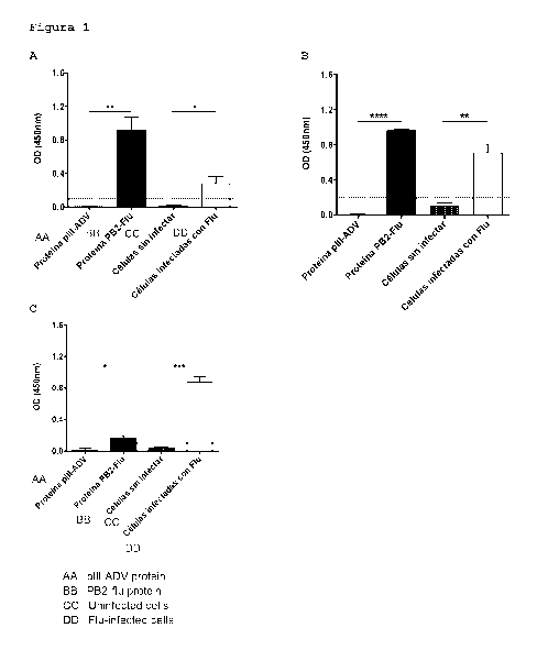
FIG. 1: Detection of flux PB2 protein by the monoclonal antibodies produced by
the hybridomas
1A3E2 and 2F11B1, by an indirect ELISA assay. The plate was activated with 50
ng of purified
recombinant Flu PB2 protein, 50 ng ADV pill protein (As specificity control)
and 20 pg of
uninfected MDCK cells (used as specificity control) and infected with Flu.No
antigen control
wells were included with primary antibody, with HRP conjugated mouse anti IgG
(without
activating) and wells without antigen or primary antibody, only with mouse
anti IgG antibody
(HRP), data not shown in the graph. The wells were then incubated with the
anti PB2 antibodies
4
Date Recue/Date Received 2021-06-28
CA 03125206 2021-06-28
from the hybridoma 1A3E2, in the amount of 170 ng (A), the hybridoma 211B1 in
the amount
of 170 ng (B) and the Anti Influenza polyclonal antibody to PB2 protein
antibody, catalog
Number G1X125926 (GeneTex) used in the amount of 170 ng (C). The data shown in
the graph
expresses the absorbance (in OD, optical density) detected at 450 nm, emitted
by the
conversion of the Tetramethylbenzidine substrate to a colored compound,
catalyzed by the
Enzyme Horseradish peroxidase (HRP) conjugated to a mouse anti mouse IgG
antibody that
specifically Binds to antibodies secreted by the geneetex hybridomas 1 A3E2,
4D8C6 And
GTX125926. The values correspond to the average +/- the standard deviation of
the
absorbance emitted by each sample in at least two independent experiments.
Where, * P <ti;
0.05***********************************************************
***************** ****** ***************************************
******************************
FIG. 2: Determination of sensitivity of monoclonal antibodies produced by the
hybridomas
1A3E2 and 2F11B1 in the detection of the flux PB2 protein. ELISA plates were
activated with
1: 2 serial dilutions, starting with 50 ng of PB2 protein and ending with 0.04
ng. The wells were
then incubated with the anti PB2 antibodies from the hybridoma 1A3E2, in
amount of 170 ng
(A) and the B11B1 hybridoma in the amount of 170 ng (B).Wells were included
without activating
as a negative control. The data shown in the graph expresses the absorbance at
450 nm emitted
by the conversion of the Tetramethylbenzidine substrate to a colored compound
catalyzed by
the enzyme Horseradish peroxidase (HRP) conjugated to the anti PB2 antibodies
from
hybridomas 1A3E2 and 2F11B1 in the amount of 170 ng (AJYB).The values
correspond to the
average +/- the standard deviation of the absorbance emitted by each sample in
at least two
independent experiments. * * * * P 0, 01; * * * * P 0.001 by the
parametric student's test
by comparing the results of the well called control versus each of the
Dilutions of the PB2
protein.
FIG. 3: Assay of serial dilutions of the anti PB2 Monoclonal antibodies of Flu
produced by the
hybridomas 1A3E2 and 2F11B1, for the detection of purified fluid antigens.
ELISA plates were
activated with 50 ng of Recombinant Flu PB2 Protein and antigen was detected
with 11 serial
dilutions 1: 2 of the anti PB2 1 A3E2 (A) or Fl 1B1 (B) antibodies, starting
from A concentration
Of 3.4 mr/hIE (170 ng per well).The data is expressed as the average +/- the
standard deviation
of the absorbance value emitted at 450 nm of each sample in duplicate, in at
least two
independent experiments. ** ******************** **** * **************** **
***************************************************************
******
FIG. 4: Detection of Flu in clinical samples by Sandwich ELISA, using the
combination of the
monoclonal antibodies secreted by the hybridomas 1A3E2 and 2F11B1.
ELISA plates were activated with 170 ng of antibody secreted by the hybridoma
1A3E2 (anti
Flu), functioning as A capture antibody. The wells activated with the capture
antibody were
incubated with 50 pl of Nasopharyngeal swabs (HNF) samples of patients having
viral
respiratory frames. Ten samples of healthy controls were analyzed as negative
controls.12
samples of Flu positive patients were used as specificity control, 3 samples
of patients positive
for the Parainfluenza virus were included. As a positive control, wells were
included to which
recombinant PB2-Flu protein was added.For detection of the protein captured by
the antibody
1A3E2, antibodies produced by the hybridoma 211B1, conjugated to the enzyme
Horseradish
Peroxidase, were used in a 1:2000 dilution (1.8 ng/ml per well). The data
shown is the median
of the absorbance value emitted at 450 nm of each sample (* * ** * * *** * **
***** * ****
***************************************************************
************** ** ************, and against the viruses used as a specificity
control).
FIG. 5: detection of PB2 protein by indirect ELISA assay, using the whole
monoclonal antibodies
and fragments thereof, secreted by the biotin conjugated 1 A3E2 and Fl1B1
hybridomas.
Date Recue/Date Received 2021-06-28
CA 03125206 2021-06-28
Detection of the PB2 protein from biotin conjugated antibodies is observed. In
black and white,
fragments of the antibody secreted by the hybridomas 1A3E2 and 2F11B1
respectively are
indicated.While in grey the activity of the whole fragments of the antibodies
secreted by the
hybridomas 1A3E2 and 2F11B1, respectively, is presented. The data shown in the
graph
expresses the absorbance at 450 nm emitted by the conversion of the
Tetramethylbenzidine
substrate to a colored compound catalyzed by the enzyme Horseradish peroxidase
(HRP).The
average of the absorbance value emitted at 450 nm of each sample is shown
(where b is equal
to p 0.0001 compared to a; by the 2-way ANOVA test comparing the non sample
well versus
the protein well with all antibodies).
Examples that allow to demonstrate the various applications of the monoclonal
antibodies of
the invention.
Example 1 Determination of the nucleotide sequence encoding the light chains
(VL) and Heavy
(VH) chains of the variable region of the Anti PB2 antibody Flow secreted by
the hybridoma
1A3E2.
The 1 A3E2 hybridoma was grown in DMEM High glucose culture medium
supplemented with
3.7 g/L Sodium bicarbonate and 10% fetal bovine Serum, at 37 C With 10% CO2,
to a cell
density of 700,000 cells/mL. The total RNA was obtained from 3.5 x 10⁶ cells,
performing a
treatment with the Trizol Compound (Invitrogen)Ø5 mg of RNA was used to
generate the cDNA
by PCR reaction with the Primer kit Try 1st Strand cDNA Synthesis, which uses
isotype specific
universal partitioning. The light and heavy chain of the antibody were
amplified according to the
standard operating procedure (SOP) of rapid amplification of the cDNA ends
(RACE) Of
GenScript. The amplified antibody fragments were separately cloned into a
standard cloning
vector.A colony PCR was performed to identify clones having inserts with the
correct size. At
least five colonies with correct sized inserts were sequenced for each
fragment. The sequences
of different clones were aligned and the consensus sequence of these clones
was provided.
The nucleotide sequences of the heavy and light chains of the antibodies
secreted by the
hybridoma 1A3E2 were identified, being Identified With SEQ ID NO 1 And SEQ ID
NO 3 for the
case of heavy chains and SEQ ID NO: 2 and SEQ ID NO: 4 for the case of light
chains.
Example 2 Determination of the nucleotide sequence encoding the light (VL) and
Heavy (VH)
chains of the variable region of the Anti PB2 antibody Flow secreted by the
211B1 hybridoma.
The Fl 1B1 hybridoma was grown in DMEM high glucose culture medium
supplemented With
3.7 G/L sodium Bicarbonate and 10% fetal bovine serum, at 37 C with 10% CO2,
to a cell
density of 700,000 cells/mL. The total RNA was obtained from 3.5 x 10⁶ cells,
performing a
treatment with the Trizol Compound (Invitrogen)Ø5 mg of RNA was used to
generate the cDNA
by PCR reaction with the Primer kit TW 1st Strand cDNA Synthesis, which uses
isotype specific
universal partitioning. The light and heavy chain of the antibody were
amplified according to the
standard operating procedure (SOP) of rapid amplification of the cDNA ends
(RACE) Of
GenScript. The amplified antibody fragments were separately cloned into a
standard cloning
vector.A colony PCR was performed to identify clones having inserts with the
correct size. At
least five colonies with correct sized inserts were sequenced for each
fragment. The sequences
of different clones were aligned and the consensus sequence of these clones
was provided.
From this, the nucleotide sequences of the heavy and light chains of the
antibodies secreted by
the Fl 1B1 hybridoma were determined, the sequences Identified As SEQ ID NO 1
And SEQ
ID NO 3 corresponding to the light chains and the Sequences identified as SEQ
ID NO 1 and
SEQ ID NO.3 to the heavy chains.
Example 3 Assay for Detection of Flu antigens, determination of specificity of
the anti PB2
monoclonal antibodies to Flow purified antigens by indirect ELISA assay.
6
Date Recue/Date Received 2021-06-28
CA 03125206 2021-06-28
This assay is intended to demonstrate the specificity for the protein PB2
protein of The
antibodies produced by the hybridomas 1A3E2 and 2F11B1. Antigen detection was
carried out
by the indirect ELISA technique, where the ELISA plate was activated with 50
ng of purified
antigen for 1 hour at 37 C likewise the plate was activated with 20 pg of cell
Lysates of
uninfected MDCK cells (as a negative control) and infected with Flu a
virusAnother negative
control included was 50 ng of ADV pill protein in an independent well. The
plate was then
washed twice with phosphate buffered saline (PBS) IX/Tween20 0.05%. The plate
was then
blocked for 2 hours at 37 C with PBS IX/Fetal Bovine Serum (FBS) 10%.The
washes were then
repeated and then each of the antibodies (1 A3E2 and F11B1) were incubated at
a final
concentration of 3.4 pg/mL (170 ng per well), diluted In 1 x PBS/10 /0 FBS,
for 1 hour at 37 C
(each antibody on an independent plate).Under the same conditions, in a
different plate, a
control assay was performed using a commercial monoclonal antibody recognizing
the flux PB2
protein (Anti influenza a virus PB2 protein antibody, catalog number
GTX125926, GeneTex) at
a concentration of 3.4 pg/mL.After the incubation time, the washes were
repeated and a
secondary anti Mouse IgG antibody labeled with Horseradish peroxidase enzyme
(Horseradish
peroxidase, HRP) was added to each of the wells in dilution 1 in 2000 (1.8
ng/ml per well) in 1
x PBS/10 /0 FBS, for 1 hour at room temperature (25 C.), in dark.Finally, the
washes were
performed and developed with 50 pL of citrate/tetramethyl benzidine buffer
(TMB, 3-3'-5-5'-
tetrahylbenzidine, 1 mg/mL, Becton Dickinson). To stop the reaction, 50 pL of
2N H2504 was
added and the result was read on an ELISA reader, at 450 nm.To determine that
the secondary
antibody reaction was specified in recognizing the primary antibody and in
addition that the
signal obtained is not caused by non specific binding of the secondary
antibody to the viral
antigen, controls were made in which only the secondary antibody without
primary antibody nor
sample (unactivated well) was used.Another control for determining that the
primary antibody
reaction is specific for the antigen, consisted of the use of antibodies on an
ELISA plate that
has not been activated with antigen (without antigen) or using antibodies on
an ELISA plate that
possessed 50 ng of ADV pill protein Or uninfected cells.The results show that
the monoclonal
antibodies of the invention are capable of recognizing 50 ng of purified
antigen, specifically, as
they do not recognize the ADV pill protein, or uninfected cell proteins (FIG.
1A and 1B). On the
other hand, it was observed that the commercial antibody (FIG. 1C) used in the
assay as A
control, although specific for detection only of the infected cellsit was not
efficient in detecting
the Purified recombinant Flu PB2 protein in our laboratory. All negative
controls used delivered
expected results (data not shown in the figures).
Example 4 Assay for the sensitivity of monoclonal antibodies for the detection
of anti PB2 viral
antigens of Flu.
The assay was performed to determine the maximum protein dilution that the
anti PB2
monoclonal antibodies from Hybridomas 1A3E2 and 2F11B1 are capable of
detecting by
indirect ELISA. For this, the same technique described in example 3 was
occupied, the plate
was activated with 11 serial dilutions 1: 2 Of the flow PB2 protein, starting
with 50 ng of purified
antigen.Anti PB2 1A3E2 or F11B1 antibodies were used at a concentration of 3.4
pg/mL (170
ng/well), diluted in 1 x PBS/10 /0 FBS. The mouse anti IgG detection antibody
was then added
at a dilution of 1: 2000 (1.8 ng/mL per well) and incubated 1 hour at room
temperature (25 C.),
in dark. Finally, the washes were performed and developed with 50 pL of
citrate/tetramethylbenzidine buffer (TMB, 3-3'-5-5'tetramethylbenzidine, 1
mg/mL, Becton
Dickinson).To stop the reaction, 50 pL of 2N H2504 was added and the result
was read on an
ELISA reader, at 450 nm. The results showed that the anti PB2 1 A3E2 antibody
is able to
recognize to 780 picograms (pg) of the Flux PB2 Protein (FIG. 2A). The anti
PB2 antibody from
the F11B1 hybridoma showed the same sensitivity as the anti PB2 antibody 1A3E2
(FIG. 2B).
Controls were included in all the controls that would allow for unspecific
reactions of both the
antibodies, which contained all components of the assay except for the sample
(Protein PB2
Flu, data not shown in the graphs).
7
Date Recue/Date Received 2021-06-28
CA 03125206 2021-06-28
Example 5 Assay for determining the efficiency of monoclonal antibodies to
detect Viral
Antigens of Flu, by indirect ELISA.
The assay was performed to determine the maximum dilution of the anti PB2
monoclonal
antibodies from Hybridomas 1A3E2 and 2F11131, which permit detection of the
viral antigen. To
this, the plate was activated with 50 ng of purified antigen (Protein PB2) and
then the plate was
blocked for 2 hours at 37 C with PBS IX/Fetal bovine Serum (FBS) 10%.Anti PB2
1A3E2 or
F11B1 antibodies were used in 1:2 dilutions, starting from the working
concentration (170 ng)
to dilution 11(0.15 ng) in 1 x 10% FBS/10% FBS. The mouse anti IgG detection
antibody was
then added at a dilution of 1: 2000 (1.8 ng/ml per well), incubated 1 hour at
room temperature
(25 C.), in dark.Finally, the washes were performed and developed with 50 pL
of
citrate/Tetramethylbenzidine buffer (TMB, 3-3'-5-5'- tetramethyl benzidine, 1
mg/mL, Becton
Dickinson). To stop the reaction, 50 pL of 2N H2504 was added and the result
was read on an
ELISA reader, at 450 nm. In FIG. 3, it is observed that the anti PB2 1A3E2
antibody can detect
50 ng of the purified antigen up to 1.3 ng per well (FIG. 3A).0n the other
hand, the anti PB2
F11I31 clone is more efficient than clone 1A3E2, since it recognizes 50 ng of
Purified PB2 with
almost all dilutions made (FIG. 3B). The negative control included in this
assay corresponds to
a non sample containing well (PB2 protein), was blocked With 1 x PBS/10 /0
FBS, primary
antibody (anti PB2 1A3E2 or anti PB2 211B1) was added and further contains HRP
conjugated
mouse anti IgG antibody.
Example 6 clinical Diagnosis of Samples of Flu infected patients using Flu
anti PB2 monoclonal
antibodies by the sandwich ELISA technique.
The availability and concentration of viral proteins is generally very poor in
clinical samples of
nasopharyngeal swabs, whereby it was necessary To modify the previously
performed ELISA
assay. A Sandwich ELISA was performed for this assay, using as a capture
antibody the anti
PB2 antibody from the Flu antibody 1A3E2 and As a detection antibody the anti
PB2 f11B1
Clone of Flu. The anti PB2 f11B1 detection Antibody of Flu was conjugated with
HRP.Wells of
an ELISA plate were activated with 3.4 mr/hlIl (170 ng/well) of anti PB2
antibody from the
antibody 1A3E2 for Flu, which was diluted in PBS IX, and incubated for 1 hour
at 37 C 2 washes
were performed With 0.05% IX Tween20 PBS and then the plate was blocked with
200 mE Of
1 x PBS/10 /0 FBS for 1 hour at 37 C.It was washed again and incubated for 1
hour at 37 C
each well with 50 mE of nasopharyngeal swabs (previously treated) of patients
positive for Flu
according to The Diagnostic Method "D3 Ultra DFA Respiratory Virus Screening
and ID Kit of
DHI (Diagnostics hybridizes) USA", commonly referred to as "viral panel", and
which were
treated as described below.As controls were included: 1) specificity control:
50 mE of sample
of patients diagnosed With PIV were used by the viral panel for anti Flu
antibodies; positive
control: 50 ng Of recombinant PB2-FIU protein; 3) Negative control:
corresponding to samples
of healthy controls.Subsequently, the two corresponding washes were performed
with 0.05%
IX Tween20 PBS and each well Was incubated for 1 hour at room temperature with
50 mE of
the anti PB2 antibody from the HRP conjugated 211B1 antibody (final
concentration of 1.8 ng/ml
per well). Detection antibodies were incubated 1 hour at room temperature (25
C.), in dark.
The plate was then washed 2 times more, developed with 50 pL of TMB Solution
and incubated
for 15 minutes in dark.The reaction was stopped with 50 ml of 2N H2504 and the
reading of
the plate was performed at 450 nm in an ELISA reader (model Epoch), certified
for clinical
diagnosis.
The results obtained for this assay are shown in FIG. 4A, where it can be seen
that the Sandwich
ELISA technique using the antibody (anti PB2) from hybridoma 1A3E2, as capture
antibody and
antibody from hybridoma 211B1-HRP as A detection antibody, allows the antigen
to be detected
in samples of Flu infected patients (FIG. 4A), which were previously confirmed
By direct
immunofluorescence in a certified clinical laboratory using the viral panel.
FIG. 4A shows the
results obtained with the anti PB2 antibodies of Flu, where 12 samples of
patients diagnosed
as Flu were used and as a specificity control, 3 samples of patients positive
for the
8
Date Recue/Date Received 2021-06-28
CA 03125206 2021-06-28
Parainfluenza virus Were included.As a positive control, wells were included
to which purified
Recombinant PB2-Flu protein was added. As a negative control 10 healthy
controls were used.
The results show that antibodies are specified in detecting only positive
patients For Flu and
not healthy controls or infected with other viruses (Ply). All samples
detected by ELISA are
those that exhibit an optical density (OD) above 0.115
This assay demonstrates the versatility of antibodies from antibodies 1A3E2
and anti PB2 anti
PB2, as they are capases from simultaneously binding to the antigen without
competing for the
binding site or interfering with each other. The above allows for the capture
and subsequent
detection of the PB2 protein in patient samples.
Treatment of clinical samples. The samples that were used for the tests were
obtained from the
naophartic swabs contained in universal transport medium (UTM). The samples
were
centrifuged at 14,000 rpm for 4 minutes at room temperature.The supernatant
was then
separated (SN1) from the pellet; the pellet was incubated with 100 mM of RIPA
Buffer (50 MM
Tris HCI pH 8.0, 150 M NaCI, 1% NP -40, 0.5% sodium Deoxycholate, 0.1% SDS,
and a cocktail
of protease inhibitors IX) for 15 minutes at 4 C. vortexing every 5 minutes.
It was then
centrifuged at 14,000 rpm for 4 minutes at room temperature. At the end, the
obtained
supernatant was taken (SN2) and mixed with SN1, vortexing.
It is of sum importance to use both antibodies for the detection of the PB2
protein, due to the
low availability of antigen in the sample. Using a Sandwich ELISA increases
the specificity and
sensitivity in the diagnostic of Flu. Assays were performed where the plate
was activated directly
with clinical samples of nasopharyngeal swabs, then the anti PB2 1A3E2 and
anti PB2 211B1
antibodies were incubated separately.An anti Mouse IgG antibody conjugated
with HRP was
then incubated and the absorbance generated by incubating the antibody complex
plus sample
was evaluated with the TMB substrate, and no positive diagnosis was observed
since the signal
delivered was very low (data not shown).
Performing a diagnostic kit using the Sandwich ELISA technique, where the
plate can be
activated and blocked, decrease the time and cost of performing diagnostics,
as this technique
is easy to perform and analyze as compared to the standard technique (PCR).
The kit need not
be highly trained personnel for making it or analyzing it.
Example 7 clinical Diagnosis of samples of FLU infected patients using FLU
anti PB2
monoclonal antibodies By the sandwich type Luminex technique.
As in the ELISA technique, the availability and concentration of the viral
proteins is generally
very poor in clinical samples of nasopharyngeal swabs, so that they can be
assessed by another
more sensitive technique obtained in the results by the ELISA technique (FIG.
4A).For this
assay, a sanberch luminometer assay was performed, using as a capture antibody
the anti PB2
1A3E2 antibody as capture antibody and anti PB2 211B1 as a detection antibody.
The FLU anti
PB2 Fl 1B1 detection antibody Was conjugated to the biotin fluorophore.Luminex
plates were
activated with 50 magnetic microspheres (internally labeled with red or near
infrared fluorophore
of different intensities) by ml, which were conjugated to the antibody
secreted by the hybridoma
1A3E2 (anti FLU), functioning as capture antibody (at a final concentration of
2.5 mM).The
conjugated microspheres were incubated with 50 ml of Nasopharyngeal swabs
(HNF) samples
of patients having viral respiratory frames, for 2 hours at room temperature
(Approximately 23
C.), stirring at 400 rpm and in dark (covered with aluminum foil). 8 Samples
of healthy controls
were analyzed as negative controls.19 samples of FLU positive patients were
used (according
to the "D3 Ultra DFA Respiratory Virus Screening And ID Kit Of DHI
(Diagnostics hybrid) USA",
commonly referred to as "viral panel", which were treated in the same manner
as mentioned
above, and as a positive control, wells were included to Which Purified PB2-
FLU protein (50
ng) was added.After 2 hours, 2 washes are performed with 100 mR PBS IX Tween20
At 0.05%
for 30 seconds using the manual magnetic washer. For detection of the protein
captured by the
9
Date Recue/Date Received 2021-06-28
CA 03125206 2021-06-28
antibody 1A3E2, antibodies produced by the hybridoma 211B1, conjugated to the
biotin
fluorophore, were used at a concentration of 4 mg/mL diluted in 1% 1 x BSA,
the wells being
incubated with 50 m. the incubation is carried out for 1 hour at room
temperature, in the dark,
stirring at 400 rpm.2 washes are again performed with 100 mB PBS 1 x Tween20
at 0.05% for
30 seconds using the manual magnetic washer. The complex formed by
microspheres
conjugated with antigen plus antigen capture antibody and detection antibody
is incubated with
50 mE of Streptavidin/beta coerythrine at a final concentration of 6 mr/hU.
The incubation is
carried out for 30 minutes at room temperature, in the dark, stirring at 400
rpm.Finally, two more
washing steps are performed and the wells are incubated with 100 mE of the
shear fluid reagent
(reagent used by Luminex equipment for the equipment to read the samples),
shake 5 minutes
at 400 rpm, in dark. The results of the mean fluorescence intensity (MFI) are
then lertwo in the
Luminex 200 equipment, Which through a red laser (621 nm), detects the
recognition region of
the microsphere and the green color laser (511 nm) detects the binding of the
detection antibody
to the analyte.
The results obtained for this assay are shown in FIG. 4B, where it can be seen
that the Luminex
technique, like that obtained by the ELISA technique, using the antibody (anti
PB2) from the
hybridoma 1A3E2, as capture antibody and antibody from the B11B1-HRP Hybridoma
as the
detection antibody, allows the antigen to be detected in samples of FLU
infected patients (FIG.
4A) with high intensity, which were previously confirmed By direct
immunofluorescence in a
certified clinical laboratory using the viral panel. FIG. 4B shows the results
obtained with the
FLU anti PB2 Antibodies, where 19 samples of patients diagnosed as Positive
FLU and 6
samples of healthy controls were used. In addition, wells were used as a
positive control to
which purified PB2-FLU protein was added.The results show that anti PB2
antibodies are
specific in detecting only positive FLU patients and not the controls. All
samples detected as
Positive by Luminex are those showing an MFI above two standard deviations of
the MFI
average of the healthy controls.
This assay, as in the ELISA assay with patient samples, demonstrates the
versatility of
antibodies from the FLU 1 A3E2 and F11B1 hybridomas, as they are capases from
simultaneously binding to the antigen without competing for the binding site
or interfering with
each other and detecting the low availability of antigen in the nasopharyngeal
swab sample.
Example 8: blind Study for the detection of PB2-FLU antigen in clinical
samples, obtained from
patients healing an infection, using FLU anti PB2 monoclonal antibodies, which
Form part of
the respiratory virus multiple detection kit.
Previously, Sandwich ELISA assays were performed where the previous diagnosis
of the
samples to be evaluated was known. Following these tests, a blind study was
performed, where
they were evaluated near 160 samples of nasopharyngeal swabs, without knowing
the
microbiological diagnosis.For all blind study assays, Sandwich ELISA were
performed where
the anti L 1 A3E2 antibody and anti L Fl1B1 antibody were used as the HRP
conjugated
detection antibody. To all assays, wells of an ELISA plate were activated with
3.4 mg/mL (170
ng/well) of the anti L antibody from the FLU 1 A3E2 Hybridoma diluted in PBS
IX for 30 minutes
at 37 C.2 washes were performed with 0.05% SDS Tween 20 and then blocked the
plate with
200 mE of 1 x PBS/10% FBS for 30 min At 37 C washed again and incubated for 1
hour At
37 C each well with 50 mE of nasopharyngeal swabs of patients, which were
evaluated in
Parallel by the standard diagnostic method (PCR), routinely referred to as
"viral panel," and
which were treated as previously described in example 6.As controls were
included: 1)
specificity control: 50 mE of the BSA protein (50 ng); 2) positive control: 50
ng Of recombinant
PB2-FLU protein; 3) negative controls were used: non sample wells and blocked
wells and
incubated with detection antibody.The corresponding washes were then performed
with 0.05%
IX Tween20 PBS and each well Was incubated for 30 min at room temperature (25
C. in dark)
with 50 mE of the anti PB2 antibody from the F11B1 hybridoma, conjugated With
HRP (1.8
ng/mE final concentration). The plate was then washed 2 times more, developed
with 50 pL of
Date Recue/Date Received 2021-06-28
CA 03125206 2021-06-28
TMB Solution and incubated for 15 minutes in dark.The reaction was stopped
with 50 mL of 2N
H2SO4 and the reading of the plate was performed at 450 nm in an ELISA reader
(Model
Epoch), certified for clinical diagnosis.
The results are shown in FIG. 4A, where the ability of antibodies to detect
PB2 protein in clinical
samples is observed, being designed from a chimeric protein. 18 Of 21 positive
PIV patients
were detected, and from these results the diagnostic accuracy of the
antibodies could be
determined, which is shown in table 1.In the table, the two concepts that
define the diagnostic
accuracy are observed in the table, where we have the specificity, ie the
ability of the antibodies
to diagnose negative and negative samples, without detecting false positives,
and on the other
hand, we have the sensitivity, ie the ability of the antibodies to diagnose as
positive those
samples that actually are, without diagnosing false negatives.The results set
forth in the table
show a high percentage of specificity (94%) and sensitivity (86%) of the
antibodies against the
standard technique (PCR).
TABLE 1. Diagnostic Accuracy of anti PB2-FLU antibodies
PIV M.160) Dtagr6gttoo por tegnica .. Capeoifloidad SensIbIltdad
rrferencla PCP
loWdadCrOgs IFailure. APOSitiV00
felite,04
$6k
di0106,141061 Le
rusk
rAlSO4 novitavos Vor4a4or0
Amvstivos
Example 9 Detection of PB2 protein by indirect ELISA assay using the whole
monoclonal
antibodies and fragments thereof.
In this application example it is demonstrated that both the monoclonal
antibody specific against
the PB2 protein can be detected by Indirect ELISA. For this, ELISA plates were
activated with
50 mE of 50 ng of PB2 protein And BSA. The inspecified sites were blocked With
10% FBS
diluted In PBS IX. 170 Ng (3.4 mg/mL) of Fab fragments of antibodies secreted
by the
hybridoma 1A3E2 (anti Flu) and F11B1 (anti Flu), both pre conjugated
biotin.Incubation of biotin
binding molecules (Streptavidin), which is conjugated with HRP (1: 2000
dilution, 75 ng per well)
(FIG. 5, dark grey bar, 1 A3E2 antibody and light grey bar, 2 Fl 1B1
antibody).
Example 10 assay for detection of Flu antigens, using F (ab ') 2 fragments of
the anti MBP anti
PB2 monoclonal antibodies by indirect ELISA assay
This assay is intended to demonstrate the ability to detect fragments of anti
Flu antibodies
produced by the hybridomas 1A3E2 and 2F11B1, by the protein PB2. Prior to the
indirect ELISA
assay, fragmentation of the IgG molecule from each anti Flu antibody was
performed
Fragmentation was performed using The "Thermo Scientific TM Kit of preparation
of F (ab ') 2
Pierce TM fragments (# 10381214, Thermo Scientific), which separates the F (ab
1)2 fragment
and The fc of the antibody of interest, by the use of the pepsin enzyme that
digest the fc
Fragment and then purification steps are performed to separate The F (ab ') 2
fragment from
The digested fc fragment.Following the fragmentation of the antibody, the
purified F (ab ') 2
fraction was checked by the western blot technique. F (ab ') 2 fractions were
conjugated to biotin
molecules using the rapid conjugation kit, Lightning Link rapid biotin type a
(# 370-0010, Issue).
Having all reagents ready, antigen detection was performed by the indirect
ELISA technique,
where the ELISA plate was activated with 50 ng of Purified PB2 antigen for 1
hour at 37 C.Two
negative controls were included, one without sample and another by incubating
the well with 50
ng BSA Protein. The plate was then washed twice with phosphate buffered saline
(PBS)
IX/Tween20 0.05%. The plate was then blocked for 2 hours at 37 C with 1 x
PBS/10% Fetal
11
Date RecuelDate Received 2021-06-28
CA 03125206 2021-06-28
Bovine Serum (FBS). The washes were then repeated and then each of the
antibodies were
incubated, without fractionation and
the biotin conjugated F (ab ') 2 (1 A3E2 and F11B1) fractions, at a final
concentration of 3.4
mg/mL (170 ng per well), diluted In 1 x PBS/10% FBS, for 1 hour at 37 C (each
antibody on an
independent plate). After the incubation time, the washes were repeated and
added to each of
the wells with a biotin (Streptavidin) binding protein labeled with the
Horseradish peroxidase
enzyme, HRP) in dilution 1 in 2000 (25 ng/mE per well) In 1 x/10% FBS/10% FBS,
for 1 hour
at room temperature (25 C.), in dark. Finally, the washes were performed and
developed with
50 L of citrate/Tetramethylbenzidine buffer (TMB, 3-3'-5-5'-
tetrahylbenzidine, 1 mg/ml, Becton
Dickinson). To stop the reaction, 50 pL of 2N H2SO4 was added and the result
was read on an
ELISA reader, at 450 nm.To determine that the secondary antibody reaction was
specified in
recognizing the primary antibody and in addition that the signal obtained is
not caused by non
specific binding of the secondary antibody to the antigen, controls were made
in which only the
secondary antibody without primary antibody nor sample (unactivated well) was
used.Another
control for determining that the primary antibody reaction is specific for the
antigen, consisted
of the use of antibodies on an ELISA plate that has not been activated with
the antigen (no
sample) or using the antibodies on an ELISA plate that possessed 50 ng of the
BSA Protein.
The results show that the monoclonal antibodies of the invention are capable
of recognizing 50
ng of purified antigen, specifically, independent of using the whole antibody
or A fragment
thereof (FIG. 5, black color bar, 1 A3E2 antibody and white color bar,
antibody 211B1).
12
Date Recue/Date Received 2021-06-28