Note: Descriptions are shown in the official language in which they were submitted.
CA 03125320 2021-06-28
DESCRIPTION
Title of Invention:
POLYETHYLENE GLYCOL-MODIFIED FORM OF HEPATOCYTE GROWTH FACTOR
OR ACTIVE FRAGMENT THEREOF
Technical Field
[0001]
The present invention relates to a polyethylene glycol-modified form of a
hepatocyte
growth factor or an active fragment thereof.
Background Art
[0002]
Hepatocyte growth factor is a growth factor having diverse biological effects
and is
known to have an anti-apoptotic effect, an angiogenic effect, a vasodilatory
effect, an anti-organ
fibrosis effect, an anti-epithelial mesenchymal transition effect, and the
like, in addition to an
originally found hepatocyte proliferative effect. Clinical applications of
hepatocyte growth
factor to various diseases have been attempted. However, the hepatocyte growth
factor needs
to be frequently administered in large amounts for sustaining their effects,
because the
hepatocyte growth factor has an in vivo half-life as short as approximately 30
minutes (Non
Patent Literature 1).
[0003]
Natural splicing variants NK1 (Non Patent Literature 2) and NK2 (Non Patent
Literature
3) as well as NK4 developed by a gene recombination technique (Non Patent
Literature 4) are
known as active fragments of the hepatocyte growth factor. These fragments
have been found
to have bio activity in vitro and in vivo.
[0004]
1
Date Recue/Date Received 2021-06-28
CA 03125320 2021-06-28
Polyethylene glycol is a highly biocompatible polymeric macromolecule and is
widely
used as a protein modifying agent aimed at in vivo half-life extension or
reduction in
immunogenicity for protein medicaments.
[0005]
Polyethylene glycol-modified forms have also been reported as to the
hepatocyte growth
factor (Patent Literature 1). A polyethylene glycol-modified form of NK4, an
antagonist
fragment of the hepatocyte growth factor, has also been reported (Patent
Literature 2).
Citation List
Patent Literature
[0006]
Patent Literature 1: US Patent No. 5,977,310 B specification
Patent Literature 2: JP Patent Publication No. 2010-174034 A
Non Patent Literature
[0007]
Non Patent Literature 1: Liu K.X. et al., American Journal of Physiology,
1998, Vol. 275, p.
835-842
Non Patent Literature 2: Jakubczak J.L. et al., Molecular and Cellular
Biology, 1998, Vol. 18,
No. 3, p. 1275-1283
Non Patent Literature 3: Otsuka T. et al., Molecular and Cellular Biology,
2000, Vol. 20, No. 6,
p. 2055-2065
Non Patent Literature 4: Date K. et al., FEBS Letters, 1997, Vol. 420, No. 1,
p. 1-6
Summary of Invention
Technical Problem
[0008]
Although polyethylene glycol modification has desirable effects, it is known
that the
polyethylene glycol modification of proteins having bioactivity can cause
reduction or loss of
the bioactivity, depending on the binding position of polyethylene glycol. For
example,
2
Date Recue/Date Received 2021-06-28
CA 03125320 2021-06-28
although a modified form of a hepatocyte growth factor in which a plurality of
molecules of
polyethylene glycol are bound to random positions of the hepatocyte growth
factor is reported,
the in vivo half-life extending effect is low and further, 30% or more
reduction in bioactivity is
observed for the modified form (Patent Literature 1).
[0009]
Thus, according to conventional techniques, the extension of the in vivo half-
life by
polyethylene glycol modification and the retainment of bioactivity are
considered to be
incompatible with each other. There is a demand for a polyethylene glycol-
modified form that
satisfies both of them at once.
[0010]
Accordingly, an objective of the present invention is to provide a
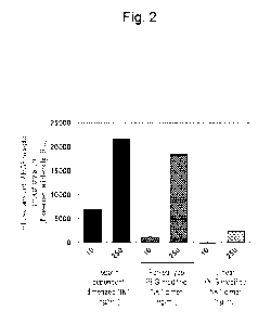
polyethylene glycol-
modified form of a hepatocyte growth factor or an active fragment thereof
which achieves both
an in vivo half-life extending effect by polyethylene glycol modification and
the retainment of
bioactivity.
Solution to Problem
[0011]
The present inventors have conducted diligent studies to attain the objective
and
consequently completed the present invention by finding a polyethylene glycol-
modified form
of a hepatocyte growth factor or an active fragment thereof which achieves
both an in vivo half-
life extending effect by polyethylene glycol modification and the retainment
of bioactivity.
[0012]
The present invention encompasses the following.
(1) A polyethylene glycol-modified form of a hepatocyte growth factor or an
active
fragment thereof, wherein one molecule of forked-type polyethylene glycol is
covalently bound
to two molecules of the hepatocyte growth factor or the active fragment
thereof at each of their
respective carboxyl-terminal regions to form a homodimer.
(2) The polyethylene glycol-modified form of a hepatocyte growth factor or an
active
fragment thereof according to (1) above, wherein the polyethylene glycol-
modified form is
represented by the general formula (I):
3
Date Recue/Date Received 2021-06-28
CA 03125320 2021-06-28
X¨HG F
PEG ¨LV
NX¨HG F
(I)
wherein PEG represents a structural moiety of the forked-type polyethylene
glycol, L represents
a hydrolytically stable branching moiety, HGF represents the hepatocyte growth
factor or the
active fragment thereof, and X represents a binding moiety that provides a
covalent binding of
the forked-type polyethylene glycol to the hepatocyte growth factor or the
active fragment
thereof.
(3) The polyethylene glycol-modified form of a hepatocyte growth factor or an
active
fragment thereof according to (2) above, wherein in the general formula (I),
PEG represents a
structure comprising -(CH2CH20)n-, the structure forms a linear structure or a
branched
structure, and n is 2 to 2300.
(4) The polyethylene glycol-modified form of a hepatocyte growth factor or an
active
fragment thereof according to any of (1) to (3) above, wherein the
polyethylene glycol-modified
form is represented by the general formula (II):
CH30¨(CH2CH20)n¨CH2 7X¨HGF
/ X¨H G Fc
CH30¨(CH2CH20)n¨...0,-, . u .2
(II)
wherein X and HGF are as defined above.
(5) The polyethylene glycol-modified form of a hepatocyte growth factor or an
active
fragment thereof according to any of (1) to (4) above, wherein a distance
between a branch atom
of the branching moiety of the forked-type polyethylene glycol and a
functional group that
provides the covalent binding of the forked-type polyethylene glycol to the
hepatocyte growth
factor or the active fragment thereof is 20 angstroms or shorter.
(6) The polyethylene glycol-modified form of a hepatocyte growth factor or an
active
fragment thereof according to any of (1) to (5) above, wherein the active
fragment of the
hepatocyte growth factor is NK1.
4
Date Recue/Date Received 2021-06-28
CA 03125320 2021-06-28
(7) The polyethylene glycol-modified form of a hepatocyte growth factor or an
active
fragment thereof according to (6) above, wherein the NK1 comprises the amino
acid sequence
represented by SEQ ID NO: 2 in the sequence listing, or an amino acid sequence
having 90%
or higher sequence identity to the amino acid sequence.
(8) A medicament comprising the polyethylene glycol-modified form of a
hepatocyte
growth factor or an active fragment thereof according to any of (1) to (7)
above as an active
ingredient.
Advantageous Effects of Invention
[0013]
The polyethylene glycol-modified form of a hepatocyte growth factor or an
active
fragment thereof according to the present invention has an extended in vivo
half-life and also
retains its bioactivity and as such, can be used as a medicament that can
exert its medicinal effect
with less frequency of administration than that of nonmodified forms.
Brief Description of Drawings
[0014]
[Figure 11 Figure 1 is a diagram showing an SDS electrophoresis image of a His-
Cys-added
human NK1 and a forked-type polyethylene glycol-modified NK1 dimer.
[Figure 21 Figure 2 is a diagram showing the bioactivity of a heparin-
dependent dimerized NK1,
a forked-type polyethylene glycol-modified NK1 dimer and a linear polyethylene
glycol-
modified NK1 dimer.
[Figure 31 Figure 3 is a diagram showing time course of mouse serum levels of
the His-Cys-
added human NK1 (dotted line) and forked-type polyethylene glycol-modified NK1
dimer
(solid line) when each was administered into the tail vein.
Description of Embodiments
[0015]
Date Recue/Date Received 2021-06-28
CA 03125320 2021-06-28
The polyethylene glycol-modified form of a hepatocyte growth factor or an
active
fragment thereof according to the present invention is characterized in that
the hepatocyte
growth factor or the active fragment thereof forms a homodimer, and one
molecule of forked-
type polyethylene glycol is covalently bound to the carboxyl-terminal regions
of both the
monomers of the homodimer to form a dimer.
[0016]
<Hepatocyte growth factor and active fragment thereof>
The hepatocyte growth factor (hereinafter, also referred to as "HGF") is a
growth factor
having diverse bioactivities. HGF is composed of N domain, kringle 1, kringle
2, kringle 3,
kringle 4 and SPH domain in this order from the amino-terminal side. The N
domain, kringle
1, kringle 2, kringle 3 and kringle 4 constitute an a chain, and the SPH
domain constitutes a f3
chain. HGF is biosynthesized as a single-chain pro-HGF and secreted along with
the removal
of its secretory signal sequence. And then, the resulting sequence is
extracellularly processed,
between an arginine residue at position 494 (494th arginine residue, or R494)
and a valine residue
at position 495 (495th valine residue, or V495) counted from the initiating
methionine, by
protease to form a heterodimer chain of the a chain and the p chain bound
through a disulfide
bond, which is active (Miyazawa K. et al., The Journal of Biological
Chemistry, 1996, Vol. 271,
No. 7, p. 3615-3618). The HGF of the present invention means an active HGF
having
bioactivity.
[0017]
A human hepatocyte growth factor (HGF) is biosynthesized as a secretory
protein
consisting of 728 amino acid residues (containing a secretory signal sequence
(31 amino acid
residues from initiating methionine)) (GenBank accession No. M29145) and, when
secreted,
becomes a protein consisting of 697 amino acid residues (SEQ ID NO: 1) by the
removal of the
secretory signal sequence.
[0018]
The hepatocyte growth factor (HGF) described above encompasses an HGF having
the
same amino acid sequence as that of naturally occurring HGF (hereinafter,
referred to as natural
HGF), as well as an amino acid mutant of HGF having an amino acid sequence
derived from
6
Date Recue/Date Received 2021-06-28
CA 03125320 2021-06-28
the amino acid sequence of natural HGF by the deletion, substitution or
addition (or insertion)
of one or several amino acids and having bioactivity as HGF, and further
encompasses even an
HGF having an altered sugar chain moiety of natural HGF and an HGF having no
sugar chain
moiety. The mutant of HGF is preferably a mutant having 90% or higher sequence
identity to
the amino acid sequence of natural HGF, more preferably a mutant having 95% or
higher
sequence identity to the amino acid sequence of natural HGF, further
preferably a mutant having
98% or higher sequence identity to the amino acid sequence of natural HGF.
Examples thereof
include a deleted variant ofHGF with a deletion of 5 amino acid residues
within kringle 1 (which
is a naturally occurring mutant) (Kinosaki M. et al., FEBS Letters, 1998, Vol.
434, p. 165-170),
which has been reported to have higher specific activity than that of natural
HGF in certain cell
lines.
[0019]
The "sequence identity" used in the present specification refers to identity
between two
sequences that can be determined using an algorithm such as BLAST or FASTA, in
which the
two sequences are aligned so as to attain the maximum degree of identity with
or without gaps,
and can generally be calculated as the percentage (%) of the number of
identical amino acids to
the total number of amino acids (including gaps) (Altschul S. et al., Journal
of Molecular
Biology, 1990, Vol. 215, No. 3, p. 403-410; and Altschul S. et al., Nucleic
Acids Research, 1997,
Vol. 25, No. 17, p. 3389-3402).
[0020]
The term "several" used in the present specification refers to an integer of 2
to 10, i.e.,
10,9, 8, 7, 6, 5, 4, 3, or 2.
[0021]
As for the "mutant" of a polypeptide used in the present specification, a
polypeptide that
comprises an amino acid sequence derived from the amino acid sequence of a
natural
polypeptide (natural hepatocyte growth factor (HGF)) by the deletion,
substitution or addition
(or insertion) of one or more amino acids to generate a different sequence
from the natural
polypeptide is referred to as a mutant of the natural polypeptide. Such a
mutant may naturally
occur or may be artificially produced by use of a common technique such as a
gene
7
Date Recue/Date Received 2021-06-28
CA 03125320 2021-06-28
recombination technique. For artificial production of mutants, it is important
not to impair the
activity of the polypeptide. For this purpose, it should be noted that, for
example: when the
mutant is produced, a mutation is introduced neither to an active site of the
polypeptide nor, if
necessary, to near the active site; substitution is performed on conservative
amino acid; and the
HGF activity of the mutant is confirmed by assay.
[0022]
The conservative amino acid substitution generally refers to substitution
between amino
acids similar in chemical properties, electric properties (or polarity and/or
hydrophobicity) or
structural properties. Since such substitution can suppress a marked change in
conformation
of a polypeptide, the polypeptide can retain its activity without large
impairment and, in some
times, can have higher activity than the natural one. Specific examples of
such amino acid
substitution include substitution between acidic amino acids (e.g., aspartic
acid (D) and glutamic
acid (E)), substitution between basic amino acids (e.g., histidine (H), lysine
(K) and arginine
(R)), substitution between aromatic amino acids (e.g., phenylalanine (F),
tyrosine (Y) and
tryptophan (W)), substitution between hydrophilic amino acids (e.g., cysteine
(C), aspartic acid
(D), glutamic acid (E), histidine (H), lysine (K), asparagine (N), glutamine
(Q), arginine (R),
serine (S) and threonine (T)), and substitution between hydrophobic amino
acids (e.g., alanine
(A), phenylalanine (F), isoleucine (I), leucine (L), norleucine (Nle),
methionine (M), valine (V),
tryptophan (W) and tyrosine (Y)).
[0023]
The hepatocyte growth factor (HGF) described above also encompasses a
recombinant
HGF produced by a gene recombination technique on the basis of the amino acid
sequence or
nucleotide sequence of natural HGF.
[0024]
It is known that c-Met is a receptor of the hepatocyte growth factor (HGF).
The diverse
bioactivities of HGF are induced by the binding of the HGF to the HGF
receptor, c-Met.
[0025]
The active fragment of the hepatocyte growth factor (HGF) described above
refers to a
protein that has a portion of the structure of HGF and exerts bioactivity
(agonistic activity) as
8
Date Recue/Date Received 2021-06-28
CA 03125320 2021-06-28
HGF by binding to an HGF receptor. The active fragment of the HGF described
above also
encompasses a protein that has a portion of the structure of HGF and acts as
an antagonist of
HGF (exerts antagonistic activity) by binding to an HGF receptor.
[0026]
Examples of the active fragment of the hepatocyte growth factor (HGF) include
NK1
and NI(2 which are natural splicing variants of HGF. NK1 is composed of N
domain and
kringle 1 on the amino-terminal side of HGF, and NI(2 is composed of N domain,
kringle 1 and
kringle 2 on the amino-terminal side of HGF. It has been reported that each of
these fragments
acts as an agonist or an antagonist of HGF in vivo (Jakubczak J.L. et al.,
Molecular and Cellular
Biology, 1998, Vol. 18, No. 3, p. 1275-1283; and Otsuka T. et al., Molecular
and Cellular
Biology, 2000, Vol. 20, No. 6, p. 2055-2065).
[0027]
Another example of the active fragment of the hepatocyte growth factor (HGF)
includes
NK4 created by a gene recombination technique. NK4 is composed of N domain,
kringle 1,
kringle 2, kringle 3 and kringle 4 on the amino-terminal side of HGF and has
been reported to
act as an antagonist of HGF (Date K. et al., FEBS Letters, 1997, Vol. 420, No.
1, p. 1-6).
[0028]
The active fragment of the hepatocyte growth factor (HGF) described above
encompasses an active fragment having the same amino acid sequence derived
from the amino
acid sequence of natural HGF, as well as a mutant having an amino acid
sequence derived from
the amino acid sequence of natural HGF by the deletion, substitution or
addition of one or
several amino acids and having bioactivity (agonistic activity) as HGF or
antagonistic activity
of HGF, and further encompasses even a mutant having an altered sugar chain
moiety derived
from natural HGF and a mutant having no sugar chain moiety derived from
natural HGF. The
mutant of the active fragment of HGF is preferably a mutant having 90% or
higher sequence
identity to the amino acid sequence of natural HGF, more preferably a mutant
having 95% or
higher sequence identity to the amino acid sequence of natural HGF, further
preferably a mutant
having 98% or higher sequence identity to the amino acid sequence of natural
HGF. Examples
of the highly active NK1 mutant include 1K1 (Lietha D. et al., The EMBO
Journal, 2001, Vol.
9
Date Recue/Date Received 2021-06-28
CA 03125320 2021-06-28
20, No. 20, p. 5543-5555) and M2.2 (Jones D.S. et al., Proceedings of the
National Academy of
Sciences of the United States of America, 2011, Vol. 108, No. 32, p. 13035-
13040).
[0029]
The hepatocyte growth factor or the active fragment thereof described above is
preferably
a hepatocyte growth factor (HGF) (also including a mutant), NK1 (also
including a mutant),
NI(2 (also including a mutant) or NK4 (also including a mutant), preferably a
natural HGF, a
deleted variant of HGF (Kinosaki M. et al., FEBS Letters, 1998, Vol. 434, p.
165-170), NK1
(also including a mutant), NI(2 (also including a mutant) or NK4 (also
including a mutant),
more preferably NK1 (also including a mutant) or NI(2 (also including a
mutant), further
preferably NK1 (also including a mutant), most preferably human NK1 consisting
of the amino
acid sequence represented by SEQ ID NO: 2. The amino acid sequence of SEQ ID
NO: 2
excludes a secretory signal sequence (MWVTKLLPALLLQHVLLHLLLLPIAIPYAEG: SEQ
ID NO: 3) derived from human HGF.
[0030]
The hepatocyte growth factor or the active fragment thereof described above
contains a
mammal-derived amino acid sequence, which is preferably a human-, cat- or dog-
derived amino
acid sequence, more preferably a human-derived amino acid sequence.
[0031]
An artificial sequence such as a tag sequence may be added to the hepatocyte
growth
factor or the active fragment thereof described above for the purpose of
protein purification or
the like. Examples of the tag sequence include a 6 x His tag, HAT tag, c-Myc
tag, FLAG tag,
DYKDDDDK tag, Strep tag, HA tag, GST tag and MBP tag. Further, an artificial
sequence
such as a spacer sequence or a protease-cleavable sequence may be inserted
between the HGF
or the active fragment thereof described above and the tag in order to remove
the tag sequence
after purification. In this case, the hepatocyte growth factor or the active
fragment thereof
described above may contain a cleaved fragment of the artificial sequence such
as a spacer
sequence or a protease-cleavable sequence.
[0032]
Date Recue/Date Received 2021-06-28
CA 03125320 2021-06-28
The hepatocyte growth factor or the active fragment thereof described above
can also be
obtained by use of a method known in the art, such as extraction from a
tissue, protein synthesis
using a gene recombination technique, or biological production using
recombinant cells
expressing the hepatocyte growth factor or the active fragment thereof (or
natural cells
expressing the hepatocyte growth factor). Alternatively, a commercially
available hepatocyte
growth factor or active fragment thereof may be used as the HGF or the active
fragment thereof.
[0033]
The gene recombination technique can be performed according to a method
described in,
for example, Molecular Cloning, 2nd ed., 1989, Cold Spring Harbor Laboratory.
An
exemplary such technique will be described below.
DNA encoding the HGF or the active fragment thereof is provided. The DNA can
be
obtained by isolating cDNA by selection from a cDNA library prepared from
human tissues or
cells, and subjecting the cDNA to a DNA amplification method such as PCR.
Alternatively,
the DNA can be chemically synthesized using, for example, a DNA synthesizer
based on a
phosphoramidite method.
[0034]
The DNA described above is incorporated into an appropriate vector to prepare
an
expression vector. Such a vector contains elements, such as regulatory
sequences, necessary
for expressing the DNA (and if necessary, for secreting a protein). Specific
examples of the
elements include a translation start codon and stop codon, a promoter, an
enhancer, a terminator,
a ribosomal binding site (or a Shine-Dalgarno sequence), a selective marker
sequence, and a
signal sequence. The necessary elements are inserted in the vector.
[0035]
A suitable promoter is selected as the promoter according to host cells.
Examples of
the promoter suitable for cells of Escherichia bacteria which are prokaryotes
include trp
promoter, lac promoter, recA promoter, and XPL promoter. Examples of the
promoter suitable
for cells of Bacillus bacteria include SPO1 promoter, 5P02 promoter, and penP
promoter.
Examples of the promoter suitable for yeast cells include PHO5 promoter, PGK
promoter, GAP
promoter, ADH promoter, and AOX promoter. Examples of the promoter suitable
for plant
11
Date Recue/Date Received 2021-06-28
CA 03125320 2021-06-28
cells include cauliflower mosaic virus (CaMV) promoter. Examples of the
promoter suitable
for insect cells include P10 promoter and polyhedrin promoter. Examples of the
promoter
suitable for mammalian cells include promoters of viruses such as Rous sarcoma
virus, polyoma
virus, fowlpox virus, adenovirus, bovine papilloma virus, avian sarcoma virus,
cytomegalovirus
(SMV), simian virus 40 (SV40), and vaccinia virus, metallothionein promoter,
and heat shock
promoter.
[0036]
The selective marker may be, for example, HIS3 gene, LEU2 gene, TRP1 gene,
URA3
gene, dihydrofolate reductase gene (methotrexate (MTX) resistance), ampicillin
resistance gene,
neomycin resistance gene, kanamycin resistance gene, or the like.
[0037]
A vector suitable for host cells is selected as the expression vector. For
example,
plasmids, phages, cosmids, virus vectors, and artificial chromosomes (e.g.,
BAC and YAC) are
vectors usually used. Examples of the vector for a prokaryote include E. coil-
derived plasmids,
for example, plasmids of pCR series, plasmids of pBR series, and plasmids of
pUC series, and
Bacillus subtilis-derived plasmids, for example, pUB110, pTP5, and pC194.
Examples of the
vector for a yeast include yeast-derived plasmids, for example, plasmids of
pSH series.
Examples of the vector for a plant cell include binary vectors. Examples of
the vector for a
mammalian cell include commercially available vectors such as pBK-CMV,
pcDNA3.1, and
pZeoSV (Invitrogen Corp and Stratagene California), and virus vectors (e.g.,
vectors of
adenovirus, adeno-associated virus, pox virus, herpes simplex virus,
lentivirus, Sendai virus,
vaccinia virus, and SV40).
[0038]
The host cells are prokaryotic cells such as E. coil and Bacillus subtilis, or
eukaryotic
cells such as yeasts, plant cells, and animal cells (e.g., mammalian cells and
insect cells).
When the host cells are transformed or transfected with the expression vector,
an approach
known in the art can be used, for example, electroporation, microinjection,
cell fusion, DEAE
dextran method, calcium phosphate method, particle gun method, or
Agrobacterium method.
[0039]
12
Date Recue/Date Received 2021-06-28
CA 03125320 2021-06-28
It is known that the hepatocyte growth factor or the active fragment thereof
can be
expressed using prokaryotic cells or eukaryotic cells. A recombinant protein
can be expressed
by transiently or stably in the cells by introducing a nucleic acid (DNA)
sequence encoding NK1
protein to the cells. A yeast expression system expresses and secretes a
recombinant protein
into a culture supernatant and is also suitable for the folding of a protein
having a disulfide bond,
and further such yeast can be cultured more conveniently than mammalian cells.
Thus, the
yeast expression system is preferred for the expression of the hepatocyte
growth factor or the
active fragment thereof having a plurality of intramolecular disulfide bonds.
[0040]
The "homodimer" in the present specification means a dimer formed by two
molecules
of a protein having the same amino acid sequence. The "monomer" in the present
specification
refers to one of the protein molecules constituting the dimer. The hepatocyte
growth factor
(HGF) exerts its bioactivity by the homodimerization of two molecules of the
HGF having the
same amino acid sequence and the binding of the homodimer to a HGF receptor
(Gherardi E. et
al., Proceedings of the National Academy of Sciences of the United States of
America, 2006,
Vol. 103, No. 11, p. 4046-4051). The active fragment of HGF exerts its
bioactivity (agonistic
activity) by the homodimerization of two molecules of the active fragment
having the same
amino acid sequence in the presence of a heparin-like substance and the
binding of the
homodimer to the HGF receptor (Chirgadze DY. et al., Nature Structural
Biology, 1999, Vol. 6,
No. 1, p. 72-79). Alternatively, the active fragment of HGF binds as a monomer
to the HGF
receptor in the absence of the heparin-like substance and thereby exerts
antagonistic activity.
[0041]
For example, the crystal structure (PDB ID: 3MKP) of a dimer of human NK1
(Chirgadze DY. et al., Nature Structural Biology, 1999, Vol. 6, No. 1, p. 72-
79) shows that two
molecules of human NK1 form a dimer in a bilateral symmetrically paired
foil'', and an amino
acid sequence within each kringle structure binds to a sequence within the
extracellular domain
of the HGF receptor c-Met.
[0042]
<Forked-type polyethylene g lyco 1>
13
Date Recue/Date Received 2021-06-28
CA 03125320 2021-06-28
Polyethylene glycol (hereinafter, referred to as PEG) is a highly
biocompatible polymer.
It is known that the binding of PEG to a protein provides effects such as
improvement in physical
stability, heat stability, resistance to proteolytic enzymes, and solubility,
decrease in in vivo
distribution volume, and improvement in blood circulation, thereby imparting
clinical
usefulness to the protein (Inada et al., J. Bioact and Compatible Polymers,
1990, Vol. 5, p. 343;
Delgado et al., Critical Reviews in Therapeutic Drug Carrier Systems, 1992,
Vol. 9, p. 249; and
Katre, Advanced Drug Delivery Systems, 1993, Vol. 10, p. 91).
[0043]
PEG is compatible and includes water-soluble poly(ethylene oxide). Typically,
PEG
has a repeat unit structure "-(CH2CH20)n-", and in the PEG, the a terminal
group or a whole
structure of the PEG moiety may vary. The PEG may contain, for example, "-
CH2CH2-
0(CH2CH20)n-CH2CH2-" or "-(OCH2CH2)n0-", depending on the presence or absence
of
substitution of the terminal oxygen. Commonly used PEG is a terminally capped
PEG. In
this terminally capped PEG, the PEG is capped at one of its ends with a
relatively inactive group
(typically, an alkoxy group such as methoxy (-0CH3)), whereas the other end
has a hydroxyl
group or the like that is able to be arbitrarily chemically modified. The PEG
may be available
as a commercial product, or can be prepared by the ring-opening polymerization
of ethylene
oxide according to a known method (Sandler and Karo, Polymer Synthesis,
Academic Press,
New York, Vol. 3, p. 138-161).
[0044]
The PEG to be used for preparing the PEG-modified form of the hepatocyte
growth factor
or the active fragment thereof described above is a forked-type PEG. The
forked-type PEG is
known in the art. Typically, the forked-type PEG has a branching moiety at one
of the ends of
PEG and two hydroxyl groups (which may further undergo chemical alteration)
linked to the
branching moiety. The forked-type PEG is described in, for example,
International Publication
No. W099/45964. The PEG structural moiety of the forked-type PEG may be linear
or
branched. Particularly preferably, the structural moiety of the forked-type
PEG is branched.
[0045]
14
Date Recue/Date Received 2021-06-28
CA 03125320 2021-06-28
The molecular weight of the forked-type PEG used in the PEG-modified form of
the
hepatocyte growth factor or the active fragment thereof described above is
preferably not less
than 5,000 and not more than 240,000, more preferably not less than 10,000 and
not more than
80,000, further preferably not less than 20,000 and not more than 45,000, most
preferably 20,000.
It is known that the in vivo half-life extending effect of PEG modification
correlates with the
molecular weight of PEG (Sundqvist T. et al., Computers and Biomedical
Research, 1988, Vol.
21, No. 2, p. 110-116). PEG having a molecular weight of 20000 or more is
expected to have
a sufficient in vivo half-life extending effect. On the other hand, it is
known that high-
molecular-weight PEG modification reduces distribution in tissues. Therefore,
the molecular
weight is most preferably 20,000.
[0046]
One molecule of PEG is composed of many repeat unit structures "-(CH2CH20)n-".
In
general, the molecular weight of PEG differs among individual molecules and is
therefore
indicated by an average molecular weight. Thus, the molecular weight of the
PEG in the
present specification means an average molecular weight.
[0047]
Depending on a method for binding the PEG to the hepatocyte growth factor or
the active
fragment thereof described above, it is necessary to activate the PEG at its
ends to be used for
covalent binding reaction. PEG activated at the ends with a N-
hydroxysuccinimide ester,
nitrobenzenesulfonate ester, maleimide, o-pyridine disulfide, vinyl sulfone,
iodoacetamide,
carboxylic acid, azide, phosphine or amine structure can be used. Such
activated PEG may be
synthesized by a method known in the art. The activated PEG can also be
obtained as a
commercially available product. Examples
of the activated forked-type PEG include
SUNBRIGHT(R) PTE2 Series from Yuka Sangyo Co., Ltd. In the present
specification, the
functional group added to the ends of PEG so as to exhibit binding reactivity
with a protein is
simply referred to as a "functional group" or a "PEG functional group", and
the PEG having
such a terminal structure is referred to as "PEG activated with functional
group", "activated
PEG" or "PEG containing functional group".
[0048]
Date Recue/Date Received 2021-06-28
CA 03125320 2021-06-28
For covalently binding of the forked-type PEG to the carboxyl-terminal region
of the
hepatocyte growth factor or the active fragment thereof described above, the
forked-type PEG
activated with two functional groups is used. The two functional groups may be
two identical
functional groups, i.e., homofunctional, or may be two different functional
groups, i.e.,
heterofunctional. The forked-type PEG described above is preferably a forked-
type PEG
activated with two identical functional groups. More specifically, the forked-
type PEG
activated with two identical functional groups is preferably represented by
the following formula
(III).
CH30¨(CH2CH20)n¨CH2 7CH2O¨CH2CH20¨functional group
C
esu
CH30¨(CH2CH20)n¨%.,, ,2V CH2O¨CH2CH20¨functional group
(III)
[0049]
In the formula, "-" represents a bond, and "n" is generally in the range of 2
to
approximately 2300. "n" can be appropriately determined depending on the
desired PEG
molecular weight.
[0050]
In this context, the binding of PEG to a protein is also referred to as the
chemical
modification or modification of a protein with PEG, or PEGylation. A covalent
conjugate in
which PEG is covalently bound to the hepatocyte growth factor or the active
fragment thereof
is also referred to as a hepatocyte growth factor or an active fragment
thereof chemically
modified with PEG, or a PEG-modified form of the hepatocyte growth factor or
the active
fragment thereof.
[0051]
For the covalent binding of the PEG to the carboxyl-terminal region of the
hepatocyte
growth factor or the active fragment thereof described above, an amino group (-
NH2), a thiol
group (-SH) and a carboxyl group (-COOH) of an amino acid contained in the
hepatocyte growth
factor or the active fragment thereof, or of a cysteine or a non-natural amino
acid artificially
introduced into the hepatocyte growth factor or the active fragment thereof by
a gene
16
Date Recue/Date Received 2021-06-28
CA 03125320 2021-06-28
recombination technique known in the art can be used. For example, the amino
group is
present in side chains of lysine residues at the 4th and 10th residues counted
from the carboxyl-
terminal amino acid (the most carboxyl-terminal amino acid) of the human
hepatocyte growth
factor (HGF), and can be selectively modified using a N-hydroxysuccinimide
(NHS) ester group
as the PEG functional group. Also, for example, if the thiol group is present
in side chains of
cysteine residues artificially inserted in the carboxyl-terminal regions, the
thiol groups in the
side chains can be selectively modified using a maleimide group as the PEG
functional group
since the thiol groups form neither intramolecular nor intermolecular
disulfide bonds. Further,
for example, the carboxyl group is present in a side chain of aspartic acid at
the 8th residue
counted from the carboxyl-terminal amino acid, a side chain of glutamic acid
at the 2nd residue
counted from the carboxyl-terminal amino acid, a side chain of the carboxyl-
terminal amino
acid glutamic acid, and the carboxyl terminus of human NK1, and can be
modified using an
amino group or the like. As a method for inserting a non-natural amino acid to
the carboxyl-
terminal regions of the hepatocyte growth factor or the active fragment
thereof, a reported
method of introducing azidophenylalanine or azido-Z-lysine, etc. by codon
alteration (JP Patent
Publication (Kokai) No. 2009-207490 A (2009)) can be used. An azide group
contained in
such a non-natural amino acid can be selectively modified using
triallylphosphine.
[0052]
For covalently binding PEG to the carboxyl-terminal region of the HGF or the
active
fragment thereof described above, it is preferred to artificially insert an
amino acid that exhibits
selective reactivity, into the carboxyl-terminal regions of the hepatocyte
growth factor or the
active fragment thereof. Particularly, it is more preferred to artificially
insert a cysteine residue,
an azidophenylalanine residue or an azido-Z-lysine residue into the carboxyl-
terminal regions
of the hepatocyte growth factor or the active fragment thereof, and it is
further preferred to
artificially insert a cysteine residue thereto. In this case, PEG activated
with a maleimide group
as the PEG functional group is preferably used. This permits a site-selective
PEG modification
at the cysteine residues inserted in the carboxyl-terminal regions of the
hepatocyte growth factor
or the active fragment thereof.
[0053]
17
Date Recue/Date Received 2021-06-28
CA 03125320 2021-06-28
<PEG-modified form of hepatocyte growth factor or active fragment thereof>
In the PEG-modified form of the hepatocyte growth factor or the active
fragment thereof
described above, one molecule of the forked-type PEG is covalently bound to
the carboxyl-
terminal region of each monomer (i.e., each one molecule of the hepatocyte
growth factor or the
active fragment thereof) constituting the homodimer, thereby forming a dimer.
The respective
amino termini of the monomers constituting the homodimer of the hepatocyte
growth factor or
the active fragment thereof described above are located close to each other on
the cell membrane
side where the hepatocyte growth factor (HGF) receptor is present, while the
respective carboxyl
termini of the monomers are located close to each other on the intercellular
space side.
Accordingly, the PEG-modified form of the hepatocyte growth factor or the
active fragment
thereof described above can retain the bioactivity of the hepatocyte growth
factor or the active
fragment thereof, because PEG is covalently bound to the carboxyl-terminal
regions of the
hepatocyte growth factor or the active fragment thereof. The PEG modification
of an amino
terminus or its neighboring region of the hepatocyte growth factor or the
active fragment thereof
cannot produce the desired effect.
[0054]
The carboxyl-terminal regions of the hepatocyte growth factor or the active
fragment
thereof to which PEG is covalently bound include the carboxyl-terminal amino
acid (the most
carboxyl-terminal amino acid) or its neighboring region of the hepatocyte
growth factor or the
active fragment thereof. Specifically, the carboxyl-terminal regions include
amino acid
residues from the carboxyl-terminal amino acid to the 10th residue counted
therefrom. For
example, in the human hepatocyte growth factor (HGF) (GenBank accession No:
M29145 (SEQ
ID NO: 5 (nucleotide sequence), SEQ ID NO: 6 (amino acid sequence)), the
carboxyl-terminal
region corresponds to a region of amino acid residues from positions 718 to
728 (to the carboxyl-
terminal amino acid) counted from the initiating methionine as position 1 in
the amino acid
sequence of SEQ ID NO: 6. For example, in the human NK1 (ranging from
glutamine at
position 32 to glutamic acid at position 210 in the amino acid sequence of SEQ
ID NO: 6 as
shown in GenBank accession No. M29145), the carboxyl-terminal region
corresponds to a
region of amino acid residues from positions 200 to 210 (to the carboxyl-
terminal amino acid)
18
Date Recue/Date Received 2021-06-28
CA 03125320 2021-06-28
counted from the initiating methionine as position 1 in the amino acid
sequence of SEQ ID NO:
6. An amino acid natively contained in the hepatocyte growth factor or the
active fragment
thereof (also including a mutant) or an artificially introduced amino acid can
be used as long as
the amino acid residue is positioned in the carboxyl-terminal region. The
carboxyl-terminal
region of the hepatocyte growth factor or the active fragment thereof is
preferably from the
carboxyl-terminal amino acid to the 10th residue counted therefrom, more
preferably from the
carboxyl-terminal amino acid to the 4th residue counted therefrom, most
preferably the
carboxyl-terminal amino acid.
[0055]
The covalent binding of PEG to the carboxyl-terminal region of the HGF or the
active
fragment thereof described above can be carried out by a method known in the
art. For
example, a method for covalently binding PEG to an amino group of a protein is
described in
U.S. Patent No. 4917888 and International Publication No. WO 1987/00056. A
method for
covalently binding PEG to a thiol group of a protein is described in
International Publication
No. WO 1999/55377. A method for covalently binding PEG to a non-natural amino
acid
introduced in a protein is described in Bioorganic & Medicinal Chemistry
Letters, 2004, Vol.
14, p. 5743-5745.
[0056]
The purification or concentration of the PEG-modified form of the hepatocyte
growth
factor or the active fragment thereof described above can be carried out by
use of a method
known in the art after the covalent binding reaction between the hepatocyte
growth factor or the
active fragment thereof and the forked-type PEG. The PEG-modified form of the
hepatocyte
growth factor or the active fragment thereof can be purified or concentrated,
for example, by
using a method such as chromatography using ion exchange, gel filtration, a
hydrophobic
support or an affinity support, or a combination thereof so as to remove an
unreacted hepatocyte
growth factor or active fragment thereof or an unreacted forked-type PEG, or
by-products.
[0057]
The PEG-modified form of the hepatocyte growth factor or the active fragment
thereof
described above is specifically represented by the following formula (I).
19
Date Recue/Date Received 2021-06-28
CA 03125320 2021-06-28
X¨HGF
PEG¨LNV
X¨HGF
(I)
[0058]
In the formula (I), "-" represents a bond, "PEG" represents a structural
moiety of the
forked-type polyethylene glycol, "L" represents a hydrolytically stable
branching moiety, "X"
represents a binding moiety that provides the covalent binding of the forked-
type polyethylene
glycol to the hepatocyte growth factor or the active fragment thereof, and
"HGF" represents the
hepatocyte growth factor or the active fragment thereof. The binding (X) of
the forked-type
polyethylene glycol to the hepatocyte growth factor or the active fragment
thereof is
hydrolytically stable.
[0059]
"PEG" is a structural moiety of the forked-type polyethylene glycol. The
structure
comprising a repeat unit "-(CH2CH20)n-" of polyethylene glycol may form a
linear structure or
may form a branched structure. Examples of PEG having a branched structure
include
dibranched, tribranched and tetrabranched PEG. The number of branches of PEG
may also
include more than four. However, among them, a dibranched PEG is preferred.
The branch
atom (branch point) of the branching structure may be contained in "L".
[0060]
"L" may represent a single group such as "-CH-", or may further contain a
longer atomic
chain (including e.g., an alkylene bond (-CH2-), an ether bond (-0-), an ester
bond (-0-00- or
-00-0-), an amide bond (-CONH- or -NHCO-) or a combination thereof) on the
side of "PEG".
Examples of the "L" group include lysine, glycerol, pentaerythritol and
sorbitol. A particular
branch atom (atom serving as a branch point) in the branching moiety is
typically a carbon atom.
[0061]
"X" is a binding moiety that provides the covalent binding of the forked-type
polyethylene glycol to the hepatocyte growth factor or the active fragment
thereof, and may
represent only an atom derived from the functional group as shown in the
formula (III), provided
that the distance between the branch atom and the functional group is 20
angstroms (A) or
Date Recue/Date Received 2021-06-28
CA 03125320 2021-06-28
shorter; or may further contain a longer atomic chain (including e.g., an
alkylene bond (-CH2-),
an ether bond (-0-), an ester bond (-0-00- or -00-0-), an amide bond (-CONH-
or -NHCO-)
or a combination thereof) on the side of "L", in addition to the atom derived
from the functional
group. The appropriate selection of the functional group depends on the
binding site to the
hepatocyte growth factor or the active fragment thereof, as mentioned above.
The
corresponding "X" in the obtained PEG-modified form of the hepatocyte growth
factor or the
active fragment thereof preferably results from the reaction between an
appropriate reaction site
of the hepatocyte growth factor or the active fragment thereof and the
activated forked-type PEG.
For example, when the activated forked-type PEG contains an activated ester
such as N-
hydroxysuccinimide ester, the binding via an amine site of the hepatocyte
growth factor or the
active fragment thereof forms a corresponding amide bond.
[0062]
In the present specification, the "functional group" is a reactive group at a
terminal
portion of the forked-type polyethylene glycol or a terminal portion of the
atomic chain
described above, for providing a covalent binding to the hepatocyte growth
factor or the active
fragment thereof.
[0063]
More specifically, the PEG-modified form of the hepatocyte growth factor or
the active
fragment thereof described above is preferably represented by the following
formula (II).
CH30¨(CH2CH20),¨CH2 ,X¨HGF
C
V
CH30¨(CH2CH20),¨CH2 X¨HGF
(II)
[0064]
In the formula (II), the definitions of X and HGF are the same as those of the
formula (I).
The number n of "-(CH2CH20)n-" can be appropriately determined so as to attain
the desired
PEG molecular weight (described above).
[0065]
21
Date Recue/Date Received 2021-06-28
CA 03125320 2021-06-28
The "hydrolytically stable" binding refers to a chemical bond (typically,
covalent
binding) that is substantially stable in water (i.e., is not detectably
hydrolyzed under
physiological conditions over a long time). Examples of the hydrolytically
stable binding
include a carbon-carbon bond (e.g., within an aliphatic chain), an ether bond,
an amide bond,
and a urethane bond. In general, the hydrolytically stable binding is a bond
that exhibits a
hydrolysis rate of less than approximately 1 to 2% per day under physiological
conditions.
[0066]
In the PEG-modified form of the hepatocyte growth factor or the active
fragment thereof
described above, the distance between the branch atom of the forked-type PEG
and the
functional group (e.g., a forked-type PEG functional group) that provides the
covalent binding
of the forked-type polyethylene glycol to the hepatocyte growth factor or the
active fragment
thereof is not limited as long as the distance allows the PEG-modified form of
the hepatocyte
growth factor or the active fragment thereof to retain its bioactivity. The
distance is preferably
20 angstroms or shorter, more preferably 1.5 to 19 angstroms, further
preferably 1.5 to 8
angstroms, most preferably 7.2 angstroms.
[0067]
The branch atom of the PEG is an atom serving as the branch point, i.e., a
branch atom,
in "L" in the formula (I). The branch atom is preferably a carbon atom.
[0068]
The distance between the branch atom of the PEG and the PEG functional group
means
a distance of the binding site (e.g., five-membered ring nitrogen atom of a
maleimide group) of
the PEG functional group from the branch atom described above. Specifically,
the distance
refers to a distance from the branch atom in "L" to the binding site of the
functional group in
"X" in the formula (I). The distance between the branch atom of the PEG and
the PEG
functional group can be calculated by, for example, structural simulation.
From the viewpoint
of the PEG-modified form of the hepatocyte growth factor or the active
fragment thereof, the
distance between the branch atom of the PEG and the PEG functional group means
a distance
of half the distance between the respective carboxyl termini of the monomers
(i.e., each of which
is one molecule of the hepatocyte growth factor or the active fragment
thereof) constituting the
22
Date Recue/Date Received 2021-06-28
CA 03125320 2021-06-28
PEG-modified form of the hepatocyte growth factor or the active fragment
thereof (homodimer).
When the distance between the branch atom of the PEG and the PEG functional
group of the
PEG-modified form is, for example, 20 angstroms, the distance between the
respective carboxyl
termini of the monomers is 40 angstroms.
[0069]
For retaining the bioactivity of the PEG-modified form of the hepatocyte
growth factor
or the active fragment thereof described above which forms a homodimer via PEG
modification,
it is desirable that the distance between the respective carboxyl termini of
the monomers (i.e.,
each of which is one molecule of the hepatocyte growth factor or the active
fragment thereof)
constituting the dimer should reproduce the crystal structure of the dimer of
the hepatocyte
growth factor or the active fragment thereof.
[0070]
In human NK1, a cysteine residue at position 206 (which corresponds to a
cysteine
residue at position 175 in the amino acid sequence of SEQ ID NO: 2) counted
from the initiating
methionine at position 1 in the amino acid sequence of SEQ ID NO: 6 forms an
intramolecular
disulfide bond. A subsequent serine residue at position 207 to glutamic acid
at position 210
(which correspond to a serine residue at position 176 to glutamic acid at
position 179 in the
amino acid sequence of SEQ ID NO: 2) can presumably fluctuate its structure.
On the basis
of the crystal structure (PDB ID: 3MKP) of a human NK1 dimer (Chirgadze D. Y.
et al., Nature
Structural Biology, 1999, Vol. 6, No. 1, p. 72-79), the distance between the
respective carboxyl
termini of the monomers (i.e., each of which is one molecule of human NK1) was
structurally
simulated, using Molecular Operating Environment (version 2013.08) from
Chemical
Computing Group, as to the formed dimer of human NK1, on the assumption that
the serine
residue at position 207 to the glutamic acid at position 210 counted from the
initiating
methionine at position 1 could be fluctuate its structure. As a result, the
minimum distance
between the respective carboxyl termini of the monomers was 3 angstroms, and
the maximum
distance therebetween was 38 angstroms (i.e., the acceptable range of the
distance between the
branch atom of the PEG and the PEG functional group is 1.5 angstroms at the
minimum and 19
angstroms at the maximum).
23
Date Recue/Date Received 2021-06-28
CA 03125320 2021-06-28
[0071]
In the case of adding an artificial sequence such as a tag sequence or a
spacer sequence
to the carboxyl termini of the hepatocyte growth factor or the active fragment
thereof described
above, the distance between the respective carboxyl termini of the monomers of
the formed
dimer of the hepatocyte growth factor or the active fragment thereof may be
longer. In the
case of adding, for example, a 6 x His tag, to the carboxyl termini of human
NK1, a region from
the serine residue at position 176 to the glutamic acid at position 179
(serine residue at position
207 to glutamic acid at position 210 counted from the initiating methionine at
position 1 in the
amino acid sequence of SEQ ID NO: 6) in the amino acid sequence of SEQ ID NO:
2 of human
NK1 and further to the 6 x His tag moiety can presumably fluctuate its
structure. As a result
of carrying out the same structural simulation as above, the distance between
the carboxyl
termini of the human NK1 dimer was a minimum distance of 3 angstroms and a
maximum
distance of 88 angstroms (i.e., the acceptable range of the distance between
the branch atom of
the PEG and the PEG functional group is 1.5 angstroms at the minimum and 44
angstroms at
the maximum).
[0072]
Thus, the distance between the respective carboxyl termini of the monomers in
the
formed dimer of the PEG-modified form of the hepatocyte growth factor or the
active fragment
thereof described above is more preferably 3 to 38 angstroms. However, as
mentioned above,
the distance between the respective carboxyl termini of the monomers may be
substantially
extended if an artificial sequence is inserted into the carboxyl termini of
the hepatocyte growth
factor or the active fragment thereof. Examples of the artificial sequence
include peptides
consisting of 2 to 30 arbitrary amino acids, for example, peptides comprising
the tag sequence
described above, for example, a peptide of SEQ ID NO: 4 (HHHHHHC).
[0073]
In the dimerization of the hepatocyte growth factor or the active fragment
thereof by PEG
modification, it is desirable that, when using a linear PEG activated at both
termini with two
functional groups, the PEG has the chain length that provides a distance
between the functional
groups of 40 angstroms or shorter, but the molecular weight of such PEG is not
more than 1,000,
24
Date Recue/Date Received 2021-06-28
CA 03125320 2021-06-28
which therefore cannot produce a sufficient in vivo half-life extending
effect. On the other
hand, in the present invention which uses the forked-type PEG activated with
two functional
groups, the PEG molecular weight is not limited even though the distance
between the branch
atom of the PEG and the PEG functional group falls within the preferred range
of 20 angstroms
or shorter. Furthermore, the modification of the hepatocyte growth factor or
the active
fragment thereof with the forked-type PEG enables to control the distance
between the
respective carboxyl termini of the monomers (i.e., each of which is one
molecule of the
hepatocyte growth factor or the active fragment thereof) constituting the
dimer, thereby enabling
to maintain the bioactivity of the hepatocyte growth factor or the active
fragment thereof even
after PEG modification.
[0074]
A preferred embodiment of the PEG-modified form of the hepatocyte growth
factor or
the active fragment thereof described above is a PEG-modified form of the
hepatocyte growth
factor or the active fragment thereof, wherein the hepatocyte growth factor or
the active
fragment thereof forms a homodimer, and one molecule of forked-type PEG is
covalently bound
to both the monomers of the homodimer at cysteine residues artificially
inserted in the carboxyl-
terminal regions of the monomers so as to form a dimer, and the forked-type
PEG has a distance
of 7.2 angstroms between the branch atom of PEG and the PEG functional group
and has a PEG
molecular weight of 20,000. This PEG-modified form of the hepatocyte growth
factor or the
active fragment thereof retains bioactivity because of the controlled distance
between the
respective carboxyl termini of the monomers constituting the dimer, and
further has an in vivo
half-life extending effect due to the modification with PEG having a molecular
weight of 20,000.
[0075]
In a more preferred embodiment, the PEG-modified form of the hepatocyte growth
factor
or the active fragment thereof described above is represented by the following
formula (IV).
Date Recue/Date Received 2021-06-28
CA 03125320 2021-06-28
0
HGF
CH2-0¨CH2¨CH2-0¨N
CH3-0-(CH2¨CH2-01CH2 \ / 11---
0
c
cH3-0-(cH2-cH2-0)-ncH2z \ 0
Jt----
cH2-0-cH2-cH2-0-N
HGF
0
Distance between the branch atom of the forked-type
PEG and the forked-type PEG functional group:
7.2A
(IV)
The binding between the hepatocyte growth factor or the active fragment
thereof (in the
formula, "HGF") and the maleimide group (functional group) is of a "-S-" bond
generated by
the involvement of the cysteine residues artificially inserted in the carboxyl-
terminal regions of
the hepatocyte growth factor or the active fragment thereof in the binding.
The number n of "-
(CH2CH20)n-" is determined depending on the PEG molecular weight (described
above).
[0076]
<Medicament>
The PEG-modified form of the hepatocyte growth factor or the active fragment
thereof
described above retains bioactivity inherently possessed by the hepatocyte
growth factor or the
active fragment thereof and further has an in vivo half-life extending effect
due to PEG
modification and as such can be used as an active ingredient of a medicament
(which can be
used interchangeably with a "therapeutic drug" or a "pharmaceutical
composition").
[0077]
The bioactivity of the PEG-modified form of the hepatocyte growth factor or
the active
fragment thereof described above can be conveniently measured using cultured
cells. For
example, a c-Met phosphorylation-inducing effect, a cell proliferative effect,
a cell migration
effect and an anti-apoptotic effect which are the effects of the natural
hepatocyte growth factor
26
Date Recue/Date Received 2021-06-28
CA 03125320 2021-06-28
can be used as indexes (Rubin J.S. et al, The Journal of Biological Chemistry,
2001, Vol. 276,
No. 35, p. 32977-32983; Lietha D. et al., The EMBO Journal, 2001, Vol. 20, No.
20, p. 5543-
5555; and Liu Y. American Journal of Physiology, 1999, Vol. 277, No. 4, p. 624-
633).
[0078]
The in vivo half-life of the PEG-modified form of the hepatocyte growth factor
or the
active fragment thereof described above can be calculated by measuring the
concentration in
blood of the PEG-modified form intravenously, intraperitoneally,
subcutaneously or
intradermally administered to experimental animals. Particularly, the
radiolabeling of the
PEG-modified form of the hepatocyte growth factor or the active fragment
thereof described
above conveniently achieves such measurement.
[0079]
The medicament comprising the PEG-modified form of the hepatocyte growth
factor or
the active fragment thereof described above as an active ingredient can be
used in the treatment
of various diseases that can exploit the bioactivity of the hepatocyte growth
factor or the active
fragment thereof. The medicament can be used in the treatment of, for example,
acute
inflammatory disease, chronic inflammatory disease, acute ischemic disease,
chronic ischemic
disease, amyotrophic lateral sclerosis, organ fibrosis, diabetes mellitus,
spinal cord injury,
peritoneal adhesion, or neuropathic pain, and further can be used for
transplantation therapy or
wound healing.
[0080]
The medicament described above can be used as a useful therapeutic drug for a
mammal
(e.g., a mouse, a rat, a hamster, a rabbit, a dog, a cat, a monkey, cattle,
sheep or a human),
particularly, a human. In the case of using the medicament described above as
a useful
therapeutic drug for a human, the hepatocyte growth factor or the active
fragment thereof
described above is preferably based on an amino acid sequence derived from a
human
hepatocyte growth factor.
[0081]
In the mode of administration of the medicament described above, the PEG-
modified
form of the hepatocyte growth factor or the active fragment thereof serving as
an active
27
Date Recue/Date Received 2021-06-28
CA 03125320 2021-06-28
ingredient can be orally or parenterally administered, either alone or as a
medicament blended
with an acceptable carrier. The administration is preferably performed by
subcutaneous,
intramuscular or intravenous injection.
[0082]
Examples of the dosage form for the oral administration of the medicament
described
above include tablets, pills, capsules, granules, syrups, emulsions and
suspensions. These
dosage forms can be produced by methods known in the art and contain a carrier
or an excipient
and an optional additive usually used in the pharmaceutical formulation field.
Examples of the
carrier and the excipient for tablets include lactose, maltose, saccharose,
starch and magnesium
stearate. Examples of the additive can include binders, disintegrants,
preservatives, delayed
releasing agents, colorants, flavoring agents, stabilizers, solubilizers,
thickeners, and emulsifiers.
[0083]
Examples of the dosage form for the parenteral administration of the
medicament
described above include injections, eye drops, ointments, wet dressings,
suppositories,
transnasal absorption agents, transpulmonary absorption agents, transdermal
absorption agents
and local sustained-release agents. These dosage forms can be produced by
methods known
in the art. Solution formulations can be prepared, for example, in a state
where the PEG-
modified form of the hepatocyte growth factor or the active fragment thereof
described above
serving as an active ingredient is dissolved in an aseptic aqueous solution
for use in injections
or suspended and emulsified in extracts, and embedded in liposomes. Solid
formulations can
be prepared, for example, as freeze-dried products of the PEG-modified form of
the hepatocyte
growth factor or the active fragment thereof described above serving as an
active ingredient
supplemented with an excipient such as mannitol, trehalose, sorbitol, lactose
or glucose. Such
solid formulations may be further pulverized for use. Such powders may be
mixed with
polylactic acid, glycolic acid, or the like and solidified for use. Gels can
be prepared, for
example, by dissolving the PEG-modified form of the hepatocyte growth factor
or the active
fragment thereof described above serving as an active ingredient in a
thickener or a
polysaccharide such as glycerin, PEG, methylcellulose, carboxymethylcellulose,
hyaluronic
28
Date Recue/Date Received 2021-06-28
CA 03125320 2021-06-28
acid or chondroitin sulfate. If necessary, these formulations can be
supplemented with the
additive described above.
[0084]
The medicament described above can generally be administered at 0.001 mg to
100
mg/kg/dosage, preferably 0.01 mg to 10 mg/kg/dosage, in the range of once a
month to once a
day, preferably once a month to once a week, though the dose is appropriately
determined
according to the age and body weight of a patient, a disease to be
administered, symptoms, the
mode of administration, an administration route, the molecular weight of PEG,
etc.
Hereinafter, the present invention will be described further specifically with
reference to
Examples. However, the technical scope of the present invention is not limited
by the
illustrated Examples.
Examples
[0085]
(Example 1) Expression of human NK1 protein
The expression of the recombinant human NK1 protein was carried out using a
commercially available yeast expression kit, Multi-Copy Pichia Expression Kit
(Invitrogen
Corp.).
[0086]
DNA encoding the human NK1 protein in which a 6 x His tag and a cysteine
residue
were added in this order (i.e., HHHHHHC (SEQ ID NO: 4) was added) to the
carboxyl terminus
of the human NK1 protein without its secretory signal sequence (hereinafter,
referred to as His-
Cys-added human NK1) was inserted into pPIC9K expression vector included in
the Multi-
Copy Pichia Expression Kit (Invitrogen Corp.) using In-Fusion HD cloning kit
(Takara Bio Inc.).
The resulting plasmid for expression was linearized with a restriction enzyme,
and then used for
gene transfer into GS115 strain included in the kit by electroporation.
[0087]
The bacterial cells after receiving the gene transfer were selectively
cultured for the first
stage on a histidine-deficient agar medium. Then, the survived colonies were
selectively
29
Date Recue/Date Received 2021-06-28
CA 03125320 2021-06-28
cultured for the second stage on an agar medium containing 4 mg/mL Geneticie)
(Roche) to
isolate a yeast strain harboring multiple copies of the introduced gene of
interest. The isolated
strain was precultured in a thermostat bath at 30 C using a BMGY medium, then
transferred to
BMMY medium containing 0.5% methanol, and main-cultured for 3 days in a
thermostat bath
at 20 C to induce the expression of the His-Cys-added human NK1, the protein
of interest.
[0088]
(Example 2) Purification of human NK1 protein
The supernatant of the yeast culture obtained in Example 1 was centrifugally
clarified,
and then subjected to purification of the His-Cys-added human NK1 in the
supernatant using a
nickel resin and a heparin resin.
[0089]
The centrifugally clarified yeast culture supernatant was passed through a
heparin resin
(GE Healthcare) equilibrated in advance with PBS(-) so that the His-Cys-added
human NK1
was attached to the heparin resin. Subsequently, buffers with varying NaCl
concentrations in
PBS(-) ranging from 150 mM to 2000 mM were passed therethrough in the order of
increasing
NaCl concentration to obtain each elution fraction. Each elution fraction was
subjected to SDS
electrophoresis to identify a His-Cys-added human NK1 elution fraction.
[0090]
Next, the His-Cys-added human NK1 elution fraction obtained by heparin resin
purification was passed through cOmplete His-Tag Purification Resin (Roche)
equilibrated in
advance with PBS(-) containing 300 mM NaCl so that the His-Cys-added human NK1
was
attached to the resin. Subsequently, buffers with imidazole (at concentration
range: 0 mM to
500 mM) added to PBS(-) containing 300 mM NaCl was passed therethrough in the
order of
increasing imidazole concentration to obtain each elution fraction. Each
elution fraction was
subjected to SDS electrophoresis to identify a His-Cys-added human NK1 elution
fraction.
The obtained His-Cys-added human NK1 elution fraction was concentrated using
Amicon
Ultra-15 (MWCO of 10000; Merck Millipore) to obtain the His-Cys-added human
NK1, the
protein of interest.
[0091]
Date Recue/Date Received 2021-06-28
CA 03125320 2021-06-28
(Example 3) Synthesis of PEG-modified NK1 dimer
NK1 dimerized by the covalent binding of one molecule of PEG to the carboxyl
termini
of two molecules of the His-Cys-added human NK1 (hereinafter, referred to as a
PEG-modified
NK1 dimer) was synthesized by the following method.
[0092]
To a solution containing the His-Cys-added human NK1 prepared in Example 2 at
the
adjusted concentration of 1.0 mg/mL in PBS(-) containing 1.0 mol/L NaCl(pH
7.4) (hereinafter,
referred to as an NK1 solution), a solution of 38 mg/mL 2-mercaptoethylamine
(hereinafter,
referred to as 2-MEA) was added in a 0.07-fold amount, and the mixture was
incubated at 37 C
for 30 minutes. The incubated NK1 solution was passed through a gel filtration
column (Zeba
desalt spin column 7 KDa; Thermo Fisher Scientific Inc.) equilibrated with a
phosphate buffer
(0.3 mol/L NaCl, 0.002 mol/L EDTA, and 0.1 mol/L phosphate (pH 6.0)) to remove
2-MEA
from the solution.
[0093]
To the NK1 solution after the 2-MEA removal treatment, a forked-type PEG
(SUNBRIGHT PTE2-200MA2, having two functional groups, PEG functional group
being
maleimide group, distance between the branch atom of PEG and the PEG
functional group being
7.2 A (angstroms), molecular weight of 20,000; Yuka Sangyo Co., Ltd.) or a
linear PEG
(SUNBRIGHT DE-100MA, having two functional groups, PEG functional group being
maleimide group, distance between the PEG functional groups being
approximately 400 A
(angstroms), molecular weight of 10,000; Yuka Sangyo Co., Ltd.) was added at a
reaction molar
ratio of 5:1 (PEG: His-Cys-added human NK1). The mixture was allowed to react
at 25 C for
16 to 18 hours to produce a PEG-modified NK1 dimer. PEG-modified NK1 dimer
with a
modification with a forked-type PEG is referred to as a forked-type PEG-
modified NK1 dimer,
and PEG-modified NK1 dimer with a modification with a linear PEG is referred
to as a linear
PEG-modified NK1 dimer.
[0094]
The solution after the reaction containing the PEG-modified NK1 dimer was
diluted 10-
fold with NaCl-free PBS(-) and then passed through an SP support (SP-Sepharose
6 Fast Flow;
31
Date Recue/Date Received 2021-06-28
CA 03125320 2021-06-28
GE Healthcare) to remove an wu-eacted PEG reagent from the solution. The PEG-
modified
NK1 dimer adsorbed on the SP support was eluted with PBS(-) containing 1.0
mol/L NaCl and
then concentrated using Amicon Ultra-30 (MWCO of 30,000; Merck Millipore).
[0095]
Figure 1 shows an SDS electrophoresis image of the His-Cys-added human NK1 and
the
forked-type PEG-modified NK1 dimer.
[0096]
In the forked-type PEG-modified NK1 dimer and the linear PEG-modified NK1
dimer,
the dimerization occurs by the covalent binding of one molecule of PEG to the
artificially added
cysteine residues present at the carboxyl termini of two molecules of the His-
Cys-added human
NK1. The forked-type PEG-modified NK1 dimer is represented by the following
formula (V),
and the linear PEG-modified NK1 dimer is represented by the following formula
(VI).
0
/------s,Cys-6XHis-NK1
CH2-0¨CH2¨CH2-0¨N
\.----
CH3-0-(CH2¨CH2-0)-nCH2 \ /
0
CH3-0-(CH2¨CH2-0)-CHI \ 0
n
)1-----
CH2-0¨CH2¨CH2-0¨N y¨s6XHis-NK1
0
Distance between the branch atom of the forked-type
PEG and the forked-type PEG functional group:
7.2A
(V)
[0097]
In the formula (V), "Cys-6xHis-NK1" represents the His-Cys-added human NK1,
and
the thiol group (-SH) of the cysteine residue artificially inserted at each
carboxyl terminus of the
32
Date Recue/Date Received 2021-06-28
CA 03125320 2021-06-28
human NK1 binds to the maleimide group (functional group) to form a "-S-"
bond. The
number n of "-(CH2CH20)n-" in the formula depends on the PEG molecular weight.
0 0
NK1-6XHis-Cys -."--S.== Cys-6XHis-NK1
s n
0 0
< _______________________________________ >
Distance between PEG functional groups:
approximately 400A
(VI)
[0098]
In the formula (VI), "Cys-6xHis-NK1" and "NK1-6xHis-Cys" each represent the
His-
Cys-added human NK1, and the thiol group (-SH) of the cysteine residue
artificially inserted at
each carboxyl terminus of the human NK1 binds to the maleimide group
(functional group) to
form a "-S-" bond. The number n of "-(CH2CH20)n-" in the formula depends on
the PEG
molecular weight.
[0099]
(Example 4) Bioactivity of PEG-modified NK1 dimer
The bioactivity of the PEG-modified NK1 dimer was evaluated by In-Cell ELISA
using
a phosphorylation inducing activity on HGF receptor present on the cell
surface of a human lung
epithelial cell line A549 as an index. The His-Cys-added human NK1 used as a
comparative
control for bioactivity was mixed in advance with MEM medium containing 1
[ig/mL purified
swine heparin (Sigma-Aldrich Co. LLC) to induce heparin-dependent
dimerization, thereby
allowing the NK1 to form a dimer (hereinafter, referred to as heparin-
dependent dimerized NK1).
[0100]
A549 cells were suspended in MEM medium containing 10% FCS, seeded at a
density
of 1.5 x 10⁴ cells/well on a 96-well plate for imaging (Becton, Dickinson and
Company), and
cultured all night and all day. After reaching 70% confluency of the cells,
the medium was
replaced with serum-free MEM medium, and the cells were cultured for 16 hours
or longer into
33
Date Recue/Date Received 2021-06-28
CA 03125320 2021-06-28
a serum-starved state. To the cells in the serum-starved state, the heparin-
dependent dimerized
NK1, the forked-type PEG-modified NK1 dimer or the linear PEG-modified NK1
dimer was
added (at 10 or 250 ng/mL), and allowed to react at 37 C for 10 minutes.
[0101]
The cells were fixed in PBS(-) containing 4% formalin. Subsequently, the cell
membranes were permeabilized with PBS(-) containing 0.3% Triton-X and 0.6%
hydrogen
peroxide. Then, the cells were blocked with 10% BSA. To the cells thus
blocked, an anti-
phosphorylated c-Met antibody as a primary antibody and a HRP-labeled
secondary antibody
as a secondary antibody were added, and reacted, followed by the addition of 1
x QuantaRed
Enhanced Chemifluorescent HRP Substrate (Thermo Fisher Scientific, Inc.).
After incubation
at room temperature, fluorescence intensity (RFU) at a wavelength of 590 nm
was measured
using a plate reader (Envision; PerkinElmer, Inc.) and used as the induced
amount of HGF
receptor phosphorylation.
[0102]
The results are shown in Figure 2. In Figure 2, the ordinate depicts the
fluorescence
intensity (RFU) that indicates the induced amount of HGF receptor
phosphorylation. The
abscissa depicts the treatment concentration (ng/mL) of the heparin-dependent
dimerized NK1,
the forked-type PEG-modified NK1 dimer or the linear PEG-modified NK1 dimer.
[0103]
The forked-type PEG-modified NK1 dimer retained bioactivity equivalent to that
of the
heparin-dependent dimerized NK1, whereas the bioactivity of the linear PEG-
modified NK1
dimer was considerably diminished as compared with the heparin-dependent
dimerized NK1.
Thus, for dimerizing NK1 by PEG modification while retaining its bioactivity,
it was shown to
be important to use a forked-type PEG that enables the distance between the
carboxyl termini
of NK1 molecules to be controlled.
[0104]
(Example 5) Kinetics in blood of forked-type PEG-modified NK1 dimer
34
Date Recue/Date Received 2021-06-28
CA 03125320 2021-06-28
The kinetics in blood of the forked-type PEG-modified NK1 dimer were evaluated
via
administering [1251] labeled forked-type PEG-modified NK1 dimer to the tail
veins of mice.
The His-Cys-added human NK1 was used as a comparative control for kinetics in
blood.
[0105]
The [1251] labeling of the His-Cys-added human NK1 or the forked-type PEG-
modified
NK1 dimer was performed by the IODO-BEADS method using Na[12511 (Iodine-125
Radionuclide; PerkinElmer, Inc.). The radioactivity of a supernatant and a
precipitate after
TCA precipitation was measured to verify that the [1251] labeling rate of the
protein to be used
for administration was no less than 90%.
[0106]
The [12511 labeled His-Cys-added human NK1 or forked-type PEG-modified NK1
dimer
was administered (at 25 jig/0.5 MBq/kg) to the tail veins of Crlj:CD1 (ICR)
male mice. The
serum level was measured at 5 minutes to 24 hours after administration using
radioactivity as
an index.
[0107]
The results are shown in Figure 3. In Figure 3, the ordinate depicts the
levels (ng
eq./mL) in mouse serum, and the abscissa depicts the time (h) after
administration. The dotted
line depicts the results of the [12511 labeled His-Cys-added human NK1, and
the solid line depicts
the results of the [12511 labeled forked-type polyethylene glycol-modified NK1
dimer.
[0108]
The forked-type PEG-modified NK1 dimer exhibited almost the same time course
of
serum levels as that of the His-Cys-added human NK1 up to 1 hour after
administration, but
reduction in serum levels of the forked-type PEG-modified NK1 dimer at 4 hours
or more after
administration was alleviated as compared with that of the His-Cys-added human
NK1.
Furthermore, at 24 hours after administration, the forked-type PEG-modified
NK1 dimer was
observed in serum with approximately 4.2 times of the level of the His-Cys-
added human NK1.
The half-life (t1/2) was 7.9 hours for the administered His-Cys-added human
NK1, while it was
extended approximately twice to 15.4 hours for the forked-type PEG-modified
NK1 dimer.
Date Recue/Date Received 2021-06-28
CA 03125320 2021-06-28
Thus, the forked-type PEG-modified NK1 dimer was shown to exhibit an in vivo
half-life
extending effect, which is an effect of PEG modification.
Industrial Applicability
[0109]
The polyethylene glycol-modified form of a hepatocyte growth factor or an
active
fragment thereof according to the present invention has an extended in vivo
half-life and retains
its bioactivity and as such can be used as a medicament that can exert its
medicinal effect with
less frequency of administration than that of nonmodified forms.
Free Text of Sequence Listing
[0110]
SEQ ID NO: 1: Amino acid sequence of human HGF containing no secretory signal
sequence
SEQ ID NO: 2: Amino acid sequence of human NK1 containing no secretory signal
sequence
SEQ ID NO: 3: Secretory signal sequence derived from human HGF
SEQ ID NO: 4: Amino acid sequence of a 6 x His tag and a cysteine residue
added to the
carboxyl terminus of human NK1 protein
SEQ ID NO: 5: Nucleotide sequence of human HGF
SEQ ID NO: 6: Amino acid sequence of human HGF
36
Date Recue/Date Received 2021-06-28