Note: Descriptions are shown in the official language in which they were submitted.
CA 03125689 2021-07-05
WO 2020/144166 PCT/EP2020/050186
1
Defensin Fragments for use in therapy or prophylaxis
The present invention relates to specific peptide sequences derived from alpha
defensins and their use in medical therapy and/or prophylaxis.
Anaerobic and aerobic microorganisms, especially bacteria and yeasts, i.e.
unicellular
microorganisms that live with oxygen, without oxygen or are oxygen-tolerant,
can
cause various clinical pictures, for example wound infections and abscesses,
sepsis,
infections, especially in the abdominal cavity, in the urogenital tract, on
the skin or in
the mouth, eyes, ears, and jaw region. Thus, these pathogenic species are
often
already found in the region of the skin and oral cavity, especially in the
inflamed
skin/eczema, the periodontium, the eyes and ears and in the region of the
stomach in
the mucosal folds of the stomach, and in the duodenum, which can then cause
local,
but also in some circumstances systemic acute and chronic inflammations. Even
in the
rather thinly colonized small intestine, a number of facultative anaerobes can
cause
pathological changes of the highly sensitive mucosa of the small intestine; in
the
rectum, the principal site of the bacterial flora, admittedly aerobic bacteria
predominate,
but here too, anaerobic representatives are also capable of causing serious
inflammatory reactions of the mucosa of the colon. Candida ssp. are also found
in the
stool of many individuals and are potentially pathogenic.
At present, in particular such diseases are treated with antibiotics, which
mainly attack
and destroy the cell walls of the bacteria. A big problem that arises when
these
inflammatory diseases are treated with antibiotics is the development of
resistance to
the antibiotics used, which in recent times has progressed even further. This
enables
the pathogenic bacteria/microorganisms to weaken or completely neutralize the
action
of antibiotic substances. If a microorganism then proves to be resistant to
the common
antibiotics, diseases can become life-threatening. The reason why in the past
the
number of multi resistant bacterial strains has increased considerably is
that, owing to
their rapid growth and their short culture period, the bacteria are
continually able to
develop new strategies for neutralizing the antibiotics. Therefore at present,
in addition
to antibiotics, for example also natural, especially plant, and synthetic oils
and
emulsions are used.
CA 03125689 2021-07-05
WO 2020/144166 PCT/EP2020/050186
2
In recent years, antimicrobial peptides, which are part of the natural immune
system
and are vitally important for epithelial defense against infection by
microorganisms,
have gained research and therapeutic appliance interests.
In a healthy person the skin and mucosa form a physical barrier to infection
by
microorganisms. The physical barrier is made up of the stratum corneum in
healthy
skin and, in the mucosa, of the mucous layer in which desquamation and mucous
secretion cause a constant renewal of the surfaces, simultaneously with
continuous
elimination of microorganisms that are adhering to the surfaces. In
interaction with the
lipids that are also present in the skin, this physical barrier prevents
microorganisms
from penetrating into the living epidermis.
Leaving aside this physical barrier, however, further factors are also
necessary in order
for the healthy skin and mucosa to defend against infection; among these
factors are
endogenous antimicrobial peptides (AMPs). Lysozyme, for example, is an
antimicrobial
peptide that is present in nasal secretions and can in particular kill Gram-
positive
bacteria. Also known as antimicrobial peptides in the intestinal mucosa are
defensins,
whose presence appears to be necessary especially given that the intestinal
epithelia
is exposed to very large quantities of bacteria. In addition to having a
mucous layer that
is difficult for microorganisms to penetrate, the intestinal mucosa contains
Paneth cells
that secrete human defensin-5 ¨ an alpha defensin ¨ that among other
functions,
protect the stems cells that are important for continuous renewal of the
intestinal
mucosa. In humans, only alpha- and beta-defensins are expressed. While alpha-
defensins are expressed primarily in neutrophils as well as in NK cells and
certain T-
lymphocyte subsets, human defensin 5 and defensin 6 are expressed exclusively
in
Paneth cells of the small intestine, where they contribute in regulating and
maintaining
microbial balance in the intestinal lumen. On the other hand, beta-defensins
are most
widely distributed, being secreted by leukocytes and epithelial cells of many
kinds.
Further known AM Ps are a peptide known as psoriasin, as well as RNas-7, which
represents an effective endogenous broad-spectrum antibiotic in humans.
In addition to the known endogenous antimicrobial peptides, numerous
antibiotics are
also known in the existing art; these include both substances of biological
origin and
synthetically manufactured substances, which are therefore either (as in the
original
CA 03125689 2021-07-05
WO 2020/144166 PCT/EP2020/050186
3
sense) naturally formed low-molecular-weight metabolic products of fungi or
bacteria,
or chemically synthesized therapeutic agents.
Especially in light of the fact that the development of resistance to natural
and synthetic
antibiotics is making microbial infectious diseases increasingly difficult to
treat, a need
also frequently arises for novel antimicrobial active agents that are notable
for few side
effects and for simple manufacture and handling.
The gastrointestinal microenvironment is comprised of a single cell layer
epithelia, a
mucus layer, a local immune system, and the microbiome, and together these
four
components play a crucial role in maintaining homeostasis during times of
health. The
human colon harbors a highly dense microbial community of 1011-1012 cells per
gram
of gut content, and human health is closely linked to the diverse set of
microorganisms
in the intestine collectively known as the gut microbiota. While on one hand,
their
abundance and prevalence are associated with disease ¨ as it can be the case
with
Faecalibacterium prausnitzii in, e.g., inflammatory bowel disease (IBD) and
infectious
colitis, it has been shown, on the other hand, that mucosal species such as
Bacteroides fragilis, and Lactobacillus reuteri can protect against colitis.
As a consequence, when applying antibiotics in case of an infection, the
microbiome
composition in human colon gets majorly affected, and the microbe balance
disturbed.
Defensins are small cationic molecules, characterized by three conserved
disulphide
bonds and represent a main group of AMPs. To date, six alpha-defensins have
been
identified in humans, namely the four Human Neutrophil Peptides (HNP) 1, 2, 3
and 4,
and the two Human Defensin (HD) 5 and 6. While the HN Ps form part of the
armory of
neutrophils, where they participate in systemic innate immunity, the HDs are
expressed
in intestinal Paneth cells. As mentioned above, in the small intestine, Paneth
cells play
a key role in balancing the microbiota composition and in protecting the host
from
invading pathogens by secretion of a variety of AM Ps but most abundantly the
two a-
defensin 5 (HD 5) and -6 (HD 6).
While HNP-1, HNP-2 and HNP-3 only differs in a single amino acid, HNP-4 varies
in its
sequence, has one additional positive charge and exhibits improved
bactericidal
activity compared to HNP-1-31,2. The activity of full length antimicrobial
peptides are
influenced by environmental conditions including salt concentration, pH or
redox
CA 03125689 2021-07-05
WO 2020/144166 PCT/EP2020/050186
4
potential 3-6. Based on its strong antimicrobial activity we used HNP-4 as a
precursor
for a new therapeutic agent with antimicrobial abilities. While large-scale
expression of
accurately folded defensins is a major issue, we focused on small fragments of
HNP-4.
We used a natural occurring protease to digest the full length peptide and
subsequently
identified the generated fragments. We tested these fragments for their
antibacterial
and antifungal potential and analyzed their cytotoxic and hemolytic abilities.
The antimicrobial activity of alpha-defensins has been intensively studied in
the past,
and it has been acknowledged that alterations in their specific sequences can
contribute to major changes of their activity and may even lead to a complete
loss of
antimicrobial activity.
Summary of the invention
The problem to be solved by the present invention is therefore to provide a
new or
alternative preventive and/or therapeutic approach, by means of which
infectious
diseases but also other diseases associated with dysbiotic conditions, e.g.
metabolic
diseases, lung diseases, urogenital diseases, diseases of the mouth, eyes and
ears
and skin diseases, can be prevented and/or effectively treated.
According to the invention, this and other problems are solved by the
provision of
peptides having antimicrobial activity and having an amino acid sequence
derived from
an alpha-defensin fragment, which peptide consists of between 6 and 27, in
particular
the shorter peptide fragments, which can be synthesized as linear peptides,
for
example those having 7, 9, 11, or 13, successive amino acids.
Common for the peptides is that they are fragments of naturally occurring
alpha
defensins, HD-5 and HNP-4 and can be produced by reducing the naturally
occurring
peptides and subjecting them to cleavage using proteases activity.
Surprisingly, a
related peptide, HD-6 which includes a number of predicted cleavage sites,
could not
be cleaved under the same conditions.
These short fragments of naturally occurring alpha-defensins have the
advantage that
they can be synthesized chemically as small linear peptide thus reducing the
cost
significantly compared to the manufacture of full length peptides.
Furthermore, several
CA 03125689 2021-07-05
WO 2020/144166 PCT/EP2020/050186
of the peptides have retained the antibiotic effects of the naturally
occurring full length
defensins, while being non-toxic at efficacious concentrations.
These peptides can be used for modulating the microbiome of intestines and/or
as an
5 antimicrobial agent without inducing major shifts/disturbing the balance
of the healthy
microbiome.
Within the present invention, peptide sequences of alpha-defensins have been
identified which, in antimicrobial tests, showed to have an increased
antimicrobial effect
on certain (in particular: pathogenic) bacteria as compared to the full length
peptide on
one hand, while on the other hand there was no effect of the newly identified
peptides
on the microbial diversity.
These results allow the appliance of the peptides according to the invention
not only for
treating microbial infections and bacterially caused diseases, even those
diseases that
are caused by antibiotic-resistant bacteria, but also for preventing bacterial
infections
and for modulating the gut microbiome and potentially the microbiome of other
epithelial surfaces e.g. lung, skin, genitourinary tract, mouth, eyes, ears
etc.
Accordingly, within the present invention, and as generally understood in the
field,
"modulating the microbiome" means the beneficial influence of the peptides on
the
microorganisms present in the gut and epithelial surfaces. As mentioned above,
gut
microbes are key to many aspects of human health including immune, metabolic
and
neuro behavioral traits. With the peptides for use according to the invention,
the
bacterial diversity of the gut microbiome and potentially the bacterial
diversity of other
epithelial surfaces can be supported and promoted.
With "gut microbiome" the gut of a mammal, in particular of a human, is meant.
Accordingly, a preferred embodiment of the invention is directed to the
peptide for use
in modulating the gut microbiome of humans.
According to an embodiment of the peptide of the invention the alpha-defensin
fragment is a fragment of HD-5 or HNP4.
CA 03125689 2021-07-05
WO 2020/144166 PCT/EP2020/050186
6
HD-5, as mentioned at the outset, is expressed in Paneth cells of the small
intestine.
Including a signal peptide and a prodomain, HD 5 comprises 94 amino acids,
with the
mature peptide comprising amino acid numbers 63 to 94.
HNP4, also as mentioned at the outset, expressed in the granules of
neutrophils.
Including the signal peptide and a prodomain, HNP4 comprises 97 amino acids,
with
the mature HNP4 peptide comprising amino acid numbers 64 to 96.
According to a preferred embodiment of the invention, the peptide for use
according to
the invention consists of between 6 and 27 successive amino acids derived from
HD-5
and consists of
the sequence HD-51_9 ATCYCRTGR (SEQ ID No. 1) or
the reverse sequence of SEQ ID No. 1, RGTRCYCTA (SQ ID No. 2),
modified HD-51_9: Ac¨atcycrtGr¨NH2 (SEQ ID No. 5),
HD-51_13, ATCYCRTGRCATR (SEQ ID No. 34),
HD-51_28, ATCYCRTGRCATRESLSGVCEISGRLYR (SEQ ID No. 12),
HD-57_32, TGRCATRESLSGVCEISGRLYRLCCR (SEQ ID No. 14
HD-510-32, CATRESLSGVCEISGRLYRLCCR (SEQ ID No. 19,
HD-514-32, ESLSGVCEISGRLYRLCCR (SEQ ID No. 25
HD-510-27 CATRESLSGVCEISGRLY (SEQ ID No. 28) or
HD-526_32 LYRLCCR (SEQ ID No. 41) of the attached sequence listing.
In general for amino acid sequences, capital letters designate L-amino acids
and small
letters designate D-amino acids.
In a preferred embodiment the peptide consists of the sequence of
ATCYCRTGR (SEQ ID No. 1),
RGTRCYCTA (SEQ ID No. 2),
Ac¨atcycrtGr¨NH2 (SEQ ID No. 5),
LYRLCCR (SEQ ID No. 41),
ATCYCRTGRCATR (SEQ ID No. 34),
ATCYCRTGRCATRESLSGVCEISGRLYR (SEQ ID No. 12), or
TGRCATRESLSGVCEISGRLYRLCCR (SEQ ID No. 14).
More preferably the peptide consists of the sequence of
CA 03125689 2021-07-05
WO 2020/144166 PCT/EP2020/050186
7
ATCYCRTGR (SEQ ID No. 1),
RGTRCYCTA (SEQ ID No. 2),
Ac¨atcycrtGr¨NH2 (SEQ ID No. 5), or
LYRLCCR (SEQ ID No. 41).
Preferred peptide include those based on HD-51_9:
ATCYCRTGR (SEQ ID No. 1),
RGTRCYCTA (SEQ ID No. 2), or
Ac¨atcycrtGr¨NH2 (SEQ ID No. 5).
According to a preferred embodiment of the invention, the peptide for use
according to
the invention consists of either 7, 9, 11, or 13 successive amino acids.
According to a preferred embodiment of the invention, the peptide for use
according to
the invention consists of 9 successive amino acids derived from HD-5, and
preferably
consists of the sequence ATCYCRTGR (SEQ ID No. 1) or the reverse sequence of
SEQ ID No. 1, RGTRCYCTA (SQ ID No. 2) of the attached sequence listing.
According to another preferred embodiment of the invention, the peptide for
use
according to the invention consists of 11 successive amino acids derived from
HNP4,
and preferably consists of the sequence VCSCRLVFCRR (SEQ ID No. 3), of the
reverse sequence of SEQ ID No. 3, RRCFVLRCSCV (SEQ ID No. 4), or of the
modified HNP-41_11: Ac¨vcscrIvfcrr¨NH2(SEQ ID NO. 6).
In one embodiment, the invention relates to a method of manufacturing the
peptides of
the invention comprising subjecting reduced HD5 or HNP-4 to protease activity,
e.g.
trypsin or chymotrypsin, followed by purification.
The peptides as herein disclosed and described, being derived from HD-5 or
HNP4,
have been shown to exhibit excellent antimicrobial activities against
pathogenic
bacteria, while at the same time not notably influencing the commensal
microbiota, e.g.
the gut microbiota.
According to a preferred embodiment, the peptide for use according to the
invention
comprises L- and/or D-amino acids.
CA 03125689 2021-07-05
WO 2020/144166 PCT/EP2020/050186
8
Presently and as generally understood, an "L-amino acid" refers to a
stereoisomer of a
particular amino acid whose amino group is on the left side in its Fisher
projection while
D-amino acid refers to the other stereoisomer of the amino acid whose amino
group is
on the right side in its Fisher projection. In sequences herein, L-amino acids
are shown
in capital and D-amino acids in small letters.
While most naturally occurring peptides are composed of amino acids in the L-
configuration, D-amino acids have shown strong resistance to proteolytic
degradation.
Thus, according to a preferred embodiment, the peptides according to the
invention
consist of D-amino acids.
According to another embodiment, the peptides according to the invention
consist of L-
amino acids.
According to another embodiment, the peptides according to the invention
consist of a
mixture of D- and L-amino acids, preferably alternating D- and L-amino acids,
or
comprises preferably one L-amino acid, with the remaining amino acids being D-
amino
acids.
According to a preferred embodiment, the peptide for use according to the
invention
comprises N- and/or C-terminal modifications.
With N- and/or C-terminal modifications it is possible to influence/enhance,
e.g., the
stability or the half-life of the peptide according to the invention, in
particular in
environments that promote degradation and/or modification of the free N-/C-
terminal
ends of peptides, e.g. due to proteases present in those environments.
Presently, and as generally understood, the C-terminus (also known as the
carboxyl-
terminus, carboxy-terminus, C-terminal tail, C-terminal end, or COOH-terminus)
is the
end of an amino acid chain (protein or polypeptide), terminated by a free
carboxyl
group (-COOH), and the N-terminus (also known as the amino-terminus, NH2-
terminus,
N-terminal end or amine-terminus) is the start of a protein or polypeptide
referring to
the free amine group (-NH2) located at the end of a polypeptide. The
convention for
CA 03125689 2021-07-05
WO 2020/144166 PCT/EP2020/050186
9
writing peptide sequences is to put the C-terminal end on the right and write
the
sequence from N- to C-terminus.
According to a preferred embodiment, in the peptide for use according to the
invention,
the C-terminal modification is selected from one of the group consisting of: -
Amide, -
Acid, -N-alkyl-Amide, -Aldehyde, -Ester, -p-Nitroanilide, and -7-Amino-4-
Methylcoumarin.
With these modifications, the C-terminal end of the peptide can be protected
without
influencing the antimicrobial activity of the peptide to a major extent.
According to a preferred embodiment, in the peptide for use according to the
invention,
the N-terminal modification is selected from one of the group consisting of
Acetyl-,
Formyl-, Pyroglutamyl-, Fatty acids-, urea-, Carbamate-, and alkylamine-.
It is to be understood, that either both ends, i.e. the N-terminus and the C-
terminus can
be modified with any of the above described modifications, or only one of the
ends, i.e.
either the N- or the C-terminus.
According to a preferred embodiment, in the peptide for use according to the
invention,
the N-terminus, carries an acetyl- (ac) modification, and no modification at
the C-
terminus.
According to a preferred embodiment, the peptide for use according to the
invention,
the peptide consists of (or comprises) D-amino acids, and carries, at the N-
terminus,
an acetyl- (ac) modification, and no modification at the C-terminus.
With an N-terminal acetylation, the charge from the amino terminus of a
peptide is
removed; also, with an acetyl modification, a peptide is meant to imitate its
natural
structure in a protein. In addition, this modification stabilizes the
resulting peptide
towards enzymatic degradation resulting from exopeptidases.
According to a preferred embodiment, the peptide according to the invention
carries a
N-terminal acetyl-modification and a C-terminal amide-modification.
CA 03125689 2021-07-05
WO 2020/144166 PCT/EP2020/050186
With a C-terminal amide modification, the peptide is meant to imitate its
natural
structure in a protein. In addition, this modification avoids the introduction
of additional
charges in the peptide molecule.
5 According to a preferred embodiment, the peptide according to the
invention comprises
9 amino acids, wherein 8 amino acids are D-amino acids, and 1 amino acid is a
L-
amino acid, and further preferably, this peptide comprises an N-terminal
acetyl-
modification and a C-terminal amide-modification.
10 According to a preferred embodiment, the peptide according to the
invention comprises
11 amino acids, wherein all amino acids are D-amino acids, and further
preferably this
peptide comprises an N-terminal acetyl-modification and a C-terminal amide-
modification.
According to another preferred embodiment, the peptide according to the
invention is a
chemically synthesized peptide or a biologically expressed peptide.
A wide variety of methods for chemically synthesizing peptides are known in
the art;
while the chemical synthesis of peptides can be carried out using classical
solution-
phase techniques, these have been replaced in most research and development
settings by solid-phase methods. An overview of peptide synthesis can be
found, e.g.
in Stawikowski etal., ("Introduction to peptide synthesis", Cur. Prot. Prot.
Sci., (2012),
suppl. 69, 18.1.1-18.1.13) 7.
According to an embodiment of the peptide for use of the invention, the
peptide is in an
oxidized or reduced state.
In this connection, "oxidized" refers to the state of the peptide where
disulfide bridges,
which occur in peptides having amino acid residues such as cysteine,
methionine,
tryptophan, histidine and tyrosine. The "reduced" state or from designates the
form of
the peptide not having formed disulfide bonds.
"Biologically expressed" peptide, within the present invention, shall
encompass the
expression of the peptide(s) for use according to the invention by a
genetically
engineered host cell that has been modified to express said peptide(s).
CA 03125689 2021-07-05
WO 2020/144166 PCT/EP2020/050186
11
As used herein, the term "host cell" is presently defined as a cell which has
been
transformed or transfected, or is capable of transformation or transfection by
an
exogenous polynucleotide sequence encoding the peptide for use according to
the
invention.
A variety of host-expression vector systems may be utilized to express the
gene coding
a peptide for use according to the invention. Such host-expression systems
represent
vehicles by which the coding sequences of interest may be produced and
subsequently
purified, but also represent cells which, when transformed or transfected with
the
appropriate nucleotide coding sequences, exhibit the peptide for use gene
product of
the invention in situ.
For recombinant production, host cells can be genetically engineered to
incorporate
expression systems or portions thereof or polynucleotides encoding the
peptides for
use of the invention. Introduction of a polynucleotide into the host cell can
be effected
by methods described in many standard laboratory manuals, such as Davis etal.,
Basic Methods in Molecular Biology, (2012) 8, and Sambrook etal., 19899.
Thus, the polynucleotide encoding the peptide according to the invention, may,
e.g., be
comprised in a vector which is to be stably transformed/transfected into host
cells. In
the vector, the polynucleotide encoding the peptide(s) of the invention is
under control
of an, e.g., inducible promoter, so that the expression of the
gene/polynucleotide can
be specifically targeted, and, if desired, the gene may be overexpressed in
that way.
A great variety of expression systems can be used to produce the polypeptides
of the
invention. Such vectors include, among others, chromosomal, episomal and virus-
derived vectors, e.g., vectors derived from bacterial plasmids, from
bacteriophage, from
transposons, from yeast episomes, from insertion elements, from yeast
chromosomal
elements, from viruses, and vectors derived from combinations thereof, such as
those
derived from plasmid and bacteriophage genetic elements, such as cosmids and
phagemids. The expression system constructs may contain control regions that
regulate as well as engender expression. Generally, any system or vector
suitable to
maintain, propagate or express polynucleotides and/or to express a polypeptide
in a
host may be used for expression in this regard. The appropriate DNA sequence
may
CA 03125689 2021-07-05
WO 2020/144166 PCT/EP2020/050186
12
be inserted into the expression system by any of a variety of well-known and
routine
techniques, such as, for example, those set forth in Sambrook etal., see above
According to a preferred embodiment, the peptide for use according to the
invention is
selected from at least one of the followings:
HD-51-9: ATCYCRTGR (SEQ ID No. 1)
HD-51 -9rev: RGTRCYCTA (SEQ ID No. 2)
HD-51-9m0d: Ac¨atcycrtGr¨NH2 (SEQ ID No. 5)
HNP-41_11: VCSCRLVFCRR (SEQ ID No. 3)
HNP-41-111ev: RRCFVLRCSCV (SEQ ID No. 4)
HNP-41-iimod: Ac¨vcscrlvfcrr¨N H2 (SEQ ID NO. 6)
According to another aspect of the invention, the use of the peptide in
modulating the
microbiome consists of the use in the treatment and/or prophylaxis of
intestinal, lung,
urogenital, mouth, eye, ear or skin or other conditions or diseases associated
with a
dysbiotic condition.
As mentioned above, a healthy gut microbiome, containing diverse bacterial
microorganisms, is mandatory not only for an intact gut, but for overall
health in
mammals, in particular humans. A lower bacterial diversity has been
reproducibly
observed in people with diseases such as, inter alia, inflammatory bowel
disease,
coeliac disease, but also metabolic diseases like obesity and type 2 diabetes
and the
efficacy of e.g. check point inhibitor treatment of cancer is also highly
influenced by the
microbiome and even CNS diseases like schizophrenia has been reported to be
influenced by the microbiome. The association between reduced diversity and
disease
indicates that a species-rich gut ecosystem is more robust against
environmental
influences, as functionally related microbes in an intact ecosystem can
compensate for
the function of other missing species.
Apart from genetically influenced intestinal diseases, also specific food and
dietary
patterns as well as medications can influence the abundance of different types
of
bacteria in the gut. While often associated with a change of the nutrition or
diet positive
effects on the gut microbiome can be observed, and also by using pre- and
probiotic
foods, the peptide according to the invention, due to its natural origin,
provides for a
broader, more convenient and highly efficient tool. Also with the peptide
according to
CA 03125689 2021-07-05
WO 2020/144166
PCT/EP2020/050186
13
the invention, a treatment of subjects is possible that are otherwise highly
sensitive
towards nutritional changes and influences.
With the peptide for use according to the invention, intestinal diseases but
also
diseases of the lungs, the skin and the brain can be efficiently prevented
and/or
treated, by positively influencing the natural microbiome of the gut.
Thus, in a preferred embodiment, the peptide according to the invention is
used in
preventing/treating a disease that is selected from inflammatory bowel
disease, in
particular Crohn's disease, ulcerative colitis, coeliac disease, necrotizing
enterocolitis,
irritable bowel syndrome, tourist diarrhea, gastro-intestinal cancers and
intestinal graft
versus host disease, and from metabolic diseases, preferably diabetes and pre-
diabetes, obesity, NAFLD, NASH, dyslipidemia, and from diseases of the lung,
preferably asthma and COPD, and from diseases of the brain, preferably
schizophrenia, Parkinsonism, bipolar disorder, autism and depression.
With the peptide according to the invention, diseases associated with the
simple
microbiomes of the skin, mouth, eye, ear, vagina or circulation can be
efficiently
prevented and/or treated.
Thus, in a preferred embodiment, the peptide according to the invention is
used in
preventing/treating a disease that is selected from sepsis, atopic dermatitis,
rosacea,
seborrheic dermatitis, eczema, carbuncles, staph infection, candidiasis,
cellulitis,
impetigo, acne, pilonidal cyst, Athletes food, ringworm, molluscum, cutaneous
lymphoma, periodontitis, caries, Dry eye, SjOgrens Disease, conjunctivitis,
blepharitis,
hordeola, chalazia, periorbital cellulitis, dacryocystitis, endolphalmitis,
uveitis, iritis,
mastoiditis, vestibular neuronitis, bullous myringitis, granular myringitis,
otitis externa,
otitis media, bacterial vaginosis, Trichomonas vaginitis, candida, non-
infectious
vaginitis, inflammatory vaginitis.
According to another aspect, the peptide as described herein can also be used
as an
antimicrobial agent against multi drug resistant bacteria induced infections.
Within the present invention it has been found that the peptide according to
the
invention can not only be used to positively influence the natural gut
microbiome, but
CA 03125689 2021-07-05
WO 2020/144166 PCT/EP2020/050186
14
also as a tool for specifically targeting multi drug resistant bacteria, thus,
being an
efficient tool for treating/preventing infections caused by multi-drug
resistant bacteria.
According to another object, the present invention also relates to a
medicament
comprising the peptide according to the invention and a pharmaceutically
acceptable
carrier.
Presently, and as generally understood in the field, a "pharmaceutically
acceptable
carrier" is understood to mean any excipient, additive, or vehicle that is
typically used in
the field of the treatment of the mentioned diseases and which simplifies or
enables the
administration of the product according to the invention to a living being,
and/or
improves its stability and/or activity. The pharmaceutical composition can
also
incorporate binding agents, diluting agents or lubricants. The selection of a
pharmaceutical carrier or other additives can be made on the basis of the
intended
administration route and standard pharmaceutical practice. As pharmaceutical
acceptable carrier use can be made of solvents, extenders, or other liquid
binding
media such as dispersing or suspending agents, surfactant, isotonic agents,
spreaders
or emulsifiers, preservatives, encapsulating agents, solid binding media,
depending
upon what is best suited for the respective dose regime and is likewise
compatible with
the compound according to the invention. An overview of such additional
ingredients
can be found in, for example, Rowe (Ed.) et al.: Handbook of Pharmaceutical
Excipients, 7th edition, 2012, Pharmaceutical Press10
.
A peptide of the present invention can be used for treatment of skin-
conditions when
formulated for topical administration. Methods for topical administration are
known in
the art.
When formulated for topical administration, the composition of the present
invention
may contain ingredients typical in topical pharmaceutical or cosmetic
compositions,
such as a carrier, vehicle or medium. Specifically, the carrier, vehicle, or
medium is
compatible with the tissues to which it will be applied, such as the skin,
hair, nail,
vagina, urethra, ear, oral cavity, nasal passage, respiratory system,
opthalmic region,
and/or mucosa. The compositions and components of the invention are suitable
for
contacting infected tissues or for use in patients in general without undue
toxicity,
incompatibility, instability, allergic response, and the like. As appropriate,
compositions
CA 03125689 2021-07-05
WO 2020/144166 PCT/EP2020/050186
of the invention may comprise any ingredient conventionally used in the fields
under
consideration.
In terms of their form, compositions of this invention may include solutions,
emulsions
5 (including microemulsions), suspensions, creams, lotions, gels, powders,
or other
typical solid or liquid compositions used for application to skin and other
tissues where
the compositions may be used. Such compositions may contain: additional
antimicrobials, moisturizers and hydration agents, penetration agents,
preservatives,
emulsifiers, natural or synthetic oils, solvents, surfactants, detergents,
gelling agents,
10 emollients, antioxidants, fragrances, fillers, thickeners, waxes, odor
absorbers,
dyestuffs, coloring agents, powders, viscosity-controlling agents and water,
and
optionally including anesthetics, anti-itch actives, botanical extracts,
conditioning
agents, darkening or lightening agents, glitter, humectants, mica, minerals,
polyphenols, silicones or derivatives thereof, sunblocks, vitamins, and
phytomedicinals.
15 In certain embodiments, the composition of the invention is formulated
with the above
ingredients so as to be stable for a long period of time, as may be beneficial
where
continual or long-term treatment is intended.
The compositions of the invention may be in the form of controlled-release or
sustained-release compositions, wherein the antimicrobial peptide along with
additional
active agents are encapsulated or otherwise contained within a material, such
that they
are released onto the skin or affected area in a controlled manner over time.
The
compositions of the invention may be contained within or on matrixes,
liposomes,
vesicles, microcapsules, microspheres and the like, or within or on a solid
particulate
material.
Administration of the composition of the invention may be to any affected or
susceptible
region, for example, to the legs, shoulders, back (including lower back),
axilla, palms,
feet, neck, groin, dorsa or the hands or feet, elbows, upper arms, knees,
upper legs,
buttocks, torso, pelvis, or any other part of the body for which treatment or
prevention
of infection may be desired. Such treatment is also contemplated for treating
and/or
dressing wounds, such as cuts, scrapes, and burns to the skin, so as to treat
or prevent
infection of the wounded area.
CA 03125689 2021-07-05
WO 2020/144166 PCT/EP2020/050186
16
The compositions of the invention are suitable for use in physiologic
environments with
a pH ranging from about 4.5 to about 6.3, and thus, the compositions may be
formulated at a similar or equivalent pH. The compositions according to this
invention
may be stored either at room temperature or under refrigerated conditions. The
composition of the invention contains an amount of antimicrobial peptide
effective for
antimicrobial action. Generally, the composition contains from about 0.01%
(wt./vol.) to
about 20% antimicrobial peptide. In certain embodiments, the composition
contains
from about 0.5% to about 10% antimicrobial peptide, such as about 0.5%, about
1%,
about 5%, or about 10% antimicrobial peptide.
The features, characteristics and advantages of the peptide according to the
invention
apply likewise to the medicament according to the invention. Accordingly, the
medicament containing the peptide according to the invention can also be used
for
treating and/or preventing a disease that is selected from inflammatory bowel
disease,
in particular Crohn's disease, ulcerative colitis, coeliac disease,
necrotizing
enterocolitis, irritable bowel syndrome, tourist diarrhea, gastro-intestinal
cancers and
intestinal graft versus host disease, but also metabolic diseases, preferably
diabetes
and pre-diabetes, obesity, NAFLD, NASH, dyslipidemia, and diseases of the
lung,
preferably asthma and COPD; and diseases of the skin such as atopic
dermatitis,
rosacea, seborrheic dermatitis, eczema, carbuncles, staph infection,
candidiasis,
cellulitis, impetigo, acne, pilonidal cyst, Athletes food, ringworm,
molluscum,
cutaneous lymphoma; diseases of the mouth such as periodontitis and caries;
diseases
of the eye such as Dry eye, SjOgrens Disease, conjunctivitis, blepharitis,
hordeola,
chalazia, periorbital cellulitis, dacryocystitis, endolphalmitis, uveitis,
iritis; diseases of
the ear such as mastoiditis, vestibular neuronitis, bullous myringitis,
granular myringitis,
otitis externa, otitis media; diseases of the vagina such as bacterial
vaginosis,
Trichomonas vaginitis, candida, non-infectious vaginitis, inflammatory
vaginitis as well
as sepsis and psychiatric diseases, preferably schizophrenia, Parkinsonism,
bipolar
disorder, depression or autism.
According to another preferred embodiment, the peptide is present as dimer,
preferably
a homodimer. The dimers are preferably bound via a covalent bond, suitably a
disulfide
bond.
CA 03125689 2021-07-05
WO 2020/144166 PCT/EP2020/050186
17
Dimerization of peptides is known in the art. The chemistry currently used for
peptide
dimerization involves chemoselective reactions between unprotected peptides.
Examples are the formation of the following bonds, e.g., Cys-maleimide
thioethers,
disulfides or triazoles.
It is to be understood that the before-mentioned features and those to be
mentioned in
the following cannot only be used in the combination indicated in the
respective case,
but also in other combinations or in an isolated manner without departing from
the
scope of the invention.
The invention is now further explained by means of embodiments resulting in
additional
features, characteristics and advantages of the invention. The embodiments are
of
pure illustrative nature and do not limit the scope or range of the invention.
The features mentioned in the specific embodiments are also features of the
invention
in general, which are not only applicable in the respective embodiment but
also in an
isolated manner in the context of any embodiment of the invention.
The invention is also described and explained in further detail by referring
to the
following drawings:
Figure 1 shows the results of experiments for proving that HD-6
nanonet formation
is not affected by duodenal fluid: (A) shows the chromatograph of HD-6
incubated with
duodenal fluid after reduction with 2 mM TCEP. Only the oxidized and reduced
full-
length peptides were detected, due to their retention time with their m/z
graphs and
their 2-, 3-, 4-, 5- and 6-fold protonated ions and neutral masses. (B) shows
incubated
reduced beads with 200 pg/ml HD-6 or 0.01% HAc (control) and duodenal fluid or
0.9%
NaCI (control). The nanonet formation was not affected as these nets look the
same as
HD-6 with 0.9% NaCI. Magnification bar = 0.2 pm.
Figure 2 shows the results of experiments where incubation of HD-5 and
duodenal
fluid led to many different fragments. HD-5 was incubated with duodenal fluid
after
reduction with 2 mM TCEP. (A) shows the overview of the chromatogram from
incubation of reduced HD-5 with duodenal fluid. All detectable fragments were
marked
in grey (a-m) and listed due to their retention time in (B). The mass-to-
charge (m/z)
graphs of all identified fragments with the detected ions and their neutral
masses. The
CA 03125689 2021-07-05
WO 2020/144166 PCT/EP2020/050186
18
peptides (a), (b), (c), (e), (i), (j), (k), (I) and (m) were chosen for
synthesis and deeper
investigation of their abilities. (C) Here the chosen fragments are listed (in
grey) with
their amino acid sequence and their distribution over the HD-5 sequence.
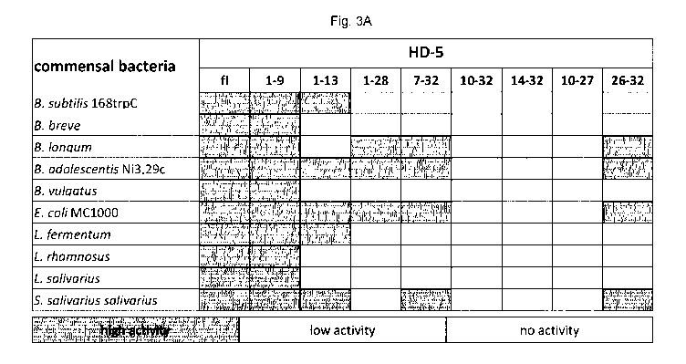
Figure 3 shows the results of experiments proving that HD-5 fragments
are
antimicrobial active peptides against commensal bacteria: (A) shows a table
summarizing the testing of different commensal bacteria due to their
susceptibility to
HD-5 fragments. In this heat map all bacteria are listed and the activity of
the fragments
in RDA against them. In the RDA 2 pg of the full length and 4 pg of each
fragment were
used. An inhibition zone greater than 5 mm was determined as high activity,
between
2.5 and 5 mm as low activity and 2.5 mm were the diameter of the punched well
and
therefore no activity. (B) Here, in diagrams, the original data from (A) are
placed with
mean and standard deviation from at least three independent experiments. (C)
shows
electron microscopy pictures investigating the mode of action of the different
peptides:
E. coil MC1000 was incubated with all different fragments and transmission
electron
microscopy was performed and the resulting phenotypes were analyzed.
Magnification
bars of all pictures are 0.5 pm except the full-length peptide, HD-5fl (1 pm).
Figure 4 shows the results of experiments proving the antimicrobial activity
of the
different fragments against pathogenic bacteria: (A) shows a table summarizing
the
testing of the antimicrobial activity of HD-5 fragments against pathogenic
bacteria. A
heat map system with high activity (inhibition zone in the RDA > 5 mm), low
activity (2.5
to 5 mm) and no activity (2.5 mm) was used. (B) Shows diagrams of the data of
(A)
with mean and standard deviation from at least three independent experiments.
Figure 5 shows the results of experiments proving that HD51_9 containing
cysteine and
arginine substitutions show hardly any antimicrobial effects against E. coil
and S.
aureus mutants. The minimal inhibition concentration (MIC) of HD51_9 and its
variants
was determined against (A) E. coil BW 25113 mutants and (B) S. aureus 5A113
mutants due to the measurement of the optical density (0D600) after 18 h.
Results from
three independent experiments with +1- SEM are represented.
Figure 6 shows diagrams of experiments summarizing that reduction of HD51_9
and
synthetic HD51_9 causes complete dissolving of its antimicrobial activity. The
minimal
inhibition concentration (MIC) was determined of (A) E. coil BW 25113 and (B)
S.
aureus SA113 with several concentrations of reduced and oxidized HD51_9 and
dimer
due to the measurement of the optical density after 18 h. Afterwards bacteria
were
plated out to confirm MIC. Results from three independent experiments with +1-
SEM
are represented.
CA 03125689 2021-07-05
WO 2020/144166 PCT/EP2020/050186
19
Figure 7 shows diagrams of experimental data showing that almost no reduction
in the
metabolic activity of HD51_9 treated cells was observed. To analyze the
metabolic
activity of HD51_9 and dimer treated cells, a WST-1 assay was performed. Cell
lines
were stimulated either with HD51_9 or HD51_9 dimer with concentrations between
3.123
¨ 100 pM and incubated for 24 h or 48 h. The activity is normalized to the
negative
control. As positive control, cells were treated with 2% Triton X-100 while
treatment
with 0.01% acetic acid was used as negative control. The results show the mean
with
+1- SEM of three independent experiments (A, B, C).
Figure 8 shows the anti-microbial mode of action of HD51_9. Cell wall damages
induced
due to HD51_9 differ between E. coli ATCC 25922 and S. aureus SA113. In order
to
detect bacterial cell damages caused by HD51_9, a flow cytometry analysis was
performed. 1.5x10⁶ E. coli ATCC 25922 and S. aureus SA113 were incubated with
different concentrations of HD51_9 (6.25 pM, 12.5 pM and 50 pM) for 1 h.
Either (A)
propidium iodide or the (B) membrane-sensitive DiBAC3(3) dye was used to stain
bacteria. As positive control, 12.5 pM hBD3 was used while untreated cells
function as
negative control. Results from three independent experiments with +1- SEM are
represented. (C) Transmission electron microcopy was performed to evaluate
morphological changes of HD51_9 (200 pg/ml) treated E. coli MC1000. For
comparison,
the full length HD5 (HD5fl) was used while treatment with 0.01% acetic acid
(HAc)
functions as negative control. Bars: Upper left panel: 1pm; Upper right and
lower left:
0.5 pm; lower right panel: 2 pm.
Figure 9 shows results of investigations and experiments regarding Akkermansia
in
feces and susceptibility to HD-51_9 treatment: (A) The amount of Akkermansia
sp. in
feces samples (day 0, 7, 14) collected from mice treated for 7 days with HD-
51_9 or PBS
is increased in HD-51_9 treated animals compared to PBS treated ones (linear
mixed-
effect model; p = 0.075). Here the mean is shown with the 80% confidence
interval
from n=6 per group. In (B) it was tested if Akkermansia muciniphila is
susceptible to
HD-51_9 treatment. (B) shows the growth rate compared to an untreated control
in %
after 72 hours of incubation at 37 C in an anaerobic jar. A MIC from HD-51_9
against of
Akkermansia could not be detected in this assay. The graph shows the mean with
standard deviation of n=3.
Figure 10 shows the pro- and anti-inflammatory immune response of HD51_9
stimulated
human PBMCs. Human peripheral blood mononuclear cells (PBMCs) were isolated
and stimulated with 10 pg/ml LPS (S. typhimurium) and different concentrations
of
HD51_9 followed by an incubation of 24 h. Supernatant of PBMCs was used to
quantify
CA 03125689 2021-07-05
WO 2020/144166 PCT/EP2020/050186
the number of produced cytokines by a multi-analyte kit (LEGENDplex). The
following
cytokines were evaluated: Pro-inflammatory cytokines (A) TNF-a (B) IFN-y (C)
IL-113
(D) IL-6 (E) IL-8 and anti-inflammatory cytokine (F) IL-10. As negative
control untreated
cells were used and the cytokine concentration normalized to the negative
control. The
5 results show the mean with +1- SEM of three independent experiments. For
statistical
analysis a Kruskal-Wallis Test was performed. p>0.05 = ns; p<1.05 = *; p<1.01
= **;
p<1.001 = ***; p<0.0001 =
Figure 11 shows that the toxicity profile of HD51.9 and its dimerized form
displays no
cytotoxicity effects on human cell line. The (A) metabolic activity and (B)
cytotoxicity of
10 HD51.9 and dimer was determined using a WST-1 assay and LDH assay. HT29-
MTX-
E12 cells were stimulated either with HD51.9 or HD51.9 dimer with
concentrations
between 3.123 ¨ 100 pM and incubated for 48 h. The activity was either
normalized to
the negative or positive control. As positive control, cells were treated with
2% Triton X-
100 while treatment with 0.01% acetic acid was used as negative control. The
results
15 show the mean with +1- SEM of three independent experiments. (C)
Further, the
hemolytic activity of HD51.9 was analyzed. Red blood cell suspension was
incubated
with different concentrations of HD51.9. Hemolytic activity was normalized to
the
hemolytic activity of 0.1% Triton X-100. The experiments were carried out in
duplicates.
Figure 12 shows additional tox data. Almost no cytotoxic effects of TR146
cells were
20 observed after peptide treatment. An LDH assay was performed in order to
evaluate
cytotoxic effects of HD51.9 and dimer. Cell lines were stimulated either with
HD51.9 or
HD51.9 dimer with concentrations between 3.123 ¨ 100 pM and incubated for 24 h
0r48
h. The activity was normalized to the positive control. As positive control,
cells were
treated with 2% Triton X-100 while treatment with 0.01% acetic acid was used
as
negative control. The results show the mean with +1- SEM of three independent
experiments. Cytotoxic effects of TR146 cells were evaluated for (A) HD51.9
after 24 h
and 48 h as well as (B) HD51.9 dimer after 24 h and 48 h. Additional, the
cytotoxicity of
(C) HD51.9 and dimer was determined of HT29-MTX-E12 cells after 24 h.
Figure 13A and 13B show the overall fecal microbial community. PCoA of
weighted
and unweighted UniFrac distances using all mice (n = 12 per group) after 1
week of
treatment comparing H D5 fl with HD51-9
Figure 14A and 14B show the overall small intestinal microbial community. PCoA
of
weighted and unweighted UniFrac distances using all mice sacrificed week 1 (n
= 6 per
group) after 1 week of treatment comparing H D5 fl with HD51.9
CA 03125689 2021-07-05
WO 2020/144166 PCT/EP2020/050186
21
Figure 15A and 15B show the bacterial genera differently affected by full
length and
fragmented HD-5 treatment. Linear mixed model adjusted for cages on fecal
microbiota
day 0, 7 and 14 using all mice (note that n= 12 per group day 0 and 7 and n =
6 per
group day 14). Only significantly different genera presented.
Figure 16: Antimicrobial activity of HD51_9 against E coli ATCC 25922 and E.
coli BW
25113 as well its LPS mutants. (A) Cell wall construction of E. coli BW 25113
mutants.
E. coli ATCC 25922 contains the full-length LPS whereas the 0-antigen is
missing in E.
coli BW 25113. The E. coli BW 25113 mutant AwaaG miss the outer core in
contrast to
AwaaY lacking some phosphate residues in the inner core. The last mutant AwaaP
contains an outer core but no phosphate residues in the inner core. (B) The
minimal
inhibition concentration (MIC) of HD51_9 was determined against E coli ATCC
25922
and E. coli BW 25113 mutants with different peptide concentrations due to the
optical
density after 18 h. Results from at least two independent experiments with
Mean +/-
SEM are represented.
Figure 17: Antimicrobial activity of HD51_9 against S. aureus SA113 as well
its cell wall
mutants. (A) S. aureus SA113 cell wall mutants were used to analyze charge-
dependent antimicrobial effects of HD51_9. S. aureus mutant AdItA lacks D-
Alanine
leading to a more negative charge of the peptidoglycan layer. Similar
characteristics
possess the mutant AmprF missing L-Lysin causing a negative charge of the cell
membrane. The S. aureus mutant AtarH contains additional teichoic acid
resulting in a
strengthening of the peptidoglycan layer. (B) The minimal inhibition
concentration
(MIC) of HD51_9 was determined against S. aureus 5A113 and mutants with
different
peptide concentrations due to the optical density after 18 h. Results from two
independent experiments with +/- SEM are represented.
Figure 18: Antimicrobial effects of HD51_9 differentiate compared to the dimer
form
against Gram-negative bacteria. The minimal inhibition concentration (MIC) of
HD51_9
and HD51_9¨dimer was determined against different Salmonella species with
different
peptide concentrations due to the optical density after 18 h. Results from at
least two
independent experiments with Mean +/- SEM are represented.
Figure 19: Antimicrobial effects of HD51_9 differentiate compared to the dimer
form
against Gram-positive bacteria. The minimal inhibition concentration (MIC) of
HD51_9
and HD51_9¨dimer was determined against S. aureus ATCC25923 and the clinical
isolate S. aureus USA300 with different peptide concentrations due to the
optical
density after 18 h. Results from two independent experiments with +/- SEM are
represented.
CA 03125689 2021-07-05
WO 2020/144166 PCT/EP2020/050186
22
Figure 20: Reduced HNP-4 is digested by trypsin. (A) Displays an overview of
the
chromatogram from an incubation of reduced HNP-4 with trypsin after reduction
with 2
mM TCEP. All detectable fragments were marked in red or grey (a-j) and listed
due to
their retention time. The full length peptide is marked as (i) and the
fragment HNP-41_11
(d).
Figure 21: HNP4-derivates display a high antimicrobial activity against
commensal and
pathogenic bacteria
We analyzed the antimicrobial potential of the identified fragment and its
modified
version against commensal and pathogenic bacteria using a RDA. Showing a heat
map, an inhibition zone greater than 5 mm was determined as highly active,
between
2.5 and 5 mm as low active, while a diameter of 2.5 mm (diameter of the
punched well)
was marked as not active. The heat map is based on at least three independent
experiments.
Figure 22: HNP-41-11 and HNP-41-11mod show only minor cytotoxic and hemolytic
activity at high concentrations. We investigated the cytotoxic activity of HNP-
41_11 and
HNP-41_11mod against (A) CaCo2/TC7 or (B) HT29 MTX E 29 cells. We seeded 1500
cells per well and treated them after 24 hours with different peptide
concentrations.
Living cells were determined after 96 hours treatment using a CellTiter Glo2.0
assay.
(C) Hemolytic activity on human erythrocytes of the peptides compared to 0.1%
Triton-
X treatment.
Material and Methods
Bacterial strains
A. baumannii 4-MRGN, K. pneumoniae 4-MRGN, P. aeruginosa AT0027853,
E. faecium 475747, B. longum, L. fermentum, L. salivarius and S. saliva rius
saliva rius
were obtained as clinical isolates from the Robert-Bosch-Hospital (Stuttgart,
Germany).
Akkermansia muciniphila, B. subtilis 168trpC and S. aureus USA300 were
received
from the Institut fur Mikrobiologie und lnfektionsmedizin (Tubingen, Germany).
B. adolescentis Ni3,29c, B. breve were provided by Ardeypharm. B. vulgatus
DSM1447
was obtained from DSMZ and L. rhamnosus were provided by InfektoPharm
(Heppenheim, Germany). Escherichia coli ATCC 25922 was obtained from Deutsche
Sammlung von Mikroorganismen und Zellkulturen GmbH (Bonn, Germany). Clinical
isolate of Salmonella species as well as Staphylococcus aureus USA300,
Staphylococcus aureus ATCC 25923, Staphylococcus aureus SA133 and its mutants
were provided by the Institute of Medical Microbiology and Hygiene Tubingen,
CA 03125689 2021-07-05
WO 2020/144166 PCT/EP2020/050186
23
Germany. Escherichia coil BW25113 as well as its mutants were obtained from
the
Interfaculty Institute for Microbiology and Infection Medicine, Tubingen,
Germany.
Peptides For all experiments oxidized peptides HD-5 and HD-6 (Peptide
Institute,
Osaka, Japan) were used. All fragments, i.e. HD-51_9 and HNP-41_11, HD-51-13,
HD-51-28,
HD-57_32, HD-510-32, HD-514-32, HD-510-27 and HD-526-32 (all peptides of the
invention),
were synthesized by EMC microcollections GmbH (Tubingen, Germany). All
peptides
were dissolved in 0.01% acetic acid (HAc) in similar concentrations.
The following sequences (peptides according to the invention) were tested (N-
>C-
terminus):
HD-51-9: ATCYCRTGR (SEQ ID No. 1)
HD-51 -9rev: RGTRCYCTA (SEQ ID No. 2)
HD-51_9m0d: Ac¨atcycrtGr¨NH2 (SEQ ID No. 5)
HNP-41-11: VCSCRLVFCRR (SEQ ID No. 3)
HNP-41-111ev: RRCFVLRCSCV (SEQ ID No. 4)
HNP-41-iimod: Ac¨vcscrlvfcrr¨N H2 (SEQ ID NO. 6)
Collection of duodenal fluid during gastroscopy.
The human duodenal fluid was collected during a routine gastroscopy from three
healthy individuals. The duodenum was washed with 0.9% NaCI solution, which
was
recollected. Patients gave their written and informed consent after they were
informed.
The sample collection had been previously approved by the Ethical Committee of
the
University Hospital of Tuebingen, Germany.
Screening for fragments of HD-5 and HD-6 using LC/MS
2.5 pg of HD-5 or HD-6 were incubated in 50 mM NH41-1CO3 buffer (pH 8.0)
(Fluka) with
2 mM tris (2-carboxyethyl) phosphine for 15 minutes at 37 C. Afterwards human
duodenal fluid was added and incubated for additional 30 minutes at 37 C. At
last,
formic acid and acetonitrile was added in a final concentration of 0.5% and
10%,
respectively, and analyzed the samples by mass spectrometry. Mass spectrometry
was
performed as a LC/MS system using an Agilent 1200 series HPLC with an Agilent
Advanced Bio Peptide Map (2.1x150 mm, 2.7 pm) column with a flow of 0.4 ml/min
at
55 C column temperature and a 6540 UHD Q-TOF LC/MS system (Agilent) for mass
analysis. The samples were separated by a gradient of acetonitrile in 0.1%
formic acid.
The gradient started at 2% acetonitrile for 4 minutes and then increased
during 35
minutes to 45%. Mass spectrometric analyses were performed in single MS mode
from
CA 03125689 2021-07-05
WO 2020/144166 PCT/EP2020/050186
24
100 to 3400 m/z with positive ion polarity and were analyzed by Agilent MassH
unter
Quantitative Analysis B 06.00 software.
Scanning-Electron-Microscopy
Scanning-Electron-Microscopy was performed as previously described. Briefly,
Protein
A coated beads (Spherotech Inc.) were incubated with reduced HD-6 (200 pg/ml)
in 10
mM sodium phosphate buffer with 1% (w/v) TSB for 1.5 hours at 37 C to allow
net
formation. We subsequently incubated the whole sample with duodenal fluid for
additional 30 minutes at 37 C. As a control 0.01% acetic acid (HAc) was used.
Beads
were centrifuged and fixed in Karnovsky's reagent. The samples were washed
with
PBS and additional fixed with 1% 0504 in H20. Then, they were dehydrated to
100%
ethanol and critical point dried from CO2 and analyzed by scanning electron
microscopy at the Max Planck Institute for Developmental Biology (Tuebingen,
Germany).
Transmission Electron Microscopy
Transmission electron microcscopy experiments were performed as previously
described". 6 x 10⁸ cfu E. coil MC1000 was incubated with 200 pg/ml of each
peptide
for 2 hours. Bacteria were fixed in Karnovsky's fixative, embedded in agarose,
coagulated, cut in small blocks and fixed again in Karnovsky's solution. After
post-
fixation and embedding in glycid ether blocks were cut using an ultra-
microtome.
Sections (30 nm) were mounted on copper grids and analyzed using a Zeiss LIBRA
120 transmissions electron microscope.
Radial diffusion assay
Antimicrobial activity of all peptides was tested with a modified radial
diffusion assay
from Lehrer et all2 Shortly described, log-phase bacteria grew (anaerobic
bacteria with
AnaeroGen, Oxoid in anaerobic jars) in liquid tryptic soy broth (TSB) (Becton
Dickinson). After several wash steps in 10 mM sodium phosphate buffer pH 7.4,
4 x 10⁶ cfu/ml was used per assay. To measure the antibacterial effect of the
identified
peptide fragments, the bacteria were incubated in 10 mM sodium phosphate (pH
7.4)
containing 0.3 mg/ml TSB powder and 1 % (w/v) low EEO-agarose (Applichem).
Peptide fragments were then pipetted into punched wells and allowed to diffuse
for 3
hours at 37 C. After that a nutrient rich gel with 6 % TSB (w/v) and 1 %
agarose in 10
mM sodium phosphate buffer was poured on top of the first gel. After 24 hours
the
inhibition zones were measured. We used 0.01 % acetic acid as a negative
control,
which did not show inhibition zones greater than the diameter of the punched
well. All
experiments were carried out at least three times.
CA 03125689 2021-07-05
WO 2020/144166 PCT/EP2020/050186
Turbidity broth assay
The tested bacteria were incubated overnight in lx TSB broth, centrifuged and
washed
with 10 mM sodium phosphate buffer containing 1% (w/v) TSB broth.
5 x 10⁵ cfu/ml bacteria were mixed with different peptide concentrations in 10
mM
5 sodium phosphate buffer with 1% (w/v) TSB (final volume 100 pl) and
incubated for 2
hours at 37 C. Afterwards we added 100 pl 2x TSB broth and we measured the
optical
density at 600 nm (Spark 10M, Tecan, Austria). Bacterial growth was monitored
for 12
hours, growing at 37 C with shaking during the measurements each 30 minutes,
except for Akkermansia muciniphila which were incubated at 37 C in an
anaerobic jar
10 and growth was measured after 72 hours.
Bactericidal activity of the E. coil and S, aureus strains in the cell wall
target
experiments as well as the H D5 dimer experiments was assessed as described
previously. Log-phase bacteria were collected by centrifugation (2500 rpm, 10
min, 4
15 C), washed twice with 10 mM sodium phosphate buffer containing 1% (w/v)
TSB broth
and the optical density at 0D600 nm (0D600 = 0.1) was determined.
Approximately 5
x 105 CFU/ml bacteria were incubated with serial peptide concentrations (1.17¨
150
pM) in a final volume of 100 pl in 10 mM sodium phosphate buffer containing 1%
(w/v)
TSB broth for 2 hours at 37 C. After incubation, 100 pl of 6% TSB (w/v) were
added
20 and absorbance was measured at 600 nm (Tecan, Switzerland) and monitored
for 18
hours. Afterwards, 100 pl per well were plated on LB- plates to determine the
numbers
of viable bacteria microbiologically. Bactericidal activity is expressed as
the L099.9, the
lowest concentration that killed 99.9 % of bacteria. The experiment was
repeated at
least three times independently.
Cell cytoxicity assay
CaCo2/TC7 (X,X) and HT29 MTX E29 (X,X) were seed in a 96 well plate in 90 pl
media
(1500 cells/well) and incubated at 37 C for 24 hours. Afterwards peptide
treatment
with different concentrations started (volume of 10 pl solved in 0.01 % acetic
acid) and
cells were incubated for 96 hours. Untreated and 1% Triton-X treated cells
were used
as controls. After the incubation we added 100 pl of CellTiter Glo2.0 solution
and
started our measurement protocol. The measurement was carried out in a Spark
10M
(Tecan), starting with 12 minutes continuously shaking followed by the
luminescence
measurement with an integration time from 1 second per well. Experiments were
carried out in duplicates.
CA 03125689 2021-07-05
WO 2020/144166 PCT/EP2020/050186
26
Hemolytic assay
Hemolytic activity was measured after an existing protoco113. We obtained
blood from
two voluntary donors (sample collection had been previously approved by the
Ethical
Committee of the University Hospital of Tubingen, Germany) and 1 ml blood was
washed with PBS two times. Afterwards we centrifuged the blood at 1000 g and
performed a 1% (v/v) blood suspension in PBS. The blood suspension was
incubated
with different peptide concentrations (final concentration 0.5%) for one hour
at 37 C.
Then the samples were centrifuged at 1000 g for 10 minutes and the supernatant
was
collected and measured at 414 nm. The hemolytic activity was relative
determined to
the hemolytic activity of 0.1% Triton X-100. These experiments were carried
out in
duplicates.
In vivo microbiota analysis
To assess proof of concept of a functional impact on microbiota composition,
HD-51_9
was administered to 9 weeks old healthy chow fed male mice housed in groups of
3
per cage. Mice were acclimatized for 3 weeks prior to the experiment start and
stratified into experimental groups based on average body weight per cage,
ensuring
equal weight distribution between groups. In more detail, wildtype C57BL/6J
mice were
treated by oral gavage for 7 days with 7.19pg/mouse HD-519 administered in
100pL
PBS solution. Control mice were treated with equal volume PBS. Initially, a 7-
day
experiment was performed with 6 mice per group. Based on these results, a new
study
was designed, also including 6 mice per group, to study the temporal impact of
gut
microbial modulations. In this study, a 7 days wash period was included, after
1 week
of oral gavage. Fresh feces samples were collected from individual mice at day
0, 7
and 14 (treated and control group n = 6 in each) at 9AM the same time body
weight
was measured. At day 14, mice were euthanized and the small intestine content
was
collected.
Bacterial DNA was extracted from snap-frozen feces collected at day 0, 7 and
14, and
content of small intestine at necropsy by the NuceloSpin 96 soil kit (Macherey-
Nagel)
following the manufacturer's instructions. BGI, Europe performed the
subsequent
library preparation and DNA sequencing using in-house standard operating
procedures. In brief, 30 ng of bacterial DNA per sample was PCR amplified
using the
primers: 515F:GTGCCAGCMGCCGCGGTAA (SEQ ID No. 7),
806R:GGACTACHVGGGTVVTCTAAT (SEQ ID No. 8) with IIlumina adapters targeting
CA 03125689 2021-07-05
WO 2020/144166 PCT/EP2020/050186
27
the V4 16S rDNA region. PCR products were then purified with AmpureXP beads
(AGENCOURT) to remove unspecific products. The average molecule length was
determined by Agilent 2100 bioanalyzer (Agilent DNA 1000 Reagents). DNA
quantification was evaluated by real-time quantitive PCR (EvaGreenTM) before
pair end
sequencing on a HiSeq2500 system.
Processing and quality control of reads was performed using the R package
DADA2,
version 1.4.014 and forward and reverse primers were trimmed off from reads.
Next, all
reads containing remaining uncalled bases or more than two expected errors
were
removed. Afterward, the parameters of the DADA2 error model were learned from
a
random subset of 1 million reads. This error model was then used to denoise
all
sequences; i.e., to infer the ASVs. Denoised reads (ASVs) were then merged and
read
pairs with one or more conflicting bases between the forward and reverse read
were
removed. ASVs shorter than 251 and longer than 254 bases were discarded.
Chimeric
sequences were then detected and removed using the function
"removeBimeraDenovo." Finally, reads (ASVs) were classified from the kingdom
to the
genus level using the Silva reference 16S rRNA gene database, version 132
resulting
in the construction of an ASV table with read counts of all ASVs in all
samples.
All animal protocols were conducted according to guidelines set out by the
Laval
University Animal Care and Handling Committee. C57BL/6J male mice (Jackson
Laboratories, Bar Harbor, ME) were housed in a pathogen-free, temperature-
controlled
environment under a 12:12 hour light-dark cycle and fed ad libitum standard
rodent
chow diet (Harlan Teklad T-2018) for the 5 weeks of accommodation in our
vivarium (3
weeks of acclimatization and 2 weeks of experimental protocol).
Statistical analysis
Apart from microbiome analyses, all data were analyzed with GraphPad Prism 7.
Values of p <0.05 were considered as statistically significant. All results
are depicted
as mean and their standard deviation, with their standard error of the mean
or as 80
% confidence interval, as indicated in the figure legend. Bioinformatical
analysis was
carried out using R Studio (R version 3.4.2 and R Studio version 1Ø136) and
packages phyloseq 1.22.315 metagenomeSeq 1.20.016 vegan 2.4-417, Ime4 1.1-15,
and ggp10t2 2.2.118. For our mice studies we used cage-adjusted p-values.
Software
For the in silico digest analysis the ExPASy PeptideMass tool from the SIB
Bioinformatics Resource Portal ( https://web.expasy.org/peptide_mass/) was
used.
Results
CA 03125689 2021-07-05
WO 2020/144166
PCT/EP2020/050186
28
Natural human duodenal fluid digests HD-5, while nanonet forming HD-6 is
protease
resistant
Since Paneth cell defensins can be reduced by the natural occurring
thioredoxin
system, their susceptibility to a proteolytic digest was investigated. Before
the
experimental procedure was started, the possible fragmentation of HD-5 and HD-
6 by
intestinal proteases was investigated. The PeptideMass module of ExPASy (SIB
Bioinformatics Resource Portal) was accordingly used to perform in silico
digests of
HD-5 and HD-6 with trypsin, chymotrypsin or a combination of both and allowed
up to
five missed cleavages. The possible fragments are listed in table 1 below on
basis of
their individual mass.
Table 1: In silico (normal letters) and ex vivo reality after duodenal mucus
incubation
(in bold letters) digest of Paneth cell HD-5 and HD-6 with trypsin or
chymotrypsin or
both in combination, maximum of 5 missed cleavages and fragments bigger than
500
Da. Determination of the different sequences with the ExPASy PeptideMass
module.
Fragments which can be identified with mass spectrometry after incubation of
the
human peptides with human duodenal mucus are bold. The first line in the table
designates the two full-length peptides, which could be identified too.
HD-5
SEQ
missed
ID position sequence
cleavages
No.
9 1-32 5 ATCYCRTGRCATRESLSGVCEISGRLYRLCCR
10 1-29 4 ATCYCRTGRCATRESLSGVCEISGRLYRL
11 CRTGRCATRESLSGVCEISGRLYRLCCR
5-32 4
12 1-28 4 ATCYCRTGRCATRESLSGVCEISGRLYR
13 1-27 3 ATCYCRTGRCATRESLSGVCEISGRLY
14 7-32 4 TGRCATRESLSGVCEISGRLYRLCCR
15 1-26 2 ATCYCRTGRCATRESLSGVCEISGRL
16 5-29 3 CRTGRCATRESLSGVCEISGRLYRL
17 1-25 3 ATCYCRTGRCATRESLSGVCEISGR
18 5-28 5 CRTGRCATRESLSGVCEISGRLYR
19 10-32 3 CATRESLSGVCEISGRLYRLCCR
5-27 2 CRTGRCATRESLSGVCEISGRLY
21 7-28 3 TGRCATRESLSGVCEISGRLYR
22 5-26 1 CRTGRCATRESLSGVCEISGRL
23 7-27 3 TGRCATRESLSGVCEISGRLY
CA 03125689 2021-07-05
WO 2020/144166
PCT/EP2020/050186
29
24 5-25 3 CRTGRCATRESLSGVCEISGR
25 14-32 2 ESLSGVCEISGRLYRLCCR
26 10-28 2 CATRESLSGVCEISGRLYR
27 7-25 2 TGRCATRESLSGVCEISGR
28 10-27 2 CATRESLSGVCEISGRLY
29 17-32 3 SGVCEISGRLYRLCCR
30 1-16 1 ATCYCRTGRCATRESL
31 14-28 1 ESLSGVCEISGRLYR
32 10-25 1 CATRESLSGVCEISGR
33 14-27 1 ESLSGVCEISGRLY
34 1-13 2 ATCYCRTGRCATR
35 17-29 2 SGVCEISGRLYRL
36 5-16 0 CRTGRCATRESL
37 14-25 0 ESLSGVCEISGR
38 17-27 1 SGVCEISGRLY
1 1-9 1 ATCYCRTGR
39 5-13 2 CRTGRCATR
40 17-26 0 SGVCEISGRL
41 26-32 1 LYRLCCR
42 27-32 2 YRLCCR
43 7-13 1 TGRCATR
44 1-6 0 ATCYCR
45 28-32 1 RLCCR
46 5-9 1 CRTGR
HD-6
SEQ
missed
ID position sequence
cleavages
No.
47 1-32 3 AFTCHCRRSCYSTEYSYGTCTVMGINHRFCCL
48 3-32 5 TCHCRRSCYSTEYSYGTCTVMGINHRFCCL
49 1-29 5 AFTCHCRRSCYSTEYSYGTCTVMGINHRF
50 1-28 2 AFTCHCRRSCYSTEYSYGTCTVMGINHR
51 3-29 4 TCHCRRSCYSTEYSYGTCTVMGINHRF
52 3-28 5 TCHCRRSCYSTEYSYGTCTVMGINHR
53 8-32 2 RSCYSTEYSYGTCTVMGINHRFCCL
54 9-32 1 SCYSTEYSYGTCTVMGINHRFCCL
55 1-23 4 AFTCHCRRSCYSTEYSYGTCTVM
56 8-29 5 RSCYSTEYSYGTCTVMGINHRF
57 3-23 3 TCHCRRSCYSTEYSYGTCTVM
58 8-28 1 RSCYSTEYSYGTCTVMGINHR
CA 03125689 2021-07-05
WO 2020/144166 PCT/EP2020/050186
59 9-29 4 SCYSTEYSYGTCTVMG I NHRF
60 12-32 4 STEYSYGTCTVMG I N HRFCC L
61 9-28 0 SCYSTEYSYGTCTVMG I NHR
62 1-17 3 AFTCHCRRSCYSTEYSY
63 12-29 3 STEYSYGTCTVMG I NHRF
64 12-28 2 STEYSYGTCTVMG I NHR
65 16-32 3 SYGTCTVMG I N HRFCC L
66 3-17 2 TCHCRRSCYSTEYSY
67 1-15 2 AFTCHCRRSCYSTEY
68 18-32 2 GTCTVMG I NH RFCC L
69 3-15 1 TCHCRRSCYSTEY
70 16-29 2 SYGTCTVMG I NHRF
71 16-28 1 SYGTCTVMG I NHR
72 1-11 1 AFTCHCRRSCY
73 12-23 2 STEYSYGTCTVM
74 18-29 1 GTCTVMG I N HRF
75 8-17 3 RSCYSTEYSY
76 18-28 0 GTCTVMG I NHR
77 3-11 2 TCHCRRSCY
78 9-17 2 SCYSTEYSY
79 24-32 1 GINHRFCCL
80 8-15 2 RSCYSTEY
81 1-8 2 AFTC HC RR
82 16-23 1 SYGTCTVM
83 9-15 1 SCYSTEY
84 1-7 0 AFTCHCR
85 3-8 1 TC HC RR
86 12-17 1 STEYSY
87 24-29 0 GINHRF
88 3-7 0 TCHCR
89 18-23 0 GTCTVM
90 8-11 1 RSCY
In theory, both Paneth cell defensins seemed to be susceptible to proteases
while HD-
6 showed a tendency of more fragmentation as compared to HD-5 (Table 1). In a
second step, the reducing agent tris(2-carboxyethyl)phosphine (TCEP) was used
to
5 reduce HD-5 or HD-6 and the peptides were challenged to natural occurring
duodenal
CA 03125689 2021-07-05
WO 2020/144166 PCT/EP2020/050186
31
fluid, which is known to be proteolytically active. After performing a mass-
spectrometric
analysis a partly reduction with 2 mM TCEP for HD-6 was found, which is
consistent
with previous published work. Beside the two full-length forms
HD-60, (expected: 3705.49 Da) and HD-61ed (3711.54 Da), surprisingly no other
fragments were identified, identifiable by mass-to-charge-ratio (m/z) signals
indicating
2-, 3-, 4-, 5-, 6-fold protonated ions. This surprising observation
demonstrates that
HD-6red is protected against proteolytic digestion although the reason remains
elusive,
since proteolytic cleaving sites were bioinformatically predicted. It is known
that both
forms of HD-6 independent of its redox state (HD-60, and HD-61,d) are able to
form
nanonets. Thus, it was hypothesized that net formation protects against
protease
degradation and thus might provide a mechanistic explanation for the observed
peptide
protection. To clarify if nanonet formation leads to a more stable structure,
scanning
electron microscopy from reduced HD-6 incubated with duodenal fluid was
performed
(Figure 1). Consistent with this hypothesis, equal nanonets were observed
independent
of duodenal fluid incubation (Figure 1B). Taken together, it appears that the
nanonet
formation at least contributes to prevent HD-6 from proteolytic digestion.
Next, the second and more abundant Paneth cell defensin, HD-5, was studied in
an
identical experimental setup. After incubation with 2 mM TCEP and duodenal
fluid, HD-
50, was below limit of detection (LOD), whereas HD-5
¨ red was highly abundant. While
HD-5 is not able to form nanonets, it was surprisingly observed that in
contrast to HD-6,
duodenal fluid had a differential effect on HD-5. Consistent with known
protease
cleaving sites, different fragments were identified which were not present
before adding
HD-5 (Fig. 2A, Table highlighted in bold letters). These fragments were
analyzed and
listed with their mass-to-charge ratios and the different observed protonated
ions with
their mass were shown for each fragment (Fig. 2B). The identified fragments
were
listed with their neutral mass and retention time, showing that the
fragmentation of
HD-5 with duodenal fluid led to abundant fragments derived from the entire
peptide
sequence. However, we still found detectable amounts of full length HD-51ed
suggesting
that the proteolytic digestion was incomplete. It is known that Zn2+ can
protect HD-5
¨ red
from proteolytic digestion, which was indeed confirmed in our setting (data
not shown).
In summary, it was shown that duodenal fluid surprisingly is affecting HD-5
and HD-6
differently and that changes of conditions in the local microenvironment have
an impact
on defensin fragmentation. Of note, HD-6 nanonet formation seems to protect
against
destruction of the reduced full length peptide and it is known that reduction
of HD-6
unmasks antimicrobial activity. Therefore, reduction changes the activity of
both Paneth
CA 03125689 2021-07-05
WO 2020/144166 PCT/EP2020/050186
32
cell defensins, but while HD-6 gains a direct antimicrobial activity, HD-5 is
digested by
intestinal proteases to form bioactive peptide fragments with potential
bioactive
antimicrobial activity.
HD-5 fragments are antimicrobial active in radial diffusion assays
To test the antimicrobial activity from the HD-5 fragments, selected fragments
were
chemically synthesized to investigate antimicrobial functions in vitro (Fig.
20). Several
radial diffusion assays were performed with an amount of 4 pg of the different
fragments and 2 pg of the full-length peptide against commensal gut and
pathogenic
bacteria. Interestingly and surprisingly, it was observed that most of HD-5
derived
fragments to varying degree were antimicrobially active against commensal and
pathogenic bacteria. Additionally, it was found that the different fragments
exhibited
distinct activity patterns (Fig. 3A and B) against the applied bacteria
strains. As
expected the full length HD-5 displayed broad antimicrobial activity. Still
and
surprisingly HD-51_9 was identified as the most active peptide in terms of
antimicrobial
activity but also in terms of versatility since it was active against all
tested bacteria. In
contrast to HD-51_9, HD-510-27 did not exhibit measurable activity. Of note,
HD-51_13,
HD-51_28, HD-57_32 and HD-526-32 also exhibited antimicrobial properties,
albeit to a
lesser extent than HD-51_9. To further assess the resulting phenotype of
peptide treated
bacteria, E. coli MC1000 was incubated with all fragments and performed
transmission
electron microscopy (TEM) (Fig. 30). It was observed that HD-5fl treatment led
to a
detached inner membrane and small vesicular structures around the bacteria
cell
envelope (Figure 8). Of interest, the bacteria surprisingly showed different
typical
phenotypes after incubation with the HD-5 different fragments which indicates
discrepant modes of action. For HD-51_9 a detached inner membrane with
additional big
vacuole structures at one pole of the bacteria could be observed, while HD-
51_13 and
HD-51_28 treatment led to bigger aggregation inside the bacteria (Figures 8).
The
findings indicate different kinds of bacterial phenotypes, and this variety
suggests that
minor sequence differences (e.g. HD-51_9 and HD-51_13) likely result in
different
mechanisms of host microbial interaction.
Of note and surprisingly, these observations showed that the antimicrobial
activity of
the different peptides did not apply to a certain group of bacteria. As an
example the
Bifidobacteria strains, B. adolescentis and B. longum were highly susceptible
to the
peptides, while B. breve was not. Collectively, the above-mentioned results
demonstrate that a proteolytic digest of Paneth cell HD-5 leads to small,
antimicrobial
active fragments, which modulate commensal gut bacteria.
CA 03125689 2021-07-05
WO 2020/144166 PCT/EP2020/050186
33
Next, the antimicrobial activity against pathogenic Gram-positive and Gram-
negative
bacteria (Fig. 4A and B) was investigated. Here it was found, similar to the
previous
tested commensal bacteria, that the fragments were antimicrobially active but
that the
anti-microbial effect varied dramatically among the fragments. Surprisingly it
was
observed that HD-51_9, HD-51_13, HD-57_32 and HD-5fl had a strong effect on
the growth
of every tested bacterium, while other fragments like HD-51_28, HD-510_32 and
HD-526_32
were only minimally active or more selective in terms of different strains. In
contrast,
HD-514-32 and HD-510-27 were inactive against the tested bacteria under tested
conditions. Of interest, the activity of HD-510_32 was limited to the two Gram-
positive
bacteria. Consistent with the findings described in the first experiment (Fig.
3), and in
terms of their efficiency to kill commensals, HD-51_9 was active against all
tested strains
independent of Gram status, suggesting that this specific fragment is highly
protective
against bacterial barrier breach. In contrast, HD-51_13 potently regulated
tested
pathogenic bacteria, with the exception of K. pneumoniae 3-MRGN. Altogether
these
results indicate that a proteolytic digestion of Paneth-cell HD-5 but not HD-6
leads to
the generation of a number of short and active antimicrobial fragments. The
significance of this surprising finding may prove highly important because
these
fragments broaden the antimicrobial variance based on single full length
peptide
depending on local environmental conditions.
HD-5 fragments and their minimal inhibitory concentration against antibiotic-
resistant bacteria
In order to further investigate the potential of these fragments for a
potential antibiotic
therapeutic use, we measured the minimal inhibitory concentration (MIC) of our
peptides against different antibiotic-resistant Gram-negative and Gram-
positive
bacterial strains. The results of the previous described RDA experiment
indicated a
promising antimicrobial activity against commensal and pathogenic bacteria. To
gain a
more detailed understanding of the antimicrobial abilities of the different HD-
5
fragments, we performed turbidity broth assays to determine the MIC of these
peptides.
We determined the MIC as the concentration in which during all experiments no
bacterial growth was detectable after 12 hours. Using this parameter we were
able to
detect antimicrobial activity of HD-51_9, HD-51-13, HD-51-28, HD-57_32 and HD-
510-32 (see
Table 2 below).
Table 2: MIC of HD-5 fragments in pM and pg/ml against pathogenic bacteria.
Each
experiment was carried out at least three times. The MIC was determined as the
CA 03125689 2021-07-05
WO 2020/144166 PCT/EP2020/050186
34
concentration without any bacterial growth in every experiment after 12 hours
of
incubation.
Bacteria
A. baumannii K. P. aeruginosa E. faecium S. aureus
4-MRGN pneumoniae ATCC 27853 475747 USA300
3-MRGN
Pep- MIC MIC MIC MIC MIC MIC MIC MIC MIC MIC,
tides pM pg/ml pM pg/ml pM pg/ml pM pg/ml pM pg/ml
fl 6,25 22.4 --- --- 25 89.7 3,125 11.2 3,125
11.2
1-9 25 25.75 --- --- 50 51.5 12,5 25.75 50 51.5
1-13 >100 >146 --- --- >100 >146 --- --- --- ---
1-28 >100 >311 --- --- >100 >311 100 311 --- ---
7-32 25 72.25 --- --- 50 144.5 25 72.2 --- ---
10-32 >100 >257 --- --- >100 >257 100 257 --- ---
14-32 --- --- --- --- >100 >214.4 --- --
- --- ---
10-27 --- --- --- --- --- --- --- --- ---
---
26-32 --- --- --- --- --- --- --- -- --- -
--
Comparing the MIC (in pM) of these fragments with the full-length peptide,
most of the
defensin fragments surprisingly were as active, as the full length peptide.
However,
when the pg/ml MIC concentrations of the peptide fragments were compared, less
was
needed and ¨based on this concentration ¨ especially HD-51_9 was more active
than
the full-length peptide. Of note, the observed M IC's were relatively high,
except for HD-
51_9 for A. baumannii 4-MRGN and E. faecium 475747, indicating that the role
of
antimicrobial active substances in the intestinal barrier is restricted to
areas with high
concentrations like the crypts. To clarify their functional capacity in vivo
it was aimed to
investigate their intestinal microbiome modulatory functions, in particular
because
Paneth cells are naturally located in the intestinal tract.
The antimicrobial activity was determined by the minimal inhibition
concentration
against Gram-negative and -positive bacteria as well as Candida species (Fejl!
Et
bognimrke kan ikke henvise til sig selv.) to investigate if the HD51_9Mode of
Action
was disulfide bond and charge dependent. Thus, several variants of HD51_9 were
synthesized including an exchange of cysteines with a-aminobutyric acid (Abu)
and
arginine substitutions with citrulline (Cit). Additionally, a reverse
(RGTRCYCTA) and
random (CTRATYCRG) amino acid structure of HD51_9 was tested. HD51_9 displayed
CA 03125689 2021-07-05
WO 2020/144166 PCT/EP2020/050186
strong bactericidal effects against E. coil BW 25113 whereas HD51_9 was less
antimicrobial active against S. enterica serovar Enteritidis. Similar effects
were
observed for the reversed variant RGTRCYCTA against both Gram-negative
bacteria.
The HD51_9 variants surprisingly lost their antimicrobial activity against
tested Gram-
5 negative bacteria if the sequence was random and when lacking either
cysteine or
arginine amino acids. For both Staphylococcus species no bactericidal effects
were
determined for HD51_9 or its variants. In contrast, HD51_9 displayed a strong
bacterial
activity against E. faecalis. However, cysteine and arginine substitutions of
HD51_9 lead
to a complete dissolving of bactericidal effects as well as random sequences
10 RGTRCYCTA and CTRATYCRG. Unlike E. faecalis, neither HD51_9 nor its
variants
showed bactericidal effects against E. faecium. The antimicrobial activity of
HD51_9 and
variants was additionally investigated against two Candida species after 24 h.
Inhibition
of fungal growth could be observed for C. tropicalis displaying a low
antimicrobial
spectrum. Cysteine and arginine substitutions of HD51_9 had no effect on
fungal growth
15 inhibition. Similar effects were observed for C. albicans while HD51_9
and variants
displayed no antimicrobial activity. In summary, results strongly emphasize
the
importance of present cysteine and arginine residues and an original amino
acid
structure of HD51_9 in order to induce antimicrobial effects.
Table 3 below shows the antimicrobial activity of HD51_9 against tested
bacteria and
20 Candida species. The minimal inhibition concentration (MIC) was
determined of E. coil
BW 25113, S. enterica serovar Enteritidis, S. aureus 5A113, S. epidermidis
Evans
1916, E. faecalis ATCC 19433, E. faecium ATCC 19434, C. tropicalis ATCC 4563
and
C. albicans ATCC 10231 with different HD51_9 concentrations due to the optical
density
after 18 h or 24 h. Results from three independent experiments are presented:
Gram negative Gram positive Candida
HD51.9 and
E. S. S. S. E. E. C. C.
variants
colt enterica aureus epidermidis faecalis faecium tropicalis albicans
ATCYCRTGR
SEQ ID NO 1
AT-Abu-YCRTGR
SEQ ID NO 91
ATCY-Abu-RTGR
SEQ ID NO 92
AT-Abu-Y-Abu-
RTGR
SEQ ID NO 93
ATCYC-Cit-TGR
SEQ ID NO 94
ATCYCRTG-Cit
CA 03125689 2021-07-05
WO 2020/144166 PCT/EP2020/050186
36
SEQ ID NO 95
ATCYC-Cit-TG-Cit
SEQ ID NO 96
RGTRCYCTA
SEQ ID NO 97
CTRATYCRG
SEQ ID NO 98
High activity - Low activity No activity
Table 4 below shows also the effect of HNP-4 fragments according to the
invention.
From this table can be seen that the HNP-4 peptide fragment according to the
invention displays similar anti-microbial behavior as the HD-51_9 peptide.
Table 4: Table 4 below shows the antimicrobial activity of HD51_9, HD51_9;mod,
HNP-41_11
and HNP-41_11 ;mod , against tested bacteria and Candida albicans. The minimal
inhibition
concentration (MIC) was determined of A. baumannii 4-MRGN, A. baumannii DSM
30007, E. faecium 475747, E. faecium DSM 20477, K. pneumoniae 3-MRGN, K.
pneumoniae DSM 301404, P. aeruginosa 4-MRGN, P. aeruginosa ATCC 27853, P.
aeruginosa PA01, P. aeruginosa XPAT1, P. aeruginosa XPAT2, S. aureus USA300,
S.
aureus ATCC 25923, S. entertidis, E. coil BW 25113, Y. enterocolitica, and C.
albicans
525L with different petide concentrations due to the optical density after 12
h. Results
from three independent experiments are presented:
MIC after 12 hours in pM HD-51_9 HD-51-9m0d
A. baumannii 4-MRGN 25 12.5
A. baumannii DSM 30007 100 25
E. faecium 475747 12.5 3.125
E. faecium DSM 20477 25 6.25
K. pneumoniae 3-MRGN >>> >
K. pneumoniae DSM 30104 > >>>
P. aeruginosa 4-MRGN 100 50
P. aeruginosa ATCC 27853 50 12.5
P. aeruginosa PA01 50 50
P. aeruginosa XPAT1 > 100 > 100
P. aeruginosa XPAT2 > 100 100
S. aureus USA300 50 12.5
S. aureus ATCC 25923 50 12.5
S. entertidis 50 >
CA 03125689 2021-07-05
WO 2020/144166 PCT/EP2020/050186
37
E. coil 25 25
Y. enterocolitica 50 - 100 50 -100
C. albicans 529L >>> > 100
HD-51_9 treatment affects certain bacterial genera, but not the microbial
diversity
To investigate the microbiome modulatory function of the identified HD-5
fragments,
chow fed mice were treated with HD-51_9 or PBS orally for 7 days (7.19
pg/mouse),
followed by 7 days washout. The microbiota composition of the two groups of
mice
were indistinguishable at baseline (Adonis PERMANOVA using Bray Curtis-
distance, p
= 0.22). After 7 days of HD-51_9 treatment there was a borderline divergence
in the
overall microbiota composition in fecal samples compared to the control
(Adonis
PERMANOVA using Bray Curtis-distance, p = 0.08) but not compared to the
group's
baseline microbiota (Adonis PERMANOVA using Bray Curtis-distance, p = 0.38,
data
not shown). As HD-5 is naturally secreted by Paneth cells of the small
intestine we
therefore investigated the microbial community of the small intestine at
necropsy. HD-
51_9 treated mice exhibited no statistical change in the microbial community
structure in
the small intestine compared to the control after 7 days of washout (Adonis
PERMANOVA using Bray Curtis-distance, p = 0.09, data not shown). The results
were
consistent with an initial experiment implying that HD-51_9 surprisingly does
not change
the overall fecal microbiota composition in healthy, chow fed mice despite its
pronounced anti-microbial efficacy (Figure 12).
The Shannon diversity of the fecal microbiota composition was equal between
the
groups at baseline (VVilcoxon test, p = 1), and remained similar after 7 days
of HD-51_9
treatment (VVilcoxon test, p = 0.18) and after the washout at day 14
(VVilcoxon test, p =
0.07) (data not shown). The diversity of the small intestine microbiota was
likewise
similar between the two groups at day 14 (VVilcoxon test, p = 0.45, Fig 5B
right), but not
at day 7, where HD-51_9 treated mice surprisingly exhibited increased
bacterial diversity
compared to vehicle gavaged control mice (p = 0.004, data now shown).
The experimental design further allowed to perform paired analyses of the
fecal
samples using a linear mixed model stratifying for potential co-caging effects
and
repeated sampling from the same mice. A significant effect of HD-51_9 on a few
low-
abundant microbial genera in the fecal samples was identified. More
specifically, an
increase in relative abundance of genera Parasutterella and Candidatus
Stoquefichus
was observed while GCA-900066575 of the Lacnospiraceae family and
CA 03125689 2021-07-05
WO 2020/144166 PCT/EP2020/050186
38
Hydrogenoanaerobacterium were decreased by HD-51_9 treatment. The genus
Akkermansia was borderline increased by HD-51_9 in the fecal samples (Linear
mixed
model, p = 0.0754, (data not shown), which confirmed the findings of an
initial
experiment in which Akkermansia was specifically increased by HD-51_9
treatment
(Linear mixed model, p = 0.0748). The small intestine microbiota was broadly
similar
between the two groups at day 14, although the relative abundance of
Akkermansia
was increased by HD-519 (Linear mixed model, p = 0.0085), matching our result
from
the previous experiment (Linear mixed model, p = 0.017. In addition the genus
Ruminococcus 1 of the Ruminococcaceae family was increased in relative
abundance
while Intestinimonas, ASF356 of the Clostridiaceae family, and
Ruminococcaceae_UCG-013 of the Ruminococcaceae family were decreased in
relative abundances (data not shown).
Combined, these results demonstrate that HD-51_9 alters the amount of certain
low-
abundant bacteria in the fecal microbiota, surprisingly without affecting the
overall
community structure or diversity in healthy chow-fed mice.
Additionally, it was tested if A. muciniphila is susceptible to HD-51_9 in a
turbidity broth
assay. The findings indicate, that even small concentrations of HD-51_9
decrease the
growth of A. muciniphila slightly, but did not kill the bacteria (Figure 9B).
The pro- and anti-inflammatory effect of HD51_9 was tested and despite the
fact that
there was a low anti-inflammatory effect with a dose dependent tendency to
reduce
IFN-y and IL-8 this was not statistically significant and neither was the dose
dependent
tendency to increase IL-10 (figure 10).
The toxic effect of HD51_9 was also tested in a number of in vitro experiments
(Figures
11 and 12) but despite HD51_9 being a short linear peptide surprisingly no
toxicity was
observed.
Identification of cell wall targets in mutants of E. coli BW 25113 and S.
aureus
SA113
The mode of action of HD51_9, was identified employing cell wall mutants of E.
coli BW
25113 and S. aureus SA113. The aim was to analyze which components of the cell
wall are important for binding of HD51_9 and whether the charge plays a
crucial role. A
turbidity assay was performed as described above. E. coli strains with
different LPS
structures were investigated by determining the MIC (Figure 16). The wild type
E. coli
BW 25113 as well as E. coli ATCC 25922 were used as controls. The last one
contains
a similar cell wall composition as E. coli BW 25113 but additionally an 0-
antigen thus
possesses the full-length LPS. The mutant E. coli BW 25113 AwaaG contains the
same
CA 03125689 2021-07-05
WO 2020/144166 PCT/EP2020/050186
39
amount of phosphate residues in the inner membrane as the wild type thus
possess a
similar charge, but the outer core is missing. The mutant AwaaY includes an
outer core
while some phosphate residues in the inner core are missing. The last E. coli
mutant
AwaaP possess an outer membrane as well, but no phosphate residues in the
inner
membrane leading to a more positive charge of the inner membrane 19.
The antimicrobial activity of HD51_9 was analyzed against different E. coli
strains and
mutants. E. coli ATCC 25922 with full-length LPS was as highly susceptible
towards
HD51_9 as the tested E. coli BW25113 lacking the 0 antigen. In order to
clarify the
function of the outer core during HD51_9 binding to the bacterial cell wall
the mutant
AwaaG was used. Determination of the M IC shows similar antimicrobial effects
of
HD51_9 against AwaaG like E. coli ATCC 25922 and E. coli BW25113.
Surprisingly, E.
coli AwaaY, lacking one inner core phosphate, and E. coli AwaaP, lacking two
inner
core phosphates, were not more resistant towards HD51_9. This observation is
surprising as it is known that cationic antimicrobial peptides interact with
anionic
phospholipids in the cell wall due to electrostatic interactions. Lacking this
negative
charge in the inner core of the cell wall would normally reduce the capability
of the
peptide to bind on the bacterial cell wall. But HD51_9 surprisingly shows
bactericidal
effects at a concentration of approximately 12.5 pM for all the described E.
coli
mutants. An explanation could be that the antimicrobial activity not only
depends on the
negative charge of the cell wall but that also an additional binding site must
be present
in Gram-negative bacteria.
The antimicrobial activity of HD51_9 and variants was investigated against
various S.
aureus 5A113 strains with different cell wall mutations (Figure 17). The aim
was to
investigate how HD51_9 is capable to bind to the cell wall of Gram-positive
bacteria and
whether the charge plays a decisive role for the antimicrobial activity of
HD51_9.
The first mutant AdItA does not contain D-alanine in the peptidoglycan layer
leading to
a more negative charge of the peptidoglycan layer 20. The mutant AmprF lacks L-
lysine
resulting in a more negative charge of the cell membrane 21. The last mutant
AtarH
contains additionally wall teichoic acid causing a strengthening of the
peptidoglycan 22.
HD51_9 showed no growth inhibition against the wild type strain S. aureus
5A113
whereas the S. aureus AdItA mutant containing a more negative peptidoglycan
layer
was much more susceptible towards HD51_9 showing a MIC at 6.25 pM (Figure 15).
HD51_9 also displayed bactericidal effects against the AmprF mutant lacking L-
lysin,
which results in a more negative charge of the bacterial cell membrane.
However,
CA 03125689 2021-07-05
WO 2020/144166 PCT/EP2020/050186
surprisingly strengthening of the peptidoglycan layer due to additional
teichoic acid in
the S. aureus AtarH mutant decreased the antimicrobial effects of HD51_9.
These results emphasize the importance of the cell wall charge for the binding
of
HD51_9 to Gram-positive bacteria, whereas the cell wall charge seems of less
5 importance for the binding to Gram-negative bacteria.
Characterization of the antimicrobial activity of H051_9 and H051_9-dimer
against
different bacteria
HD5 possess leucine residues enabling it to form dimers. Leucine substitutions
of HD5
cause a decline of the antimicrobial activity and ability to kill
microorganisms indicating
10 that dimerization of HD5 is important for its function (Rajabi et al.,
2008, 2012; Szyk et
al., 2006) 23-25. In addition, cysteine residues of HD5 are capable of
composing dimers
due to disulfide or hydrogen bonds. Studies showed that cysteine mutations of
HD5
influence the oxidative folding, antibacterial activity, Gram-negative
bacterial
membrane permeabilization as well as proteolytic stability (VVanniarachchi et
al., 2011)
15 26.
It is thus reasonable to assume that also HD51_9 is able to form dimers due to
the
existence of cysteine residues. Two monomers of HD51_9 were linked via
disulfide
bonds resulting in dimerization. The aim was the investigation of the
antimicrobial
activity of HD51_9 structured in its dimer form compared to the monomer form
against
20 selected bacteria. HLPC and Mass Spectometry identified the dimer as
having a MW of
2058 consistent with the existence of one disulphide bridge between the two
monomers.
The minimal inhibition concentration was determined in a turbidity assay for
Gram-
positive bacteria (S. aureus species) and Gram-negative bacteria (Salmonella
species)
25 treated with the same concentrations as for HD51_9.
The conducted experiments demonstrated, that the antimicrobial activity of
HD51_9
surprisingly is similar to its dimer form or even better against tested
bacteria. The
antimicrobial activity of HD51_9 as well as its dimer form is almost the same
against
different Salmonella species (Figure 18). However, surprisingly dimerized
HD51_9
30 displayed a much better bactericidal activity against S. aureus species
compared to the
monomer form of HD51_9 (Figure 19). This implicates a surprising different
mode of
action for the two forms of HD51-9.
Identification of a novel HNP-4 fragment after a tryptic digestion
We incubated HNP-4 with 2 mM TCEP to open the disulphide bonds leading to a
more
35 linear structure susceptible to proteolytic digest. We analyzed the
trypsin incubated
CA 03125689 2021-07-05
WO 2020/144166 PCT/EP2020/050186
41
reduced HNP-4 via LC/MS methods and were able to detect several fragments
(Fig.
20A). According to the observed ions and their mass to charge ratio we were
able to
clearly identify these fragments, which are mostly located in the N-terminal
region
based on the cleaving sites of trypsin. As it is commonly accepted that the
net charge
of AMPs could play an important role to their antimicrobial activity, we
focused on HNP-
41_1i with a positive net charge of +3.
Antimicrobial efficacy of HNP-41_11
Because the natural stability of short linear peptides is weak, we used an
additional
modified form of HNP-41_11 (HNP-41-11m0d). Here we exchanged the L-amino acids
with
D-amino acids and modified the N-terminus (acetylation) and C-terminus
(amidation).
Both modifications should result in a gain of stability27,28, hence
potentially leading to a
stronger antimicrobial activity. To analyze the antimicrobial activity of HNP-
4fl, HNP-41_
ii and HNP-41-ilmod we used RDAs against a subset of different commensal and
pathogenic bacteria. All of our tested peptides showed a strong antimicrobial
activity
against most of the tested bacteria (Figure 21). While the RDA is the suitable
assay to
determine a general antimicrobial activity of different peptides, a comparison
between
different peptides is not possible according to their different abilities
(e.g. diffusion) in
an agarose gel. We therefore next used a turbidity broth assay to determine
the MIC of
HNP-4fl, HNP-41_11 and HNP-41-ilmod against pathogenic (some multi-drug
resistant)
Gram negative and positive bacteria and one fungal strain (Table 5). While all
peptides
displayed antimicrobial activity against tested bacteria (sole exception: HNP-
4fl against
K. pneumoniae DSM30104), HNP-41_11 was surprisingly equimolar to HNP-4fl,
indicating that the antimicrobial efficacy of the natural complex-to-produce
HNP-4fl is
solely dependent on the first 11 amino acids (HNP-41_11). Pointing further
towards
enhanced bactericidal efficacy of this linear fragment, HNP-4i_iimod, which is
expected
to exhibit increased stability over the non-modified version, was superior to
both HNP-
4fl and HNP-4111with a MIC several fold lower than the one observed for the
natural
occurring full length peptide. We have thus unleashed the antimicrobial
activity of the
full length peptide by tryptic digestion whereby we identified a single
fragment with a
remarkable antimicrobial potential, exceeding that of the full length peptide
on molar
level. Surprisingly, we observed the antimicrobial efficacy of the peptides to
be equally
efficient between multi-drug- resistant and non-resistant strains. Thus, a
proteolytic
digestion of AMPs could be used to generate new active sequences which could
lead
to new strategies to overcome antibiotic-resistant bacteria.
CA 03125689 2021-07-05
WO 2020/144166 PCT/EP2020/050186
42
Table 5: Comparison of the MIC of HNP-4f1, HNP-41-11 and HNP-41-iimod
HNP-4f1 HNP-41-11 HNP-41-iimod
MIC after 12 hours
pM pg/ml pM pg/ml pM pg/ml
A. baumannii 4-MRGN 12.5 46.4 25 33.5 6.25 8.6
A. baumannii DSM30007 3.125 11.6 6.25 8.4 6.25 8.6
E. coli JM83 12.5 46.4 6.25 8.4 6.25 8.6
K. pneumoniae 3-MRGN > > 25 33.5 6.25 8.6
K. pneumoniae DSM30104 100 371.5 25 33.5 6.25 8.6
P. aeruginosa 4-MRGN 50 185.8 50 67.1 6.25 8.6
P. aeruginosa ATCC27853 6.25 23.2 12.5 16.8 6.25 8.6
P. aeruginosa PA01 12.5 46.4 12.5 16.8 12.5 17.3
a)
>
- P aeruginosa XPAT1 12.5 46.4 12.5 16.8 12.5 17.3
lts
0)
2 P. aeruginosa XPAT2 12.5 46.4 12.5 16.8 12.5
17.3
E
02 Y enterocolitica 12.5 46.4 12.5 16.8 6.25 8.6
0)
S. aureus USA300 6.25 23.2 50 67.1 12.5 17.3
S. aureus ATCC25923 6.25 23.2 12.5 16.8 3.125 4.3
S. enterica serovar
a) 100 371.5 25 33.5 12.5 17.3
> enteriditis
==,
u)
a E. faecium 475747 3.125 11.6 3.125 4.2 3.125 4.3
E
Es E. faecium DSM20477 6.25 23.2 12.5 16.8 6.25
8.6
0)
MIC after 24 hours
C. albicans 529L 50 185.8 100 134.2 25 34.6
We determine the MIC as the lowest concentration without detectable bacterial
growth in all experiment
after 12 hours of incubation at 37 C. For C. albicans we used 24 hours of
incubation.. Each
experiment was carried out at least three independent times. The concentration
is expressed both in
molarity (pM) and mass (pg/ml).
Cytotoxic and hemolytic effects of HNP-41_11 and HNP-41-11m0d
To determine the potential of HNP-41_11 and HNP-4i_iimod for an in vivo
application as
therapeutic agents, we used two different cell lines to investigate their
cytotoxic
abilities. While we only observed minor cytotoxic effects on CaCo2/TC7 cells
at higher
peptide concentration (Fig. 22A), HT29 MTX E29 cells were more susceptible to
both
tested peptide-derivates (Fig. 22B). Importantly, at lower concentrations
(e.g. 12.5 pM,
where HNP-4i_iimod has a strong antibacterial effect), the fragments exhibited
only
modest cytotoxicity. We additionally examined the hemolytic activity of said
peptides
(Fig. 220). While HNP-41_11m0d has a 20 % hemolytic effect at 150 pM (by far
exceeding
CA 03125689 2021-07-05
WO 2020/144166 PCT/EP2020/050186
43
the highest concentration needed for bactericidal efficacy) there were
negligible toxicity
at 18.75 pM, i.e. the highest biological relevant concentration. Thus,
compared to the
honey bee toxin, Melittin, which showed an 80 % hemolytic effect at 1.25 pM
both
HNP-41_11 and HNP-41-ilmod appeared with low hemolytic activity. In
conclusion, the
cytotoxic concentrations identified were magnitudes higher the corresponding
bactericidal concentration.
Summary
Within the study and invention as presented above several new findings
regarding
alpha-defensins and their susceptibility to proteases were made. Indicating
from the in
silico digest of HD-6, it was rather surprising that HD-6 was unaffected by
the naturally
occurring proteases in the duodenal fluid. Unlike HD-6, HD-5 was degraded and
its
fragments surprisingly contained antimicrobial activity against commensal and
pathogenic bacteria.
Determination of the antimicrobial spectrum of the HD-5 fragments revealed a
high
antimicrobial activity against both commensal and pathogenic bacteria. The
antimicrobial spectrum was different from the spectrum of the mother peptide
as well
as different between the HD5 fragments, and these HD-5 fragments thus seem to
add
additional bacterial killing or microbiota modulation capability. This
interesting and
surprising phenomenon also contributes to the understanding of how a few
intestinal
defensins can modulate or support very different commensal colonizations in
different
parts of the intestine.
Within the present study and the invention, it was shown that HD51_9, HD51_13
, HD51_28,
HD57_32, HD510-32, HD514-32, HD510-27 and HD526-32 possessed microbiota
modulating
effects. Beside the effects on some low abundant bacterial strains, the
present results
showed that surprisingly only mice treated orally with HD51_9 had an increased
amount
of A. muciniphila compared to non-treated mice, moreover different to other
bacteria A.
muciniphila was not susceptible to HD51_9 in a turbidity broth assay, fitting
to the
findings that Akkermansia sp. was increased in the microbiome analysis. Such a
surprising influence on A. muciniphila has not previously been described for
the full
length in the HD-5 peptide. This underlines the different spectra between the
full-length
peptides and its fragments.
The present study and the invention emphasize the importance of the cell wall
charge
for the binding of HD51_9 to Gram-positive bacteria, whereas the cell wall
charge
seems of less importance for the binding to Gram-negative bacteria. The
conducted
experiments further surprisingly demonstrated that dimerized HD51_9 displayed
a much
CA 03125689 2021-07-05
WO 2020/144166 PCT/EP2020/050186
44
better bactericidal activity against S. aureus species compared to the monomer
form of
HD51_9.This implicates a different mode of action for the two forms of HD51_9.
HNP-41_11 was surprisingly equimolar to HNP-4fl, indicating that the
antimicrobial
efficacy of the natural complex-to-produce HNP-4fl is solely dependent on the
first 11
amino acids (HNP-41_11). Surprisingly and pointing further towards enhanced
bactericidal efficacy of the linear fragment, HN P-41-1 I mod, which was
expected to exhibit
increased stability over the non-modified version, was superior to both HNP-
4fl and
HNP-4111with a M IC several fold lower than the one observed for the natural
occurring
full length peptide. We have thus unleashed the antimicrobial activity of the
full length
HNP-4 peptide by tryptic digestion, whereby we have identified a single
fragment with a
remarkable antimicrobial potential, exceeding that of the full length peptide
on molar
level. Surprisingly, we observed the antimicrobial efficacy of the peptides to
be equally
efficient between multi-drug- resistant and non-resistant strains.
Beside their microbiota modulating abilities another important field for
antimicrobial
active peptides is the rapidly increasing number of antibiotic-resistant
bacteria. The
antimicrobial spectrum of the peptide fragments identified herein allows to
use these
peptides as a source of new antibiotics against multi drug resistant bacteria.
Also, the
discovery of these easy and cheap to produce peptide fragments is a new
alternative
approach to therapeutically manipulate microbiome composition and treat Paneth
cell
associated diseases such as Crohn's disease of the small intestine.
References:
1. Lehrer, R. I. & Lu, W. a-Defensins in human innate immunity. lmmunol. Rev.
245,
84-112 (2012).
2. Ericksen, B., Wu, Z., Lu, W. & Lehrer, R. I. Antibacterial Activity and
Specificity of
the Six Human a-Defensins. Antimicrob. Agents Chemother. 49, 269-275 (2005).
3. Schroeder, B. 0. et al. Reduction of disulphide bonds unmasks potent
antimicrobial
activity of human 8-defensin 1. Nature 469, 419-423 (2011).
4. Chu, H. et al. Human a-defensin 6 promotes mucosal innate immunity through
self-
assembled peptide nanonets. Science 337, 477-481 (2012).
5. Schroeder, B. 0., Stange, E. F. & Wehkamp, J. Waking the wimp: redox-
modulation activates human beta-defensin 1. Gut Microbes 2, 262-266 (2011).
CA 03125689 2021-07-05
WO 2020/144166 PCT/EP2020/050186
6. Wendler, J. et al. Bacterial periplasmic oxido-reductases are essential for
the
activity of oxidized human antimicrobial 13-defensin 1. Infect. lmmun.
IA1.00875-17
(2018) doi:10.1128/IA1.00875-17.
7. Stawikowski, M. & Fields, G. B. Introduction to peptide synthesis. Curr
Protoc
5 Protein Sci Chapter 18, Unit 18.1 (2012).
8. Davis, L. Basic Methods in Molecular Biology. (Elsevier, 2012).
9. Sambrook, J., Fritsch, E. F. & Maniatis, T. Molecular cloning: a laboratory
manual.
Molecular cloning: a laboratory manual. (1989).
10. Rowe, R. C., Sheskey, P. J., Cook, W. G. & Fenton, M. E. Handbook of
10 Pharmaceutical Excipients. (Pharmaceutical Press, 2012).
11. Schroeder, B. 0. et al. Reduction of disulphide bonds unmasks potent
antimicrobial
activity of human 13-defensin 1. Nature 469, 419-423 (2011).
12. Lehrer, R. I., Rosenman, M., Harwig, S. S., Jackson, R. & Eisenhauer, P.
Ultrasensitive assays for endogenous antimicrobial polypeptides. J. lmmunol.
15 Methods 137, 167-173 (1991).
13. Oddo, A. & Hansen, P. R. Hemolytic Activity of Antimicrobial Peptides.
Methods
Mol. Biol. 1548, 427-435 (2017).
14. Callahan, B. J. etal. DADA2: High-resolution sample inference from
Illumina
amplicon data. Nat. Methods 13, 581-583 (2016).
20 15. McMurdie, P. J. & Holmes, S. phyloseq: An R Package for Reproducible
Interactive
Analysis and Graphics of Microbiome Census Data. PLoS One 8, (2013).
16. Paulson, J. N., Stine, 0. C., Bravo, H. C. & Pop, M. Differential
abundance analysis
for microbial marker-gene surveys. Nature Methods 10, 1200-1202 (2013).
17. Oksanen, J. etal. vegan: Community Ecology Package. (2018).
25 18. ggp10t2 - Elegant Graphics for Data Analysis (2nd Edition) I Gomez-
Rubio I Journal
of Statistical Software. doi:10.18637/jss.v077.b02.
19. Baba, T. etal. Construction of Escherichia coli K-12 in-frame, single-gene
knockout
mutants: the Keio collection. Mol Syst Biol 2, 2006.0008 (2006).
20. Weidenmaier, C. et al. Lack of wall teichoic acids in Staphylococcus
aureus leads
30 to reduced interactions with endothelial cells and to attenuated
virulence in a rabbit
model of endocarditis. J. Infect. Dis. 191, 1771-1777 (2005).
21. Peschel, A. & Collins, L. V. Staphylococcal resistance to antimicrobial
peptides of
mammalian and bacterial origin. Peptides 22, 1651-1659 (2001).
22. Wanner, S. et al. Wall teichoic acids mediate increased virulence in
35 Staphylococcus aureus. Nat Microbiol 2, 16257 (2017).
CA 03125689 2021-07-05
WO 2020/144166 PCT/EP2020/050186
46
23. Rajabi, M. etal. The conserved salt bridge in human alpha-defensin 5 is
required
for its precursor processing and proteolytic stability. J. Biol. Chem. 283,
21509-
21518 (2008).
24. Rajabi, M. etal. Functional determinants of human enteric a-defensin HD5:
crucial
role for hydrophobicity at dimer interface. J. Biol. Chem. 287, 21615-21627
(2012).
25. Szyk, A. etal. Crystal structures of human alpha-defensins HNP4, HD5, and
HD6.
Protein Sci. 15, 2749-2760 (2006).
26. Wanniarachchi, Y. A., Kaczmarek, P., Wan, A. & Nolan, E. M. Human Defensin
5
Disulfide Array Mutants: Disulfide Bond Deletion Attenuates Antibacterial
Activity
Against Staphylococcus aureus. Biochemistry 50, 8005-8017 (2011).
27. Hong, S. Y., Oh, J. E. & Lee, K. H. Effect of D-amino acid substitution on
the
stability, the secondary structure, and the activity of membrane-active
peptide.
Biochem. Pharmacol. 58, 1775-1780 (1999).
28. Brinckerhoff, L. H. etal. Terminal modifications inhibit proteolytic
degradation of an
immunogenic MART-1(27-35) peptide: implications for peptide vaccines. Int. J.
Cancer 83, 326-334 (1999).