Note: Descriptions are shown in the official language in which they were submitted.
CA 03128401 2021-07-30
WO 2020/157177 1
PCT/EP2020/052248
PSMA binding dual mode radiotracer and -therapeutics
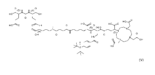
The present invention relates to a compound according to formula (V):
o o
(s) H H (S)
H H
0
H0
H 0 0 ONH H 0 0 HO
0
(R)
N2 N (R)
0 N
(s)
0 H 0 0
H 0
0
H
0
HO'
(V)
or a pharmaceutically acceptable salt thereof, containing either a chelated
radioactive cation
or wherein F is optionally 18F. The present invention also relates to a method
of synthesizing
the compound which prevents racemization during synthesis.
In this specification, a number of documents including patent applications and
manufacturer's manuals are cited. The disclosure of these documents, while not
considered
relevant for the patentability of this invention, is herewith incorporated by
reference in its
entirety. More specifically, all referenced documents are incorporated by
reference to the
same extent as if each individual document was specifically and individually
indicated to be
incorporated by reference.
Prostate cancer
Prostate Cancer (PCa) remained over the last decades the most common malignant
disease
in men with high incidence for poor survival rates. Due to its overexpression
in prostate
cancer, prostate-specific membrane antigen (PSMA) or glutamate
carboxypeptidase II (GCP
II) proved its eligibility as excellent target for the development of highly
sensitive
radiolabelled agents for endoradiotherapy and imaging of PCa. Prostate-
specific membrane
antigen is an extracellular hydrolase whose catalytic center comprises two
zinc(II) ions with a
bridging hydroxido ligand. It is highly upregulated in metastatic and hormone-
refractory
prostate carcinomas, but its physiologic expression has also been reported in
kidneys,
salivary glands, small intestine, brain and, to a low extent, also in healthy
prostate tissue. In
the intestine, PSMA facilitates absorption of folate by conversion of
pteroylpoly-y-glutamate
to pteroylglutamate (folate). In the brain, it hydrolyses N-acetyl-Laspartyl-L-
glutamate
(NAAG) to N-acetyl-L-aspartate and glutamate.
CA 03128401 2021-07-30
WO 2020/157177 2
PCT/EP2020/052248
Prostate-specific membrane antigen (PSMA)
Prostate-specific membrane antigen (PSMA) is a type II transmembrane
glycoprotein that is
highly overexpressed on prostate cancer epithelial cells. Despite its name,
PSMA is also
expressed, to varying degrees, in the neovasculature of a wide variety of
nonprostate
cancers. Among the most common nonprostate cancers to demonstrate PSMA
expression
include breast, lung, colorectal, and renal cell carcinoma.
The general necessary structures of PSMA targeting molecules comprise a
binding unit that
encompasses a zinc-binding group (such as urea, phosphinate or
phosphoramidate)
connected to a P1' glutamate moiety, which warrants high affinity and
specificity to PSMA
and is typically further connected to an effector functionality. The effector
part is more
flexible and to some extent tolerant towards structural modifications. The
entrance tunnel
accommodates two other prominent structural features, which are important for
ligand
binding. The first one is an arginine patch, a positively charged area at the
wall of the
entrance funnel and the mechanistic explanation for the preference of
negatively charged
functionalities at the P1 position of PSMA. This appears to be the reason for
the preferable
incorporation of negative charged residues within the ligand-scaffold. An in-
depth analysis
about the effect of positive charges on PSMA ligands has been, to our
knowledge, so far not
conducted. Upon binding, the concerted repositioning of the arginine side
chains can lead to
the opening of an Si hydrophobic accessory pocket, the second important
structure that has
been shown to accommodate an iodo-benzyl group of several urea based
inhibitors, thus
contributing to their high affinity for PSMA.
Zhang et al. discovered a remote binding site of PSMA, which can be employed
for bidentate
.. binding mode (Zhang et al., Journal of the American Chemical Society 132,
12711-12716
(2010)). The so called arene-binding site is a simple structural motif shaped
by the side
chains of Arg463, Arg511 and Trp541, and is part of the GOP!! entrance lid.
The
engagement of the arene binding site by a distal inhibitor moiety can result
in a substantial
increase in the inhibitor affinity for PSMA due to avidity effects. PSMA l&T
was developed
with the intention to interact this way with PSMA, albeit no crystal structure
analysis of
binding mode is available. A necessary feature according to Zhang et al. is a
linker unit
(Suberic acid in the case of PSMA l&T) which facilitates an open conformation
of the
entrance lid of GOP!! and thereby enabling the accessibility of the arene-
binding site. It was
further shown that the structural composition of the linker has a significant
impact on the
tumor-targeting and biologic activity as well as on imaging contrast and
pharmacokinetics
(Liu et al., Bioorganic & medicinal chemistry letters 21, 7013-7016 (2011)),
properties which
are crucial for both high imaging quality and efficient targeted
endoradiotherapy.
CA 03128401 2021-07-30
WO 2020/157177 3
PCT/EP2020/052248
Two categories of PSMA targeting inhibitors are currently used in clinical
settings. On the
one side there are tracers with chelating units for radionuclide complexation
such as PSMA
l&T or related compounds. On the other side there are small molecules,
comprising a
targeting unit and effector molecules.
The most often used agents for selective PSMA imaging are PSMA HBED-CC, PSMA-
617
and PSMA l&T, which are predominantly labelled with 68Ga (88.9% fr,
= 1.89 MeV,
t%= 68 min). Among these 68Ga-PSMA-HBED-CC (also known as 68Ga-PSMA-11), is so
far
considered as the golden standard for PET imaging of PCa.
18F labelling
Recently, several groups have focused on the development of novel 18F-labelled
urea-based
inhibitors for PCa diagnosis. In contrast to the radiometal 68Ga, which can be
obtained from
commercially distributed 68Ge/68Ga radionuclide generators (68Ge; t%= 270.8
d), the
radioisotope 18F-fluoride (96.7% 6+, = 634
keV) requires an on-site cyclotron for its
production. Despite this limitation, 18F offers due to its longer half-live
(tyz= 109.8 min) and its
lower positron energy, significant advantages in terms of routine-handling and
image quality.
Additionally, there is the possibility for largescale production in a
cyclotron, which would be
beneficial for a higher patient throughput and reduction of production
costs.The 18F-labelled
urea-based PSMA inhibitor 18F-DCFPyl demonstrated promising results in the
detection of
primary and metastatic PCa (Rowe et al., Molecular Imaging and Biology, 1-9
(2016)) and
superiority to 68Ga-PSMA-HBED-CC in a comparative study (Dietlein et al.,
Molecular
Imaging and Biology 17, 575-584 (2015)). Based on the structure of PSMA-617,
the 18F-
labelled analogue PSMA-1007 was recently developed, which showed comparable
tumor-to-
organ ratios (Cardinale et al., Journal of nuclear medicine: official
publication, Society of
Nuclear Medicine 58, 425-431 (2017); Giesel et al., European journal of
nuclear medicine
and molecular imaging 43, 1929-1930 (2016)). A comparative study with 68Ga-
PSMA-HBED-
CC revealed similar diagnostic accuracy of both tracers and a reduced urinary
clearance of
18F-PSMA-1007, enabling a better assessment of the prostate (Giesel et al.,
European
journal of nuclear medicine and molecular imaging 44, 678-688 (2017)).
An attractive approach for introducing 18F labels is the use of silicon
fluoride acceptors
(SI FA). Silicon fluoride acceptors are described, for example, in Lindner et
al., Bioconjugate
Chemistry 25, 738-749 (2014). In order to preserve the silicon-fluoride bond,
the use of
silicon fluoride acceptors introduces the necessity of sterically demanding
groups around the
silicone atom. This in turn renders silicon fluoride acceptors highly
hydrophobic. In terms of
binding to the target molecule, in particular to the target molecule which is
PSMA, the
CA 03128401 2021-07-30
WO 2020/157177 4
PCT/EP2020/052248
hydrophobic moiety provided by the silicone fluoride acceptor may be exploited
for the
purpose of establishing interactions of the radio-diagnostic or -therapeutic
compound with
the hydrophobic pocket described in Zhang et al., Journal of the American
Chemical Society
132, 12711-12716 (2010). Yet, prior to binding, the higher degree of
lipophilicity introduced
into the molecule poses a severe problem with respect to the development of
radiopharmaceuticals with suitable in vivo biodistribution, i.e. low
unspecific binding in non-
target tissue.
Failure to solve the hydrophobicity problem
Despite many attempts, the hydrophobicity problem caused by silicon fluoride
acceptors has
not been satisfactorily solved in the prior art.
To explain further, Schirrmacher E. et al. (Bioconjugate Chem. 2007, 18, 2085-
2089)
synthesized different 18F-labelled peptides using the highly effective
labelling synthon p-(di-
tert-butylfluorosily1) benzaldehyde ([189SIFA-A), which is one example of a
silicon fluoride
acceptor. The SIFA technique resulted in an unexpectedly efficient isotopic
19F-18F
exchange and yielded the 18F-synthon in almost quantitative yields in high
specific activities
between 225 and 680 GBq/pmol (6081-18 378 Ci/mmol) without applying H PLC
purification.
[189SIFA-benzaldehyde was finally used to label the N-terminal amino-oxy (N-
AO)
derivatized peptides AO-Tyr3 -octreotate (AO-TATE), cyclo(fK(AO-N)RGD) and N-
AO-PEG2-
[D-Tyr-Gln-Trp-Ala-Val-Ala-His-Thi-Nle-NH2] (AO-BZH3, a bombesin derivative)
in high
radiochemical yields. Nevertheless, the labelled peptides are highly
lipophilic (as can be
taken from the HPLC retention times using the conditions described in this
paper) and thus
are unsuitable for further evaluation in animal models or humans.
In Wangler C. et al. (Bioconjugate Chem., 2009, 20 (2), pp 317-321), the first
SIFA-based
Kit-like radio-fluorination of a protein (rat serum albumin, RSA) has been
described. As a
labelling agent, 4-(di-tert-butyl[189fluorosily1)benzenethiol (Si[189FA-SH)
was produced by
simple isotopic exchange in 40-60% radiochemical yield (RCY) and coupled the
product
directly to maleimide derivatized serum albumin in an overall RCY of 12%
within 20-30 min.
The technically simple labelling procedure does not require any elaborated
purification
procedures and is a straightforward example of a successful application of Si-
18F chemistry
for in vivo imaging with PET. The time-activity cureves and pPET images of
mice showed
that most of the activity was localized in the liver, thus demonstrating that
the labelling agent
is too lipophilic and directs the in vivo probe to hepatobiliary excretion and
extensive hepatic
metabolism.
CA 03128401 2021-07-30
WO 2020/157177 5
PCT/EP2020/052248
Wangler C. et al. (see Bioconjug Chem. 2010 Dec 15;21(12):2289-96)
subsequently tried to
overcome the major drawback of the SIFA technology, the high lipophilicity of
the resulting
radiopharmaceuticals, by synthesizing and evaluating new SIFA-octreotate
analogues
(SI FA-Tyr3-octreotate, SI FA-Asn(AcNH-8-G1c)-Tyr3-octreotate and SI FA-
Asn(AcN H-8-G1c)-
PEG-Tyr3-octreotate). In these compounds, hydrophilic linkers and
pharmacokinetic
modifiers were introduced between the peptide and the SIFA-moiety, i.e. a
carbohydrate and
a PEG linker plus a carbohydrate. As a measure of lipophilicity of the
conjugates, the log
P(ow) was determined and found to be 0.96 for SIFA-Asn(AcNH-8-G1c)-PEG-Tyr3-
octreotate
and 1.23 for SIFA-Asn(AcNH-8-G1c)-Tyr3-octreotate. These results show that the
high
lipophilicity of the SIFA moiety can only be marginally compensated by
applying hydrophilic
moieties. A first imaging study demonstrated excessive hepatic clearance
/liver uptake and
thus has never been transferred into a first human study.
Bernard-Gauthier et al. (Biomed Res Int. 2014;2014:454503) reviews a great
plethora of
different SIFA species that have been reported in the literature ranging from
small prosthetic
groups and other compounds of low molecular weight to labelled peptides and
most recently
affibody molecules. Based on these data the problem of lipophilicity of SIFA-
based
prosthetric groups has not been solved sofar; i.e. a methodology that reduces
the overall
lipophilicity of a SIFA conjugated peptide to a log D lower than approx -2,0
has not been
described.
In Lindner S. et al. (Bioconjug Chem. 2014 Apr 16;25(4):738-49) it is
described that
PEGylated bombesin (PESIN) derivatives as specific GRP receptor ligands and
RGD (one-
letter codes for arginine-glycine-aspartic acid) peptides as specific av133
binders were
synthesized and tagged with a silicon-fluorine-acceptor (SIFA) moiety. To
compensate the
high lipophilicity of the SIFA moiety various hydrophilic structure
modifications were
introduced leading to reduced logD values. SIFA-Asn(AcNH-8-G1c)-PESIN, SIFA-
Ser(8-
Lac)-PESIN, SIFA-Cya-PESIN, SIFA-LysMe3-PESIN, SIFA-y-carboxy-d-Glu-PESIN,
SIFA-
Cya2-PESI N, SI FA-LysMe3-y-carboxy-d-Glu-PESIN, SI FA-(y-carboxy-d-Glu)2-PESI
N, SI FA-
RGD, SI FA-y-carboxy-d-Glu-RGD, SI FA-(y-carboxy-d-Glu)2-RGD, SI FA-LysM e3-y-
carboxy-
d-Glu-RGD. All of these peptides ¨ already improved and derivatized with the
aim to reduce
the lipophilicity ¨ showed a logD value in the range between +2 and -1.22.
In Niedermoser S. et al. (J Nucl Med. 2015 Jul;56(7):1100-5), newly developed
18F-SIFA-
and 18F-SIFAlin- (SIFA = silicon-fluoride acceptor) modified TATE derivatives
were
compared with the current clinical gold standard 88Ga-DOTATATE for high-
quality imaging of
somatostatin receptor-bearing tumors. For this purpose, 18F-SIFA-TATE and two
quite
CA 03128401 2021-07-30
WO 2020/157177 6
PCT/EP2020/052248
complex analogues, 18F-SIFA-Glc-PEG1-TATE, 18F-SIFAlin-Glc-Asp2-PEG1-TATE were
developed. None of the agents showed a logD <-1.5.
In view of the above, the technical problem underlying the present invention
can be seen in
providing radio-diagnostics and radio-therapeutics which contain a silicone
fluoride acceptor
and which are, at the same time, characterized by favourable in-vivo
properties.
As will be become apparent in the following, the present invention established
a proof-of-
principle using specific conjugates which bind with high affinity to prostate-
specific antigen
(PSMA) as target. Accordingly, a further technical problem underlying the
present invention
can be seen in providing improved radio-therapeutics and ¨diagnostics for the
medical
indication which is cancer, preferably prostate cancer.
PCT/EP2018/070533 discloses a genus of PSMA binding compounds. Disclosed
herein is
an advantageous subset of the compounds from the earlier application. The
application
herein is a selection of advantageous features not appreciated by the
inventors at the time of
filing PCT/EP2018/070533.
These technical problems are solved by the subject-matter of the claims.
Accordingly, in
some aspects, the present invention relates to the compound according to
formula (V):
o 0
(s)H H(S)
H 0 H
0
0
0N H H 0 0
H
H 0 0
(R) (R) H
N H
N
OH 0 H (S) N
0 0
H N
0
NOH
0
HO'
(V)
or a pharmaceutically acceptable salt thereof, containing either a chelated
radioactive cation
or wherein F is optionally 18F.
Compounds disclosed can be in the form of salts. The present invention also
relates to
compounds of formula (Va):
CA 03128401 2021-07-30
WO 2020/157177 7
PCT/EP2020/052248
0 0
X S)
H H
(S) N N (
0
0
X 0 X 0
X 0 ON H 0 0
0
(R) H =
0 0
X HN
N
0
0
X
Si
(Va)
or a pharmaceutically acceptable salt thereof, wherein;
each X is independently OH or 0-;
M is either a chelated radioactive cation or is absent;
and F is optionally 18F.
In addition, the combination of the use of a chelator and an isotopic exchange
on SIFA by
means of 18F-fluoride also results in "paired" diagnostic tracers that can
either be used as
r18
FllnatIon]tracers at centers with onsite cyclotron or centers that obtain 18F-
fluoride by
shipment from cyclotron centers, whereas in centers, that do not have access
to 18F-fluoride
but have access to radioisotope generators, such as a Ge-68/Ga-68 generator,
the
corresponding versions, e.g. [natF][68Ga]tracers can be used.
Importantly, in both cases, the chemically identical radiopharmaceutical is
injected, and thus
no differences in the in vivo behavior are expected. Whereas currently, due to
chemical
differences, the clinical data of a 18F-labelled compound provided by a
patient cohort at one
site cannot be directly compared with the clinical data of a 68Ga-analogue
provided by
another group at another site, radiopharmaceuticals and/or diagnostics
according to the
invention can be directly compared and thus will allow to link such data (e.g.
data from a
center in Europe working with F-18 and another center in India working with Ga-
68).
Furthermore, when suitably selected, the chelate can also be used for
labelling with a
therapeutic isotope, such as the beta-emitting isotopes Lu-177, Y-90, or the
alpha emitting
isotope Ac-225, thus allowing to expand the concept of "paired" tracers to
bridge diagnostic
fr18
FllnatLu]tracers) and therapeutic radiopharmaceuticals ([natF][l 77Lui
A further advantage of the compounds, especially of PSMA targeted compounds of
the
CA 03128401 2021-07-30
WO 2020/157177 8
PCT/EP2020/052248
present invention is their surprisingly low accumulation in the kidneys of
mice when
compared to other PSMA targeted radiopharmaceuticals, such as PSMA l&T.
Without
wishing to be bound by a particular theory, it seems to be the combination of
the structural
element SIFA with a chelator which provides for the unexpected reduction of
accumulation in
the kidneys.
In terms of lipophilicity/hydrophilicity, the logP value (sometimes also
referred to as logD
value) is an art-established measure.
The term õlipophilicity" relates to the strength of being dissolved in, or be
absorbed in lipid
solutions, or being adsorbed at a lipid-like surface or matrix. It denotes a
preference for lipids
(literal meaning) or for organic or apolar liquids or for liquids, solutions
or surfaces with a
small dipole moment as compared to water. The term "hydrophobicity" is used
with
equivalent meaning herein. The adjectives lipophilic and hydrophobic are used
with
corresponding meaning to the substantives described above.
The mass flux of a molecule at the interface of two immiscible or
substantially immiscible
solvents is governed by its lipophilicity. The more lipophilic a molecule is,
the more soluble it
is in the lipophilic organic phase. The partition coefficient of a molecule
that is observed
between water and n-octanol has been adopted as the standard measure of
lipophilicity. The
partition coefficient P of a species A is defined as the ratio P = [A]n-
octanol / [A]water. A figure
commonly reported is the logP value, which is the logarithm of the partition
coefficient. In
case a molecule is ionizable, a plurality of distinct microspecies (ionized
and not ionized
forms of the molecule) will in principle be present in both phases. The
quantity describing the
overall lipophilicity of an ionizable species is the distribution coefficient
D, defined as the
ratio D = [sum of the concentrations of all microspecies]n-octanol / [SUM of
the concentrations of
all microspecies] water. Analogous to logP, frequently the logarithm of the
distribution
coefficient, logD, is reported. Often, a buffer system, such as phosphate
buffered saline is
used as alternative to water in the above described determination of logP.
If the lipophilic character of a substituent on a first molecule is to be
assessed and/or to be
determined quantitatively, one may assess a second molecule corresponding to
that
substituent, wherein said second molecule is obtained, for example, by
breaking the bond
connecting said substituent to the remainder of the first molecule and
connecting (the) free
valence(s) obtained thereby to hydrogen(s).
CA 03128401 2021-07-30
WO 2020/157177 9
PCT/EP2020/052248
Alternatively, the contribution of the substituent to the logP of a molecule
may be
determined. The contribution 7CX x of a substituent X to the logP of a
molecule R-X is defined
as 7CXx =10gPR_x- logPR_H, wherein R-H is the unsubstituted parent compound.
Values of P and D greater than one as well as logP, logD and 7CX x values
greater than zero
indicate lipophilic/hydrophobic character, whereas values of P and D smaller
than one as
well as logP, logD and 7CX x values smaller than zero indicate hydrophilic
character of the
respective molecules or substituents.
The above described parameters characterizing the lipophilicity of the
lipophilic group or the
entire molecule according to the invention can be determined by experimental
means and/or
predicted by computational methods known in the art (see for example Sangster,
Octanol-
water Partition Coefficients: fundamentals and physical chemistry, John Wiley
& Sons,
Chichester. (1997)).
In a preferred embodiment, the logP value of the compounds of the invention is
between -5
and -1.5. It is particularly preferred that the logP value is between -3.5 and
-2Ø
In a preferred embodiment the chelating group comprises a chelated cation
which is
radioactive. More preferred is a chelated radioactive metal isotope.
Preferred examples of cations that may be chelated by the chelating group are
the cations of
"sc, "sc, 475c, sicr, 52mmn, 58co, 52Fe, 56Ni, 57Ni, 62ou, "cu, 67Cu, "Ga,
67Ga "Ga, "Zr,
90y,
r <Tc, 99mTc, 97Ru, 166Rh, 109pd, iiiAg,iiomin, 1111n, 3min,
4min, 117msn, 1215n, 127Te,
142pr, 143pr, 149pm, 151 Pm, 149Tb, 152Tb, 155Tb, 161Tb, 1535m, 157Gd, 161Tb,
166H0, 165Dy, 169E1-,
169yb, 175yb, 172Tm, 177Lu, 186Re, 188Re, 191pt, 197Fig, 198Au, 199Au, 212pb,
203pb, 211At, 212Bi,
213Bi, 223Ra, 225Ab,
n a cationic molecule comprising 18F or a cation such as 18F4A192+;
more preferably the cations of 445c, 475c, 64Cu, 67Cu, 68Ga, 90Y,
ln, 161-b, 166H0, 177Lu,
188Re, 212pb,212Bi,213Bi, 225,,AC ,
and 227Th or a cationic molecule comprising 18F. Cations may
be selected from Lu-177, Y-90, or Ac-225.
In a further aspect, the present invention provides a pharmaceutical
composition comprising
or consisting of one or more compounds of the invention as disclosed herein
above.
In a further aspect, the present invention provides a diagnostic composition
comprising or
consisting of one or more compounds of the invention as disclosed herein
above.
CA 03128401 2021-07-30
WO 2020/157177 10
PCT/EP2020/052248
In a further aspect, the present invention provides a therapeutic composition
comprising or
consisting of one or more compounds of the invention as disclosed herein
above.
The pharmaceutical composition may further comprise pharmaceutically
acceptable carriers,
excipients and/or diluents. Examples of suitable pharmaceutical carriers,
excipients and/or
diluents are well known in the art and include phosphate buffered saline
solutions, water,
emulsions, such as oil/water emulsions, various types of wetting agents,
sterile solutions etc.
Compositions comprising such carriers can be formulated by well known
conventional
methods. These pharmaceutical compositions can be administered to the subject
at a
suitable dose. Administration of the suitable compositions may be effected by
different ways,
e.g., by intravenous, intraperitoneal, subcutaneous, intramuscular, topical,
intradermal,
intranasal or intrabronchial administration. It is particularly preferred that
said administration
is carried out by injection and/or delivery, e.g., to a site in the pancreas
or into a brain artery
or directly into brain tissue. The compositions may also be administered
directly to the target
site, e.g., by biolistic delivery to an external or internal target site, like
the pancreas or brain.
The dosage regimen will be determined by the attending physician and clinical
factors. As is
well known in the medical arts, dosages for any one patient depends upon many
factors,
including the patient's size, body surface area, age, the particular compound
to be
administered, sex, time and route of administration, general health, and other
drugs being
administered concurrently. Pharmaceutically active matter may be present in
amounts
between 0,1 ng and 10 mg/kg body weight per dose; however, doses below or
above this
exemplary range are envisioned, especially considering the aforementioned
factors.
In a further aspect, the present invention provides one or more compounds of
the invention
as disclosed herein above for use in medicine.
Preferred uses in medicine are in nuclear medicine such as nuclear diagnostic
imaging, also
named nuclear molecular imaging, and/or targeted radiotherapy of diseases
associated with
an overexpression, preferably of PSMA on the diseased tissue.
In a further aspect, the present invention provides a compound of the
invention as defined
herein above for use in a method of diagnosing and/or staging cancer,
preferably prostate
cancer. Prostate cancer is not the only cancer to express PSMA. Nonprostate
cancers
known to demonstrate PSMA expression include breast, lung, colorectal, and
renal cell
carcinoma. Thus any compound described herein having a PSMA binding moiety can
be
used in the diagnosis, imaging or treatment of a cancer having PSMA
expression.
CA 03128401 2021-07-30
WO 2020/157177 11 PC
T/EP2020/052248
Preferred indications are the detection or staging of cancer, such as, but not
limited high
grade gliomas, lung cancer and especially prostate cancer and metastasized
prostate
cancer, the detection of metastatic disease in patients with primary prostate
cancer of
intermediate-risk to high-risk, and the detection of metastatic sites, even at
low serum PSA
values in patients with biochemically recurrent prostate cancer. Another
preferred indication
is the imaging and visualization of neoangiogensis.
In terms of medical indications to be subjected to therapy, especially
radiotherapy, cancer is
.. a preferred indication. Prostate cancer is a particularly preferred
indication.
In a further aspect, the present invention provides a compound of the
invention as defined
herein above for use in a method of diagnosing and/or staging cancer,
preferably prostate
cancer.
The present disclosure furthermore relates to the following items.
The compound according to formula (V):
o 0(s) H H (S)
N N
H
0
H0
H 0 0 ONH H 0 0
0 0
(R) (R) H
ONN N
H (S) N
0
OH 0 0
H N
0
NOH
0
HO'
(V)
.. or a pharmaceutically acceptable salt thereof, containing either a chelated
radioactive cation
or wherein F is optionally 18F.
The compound may contain a chelated cation selected from the cations of Sc,
Cu, Ga, Y, In,
Tb, Ho, Lu, Re, Pb, Bi, Ac, Er and Th. The chelated cation may be radioactive.
The chelated
radioactive cation may be any radioactive isotope(s) of Gallium, Erbium,
copper, scandium,
Lutetium or Yttrium.
The compound of formula (Va):
CA 03128401 2021-07-30
WO 2020/157177 12
PCT/EP2020/052248
0 S) 0
H H
( N N (
X S) X
0
0
X 0 X 0
X 0 ON H 0 0 L,
(R) (R) I\11 y)C N
0
H (S)( 0
0 0
X HN
0 N X
0
Si
(Va)
or a pharmaceutically acceptable salt thereof, wherein;
each X is independently OH or 0-;
M is a chelated radioactive cation or is absent;
and F is optionally 18F.
In the compound of formula (Va), X may be OH. X may be 0. One or more of
groups X may
be chelated to M when M is present.
In the compound of formula (Va), M may be a chelated radioactive cation. M may
be absent.
M may be a radioactive cation chelated to one or more X groups. M may be a
radioactive
cation chelated to one or more N atoms. M may be a radioactive cation chelated
to one or
more N atoms or one or more X groups. M may be a radioactive cation chelated
to one or
more N atoms and one or more X groups.
In the compound of formula (V) or (Va), F may be 18F. F may be 19F.
In the compound of formula (V) or (Va), the chelated radioactive cation may be
selected from
the cations of "Sc, "Sc, 47Sc, 51Cr, 52mMn, 5800, 52Fe, "Ni, 57Ni, 62Cu, "Cu,
67Cu, "Ga, 67Ga
68Ga, 89Zr, 99Y, 89Y, <Tc, 99mTc, 97Ru, 1051Th, 109Pd, iiiAg,iiomin, 1111n,
3min, 4min, 117msn,
121Bn, 127Te, 142pr, 143pr, 149pm, 151pm, 149Tb, 152Tb, 155Tb, 161Tb, 153Bm,
157Gd, 161Tb, 166H0,
165Dy, 169E1-, 169yb, 175yb, 172Tm, 177Lu, 186Re, 188Re, 191 Pt, 197Hg, 198Au,
199Au, 212pb, 203pb,
211At, 212Bi, 213Bi, 223Ra, 225Ab,
n a cationic molecule comprising 18F or a cation such as
18-
F [A192+; more preferably the cations of 44Sc, 47Sc, "Cu, 67Cu, 68Ga, 99Y,
ln, 161-b, 166H0,
177Lu, 188Re, 212pb,212Bi,213Bi, 225,,AC ,
and 227Th or a cationic molecule comprising 18F. The
chelated radioactive cation may be selected from the cations of Sc, Cu, Ga, Y,
In, Tb, Ho,
CA 03128401 2021-07-30
WO 2020/157177 13
PCT/EP2020/052248
Lu, Re, Pb, Bi, Ac, Er and Th. The chelated radioactive cation may be Ga. The
chelated
radioactive cation may be Lu-177, Y-90, or Ac-225.
In the compound of formula (Va), M may be selected from the cations of 43Sc,
44Sc, 47Sc,
51Cr, 52mMn, 5800, 52Fe, 56Ni, 57Ni, 62Cu, 64Cu, 67Cu, "Ga, 67Ga "Ga, "Zr, "Y,
"Y, <Tc, "mTc,
67Ru, 165Rh, 109pd, iiiAg,iiomin, 1111n,
3min,
4min, 117msn, 121Bn, 127Te, 142pr, 143pr, 149pm,
151pm, 149Tb, 152Tb, 155Tb, 161Tb, 153Bm, 157Gd, 161Tb, 166H0, 165Dy, 169Er,
169yb, 175yb, 172Tm,
177Lu, 186Re, 188Re, 191pt, 197Hg, 198Au, 199Au, 212pb, 203pb, 211At, 212Bi,
213Bi, 223Ra, 225Ab,
n a cationic molecule comprising 18F or a cation such as 18F4A192+; more
preferably the
cations of 44Sc, 47Sc, 64Cu, 67Cu, 68Ga, 90Y, ln, 161-b, 166H0, 177Lu,
188Re, 212pb,212Bi,213Bi,
225 Akk -
G and 227Th or a cationic molecule comprising 18F. M may be selected from the
cations
of Sc, Cu, Ga, Y, In, Tb, Ho, Lu, Re, Pb, Bi, Ac, Er and Th. M may be Ga. M
may be Lu-177,
Y-90, or Ac-225.
It is advantageous to synthesise compounds with as few isomers as possible.
Whilst the
isomers can be separated, only a single isomer is used in-vivo and hence the
wrong isomer
is simply discarded and not used. Thus conditions which minimise racemisation
or inversion
of any of the defined chiral centres are ideally avoided.
Also described is a method producing the compound comprising the steps of:
a) reacting a compound of formula (I):
(s) H H(s)
PG1
0
PG1 ON H 0 PG
1
(R)
(R)
....N H2
OH 0
(I)
with a compound of formula (II):
0
N ,
P G3
(II)
CA 03128401 2021-07-30
WO 2020/157177 14 PCT/EP2020/052248
to form a compound of formula (III):
0 0
(S) H H (S)
PG1 PG1
0
PG1 0_ 0
0' 0 ON H 0 0
, H
N PG2
OH 0
Ni
PG3
(III)
wherein PG1 is tBu, PG2 is Fmoc and PG3 is Dde;
.. and the reaction conditions involve the use of a base, wherein the base is
2,4,6-collidine or
2,6-dimethylpyridine;
b) reacting the compound of formula (Ill) under conditions suitable for
forming a
compound of formula (IV):
o (s) H H (S)
PG1 GP
"--09N
0
0N H 0 0
0 0
= ,(R)
0 NH
= N_ õ
(R)
OH 0
NV
I
Si
(IV)
and
c) reacting the compound of formula (IV) under conditions suitable for forming
compound (V):
CA 03128401 2021-07-30
WO 2020/157177 15 PCT/EP2020/052248
o o(s) H H (S)
N N.,..õ--
H 0------j- -,1T-- 0 H
0
H
)
H 0 0 ONH H H 0 ...- 0 0 -`--C) 0
H (R) (R) H I /-------N
0,.............11R..y,....õ-...õ___N,....in....õ--..õ
NK-''-------''--,""- 1 \1''-''-- N N
H ) 0
OH 0 H I (S)
, 0
H N
N
0 (N_______/ -7.0 H
_ \ Fl 0 0
HO'
/
'"Si
(V)
The method according includes wherein compound (II) is preactivated by
reaction with 2-
(1H-Benzotriazole-1-yI)-1,1,3,3-tetramethylaminium tetrafluoroborate (TBTU), 1-
Hydroxy-7-
azabenzotriazole (HOAt) and 2,4,6-collidine prior to reaction with compound
(I). The
preactivation takes place for 5 minutes or less.
The use of 2,4,6-collidine or 2,6-dimethylpyridine as base, along with the
short activation
time helps to minimise racemisation of the activated chiral compound (II) when
compared to
other nitrogenous bases such as DIPEA. The more sterically hindered base does
not extract
the acidic proton on the chiral centre of the acid, hence lowering
racemisation before
coupling to the amine.
.. Disclosed herein is the compound according to formula (VI)
0 0
? 63) ENIT EN1O
HO
L OH
j,
0 -
n -0
, 1
HO 0 ONH 0 H 0 0 H 0 0
.,:stpQ19 Nyj Nr-- N Oyik.......s.õ., Ny......), N
H H (St 177Lu ) o
OH 0 ...7 0
H N I \I.A _
0 N........../ .. 0
FI I.1 0
-0)-1
Si
(VI)
or a pharmaceutically acceptable salt thereof.
Where the chelated metal is shown, the acid groups to which it is chelated are
merely
representatively shown as 000-, the equivalent fourth acid may also be partly
chelated and
hence may not literally be COOH.
CA 03128401 2021-07-30
WO 2020/157177 16
PCT/EP2020/052248
Disclosed herein is the compound according to formula (VII)
0 o pH H (s)ii
HO NyN't=****-'0H
0
HO 00 HO .0 -0 0
HO 0 0.."'NH 0
H T(R) a (R) N :5
0 y.:.......,........(R) N sir=-)1." N µ`µ. N N
H H ' OH 0 (St, ) 0
j- 0 90v
HN I NI.Ao-
0 Ns........j
A 7 0 0
-0'-j
'1 (VII)
or a pharmaceutically acceptable salt thereof.
Disclosed herein is a pharmaceutical or diagnostic composition comprising the
compound of
formula (V) or (VI). The conjugate, compound or composition may be used as a
cancer
diagnostic or imaging agent.
Disclosed is a method of imaging and/or diagnosing cancer comprising
administering a
conjugate, compound or composition according to formula (V) or (VI) to a
patient in need
thereof.
Disclosed is a conjugate, compound or composition according to formula (V) or
(VI) for use
in the treatment of cancer.
Disclosed is a conjugate, compound or composition according to formula (V) or
(VI) for use
in the diagnosis, imaging or prevention of neoangiogenesis/angiogenesis.
Disclosed is a conjugate, compound or composition according to formula (V) or
(VI) for use
as a cancer diagnostic or imaging agent or for use in the treatment of cancer
wherein the
cancer is prostate, breast, lung, colorectal or renal cell carcinoma.
The Figures illustrate:
Figure 1: Exemplary correlation of determination of the nine reference
substances in
OriginPro 2016G.
Figure 2a: Quality Control of [19F][natGa]-rhPSMA7-rac ([19F][natGa]D/L-
Dap-R/S-DOTAGA-
rhPSMA-7-rac), (batch 10, precursor for the production of ([18F][natGa]D/L-Dap-
R/S-
DOTAGA-rhPSMA-7 at the Department of Nuclear Medicine, TUM). HPLC-conditions:
Solvent A: H20 + 0.1% TEA; Solvent 13: MeCN + 0.1% TEA. Gradient: 25 ¨ 35 %13
0-40
CA 03128401 2021-07-30
WO 2020/157177 17
PCT/EP2020/052248
min, 95 - 95 % B 40-45 min, 35 - 35 % B 45-50 min; flow: 1 mL/min, column:
Nucleosil 100-5 C18, 125 x 4.6 mm, Sample: 1 mM (DMSO), 10 L.
Figure 2b: Peak assignment: D-Dap-R-DOTAGA-rhPSMA-7.1; rhPSM7-rac from
Fig.2a coinjected
with enantiopure D-Dap-R-DOTAGA-rhPSMA-7.1. HPLC-conditions: Solvent A: H20 +
0.1% TEA; Solvent B: MeCN + 0.1% TEA. Gradient: 25 - 35 % B 0-40 min, 95 - 95
% B
40-45 min, 35 - 35 % B 45-50 min; flow: 1 mL/min, column: Nucleosil 100-5 C18,
125
x 4.6 mm, Sample: 1 mM (DMSO), 10 L.
Figure 2c: HPLC profile of D-Dap-R-DOTAGA-rhPSMA-7.1; HPLC-conditions:
Solvent A: H20 +
0.1% TEA; Solvent B: MeCN + 0.1% TEA. Gradient: 25 - 35 % B 0-40 min, 95 - 95
% B
40-45 min, 35 - 35 % B 45-50 min; flow: 1 mL/min, column: Nucleosil 100-5 C18,
125
x 4.6 mm, Sample: 1 mM (DMSO), 10 L.
Figure 3a: Peak assignment: L-Dap-R-DOTAGA-rhPSMA-7.2; rhPSM7-rac from
Fig.2a coinjected
with enantiopure L-Dap-R-DOTAGA-rhPSMA-7.2. HPLC-conditions: Solvent A: H20 +
0.1% TEA; Solvent B: MeCN + 0.1% TEA. Gradient: 25 - 35 % B 0-40 min, 95 - 95
% B
40-45 min, 35 - 35 % B 45-50 min; flow: 1 mL/min, column: Nucleosil 100-5 C18,
125
x 4.6 mm, Sample: 1 mM (DMSO), 10 L.
Figure 3b: HPLC profile of L-Dap-R-DOTAGA-rhPSMA-7.2; HPLC-conditions:
Solvent A: H20 +
0.1% TEA; Solvent B: MeCN + 0.1% TEA. Gradient: 25 - 35 % B 0-40 min, 95 - 95
% B
40-45 min, 35 - 35 % B 45-50 min; flow: 1 mL/min, column: Nucleosil 100-5 C18,
125
x 4.6 mm, Sample: 1 mM (DMSO), 10 L.
Figure 4a: Peak assignment: D-Dap-S-DOTAGA-rhPSMA-7.3; rhPSM7-rac from
Fig.2a coinjected
with enantiopure D-Dap-S-DOTAGA-rhPSMA-7.3. HPLC-conditions: Solvent A: H20 +
0.1% TEA; Solvent B: MeCN + 0.1% TEA. Gradient: 25 - 35 % B 0-40 min, 95 - 95
% B
40-45 min, 35 - 35 % B 45-50 min; flow: 1 mL/min, column: Nucleosil 100-5 C18,
125
x 4.6 mm, Sample: 1 mM (DMSO), 10 L.
Figure 4b: HPLC profile of D-Dap-D-DOTAGA-rhPSMA-7.3; HPLC-conditions:
Solvent A: H20 +
0.1% TEA; Solvent B: MeCN + 0.1% TEA. Gradient: 25 - 35 % B 0-40 min, 95 - 95
% B
40-45 min, 35 - 35 % B 45-50 min; flow: 1 mL/min, column: Nucleosil 100-5 C18,
125
x 4.6 mm, Sample: 1 mM (DMSO), 10 L.
Figure 5a: Peak assignment: L-Dap-S-DOTAGA-rhPSMA-7.4; rhPSM7-rac from
Fig.2a coinjected
with enantiopure D-Dap-S-DOTAGA-rhPSMA-7.3. HPLC-conditions: Solvent A: H20 +
0.1% TEA; Solvent B: MeCN + 0.1% TEA. Gradient: 25 - 35 % B 0-40 min, 95 - 95
% B
40-45 min, 35 - 35 % B 45-50 min; flow: 1 mL/min, column: Nucleosil 100-5 C18,
125
x 4.6 mm, Sample: 1 mM (DMSO), 10 L.
CA 03128401 2021-07-30
WO 2020/157177 18
PCT/EP2020/052248
Figure 5b: HPLC profile of L-Dap-D-DOTAGA-rhPSMA-7.4; HPLC-conditions:
Solvent A: H20 +
0.1% TEA; Solvent B: MeCN + 0.1% TEA. Gradient: 25 ¨ 35 % B 0-40 min, 95 ¨ 95
% B
40-45 min, 35 ¨ 35 % B 45-50 min; flow: 1 mL/min, column: Nucleosil 100-5 C18,
125
x 4.6 mm, Sample: 1 mM (DMSO), 10 L.
Figure 6a: Binding affinities (1050 [nM]) of rhPSMA7.1 and 7.2 to PSMA.
Affinities were
determined using LNCaP cells (150000 cells/well) and ((4-[1251]iodobenzoyl)KuE
([1251]IB-KuE; c = 0.2 nM) as the radioligand (1 h, 4 C, HBSS + 1% BSA). Data
are
expressed as mean SD (n = 3, in 3 different experiments).
Figure 6b: Determination of the binding affinities [nM] of the rhPSMA7
isomers to PSMA. Each
of the four column shows the individual affinity measurements for rhPSAM7.1
(left)
to rhPSMA7.4 (right). Conditions as described in the legend to Fig 6a.
Figure 7: Depiction of the individual 1050 [nM] measurements shown in
Fig 6a and 6b. Value
No 5 of rhPSMA7.1 was deleted Conditions as described in the legend to Fig 6a
Figure 8: Depiction of the individual internalization measurements [% of
[1251]IB-KuE].
Internalized activity (c = 0.5 nM) at 1 hour as % of the reference ligand
([1251]1-BA)KuE
(c = 0.2 nM), determined on LNCaP cells (37 C, DMEM E12 + 5% BSA, 125000
cells/well). Data is corrected for non-specific binding (10 limo! PM PA) and
expressed
as mean SD (n = 3).
Figure 9: Depiction of the individual measurements of the logP's of the
rhPSMA isomers.
Figure 10: Biodistribution (in %ID/g) of 18E-labeled rhPSMA tracers at 1 h
p.i in LNCaP tumor-
bearing SCID mice. Data are expressed as mean SD (n=4 for rhPSMA7.1, n=5 for
7.2, n=4 for 7.3, n=5 for 7.4 and n=3 for 7-rac).
Figure 11: Biodistribution [ %ID/g] of 18E-rhPSMAs co-injected with PMPA
(8 mg/kg) at 1 h p.i in
LNCaP tumor-bearing SCID mice. Data are expressed as mean SD (n=3).
Figure 12: Possible species generated by metabolic cleavage of amide bonds.
iL: cleavage forms
a species with increased lipophilicity; DE: defluorination; nd: not
detectable, since
not radioactive.
Figure 13: Left: Graphical analysis of overlapping peaks 1 (rhPSMA7.2)
and 2 (rhPSMA7.3);
right: deconvolution and integration of peak profiles by Systat PeakFit
Software);
top: experimental data fitted, bottom: deconvoluted single peaks.
Figure 14a: Quantification of relative changes (in % change of injected
racemic mixture) to
evaluate the reproducibility of classical integration (by HPLC program) and
deconvolution (by "PeakFit") of peak 4 (rhPSMA7.1). Both methods demonstrate
similar performance for this peak.
CA 03128401 2021-07-30
WO 2020/157177 19
PCT/EP2020/052248
Figure 14b: Quantification of relative changes (in % change of injected
racemic mixture) to
evaluate the reproducibility of classical integration (by HPLC program) and
deconvolution (by 'PeakFit") of peak 3 (rhPSMA7.4). Both methods demonstrate
similar performance for this peak.
Figure 15: Percentage change of each rhPSAM7.1-7.4 isomer in blood, liver,
kidney, tumor and
urine with respect to its proportion the injected solution ([l8F][1at-a.
jrhPSMA7-rac.
Data expressed as mean values SD (n=4; see also Fig 16).
Figure 16: Percentage change of each rhPSAM7.1-7.4 isomer for each sample
and experiment)
in blood, liver, kidney, tumor and urine with respect to its proportion in the
injected
solution ([l.F][nat.m.
jrhPSMA7-rac). Analyses were carried out with Systat PeakFit.
Figure 17: Left: TLC scanner profile of a TLC plate with liver sample
(30.07.2018, overall cts: 142
cts). Due to their bad statistics and limited validity dataset with cts<200
were
removed. Right: Phophoimage of a TLC plate with liver sample: long tailing of
the
moving tracer.
Figure 18: Left: TLC scanner profile of a TLC plate with a quality control
sample (01.08.2018,
overall cts: 384). Right: Exemplay phophoimage of a TLC plate with Urine,
kidney,
liver, tumor, blood and QK sample.
Figure 19: Radio-TLC of [F-18]rhPSMA7-rac (30.07.2018) as part of the
Quality Control in the
Department of Nuclear Medicine prior to clinical application of the tracer.
Note that
a tailing of the tracer is even observed in the formulation buffer (and thus
in the
absence of proteins).
Figure 20: Quantification of free [F-18]Fluoride and 'intact' [F-
18]rhPSMA7-rac by radio-TLC of
a urine sample (30.07.2018).
Figure 21: Left: Radio-H PLC analysis of urine collected and pooled from
4 normal mice injected
with the respective [F-18]rhPSAM-7.x tracer.
Right: Radio-HPLC analysis of "cold' urine spiked with the respective
[F-18]rhPSAM-7.x tracer for a period of 1h (7.1., 7.2.), 0.5h (7.3.) and 2h
(7.4.).
HPLC-conditions: Solvent A: H20 + 0.1% TFA; Solvent B: MeCN + 0.1% TFA;
Gradient:
5% isocratic 0-3 min, 25 - 35 % B 3-43 min, 95 - 95 % B 43-48 min; flow: 1
mUmin,
column: Nucleosil 100-5 C18, 125 x 4.6 mm.
Figure 22: Separation of radioactive species in urine by cartridge
fixation and TLC.
Top: Radio-HPLC analysis of urine 30min p.i. of [F-18]rhPSMA7.3 in mice
showing a
small proportion at 1.6 min and intact tracer at ca. 34.5 min.
Bottom (left): urine of mice, 30min p.i. of [F-18]rhPSMA7.3, was diluted and
CA 03128401 2021-07-30
WO 2020/157177 20
PCT/EP2020/052248
subjected to STRATA-X cartridge fixation. The cartridge was washed and eluted
with
MeCN/water (60/40 v/v +1% TEA); only intact tracer was detected.
Bottom (right): both the breakthrough from the cartridge fixation (non-
retained
components) and the fraction finally eluted MeCN/water from the cartridge were
analysed by TLC (bottom, right). Whereas 96.1% [F-18]rhPSMA7.3 and only 3.9%
[F-
18]fluoride were found in the eluate of the cartridge, the reverse ratio was
found in
the breakthrough of the cartridge (3.4% [F-18]rhPSMA7.3 and only 96.6% [F-
18]fluoride).
Figure 23: To fresh and nonradioactive urine of mice [F-18]rhPSMA7.3 was
added, followed by
0.51imo1 cold F-19-fluoride; incubation for 2h.
Radioactivity was completely (98.5%) converted to a very hydrophilic fraction
representing [F-18]fluoride (peak at 1.6 min). Note: peak at 1.6 min was
subsequently immobilized on a QMA cartridge and eluted with with NaCI (1M)
(=fluoride).
Figure 24: Clinical biodistribution and uptake in tumor lesions of 18F-
rhPSMA-7 (left) and 18F-
rhPSMA-7.3 (right) as demonstrated by SUVmax. Data are expressed as mean SD.
Figure 25: Clinical biodistribution and uptake in tumor lesions of 18F-
rhPSMA-7 (left) and 18F-
rhPSMA-7.3 (right) as demonstrated by SUVmean. Data are expressed as mean
SD.
Figure 26. Clinical biodistribution and uptake in tumor lesions of 18F-
rhPSMA-7 (left) and 18F-
rhPSMA-7.3 (right) as demonstrated by the ratio SUVmax to background. Data are
expressed as mean SD.
Figure 27: Clinical biodistribution and uptake in tumor lesions of 18F-
rhPSMA-7 (left) and 18F-
rhPSMA-7.3 (right) as demonstrated by the ratio SUVmean to background. Data
are
expressed as mean SD.
Figure 28: Two clinical case examples of 18F-rhPSMA-7.3 PET-imaging.
The Examples illustrate the invention.
Example 1: Material and Methods
The Fmoc-(9-fluorenylmethoxycarbonyl-) and all other protected amino acid
analogs were
purchased from Bachem (Bubendorf, Switzerland) or Iris Biotech (Marktredwitz,
Germany). The
tritylchloride polystyrene (TCP) resin was obtained from PepChem (Tubingen,
Germany). Chematech
(Dijon, France) delivered the chelators DOTAGA-anhydride, (R)-DOTA-GA(tBu)4
and (S)-DOTA-
GA(tBu)4. All necessary solvents and other organic reagents were purchased
from either, Alfa Aesar
CA 03128401 2021-07-30
WO 2020/157177 21
PCT/EP2020/052248
(Karlsruhe, Germany), Sigma-Aldrich (Munich, Germany) or VWR (Darmstadt,
Germany). Solid phase
synthesis of the peptides was carried out by manual operation using an Intelli-
Mixer syringe shaker
(Neolab, Heidelberg, Germany). Analytical and preparative reversed-phase high
pressure
chromatography (RP-HPLC) were performed using Shimadzu gradient systems
(Shimadzu
Deutschland GmbH, Neufahrn, Germany), each equipped with a SPD-20A UV/Vis
detector
(220 nm, 254 nm). A Nucleosil 100 C18 (125 x 4.6 mm, 5 pm particle size)
column ( CS GmbH,
Langerwehe, Germany) was used for analytical measurements at a flow rate of 1
mL/min. Both
specific gradients and the corresponding retention times tR are cited in the
text. Preparative HPLC
purification was done with a Multospher 100 RP 18 (250 x 10 mm, 5 p.m particle
size) column (CS
GmbH, Langerwehe, Germany) at a constant flow rate of 5 mL/min. Analytical and
preparative radio
RP-HPLC was performed using a Nucleosil 100 C18 (5 pm, 125 x 4.0 mm) column
(CS GmbH,
Langerwehe, Germany). Eluents for all HPLC operations were water (solvent A)
and acetonitrile
(solvent B), both containing 0.1% trifluoroacetic acid. Electrospray
ionization-mass spectra for
characterization of the substances were acquired on an expressionl- CMS mass
spectrometer (Advion
Ltd., Harlow, UK). NMR spectra were recorded on Bruker AVHD-300 or AVHD-400
spectrometers at
300K. pH values were measured with a Seven Easy pH-meter (Mettler Toledo,
GieRen, Germany).
Synthesis protocols
1) Solid-phase peptide synthesis following the Fmoc-strategy
TCP-resin loading (GP1)
Loading of the tritylchloride polystyrene (TCP) resin with a Fmoc-protected
amino acid (AA) was
carried out by stirring a solution of the TCP-resin (1.95 mmol/g) and Fmoc-AA-
OH (1.5 eq.) in
anhydrous DCM with DIPEA (4.5 eq.) at room temperature for 2 h. Remaining
tritylchloride was
capped by the addition of methanol (2 mL/g resin) for 15 min. Subsequently the
resin was filtered
and washed with DCM (2 x 5 mL/g resin), DMF (2 x 5 mL/g resin), methanol (5
mL/g resin) and dried
in vacuo. Final loading / of Fmoc-AA-OH was determined by the following
equation:
m2= mass of loaded resin [g]
[mmo /1 = (m2 ¨ m1) X 1000 m1= mass of unloaded resin [g]
[ [ (Mw Mlict) m2 Mw = molecular weight of AA
[g/mol]
MHC1 = molecular weight of HC1 [g/mol]
On-resin amide bond formation (GP2)
For conjugation of a building block to the resin bound peptide, a mixture of
TBTU and HOBT is used
for pre-activation with DIPEA or 2,4,6-trimethylpyridine as a base in DMF (10
mL/g resin) for 5 min.
CA 03128401 2021-07-30
WO 2020/157177 22
PCT/EP2020/052248
The exact stoichiometry and reaction time for each conjugation step is given
in the synthesis
protocol. After reaction, the resin was washed with DMF (6 x 5 mL/g resin).
On-resin Fmoc-deprotection (GP3)
.. The resin-bound Fmoc-peptide was treated with 20% piperidine in DMF (v/v, 8
mL/g resin) for 5 min
and subsequently for 15 min. Afterwards, the resin was washed thoroughly with
DMF (8 x 5 mL/g
resin).
On-resin Dde-deprotection (GP4)
The Dde-protected peptide (1.0 eq.) was dissolved in a solution of 2%
hydrazine monohydrate in
DMF (v/v, 5 mL/g resin) and shaken for 20 min (GP4a). In the case of present
Fmoc-groups, Dde-
deprotection was performed by adding a solution of imidazole (0.92 g/g resin),
hydroxylamine
hydrochloride (1.26 g/g reisn) in NMP (5.0 mL) and DMF (1.0 mL) for 3 h at
room temperature
(GP4b). After deprotection the resin was washed with DMF (8 x 5 mL/g resin).
Peptide cleavage from the resin with simultaneous deprotection of acid labile
protecting
groups (GP 5)
The fully protected resin-bound peptide was dissolved in a mixture of
TEA/TIPS/water (v/v/v;
95/2.5/2.5) and shaken for 30 min. The solution was filtered off and the resin
was treated in the
.. same way for another 30 min. Both filtrates were combined, stirred for
additional 5 h and
concentrated under a stream of nitrogen. After dissolving the residue in a
mixture of tert-butanol
and water and subsequent lyophilisation the crude peptide was obtained.
natGa-complexation (GP6)
For natGa-co m pl exat ion, the peptide (1.0 eq.) was dissolved in a 3:1 (v/v)
mixture of tBuOH in H20
and an aqueous solution of Ga(NO3)3 (3.5 eq.) was added. After heating the
resulting mixture for 30
min at 75 C the peptide was purified by RP-HPLC.
2) Synthesis of the PSMA binding motif
Glu-urea-Glu ((i-BuO)EuE(01-Bu)2)
0
H H
tBu,o)-)NN
OtBu
0
tBu,
o 0 0 OH
CA 03128401 2021-07-30
WO 2020/157177 23
PCT/EP2020/052248
The tBu-protected Glu-urea-Glu binding motif (EuE) was synthesized according
to a previously
published procedure (scheme 1) for tBu-protected Glu-urea-Lys (EuK).
Di-tert-butyl (1H-imidazole-1-carbonyl)-L-glutamate (i)
A solution of DCM containing 2.0 g (7.71 mmol, 1.0 eq.) l-di-tert-butyl-L-
glutannate=HCI was cooled on ice for 30
min and afterwards treated with 2.69 mL TEA (19.28 mmol, 2.5 eq.) and 3.3 mg
(0.3 mmol, 0.04 eq.) DMAP.
After additional stirring for 5 min, 1.38 g (8.84 mmol, 1.1 eq.) of 1,1'-
carbonyldiinnidazole (CDI) dissolved in
DCM were slowly added over a period of 30 min. The reaction mixture was
further stirred overnight and
enabled to warm to RT. The reaction was stopped using 8 mL saturated NaHCO3
with concomitant washing
steps of water (2 x) and brine (2 x) and dried over Na2SO4. The remaining
solvent was removed in vacuo and
the crude product (S)-Di-tert-butyl 2-(1H-innidazole-1-
carboxannido)pentanedioate (i) was used without further
purification.
5-benzyl 1-(tert-butyl) (((S)-1,5-di-tert-butoxy-1,5-dioxopentan-2-
yl)carbamoyI)-L-
glutamate (ii)
2.72 g (7.71 mmol, 1.0 eq.) of the crude product (S)-Di-tert-butyl-2-(1H-
imidazole-1-carboxamido)
pentanedioate (i) were dissolved in 1,2-dichloroethane (DCE) and cooled on ice
for 30 min. To this
solution were added 2.15 mL (15.42 mmol, 2.0 eq.) TEA and 2.54 g (7.71 mmol,
1.0 eq.) H-L-
Glu(OBz1)-0tBu=HCI and the solution was stirred overnight at 40 C. The
remaining solvent was
evaporated and the crude product purified using silica gel flash-
chromatography with an eluent
mixture containing ethyl acetate/hexane/TEA (500:500:0.8 ; v/v/v). After
removal of the solvent, 5-
benzy1-1-(tert-butyl)-(((S)-1,5-di-tert-butoxy-1,5-dioxopentan-2-yl)carbamoy1)-
L-glutamate (ii) was
obtained as a colorless oil.
(tBuO)EuE(OtBu)2 (iii)
To synthesize (tBuO)EuE(OtBu)2, 3.17 g (5.47 mmol, 1.0 eq.) of 5-benzy1-1-
(tert-butyl)-(((S)-1,5-di-
tert-butoxy-1,5-dioxopentan-2-yl)carbamoy1)-L-glutamate (ii) were dissolved in
75 mL Et0H and
0.34 g (0.57 mmol, 0.1 eq.) palladium on activated charcoal (10%) were given
to this solution. The
flask containing the reaction mixture was initially purged with H2 and the
solution was stirred over
night at room temperature under light H2-pressure (balloon). The crude product
was purified
through celite and the solvent evaporated in vacuo. The product (iii) was
obtained as a hygroscopic
solid (84%). HPLC
(10% to 90% B in 15 min): tR = 11.3 min. Calculated monoisotopic mass
(C23H49N209): 488.3; found:
m/z = 489.4 [M+H], 516.4 [M+Na].
CA 03128401 2021-07-30
WO 2020/157177 24 PCT/EP2020/052248
0
H H
2 a b 0J< c (0)(\lyr\IO
0 y
0
0 0
======
0 0 ====== 00 00 0 0 0 OH
0 0 =
ii iii
Scheme 1 Synthesis of (tBuO)EuE(OtBu)2: a) DCI, TEA, DMAP (DCM); b) H-L-
Glu(OBz1)-0tBu=HCI, TEA
(DCE); c) Pd/C (10%), H2 (Et0H).
3) Synthesis of the silicon-fluoride acceptor
4-(Di-tert-butylfluorosilyl)benzoic acid (SiFA-BA)
0 OH
SiFA-BA was synthesized according to a previously published procedure (scheme
2). All reactions
were carried out in dried reaction vessels under argon using a vacuum gas
manifold.
((4-bromobenzyl)oxy)(tert-butyl)dimethylsilane (i)
To a stirred solution of 4-bromobenzylalcohol (4.68 g, 25.0 mmol, 1.0 eq.) in
anhydrous DMF (70 mL)
imidazole (2.04 g, 30.0 mmol, 1.2 eq.) and TBDMSCI (4.52 g, 30.0 mmol, 1.2
eq.) were added and the
resulting mixture was stirred at room temperature for 16 h. The mixture was
then poured into ice-
cold H20 (250 mL) and extracted with Et20 (5 x 50 mL). The combined organic
fractions were washed
with sat. aq. NaHCO3 (2 x100 mL) and brine (100 mL), dried, filtered and
concentrated in vacuo to
give the crude product which was purified by flash column chromatography
(silica, 5% Et0Ac/petrol)
to give i as a colourless oil (7.18 g, 95%). 1H NMR (400 MHz, CDCI3): 5 [ppm]
= 0.10 (6H, s, SiMe2t-
Bu), 0.95 (9H, s, SiMe2t8u), 4.69 (2H, s, CH20Si), 7.21 (2H, d), 7.46 (2H, d).
HPLC (50 to 100% B in 15
min): tR = 15 min.
Di-tert-buty1{4-[(tert-butyldimethylsilyloxy)methyl] phenyllfluorosilane (ii)
At ¨78 C under magnetic stirring, a solution of tBuLi in pentane (7.29 mL,
1.7 mol/L, 12.4 mmol
2.4 eq.) was added to a solution of ((4-bromobenzyl)oxy)(tert-
butyl)dimethylsilane (i) (1.56 g, 5.18
mmol, 1.0 eq.) in dry THE (15 mL). After the reaction mixture had been stirred
for 30 min at ¨78 C,
the suspension obtained was added dropwise over a period of 30 min to a cooled
(-78 C) solution
of di-tert-butyldifluorosilane (1.12 g, 6.23 mmol, 1.2 eq.) in dry THE (10
mL). The reaction mixture
CA 03128401 2021-07-30
WO 2020/157177 25
PCT/EP2020/052248
was allowed to warm to room temperature over a period of 12 h and then
hydrolyzed with
saturated aqueous NaCI solution (100 mL). The organic layer was separated and
the aqueous layer
was extracted with diethyl ether (3 x 50 mL). The combined organic layers were
dried over
magnesium sulfate and filtered. The filtrate was concentrated in vacuo to
afford ii as a yellowish oil
.. (1.88 g, 95%). It was used for subsequent reactions without further
purification. NMR spectra were
in accordance with the data reported in the literature[21. HPLC (50 to 100% B
in 20 min): tR = 19 min.
4-(Di-tert-butylfluorosilanyl)benzyl alcohol (iii)
A catalytic amount of concentrated aqueous HCI (0.5 mL) was added to a
suspension of ii (1.88 g,
.. 4.92 mmol, 1.0 eq.) in methanol (50 mL). The reaction mixture was stirred
for 18 h at room
temperature and then the solvent and the volatiles were removed under reduced
pressure. The
residue was redissolved in diethyl ether (40 mL) and the solution was washed
with saturated
aqueous NaHCO3 solution. The aqueous layer was extracted with diethyl ether (3
x 50 mL). The
combined organic layers were dried over magnesium sulfate and filtered. The
filtrate was
.. concentrated in vacuo to afford iii as a yellowish oil (1.29 g, 98%) that
solidified. The product was
used without further purification. NMR spectra were in accordance with the
data reported in the
literature[21. HPLC (50 to 100% B in 15 min): tR = 8.2 min.
4-(Di-tert-butylfluorosilyl)benzaldehyde (iv)
A solution of the alcohol iii (1.37 g, 5.10 mmol, 1.0 eq.) in dry
dichloromethane (20 mL) was added
dropwise to a stirred ice-cooled suspension of pyridinium chlorochromate (2.75
g, 12.8 mmol,
2.5 eq.) in dry dichloromethane (60 mL). After the reaction mixture had been
stirred for 30 min at
0 C and for 2.5 h at room temperature, anhydrous diethyl ether (40 mL) was
added and the
supernatant solution was decanted from the black gum-like material. The
insoluble material was
washed thoroughly with diethyl ether and the combined organic phases were
passed through a
short pad of silica gel (10 cm per g crude product) for filtration. The
solvents were removed in vacuo
to yield aldehyde iv as a yellowish oil (1.31 g, 96%). NMR spectra were in
accordance with the data
reported in the literature[21. HPLC (50 to 100% B in 15 min): tR = 10.5 min.
.. 4-(Di-tert-butylfluorosilyl)benzoic acid (v)
At room temperature, 1 M aqueous KMn04 (30 mL) was added to a mixture of iv
(1.31 g, 4.92 mmol,
1.0 eq.), tert-butanol (30 mL), dichloromethane (3.3 mL), and 1.25 M
NaH2PO4.1120 buffer (20 mL) at
pH 4.0-4.5. After the mixture had been stirred for 25 min, it was cooled to 5
C, whereupon excess
KMn04 (0.78 g, 4.92 mmol, 1.0 eq.) was added. The reaction was then quenched
by the addition of
CA 03128401 2021-07-30
WO 2020/157177 26
PCT/EP2020/052248
saturated aqueous Na2S03 solution (50 mL). Upon addition of 2 M aqueous HCI,
all of the Mn02
dissolved. The resulting solution was extracted with diethyl ether (3 x 100
mL). The combined
organic layers were washed with saturated aqueous NaHCO3 solution, dried over
MgSO4, filtered,
and concentrated under reduced pressure to provide a white solid, which was
purified by
recrystallization from Et20/n-hexane (1:3, for 12 h) to give v (0.84 g, 60%).
NMR spectra were in
accordance with the data reported in the literature[21. HPLC (50 to 100% B in
15 min): tR = 8.5 min.
Br Br ) (
) ( )(
SiFA-BA
HO (i) (i) HO ICii
iv
Scheme 2 Synthesis of SiFA-BA: a) TBDMSCI, imidazole (DMF); b) tBuLi, di-tert-
butyldifluorosilane
(THE); c) HCI (Me0H); d) pyridinium chlorochromate (DCM); e) KMn04 (DCM, tert-
butanol, NaH2PO4
buffer).
4) Synthesis of rhPSMA-7.1¨ 7.4
The first synthetic steps for preparation of the four different isomers of
rhPSMA-7 are identical and
carried out together, applying the standard Fmoc-SPPS protocol described
above, starting from resin
bound Fmoc-D-Orn(Dde)-0H. After cleavage of the Fmoc group with 20% piperidine
in DMF (GP3),
(tBuO)EuE(OtBu)2 (2.0 eq.) was conjugated with HOAt (2.0 eq.), TBTU (2.0 eq.)
and DIPEA (6.0 eq.) in
DMF for 4.5 h. After cleavage of the Dde-group with a mixture of 2% hydrazine
in DMF (GP4a), a
solution of succinic anhydride (7.0 eq.) and DIPEA (7.0 eq.) in DMF was added
and left to react for
2.5 h. Conjugation of Fmoc-D-Lys(OtBu).1-1C1 (2.0 eq.) was achieved by adding
a mixture of HOAt
(2.0 eq.), TBTU (2.0 eq.) and DIPEA (6.0 eq.) in DMF to the resin. After pre-
activation for 5 min,
Fmoc-D-Lys(OtBu).1-1C1 (2.0 eq.) dissolved in DMF was added and left to react
for 2.5 h (GP2).
Subsequent cleavage of the Fmoc-group was performed, by adding a mixture of
20% piperidine in
DMF (GP3). Finally, the resin was split in order to synthesize rhPSMA-7.1-7.4
(scheme 3).
CA 03128401 2021-07-30
WO 2020/157177 27
PCT/EP2020/052248
rhPSMA-7.1 (D-Dap-(R)-DOTA-GA):
0 0
Ho (slyERI '-s)L0H
0 7)...
-
I
HO 0 0 NH 0 HO 0 0 HO 0
Ga" 0
OH 0
HN, 0
-
0
101 0
Fmoc-D-Dap(Dde)-OH (2.0 eq.) was pre-activated in a mixture of HOAt (2.0 eq.),
TBTU (2.0 eq.) and
2,4,6-trimethylpyridine (6.7 eq.) in DMF and added to the resin-bound peptide
for 2.5 h. Following
orthogonal Dde-deprotection was done using imidazole and hydroxylamine
hydrochloride dissolved
in a mixture of NMP and DMF for 3 h. SiFA-BA (1.5 eq.) was reacted with the
free amine of the side
chain with HOAt (1.5 eq.), TBTU (1.5 eq.) and DIPEA (4.5 eq.), as activation
reagents in DMF for 2 h.
After Fmoc-deprotection with piperidine (GP3), (R)-DOTA-GA(tBu)4 (2.0 eq.) was
conjugated with
HOAT (2.0 eq.), TBTU (2.0 eq.) and 2,4,6-trimethylpyridine (6.7 eq.) in DMF
for 2.5 h. Cleavage from
the resin with simultaneous deprotection of acid labile protecting groups was
performed in TEA,
according to GP5. natGa-complexation of the peptide was carried out, as
described in GP6.
rhPSMA-7.2 (L-Dap-(R)-DOTA-GA):
0 0
HO (s) 63)LOH
0 -
IHO 0 0 NH w 0 HO HO 0..rO s
kiLN
OH 0
HN -
0 (:µ, N) 0 0
7-
0
-0
Fmoc-L-Dap(Dde)-OH (2.0 eq.) was pre-activated in a mixture of HOAt (2.0 eq.),
TBTU (2.0 eq.) and
2,4,6-trimethylpyridine (6.7 eq.) in DMF for 2.5 h. Following orthogonal Dde-
deprotection,
conjugation of SiFA-BA and Fmoc-cleavage was carried out as described for
rhPSMA-7.1. (R)-DOTA-
GA(tBu)4 (2.0 eq.) was conjugated with HOAT (2.0 eq.), TBTU (2.0 eq.) and
2,4,6-trimethylpyridine
(6.7 eq.) in DMF for 2.5 h. Cleavage from the resin with simultaneous
deprotection of acid labile
protecting groups was performed in TEA according to GP5. natGa-complexation of
the peptide was
carried out, as described in GP6.
CA 03128401 2021-07-30
WO 2020/157177 28
PCT/EP2020/052248
rhPSMA-7.3 (D-Dap-(S)-DOTA-GA):
HO (s) NTN (PLOH
0 7),
HO 0 0 NH 0 HO 0 HO 0
H = 3*
OH 0
HN, 0 Ga
0 0
40 0 0_/
Fmoc-D-Dap(Dde)-OH (2.0 eq.) was pre-activated in a mixture of HOAt (2.0 eq.),
TBTU (2.0 eq.) and
2,4,6-trimethylpyridine (6.7 eq.) in DMF for 2.5 h. Following orthogonal Dde-
deprotection,
conjugation of SiFA-BA and Fmoc-cleavage was carried out as described for
rhPSMA-7.1. (S)-DOTA-
GA(tBu)4 (2.0 eq.) was conjugated with HOAT (2.0 eq.), TBTU (2.0 eq.) and
2,4,6-trimethylpyridine
(6.7 eq.) in DMF for 2.5 h. Cleavage from the resin with simultaneous
deprotection of acid labile
protecting groups was performed in TEA according to GP5. natGa-complexation of
the peptide was
carried out, as described in GP6.
rhPSMA-7.4 (L-Dap-(S)-DOTA-GA):
0
HO (s)rly"\OH
Is)(
o
HO 0 0 NH 0 HO ,..t.0 H HO 0 I
OH 0 HN 0 0a3. ),)0(
Fi 40 0
Fmoc-L-Dap(Dde)-OH (2.0 eq.) was pre-activated in a mixture of HOAt (2.0 eq.),
TBTU (2.0 eq.) and
2,4,6-trimethylpyridine (6.7 eq.) in DMF for 2.5 h. Following orthogonal Dde-
deprotection,
conjugation of SiFA-BA and Fmoc-cleavage was carried out as described for
rhPSMA-7.1. (S)-DOTA-
GA(tBu)4 (2.0 eq.) was conjugated with HOAT (2.0 eq.), TBTU (2.0 eq.) and
2,4,6-trimethylpyridine
(6.7 eq.) in DMF for 2.5 h. Cleavage from the resin with simultaneous
deprotection of acid labile
protecting groups was performed in TEA according to GP5. natGa-complexation of
the peptide was
carried out, as described in GP6.
rhPSMA-7.1:
HPLC (10 to 70% B in 15 min): tR = 10.5 min.
HPLC (25 to 35%6 in 40 min): tR = 31.4 min.
rhPSMA-7.2:
HPLC (10 to 70% B in 15 min): tR = 10.4 min.
CA 03128401 2021-07-30
WO 2020/157177 29 PCT/EP2020/052248
HPLC (25 to 35%6 in 40 min): tR = 27.9 min.
rhPSMA-7.3:
HPLC (10 to 70% B in 15 min): tR = 10.4 min.
HPLC (25 to 35%6 in 40 min): tR = 28.1 min.
rhPSMA-7.4:
HPLC (10 to 70% B in 15 min): tR = 10.5 min.
HPLC (25 to 35%6 in 40 min): tR = 29.1 min.
rhPSMA-7.1-7.4:
Calculated monoisotopic mass (C63H96FGaN12025Si): 1536.6 found: m/z = 1539.4
[M+H]+, 770.3
[M+2H]2+.
H H - ----==
-'.. tl3u.. N y N,.....,A,0 õt13u.-, )013u0)2-EuE(OtBu)
RBuO)2-Eu...THtBu)
i 0
, 0 tBu'a a
UNFmoc H
Dde =-) '',,,.,2 c, d, e, a ,
0õ....µ...õ.....--õHy.,...,õ-U,N,--.........---õ,:t NH2
00 ---"9- 0 0 1L4H--
H .............. ,--- _ 00 0 H
...N-Dde
0,0
f2, g, h, a
(tBu0)2-Ey...Er0tBu) H (tBu0)2-Ei..!..E(OtBu)
0 tBu'a-ra 0 0......LIN NH
,.., ii ili....____t13u'a01:
H
1)01),NH2
0...,..---.....õ..-.......õNlc,õõA.N...---,..Ø.= rd..-11,...,-NH2
00 0 H
_______________________________ NW.' 0,0 ir ----- -
____________________________________________________________________ HN
I I
EuE-LINKER ... J AO 0 EuE-LINKER ...I F 0 0
+ +
a H HO..Ø0 al
HO ....,.0 1
12,j, k ,2, j,
k
EuE-LINKER )1,N11-..."-NrN) '
4Fdy------Nr*-)
HN a I-
EuE-LINKER
NV' a ONJN 010 0 0 Nõ./ ¨ 0
, -*--....''
6 0 (sy_/
+
+
- -
r
rhPSMA-7.1 hPSMA-7.2
HO 0 0
Of
HO 0 Of
H/----'N
EuE-LINKER)IPFd141---N EuE-LINKER
FIN) a Ga3+)3t- HNI a
Ga3 NI) jt
lai -
0 N,/ 0 6 0 N,/ 0
F, 0 (3)--/ F, 0 (sy_/
---'µSi 'We" ---'.-Si 4.1111r.
+ + ___________
rhPSMA-7.3 rhPSMA-
7.4
Scheme 3 Synthesis of rhPSMA-7.1-7.4: a) 20% piperidine, (DMF); b)
(tBuO)EuE(OtBu)2, HOAt, TBTU,
DIPEA, (DMF); c) 2% hydrazine (DMF); d) succinic anhydride, DIPEA, (DMF); e)
Fmoc-D-
Lys(OtBu).1-1C1, HOAt, TBTU, DIPEA, (DMF); fl-) Fmoc-D-Dap(Dde)-0H, HOAt,
TBTU, 2,4,6-
collidine, (DMF); f2) Fmoc-L-Dap(Dde)-0H, HOAt, TBTU, 2,4,6-collidine, (DMF);
g)
CA 03128401 2021-07-30
WO 2020/157177 30
PCT/EP2020/052248
imidazole, hydroxylamine hydrochloride, (NMP, DMF); h) SiFA-BA, HOAt, TBTU,
DIPEA
(DMF); i1) (R)-DOTA-GA(tBu)4, HOAt, TBTU, 2,4,6-collidine (DMF); i2) (S)-DOTA-
GA(tBu)4,
HOAt, TBTU, 2,4,6-collidine (DMF); j) cleavage and deprotection: TEA, TIPS,
H20; k)
Ga(NO3)3, (tBuOH, H20).
5) 18F-labelling
For 18F-labelling a previously published procedure was applied, which was
slightly modified. Briefly,
aqueous 18F was passed through a SAX cartridge (Sep-Pak Accell Plus QMA
Carbonate light), which
was preconditioned with 10 mL of water. After drying with 10 mL of air, water
was removed, by
rinsing the cartridge with 10 mL of anhydrous acetonitrile followed by 20 mL
of air. 18F was eluted
with 100 limo! of [K+c2.2.2]0H- dissolved in 500 pi of anhydrous acetonitrile.
Before labelling,
30 limol of oxalic acid in anhydrous acetonitrile (1 M, 30 u.L) were added.
This mixture was used as a
whole or aliquot for fluorination of 10-25 nmol of PSMA-SiFA (1 mM in
anhydrous DMSO). The
resulting reaction mixture was incubated for 5 minutes at room temperature.
For purification of the
tracer, a Sep-Pak C18 light cartridge, preconditioned with 10 mL Et0H,
followed by 10 mL of H20 was
used. The labelling mixture was diluted with 9 mL PBS (pH 3) and passed
through the cartridge
followed by 10 mL of H20. The peptide was eluted with 500 u.1_ of a 4:1
mixture (v/v) of Et0H in
water. Radiochemical purity of the labelled compound was determined by radio
RP-HPLC and radio-
TLC (Silica gel 60 RP-18 F254S, mobile phase: 3:2 mixture (v/v) of MeCN in H20
supplemented with
10% of 2 M aqueous Na0Ac and 1% of TEA).
6)1251-labelling
1251]
The reference ligand for in vitro studies ([ I-BA)KuE was prepared
according to a previously
published procedure. Briefly, 0.1 mg of the stannylated precursor (SnBu3-
BA)(0tBu)KuE(OtBu)2 was
dissolved in a solution containing 20 u.1_ peracetic acid, 5.0 u.1_ (21 MBq)
[1251]Nal (74 TBq/mmol, 3.1
GBq/mL, 40 mM NaOH, Hartmann Analytic, Braunschweig, Germany), 20 u.1_ MeCN
and 10 u.1_ acetic
acid. The reaction solution was incubated for 10 min at RT, loaded on a
cartridge and rinsed with 10
mL water (C18 Sep Pak Plus cartridge, preconditioned with 10 mL Me0H and 10 mL
water). After
elution with 2.0 mL of a 1:1 mix (v/v) of Et0H/MeCN, the radioactive solution
was evaporated to
dryness under a gentle nitrogen stream and treated with 200 u.1_ TEA for 30
min with subsequent
evaporation of TEA. The crude product of ([1251]I-BA)KuE was purified by RP-
HPLC (20% to 40% B in
20 min): tR = 13.0 min.
CA 03128401 2021-07-30
WO 2020/157177 31
PCT/EP2020/052248
In vitro experiments
1) Determination of IC50
The PSMA-posivite LNCaP cells were grown in Dublecco modified Eagle
medium/Nutrition Mixture F-
12 with Glutamax-I (1:1) (Invitrigon), supplemented with 10% fetal calf serum
and maintained at
37 C in a humidified 5% CO2 atmosphere. For determination of the PSMA
affinity (IC50), cells were
harvested 24 2 hours before the experiment and seeded in 24-well plates (1.5
x 105 cells in
1 mL/well). After removal of the culture medium, the cells were treated once
with 500 u.1_ of HBSS
(Hank's balanced salt solution, Biochrom, Berlin, Germany, with addition of 1%
bovine serum
albumin (BSA)) and left 15 min on ice for equilibration in 200 u.1_ HBSS (1%
BSA). Next, 25 pi per well
of solutions, containing either HBSS (1% BSA, control) or the respective
ligand in increasing
concentration (10-10 _ 4
10- M in HBSS, were added with subsequent addition of 25 u.1_ of ([1251]I-
BA)KuE (2.0 nM) in HBSS (1% BSA). All experiments were performed at least
three times for each
concentration. After 60 min incubation on ice, the experiment was terminated
by removal of the
medium and consecutive rinsing with 200 u.1_ of HBSS. The media of both steps
were combined in
one fraction and represent the amount of free radioligand. Afterwards, the
cells were lysed with
250 u.1_ of 1 M NaOH and united with the 200 u.1_ HBSS of the following wash
step. Quantification of
bound and free radioligand was accomplished in a y-counter.
2) Internalization
For internalization studies, LNCaP cells were harvested 24 2 hours before
the experiment and
seeded in 24-well plates (1.25 x 10⁵ cells in 1 mL/well). Subsequent to the
removal of the culture
medium, the cells were washed once with 500 u.1_ DMEM-F12 (5% BSA) and left to
equilibrate for at
least 15 min at 37 C in 200 u.1_ DMEM-F12 (5% BSA). Each well was treated
with either 25 u.1_ of
either DMEM-F12 (5% BSA) or a 100 u.M PMPA solution for blockade. Next, 25
u.1_ of the 68Ga/18F-
labeled PSMA inhibitor (5.0 nM) was added and the cells incubated at 37 C for
60 min. The
experiment was terminated by placing the 24-well plate on ice for 3 min and
consecutive removal of
the medium. Each well was rinsed with 250 u.1_ HBSS and the fractions from
these first two steps
combined, representing the amount of free radioligand. Removal of surface
bound activity was
accomplished by incubation of the cells with 250 u.1_ of ice-cold PMPA (10 u.M
in PBS) solution for 5
min and rinsed again with another 250 u.1_ of ice-cold PBS. The internalized
activity was determined
by incubation of the cells in 250 u.1_ 1 M NaOH and the combination with the
fraction of a subsequent
wash step with 250 pi 1.0 M NaOH. Each experiment (control and bloackde) was
performed in
triplicate. Free, surface bound and internalized activity was quantified in a
y-counter. All
g
1251]([internalization studies were accompanied by reference studies usinI-
BA)KuE (c = 0.2 nM),
CA 03128401 2021-07-30
WO 2020/157177 32
PCT/EP2020/052248
which were performed analogously. Data were corrected for non-specific
internalization and
normalized to the specific-internalization observed for the radioiodinated
reference compound.
3) Octanol-water partition coefficient
Approximately 1 MBq of the labeled tracer was dissolved in 1 mL of a 1:1
mixture (by volumes) of
phosphate buffered saline (PBS, pH 7.4) and n-octanol in an Eppendorf tube.
After vigorous mixing of
the suspension for 3 minutes at room temperature, the vial was centrifuged at
15000 g for 3 minutes
(Biofuge 15, Heraus Sepatech, Osterode, Germany) and 100 u.1_ aliquots of both
layers were
measured in a gamma counter. The experiment was repeated at least six times.
4) HSA binding
For the determination of HSA binding, a Chiralpak HSA column (50 x 3 mm, 5 um,
H13H-2433) was
used at a constant flow rate of 0.5 mL/min. The mobile phase (A: NH40Ac, 50 mM
in water, pH 7 and
B: isopropanol) was freshly prepared for each experiment and only used for one
day. The column
was kept at room temperature and each run was stopped after detection of the
signal to reduce the
acquisition time. All substances were dissolved in a 0.5 mg/ml concentration
in 50% 2-propanol and
50% 50 mM pH 6.9 ammonium acetate buffer. The chosen reference substances
display a range of
HSA binding from 13% to 99% since a broad variety of albumin binding regarding
the peptides was
assumed. All nine reference substances were injected consecutively to
establish a non-linear
regression with Origin Pro 2016G.
Table 1: Reference substances used for the calibration of the HSA-column.
Reference tR Log tR Lit. HSA% Log
K HSA
p-benzylalcohol 2.40 0.38 13.15 -0.82
Aniline 2.72 0.43 14.06 -0.79
Phenol 3.28 0.52 20.69 -0.59
Benzoic Acid 4.08 0.61 34.27 -0.29
Carbamazepine 4.15 0.62 75.00 0.46
p-nitrophenol 5.62 0.75 77.65 0.52
Estradiol 8.15 0.91 94.81 1.19
Probenecid 8.84 0.95 95.00 1.20
Glibenclamide 29.18 1.47 99.00 1.69
CA 03128401 2021-07-30
WO 2020/157177 33
PCT/EP2020/052248
The retention time is shown exemplary for a conducted experiment; tR retention
time; Lit. HSA literature value of human
serum albumin binding in [%]; Log K HAS logarithmic K of human serum albumin
binding.
In vivo experiments
All animal experiments were conducted in accordance with general animal
welfare regulations in
Germany and the institutional guidelines for the care and use of animals. To
establish tumor
xenografts, LNCaP cells (107 cells / 200 pi) were suspended in a 1:1 mixture
(v/v) of Dulbecco
modified Eagle medium / Nutrition Mixture F-12 with Glutamax-I (1:1) and
Matrigel (BD Biosciences,
Germany), and inoculated subcutaneously onto the right shoulder of 6-8 weeks
old CB17-SCID mice
.. (Charles River, Sulzfeld, Germany). Mice were used for when tumors had
grown to a diameter of 5-8
mm (3-4 weeks after inoculation).
1) Biodistribution
Approximately 1-2 MBq (<0.2 nmol) of the 18F-labeled PSMA inhibitor was
injected into the tail vein
of LNCaP tumor-bearing male CB-17 SCID mice and sacrificed after 1 h post
injection (n = 4-5).
Selected organs were removed, weighted and measured in a y-counter
2) Metabolism studies
a) Analytical set-up
Analytical reversed-phase high pressure chromatography (RP-HPLC) were
performed using Shimadzu
gradient systems (Shimadzu Deutschland GmbH, Neufahrn, Germany), equipped with
a SPD-20A
UV/Vis detector (220 nm, 254 nm). A Multospher 100 RP18 (125 x 4.6 mm, 5 pm
particle size)
column (CS GmbH, Langerwehe, Germany) was used for analytical measurements at
a flow rate of
1 mL/min. Eluents for all HPLC operations were water (solvent A) and
acetonitrile (solvent B), both
containing 0.1% trifluoroacetic acid. Radioactivity was detected through
connection of the outlet of
the UV-photometer to a HERM LB 500 detector (Berthold Technologies GmbH, Bad
Wildbad,
Germany). The gradient for all HPLC operations was: 5% B isocratic 0-3 min, 25
¨ 35 % B 3-43 min, 95
¨ 95 % B 43-48 min.
For radio-thin-layer chromatography, aluminum sheets coated with silica gel 60
RP-18 F254S were
used with a mobile phase consisting of a 3:2 mixture (v/v) of MeCN in H20
supplemented with 10%
of 2 M aqueous Na0Ac and 1% of TFA. Analysis was performed using either a Scan-
RAM radio-TLC
detector (LabLogic Systems Ltd., Sheffield, United Kingdom) or a CR 35 BIO
phosphorimager (Duerr
Medical GmbH, Bietigheim-Bissingen, Germany).
CA 03128401 2021-07-30
WO 2020/157177 34
PCT/EP2020/052248
b) Determination of metabolic stability of rhPSMA-7.1¨ 7.4
For in vivo metabolism studies, 8 ¨ 12 MBq (< 0.6 nmol) of the respective 18F-
labeled ligand
(rhPSMA-7.1 ¨7.4) was injected into the tail vein of female healthy CB17-SCID
mice (n=4). Mice were
left under anesthesia for 30 min and the urine was collected using a bladder
catheter. Urine samples
were pooled and centrifuged for 5 min at 9000 rpm to remove suspended solids.
The supernatant
was directly used for radio-HPLC analysis with the above mentioned conditions.
In order to
demonstrate that isotopic exchange of 19F with peptide-bound 18F is taking
place in urine, each
compound was incubated for certain time intervals with urine samples of female
healthy CB-17-SCID
mice, which where analysed by radio-HPLC and/or radio-TLC. Additionally, this
experiment was
carried out with the addition of excess Na19F (0.5 mop and incubation for 2 h
with 18F-labeled
rhPSMA-7.3.
c) Determination of in vivo distribution of rhPSMA-7.1¨ 7.4
In order to quantify the relative uptake of each isomer (rhPSMA-7.1 ¨ 7.4), a
tumor-bearing male CB-
17-SCID mouse was injected with the racemic mixture of rhPSMA-7 (180-280 M6q,
SA = 247-349
GBq/u.mol, produced at the Klinikum rechts der Isar in a fully automated
procedure). The animal was
left under anesthesia for 30 min and sacrificed. Urine, blood, liver, kidneys
and tumor were collected
and processed to the hereafter described procedures. The urine sample was
centrifuged for 5 min at
9000 rpm to yield a clear solution and directly subjected to radio-H PLC
analysis. Blood was diluted to
1 mL with H20 and centrifuged twice at 13000 g for 5 min. The supernatant was
collected and loaded
on a Strata X cartridge (33 um Polymeric Reversed Phase 500 mg, pre-
conditioned with 5 mL Me0H,
followed by 5 mL H20). After washing with 5 mL H20, the cartridge was eluted
with a 6:4 mixture
(v/v) of MeCN in H20, supplemented with 1% TFA. The eluate was diluted with
water and analysed
by radio-HPLC. Tumour, kidneys and liver were homogenised using either a
Potter-Elvehjem tissue
grinder (Kontes Glass Co, Vineland, USA) or a MM-400 ball mill (Retsch GmbH,
Haan, Germany).
Potter-Elvehjem tissue grinder
Tumour and kidneys were separately homogenised in the tissue homogeniser with
1 mL of
extraction buffer (850 u.1_ 1 M HEPES pH7.4, 100 u.1_ 20 mM PMPA and 100 u.1_
1M NaCI) for 30 min.
The resulting homogenate was collected and centrifuged at 13000 g for 5 min.
Subsequently the
supernatant was collected, centrifuged again (13000 g, 5 min) and loaded on a
Strata X cartridge
(33 um Polymeric Reversed Phase 500 mg, pre-conditioned with 5 mL Me0H,
followed by 5 mL H20).
After washing with 5 mL H20, the cartridge was eluted with a 6:4 mixture (v/v)
of MeCN in H20,
supplemented with 1% TFA. The eluate of each organ was diluted with water and
analysed by radio-
HPLC.
CA 03128401 2021-07-30
WO 2020/157177 35
PCT/EP2020/052248
II) MM-400 ball mill
The organs (tumour, kidney, liver) were separately homogenised in a 2 mL tube
together with 3
grinding balls (3 mm diameter) and 1 mL of extraction buffer (850 u.1_ 1 M
HEPES pH7.4, 100 u.1_ 20
mM PMPA and 100 pi 1M NaCI) for 10 min at 30 Hz. The homogenate was
centrifuged at 13000 g for
5 min and the supernatant was collected. Subsequently, the pellet was
suspended in 1 mL of
extraction buffer and homogenized again with the ball mill for 10 min at 30
Hz. After centrifugation
(13000 g, 5 min), both supernatants were combined and loaded on a Strata X
cartridge (33 um
Polymeric Reversed Phase 500 mg, pre-conditioned with 5 mL Me0H, followed by 5
mL H20). After
washing with 5 mL H20, the cartridge was eluted with a 6:4 mixture (v/v) of
MeCN in H20,
supplemented with 1% TEA. The eluate of each organ was diluted with water and
analysed by radio-
HPLC. In order to demonstrate that the breakthrough during cartridge loading,
is not a result of
unbound F-18, the supernatant was also examined by radio-TLC after
centrifugation.
Finally the ratios of the individual isomers were determined from the HPLC
profiles of the extracted
samples and compared to the ratios of the isomers from the quality control of
the racemic mixture
of rhPSMA-7. The decay corrected extraction- and cartridge loading-efficiency,
as well as the overall
extracted activity of the examined samples are given in table 2. The cartridge
elution-efficiency was
>99% for all experiments.
Example 2: Results
Chromatographic Peak assignment
The chromatographic peak assignment was carried out by comparison of the UV
profiles of
a) the rhPSMA7-rac micture with
b) the rhPSMA7-rac micture coninjected with each enantiopure rhPSMA7 compound.
The following names are used for the different isomers:
rhPSMA-rac: [19F][natGa]D/L-Dap-R/S-DOTAGA-rhPSMA7
rhPSMA-7-1: [19F][natGa]D-Dap-R-DOTAGA-rhPSMA7
rhPSMA-7-2: [19F][natGa]L-Dap-R-DOTAGA-rhPSMA7
rhPSMA-7-3: [19F][natGa]D-Dap-S-DOTAGA-rhPSMA7
rhPSMA-7-4: [19F][natGa]L-Dap-S-DOTAGA-rhPSMA7
CA 03128401 2021-07-30
WO 2020/157177 36 PCT/EP2020/052248
Table 2: Assignment of the different isomers, names, typical retention
times (H PLC
conditions are given in Fig. 2a-4b, and percentage of each isomer on a typical
rhPSAM7-rac mixture.
The exact amount can vary for each isomer.
typical percentage of
ligand Name tR [min]
whole mixture
[19F][natGa]D-Dap-R-DOTAGA-rhPSMA7 rhPSMA-7-1 31.6 21
[19F][natGa]L-Dap-R-DOTAGA-rhPSMA7 rhPSMA-7-2 28.3 22
[19F][natGa]D-Dap-S-DOTAGA-rhPSMA7 rhPSMA-7-3 28.9 37
[19F][natGa]L-Dap-S-DOTAGA-rhPSMA7 rhPSMA-7-4 30.1 20
Binding Affinities
The first set of values (rhPSMA-7.1 and rhPSMA-7.2; Fig 6a) were determined by
using for the
dilution series a solution directly obtained after natGa-complexation of the
respective ligand. In the
second data set (Fig 6b), the complexed ligands were purified by RP-HPLC in
order to separate
uncomplexed natGa-salts. Since there were no significant differences observed,
both series were
.. merged and used for the calculation of the mean values ( SD).
Table 3: Depiction of the individual IC50 [nM] measurements (as shown in
Fig 6a and 6b).
Conditions as described in the legend to Fig 6a.
No rhPSMA7.1 rhPSMA7.2 rhPSMA7.3 rhPSMA7.4
1 8.74 3.17 nd nd
2 6.91 2.97 nd nd
3 7.27 3.36 nd nd
4 5.04 2.64 3.17 3.4
5 1.21 (*) 2.76 2.91 3.56
6 7.11 3.94 5.35 2.79
7 8.31 5.8 5.74 4.57
8 4.97 4.31 4.59 3.78
9 6.44 4.32 4.45 nd
CA 03128401 2021-07-30
WO 2020/157177 37 PCT/EP2020/052248
Mean 6.85 3.70 4.37 3.62
SD 1.36 1.01 1.14 0.65
* Value no 5 of the rhPSMA7.1 series was deleted (statistical outlier).
Table 4: Binding affinities (IC50 [nM]) of other selected PSMA
inhibitors (*).
No Inhibitor IC50 [nM]
1 (l-BA)KuE 7.1 2.4 nM
2 DCFPyL 12.3 1.2 nM
3 DKFZ1007 4.2 0.5 nM
* ________________ carried out in our lab using the identical binding assay
(Robu et al. EJNMMI Research 2018; 8:30).
Internalization studies
Table 5: Depiction of the individual internalization rates [% of
[1251]IB-KuE].
rhPSMA7.1 rhPSMA7.2 rhPSMA7.3 rhPSMA7.4
1 61,8 188,7 156,6 209,6
1 70,6 182,7 156,0 202,6
1 68,0 169,5 171,7 209,8
1 67,9 205,3 - -
1 71,5 212,6 - -
1 77,5 192,3 - -
Mean 69,55 191,83 161,41 207,33
SD 5,19 15,54 8,88 4,06
Table 6: Internalization values [% of [1251]IB-KuE] of other selected PSMA
inhibitors (*).
No Inhibitor internalization [%]
1 PSMA-1007 118 4
2 DCFPyL 118 5
* ________________ carried out in our lab using the identical binding assay
(Robu et al. EJNMMI Research 2018; 8:30).
CA 03128401 2021-07-30
WO 2020/157177 38
PCT/EP2020/052248
Lipophilicities (Octanol-water partition coefficient)
Determianation of the logP values was carried out in phosphate buffered saline
(PBS, pH 7.4) and n-
octanol (= logPoct/pBs).
Table 7:
Individual log P measurements for rhPSMA7-isomers 7.1-7.4 isomers, determined
in
octanol / PBS, 4 mixtures.
rhPSMA7.1 rhPSMA7.2 rhPSMA7.3 rhPSMA7.4 rhPSMA7-rac
1 -2,79 -3,00 -3,03 -3,09 -3,23
2 -2,82 -3,01 -3,08 -3,12 -3,15
3 -2,80 -3,00 -3,07 -3,06 -3,17
4 -2,84 -3,00 -3,02 -3,07 -3,10
5 -2,88 -3,03 -3,06 -3,10 -3,17
6 -2,90 -2,96 -3,07 -3,04 -3,24
7 -2,85 -2,94 -3,06 -2,99 -3,80
8 -2,86 -2,89 -2,86 -2,97 -3,60
9 -3,14 -3,02 -3,39 -3,37 -3,87
-3,29 -3,02 -3,33 -3,43 -3,61
11 -3,26 -3,06 -3,34 -3,29 -3,76
12 -3,02 -3,02 -3,34 -3,48 -3,65
13 -3,15 -2,99 -3,20 -3,52 -3,67
14 -3,57 -3,02 -3,39 -3,50 -
-3,40 -3,06 -3,40 -3,44 -
16 -3,32 -3,14 -3,41 -3,41 -
17 -3,64 -3,40 -3,48 -3,56 -
18 -3,92 -3,50 -3,49 -3,61 -
19 - -3,45 -3,32 -3,58 -
- -3,45 -3,45 -3,54 -
21 - -3,53 -3,53 -3,42 -
22 - -3,48 -3,43 -3,57 -
23 - - -3,56 -3,67 -
Mean -3,14 -3,13 -3,26 -3,33 -3,46
SD 0,34 0,22 0,19 0,22 0,29
CA 03128401 2021-07-30
WO 2020/157177 39
PCT/EP2020/052248
Table 8: log P values of PSMA-1007, DCFPYL, rhPSMA7-rac and rhPSAM7.1-
7.4 isomers; (n = 6),
octanol/PBS7.4.
Inhibitor log P
PSMA-1007 -1.6
DCFPyL -3.4
natGa-18F-rhPSMA7-rac,
-3.46 0.29
68Ga-natF-rhPSMA7-rac
natGa-18F-rhPSMA7.1 - 3.14 0.34
natGa-18F-rhPSMA7.2 - 3.13 0.22
natGa-18F-rhPSMA7.3 - 3.26 0-19
natGa-18F-rhPSMA7.4 - 3.33 0.22
Binding of PSMA inhibitors to Human Plasma Protein
Table 9: HSA binding of of PSMA-1007, DCFPYL, rhPSMA7-rac and rhPSAM7.1-
7.4 isomers; (n =
6). Determined on a Chiralpak HSA column (50 x 3 mm, 5 um, H13H-2433).
Inhibitor HSA Binding [ /0]
PSMA-1007 97.8
DCFPyL 14.3
68Ga-natF-rhPSMA7-rac 96.7
natGa-18F-rhPSMA7.1 97.7
natGa-18F-rhPSMA7.2 97.8
natGa-18F-rhPSMA.3 96.9
natGa-18F-rhPSMA7.4 96.6
Biodistribution of csn[nat¨a,
yhPSMA7.1-7.4 at lh pi
Table 10: Biodistribution (in %ID/g) of 18F-rhPSMAs at 1 h p.i in LNCaP
tumor-bearing SCID mice.
Data are expressed as mean SD (n=4 for rhPSMA7.1, n=5 for 7.2, n=4 for 7.3,
n=5 for
7.4 and n=3 for 7-rac).
CA 03128401 2021-07-30
WO 2020/157177 40
PCT/EP2020/052248
[l8F][natGa]_ [l8F][natGa]_ [l8F][natGa]_ [l8F][natGa]_ [l8F][natGa]_
rhPSMA-7-1 rhPSMA-7-2 rhPSMA-7-3 rhPSMA-7-4 rhPSMA-rac
blood 0.53 0.13 0.56 0.20 0.96 0.24 1.15
0.30 1.1 0.03
heart 0.53 0.03 0.32 0.13 0.87 0.17
0.71 0.26 0.69 0.07
lung 1.1 0.21 0.89 0.38 2.2 0.35 1.59
0.61 1.4 0.17
liver 0.75 0.62 0.35 0.08 0.69 0.13
0.69 0.20 0.67 0.07
spleen 20.0 4.2 10.1 6.3 16.6 2.6 18.4 9.77 11.1
2.3
pancreas 0.45 0.12 0.21 0.08 0.63 0.44
0.50 0.30 0.60 0.10
stomach 0.28 0.17 0.19 0.08 0.44 0.23
0.25 0.06 0.49 0.07
intestine 0.30 0.16 0.18 0.07 0.35 0.07
0.37 0.09 0.60 0.27
kidneys 220 24.8 87.6 28.8 292 45.1 153
80.3 71.3 13.3
adrenals 2.0 0.25 1.3 0.8 2.2 0.83 3.57
2.38 3.0 0.45
muscle 0.32 0.30 0.13 0.07 0.33 0.15
0.31 0.08 0.36 0.06
bone 0.50 0.31 0.31 0.24 0.38 0.32
0.62 0.30 0.91 0.11
tumor 14.1 4.1 6.5 2.3 18.3 7.2 18.9
3.27 10.4 0.67
Biodistribution of r8H[nat-a,
yhPSMA7.1-7.4 at lh pi with competition
Table 11: Biodistribution [%ID/g] of 18F-labeled rhPSMA tracers co-
injected with PMPA (8 mg/kg)
at 1 h p.i in LNCaP tumor-bearing SCID mice. Data are expressed as mean SD
(n=3).
[18F][natGa] [18F][natGa] [18F][natGa] __
[18F][natGa]
rhPSMA-7-1 rhPSMA-7-2 rhPSMA-7-3 rhPSMA-7-4
blood 0.86 0.40 1.1 0.31 0.55 0.14 0.82
0.17
heart 0.37 0.16 0.47 0.09 0.26 0.04 0.37
0.05
lung 0.85 0.29 1.1 0.32 0.69 0.10 0.74
0.14
liver 0.43 0.07 0.46 0.07 0.46 0.14 0.48
0.14
spleen 0.21 0.08 0.26 0.07 0.35 0.02 0.28
0.15
pancreas 0.16 0.10 0.12 0.05 0.11 0.02 0.18
0.09
stomach 0.97 0.81 0.21 0.06 0.76 0.74 0.20
0.07
intestine 0.66 0.32 0.33 0.10 0.94 0.97 0.36
0.08
kidneys 10.9 2.5 10.9 1.0 15.5 2.2 7.2
2.4
adrenals 0.003 0.004 0.07 0.10 0.07 0.09 0.03
0.04
muscle 0.17 0.15 0.09 0.03 0.09 0.02 0.20
0.05
CA 03128401 2021-07-30
WO 2020/157177 41 PCT/EP2020/052248
bone 0.33 0.24 0.57 0.39 0.34 0.22 1.0 0.8
tumor 0.94 0.22 1.0 0.13 1.5 0.4 0.99 0.19
Quantification of Relative Changes of the amount of each rhPSMA7.x isomer in
Blood, Kindey, Liver, Urine and Tumor after application of [18F]rhPSMA7-rac
With the aim to quantify the relative changes of each rhPSMA7 isomer in blood,
liver, kidney, urine
and tumor 30 min after injection of [18F]rhPSMA7-rac into a LNCaP tumor
bearing mouse, two
different homogenization methods (a potter and a ball mill) were used to
extract the tracer from
kidney, liver and tumor tissue (see Materials and Methods).
Table 12 summarizes the observed efficiencies for both homogenization methods
and the efficancy
of the subsequent solid phase extraction procedure (to separate the tracer
from the protein
fraction).
Table 12: Determination of the decay corrected extracted activities from
the examined tissue
samples via the Potter-Elvehjem tissue grinder (n=1) and the MM-400 ball mill
(n=3).
Potter-Elvehjem tissue grinder (n = 1):
EFFICIENCY [%]
Sample SPE cartridge-
extraction loading overall
blood 93 93 86
kidney 91 66 60
tumor 90 59 53
MM-400 ball mill (n = 3):
EFFICIENCY [%]
Sample SPE cartridge-
extraction loading overall
blood 98 2 94 2 92 3
liver 97 89 2 86 2
kidney 63 5 68 8 43 8
tumor 64 18 65 3 42 14
CA 03128401 2021-07-30
WO 2020/157177 42
PCT/EP2020/052248
Whereas the extraction of activity from the samples using the potter was quite
efficient, the use of
the ball mill was disappointing. Nevertheless, even with the ball mill >60%
extraction efficiency was
reached.
Taking into account the possible species that could be formed by metabolic
cleavage of amide bonds
of rhPSMA7, only a) species with significantly increase lipophilicity of b)
F18-fluoride seem probable.
Thus in principle it seem possible that "iL" species depicted in Fig.12 are
not extracted from tissue
sample (aqueous extraction) and thus do not appear in the final analysis.
However, it should be noted that such species would appear in vivo in the
liver and intestine
(hepatobiliary excretion of lipophilic compounds) or should be bound to plasma
proteins (resulting
in high activity levels for the blood, which on the other hand showed
excellent extraction efficiency).
For quantification of each isomer in the racemic mixture and especially for
the poorly separated first
and second peak (rhPSMA 7.2 and rhPSMA7.3) a graphical approximation was
initially used. This
approach was based on the assumption that a) each isomer is eluted from the
HPLC column with an
identical peak shape and b) the different peak heights can be used as first
approximation to calculate
by means of linear factors less separated peaks (i.e. rhPSMA 7.2 and
rhPSMA7.3).
Based on these assumptions, the first analysis was performed by using one
LNCaP tumor bearing
18
mice coinjected with . FVGa]rhPSMA7-rac. With the aim to validate these
experiments by means
of three additional experiments and to improve the graphical analyses by a
more valid procedure,
the Systat softare package "PeakFit" was used. PeakFit allows for automated
nonlinear separation,
analysis and quantification of HPLC elution profiles by deconvolution
procedures that uses a
Gaussian response function with a Fourier deconvolution / filtering algorithm
(https://systatsoftware.com/products/peakfit/).
A comparison of the graphical analysis of the first experiments revealed that
the graphical analysis
overestimated the second peak (rhPSMA7.3), whereas the first peak was
underestimated.
Consequently, all data sets were reanalyzed and quantified by means of
PeakFit.
HPLC-Analyses of 4 independent experiments in tumor bearing mice 30min p.i.
/. Evaluation of Peak 3 and 4 (rhPSMA7.4 and rhPSMA 7.1) by radio-HPLC
It was first examined, whether the deconvolution technique shows similar data
for the last two
peaks (rhPSMA7.4 and 7.1) that have a good separation (although they are not
baseline separated).
CA 03128401 2021-07-30
WO 2020/157177 43
PCT/EP2020/052248
2. Evaluation of all peaks (rhPSMA7.1, 7.2, 7.3 and 7.4) by radio-HPLC
Figures 14a and 14b summarise the percentage change of each rhPSAM7.n isomer
in a given sample
with respect to its percentage in the injected solution ([l8F][nat-a.
jrhPSMA7-rac); the results for the
individual experiment are shown in Fig 14. The proportion of each isomer was
quantified by analysis
of the HPLC elution profile by Systat "PeakFit". Subsequently, the percentage
change of each isomer
in a given sample with repect to its percentage in the injected solution was
calculated.
3. Discussion of the HPLC Data
The radio-HPLC analyses of the radioactivity extracted from the homogenized
(kidney, liver, tumor)
or diluted (blood) tissues and subsequently immobilized on and eluted from the
solid phase
extraction cartridge did show no signs of metabolic instability. Thus, no
lipophilic metabolic
fragments were observed. It should be noted that F-18-fluoride cannot be
accurately detected by
HPLC under the conditions used for sample preparation (see TLC analysis).
Although there is a clear trend towards the D-Dap-derivative rhPSMA7.1 and
7.3, the overall changes
are low (max 15%). It is also important to stress in this context, that Fig.
15 and 16 show "relative
changes" without taking the absolute uptake values into account.
Although rhPSMA7.1 has the weakest affinity and internalization of all rhPSMA7
compounds, it
shows the largest positive percentage change in blood liver, kidney and tumor.
Although the reason for this result is unclear, one can speculate that
homogenization of the tissue
samples, even with the ball mill, did not resulted in a quantitative cell
disruption. Thus, the rhPSMA7
tracers with the highest internalization (rhPSMA7.2: 191.83% 15.54%,
rhPSMA7.4: 207.33 4.06%
and rhPSMA7.3: 161.41% 8.88%) might have been extracted in a less efficient
manner, whereas
rhPSMA7.1 with its low internalization of only 69.55% 5.29% was efficiently
extracted and is
consequently overestimated in the HPLC analysis.
In addition, it seems that the rhPSMA compounds 7.2 and 7.4 are somewhat more
rapidly excreted
(see values for urine). These compounds show generally negative changes in
solid tissues and blood,
although both compounds exhibit higher affinities and internalization rates
when compared with
rhPSMA7.1. Whether this might be caused by metabolic degradation of 7.2 and
7.4 (both are L-Dap
derivative) is unclear, since no metabolites, i.e. lyophilic metabolites have
been detected. It might
however be possible, that such metabolites (see Fig. 11), due to their high
logP value, are not
extractable in aqueous buffer solutions. In this case, they should appear in
the liver (see
biodistribution) and perhaps in blood samples (high probability for high serum
protein binding).
CA 03128401 2021-07-30
WO 2020/157177 44
PCT/EP2020/052248
Since no elevated activity accumulation has been observed for liver tissue in
the course of the
biodistribution studies and the activity extraction from blood was highly
efficient (see table 3), we
assume that no significant degradation for rhPSMA7.2 and 7.4 occurred. This
assumption is
supported by unsuspicious SUV-values for liver tissue (gall bladder,
intestine) in the context of the
clinical use of [18F]rhpsma-rac in humans.
TLC-Analysis in tumor bearing mice 30min p.i.
Radio-TLC Analysis was carried out a) on urine samples by directly subjecting
a small volume onto a
TLC strip, b) by analysis of a small volume of the non-immobilized activity
during the SPE process (the
"breakthrough fraction"), and c) by analysis of a small volume of the
cartridge eluates.
Table 13: TLC analysis of blood, organ and urine samples
Date Sample TLC Scanner Phosphoimager Comment *
"F- "F-
Intact tracer Intact
TLC signal intensity [cts]
Fluoride Fluoride
[%] tracer [ /0]
(overall very low-low)
[%] [%]
30.07.2 QK
018
Blood 8-5794 14,04 5.7
Liver 80,99 19,01 142
Kidney 94,04 5,96 Methodological problems 369
Urine 82,51 17,49 726
Tumor 94,19 5,81 172
01.08.2 QK 94,27 5,73 96,02 3,98 384
018
Blood 90,24 9,76 44
Liver 92,49 7,51 93,92 6,084 4,74
Kidney 94,58 5,42 572
Urine 96,2 3,80 98,55 1,55 395
Tumor 90,53 9,47 4130
02.08.2 QK 97,43 2,57
016
Activity level
Blood 96,80 3,20
too low for TLC
Liver 74,154 25,854
Kidney 96,48 3,52
CA 03128401 2021-07-30
WO 2020/157177 45
PCT/EP2020/052248
Urine 95,85 4,15
Tumor 96,47 4,15
(*) due to the low activity level, the TLC measurements with signal intensity
<200cts have been deleted.
Discussion of the TLC Data
Since it is very difficult to detect n.c.a. 18F-fluoride by means of RP-18
chromatography (due to free
Si-OH groups of the matrix that interact with nca fluoride), thin layer
chromatography was
performed to investigate to quantify F-18-Fluoride in the extracted solutions.
Since none of the reagents and salts normally used for protein precipitation
are tested for cold
fluoride and to avoid possible liberation of F-18-fluoride from the tracer by
isotopic exchange,
protein precipitation was not implemented in the sample preparation process -
although such
protein load often result in limited peak separation, peak tailing and
activity that sticks at the start
line. The solutions obtained after tissue extraction (or blood centrifugation)
were directly used for
TLC analysis.
Although the activity available for analysis was quite low in all samples, the
TLC results reveal that
the overall content of F-18-fluride was below approx. 6% in the tissue
investigated, except:
- the urine sample obtained on July 30, 2018 (17.49% free fluoride),
- the liver sample obtained on August 02, 2018 (25.85% free fluoride).
Whereas the analysis of the urine by TLC is regarded as valid result (see
Profile in the Fig. 20), the
result obtained with the liver sample is caused by extensive tailing of the
peak representing the
intact tracer (see Fig 18). In addition it can be concluded that the above
mentioned max. 6% free
fluoride represent an overestimation, since peak tailing, even obtained during
the UK and release of
[F-18]rhPSMA7-rac in PBS (Fig. 18) for clinical application show a tailing of
the product peak. As
demonstrated by the phosphoimages, this tailing is observed in almost every
TLC analysis and
contributes to the integrated area of F-18-fluoride.
It need to be noted that neither the biodistribution studies, nor the clinical
PET scans in humans
(status July 2018: approx 1400 scans with [F-18]rhPSMA7-rac) resulted in any
suspicious or
identifiable F-18-acculuation in bone by liberated F-18-fluoride. To further
investigate the liberation
of F-18 fluoride from [F-18]rhPSMA7-rac (as observed in one urine sample) we
investigated the
CA 03128401 2021-07-30
WO 2020/157177 46
PCT/EP2020/052248
occurrence of F-18-Fluoride in further urine samples (normal mice) by means of
RP-18 HPLC (new
RP-18 end-capped column) and TLC analyses.
Radio-TLC-Analysis of the formation of F-18-fluoride in normal mice 30min p.i.
For this purpose normal mice were used. Urine samples were collected by means
of a catheter over
a period of 30 min. The urine was centrifuged and directly subjected to HPLC
and TLC.
As shown in Fig 21, left column, free F-18-fluoride was found in urine samples
of all isomers and is
also formed when fresh urine is "incubated with [F-18]rhPSMA7.4. (right
column). Identification of F-
18-fluoride was performed by demonstrating that a) this species is retained on
QMA cartridges (data
.. not shown), b) is eluted in the dead volume of RP-18 columns and c) can not
be retained or
mobilized on RP-18 columns or RP-18 TLC plates, respectively, irrespective of
the mobile phases
used.
Due to the fact that such high amounts of F-18 fluoride were not detected in
the HPLC analyses of
blood or organs, such as kidneys, tumor, liver etc., that no elevated activity
uptake in bone was
observed in the biodistribution studies in mice and no elevated activity
uptake in bone was observed
during the clinical PET scans with the [F-18]rhPSMA7-rac compound since [F-
18]rhPSMA7-rac has
been established for clinical scanning end of 2017 at the TUM (status end
July, 2018: approx. 1400
PET scans in patients with prostate cancer) we concluded that [F-18]fluoride
might be formed
downstream from glomerular filtration of the tracer, resulting in the
formation and subsequent
excretion of [F-18]fluoride WITHOUT detectable uptake of F-18-fluoride in
blood, organs or bones.
This assumption is supported by the literature on the toxicology of fluoride
that describes relevant
amounts of fluoride in KIDNEYS AND URINE. Normal urinary fluoride levels of
0.3ppm were observed
in mice (Bouaziz H et al., Fluoride 2005;38(1):23-31). In another publication,
the average fluoride
concentration in the urine of normal mice was determined to be 0.13-0.14u.g/mL
(Poesina ND et al.
Rom J Morphol Embryo! 2014, 55(2):343-349), and Inkielewicz I. et al. found
that the fluoride
content in the serum of rats is about 5% of the concentration of fluoride in
the kidneys (serum:
0.051 ug/mL, kidneys: 0.942 ug/mL) (Fluoride; 36 (4); 263-266). Taken into
account that most of the
tracer is specifically taken up into and also physiologically cleared by the
kidneys, an elevated
fluoride level in the kidney, combined with a body temperature of 36.6 C,
might result in a
continuous elimination of F-18-fluoride from the rhPSMA-compounds in kidneys.
Consequently, fresh and nonradioactive urine samples collected from normal
mice were incubated
with [F-18]rhPSMA7.x for various time periods (see legend to Fig. 21). Fig 22,
right column, clearly
CA 03128401 2021-07-30
WO 2020/157177 47
PCT/EP2020/052248
demonstrate that incubation of urine with [F-18]rhPSMA7.x EX VIVO result in
the formation of free
[F-18]fluoride to various degrees, promoted by the different concentrations of
cold F-19-Fluoride in
the urine samples and increasing over time.
To further support the hypothesis, 500 nmol cold F-19-fluoride was added to
fresh and
nonradioactive urine of mice, followed by the addition of [F-18]rhPSMA7.3 and
incubation for 2h.
According to the hypothesis, the high concentration of [F-19]fluoride should
result in the formation
of a significant amount of [F-18]fluoride. Fig. 23 shows that under these
conditions 98.5 % of the
radioactivity is exchanged and forms [F-18]fluoride within 2h (Fig 23).
Since isotopic exchange rates are depending on the concentration of the four
relevant species in the
equilibrium ([F-18]Fluoride, [F-19]fluoride, [F-18]rhPSMA7.3 and [F-
19]rhPSMA7.3), it was also
investigated, whether the addition of [F-18]fluoride to fresh and radioactive
urine (20,6% [F-
18]Fluoride, 79,4% [F-18]rhpsma7.3) followed by the addition of cold [F-
19]rhPSMA7.3 tracer also
result in the labelling of the radiopharmaceutical [F-18]rhPSMA7-3.
Unexpectedly, even a small
amount of 5 nmol [F-19]rhPSMA7-3 to the urine above resulted in an increase of
[F-18]rhPSMA7.3
from 79.4% to 85.8% (F-18]Fluoride decreased from 20.6% to 14.2%) at room
temperature.
The results obtained by isotopic exchange in urine are considered
representative for all tracers
conjugated with the 4-(di-tert-butyl[(18)F]fluorosily1)-benzyl)oxy moiety and
thus for all rhPSAM7
isomers.
Preclinical dosimetry, human biodistribution and uptake in tumor lesions
Please note that in the following 18F-rhPSMA-7 refers to "tGa-18F-rhPSMA7-rac
and 18F-rhPSMA-7.3
to natGa-18F-rh PSMA7.3
ALPreclinical dosimetry of 18F-rhPSMA-7 and 18F-rhPSMA-7.3 in Mice
Aim was to assess the distribution and excretion of 18F-rhPSMA-7 and 18F-
rhPSMA-7.3 at different
time-points up to 300 minutes following a single intravenous administration in
mice and to perform
calculations for internal dosimetry.
Methods
3-5 mice were injected per timepoint with a mean 25.6 3.6 MBq of 18F-rhPSMA-
7 and 28.5 4.8
MBq of 18F-rhPSMA-7.3, respectively. Mice, severe combined immunodeficiency
(SCID) were used
for the experiments. All animal experiments were conducted in accordance with
general animal
CA 03128401 2021-07-30
WO 2020/157177 48
PCT/EP2020/052248
welfare regulations in Germany and the institutional guidelines for the care
and use of animals.
Mice were sacrificed at the following timepoints:
18F-rhPSMA-7: 10, 20, 40, 60, 120 and 180 minutes after administration.
18F-rhPSMA-7.3: 10, 60, 120, 180 and 300 minutes after administration.
Please note that based on initial experiments exhibiting prolonged renal
kidney uptake for 18F-
rhPSMA-7.3 a late timepoint (300 min) was used for the final experiments.
The following tissues / fluids were harvested:
Urine, blood, heart, lung, spleen, pancreas, liver, stomach (emptied), small
intestine (emptied), large
intestine (emptied), kidneys, bladder, testis, fat, muscle (partial, femoral),
femur, tail and brain.
Urine was collected with a pipette in the CO2 gas chamber. In case of missing
urination in the
chamber the bladder was aspirated with an insulin syringe. Blood was withdrawn
instantly after
sacrifice with an insulin syringe from the heart. All other tissues and organs
were dissected and
.. transferred directly in plastic containers.
The weights of the samples in the plastic containers were measured using an
electronic balance. The
weights of the empty and pre-labeled plastic containers for the dedicated
samples were measured
beforehand. The tare weight of the plastic containers was subtracted from the
weight of the
measurement sample with the plastic container. The thus-calculated weight was
designated as the
weight of the measurement sample.
The plastic containers containing the measurement samples were placed in
specific racks of an
automatic gamma counter (PerkinElmer - Wallac, Waltham, USA) for measuring the
counting rate
over 60 seconds (counts per minute = cpm). In addition, a 1% (v/v) standard
(n=5) with a known
amount of radioactivity was measured together with the samples to convert the
counting rate of the
organ samples into activity.
Data Analysis
The counting rates of measurement samples were automatically corrected for
decay. The
radioactivity distribution ratios (unit: percentage of the injected dose (%ID)
in the measurement
samples were determined using the equation below. The sum of the counting
rates from all
measurement samples obtained from one mouse was designated as the counting
rate for
administrated radioactivity.
counting rate for measurement sample
Percentage of injected dose (%ID) = * 100
sum of counting rates for all measurement samples from one mouse
CA 03128401 2021-07-30
WO 2020/157177 49
PCT/EP2020/052248
The radioactivity distribution ratio per unit weight of the measurement sample
(unit: %ID/g)
excluding urine and feces samples was determined by using the equation below.
The weight of the
measurement sample was determined by subtraction of the empty measurement
container from the
container including the sample.
Percentage of injected dose (%ID)
Percentage of injected dose (%ID/g) =
weight of measurement sample (g)
Dosimetry analysis
For consistency of statistical calculations for each radiotracer the same
number of time-points for
18F-rhPSMA-7 and 18F-rhPSMA-7.3 was used. Therefore, for 18F-rhPSMA-7 the 10
min and 20 min
time points were combined creating a 15 min endpoint.
The time-integral of activity for the accumulation in the significant source
organs (AUCs) were
generated both with numerical integration and physical decay according to J
Juan et al., Journal of
Pharmaceutical Sciences, 1993, 82:762-763.
Kirshner et al. established a method that uses linear scaling of the percent
injected dose in the
animal by the ratio of the organ weights and total body weights of phantoms in
both species.
- Kirschner AS, Ice RD, Beierwaltes WH. Radiation Dosimetry of 1311-19-
lodocholesterol. J Nucl Med.
1973 Sep 1;14(9):713-7.
- Kirschner A, Ice R, Beierwaltes W. Letters to the editor. J Nucl Med.
(1975):248-9.
In brief, to calculate a human dosimetry from the biodistribution in the mice,
an extrapolation was
necessary to account for the differences between the animals and humans.
Normal-organ radiation
doses were estimated for the 70-kg Standard Adult anatomic model using time-
depending organ
activity concentrations (in percent of the injected dose per gram, %ID/g) and
total-body activities
measured in the biodistribution studies in mice.
Tissue activity concentrations in mice were converted to tissue fractional
activities in the 70-kg
Standard Adult using the relative fractional organ masses in the Standard
Adult and the "standard"
25-gramm mouse. Time-dependent total-body activity was fit to an exponential
function and the
difference between the injected activity and the total-body activity was
assumed to be excreted to
the urine because activity concentrations in the liver and GI tracer were low
at all time points
studied.
Organ residence time was calculated by numerical integration using the
trapezoidal rule and the
CA 03128401 2021-07-30
WO 2020/157177 50
PCT/EP2020/052248
rest-of-body 18F residence times was calculated as the difference between the
total-body residence
time and the sum of the organ and urine residence times. The bladder contents
residence time was
estimated using the dynamic voiding model in the OLINDA/EXM 1.0 dosimetry
software. Finally, the
Standard Adult mean organ dose equivalents (in mSv/MBq) and effective dose
(also in mSv/MBq)
were then calculated using OLINDA/EXM 1Ø
Final calculation of radiation absorbed dose and dosimetry from
biodistribution in mice: The tissues
or organs in which a significant accumulation of radioactivity occurs (i.e.,
source organ) were kidney,
spleen, lung, liver and heart. With respect to activity accumulation and
clearance, a rapid clearance
from blood and clearance to urine but relatively slow build-up in kidney was
found.
Results
Table 14. Dosimetry results for 18F-rhPSMA-7 using a 3.5 h bladder voiding
interval.
Target Organ Alpha Beta Photon Total EDE Cont.
ED Cont.
1 5
Adrenals 0.00E000 1.95E-03 5.85E-03
7.80E-03 0.00E000 3.90E-05
Brain 0.00E000 1.95E-03 2.54E-03
4.49E-03 0.00E000 2.24E-05
Breasts 0.00E000 1.95E-03 2.29E-03
4.24E-03 6.36E-04 2.12E-04
Gallbladder Wall 0.00E000 1.95E-03 5.54E-03
7.49E-03 0.00E000 0.00E000
'AI wall 0.00E000 1.95E-03 1.41E-02 1.61E-02
9.66E-04 1.93E-03
Small Intestine 0.00E000 1.95E-03 8.40E-03
1.04E-02 0.00E000 5.18E-05
Stomach Wall 0.00E000 1.95E-03 5.05E-03
7.00E-03 0.00E000 8.40E-04
ULI Wall 0.00E000 1.95E-03 7.31E-03
9.26E-03 0.00E000 4.63E-05
Heart Wall 0.00E000 8.82E-04 3.54E-03
4.42E-03 0.00E000 0.00E000
Kidneys 0.00E000 4.70E-02 1.80E-02 6.51E-02
3.91E-03 3.25E-04
Liver 0.00E000 6.35E-04 3.63E-03
4.27E-03 0.00E000 2.13E-04
Lungs 0.00E000 1.25E-03 3.04E-03
4.30E-03 5.16E-04 5.16E-04
Muscle 0.00E000 1.95E-03 5.73E-03
7.68E-03 0.00E000 3.84E-05
Ovaries 0.00E000 1.95E-03 1.35E-02
1.55E-02 3.87E-03 3.09E-03
Pancreas 0.00E000 1.95E-03 6.13E-03 8.08E-
03 0.00E000 4.04E-05
Red Marrow 0.00E000 1.39E-03 5.41E-03
6.80E-03 8.16E-04 8.16E-04
Osteogenic Cells 0.00E000 4.18E-03 4.75E-03
8.93E-03 2.68E-04 8.93E-05
Skin 0.00E000 1.95E-03 2.98E-03
4.93E-03 0.00E000 4.93E-05
Spleen 0.00E000 1.59E-02 1.01E-02
2.61E-02 1.56E-03 1.30E-04
Testes 0.00E000 1.95E-03 9.69E-03 1.16E-
02 0.00E000 0.00E000
Thymus 0.00E000 1.95E-03 3.24E-03
5.18E-03 0.00E000 2.59E-05
Thyroid 0.00E000 1.95E-03 3.23E-03
5.18E-03 1.55E-04 2.59E-04
Urinary Bladder Wall 0.00E000 2.45E-01 1.08E-01
3.54E-01 2.12E-02 1.77E-02
Uterus 0.00E000 1.95E-03 2.61E-02
2.80E-02 1.68E-03 1.40E-04
Total Body 0.00E000 2.37E-03 5.39E-03 7.77E-03
0.00E000 0.00E000
Effective Dose Equivalent (mSv/MBq) 3.56E-
02
Effective Dose (mSv/MBq) 2.66E-
02
Table 15. Dosimetry results for 18F-rhPSMA-7 using a 1.0 h bladder voiding
interval.
Target Organ Alpha Beta Photon Total EDE Cont.
ED Cont.
Adrenals 0.00E000 1.95E-03 5.64E-03
7.59E-03 0.00E000 3.79E-05
Brain 0.00E000 1.95E-03 2.54E-03
4.49E-03 0.00E000 2.24E-05
Breasts 0.00E000 1.95E-03 2.25E-03 4.20E-03
6.30E-04 2.10E-04
Gallbladder Wall 0.00E000 1.95E-03 4.96E-03
6.91E-03 0.00E000 0.00E000
LLI Wall 0.00E000 1.95E-03 7.25E-03
9.20E-03 5.52E-04 1.10E-03
Small Intestine 0.00E000 1.95E-03 5.79E-03
7.73E-03 0.00E000 3.87E-05
Stomach Wall 0.00E000 1.95E-03 4.70E-03
6.65E-03 0.00E000 7.98E-04
ni Wall 0.00E000 1.95E-03 5.33E-03 7.27E-03
0.00E000 3.64E-05
Heart Wall 0.00E000 8.82E-04 3.48E-03
4.36E-03 0.00E000 0.00E000
CA 03128401 2021-07-30
WO 2020/157177 51
PCT/EP2020/052248
Kidneys 0.00E000 4.70E-02 1.76E-02
6.47E-02 3.88E-03 3.23E-04
Liver 0.00E000 6.35E-04 3.39E-03
4.02E-03 0.00E000 2.01E-04
Lungs 0.00E000 1.25E-03 3.01E-03
4.26E-03 5.11E-04 5.11E-04
Muscle 0.00E000 1.95E-03 4.01E-03
5.96E-03 0.00E000 2.98E-05
Ovaries 0.00E000 1.95E-03 7.21E-03 9.16E-03
2.29E-03 1.83E-03
Pancreas 0.00E000 1.95E-03 5.86E-03
7.80E-03 0.00E000 3.90E-05
Red Marrow 0.00E000 1.39E-03 4.29E-03
5.68E-03 6.82E-04 6.82E-04
Osteogenic Cells 0.00E000 4.18E-03 4.09E-03
8.27E-03 2.48E-04 8.27E-05
Skin 0.00E000 1.95E-03 2.38E-03
4.32E-03 0.00E000 4.32E-05
Spleen 0.00E000 1.59E-02 9.90E-03 2.58E-
02 1.55E-03 1.29E-04
Testes 0.00E000 1.95E-03 5.09E-03
7.03E-03 0.00E000 0.00E000
Thymus 0.00E000 1.95E-03 3.20E-03
5.15E-03 0.00E000 2.57E-05
Thyroid 0.00E000 1.95E-03 3.23E-03
5.17E-03 1.55E-04 2.59E-04
Urinary Bladder Wall 0.00E000 7.87E-02 3.66E-02 1.15
E-01 6.92E-03 5.76E-03
Uterus 0.00E000 1.95E-03 1.12E-02 1.31E-02
7.87E-04 6.56E-05
Total Body 0.00E000 2.27E-03 3.93E-03
6.19E-03 0.00E000 0.00E000
Effective Dose Equivalent (mSv/M6q) 1.82E-
02
Effective Dose (mSv/M6q) 1.22E-
02
Table 16. Dosimetry results for 18F-rhPSMA-7.3 using a 3.5 h bladder voiding
interval.
Target Organ Alpha Beta Photon Total EDE Cont.
ED Cont.
Adrenals 0.00E000 3.12E-03 7.93E-03 1.10E-
02 0.00E000 5.52E-05
Brain 0.00E000 3.12E-03 4.07E-03
7.19E-03 0.00E000 3.59E-05
Breasts 0.00E000 3.12E-03 3.55E-03
6.67E-03 1.00E-03 3.34E-04
Gallbladder Wall 0.00E000 3.12E-03 7.46E-03
1.06E-02 0.00E000 0.00E000
LLI Wall 0.00E000 3.12E-03 1.28E-02
1.59E-02 9.53E-04 1.91E-03
Small Intestine 0.00E000 3.12E-03 9.42E-03 1.25E-02
0.00E000 6.27E-05
Stomach Wall 0.00E000 3.12E-03 7.02E-03
1.01E-02 0.00E000 1.22E-03
ULI Wall 0.00E000 3.12E-03 8.57E-03
1.17E-02 0.00E000 5.85E-05
Heart Wall 0.00E000 1.32E-03 5.39E-03
6.71E-03 0.00E000 0.00E000
Kidneys 0.00E000 5.11E-02 2.07E-02
7.18E-02 4.31E-03 3.59E-04
Liver 0.00E000 9.70E-04 5.02E-03 5.99E-03
0.00E000 3.00E-04
Lungs 0.00E000 1.95E-03 4.66E-03
6.61E-03 7.93E-04 7.93E-04
Muscle 0.00E000 3.12E-03 6.55E-03
9.67E-03 0.00E000 4.83E-05
Ovaries 0.00E000 3.12E-03 1.26E-02
1.57E-02 3.92E-03 3.14E-03
Pancreas 0.00E000 3.12E-03 8.29E-03
1.14E-02 0.00E000 5.70E-05
Red Marrow 0.00E000 2.22E-03 6.79E-03 9.01E-03
1.08E-03 1.08E-03
Osteogenic Cells 0.00E000 6.70E-03 6.52E-03
1.32E-02 3.97E-04 1.32E-04
Skin 0.00E000 3.12E-03 3.83E-03
6.95E-03 0.00E000 6.95E-05
Spleen 0.00E000 1.52E-02 1.15E-02
2.67E-02 1.60E-03 1.34E-04
Testes 0.00E000 3.12E-03 8.96E-03
1.21E-02 0.00E000 0.00E000
Thymus 0.00E000 3.12E-03 5.08E-03 8.20E-
03 0.00E000 4.10E-05
Thyroid 0.00E000 3.12E-03 5.15E-03
8.27E-03 2.48E-04 4.14E-04
Urinary Bladder Wall 0.00E000 1.56E-01 7.14E-02
2.27E-01 1.36E-02 1.14E-02
Uterus 0.00E000 3.12E-03 2.04E-02
2.36E-02 1.41E-03 1.18E-04
Total Body 0.00E000 3.52E-03 6.33E-03
9.86E-03 0.00E000 0.00E000
Effective Dose Equivalent (mSv/M6q) 2.94E-
02
Effective Dose (mSv/M6q) 2.17E-
02
Table 17. Dosimetry results for 18F-rhPSMA-7.3 using a 1.0 h bladder voiding
interval.
Target Organ Alpha Beta Photon Total EDE Cont.
ED Cont.
Adrenals 0.00E000 3.12E-03 7.79E-03
1.09E-02 0.00E000 5.46E-05
Brain 0.00E000 3.12E-03 4.06E-03
7.18E-03 0.00E000 3.59E-05
Breasts 0.00E000 3.12E-03 3.53E-03 6.65E-
03 9.97E-04 3.32E-04
Gallbladder Wall 0.00E000 3.12E-03 7.11E-03
1.02E-02 0.00E000 0.00E000
LLI Wall 0.00E000 3.12E-03 8.45E-03
1.16E-02 6.94E-04 1.39E-03
Small Intestine 0.00E000 3.12E-03 7.78E-03
1.09E-02 0.00E000 5.45E-05
Stomach Wall 0.00E000 3.12E-03 6.80E-03
9.92E-03 0.00E000 1.19E-03
ni wall 0.00E000 3.12E-03 7.33E-03 1.05E-02
0.00E000 5.23E-05
Heart Wall 0.00E000 1.32E-03 5.36E-03
6.68E-03 0.00E000 0.00E000
Kidneys 0.00E000 5.11E-02 2.04E-02
7.16E-02 4.29E-03 3.58E-04
Liver 0.00E000 9.70E-04 4.87E-03
5.84E-03 0.00E000 2.92E-04
Lungs 0.00E000 1.95E-03 4.63E-03
6.58E-03 7.90E-04 7.90E-04
CA 03128401 2021-07-30
WO 2020/157177 52
PCT/EP2020/052248
Muscle 0.00E000 3.12E-03 5.47E-03
8.59E-03 0.00E000 4.30E-05
Ovaries 0.00E000 3.12E-03 8.61E-03
1.17E-02 2.93E-03 2.35E-03
Pancreas 0.00E000 3.12E-03 8.12E-03
1.12E-02 0.00E000 5.62E-05
Red Marrow 0.00E000 2.22E-03 6.09E-03
8.31E-03 9.97E-04 9.97E-04
Osteogenic Cells 0.00E000 6.70E-03 6.11E-03 1.28E-02
3.84E-04 1.28E-04
Skin 0.00E000 3.12E-03 3.45E-03
6.57E-03 0.00E000 6.57E-05
Spleen 0.00E000 1.52E-02 1.14E-02
2.66E-02 1.59E-03 1.33E-04
Testes 0.00E000 3.12E-03 6.08E-03
9.20E-03 0.00E000 0.00E000
Thymus 0.00E000 3.12E-03 5.06E-03
8.18E-03 0.00E000 4.09E-05
Thyroid 0.00E000 3.12E-03 5.15E-03 8.27E-
03 2.48E-04 4.13E-04
Urinary Bladder Wall 0.00E000 5.18E-02 2.66E-02
7.84E-02 4.70E-03 3.92E-03
Uterus 0.00E000 3.12E-03 1.11E-02
1.43E-02 8.55E-04 7.13E-05
Total Body 0.00E000 3.45E-03 5.42E-03
8.87E-03 0.00E000 0.00E000
Effective Dose Equivalent (mSv/MBq) 1.85E-02
Effective Dose (mSv/MBq) 1.28E-
02
Conclusion
The radioactivity distribution ratios were highest in kidneys after
administration of both 18F-rhPSMA-
7 and 18F-rhPSMA-7.3 at all examined time points in mice. Moreover, it was
high in the spleen and in
the bladder for both radiotracers compared to all other assessed tissues,
where activity ratios were
lower than 8%ID/g.
Since the majority of 18F-rhPSMA-7/ 18F-rhPSMA-7.3 activity augment in the
kidneys and the
excretion via bladder reveal high activities, the main excretion route is
defined via kidneys and the
urinary system.
Using a 3.5h and 1.0h bladder voiding interval the extrapolated total
effective doses were 2.66E-02
and 1.22E-02 mSv/MBq for 18F-rhPSMA-7 and 2.17E-02 and 1.28E-02 mSv/MBq for
18F-rhPSMA-7.3,
respectively. An injection of up to 370 MBq (10 mCi) for a clinical scan would
result in a favorable
radiation exposure of less than 5 mSy for both agents assuming a 1h voiding
interval.
Differences worth to mention between both radiotracers are only evident
regarding kidney uptake
as 18F-rhPSMA-7.3 tends to accumulate more gradual with longer retention. Yet
radiation exposure is
comparable between both agents.
B) Human biodistribution and uptake in tumor lesions of 18F-rhPSMA-7 and 18F-
rhPSMA-
7.3
The following sections describe biodistribution of 18F-rhPSMA-7 and 18F-rhPSMA-
7.3. Proof-of-
concept evaluation was conducted under compassionate use. The agent was
applied in compliance
with The German Medicinal Products Act, AMG 13 2b, and in accordance with the
responsible
regulatory body (Government of Oberbayern).
CA 03128401 2021-07-30
WO 2020/157177 53
PCT/EP2020/052248
All subjects were examined on a Biograph mCT scanner (Siemens Medical
Solutions, Erlangen,
Germany). All PET scans were acquired in 3D-mode with an acquisition time of 2-
4 min per bed
position. Emission data were corrected for randoms, dead time, scatter, and
attenuation and were
reconstructed iteratively by an ordered-subsets expectation maximization
algorithm (four iterations,
eight subsets) followed by a postreconstruction smoothing Gaussian filter (5-
mm full width at one-
half maximum).
Methods
Human biodistribution was estimated by analysing clinical 1-8F-rhPSMA-7- and 1-
8F-rhPSMA-7.3-PET/CT
.. exams in 47 and 32 patients, respectively. Mean injected activities were
324 (range 236-424) MBq
vs. 345 (range 235-420) MBq and uptake times were 84 (range 42-166) min and
vs. 76 (range 59-
122) min for 1-8F-rhPSMA-7 vs. 1-8F-rhPSMA-7.3, respectively.
The mean and maximum standardized uptake values (SUVmean/SUVmax) were
determined for
.. background (gluteal muscle), normal organs (salivary glands, blood pool,
lung, liver, spleen,
pancreas, duodenum, kidney, bladder, bone) and three representative tumor
lesions. Tumor uptake
was analyzed in 89 lesions (26 primary tumors/local recurrences, 23 bone, 38
lymph node and 2
visceral metastases) and 63 lesions (14 primary tumors/local recurrences, 30
bone, 18 lymph node
and 1 visceral metastases) for 1-8F-rhPSMA-7 and 1-8F-rhPSMA-7.3,
respectively.
For calculation of the SUV, circular regions of interest were drawn around
areas with focally
increased uptake in transaxial slices and automatically adapted to a three-
dimensional volume of
interest (V01) at a 50 % isocontour. Organ-background and Tumor-background
ratios were
calculated.
Results
Human biodistribution of 1-8F-rhPSMA-7 and 1-8F-rhPSMA-7.3 showed the typical
pattern known from
other PSMA-ligands. Uptake parameters for 1-8F-rhPSMA-7 and 1-8F-rhPSMA-7.3
were very similar with
a lower activity retention in the bladder and higher uptake in tumor lesions
for 1-8F-rhPSMA-7.3:
SUVmean for 1-8F-rhPSMA-7 vs. 1-8F-rhPSMA-7.3 were 16.9 vs. 16.0 (parotid
gland), 19.6 vs. 19.6
(submandibular gland), 2.0 vs. 1.9 (blood pool), 0.7 vs. 0.7 (lungs), 7.0 vs.
7.3 (liver), 9.1 vs. 8.5
(spleen), 32.4 vs. 35.5 (kidney), 2.5 vs. 2.8 (pancreas), 10.9 vs. 11.0
(duodenum), 1.1 vs. 1.3 (non-
diseased bone) and 10.2 vs. 2.0 (bladder) for 1-8F-rhPSMA-7 vs. 1-8F-rhPSMA-
7.3, respectively. In
particular, uptake values of 1-8F-rhPSMA-7.3 vs. 1-8F-rhPSMA-7 were
significantly lower for retention in
the bladder (2.0 0.8 vs. 6.3 21.2, p <0.05) and significantly higher for
tumor lesions (32.5 42.7
vs. 20.0 20.2, p <0.05).
CA 03128401 2021-07-30
WO 2020/157177 54 PCT/EP2020/052248
Table 18. SUVmax and SUVmean of normal organs and tumor lesions using 18F-
rhPSMA-7. Data are
shown as mean, minimum and maximum.
SUVmax SUVmean
mean min max mean min max
background 1,0 0,6 1,8 0,6 0,4 1,2
parotic gland 23,8 8,2 42,3 16,9 5,5 32,7
submandibular gland 27,0 10,1 43,8 19,6 7,0 29,7
bloodpool 2,4 1,6 3,9 2,0 1,1 17,0
lungs 1,1 0,5 3,1 0,7 0,3 2,0
liver 9,5 4,5 25,2 7,0 3,2 17,7
spleen 11,8 4,7 21,0 9,1 3,4 17,1
kidney 44,8 19,1 75,2 32,4 13,2 54,7
pancreas 3,7 1,8 7,9 2,5 1,3 5,5
duodenum 14,8 2,8 32,7 10,9 1,9 23,9
bone 1,7 0,8 3,1 1,1 0,6 2,1
bladder 8,5 0,5 112,0 6,3 0,3 85,7
tumor 27,6 3,1 167,2 20,0 2,1 115,7
Table 19. SUVmax and SUVmean of normal organs and tumor lesions using 18F-
rhPSMA-7.3. Data are
shown as mean, minimum and maximum.
SUVmax SUVmean
mean min max mean min max
background 1,0 0,6 1,7 0,7 0,4 1,1
parotic gland 24,6 11,2 38,3 16,0 8,2 25,0
submandibular gland 28,4 14,6 47,4 19,6 10,4 33,4
bloodpool 2,8 1,9 3,9 1,8 1,3 2,5
lungs 1,1 0,7 1,9 0,7 0,4 1,1
liver 9,7 4,6 15,4 7,3 3,2 12,3
spleen 11,4 5,0 22,5 8,5 3,7 17,9
kidney 51,9 30,9 99,9 35,5 20,7 70,6
pancreas 4,2 2,4 7,8 2,8 1,6 5,2
duodenum 16,4 6,1 32,2 11,0 3,0 23,0
bone 2,1 1,1 3,4 1,3 0,7 2,2
bladder 3,1 1,1 6,0 2,0 0,7 4,1
tumor 44,0 2,4 316,0 32,5 1,6 224,1
Table 20. Ratio SUVmax and SUVmean to background of normal organs and tumor
lesions using 18F-
rhPSMA-7. Data are shown as mean, minimum and maximum.
ratio SUVmax ratio SUVmean
mean min max mean min
max
parotid gland 25,2 8,2 45,3 28,3 9,2
54,5
submandibular gland 28,7 10,1 54,7 33,3 11,7
61,8
bloodpool 2,5 1,3 4,8 3,2 1,6
21,3
CA 03128401 2021-07-30
WO 2020/157177 55
PCT/EP2020/052248
lungs 1,1 0,4 3,3 1,1 0,4 4,0
liver 10,4 4,7 42,0 11,9 4,6
44,3
spleen 12,5 4,7 35,0 15,1 5,7
39,5
kidney 48,1 18,2 98,7 55,2 19,8
109,3
pancreas 3,9 1,5 11,3 4,3 1,9
10,8
duodenum 15,7 2,8 31,3 18,4 3,2
35,3
bone 1,7 0,9 2,9 1,8 1,0 3,2
bladder 8,7 0,6 112,0 10,2 0,5
142,8
tumor 32,0 3,1 278,6 36,0 3,5
289,3
Table 21. Ratio SUVmax and SUVmean to background of normal organs and tumor
lesions using 18F-
rhPSMA-7.3. Data are shown as mean, minimum and maximum.
ratio SUVmax ratio SUVmean
mean min max mean min
max
parotid gland 24,7 11,9 46,2 25,2 12,4
44,6
submandibular gland 28,2 14,0 62,1 30,6 15,7
62,3
bloodpool 2,8 1,5 5,2 2,9 1,7
4,9
lungs 1,0 0,6 1,8 1,0 0,6
1,8
liver 9,7 4,0 19,0 11,4 4,1
20,7
spleen 11,4 3,6 22,4 13,3 3,9
28,6
kidney 51,8 25,7 93,0 55,6 27,6
95,5
pancreas 4,1 2,2 6,9 4,4 2,3
7,8
duodenum 16,2 6,9 34,3 17,1 4,7
39,4
bone 2,0 1,1 3,2 2,1 1,0
3,6
bladder 3,1 0,9 5,5 3,1 0,9
6,8
tumor 43,6 1,7 321,2 50,8 1,8
356,4
Conclusion:
Human biodistribution is similar between 18F-rhPSMA-7 and 18F-rhPSMA-7.3 for
most normal organs.
However, tracer retention in the bladder is significantly lower and uptake in
tumor lesions
significantly higher for 18F-rhPSMA-7.3 posing a clear advantage for clinical
imaging. Imaging
examples with favorable human biodistribution and high uptake of tumor lesions
of 18F-rhPSMA-7.3
are shown in figure 28.