Note: Descriptions are shown in the official language in which they were submitted.
CA 03129020 2021-08-03
WO 2020/163820 PCT/US2020/017361
RIVET SHUNT AND METHOD OF DEPLOYMENT
RELATED APPLICATIONS
[0001]
This application claims priority to U.S. Provisional Application Serial No.
62/802,656 filed February 7, 2019 entitled Method and Technology for Creating
Connections and Shunts Between Vessels and Chambers of Biological Structures,
U.S.
Provisional Application Serial No. 62/896,144 filed September 5, 2019 entitled
Rivet Stent,
and U.S. Provisional Application Serial No. 62/942,631 filed December 2, 2019
entitled
Resizable Rivet Stent, all of which are hereby incorporated herein by
reference in their
entireties.
BACKGROUND OF THE INVENTION
[0002]
An artificial shunt serves as a hole or small passage that allows movement of
fluid from one part of a patient's body to another, or, more specifically,
from one body
lumen to another body lumen. Such body lumens can be associated with virtually
any
organ in the body but are most commonly associated with lumens in the heart,
lungs,
cranium and the liver.
[0003]
Shunts can be used to treat many different conditions. Such conditions
include,
but are not limited to, pulmonary hypertension, heart failure, hypertension,
kidney failure,
volume overload, hypertrophic cardiomyopathy, valve regurgitation, and
numerous
congenital diseases.
[0004]
Numerous prior art shunt designs exist as exemplified by U.S. Patent No.
9,5510,832, the contents of which is hereby incorporated by reference. As is
appreciated
by one of skill in the art, the efficacy and safety of a shunt in its intended
application largely
depends on such attributes as precise shunt placement, secure shunt fixation,
shunt
durability, minimization of regions of possible fluid stasis, ease of
deployment, and
adjustability over time, to name a few.
[0005]
As such, there is a need to constantly improve and refine prior art shunt
designs
to arrive at a shunt that effectively and safely treats multiple conditions
while at the same
time allows for ease of use and reduced costs.
¨ 1 ¨
CA 03129020 2021-08-03
WO 2020/163820 PCT/US2020/017361
SUMMARY OF THE INVENTION
[0006] In one embodiment, the present invention is directed to a shunt that
expands
to an hourglass shape. As the shunt expands, both of its ends radially flare
outwards
relative to its middle section. Additionally, the length of the shunt
foreshortens which
causes the flared ends to engage the tissue surrounding a puncture or aperture
within a
patient's tissue, not unlike a rivet. In an alternate embodiment, only one of
its ends radially
flares outwards relative to its middle section, while the opposite end
maintains a diameter
similar to its middle section.
[0007] In one embodiment, the shunt achieves this shape by having a laser-
cut body
that forms a plurality of cells. The cells near the middle of the shunt have a
smaller size
(e.g., length, width) than the remaining cells. The cells near both the
proximal and distal
ends of the shunt have a larger size (e.g., length, width) than the middle
cells, causing
them to radially expand to a greater diameter. Further, as the cells radially
expand, they
increase in width, which causes their length to decrease. The decreased cell
length
causes the shunt, as a whole, to foreshorten or decrease in length.
[0008] In one embodiment, the shunt can be deployed with a balloon
catheter. The
shunt is compressed over the balloon catheter and, when inflated, causes the
shunt to
expand.
[0009] In one embodiment, the balloon catheter has a balloon that inflates
to an
hourglass shape. In other words, the balloon's proximal and distal regions
expand to a
larger diameter relative to the middle portion.
[0010] In one example method of the present invention, a distal end of a
balloon
catheter has a shunt disposed over its balloon. The shunt and balloon are
positioned
about halfway through an opening in a patient's tissue. The balloon is
inflated to an
hourglass shape, causing the shunt to similarly expand to an hourglass shape
while also
foreshortening. The flared ends of the shunt are thereby caused to engage the
tissue
surrounding the opening.
[0011] The prior method can further include a later, secondary expansion of
the shunt
to further increase its diameter. This can be achieved by advancing a second
balloon
catheter into the shunt and expanding its balloon to a desired shunt passage
diameter.
¨2¨
CA 03129020 2021-08-03
WO 2020/163820 PCT/US2020/017361
[0012] In another embodiment of the present invention, the shunt includes
barbs,
hooks or similar anchoring mechanisms on its outer surface.
[0013] In another embodiment of the present invention, the shunt may
include a cover
located either along its entire length or along only a portion of its length
(e.g., a middle
portion).
[0014] In another embodiment, the balloon delivery catheter may include
positioning
devices that provide a tactile resistance to indicate the shunt is aligned at
a desired
position. For example, the positioning device may include a plurality of arms
extending
from the catheter body, an annular ring positioned on the outer surface of the
shunt, or
portions of the shunt that are heat-set to radially expand.
BRIEF DESCRIPTION OF THE DRAWINGS
[0015] These and other aspects, features and advantages of which
embodiments of
the invention are capable of will be apparent and elucidated from the
following description
of embodiments of the present invention, reference being made to the
accompanying
drawings, in which
[0016] Fig. 1A is an illustration of a shunt in a compressed configuration
according to
the present invention.
[0017] Fig. 1B is an illustration of the shunt of Fig. 1A in a radially
expanded position.
[0018] Fig. 2 is a perspective view of the shunt of Fig. 1A in a radially
expanded
position.
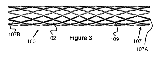
[0019] Fig. 3 is a side view of the shunt of Fig. 1A in a compressed
configuration.
[0020] Fig. 4A is a top view of the cell pattern of the shunt of Fig. 1A.
[0021] Fig. 4B is a top view of two cells from the cell pattern in Figure
4A.
[0022] Fig. 5 illustrates a balloon catheter in a deflated configuration
according to the
present invention.
¨3¨
CA 03129020 2021-08-03
WO 2020/163820 PCT/US2020/017361
[0023] Fig. 6 illustrates the balloon catheter of Fig. 5 in an expanded
configuration
according to the present invention.
[0024] Fig. 7 illustrates the balloon catheter of Fig. 5 with a shunt
compressed over it
according to the present invention.
[0025] Fig. 8 illustrates the balloon catheter of Fig. 6 with a shunt in
its expanded
position according to the present invention.
[0026] Fig. 9 illustrates a delivery procedure for a shunt according to the
present
invention.
[0027] Fig. 10 illustrates a delivery procedure for a shunt according to
the present
invention.
[0028] Fig. 11 illustrates a delivery procedure for a shunt according to
the present
invention.
[0029] Fig. 12 illustrates a delivery procedure for a shunt according to
the present
invention.
[0030] Fig. 13 illustrates a perspective view of another embodiment of a
shunt
according to the present invention.
[0031] Fig. 14 illustrates a side view of another embodiment of a shunt
according to
the present invention.
[0032] Fig. 15 illustrates a side view of another embodiment of a shunt
according to
the present invention.
[0033] Fig. 16A illustrates a side view of another embodiment of a shunt
according to
the present invention.
[0034] Fig. 16B illustrates a side view of the shunt of Fig. 16A.
[0035] Fig. 17 illustrates a side view of another embodiment of a shunt
according to
the present invention.
¨4¨
CA 03129020 2021-08-03
WO 2020/163820 PCT/US2020/017361
[0036] Fig. 18 illustrates a side view of the shunt of Fig. 17.
[0037] Fig. 19 illustrates an alternate embodiment of a balloon catheter
according to
the present invention.
[0038] Fig. 20 illustrates an alternate embodiment of a balloon catheter
according to
the present invention.
[0039] Fig. 21 illustrates an alternate embodiment of a balloon catheter
according to
the present invention.
DESCRIPTION OF EMBODIMENTS
[0040] Specific embodiments of the invention will now be described with
reference to
the accompanying drawings. This invention may, however, be embodied in many
different
forms and should not be construed as limited to the embodiments set forth
herein; rather,
these embodiments are provided so that this disclosure will be thorough and
complete,
and will fully convey the scope of the invention to those skilled in the art.
The terminology
used in the detailed description of the embodiments illustrated in the
accompanying
drawings is not intended to be limiting of the invention. In the drawings,
like numbers refer
to like elements.
[0041] The present invention is generally directed to a shunt and a method
of deploying
a shunt. More specifically, the shunt radially expands to an hourglass or
rivet shape while
also longitudinally foreshortening. The shunt is initially positioned within a
tissue opening
and then expanded, which causes the distal and proximal ends of the shunt to
flare radially
outwards and move towards each other. When fully expanded, these radially
flared ends
engage the tissue surrounding the opening, creating a smooth transition
between either
side of the tissue.
[0042] This shunt design provides several advantages over prior shunt
designs. For
example, the shunt may "self-position" itself within the tissue opening due to
its flared
shape and therefore provides increased precision in its positioning than prior
designs.
The flared portions also provide strong attachment to the surrounding tissue
as compared
with prior shunt designs. Finally, the shunt may have a small collapsed
profile and yet
can expand to a consistent inner diameter with high radial force. This allows
the use of
¨5¨
CA 03129020 2021-08-03
WO 2020/163820 PCT/US2020/017361
low-profile balloons to assist in the expansion of the shunt to achieve
consistent and
reliable implantation results.
[0043] A stent design that can be modified for use as a shunt in accordance
with the
principles of the present invention as explained herein is disclosed in U.S.
Patent No.
6,068,656 to Oepen, the entire contents of which is incorporated herein by
reference.
[0044] As discussed in greater detail in this specification, the
foreshortening and
hourglass shape can be achieved in several different ways and the shunts
themselves
may have several different features. It should be explicitly understood that
the features
shown in the different embodiments of this specification can be
interchangeably used with
features of other embodiments in this specification. In other words, it is
intended that the
features of the embodiments can be "mixed and matched" with each other.
[0045] Figures 1A and 1B illustrate the change in shape of one embodiment
of a
tubular shunt 100 of the present invention. In Figure 1A, the shunt 100 is
shown in a
radially compressed configuration having a relatively long length 101 and a
relatively
small, uniform diameter 103. As the shunt 100 is deployed, its length
substantially
decreases to 101' and its diameter increases. More specifically, end portions
100A
increase to a maximum radial diameter of 103' and then decrease in diameter
towards a
middle region 100B, which has a diameter of 103".
[0046] In one example, when compressed, the shunt 100 has a length 101 of
about 20
mm and a diameter 103 of about 1.5 mm, and when expanded the shunt 100 has a
diameter 103' of the end portions 100A of about 8 mm and a diameter 103" of
the middle
region 100B of about 5 mm.
[0047] In another example, when compressed, the shunt 100 has a length 101
of about
30 mm and a diameter 103 of about 2.2 mm, and when expanded the shunt 100 has
a
diameter 103' of the end portion 100A of about 8 mm and a diameter 103" of the
middle
region 100B of about 4 mm.
[0048] In another example, when compressed, the shunt 100 has a length 101
of about
22 mm and a diameter 103 of about 3.5 mm, and when expanded the shunt 100 has
a
diameter 103' of the end portion 100A of about 24 mm and a diameter 103" of
the middle
region 100B of about 20 mm.
¨6¨
CA 03129020 2021-08-03
WO 2020/163820 PCT/US2020/017361
[0049] As seen in Figures 2-4, this embodiment of the shunt 100 includes a
plurality of
tubular radial bands 107 that are each formed from a plurality of uniform,
alternating
waves that create the shunt passage 100C. Put another way, and referring
particularly to
Figures 3 and 4A, each radial band 107 comprises a plurality of straight
regions 107B
joined together to create a pattern of triangular peaks 107A that alternate
their longitudinal
directions. The peaks 107A of each radial band 107 are aligned with each other
and
connected via a small, straight portion 109, which effectively creates diamond-
shaped
cells 102 when radially compressed. As a result of this design, the angle of
each peak
107A increases as the shunt 100 is radially expanded and the radial bands 107
become
closer together to each other, which causes longitudinal foreshortening (i.e.,
a decrease
in length of the shunt 100).
[0050] One mechanism for causing the radial flaring of the ends 100A of the
shunt 100
can be seen in Figures 4A and 4B, which illustrate the pattern of the shunt
100 as if it
were longitudinally cut and flattened. Specifically, a pattern of cells 102
can be created
in which some cells 102A, 102B, 102C, 102D are longer in their proximal-to-
distal length
than other cells (i.e., they have longer straight regions 107B). Preferably,
cells 102 in the
middle of the shunt 100 have the smallest length and each row of cells 102
progressively
increase in length the further away from the middle they are. Alternately,
larger length
cells 102 can be located only near the ends of the shunt 100.
[0051] For example, middle cell 102A has a first length; longitudinally
adjacent cell
102B has a second, longer length than cell 102A; longitudinally adjacent cell
102C has a
third, longer length than cell 102B; and longitudinally adjacent cell 102D has
a fourth,
longer length than cell 102C.
[0052] To better see this distinction, Figure 4B comparatively illustrates
cells 102A and
102D next to each other. In a compressed configuration, the larger cell 102D
will have
longer straight portions 107B and a smaller angle of peak 107A relative to
cell 102A.
However, when expanding, the larger straight portions 107B allow those cells
to expand
to a larger diameter and foreshorten more than cell 102A. In this manner, the
expanded
shape and amount of foreshortening can be determined.
[0053] The size and ratio of the cells 102 and straight portions 107B can
vary,
depending on the desired expanded shape of the shunt 100. For example, having
¨7¨
CA 03129020 2021-08-03
WO 2020/163820 PCT/US2020/017361
dramatically larger end cells (e.g., cells 102C and 102D) may cause the
expanded
configuration of the shunt 100 to have a larger flare diameter size relative
to its middle
portion. In one specific example, the size increases of the straight portion
107B (i.e.,
struts) of each radial band 107 can be seen in the following listing, which
begins with the
straight portion 107B in the middle cell 102A and progresses towards the end
of the shunt
100. For a shunt with flaring on both ends, the progression of size increase
would be the
same on either side of the center region of the shunt. It will be appreciated
that through
creative configurations of the size progression described herein, one flare
could be a
different size or configuration from its opposite flare and thus the shunt can
be specifically
tailored to the particular use and location in the patient's body. Note, this
specific example
illustrates a greater number of straight portions 107B and therefore cells 102
than that
shown in Figure 4A. However, the shunt 100 may include a variety of different
cell
numbers. Straight portion 107B example sizes: 1.218 mm, 1.242 mm, 1.287 mm,
1.351
mm, 1.432 mm, 1.528 mm, 1.638 mm, 1.763 mm, 1.897 mm, 2.036 mm.
[0054] In addition to the variable size of the cells 102 along the length
of the shunt 100,
the shunt 100 can be heat set to an hourglass shape when unconstrained to
provide
additional expansion force, either with or without the assistance of a balloon
catheter.
[0055] Notwithstanding the above cell design, it is noted that multiple
cell variations
are contemplated in accordance with the present invention. In this regard, a
key design
parameter is that each "row" or band in the shunt body reaches maximum
expansion at a
particular diameter to achieve the final desired shape.
[0056] The shunt 100 can be delivered and expanded via a balloon catheter
110, as
seen in Figures 5 and 6. In one embodiment, a balloon 114 is disposed on its
distal end
of a tubular catheter body 112. The interior of the catheter body 112 has an
inflation
lumen 112A that opens to proximal and distal inflation ports 112B within the
balloon 114.
A guidewire lumen 116 is located within the catheter body 112, opening on the
proximal
and distal ends of the body 112.
[0057] As seen in Figure 6, the balloon 114 may inflate to an hourglass
shape that has
a smaller diameter middle region 114C than the proximal region 114A and distal
region
114B of the balloon 114. There are several different techniques to achieve
this inflated
shape of the balloon 114. For example, the balloon 114 can be composed of a
compliant
¨8¨
CA 03129020 2021-08-03
WO 2020/163820 PCT/US2020/017361
material and a non-compliant band (not shown) can be positioned around the
middle
region 114C. In another example, the proximal region 114A and distal region
114B can
be composed of a material with different expansion properties than the middle
region
114C (e.g., a compliant middle region with noncompliant proximal/distal
regions, or a
noncompliant middle region with compliant proximal/distal regions).
[0058] Figures 7 and 8 illustrate the shunt 100 positioned over the balloon
114.
Preferably, the shunt 100 is loaded onto the balloon 114 so that the middle
region 100B
of the shunt 100 is aligned with the middle region 114C of the balloon 114. In
that regard,
as the balloon 114 expands, the proximal region 114A and distal region 114B
cause the
end regions 100A of the shunt 100 to expand to a larger diameter than the
middle region
100B.
[0059] Figures 9 and 10 illustrate how the shunt 100 may be delivered
relative to an
area of target tissue 10. First, an initial puncture is made at the desired
location (e.g.,
with a needle). Next, the distal end of the delivery catheter 110 is advanced
through the
puncture in the tissue 10 such that there are roughly equal portions of the
shunt 100 on
either side of the tissue 10. Either the shunt 100 or the delivery catheter
110 can include
radiopaque markers at various known locations to assist a physician with
achieving a
desired alignment.
[0060] When the desired alignment is achieved, the balloon 114 is inflated,
causing
the shunt 100 to increase in radial diameter to an hourglass shape and to
foreshorten.
The shunt 100 is configured such that the foreshortening causes the flared end
regions
100A to engage and press into the tissue 10. These flared end regions 100A, as
well as
the proximal region 114A and distal region 114B of the balloon help "self-
center" the shunt
100 to an appropriate position. The end result is an opening in the tissue 10
with a
smooth, funnel-like transition on each side of the tissue.
[0061] One variation on this delivery technique allows for the passage
through the
shunt 100 (i.e., the narrowed middle region 110B) to be resized after
delivery, if needed.
Specifically, the shunt 100 can be delivered as previously described, but the
narrowed
middle region 110B is expanded to an initial diameter that is smaller than the
middle region
110B is capable of expanding to. This may be achieved, for example, by
limiting the
expansion size of the middle region 114C of the balloon 114. If the physician
determines
¨9¨
CA 03129020 2021-08-03
WO 2020/163820 PCT/US2020/017361
that increasing the size of the middle region 100B of the shunt 100 would be
beneficial,
the middle region 100B can be further expanded in diameter by either a
different portion
of the balloon (e.g., 114A or 114B) or by a second balloon catheter that
inflates to a
desired passage diameter.
[0062] Alternately, if the physician determines that the middle region 100B
of the shunt
100 was initially deployed with a diameter that is larger than desired, a
second delivery
catheter may be used to deliver a tubular spacer having a thickness that
reduces the size
of the passage through the middle region 100B. In one example, the tubular
spacer may
be a second shunt 100, similar to the shunt initially deployed but deployed
inside of the
first shunt.
[0063] This ability to resize the shunt 100 after delivery allows a
physician to customize
the amount of shunted fluid for each individual patient. It also allows the
shunt 100 to be
modified at a later date if the patient's hemodynamic needs change.
[0064] In an alternate embodiment, the balloon catheter may include two or
three
separate, independently inflatable balloons that can be inflated to different
sizes to
achieve a similar hourglass shape. This may allow the physician to limit
expansion of the
middle of the shunt 100 to a desired diameter while ensuring the ends of the
shunt 100
radially expand sufficiently to engage the surrounding tissue.
[0065] In another alternate embodiment, a mechanical device on a catheter
can be
used to expand the shunt 100 instead of using a balloon. For example, such a
catheter
may include two cone shaped structures that can be longitudinally slid towards
each other.
The shunt 100 may be positioned between these two structures so that when the
cone
shaped structures are moved toward each other, they cause the shunt 100 to
expand.
[0066] As previously discussed, the shunt 100' may be composed of a shape-
memory
material and heat set to the expanded hourglass shape when unconstrained. In
such an
embodiment, a balloon catheter 110 may not be necessary. Figures 11 and 12
illustrate
a similar delivery procedure with a delivery catheter 120 configured for
deployment of a
heat-set shunt 100. The catheter 120 includes an elongated catheter body 122
with a
retractable sheath 124 disposed over the shunt 100'. Similar to the previously
described
deployment procedure, a distal end of the catheter 120 is positioned through
the opening
¨10¨
CA 03129020 2021-08-03
WO 2020/163820 PCT/US2020/017361
in the tissue 10 such that roughly equal portions of the shunt 100 are
positioned on each
side of the tissue 10. When the desired alignment has been achieved (e.g., by
referencing
radiopaque markers of a known position), the sheath 124 is proximally
retracted, causing
the shunt 100' to radially expand to an hourglass shape and foreshorten as
shown in
Figure. 12.
[0067] In one embodiment, the shunt 100 may include a plurality of barbs
113, hooks,
or similar fastening structures, as seen in Figure 13. These may be positioned
on the
outside of the flared regions such that they pierce into the tissue of the
patient when the
shunt 100 is expanded. Alternately, the barbs 113 or similar anchoring
structure can be
located at various locations along the length of the shunt 100, pointing
radially outwards.
[0068] In one embodiment, the shunt 100 lacks any type of cover and acts to
maintain
the opening through the tissue by mechanical force. Figure 14 illustrates
another
embodiment of a shunt 130 having a similar laser-cut structure 132 as shunt
100 but also
a cover layer 134 that is attached to the laser-cut structure 132 (either on
the outside or
inside of the structure 132) and forms a similar tubular and hourglass shape.
To
accommodate the tubular-to-hourglass shape change, part or all of the material
134 may
be elastic or stretchable. Alternately, a tubular cover layer 134 can be
included only at
the middle region of the laser-cut structure 138 of the shunt 136, as seen in
Figure 15.
[0069] In another embodiment, either of the shunts may have two laser-cut
structural
layers that are positioned on the inner and outer surfaces of the cover layer
so as to
"sandwich" the cover layer.
[0070] It is sometimes desirable to occlude an existing shunt (e.g., a
naturally
occurring tissue passage) or chamber such as a left atrial appendage. In that
regard, any
of the shunt embodiments in this specification may include a material that
extends across
and occludes the central lumen of the shunt. For example, the material can be
a polymer
sheet that is attached to an end of the device with a small hole in the
center. The polymer
sheet may be elastic so that the enter hole expands with the balloon from the
delivery
catheter and then recovers back down to effectively seal the opening once the
balloon is
removed.
¨11¨
CA 03129020 2021-08-03
WO 2020/163820 PCT/US2020/017361
[0071] While the shunt 100 and its variations have been previously
described to
expand to a flared, hourglass style shape, other variations of the expanded
shape are
possible. For example, Figure 22 illustrates a shunt 180 in which only one end
is radially
flared outwards while the opposite end 180C maintains a diameter similar to
that of the
middle region 180B. Since the shunt 180 foreshortens in length, it may be
beneficial to
have barbs or other anchoring mechanisms along the middle region 180B and end
180C
to help anchor the shunt 180 during radial expansion.
[0072] In another example, neither end of the shunt expands to a flared
shape.
[0073] The shunts of this specification can be composed of biocompatible
materials
such as Nitinol or similar alloys, or bioabsorbable materials such as
magnesium, PLA, or
PLA-PGA. The shunts of this specification may also have features to promote
endothelization, such as open surface pores around 60 microns in diameter or a
polymer
coating known to promote tissue growth.
[0074] While the shunt 100 was previously described with a specific
pattern, it should
be appreciated that other patterns and designs are possible to achieve similar
functionality. For example, Figures 16A and 16B illustrate a shunt 140
comprising a
plurality of rings 144 comprising a plurality of alternating peaks. These
rings 144 are fixed
to a cover 142 and may either be free of connection to each other (other than
the cover)
or may have connection members 148 that connect to longitudinally adjacent
peaks. The
ends of the shunt 140 each include end rings 146 that are composed of a
plurality of
alternating peaks that are larger than those of rings 144. As seen in Figure
16B, when
radially expanded, the peaks 144 longitudinally compress together and fit
within each
other.
[0075] With respect to Figure 16B, in one embodiment, the shape depicted
therein
may be achieved by over-expanding the shunt by a balloon, which would cause
the ends
to flare open as shown and the central section expansion would be limited by
the cover
142.
[0076] In addition to having different laser-cut patterns, alternate
embodiments may
instead be comprised of a plurality of braided wires, such as the shunt 180
shown in
Figures 17 and 18. The shunt 180 can be braided on an hourglass-shaped mandrel
with
¨12¨
CA 03129020 2021-08-03
WO 2020/163820 PCT/US2020/017361
a plurality of shape-memory wires. After braiding, the shunt 180 can be heat-
set on the
mandrel and then removed, allowing it to compress to a tubular shape and
radially expand
to the hourglass shape (i.e., flared end regions 180A and a smaller diameter
middle region
180B).
[0077] As previously discussed, the delivery catheters 110 and 120 can
include
radiopaque markers to help a physician align the shunt 100. However, other
positioning
devices can also be used to aid in positioning.
[0078] For example, Figure 19 illustrates a delivery device 150 that
includes elongated
arms 152 that are connected to the catheter body at their proximal ends and
are
configured to radially expand away from the shunt 100 at their distal ends
154. The arms
152 are preferably of a length that the blunt distal ends contact the tissue
10 when the
shunt 100 is positioned at a desired alignment position (e.g., roughly halfway
through the
opening). This contact by the arms 152 provides the user with tactile feedback
in addition
to the visualization of the radiopaque markers. To prevent damage to the
tissue 10, the
arms 152 are preferably composed of flexible material, such as nitinol,
stainless steel,
pebax, nylon, polyurethane, or other plastics. The arms 152 can be relatively
straight or
can form a plurality of waves to provide further flexibility and compression.
[0079] Figure 20 illustrates another embodiment of a delivery device 160
that includes
an annular ring 162 located over the shunt 100 to assist with a desired
alignment of the
shunt 100. The annular ring 162 preferably has a thickness such that it is
larger than the
opening of the tissue 10 when the shunt 100 is compressed. The ring 162 is
longitudinally
positioned on a proximal side of the shunt 100 such that when contact is made
between
the ring 162 and tissue 10, the shunt 100 will have achieved a desired
longitudinal
alignment through the tissue opening. The ring 162 can be composed of cloth,
polymer,
or bioabsorbable material.
[0080] Alternately, instead of an annular ring 162, the shunt 100 itself
may include
structures 172 on the shunt 100 that are heat-set to radially expand, as seen
on device
170 in Figure 21. For example, the structures may be a loop, flap, or similar
structure that
radially pops up when an overlying sheath is withdrawn from the shunt. Similar
to the ring
162, these structures 172 are positioned at a location so as to provide
tactile feedback to
the physician to indicate a desired alignment of the shunt 100 within the
tissue opening.
¨13¨
CA 03129020 2021-08-03
WO 2020/163820 PCT/US2020/017361
[0081] While the specification has focused on various embodiments of a
shunt that are
used for creating a shunt within a patient or closing a hole between two
vessels or heart
chambers, other uses are also possible. For example, the shunt 100 may be used
an
anchor and/or attachment point for additional structures (e.g., tubes, other
shunts, etc.).
In another example, the shunt 100 may be used as an anchoring point for
artificial valves,
such as a mitral valve or aortic valve. In another example, the shunt 100 may
be used to
help restore a circular shape to a structure (e.g., aortic coarctation).
[0082] The shunts and delivery methods described in this specification can
be used
for a wide variety of shunt procedures. One example is a right-to-right shunt
between the
right pulmonary artery to superior vena cave, between the pulmonary artery to
right atrial
appendage, between the pulmonary artery or right ventricle to the venous
system, or
between the azygous vein to the inferior vena cava. These techniques can be
seen in
more detail in application number 16/576,704 entitled Methods And Technology
For
Creating Connections And Shunts Between Vessels And Chambers Of Biologic
Structures, filed September 19, 2019 which is herein incorporated by
reference. Other
possible uses include the creation of shunts between chambers of the heart,
such as atrial
septostomy, arteriovenous shunt creation for treating hypertension,
arteriovenous shunt
for fistula creation for dialysis patients, left atrium to coronary sinus,
pulmonary artery to
left aortic artery, or aorta to pulmonary artery.
[0083] Although the invention has been described in terms of particular
embodiments
and applications, one of ordinary skill in the art, in light of this teaching,
can generate
additional embodiments and modifications without departing from the spirit of
or
exceeding the scope of the claimed invention. Accordingly, it is to be
understood that the
drawings and descriptions herein are proffered by way of example to facilitate
comprehension of the invention and should not be construed to limit the scope
thereof.
¨14¨