Note: Descriptions are shown in the official language in which they were submitted.
CA 03129599 2021-08-09
WO 2020/167712 PCT/US2020/017597
Chromatin Mapping Assays and Kits using Long-Read Sequencing
Statement of Priority
[0001] This application claims the benefit of U.S. Provisional Application
Serial No.
62/803,829, filed February 11, 2019, the entire contents of which are
incorporated by
reference herein.
Field of Invention
[0002] This present invention relates to methods for carrying out chromatin
mapping assays
that use enzymes to incorporate barcoded DNA at targeted genomic regions
followed by
long-read sequencing (e.g., Third Generation Sequencing (TGS)). This approach
enables the
mapping of chromatin targets using TGS and can be used for a wide range of
elements or
features, including histone post-translational modifications, chromatin
associated proteins,
nucleosome positioning, and chromatin accessibility. The invention further
relates to kits and
reagents for carrying out the methods on chromatin samples that include one or
more cells.
Background of Invention
[0003] Genomic mapping assays are widely used to study chromatin structure and
function.
These include assays that analyze genomic location and abundance of chromatin
modifications, chromatin associated proteins (ChAPs), chromatin accessibility,
and
nucleosome positioning. Chromatin modifications include those that are added
to residues on
histone proteins or DNA. Histones residues on nucleosomes can be post-
translationally
modified (PTM) with a variety of chemical moieties, including lysine
methylation, lysine
acylation, arginine methylation, serine phosphorylation, etc., whereas DNA
residues are
modified with a number of different methylation variants (e.g., 5-
methylcytosine, 5-
hydrozxymethylcytosine, 5-formylcytosine, etc.). ChAPs include any protein
that directly
interacts with chromatin, including transcription factors that bind directly
to DNA and
"reader" proteins and enzymes that interact with or modify histone and/or DNA.
ChAPs also
include proteins that indirectly interact with chromatin via interactions with
macromolecular
complexes that regulate chromatin function, such as transcriptional regulation
and chromatin
remodeling complexes. Genomic regions devoid of nucleosomes are associated
with gene
transcription and activation as these chromatin regions are "accessible" to
transcriptional
machinery, whereas genomic regions of high nucleosome density are generally
correlated
with gene inactivation.
1
CA 03129599 2021-08-09
WO 2020/167712 PCT/US2020/017597
[0004] Histone modifications and ChAPs are routinely mapped genome-wide using
chromatin immunoprecipitation followed by next-generation sequencing (ChIP-
seq). Of
note, alternative chromatin mapping methods have been developed beyond ChIP,
including
those that tether enzymes to genomic regions, resulting in release,
enrichment, and
subsequent analysis of target material (e.g., DamID, ChIC, ChEC, CUT&RUN, and
CUT&Tag) [1-3]. For example, the related ChIC (Chromatin ImmunoCleavage [4,
5]) and
CUT&RUN (Cleavage Under Targets & Release Using Nuclease) methods use a PTM-
or
factor-specific antibody to tether a fusion of protein A and protein G-
Micrococcal Nuclease
(pAG-MNase) to genomic binding sites in intact cells or extracted nuclei,
which is then
activated by calcium addition to cleave DNA. pAG-MNase provides a cleavage
tethering
system for antibodies to any PTM or ChAP. The CUT&RUN protocol has been
streamlined
(vs. ChIC) by using a solid support (e.g., lectin-coated magnetic beads) to
adhere cells (or
nuclei). Similarly, CUT&Tag uses protein A tethered to a hyperactive
transposase (pA-Tn5),
followed by controlled activation of Tn5 to deliver sequencing adaptors for
paired-end
sequencing. This method is ultrasensitive and fast by removing the library
preparation step,
providing a tractable approach for chromatin mapping of select targets from
single cells [6].
[0005] There are several commercially available assays for genome-wide
analysis of
chromatin accessibility. Early assays used DNase I followed by sequencing
(DNase-Seq) to
identify nucleosome depleted regions genome-wide (termed DNase I
hypersensitivity sites;
DHSs) [7, 8]. A related approach using micrococcal nuclease I (MNase I) has
also been
developed to map nucleosome positioning [9], the inverse of chromatin
accessibility. While
these approaches work with both native (i.e., unfixed) and fixed cells, they
require extensive
enzyme optimization and high cell requirements. Recent advances to the DNase I
protocol
have enabled lower cell requirements with DHS mapping using a single cell;
however, DHSs
mapped to <2% of the reference genome, significantly limiting its utility
[10]. FAIRE-seq
(formaldehyde assisted isolation of regulator element sequencing) is a highly
sensitive
approach to enrich nucleosome-depleted regions, but as the name suggests, it
requires
formaldehyde fixation [11]. ATAC-seq uses a Tn5 transposase that
preferentially targets and
delivers its sequencing adaptor payload into accessible chromatin vs.
inaccessible regions
[12]. This method is quickly garnering adoption in the field due to its ease,
speed, and low
cell requirements. Indeed, ATAC-seq assays can be performed in a single day,
demonstrating
the potential use of this approach for clinical applications [13].
2
CA 03129599 2021-08-09
WO 2020/167712 PCT/US2020/017597
[0006] The application of hyperactive transposases in chromatin mapping assays
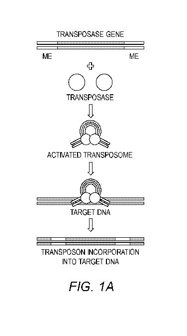
(e.g.,
CUT&Tag and ATAC-seq) has dramatically improved assay throughput and increased
assay
sensitivity. In these assays, the transposon is activated in vitro containing
engineered DNA
barcodes, which can be subsequently amplified using PCR and analyzed using
massively
parallel second-generation sequencing [13]. Native transpo sons encode the
transposase gene
flanked by two 19 bp sequences that are activated for genome targeting by
interactions with
the transposase protein (FIG. 1A). Current genomic assays (e.g., ATAC-seq,
CUT&Tag) use
modified transposons that lack an internal DNA region linking the activated
transposase-
bound DNA oligos, resulting in double-strand breaks and the release of DNA
fragments after
chromatin targeting (FIG. 1B) [13]. This modification is advantageous for
massive parallel
second-generation sequencing as it breaks chromatin into smaller DNA
fragments. See FIG.
2 for an example of a typical CUT&Tag workflow.
[0007] Third Generation Sequencing (TGS) platforms generate long reads from
native
DNA at relatively low cost, facilitating novel applications that are
inaccessible by standard
approaches. TGS platforms, such as Oxford Nanopore (ONT) and Pacific
BioSciences
(PacBio), are radically altering the field of genomics research, increasing
sequencing
technology accessibility and providing crucial insights into human disease
[14]. In nanopore
sequencing, long fragments of DNA are passed through nanopores, which use
changes in
electrical impulses to denote different DNA nucleotides [14]. The use of long
fragments of
DNA is unique to TGS platforms, and enables mapping to repetitive regions and
complex
DNA sequences [14, 15]. Indeed, the ONT nanopore sequencers can generate reads
>1 Mb
[16], and have been used to detect structural variants in breast [17] and
pancreatic cancer
[18]. Recent studies have also applied nanopore sequencing to transcriptome
profiling of
mouse B-lymphocytes at single-cell resolution [19, 20], demonstrating the
potential
application of TGS to ultra-low cell inputs. Importantly, because there is no
required PCR
amplification step, TGS enables the direct detection of unique base
modifications, including
DNA methylation (5mC), which has been challenging to directly measure using
standard
second generation sequencing (FIG. 4, left panel) [21, 22]. These rich
datasets enable
"multiomic" analyses (e.g., DNA sequence variation in combination with 5mC),
which have
helped delineate distinct types of brain tumors [23]. Combined with the
reduced costs,
improved coverage, and real-time sequencing capabilities [23-26], TGS is
redefining the
boundaries of modern genomics research. However, this approach is not suitable
for most
chromatin mapping studies, which result in fragmentation of chromatin and thus
are best
suited for second-generation sequencing. New methods are needed to enable
chromatin
3
CA 03129599 2021-08-09
WO 2020/167712 PCT/US2020/017597
mapping studies that preserve sample integrity and are suitable for TGS. Such
advances will
provide low-cost sequencing solutions as well as novel multiomic analyses that
include DNA
methylation and chromatin profiling analysis. Further, use of TGS for single
cell applications
may result in increased genomic coverage per cell, a major limitation of
current SGS-based
single cell assays [22].
Summary of the Invention
[0008] Current chromatin mapping assays known in the art result in chromatin
fragmentation during sample processing (e.g., ChIP-seq, ATAC-seq, CUT&Tag,
etc.),
making them well suited for short-read second-generation sequencing (SGS). As
such,
current methods are not compatible with TGS, with the exception of DNA
methylation [27].
New mapping methods that maintain chromatin integrity (i.e., are non-
destructive) would be
amenable to the mapping of chromatin elements (e.g., histone PTMs, ChAPs,
nucleosome
positioning, and chromatin accessibility) by TGS. These assays will have
significant
advantages over current SGS approaches, including increased access to
chromatin mapping
assays without the need for costly second-generation sequencing machines
(e.g., nanopore
sequencers), no PCR bias, and next-generation multiomic analysis, e.g.,
integration of DNA
methylation with other genomic features, such as histone PTMs, ChAPs, and
chromatin
accessibility.
[0009] The present invention relates to novel methods for chromatin mapping
assays using
TGS. The approach uses enzymes to modify DNA by non-destructive means to
include a
unique molecular identifier that can be used to determine the location of a
genomic element
as well as sample multiplexing for bulk (i.e., more than one cell) or single
cell analysis. The
resulting chromatin sample can then be processed for TGS, such as nanopore or
single
molecule real time sequencing, wherein the location of genomic elements (e.g.,
histone
PTMs, ChAPs, nucleosome position, chromatin accessibility, etc.) are mapped
via the
selective integration of barcoded DNA into sample chromatin. Samples may be
sequenced
using PCR-amplified chromatin or native chromatin. Sample genomic DNA may come
from
a single or multiple cells and be analyzed individually or multiplexed by
pooling samples,
each demarked by a unique DNA barcode, prior to whole genome sequencing. The
methods
described here may be used in any genome-wide assay known in the art which
uses enzymes
for chromatin mapping studies, including but not limited to ATAC-seq [13],
CUT&Tag [6],
and ChIL-seq [28]. This approach will result in long-sequencing reads, which
results in
better sequence coverage in areas of the genome that are challenging to map,
such as
4
CA 03129599 2021-08-09
WO 2020/167712 PCT/US2020/017597
repetitive regions. Long-reads will also result in greater sequencing coverage
when input
chromatin is limiting, such as single cell applications; these samples may be
PCR amplified
to increase chromatin input prior to sequencing. Further, TGS enables the use
of native
samples, which contain DNA modifications, that can be directly measured using
TGS. This
enables multiomic analyses, wherein DNA methylation is assessed in the context
of other
genomic elements, such as histone PTMs or chromatin accessibility; these
samples would not
be PCR amplified to preserve native DNA modifications. Of note, DNA
methylation
information is typically lost in current SGS-based approaches following PCR
amplification.
[0010] In some embodiments, a modified Tn5 can used to map chromatin
accessibility. In
these assays, the canonical function of the Tn5 enzyme are leveraged, loading
the hyperactive
Tn5 with a transposon carrying a unique identifier sequence, to insert its DNA-
barcoded
payload at open chromatin. Following insertion, DNA is repaired using
molecular biology
techniques known in the art (e.g., a combined treatment with T4 DNA polymerase
and T4
DNA ligase (as done previously [28]) and sequenced using TGS (e.g., PacBio or
Nanopore).
Finally, the insertion of barcoded DNA is used to map the chromatin loci with
high
accessibility (similar to ATAC-seq) and can be used to analyze one or more
cells in a single
assay. In some embodiments, a library of Tn5 transposons is assembled, each
denoted by a
unique DNA barcode. This library can be used to treat various bulk samples
(i.e., more than
one cell), which can then be pooled, sequenced, and deconvolved using their
unique DNA
barcode (i.e., multiplex analysis). This library can also be used for single
cell analysis using
a combinatorial indexing approach [29], wherein the assay is performed on a
population of
cells, which are then split into a multi-well plate (e.g., 96-, 384-, 1536-
well) containing ---20
cells per well. Each well is then processed for native chromatin sequencing
using adaptors
that include a second barcode or PCR amplified using primers that include a
second barcode
and sequenced using TGS. This approach provides a double barcode signature
that can be
used to assign reads to a specific single cell (SC). In some embodiments,
assays can be
deployed using single cell droplet-based methods, such as those commercially
available by
10X genomics or BioRad. In some embodiments, native chromatin is sequenced.
These
assays may be used to perform multiomic analyses wherein DNA modifications are
analyzed
in concert with chromatin accessibility. In some embodiments, samples are PCR-
amplified
prior to sequencing. In some embodiments, other enzymes that modify chromatin
are used in
place of Tn5, such as integrases or DNA methyltransferases.
[0011] In some embodiments, a modified Tn5 can be used to map histone PTMs,
ChAPs, or
nucleosome positioning (e.g., pAG-Tn5). In these assays, the canonical
function of the Tn5
CA 03129599 2021-08-09
WO 2020/167712 PCT/US2020/017597
enzyme is leveraged, loading the hyperactive Tn5 with a transposon carrying a
unique
identifier sequence to insert its DNA-barcoded payload. Unlike the modified
Tn5 used for
chromatin accessibility mapping, this modified Tn5 is fused to an antibody
binding moiety to
enable antibody-targeting (a modified version of pAG-Tn5 as used in CUT&Tag
[pAG-
mTn5]). Antibodies used in this assay can target any chromatin element or
binding protein,
such as histone PTMs, nucleosomes, ChAPs, and DNA methylation. Following
insertion,
DNA is repaired using molecular biology techniques known in the art (e.g., a
combined
treatment with T4 DNA polymerase and T4 DNA ligase as done previously [28])
and
sequenced using TGS (e.g., nanopore or single molecule real time sequencing).
Finally, the
insertion of barcoded DNA is used to map antibody targeted chromatin regions,
generating
chromatin maps similar to CUT&Tag and can be used to analyze one or more cells
in a single
assay. See FIG. 4 for an example workflow of how the modified pAG-Tn5 (pAG-
mTn5) can
be used to integrate a barcode into chromatin, followed by DNA repair and TGS.
Tn5 can be
fused to any protein binding moiety, such as Protein A, Protein G, Biotin,
GST, etc. In some
embodiments, a library of pAG-mTn5 transposons is assembled, each denoted by a
unique
DNA barcode. This library can be used to treat various bulk samples (i.e.,
more than one
cell), which can then be pooled, sequenced, and data can be deconvolved using
each samples
DNA barcode (i.e., multiplex analysis). DNA barcodes are used to indicate
genomic regions
where the antibody targeted, such as a PTM or ChAP. This library can also be
used for single
cell analysis using a combinatorial indexing approach [29], wherein the assay
is performed on
a population of cells, which are then split into a multi-well plate (e.g., 96-
, 384-, 1536-well)
containing ¨20 cells per well. Each well is then PCR amplified using primers
that contain a
second barcode (i.e., molecular identifier) and are sequenced using TGS. This
approach
provides a double barcode signature that can be used to assign reads to a
specific SC. In
some embodiments, assays can be deployed using single cell droplet-based
methods, such as
those commercially available by 10X genomics or BioRad. In some embodiments,
native
chromatin is sequenced. These assays may be used to perform multiomic analyses
wherein
DNA modifications are analyzed in concert with other chromatin features (e.g.,
histone PTMs
or ChAPs). In some embodiments, samples are PCR-amplified prior to sequencing,
which
may be useful for low cell input or single cell applications. In some
embodiments, other
enzymes that modify chromatin are used in place of Tn5, such as integrases or
DNA
methyltransferas es .
6
CA 03129599 2021-08-09
WO 2020/167712 PCT/US2020/017597
[0012] Thus, one aspect of the invention relates to a synthetic transposon
comprising a
DNA barcode region linked on its 5' and 3' end to a flanking region that is
recognized by a
transposase, wherein the synthetic transposon does not encode a transposase.
[0013] Another aspect of the invention relates to a transposome comprising the
synthetic
transposon of the invention and a transposase bound to each of the terminal
inverted repeats.
[0014] A further aspect of the invention relates to a library comprising two
or more of the
synthetic transposons of the invention and/or two or more of the transposomes
of the
invention, wherein each synthetic transposon comprises a unique DNA barcode.
[0015] An additional aspect of the invention relates to a kit comprising the
synthetic
transposon, transposome, or the library of the invention.
[0016] Another aspect of the invention relates to a method for chromatin
mapping,
comprising:
a) targeting an enzyme to a specific feature in chromatin in a sample;
b) activating the enzyme to alter or label DNA local to the feature;
c) preparing the chromatin for sequencing;
d) sequencing the chromatin using long-read sequencing; and
e) mapping the location of the chromatin feature based on the locations of
altered
or labeled DNA.
[0017] In some embodiments, the methods may be used to map chromatin
accessibility. In
some embodiments, the methods may be used to map chromatin modifications,
chromatin-
associated proteins, or nucleosome positioning. In some embodiments, the
methods are part
of multiomics assays.
[0018] In some embodiments, the methods of the invention may further comprise
steps of
using the sequencing results to compare chromatin features between healthy and
disease
tissues, predict a disease state, monitor response to therapy, and/or analyze
tumor
heterogeneity.
[0019] These and other aspects of the invention are set forth in more detail
in the
description of the invention below.
Brief Description of the Drawings
[0020] Figures 1A-1B show transposon schematics. (A) A cartoon showing
sequence
layout of native transposase, with transposase gene flanked by defined ends.
This transposon
DNA sequence interacts with the transposase enzyme to create an activated
transposome,
which can then target and deliver its payload into target DNA. (B) Cartoon
showing mutated
7
CA 03129599 2021-08-09
WO 2020/167712 PCT/US2020/017597
hyperactive transposome (e.g., Tn5) used in ATAC-seq. This hyperactive
transposome lacks
the transposon gene, which causes chromatin to fragment following
transposition. This
process has been termed tagmentation. The resulting DNA fragments can then be
PCR
amplified and sequenced using massive parallel sequencing (i.e., second-
generation
sequencing).
[0021] Figure 2 shows a summary of the CUT&Tag protocol as described in [6].
[0022] Figure 3 shows a schematic of the invention. Transposons are modified
to contain
an internal identified sequence (i.e., barcode) in place of the transposase
gene. This allows
the payload to be incorporated into target DNA and importantly does not result
in sequence
fragmentation. This method can be performed on multiple samples, which are
then pooled
and processed for whole chromatin sequencing. The incorporated DNA sequence
can be
used for chromatin mapping and distinguish samples when multiplexing. This
method can
also be used for single cell analysis using various split and pool strategies,
similar to those
previously described [29, 30].
[0023] Figure 4 shows the advantages of chromatin profiling using TGS vs.
current SGS
approaches (i.e. CUT&Tag, ChIP-seq).
Detailed Description of the Invention
[0024] The present invention is explained in greater detail below. This
description is not
intended to be a detailed catalog of all the different ways in which the
invention may be
implemented, or all the features that may be added to the instant invention.
For example,
features illustrated with respect to one embodiment may be incorporated into
other
embodiments, and features illustrated with respect to a particular embodiment
may be deleted
from that embodiment. In addition, numerous variations and additions to the
various
embodiments suggested herein will be apparent to those skilled in the art in
light of the
instant disclosure which do not depart from the instant invention. Hence, the
following
specification is intended to illustrate some particular embodiments of the
invention, and not
to exhaustively specify all permutations, combinations and variations thereof.
[0025] Unless the context indicates otherwise, it is specifically intended
that the various
features of the invention described herein can be used in any combination.
Moreover, the
present invention also contemplates that in some embodiments of the invention,
any feature
or combination of features set forth herein can be excluded or omitted. To
illustrate, if the
specification states that a complex comprises components A, B and C, it is
specifically
8
CA 03129599 2021-08-09
WO 2020/167712 PCT/US2020/017597
intended that any of A, B or C, or a combination thereof, can be omitted and
disclaimed
singularly or in any combination.
[0026] Unless otherwise defined, all technical and scientific terms used
herein have the
same meaning as commonly understood by one of ordinary skill in the art to
which this
invention belongs. The terminology used in the description of the invention
herein is for the
purpose of describing particular embodiments only and is not intended to be
limiting of the
invention.
[0027] Nucleotide sequences are presented herein by single strand only, in the
5' to 3'
direction, from left to right, unless specifically indicated otherwise.
Nucleotides and amino
acids are represented herein in the manner recommended by the IUPAC-IUB
Biochemical
Nomenclature Commission, or (for amino acids) by either the one-letter code,
or the three
letter code, both in accordance with 37 C.F.R. 1.822 and established usage.
[0028] Except as otherwise indicated, standard methods known to those skilled
in the art
may be used for production of recombinant and synthetic polypeptides,
antibodies or antigen-
binding fragments thereof, manipulation of nucleic acid sequences, production
of transformed
cells, the construction of nucleosomes, and transiently and stably transfected
cells. Such
techniques are known to those skilled in the art. See, e.g., SAMBROOK et al.,
MOLECULAR CLONING: A LABORATORY MANUAL 4th Ed. (Cold Spring Harbor,
NY, 2012); F. M. AUSUBEL et al. CURRENT PROTOCOLS IN MOLECULAR
BIOLOGY (Green Publishing Associates, Inc. and John Wiley & Sons, Inc., New
York).
[0029] All publications, patent applications, patents, nucleotide sequences,
amino acid
sequences and other references mentioned herein are incorporated by reference
in their
entirety.
[0030] As used in the description of the invention and the appended claims,
the singular
forms "a," "an" and "the" are intended to include the plural forms as well,
unless the context
clearly indicates otherwise.
[0031] As used herein, "and/or" refers to and encompasses any and all possible
combinations of one or more of the associated listed items, as well as the
lack of
combinations when interpreted in the alternative ("or").
[0032] Moreover, the present invention also contemplates that in some
embodiments of the
invention, any feature or combination of features set forth herein can be
excluded or omitted.
[0033] Furthermore, the term "about," as used herein when referring to a
measurable value
such as an amount of a compound or agent of this invention, dose, time,
temperature, and the
9
CA 03129599 2021-08-09
WO 2020/167712 PCT/US2020/017597
like, is meant to encompass variations of 10%, 5%, 1%, 0.5%, or even
0.1% of the
specified amount.
[0034] The term "consisting essentially of' as used herein in connection with
a nucleic acid,
protein means that the nucleic acid or protein does not contain any element
other than the
recited element(s) that significantly alters (e.g., more than about 1%, 5% or
10%) the function
of interest of the nucleic acid or protein.
[0035] As used herein, the term "polypeptide" encompasses both peptides and
proteins,
unless indicated otherwise.
[0036] A "nucleic acid" or "nucleotide sequence" is a sequence of nucleotide
bases, and
may be RNA, DNA or DNA-RNA hybrid sequences (including both naturally
occurring and
non-naturally occurring nucleotide), but is preferably either single or double
stranded DNA
sequences.
[0037] As used herein, an "isolated" nucleic acid or nucleotide sequence
(e.g., an "isolated
DNA" or an "isolated RNA") means a nucleic acid or nucleotide sequence
separated or
substantially free from at least some of the other components of the naturally
occurring
organism or virus, for example, the cell or viral structural components or
other polypeptides
or nucleic acids commonly found associated with the nucleic acid or nucleotide
sequence.
[0038] Likewise, an "isolated" polypeptide means a polypeptide that is
separated or
substantially free from at least some of the other components of the naturally
occurring
organism or virus, for example, the cell or viral structural components or
other polypeptides
or nucleic acids commonly found associated with the polypeptide.
[0039] By "substantially retain" a property, it is meant that at least about
75%, 85%, 90%,
95%, 97%, 98%, 99% or 100% of the property (e.g., activity or other measurable
characteristic) is retained.
[0040] The term "synthetic" refers to a compound, molecule, or complex that
does not exist
in nature.
[0041] The term "DNA barcode" refers to a nucleic acid sequence that can be
used to
unambiguously identify a DNA molecule in which it is located. The length of
the barcode
determines how many unique sequences can be present in a library. For example,
a 1
nucleotide (nt) barcode can code for 4 library members, a 2 nt barcode 16
variants, 3 nt
barcode 64 variants, 4 nt 256 variants, 5 nt 1,024 variants and so on. The
barcode(s) can be
single-stranded (ss) DNA or double-stranded (ds) DNA or a combination thereof.
CA 03129599 2021-08-09
WO 2020/167712 PCT/US2020/017597
[0042] A first aspect of the invention relates to a synthetic transposon
comprising,
consisting essentially of, or consisting of a DNA barcode region linked on its
5' and 3' end to
a flanking region that is recognized by a transposase, wherein the synthetic
transposon does
not encode a transposase. A flanking region that is "recognized" by a
transposase is one that
is specifically bound by a cognate transposase and functions to insert the
transposon into
DNA. In some embodiments, the flanking region is identical to or derived from
one found in
a naturally-occurring DNA transposon, such as the 19 bp Mosaic Ends (ME) of
Tn5. In some
embodiments, the flanking region may have a length of 7-40 nucleotides, e.g.,
7, 8, 9, 10, 11,
12, 13, 14, 15, 16, 17, 18, 19, 20, 22, 24, 26, 28, 30, 32, 34, 36, 38, or 40
nucleotides or any
range therein. In some embodiments, the flanking regions comprise terminal
inverted repeats
flanked by a short direct repeat. In some embodiments, the flanking region
comprises a DNA
barcode. The DNA barcode may have a length of less than 400, 300, 200, or 50
nucleotides.
In some embodiments, the DNA barcode may have a length of at least 6, 7, 8, 9,
10, 12, 14,
16, 18, 20, 22, 24, 26, 28, or 30 nucleotides. In some embodiments, one or
more of the
nucleotides in the flanking region may be modified, e.g., by methylation or
labeling, e.g.,
with biotin.
[0043] Another aspect of the invention relates to a transposome comprising the
synthetic
transposon of the invention and a transposase bound to each of the terminal
inverted repeats.
In some embodiments, the transposase may be a wild-type transposase, e.g.,
Tn5, Mu, IS5,
IS91, Tn552, Tyl, Tn7, Tn/O, Mariner, P Element, Tn3, Tn10, or Tn903. In some
embodiments, the transposase is modified from a wild-type transposase, e.g., a
mutated
hyperactive transposase. Such modified transposases are known in the art. In
some
embodiments, the transposase is Tn5 or a modified Tn5, e.g., a hyperactive Tn5
comprising
one or more of the mutations E54K, M56A, or L372P.
[0044] A further aspect of the invention relates to a library comprising two
or more of the
synthetic transposon of invention and/or two or more of the transposome of the
invention,
wherein each synthetic transposon comprises a unique DNA barcode. In some
embodiments,
the library may comprise 5, 10, 50, 100, 250, 500, 1000, 5000 or more
transposons and/or
transposomes, each with a unique DNA barcode.
[0045] An additional aspect of the invention relates to a kit comprising the
synthetic
transposon, transposome, and/or the library of the invention. In some
embodiments, the kit
further comprises one or more transposases that recognize the sequence of the
synthetic
transposon. The kit may further comprise additional components for carrying
out the
11
CA 03129599 2021-08-09
WO 2020/167712 PCT/US2020/017597
methods of the invention, including but not limited to enzymes, antibodies,
nucleotides,
beads, buffers, containers, instructions, etc.
[0046] Another aspect of the invention relates to a method for chromatin
mapping,
comprising:
a) targeting an enzyme to a specific feature in chromatin in a sample;
b) activating the enzyme to alter or label DNA local to the feature;
c) preparing the chromatin for sequencing;
d) sequencing the chromatin using long-read sequencing; and
e) mapping the location of the chromatin feature based on the locations of
altered
or labeled DNA.
[0047] The chromatin to be mapped in the assays of the invention may be from
any source,
including organs, tissues, cells, or cell-free. The amount of chromatin to be
used may vary
widely due to the sensitivity of the assay. In some embodiments, the sample
comprises
chromatin from less than 1000, 500, 100, 10, or 5 cells. In some embodiments,
the sample
comprises chromatin from 1 cell.
[0048] The methods of the invention can be carried out on any scale depending
on the size
of the sample. In some embodiments, the steps are carried out in a well of a
multiwall plate.
In some embodiments, the steps are carries out on a single cell scale, e.g.,
using a single cell
droplet-based method or combinatorial indexing method.
[0049] The sample comprising the chromatin to be mapped in the assays of the
invention
may comprise cells or nuclei comprising the chromatin. In some embodiments,
the cells or
nuclei are attached to a solid support for ease of manipulation during the
steps of the method.
The solid support may be, without limitation, a well or a bead, e.g., a
magnetic bead. In some
embodiments, the cell or nuclei are not attached to a solid support.
[0050] In some embodiments, the cells or nuclei are permeabilized to enhance
access of
components to the chromatin. For example, cells can be permeabilized with
digitonin, e.g.,
about 0.01% digitonin. In some embodiments, the cells or nuclei are not
permeabilized.
[0051] In some embodiments, the sample comprises chromatin that has been
isolated from
cells or nuclei.
[0052] The sample comprising chromatin to be mapped may be from any source. In
some
embodiments, the chromatin is obtained from a biological sample. The
biological sample
may be, without limitation, blood, serum, plasma, urine, saliva, semen,
prostatic fluid, nipple
12
CA 03129599 2021-08-09
WO 2020/167712 PCT/US2020/017597
aspirate fluid, lachrymal fluid, perspiration, feces, cheek swabs,
cerebrospinal fluid, cell
lysate samples, amniotic fluid, gastrointestinal fluid, biopsy tissue,
lymphatic fluid, or
cerebrospinal fluid.
[0053] In some embodiments, the chromatin is from a diseased tissue or sample.
In some
embodiments, the chromatin is from non-diseased tissue or sample. In some
embodiments,
the chromatin is from a peripheral tissue or cell, e.g., a peripheral blood
mononuclear cell.
[0054] In some embodiments, the chromatin is from cultured cells, e.g., a cell
line or
primary cells. In some embodiments, the chromatin is from an animal model of a
disease or
disorder. In some embodiments, the chromatin is from a subject, e.g., a
patient, having or
suspected of having a disease or disorder.
[0055] The methods of the invention can be used to perform any type of
chromatin
mapping, e.g., mapping any kind of specific feature of interest, including but
not limited to
genomic location and abundance of chromatin modifications, chromatin
associated proteins
(ChAPs), chromatin accessibility, and nucleosome positioning.
[0056] In one aspect, the methods of the invention include a method for
mapping chromatin
accessibility. The enzyme used in the mapping of chromatin accessibility may
be any
enzyme capable of detectably altering or labeling DNA where it is accessible.
In one
embodiment, the enzyme is an integrase or a DNA methyl transferase. In one
embodiment,
the enzyme used in the mapping of chromatin accessibility is a transposase. In
some
embodiments, the transposase may be a wild-type transposase, e.g., Tn5, Mu,
IS5, IS91,
Tn552, Tyl, Tn7, Tn/O, Mariner, P Element, Tn3, Tn10, or Tn903. In some
embodiments,
the transposase is modified from a wild-type transposase, e.g., a mutated
hyperactive
transposase. Such modified transposases are known in the art. In some
embodiments, the
transposase is Tn5 or a modified Tn5.
[0057] In some embodiments, the method comprises contacting a sample
comprising
chromatin with the synthetic transposon, transposome, or library of the
invention under
conditions in which the synthetic transposon can be inserted into the
chromatin.
[0058] In some embodiments, the activating of the enzyme in step b) comprises
adding a
factor necessary for enzyme activity, e.g., by adding an ion such as calcium
or magnesium.
Once activated, the enzyme alters or labels DNA local to the feature. The term
"local" in this
context refers to DNA within 5-30 nucleotides (e.g., 5, 6, 7, 8, 9, 10, 11,
12, 13, 14, 15, 16,
17, 18, 19, 20, 21, 22, 23, 24, 25, 26, 27, 28, 29, or 30 nucleotides or any
range therein, e.g.,
13
CA 03129599 2021-08-09
WO 2020/167712 PCT/US2020/017597
less than 6, 7, 8, 9, 10, 11, 12, 13, 14, 15, 16, 17, 18, 19, 20, 21, 22, 23,
24, 25, 26, 27, 28, 29,
or 30 nucleotides) or 3-18 nm (e.g., 3,4, 5, 6, 7, 8, 9, 10, 11, 12, 13, 14,
15, 16, 17, or 18 nm
or any range therein, e.g., less than 4, 5, 6, 7, 8, 9, 10, 11, 12, 13, 14,
15, 16, 17, or 18 nm) of
the feature.
[0059] In some embodiments, the method further comprises repairing transposon
ligation
sites prior to sequencing, e.g., using a DNA polymerase such as DNA polymerase
I and a
DNA ligase such as T4 DNA ligase.
[0060] In some embodiments of the method of mapping chromatin accessibility,
two or
more samples are contacted with a synthetic transposon and each sample is
contacted with a
different synthetic transposon comprising a unique DNA barcode. In some
embodiments, 2,
3, 4, 5, 6, 7, 8, 9, 10, 15, 20, 25, 50, 100, 250, 500, or 1000 or more
samples are each
contacted with a different synthetic transposon comprising a unique DNA
barcode. In some
embodiments, the two or more samples may be pooled after step b).
[0061] In one aspect, the methods of the invention include a method for
mapping chromatin
modifications, chromatin-associated proteins, or nucleosome positioning. In
some
embodiments, the chromatin modification is a histone modification (e.g., a
post-translational
modification), histone variant, or a DNA modification (e.g., a post-
transcriptional
modification).
[0062] The histone PTM may be any PTM for which measurement is desirable. In
some
embodiments, the histone PTM is, without limitation, N-acetylation of serine
and alanine;
phosphorylation of serine, threonine and tyrosine; N-crotonylation, N-
acylation of lysine; N6-
methylation, N6,N6-dimethylation, N6,N6,N6-trimethylation of lysine; omega-N-
methylation, symmetrical-dimethylation, asymmetrical-dimethylation of
arginine;
citrullination of arginine; ubiquitinylation of lysine; sumoylation of lysine;
0-methylation of
serine and threonine, ADP-ribosylation of arginine, aspartic acid and glutamic
acid, or any
combination thereof.
[0063] Several naturally occurring histone variants are known in the art and
any one or
more of them may be included in a nucleo some. Histone variants include,
without limitation,
H3.3, H2A.Bbd, H2A.Z.1, H2A.Z.2, H2A.X, mH2A1.1, mH2A1.2, mH2A2, TH2B, or any
combination thereof.
[0064] The DNA post-transcriptional modification may be any modification for
which
measurement is desirable. In some embodiments, the DNA post-transcriptional
modification
14
CA 03129599 2021-08-09
WO 2020/167712 PCT/US2020/017597
is 5-methylcytosine, 5-hydroxymethylcytosine, 5-formylcytosine, 5-
carboxylcytosine, 3-
methylcytosine, or any combination thereof.
[0065] The chromatin-associated protein may be any chromatin-associated
protein for
which measurement is desirable. In some embodiments, the chromatin-associated
protein is a
transcription factor, a histone binding protein, or a DNA binding protein.
[0066] In methods for mapping chromatin modifications, chromatin-associated
proteins, or
nucleosome positioning, the step of targeting an enzyme to a specific feature
in chromatin in
a sample comprises contacting the chromatin with an antibody, aptamer, or
recognition agent
that specifically binds to the feature. The antibody, aptamer, or recognition
agent used in the
methods of the invention may be any agent that specifically recognizes and
binds to a target,
e.g., an antigen. The term "antibody" includes antigen-binding fragments
thereof, such as
scFv, Fab, Fv, Fab', F(ab')2 fragments, dAb, VHH, nanobodies, V(NAR) or
minimal
recognition units.
[0067] For methods of mapping chromatin modifications, chromatin-associated
proteins, or
nucleosome positioning, the enzyme is linked to a protein that binds the
antibody, aptamer, or
recognition agent, e.g., an antibody binding protein. In some embodiments, the
antibody-
binding protein may be, without limitation, protein A, protein G, a fusion
between protein A
and protein G, protein L, or protein Y.
[0068] The enzyme used in the mapping of mapping chromatin modifications,
chromatin-
associated proteins, or nucleosome positioning may be any enzyme capable of
detectably
altering or labeling DNA where it is accessible. In one embodiment, the enzyme
is an
integrase or a DNA methyl transferase. In one embodiment, the enzyme used in
the mapping
of chromatin accessibility is a transposase. In some embodiments, the
transposase may be a
wild-type transposase, e.g., Tn5, Mu, IS5, IS91, Tn552, Tyl, Tn7, Tn/O,
Mariner, P Element,
Tn3, Tn10, or Tn903. In some embodiments, the transposase is modified from a
wild-type
transposase, e.g., a mutated hyperactive transposase. Such modified
transposases are known
in the art. In some embodiments, the transposase is Tn5 or a modified Tn5.
[0069] In some embodiments, the method comprises contacting a sample
comprising
chromatin with the synthetic transposon, transposome, or library of the
invention under
conditions in which the synthetic transposon can be inserted into the
chromatin.
[0070] In some embodiments, the activating of the enzyme in step b) comprises
adding a
factor necessary for enzyme activity, e.g., by adding an ion such as calcium
or magnesium.
CA 03129599 2021-08-09
WO 2020/167712 PCT/US2020/017597
[0071] In some embodiments, the method further comprises repairing transposon
ligation
sites prior to sequencing, e.g., using a DNA polymerase such as DNA polymerase
I and a
DNA ligase such as T4 DNA ligase.
[0072] In some embodiments of the method of mapping chromatin modifications,
chromatin-associated proteins, or nucleosome positioning, two or more samples
are contacted
with a synthetic transposon and each sample is contacted with a different
synthetic
transposon comprising a unique DNA barcode. In some embodiments, 2, 3, 4, 5,
6, 7, 8, 9,
10, 15, 20, 25, 50, 100, 250, 500, or 1000 or more samples are each contacted
with a different
synthetic transposon comprising a unique DNA barcode. In some embodiments, the
two or
more samples may be pooled after step b).
[0073] For all of the methods of the invention, the methods may be carried out
using a
combinatorial cellular indexing technique. In some embodiments, the method may
be carried
out on a population of cells and step c) comprises dividing the population of
cells into groups
and processing the cells for sequencing using adaptors that include a second
barcode or PCR
amplification using primers that include a second barcode so that each cell
comprises a
double barcode signature. In some embodiments, each group of cells may
comprise less than
about 1000, 500, 250, 100, or 50 cells, e.g., about 10 to about 30 cells,
e.g., about 20 cells.
[0074] For all of the methods of the invention, the methods may be carried out
as part of a
multiomic process wherein additional analyses are performed on the same
samples, e.g.,
based on the long-read sequencing information. In some embodiments, the
methods further
comprise analyzing a DNA modification in the chromatin, e.g., DNA methylation.
[0075] As defined herein "long-read sequencing" refers to third generation
sequencing
techniques that work on the single molecule level and provide sequence reads
of at least 10
kb, e.g., at least 50 kb or 100 kb. The long-read sequencing may be carried
out by any
method known in the art. In some embodiments, the long-read sequencing
comprises
nanopore sequencing, such as techniques available from Oxford Nanopore (ONT).
In some
embodiments, the long-read sequencing comprises single molecule real time
sequencing,
such as techniques available from Pacific BioSciences .
[0076] In some embodiments of the methods of the invention, the methods may
further
comprise a step of mechanically or enzymatically shearing the sample prior to
sequencing. In
other embodiments, no shearing occurs prior to sequencing.
16
CA 03129599 2021-08-09
WO 2020/167712 PCT/US2020/017597
[0077] In some embodiments of the methods of the invention, the methods may
further
comprise a step of amplifying the sample prior to sequencing. In other
embodiments, no
amplifying occurs prior to sequencing, enabling analysis of native DNA
modifications.
[0078] The results obtained from the methods of the invention may be used for
any purpose
where information on chromatin structure and/or modification, e.g., epigenetic
changes,
would be useful. In some embodiments, the methods may further comprise the
step of using
the sequencing results to compare chromatin features between healthy and
disease tissues. In
some embodiments, the methods may further comprise the step of using the
sequencing
results to predict a disease state. In some embodiments, the methods may
further comprise
the step of using the sequencing results to monitor response to therapy. In
some
embodiments, the methods may further comprise the step of using the sequencing
results to
analyze tumor heterogeneity.
[0079] The methods of the invention may be used for detecting and quantitating
the
presence of an epigenetic modification in chromatin. An antibody, aptamer, or
recognition
agent that specifically binds to the epigenetic modification may be used to
detect and
quantitate the chromatin element or modification at various genomic loci.
[0080] The methods of the invention may be used for determining and
quantitating the
epigenetic status of chromatin in a subject having a disease or disorder. An
antibody,
aptamer, or recognition agent that specifically binds to one or more
epigenetic modifications
that may be associated with the disease or disorder of the subject may be used
to detect and
quantitate the chromatin element or modification at various genomic loci. By
this method,
one can determine if a subject having a disease or disorder, e.g., a tumor,
has an epigenetic
modification that is known to be associated with the tumor type.
[0081] The methods of the invention may be used for monitoring changes in
epigenetic
status of chromatin over time in a subject. This method may be used to
determine if the
epigenetic status is improving, stable, or worsening over time. The steps of
the method may
be repeated as many times as desired to monitor changes in the status of an
epigenetic
modification, e.g., 2, 3, 4, 5, 6, 7, 8, 9, 10, 25, 50, or 100 or more times.
The method may be
repeated on a regular schedule (e.g., daily, weekly, monthly, yearly) or on an
as needed basis.
The method may be repeated, for example, before, during, and/or after
therapeutic treatment
of a subject; after diagnosis of a disease or disorder in a subject; as part
of determining a
diagnosis of a disease or disorder in a subject; after identification of a
subject as being at risk
17
CA 03129599 2021-08-09
WO 2020/167712 PCT/US2020/017597
for development of a disease or disorder; or any other situation where it is
desirable to
monitor possible changes in the chromatin element or modification at various
genomic loci.
[0082] The methods of the invention may be used for measuring on-target
activity of an
epigenetic-targeting drug. The methods may be carried out before, during,
and/or after
administration of an epigenetic-targeting drug to determine the capability of
the drug to alter
the epigenetic status of the subject.
[0083] The methods of the invention may be used for monitoring the
effectiveness of an
epigenetic therapy in a subject having a disease or disorder associated with
epigenetic
modifications.
[0084] Epigenetic therapies are those designed to alter the epigenetic status
of proteins
(e.g., histones) or DNA. One example of an epigenetic therapy includes lysine
deacetylase
inhibitors (formerly termed histone deacetylase inhibitors) (e.g., vorinostat
(suberoylanilide
hydroxamic acid), CI-994 (tacedinaline), MS-275 (entinostat), BMP-210, M344,
NVP-
LAQ824, LBH-529 (panobinostat), MGCD0103 (mocetinostat), PXD101 (belinostat),
CBHA, PCI-24781, ITF2357, valproic acid, trichostatin A, and sodium butyrate),
which are
used to treat cutaneous T-cell lymphoma (CTCL) or in clinical trials for the
treatment of
hematologic and solid tumors, including lung, breast, pancreas, renal, and
bladder cancers,
melanoma, glioblastoma, leukemias, lymphomas, and multiple myeloma. A further
example
of an epigenetic therapy is histone acetyltransferase inhibitors (e.g.,
epigallocatechin-3-
gallate, garcinol, anacardic acid, CPTH2, curcumin, MB-3, MG149, C646, and
romidepsin).
Another example of an epigenetic therapy is DNA methyltransferase inhibitors
(e.g.,
azacytidine, decitabine, zebularine, caffeic acid, chlorogenic acid,
epigallocatechin,
hydralazine, procainamide, procaine, and RG108), which have been approved for
treatment
of acute myeloid leukemia, myelodysplastic syndrome, and chronic
myelomonocytic
leukemia and in clinical trials for treatment of solid tumors. Other
epigenetic therapies
include, without limitation, lysine methyltransferases (e.g., pinometostat,
tazometostat, CPI-
1205); lysine demethylases (e.g., ORY1001); arginine methyltransferases (e.g.,
EPZ020411);
arginine deiminases (e.g., GSK484); and isocitrate dehydrogenases (e.g.,
enasidenib,
ivosidenib). See Fischle et al., ACS Chem. Biol. 11:689 (2016); DeWoskin et
al., Nature
Rev. /2:661 (2013); Campbell et al., I Cl/n. Invest. 124:64 (2014); and Brown
et al., Future
Med. Chem. 7:1901(2015); each incorporated by reference herein in its
entirety.
[0085] The steps of the method may be repeated as many times as desired to
monitor
effectiveness of the treatment, e.g., 2, 3, 4, 5, 6, 7, 8, 9, 10, 25, 50, or
100 or more times. The
method may be repeated on a regular schedule (e.g., daily, weekly, monthly,
yearly) or on as
18
CA 03129599 2021-08-09
WO 2020/167712 PCT/US2020/017597
needed basis, e.g., until the therapeutic treatment is ended. The method may
be repeated, for
example, before, during, and/or after therapeutic treatment of a subject,
e.g., after each
administration of the treatment. In some embodiments, the treatment is
continued until the
method of the invention shows that the treatment has been effective.
[0086] The methods of the invention may be used for selecting a suitable
treatment for a
subject having a disease or disorder associated with epigenetic modifications
based on the
epigenetic status of chromatin in the subject.
[0087] The methods may be applied, for example, to subjects that have been
diagnosed or
are suspected of having a disease or disorder associated with epigenetic
modifications. A
determination of the epigenetic status of an epitope may indicate that the
status of an epitope
has been modified and an epigenetic therapy should be administered to the
subject to correct
the modification. Conversely, a determination that the status of an epitope
has not been
modified would indicate that an epigenetic therapy would not be expected to be
effective and
should be avoided. For example, a determination that a particular genomic
locus has been
acetylated or deacetylated may indicate that treatment with a histone
deacetylase inhibitor
would be appropriate. Similarly, a determination that a particular genomic
locus has been
hyper- or hypomethylated may indicate that treatment with a DNA
methyltransferase
inhibitor would be appropriate.
[0088] The methods of the invention may be used for determining a prognosis
for a subject
having a disease or disorder associated with epigenetic modifications based on
the epigenetic
status of chromatin in the subject.
[0089] In some instances, the epigenetic status of an epitope is indicative of
the prognosis
of a disease or disorder associated with epigenetic modifications. Thus, a
determination of
the epigenetic status of an epitope in a subject that has been diagnosed with
or is suspected of
having a disease or disorder associated with epigenetic modifications may be
useful to
determine the prognosis for the subject. Many such examples are known in the
art. One
example is prostate cancer and hypermethylation of the glutathione-S
transferase P1 (GSTP1)
gene promoter, the adenomatous polyposis coli (APC) gene, the genes PITX2, Cl
orf114, and
GABRE¨miR-452¨miR-224, as well as the three-gene marker panel
A0X1/Clorf114/HAPLN3 and the 13-gene marker panel GSTP1, GRASP, TMP4, KCNC2,
TBX1, ZDHHC1, CAPG, RARRES2, SAC3D1, NKX2-1, FAM107A, SLC13A3, FILIP1L.
Another example is prostate cancer and histone PTMs, including, without
limitation,
increased H3K18Acetylation and H3K4diMethylation associated with a
significantly higher
risk of prostate tumor recurrence, H4K12Acety1ation and H4R3diMethylation
correlated with
19
CA 03129599 2021-08-09
WO 2020/167712 PCT/US2020/017597
tumor stage, and H3K9diMethylation associated with low-grade prostate cancer
patients at
risk for tumor recurrence. Another example is the link between overall
survival in breast
cancer patients and methylation status of CpGs in the genes CREB5, EXPH5,
ZNF775,
ADCY3, and ADMA8. Another example is glioblastoma and hypermethylation of
intronic
regions of genes like EGFR, PTEN, NF1, PIK3R1, RB1, PDGFRA, and QKI. A further
example is inferior prognosis for colon cancer and methylation status of the
promoter of the
CNRIP1, FBN1, INA, MAL, SNCA, and SPG20 genes.
[0090] The methods of the invention may be used for identifying a biomarker of
a disease
or disorder associated with epigenetic modifications based on the epigenetic
status of
chromatin in a subject.
[0091] In this method, biological samples of diseased tissue may be taken from
a number of
patients have a disease or disorder and the epigenetic status of one or more
epitopes
determined. Correlations between the epitope status and the occurrence, stage,
subtype,
prognosis, etc., may then be identified using analytical techniques that are
well known in the
art.
[0092] In any of the methods of the invention, the disease or disorder
associated with
epigenetic modifications may be a cancer, a central nervous system (CNS)
disorder, an
autoimmune disorder, an inflammatory disorder, or an infectious disease.
[0093] The cancer may be any benign or malignant abnormal growth of cells,
including but
not limited to acoustic neuroma, acute granulocytic leukemia, acute
lymphocytic leukemia,
acute myelogenous leukemia, adenocarcinoma, adrenal carcinoma, adrenal cortex
carcinoma,
anal cancer, anaplastic astrocytoma, angiosarcoma, basal cell carcinoma, bile
duct carcinoma,
bladder cancer, brain cancer, breast cancer, bronchogenic carcinoma, cervical
carcinoma,
cervical hyperplasia, chordoma, choriocarcinoma, chronic granulocytic
leukemia, chronic
lymphocytic leukemia, chronic myelogenous leukemia, colon cancer, colorectal
cancer,
craniopharyngioma, cystadenosarcoma, embryonic carcinoma, endometrium cancer,
endotheliosarcoma, ependymoma, epithelial carcinoma, esophageal carcinoma,
essential
thrombocytosis, Ewing's tumor, fibrosarcoma, genitourinary carcinoma,
glioblastoma,
glioma, gliosarcoma, hairy cell leukemia, head and neck cancer,
hemangioblastoma, hepatic
carcinoma, Hodgkin's disease, Kaposi's sarcoma, leiomyosarcoma, leukemia,
liposarcoma,
lung cancer, lymphangioendotheliosarcoma, lymphangiosarcoma, lymphoma,
malignant
carcinoid carcinoma, malignant hypercalcemia, malignant melanoma, malignant
pancreatic
insulinoma, mastocytoma, medullar carcinoma, medulloblastoma, melanoma,
meningioma,
mesothelioma, multiple myeloma, mycosis fungoides, myeloma, myxoma,
myxosarcoma,
CA 03129599 2021-08-09
WO 2020/167712 PCT/US2020/017597
neuroblastoma, non-Hodgkin's lymphoma, non-small cell lung carcinoma,
oligodendroglioma, osteogenic sarcoma, ovarian cancer, pancreatic cancer,
papillary
adenosarcoma, papillary sarcoma, pinealoma, polycythemia vera, primary brain
carcinoma,
primary macroglobulinemia, prostate cancer, rectal cancer, renal cell
carcinoma,
retinoblastoma, rhabdomyosarcoma, sebaceous gland sarcoma, seminoma, skin
cancer, small
cell lung carcinoma, soft-tissue sarcoma, squamous cell carcinoma, stomach
carcinoma,
sweat gland carcinoma, synovioma, testicular carcinoma, throat cancer, thyroid
carcinoma,
and Wilms' tumor.
[0094] CNS disorders include genetic disorders, neurodegenerative disorders,
psychiatric
disorders, and tumors. Illustrative diseases of the CNS include, but are not
limited to,
Alzheimer's disease, Parkinson's disease, Huntington's disease, Canavan
disease, Leigh's
disease, Refsum disease, Tourette syndrome, primary lateral sclerosis,
amyotrophic lateral
sclerosis, progressive muscular atrophy, Pick's disease, muscular dystrophy,
multiple
sclerosis, myasthenia gravis, Binswanger's disease, trauma due to spinal cord
or head injury,
Tay Sachs disease, Lesch-Nyan disease, epilepsy, cerebral infarcts,
psychiatric disorders
including mood disorders (e.g., depression, bipolar affective disorder,
persistent affective
disorder, secondary mood disorder, mania, manic psychosis,), schizophrenia,
schizoaffective
disorder, schizophreniform disorder, drug dependency (e.g., alcoholism and
other substance
dependencies), neuroses (e.g., anxiety, obsessional disorder, somatoform
disorder,
dissociative disorder, grief, post-partum depression), psychosis (e.g.,
hallucinations and
delusions, psychosis not otherwise specified (Psychosis NOS),), dementia,
aging, paranoia,
attention deficit disorder, psychosexual disorders, sleeping disorders, pain
disorders, eating or
weight disorders (e.g., obesity, cachexia, anorexia nervosa, and bulemia),
ophthalmic
disorders involving the retina, posterior tract, and optic nerve (e.g.,
retinitis pigmentosa,
diabetic retinopathy and other retinal degenerative diseases, uveitis, age-
related macular
degeneration, glaucoma), and cancers and tumors (e.g., pituitary tumors) of
the CNS.
[0095] Autoimmune and inflammatory diseases and disorders include, without
limitation,
myocarditis, postmyocardial infarction syndrome, postpericardiotomy syndrome,
Subacute
bacterial endocarditis, anti-glomerular basement membrane nephritis,
interstitial cystitis,
lupus nephritis, autoimmune hepatitis, primary biliary cirrhosis, primary
sclerosing
cholangitis, antisynthetase syndrome, sinusitis, periodontitis,
atherosclerosis, dermatitis,
allergy, allergic rhinitis, allergic airway inflammation, chronic obstructive
pulmonary
disease, eosinophilic pneumonia, eosinophilic esophagitis, hypereosinophilic
syndrome,
graft-versus-host disease, atopic dermatitis, tuberculosis, asthma, chronic
peptic ulcer,
21
CA 03129599 2021-08-09
WO 2020/167712 PCT/US2020/017597
alopecia areata, autoimmune angioedema, autoimmune progesterone dermatitis,
autoimmune
urticaria, bullous pemphigoid, cicatricial pemphigoid, dermatitis
herpetiformis, discoid lupus
erythematosus, epidermolysis bullosa acquisita, erythema nodosum, gestational
pemphigoid,
hidradenitis suppurativa, lichen planus, lichen sclerosus, linear IgA disease,
morphea,
pemphigus vulgaris, pityriasis lichenoides et varioliformis acuta, Mucha-
Habermann disease,
psoriasis, systemic scleroderma, vitiligo, Addison's disease, autoimmune
polyendocrine
syndrome type 1, autoimmune polyendocrine syndrome type 2, autoimmune
polyendocrine
syndrome type 3, autoimmune pancreatitis, diabetes mellitus type 1, autoimmune
thyroiditis,
Ord's thyroiditis, Graves' disease, autoimmune oophoritis, endometriosis,
autoimmune
orchitis,. Sjogren's syndrome, autoimmune enteropathy, celiac disease, Crohn's
disease,
irritable bowel syndrome, diverticulitis, microscopic colitis, ulcerative
colitis,
antiphospholipid syndrome, aplastic anemia, autoimmune hemolytic anemia,
autoimmune
lymphoproliferative syndrome, autoimmune neutropenia, autoimmune
thrombocytopenic
purpura, cold agglutinin disease, essential mixed cryoglobulinemia, Evans
syndrome,
pernicious anemia, pure red cell aplasia, thrombocytopenia, adiposis dolorosa,
adult-onset
Still's disease, ankylosing spondylitis, CREST syndrome, drug-induced lupus,
enthesitis-
related arthritis, eosinophilic fasciitis, Felty syndrome, IgG4-related
disease, juvenile
arthritis, Lyme disease (chronic), mixed connective tissue disease,
palindromic rheumatism,
Parry Romberg syndrome, Parsonage-Turner syndrome, psoriatic arthritis,
reactive arthritis,
relapsing polychondritis, retroperitoneal fibrosis, rheumatic fever,
rheumatoid arthritis,
sarcoidosis, Schnitzler syndrome, systemic lupus erythematosus,
undifferentiated connective
tissue disease, dermatomyositis, fibromyalgia, myositis, myasthenia gravis,
neuromyotonia,
paraneoplastic cerebellar degeneration, polymyositis, acute disseminated
encephalomyelitis,
acute motor axonal neuropathy, anti-N-methyl-D-aspartate receptor
encephalitis, Balo
concentric sclerosis, Bickerstaff s encephalitis, chronic inflammatory
demyelinating
polyneuropathy, Guillain¨Barre syndrome, Hashimoto's encephalopathy,
idiopathic
inflammatory demyelinating diseases, Lambert-Eaton myasthenic syndrome,
multiple
sclerosis, Oshtoran syndrome, pediatric autoimmune neuropsychiatric disorder
associated
with Streptococcus (PANDAS), progressive inflammatory neuropathy, restless leg
syndrome,
stiff person syndrome, Sydenham chorea, transverse myelitis, autoimmune
retinopathy,
autoimmune uveitis, Cogan syndrome, Graves ophthalmopathy, intermediate
uveitis, ligneous
conjunctivitis, Mooren's ulcer, neuromyelitis optica, opsoclonus myoclonus
syndrome, optic
neuritis, scleritis, Susac's syndrome, sympathetic ophthalmia, Tolosa-Hunt
syndrome,
autoimmune inner ear disease, Meniere's disease, Behcet's disease,
eosinophilic
22
CA 03129599 2021-08-09
WO 2020/167712 PCT/US2020/017597
granulomatosis with polyangiitis, giant cell arteritis, granulomatosis with
polyangiitis, IgA
vasculitis, Kawasaki's disease, leukocytoclastic vasculitis, lupus vasculitis,
rheumatoid
vasculitis, microscopic polyangiitis, polyarteritis nodosa, polymyalgia
rheumatic, urticarial
vasculitis, vasculitis, and primary immune deficiency.
[0096] The term "infectious diseases," as used herein, refers to any disease
associated with
infection by an infectious agent. Examples of infectious agents include,
without limitation,
viruses and microorganisms (e.g., bacteria, parasites, protozoans,
cryptosporidiums). Viruses
include, without limitation, Hepadnaviridae including hepatitis A, B, C, D, E,
F, G, etc.;
Flaviviridae including human hepatitis C virus (HCV), yellow fever virus and
dengue
viruses; Retroviridae including human immunodeficiency viruses (HIV) and human
T
lymphotropic viruses (HTLV1 and HTLV2); Herpesviridae including herpes simplex
viruses
(HSV-1 and HSV-2), Epstein Barr virus (EBV), cytomegalovirus, varicella-zoster
virus
(VZV), human herpes virus 6 (HHV-6) human herpes virus 8 (HHV-8), and herpes B
virus;
Papovaviridae including human papilloma viruses; Rhabdoviridae including
rabies virus;
Paramyxoviridae including respiratory syncytial virus; Reoviridae including
rotaviruses;
Bunyaviridae including hantaviruses; Filoviridae including Ebola virus;
Adenoviridae;
Parvoviridae including parvovirus B-19; Arenaviridae including Lassa virus;
Orthomyxoviridae including influenza viruses; Poxviridae including Orf virus,
molluscum
contageosum virus, smallpox virus and Monkey pox virus; Togaviridae including
Venezuelan
equine encephalitis virus; Coronaviridae including corona viruses such as the
severe acute
respiratory syndrome (SARS) virus; and Picornaviridae including polioviruses;
rhinoviruses;
orbiviruses; picodnaviruses; encephalomyocarditis virus (EMV); Parainfluenza
viruses,
adenoviruses, Coxsackieviruses, Echoviruses, Rubeola virus, Rubella virus,
human
papillomaviruses, Canine distemper virus, Canine contagious hepatitis virus,
Feline
calicivirus, Feline rhinotracheitis virus, TGE virus (swine), Foot and mouth
disease virus,
simian virus 5, human parainfluenza virus type 2, human metapneuomovirus,
enteroviruses,
and any other pathogenic virus now known or later identified (see, e.g.,
Fundamental
Virology, Fields et al., Eds., 3rd ed., Lippincott-Raven, New York, 1996, the
entire contents of
which are incorporated by reference herein for the teachings of pathogenic
viruses).
[0097] Pathogenic microorganisms include, but are not limited to, Rickettsia,
Chlamydia,
Chlamydophila, Mycobacteria, Clostridia, Corynebacteria, Mycoplasma,
Ureaplasma,
Legionella, Shigella, Salmonella, pathogenic Escherichia coli species,
Bordatella, Neisseria,
Treponema, Bacillus, Haemophilus, Moraxella, Vibrio, Staphylococcus spp.,
Streptococcus
spp., Campylobacter spp., Borrelia spp., Leptospira spp., Erlichia spp.,
Klebsiella spp.,
23
CA 03129599 2021-08-09
WO 2020/167712 PCT/US2020/017597
Pseudomonas spp., Helicobacter spp., and any other pathogenic microorganism
now known
or later identified (see, e.g., Microbiology, Davis et al, Eds., 4th ed.,
Lippincott, New York,
1990, the entire contents of which are incorporated herein by reference for
the teachings of
pathogenic microorganisms). Specific examples of microorganisms include, but
are not
limited to, Helicobacter pylori, Chlamydia pneumoniae, Chlamydia trachomatis,
Ureaplasma
urealyticum, Mycoplasma pneumoniae, Staphylococcus aureus, Streptococcus
pyogenes,
Streptococcus pneumoniae, Streptococcus viridans, Enterococcus faecalis,
Neisseria
meningitidis, Neisseria gonorrhoeae, Treponema pallidum, Bacillus anthracis,
Salmonella
typhi, Vibrio cholera, Pasteurella pestis (Yersinia pestis), Pseudomonas
aeruginosa,
Campylobacter jejuni, Clostridium difficile, Clostridium botulinum,
Mycobacterium
tuberculosis, Borrelia burgdorferi, Haemophilus ducreyi, Corynebacterium
diphtheria,
Bordetella pertussis, Bordetella parapertussis, Bordetella bronchiseptica,
Haemophilus
influenza, Listeria monocytogenes, Shigella flexneri, Anaplasma
phagocytophilum,
enterotoxic Escherichia coli, and Schistosoma haematobium.
[0098] In some embodiments, the disease or disorder includes, but is not
limited to, obesity,
diabetes, heart disease, autism, fragile X syndrome, ATR-X syndrome, Angelman
syndrome,
Prader-Willi syndrome, Beckwith Wiedemann syndrome, Rett syndrome, Rubinstein-
Taybi
syndrome, Coffin-Lowry syndrome Immunodeficiency-centrometric instability-
facial
anomalies syndrome, a-thalassaemia, leukemia, Cornelia de Langue syndrome,
Kabuki
syndrome, progressive systemic sclerosis, and cardiac hypertrophy.
[0099] Having described the present invention, the same will be explained in
greater detail
in the following examples, which are included herein for illustration purposes
only, and
which are not intended to be limiting to the invention.
EXAMPLES
Example 1: Chromatin accessibility assays using long-read sequencing
[0100] This example describes a protocol for carrying out a chromatin
accessibility assay
using the present invention.
[0101] Part I: ConA Bead Activation
[0102] 1. Gently resuspend the ConA beads (Concanavalin A) and transfer
11121/sample to
1.5 ml tube for batch processing.
[0103] 2. Place the tube on a magnet until slurry clears and pipet to remove
supernatant
(supe).
24
CA 03129599 2021-08-09
WO 2020/167712 PCT/US2020/017597
[0104] 3. Add 100 1/sample cold Bead Activation Buffer and pipet to mix.
Place the tube
on a magnet until slurry clears and pipet to remove supe.
[0105] 4. Repeat previous step for total of two washes.
[0106] 5. Resuspend beads in 11 ,l/sample cold Bead Activation Buffer. Split
activated
ConA beads into separate tubes for different cell types and/or antibodies.
[0107] 6. Aliquot 10 ill/sample of activated bead slurry into 8-strip tube.
Keep beads on
ice until needed.
[0108] Part II: Binding Cells to Activated Beads
[0109] 7. Harvest 0.5 million cells/sample by spinning for 3 min at 600g at
room
temperature (RT) in 1.5 ml tube, and decant supe.
[0110] 8. Resuspend cells in 100 1/sample RT Wash Buffer; spin for 3 min at
600g at RT;
decant supe.
[0111] 9. Repeat previous step for total of two washes with Wash Buffer.
[0112] 10. Resuspend cells in 100 1/sample in RT Wash Buffer and aliquot
1001.11 washed
cells to each 8-strip tube containing 10 1 of activated bead. Gently vortex
(setting #7) to
mix.
[0113] 11. Incubate cell:bead slurry for 10 min at RT (cells will adsorb to
the activated
ConA beads).
[0114] 12. Place the tube on a magnet until slurry clears and pipet to remove
supe.
[0115] 13. While beads are on magnet, add 200 ill cold Wash Buffer directly
onto beads of
each sample, and then pipet to remove supe.
[0116] 14. Repeat previous step for total of 2 washes and remove supe.
[0117] 15. Add 50 p.1 cold Wash300 Buffer to each 8-strip tube, pipet to mix.
[0118] Part III: Binding of Barcoded pAG-mTn5
[0119] 16. Add 2.5 filbarcoded pAG-m1n5 to each sample, and gently vortex.
[0120] 17. Incubate samples for 1 hr at RT on nutator.
[0121] 18. Chill tubes in magnet on ice until slurry clears and pipet to
remove supe.
[0122] 19. While beads are on magnet, add 200 1 cold Wash300 Buffer directly
onto
beads of each sample, and then pipet to remove supe.
[0123] 20. Repeat previous step for total of 2 washes and remove supe.
[0124] Part IV: Targeted Chromatin Tagmentation
[0125] 21. Add 5 1 cold TagMg10 Buffer to each sample, and pipet to mix.
[0126] 22. Incubate 8-strip tube on thermocycler for 1 hr at 37 C.
[0127] 23. Place the tube on a magnet until slurry clears and pipet to remove
supe.
CA 03129599 2021-08-09
WO 2020/167712 PCT/US2020/017597
[0128] 24. Add 5.5 IA TagStop Buffer
[0129] Part V: DNA Repair and Ligation
[0130] 25. Wash the sample using 100 I of 0.2% SDS and then lx PBS for total
of 2
washes.
[0131] 26. Centrifuge at 1000g at 4 C for 5 minutes and remove supe.
[0132] 27. Incubate the sample in 10 U of DNA Polymerase I (NEB #M0209S) and
30 ttM
dNTPs in 200 pi of DNA Repair and Ligation Buffer at 37 C for 2 hours.
[0133] 28. Stop the reaction by adding 20 .1 of 0.5 M EDTA and 2 g of RNase
A to the
reaction and incubate for 30 min at 37 C.
[0134] 29. Centrifuge at 1000g at 4 C for 5 minutes and remove supe.
[0135] Part VI: High MW genomic DNA purification for nanopore sequencing
(using
QIAGEN Genomic-tips kit; cat #10223)
[0136] 30. Add 1 ml of Buffer G2 to each sample, and pipet to mix.
[0137] 31. Add 25 .1 of QIAGEN Protease stock solution (cat #19157), and
incubate at
50 C for 30-60 min.
[0138] 32. Equilibrate a QIAGEN Genomic-tip 20/G with 1 ml of Buffer QBT, and
allow
the QIAGEN Genomictip to empty by gravity flow.
[0139] 33. Vortex the sample for 10 sec at maximum speed and apply it to the
equilibrated
QIAGEN Genomic-tip. Allow it to enter the resin by gravity flow.
[0140] 34. Wash the QIAGEN Genomic-tip with 3 x 1 ml of Buffer QC.
[0141] 35. Elute the genomic DNA with 2 x 1 ml of Buffer QF (prewarm up to 50
C).
[0142] 36. Precipitate the DNA by adding 1.4 ml (0.7 volumes) room temperature
isopropanol to the eluted DNA.
[0143] 37. Mix and centrifuge immediately at 4300g for at least 15 min at 4 C.
Carefully
remove the supe.
[0144] 38. Wash the centrifuged DNA pellet with 1 ml of cold 70% ethanol.
Vortex
briefly and centrifuge at 4400g for 10 mM at 4 C. Carefully remove the
supernatant without
disturbing the pellet. Air-dry for 5-10 min.
[0145] 39. Resuspend the DNA in 0.1-2 ml of 1 ml of sterile TE (10 mM Tris-HC1
1 mM
EDTA, pH 8.0) on a platform shaker overnight at room temperature.
[0146] Part VII: Quality Control Check of DNA
[0147] 40. Determine the purity of DNA using Nanodrop. The 0D260/280 ratio
should be
at least 1.8, and the 0D260/230 should be between 2.0-2.2.
26
CA 03129599 2021-08-09
WO 2020/167712 PCT/US2020/017597
[0148] 41. Determine average fragment size using the Agilent 2100 Bioanalyzer
and
appropriate Bioanalyzer kit (e.g., Agilent DNA 7500 or 12000, cat #5067-1508).
[0149] 42. Determine mass of DNA using Qubit fluorimetry analysis
(Invitrogen); should
be at least 1 Kg (or 100-200 fmol), if sequencing on MinION Oxford Nanopore
sequencer.
[0150] Part VIII: Preparation of DNA libraries for nanopore sequencing (using
Oxford Nanopore Ligation Sequencing Kit; cat #SQK-LSK109 and protocol
GDE_9063_v109_revQ_14Aug2019, and NEBNext Companion Module for Oxford
Nanopore Technologies Ligation Sequencing; cat # E7180S)
[0151] 43. Transfer 1 mg DNA into a DNA LoBind tube, in a final volume of 50
ml
nuclease-free water.
[0152] 44. Perform end prep and DNA repair. Combine 47 ml DNA with DNA repair
enzymes and buffers from the NEBNext Companion Module for Oxford Nanopore
Technologies Ligation Sequencing (cat #E7180S).
[0153] 45. Purify DNA following end prep, using AMPure XP beads as described
in the
Oxford Nanopore Technologies protocol (GDE_9063_v109_revQ_14Aug2019).
[0154] 46. Quantify 1 IA of purified DNA using the Qubit fluorometer.
[0155] 47. Perform adapter ligation. Combine 60 1 purified DNA with the
Adaptor Mix
(from the Oxford Nanopore Ligation Sequencing Kit), T4 DNA Ligase (NEB) and
buffer, as
described in the Oxford Nanopore Technologies
protocol
(GDE_9063_v109_revQ_14Aug2019).
[0156] 48. Purify DNA following adapter ligation, using AMPure XP beads as
described in
the Oxford Nanopore Technologies protocol (GDE_9063_v109_revQ_14Aug2019).
[0157] 49. Quantify 1 1 of purified DNA using the Qubit fluorometer.
[0158] Part IX: Nanopore sequencing, using the Oxford Nanopore Technologies
MinION nanopore sequencer (note; could be used with other Oxford Nanopore
sequencers, such as the PromethIONO and GridION0).
[0159] 50. Prepare flow cell: flush MinION flow cell (R9.4.1) with a mixture
of Flush
Buffer and Flush Tether. Steps described in detail in in the Oxford Nanopore
Technologies
protocol (GDE_9063_v109_revQ_14Aug2019).
[0160] 51. Prepare Pre-Sequencing Mix, containing DNA library, sequencing
buffer, and
loading beads. For the R9.4.1 MinION flow cell, Oxford Nanopore recommends
using 5-50
fmol of DNA in the sequencing library.
[0161] 52. Load MinION Flow Cell, per Oxford Nanopore Manufacturing
Instructions.
27
CA 03129599 2021-08-09
WO 2020/167712 PCT/US2020/017597
[0162] 53. Start sequencing run: Connect MinION to computer via a USB 3.0
port. Run
MinION sequencing reaction using MinKNOW software, selecting kit (SQK-LSK109),
"Fast" base-calling options, and setting the run length to 8 hours. Set output
to FASTQ and
FASTS files. Note- Run length may change with type of flow cells, multiplexed
samples, and
other variations on the assay setup.
[0163] Part X: Bioinformatics analysis
[0164] 54. Transfer sequencing data to EPI2ME software for bioinformatic
analysis.
[0165] 55. Map sequencing data to the human genome GRCh38 (or the most up-to-
date
reference genome) taking into account the inserted transposon/identifier
sequence. Notably,
the insertion of a transposon by pAG-mTn5 results in a 9bp duplication on each
side of the
inserted transposon [31]. Thus, an algorithm that recognizes this identifier
sequence and/or
these duplicated sites allows the user to determine the site of transposition
and the
localization of PTMs on chromatin.
[0166] Oxford Nanopore has published software specifically designed for
barcode
identification and deconvolution in nanopore sequencing data (i.e., Albacore),
which will be
utilized in the development of the bioinformatic pipeline.
[0167] Barcoded pAG-mTn5 - Protein A/G fused hyperactive Tn5 loaded with
barcoded
transposon
[0168] Buffers
[0169] Bead Activation Buffer
20 mM HEPES, pH 7.9
mM KC1
1 mM CaCl2
1 mM MnC12
Filter sterilize
[0170] Wash Buffer
mM HEPES, pH 7.5
150 mM NaC1
0.5 mM Spermidine
lx Roche Complete Protease Inhibitor-mini (CPI-mini), EDTA-free (Roche
catalog # 11836170001), 1 tab/10m1
Filter sterilize
[0171] Wash300 Buffer
20 mM HEPES, pH 7.5
300 mM NaC1
28
CA 03129599 2021-08-09
WO 2020/167712 PCT/US2020/017597
[0172] TagMg10 Buffer
20mM HEPES pH 7.5, 300mM NaC1
mM MgC12
0.5 M spermidine (0.51.11/m1)
lx CPI-mini
[0173] TagStop Buffer
10 mM TAPS, pH 8.5
0.03% SDS
[0174] DNA Repair and Ligation Buffer
10 mM Tris-HC1
10 mM MgC12
50 mM NaC1
1 mM DTT
[0175] Buffer G2
800 mM guanidine HC1
30 mM Tris=Cl, pH 8.0
30 mM EDTA, pH 8.0
5% Tween-20
0.5% Triton X-100
[0176] Buffer QBT (Equilibration Buffer)
750 mM NaC1
50 mM MOPS, pH 7.0
15% isopropanol
0.15% Triton X-100
[0177] Buffer QC (Wash Buffer)
1.0 M NaC1
50 mM MOPS, pH 7.0
15% isopropanol
[0178] Buffer QF (Elution Buffer)
1.25 M NaC1
50 mM Tris=Cl, pH 8.5
15% isopropanol
Example 2: Post-translation modification and chromatin associated protein
assays using
long-read sequencing
[0179] Part I: ConA Bead Activation
29
CA 03129599 2021-08-09
WO 2020/167712 PCT/US2020/017597
[0180] 1. Gently resuspend the ConA beads (Concanavalin A) and transfer 11
IA/sample to
1.5 ml tube for batch processing.
[0181] 2. Place the tube on a magnet until slurry clears and pipet to remove
supernatant
(supe).
[0182] 3. Add 100 1/sample cold Bead Activation Buffer and pipet to mix.
Place the tube
on a magnet until slurry clears and pipet to remove supe.
[0183] 4. Repeat previous step for total of two washes.
[0184] 5. Resuspend beads in 11 gsample cold Bead Activation Buffer. Split
activated
ConA beads into separate tubes for different cell types and/or antibodies.
[0185] 6. Aliquot 10 Ill/sample of activated bead slurry into 8-strip tube.
Keep beads on ice
until needed.
[0186] Part II: Binding Cells to Activated Beads
[0187] 7. Harvest 0.5 million cells/sample by spinning for 3 min at 600g at
room
temperature (RT) in 1.5 ml tube, and decant supe.
[0188] 8. Resuspend cells in 100 !Al/sample RT Wash Buffer; spin for 3 min at
600g at RT;
decant supe.
[0189] 9. Repeat previous step for total of two washes with Wash Buffer.
[0190] 10. Resuspend cells in 100 }Al/sample in RT Wash Buffer and aliquot
100111 washed
cells to each 8-strip tube containing 10 I of activated bead. Gently vortex
(setting #7) to
mix.
[0191] 11. Incubate cell:bead slurry for 10 min at RT (cells will adsorb to
the activated
ConA beads).
[0192] Part III: Binding of Primary Antibodies (PTMs or ChAPs)
[0193] 12. Place the tube on a magnet until slurry clears and pipet to remove
supe.
[0194] 13. Add 50 IA cold Antibody Buffer to each sample and gently vortex.
[0195] 14. Add 0.5 i_t1 antibody to each sample and gently vortex.
[0196] 15. Incubate 8-strip tube on nutator overnight at 4 C.
[0197] Part IV: Binding of Secondary Antibody
[0198] 16. Place the tube on a magnet until slurry clears and pipet to remove
supe.
[0199] 17. Add 50 [11 cold Wash Buffer to each sample and gently vortex.
[0200] 18. Add 0.5 p1 secondary antibody (1:100 dilution) to each sample and
gently
vortex.
[0201] 19. Incubate 8-strip tube on nutator for 30 min at RT.
[0202] 20. Place the tube on a magnet until slurry clears and pipet to remove
supe.
CA 03129599 2021-08-09
WO 2020/167712 PCT/US2020/017597
[0203] 21. While beads are on magnet, add 200 pl cold Wash Buffer directly
onto beads of
each sample, and then pipet to remove supe.
[0204] 22. Repeat previous step for total of 2 washes and remove supe.
[0205] 23. Add 50 ,1 cold Wash300 Buffer to each 8-strip tube, pipet to mix.
[0206] Part V: Binding of Barcoded pAG-mTn5
[0207] 23. Add 2.5 p,1 barcoded pAG-mTn5 to each sample, and gently vortex.
[0208] 24. Incubate samples for 1 hr at RT on nutator.
[0209] 25. Chill tubes in magnet on ice until slurry clears and pipet to
remove supe.
[0210] 26. While beads are on magnet, add 200 IA cold Wash300 Buffer directly
onto
beads of each sample, and then pipet to remove supe.
[0211] 27. Repeat previous step for total of 2 washes and remove supe.
[0212] Part VI: Targeted Chromatin Tagmentation
[0213] 28. Add 50 pl. cold TagMg10 Buffer to each sample, and pipet to mix.
[0214] 29. Incubate 8-strip tube on thermocycler for 1 hr at 37 C.
[0215] 30. Place the tube on a magnet until slurry clears and pipet to remove
supe.
[0216] 31. Add 5.5 p.1 TagStop Buffer
[0217] Part VII: DNA Repair and Ligation
[0218] 32. Wash the sample using 100 p.1 of 0.2% SDS and then lx PBS for total
of 2
washes.
[0219] 33. Centrifuge at 1000g at 4 C for 5 minutes and remove supe.
[0220] 34. Incubate the sample in 10 U of DNA Polymerase I (NEB #M0209S) and
30 M
dNTPs in 200 ,1 of DNA Repair and Ligation Buffer at 37 C for 2 hours.
[0221] 35. Stop the reaction by adding 20 1 of 0.5 M EDTA and 2 p,g of RNase
A to the
reaction and incubate for 30 min at 37 C.
[0222] 36. Centrifuge at 1000g at 4 C for 5 minutes and remove supe.
[0223] Part VIII: High MW genomic DNA purification for nanopore sequencing
(using QIAGEN Genomic-tips kit; cat #10223)
[0224] 37. Add 1 ml of Buffer G2 to each sample, and pipet to mix.
[0225] 38. Add 25 1 of QIAGEN Protease stock solution (cat #19157), and
incubate at
50 C for 30-60 min.
[0226] 39. Equilibrate a QIAGEN Genomic-tip 20/G with 1 ml of Buffer QBT, and
allow
the QIAGEN Genomictip to empty by gravity flow.
[0227] 40. Vortex the sample for 10 sec at maximum speed and apply it to the
equilibrated
QIAGEN Genomic-tip. Allow it to enter the resin by gravity flow.
31
CA 03129599 2021-08-09
WO 2020/167712 PCT/US2020/017597
[0228] 41. Wash the QIAGEN Genomic-tip with 3 x 1 ml of Buffer QC.
[0229] 42. Elute the genomic DNA with 2 x 1 ml of Buffer QF (prewarm up to 50
C).
[0230] 43. Precipitate the DNA by adding 1.4 ml (0.7 volumes) room temperature
isopropanol to the eluted DNA.
[0231] 44. Mix and centrifuge immediately at 4300g for at least 15 min at 4 C.
Carefully
remove the supe.
[0232] 45. Wash the centrifuged DNA pellet with 1 ml of cold 70% ethanol.
Vortex
briefly and centrifuge at 4400g for 10 min at 4 C. Carefully remove the
supernatant without
disturbing the pellet. Air-dry for 5-10 mM.
[0233] 46. Resuspend the DNA in 0.1-2 ml of 1 ml of sterile TE (10 mM Tris-HC1
1 mM
EDTA, pH 8.0) on a platform shaker overnight at room temperature.
[0234] Part IX: Quality Control Check of DNA
[0235] 47. Determine the purity of DNA using Nanodrop. The 0D260/280 ratio
should be
at least 1.8, and the 0D260/230 should be between 2.0-2.2.
[0236] 48. Determine average fragment size using the Agilent 2100 Bioanalyzer
and
appropriate Bioanalyzer kit (e.g., Agilent DNA 7500 or 12000, cat #5067-1508).
[0237] 49. Determine mass of DNA using Qubit fluorimetry analysis
(Invitrogen); should
be at least lg (or 100-200 fmol), if sequencing on MinIONS Oxford Nanopore
sequencer.
[0238] Part IX: Preparation of DNA libraries for nanopore sequencing (using
Oxford
Nanopore Ligation Sequencing Kit; cat #SQK-LSK109 and protocol
GDE_9063_v109_revQ_14Aug2019, and NEBNext Companion Module for Oxford
Nanopore Technologies Ligation Sequencing; cat # E7180S)
[0239] 50. Transfer 1 tg DNA into a DNA LoBind tube, in a final volume of 50
pd
nuclease-free water.
[0240] 51. Perform End Prep and DNA Repair: Combine the following 47111 DNA
with
DNA repair enzymes and buffers from the NEBNext Companion Module for Oxford
Nanopore Technologies Ligation Sequencing (cat #E7180S).
[0241] 52. Purify DNA following end prep, using AMPure XP beads as described
in the
Oxford Nanopore Technologies protocol (GDE_9063_v109_revQ_14Aug2019).
[0242] 53. Quantify 1 pi of purified DNA using the Qubit fluorometer.
[0243] 54. Perform Adapter Ligation: Combine 60 ill purified DNA with the
Adaptor Mix
(from the Oxford Nanopore Ligation Sequencing Kit), T4 DNA Ligase (NEB) and
buffer, as
32
CA 03129599 2021-08-09
WO 2020/167712 PCT/US2020/017597
described in the Oxford Nanopore Technologies
protocol
(GDE_9063_v109_revQ_14Aug2019).
[0244] 55. Purify DNA following adapter ligation, using AMPure XP beads as
described in
the Oxford Nanopore Technologies protocol (GDE_9063_v109_revQ_14Aug2019).
[0245] 56. Quantify 1 p,1 of purified DNA using the Qubit fluorometer.
[0246] Part X: Nanopore sequencing, using the Oxford Nanopore Technologies
MinION nanopore sequencer (note; could be used with other Oxford Nanopore
sequencers, such as the PromethION and GridION ).
[0247] 57. Prepare flow cell: flush MinION flow cell (R9.4.1) with a mixture
of Flush
Buffer and Flush Tether. Steps described in detail in in the Oxford Nanopore
Technologies
protocol (GDE_9063_v109_revQ_14Aug2019).
[0248] 58. Prepare Pre-Sequencing Mix, containing DNA library, sequencing
buffer, and
loading beads. For the R9.4.1 MinION flow cell, Oxford Nanopore recommends
using 5-
50fmo1 of DNA in the sequencing library.
[0249] 59. Load MinION Flow Cell, per Oxford Nanopore Manufacturing
Instructions.
[0250] 60. Start sequencing run: Connet MinION to computer via a USB 3.0 port.
Run
MinION sequencing reaction using MinKNOW software, selecting kit (SQK-LSK109),
"Fast" base-calling options, and setting the run length to 8 hours. Set output
to FASTQ and
FASTS files. Note- Run length may change with type of flow cells, multiplexed
samples, and
other variations on the assay setup.
[0251] Part XI: Bioinformatics analysis
[0252] 61. Transfer sequencing data to EPI2ME software for bioinformatic
analysis.
[0253] 62. Map sequencing data to the human genome GRCh38 (or the most up-to-
date
reference genome) taking into account the inserted transposon/identifier
sequence. Notably,
the insertion of a transposon by pAG-mTn5 results in a 9bp duplication on each
side of the
inserted transposon [31]. Thus, an algorithm that recognizes this identifier
sequence and/or
these duplicated sites allows the user to determine the site of transposition
and the
localization of PTMs on chromatin.
[0254] Oxford Nanopore has published software specifically designed for
barcode
identification and deconvolution in nanopore sequencing data (i.e., Albacore),
which will be
utilized in the development of the bioinformatic pipeline.
[0255] Barcoded pAG-mTn5 - Protein A/G fused hyperactive Tn5 loaded with
barcoded
transposon
[0256] Buffers
33
CA 03129599 2021-08-09
WO 2020/167712
PCT/US2020/017597
[0257] Bead Activation Buffer
20 mM HEPES, pH 7.9
mM KC1
1 mM CaCl2
1 mM MnC12
Filter sterilize
[0258] Wash Buffer
Wash Buffer +2 mM EDTA + 0.01% digitonin
[0259] Antibody Buffer
mM HEPES pH 7.5, 150 mM NaC1
2 mM EDTA
0.1% BSA
0.5 M spermidine (0.5u1/m1)
lx CPI-mini
[0260] Wash300 Buffer
20 mM HEPES, pH 7.5
300 mM NaCl
[0261] TagMg10 Buffer
20mM HEPES pH 7.5, 300mM NaC1
10 mM MgC12
0.5 M spermidine (0.5111/m1)
lx CPI-mini
[0262] TagStop Buffer
10 mM TAPS, pH 8.5
0.03% SDS
[0263] DNA Repair and Ligation Buffer
10 mM Tris-HC1
10 mM MgC12
50 mM NaC1
1 mM DTT
[0264] Buffer G2
800 mM guanidine HC1
mM Tris=Cl, pH 8.0
30 mM EDTA, pH 8.0
5% Tween-20
0.5% Triton X-100
34
CA 03129599 2021-08-09
WO 2020/167712 PCT/US2020/017597
[0265] Buffer QBT (Equilibration Buffer)
750 mM NaCl
50 mM MOPS, pH 7.0
15% isopropanol
0.15% Triton X-100
[0266] Buffer QC (Wash Buffer)
1.0 M NaC1
50 mM MOPS, pH 7.0
15% isopropanol
[0267] Buffer QF (Elution Buffer)
1.25 M NaC1
50 mM Tris=Cl, pH 8.5
15% isopropanol
[0268] The foregoing examples are illustrative of the present invention and
are not to be
construed as limiting thereof. Although the invention has been described in
detail with
reference to preferred embodiments, variations and modifications exist within
the scope and
spirit of the invention as described and defined in the following claims.
References
1. Skene, P.J., J.G. Henikoff, and S. Henikoff, Targeted in situ genome-
wide profiling
with high efficiency for low cell numbers. Nat Protoc, 2018. 13(5): p. 1006-
1019.
(PubMed PMID: 29651053)
2. Skene, P.J. and S. Henikoff, An efficient targeted nuclease strategy for
high-
resolution mapping of DNA binding sites. Elife, 2017. 6. (PubMed PMID:
28079019)
(PMC5310842)
3. van Steensel, B. and S. Henikoff, Identification of in vivo DNA targets
of chromatin
proteins using tethered dam methyltransferase. Nat Biotechnol, 2000. 18(4): p.
424-8.
(PubMed PMID: 10748524)
4. Ku, W.L., et al., Single-cell chromatin immunocleavage sequencing
(scChIC-seq) to
profile histone modification. Nat Methods, 2019. 16(4): p. 323-325. (PubMed
PMID:
30923384)
5. Schmid, M., T. Durussel, and U.K. Laemmli, ChIC and ChEC; genomic
mapping of
chromatin proteins. Mol Cell, 2004. 16(1): p. 147-57. (PubMed PMID: 15469830)
6. Kaya-Okur, H.S., et al., CUT&Tag for efficient epigenomic profiling of
small samples
and single cells. Nat Commun, 2019. 10(1): p. 1930. (PubMed PMID: 31036827)
(PMC6488672)
7. Crawford, G.E., et al., Genome-wide mapping of DNase hypersensitive
sites using
massively parallel signature sequencing (MPSS). Genome Res, 2006. 16(1): p.
123-,
31. (PubMed PMID: 16344561) (PMC1356136)
CA 03129599 2021-08-09
WO 2020/167712 PCT/US2020/017597
8. Boyle, A.P., et al., High-resolution mapping and characterization of
open chromatin
across the genome. Cell, 2008. 132(2): p. 311-22. (PubMed PMID: 18243105)
(PMC2669738)
9. Schlesinger, F., et al., De novo DNA demethylation and noncoding
transcription
define active intergenic regulatory elements. Genome Res, 2013. 23(10): p.
1601-14.
(PubMed PMID: 23811145) (PMC3787258)
10. Jin, W., et al., Genome-wide detection of DNase I hypersensitive sites
in single cells
and FFPE tissue samples. Nature, 2015. 528(7580): p. 142-6. (PubMed PMID:
26605532) (PMC4697938)
11. Giresi, P.G., et al., FAIRE (Formaldehyde-Assisted Isolation of
Regulatory Elements)
isolates active regulatory elements from human chromatin. Genome Res, 2007.
17(6):
p. 877-85. (PubMed PMID: 17179217) (PMC1891346)
12. Buenrostro, J.D., et al., ATAC-seq: A Method for Assaying Chromatin
Accessibility
Genome-Wide. Curr Protoc Mol Biol, 2015. 109: p. 21 29 1-9. (PubMed PMID:
25559105) (PMC4374986)
13. Buenrostro, J.D., et al., Transposition of native chromatin for fast
and sensitive
epigenomic profiling of open chromatin, DNA-binding proteins and nucleosome
position. Nat Methods, 2013. 10(12): p. 1213-8. (PubMed PMID: 24097267)
(PMC3959825)
14. Ameur, A., W.P. Kloosterman, and M.S. Hestand, Single-Molecule
Sequencing:
Towards Clinical Applications. Trends Biotechnol, 2019. 37(1): p. 72-85.
(PubMed
PMID: 30115375)
15. van Dijk, E.L., et al., The Third Revolution in Sequencing Technology.
Trends Genet,
2018. 34(9): p. 666-681. (PubMed PMID: 29941292)
16. Belser, C., et al., Chromosome-scale assemblies of plant genomes using
nanopore
long reads and optical maps. Nat Plants, 2018. 4(11): p. 879-887. (PubMed
PMID:
30390080)
17. Minervini, C.F., et al., Mutational analysis in BCR-ABL I positive
leukemia by deep
sequencing based on nanopore MinION technology. Exp Mol Pathol, 2017. 103(1):
p.
33-37. (PubMed PMID: 28663031)
18. Norris, A.L., et al., Nanopore sequencing detects structural variants
in cancer.
Cancer Biol Ther, 2016. 17(3): p. 246-53. (PubMed PMID: 26787508)
(PMC4848001)
19. Volden, R., et al., Improving nanopore read accuracy with the R2C2
method enables
the sequencing of highly multiplexed full-length single-cell cDNA. Proc Natl
Acad Sci
USA, 2018. 115(39): p. 9726-9731. (PubMed PMID: 30201725) (PMC6166824)
20. Byrne, A., et al., Nanopore long-read RNAseq reveals widespread
transcriptional
variation among the surface receptors of individual B cells. Nat Commun, 2017.
8: p.
16027. (PubMed PMID: 28722025) (PMC5524981)
21. Kurdyukov, S. and M. Bullock, DNA Methylation Analysis: Choosing the
Right
Method. Biology (Basel), 2016. 5(1). (PubMed PMID: 26751487) (PMC4810160)
22. Ludwig, C.H. and L. Bintu, Mapping chromatin modifications at the
single cell level.
Development, 2019. 146(12). (PubMed PMID: 31249006) (PMC6602357)
23. Euskirchen, P., et al., Same-day genomic and epigenomic diagnosis of
brain tumors
using real-time nanopore sequencing. Acta Neuropathol, 2017. 134(5): p. 691-
703.
(PubMed PMID: 28638988) (PMC5645447)
24. Quick, J., et al., Rapid draft sequencing and real-time nanopore
sequencing in a
hospital outbreak of Salmonella. Genome Biol, 2015. 16: p. 114. (PubMed PMID:
26025440) (PMC4702336)
36
CA 03129599 2021-08-09
WO 2020/167712 PCT/US2020/017597
25. Quick, J., et al., Real-time, portable genome sequencing for Ebola
surveillance.
Nature, 2016. 530(7589): p. 228-232. (PubMed PMID: 26840485) (PMC4817224)
26. Faria, N.R., et al., Establishment and cryptic transmission of Zika
virus in Brazil and
the Americas. Nature, 2017. 546(7658): p. 406-410. (PubMed PMID: 28538727)
(PMC5722632)
27. Rand, A.C., et al., Mapping DNA methylation with high-throughput
nanopore
sequencing. Nat Methods, 2017. 14(4): p. 411-413. (PubMed PMID: 28218897)
(PMC5704956)
28. Harada, A., et al., A chromatin integration labelling method enables
epigenomic
profiling with lower input. Nat Cell Biol, 2019. 21(2): p. 287-296. (PubMed
PMID:
30532068)
29. Cusanovich, D.A., et al., Multiplex single cell profiling of chromatin
accessibility by
combinatorial cellular indexing. Science, 2015. 348(6237): p. 910-4. (PubMed
PMID:
25953818) (PMC4836442)
30. Lareau, C.A., et al., Droplet-based combinatorial indexing for massive-
scale single-
cell chromatin accessibility. Nat Biotechnol, 2019. 37(8): p. 916-924. (PubMed
PMID: 31235917)
31. Reznikoff, W.S., Transposon Tn5. Annu Rev Genet, 2008. 42: p. 269-86.
(PubMed
PMID: 18680433)
37