Note: Descriptions are shown in the official language in which they were submitted.
CA 03129656 2021-08-10
WO 2020/165059
PCT/EP2020/053237
1
Title
Generation of human pluripotent stem cell derived artificial tissue structures
without three
dimensional matrices
Field of invention
The present invention relates to the field of in vitro generation of
artificial tissue structures such
as organoids e.g. brain organoids derived from human pluripotent stem cells,
in particular to a
differentiation medium with defined viscosity allowing for the generation of
said artificial
tissue structures, e.g. said organoids independent of extracellular matrices.
Background of the invention
Depending on the method used for the generation of human pluripotent stem cell
(PSC) derived
artificial tissue structures, different levels of tissue complexity can be
modeled. One can
distinguish between two major 3 dimensional (3 D) model systems: spheroids and
organoids.
While spheroid structures are considered as less complex and a random mixture
of cells and
cell types, organoids can recapitulate very complex tissue architectures close
to the original (in
vivo) organ structure and function. Both 3 D structures can be generated from
human pluripotent
stem cells. One of the most prominent examples of the generation of 3 D
structures, are among
others (e.g. artificial kidney, heart and retinal tissues) the generation of
human brain organoids
(artificial neural tissues). Several different protocols have been published
to generate different
levels of structural complexity in 3D. The most prominent state of the art
protocols for the
generation of cerebral organoids were published by Lancaster et al. (2013,
Nature: 501:373)
and Quian et al. (2016, Ce11:165:1238; 2018, Nature Protocols, 13:565) and in
W02014090993A1. They describe the stepwise differentiation of human
pluripotent stem cells
along the developmental pathway towards the formation of brain organoids. All
protocols
include a MatrigelTM (Corning) based embedding step. It is widely believed
that MatrigelTm
embedding promotes self-organization of the brain organoid. Moreover, it is
supposed to play
a role in neuroepithelia expansion and ventricle formation.
Moreover, protocols for cortical spheres have been described e.g. by PaKa et
al. (Nat Methods.
2015;12(7):671-8). This protocol described the generation of cortical spheres
in suspension,
without the use of a MatrigelTm embedding step. As a result, the
neuroepithelium is less
expanded, the size of the progenitor zones is smaller and less structured.
These progenitor zones
are referred to as neural rosette like structures rather than ventricle like
structures. Moreover,
CA 03129656 2021-08-10
WO 2020/165059
PCT/EP2020/053237
2
they display a less complex tissue architecture, even though the size reached
after 2 months in
culture is comparable with brain organoids as described by e.g. Lancaster et
al., 2013.
In summary, both types of in vitro brain modeling systems differ in size and
structural
complexity. While organoids generate big ventricle like zones comprising
tightly packed neural
progenitors, cortical spheres are smaller, show smaller, more neural rosette
like structures and
represent simplified architecture of the brain.
However high similarities of the model system to human brains, concerning
tissue architecture
and cellular composition, are desired to study human development and neural
diseases in detail.
For that reason brain organoids are favored for a lot of applications, even
though their
generation is time consuming and contains several critical steps e.g.
embedding of organoids in
MatrigelTm. Especially this protocol step is time consuming, requires skilled
personal, specific
lab equipment and impairs development of scale up (number of paralleled
experiments)/ large
scale processes (high volumes). Moreover, MatrigelTM is a non-defined matrix
in which the
composition of matrix components differs from lot to lot. This might also
influence the
differentiation efficiency and lead to batch to batch variations, thereby
impairing
standardization of manufacturing processes
For these reasons there is a need in the art for an improved or alternative
differentiation medium
for generation of artificial tissue structures (organoids) such as brain
organoids derived from
human pluripotent stem cells and/or methods for using said differentiation
medium, in
particular for the generation of artificial neural tissues (brain organoids).
Summary of the invention
Current methods for generation of cerebral or brain organoids comprise 4 steps
as for example
disclosed in W02014090993A1:
1) forming a multicellular aggregation of human pluripotent stem cells in a
first medium,
2) culturing said multicellular aggregation in a second medium, i.e. a neural
induction medium,
thereby inducing the multicellular aggregation to differentiate to neural
tissue,
3) culturing said differentiated multicellular aggregation in a three-
dimensional matrix such as
MatrigelTm in a third medium, i.e. a (cerebral organoid) differentiation
medium, thereby
expanding said cells in a multicellular aggregation, wherein said cells are
allowed to
differentiate further, and
4) culturing said expanded multicellular aggregation of cells from step 3) in
a suspension culture
in a fourth medium.
CA 03129656 2021-08-10
WO 2020/165059
PCT/EP2020/053237
3
Surprisingly, the inventors now found that the in vitro procedure of
generating artificial tissue
structures (or organoids) such as brain organoids derived from a multicellular
aggregation in
suspension, derived from human pluripotent stem cells that have been induced
to differentiate,
is feasible without a three-dimensional matrix if the three-dimensional matrix
is replaced by a
certain viscosity of the corresponding medium (in above mentioned process of
W02014090993A1 the third medium). Said artificial tissue structures such as
brain organoids
may subsequently be further cultured as displayed in step 4 for of the above
described process
of W02014090993A1. The viscosity of said differentiation medium can be
achieved by
addition of a viscosity enhancer to said differentiation medium. The viscosity
enhancer may be
any substance that can increase the viscosity of a liquid such as a medium and
is biocompatible
to cells that are contained in such medium. The viscosity enhancer may be a
substance that
allows to adjust the viscosity of a liquid such as a (cell) medium to a
viscosity between 1.7
mPa*s and 1500 mPa*s, between 2 mPa*s and 1400 mPa*s, between 2 mPa*s and 1000
mPa*s,
between 2 mPa*s and 500 mPa*s, between 4 mPa*s and 1000 mPa*s, between 4 mPa*s
and
500 mPa*s, between 4 mPa*s and 200 mPa*s, between 4 mPa*s and 100 mPa*s,
between 6
mPa*s and 80 mPa*s, between 10 mPa*s and 80 mPa*s or between 10 mPa*s and 50
mPa*s.
The viscosity enhancer may be for example selected from the group consisting
of non-gelling,
biocompatible rheology modifiers such as carrageenans, xanthan gum, and
cellulose ether
derivates such as methyl cellulose, carboxymethyl cellulose, and hydroxy ethyl
cellulose, and
mixtures thereof.
The cells are now in suspension in the differentiation medium and there is no
need to embed
the cells into a three-dimensional matrix for generating artificial tissue
structures (or organoids)
such as brain organoids.
The omission of a complex three-dimensional matrix such as a gel facilitates
the generation of
an artificial tissue structure such as brain organoids in a standardized, e.g.
automated manner,
enabling as well for scale up and large-scale manufacturing processes. Further
standardization
is achieved by removing lot-lot variations of the MatrigelTM from the system
and a more easy
handling, since no organoid embedding is needed.
Surprisingly, the herein generated brain organoids obtained by the methods as
disclosed herein,
that use the herein disclosed differentiation medium are similar to those
generated with methods
of the prior art that include a step of embedding the cells into the three
dimensional matrix.
Although being similar and therefore comparable to the brain organoids
generated by the
methods known in the art they are distinctive from them and have benefits as
disclosed herein.
CA 03129656 2021-08-10
WO 2020/165059
PCT/EP2020/053237
4
Brief description of the drawings
FIG 1: Schematic overview of the differentiation procedure.
A single cell suspension of human pluripotent stem cells was seeded in 96 well
ultralow
attachment plates. 24h later, EB like structures formed and the first medium
(M1) was replaced
.. by neural induction medium (Medium 2). On day 5 early neural tissue-like
structures were
transferred to 24 well plates and Medium 3 containing the viscosity enhancer
was added. On
day 15, the developing neural tissue is then transferred to 10 cm dishes
(containing cerebral
organoid differentiation medium, i.e. Medium 4), which are placed on a shaker.
Depending on
the desired developmental stage, the organoids can be cultivated > 100 days.
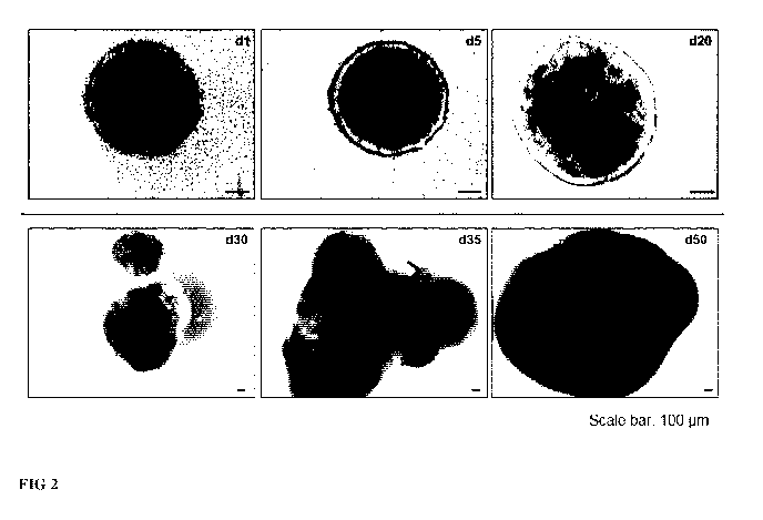
FIG 2: Generation of human brain organoids
Representative transmitted light microscopy pictures of the generation of
human brain
organoids are shown. On day 1 cells formed round embryoid body like
structures, which show
clear surrounding and an integrated structure. The black inner core is
surrounded by a clear ring.
Five days after seeding, the neural tissue still shows a round structure. The
inner of the organoid
starts to show some small structures. The organoids seem to be less compact.
On day 20 and 30
round structures in the inner of the organoids can be observed. These
structures represent neural
progenitor zones. As the organoids grow older the structure becomes more
dense. No inner
structures can be detected by light microscopy at this stage of
differentiation.
FIG 3: Characterization of the brain organoids using flow cytometry
The change in neural marker expression during organoid development was
measured over the
time of development in order to assess the degree of neural induction. The
organoids were
analyzed on day 5, 15 and day 30 of differentiation. To that end organoids
were harvested using
the Multi Tissue Dissociation Kit 3TM in order to obtain single cells. FIG 3A
shows exemplary
dot plots (Flow cytometry) of analyzed brain organoid cultures on day 5 and
15. Co-expression
of the nuclear neural progenitor markers Pax6 and 5ox2 is shown. The amount of
double-
positive cells reaches ¨90% on day 5, indicating a successful neural induction
and the presence
of a large neural progenitor population. Contrary to that the amount of
positive cells in the
progenitor population decreases on day 15. Collected data of different
experiments are
presented in FIG 3B. The diagram shows high Pax6 expression on day 5, which is
subsequently
decreased on day 15 and 30 to ¨35%, indicating a decrease in the neural
progenitor population.
This is in line with the processes taking place during neural development,
because the number
CA 03129656 2021-08-10
WO 2020/165059
PCT/EP2020/053237
of neural progenitor cells decreases over time due to derivation of neurons,
thus explaining the
decrease in progenitor population.
FIG 4: Characterization of brain organoids on day 30 and 50
5 FIG 4A shows the expression of N-cadherin, which is a typical marker for
the apical membrane.
The apical membrane is observed in all progenitor zones (ventricle like
structures) lining the
ventricle. The expression is independent of the analysis time point FIG 4B:
shows the
expression of the nuclear located neural progenitor marker Sox2. The
expression of Sox2 is
mainly observed in cells in close proximity to the ventricles, which
correlates with normal
neural development. Moreover, a Tull_ staining is shown. This cytoskeletal
marker is detected
in early neurons. The arrangement of progenitor markers at the ventricle and a
surrounding
Tull_ staining correlates with standard neural developmental processes. A
similar cellular
structure is also observed on day 50.
FIG 4C: Further markers, important during neural development, are TBR2 and
Pax6. Pax6
.. labels the nuclei of neural progenitor cells that are localized near to the
ventricle. TBR-2
positive cells represent a different neural progenitor population, which is
positioned more
basally, making up a subventricular zone. FIG 4D shows the expression of the
nuclear cortical
plate marker TBR1 and the deep layer neurons. On day 50 both markers can be
detected basally
of the ventricular zone. As observed in neural development TBR-1 is found at
the very basal
site representing the developing cortical plate. In contrast to that CT1P2 is
found apically of the
cortical plate, representing the formation of deep layer neurons.
FIG 5 Experimental set up to compare the use of MatrigelTM against medium
containing the
viscosity enhancer
Single cell suspensions of human pluripotent stem cells were seeded in 96 well
ultra-low
attachment plates. 24h later EB like structures formed and the first medium
was replaced by
neural induction medium (Medium 2). On day 5 early neural tissues were
transferred to 24 well.
For experimental set up using the viscosity enhancer, medium with methyl
cellulose was added.
For the experimental set up using MatrigelTm, organoids were embedded in a
MatrigelTm droplet
using standard procedures and cultivated in a medium without the viscosity
enhancer. On day
15 the developing neural tissue is then transferred to 10 cm dishes
(containing cerebral organoid
differentiation medium), which are placed onto a shaker. Depending on the
desired
developmental stage the organoids can be cultivated > 100 days.
CA 03129656 2021-08-10
WO 2020/165059
PCT/EP2020/053237
6
FIG 6: Comparison of the use of MatrigelTm against medium containing viscosity
enhancer for
the generation of brain organoids
Transmitted light microscopy images of organoids generated using the standard
method
described by Lancaster et al (2013, Nature: 501:373) and Quian et al (2016,
Ce11:165: 1238;
2018, Nature Protocols, 13:565) and in W02014090993A1 are shown. Organoids are
embedded in MatrigelTM for differentiation and cultivation. A dense organoid
structure can be
observed. Moreover, some neural outgrowth indicated by arrows can be shown.
Some cells
seem to migrate into the MatrigelTm. A smooth surface cannot be observed.
Depending on the
batch of organoids, less dense structures and fluid-filled cavities can be
observed, indicating
partial non-specific differentiation, which cannot be observed when using the
medium
supplemented with viscosity enhancer.
In contrast to that organoids generated without MatrigelTM but using the
viscosity enhancer
show a smooth surface without any neural outgrowth or fluid filled cavities.
FIG 7: Titration of different media viscosities
In order to determine the range of viscosity that supports brain organoid
formation, different
methyl cellulose viscosities were tested. Transmitted light data (day 30
organoids) obtained
from brain organoids cultivated in 0%, 0.25%, 0.5%, 1% or 2% methyl cellulose
are shown.
The cultivation of organoids without any viscosity enhancer leads to a "lose"
structure of the
organoids. They become less compact and more fringy. Over time, the majority
of these
organoids dissolve completely, thus leading to highly decreased yields in
organoids.
Brain organoids generated by the use of 0.25% - 1 % Methyl cellulose are more
dense and show
very compact structures. Moreover, they have a smooth border and some cellular
structures
within the organoids can be observed. This indicates the successful generation
of brain
organoids containing typical progenitor zones.
The addition of 2% methyl cellulose to the medium, leads to a highly increased
viscosity. The
organoids are smaller compared to other conditions. They are very compact and
without any
visible specific structures inside.
FIG 8: Tissue clearing of organoids obtained from different methyl cellulose
concentrations
In order to find out whether progenitor cells formed in the inner core of the
organoid, the
organoids were stained for the proliferation marker Ki67 and cleared using a
tissue clearing
procedure based on ethyl cinnamate as organic solvent. The cleared brain
organoids were
analyzed using confocal microcopy and Z stacks, which were reconstructed to
illustrate
CA 03129656 2021-08-10
WO 2020/165059
PCT/EP2020/053237
7
complete organoids including the ventricle-like zones. In the 0.5% and 1%
sample circular
ventricle like structures were observed. These structures were found all over
the organoid. In
contrast to that organoids generated using 2% methyl cellulose do not show a
Ki67 positive
cells, indicating the absence of ventricle like zones.
FIG 9: Comparison of different viscosity enhancer based on organoid morphology
Representative transmitted light microscopy pictures of the generation of
human brain
organoids at day 7 and day 25 are shown. 0,5% methyl cellulose, 0,21%
carboxymethyl
cellulose and 0,25% hydroxy ethyl cellulose were used as viscosity enhancer in
Medium 3.
On day 7 organoids in all three conditions show some small structures in the
inner parts and
bulges at the surface, indicating ongoing differentiation and proliferation.
On day 30 round
structures in the inner of the organoids can be observed, representing neural
progenitor zones.
On both days all three conditions look comparable.
FIG 10: Comparison of different viscosity enhancer based on flow cytometry
Neural marker expression was measured on day 30 of organoid development to
compare
differentiation efficiency using different viscosity enhancer in Medium 3. To
that end organoids
were harvested using the Multi Tissue Dissociation Kit 3' in order to obtain
single cells. The
expression of the nuclear neural progenitor markers Pax6 and Sox2 and the
cytoskeletal marker
in early neurons Tull_ is shown. All three conditions show similar marker
expression with an
expression between 33-40% Sox2, 10-15% Pax6 and 45-50% Tull_
Detailed description of the invention
In a first aspect the present invention provides a differentiation medium for
differentiation and
expansion of a multicellular aggregation in suspension derived from human
pluripotent stem
cells that has been induced to differentiate to an artificial tissue
structure, e.g. artificial neural
tissue (brain organoid), said medium comprising a basal medium for animal or
human cells,
wherein said differentiation medium has a viscosity between 1.7 mPa*s and 1500
mPa*s,
between 2 mPa*s and 1400 mPa*s, between 2 mPa*s and 1000 mPa*s, between 2
mPa*s and
500 mPa*s, between 4 mPa*s and 1000 mPa*s, between 4 mPa*s and 500 mPa*s,
between 4
mPa*s and 200 mPa*s, between 4 mPa*s and 100 mPa*s, between 6 mPa*s and 80
mPa*s,
between 10 mPa*s and 80 mPa*s or between 10 mPa*s and 50 mPa*s.
CA 03129656 2021-08-10
WO 2020/165059
PCT/EP2020/053237
8
Preferentially, said viscosity is between 4 mPa*s and 100 mPa*s, more
preferentially the
viscosity is between 6 mPa*s and 80 mPa*s, most preferentially the viscosity
is between 10
mPa*s and 80 mPa*s.
Said differentiation medium, wherein said viscosity between 1.7 mPa*s and 1500
mPa*s,
between 2 mPa*s and 1400 mPa*s, between 2 mPa*s and 1000 mPa*s, between 2
mPa*s and
500 mPa*s, between 4 mPa*s and 1000 mPa*s, between 4 mPa*s and 500 mPa*s,
between 4
mPa*s and 200 mPa*s, between 4 mPa*s and 100 mPa*s, between 6 mPa*s and 80
mPa*s,
between 10 mPa*s and 80 mPa*s or between 10 mPa*s and 50 mPa*s is achieved by
the
presence of a viscosity enhancer in said differentiation medium, therefore
said differentiation
medium may comprise
i) a basal medium for animal or human cells, and
ii) a viscosity enhancer,
wherein said differentiation medium has a viscosity between 1.7 mPa*s and 1500
mPa*s,
between 2 mPa*s and 1400 mPa*s, between 2 mPa*s and 1000 mPa*s, between 2
mPa*s and
500 mPa*s, between 4 mPa*s and 1000 mPa*s, between 4 mPa*s and 500 mPa*s,
between 4
mPa*s and 200 mPa*s, between 4 mPa*s and 100 mPa*s, between 6 mPa*s and 80
mPa*s,
between 10 mPa*s and 80 mPa*s or between 10 mPa*s and 50 mPa*s.
Said differentiation medium may be without a three-dimensional matrix.
Said viscosity enhancer does not build a three-dimensional matrix in the cell
culture medium.
Said viscosity enhancer may be biocompatible for the cells of said
differentiation medium.
Said viscosity enhancer may be for example a non-gelling, biocompatible
rheology modifier.
Rheology modifiers may be carrageenans, xanthan gum, and cellulose ether
derivates such as
methyl cellulose, carboxymethyl cellulose, and hydroxy ethyl cellulose, and
mixtures thereof.
Said viscosity enhancer may be selected for example from the group of
biocompatible rheology
modifiers consisting of carrageenans, xanthan gum, and cellulose ether
derivates such as methyl
cellulose, carboxymethyl cellulose, and hydroxy ethyl cellulose, and mixtures
thereof.
Preferentially said viscosity enhancer is methyl cellulose, carboxymethyl
cellulose, hydroxy
ethyl cellulose, or a combination thereof.
Said viscosity enhancer, wherein said viscosity enhancer is methyl cellulose,
carboxymethyl
cellulose or hydroxy ethyl cellulose, and wherein the concentration of methyl
cellulose,
carboxymethyl cellulose, or hydroxy ethyl cellulose is between 0,1% and 2%
methyl cellulose,
carboxymethyl cellulose, or hydroxy ethyl cellulose in said medium.
CA 03129656 2021-08-10
WO 2020/165059
PCT/EP2020/053237
9
Said viscosity enhancer, preferentially methyl cellulose, carboxymethyl
cellulose, or hydroxy
ethyl cellulose, wherein said viscosity enhancer increases the viscosity of
said differentiation
medium to a value between 1.7 mPa*s and 1500 mPa*s.
Said differentiation medium may comprise additionally one or more
differentiation factors for
.. differentiation of said multicellular aggregation to an artificial tissue
structure.
Said one or more differentiation factors may differentiate said multicellular
aggregation to
artificial neural tissue, to artificial cardiac tissue, to artificial kidney
tissue, or artificial retinal
tis sue.
Said differentiation medium, wherein said artificial tissue structure may be
artificial neural
tissue, and wherein said differentiation medium optionally may comprise one or
more
differentiation factors selected from the group consisting of activator of Wnt
signaling and an
inhibitor for TGF-beta, activin and nodal signaling pathway.
Surprisingly, said differentiation and expansion of said multicellular
aggregation in suspension
derived from human pluripotent stem cells that has been induced to
differentiate to an artificial
neural tissue works in said differentiation medium also without the addition
of said one or more
differentiation factors.
Said differentiation medium, wherein said artificial tissue structure may be
cardiac organoids,
and wherein said differentiation medium may comprise one or more
differentiation factors
selected from the group consisting of Wnt activators and inhibitors,
activators of the BMP,
activin and bFGF pathway.
Said differentiation medium, wherein said artificial tissue structure may be
kidney organoids,
and wherein said differentiation medium may comprise one or more
differentiation factors
selected from the group consisting of Wnt activators and activators of FGF
signaling.
Said differentiation medium, wherein said artificial tissue structure may be
retinal organoids,
and wherein said differentiation medium may comprise one or more
differentiation factors
selected from the group consisting of Wnt inhibitors and activators of sonic
hedgehog pathway.
Said differentiation medium may be used within the method for obtaining
artificial neural
tissues (brain organoids) as disclosed herein or as disclosed in
W02014090993A1. Of course,
CA 03129656 2021-08-10
WO 2020/165059
PCT/EP2020/053237
in the method as disclosed in W02014090993A1 said differentiation medium
replaces the
medium of step 3 (culturing said differentiated multicellular aggregation in a
three-dimensional
matrix such as MatrigelTm).
The steps for methods of obtaining artificial tissue structures (organoids)
other than artificial
5 neural tissue (brain organoids), e.g. artificial cardiac tissue (cardiac
organoids), artificial kidney
tissue (kidney organoids) or artificial retinal tissue (retinal organoids) may
vary from the steps
of obtaining artificial neural tissues (brain organoids). But all these
methods for obtaining
artificial tissue structures (organoids) have in common, that the step,
wherein a three-
dimensional matrix is used can be replaced by the use of the differentiation
medium as disclosed
10 herein.
Said pluripotent stem cells may be human embryonic stem cells or human induced
pluripotent
stem cells.
In a further aspect, the present invention provides an in vitro method for
obtaining a brain
organoid (an artificial neural tissue) comprising
a) providing a multicellular aggregation of human pluripotent stem cells,
b) culturing said multicellular aggregation in an induction medium (neural
induction medium)
thereby inducing the multicellular aggregation to differentiate to a brain
organoid (an artificial
neural tissue),
c) culturing said differentiated multicellular aggregation in suspension in a
differentiation
medium, wherein said differentiation medium has a viscosity between 1.7 mPa*s
and 1500
mPa*s, between 2 mPa*s and 1400 mPa*s, between 2 mPa*s and 1000 mPa*s, between
2
mPa*s and 500 mPa*s, between 4 mPa*s and 1000 mPa*s, between 4 mPa*s and 500
mPa*s,
between 4 mPa*s and 200 mPa*s, between 4 mPa*s and 100 mPa*s, between 6 mPa*s
and 80
mPa*s, between 10 mPa*s and 80 mPa*s or between 10 mPa*s and 50 mPa*s, thereby
expanding the cells in a multicellular aggregation, wherein said cells are
allowed to differentiate
further.
Said method for obtaining a brain organoid, wherein said method comprises the
additional step:
.. d) culturing said expanded multicellular aggregation of cells from Step c)
in a suspension
culture.
CA 03129656 2021-08-10
WO 2020/165059
PCT/EP2020/053237
11
Said multicellular aggregation of human pluripotent stem cells that may be
provided in step a)
of said method may be generated in a "medium for generation of multicellular
aggregation from
human pluripotent stem cells".
Media for generation of multicellular aggregation from human pluripotent stem
cells are well-
known in the art and disclosed for example in Eiraku et al (Cell Stem Cell,
2008, 3:519-532),
U520110091869, W02011055855A1 and W02014090993A1.
Said medium for generation of multicellular aggregation from human pluripotent
stem cells (or
medium A) may comprise a) a basal medium for animal or human cells, and ii) a
Rock inhibitor.
The addition of a Rock inhibitor e.g. Thiazovivin, Y27632 is preferred. Such
medium is used
e.g. in the examples.
Media for induction of multicellular aggregation from human pluripotent stem
cells to
differentiate to artificial neural tissue (neural induction medium) are well-
known in the art and
are disclosed for example in Eiraku et al (Cell Stem Cell, 2008, 3:519-532),
U520110091869,
W02011055855A1 and W02014090993A1.
Said neural induction medium (or medium B) of step b) of said method for the
differentiation
of the multicellular aggregates into artificial neural tissue may comprise i)
a basal medium for
animal or human cells, ii) an inhibitor for TGF-beta, Activin and Nodal
signaling pathway, and
iii) a Bone Morphogenetic Protein (BMP) inhibitor.
Said method for obtaining a brain organoid (an artificial neural tissue),
wherein said
differentiation medium comprises
i) said basal medium for animal or human cells, and
ii) said viscosity enhancer; and optionally
iii) an activator of Wnt signaling and/or an inhibitor for TGF-beta, activin
and nodal signaling
pathway.
Said culturing human pluripotent stem cells such as iPS cells as multicellular
aggregates in said
medium for generation of multicellular aggregation from human pluripotent stem
cells may be
performed for 1-5 days, preferentially for 24 h. In particular this culture
step is performed from
day 0-1. Induction of artificial neural tissues from these multicellular
aggregates in said neural
induction medium may be performed for 4-7 days, preferentially for 4 days,
e.g. from day 1-4
of differentiation. The culture step of cultivating cells in differentiation
medium may be
CA 03129656 2021-08-10
WO 2020/165059
PCT/EP2020/053237
12
performed for 8-12 days, preferentially 10 days. In particular, said step may
be performed from
day 5-15. The suspension culture (after culturing cells in differentiation
medium containing a
viscosity enhancer) in said medium for culturing the expanded multicellular
aggregation may
be a stirring and/or shaking culture (shaker, bioreactor etc.). Dependent on
the development
stage said suspension culture may be maintained in said medium for culturing
the expanded
multicellular aggregation under stirring and/or shaking conditions for up to
100 days or even
more days.
The artificial neural tissue (brain organoid) developed by the method as
disclosed herein may
be e.g. a cerebral organoid, a midbrain organoid or a hindbrain organoid. The
development of
the kind or type of brain organoid may dependent on the addition or exclusion
of different small
molecules such as sonic hedgehog leads to the generation of a ventral type of
forebrain organoid,
while the addition of CH1R and sonic hedgehog leads to caudalization of the
brain regions
generated in the organoid. Dependent on the combination of small molecules
organoids for
different brain regions can be generated.
Said method, wherein said differentiation medium comprises a viscosity
enhancer that is
biocompatible for the cells of said medium as disclosed above, thereby
adjusting said viscosity
of said differentiation medium between 1.7 mPa*s and 1500 mPa*s, between 2
mPa*s and 1400
mPa*s, between 2 mPa*s and 1000 mPa*s, between 2 mPa*s and 500 mPa*s, between
4 mPa*s
and 1000 mPa*s, between 4 mPa*s and 500 mPa*s, between 4 mPa*s and 200 mPa*s,
between
4 mPa*s and 100 mPa*s, between 6 mPa*s and 80 mPa*s, between 10 mPa*s and 80
mPa*s or
between 10 mPa*s and 50 mPa*s.
Said differentiation medium may be without a three-dimensional matrix.
Said viscosity enhancer does not build a three-dimensional matrix in the cell
culture medium.
Preferentially said viscosity enhancer is methyl cellulose, carboxymethyl
cellulose, hydroxy
ethyl cellulose, or a combination thereof.
Said viscosity enhancer, wherein said viscosity enhancer is methyl cellulose,
carboxymethyl
cellulose or hydroxy ethyl cellulose, and wherein the concentration of methyl
cellulose,
carboxymethyl cellulose, or hydroxy ethyl cellulose is between 0,1% and 2%
methyl cellulose,
carboxymethyl cellulose, or hydroxy ethyl cellulose in said medium.
Said viscosity enhancer, preferentially methyl cellulose, carboxymethyl
cellulose, or hydroxy
ethyl cellulose, wherein said viscosity enhancer increases the viscosity of
said differentiation
medium to a value between 1.7 mPa*s and 1500 mPa*s.
CA 03129656 2021-08-10
WO 2020/165059
PCT/EP2020/053237
13
Said differentiation medium (Medium C) of step c) of said method may be used
for further cell
specification and neural epithelia expansion. Said differentiation medium may
comprise a basal
medium for animal or human cells, wherein said differentiation medium has a
viscosity
between 1.7 mPa*s and 1500 mPa*s, between 2 mPa*s and 1400 mPa*s, between 2
mPa*s and
1000 mPa*s, between 2 mPa*s and 500 mPa*s, between 4 mPa*s and 1000 mPa*s,
between 4
mPa*s and 500 mPa*s, between 4 mPa*s and 200 mPa*s, between 4 mPa*s and 100
mPa*s,
between 6 mPa*s and 80 mPa*s, between 10 mPa*s and 80 mPa*s or between 10
mPa*s and
50 mPa*s.
Said differentiation medium, wherein said viscosity between 1.7 mPa*s and 1500
mPa*s,
between 2 mPa*s and 1400 mPa*s, between 2 mPa*s and 1000 mPa*s, between 2
mPa*s and
500 mPa*s, between 4 mPa*s and 1000 mPa*s, between 4 mPa*s and 500 mPa*s,
between 4
mPa*s and 200 mPa*s, between 4 mPa*s and 100 mPa*s, between 6 mPa*s and 80
mPa*s,
between 10 mPa*s and 80 mPa*s or between 10 mPa*s and 50 mPa*s is achieved by
the
presence of a viscosity enhancer in said differentiation medium, therefore
said differentiation
medium may comprise
i) a basal medium for animal or human cells, and
ii) a viscosity enhancer,
wherein said differentiation medium has a viscosity between 1.7 mPa*s and 1500
mPa*s,
between 2 mPa*s and 1400 mPa*s, between 2 mPa*s and 1000 mPa*s, between 2
mPa*s and
500 mPa*s, between 4 mPa*s and 1000 mPa*s, between 4 mPa*s and 500 mPa*s,
between 4
mPa*s and 200 mPa*s, between 4 mPa*s and 100 mPa*s, between 6 mPa*s and 80
mPa*s,
between 10 mPa*s and 80 mPa*s or between 10 mPa*s and 50 mPa*s.
Said viscosity enhancer may be biocompatible for the cells of said
differentiation medium.
Said viscosity enhancer may be for example a non-gelling, biocompatible
rheology modifier.
Rheology modifiers may be carrageenans, xanthan gum, and cellulose ether
derivates such as
methyl cellulose, carboxymethyl cellulose, and hydroxy ethyl cellulose, and
mixtures thereof.
Said viscosity enhancer may be selected for example from the group of
biocompatible rheology
modifiers consisting of carrageenans, xanthan gum, and cellulose ether
derivates such as methyl
cellulose, carboxymethyl cellulose, and hydroxy ethyl cellulose, and mixtures
thereof.
CA 03129656 2021-08-10
WO 2020/165059
PCT/EP2020/053237
14
Preferentially said viscosity enhancer is methyl cellulose, carboxymethyl
cellulose, hydroxy
ethyl cellulose, or a combination thereof.
Said viscosity enhancer, wherein said viscosity enhancer is methyl cellulose,
carboxymethyl
cellulose or hydroxy ethyl cellulose, and wherein the concentration of methyl
cellulose,
carboxymethyl cellulose, or hydroxy ethyl cellulose is between 0,1% and 2%
methyl cellulose,
carboxymethyl cellulose, or hydroxy ethyl cellulose in said medium.
Said viscosity enhancer, preferentially methyl cellulose, carboxymethyl
cellulose, or hydroxy
ethyl cellulose, wherein said viscosity enhancer increases the viscosity of
said differentiation
medium to a value between 1.7 mPa*s and 1500 mPa*s.
Said differentiation medium may comprise additionally one or more
differentiation factors for
differentiation of said multicellular aggregation to artificial neural tissue.
Said differentiation medium optionally may comprise one or more
differentiation factors
selected from the group consisting of activator of Wnt signaling and an
inhibitor for TGF-beta,
activin and nodal signaling pathway.
As mentioned above, said differentiation and expanding of said multicellular
aggregation in
suspension derived from human pluripotent stem cells that has been induced to
differentiate to
an artificial neural tissue works in said differentiation medium also without
the addition of said
one or more differentiation factors.
Therefore, said differentiation medium may be composed of a medium containing
a viscosity
enhancer as disclosed herein such as methyl cellulose generating a viscosity
as disclosed herein.
The medium may be further composed of: N2 (transferrin, insulin, Progesterone,
Putrescine,
Selenite), L-glutamine. For further cell specification and neuroepithelia
expansion a Wnt
activator e.g. CH1R99021 and/or activator of TGF-f3, Activin and Nodal
signaling pathway e.g.
SB431542 may be added. Such medium is used e.g. in the examples.
Said differentiation medium (or medium C) may be used for further cell
specification and neural
epithelia expansion (step c of the said method) without the use or the need of
a three-
dimensional matrix such as MatrigelTm as disclosed e.g. in W02014090993A1.
Said culturing of said expanded multicellular aggregation of cells from step
c) in suspension
culture (step d of said method) may be performed in "medium for culturing the
expanded
multicellular aggregation". Such media for culturing the expanded
multicellular aggregation
are well-known in the art and disclosed e.g. in W02014/090993A1. Said medium
for culturing
CA 03129656 2021-08-10
WO 2020/165059
PCT/EP2020/053237
the expanded multicellular aggregation (or medium D) may comprise i) a basal
medium for
animal or human cells, and ii) retinoic acid and retinol.
Therefore, said medium for culturing the expanded multicellular aggregation
may be used for
culturing the brain organoids in suspension culture. The medium may be
composed e.g. NB21
5 supplement (MACS NeuroBrew -21, Miltenyi Biotec)) or any components
thereof. Such
medium is used e.g. in the examples.
The suspension culture of step d of the method as disclosed herein (i.e. after
culturing cells in
differentiation medium containing a viscosity enhancer in said medium for
culturing the
expanded multicellular aggregation) may be a stirring and/or shaking culture
(e.g. a shaker or
10 bioreactor).
Any of the above described media further may contain nutrients, buffers and
oxygen. The
medium may further comprise growth factors or lack growth factors. Preferred
nutrients include
a carbohydrate, especially a mono-hexose or mono-pentose, such as glucose or
fructose. In a
15 preferred embodiment any media is serum and Xeno free.
Said method for obtaining a brain organoid, wherein said pluripotent stem
cells are human
induced pluripotent stem cells.
In a further aspect the present method provides the use of a viscosity
enhancer for adjusting the
viscosity of a cell medium used for obtaining an artificial tissue structure
derived from human
pluripotent stem cells, wherein said viscosity is between 1.7 mPa*s and 1500
mPa*s, between
2 mPa*s and 1400 mPa*s, between 2 mPa*s and 1000 mPa*s, between 2 mPa*s and
500 mPa*s,
between 4 mPa*s and 1000 mPa*s, between 4 mPa*s and 500 mPa*s, between 4 mPa*s
and
200 mPa*s, between 4 mPa*s and 100 mPa*s, between 6 mPa*s and 80 mPa*s,
between 10
mPa*s and 80 mPa*s or between 10 mPa*s and 50 mPa*s.
In an aspect the present invention provides a brain organoid obtainable by the
in-vitro methods
for obtaining a brain organoid as disclosed herein.
The brain organoid obtained by said method for obtaining a brain organoid is
an artificial neural
tissue because said method is performed in vitro and the neural tissue does
not reach the
complexity of a naturally grown neural tissue.
CA 03129656 2021-08-10
WO 2020/165059
PCT/EP2020/053237
16
The brain organoid (artificial neural tissue) obtainable by the methods as
disclosed herein
resembles the brain organoid known in the art and disclosed for example in
W02014/090993
that needs three-dimensional matrix such as MatrigelTM and that has been
characterized as
follows:
The brain organoid is comprised of a heterogenous population of cells of at
least two different
progenitor and neuronal differentiation layers, wherein at least one
progenitor layer comprises
outer radial glia cells. The brain organoids display a well-organized cerebral
cortex.
Furthermore, these tissues display several characteristics specific to humans,
namely the
presence of a substantial outer radial glial population and the organization
of extra cortical
subventricular zone layers not present in mouse. The presence of outer radial
glia cells appears
to be one of the most distinguishing features, but of course others exist as
well. Eiraku et al.
(2008) for example describes that in their culture radial glia of cortical
tissues decreased after
day 12 and apparently failed to develop into outer radial glia cells, outer
radial glia being
characterized by their position as well as morphology (lack of an apical
connection to the fluid-
filled ventricular-like cavity). According to the three-dimensional neural
tissue culture of
W02014/090993, the outer radial glia cells are preferably in a progenitor
layer, in particular,
in a subventricular zone localized basally of the ventricular zone where
radial glia reside.
The brain organoid disclosed in W02014/090993 may develop into a
differentiated tissue
comprising layers of different differentiation grade. In a 3D structure this
may be observable as
separate sections of the organoid. In preferred embodiments, the culture
artificial neural tissue
comprises sections from at least two layers. Such a layer may be shaped around
a globular tissue
body, e.g. a body from which the distinct layer (s) have developed. In
particular, the tissue may
show a distinctive development of apical and dorsal tissue sections.
The brain organoid disclosed in W02014/090993 is or resembles cerebral tissue
comprising
substantially all cells found in the brain or progenitors thereof. Such cells
can be identified by
selective gene expression markers, which are on a level above the average of
not differentiated
cells, in particular including confidence intervals. Such markers can be
identified by specific
antibodies that are used for flow cytometry and immunofluorescence.
Preferably cells of the brain organoids express one or more gene expression
markers selected
from forebrain markers FoxG1 and Pax6.
The brain organoids can alternatively or in addition be characterized by
comprising cells
expressing one or more expression markers selected from N-Catherin, 5ox2,
Tull, Pax6 0tx2,
FoxG1, Tbrl, Tbr2, 5atb2, Ctip2õ or any combination thereof.
CA 03129656 2021-08-10
WO 2020/165059
PCT/EP2020/053237
17
Preferably the brain organoid comprises cells, which express FoxG1. FoxG1 is
expressed in cells
of dorsal cortex identity.
Preferably the brain organoid comprises cells, which express Pax6. Pax is
expressed in cells of
frontal cortex identity.
.. Preferentially brain organoid comprises cells, which express Sox2 and Pax6
localized near to a
ventricle. These markers are expressed in forebrain progenitor populations.
Preferably the brain organoid comprises cells, which express TBR-2. TBR-2 is
expressed in
intermediate progenitors.
Preferably the brain organoid comprises cells, which express Tujl. Tujl is
expressed in cells of
a cortical inner fiber layer identity.
Preferably the brain organoid comprises cells, which express Brn2. Brn2 is
expressed in cells
of a later born neuron (neuron of outer region).
Preferably the brain organoid comprises cells, which express Satb2. Satb2 is
expressed in cells
of a later born neuron (neuron of outer region).
Preferably the brain organoid comprises cells, which express Ctip2. Ctip2 is
expressed in cells
of earlier born neuron (neuron of inner region).
Preferably the brain organoid comprises cells, which express TBR-1. TBR-1 is
expressed in
cells of cortical interneurons within the dorsal cortical plate.
Although the brain organoid obtainable by the method for obtaining brain
organoids as
disclosed herein has most or many of the features of the cerebral organoid as
disclosed in
W02014/090993 in common, some differences exists between these two kinds of
organoids.
The differences may be traced back to the different methods used for obtaining
the organoids.
The brain organoids obtained by the method as disclosed herein have less
neural outgrowths
compared to the brain organoids obtained by the methods known in the art that
use ECM (see
FIG 6). The brain organoid obtained by the method as disclosed herein may have
at least 20%,
30%, 40%, 50%, 60%, 70%, 80%, 90%. 95%, 99% less neural outgrowths than the
brain
organoids obtained by the methods known in the art that use three dimensional
matrix such as
ECM. Preferentially, the brain organoids as disclosed herein may have no
neural outgrowths
and therefore may have a smooth surface. The brain organoids of the prior art
do not have a
smooth surface as cells of the organoid invade into the ECM and therefore
generate neural
outgrowths.
Brain organoids obtained by the method as disclosed herein have benefits
compared to the brain
organoids obtained by methods of the prior art:
CA 03129656 2021-08-10
WO 2020/165059
PCT/EP2020/053237
18
- less unspecific neural differentiation and less directed neural migration
to the outside of the
organoid (FIG 6)
- Organoids remodel neural development more closely since there is no
MatrigelTM present
during (embryonic) development
- Generated neurons stay within the organoid, this might improve neural
differentiation and
cortical plate development neural layering
It is self-explaining that the brain organoid developed by the methods of the
present invention
has also a biochemical distinction to the brain organoids developed by the
method of the prior
art that need the presence of an ECM such as disclosed in W02014/090993A1.
This is e.g.
indicative by the missing of a contact area between the cells of the
developing organoid as
disclosed herein and an ECM (FIG 6).
In a further aspect the present invention provides a kit comprising a
differentiation medium for
differentiation and expansion of a multicellular aggregation in suspension
derived from human pluripotent stem cells that has been induced to
differentiate to an artificial
tissue structure, said medium comprising a basal medium for animal or human
cells, wherein
said differentiation medium has a viscosity between 1.7 mPa*s and 1500 mPa*s,
between 2
mPa*s and 1400 mPa*s, between 2 mPa*s and 1000 mPa*s, between 2 mPa*s and 500
mPa*s,
between 4 mPa*s and 1000 mPa*s, between 4 mPa*s and 500 mPa*s, between 4 mPa*s
and
200 mPa*s, between 4 mPa*s and 100 mPa*s, between 6 mPa*s and 80 mPa*s,
between 10
mPa*s and 80 mPa*s or between 10 mPa*s and 50 mPa*s.
Said kit, wherein said viscosity between 1.7 mPa*s and 1500 mPa*s, between 2
mPa*s and
1400 mPa*s, between 2 mPa*s and 1000 mPa*s, between 2 mPa*s and 500 mPa*s,
between 4
mPa*s and 1000 mPa*s, between 4 mPa*s and 500 mPa*s, between 4 mPa*s and 200
mPa*s,
between 4 mPa*s and 100 mPa*s, between 6 mPa*s and 80 mPa*s, between 10 mPa*s
and 80
mPa*s or between 10 mPa*s and 50 mPa*s of said differentiation medium is
achieved by the
presence of a viscosity enhancer in said differentiation medium, therefore
said differentiation
medium may comprise
i) a basal medium for animal or human cells, and
ii) a viscosity enhancer,
wherein said differentiation medium has a viscosity between 1.7 mPa*s and 1500
mPa*s,
between 2 mPa*s and 1400 mPa*s, between 2 mPa*s and 1000 mPa*s, between 2
mPa*s and
CA 03129656 2021-08-10
WO 2020/165059
PCT/EP2020/053237
19
500 mPa*s, between 4 mPa*s and 1000 mPa*s, between 4 mPa*s and 500 mPa*s,
between 4
mPa*s and 200 mPa*s, between 4 mPa*s and 100 mPa*s, between 6 mPa*s and 80
mPa*s,
between 10 mPa*s and 80 mPa*s or between 10 mPa*s and 50 mPa*s.
Said kit, wherein said viscosity enhancer may be biocompatible for the cells
of said
differentiation medium.
Said viscosity enhancer may be for example a non-gelling, biocompatible
rheology modifier.
Rheology modifiers may be carrageenans, xanthan gum, and cellulose ether
derivates such as
methyl cellulose, carboxymethyl cellulose, and hydroxy ethyl cellulose, and
mixtures thereof.
Said viscosity enhancer may be selected for example from the group of
biocompatible rheology
modifiers consisting of carrageenans, xanthan gum, and cellulose ether
derivates such as methyl
cellulose, carboxymethyl cellulose, and hydroxy ethyl cellulose, and mixtures
thereof.
Preferentially said viscosity enhancer is methyl cellulose, carboxymethyl
cellulose, hydroxy
ethyl cellulose, or a combination thereof.
Said viscosity enhancer, wherein said viscosity enhancer is methyl cellulose,
carboxymethyl
cellulose or hydroxy ethyl cellulose, and wherein the concentration of methyl
cellulose,
carboxymethyl cellulose, or hydroxy ethyl cellulose is between 0,1% and 2%
methyl cellulose,
carboxymethyl cellulose, or hydroxy ethyl cellulose in said medium.
Said viscosity enhancer, preferentially methyl cellulose, carboxymethyl
cellulose, or hydroxy
ethyl cellulose, wherein said viscosity enhancer increases the viscosity of
said differentiation
medium to a value between 1.7 mPa*s and 1500 mPa*s.
Said kit, wherein said differentiation medium may comprise additionally one or
more
differentiation factors.
Said kit, wherein said one or more differentiation factors may be
differentiation factors for
differentiation of said multicellular aggregation to artificial neural tissue,
to artificial cardiac
tissue, to artificial kidney tissue or artificial retinal tissue.
Said kit, wherein said differentiation medium is for differentiation to
artificial neural tissue, and
wherein said differentiation medium optionally may comprise one or more
differentiation
factors selected from the group consisting of activator of Wnt signaling and
an inhibitor for
TGF-beta, activin and nodal signaling pathway.
CA 03129656 2021-08-10
WO 2020/165059
PCT/EP2020/053237
Said kit, wherein said differentiation medium is for differentiation to
artificial neural tissue, the
kit may comprise
a) a differentiation medium comprising a basal medium for animal or human
cells, wherein said
5 differentiation medium has a viscosity between 1.77 mPa*s and 1496.82
mPa*s, between 2
mPa*s and 1400 mPa*s, between 2 mPa*s and 1000 mPa*s, between 2 mPa*s and 500
mPa*s,
between 4 mPa*s and 1000 mPa*s, between 4 mPa*s and 500 mPa*s, between 4 mPa*s
and
200 mPa*s, between 4 mPa*s and 100 mPa*s, between 6 mPa*s and 80 mPa*s,
between 10
mPa*s and 80 mPa*s or between 10 mPa*s and 50 mPa*s,
10 b) a medium for generation of multicellular aggregation from human
pluripotent stem cells
comprising
i) a basal medium for animal or human cells
ii) a Rock inhibitor
c) a neural induction medium comprising
15 i) a basal medium for animal or human cells
ii) an inhibitor for TGF-beta, Activin and Nodal signaling pathway
iii) a Bone Morphogenetic Protein (BMP) inhibitor.
Said kit may further comprise
d) a medium for culturing the expanded multicellular aggregation according to
the method as
20 .. disclosed herein comprising
i) a basal medium for animal or human cells
ii) retinoic acid and retinol.
Said differentiation medium of said kit optionally may comprise one or more
differentiation
factors selected from the group consisting of activator of Wnt signaling and
an inhibitor for
TGF-beta, activin and nodal signaling pathway.
All definitions, characteristics and embodiments defined herein with regard to
an aspect of the
invention, e.g. the first aspect of the invention, also apply mutatis mutandis
in the context of
.. the other aspects of the invention as disclosed herein.
CA 03129656 2021-08-10
WO 2020/165059
PCT/EP2020/053237
21
Embodiments
In an embodiment of the invention, a differentiation medium as disclosed
herein comprises a
basal medium for animal or human cells and 0.5% methyl cellulose as a
viscosity enhancer
leading to a viscosity of said medium of about 10 to 15 mPA*sec, an activator
of Wnt signaling
and an inhibitor for TGF-beta, activin and nodal signaling pathway.
Pluripotent stem cells such as human induced pluripotent stem cells (iPSC) may
be developed
to a multicellular aggregation in medium for generation of multicellular
aggregation from
human pluripotent stem cells within 24 h. Said medium for generation of
multicellular
aggregation from human pluripotent stem cells may comprise a) a basal medium
for animal or
human cells, and ii) a Rock inhibitor.
Said multicellular aggregation may be cultured in a neural induction medium
and may
differentiate to artificial neural tissue within 4 days. The neural induction
medium may
comprise i) a basal medium for animal or human cells, ii) an inhibitor for TGF-
beta, Activin
and Nodal signaling pathway, and iii) a Bone Morphogenetic Protein (BMP)
inhibitor.
Then the differentiated multicellular aggregation is cultured in suspension in
above-mentioned
differentiation medium for about 10 days for differentiation of the artificial
neural tissue.
Optionally these artificial neural tissues may be cultured further by
culturing said expanded
multicellular aggregation in suspension culture in a medium for culturing the
expanded
multicellular aggregation comprising i) a basal medium for animal or human
cells, and ii)
retinoic acid and retinol for 10 to 15 days.
In one embodiment of the present invention the in vitro method for obtaining a
brain organoid
as disclosed herein comprises the additional step of investigating a
developmental neurological
tissue effect comprising decreasing or increasing the expression in a gene of
interest in a cell at
any stage during said method.
In one embodiment of the present invention the in vitro method for obtaining a
brain organoid
as disclosed herein comprises the additional step of screening a candidate
therapeutic agent
suitable for treating a developmental neurological tissue defect of interest,
comprising
performing said method of investigating a developmental neurological tissue
effect as and
administering the candidate agent to said cells at any stage during the
method, preferably at all
stages.
CA 03129656 2021-08-10
WO 2020/165059
PCT/EP2020/053237
22
In one embodiment of the invention the brain organoid as disclosed herein is
used in an invitro
method of testing a candidate drug for neurological effects, comprising
administering a
candidate drug to said organoid and determining an activity of interest of the
cells of said
organoid and comparing said activity to an activity of cells to the organoid
without
.. administering said candidate drug, wherein a differential activity
indicates a neurological effect.
In one embodiment of the invention the brain organoid as disclosed herein is
used in an in-vitro
method of obtaining a differentiated neural cell comprising the step of
providing said organoid
and isolating a differentiated neural cell of interest, or comprising the step
of generating said
organoid according to the method for obtaining a brain organoid as disclosed
herein further
comprising the step of isolating a differentiated neural cell of interest.
Definitions
Unless defined otherwise, technical and scientific terms used herein have the
same meaning
as commonly understood by one of ordinary skill in the art to which this
invention belongs.
The term õpluripotent stem cell" as used herein refers to cells being capable
to self-renew and
have the potential to differentiate into any of the embryonic germ layers
endoderm, mesoderm
and ectoderm and cells derived from this. These criteria hold true for
embryonic stem cells
(ESC) and induced pluripotent stem cells (iPSC). Normally, these cells are of
human origin, i.e.
-- human cells. Different degrees of pluripotency are known in the art,
referred to as "primed
state" pluripotent stem cells, "naive state" pluripotent stem cells or "reset
stage" pluripotent
stem cells.
The term embryonic stem cells (ESCs) as used herein refers to human
pluripotent stem cells
derived from the inner cell mass of a blastocyst at an early-stage before
implantation. ESCs are
capable to self-renew and have the potential to differentiate into any of the
embryonic germ
layers endoderm, mesoderm and ectoderm and cells derived from this. ESCs show
expression
of the pluripotency marker OCT3/4. Human embryonic stem cells can be isolated
from embryos
without destruction as disclosed e.g. in WO 03/046141.
The term õinduced pluripotent stem cells (iPSC)" as used herein refers to
human pluripotent
.. cells generated by conversion of cells of lower potency, i.e. more
differentiated cells, typically
a somatic cell, to a state of pluripotency, the resulting cells being capable
to self-renew and
having the potential to differentiate into any of the embryonic germ layers
endoderm, mesoderm
and ectoderm and cells derived from this. iPSCs show expression of the
pluripotency marker
OCT3/4. Reprogramming may be achieved by methods known in the art such as
nuclear
CA 03129656 2021-08-10
WO 2020/165059
PCT/EP2020/053237
23
transfer, cell fusion, or factor induced reprogramming, i.e. induced
expression of one or more
reprogramming factors, such as but not limited to OCT3/4, SOX2, KLF4, C-MYC,
NANOG,
LIN28, etc. Reprogramming factors may be introduced as nucleic acids, or
proteins by viral
transduction or by transfection. Different culture conditions and
reprogramming factor
combinations may result in different degrees of pluripotency, referred to as
"primed state"
pluripotent stem cells, "naive state" pluripotent stem cells or "reset stage"
pluripotent stem
cells.
The terms "artificial tissue structure" or "organoid" may be used
interchangeably, these terms
as used herein refer to a network of cells that has been developed from
pluripotent stem cells
resembling in morphology and/or physiology a human tissue. The cellular
network recapitulates
cellular structures/tissue architectures seen in processes of human organ
development. Due to
that an artificial tissue structure can show similarities to different
developmental stages,
depending on the time point of analysis. Depending on the developing organ
different tissue
architectures are expected. As the artificial tissue structures are developed
in-vitro and are not
identical to naturally (in-vivo) grown tissue structures that develop during
e.g. embryogenesis
they are "artificial".
The terms "artificial neural tissue" or "brain organoid" may be used
interchangeably, these
terms as used herein refer to a multicellular structure resembling the
morphology of developing
human brain parts. These multicellular structures show the expression of
typical neural markers,
observed during human brain development. Moreover the overall tissue
architecture, cell types,
cell localization and cell complexity is comparable with the developing human
brain. Contrary
to that brain spheroids show less complex structures and lack tissue
complexity. For that reason
they are not part of the definition for a brain organoid. As the artificial
neural tissue is developed
in vitro and is not identical to naturally (in vivo) grown neural tissue that
develop during e.g.
embryogenesis it is "artificial".
The term "a multicellular aggregation derived from pluripotent stem cells" as
used herein refers
to an aggregate of cells comprising pluripotent stem cells that emerges when
pluripotent stem
cells are cultured in a pluripotent stem cell medium such as the "medium for
generation of
multicellular aggregation from pluripotent stem cells comprising" as disclosed
herein. Said
multicellular aggregation may also be termed "embryoid body", a further
standard term in the
prior art. The multicellular aggregation may be developed further to a more
specialized artificial
tissue structure or specialized tissue.
The term multicellular aggregation as used herein defines an assembly of
several cells in one
three dimensional structures. Cells within a multicellular aggregation might
be of the same kind
CA 03129656 2021-08-10
WO 2020/165059
PCT/EP2020/053237
24
e.g. pluripotent stem cells or of different differential stages, depending on
the time point of
differentiation.
A three-dimensional matrix is a three-dimensional structure of a biocompatible
matrix such as
an extracellular matrix.
The term "extracellular matrix" (ECM) as used herein refers to a collection of
extracellular
molecules secreted by connective tissue that provides structural and
biochemical support to the
surrounding cells (naturally occurring ECM) and/or refers to natural, semi-
synthetic and
synthetic biomaterials or mixtures thereof that can build matrices or
scaffolds that mimic a
cellular niche e.g. for stem cells during culturing them. All these structural
supports, matrices
and scaffolds have the inherent feature that cells such as pluripotent stem
cells can attach to
these structures, i.e. to the ECM a three-dimensional matrix), and therefore
said cells are not in
suspension in a cell culture medium.
A scaffold provides a three-dimensional network. Suitable synthetic materials
for said scaffold
comprise polymers selected from porous solids, nanofibers, and hydrogels such
as, for example,
peptides including self-assembling peptides, hydrogels composed of
polyethylene glycol
phosphate, polyethylene glycol fumarate, polyacrylamide,
polyhydroxyethyl methacrylate, polycellulose acetate, and/or co-polymers
thereof.
ECM is composed of a variety of polysaccharides, water, elastin, and
glycoproteins, wherein
the glycoproteins comprise collagen, entactin (nidogen), fibronectin, and
laminin. ECM is
secreted by connective tissue cells. Different types of ECM are known,
comprising different
compositions including different types of glycoproteins and/or different
combination of
glycoproteins. Said ECM can be provided by culturing ECM-producing cells, such
as for
example fibroblast cells, in a receptacle, prior to the removal of these cells
and the addition of
e.g. pluripotent stem cells. Examples of extracellular matrix-producing
cells are chondrocytes, producing mainly collagen and proteoglycans,
fibroblast cells,
producing mainly type IV collagen, laminin, interstitial procollagens, and
fibronectin,
and colonic myofibroblasts producing mainly collagens (type I, III, and V),
chondroitin sulfate
proteoglycan, hyaluronic acid, fibronectin, and tenascin-C. Alternatively,
said ECM is
commercially provided. Examples of commercially available extracellular
matrices are
extracellular matrix proteins (Invitrogen) and Matrigel ' (BD Biosciences).
Again, the ECM has a solidified structure that allows for attachment /adhesion
of cells in culture.
Cell culture is the process by which cells are grown under controlled
conditions (also termed
"culturing"), generally outside their natural environment. After the cells of
interest have been
isolated e.g. from living tissue, they can subsequently be maintained under
carefully controlled
CA 03129656 2021-08-10
WO 2020/165059
PCT/EP2020/053237
conditions. These conditions vary for each cell type, but generally consist of
a suitable vessel
with a substrate or medium that supplies the essential nutrients (amino acids,
carbohydrates,
vitamins, minerals), growth factors, hormones, and gases (CO2, 02), and
regulates the physio-
chemical environment (pH buffer, osmotic pressure, temperature). Most cells
require a surface
5 or an artificial substrate (adherent or monolayer culture) whereas others
can be grown free
floating in culture medium (suspension culture, or "in suspension").
Therefore, the term
"suspension (cell) culture" means that the cells or multicellular units or
multicellular aggregates
of a culture grow free floating in the culture medium, i.e. they are in
suspension.
A "multicellular aggregation derived from human pluripotent stem cells that
has been induced
10 to differentiate to an artificial tissue structure" means for example in
case of artificial neural
tissue, during the development, the multicellular cell aggregates form
polarized neuroepithelial
structures and a neuroepithelial sheet, which will develop several round
clusters (rosettes).
These steps may be controlled by neural induction medium as disclosed herein
and e.g.
described by Eiraku (2008), US 2011/0091869 Al and WO 2011/055855 Al
15 The term "differentiation medium for differentiation and expansion of a
multicellular
aggregation in suspension derived from human pluripotent stem cells that has
been induced to
differentiate to an artificial tissue structure" as used herein means a
further differentiation
and/or development of said multicellular aggregation to an artificial tissue
structure, i.e. a more
differentiated cellular structure than said multicellular aggregation.
Therefore, alternatively the
20 term "differentiation medium for further differentiation and expansion
of a multicellular
aggregation in suspension derived from human pluripotent stem cells that has
been induced to
differentiate to an artificial tissue structure" may be used herein.
The term "differentiation and expanding of a multicellular aggregation that
has been induced
to differentiate to an artificial tissue structure" means for example in case
artificial neural tissue,
25 .. the polarized neuroepithelial structures and a neuroepithelial sheet,
which will develop several
round clusters (rosettes) will develop further to more differentiated
structures.
The term "basal medium for animal or human cells" as used herein refers to a
defined synthetic
medium for animal or human cells that is buffered preferably at a pH between
7. 2 and 7.6,
preferentially at about a pH of 7.4 with a carbonate-based buffer, while the
cells are cultured in
an atmosphere comprising between 5 % and 10% CO2, preferably about 5 % CO2. A
preferred
basal medium suited for animal or human cells may be selected from DMEM/F12
and RPMI
1640 supplemented with glutamine, insulin, Penicillin/streptomycin and
transferrin. In a further
preferred embodiment, Advanced DMEM/F12 or Advanced RPMI is used, which is
optimized
for serum free culture and already includes insulin. In this case, said
Advanced DMEM/F12 or
CA 03129656 2021-08-10
WO 2020/165059
PCT/EP2020/053237
26
Advanced RPMI medium is preferably supplemented with glutamine and
Penicillin/streptomycin. It is furthermore preferred that said medium is
supplemented with a
purified, natural, semi-synthetic and/or synthetic growth factor and does not
comprise an
undefined component such as fetal bovine serum or fetal calf serum.
Supplements such as, for
example, B27, N-Acetylcysteine and N2 stimulate proliferation
of some cells and can further be added to the medium, if required.
The viscosity of a fluid is the measure of its resistance to gradual
deformation by shear stress.
For liquids such as cell media, it corresponds to the informal concept of
"thickness".
One way for measuring kinematic viscosity is the glass capillary viscometer.
Another option
may be the calculation from x gram or % of viscosity enhancer in solution to
viscosity (Pa*s)
by using the following formula
nio = (c = a) + 1
ii = (c = a + 1)8
ri = solution viscosity in mPa*s
a= constant specific for each methyl cellulose
c = concentration of methyl cellulose in solution in %
Example 0.5% Methyl cellulose; a=0.747
ri = (0.5% = 0.474 + 1)8
77 = 12.66 mP a = s
The physical unit of viscosity is pascal second (Pa*s). mPa*s means milli-
pascal second.
The range of viscosity that can be used in the differentiation medium as
disclosed herein was
exemplary determined by using the viscosity enhancer methyl cellulose. A
viscosity of said
medium of 1.7 mPa*s correlates to 0.1% methyl cellulose in said medium, a
viscosity of said
medium of 3.9 mPa*s correlates to 0.25% methyl cellulose in said medium, a
viscosity of said
medium of 12.66 mPa*s correlates to 0.5% methyl cellulose in said medium, a
viscosity of said
medium of 86.76 mPa*s correlates to 1% methyl cellulose in said medium, a
viscosity of said
medium of 1500 mPa*s correlates to 2% methyl cellulose in said medium.
The term "viscosity enhancer" may be any substance that can increase the
viscosity of a liquid
such as a medium to a value between 1.7 mPa*s and 1500 mPa*s, between 2 mPa*s
and 1400
mPa*s, between 2 mPa*s and 1000 mPa*s, between 2 mPa*s and 500 mPa*s, between
4 mPa*s
and 1000 mPa*s, between 4 mPa*s and 500 mPa*s, between 4 mPa*s and 200 mPa*s,
between
4 mPa*s and 100 mPa*s, between 6 mPa*s and 80 mPa*s, between 10 mPa*s and 80
mPa*s or
CA 03129656 2021-08-10
WO 2020/165059
PCT/EP2020/053237
27
between 10 mPa*s and 50 mPa*s and may be biocompatible to cells that are
contained in such
medium. The viscosity enhancer may be for example selected from the group
consisting of non-
gelling, biocompatible rheology modifiers such as carrageenans, xanthan gum,
cellulose ether
derivates such as methyl cellulose, carboxymethyl cellulose, and hydroxy ethyl
cellulose, and
mixtures thereof.
It is a feature of the viscosity enhancer that it does not build a three-
dimensional matrix in the
liquid such as a cell culture medium.
The viscosity enhancer may be cellulose ether derivates selected from the
group consisting of
methyl cellulose, carboxymethyl cellulose, and hydroxy ethyl cellulose, and
mixtures thereof.
.. In a preferred embodiment of the invention, the viscosity enhancer may be
methyl cellulose.
Rheology modifiers (thickeners) as used herein affect the stability and flow
properties of a
liquid such as a cell culture medium. They should be non-gelling, i.e. they
should not form a
gel. They also should be biocompatible. Examples may be carrageenans, xanthan
gum, and
cellulose ether derivates such as methyl cellulose, carboxymethyl cellulose,
and hydroxy ethyl
cellulose, and mixtures thereof.
Preferentially said viscosity enhancer may be methyl cellulose, carboxymethyl
cellulose,
hydroxy ethyl cellulose, or a combination thereof.
Said viscosity enhancer, wherein said viscosity enhancer may be methyl
cellulose,
carboxymethyl cellulose or hydroxy ethyl cellulose, and wherein the
concentration of methyl
cellulose, carboxymethyl cellulose, or hydroxy ethyl cellulose may be between
0,1% and 2%
methyl cellulose, carboxymethyl cellulose, or hydroxy ethyl cellulose in said
medium.
Said viscosity enhancer, preferentially methyl cellulose, carboxymethyl
cellulose, hydroxy
ethyl cellulose, wherein said viscosity enhancer increases the viscosity of
said differentiation
medium to a value between 1.7 mPa*s and 1500 mPa*s.
Carragenans (or carragenins) are a family of linear sulfated polysaccharides
that are extracted
from red edible seaweeds. There are three main varieties of carrageenan, which
differ in their
degree of sulfation. Kappa-carrageenan has one sulfate group per disaccharide,
iota-
carrageenan has two, and lambda-carrageenan has three.
Xanthan gum is a polysaccharide with many industrial uses. It is an effective
thickening agent
and stabilizer to prevent ingredients from separating. It can be produced from
simple sugars
using a fermentation process, and derives its name from the species of
bacteria used,
Xanthomonas campestris.
The term biocompatible in the context of a biocompatible material/substance
means that the
material/substance is inert and/or non-toxic to cells, e.g. of a cell culture
or of a human body.
CA 03129656 2021-08-10
WO 2020/165059
PCT/EP2020/053237
28
The term "differentiation factor" or "differentiation agent" as used herein
refers to an agent that
triggers and/or induces differentiation or further differentiation from a less
specified cell or
tissue to a more specified cell or tissue.
"Inhibitor" as used herein, refers to a compound or molecule (e.g., small
molecule, peptide,
peptidomimetic, natural compound, siRNA, anti-sense nucleic acid, aptamer, or
antibody) that
interferes with (e.g., reduces, decreases, suppresses, eliminates, or blocks)
the signaling
function of the molecule or pathway. An inhibitor can be any compound or
molecule that
changes any activity of a named protein (signaling molecule, any molecule
involved with the
named signaling molecule, a named associated molecule
"Activators," as used herein, refer to compounds that increase, induce,
stimulate, activate,
facilitate, or enhance activation the signaling function of the molecule or
pathway, e.g., Wnt
signaling,
As used herein, the term "differentiation" refers to a process whereby an
unspecialized cell such
as a pluripotent stem cell acquires the features of a specialized cell such as
a neuron, heart, liver,
or muscle cell. Differentiation is controlled by the interaction of a cell's
genes with the physical
and chemical conditions outside the cell, usually through signaling pathways
involving proteins
embedded in the cell surface.
As used herein, the term "inducing differentiation" in reference to a cell
refers to changing the
default cell type (genotype and/or phenotype) to a non-default cell type
(genotype and/or
phenotype). Thus, "inducing differentiation in a (human) pluripotent stem
cell" refers to
inducing the pluripotent stem cell to divide into progeny cells with
characteristics that are
different from the pluripotent stem cell, such as genotype (e.g., change in
gene expression)
and/or phenotype (e.g., change in expression of a protein marker).
The Wnt signaling pathway is defined by a series of events that occur when a
Wnt protein binds
to a cell-surface receptor of a Frizzled receptor family member. This results
in the activation of
Disheveled family proteins which inhibit a complex of proteins that includes
axin, GSK-3, and
the protein APC to degrade intracellular 13-catenin. The resulting enriched
nuclear 13-catenin enhances transcription by TCF/LEF family transcription
factors.
A Wnt agonist (or Wnt activator) is defined herein as an agent that activates
TCF/LEF-mediated
transcription in a cell. Wnt agonists are therefore selected from true Wnt
agonists that bind and
activate a Frizzled receptor family member including any and all of the Wnt
family proteins, an
inhibitor of intracellular 13-catenin degradation, and activators of TCF/LEF.
CA 03129656 2021-08-10
WO 2020/165059
PCT/EP2020/053237
29
Said Wnt agonist may be selected from the group consisting of Wnt family
member, R-spondin
family, Norrin, and an GSK-inhibitor.
The Wnt family member includes Wnt-1/Int-1; Wnt-2/Irp (Int- 1 -related
Protein);
Wnt-2b/13; Wnt-3/Int-4; Wnt-3a; Wnt-4; Wnt-5a; Wnt-5b; Wnt-6; Wnt-7a; Wnt-7b;
Wnt-8a/8d;
Wnt-8b; Wnt-9a/14; Wnt-9b/14b/15; Wnt-10a; Wnt-10b/12; Wnt-11; and Wnt-16.
The R-spondin family comprises R-spondin-1, R-spondin-2, R-spondin-3, and R-
spondin-4.
Known GSK-inhibitors comprise small-interfering RNAs (siRNA), lithium,
kenpaullone, SB
216763 and SB 415286 (Sigma-Aldrich), and FRAT-family members and FRAT-derived
peptides that prevent interaction of GSK-3 with axin.
In an embodiment of the invention, said Wnt agonist comprises or consists of R-
spondin 1. R-
spondin 1 may be preferably added to the cell culture medium at a
concentration of at least 50
ng/ml, more preferred at least 100 ng/ml, more preferred at least 200 ng/ml,
more preferred at
least 300 ng/ml, more preferred at least 500 ng/ml. A most preferred
concentration of R-spondin
1 is approximately 500 ng/ml or 500 ng/ml. During culturing of stem cells,
said Wnt family
member is preferably added to the cell culture medium every second day, while
the culture
medium is refreshed preferably every fourth day.
In another embodiment of the invention, a Wnt agonist is selected from the
group consisting of:
R-spondin, Wnt-3a and Wnt-6. More preferably, R-spondin and Wnt-3a are both
used as Wnt
agonist. Preferred concentrations may be approximately 500 ng/ml or 500 ng/ml
for Rspondin
and approximately 100 ng/ml or 100 ng/ml for Wnt3a.
Inhibitors for TGF-beta, activin and nodal signaling pathway are substances
either naturally
occurring cytokines or chemically synthesized small molecules that prevent the
activation of
the signaling cascade (of a specific pathway). Downstream cascades will not
become activated
and therefore the activation or inhibition of downstream genes is prevented.
Signaling pathway
inhibitors might act on different levels of the pathway e.g. signaling
receptor, key regulating
e.g. enzymes.
An organoid is a miniaturized and simplified version of an organ produced in
vitro in three
dimensions that shows realistic micro-anatomy. They are derived from one or a
few cells from
a tissue, embryonic stem cells or induced pluripotent stem cells, which can
self-organize in
.. three-dimensional culture owing to their self-renewal and differentiation
capacities.
A brain organoid is a miniaturized and simplified version of a brain produced
in vitro in three
dimensions that shows realistic micro-anatomy of a brain. Structures of such
organoid are
described e.g. herein. A specific variant of a brain organoid is a cerebral
organoid.
CA 03129656 2021-08-10
WO 2020/165059
PCT/EP2020/053237
This invention is further illustrated by the following examples, which are not
to be construed
in any way as imposing limitations upon the scope thereof.
Examples
5 Example 1: Generation of PSC derived cerebral organoids using a medium
with viscosity
enhancer.
For the generation of human brain organoids human pluripotent stem cells were
dissociated into
single cells using standard procedures. Depending on the stem cell clone 7500
¨ 20000 cells
were seeded into 96 well ultra-low attachment plates in standard stem cell
medium lacking
10 typical cytokines such as activin A, bFGF or TGF beta. Within 24 h cells
clustered and the
formation of round dense structures was observed. Roughly 24 h after seeding
the medium was
replaced by neural induction media such as shown in Quian et al (2016,
Ce11:165: 1238; 2018,
Nature Protocols, 13:565) and in W02014090993A1(neural induction medium) (FIG
1). Media
exchanges were done every other day until day five. On day 5 early neural
tissues were
15 transferred to 24 well plates and medium 3 containing 0.5 % methyl
cellulose as containing the
viscosity enhancer as disclosed herein was added. On day 15 the developing
neural tissue was
transferred to 10 cm dishes, which are placed onto a shaker (FIG 1). Depending
on the desired
developmental stage the organoids could be cultivated > 100 days. From day 15
organoids
were cultured in cerebral organoid differentiation medium such as described in
Quian et al
20 (2016, Ce11:165: 1238; 2018, Nature Protocols, 13:565) and in
W02014090993A1.
During the generation of the organoid structure a several morphological
changes could be
observed using transmitted light microscopy (FIG 2). 24 h after seeding round
multicellular
aggregates formed. These structures showed an integrated border and a dense
core, which was
surrounded by a more transparent ring. Until day 5 the overall size of the
multicellular structure
25 increased (FIG 2 d5). Moreover the inner core of the organoid showed a
more heterogenous
structure, indicating structural rearrangements in the inner of the organoid.
The structure was
still dense and compact. As development proceeds the organoids grow in size
and some
structural rearrangements could be observed. On day 20 and 30 round structures
in the inner of
the organoids developed. Typically these structures showed an inner ring that
surrounds a
30 "hollow" black cavity. The inner ring was further surrounded by an outer
ring, showing the
edges of the structure. This arrangement is similar to the embryonic brain
development, where
the fluid filled ventricle is lined by a progenitor zone that has a dominant
apical membrane near
to the ventricle and a basal membrane on the basal site. These structures
morphologically
resemble the neural progenitor zones. As the organoids grew
CA 03129656 2021-08-10
WO 2020/165059
PCT/EP2020/053237
31
older the structure became more dense. No inner structures could be detected
by transmitted
light microscopy.
Example 2: Characterization of cerebral organoids using flow cytometry
The change in neural marker expression during organoid development was
measured over the
time of development in order to assess the degree of neural induction. The
organoids were
analyzed on day 5, 15 and day 30 of differentiation. To that end organoids
were harvested using
the Multi Tissue Dissociation Kit 3TM (Miltenyi Biotec GmbH) in order to
obtain single cells.
In short: The organoids were transferred into an Eppendorf cup, washed twice
with dPBS and
then the enzyme mix was added. Depending on the developmental stage the
cerebral organoids
were incubated for 10 minutes @ 37 C (day 5 / Day 15 organoids) or 15 minutes
(day 30
organoids). Afterwards a stopping reagent was added and organoids were
dissociated by
pipetting up and down. The single cell suspension was stained for the
expression of the neural
progenitor markers Pax6 and Sox2 using the FoxP3 Staining Buffer Set (Miltenyi
Biotec
GmbH). Stained cells were analyzed using the MACS Quant Analyzer and MACS
Quantify
Software.
On day 5 high expression of the neural progenitor markers Pax6 and 5ox2 could
be observed
(FIG 3). The Dot pots show an overlapping expression of >90%, indicating a
high neural
induction of the stem cells and the presence of neural progenitor cells. In
contrast to that on day
15 and 30 the expression of both markers was decreased to ¨35%, indicating a
decrease in the
neural progenitor population. This is in line with the processes taking place
during neural
development, because neural progenitor cells deplete over time and generate
neurons, thus
explaining the decrease in progenitor population.
Example 3: Characterization of organoids
Cerebral organoids were generated as described in Example 1. The
characterization of the brain
organoids was performed on day 30 and day 50. To that end organoids were
fixed, cryo-
sectioned (20 M) and stained with specific antibodies that are typical for
neural development.
The complete protocol is described W02014090993A1.
Representative cross sections are shown in FIG 4. In order to show the
integrity of the apical
membrane, the organoids were stained for the expression of N-Catherin. High
expression was
observed surround the ventricles, showing the presence of an apical membrane.
The expression
was independent of the analysis time point.
CA 03129656 2021-08-10
WO 2020/165059
PCT/EP2020/053237
32
Moreover the organoid were analyzed for the expression of the neural
progenitor marker Sox2
The expression of Sox2 was mainly observed near to the ventricles,
representing neural
progenitor layers, which are expected during neural development.
The neural progenitor layer is surrounded by TuJ1 positive cell layers. This
marker is expressed
in early neurons, which confirms the early neural output in the organoids. The
arrangement of
progenitor markers at the ventricle and a surrounding Tull_ staining
correlates with standard
neural developmental processes. This arrangement is also observed on day 50.
Further markers known for neural development are TBR2 and Pax6. Pax 6 labels
neural
progenitor cells that are localized near to the ventricle. The expression of
Sox2 and Pax 6
overlaps. Both markers label cells localized near to the ventricle. TBR-2
positive cells represent
a different neural progenitor population which is positioned more basally,
making up a
subventricular zone.
Furthermore the expression of the cortical plate marker TBR1 and the deep
layer neurons was
analyzed. On day 50 both markers can be detected basally to the ventricular
zone. As observed
in neural development TBR1 is found at the very basal site representing the
developing cortical
plate. In contrast to that CTIP2 is found apically of the cortical plate,
representing the formation
of deep layer neurons.
At the end we can say that all characteristic markers for organoids are
expressed.
Example 4: Comparison differentiation medium with methyl cellulose as
viscosity enhancer
and MatrigelTM embedding
To compare the methyl cellulose media condition with MatrigelTm embedded
organoids, two
different protocols were used. Organoids of the methyl cellulose media
condition were
generated using the protocol explained in example 1. In contrast to that the
protocol was adapted
for the MatrigelTm condition organoids. In this condition the neural tissue
was embedded into a
MatrigelTm droplet on day 5. The embedding steps are described Lancaster et
al.; Nature
Protocols volume 9, pages 2329-2340 (2014). No medium with viscosity enhancer
was used in this condition (FIG 5). All other steps were the same as in
example 1. Comparing
both conditions by transmitted light microscopy with each other some
differences became
visible (FIG 6). A dense structure could be observed for MatrigelTm embedded
organoids.
Moreover some neural outgrowth indicated by arrows can be shown. Some cells
seem to
migrate into the MatrigelTM. No smooth surface can be observed.
In contrast to that organoids generated without MatrigelTm but using the
methyl cellulose
medium showed a smooth surface without any neural outgrowth. Moreover
organoids in the
CA 03129656 2021-08-10
WO 2020/165059
PCT/EP2020/053237
33
MatrigelTm condition showed a tendency towards formation of unspecific
structures containing
fluid filled cavities (cyst like structures). These structures were missing,
when using the
viscosity enhancer.
Furthermore the amount of ventricle like structures/organoid was analyzed. In
order to count
the ventricle like structures organoids were stained with the neural
progenitor marker Sox 2.
Afterwards organoids were made transparent using the ECi tissue clearing
protocol Klingberg
et al.; JASN February 2017, 28 (2) 452-459. Fluorescent pictures were taken
using confocal
microscopy. Ventricle like zones were counted for MatrigelTM and viscosity
enhancer
conditions and 2 different iPS cell clones and presented in a diagram. For F10
and K10 <5
ventricles were counted (FIG 8). In contrast to that, an increased ventricle
count was observed
in organoids that were generated using the media as disclosed herein.
Example 5: Titration of different media viscosities using different methyl
cellulose
concentrations
In order to determine the range of viscosity that support organoid formation,
different methyl
cellulose viscosities were tested. To that end the concentration of the
viscosity enhancer was
adjusted to 0%, 0.25%, 0.5%, 1% or 2%. All other steps in the protocol stayed
the same. FIG
7A shows transmitted light microscopy data obtained from organoids cultivated
in 0%, 0.25%,
0.5%, 1% or 2% methyl cellulose. The cultivation of organoids without any
viscosity enhancer
leads to a dissolved structure of the organoids. They become less compact and
more fringy.
Over time the majority of these organoids dissolve completely, thus leading to
highly decreased
yields in organoids. In order to find out whether progenitor zones formed in
the inner of the
organoid, the organoids were stained for the proliferation marker Ki67 and
cleared using the
standard tissue clearing procedures based on ECi. The cleared organoids were
analyzed using
confocal microcopy and Z stacks were reconstructed to illustrate a complete
organoid including
the ventricle like zones (FIG 7B). Moreover looking at the amount of ventricle
like structures,
no progenitor zones could be observed. Therefore the generation of organoids
without a
viscosity enhancer is not favorable.
Looking at the pictures generated for 0.25% - 1 % Methyl cellulose these
organoids were more
dense and show a very compact structures. Moreover they had an integrated
border, and some
cellular structures within the organoids can be observed. This indicates the
successful
generation of brain organoids containing typical progenitor zones. This can be
further
emphasized by tissue clearing data, where a high number of ventricle like
structures could be
observed.
CA 03129656 2021-08-10
WO 2020/165059
PCT/EP2020/053237
34
Interestingly, after adding 2% methyl cellulose to the medium, the medium
becomes highly
viscous. The organoids show decreased sizes compared to other conditions. They
are very
compact and without any morphological structures inside. This indicates that
the organoids
might not form typical progenitor zones. Moreover no ventricle like structures
could be detected
after tissue clearing.
Example 6: Comparison of different viscosity enhancers
In order to evaluate availability of other viscosity enhances we compared
morphology of
organoids generated after addition of methylcellulose or carboxymethyl
cellulose and hydroxy
ethyl cellulose. To that end viscosity of medium 3 was enhanced using 0,5%
methylcellulose,
0,21% carboxymethyl cellulose and 0,25% hydroxy ethyl cellulose. All other
steps in the
protocol stayed the same (Example 1). FIG 9 shows transmitted light microscopy
data obtained
from organoids cultivated in 0,5% methylcellulose, 0,21% carboxymethyl
cellulose or 0,25%
hydroxy ethyl cellulose. On day 7 of differentiation organoids in all three
conditions show
similar morphologies. Some small structures can be observed in the inner parts
and bulges at
the surface, both indicating ongoing differentiation and proliferation. Until
day 25 the size of
the organoids increased and structural rearrangements could be observed. In
all three conditions
round structures in the inner of the organoids developed, morphologically
resembling the
ventricle, surrounded by neural progenitor zones.
Moreover the organoids were analyzed for the expression of the neural
progenitor markers 5ox2
and Pax6 and cytoskeletal marker in early neurons Tull_ on day 30. Therefor
flow cytometric
measurement was performed as described in Example 2. FIG 10 shows the marker
expression
at day 30 which is similar in all three conditions. Tull_ expression is around
45-50%, whereas
5ox2 and Pax6 expression is less strong, which is in line with the processes
taking place during
neural development. Neural progenitor cells deplete over time and generate
neurons, thus
explaining the decrease in progenitor population.