Note: Descriptions are shown in the official language in which they were submitted.
CA 03129907 2021-08-11
WO 2020/168151 PCT/US2020/018216
Quantitative Mapping of Chromatin Associated Proteins
Statement of Priority
[0001] This application claims the benefit of U.S. Provisional Application
Serial No.
62/806,174, filed February 15, 2019, the entire contents of which are
incorporated by
reference herein.
Field of Invention
[0002] The present invention relates to DNA-barcoded recombinant nucleosomes
and
polynucleosomes engineered as spike-in controls for the quantitative mapping
of chromatin
associated proteins using chromatin immunoprecipitation (ChIP) assays,
tethered enzyme-
based mapping assays, and other chromatin mapping assays. The invention
further relates to
methods of using the engineered DNA-barcoded recombinant nucleosomes in ChIP
assays,
tethered enzyme-based mapping assays, and other chromatin mapping assays.
Background of Invention
[0003] Chromatin Immunoprecipitation followed by next-generation sequencing
(ChIP-seq)
is widely used to map the genomic location of chromatin elements, such as
histone post-
translational modifications (PTMs) and chromatin associated proteins (ChAPs;
e.g.,
transcription factors (TFs) or chromatin binding proteins (CBPs) (Collas 2010,
Nakato and
Shirahige 2017). In this approach, specific antibodies (or analogous affinity
reagents) are
used to enrich chromatin fragments containing specific PTMs or ChAPs. The
associated
DNA is then isolated and quantified using next-generation sequencing (NGS) or
qPCR,
respectively providing a genome-wide or local view of the target under study.
ChIP-Seq has
become a fundamental strategy to dissect genomic function and plays an
essential role in drug
target identification / pre-clinical drug validation studies. However, the
approach is
hampered by poor yields and low accuracy / reliability. Such limitations stem
from the use of
poorly validated ChIP-grade antibodies (Bock, Dhayalan et al. 2011, Egelhofer,
Minoda et al.
2011, Fuchs, Krajewski et al. 2011, Fuchs and Strahl 2011, Nishikori, Hattori
et al. 2012,
Rothbart, Lin et al. 2012, Hattori, Taft et al. 2013, Rothbart, Dickson et al.
2015, Shah,
Grzybowski et al. 2018), the inevitable background when enriching / amplifying
specific
regions from a vast excess of fragmented competitor chromatin, and the lack of
internal
controls capable of monitoring variability during chromatin enrichment /
quantifying signals
at target loci (Chen, Hu et al. 2015). Of note, exogenous xeno-chromatin
(typically from
1
CA 03129907 2021-08-11
WO 2020/168151 PCT/US2020/018216
yeast or Drosophila) has been employed as a spike-in control for sample
normalization
(Orlando, Chen et al. 2014, Egan, Yuan et al. 2016); however, natural
chromatin as a reagent
is poorly defined, and thus highly variable by many performance metrics.
Moreover, this
approach provides no insight to on-target antibody enrichment, leaving a
significant need for
quantitative mapping tools for ChAP targets, including TFs and other CBPs.
[0004] Alternative chromatin mapping methods have been developed beyond ChIP,
including those that tether enzymes to genomic regions, resulting in release,
enrichment, and
subsequent analysis of target material (e.g., DamID, ChIC, ChEC, CUT&RUN, and
CUT&Tag). For example, the related ChIC (Chromatin ImmunoCleavage (Schmid,
Durussel
et al. 2004)) and CUT&RUN (Cleavage Under Targets & Release Using Nuclease
(Skene
and Henikoff 2017, Skene, Henikoff et al. 2018)) methods use a factor-specific
antibody to
tether a fusion of protein A-Micrococcal Nuclease (pA-MNase) to genomic
binding sites in
intact cells or extracted nuclei, which is then activated by calcium addition
to cleave DNA.
pA-MNase provides a cleavage tethering system for antibodies to any PTM or
ChAP. The
CUT&RUN protocol is further streamlined by using a solid support (e.g., lectin-
coated
magnetic beads) to adhere cells (or nuclei). These advances simplify
processing, increase
sample recovery, and enable protocol automation. It is important to note that
CUT&RUN
assays are incredibly sensitive, requiring > 100-fold less input material
(i.e., cells) and 10- to
100-fold less sequencing depth than ChIP-Seq for selected PTMs (e.g., H3K27me3
(Skene
and Henikoff 2017)) or transcription factors (e.g., CTCF [191). Similar to
CUT&RUN,
CUT&Tag uses antibodies to bind chromatin proteins in situ, and then tethers a
protein A and
hyperactive Tn5 transposase (pA-Tn5) fusion to these sites. Upon controlled
activation, the
Tn5 selectively fragments and integrates adapter sequences at the genomic
sites. The tagged
target DNA is then amplified and sequenced, thereby bypassing several library
preparation
steps, saving time (total workflow time of <1 day) and eliminating a source of
experimental
bias. The high sensitivity (i.e., signal-to-noise) of the CUT&Tag approach
make it amenable
to ultra-low inputs, including single cell (Kaya-Okur, Wu et al. 2019).
[0005] Spike-in standards are essential for genome-wide analyses as they: i)
are vital for
normalization to enable cross-sample comparisons; and ii) can be used as
internal controls to
monitor assay performance (e.g., antibody specificity or technical
variability). DNA-
barcoded recombinant nucleosomes carrying defined histone PTMs were recently
developed
as spike-in controls to standardize ChIP methodology (named Internally
Calibrated ChIP or
ICeChIP; W02015117145A1). A version of the ICeChIP approach has been
commercialized
under the SNAP-ChIP spike-in platform. ICeChIP technology utilizes pools of
DNA-
2
CA 03129907 2021-08-11
WO 2020/168151 PCT/US2020/018216
barcoded dNucs carrying specific histone PTMs as internal standards to monitor
antibody
performance (i.e., specificity/efficiency and technical variability in situ)
and for quantitative
sample normalization. In this approach, DNA-barcoded nucleosome panels,
comprised of
one or more nucleosomes carrying unique PTMs at a single or range of
concentrations(s), are
spiked into samples before or after chromatin fragmentation. The resulting
nucleosome mix
(dNuc and cell derived) is immunoprecipitated with a bead-immobilized antibody
specific for
the PTM of interest. After subsequent processing, qPCR (or NGS) data from the
IP and
INPUT pools is analyzed for the number of reads detected for: 1) each DNA
barcode; and, 2)
sample DNA. Read numbers for each IP are then normalized to the INPUT
concentration for
each barcoded dNuc, providing a direct quantitation of sample DNA reads. dNucs
serve as
direct performance reagents/calibrators as they mimic the endogenous antibody
target
(modified mononucleosomes) and are subject to the same sources of variability
experienced
by the sample chromatin during ChIP processing. This technology was recently
used to
systematically examine the specificity of antibodies that target various
methylforms of H3K4
(e.g., mel, me2, or me3) (Shah, Grzybowski et al. 2018).
[0006] In addition to ChIP methodology, DNA-barcoded recombinant nucleosomes
have
also been applied to develop medium-throughput chromatin binding ((Nguyen,
Bittova et al.
2014); WO 2013/184930) and remodeling (Dann, Liszczak et al. 2017) assays. In
each
application, DNA-barcoded nucleosomes are comprised of synthetic DNA template
encoding
a unique 'identifier sequence' (or `barcode') wrapped around a histone octamer
carrying one
or more PTM(s). For genomic mapping, DNA-barcoded nucleosomes can be pooled at
one
or more= concentrations to represent multiple related marks for antibody
specificity testing or
ChIP assay normalization. However current DNA-barcoded recombinant nucleosomes
cannot be applied to genomic mapping studies (e.g., ChIP, ChIC, CUT&RUN, or
CUT&Tag)
for ChAPs as they lack the epitopes required for representative antibody
capture.
[0007] Given the above, there is a need in the art for improved controls for
chromatin
assays, such as ChIP assays and chromatin mapping assays using tethered
enzymes.
Summary of Invention
[0008] The present invention relates to the development and application of DNA-
barcoded
recombinant nucleosomes as spike-in controls for ChAP mapping studies. ChAPs
include
any protein that directly interacts with chromatin, including transcription
factors that bind
directly to DNA and 'reader' proteins / enzymes that interact with and / or
modify histones
and / or DNA. ChAPs also include proteins that indirectly interact with
chromatin via
3
CA 03129907 2021-08-11
WO 2020/168151 PCT/US2020/018216
macromolecular complexes, e.g., transcriptional regulation and chromatin
remodeling
complexes. Key changes to DNA-barcoded nucleosomes described in the prior art,
include: a
ChAP capture epitope such as 1) a ChAP epitope; or 2) a Short Peptide Tag
(SPT; e.g.,
FLAG) fused to the N- or C-terminus of one of the histone subunits (e.g.,
histone H3, 144,
H2A, or H2B) to capture ChAP- or SPT-specific antibodies for chromatin mapping
studies
(e.g., ChIP, ChIC, or CUT&RUN). The ChAP epitope may be an antibody binding
sequence
present in the ChAP being measured. The SPT may be one that has been added to
the ChAP
being measured. These spike-in controls may be used in several application
formats,
including assay optimization, antibody specificity testing, technical
variability monitoring,
and quantitative normalization for cross-sample comparisons.
[0009] In some embodiments, the ChAP capture epitope can be fused to the N- or
C-
terminus of a histone subunit (e.g., histone H3, 144, H2A, or H2B). In some
embodiments,
the ChAP capture epitope can replace a segment of a histone subunit. In some
embodiments,
the ChAP-histone protein can be recombinantly expressed as a fusion protein.
In some
embodiments, the ChAP capture epitope or histone protein can be chemically
synthesized. In
some embodiments, the ChAP-histone fusion protein can be generated by chemical
or
enzymatic linkage methods. These fusion linkages can be generated using two
recombinant
proteins, a synthetic peptide and a recombinant protein, or two synthetic
peptides. Thus,
ChAP-histone fusion proteins may be fully recombinant, semi-synthetic, or
fully synthetic.
[0010] In some embodiments, DNA-barcoded nucleosomes containing a ChAP capture
epitope can be used as spike-in controls for chromatin immunoprecipitation
assays (i.e.,
ChIP, ChIP-qPCR, and ChIP-Seq). In some embodiments, these nucleosomes are
assembled
with 147bp DNA. In some embodiments, these nucleosomes are assembled with DNA
longer than 147bp; i.e., comprising 'linker' DNA that extends beyond the
nucleosome core
particle (NCP). For example, a nucleosome may contain about 10, 20, 30, 40,
50, 60, 70, 80,
90, or 100 bp DNA on either side of the 147bp NCP. In some embodiments, linker
DNA is
longer on one side of the NCP than the other. For example, a nucleosome may
contain a
20bp linker at the 5' end and a 60bp linker at the 3' end. In some
embodiments, DNA can be
further modified to contain a binding moiety at the 5' or 3' end. Various DNA-
barcoded
nucleosomes carrying one or more ChAPs can be pooled at a single or range of
concentrations. This nucleosome pool can be spiked into a ChIP reaction prior
to the IP step.
Capture efficiency of the on-target DNA-barcoded nucleosome by qPCR or NGS can
be used
to determine antibody specificity (by comparing on-target vs. off-target
capture) or for
sample normalization (by comparing on-target nucleosome capture between
biological
4
CA 03129907 2021-08-11
WO 2020/168151 PCT/US2020/018216
samples). In some embodiments, samples can be comprised of cells, tissues, or
biological
fluids (e.g., blood, plasma, serum, spinal fluid, saliva, etc.). In some
embodiments, DNA
length and modifications may be incorporated to make it forward compatible
with other
chromatin mapping approaches, including ChIP, ChIC, CUT&RUN, and CUT&Tag.
[0011] In some embodiments, DNA-barcoded nucleosomes containing a ChAP capture
epitope can be used as spike-in controls for chromatin tethering assays (e.g.,
ChIC,
CUT&RUN, and CUT&Tag). In some embodiments, these nucleosomes are assembled
with
DNA longer than 147bp; i.e., comprising 'linker' DNA that extends beyond the
NCP. The
length of this linker may be optimized for maximum enzyme activity. For
example,
CUT&RUN assays, which target MNase to antibody targeted chromatin for
subsequence
cleavage, may require a different linker length for optimal MNase cleavage vs.
CUT&Tag
assays, which target chromatin using the hyperactive transposase Tn5. A
nucleosome may
contain about 10, 20, 30, 40, 50, 60, 70, 80, 90, or 100 bp DNA on either side
of the 147bp
NCP. In some embodiments, linker DNA is longer on one side of the nucleosome
core
particle than the other. For example, a nucleosome may contain a 20bp linker
at the 5' end
and a 60bp linker at the 3' end. In some embodiments, DNA can be further
modified to
contain a binding moiety at the 5' or 3' end. This binding moiety can be used
to bind
nucleosomes to a solid support. Various DNA-barcoded nucleosomes carrying one
or more
ChAPs can be pooled at a single or range of concentrations. This nucleosome
pool can be
spiked into a chromatin tethering reaction and bound to a solid support prior
to the ChAP-
antibody incubation step. Capture efficiency of the on-target DNA-barcoded
nucleosome can
be determined via qPCR or NGS and be used to determine antibody specificity
(by
comparing on-target vs. off-target capture) or for sample normalization (by
comparing on-
target nucleosome capture between biological samples). In some embodiments,
samples can
be comprised of cells, tissues, or biological fluids (e.g., blood, plasma,
serum, spinal fluid,
saliva, etc.). In some embodiments, DNA length and modifications may be
incorporated to
make it forward compatible with other chromatin mapping approaches, including
ChIP,
ChIC, CUT&RUN, and CUT&Tag.
[0012] These and other aspects of the invention are set forth in more detail
in the
description of the invention below.
Brief Description of the Drawings
CA 03129907 2021-08-11
WO 2020/168151 PCT/US2020/018216
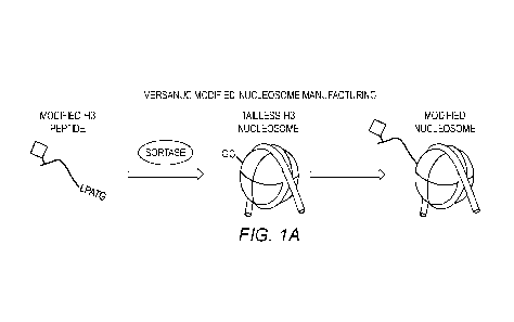
[0013] Figures 1A-1B show (A) Overview of verSaNuc on-nucleosome ligation
strategy
using modified peptides. (B) Schematic of how verSaNuc approach can be used to
rapidly
generate ChAP-CUT&RUN dNucs (SEQ ID NO:9).
Detailed Description
[0014] The present invention is explained in greater detail below. This
description is not
intended to be a detailed catalog of all the different ways in which the
invention may be
implemented, or all the features that may be added to the instant invention.
For example,
features illustrated with respect to one embodiment may be incorporated into
other
embodiments, and features illustrated with respect to a particular embodiment
may be deleted
from that embodiment. In addition, numerous variations and additions to the
various
embodiments suggested herein will be apparent to those skilled in the art in
light of the
instant disclosure which do not depart from the instant invention. Hence, the
following
specification is intended to illustrate some particular embodiments of the
invention, and not
to exhaustively specify all permutations, combinations and variations thereof.
[0015] Unless the context indicates otherwise, it is specifically intended
that the various
features of the invention described herein can be used in any combination.
Moreover, the
present invention also contemplates that in some embodiments of the invention,
any feature
or combination of features set forth herein can be excluded or omitted. To
illustrate, if the
specification states that a complex comprises components A, B and C, it is
specifically
intended that any of A, B or C, or a combination thereof, can be omitted and
disclaimed
singularly or in any combination.
[0016] Unless otherwise defined, all technical and scientific terms used
herein have the
same meaning as commonly understood by one of ordinary skill in the art to
which this
invention belOngs. The terminology used in the description of the invention
herein is for the
purpose of describing particular embodiments only and is not intended to be
limiting of the
invention.
[0017] Nucleotide sequences are presented herein by single strand only, in the
5' to 3'
direction, from left to right, unless specifically indicated otherwise.
Nucleotides and amino
acids are represented herein in the manner recommended by the IUPAC-IUB
Biochemical
Nomenclature Commission, or (for amino acids) by either the one-letter code,
or the three
letter code, both in accordance with 37 C.F.R. 1.822 and established usage.
[0018] Except as otherwise indicated, standard methods known to those skilled
in the art
may be used for production of recombinant and synthetic polypeptides,
antibodies or antigen-
6
CA 03129907 2021-08-11
WO 2020/168151 PCT/US2020/018216
binding fragments thereof, manipulation of nucleic acid sequences, production
of transformed
cells, the construction of nucleosomes, and transiently and stably transfected
cells. Such
techniques are known to those skilled in the art. See, e.g., SAMBROOK et al.,
MOLECULAR CLONING: A LABORATORY MANUAL 4th Ed. (Cold Spring Harbor,
NY, 2012); F. M. AUSUBEL et al. CURRENT PROTOCOLS IN MOLECULAR
BIOLOGY (Green Publishing Associates, Inc. and John Wiley & Sons, Inc., New
York).
[0019] All publications, patent applications, patents, nucleotide sequences,
amino acid
sequences and other references mentioned herein are incorporated by reference
in their
entirety.
[0020] As used in the description of the invention and the appended claims,
the singular
forms "a," "an" and "the" are intended to include the plural forms as well,
unless the context
clearly indicates otherwise.
[0021] As used herein, "and/or" refers to and encompasses any and all possible
combinations of one or more of the associated listed items, as well as the
lack of
combinations when interpreted in the alternative ("or").
[0022] Moreover, the present invention also contemplates that in some
embodiments of the
invention, any feature or combination of features set forth herein can be
excluded or omitted.
[0023] Furthermore, the term "about," as used herein when referring to a
measurable value
such as an amount of a compound or agent of this invention, dose, time,
temperature, and the
like, is meant to encompass variations of 10%, 5%, 1%, 0.5%, or even
0.1% of the
specified amount.
[0024] The term "consisting essentially of' as used herein in connection with
a nucleic acid,
protein means that the nucleic acid or protein does not contain any element
other than the
recited element(s) that significantly alters (e.g., more than about 1%, 5% or
10%) the function
of interest of the nucleic acid or protein.
[0025] As used herein, the term "polypeptide" encompasses both peptides and
proteins,
unless indicated otherwise.
[0026] A "nucleic acid" or "nucleotide sequence" is a sequence of nucleotide
bases, and
may be RNA, DNA or DNA-RNA hybrid sequences (including both naturally
occurring and
non-naturally occurring nucleotide), but is preferably either single or double
stranded DNA
sequences.
[0027] As used herein, an "isolated" nucleic acid or nucleotide sequence
(e.g., an "isolated
DNA" or an "isolated RNA") means a nucleic acid or nucleotide sequence
separated or
7
CA 03129907 2021-08-11
WO 2020/168151 PCT/US2020/018216
substantially free from at least some of the other components of the naturally
occurring
organism or virus, for example, the cell or viral structural components or
other polypeptides
or nucleic acids commonly found associated with the nucleic acid or nucleotide
sequence.
[0028] Likewise, an "isolated" polypeptide means a polypeptide that is
separated or
substantially free from at least some of the other components of the naturally
occurring
organism or virus, for example, the cell or viral structural components or
other polypeptides
or nucleic acids commonly found associated with the polypeptide.
[0029] By "substantially retain" a property, it is meant that at least about
75%, 85%, 90%,
95%, 97%, 98%, 99% or 100% of the property (e.g., activity or other measurable
characteristic) is retained.
[0030] The term "synthetic" refers to a compound, molecule, or complex that
does not exist
in nature.
[0031] The term "DNA barcode" refers to a nucleic acid sequence that can be
used to
unambiguously identify a DNA molecule in which it is located. The length of
the barcode
determines how many unique sequences can be present in a library. For example,
a 1
nucleotide (nt) barcode can code for 4 library members, a 2 nt barcode 16
variants, 3 nt
barcode 64 variants, 4 nt 256 variants, 5 nt 1,024 variants and so on. The
barcode(s) can be
single-stranded (ss) DNA or double-stranded (ds) DNA or a combination thereof.
[0032] One aspect of the invention relates to a nucleosome comprising:
a. a protein octamer, containing two copies each of histones H2A, H2B, H3,
and H4,
and optionally, linker histone Hl;
b. a DNA molecule, comprising:
i. a nucleosome positioning sequence,
a DNA barcode indicative of a chromatin associated protein (ChAP) capture
epitope; and
c. the ChAP capture epitope fused to the N- and/or C-terminal end of one or
more of the
histones, or anywhere in the DNA molecule.
[0033] The nucleosome positioning sequence (NPS) can be any NPS known in the
art.
Examples include, without limitation, the Widom 601 sequence and the 601.2 and
601.3
variants, the Lytechinus variegatus 5S rDNA sequence, and the MMTV LTR
nucleosomes A
and B sequences.
[0034] The ChAP may be, without limitation, a transcription factor, a
chromatin reader, a
histone / DNA modifying enzyme, or a chromatin regulatory complex. Examples of
transcription factors include, without limitation, those listed at:
8
CA 03129907 2021-08-11
WO 2020/168151 PCT/US2020/018216
en.wikipedia.org/wiki/List_of_human_transcription_factors, incorporated by
reference herein
in its entirety. Examples of readers include, without limitation, BRD4,
YEATS2, and
PWWP. Examples of histone / DNA modifying enzymes include, without limitation,
NSD2,
JMJD2A, CARM1, MLL1, DOT1L, EZH2, and DNMT3A/B. Examples of chromatin
regulatory complexes include, without limitation, RNA Polymerase II, SMARCA2,
and ACF.
[0035] The ChAP capture epitope may be any amino acid sequence that is present
in the
ChAP of interest and can be specifically bound by an antibody or other
recognition or
binding agent.
[0036] In some embodiments, the ChAP capture epitope is one or more short
peptide tags.
Examples of short peptide tags include, without limitation, FLAG (DYKDDDDK
(SEQ ID
NO:!)), HA (YPYDVPDYA (SEQ ID NO:2)), 6His (HHHHHH (SEQ ID NO:3)), Myc
(EQKLISEEDL (SEQ ID NO:4)), Strep-I (AWRHPQFGG (SEQ ID NO:5)), Strep-II
(NWSHPQFEK (SEQ ID NO:6)), protein C (EDQVDPRLIDGK (SEQ ID NO:7)), V5, or
GST or 2, 3, 4 or more repeats of the tags.
[0037] In some embodiments, the ChAP capture epitope is an antibody binding
sequence,
i.e., an epitope recognized and specifically bound by an antibody or other
recognition or
binding agent. In some embodiments, the epitope is one that is unique to the
ChAP, e.g.,
having low sequence homology with related proteins (i.e., family members). In
some
embodiments, the epitope is one recognized by known antibodies to the ChAP.
[0038] Each of the histones in the nucleosome is independently fully synthetic
(e.g.,
chemically synthesized), semi-synthetic (e.g., produced recombinantly then
synthetically
altered, e.g., by chemically or enzymatically adding a peptide sequence), or
recombinant.
[0039] The DNA molecule may comprise further elements. In some embodiments,
the
DNA molecule further comprises a binding member linked to the DNA molecule,
wherein
the binding member specifically binds to a binding partner. Examples of the
binding member
and its binding partner include, without limitation, biotin with avidin or
streptavidin, a nano-
tag with streptavidin, glutathione with glutathione transferase, an
antigen/epitope with an
antibody, polyhistidine with nickel, a polynucleotide with a complementary
polynucleotide,
an aptamer with its specific target molecule, or Si-tag and silica. In some
embodiments, the
binding member is linked to the 5' end of the DNA molecule. In some
embodiments, the
binding member is linked to the 3' end of the DNA molecule.
[0040] In some embodiments, the DNA molecule comprises a linker between the
nucleo some positioning sequence and the binding member that is about 10 to
about 80
nucleotides in length, e.g., about 15 to about 40 nucleotides in length or
about 15 to about 30
9
CA 03129907 2021-08-11
WO 2020/168151
PCT/US2020/018216
nucleotides in length, e.g., about 10, 15, 20, 25, 30, 35, 40, 45, 50, 55, 60,
65, 70, 75, or 80
nucleotides in length or any range therein.
[0041] In some embodiments, the DNA molecule comprises a nuclease or
transposase
recognition sequence, e.g., in the linker.
[0042] The nuclease or transposase recognition sequence may be any nucleotide
sequence
that is preferably recognized by a nuclease or transposase. In some
embodiments, the
nuclease or transposase recognition sequence is recognized by an
endodeoxyribonuclease.
Suitable endodeoxyribonucleases include, without limitation, micrococcal
nuclease (MNase),
Si nuclease, mung bean nuclease, pancreatic DNase I, yeast HO or I-SceI
endonuclease, a
restriction endonuclease, or a homing endonuclease, and modified or enhanced
versions
thereof. In some embodiments, the recognition sequence is an A/T-rich region.
[0043] In some embodiments, the nuclease or transposase recognition sequence
is
recognized by a transposase. Suitable transposases include, without
limitation, Tn5, Mu, I55,
IS91, Tn552, Tyl, Tn7, Tn/O, Mariner, P Element, Tn3, Tn10, or Tn903, and
modified or
enhanced versions thereof, e.g., a mutated hyperactive transposase. Such
modified
transposases are known in the art. In some embodiments, the transposase is Tn5
or a
modified Tn5, e.g., a hyperactive Tn5 comprising one or more of the mutations
E54K,
M56A, or L372P. In some embodiments, the recognition sequence is a G/C-rich
region.
[0044] In somµ e embodiments, the linker comprises both a nuclease recognition
sequence
(e.g., one or more patches of A/T rich sequences) and a transposase
recognition sequence
(e.g., one or more patches of G/C rich sequences) so that the nucleosomes of
the invention
can be used for multiple methods. An A/T rich region or G/C rich region is one
that contains
more than 50%, A/T bases or G/C bases, respectively, e.g., more than 50%, 55%,
60%, 65%,
70%, 75%, or 80%.
[0045] In some embodiments, the DNA barcode has a length of about 6 to about
50
basepairs, e.g., about 7 to about 30 basepairs or about 8 to about 20
basepairs. In some
embodiments, the DNA barcode may have a length of less than 50, 45, 40, 35,
30, 25, 20, 15,
or 10 nucleotides. In some embodiments, the DNA barcode may have a length of
at least 6,
7, 8, 9, 10, 12, 14, 16, 18, 20, 22, 24, 26, 28, or 30 nucleotides.
[0046] Another aspect of the invention relates to a panel (e.g., a collection)
of the
nucleosomes of the invention, wherein the nucleosomes in the panel comprise a
ChAP
capture epitope at one or more concentrations in the panel and the DNA barcode
of each
nucleosome indicates the concentration at which that nucleosome is present in
the panel.
CA 03129907 2021-08-11
WO 2020/168151
PCT/US2020/018216
[0047] In some embodiments, the panel comprises at least two nucleosomes
comprising
different ChAP capture epitopes. In some embodiments, each nucleosome
comprising a
different ChAP capture epitope is present at the same concentration in the
panel. In other
embodiments, each nucleosome comprising a different ChAP capture epitope is
present at
multiple concentrations in the panel and the DNA barcode of each indicates
that
concentration at which that nucleosome is present in the panel.
[0048] In some embodiments, the panel may further comprise a synthetic
nucleosome
which does not comprise a ChAP capture epitope, e.g., as a control.
[0049] A further aspect of the invention relates to a polynucleosome
comprising:
a. a protein octamer, containing two copies each of histones H2A, H2B, H3,
and H4,
and optionally, linker histone Hl;
b. a DNA molecule, comprising:
i. a nucleosome positioning sequence,
a DNA barcode indicative of a ChAP capture epitope; and
c. the ChAP capture epitope fused to the N- and/or C-terminal end of one or
more of the
histones, or anywhere in the DNA molecule.
[0050] In some embodiments, the polynucleosome may comprise 2-10 nucleosomes,
e.g., 2,
3, 4, 5, 6, 7, 8, 9, or 10 nucleosomes or any range therein.
[0051] The nucleosome positioning sequence (NPS) can be any NPS known in the
art.
Examples include, without limitation, the Widom 601 sequence and the 601.2 and
601.3
variants, the Lytechinus variegatus 5S rDNA sequence, and the MMTV LTR
nucleosomes A
and B sequences.
[0052] The ChAP capture epitope may be any amino acid sequence that is present
in the
ChAP of interest and can be specifically bound by an antibody or other binding
agent.
[0053] In some embodiments, the ChAP capture epitope is one or more short
peptide tags.
Examples of short peptide tags include, without limitation, FLAG (DYKDDDDK
(SEQ ID
NO:1)), HA (YPYDVPDYA (SEQ ID NO:2)), 6His (HHHHHH (SEQ ID NO:3)), Myc
(EQKLISEEDL (SEQ ID NO:4)), Strep-I (AWRHPQFGG (SEQ ID NO:5)), Strep-II
(NWSHPQFEK (SEQ ID NO:6)), protein C (EDQVDPRLIDGK (SEQ ID NO:7)), V5,
TY1, or GST or 2, 3, 4 or more repeats of the tags.
[0054] In some embodiments, the ChAP capture epitope is an antibody binding
sequence,
i.e., an epitope recognized and specifically bound by an antibody.
11
CA 03129907 2021-08-11
WO 2020/168151 PCT/US2020/018216
[0055] Each of the histones in the nucleosome is independently fully synthetic
(e.g.,
chemically synthesized), semi-synthetic (e.g., produced recombinantly then
synthetically
altered, e.g., by chemically or enzymatically adding a peptide sequence), or
recombinant.
[0056] The DNA molecule may comprise further elements. In some embodiments,
the
DNA molecule further comprises a binding member linked to the DNA molecule,
wherein
the binding member specifically binds to a binding partner. Examples of the
binding member
and its binding partner include, without limitation, biotin with avidin or
streptavidin, a nano-
tag with streptavidin, glutathione with glutathione transferase, an
antigen/epitope with an
antibody, polyhistidine with nickel, a polynucleotide with a complementary
polynucleotide,
an aptamer with its specific target molecule, or Si-tag and silica. In some
embodiments, the
binding member is linked to the 5' end of the DNA molecule. In some
embodiments, the
binding member is linked to the 3' end of the DNA molecule.
[0057] In some embodiments, the DNA molecule comprises a linker between the
nucleosome positioning sequence and the binding member that is about 10 to
about 80
nucleotides in length, e.g., about 15 to about 40 nucleotides in length or
about 15 to about 30
nucleotides in length, e.g., about 10, 15, 20, 25, 30, 35, 40, 45, 50, 55, 60,
65, 70, 75, or 80
nucleotides in length or any range therein.
[0058] In some embodiments, the DNA molecule comprises a nuclease or
transposase
recognition sequence, e.g., in the linker.
[0059] The nuclease or transposase recognition sequence may be any nucleotide
sequence
that is preferably recognized by a nuclease or transposase. In some
embodiments, the
nuclease or transposase recognition sequence is recognized by an
endodeoxyribonuclease.
Suitable endodeoxyribonucleases include, without limitation, micrococcal
nuclease, Si
nuclease, mung bean nuclease, pancreatic DNase I, yeast HO or I-SceI
endonuclease, a
restriction endonuclease, or a homing endonuclease, and modified or enhanced
versions
thereof. In some embodiments, the recognition sequence is an A/T-rich region.
[0060] In some embodiments, the nuclease or transposase recognition sequence
is
recognized by a transposase. Suitable transposases include, without
limitation, Tn5, Mu, IS5,
IS91, Tn552, Tyl, Tn7, Tn/O, Mariner, P Element, Tn3, Tnl 0, or Tn903, and
modified or
enhanced versions thereof, e.g., a mutated hyperactive transposase. Such
modified
transposases are known in the art. In some embodiments, the transposase is Tn5
or a
modified Tn5, e.g., a hyperactive Tn5 comprising one or more of the mutations
E54K,
M56A, or L372P. In some embodiments, the recognition sequence is a G/C-rich
region.
12
CA 03129907 2021-08-11
WO 2020/168151 PCT/US2020/018216
[0061] In some embodiments, the linker comprises both a nuclease recognition
sequence
(e.g., one or more patches of A/T rich sequences) and a transposase
recognition sequence
(e.g., one or more patches of G/C rich sequences) so that the nucleosomes of
the invention
can be used for multiple methods. An A/T rich region or G/C rich region is one
that contains
more than 50%, A/T bases or G/C bases, respectively, e.g., more than 50%, 55%,
60%, 65%,
70%, 75%, or 80%.
[0062] In some embodiments, the DNA barcode has a length of about 6 to about
50
basepairs, e.g., about 7 to about 30 basepairs or about 8 to about 20
basepairs. In some
embodiments, the DNA barcode may have a length of less than 50, 45, 40, 35,
30, 25, 20, 15,
or 10 nucleotides. In some embodiments, the DNA barcode may have a length of
at least 6,
7, 8, 9, 10, 12, 14, 16, 18, 20, 22, 24, 26, 28, or 30 nucleotides.
[0063] An additional aspect of the invention relates to an array comprising
the
polynucleosome of the invention. The polynucleosome array can contain a single
ChAP
capture epitope or be comprised of an ensemble of different ChAP capture
epitopes. DNA
barcodes on the array can be used to denote the entire array or unique
features within the
array.
[0064] A further aspect of the invention relates to a pool of the array of the
invention,
wherein each array comprises a unique ChAP capture epitope. In some
embodiments, the
polynucleosome array panel comprises, 2, 3, 4, 5, 6, 7, 8, 9, 10, 12, 15, 18,
21, 25, 30, 35, or
40 or more polynucleosome arrays comprising different ChAP capture epitopes or
any range
therein. In some embodiments, each polynucleosome array comprising a different
ChAP
capture epitope is present at the same concentration in the array. In other
embodiments, each
nucleosome array comprising a different ChAP capture epitope is present at
multiple
concentrations in the array and the DNA barcode of each polynucleosome
indicates the
concentration at which the polynucleosome is present in the array. In some
embodiments, the
array further comprises a polynucleosome array which does not comprise a ChAP
capture
epitope, e.g., for use as a control.
[0065] Another aspect of the invention relates to a solid support, e.g., a
bead, comprising a
binding partner to the binding member of the nucleosome, panel,
polynucleosome, array, or
pool of the invention, wherein the bead is bound to the nucleosome, panel,
polynucleosome,
array, or pool. The bead may be any bead suitable for separating chromatin,
nucleosomes, or
polynucleosomes from a sample and/or to attach the chromatin, nucleosomes, or
polynucleosomes to a solid support. The bead may be composed of natural
materials (e.g.,
13
CA 03129907 2021-08-11
WO 2020/168151
PCT/US2020/018216
alginate) or synthetic materials (e.g., polystyrene). In some embodiments, the
bead is a
magnetic bead that can be separated by exposure to a magnetic field.
[0066] An additional aspect of the invention relates to a kit comprising the
nucleosome,
panel, polynucleosome, array, pool, or bead of the invention. In some
embodiments, the kit
may further comprise an antibody, aptamer, nanobody, or other recognition or
binding agent
that specifically binds to a ChAP capture epitope or a nucleosome feature
(e.g., histone post-
translational modification (PTM), histone mutation, histone variant, or DNA
post-
transcriptional modification). In some embodiments, the kit may further
comprise a nuclease
or transposase linked to an antibody-binding protein or to an entity that
binds the recognition
agent. In certain embodiments, the antibody-binding protein may be, without
limitation,
protein A, protein G, a fusion between protein A and protein G, protein L, or
protein Y. In
some embodiments, the entity that binds the recognition agent is a protein. In
other
embodiments, the kit may further comprise a nuclease or transposase that is
not linked to an
antibody-binding protein or to an entity that binds the recognition agent. In
certain
embodiments, the kit may further comprise a bead comprising a binding partner
to the
binding member, e.g., a magnetic bead. The kit may further comprise reagents
and/or
containers for carrying out the methods of the invention, e.g., buffers,
enzymes (e.g.,
nucleases, transposases, polymerases, ligases), detection agents, etc. In some
embodiments,
the kit may further comprise instructions for carrying out the methods of the
invention.
[0067] The spike-in controls may be used in any chromatin assay known in the
art in which
an improved control/calibrator would be useful. Examples include, without
limitation, the
CUT&RUN assay (WO 2019/060907), the ChIC assay (US Patent No. 7,790,379), and
the
ICeChIP assay (WO 2015/117145). Each of these references are incorporated
herein in their
entirety.
[0068] One aspect of the invention relates to a method for chromatin mapping
using
tethered enzymes, wherein the improvement is the use of the nucleosome, panel,
polynucleosome, array, pool, or bead of the invention in the assay as a spike-
in control.
[0069] Another aspect of the invention relates to a method for mapping
chromatin using
tethered enzymes, comprising the steps of:
a) binding a nucleus, organelle, cell, or tissue to a solid support;
b) permeabilizing the nucleus, organelle, cell, or tissue;
c) binding the nucleosome, panel, polynucleosome, array, or pool of the
invention to a
solid support;
14
CA 03129907 2021-08-11
WO 2020/168151 PCT/US2020/018216
d) contacting the permeabilized nucleus, organelle, cell, or tissue of b)
and the bound
nucleosome, panel, polynucleosome, array, or pool of c) with an antibody,
aptamer,
nanobody, or recognition agent that specifically binds to the ChAP capture
epitope;
e) adding an antibody-binding agent, aptamer-binding agent, nanobody-
binding agent, or
recognition agent-binding agent linked to a nuclease or transposase;
allowing the nuclease or transposase to cleave or label DNA in the nucleus,
organelle,
cell, or tissue and the nuclease or transposase recognition sequence in the
nucleosome, panel,
polynucleosome, array, or pool;
separating cleaved or labeled DNA; and
h) identifying the cleaved or labeled DNA;
thereby mapping chromatin.
[0070] In some embodiments, the nuclease or transposase of step (e) is
inactive and step (f)
comprises activating the nuclease or transposase, e.g., by adding an ion such
as calcium or
magnesium.
[0071] In some embodiments, identifying the cleaved DNA comprises subjecting
the
cleaved DNA to amplification and/or sequencing. The sequencing may comprise,
for
example, qPCR, Next Generation Sequencing, or Nanostring.
[0072] In some embodiments, the method may further comprise determining the
identity of
the nucleosome, panel, polynucleosome, array, or pool based on the sequence of
the DNA
barcode in the cleaved or labeled DNA.
[0073] In some embodiments, the method further comprises optimizing the method
based
on the results detected with the nucleosome, panel, polynucleosome, array, or
pool. For
example, the recovery of on-target / off-target DNA-barcoded nucleosomes could
be used to
optimize enzyme concentration, enzyme activation time, cell-to-enzyme ratio,
etc.
[0074] The methods may be carried out using any suitable format that provides
a solid
support for the cell, nucleus, organelle, or tissue. In some embodiments, the
solid support is a
bead, e.g., a magnetic bead. In some embodiments, the solid support is a well
of a plate, e.g.,
6, 12, 24, 96, 384, or 1536-well plates.
[0075] The results obtained from the methods of the invention may be used for
any purpose
where information on ChAPs and chromatin structure and/or modification, e.g.,
epigenetic
changes, would be useful. In some embodiments, the methods may further
comprise the step
of using the sequencing results to compare chromatin features between healthy
and disease
tissues. In some embodiments, the methods may further comprise the step of
using the
CA 03129907 2021-08-11
WO 2020/168151 PCT/US2020/018216
sequencing results to predict a disease state. In some embodiments, the
methods may further
comprise the step of using the sequencing results to monitor response to
therapy. In some
embodiments, the methods may further comprise the step of using the sequencing
results to
analyze tumor heterogeneity.
[0076] The methods of the invention may be used for detecting and quantitating
the
presence of a ChAP on chromatin. An antibody, aptamer, nanobody, or
recognition agent
that specifically binds to a ChAP capture epitope may be used to detect and
quantitate the
ChAP at various genomic loci.
[0077] The methods of the invention may be used for determining and
quantitating a ChAP
on chromatin in a subject having a disease or disorder. An antibody, aptamer,
nanobody, or
recognition agent that specifically binds to a ChAP that may be associated
with the disease or
disorder of the subject or relevant to expression of a gene associated with
the disease or
disorder may be used to detect and quantitate the ChAP at various genomic
loci. By this
method, one can determine if a subject having a disease or disorder, e.g., a
tumor, has a
ChAP that is known to be associated with, e.g., the tumor type.
[0078] The methods of the invention may be used for monitoring changes in a
ChAP on
chromatin over time in a subject. This method may be used to determine if the
status of the
ChAP (e.g., level and/or activity) is improving, stable, or worsening over
time. The steps of
the method may be repeated as many times as desired to monitor changes in the
status of a
ChAP, e.g., 2, 3, 4, 5, 6, 7, 8, 9, 10, 25, 50, or 100 or more times. The
method may be
repeated on a regular schedule (e.g., daily, weekly, monthly, yearly) or on an
as needed basis.
The method may be repeated, for example, before, during, and/or after
therapeutic treatment
of a subject; after diagnosis of a disease or disorder in a subject; as part
of determining a
diagnosis of a disease or disorder in a subject; after identification of a
subject as being at risk
for development of a disease or disorder; or any other situation where it is
desirable to
monitor possible changes in the ChAP at various genomic loci.
[0079] The methods of the invention may be used for measuring on-target
activity of a
drug. The methods may be carried out before, during, and/or after
administration of a drug to
determine the capability of the drug to alter the ChAP status of the subject.
[0080] The methods of the invention may be used for monitoring the
effectiveness of
therapy in a subject having a disease or disorder. The steps of the method may
be repeated as
many times as desired to monitor effectiveness of the treatment, e.g., 2, 3,
4, 5, 6, 7, 8, 9, 10,
25, 50, or 100 or more times. The method may be repeated on a regular schedule
(e.g., daily,
weekly, monthly, yearly) or on as needed basis, e.g., until the therapeutic
treatment is ended.
16
CA 03129907 2021-08-11
WO 2020/168151 PCT/US2020/018216
The method may be repeated, for example, before, during, and/or after
therapeutic treatment
of a subject, e.g., after each administration of the treatment. In some
embodiments, the
treatment is continued until the method of the invention shows that the
treatment has been
effective.
[0081] The methods of the invention may be used for selecting a suitable
treatment for a
subject having a disease or disorder based on the ChAP status on chromatin in
the subject.
The methods may be applied, for example, to subjects that have been diagnosed
or are
suspected of having a disease or disorder. A determination of the ChAP status
may indicate
that the status of the ChAP has been modified and a therapy should be
administered to the
subject to correct the modification. Conversely, a determination that the
status of the ChAP
has not been modified would indicate that a therapy would not be expected to
be effective
and should be avoided.
[0082] The methods of the invention may be used for determining a prognosis
for a subject
having a disease or disorder based on the ChAP status on chromatin in the
subject. In some
instances, the ChAP is indicative of the prognosis of a disease or disorder.
Thus, a
determination of the ChAP status of an epitope in a subject that has been
diagnosed with or is
suspected of having a disease or disorder may be useful to determine the
prognosis for the
subject.
[0083] The methods of the invention may be used for identifying a biomarker of
a disease
or disorder based on the ChAP status on chromatin in a subject. In this
method, biological
samples of diseased tissue may be taken from a number of patients have a
disease or disorder
and the ChAP status determined. Correlations between the ChAP status and the
occurrence,
stage, subtype, prognosis, etc., may then be identified using analytical
techniques that are
well known in the art.
[0084] The methods of the invention may be used for screening for an agent
that modifies
the status of a ChAP on chromatin in a subject.
[0085] The screening method may be used to identify agents that increase or
decrease the
expression, level and/or activity of a ChAP. In some embodiments, the detected
increase or
decrease is statistically significant, e.g., at least p < 0.05, e.g., p <0.01,
0.005, or 0.001. In
other embodiments, the detected increase or decrease is at least about 10%,
20%, 30%, 40%,
50%, 60%, 70%, 80%, 90%, 100% or more.
[0086] Any compound of interest can be screened according to the present
invention.
Suitable test compounds include organic and inorganic molecules. Suitable
organic
molecules can include but are not limited to small molecules (compounds less
than about
17
CA 03129907 2021-08-11
WO 2020/168151 PCT/US2020/018216
1000 Daltons), polypeptides (including enzymes, antibodies, and antibody
fragments),
carbohydrates, lipids, coenzymes, and nucleic acid molecules (including DNA,
RNA, and
chimeras and analogs thereof) and nucleotides and nucleotide analogs.
[0087] Further, the methods of the invention can be practiced to screen a
compound library,
e.g., a small molecule library, a combinatorial chemical compound library, a
polypeptide
library, a cDNA library, a library of antisense nucleic acids, and the like,
or an arrayed
collection of compounds such as polypeptide and nucleic acid arrays.
[0088] Any suitable screening assay format may be used, e.g., high throughput
screening.
[0089] The method may also be used to characterize agents that have been
identified as an
agent that modifies the ChAP status on chromatin. Characterization, e.g.,
preclinical
characterization, may include, for example, determining effective
concentrations, determining
effective dosage schedules, and measuring pharmacokinetics and
pharmacodynamics.
[0090] In some embodiments, the nucleus, organelle, cell, or tissue is from a
diseased tissue
or sample. In some embodiments, the nucleus, organelle, cell, or tissue is
from non-diseased
tissue or sample. In some embodiments, the nucleus, organelle, cell, or tissue
is or is from a
peripheral tissue or cell, e.g., a peripheral blood mononuclear cell. In some
embodiments, the
nucleus, organelle, cell, or tissue is or is from cultured cells, e.g.,
primary cells.
[0091] One aspect of the invention relates to a method for assaying chromatin
for a ChAP,
wherein the improvement is the use of the nucleosome, panel, polynucleo some,
array, pool,
or bead of the invention in the assay as a spike-in control.
[0092] Another aspect of the invention relates to a method for quantifying the
abundance of
a chromatin associated protein (ChAP) in a biological sample using Chromatin
ImmunoPrecipitation (ChIP), the method comprising:
a. isolating a biological sample;
b. preparing a library of native nucleosomes from the biological sample,
wherein the
library additionally comprises one or more ChAPs;
c. providing the nucleosome, panel, polynucleosome, array, pool, or bead of
the
invention comprising a ChAP capture epitope present in the ChAP to create a
reference
standard;
d. adding an antibody, aptamer, nanobody, or recognition agent that
specifically binds to
the ChAP capture epitope in the native nucleosome library and reference
standard;
e. performing an affinity reagent-based assay to measure the amount of ChAP
in the
native nucleosome library and reference standard; and
18
CA 03129907 2021-08-11
WO 2020/168151 PCT/US2020/018216
f. quantifying ChAP abundance by comparing its relative abundance in the
native
nucleosome library to the reference standard.
[0093] An additional aspect of the invention relates to a method for
quantifying the
abundance of two or more ChAPs in a biological sample, the method comprising:
a. isolating a biological sample;
b. preparing a library of native nucleosomes from the biological sample,
wherein the
library comprises nucleosomes comprising two or more ChAPs;
c. providing the nucleosome, panel, polynucleosome, array, pool, or bead of
the
invention comprising ChAP capture epitopes present in the ChAPs to create a
reference
standard;
d. adding two or more antibodies, aptamers, nanobodies, or recognition
agents that
specifically bind to the ChAP capture epitopes to the native nucleosome
library and the
reference standard;
e. performing an affinity reagent-based assay to measure the amount of each
ChAP in
the native nucleosome library and the reference standard; and
f. quantifying the abundance of each ChAP by comparing the relative
abundance in the
native nucleosome library to the reference standard.
[0094] Another aspect of the invention relates to a method for quantifying the
abundance of
one or more ChAPs in a biological sample from a subject having a disease or
disorder, the
method comprising:
a. isolating a biological sample from the subject;
b. preparing a library of native nucleosomes from the biological sample,
wherein the
library comprises nucleosomes comprising one or more ChAPs;
c. providing the nucleosome, panel, polynucleosome, array, pool, or bead of
the
invention comprising ChAP capture epitopes present in the ChAPs to create a
reference
standard;
d. adding one or more antibodies, aptamers, nanobodies, or recognition
agents that
specifically bind to the ChAP capture epitopes to the native nucleosome
library and the
reference standard;
e. performing an affinity reagent-based assay to measure the amount of ChAP
in the
native nucleosome library and the reference standard; and
f. quantifying the abundance of ChAPs by comparing the relative abundance
in the
native nucleosome library to the reference standard.
19
CA 03129907 2021-08-11
WO 2020/168151 PCT/US2020/018216
[0095] A further aspect of the invention relates to a method for determining a
prognosis for
a subject having a disease or disorder based on the absolute quantification of
one or more
ChAPs, the method comprising:
a. isolating a biological sample from the subject;
b. preparing a library of native nucleosomes from the biological sample,
wherein the
library comprises nucleosomes comprising one or more ChAPs;
c. providing the nucleosome, panel, polynucleosome, array, pool, or bead of
the
invention comprising ChAP capture epitopes present in the ChAPs to create a
reference
standard;
d. adding one or more antibodies, aptamers, nanobodies, or recognition
agents that
specifically bind to the ChAP capture epitopes to the native nucleosome
library and the
reference standard;
e. performing an affinity reagent-based assay to measure the amount of ChAP
in the
native nucleosome library and the reference standard;
f. quantifying the abundance of ChAP by comparing the relative abundance in
the native
nucleosome library to the reference standard; and
g. determining the prognosis of the subject based on the absolute abundance
of the one
or more ChAPs.
[0096] An additional aspect of the invention relates to a method for
identifying a biomarker
of a disease or disorder based on the absolute quantification of one or more
ChAPs, the
method comprising:
a. isolating a biological sample from the subject;
b. preparing a library of native nucleosomes from the biological sample,
wherein the
library comprises nucleosomes comprising one or more ChAPs;
c. providing the nucleosome, panel, polynucleosome, array, pool, or bead of
the
invention comprising ChAP capture epitopes present in the ChAPs to create a
reference
standard;
d. adding one or more antibodies, aptamers, nanobodies, or recognition
agents that
specifically bind to the ChAP capture epitopes to the native nucleosome
library and the
reference standard;
e. performing an affinity reagent-based assay to measure the amount of ChAP
in the
native nucleosome library and the reference standard;
f. quantifying the abundance of ChAP by comparing the relative abundance in
the native
nucleosome library to the reference standard; and
CA 03129907 2021-08-11
WO 2020/168151 PCT/US2020/018216
g. correlating the absolute abundance of the one or more ChAPs with the
disease or
disorder; thereby identifying a biomarker of the disease or disorder.
[0097] Another aspect of the invention relates to a method of screening for an
agent that
modifies the ChAP status on chromatin from a biological sample of a subject,
the method
comprising determining the absolute quantification of one or more ChAPs in the
presence
and absence of the agent, wherein determining the absolute quantification of
the one or more
ChAPs comprises:
a. isolating a biological sample from the subject;
b. preparing a library of native nucleosomes from the biological sample,
wherein the
library comprises nucleosomes comprising one or more ChAP(s) in a target
epitope(s);
c. providing the nucleosome, panel, polynucleosome, array, pool, or bead of
the
invention comprising ChAP capture epitopes present in the ChAPs to create a
reference
standard;
d. adding one or more antibodies, aptamers, nanobodies, or recognition
agents that
specifically bind to the ChAP capture epitopes to the native nucleosome
library and the
reference standard;
e. performing an affinity reagent-based assay to measure the amount of ChAP
in the
native nucleosome library and the reference standard;
f. quantifying the abundance of ChAP by comparing the relative abundance in
the native
nucleosome library to the reference standard;
wherein a change in the ChAP status in the presence and absence of the agent
identifies an
agent that modifies the ChAP status on chromatin.
[0098] The antibody, aptamer, nanobody, or recognition agent used in the
methods of the
invention may be any agent that specifically recognizes and binds to a ChAP of
interest. In
some embodiments, the affinity agent is an antibody or antibody fragment
directed towards
the ChAP capture epitope. The antibody or fragment thereof may be a full-
length
immunoglobulin molecule, an Fab, an Fab', an F(ab)'2, an scFv, an Fv fragment,
a nanobody,
a VHH or a minimal recognition unit. The agent may be an aptamer or a non-
immunoglobulin scaffold such as an affibody, an affilin molecule, an AdNectin,
a lipocalin
mutein, a DARPin, a Knottin, a Kunitz-type domain, an Avimer, a Tetranectin or
a trans-
body. In some embodiments, the agent is an antibody or analogous enrichment
reagent
directed towards the ChAP capture epitope.
[0099] In some embodiments, the quantification of one or more ChAPs is
determined by
an affinity agent (e.g., antibody or analogous enrichment reagent)-based
detection assay.
21
CA 03129907 2021-08-11
WO 2020/168151
PCT/US2020/018216
Examples of antibody-based detection methods include, without limitation,
ChIP, ELISA,
AlphaLISA, AlphaSCREEN, Luminex, and immunoblotting. In some embodiments, the
antibody-based detection assay uses two different antibodies for substrate
capture and
detection. In some embodiments, the antibody-based detection assay uses the
same antibody
for both substrate capture and detection.
[0100] In each of the methods of the invention, the biological sample may
be any sample
from which chromatin can be isolated. The biological sample may be, for
example, blood,
serum, plasma, urine, saliva, semen, prostatic fluid, nipple aspirate,
lachrymal fluid,
perspiration, feces, cheek swabs, cerebrospinal fluid, cell lysate samples,
amniotic fluid,
gastrointestinal fluid, biopsy tissue, lymphatic fluid, or cerebrospinal
fluid. In some
embodiments, the biological sample comprises cells and the chromatin is
isolated from the
cells. In some embodiments, the cells are cells from a disease of disorder
associated with
changes in one or more ChAPs, e.g., a diseased cell. In some embodiments, the
cells are cells
from a tissue or organ affected by a disease or disorder associated with
changes in one or
more ChAPs, e.g., a diseased tissue or organ. The cells may be obtained from
the diseased
organ or tissue by any means known in the art, including but not limited to
biopsy, aspiration,
and surgery. In some embodiments, the cells are cultured cells, e.g., primary
cells.
[0101] In other embodiments, the cells are not cells from a tissue or organ
affected by a
disease or disorder associated with changes in ChAPs. The cells may be, e.g.,
cells that serve
as a proxy for the diseased cells. The cells may be cells that are more
readily accessible than
the diseased cells, e.g., that can be obtained without the need for
complicated or painful
procedures such as biopsies. Examples of suitable cells include, without
limitation,
peripheral blood mononuclear cells.
[0102] In some embodiments, the biological sample is a biopsy. In other
embodiments,
the biological sample is a biological fluid. In some embodiments, the
biological sample
comprises peripheral blood mononuclear cells. In other embodiments, the
biological sample
comprises circulating nucleosomes, e.g., as released from dying cells. The
circulating
nucleosomes may be, e.g., from blood or from cells from a disease or disorder.
In certain
embodiments, the biological sample is plasma, urine, saliva, stool, lymphatic
fluid, or
cerebrospinal fluid. In some embodiments, the biological sample may be treated
with an
enzyme to digest chromatin into mono- and/or polynucleosomes. The enzyme may
be,
without limitation, a nuclease, e.g., micrococcal nuclease.
[0103] The subject may be any subject for which the methods of the present
invention are
desired. In some embodiments, the subject is a mammal, e.g., a human. In some
22
CA 03129907 2021-08-11
WO 2020/168151 PCT/US2020/018216
embodiments, the subject is a laboratory animal, e.g., a mouse, rat, dog, or
monkey, e.g., an
animal model of a disease. In certain embodiments, the subject may be one that
has been
diagnosed with or is suspected of having a disease or disorder. In some
embodiments, the
subject may be one that is at risk for developing a disease or disorder, e.g.,
due to genetics,
family history, exposure to toxins, etc.
[0104] Having described the present invention, the same will be explained in
greater detail
in the following examples, which are included herein for illustration purposes
only, and
which are not intended to be limiting to the invention.
EXAMPLES
Example 1: Generation of ChAP-containing nucleosomes using recombinant methods
[0105] Nucleosomes containing ChAP epitopes can be generated using any
approach
known in the art. Below we describe two methods. First, ChAP-histone fusion
proteins can
be directly expressed using recombinant methods. A series of nucleosomes
(termed
"verSaNuc") were generated that contain common SPTs, including 3xFLAG, 3xTY1,
and
3xHA. For these nucleosomes, the SPT followed by a GGGGS (SEQ ID NO:8) linker
fused
to histone H3 was expressed. This modified histone was then incorporated into
a
recombinant nucleosome using 250bp DNA (-50bp linker DNA on each side of the
nucleosome core particle). A similar approach can be used for other ChAP
fragments, such
as CTCF. Second, ChAP-containing histones can be generated by linking
synthetic peptides
or recombinantly expressed proteins by chemical or enzymatic ligation.
[0106] Nucleosome spike-ins were engineered to contain a CTCF, BRD4 or 3xFLAG
epitope (DYKDDDDK (SEQ ID NO:1)) fused to the N-terminus of histone H3 to
capture
ChAP- or SPT-specific antibodies in genomic mapping assays (e.g., ChIP-seq,
CUT&RUN,
CUT&Tag). As an example, both ChAP epitopes and SPTs were selected that
maximize user
flexibility regarding antibody selection, enrichment strategy, and
experimental design.
Human CTCF (aa 650-727) and human BRD4 (aa 1031-1362) epitope regions were
selected
based on low sequence homology with related proteins (i.e., family members; C-
terminal
regions for both proteins) and contain the target epitope for the most widely
used CTCF or
BRD4 antibodies. To generate these nucleosomes, fusion histone proteins (e.g.,
CTCF-H3)
were expressed in E. coil and purified, and then assembled into DNA-barcoded
recombinant
nucleosomes.
[0107] DNA-barcoded dNucs may be wrapped with a Widom 601 sequence (Lowary and
Widom 1998) engineered with an embedded 22 bp barcode (composed of two
catenated 11
23
CA 03129907 2021-08-11
WO 2020/168151 PCT/US2020/018216
bps) near the 3' end (Herold, Kurtz et al. 2008), similar to spike-ins used
for SNAP-ChIP
spike-ins (e.g., EpiCypher K-MetStat; 19-1001). In SNAP-ChIP, spike-ins are
assembled
without 'linker' DNA (i.e., 147bp). To make dNucs compatible with chromatin
tethering
technology (e.g., CUT&RUN / CUT&Tag), the DNA assembly sequence can be
modified to
include a 5' biotin, which is used to immobilize the calibrators to a
streptavidin-coated
magnetic bead solid support. In addition, the nucleosome assembly sequence
(i.e., 601 with
embedded barcodes) was modified to include >20bp linker DNA (i.e., DNA not
wrapped
around the histone octamer) to allow MNase to cleave and release the
calibrator from the
magnetic bead. Of note, MNase can reliably digest 15bp linker regions in yeast
and humans
(Cole, Cui et al. 2016). To quality assess DNA-barcoded nucleosomes, they can
be
immobilized on beads and treated with MNase (digesting unassembled DNA)
followed by
qPCR to measure the nucleosome barcode sequence.
Example 2: Generation of ChAP-containing nucleosomes using enzyme linkage
[0108] To accelerate nucleosome manufacturing, the S. aureus Sortase A (SrtA)
transpeptidase can be used to ligate modified peptides directly onto fully
assembled tailless
nucleosomes (FIG. 1A). This approach delivers two capabilities: a) the rapid
development of
modified nucleosomes in small batches (.ig vs. mg scale for standard dNuc
assembly); and b)
the multiplexing of modified nucleosome syntheses. This approach is very well-
suited for
ChAP-containing nucleosome development, which will require small quantities
for each
assay yet great diversity to meet market needs.
[0109] In one example, verSaNuc nucleosomes were assembled that contain: (i) a
unique
DNA barcode identifier, and (ii) a GGGGS (SEQ ID NO:8) motif at the H3 N-
terminus.
Next, sortase-mediated on-nucleosome ligation reactions were performed using
recombinant
proteins (or synthetic peptides) encoding a ChAP epitope (or SPT) and a C-
terminal native
sortase target motif (LPATG (SEQ ID NO:9); FIG. 1B).
[0110] Using this 'on-nucleosome' ligation approach, ChAP-containing
nucleosome
standards were generated for a set of ChAP epitopes and SPTs. Here, ChAP
epitopes were
focused on the BET family of bromodomain-containing proteins, including BRD2,
BRD3,
and BRD4. To generate this nucleosome panel, a C-terminal fragment of each
protein was
used, since this is divergent across the protein family and thus used for Ab
development.
ChAP epitopes were then generated by recombinant expression in E. coli. Each
of these
recombinant proteins were then ligated to a nucleosome containing a unique DNA-
barcode.
24
CA 03129907 2021-08-11
WO 2020/168151 PCT/US2020/018216
These nucleosomes can then be pooled, and doped into ChIP or chromatin
tethering
experiments, and used for antibody specificity testing, assay optimization,
technical
variability monitoring, or sample normalization.
[0111] The foregoing examples are illustrative of the present invention and
are not to be
construed as limiting thereof. Although the invention has been described in
detail with
reference to preferred embodiments, variations and modifications exist within
the scope and
spirit of the invention as described and defined in the following claims.
References:
Bock, I., A. Dhayalan, S. Kudithipudi, 0. Brandt, P. Rathert and A. Jeltsch
(2011). "Detailed
specificity analysis of antibodies binding to modified histone tails with
peptide arrays."
Epigenetics 6(2): 256-263.
Chen, K., Z. Hu, Z. Xia, D. Zhao, W. Li and J. K. Tyler (2015). "The
Overlooked Fact:
Fundamental Need for Spike-In Control for Virtually All Genome-Wide Analyses."
Mol Cell
Biol 36(5): 662-667.
Cole, H. A., F. Cui, J. Ocampo, T. L. Burke, T. Nikitina, V. Nagarajavel, N.
Kotomura, V. B.
Zhurkin and D. J. Clark (2016). "Novel nucleosomal particles containing core
histones and
linker DNA but no histone Hl." Nucleic Acids Res 44(2): 573-581.
Collas, P. (2010). "The current state of chromatin immunoprecipitation." Mol
Biotechnol
45(1): 87-100.
Dann, G. P., G. Liszczak, J. D. Bagert, M. M. Muller, U. T. T. Nguyen, F.
Wojcik, Z. Z.
Brown, J. Bos, T. Panchenko, R. Pihl, S. B. Pollock, K. L. Diehl, C. D. Allis
and T. W. Muir
(2017). "ISWI chromatin remodellers sense nucleosome modifications to
determine substrate
preference." Nature.
Egan, B., C. C. Yuan, M. L. Craske, P. Labhart, G. D. Guler, D. Arnott, T. M.
Maile, J.
Busby, C. Henry, T. K. Kelly, C. A. Tindell, S. Jhunjhunwala, F. Zhao, C.
Hatton, B. M.
Bryant, M. Classon and P. Trojer (2016). "An Alternative Approach to ChIP-Seq
Normalization Enables Detection of Genome-Wide Changes in Histone H3 Lysine 27
Trimethylation upon EZH2 Inhibition." PLoS One 11(11): e0166438.
Egelhofer, T. A., A. Minoda, S. Klugman, K. Lee, P. Kolasinska-Zwierz, A. A.
Alekseyenko,
M. S. Cheung, D. S. Day, S. Gadel, A. A. Gorchakov, T. Gu, P. V. Kharchenko,
S. Kuan, I.
Latorre, D. Linder-Basso, Y. Luu, Q. Ngo, M. Perry, A. Rechtsteiner, N. C.
Riddle, Y. B.
Schwartz, G. A. Shanower, A. Vielle, J. Ahringer, S. C. Elgin, M. I. Kuroda,
V. Pirrotta, B.
Ren, S. Strome, P. J. Park, G. H. Karpen, R. D. Hawkins and J. D. Lieb (2011).
"An
assessment of histone-modification antibody quality." Nat Struct Mol Biol
18(1): 91-93.
Fuchs, S. M., K. Krajewski, R. W. Baker, V. L. Miller and B. D. Strahl (2011).
"Influence of
combinatorial histone modifications on antibody and effector protein
recognition." Curr Biol
21(1): 53-58.
Fuchs, S. M. and B. D. Strahl (2011). "Antibody recognition of histone post-
translational
modifications: emerging issues and future prospects." Epigenomics 3(3): 247-
249.
Hattori, T., J. M. Taft, K. M. Swist, H. Luo, H. Witt, M. Slattery, A. Koide,
A. J. Ruthenburg,
K. Krajewski, B. D. Strahl, K. P. White, P. J. Farnham, Y. Zhao and S. Koide
(2013).
"Recombinant antibodies to histone post-translational modifications." Nat
Methods 10(10):
992-995.
Herold, J., S. Kurtz and R. Giegerich (2008). "Efficient computation of absent
words in
genomic sequences." BMC Bioinformatics 9: 167.
CA 03129907 2021-08-11
WO 2020/168151 PCT/US2020/018216
Kaya-Okur, H. S., S. J. Wu, C. A. Codomo, E. S. Pledger, T. D. Bryson, J. G.
Henikoff, K.
Ahmad and S. Henikoff (2019). "CUT&Tag for efficient epigenomic profiling of
small
samples and single cells." Nat Commun 10(1): 1930.
Lowary, P. T. and J. Widom (1998). "New DNA sequence rules for high affinity
binding to
histone octamer and sequence-directed nucleosome positioning." J Mol Biol
276(1): 19-42.
Nakato, R. and K. Shirahige (2017). "Recent advances in ChIP-seq analysis:
from quality
management to whole-genome annotation." Brief Bioinform 18(2): 279-290.
Nguyen, U. T., L. Bittova, M. M. Muller, B. Fierz, Y. David, B. Houck-Loomis,
V. Feng, G.
P. Dann and T. W. Muir (2014). "Accelerated chromatin biochemistry using DNA-
barcoded
nucleosome libraries." Nat Methods 11(8): 834-840.
Nishikori, S., T. Hattori, S. M. Fuchs, N. Yasui, J. Wojcik, A. Koide, B. D.
Strahl and S.
Koide (2012). "Broad ranges of affinity and specificity of anti-histone
antibodies revealed by
a quantitative peptide immtmoprecipitation assay." J Mol Biol 424(5): 391-399.
Orlando, D. A., M. W. Chen, V. E. Brown, S. Solanki, Y. J. Choi, E. R. Olson,
C. C. Fritz, J.
E. Bradner and M. G. Guenther (2014). "Quantitative ChIP-Seq normalization
reveals global
modulation of the epigenome." Cell Rep 9(3): 1163-1170.
Rothbart, S. B., B. M. Dickson, J. R. Raab, A. T. Grzybowski, K. Krajewski, A.
H. Guo, E.
K. Shanle, S. Z. Josefowicz, S. M. Fuchs, C. D. Allis, T. R. Magnuson, A. J.
Ruthenburg and
B. D. Strahl (2015). "An Interactive Database for the Assessment of Histone
Antibody
Specificity." Mol Cell 59(3): 502-511.
Rothbart, S. B., S. Lin, L. M. Britton, K. Krajewski, M. C. Keogh, B. A.
Garcia and B. D.
Strahl (2012). "Poly-acetylated chromatin signatures are preferred epitopes
for site-specific
histone H4 acetyl antibodies." Sci Rep 2: 489.
Schmid, M., T. Durussel and U. K. Laemmli (2004). "ChIC and ChEC; genomic
mapping of
chromatin proteins." Mol Cell 16(1): 147-157.
Shah, R. N., A. T. Grzybowski, E. M. Cornett, A. L. Johnstone, B. M. Dickson,
B. A. Boone,
M. A. Cheek, M. W. Cowles, D. Maryanski, M. J. Meiners, R. L. Tiedemarm, R. M.
Vaughan, N. Arora, Z. W. Sun, S. B. Rothbart, M. C. Keogh and A. J. Ruthenburg
(2018).
"Examining the Roles of H3K4 Methylation States with Systematically
Characterized
Antibodies." Mol Cell.
Skene, P. J., J. G. Henikoff and S. Henikoff (2018). "Targeted in situ genome-
wide profiling
with high efficiency for low cell numbers." Nat Protoc 13(5): 1006-1019.
Skene, P. J. and S. Henikoff (2017). "An efficient targeted nuclease strategy
for high-
resolution mapping of DNA binding sites." Elife 6.
26