Note: Descriptions are shown in the official language in which they were submitted.
CA 03130759 2021-08-18
WO 2020/172673 PCT/US2020/019530
SYSTEMS AND METHODS FOR PERFORMING MAGNETIC
RESONANCE IMAGING
BACKGROUND
[0001] Magnetic resonance imaging (MRI) systems have primarily been focused
on
leveraging an enclosed form factor. This form factor includes surrounding the
imaging region
with electromagnetic field producing materials and imaging system components.
A typical MRI
system includes a cylindrical bore magnet where the patient is placed within
the tube of the
magnet for imaging. Components, such as radio frequency (RF) transmission (TX)
and
reception (RX) coils, gradient coils and permanent magnet are positioned
accordingly to produce
the necessary magnetic field within the tube for imaging the patient.
[0002] The majority of current MRI systems thus suffer from multiple
disadvantages, some
examples of which are provided as follows. First, the footprint for these
systems is substantial,
often requiring that MRI systems be housed in hospitals or external imaging
centers. Second,
closed MRI systems make interventions (e.g., image guided interventions such
as MRI guided
biopsies, treatment planning, robotic surgeries and radiation treatments) much
more
difficult. Third, the placement of the primary magnet components discussed
above to virtually
surround the patient, as is the case in most current MRI systems, severely
limits the movement of
the patient, often causing panic in patients situated inside the MRI system as
well as additional
burdens during situating or removing the patient to and from within the
imaging region. In other
current MRI systems, the patient is placed between two large plates to relieve
some physical
restrictions on patient placement. Regardless, a need exists to provide modern
imaging
configurations in next generation MRI systems to reduce footprint, allowing
for in office MRI
procedures across various regions of interest. A need also exists to provide
MRI system designs
that allow for various image guided interventions. Moreover, a need exists to
provide MRI
system designs that improve the patient experience and ease at which a patient
can be scanned.
SUMMARY
[0003] In accordance with various embodiments, a magnetic resonance imaging
system is
provided. In accordance with various embodiments, the system includes a
housing having a
front surface, a permanent magnet for providing a static magnetic field, a
radio frequency
transmit coil, and a single-sided gradient coil set. In accordance with
various embodiments, the
radio frequency transmit coil and the single-sided gradient coil set are
positioned proximate to
the front surface. In accordance with various embodiments, the system includes
an
1
CA 03130759 2021-08-18
WO 2020/172673 PCT/US2020/019530
electromagnet, a radio frequency receive coil, and a power source. In
accordance with various
embodiments, the power source is configured to flow current through at least
one of the radio
frequency transmit coil, the single-sided gradient coil set, or the
electromagnet to generate an
electromagnetic field in a region of interest. In accordance with various
embodiments, the region
of interest resides outside the front surface.
[0004] In accordance with various embodiments, a magnetic resonance imaging
system is
provided. In accordance with various embodiments, the system includes a
housing having a
concave front surface, a permanent magnet for providing a static magnetic
field, a radio
frequency transmit coil, and at least one gradient coil set. In accordance
with various
embodiments, the radio frequency transmit coil and the at least one gradient
coil set are
positioned proximate to the concave front surface. In accordance with various
embodiments, the
radio frequency transmit coil and the at least one gradient coil set are
configured to generate an
electromagnetic field in a region of interest. In accordance with various
embodiments, the region
of interest resides outside the concave front surface. In accordance with
various embodiments,
the system includes a radio frequency receive coil for detecting signal in the
region of interest.
[0005] In accordance with various embodiments, a method of performing magnetic
resonance
imaging is provided. The method includes inputting patient parameters into a
magnetic
resonance imaging system, the system comprising: a housing comprising: a front
surface, a
permanent magnet for providing a static magnetic field, a radio frequency
transmit coil, and a
single-sided gradient coil set, wherein the radio frequency transmit coil and
the single-sided
gradient coil set are positioned proximate to the front surface; an
electromagnet; a radio
frequency receive coil; and a power source, wherein the power source is
configured to flow
current through at least one of the radio frequency transmit coil, the single-
sided gradient coil
set, or the electromagnet to generate an electromagnetic field in a region of
interest, wherein the
region of interest resides outside the front surface; executing a patient
positioning protocol
comprising running at least one first scan; running at least one second scan;
reviewing the at least
one second scan; and determining at least one path for conducting a biopsy
based on review of
the at least one second scan.
[0006] In accordance with various embodiments, a method of performing magnetic
resonance
imaging is provided. The method includes inputting patient parameters into a
magnetic
resonance imaging system, the system comprising: a housing comprising: a
concave front
surface, a permanent magnet for providing a static magnetic field, a radio
frequency transmit
coil, and at least one gradient coil set, wherein the radio frequency transmit
coil and the at least
one gradient coil set are positioned proximate to the concave front surface,
wherein the radio
2
CA 03130759 2021-08-18
WO 2020/172673 PCT/US2020/019530
frequency transmit coil and the at least one gradient coil set are configured
to generate an
electromagnetic field in a region of interest, wherein the region of interest
resides outside the
concave front surface; and a radio frequency receive coil for detecting signal
in the region of
interest; executing a patient positioning protocol comprising running at least
one first scan;
running at least one second scan; reviewing the at least one second scan; and
determining at least
one path for conducting a biopsy based on review of the at least one second
scan.
[0007] In accordance with various embodiments, a method of performing a scan
on a
magnetic resonance imaging system is provided. The method includes providing a
housing
comprising: a front surface, a permanent magnet for providing a static
magnetic field, a radio
frequency transmit coil, and a single-sided gradient coil set, wherein the
radio frequency transmit
coil and the single-sided gradient coil set are positioned proximate to the
front surface; providing
an electromagnet; activating at least one of the radio frequency transmit
coil, the single-sided
gradient coil set, or the electromagnet to generate an electromagnetic field
in a region of interest,
wherein the region of interest resides outside the front surface; activating a
radio frequency
receive coil to obtain imaging data; reconstructing obtained imaging data to
produce an output
image for analysis; and displaying the output image for user review and
annotation.
[0008] In accordance with various embodiments, a method of performing a scan
on a
magnetic resonance imaging system is provided. The method includes providing a
housing
comprising: a concave front surface, a permanent magnet for providing a static
magnetic field, a
radio frequency transmit coil, and a single-sided gradient coil set, wherein
the radio frequency
transmit coil and the single-sided gradient coil set are positioned proximate
to the front surface;
activating at least one of the radio frequency transmit coil and the at least
one gradient coil set to
generate an electromagnetic field in a region of interest, wherein the region
of interest resides
outside the concave front surface; activating a radio frequency receive coil
to obtain imaging
data; reconstructing obtained imaging data to produce an output image for
analysis; and
displaying the output image for user review and annotation.
[0009] These and other aspects and implementations are discussed in detail
herein. The
foregoing information and the following detailed description include
illustrative examples of
various aspects and implementations, and provide an overview or framework for
understanding
the nature and character of the claimed aspects and implementations. The
drawings provide
illustration and a further understanding of the various aspects and
implementations, and are
incorporated in and constitute a part of this specification.
3
CA 03130759 2021-08-18
WO 2020/172673 PCT/US2020/019530
BRIEF DESCRIPTION OF THE DRAWINGS
[0010] The accompanying drawings are not intended to be drawn to scale. Like
reference
numbers and designations in the various drawings indicate like elements. For
purposes of
clarity, not every component may be labeled in every drawing. In the drawings:
[0011] Figure 1 is a schematic illustration of a magnetic resonance imaging
system, in
accordance with various embodiments.
[0012] Figure 2A is a schematic illustration of a magnetic resonance
imaging system, in
accordance with various embodiments.
[0013] Figure 2B illustrates an exploded view of the magnetic resonance
imaging system
shown in Figure 2A.
[0014] Figure 2C is a schematic front view of the magnetic resonance imaging
system shown
in Figure 2A, in accordance with various embodiments.
[0015] Figure 2D is a schematic side view of the magnetic resonance imaging
system shown
in Figure 2A, in accordance with various embodiments.
[0016] Figure 3 is a schematic view of an implementation of a magnetic
imaging apparatus,
according to various embodiments.
[0017] Figure 4 is a schematic view of an implementation of a magnetic
imaging apparatus,
according to various embodiments.
[0018] Figure 5 is a schematic front view of a magnetic resonance imaging
system 500,
according to various embodiments.
[0019] Figure 6A is an example schematic illustration of a radio frequency
receive coil (RF-
RX) array including individual coil elements, in accordance with various
embodiments.
[0020] Figure 6B is an example illustration of a loop coil along with
example calculations for
a loop coil magnetic field, in accordance with various embodiments.
[0021] Figure 6C is an example X-Y chart illustrating the magnetic field as
a function of
radius of a loop coil, in accordance with various embodiments disclosed
herein.
[0022] Figure 6D is a cross-sectional illustration of a portion of the
human body, namely in
the area of the prostate.
[0023] Figure 7 is a flowchart for a method of performing magnetic resonance
imaging,
according to various embodiments.
[0024] Figure 8 is a flowchart for another method of performing magnetic
resonance imaging,
according to various embodiments.
[0025] Figure 9 is a flowchart for a method of performing a scan on a magnetic
resonance
imaging system, according to various embodiments.
4
CA 03130759 2021-08-18
WO 2020/172673 PCT/US2020/019530
[0026] Figure 10 is a flowchart for another method of performing a scan on a
magnetic
resonance imaging system, according to various embodiments.
[0027] Figures 11A-11X illustrate various positions of patient depending on
the type of
anatomical scan for imaging in a magnetic resonance imaging system, according
to various
embodiments.
[0028] It is to be understood that the figures are not necessarily drawn to
scale, nor are the
objects in the figures necessarily drawn to scale in relationship to one
another. The figures are
depictions that are intended to bring clarity and understanding to various
embodiments of
apparatuses, systems, and methods disclosed herein. Wherever possible, the
same reference
numbers will be used throughout the drawings to refer to the same or like
parts. Moreover, it
should be appreciated that the drawings are not intended to limit the scope of
the present
teachings in any way.
DETAILED DESCRIPTION
[0029] The following description of various embodiments is exemplary and
explanatory only
and is not to be construed as limiting or restrictive in any way. Other
embodiments, features,
objects, and advantages of the present teachings will be apparent from the
description and
accompanying drawings, and from the claims.
[0030] It should be understood that any use of subheadings herein are for
organizational
purposes, and should not be read to limit the application of those subheaded
features to the
various embodiments herein. Each and every feature described herein is
applicable and usable in
all the various embodiments discussed herein and that all features described
herein can be used
in any contemplated combination, regardless of the specific example
embodiments that are
described herein. It should further be noted that exemplary description of
specific features are
used, largely for informational purposes, and not in any way to limit the
design, subfeature, and
functionality of the specifically described feature.
[0031] Unless defined otherwise, all technical and scientific terms used
herein have the same
meaning as commonly understood by one of ordinary skill in the art to which
there various
embodiments belong.
[0032] All publications mentioned herein are incorporated herein by
reference for the purpose
of describing and disclosing devices, compositions, formulations and
methodologies which are
described in the publication and which might be used in connection with the
present disclosure.
[0033] As used herein, the terms "comprise", "comprises", "comprising",
"contain",
"contains", "containing", have, "having" "include", "includes", and
"including" and their
CA 03130759 2021-08-18
WO 2020/172673 PCT/US2020/019530
variants are not intended to be limiting, are inclusive or open-ended and do
not exclude
additional, unrecited additives, components, integers, elements or method
steps. For example, a
process, method, system, composition, kit, or apparatus that comprises a list
of features is not
necessarily limited only to those features but may include other features not
expressly listed or
inherent to such process, method, system, composition, kit, or apparatus.
[0034] As discussed herein, and in accordance with various embodiments, the
various
systems, and various combinations of features that make up the various system
embodiments,
can include a magnetic resonance imaging system. In accordance with various
embodiments, the
magnetic resonance imaging system is a single-sided magnetic resonance imaging
system that
comprises a magnetic resonance imaging scanner or a magnetic resonance imaging
spectrometer.
In accordance with various embodiments, the magnetic resonance imaging system
can include a
magnet assembly for providing a magnetic field required for imaging an
anatomical portion of a
patient. In accordance with various embodiments, the magnetic resonance
imaging system can
be configured for imaging in a region of interest which resides outside of the
magnet assembly.
[0035] Typical magnet resonant assemblies used in modern magnetic resonance
imaging
systems include, for example, a birdcage coil configuration. A typical
birdcage configuration
includes, for example, a radio frequency transmission coil that can include
two large rings placed
on opposite sides of the imaging region (i.e., the region of interest where
the patient resides) that
are each electrically connected by one or more rungs. Since the imaging signal
improves the
more the coil surrounds the patient, the birdcage coil is typically configured
to encompass a
patient so that the signal produced from within the imaging region, i.e., the
region of interest
where the anatomical target portion of the patient resides, is sufficiently
uniform. To improve
patient comfort and reduce burdensome movement limitations of the current
magnetic resonance
imaging systems, the disclosure as described herein generally relates to a
magnetic resonance
imaging system that includes a single-sided magnetic resonance imaging system
and its
applications.
[0036] As described herein, the disclosed single-sided magnetic resonance
imaging system
can be configured to image the patient from one side while providing access to
the patient from
both sides. This is possible due to the single-sided magnetic resonance
imaging system that
contains an access aperture (also referred to herein as "aperture", "hole" or
"bore"), which is
configured to project magnetic fields in the region of interest which resides
completely outside of
the magnet assembly and the magnetic resonance imaging system. Since not being
completely
surrounded by the electromagnetic field producing materials and imaging system
components as
in current state of the art systems, the novel single-sided configuration as
described herein offer
6
CA 03130759 2021-08-18
WO 2020/172673 PCT/US2020/019530
less restriction in patient movement while reducing unnecessary burden during
situating and/or
removing of the patient from the magnetic resonance imaging system. In
accordance with
various embodiments as described herein, the patient would not feel entrapped
in the disclosed
magnetic resonance imaging system with the placement of the magnet assembly on
the side of
the patient during imaging. The configuration that enables single-sided or
imaging from a side is
made possible by the disclosed system components as discussed herein.
[0037] In accordance with various embodiments, the various systems, and
various
combinations of features that make up the various system components and
embodiments of the
disclosed magnetic resonance imaging system are disclosed herein.
[0038] In accordance with various embodiments, a magnetic resonance imaging
system is
disclosed herein. In accordance with various embodiments, the system includes
a housing
having a front surface, a permanent magnet for providing a static magnetic
field, an access
aperture (also referred to herein as "aperture", "hole" or "bore") within the
permanent magnet
assembly, a radio frequency transmit coil, and a single-sided gradient coil
set. In accordance
with various embodiments, the radio frequency transmit coil and the single-
sided gradient coil
set are positioned proximate to the front surface. In accordance with various
embodiments, the
system includes an electromagnet, a radio frequency receive coil, and a power
source. In
accordance with various embodiments, the power source is configured to flow
current through at
least one of the radio frequency transmit coil, the single-sided gradient coil
set, or the
electromagnet to generate an electromagnetic field in a region of interest. In
accordance with
various embodiments, the region of interest resides outside the front surface.
[0039] In accordance with various embodiments, the radio frequency transmit
coil and the
single-sided gradient coil set are located on the front surface. In accordance
with various
embodiments, the front surface is a concave surface. In accordance with
various embodiments,
the permanent magnet has an aperture through center of the permanent magnet.
In accordance
with various embodiments, the static magnetic field of the permanent magnet
ranges from 1 mT
to 1 T. In accordance with various embodiments, the static magnetic field of
the permanent
magnet ranges from 10 mT to 195 mT.
[0040] In accordance with various embodiments, the radio frequency transmit
coil includes a
first ring and a second ring that are connected via one or more capacitors
and/or one or more
rungs. In accordance with various embodiments, the radio frequency transmit
coil is non-planar
and oriented to partially surround the region of interest. In accordance with
various
embodiments, the single-sided gradient coil set is non-planar and oriented to
partially surround
the region of interest. In accordance with various embodiments, the single-
sided gradient coil set
7
CA 03130759 2021-08-18
WO 2020/172673 PCT/US2020/019530
is configured to project a magnetic field gradient to the region of interest.
In accordance with
various embodiments, the single-sided gradient coil set includes one or more
first spiral coils at a
first position and one or more second spiral coils at a second position, the
first position and the
second position being located opposite each other about a center region of the
single-sided
gradient coil set. In accordance with various embodiments, the single-sided
gradient coil set has
a rise time less than 10 us.
[0041] In accordance with various embodiments, the electromagnet is
configured to alter the
static magnetic field of the permanent magnet within the region of interest.
In accordance with
various embodiments, the electromagnet has a magnetic field strength from 10
mT to 1 T. In
accordance with various embodiments, the radio frequency receive coil is a
flexible coil
configured to be affixed to an anatomical portion of a patient for imaging
within the region of
interest. In accordance with various embodiments, the radio frequency receive
coil is in one of a
single-loop coil configuration, figure-8 coil configuration, or butterfly coil
configuration,
wherein the coil is smaller than the region of interest. In accordance with
various embodiments,
the radio frequency transmit coil and the single-sided gradient coil set are
concentric about the
region of interest. In accordance with various embodiments, the magnetic
resonance imaging
system is a single-sided magnetic resonance imaging system that comprises a
bore having an
opening positioned about a center region of the front surface.
[0042] In accordance with various embodiments, a magnetic resonance imaging
system is
disclosed herein. In accordance with various embodiments, the system includes
a housing
having a concave front surface, a permanent magnet for providing a static
magnetic field, a radio
frequency transmit coil, and at least one gradient coil set. In accordance
with various
embodiments, the radio frequency transmit coil and the at least one gradient
coil set are
positioned proximate to the concave front surface. In accordance with various
embodiments, the
radio frequency transmit coil and the at least one gradient coil set are
configured to generate an
electromagnetic field in a region of interest. In accordance with various
embodiments, the region
of interest resides outside the concave front surface. In accordance with
various embodiments,
the system includes a radio frequency receive coil for detecting signal in the
region of interest.
[0043] In accordance with various embodiments, the radio frequency transmit
coil and the
single-sided gradient coil set are located on the concave front surface. In
accordance with
various embodiments, the static magnetic field of the permanent magnet ranges
from 1 mT to 1
T. In accordance with various embodiments, the static magnetic field of the
permanent magnet
ranges from 10 mT to 195 mT. In accordance with various embodiments, the radio
frequency
transmit coil comprises a first ring and a second ring that are connected via
one or more
8
CA 03130759 2021-08-18
WO 2020/172673 PCT/US2020/019530
capacitors and/or one or more rungs. In accordance with various embodiments,
the radio
frequency transmit coil is non-planar and oriented to partially surround the
region of interest. In
accordance with various embodiments, the at least one gradient coil set is non-
planar, single-
sided, and oriented to partially surround the region of interest. In
accordance with various
embodiments, the at least one gradient coil set is configured to project
magnetic field gradient in
the region of interest.
[0044] In accordance with various embodiments, the at least one gradient
coil set comprises
one or more first spiral coils at a first position and one or more second
spiral coils at a second
position, the first position and the second position being located opposite
each other about a
center region of the at least one gradient coil set. In accordance with
various embodiments, the
at least one gradient coil set has a rise time less than 10 us. In accordance
with various
embodiments, the permanent magnet has an aperture through center of the
permanent magnet. In
accordance with various embodiments, the system further includes an
electromagnet configured
to alter the static magnetic field of the permanent magnet within the region
of interest. In
accordance with various embodiments, the electromagnet has a magnetic field
strength from 10
mT to 1 T. In accordance with various embodiments, the radio frequency receive
coil is a
flexible coil configured to be affixed to an anatomical portion of a patient
for imaging within the
region of interest. In accordance with various embodiments, the radio
frequency receive coil is
in one of a single-loop coil configuration, figure-8 coil configuration, or
butterfly coil
configuration, where the coil is smaller than the region of interest.
[0045] In accordance with various embodiments, the radio frequency transmit
coil and the at
least one gradient coil set are concentric about the region of interest. In
accordance with various
embodiments, the magnetic resonance imaging system is a single-sided magnetic
resonance
imaging system that comprises a magnetic resonance imaging scanner or a
magnetic resonance
imaging spectrometer.
[0046] Figure 1 is a schematic illustration of a magnetic resonance imaging
system 100, in
accordance with various embodiments. The system 100 includes a housing 120. As
shown in
Figure 1, the housing 120 includes a permanent magnet 130, a radio frequency
transmit coil 140,
a gradient coil set 150, an optional electromagnet 160, a radio frequency
receive coil 170, and a
power source 180. In accordance with various embodiments, the system 100 can
include various
electronic components, such as for example, but not limited to a varactor, a
PIN diode, a
capacitor, or a switch, including a micro-electro-mechanical system (MEMS)
switch, a solid
state relay, or a mechanical relay. In accordance with various embodiments,
the various
electronic components listed above can be configured with the radio frequency
transmit coil 140.
9
CA 03130759 2021-08-18
WO 2020/172673 PCT/US2020/019530
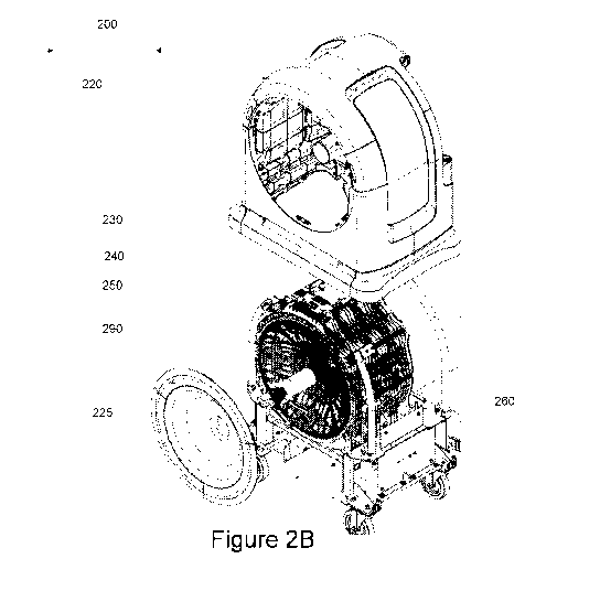
[0047] Figure 2A is a schematic illustration of a magnetic resonance
imaging system 200, in
accordance with various embodiments. Figure 2B illustrates an exploded view of
the magnetic
resonance imaging system 200. Figure 2C is a schematic front view of the
magnetic resonance
imaging system 200, in accordance with various embodiments. Figure 2D is a
schematic side
view of the magnetic resonance imaging system 200, in accordance with various
embodiments.
As shown in Figures 2A and 2B, the magnetic resonance imaging system 200
includes a housing
220. The housing 220 includes a front surface 225. In accordance with various
embodiments,
the front surface 225 can be a concave front surface. In accordance with
various embodiments,
the front surface 225 can be a recessed front surface.
[0048] As shown in Figures 2A and 2B, the housing 220 includes a permanent
magnet 230, a
radio frequency transmit coil 240, a gradient coil set 250, an optional
electromagnet 260, and a
radio frequency receive coil 270. As shown in Figures 2C and 2D, the permanent
magnet 230
can include a plurality of magnets disposed in an array configuration. The
plurality of magnets
of the permanent magnet 230 are illustrated to cover an entire surface as
shown in the front view
of Figure 2C and illustrated as bars in a horizontal direction as shown in the
side view of Figure
2D. As shown in Figure 2A, the main permanent magnet might include an access
aperture 235
for accessing the patient from multiple sides of the system.
[0049] It should be understood that any use of subheadings herein are for
organizational
purposes, and should not be read to limit the application of those subheaded
features to the
various embodiments herein. Each and every feature described herein is
applicable and usable in
all the various embodiments discussed herein and that all features described
herein can be used
in any contemplated combination, regardless of the specific example
embodiments that are
described herein. It should further be noted that exemplary description of
specific features are
used, largely for informational purposes, and not in any way to limit the
design, subfeature, and
functionality of the specifically described feature.
PERMANENT MAGNET
[0050] As discussed herein, and in accordance with various embodiments, the
various
systems, and various combinations of features that make up the various system
embodiments,
can include a permanent magnet.
[0051] In accordance with various embodiments, the permanent magnet 230
provides a static
magnetic field in a region of interest 290 (also referred to herein as "given
field of view"). In
accordance with various embodiments, the permanent magnet 230 can include a
plurality of
cylindrical permanent magnets in parallel configuration as shown in Figures 2C
and 2D. In
CA 03130759 2021-08-18
WO 2020/172673 PCT/US2020/019530
accordance with various embodiments, the permanent magnet 230 can include any
suitable
magnetic materials, including but not limited, to rare-earth based magnetic
materials, such as for
example, Nd-based magnetic materials, and the like. As shown in Figure 2A, the
main permanent
magnet might include an access aperture 235 for accessing the patient from
multiple sides of the
system.
[0052] In accordance with various embodiments, the static magnetic field of
the permanent
magnet 230 may vary from about 50 mT to about 60 mT, about 45 mT to about 65
mT, about 40
mT to about 70 mT, about 35 mT to about 75 mT, about 30 mT to about 80 mT,
about 25 mT to
about 85 mT, about 20 mT to about 90 mT, about 15 mT to about 95 mT and about
10 mT to
about 100 mT to a given field of view. The magnetic field may also vary from
about 10 mT to
about 15 mT, about 15 mT to about 20 mT, about 20 mT to about 25 mT, about 25
mT to about
30 mT, about 30 mT to about 35 mT, about 35 mT to about 40 mT, about 40 mT to
about 45 mT,
about 45 mT to about 50 mT, about 50 mT to about 55 mT, about 55 mT to about
60 mT, about
60 mT to about 65 mT, about 65 mT to about 70 mT, about 70 mT to about 75 mT,
about 75 mT
to about 80 mT, about 80 mT to about 85 mT, about 85 mT to about 90 mT, about
90 mT to
about 95 mT, and about 95 mT to about 100 mT. In accordance with various
embodiments, the
static magnetic field of the permanent magnet 230 may also vary from about 1
mT to about 1 T,
about 10 mT to about 195 mT, about 15 mT to about 900 mT, about 20 mT to about
800 mT,
about 25 mT to about 700 mT, about 30 mT to about 600 mT, about 35 mT to about
500 mT,
about 40 mT to about 400 mT, about 45 mT to about 300 mT, about 50 mT to about
200 mT,
about 50 mT to about 100 mT, about 45 mT to about 100 mT, about 40 mT to about
100 mT,
about 35 mT to about 100 mT, about 30 mT to about 100 mT, about 25 mT to about
100 mT,
about 20 mT to about 100 mT, and about 15 mT to about 100 mT.
[0053] In accordance with various embodiments, the permanent magnet 230 can
include a
bore 235 in its center. In accordance with various embodiments, the permanent
magnet 230 may
not include a bore. In accordance with various embodiments, the bore 235 can
have a diameter
between 1 inch and 20 inches. In accordance with various embodiments, the bore
235 can have a
diameter between 1 inch and 4 inches, between 4 inches and 8 inches, and
between 10 inches and
20 inches. In accordance with various embodiments, the given field of view can
be a spherical
or cylindrical field of view, as shown in Figures 2A and 2B. In accordance
with various
embodiments, the spherical field of view can be between 2 inches and 20 inches
in diameter. In
accordance with various embodiments, the spherical field of view can have a
diameter between 1
inch and 4 inches, between 4 inches and 8 inches, and between 10 inches and 20
inches. In
accordance with various embodiments, the cylindrical field of view is
approximately between 2
CA 03130759 2021-08-18
WO 2020/172673 PCT/US2020/019530
inches and 20 inches in length. In accordance with various embodiments, the
cylindrical field of
view can have a length between 1 inch and 4 inches, between 4 inches and 8
inches, and between
inches and 20 inches.
[0054] It should be understood that any use of subheadings herein are for
organizational
purposes, and should not be read to limit the application of those subheaded
features to the
various embodiments herein. Each and every feature described herein is
applicable and usable in
all the various embodiments discussed herein and that all features described
herein can be used
in any contemplated combination, regardless of the specific example
embodiments that are
described herein. It should further be noted that exemplary description of
specific features are
used, largely for informational purposes, and not in any way to limit the
design, subfeature, and
functionality of the specifically described feature.
RADIO FREQUENCY TRANSMIT COIL
[0055] As discussed herein, and in accordance with various embodiments, the
various
systems, and various combinations of features that make up the various system
embodiments,
can also include a radio frequency transmit coil.
[0056] Figure 3 is a schematic view of an implementation of a magnetic
imaging apparatus
300, according to various embodiments. As shown in Figure 3, the apparatus 300
includes a
radio frequency transmit coil 320 that projects the RF power outwards away
from the coil 320.
The coil 320 has two rings 322 and 324 that are connected by one or more rungs
326. As shown
in Figure 3, the coil 320 is also connected to a power source 350a and/or a
power source 350b
(collectively referred to herein as "power source 350"). In accordance with
various
embodiments, power sources 350a and 350b can be configured for power input
and/or signal
input, and can generally be referred to as coil input. In accordance with
various embodiments,
the power source 350a and/or 350b are configured to provide contact via
electrical contacts 352a
and/or 352b (collectively referred to herein as "electrical contact 352"), and
electrical contacts
354a and/or 354b (collectively referred to herein as "electrical contact
354b") by attaching the
electrical contacts 352 and 354 to one or more rungs 326. The coil 320 is
configured to project a
uniform RF field within a field of view 340. In accordance with various
embodiments, the field
of view 340 is a region of interest for magnetic resonance imaging (i.e.,
imaging region) where a
patient resides. Since the patient resides in the field of view 340 away from
the coil 320, the
apparatus 300 is suitable for use in a single-sided magnetic resonance imaging
system. In
accordance with various embodiments, the coil 320 can be powered by two
signals that are 90
degrees out of phase from each other, for example, via quadrature excitation.
12
CA 03130759 2021-08-18
WO 2020/172673 PCT/US2020/019530
[0057] In accordance with various embodiments, the coil 320 includes the
ring 322 and the
ring 324 that are positioned co-axially along the same axis but at a distance
away from each
other, as shown in Figure 3. In accordance with various embodiments, the ring
322 and the ring
324 are separated by a distance ranging from about 0.1 m to about 10 m. In
accordance with
various embodiments, the ring 322 and the ring 324 are separated by a distance
ranging from
about 0.2 m to about 5 m, about 0.3 m to about 2 m, about 0.2 m to about 1 m,
about 0.1 m to
about 0.8 m, or about 0.1 m to about 1 m, inclusive of any separation distance
therebetween. In
accordance with various embodiments, the coil 320 includes the ring 322 and
the ring 324 that
are positioned non-co-axially but along the same direction and separated at a
distance ranging
from about 0.2 m to about 5m. In accordance with various embodiments, the ring
322 and the
ring 324 can also be tilted with respect to each other. In accordance with
various embodiments,
the tilt angle can be from 1 degree to 90 degrees, from 1 degree to 5 degrees,
from 5 degrees to
degrees, from 10 degrees to 25 degrees, from 25 degrees to 45 degrees, and
from 45 degrees
to 90 degrees.
[0058] In accordance with various embodiments, the ring 322 and the ring
324 have the same
diameter. In accordance with various embodiments, the ring 322 and the ring
324 have different
diameters and the ring 322 has a larger diameter than the ring 324, as shown
in Figure 3. In
accordance with various embodiments, the ring 322 and the ring 324 have
different diameters
and the ring 322 has a smaller diameter than the ring 324. In accordance with
various
embodiments, the ring 322 and the ring 324 of the coil 320 are configured to
create the imaging
region in the field of view 340 containing a uniform RF power profile within
the field of view
340, a field of view that is not centered within the RF-TX coil and is instead
projected outwards
in space from the coil itself.
[0059] In accordance with various embodiments, the ring 322 has a diameter
between about
10 um and about 10 m. In accordance with various embodiments, the ring 322 has
a diameter
between about 0.001 m and about 9 m, between about 0.01 m and about 8 m,
between about 0.03
m and about 6 m, between about 0.05 m and about 5 m, between about 0.1 m and
about 3 m,
between about 0.2 m and about 2 m, between about 0.3 m and about 1.5 m,
between about 0.5 m
and about 1 m, or between about 0.01 m and about 3 m, inclusive of any
diameter therebetween.
[0060] In accordance with various embodiments, the ring 324 has a diameter
between about
10 um and about 10 m. In accordance with various embodiments, the ring 324 has
a diameter
between about 0.001 m and about 9 m, between about 0.01 m and about 8 m,
between about 0.03
m and about 6 m, between about 0.05 m and about 5 m, between about 0.1 m and
about 3 m,
13
CA 03130759 2021-08-18
WO 2020/172673 PCT/US2020/019530
between about 0.2 m and about 2 m, between about 0.3 m and about 1.5 m,
between about 0.5 m
and about 1 m, or between about 0.01 m and about 3 m, inclusive of any
diameter therebetween.
[0061] In accordance with various embodiments, the ring 322 and the ring
324 are connected
by one or more rungs 326, as shown in Figure 3. In accordance with various
embodiments, the
one or more rungs 326 are connected to the ring 322 and 324 so as to form a
single electrical
circuit loop (or single current loop). As shown in Figure 3, for example, one
end of the one or
more rungs 326 is connected to the electrical contact 352 of the power source
350 and another
end of the one or more rungs 326 be connected to the electrical contact 354 so
that the coil 320
completes an electrical circuit.
[0062] In accordance with various embodiments, the ring 322 is a
discontinuous ring and the
electrical contact 352 and the electrical contact 354 can be electrically
connected to two opposite
ends of the ring 322 to form an electrical circuit powered by the power source
350. Similarly, in
accordance with various embodiments, the ring 324 is a discontinuous ring and
the electrical
contact 352 and the electrical contact 354 can be electrically connected to
two opposite ends of
the ring 324 to form an electrical circuit powered by the power source 350.
[0063] In accordance with various embodiments, the rings 322 and 324 are
not circular and
can instead have a cross section that is elliptical, square, rectangular, or
trapezoidal, or any shape
or form having a closed loop. In accordance with various embodiments, the
rings 322 and 324
may have cross sections that vary in two different axial planes with the
primary axis being a
circle and the secondary axis having a sinusoidal shape or some other
geometric shape. In
accordance with various embodiments, the coil 320 may include more than two
rings 322 and
324, each connected by rungs that span and connect all the rings. In
accordance with various
embodiments, the coil 320 may include more than two rings 322 and 324, each
connected by
rungs that alternate connection points between rings. In accordance with
various embodiments,
the ring 322 may contain a physical aperture for access. In accordance with
various
embodiments, the ring 322 may be a solid sheet without a physical aperture.
[0064] In accordance with various embodiments, the coil 320 generates an
electromagnetic
field (also referred to herein as "magnetic field") strength between about 1
uT and about 10 mT.
In accordance with various embodiments, the coil 320 can generate a magnetic
field strength
between about 10 uT and about 5 mT, about 50 uT and about 1 mT, or about 100
uT and about 1
mT, inclusive of any magnetic field strength therebetween.
[0065] In accordance with various embodiments, the coil 320 generates an
electromagnetic
field that is pulsed at a radio frequency between about 1 kHz and about 2 GHz.
In accordance
with various embodiments, the coil 320 generates a magnetic field that is
pulsed at a radio
14
CA 03130759 2021-08-18
WO 2020/172673 PCT/US2020/019530
frequency between about 1 kHz and about 1 GHz, about 10 kHz and about 800 MHz,
about 50
kHz and about 300 MHz, about 100 kHz and about 100 MHz, about 10 kHz and about
10 MHz,
about 10 kHz and about 5 MHz, about 1 kHz and about 2 MHz, about 50 kHz and
about 150
kHz, about 80 kHz and about 120 kHz, about 800 kHz and about 1.2 MHz, about
100 kHz and
about 10 MHz, or about 1 MHz and about 5 MHz, inclusive of any frequencies
therebetween.
[0066] In accordance with various embodiments, the coil 320 is oriented to
partially surround
the region of interest. In accordance with various embodiments, the ring 322,
the ring 324, and
the one or more rungs 326 are non-planar to each other. Said another way, the
ring 322, the ring
324, and the one or more rungs 326 form a three-dimensional structure that
surrounds the region
of interest where a patient resides. In accordance with various embodiments,
the ring 322 is
closer to the region of interest than the ring 324, as shown in Figure 3. In
accordance with
various embodiments, the region of interest has a size of about 0.1 m to about
1 m. In
accordance with various embodiments, the region of interest is smaller than
the diameter of the
ring 322. In accordance with various embodiments, the region of interest is
smaller than both the
diameter of the ring 324 and the diameter of the ring 322, as shown in Figure
3. In accordance
with various embodiments, the region of interest has a size that is smaller
than the diameter of
the ring 322 and larger than the diameter of the ring 324.
[0067] In accordance with various embodiments, the ring 322, the ring 324,
or the rungs 326
include the same material. In accordance with various embodiments, the ring
322, the ring 324,
or the rungs 326 include different materials. In accordance with various
embodiments, the ring
322, the ring 324, or the rungs 326 include hollow tubes or solid tubes. In
accordance with
various embodiments, the hollow tubes or solid tubes can be configured for air
or fluid cooling.
In accordance with various embodiments, each of the ring 322 or the ring 324
or the rungs 326
includes one or more electrically conductive windings. In accordance with
various
embodiments, the windings include litz wires or any electrical conducting
wires. These
additional windings could be used to improve performance by lowering the
resistance of the
windings at the desired frequency. In accordance with various embodiments, the
ring 322, the
ring 324, or the rungs 326 include copper, aluminum, silver, silver paste, or
any high electrical
conducting material, including metal, alloys or superconducting metal, alloys
or non-metal. In
accordance with various embodiments, the ring 322, the ring 324, or the rungs
326 may include
metamaterials.
[0068] In accordance with various embodiments, the ring 322, the ring 324,
or the rungs 326
may contain separate electrically non-conductive thermal control channels
designed to maintain
the temperature of the structure to a specified setting. In accordance with
various embodiments,
CA 03130759 2021-08-18
WO 2020/172673 PCT/US2020/019530
the thermal control channels can be made from electrically conductive
materials and integrated
as to carry the electrical current.
[0069] In accordance with various embodiments, the coil 320 includes one or
more electronic
components for tuning the magnetic field. The one or more electronic
components can include a
varactor, a PIN diode, a capacitor, or a switch, including a micro-electro-
mechanical system
(MEMS) switch, a solid state relay, or a mechanical relay. In accordance with
various
embodiments, the coil can be configured to include any of the one or more
electronic
components along the electrical circuit. In accordance with various
embodiments, the one or
more components can include mu metals, dielectrics, magnetic, or metallic
components not
actively conducting electricity and can tune the coil. In accordance with
various embodiments,
the one or more electronic components used for tuning includes at least one of
dielectrics,
conductive metals, metamaterials, or magnetic metals. In accordance with
various embodiments,
tuning the electromagnetic field includes changing the current or by changing
physical locations
of the one or more electronic components. In accordance with various
embodiments, the coil is
cryogenically cooled to reduce resistance and improve efficiency. In
accordance with various
embodiments, the first ring and the second ring comprise a plurality of
windings or litz wires.
[0070] In accordance with various embodiments, the coil 320 is configured
for a magnetic
resonance imaging system that has a magnetic field gradient across the field
of view. The field
gradient allows for imaging slices of the field of view without using an
additional
electromagnetic gradient. As disclosed herein, the coil can be configured to
generate a large
bandwidth by combining multiple center frequencies, each with their own
bandwidth. By
superimposing these multiple center frequencies with their respective
bandwidths, the coil 320
can effectively generate a large bandwidth over a desired frequency range
between about 1 kHz
and about 2 GHz. In accordance with various embodiments, the coil 320
generates a magnetic
field that is pulsed at a radio frequency between about 10 kHz and about 800
MHz, about 50 kHz
and about 300 MHz, about 100 kHz and about 100 MHz, about 10 kHz and about 10
MHz, about
kHz and about 5 MHz, about 1 kHz and about 2 MHz, about 50 kHz and about 150
kHz,
about 80 kHz and about 120 kHz, about 800 kHz and about 1.2 MHz, about 100 kHz
and about
10 MHz, or about 1 MHz and about 5 MHz, inclusive of any frequencies
therebetween.
[0071] It should be understood that any use of subheadings herein are for
organizational
purposes, and should not be read to limit the application of those subheaded
features to the
various embodiments herein. Each and every feature described herein is
applicable and usable in
all the various embodiments discussed herein and that all features described
herein can be used
in any contemplated combination, regardless of the specific example
embodiments that are
16
CA 03130759 2021-08-18
WO 2020/172673 PCT/US2020/019530
described herein. It should further be noted that exemplary description of
specific features are
used, largely for informational purposes, and not in any way to limit the
design, subfeature, and
functionality of the specifically described feature.
GRADIENT COIL SET
[0072] As discussed herein, and in accordance with various embodiments, the
various
systems, and various combinations of features that make up the various system
embodiments,
can also include a gradient coil set.
[0073] Figure 4 is a schematic view of an implementation of a magnetic
imaging apparatus
400, according to various embodiments. As shown in Figure 4, the apparatus 400
includes a
gradient coil set 420 (also referred to herein as single-sided gradient coil
set 420) that is
configured to project a gradient magnetic field outwards away from the coil
set 420 and within a
field of view 430. In accordance with various embodiments, the field of view
430 is a region of
interest for magnetic resonance imaging (i.e., imaging region) where a patient
resides. Since the
patient resides in the field of view 430 away from the coil set 420, the
apparatus 400 is suitable
for use in a single-sided MRI system.
[0074] As shown in the figure, the coil set 420 includes variously sized
spiral coils in various
sets of spiral coils 440a, 440b, 440c, and 440d (collectively referred to as
"spiral coils 440").
Each set of the spiral coils 440 include at least one spiral coil and Figure 4
is shown to include 3
spiral coils. In accordance with various embodiments, each spiral coil in the
spiral coils 440 has
an electrical contact at its center and an electrical contact output on the
outer edge of the spiral
coil so as to form a single running loop of electrically conducting material
spiraling out from the
center to the outer edge, or vice versa. In accordance with various
embodiments, each spiral coil
in the spiral coils 440 has a first electrical contact at a first position of
the spiral coil and a second
electrical contact at a second position the spiral coil so as to form a single
running loop of
electrically conducting material from the first position to the second
position, or vice versa.
[0075] As shown in Figure 4, the coil set 420 also includes an aperture 425
at its center where
the spiral coils 440 are disposed around the aperture 425. The aperture 425
itself does not
contain any coil material within it for generating magnetic material. The coil
set 420 also
includes an opening 427 on the outer edge of the coil set 420 to which the
spiral coils 440 can be
disposed. Said another way, the aperture 425 and the opening 427 define the
boundaries of the
coil set 420 within which the spiral coils 440 can be disposed. In accordance
with various
embodiments, the coil set 420 forms a bowl shape with a hole in the center.
17
CA 03130759 2021-08-18
WO 2020/172673 PCT/US2020/019530
[0076] In accordance with various embodiments, the spiral coils 440 form
across the aperture
425. For example, the spiral coils 440a are disposed across from the spiral
coils 440c with
respect to the aperture 425. Similarly, the spiral coils 440b are disposed
across from the spiral
coils 440d with respect to the aperture 425. In accordance with various
embodiments, the spiral
coils 440 in the coil set 420 shown in Figure 4 are configured to create
spatial encoding in the
magnetic gradient field within the field of view 430.
[0077] As shown in Figure 4, the coil set 420 is also connected to a power
source 450 via
electrical contacts 452 and 454 by attaching the electrical contacts 452 and
454 to one or more of
the spiral coils 440. In accordance with various embodiments, the electrical
contact 452 is
connected to one of the spiral coils 440, which is then connected to other
spiral coils 440 in
series and/or in parallel, and one other spiral coil 440 is then connected to
the electrical contact
454 so as to form an electrical current loop. In accordance with various
embodiments, the spiral
coils 440 are all electrically connected in series. In accordance with various
embodiments, the
spiral coils 440 are all electrically connected in parallel. In accordance
with various
embodiments, some of the spiral coils 440 are electrically connected in series
while other spiral
coils 440 are electrically connected in parallel. In accordance with various
embodiments, the
spiral coils 440a are electrically connected in series while the spiral coils
440b are electrically
connected in parallel. In accordance with various embodiments, the spiral
coils 440c are
electrically connected in series while the spiral coils 440d are electrically
connected in parallel.
The electrical connections between each spiral coil in the spiral coils 440 or
each set of spiral
coils 440 can be configured as needed to generate the magnetic field in the
field of view 430.
[0078] In accordance with various embodiments, the coil set 420 includes
the spiral coils 440
spread out as shown in Figure 4. In accordance with various embodiments, each
of the sets of
spiral coils 440a, 440b, 440c, and 440d are configured in a line from the
aperture 425 to the
opening 427 so that each set of spiral coils is set apart from another by an
angle of 90 . In
accordance with various embodiments, 440a and 440b are set at 450 from one
another, and 440c
and 440d are set at 450 from one another, while 440c is set 135 on the other
side of 440b and
440d is set 135 on the other side of 440a. In essence, any of the sets of
spiral coils 440 can be
configured in any arrangement for any number "n" of sets of spiral coils 440.
[0079] In accordance with various embodiments, the spiral coils 440 have
the same diameter.
In accordance with various embodiments, each of the sets of spiral coils 440a,
440b, 440c, and
440d have the same diameter. In accordance with various embodiments, the
spiral coils 440
have different diameters. In accordance with various embodiments, each of the
sets of spiral
coils 440a, 440b, 440c, and 440d have different diameters. In accordance with
various
18
CA 03130759 2021-08-18
WO 2020/172673 PCT/US2020/019530
embodiments, the spiral coils in each of the sets of spiral coils 440a, 440b,
440c, and 440d have
different diameters. In accordance with various embodiments, 440a and 440b
have the same first
diameter and 440c and 440d have the same second diameter, but the first
diameter and the
second diameter are not the same.
[0080] In accordance with various embodiments, each spiral coil in the
spiral coils 440 has a
diameter between about 10 vm and about 10 m. In accordance with various
embodiments, each
spiral coil in the spiral coils 440 has a diameter between about 0.001 m and
about 9 m, between
about 0.005 m and about 8 m, between about 0.01 m and about 6 m, between about
0.05 m and
about 5 m, between about 0.1 m and about 3 m, between about 0.2 m and about 2
m, between
about 0.3 m and about 1.5 m, between about 0.5 m and about 1 m, or between
about 0.01 m and
about 3 m, inclusive of any diameter therebetween.
[0081] In accordance with various embodiments, the spiral coils 440 are
connected to form a
single electrical circuit loop (or single current loop). As shown in Figure 4,
for example, one
spiral coil in the spiral coils 440 is connected to the electrical contact 452
of the power source
450 and another spiral coil be connected to the electrical contact 454 so that
the spiral coils 440
completes an electrical circuit.
[0082] In accordance with various embodiments, the coil set 420 generates
an
electromagnetic field strength (also referred to herein as "electromagnetic
field gradient" or
"gradient magnetic field") between about 1 vT and about 10 T. In accordance
with various
embodiments, the coil set 420 can generate an electromagnetic field strength
between about 100
vT and about 1 T, about 1 mT and about 500 mT, or about 10 mT and about 100
mT, inclusive
of any magnetic field strength therebetween. In accordance with various
embodiments, the coil
set 420 can generate an electromagnetic field strength greater than about 1
vT, about 10 vT,
about 100 vT, about 1 mT, about 5 mT, about 10 mT, about 20 mT, about 50 mT,
about 100 mT,
or about 500 mT.
[0083] In accordance with various embodiments, the coil set 420 generates
an
electromagnetic field that is pulsed at a rate with a rise-time less than
about 100 vs. In
accordance with various embodiments, the coil set 420 generates an
electromagnetic field that is
pulsed at a rate with a rise-time less than about 1 vs, about 5 vs, about 10
vs, about 20 vs, about
30 vs, about 40 vs, about 50 vs, about 100 vs, about 200 vs, about 500 vs,
about 1 ms, about 2
ms, about 5 ms, or about 10 ms.
[0084] In accordance with various embodiments, the coil set 420 is oriented
to partially
surround the region of interest in the field of view 430. In accordance with
various
embodiments, the spiral coils 440 are non-planar to each other. In accordance
with various
19
CA 03130759 2021-08-18
WO 2020/172673 PCT/US2020/019530
embodiments, the sets of spiral coils 440a, 440b, 440c, and 440d are non-
planar to each other.
Said another way, the spiral coils 440 and each of the sets of spiral coils
440a, 440b, 440c, and
440d form a three-dimensional structure that surrounds the region of interest
in the field of view
430 where a patient resides.
[0085] In accordance with various embodiments, the spiral coils 440 include
the same
material. In accordance with various embodiments, the spiral coils 440 include
different
materials. In accordance with various embodiments, the spiral coils in set
440a include the same
first material, the spiral coils in set 440b include the same second material,
the spiral coils in set
440c include the same third material, the spiral coils in set 440d include the
same fourth material,
but the first, second, third and fourth materials are different materials. In
accordance with
various embodiments, the first and second materials are the same material, but
that same material
is different from the third and fourth materials, which are the same. In
essence, any of the spiral
coils 440 can be of the same material or different materials depending on the
configuration of the
coil set 420.
[0086] In accordance with various embodiments, the spiral coils 440 include
hollow tubes or
solid tubes. In accordance with various embodiments, the spiral coils 440
include one or more
windings. In accordance with various embodiments, the windings include litz
wires or any
electrical conducting wires. In accordance with various embodiments, the
spiral coils 440
include copper, aluminum, silver, silver paste, or any high electrical
conducting material,
including metal, alloys or superconducting metal, alloys or non-metal. In
accordance with
various embodiments, the spiral coils 440 include metamaterials.
[0087] In accordance with various embodiments, the coil set 420 includes
one or more
electronic components for tuning the magnetic field. The one or more
electronic components
can include a PIN diode, a mechanical relay, a solid state relay, or a switch,
including a micro-
electro-mechanical system (MEMS) switch. In accordance with various
embodiments, the coil
can be configured to include any of the one or more electronic components
along the electrical
circuit. In accordance with various embodiments, the one or more components
can include mu
metals, dielectrics, magnetic, or metallic components not actively conducting
electricity and can
tune the coil. In accordance with various embodiments, the one or more
electronic components
used for timing includes at least one of conductive metals, metamaterials, or
magnetic metals. In
accordance with various embodiments, tuning the electromagnetic field includes
changing the
current or by changing physical locations of the one or more electronic
components. In some
implementations, the coil is cryogenically cooled to reduce resistance and
improve efficiency.
CA 03130759 2021-08-18
WO 2020/172673 PCT/US2020/019530
[0088] It should be understood that any use of subheadings herein are for
organizational
purposes, and should not be read to limit the application of those subheaded
features to the
various embodiments herein. Each and every feature described herein is
applicable and usable in
all the various embodiments discussed herein and that all features described
herein can be used
in any contemplated combination, regardless of the specific example
embodiments that are
described herein. It should further be noted that exemplary description of
specific features are
used, largely for informational purposes, and not in any way to limit the
design, subfeature, and
functionality of the specifically described feature.
ELECTROMAGNET
[0089] As discussed herein, and in accordance with various embodiments, the
various
systems, and various combinations of features that make up the various system
embodiments,
can also include an electromagnet.
[0090] Figure 5 is a schematic front view of a magnetic resonance imaging
system 500,
according to various embodiments. In accordance with various embodiments, the
system 500
can be any magnetic resonance imaging system, including for example, a single-
sided magnetic
resonance imaging system that comprises a magnetic resonance imaging scanner
or a magnetic
resonance imaging spectrometer, as disclosed herein.
[0091] As shown in Figure 5, the system 500 includes a housing 520 that can
house various
components, including, for example but not limited to, magnets,
electromagnets, coils for
producing radio frequency fields, various electronic components, for example
but not limited to,
for controlling, powering, and/or monitoring of the system 500. In accordance
with various
embodiments, the housing 520 can house, for example, the permanent magnet 230,
the radio
frequency transmit coil 240, and/or the gradient coil set 250 within the
housing 520. In
accordance with various embodiments, the system 500 also includes a bore 535
in its center. As
shown in Figure 5, the housing 520 also includes a front surface 525 of the
system 500. In
accordance with various embodiments, the front surface 525 can be curved,
flat, concave,
convex, or otherwise have a straight or curvilinear surface. In accordance
with various
embodiments, the magnetic resonance imaging system 500 can be configured to
provide a region
of interest in field of view 530.
[0092] As shown in Figure 5, the system 500 includes an electromagnet 560
disposed
proximate to the front surface 525 of the system 500. In accordance with
various embodiments,
the electromagnet 560 is disposed proximate to the center of the front surface
525 on the front
21
CA 03130759 2021-08-18
WO 2020/172673 PCT/US2020/019530
side of the system 500. In accordance with various embodiments, the
electromagnet 560 can be
a solenoid coil configured to create a field that either adds or subtracts
from the magnetic field,
for example, of the permanent magnet 230. In accordance with various
embodiments, this field
can create a prepolarizing field for enhancing the signal or contrast from the
nuclear magnetic
resonance.
[0093] As shown in Figure 5, the given field of view 530 resides at the
center of the front
surface 525 of the system 500. In accordance with various embodiments, the
electromagnet 560
is disposed within the given field of view 530. In accordance with various
embodiments, the
electromagnet 560 is disposed concentrically with the given field of view 530.
In accordance
with various embodiments, the electromagnet 560 can be inserted in the bore
535. In accordance
with various embodiments, the electromagnet 560 can be placed proximate to the
bore 535. For
example, the electromagnet 560 can be placed in front, back or middle of the
bore 535. In
accordance with various embodiments, the electromagnet 560 can be placed
proximate to, or at
the entrance of the bore 535.
[0094] It should be understood that any use of subheadings herein are for
organizational
purposes, and should not be read to limit the application of those subheaded
features to the
various embodiments herein. Each and every feature described herein is
applicable and usable in
all the various embodiments discussed herein and that all features described
herein can be used
in any contemplated combination, regardless of the specific example
embodiments that are
described herein. It should further be noted that exemplary description of
specific features are
used, largely for informational purposes, and not in any way to limit the
design, subfeature, and
functionality of the specifically described feature.
RADIO FREQUENCY RECEIVE COIL
[0095] As discussed herein, and in accordance with various embodiments, the
various
systems, and various combinations of features that make up the various system
embodiments,
can also include a radio frequency receive coil.
[0096] Typical MR systems create a uniform field within the imaging region.
This uniform
field then generates a narrow band of magnetic resonance frequencies that can
then be captured
by a receive coil, amplified, and digitized by a spectrometer. Since
frequencies are within a
narrow well-defined bandwidth, hardware architecture is focused on creating a
statically tuned
RF-RX coil with an optimal coil quality factor. Many variations in coil
architectures have been
created that explore large single volume coils, coil arrays, parallelized coil
arrays, or body
22
CA 03130759 2021-08-18
WO 2020/172673 PCT/US2020/019530
specific coil arrays. However, these structures are all predicated on imaging
a specific frequency
close to the region of interest at high field strengths and small as possible
within a magnetic bore.
[0097] In accordance with various embodiments, an MRI system is provided
that can include
a unique imaging region that can be offset from the face of a magnet and
therefore unobstructed
as compared to traditional scanners. In addition, this form factor can have a
built-in magnetic
field gradient that creates a range of field values over the region of
interest. Lastly, this system
can operate at a lower magnetic field strength as compared to typical MRI
systems allowing for a
relaxation on the RX coil design constraints and allowing for additional
mechanisms like
robotics to be used with the MRI.
[0098] The unique architecture of the main magnetic field of the MRI
system, in accordance
with various embodiments, can create a different set of optimization
constraints. Because the
imaging volume now extends over a broader range of magnetic resonance
frequencies, the
hardware can be configured to be sensitive to and capture the specific
frequencies that are
generated across the field of view. This frequency spread is usually much
larger than a single
receive coil tuned to a single frequency can be sensitive to. In addition,
because the field strength
can be much lower than traditional systems, and because signal intensity can
be proportional to
the field strength, it is generally considered to be beneficial to maximize
the signal to noise ratio
of the receive coil network. Methods are therefore provided, in accordance
with various
embodiments, to acquire the full range of frequencies that are generated
within the field of view
without loss of sensitivity.
[0099] In accordance with various embodiments, several methods are provided
that can
enable imaging within the MRI system. These methods can include combining 1) a
variable
tuned RF-RX coil; 2) a RF-RX coil array with elements tuned to frequencies
that are dependent
upon the spatial inhomogeneity of the magnetic field; 3) a ultralow-noise pre-
amplifier design;
and 4) an RF-RX array with multiple receive coils designed to optimize the
signal from a defined
and limited field of view for a specific body part. These methods can be
combined in any
combination as needed.
[0100] In accordance with various embodiments, a variable tuned RF-RX coil
can comprise
one or more electronic components for tuning the electromagnetic receive
field. In accordance
with various embodiments, the one or more electronic components can include at
least one of a
varactor, a PIN diode, a capacitor, an inductor, a MEMS switch, a solid state
relay, or a
mechanical relay. In accordance with various embodiments, the one or more
electronic
components used for tuning can include at least one of dielectrics,
capacitors, inductors,
conductive metals, metamaterials, or magnetic metals. In accordance with
various embodiments,
23
CA 03130759 2021-08-18
WO 2020/172673 PCT/US2020/019530
tuning the electromagnetic receive field includes changing the current or by
changing physical
locations of the one or more electronic components. In accordance with various
embodiments,
the coil is cryogenically cooled to reduce resistance and improve efficiency.
[0101] In accordance with various embodiments, the RF-RX array can be
comprised of
individual coil elements that are each tuned to a variety of frequencies. The
appropriate
frequency can be chosen, for example, to match the frequency of the magnetic
field located at the
specific spatial location where the specific coil is located. Because the
magnetic field can vary as
a function of space, as shown in Figure 6A, the field and frequency of the
coil can be adjusted to
approximately match the spatial location. Here the coils can be designed to
image the field
locations Bl, B2, and B3, which are physically separated along a single axis.
[0102] For this low field system, in accordance with various embodiments, a
low-noise
preamplifier can be designed and configured to leverage the low signal
environment of the MRI
system. This low noise amplifier can be configured to utilize components that
do not generate
significant electronic and voltage noise at the desired frequencies (for
example, <3 MHz and >2
MHz). Typical junction field effect transistor designs (J-FET) generally do
not have the
appropriate noise characteristics at this frequency and can create high
frequency instabilities at
the GHz range that can bleed into, although several decades of dB lower, into
the measured
frequency range. Since the gain of the system can preferably be, for example,
> 80 dB overall,
any small instabilities or intrinsic electrical noise can be amplified and
degrade signal integrity.
[0103] Referring to Figure 6B, RF-RX coils can be designed to image
specific limited field of
views based upon the target anatomy. The prostate, for example, is about 60
millimeters deep
within the human body (see Figure 6D), so to design a RX coil for prostate
imaging, the coil
should be configured to enable imaging 60 mm deep inside human body. According
to Biot-
Savart law, the magnetic field of a loop coil can be calculated by the
following equation,
P-o * R2 * I
Bz = - * 3
z2
where it0 = 4-rr * 10-7H/m is the vacuum permeability, R is the radius of the
loop coil, z is
distance along the center line of the coil from its center, and I is the
current on the coil (see
Figure 6B). Assuming I = 1 Ampere, with the goal of locating a figure of
magnetic field (Bz) at z
= 60 mm, the maximum position is when R is 85 mm according to the chart shown
in Figure 6C.
[0104] Based upon the geometrical constraints of the body, the loop coil
can be set up at the
space between the human legs upon the torso. As such, it is extremely
difficult, if not
impossible, to fit a 170-mm diameter coil there. According to Figure 6C, the
Bz field value is
24
CA 03130759 2021-08-18
WO 2020/172673 PCT/US2020/019530
proportional to the radius of the loop when R is less than 85 mm. As such, it
is advantageous
that the coil be as large as it can be. For example, the largest loop coil
that can be placed between
people is about 10 mm large.
[0105] As the size of the coil is limited by the space between legs, the
magnetic field of a 10-
mm diameter coil is generally not capable of reaching the depth of prostate.
Therefore, single
coil may not be enough for prostate imaging thus, in this case, multiple coils
could prove
beneficial in getting signal from different directions. In various embodiments
of the MRI system,
the magnetic field is provided in the z-direction and RF coils are sensitive
to x- and y-direction.
In this example case, a loop coil in x-y plane would not collect RF signal
from a human since it
is sensitive to z-direction, while a butterfly coil can be used in this case.
Then based on the
location and orientation, RF coil could be a loop coil or butterfly coil. In
addition, a coil can be
placed in under the body and there is no limitation for its size.
[0106] As for the needs of multiple RX coils, in various embodiments,
decoupling between
them can prove beneficial for various embodiments of an MRI system RX coil
array. In those
cases, each coil can be de-coupled with the other coils, and the decoupling
techniques can
include, for example, 1) geometry decoupling, 2) capacitive/inductive
decoupling, and 3) low-
/high impedance pre-amplifier coupling.
[0107] The MRI system, in accordance with various embodiments, can have a
variant
magnetic field from the magnet, and its strength can vary linearly along the z
direction. The RX
coils can be located in different positions in z-direction, and each coil can
be tuned to different
frequencies, which can depend on the location of the coils in the system.
[0108] Based upon the simplicity of single coil loops, these coils can be
constructed from
simple conductive traces that can be pre-tuned to a desired frequency and
printed, for example,
on a disposable substrate. This cheaply fabricated technology can allow a
clinician to place the
RX coil (or coil array) upon the body at the region of interest for a given
procedure and dispose
of the coil afterwards. For example, and in accordance with various
embodiments, the RX coils
can be surface coils, which can be affixed to, e.g., worn or taped to, a
patient's body. For other
body parts, e.g. an ankle or a wrist, the surface coil might be a single-loop
configuration, figure-8
configuration, or butterfly coil configuration wrapped around the region of
interest. For regions
that require significant penetration depth, e.g. the torso or knee, the coil
might consist of a
Helmholtz coil pair. The main restriction to the receive coil is similar to
other MRI systems: the
coil must be sensitive to a plane that is orthogonal to the main magnetic
field, BO, axis.
CA 03130759 2021-08-18
WO 2020/172673 PCT/US2020/019530
[0109] In accordance with various embodiments, the coils might be
inductively coupled to
another loop that is electrically connected to the receive preamplifier. This
design would allow
for easier and unobstructed access of the receive coils.
[0110] In accordance with various embodiments, the size of coils can be
limited by the
structure of human body. For example, the coils' size should be positioned and
configured to fit
in the space between human legs when imaging the prostate.
[0111] It should be understood that any use of subheadings herein are for
organizational
purposes, and should not be read to limit the application of those subheaded
features to the
various embodiments herein. Each and every feature described herein is
applicable and usable in
all the various embodiments discussed herein and that all features described
herein can be used
in any contemplated combination, regardless of the specific example
embodiments that are
described herein. It should further be noted that exemplary description of
specific features are
used, largely for informational purposes, and not in any way to limit the
design, subfeature, and
functionality of the specifically described feature.
PROGRAMMABLE LOGIC CONTROLLER
[0112] As discussed herein, and in accordance with various embodiments, the
various
systems, and various combinations of features that make up the various system
embodiments,
can also include a programmable logic controller (PLC). PLCs are industrial
digital computers
which can be designed to operate reliably in harsh usage environments and
conditions. PLCs can
be designed to handle these types of conditions and environments, not just in
the external
housing, but in the internal components and cooling arrangements as well. As
such, PLCs can be
adapted for the control of manufacturing processes, such as assembly lines, or
robotic devices, or
any activity that requires high reliability control and ease of programming
and process fault
diagnosis.
[0113] In accordance with various embodiments, the system can contain a PLC
that can
control the system in pseudo real-time. This controller can manage the power
cycling and
enabling of the gradient amplifier system, the radio frequency transmission
system, the
frequency tuning system, and sends a keep alive signal (e.g., a message sent
by one device to
another to check that the link between the two is operating, or to prevent the
link from being
broken) to the system watchdog. The system watchdog can continually look for a
strobe signal
supplied by the computer system. If the computer threads stall, a strobe is
missed that can trigger
26
CA 03130759 2021-08-18
WO 2020/172673 PCT/US2020/019530
the watchdog to enter a fault condition. If the watchdog enters a fault
condition, the watchdog
can operated to depower the system.
[0114] The PLC can generally handle low level logic functions on incoming
and outgoing
signals into system. This system can monitor the subsystem health and control
when subsystems
needed to be powered or enabled. The PLC can be designed in different ways.
One design
example includes a PLC with one main motherboard with four expansion boards.
Due to the
speed of the microcontroller on the PLC, subsystems can be managed in pseudo
real-time, while
real-time applications can be handled by the computer or spectrometer on the
system.
[0115] The PLC can serve many functional responsibilities including, for
example, powering
on/off the gradient amplifiers (discussed in greater detail herein) and the RF
amplifier (discussed
in greater detail herein), enabling/disabling the gradient amplifiers and the
RF amplifier, setting
the digital and analog voltages for the RF coil tuning, and strobing the
system watchdog.
[0116] As discussed above, it should be understood that any use of
subheadings herein are for
organizational purposes, and should not be read to limit the application of
those subheaded
features to the various embodiments herein. Each and every feature described
herein is
applicable and usable in all the various embodiments discussed herein and that
all features
described herein can be used in any contemplated combination, regardless of
the specific
example embodiments that are described herein. It should further be noted that
exemplary
description of specific features are used, largely for informational purposes,
and not in any way
to limit the design, subfeature, and functionality of the specifically
described feature.
ROBOT
[0117] As discussed herein, and in accordance with various embodiments, the
various
systems, and various combinations of features that make up the various system
embodiments,
can also include a robot.
[0118] In some medical procedures, such as a prostate biopsy, it is typical
for the patient to
endure a lengthy procedure in an uncomfortable prone position, which often
includes remaining
motionless in one specific body position during the entire procedure. In such
long procedures, if
a metallic ferromagnetic needle is used for the biopsy with guidance from an
MRI system, the
needle may experience attraction force from the strong magnets of the MRI
system, and thus
may cause it to deviate from its path during the length of the procedure. Even
in the case of
using a non-magnetic needle, the local field distortions can cause distortions
in the magnetic
resonance images, and therefore, the image quality surrounding the needle may
result in a poor
quality. To avoid such distortions, pneumatic robots with complex compressed
air mechanism
27
CA 03130759 2021-08-18
WO 2020/172673 PCT/US2020/019530
have been designed to work in conjunction with conventional MRI systems. Even
then, access
to target anatomy remains challenging due to the form factor of currently
available MRI systems.
[0119] The various embodiments presented herein include improved MRI
systems that are
configured to use for guiding in medical procedures, including, for example,
robot-assisted,
invasive medical procedures. The technologies, methods and apparatuses
disclosed herein relate
to a guided robotic system using magnetic resonance imaging as a guidance to
automatically
guide a robot (generally referred to herein as "a robotic system") in medical
procedures. In
accordance with various embodiments, the disclosed technologies combine a
robotic system with
magnetic resonance imaging as guidance. In accordance with various
embodiments, the robotic
system disclosed herein is combined with other suitable imaging techniques,
for example,
ultrasound, x-ray, laser, or any other suitable diagnostic or imaging
methodologies.
[0120] It should be understood that any use of subheadings herein are for
organizational
purposes, and should not be read to limit the application of those subheaded
features to the
various embodiments herein. Each and every feature described herein is
applicable and usable in
all the various embodiments discussed herein and that all features described
herein can be used
in any contemplated combination, regardless of the specific example
embodiments that are
described herein. It should further be noted that exemplary description of
specific features are
used, largely for informational purposes, and not in any way to limit the
design, subfeature, and
functionality of the specifically described feature.
SPECTROMETER
[0121] As discussed herein, and in accordance with various embodiments, the
various
systems, and various combinations of features that make up the various system
embodiments,
can also include a spectrometer.
[0122] A spectrometer can operate to control all real-time signaling used
to generate images.
It creates the RF transmission (RF-TX) waveform, gradient waveforms, frequency
tuning trigger
waveform, and blanking bit waveforms. These waveforms are then synchronized
with the RF
receiver (RF-RX) signals. This system can generate frequency swept RF-TX
pulses and phase
cycled RF-TX pulses. The swept RF-TX pulses allow for an inhomogeneous B1+
field (RF-TX
field) to excite a sample volume more effectively and efficiently. It can also
digitize multiple RF-
RX channels with the current configuration set to four receiver channels.
However, this system
architecture allows for an easy system scale-up to increase the number of
transmit and receive
channels to a maximum of 32 transmit channels and 16 receive channels without
having to
change the underlying hardware or software architecture.
28
CA 03130759 2021-08-18
WO 2020/172673 PCT/US2020/019530
[0123] The spectrometer can serve many functional responsibilities
including, for example,
generating and synchronizing the RF-TX (discussed in greater detail herein)
waveforms, X-
gradient waveforms, Y-gradient waveforms, blanking bit waveforms, frequency
tuning trigger
waveform and RF-RX windows, and digitizing and signal processing the RF-RX
data using, for
example, quadrature demodulation followed by a finite impulse response filter
decimation such
as, for example, a cascade integrating comb (CIC) filter decimation.
[0124] The spectrometer can be designed in different ways. One design
example includes a
spectrometer with three main components: 1) a first software design radio (SDR
1) operating
with Basic RF-TX daughter cards and Basic RF-RX daughter cards; 2) a second
software design
radio (SDR 2) operating with LFRF TX daughter cards and Basic RF-RX daughter
cards; and 3)
a clock distribution module (octoclock) that can synchronize the two devices.
[0125] SDRs are the real-time communication device between the transmitted
signals and
received MRI signals. They can communicate over 10Gbit optical fiber to the
computer using a
Small Form-factor Pluggable Plus transceiver (SFP+) communication protocol.
This
communication speed can allows the waveforms to be generated with high
fidelity and high
reliability.
[0126] Each SDR can include a motherboard with an integrated field-
programmable gate
array (FPGA), digital to analog converters, analog to digital converters, and
four module slots for
integrating different daughtercards. Each of these daughtercards can function
to change the
frequency response of the associated TX or RX channel. In accordance with
various
embodiments, the system can utilizes many variations daughtercards including,
for example, a
Basic RF version, and a low frequency (LF) RF version. The Basic RF
daughtercards can be
used for generating and measuring RF signals. The LF RF version can be used
for generating
gradient, trigger and blanking bit signals.
[0127] The octoclock can be used to synchronize a multi-channel SDR system to
a common
timing source while providing high-accuracy time and frequency reference
distribution. It can do
so, for example, with 8-way time and frequency distribution (1 PPS and 10MHz).
An example
of an octoclock is the Ettus Octoclock CDA, which can distribute a common
clock to up to eight
SDRs to ensure phase coherency between the two or more SDR sources.
[0128] It should be understood that any use of subheadings herein are for
organizational
purposes, and should not be read to limit the application of those subheaded
features to the
various embodiments herein. Each and every feature described herein is
applicable and usable in
all the various embodiments discussed herein and that all features described
herein can be used
in any contemplated combination, regardless of the specific example
embodiments that are
29
CA 03130759 2021-08-18
WO 2020/172673 PCT/US2020/019530
described herein. It should further be noted that exemplary description of
specific features are
used, largely for informational purposes, and not in any way to limit the
design, subfeature, and
functionality of the specifically described feature.
RF AMP/GRADIENT AMP
[0129] As discussed herein, and in accordance with various embodiments, the
various
systems, and various combinations of features that make up the various system
embodiments,
can also include a radio frequency amplifier (RF amplifier) and a gradient
amplifier.
[0130] A RF amplifier is a type of electronic amplifier that can converts a
low-power radio-
frequency signal into a higher power signal. In operation, the RF amplifier
can accept signals at
low amplitudes and provide, for example, up to 60 dB of gain with a flat
frequency response.
This amplifier can accept three phase AC input voltage and can have a 10% max
duty cycle. The
amplifier can be gated by a 5V digital signal so that unwanted noise is not
generated when the
MRI is receiving signal.
[0131] In operation, a gradient amplifier can increase the energy of the
signal before it
reaches the gradient coils such that the field strength can be intense enough
to produce the
variations in the main magnetic field for localization of the later received
signal. The gradient
amplifier can have two active amplification channels that can be controlled
independently. Each
channel can send out current to either the X or Y channel respectively. The
third axis of spatial
encoding is generally handled by a permanent gradient in the main magnetic
field (BO). With
varying combinations of pulse sequences, the signal can be localized in three
dimensions and
reconstructed to create an object.
[0132] It should be understood that any use of subheadings herein are for
organizational
purposes, and should not be read to limit the application of those subheaded
features to the
various embodiments herein. Each and every feature described herein is
applicable and usable in
all the various embodiments discussed herein and that all features described
herein can be used
in any contemplated combination, regardless of the specific example
embodiments that are
described herein. It should further be noted that exemplary description of
specific features are
used, largely for informational purposes, and not in any way to limit the
design, subfeature, and
functionality of the specifically described feature.
DISPLAY/GUI
[0133] As discussed herein, and in accordance with various embodiments, the
various
systems, and various combinations of features that make up the various system
embodiments,
CA 03130759 2021-08-18
WO 2020/172673 PCT/US2020/019530
can also include a display in the form of, for example, a graphical user
interface (GUI). In
accordance with various embodiments, the GUI can take any contemplated form
necessary to
convey the information necessary to run magnetic resonance imaging procedures.
[0134] Further, it should be appreciated that the display may be embodied
in any of a number
of other forms, such as, for example, a rack-mounted computer, mainframe,
supercomputer,
server, client, a desktop computer, a laptop computer, a tablet computer, hand-
held computing
device (e.g., PDA, cell phone, smart phone, palmtop, etc.), cluster grid,
netbook, embedded
systems, or any other type of special or general purpose display device as may
be desirable or
appropriate for a given application or environment.
[0135] The GUI is a system of interactive visual components for computer
software. A GUI
can display objects that convey information, and represent actions that can be
taken by the user.
The objects change color, size, or visibility when the user interacts with
them. GUI objects
include, for example, icons, cursors, and buttons. These graphical elements
are sometimes
enhanced with sounds, or visual effects like transparency and drop shadows.
[0136] A user can interact with a GUI using an input device, which can
include, for example,
alphanumeric and other keys, mouse, a trackball or cursor direction keys for
communicating
direction information and command selections to a processor and for
controlling cursor
movement on the display. An input device may also be the display configured
with touchscreen
input capabilities. This input device typically has two degrees of freedom in
two axes, a first axis
(i.e., x) and a second axis (i.e., y), that allows the device to specify
positions in a plane.
However, it should be understood that input devices allowing for 3 dimensional
(x, y and z)
cursor movement are also contemplated herein.
[0137] In accordance with various embodiments, the touchscreen, or
touchscreen monitor,
can serves as the primary human interface device that allows a user to
interact with the MRI. The
screen can have a projected capacitive touch sensitive display with an
interactive virtual
keyboard. The touchscreen can have several functions including, for example,
displaying the
graphical user interface (GUI) to the user, relaying user input to the
system's computer, and
starting or stopping a scan.
[0138] In accordance with various embodiments, GUI views can be typically
screens
displayed (Qt widgets) to the user with appropriate buttons, edit fields,
labels, images, etc. These
screens can be constructed using a designer tool such as, for example, the Qt
designer tool, to
control placement of widgets, their alignment, fonts, colors, etc. A user
interface (UI) sub
controller can possess modules configured to control the behavior (display and
responses) of the
respective view modules.
31
CA 03130759 2021-08-18
WO 2020/172673 PCT/US2020/019530
[0139] Several application utilities (App Util) modules can performs
specific functions. For
example, S3 modules can handle data communication between the system and, for
example,
Amazon Web Services (AWS). Event Filters can be present to ensure valid
characters are
displayed on screen when user inputs are required. Dialog messages can be used
to show various
status, progress messages or require user prompts. Moreover, a system
controller module can be
utilized to handle coordination between the sub controller modules, and key
data processing
blocks in the system, the pulse sequence generator, pulse interpreter,
spectrometer and
reconstruction.
[0140] It should be understood that any use of subheadings herein are for
organizational
purposes, and should not be read to limit the application of those subheaded
features to the
various embodiments herein. Each and every feature described herein is
applicable and usable in
all the various embodiments discussed herein and that all features described
herein can be used
in any contemplated combination, regardless of the specific example
embodiments that are
described herein. It should further be noted that exemplary description of
specific features are
used, largely for informational purposes, and not in any way to limit the
design, subfeature, and
functionality of the specifically described feature.
PROCESSING MODULE
[0141] As discussed herein, and in accordance with various embodiments, the
various
workflows or methods, and various combinations of steps that make up the
various workflow or
method embodiments, can also include a processing module.
[0142] In accordance with various embodiments, a processing module serves
many
functions. For example, a processing module can generally operate to receive
signal data
acquired during the scan, process the data, and reconstruct those signals to
produce an image that
can be viewed (for example, via a touchscreen monitor that displays a GUI to
the user), analyzed
and annotated by system users. Generally, to create an image, an NMR signal
must be localized
in three-dimensional space. Magnetic gradient coils localize the signal and
are operated before or
during the RF acquisition. By prescribing a RF and gradient coil application
sequence, called a
pulse sequence, the signals acquired correspond to a specific magnetic field
and RF field
arrangement. Using mathematical operators and image reconstruction techniques,
arrays of these
acquired signals can be reconstructed into an image. Usually these images are
generated from
simple linear combinations of magnetic field gradients. In accordance with
various
embodiments, the system can operate to reconstruct the acquired signals from a-
priori knowledge
of, for example, the gradient fields, RF fields, and pulse sequences.
32
CA 03130759 2021-08-18
WO 2020/172673 PCT/US2020/019530
[0143] In accordance with various embodiments, the processing module can
also operate to
compensate for patient motion during a scan procedure. Motion (e.g., beating
heart, breathing
lungs, bulk patient movement) is one of the most common sources of artifacts
in MRI, with such
artifacts affecting image quality by leading to misinterpretations in the
images and a subsequent
loss in diagnostic quality. Therefore, motion compensation protocols can help
address these
issues at minimal cost in time, spatial resolution, temporal resolution, and
signal-to-noise ratio.
[0144] In accordance with various embodiments, the processing module might
include
artificial intelligence machine learning modules designed to denoise the
signal and improve the
image signal-to-noise ratio.
[0145] In accordance with various embodiments, the processing module can
also operate to
assist clinicians in planning a path for subsequent patient intervention
procedures, such as
biopsy. In accordance with various embodiments, a robot can be provided as
part of the system
to perform the intervention procedure. The processing module can communicate
instructions to
the robot, based on image analysis, to properly access, for example, the
appropriate region of the
body requiring a biopsy.
[0146] It should be understood that any use of subheadings herein are for
organizational
purposes, and should not be read to limit the application of those subheaded
features to the
various embodiments herein. Each and every feature described herein is
applicable and usable in
all the various embodiments discussed herein and that all features described
herein can be used
in any contemplated combination, regardless of the specific example
embodiments that are
described below. It should further be noted that exemplary description of
specific features are
used, largely for informational purposes, and not in any way to limit the
design, subfeature, and
functionality of the specifically described feature.
[0147] In accordance with various embodiments, the various systems, and
various
combinations of features that make up the various system components and
embodiments of the
disclosed magnetic resonance imaging system are disclosed herein.
[0148] Figure 7 is a flowchart for a method S100 of performing magnetic
resonance imaging,
according to various embodiments. In accordance with various embodiments, the
method S100
includes inputting patient parameters into a magnetic resonance imaging system
at step S110. In
accordance with various embodiments, the system includes a housing having a
front surface, a
permanent magnet for providing a static magnetic field, a radio frequency
transmit coil, and a
single-sided gradient coil set. In accordance with various embodiments, the
radio frequency
transmit coil and the single-sided gradient coil set are positioned proximate
to the front surface.
In accordance with various embodiments, the system includes an electromagnet,
a radio
33
CA 03130759 2021-08-18
WO 2020/172673 PCT/US2020/019530
frequency receive coil, and a power source. In accordance with various
embodiments, the power
source is configured to flow current through at least one of the radio
frequency transmit coil, the
single-sided gradient coil set, or the electromagnet to generate an
electromagnetic field in a
region of interest. In accordance with various embodiments, the region of
interest resides outside
the front surface.
[0149] As shown in Figure 7, the method S100 also includes executing a
patient positioning
protocol comprising running at least one first scan at step S120, running at
least one second scan
at step S130, reviewing the at least one second scan at step S140, and
determining at least one
path for conducting a biopsy based on review of the at least one second scan
at step S150.
[0150] In accordance with various embodiments, the radio frequency transmit
coil and the
single-sided gradient coil set are located on the front surface. In accordance
with various
embodiments, the front surface is a concave surface. In accordance with
various embodiments,
the permanent magnet has an aperture through center of the permanent magnet.
In accordance
with various embodiments, the static magnetic field of the permanent magnet
ranges from 1 mT
to 1 T. In accordance with various embodiments, the static magnetic field of
the permanent
magnet ranges from 10 mT to 195 mT.
[0151] In accordance with various embodiments, the radio frequency transmit
coil includes a
first ring and a second ring that are connected via one or more capacitors
and/or one or more
rungs. In accordance with various embodiments, the radio frequency transmit
coil is non-planar
and oriented to partially surround the region of interest. In accordance with
various
embodiments, the single-sided gradient coil set is non-planar and oriented to
partially surround
the region of interest. In accordance with various embodiments, the single-
sided gradient coil set
is configured to project a magnetic field gradient to the region of interest.
In accordance with
various embodiments, the single-sided gradient coil set includes one or more
first spiral coils at a
first position and one or more second spiral coils at a second position, the
first position and the
second position being located opposite each other about a center region of the
single-sided
gradient coil set. In accordance with various embodiments, the single-sided
gradient coil set has
a rise time less than 10 us.
[0152] In accordance with various embodiments, the electromagnet is
configured to alter the
static magnetic field of the permanent magnet within the region of interest.
In accordance with
various embodiments, the electromagnet has a magnetic field strength from 10
mT to 1 T. In
accordance with various embodiments, the radio frequency receive coil is a
flexible coil
configured to be affixed to an anatomical portion of a patient for imaging
within the region of
interest. In accordance with various embodiments, the radio frequency receive
coil is in one of a
34
CA 03130759 2021-08-18
WO 2020/172673 PCT/US2020/019530
single-loop coil configuration, figure-8 coil configuration, or butterfly coil
configuration,
wherein the coil is smaller than the region of interest. In accordance with
various embodiments,
the radio frequency transmit coil and the single-sided gradient coil set are
concentric about the
region of interest. In accordance with various embodiments, the magnetic
resonance imaging
system is a single-sided magnetic resonance imaging system that comprises a
bore having an
opening positioned about a center region of the front surface.
[0153] Figure 8 is a flowchart for a method S200 of performing magnetic
resonance imaging,
according to various embodiments. In accordance with various embodiments, the
method S200
includes inputting patient parameters into a magnetic resonance imaging system
at step S210. In
accordance with various embodiments, the system includes a housing having a
concave front
surface, a permanent magnet for providing a static magnetic field, a radio
frequency transmit
coil, and at least one gradient coil set. In accordance with various
embodiments, the radio
frequency transmit coil and the at least one gradient coil set are positioned
proximate to the
concave front surface. In accordance with various embodiments, the radio
frequency transmit
coil and the at least one gradient coil set are configured to generate an
electromagnetic field in a
region of interest. In accordance with various embodiments, the region of
interest resides outside
the concave front surface. In accordance with various embodiments, the system
includes a radio
frequency receive coil for detecting signal in the region of interest.
[0154] As shown in Figure 8, the method S200 includes executing a patient
positioning
protocol comprising running at least one first scan at step S220, running at
least one second scan
at step S230, reviewing the at least one second scan at step S240, and
determining at least one
path for conducting a biopsy based on review of the at least one second scan
at step S250.
[0155] In accordance with various embodiments, the radio frequency transmit
coil and the
single-sided gradient coil set are located on the concave front surface. In
accordance with
various embodiments, the static magnetic field of the permanent magnet ranges
from 1 mT to 1
T. In accordance with various embodiments, the static magnetic field of the
permanent magnet
ranges from 10 mT to 195 mT. In accordance with various embodiments, the radio
frequency
transmit coil comprises a first ring and a second ring that are connected via
one or more
capacitors and/or one or more rungs. In accordance with various embodiments,
the radio
frequency transmit coil is non-planar and oriented to partially surround the
region of interest. In
accordance with various embodiments, the at least one gradient coil set is non-
planar, single-
sided, and oriented to partially surround the region of interest. In
accordance with various
embodiments, the at least one gradient coil set is configured to project
magnetic field gradient in
the region of interest.
CA 03130759 2021-08-18
WO 2020/172673 PCT/US2020/019530
[0156] In accordance with various embodiments, the at least one gradient
coil set comprises
one or more first spiral coils at a first position and one or more second
spiral coils at a second
position, the first position and the second position being located opposite
each other about a
center region of the at least one gradient coil set. In accordance with
various embodiments, the
at least one gradient coil set has a rise time less than 10 us. In accordance
with various
embodiments, the permanent magnet has an aperture through center of the
permanent magnet. In
accordance with various embodiments, the system further includes an
electromagnet configured
to alter the static magnetic field of the permanent magnet within the region
of interest. In
accordance with various embodiments, the electromagnet has a magnetic field
strength from 10
mT to 1 T. In accordance with various embodiments, the radio frequency receive
coil is a
flexible coil configured to be affixed to an anatomical portion of a patient
for imaging within the
region of interest. In accordance with various embodiments, the radio
frequency receive coil is
in one of a single-loop coil configuration, figure-8 coil configuration, or
butterfly coil
configuration, where the coil is smaller than the region of interest.
[0157] In accordance with various embodiments, the radio frequency transmit
coil and the at
least one gradient coil set are concentric about the region of interest. In
accordance with various
embodiments, the magnetic resonance imaging system is a single-sided magnetic
resonance
imaging system that comprises a magnetic resonance imaging scanner or a
magnetic resonance
imaging spectrometer.
[0158] Figure 9 is a flowchart for a method S300 of performing a scan on a
magnetic
resonance imaging system, according to various embodiments. In accordance with
various
embodiments, the method S300 includes at step S310 providing a housing having
a front surface,
a permanent magnet for providing a static magnetic field, a radio frequency
transmit coil, and a
single-sided gradient coil set. In accordance with various embodiments, the
radio frequency
transmit coil and the single-sided gradient coil set are positioned proximate
to the front surface.
In accordance with various embodiments, the method S300 includes providing an
electromagnet
at step S320. In accordance with various embodiments, the method S300 includes
at step S330
activating at least one of the radio frequency transmit coil, the single-sided
gradient coil set, or
the electromagnet to generate an electromagnetic field in a region of
interest. In accordance with
various embodiments, the region of interest resides outside the front surface.
[0159] In accordance with various embodiments, the method S300 includes
activating a radio
frequency receive coil to obtain imaging data at step S340, reconstructing
obtained imaging data
to produce an output image for analysis at step S350, and displaying the
output image for user
review and annotation at step S360.
36
CA 03130759 2021-08-18
WO 2020/172673 PCT/US2020/019530
[0160] In accordance with various embodiments, the radio frequency transmit
coil and the
single-sided gradient coil set are located on the front surface. In accordance
with various
embodiments, the front surface is a concave surface. In accordance with
various embodiments,
the permanent magnet has an aperture through center of the permanent magnet.
In accordance
with various embodiments, the static magnetic field of the permanent magnet
ranges from 1 mT
to 1 T. In accordance with various embodiments, the static magnetic field of
the permanent
magnet ranges from 10 mT to 195 mT.
[0161] In accordance with various embodiments, the radio frequency transmit
coil includes a
first ring and a second ring that are connected via one or more capacitors
and/or one or more
rungs. In accordance with various embodiments, the radio frequency transmit
coil is non-planar
and oriented to partially surround the region of interest. In accordance with
various
embodiments, the single-sided gradient coil set is non-planar and oriented to
partially surround
the region of interest. In accordance with various embodiments, the single-
sided gradient coil set
is configured to project a magnetic field gradient to the region of interest.
In accordance with
various embodiments, the single-sided gradient coil set includes one or more
first spiral coils at a
first position and one or more second spiral coils at a second position, the
first position and the
second position being located opposite each other about a center region of the
single-sided
gradient coil set. In accordance with various embodiments, the single-sided
gradient coil set has
a rise time less than 10 us.
[0162] In accordance with various embodiments, the electromagnet is
configured to alter the
static magnetic field of the permanent magnet within the region of interest.
In accordance with
various embodiments, the electromagnet has a magnetic field strength from 10
mT to 1 T. In
accordance with various embodiments, the radio frequency receive coil is a
flexible coil
configured to be affixed to an anatomical portion of a patient for imaging
within the region of
interest. In accordance with various embodiments, the radio frequency receive
coil is in one of a
single-loop coil configuration, figure-8 coil configuration, or butterfly coil
configuration,
wherein the coil is smaller than the region of interest. In accordance with
various embodiments,
the radio frequency transmit coil and the single-sided gradient coil set are
concentric about the
region of interest. In accordance with various embodiments, the magnetic
resonance imaging
system is a single-sided magnetic resonance imaging system that comprises a
bore having an
opening positioned about a center region of the front surface.
[0163] Figure 10 is a flowchart for a method S400 of performing a scan on a
magnetic
resonance imaging system, according to various embodiments. In accordance with
various
embodiments, the method S400 includes at step S410 providing a housing having
a concave
37
CA 03130759 2021-08-18
WO 2020/172673 PCT/US2020/019530
front surface, a permanent magnet for providing a static magnetic field, a
radio frequency
transmit coil, and a single-sided gradient coil set. In accordance with
various embodiments, the
radio frequency transmit coil and the single-sided gradient coil set are
positioned proximate to
the front surface.
[0164] In accordance with various embodiments, the method S400 includes at
step S420
activating at least one of the radio frequency transmit coil and the at least
one gradient coil set to
generate an electromagnetic field in a region of interest. In accordance with
various
embodiments, the region of interest resides outside the concave front surface.
[0165] In accordance with various embodiments, the method S400 includes
activating a radio
frequency receive coil to obtain imaging data at step S430, reconstructing
obtained imaging data
to produce an output image for analysis at step S440, and displaying the
output image for user
review and annotation at step S450.
[0166] In accordance with various embodiments, the radio frequency transmit
coil and the
single-sided gradient coil set are located on the concave front surface. In
accordance with
various embodiments, the static magnetic field of the permanent magnet ranges
from 1 mT to 1
T. In accordance with various embodiments, the static magnetic field of the
permanent magnet
ranges from 10 mT to 195 mT. In accordance with various embodiments, the radio
frequency
transmit coil comprises a first ring and a second ring that are connected via
one or more
capacitors and/or one or more rungs. In accordance with various embodiments,
the radio
frequency transmit coil is non-planar and oriented to partially surround the
region of interest. In
accordance with various embodiments, the at least one gradient coil set is non-
planar, single-
sided, and oriented to partially surround the region of interest. In
accordance with various
embodiments, the at least one gradient coil set is configured to project
magnetic field gradient in
the region of interest.
[0167] In accordance with various embodiments, the at least one gradient
coil set comprises
one or more first spiral coils at a first position and one or more second
spiral coils at a second
position, the first position and the second position being located opposite
each other about a
center region of the at least one gradient coil set. In accordance with
various embodiments, the
at least one gradient coil set has a rise time less than 10 us. In accordance
with various
embodiments, the permanent magnet has an aperture through center of the
permanent magnet. In
accordance with various embodiments, the system further includes an
electromagnet configured
to alter the static magnetic field of the permanent magnet within the region
of interest. In
accordance with various embodiments, the electromagnet has a magnetic field
strength from 10
mT to 1 T. In accordance with various embodiments, the radio frequency receive
coil is a
38
CA 03130759 2021-08-18
WO 2020/172673 PCT/US2020/019530
flexible coil configured to be affixed to an anatomical portion of a patient
for imaging within the
region of interest. In accordance with various embodiments, the radio
frequency receive coil is
in one of a single-loop coil configuration, figure-8 coil configuration, or
butterfly coil
configuration, where the coil is smaller than the region of interest.
[0168] In accordance with various embodiments, the radio frequency transmit
coil and the at
least one gradient coil set are concentric about the region of interest. In
accordance with various
embodiments, the magnetic resonance imaging system is a single-sided magnetic
resonance
imaging system that comprises a magnetic resonance imaging scanner or a
magnetic resonance
imaging spectrometer.
[0169] It should be understood that any use of subheadings herein are for
organizational
purposes, and should not be read to limit the application of those subheaded
features to the
various embodiments herein. Each and every feature described herein is
applicable and usable in
all the various embodiments discussed herein and that all features described
herein can be used
in any contemplated combination, regardless of the specific example
embodiments that are
described herein. It should further be noted that exemplary description of
specific features are
used, largely for informational purposes, and not in any way to limit the
design, subfeature, and
functionality of the specifically described feature.
PATIENT INTAKE
[0170] As discussed herein, and in accordance with various embodiments, the
various
workflows or methods, and various combinations of steps that make up the
various workflow or
method embodiments, can also include a patient intake step.
[0171] As part of this step, and any all relevant information can be part
of the patient intake
step, including the intake of all data relevant to the performance of the
magnetic resonance
system, in accordance with various embodiments herein.
[0172] In accordance with various embodiments, the patient intake step can
include, not only
data inputted by user, but also data downloaded from any memory source,
whether it be, for
example, data from a remote data storage component (e.g., the cloud), an on-
board data storage
component, or portable data storage component (e.g., external flash/solid
state drives and
external hard drives).
[0173] In accordance with various embodiments, and further related to
memory sources, an
on-board data storage component (e.g., on-board a computing system within an
MRI system) can
be a random access memory (RAM) or other dynamic memory, or a read only memory
(ROM)
or other static storage device.
39
CA 03130759 2021-08-18
WO 2020/172673 PCT/US2020/019530
[0174] In accordance with various embodiments, and further related to
memory sources, a
remote or portable data storage component can include, for example, a magnetic
disk, optical
disk, solid state drive (SSD), and a media drive and a removable storage
interface. A media
drive may include a drive or other mechanism to support fixed or removable
storage media, such
as a hard disk drive, a floppy disk drive, a magnetic tape drive, an optical
disk drive, a CD or
DVD drive (R or RW), flash drive, or other removable or fixed media drive. As
these examples
illustrate, the storage media may include a computer-readable storage medium
having stored
therein particular computer software, instructions, or data.
[0175] In accordance with various embodiments, a storage device may include
other similar
instrumentalities for allowing computer programs or other instructions or data
to be loaded into
computing system. Such instrumentalities may include, for example, a removable
storage unit
and an interface, such as a program cartridge and cartridge interface, a
removable memory (for
example, a flash memory or other removable memory module) and memory slot, and
other
removable storage units and interfaces that allow software and data to be
transferred from the
storage device to computing system.
[0176] In accordance with various embodiments, the data types that can be
user inputted,
uploaded, downloaded, etc., can include, for example, patient name, patient
sex, patient weight,
patient height, patient contact information, patient birthdate, patient's
referring physician, and
patient race. In addition, a clinical baseline can be user inputted that
includes information such as
the patient's Gleason score for any past biopsies, the frequency of sexual
intercourse, the last
time the patient had food, and the patient's PSA level.
[0177] It should be understood that any use of subheadings herein are for
organizational
purposes, and should not be read to limit the application of those subheaded
features to the
various embodiments herein. Each and every feature described herein is
applicable and usable in
all the various embodiments discussed herein and that all features described
herein can be used
in any contemplated combination, regardless of the specific example
embodiments that are
described herein. It should further be noted that exemplary description of
specific features are
used, largely for informational purposes, and not in any way to limit the
design, subfeature, and
functionality of the specifically described feature.
PATIENT POSITIONING
[0178] As discussed herein, and in accordance with various embodiments, the
various
workflows or methods, and various combinations of steps that make up the
various workflow or
method embodiments, can also include a patient positioning step.
CA 03130759 2021-08-18
WO 2020/172673 PCT/US2020/019530
[0179] As a precursor to the positioning, a patient will generally undergo
a patient preparation
and screening process, whereby the patient is screened for foreign bodies and
devices such as
pacemakers that may represent a contraindication to imaging. The patient's
important health
conditions, including allergies, as well as patient data received as part of
the patient intake
process, are also reviewed.
[0180] For positioning in standard full-body MRIs, a patient would
generally be placed on a
table, usually in the supine position. Receiver imaging coils are arranged
around the body part of
interest (head, chest, knee, etc.) If EKG or respiratory gating is required,
then these devices are
attached at this time. A key anatomic structure such as the bridge of the nose
or umbilicus is
identified as a landmark using laser guidance, and this is correlated with
table position by
pressing a button on the gantry.
[0181] In accordance with various embodiments, using the example system
illustrated in
Figures 11A-11X as a basis herein, a patient is positioned in any number of
different positions
depending on the type of anatomical scan.
[0182] As illustrated in Figure 11A, when the abdomen is the region
scanned, the patient can
be laid on a surface at a lateral position. As illustrated, for the abdominal
scan, a patient can be
positioned to lay sideways facing the bore, with the arm closest to the table
stretched out and the
other at the side of the body. The abdomen region can be positioned such that
it is directly in
front of the bore.
[0183] As illustrated in Figure 11B, when an appendage (e.g., arm or hand)
is the region
scanned, the patient can be laid on a surface at a supine position. As
illustrated, for the
appendage scan, a patient can be positioned to be laid down with the arm or
hand to be scanned
situated directly in front of the bore.
[0184] As illustrated in Figure 11C, when an appendage (e.g., arm or hand)
is the region
scanned, the patient can also be placed at a seated position. As illustrated,
for the appendage
scan, a patient can be positioned to be seated with arm to be scanned raised
up against the system
such that it is situated directly in front of the bore.
[0185] As illustrated in Figure 11D, when an appendage (e.g., elbow) is the
region scanned,
the patient can also be placed at a seated position. As illustrated, for the
appendage scan, a
patient can be positioned to be seated with elbow to be scanned raised up
against the system such
that it is situated directly in front of the bore and the other arm resting
comfortably.
[0186] As illustrated in Figure 11E, when an appendage (e.g., knee) is the
region scanned, the
patient can also be situated to stand with the one leg lifted that is to be
scanned. As illustrated,
for the appendage scan, a patient can be positioned to be standing and facing
the bore such that
41
CA 03130759 2021-08-18
WO 2020/172673 PCT/US2020/019530
the leg of interest is lifted with the knee resting directly in front of the
bore and the other leg
placed firmly on the ground for stability.
[0187] As illustrated in Figure 11F, when an appendage (e.g., knee) is the
region scanned, the
patient can also be situated in a lateral position. As illustrated, for the
appendage scan, a patient
can be positioned to lay sideways facing the bore, with the leg of interest
bent and the other leg
resting on the table and extended out. The patient's knee can be placed such
that it is directly in
front of the bore.
[0188] As illustrated in Figure 11G, when an appendage (e.g., foot) is the
region scanned, the
patient can also be situated in a lateral position. As illustrated, for the
appendage scan, a patient
can be positioned to lay sideways facing away from the bore, with the leg of
interest bent and
resting on the table and the other leg extended out. The patient's foot can be
placed such that it is
directly in front of the bore.
[0189] As illustrated in Figure 11H, when an appendage (e.g., foot) is the
region scanned, the
patient can also be situated in a seated position. As illustrated, for the
appendage scan, a patient
can be positioned to be seated facing the bore, with the leg of interest
extended out toward the
bore and the other leg resting comfortably. The patient's foot can be placed
such that it is directly
in front of the bore.
[0190] As illustrated in Figure 111, when an appendage (e.g., wrist) is the
region scanned, the
patient can be situated in a seated position. As illustrated, for the
appendage scan, a patient can
be positioned to be seated parallel to the system, such that the wrist of
interest is directly in front
of the bore with and the other arm is resting comfortably to the side.
[0191] As illustrated in Figure 11J, when the breast is the region scanned,
the patient can be
laid on a surface in a lateral position. As illustrated, for the breast scan,
a patient can be
positioned to lay sideways facing the bore, with one arm extended out above
the head and the
other hand resting to the side of the body. The breast region can be
positioned to be directly in
front of the bore.
[0192] As illustrated in Figure 11K, when the breast is the region scanned,
the patient can
also be placed at a seated position. As illustrated, for the breast scan, a
patient can be positioned
to be seated and facing the bore such that arms are extended out and resting
on the top of the
system. The breast region can be positioned to be directly in front of the
bore.
[0193] As illustrated in Figure 11L, when the breast is the region scanned,
the patient can also
be placed at a kneeling position. As illustrated, for the breast scan, a
patient can be positioned to
be kneeling and facing the bore such that arms are extended out and resting on
the top of the
system. The breast region can be positioned to be directly in front of the
bore.
42
CA 03130759 2021-08-18
WO 2020/172673 PCT/US2020/019530
[0194] As illustrated in Figure 11M, when the head is the region scanned,
the patient can be
laid on a surface at a lateral position. As illustrated, for the head scan, a
patient can be
positioned to lay sideways facing away from the bore, with the head placed
directly in front of
the bore.
[0195] As illustrated in Figure 11N, when the head is the region scanned,
the patient can also
be laid on a surface at a supine position. As illustrated, for the head scan,
a patient can be
positioned to lay down face up, with the top of the head against the system,
such that it is
situated directly in front of the bore.
[0196] As illustrated in Figure 110, when the heart is the region scanned,
the patient can be
placed at a seated or standing position. As illustrated, for the heart scan, a
patient can be
positioned to be seated facing the bore such that the heart region is situated
directly in front of
the bore.
[0197] As illustrated in Figure 11P, when the kidney is the region scanned,
the patient can be
laid on a surface at a lateral position. As illustrated, for the kidney scan,
a patient can be
positioned to lay sideways facing the bore, with the arm closest to the table
stretched out and the
other at the side of the body. The kidney region can be positioned such that
it is directly in front
of the bore.
[0198] As illustrated in Figure 11Q, when the liver is the region scanned,
the patient can be
laid on a surface at a lateral position. As illustrated, for the liver scan, a
patient can be positioned
to lay sideways facing the bore, with the arm closest to the table stretched
out or bent to rest the
head, and the other at the side of the body. The liver region can be
positioned such that it is
directly in front of the bore.
[0199] As illustrated in Figure 11R, when the lung is the region scanned,
the patient can be
placed at a seated position. As illustrated, for the lung scan, a patient can
be positioned to be
seated facing away from the bore such that the lung region is situated
directly in front of the
bore.
[0200] As illustrated in Figure 11S, when the neck is the region scanned,
the patient can be
laid on a surface at a lateral position. As illustrated, for the neck scan, a
patient can be
positioned to lay sideways and face away from the bore. The neck region can be
positioned to be
directly in front of the bore.
[0201] As illustrated in Figure 11T, when the pelvis is the region scanned,
the patient can be
laid on a surface at a lithotomy position. As illustrated, for the pelvic
scan, a patient can be
positioned to have their back resting on the table and legs raised up to be
resting against the top
of the system. The pelvic region can be positioned to be directly in front of
the bore.
43
CA 03130759 2021-08-18
WO 2020/172673 PCT/US2020/019530
[0202] As illustrated in Figure 11U, when the pelvis is the region scanned,
the patient can
also be laid on a surface at a lateral position. As illustrated, for the
pelvic scan, a patient can be
positioned to lay sideways and face away from the bore. The pelvic region of
the body can be
positioned to be directly in front of the bore.
[0203] As illustrated in Figure 11V, when the pelvis is the region scanned,
the patient can
also be placed at a prone position. As illustrated, for the pelvic scan, a
patient can be positioned
to rest with the chest against a surface, facing away from the bore. The
pelvic region can be
positioned such that it is directly in front of the bore.
[0204] As illustrated in Figure 11W, when the shoulder is the region
scanned, the patient can
be placed at a seated position. As illustrated, for the shoulder scan, a
patient can be positioned to
be seated next to the system with the shoulder to be scanned situated directly
in front of the bore.
[0205] As illustrated in Figure 11X, when the spine is the region scanned,
the patient can be
placed at a seated position. As illustrated, for the spine scan, a patient can
be positioned to be
seated with back facing away from the bore and spine situated directly in view
of the bore.
[0206] It should be understood that any use of subheadings herein are for
organizational
purposes, and should not be read to limit the application of those subheaded
features to the
various embodiments herein. Each and every feature described herein is
applicable and usable in
all the various embodiments discussed herein and that all features described
herein can be used
in any contemplated combination, regardless of the specific example
embodiments that are
described herein. It should further be noted that exemplary description of
specific features are
used, largely for informational purposes, and not in any way to limit the
design, subfeature, and
functionality of the specifically described feature.
BIOPSY GUIDANCE
[0207] As discussed herein, and in accordance with various embodiments, the
various
workflows or methods, and various combinations of steps that make up the
various workflow or
method embodiments, can also include biopsy guidance using the disclosed MRI
system.
[0208] In accordance with various embodiments, the procedure for biopsy
guidance using the
disclosed MRI system may include one from the list of medical procedures
consisting of
transperineal biopsy, transperineal LDR brachytherapy, transperineal HDR
brachytherapy,
transperineal laser ablation, transperineal cryoablation, transrectal HIFU,
breast biopsies, deep
brain stimulation (DBS), brain biopsy, liver biopsy, kidney biopsy, lung
biopsy, coronary stent
insertion, brain stent insertion, and intensity modulated radiation treatment
guidance.
44
CA 03130759 2021-08-18
WO 2020/172673 PCT/US2020/019530
[0209] It should be understood that any use of subheadings herein are for
organizational
purposes, and should not be read to limit the application of those subheaded
features to the
various embodiments herein. Each and every feature described herein is
applicable and usable in
all the various embodiments discussed herein and that all features described
herein can be used
in any contemplated combination, regardless of the specific example
embodiments that are
described herein. It should further be noted that exemplary description of
specific features are
used, largely for informational purposes, and not in any way to limit the
design, subfeature, and
functionality of the specifically described feature.
CALIBRATION
[0210] As discussed herein, and in accordance with various embodiments, the
various
workflows or methods, and various combinations of steps that make up the
various workflow or
method embodiments, can also include a calibration step.
[0211] Calibration can take many forms of processes. Generally, calibration
involves running
a full scan, similar to the scan run on a patient, in order to ensure image
quality. In accordance
with various embodiments, after a predetermined period, a user can be prompted
to initiate a
calibration routine such as, for example, a RF calibration routine. As part of
initiating a
calibration, a calibration phantom is positioned to allow calibration to
advance. A calibration
phantom can take many forms. Generally, a calibration phantom can be an object
(usually an
artificial object) of known size and composition that is imaged to test,
adjust or monitor an MRI
systems homogeneity, imaging performance and orientation aspects. A phantom
can be a fluid-
filled container or bottle often filled with a plastic structure of various
sizes and shapes.
[0212] RF Calibration routine, in particular, optimizes RF pulse parameters
such as, for
example, signal power, signal duration and signal bandwidth to ensure image
quality. The
calibration routine acquires signal data from a calibration phantom using a
predetermined set of
parameters and sequence. Calibration data can be processed to determine the
parameter set that
should be used during imaging scans.
[0213] It should be understood that any use of subheadings herein are for
organizational
purposes, and should not be read to limit the application of those subheaded
features to the
various embodiments herein. Each and every feature described herein is
applicable and usable in
all the various embodiments discussed herein and that all features described
herein can be used
in any contemplated combination, regardless of the specific example
embodiments that are
described herein. It should further be noted that exemplary description of
specific features are
CA 03130759 2021-08-18
WO 2020/172673 PCT/US2020/019530
used, largely for informational purposes, and not in any way to limit the
design, subfeature, and
functionality of the specifically described feature.
PRE-POLARIZER
[0214] As discussed herein, and in accordance with various embodiments, the
various
workflows or methods, and various combinations of steps that make up the
various workflow or
method embodiments, can also include a pre-polarzation step.
[0215] In some emobodiments, the prepolarizer can be charged by a system power
supply.
The powering of this polarizer would temporarily change the magnetic field
within the field of
view either by increasing or decreasing the main magnetic field strength. This
change in the
magnetic field then creates a change in the total number of nuclear spins that
are aligned within
the field of view and it changes the time constants by which the nuclear spins
relax. An increase
in the field allows for more nuclear spins to be aligned with the field, thus
temporarily increasing
the signal from a given voxel. A decrease in the field changes the relaxation
properties of the
objects and could allow for increased contrast within the field of view.
[0216] In accordance with various embodiments, the prepolarizer might be
first charged to
increase the field strength and therefore the signal strength. Then after
waiting an appropriate
amount of time for the nuclear spins to align (as dictated by the Ti time of
the desired spins), the
prepolarizer can be removed. As this prepolarizer is depowered, the spins that
are aligned will
begin to relax and loose energy but can still be imaged by the magnetic
resonance system at an
increased signal level than when the system did not apply a prepolarizing
pulse.
[0217] It should be understood that any use of subheadings herein are for
organizational
purposes, and should not be read to limit the application of those subheaded
features to the
various embodiments herein. Each and every feature described herein is
applicable and usable in
all the various embodiments discussed herein and that all features described
herein can be used
in any contemplated combination, regardless of the specific example
embodiments that are
described herein. It should further be noted that exemplary description of
specific features are
used, largely for informational purposes, and not in any way to limit the
design, subfeature, and
functionality of the specifically described feature
RECITATION OF EMBODIMENTS
[0218] 1. A magnetic resonance imaging system comprising: a housing
comprising: a front
surface, a permanent magnet for providing a static magnetic field, a radio
frequency transmit
coil, and a single-sided gradient coil set, wherein the radio frequency
transmit coil and the
46
CA 03130759 2021-08-18
WO 2020/172673 PCT/US2020/019530
single-sided gradient coil set are positioned proximate to the front surface;
an electromagnet; a
radio frequency receive coil; and a power source, wherein the power source is
configured to flow
current through at least one of the radio frequency transmit coil, the single-
sided gradient coil
set, or the electromagnet to generate an electromagnetic field in a region of
interest, wherein the
region of interest resides outside the front surface.
[0219] 2. The system of embodiment 1, wherein the radio frequency transmit
coil and the
single-sided gradient coil set are located on the front surface.
[0220] 3. The system of anyone of embodiments 1-2, wherein the front
surface is a concave
surface.
[0221] 4. The system of anyone of embodiments 1-3, wherein the permanent
magnet has an
aperture through center of the permanent magnet.
[0222] 5. The system of anyone of embodiments 1-4, wherein the static
magnetic field of
the permanent magnet ranges from 1 mT to 1 T.
[0223] 5-1. The system of anyone of embodiments 1-4, wherein the static
magnetic field of
the permanent magnet ranges from 10 mT to 195 mT.
[0224] 6. The system of anyone of embodiments 1-5, wherein the radio
frequency transmit
coil comprises a first ring and a second ring that are connected via one or
more capacitors and/or
one or more rungs.
[0225] 7. The system of anyone of embodiments 1-6, wherein the radio
frequency transmit
coil is non-planar and oriented to partially surround the region of interest.
[0226] 8. The system of anyone of embodiments 1-7, wherein the single-sided
gradient coil
set is non-planar and oriented to partially surround the region of interest,
and wherein the single-
sided gradient coil set is configured to project a magnetic field gradient to
the region of interest.
[0227] 9. The system of anyone of embodiments 1-8, wherein the single-sided
gradient coil
set comprises one or more first spiral coils at a first position and one or
more second spiral coils
at a second position, the first position and the second position being located
opposite each other
about a center region of the single-sided gradient coil set.
[0228] 10. The system of anyone of embodiments 1-9, wherein the single-
sided gradient coil
set has a rise time less than 10 us.
[0229] 11. The system of anyone of embodiments 1-10, wherein the
electromagnet is
configured to alter the static magnetic field of the permanent magnet within
the region of
interest.
[0230] 12. The system of anyone of embodiments 1-11, wherein the
electromagnet has a
magnetic field strength from 10 mT to 1 T.
47
CA 03130759 2021-08-18
WO 2020/172673 PCT/US2020/019530
[0231] 13. The system of anyone of embodiments 1-12, wherein the radio
frequency receive
coil is a flexible coil configured to be affixed to an anatomical portion of a
patient for imaging
within the region of interest.
[0232] 14. The system of anyone of embodiments 1-13, wherein the radio
frequency receive
coil is in one of a single-loop coil configuration, figure-8 coil
configuration, or butterfly coil
configuration, wherein the coil is smaller than the region of interest.
[0233] 15. The system of anyone of embodiments 1-14, wherein the radio
frequency transmit
coil and the single-sided gradient coil set are concentric about the region of
interest.
[0234] 16. The system of anyone of embodiments 1-15, wherein the magnetic
resonance
imaging system is a single-sided magnetic resonance imaging system that
comprises a bore
having an opening positioned about a center region of the front surface.
[0235] 17. A magnetic resonance imaging system comprising: a housing
comprising: a
concave front surface, a permanent magnet for providing a static magnetic
field, a radio
frequency transmit coil, and at least one gradient coil set, wherein the radio
frequency transmit
coil and the at least one gradient coil set are positioned proximate to the
concave front surface,
wherein the radio frequency transmit coil and the at least one gradient coil
set are configured to
generate an electromagnetic field in a region of interest, wherein the region
of interest resides
outside the concave front surface; and a radio frequency receive coil for
detecting signal in the
region of interest.
[0236] 18. The system of embodiment 17, wherein the radio frequency
transmit coil and the
at least one gradient coil set are located on the concave front surface.
[0237] 19. The system of anyone of embodiments 17-18, wherein the static
magnetic field of
the permanent magnet ranges from 1 mT to 1 T.
[0238] 20. The system of anyone of embodiments 17-19, wherein the static
magnetic field of
the permanent magnet ranges from 10 mT to 195 mT.
[0239] 21. The system of anyone of embodiments 17-20, wherein the radio
frequency
transmit coil comprises a first ring and a second ring that are connected via
one or more
capacitors and/or one or more rungs.
[0240] 22. The system of anyone of embodiments 17-21, wherein the radio
frequency
transmit coil is non-planar and oriented to partially surround the region of
interest.
[0241] 23. The system of anyone of embodiments 17-22, wherein the at least
one gradient
coil set is non-planar, single-sided, and oriented to partially surround the
region of interest, and
wherein the at least one gradient coil set is configured to project magnetic
field gradient in the
region of interest.
48
CA 03130759 2021-08-18
WO 2020/172673 PCT/US2020/019530
[0242] 24. The system of anyone of embodiments 17-23, wherein the at least
one gradient
coil set comprises one or more first spiral coils at a first position and one
or more second spiral
coils at a second position, the first position and the second position being
located opposite each
other about a center region of the at least one gradient coil set.
[0243] 25. The system of anyone of embodiments 17-24, wherein the at least
one gradient
coil set has a rise time less than 10 us.
[0244] 26. The system of anyone of embodiments 17-25, wherein the permanent
magnet has
an aperture through center of the permanent magnet.
[0245] 27. The system of anyone of embodiments 17-26, further comprising:
an
electromagnet configured to alter the static magnetic field of the permanent
magnet within the
region of interest.
[0246] 28. The system of anyone of embodiments 17-27, wherein the radio
frequency receive
coil is a flexible coil configured to be affixed to an anatomical portion of a
patient for imaging
within the region of interest.
[0247] 29. The system of anyone of embodiments 17-28, wherein the radio
frequency receive
coil is in one of a single-loop coil configuration, figure-8 coil
configuration, or butterfly coil
configuration, where the coil is smaller than the region of interest.
[0248] 30. The system of anyone of embodiments 17-29, wherein the radio
frequency
transmit coil and the at least one gradient coil set are concentric about the
region of interest.
[0249] 31. The system of embodiment 27, wherein the electromagnet has a
magnetic field
strength from 10 mT to 1 T.
[0250] 32. The system of anyone of embodiments 17-31, wherein the magnetic
resonance
imaging system is a single-sided magnetic resonance imaging system that
comprises a magnetic
resonance imaging scanner or a magnetic resonance imaging spectrometer.
[0251] 33. A method of performing magnetic resonance imaging comprising:
inputting
patient parameters into a magnetic resonance imaging system, the system
comprising: a housing
comprising: a front surface, a permanent magnet for providing a static
magnetic field, a radio
frequency transmit coil, and a single-sided gradient coil set, wherein the
radio frequency transmit
coil and the single-sided gradient coil set are positioned proximate to the
front surface; an
electromagnet; a radio frequency receive coil; and a power source, wherein the
power source is
configured to flow current through at least one of the radio frequency
transmit coil, the single-
sided gradient coil set, or the electromagnet to generate an electromagnetic
field in a region of
interest, wherein the region of interest resides outside the front surface;
executing a patient
positioning protocol comprising running at least one first scan; running at
least one second scan;
49
CA 03130759 2021-08-18
WO 2020/172673 PCT/US2020/019530
reviewing the at least one second scan; and determining at least one path for
conducting a biopsy
based on review of the at least one second scan.
[0252] 34. The method of embodiment 33, wherein the radio frequency
transmit coil and the
single-sided gradient coil set are located on the front surface.
[0253] 35. The method of anyone of embodiments 33-34, wherein the front
surface is a
concave surface.
[0254] 36. The method of anyone of embodiments 33-35, wherein the permanent
magnet has
an aperture through center of the permanent magnet.
[0255] 37. The method of anyone of embodiments 33-36, wherein the static
magnetic field of
the permanent magnet ranges from 1 mT to 1 T.
[0256] 37-1.The method of anyone of embodiments 33-36, wherein the static
magnetic field
of the permanent magnet ranges from 10 mT to 195 mT.
[0257] 38. The method of anyone of embodiments 33-37, wherein the radio
frequency
transmit coil comprises a first ring and a second ring that are connected via
one or more
capacitors and/or one or more rungs.
[0258] 39. The method of anyone of embodiments 33-38, wherein the radio
frequency
transmit coil is non-planar and oriented to partially surround the region of
interest.
[0259] 40. The method of anyone of embodiments 33-39, wherein the single-sided
gradient
coil set is non-planar and oriented to partially surround the region of
interest, and wherein the
single-sided gradient coil set is configured to project a magnetic field
gradient to the region of
interest.
[0260] 41. The method of anyone of embodiments 33-40, wherein the single-sided
gradient
coil set comprises one or more first spiral coils at a first position and one
or more second spiral
coils at a second position, the first position and the second position being
located opposite each
other about a center region of the single-sided gradient coil set.
[0261] 42. The method of anyone of embodiments 33-41, wherein the single-
sided gradient
coil set has a rise time less than 10 us.
[0262] 43. The method of anyone of embodiments 33-42, wherein the
electromagnet is
configured to alter the static magnetic field of the permanent magnet within
the region of
interest.
[0263] 44. The method of anyone of embodiments 33-43, wherein the
electromagnet has a
magnetic field strength from 10 mT to 1 T.
CA 03130759 2021-08-18
WO 2020/172673 PCT/US2020/019530
[0264] 45. The method of anyone of embodiments 33-44, wherein the radio
frequency
receive coil is a flexible coil configured to be affixed to an anatomical
portion of a patient for
imaging within the region of interest.
[0265] 46. The method of anyone of embodiments 33-45, wherein the radio
frequency
receive coil is in one of a single-loop coil configuration, figure-8 coil
configuration, or butterfly
coil configuration, wherein the coil is smaller than the region of interest.
[0266] 47. The method of anyone of embodiments 33-46, wherein the radio
frequency
transmit coil and the single-sided gradient coil set are concentric about the
region of interest.
[0267] 48. The method of anyone of embodiments 33-47, wherein the magnetic
resonance
imaging system is a single-sided magnetic resonance imaging system that
comprises a bore
having an opening positioned about a center region of the front surface.
[0268] 49. A method of performing magnetic resonance imaging comprising:
inputting
patient parameters into a magnetic resonance imaging system, the system
comprising: a housing
comprising: a concave front surface, a permanent magnet for providing a static
magnetic field, a
radio frequency transmit coil, and at least one gradient coil set, wherein the
radio frequency
transmit coil and the at least one gradient coil set are positioned proximate
to the concave front
surface, wherein the radio frequency transmit coil and the at least one
gradient coil set are
configured to generate an electromagnetic field in a region of interest,
wherein the region of
interest resides outside the concave front surface; and a radio frequency
receive coil for detecting
signal in the region of interest; executing a patient positioning protocol
comprising running at
least one first scan; running at least one second scan; reviewing the at least
one second scan; and
determining at least one path for conducting a biopsy based on review of the
at least one second
scan.
[0269] 50. The method of embodiment 49, wherein the radio frequency
transmit coil and the
at least one gradient coil set are located on the concave front surface.
[0270] 51. The method of anyone of embodiments 49-50, wherein the static
magnetic field of
the permanent magnet ranges from 1 mT to 1 T.
[0271] 52. The method of anyone of embodiments 49-51, wherein the static
magnetic field of
the permanent magnet ranges from 10 mT to 195 mT.
[0272] 53. The method of anyone of embodiments 49-52, wherein the radio
frequency
transmit coil comprises a first ring and a second ring that are connected via
one or more
capacitors and/or one or more rungs.
[0273] 54. The method of anyone of embodiments 49-53, wherein the radio
frequency
transmit coil is non-planar and oriented to partially surround the region of
interest.
51
CA 03130759 2021-08-18
WO 2020/172673 PCT/US2020/019530
[0274] 55. The method of anyone of embodiments 49-54, wherein the at least
one gradient
coil set is non-planar, single-sided, and oriented to partially surround the
region of interest, and
wherein the at least one gradient coil set is configured to project magnetic
field gradient in the
region of interest.
[0275] 56. The method of anyone of embodiments 49-55, wherein the at least
one gradient
coil set comprises one or more first spiral coils at a first position and one
or more second spiral
coils at a second position, the first position and the second position being
located opposite each
other about a center region of the at least one gradient coil set.
[0276] 57. The method of anyone of embodiments 49-56, wherein the at least
one gradient
coil set has a rise time less than 10 us.
[0277] 58. The method of anyone of embodiments 49-57, wherein the permanent
magnet has
an aperture through center of the permanent magnet.
[0278] 59. The method of anyone of embodiments 49-58, further comprising:
an
electromagnet configured to alter the static magnetic field of the permanent
magnet within the
region of interest.
[0279] 60. The method of anyone of embodiments 49-59, wherein the radio
frequency
receive coil is a flexible coil configured to be affixed to an anatomical
portion of a patient for
imaging within the region of interest.
[0280] 61. The method of anyone of embodiments 49-60, wherein the radio
frequency
receive coil is in one of a single-loop coil configuration, figure-8 coil
configuration, or butterfly
coil configuration, where the coil is smaller than the region of interest.
[0281] 62. The method of anyone of embodiments 49-61, wherein the radio
frequency
transmit coil and the at least one gradient coil set are concentric about the
region of interest.
[0282] 63. The method of embodiment 59, wherein the electromagnet has a
magnetic field
strength from 10 mT to 1 T.
[0283] 64. The method of anyone of embodiments 49-63, wherein the magnetic
resonance
imaging system is a single-sided magnetic resonance imaging system that
comprises a magnetic
resonance imaging scanner or a magnetic resonance imaging spectrometer.
[0284] 65. A method of performing a scan on a magnetic resonance imaging
system
comprising: providing a housing comprising: a front surface, a permanent
magnet for providing a
static magnetic field, a radio frequency transmit coil, and a single-sided
gradient coil set, wherein
the radio frequency transmit coil and the single-sided gradient coil set are
positioned proximate
to the front surface; providing an electromagnet; activating at least one of
the radio frequency
transmit coil, the single-sided gradient coil set, or the electromagnet to
generate an
52
CA 03130759 2021-08-18
WO 2020/172673 PCT/US2020/019530
electromagnetic field in a region of interest, wherein the region of interest
resides outside the
front surface; activating a radio frequency receive coil to obtain imaging
data; reconstructing
obtained imaging data to produce an output image for analysis; and displaying
the output image
for user review and annotation.
[0285] 66. The method of embodiment 65, wherein the radio frequency
transmit coil and the
single-sided gradient coil set are located on the front surface.
[0286] 67. The method of anyone of embodiments 65-66, wherein the front
surface is a
concave surface.
[0287] 68. The method of anyone of embodiments 65-67, wherein the permanent
magnet has
an aperture through center of the permanent magnet.
[0288] 69. The method of anyone of embodiments 65-68, wherein the static
magnetic field of
the permanent magnet ranges from 1 mT to 1 T.
[0289] 69-1. The method of anyone of embodiments 65-68, wherein the static
magnetic field
of the permanent magnet ranges from 10 mT to 195 mT.
[0290] 70. The method of anyone of embodiments 65-69, wherein the radio
frequency
transmit coil comprises a first ring and a second ring that are connected via
one or more
capacitors and/or one or more rungs.
[0291] 71. The method of anyone of embodiments 65-70, wherein the radio
frequency
transmit coil is non-planar and oriented to partially surround the region of
interest.
[0292] 72. The method of anyone of embodiments 65-71, wherein the single-sided
gradient
coil set is non-planar and oriented to partially surround the region of
interest, and wherein the
single-sided gradient coil set is configured to project a magnetic field
gradient to the region of
interest.
[0293] 73. The method of anyone of embodiments 65-72, wherein the single-sided
gradient
coil set comprises one or more first spiral coils at a first position and one
or more second spiral
coils at a second position, the first position and the second position being
located opposite each
other about a center region of the single-sided gradient coil set.
[0294] 74. The method of anyone of embodiments 65-73, wherein the single-
sided gradient
coil set has a rise time less than 10 us.
[0295] 75. The method of anyone of embodiments 65-74, wherein the
electromagnet is
configured to alter the static magnetic field of the permanent magnet within
the region of
interest.
[0296] 76. The method of anyone of embodiments 65-75, wherein the
electromagnet has a
magnetic field strength from 10 mT to 1 T.
53
CA 03130759 2021-08-18
WO 2020/172673 PCT/US2020/019530
[0297] 77. The method of anyone of embodiments 65-76, wherein the radio
frequency
receive coil is a flexible coil configured to be affixed to an anatomical
portion of a patient for
imaging within the region of interest.
[0298] 78. The method of anyone of embodiments 65-77, wherein the radio
frequency
receive coil is in one of a single-loop coil configuration, figure-8 coil
configuration, or butterfly
coil configuration, wherein the coil is smaller than the region of interest.
[0299] 79. The method of anyone of embodiments 65-78, wherein the radio
frequency
transmit coil and the single-sided gradient coil set are concentric about the
region of interest.
[0300] 80. The method of anyone of embodiments 65-79, wherein the magnetic
resonance
imaging system is a single-sided magnetic resonance imaging system that
comprises a bore
having an opening positioned about a center region of the front surface.
[0301] 81. A method of performing a scan on a magnetic resonance imaging
system
comprising: providing a housing comprising: a concave front surface, a
permanent magnet for
providing a static magnetic field, a radio frequency transmit coil, and at
least one gradient coil
set, wherein the radio frequency transmit coil and the at least one gradient
coil set are positioned
proximate to the front surface; activating at least one of the radio frequency
transmit coil and the
at least one gradient coil set to generate an electromagnetic field in a
region of interest, wherein
the region of interest resides outside the concave front surface; activating a
radio frequency
receive coil to obtain imaging data; reconstructing obtained imaging data to
produce an output
image for analysis; and displaying the output image for user review and
annotation.
[0302] 82. The method of embodiment 81, wherein the radio frequency
transmit coil and the
at least one gradient coil set are located on the concave front surface.
[0303] 83. The method of anyone of embodiments 81-82, wherein the static
magnetic field of
the permanent magnet ranges from 1 mT to 1 T.
[0304] 84. The method of anyone of embodiments 81-83, wherein the static
magnetic field of
the permanent magnet ranges from 10 mT to 195 mT.
[0305] 85. The method of anyone of embodiments 81-84, wherein the radio
frequency
transmit coil comprises a first ring and a second ring that are connected via
one or more
capacitors and/or one or more rungs.
[0306] 86. The method of anyone of embodiments 81-85, wherein the radio
frequency
transmit coil is non-planar and oriented to partially surround the region of
interest.
[0307] 87. The method of anyone of embodiments 81-86, wherein the at least
one gradient
coil set is non-planar, single-sided, and oriented to partially surround the
region of interest, and
54
CA 03130759 2021-08-18
WO 2020/172673 PCT/US2020/019530
wherein the at least one gradient coil set is configured to project magnetic
field gradient in the
region of interest.
[0308] 88. The method of anyone of embodiments 81-87, wherein the at least
one gradient
coil set comprises one or more first spiral coils at a first position and one
or more second spiral
coils at a second position, the first position and the second position being
located opposite each
other about a center region of the at least one gradient coil set.
[0309] 89. The method of anyone of embodiments 81-88, wherein the at least
one gradient
coil set has a rise time less than 10 us.
[0310] 90. The method of anyone of embodiments 81-89, wherein the permanent
magnet has
an aperture through center of the permanent magnet.
[0311] 91. The method of anyone of embodiments 81-90, further comprising:
an
electromagnet configured to alter the static magnetic field of the permanent
magnet within the
region of interest.
[0312] 92. The method of anyone of embodiments 81-91, wherein the radio
frequency
receive coil is a flexible coil configured to be affixed to an anatomical
portion of a patient for
imaging within the region of interest.
[0313] 93. The method of anyone of embodiments 81-92, wherein the radio
frequency
receive coil is in one of a single-loop coil configuration, figure-8 coil
configuration, or butterfly
coil configuration, where the coil is smaller than the region of interest.
[0314] 94. The method of anyone of embodiments 81-93, wherein the radio
frequency
transmit coil and the at least one gradient coil set are concentric about the
region of interest.
[0315] 95. The method of embodiment 91, wherein the electromagnet has a
magnetic field
strength from 10 mT to 1 T.
[0316] 96. The method of anyone of embodiments 81-95, wherein the magnetic
resonance
imaging system is a single-sided magnetic resonance imaging system that
comprises a magnetic
resonance imaging scanner or a magnetic resonance imaging spectrometer.
[0317] While this specification contains many specific implementation
details, these should
not be construed as limitations on the scope of any embodiments or of what may
be claimed, but
rather as descriptions of features specific to particular implementations of
particular
embodiments. Certain features that are described in this specification in the
context of separate
implementations can also be implemented in combination in a single
implementation.
Conversely, various features that are described in the context of a single
implementation can also
be implemented in multiple implementations separately or in any suitable sub-
combination.
Moreover, although features may be described above as acting in certain
combinations and even
CA 03130759 2021-08-18
WO 2020/172673 PCT/US2020/019530
initially claimed as such, one or more features from a claimed combination can
in some cases be
excised from the combination, and the claimed combination may be directed to a
sub-
combination or variation of a sub-combination.
[0318] Similarly, while operations are depicted in the drawings in a
particular order, this
should not be understood as requiring that such operations be performed in the
particular order
shown or in sequential order, or that all illustrated operations be performed,
to achieve desirable
results. In certain circumstances, multitasking and parallel processing may be
advantageous.
Moreover, the separation of various system components in the implementations
described above
should not be understood as requiring such separation in all implementations,
and it should be
understood that the described program components and systems can generally be
integrated
together in a single software product or packaged into multiple software
products.
[0319] References to "or" may be construed as inclusive so that any terms
described using
"or" may indicate any of a single, more than one, and all of the described
terms. The labels
"first," "second," "third," and so forth are not necessarily meant to indicate
an ordering and are
generally used merely to distinguish between like or similar items or
elements.
[0320] Various modifications to the implementations described in this
disclosure may be
readily apparent to those skilled in the art, and the generic principles
defined herein may be
applied to other implementations without departing from the spirit or scope of
this disclosure.
Thus, the claims are not intended to be limited to the implementations shown
herein, but are to
be accorded the widest scope consistent with this disclosure, the principles
and the novel features
disclosed herein.
56