Note: Descriptions are shown in the official language in which they were submitted.
WO 2018/013646 PCT/US2017/041656
REMOVING BUBBLES IN A MICROFLUIDIC DEVICE
Field of the Invention
Methods of removing bubbles from a microfluidic device are described where the
flow is
not stopped. Indeed, methods are described that combine pressure and flow to
remove bubbles
from a microfluidic device. Bubbles can be removed even where the device is
made of a
polymer that is largely gas impermeable.
Background
Bubbles inadvertently introduced into a microfluidic system can significantly
and
negatively affect device operation. It is nearly impossible to operate and
fill these devices under
bubble-free conditions. This is especially true for microfluidic perfusion
culture systems, which
typically require sterilization and pre-conditioning of the surface prior to
cell seeding.
If the bubble makes it into the growth area, poor cell viability can result.
Bubbles are
typically cytotoxic to the cells and will rupture their cell membranes.
Moreover, bubbles can
interfere with mixing and flow. As such, microfluidic systems are extremely
sensitive to even a
small bubble introduced into the device at any time during cell culture.
One solution to mitigate bubble-based problems is to integrate microfluidic
features to
prevent bubbles from entering critical areas of a device. There are, in
general, two different
approaches: trapping versus clebubbling. A bubble trap is a structure
integrated into the flow
system that halts further progress of a bubble through a device. The trapping
approach has the
advantage that device operation is maintained while the bubbles are trapped.
However, because
the bubble trap does not remove bubbles from the system, the bubble trap can
completely fill
with bubbles. At this point, any additional bubbles are sent through the
system and lead to
problems. In addition, the trap may not catch all the bubbles in the system.
The alternative to the trap is the debubbling demonstrated by Kang et al. Lab
Chip
8:176-178 (2008). They actively removed bubbles from the system. This method
relies upon the
gas permeability of PDMS and uses positive pressure to force bubbles out of
the channel and up
into the polymer. The advantage here is that the bubbles are removed from the
system.
However, in order to achieve this, the device has to be sealed, the flow
stopped, and the device
pressurized to force bubbles out through the polymer. For a microfluidic
perfusion system, this
1
Date Recue/Date Received 2021-09-15
WO 2018/013646 PCT/US2017/041656
means that the media supply to the cells is stopped, altering the environment
cells and possibly
leading to nutritional deficiencies.
What is needed is a method of removing bubbles from a microfluidic device
where the
flow is not stopped.
Summary of the Invention
Methods of removing gas or air bubbles from a microfluidic device are
described,
including one or more bubbles in a microchannel of a microfluidic device,
where the flow is not
stopped. Indeed, embodiments of methods are described that combine pressure
and flow to
remove bubbles from a microfluidic device. Bubbles can be removed even where
the device is
made of a polymer that is largely gas impermeable, since embodiments of the
method do not
involve forcing bubbles out through the polymer. In one embodiment, at least a
portion of a
microchannel is treated to make it hydrophilic (or at least more hydrophilic).
in one embodiment, the present invention contemplates a method of reducing
bubble
volume, comprising: a) providing a microfluidic device comprising a
microchannel, said
microchannel comprising a bubble, said bubble having a volume; and b) flowing
fluid under
pressure through said microchannel under conditions such that said bubble
volume is reduced.
While gas permeable polymers, in a preferred embodiment said microchannel is
made of a
polymer that is substantially gas impermeable. It is not intended that the
present invention be
limited to any particular measurement of gas impermeability; however in one
embodiment, it is
measured by the rate of oxygen transmission (e.g. oxygen transmission rate
properties on the
order of less than 0.2 cc/100 in2 /day, more preferably less than 0.1 cc/100
in2 /day, and still
more preferably less than 0.01 cc/100 in2 /day).
It is not intended that the present invention be limited to any particular
polymer that is
substantially gas impermeable. In one embodiment, said polymer is a cyclic
olefin polymer.
In one embodiment, said microchannel is in fluidic communication with a first
reservoir
at a first end of said microchannel, and a second reservoir at a second end of
said microchannel.
In one embodiment, said first reservoir comprises fluid under a first pressure
and said
second reservoir comprises fluid under a second pressure, wherein said first
pressure is greater
than said second pressure. In one embodiment, said microchannel is in a
perfusion manifold
(and the reservoirs are in the perfusion manifold). In one embodiment, said
perfusion manifold
2
Date Recue/Date Received 2021-09-15
WO 2018/013646 PCT/US2017/041656
is engaged with and in fluidic communication with a microfluidic chip. In one
embodiment, said
perfusion manifold comprises a skirt, said skirt comprising a side track
engaging said
microfluidic chip. In one embodiment, said microfluidic chip comprises one or
more ports and
said perfusion manifold is in fluidic communication with said microfluidic
chip through said one
or more ports. In one embodiment, said perfusion manifold delivers fluid to
said microfluidic
chip at a flow rate through said one or more ports. In one embodiment, said
first pressure is
21kPa and said second pressure is 20kPa. In one embodiment, said bubble is a
gas bubble. In
one embodiment, said gas is oxygen, nitrogen or a mixture thereof. In one
embodiment, said
bubble is an air bubble. In one embodiment, said flow rate is 40uL/hr. In one
embodiment, said
flow rate is greater than 40uL/hr. In one embodiment, said flow rate is
50uL/hr. In one
embodiment, said flow rate is between 50 and 75uL/hr. In one embodiment, said
microfluidic
device comprises viable cells in said microchannel and said fluid comprises
media supplied to
said viable cells (e.g. via a perfusion manifold of the type shown in Figures
IA and 1B). In one
embodiment, said media prior to step b) was degassed. In one embodiment, said
media of step b)
is unsaturated. In one embodiment, said media prior to step b) was not
degassed. In one
embodiment, step b) is performed for at least one 1 hour. In one embodiment,
step b) is
performed for 2 hours. In one embodiment, the method further comprises c)
introducing fluid
into said microchannel, wherein said fluid has not been degassed.
In yet another embodiment, the present invention contemplates a method of
reducing
bubble volume, comprising: a) providing a microfluidic device comprising a
microchannel, said
microchannel made of a polymer that is substantially gas impermeable, said
microchannel
comprising a bubble, said bubble having a volume; and b) flowing fluid under
pressure through
said microchannel under conditions such that said bubble volume is reduced. In
one
embodiment, step b) is performed for between I and 2 hours.
In one embodiment, said microchannel is in fluidic communication with a first
reservoir
at a first end of said microchannel, and a second reservoir at a second end of
said microchannel.
In one embodiment, said first reservoir comprises fluid under a first pressure
and said second
reservoir comprises fluid under a second pressure, wherein said first pressure
is greater than said
second pressure. In one embodiment, said microchannel is in a perfusion
manifold (e.g.
containing the reservoirs). In one embodiment, said perfusion manifold is
engaged with and in
fluidic communication with a microfluidic chip. In one embodiment, said
perfusion manifold
3
Date Recue/Date Received 2021-09-15
WO 2018/013646 PCT/US2017/041656
comprises a skirt, said skirt comprising a side track engaging said
microfluidic chip. In one
embodiment, said microfluidic chip comprises one or more ports and said
perfusion manifold is
in fluidic communication with said microfluidic chip through said one or more
ports. In one
embodiment, said first pressure is 21kPa and said second pressure is 20kPa. In
one embodiment,
said bubble is a gas bubble. In one embodiment, said gas is oxygen, nitrogen
or a mixture
thereof. In one embodiment, said bubble is an air bubble. In one embodiment,
said flowing of
fluid is at a flow rate of 40uL/hr. In one embodiment, said flow rate is
greater than 40uL/hr. In
one embodiment, said flow rate is 50uL/hr. In one embodiment, said flow rate
is between 50 and
75uL/hr. In one embodiment, said microfluidic device comprises viable cells
in said
microchannel and said fluid comprises media supplied to said viable cells. In
one embodiment,
said media prior to step b) was degassed. In one embodiment, said media of
step b) is
unsaturated. In one embodiment, said media prior to step b) was not degassed.
In one
embodiment, step b is performed for less than one hour. In one embodiment,
step b) is
performed for at least one hour. In one embodiment, step II) is performed for
2 hours. In one
embodiment, the method further comprises c) introducing fluid into said
microchannel, wherein
said fluid has not been degassed.
In yet another embodiment, the present invention contemplates a method of
reducing
bubble volume, comprising: a) providing a microfluidic device comprising a
microchannel, said
microchannel comprises living cells attached thereto; b) flowing fluid at a
flow rate through said
microchannel over said cells; c) detecting a bubble, said bubble having a
volume; and d)
reducing said bubble volume with pressure without stopping said flowing of
said fluid.
In one embodiment, said microchannel is in fluidic communication with a first
reservoir
at a first end of said microchannel, and a second reservoir at a second end of
said microchannel.
In one embodiment, said bubble of step c) is positioned against a polymer that
is substantially
gas impermeable. In one embodiment, said first reservoir comprises fluid under
a first pressure
and said second reservoir comprises fluid under a second pressure, wherein
said first pressure is
greater than said second pressure. In one embodiment, said first pressure is
21kPa and said
second pressure is 20kPa. In one embodiment, said bubble is a gas bubble. In
one embodiment,
said gas is oxygen, nitrogen or a mixture thereof. In one embodiment, said
bubble is an air
bubble. In one embodiment, said flow rate is 40uL/hr. In one embodiment, said
flow rate is
greater than 40uL/hr. In one embodiment, said flow rate is 50uL/hr. In one
embodiment, said
4
Date Recue/Date Received 2021-09-15
WO 2018/013646 PCT/US2017/041656
flow rate is between 50 and 75uL/hr. In one embodiment, said fluid comprises
culture media
supplied to said living cells and said cells are still living after step d).
In one embodiment, said
media prior to step d) was degassed. In one embodiment, said media of step d)
is unsaturated. In
one embodiment, said media prior to step d) was not degassed. In one
embodiment, step d) is
performed for at least one 1 hour. In one embodiment, step d) is performed for
2 hours. In one
embodiment, the method further comprises e) introducing fluid into said
microchannel, wherein
said fluid has not been degassed.
In yet another embodiment, the present invention contemplates a method for
establishing
a fluidic connection, comprising: a) providing a first substrate comprising a
first fluidic port, a
second substrate comprising a second fluidic port; b) aligning the first and
second sets of fluidic
ports; c) contacting the first and second fluidic ports to establish a fluidic
connection under
conditions such that a bubble forms, said bubble having a volume; and d)
flowing fluid under
pressure through said first or second port under conditions such that said
bubble volume is
reduced. In one embodiment, said first substrate comprises a guide mechanism
adapted to guide
the second substrate. In one embodiment, the method further comprises prior to
step b) engaging
the second substrate with the guide mechanism. In one embodiment, said
aligning of step b) is
performed with the guide mechanism. In one embodiment, said guide mechanism
comprises a
guide track positioned on said first substrate, said guide track configured to
engage a portion of
said second substrate. In one embodiment, said bubble of step c) is positioned
against a polymer
that is substantially gas impermeable. In one embodiment, said bubble is a gas
bubble. In one
embodiment, said gas is oxygen, nitrogen or a mixture thereof. In one
embodiment, said bubble
is an air bubble. In one embodiment, flowing of fluid is at a flow rate of 30-
40uL/hr. In one
embodiment, said flow rate is greater than 40uL/hr. In one embodiment, said
flow rate is
50tiL/hr. In one embodiment, said flow rate is between 50 and 75uL/hr. In one
embodiment,
said first substrate comprises a channel in fluidic communication with said
port. In one
embodiment, said channel is a microchannel. In one embodiment, said first
substrate is a
perfusion manifold (e.g. of the type shown in Figures IA and 1B). In one
embodiment, said
second substrate is a microfluidic device. In one embodiment, said perfusion
manifold engages
said microfluidic device at step c) (e.g. as illustrated in Figures 1A, 1B, 2C-
D, or 2E-1, E2, E3).
In one embodiment, said microfluidic device comprises a microchannel, said
microchannel
comprising living cells, and said fluid comprises media supplied to said
cells. In one
Date Recue/Date Received 2021-09-15
WO 2018/013646 PCT/US2017/041656
embodiment, said media prior to step d) was degassed. In one embodiment, said
media of step d)
is unsaturated. In one embodiment, said media prior to step d) was not
degassed. In one
embodiment, step d) is performed for at least one 1 hour. In one embodiment,
step d) is
performed for 2 hours. In one embodiment, the method further comprises e)
introducing fluid
into said microchannel, wherein said fluid has not been degassed.
In yet another embodiment, the present invention contemplates a method of
reducing
bubble volume, comprising: a) providing a microfluidic device comprising a
microchannel, said
microchannel comprises living cells attached thereto; b) flowing fluid at a
flow rate through said
microchannel over said cells, wherein said fluid was treated prior to said
flowing so as to render
the fluid unsaturated; c) detecting a bubble, said bubble having a volume; and
d) reducing said
bubble volume with pressure over a period of time without stopping said
flowing of said fluid,
wherein living cells are in said microchannel after said period of time. In
one embodiment, said
microchannel is in fluidic communication with a first reservoir at a first end
of said
microchannel, and a second reservoir at a second end of said microchannel. In
one embodiment,
said bubble of step c) is positioned against a polymer that is substantially
gas impermeable. In
one embodiment, said first reservoir comprises fluid under a first pressure
and said second
reservoir comprises fluid under a second pressure, wherein said first pressure
is greater than said
second pressure. In one embodiment, the first pressure is greater by at least
0.5kPa. In one
embodiment, said first pressure is 2.1kPa and said second pressure is 20kPa.
In one embodiment,
said first pressure is 31kPa and said second pressure is 30kPa. In one
embodiment, said first
pressure is 33kPa and said second pressure is 32kPa. In one embodiment, said
bubble is a gas
bubble. In one embodiment, said gas is oxygen, nitrogen or a mixture thereof.
In one
embodiment, said bubble is an air bubble. In one embodiment, said flowing of
fluid is at a flow
rate of 30-40uL/hr. In one embodiment, said flow rate is greater than 40uL/hr.
In one
embodiment, said flow rate is 50uL/hr. In one embodiment, said flow rate is
between 50 and
75uL/hr.
In yet another embodiment, the present invention contemplates a method of
using non-
equilibrated culture media, comprising: a) providing i) non-equilibrated
culture media, and ii) a
microfluidic device comprising a microchannel, said microchannel comprises
living cells
attached thereto; and b) flowing said non-equilibrated culture media at a flow
rate under pressure
over a period of time through said microchannel over said cells, without
stopping said flowing of
6
Date Recue/Date Received 2021-09-15
WO 2018/013646 PCT/US2017/041656
said fluid, wherein living cells are in said microchannel after said period of
time and no bubbles
are visible in said microchannel. In one embodiment, said microchannel is in
fluidic
communication with a first reservoir at a first end of said microchannel, and
a second reservoir at
a second end of said microchannel. In one embodiment, said first reservoir
comprises fluid
under a first pressure and said second reservoir comprises fluid under a
second pressure, wherein
said first pressure is greater than said second pressure. In one embodiment,
the first pressure is
greater by at least 0.5kPa. In one embodiment, said first pressure is greater
by less than 2kPa. In
one einbodiment, said first pressure is 21kPa and said second pressure is
20kPa. In one
embodiment, said first pressure is 3 lkPa and said second pressure is 30kPa.
In one embodiment,
said first pressure is 33kPa and said second pressure is 32kPa. In one
embodiment, said first
pressure is 34kPa and said second pressure is 33kPa. In one embodiment, said
bubble is a gas
bubble. In one embodiment, said gas is oxygen, nitrogen or a mixture thereof.
In one
embodiment, said bubble is an air bubble. In one embodiment, said flowing of
non-equilibrated
culture media is at a flow rate of 30-40uL/hr. In one embodiment, said flow
rate is greater than
40uL/hr. In one embodiment, said flow rate is 50uL/hr. In one embodiment, said
flow rate is
between 50 and 75uL/hr.
In still another embodiment, the present invention contemplates, a method of
reducing
bubble volume in a microfluidic device with two microchannels, comprising: a)
providing a
microfluidic device comprising first and second microchannels separated by a
deformable
membrane, wherein a bubble is in said first or second microchannel or both,
said bubble having a
volume; and b) flowing fluid under pressure through said first and second
microchannels under
conditions such that said bubble volume is reduced and said deformable
membrane is not
deformed (or deformed less than 20%, more preferably less than 10% and most
preferably less
than 5%). In one embodiment, i) said first microchannel is in fluidic
communication with a first
reservoir at a first end of said first microchannel, and a second reservoir at
a second end of said
first microchannel and ii) said second microchannel is in fluidic
communication with a third
reservoir at a first end of said second microchannel, and a fourth reservoir
at a second end of said
second microchannel. In one embodiment, i) said first reservoir comprises
fluid under a first
pressure and said second reservoir comprises fluid under a second pressure,
wherein said first
pressure is greater than said second pressure and ii) said third reservoir
comprises fluid under a
first pressure and said fourth reservoir comprises fluid under a second
pressure, wherein said first
7
Date Recue/Date Received 2021-09-15
pressure is greater than said second pressure. In one embodiment, said first
pressure is 21kPa and
said second pressure is 20kPa. In one embodiment, said first pressure is 31kPa
and said second
pressure is 30kPa. In one embodiment, said first pressure is 33kPa and said
second pressure is 32kPa.
In one embodiment, said first pressure is 34kPa and said second pressure is
33kPa. In one
embodiment, said second reservoir and said fourth reservoir share a pressure
regulator (in order to
maintain equal, or very nearly equal, pressures within the two microchannels).
In preferred embodiments, the present invention contemplates utilizing non-
equilibrated and
non-degassed culture media with microfluidic devices. In one embodiment, the
present invention
contemplates equilibrating via the process of degassing (physically removing
dissolved gas from
solution) media before a first pressure/flow cycle ¨ but using non-
equilibrated and non-degassed
media when replacing media thereafter, i.e. during long-term culture. That is
to say, culture media is
equilibrated and/or de-gassed once, e.g. at the beginning of the experiment,
and then a pressure/flow
treatment is utilized for a period of time. In another preferred embodiment,
the present invention
contemplates using non-equilibrated and non-degassed media even in a first
pressure/flow cycle
(albeit with higher pressures) whenever culture media is placed into the
perfusion manifold or "pod"
reservoir(s). In this embodiment, culture media is not equilibrated (i.e. it
is non-equilibrated culture
media) and has not gone the physical removal of dissolved gas via degassing.
It has been found empirically that 1) cells (including cells sensitive to
shear forces such as
motor neurons) are capable of handling elevated flow rates, i.e. flow rates
that help to facilitate
bubble removal, without loss of viability or inhibition of development (e.g.
no inhibition of axon
growth), 2) capable of handling multiple pressure/flow cycles at 20kPa applied
pressure and that 3)
the use of cold media to refill inlet reservoirs during normal media
refresh/addition steps did not
cause the formation of bubbles after the initial pressure/flow step to remove
system bubbles.
Aspects of the disclosure relate to a method of dissolving gas in a bubble in
a fluid,
comprising: a) providing a microfluidic device comprising a microchannel, said
microchannel
comprising viable cells and fluid comprising a bubble, said bubble having gas;
b) flowing fluid under
pressure through said microchannel for a period of time such that said
pressure increases the gas
carrying capacity of said fluid which dissolves said gas in said bubble, and
such that said viable cells
are still living after step b); and c) culturing the viable cells in the
absence of said pressure.
Aspects of the disclosure relate to a method of dissolving gas in a bubble in
a fluid,
comprising: a) providing a microfluidic device comprising a microchannel, said
8
Date Recue/Date Received 2021-09-15
microchannel made of a polymer that is substantially gas impermeable, said
microchannel comprising
viable cells and fluid comprising a bubble, said bubble having a gas; and b)
flowing fluid under pressure
through said microchannel for a period of time such that said pressure
increases the gas carrying capacity of
said fluid which dissolves said gas in said bubble, and such that said viable
cells are still living after step b).
Aspects of the disclosure relate to a method of reducing bubble volume,
comprising: a)
providing a microfluidic device comprising a microchannel, said microchannel
comprising living
cells attached thereto; b) flowing fluid at a flow rate through said
microchannel over said cells; c)
detecting a bubble in said microchannel, said bubble having a bubble volume;
and d) applying
pressure to said fluid such that said flowing and said pressure reduce said
bubble volume without
stopping said flowing of said fluid, and such that said cells are still living
after step d).
Aspects of the disclosure relate to a method of reducing bubble volume,
comprising: a)
providing a microfluidic device comprising a microchannel, said microchannel
comprising living
cells attached thereto; b) flowing fluid at a flow rate through said
microchannel over said cells,
wherein said fluid was treated prior to said flowing so as to render the fluid
unsaturated; c) detecting
a bubble in said microchannel, said bubble having a bubble volume; and d)
applying pressure to said
fluid for a period of time such that said flowing and said pressure increase
the gas carrying capacity
of said fluid which reduces said bubble volume, wherein living cells are in
said microchannel after
said period of time.
Aspects of the disclosure relate to a method of reducing bubble volume in a
microfluidic
device with two microchannels, comprising: a) providing a microfluidic device
comprising first and
second microchannels separated by a deformable membrane, wherein viable cells
and a fluid
comprising a bubble are in said first or second microchannel or both, said
bubble having a bubble
volume; and b) flowing fluid under pressure for a period of time through said
first and second
microchannels such that said flowing and said pressure increase the gas
carrying capacity of said
fluid which reduces said bubble volume and said deformable membrane is not
deformed, and such
that said viable cells are still living after step b).
Various embodiments of the claimed invention relate to a method of using non-
equilibrated
culture media, comprising: a) providing i) non-equilibrated culture media, and
ii) a microfluidic
device comprising a microchannel, said microchannel comprises living cells
attached thereto; and b)
flowing said non-equilibrated culture media at a flow rate under pressure over
a period of time
through said microchannel over said cells, wherein living cells are in said
microchannel after said
period of time and no bubbles are visible in said microchannel.
8a
Date Recue/Date Received 2021-09-15
Various embodiments of the claimed invention relate to a method of using non-
equilibrated
culture media, comprising: a) providing i) culture media oversaturated with
gas that has not been
treated to physically remove dissolved gas from solution, and ii) a
microfluidic device comprising a
microchannel with an inlet and an outlet, said microchannel comprising living
cells attached thereto;
and b) flowing said culture media at a flow rate under a first applied
pressure at said inlet and a
second applied pressure at said outlet over a period of time through said
microchannel over said cells,
said flowing comprising using a first pressure means to apply a first pressure
to said flowing culture
media at said inlet, and using a second pressure means to apply a second
pressure to said flowing
culture media at said outlet, wherein said first applied pressure is greater
than said second applied
pressure and wherein living cells are in said microchannel after said period
of time.
Various embodiments of the claimed invention relate to a method of using
culture media,
comprising: a) providing i) culture media oversaturated with gas, said culture
media having a
temperature that is lower than 37 C, and ii) a microfluidic device comprising
a microchannel, said
microchannel comprising living cells attached thereto; and b) flowing said
culture media at a flow
rate under pressure over a period of time through said microchannel over said
cells, without stopping
said flowing of said fluid, such that, after said period of time, living cells
are in said microchannel
and no bubbles are visible in said microchannel.
Various embodiments of the claimed invention relate to a method of using
culture media,
comprising: a) providing i) culture media oversaturated with gas, said culture
media having a
temperature that is from room temperature to 37 C, and ii) a microfluidic
device comprising a
microchannel comprising an inlet and outlet, said microchannel comprises
living cells attached
thereto; and b) flowing said culture media at a flow rate under a first
applied pressure at said inlet and
a second applied pressure at said outlet over a period of time through said
microchannel over said
cells such that, after said period of time, living cells are in said
microchannel, wherein said first
applied pressure is greater than said second applied pressure.
Various embodiments of the claimed invention relate to a method of using
culture media,
comprising: a) providing i) culture media oversaturated with gas, said culture
media having a
temperature that is from room temperature to 37 C, and ii) a microfluidic
device comprising a
microchannel comprising an inlet and an outlet; and b) flowing said culture
media at a flow rate
under a first applied pressure at said inlet and a second applied pressure at
said outlet over a period of
time through said microchannel such that, after said period of time, no
bubbles are visible in said
microchannel.
8b
Date Recue/Date Received 2021-09-15
Description of the Invention
In one embodiment, the present invention contemplates putting a microfluidic
device in
fluidic communication with another microfluidic device, including but not
limited to, putting a
microfluidic device in fluidic communication with the perfusion manifold
assembly. Unfortunately,
putting devices in fluidic communication with each other can result in the
8c
Date Recue/Date Received 2021-09-15
WO 2018/013646 PCT/US2017/041656
formation of bubbles (40), as shown schematically in Figures 3A and 3B. These
can also be
trapped when initially filling a gas/air filled chip with fluid. Air bubbles
are particularly
challenging in microfluidic geometries because they get pinned to surfaces and
are hard to flush
away with just fluid flow. They pose additional challenges in cell culture
devices because they
can damage cells through various means.
Moreover, bubbles may grow. For example, they may grow because of
equilibration with
5% CO2 and a humid environment. They may grow because of capillary force from
hydrophobic surfaces. On the other hand, they may grow because of an
oversaturated media due
to a pressure drop within the perfusion disposable ("PD").
As noted above, one approach to removing bubbles is the debubbling
demonstrated by
Kang et al. Lab Chip 8:176-178 (2008). They actively removed bubbles from the
system by
utilizing the gas permeability of PDMS; positive pressure was used to force
bubbles out of the
channel and up into the polymer. The advantage here is that the bubbles are
removed from the
system. However, in order to achieve this, the device has to be sealed, the
flow stopped, and the
device pressurized to force bubbles out. For a microfluidic perfusion system,
this means that the
media supply to the cells is stopped, altering the environment cells and
possibly leading to
nutritional deficiencies.
In addition, the Kang et al. approach relies on the gas permeability PDMS.
While PDMS
is commonly used in microfluidics, there are good reasons for not using such
gas permeable
materials, i.e. good reasons for using materials that are substantially not
gas permeable in a chip.
First, it can be difficult to control the gas content of liquids present in a
chip if the surrounding
material is gas permeable, as the liquid may gain or lose gas content through
the gas permeable
material. This can be relevant, for example, where one wants to model hypoxic
conditions, e.g.
hypoxic conditions present in some portions of the intestinal tract (modeled
by the so-called
"gut-on-chip.") Second, gas permeability can exacerbate bubbles, as bubble can
gain gas
through the gas permeable material. Third, gas permeable materials often also
possess higher
gas-carrying capacity, which can fuel bubbles even in the absence of
convective gas transport.
Fourth, materials that are permeable to gasses such as oxygen are often also
more permeable to
water vapor. Accordingly, gas permeability of surrounding material can lead to
evaporation
from the microfluidic device.
9
Date Recue/Date Received 2021-09-15
WO 2018/013646 PCT/US2017/041656
While there are good reasons for not using materials such as PDMS, there is
more to
consider. Materials that may happen to be substantially gas impermeable can be
favored for
other reasons. For example, COP (cyclic polyolefin), polycarbonate, acrylic or
polystyrene
materials may be selected due to their compatibility with injection molding,
optical clarity,
strength or a variety of other parameters. These materials tend to be
substantially gas
impermeable (at least at typical thicknesses and in comparison with the gas
permeability of
PDMS), but their selection is based on other factors.
In any event, the use of materials that may happen to be substantially gas
impermeable
makes the debubbling approach of Kang et al. unworkable. The bubbles will not
be driven into
the polymer.
Of course, one approach is to make the conditions less likely for generating
bubbles. For
example, one approach is to make the fluid layer hydrophilic or more
hydrophilic. This reduces
the chance of trapped bubbles during priming. Moreover, bubbles should want to
shrink
normally if media is at equilibrium.
But once there are bubbles, the present invention contemplates active
reduction and/or
removal using a combination of pressure and flow. In one embodiment, two
reservoirs are
employed. One can then utilize either a push based flow method (Figure 6) or a
pull based flow
method (Figure 7). In the pull based flow method, oversaturated media won't be
in the critical
areas of system. This requires swapping positive pressure regulators with
vacuum regulators.
A preferred method, however, utilizes a pressure differential and flow. As
shown in
Figure 8, even small pressure differentials (P1 versus 13/) result in good
pressure (sufficient to
reduce bubbles) without requiring unrealistic flow rates. In this method,
going below a certain
applied pressure results in very long (impractical) periods of time to reduce
the bubble volume.
Moreover, utilizing such low pressure makes the system sensitive to small
changes and
inconsistencies.
This does not mean that very high pressures need to be used. Indeed, above a
certain
pressure there are only diminishing returns, i.e. it takes about the same
amount of time (short
period) to reduce the bubble volume.
While it is not intended that the present invention be limited to any
particular mechanism,
it is believed that a) the bubble shrinks due to equilibration with dissolved
gas in the media, b)
there is insignificant capillary pressure to cause the bubble to shrink, c)
there is insignificant
Date Recue/Date Received 2021-09-15
WO 2018/013646 PCT/US2017/041656
vapor pressure so as to cause the bubble to grow, and d) there is no gas
permeation through
either the chip or the perfusion disposable. Said another way, where the media
passing by the
bubble is unsaturated or under-saturated, it has the ability to take
in/dissolve gas from the bubble.
One can increase the amount or volume of gas that the media can consume
(dissolve) by either
actively removing the dissolved gas (degassing) or by increasing the fluid
pressure. In one
embodiment, both of these are done concurrently/simultaneously, with the
increased pressure
actually increasing the dissolved gas carrying capacity of the media. The
greater the applied
pressure, the greater the increase in media gas carrying capacity, the
bigger/faster a bubble can
be crushed. However, there is a practical limit to this.
It has been found that it would be difficult to effectively crush bubbles if
the media
remained static (did not flow past the bubble). The reason for this is the
relatively long and
narrow geometry of the microchannels. As the media dissolves the bubble, it
comes closer and
closer to equilibrium/saturation and cannot dissolve any more gas. There is
not enough volume
of media in the microchannels to fully dissolve the bubbles at "reasonable"
applied pressures (not
enough gas carrying capacity). However, by flowing new (fresh), under-
saturated media past the
bubbles, this new media can continue dissolving the bubbles.
Looked at another way, the small geometry of a microchannel puts a limit on
the size of
the bubble. The bubble is small because the space in the microchannel is
small. Thus, the
ability/time to dissolves bubbles is dependent on applied pressure, flow rate,
and initial volume
of the bubble (the bigger the bubble, the longer it takes to fully dissolve).
Using small pressure
differentials that generate significant absolute pressure, the bubble comes to
equilibration with
media very quickly (nearly instantaneously) and completely. In a preferred
embodiment, the
following conditions are used:
Pressure IN = 21kPa
Pressure OUT = 20kPa
Time bobble CRUSH = 2hrs
These conditions work well in practice (i.e. crushing/dissolving bubbles
without killing cells).
Under these conditions, one should be able to fully remove all the bubbles in
1 hr, but in an
abundance of caution, one can run the bubble crush cycle for 2hrs.
11
Date Recue/Date Received 2021-09-15
WO 2018/013646 PCT/US2017/041656
Description of the Figures
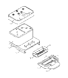
Figure IA is an exploded view of one embodiment of the perfusion manifold
assembly
showing the cover off of the reservoirs, the reservoirs above the backplane,
the backplane in
fluidic communication with the skirt, the skirt with a side track for engaging
a representative
microfluidic device or "chip" having one or more inlet, outlet and vacuum
ports, the chip shown
next to (but not in) one embodiment of a chip carrier, the carrier is
configured to support and
carrier the chip. Figure 1B shows the same embodiment of the perfusion
manifold assembly
with the cover on and over the reservoirs, and the chip inside the chip
carrier fully linked to the
skirt of the perfusion manifold assembly, and thereby in fluidic communication
with the
reservoirs. Figure IC shows an exploded view of one embodiment of the cover
assembly
comprising a pressure cover or pressure lid, and an associated gasket
thereunder.
Figure 2A shows a side view of one embodiment of a chip carrier (with the chip
inside)
approaching (but not yet engaging) a side track of a skirt of one embodiment
of the perfusion
manifold assembly, the carrier aligned at an angle matching an angled front
end portion of the
side track, the carrier comprising a retention mechanism configured as a
upwardly protecting
clip. Figure 2B shows a side view of one embodiment of a chip carrier (with
the chip inside)
engaging a side track of a skirt of one embodiment of (but not yet linked to)
the perfusion
manifold assembly. Figure 2C shows a side view of one embodiment of a chip
carrier (with the
chip inside) fully engaging a side track of a skirt of one embodiment of (but
not yet linked to) the
perfusion manifold assembly (with an arrow showing the necessary direction of
movement to get
a snap fit whereby the retention mechanism will engage to prevent movement).
Figure 2D shows
a side view of one embodiment of a chip carrier (with the chip inside)
detachably linked to the
perfusion manifold assembly, where the retention mechanism is engaged to
prevent movement.
Figure 2E is a summary slide schematically showing one embodiment of a linking
approach to
the manifold comprising a sliding action (2E-1), pivoting (2E-2), and snap fit
(2E-3) in a single
action.
Figure 3 is a schematic showing one embodiment of connecting two microfluidic
devices,
resulting in the introduction of air or gas bubbles into the microchannels.
Figure 3A shows two
fluidically primed devices with microchannels that are not yet connected.
Figure 3B shows the
devices of Figure 3A contacting in a manner that results in the introduction
of air bubbles into
the microchannels.
12
Date Recue/Date Received 2021-09-15
WO 2018/013646 PCT/US2017/041656
Figure 4A is a schematic showing the location of a bubble where a chip is
engaged by the
perfusion manifold assembly (also called the perfusion disposable or POD).
Figure 4B is a
drawing from a photograph of a bubble (see arrow) caught in the perfusion
disposable (not the
chip) at the location circled in Figure 4A. The perfusion disposable is
comprised of COP (cyclic
polyolefin) with a SEBS (Styrene Ethylene Butylene Styrene) capping layer. COP
and SEBS are
both substantially less gas permeable than PDMS.
Figure 5 is a schematic of an illustrative microfluidic device or "organ-on-
chip" device
(which can be fabricated out of plastic, such as PDMS) with a mating surface
(21). The
assembled device in Figure 5A includes a plurality of ports. Figure 5B shows
an exploded view
of the device of Figure 5A, showing a tissue-tissue interface simulation
region (SR) comprising a
membrane, where cell behavior and/or passage of gases, chemicals, molecules,
particulates and
cells are monitored.
Figure 6 is a schematic of a "push" based flow approach where fluid flows from
a
reservoir (on the right) where the fluid is put under high pressure. The fluid
exits the reservoir
(on the right) and flows in the direction of the chip (see arrows showing the
direction of flow)
through a resistor (switchback). There is no pressure applied to the other
reservoir (on the left)
Figure 7 is a schematic of a "pull" based flow approach where fluid flows from
a
reservoir (on the left) where the fluid is under no pressure, but where the
other reservoir (on the
right) has low pressure (e.g. because of a vacuum). The fluid exits the
reservoir (on the left) and
flows in the direction of the chip (see arrows showing the direction of flow).
Figure 8 is a schematic of a pressure differential ("delta P") based flow
approach where
fluid in both reservoirs are under pressure (P, is not zero but is less than
P1). The fluid exits the
reservoir (on the right) and flows in the direction of the chip (see arrows
showing the direction of
flow) through a resistor (switchback).
Figure 9 show experimental results for reducing bubble volume using the
pressure
differential based flow approach of Figure 8. The flow rates in Figure 9 are
given in terms of
pressure differentials (delta) - lkPa corresponds to 40uL/hr, 1.5kPa is
60uL/hr, 3kPa is 120uL/hr,
8kPa is 320uL/hr, 28.3kPa is 1.13mL/hr. Pressures applied to the outlets and
therefore "felt" by
the bubbles are given in terms of "felt" pressure (units of kPa).
13
Date Recue/Date Received 2021-09-15
WO 2018/013646 PCT/US2017/041656
Figure 10 is a chart showing how large, multi-week experiments are performed
with
many microfluidic devices or "chips," underscoring that the task of refreshing
the media (e.g.
every other day or at key time points) can be burdensome.
Figure 11 shows the results of an experiment involving the use of non-
equilibrated media
in 19 pods engaging organs-on-chip, with flow rate as the read-out for
detecting bubbles. Figure
11A shows the results for 8 pods and Figure 11B shows the results for 11 pods
(the dotted lines
show the 20% deviation from the bottom average and the top average).
Figure 12 shows a plot of pressure versus time in order to test for lid
failure when higher
pressures are used for bubble treatment. Figure 12A shows that, as the
pressure was raised and
approached 25-30kPa, the perfusion system with a thin gasket exhibited lid
failure. Figure 12B
shows that, as the pressure was raised and approached 33kPa, the perfusion
system with thicker
gasket (2-3 times thicker than the thin gasket) did not exhibit lid failure.
Figure 13 is a bar graph showing axon growth in a microfluidic device over
time (e.g.
Day 0, Day 1, Day 4 and Day 5) with 504/hr control conditions versus 754/hr
test conditions.
Definitions
"Channels" are pathways (whether straight, curved, single, multiple, in a
network, etc.)
through a medium (e.g., silicon, glass, polymer, etc.) that allow for movement
of liquids and
gasses. Channels thus can connect other components, i.e., keep components "in
communication"
and more particularly, "in fluidic communication" and still more particularly,
"in liquid
communication." Such components include, but are not limited to, liquid-intake
ports and gas
vents. Microchannels are channels with dimensions less than 1 millimeter and
greater than 1
micron. It is not intended that the present invention be limited to only
certain microchannel
geometries. In one embodiment, a four-sided microchannel is contemplated. In
another
embodiment, the microchannel is circular (in the manner of a tube) with curved
walls. In yet
another embodiment, combination of circular or straight walls are used.
It is not intended that the present invention be limited by the number or
nature of
channels in the microfluidic device. In some embodiments, the surface can be a
surface of a
fluid-flowing conduit or passageway disposed in a solid substrate. In some
embodiments, the
surface can be a solid surface. For example, in one embodiment, the solid
surface can be a wall
14
Date Recue/Date Received 2021-09-15
surface of a fluid channel, e.g., a microfluidic channel. However, the method
need not be limited
to microchannels, since it will work in any confined space where fluid flows.
Additionally, the term "microfluidic" as used herein relates to components
where moving
fluid is constrained in or directed through one or more channels wherein one
or more dimensions
are 1 mm or smaller (microscale). Microfluidic channels may be larger than
microscale in one or
more directions, though the channel(s) will be on the microscale in at least
one direction. In some
instances the geometry of a microfluidic channel may be configured to control
the fluid flow rate
through the channel (e.g. increase channel height to reduce shear or
resistance). Microfluidic
channels can be formed of various geometries to facilitate a wide range of
flow rates through the
channels.
A "perfusion manifold assembly" is contemplated that allows for perfusion of a
microfluidic device, such as an organ on a chip microfluidic device comprising
cells that mimic
cells in an organ in the body, that is detachably linked with said assembly so
that fluid enters
ports of the microfluidic device from a fluid reservoir, without tubing, at a
controllable flow rate.
In one embodiment (see Figure 1A and 1B), the perfusion manifold assembly
comprises i) a
cover or lid configured to serve as the top of ii) one or more fluid
reservoirs, iii) a capping layer
under said fluid reservoir(s), iv) a fluidic backplane under, and in fluidic
communication with,
said fluid reservoir(s), said fluidic backplane comprising a resistor, and v)
a skirt (for engaging
the microfluidic device). In one embodiment, a combination of pressure and
flow reduces
bubble volume in a perfusion manifold assembly. In one embodiment, the
perfusion manifold
assembly is made of a polymer that is less gas permeable than PDMS.
In one embodiment, the perfusion manifold is linked to a microfluidic device
(e.g. in
fluidic communication therewith). Microfluidic devices (or "chips") containing
living cells
recreate the physiological tissue-tissue interfaces and permit fluid flow. See
U.S. Patent No.
8647861. Such devices subject the cells to shear stress. In contrast to static
2D culture,
microchannels allow the perfusion of cell culture medium throughout the cell
culture during in
vitro studies and as such offer a more in vivo-like physical environment. In
simple terms, an inlet
port allows injection of fluids such as blood, serum, plasma, cell culture
medium (and the like)
into a microfluidic channel or chamber (with or without cells). In one
embodiment, the present
invention contemplates a cell-laden microfluidic channel or chamber. An outlet
port then
permits the exit of remaining fluid as well as harmful
Date Recue/Date Received 2021-09-15
WO 2018/013646 PCT/US2017/041656
metabolic by-products. In one embodiment, only flow is used with media
previously under-
saturated.
In some embodiments, a bubble is trapped in a microfluidic device against a
polymer that
is largely gas impermeable, such as (but not limited to ) a COP. Cyclic olefin
copolymers
(COCs) and cyclic olefin polymers (COPs) are very attractive thermoplastic
resins with potential
enhanced properties such as outstanding transparency, good heat resistance,
low moisture
absorption, good chemical resistance, and low double refraction. COCs are
obtained through
copolymerization of cycloolefin with ethylene or a-olefin, and commercialized
under the trade
names APEL by Mitsui and TOPAS by TOPAS advanced polymers (TAP: formerly
Ticona
and Hoechst). COPs are prepared via ring-opening metathesis polymerization
(ROMP) of
cycloolefin followed by hydrogenation, and commercialized under the trade
names Zeonex and
Zeonor by Zeon [25] and Arton by Japan Synthetic Rubber (JSR).
Description of Preferred Embodiments
Methods of removing gas or air bubbles from a microfluidic device are
described,
including one or more bubbles in a microchannel of a microfluidic device. It
is not the presence
of air, or gas, in the medium which causes the problem. It is the formation of
the bubbles from
these gases which cause the problem. The question is why and how these bubbles
are formed. If
the source of bubble formation is established and then removed, only then this
problem can be
addressed.
One source of the bubble formation may be explained as follows: cells are
provided
nutrients from culture media maintained at 37 CC. However, the culture media
used are
generally stored at room temperature (or less) which is lower than 37 C. When
a medium is
transferred out of storage and heated up to 37 C, there is a change in
solubility of the dissolved
gasses. The decrease in solubility of the gasses at higher temperatures causes
the dissolved
gasses to come out of the medium in the form of tiny bubbles which tend to
stick to surfaces of
the microfluidic device housing the cells, including channel surfaces (and, in
particular,
mierodefects in the channel surfaces). While not intending in any way to limit
the present
invention to any particular mechanism, it is believed that this process of
"bubble growth"
requires an initial bubble, sometimes referred to as a nucleation point or
"seed bubble," for the
gas in solution to diffuse into and transition from dissolved gas into non-
dissolved gas pockets or
16
Date Recue/Date Received 2021-09-15
WO 2018/013646 PCT/US2017/041656
bubbles. However, once the medium is equilibrated at 37 C the formation of
the bubbles slows.
Therefore, one partial answer to the question of why and how the bubbles are
formed is because
of a transitory stage during the heating process of the culture media.
Up to now, it has been believed that a simple solution to avoid this problem
is to remove
the temperature gradient effect, i.e., avoid transferring low temperature
medium directly into the
microfluidic device. In other words, one should warm the medium to 37 C
outside the
microfluidic device and/or give sufficient time for the medium to equilibrate
in a vessel or
reservoir at 37 C (with moderate stirring if needed). Of course, this takes
time and the culture
media needs to be sterile.
While the practice of de-aeration or "de-gassing" has been introduced to
address this
problem of bubble formation, it is a practice that has practical limitations.
The commonly
suggested procedure of de-aerating, which is based on heating/vacuum steps, is
oftentimes
without a measurable endpoint and highly dependent on the equipment being used
to perform the
procedure. Therefore, the de-aeration step will be unpredictable with a high
degree of variability
stemming from exact process parameters and equipment used. Additionally, "de-
gassing" can
have the consequence of removing gasses from solution that are needed to
maintain culture, like
oxygen (for cellular respiration) and CO-, (for pH buffering). Moreover, no
matter how
reproducible one tries to he with the de-aeration step, after de-aeration the
medium will quickly
start equilibrating itself with the atmospheric gasses. Therefore, until this
equilibrium is exactly
reached, the system will remain unstable and unreliable.
Where large, multi-week experiments are performed with many microfluidic
devices or
"chips," the task of refreshing the media (e.g. every other day or at key time
points) can be
burdensome. This is illustrated in Figure 10 for a 2 week experiment involving
organ-on-chips.
Of course, the physiological environment of the cells in a microfluidic device
does not
require a de-aerated medium. The degassing is only being done to address the
bubble problem.
This brings one to the question of whether (and to what extent) non-
equilibrated and non-
degassed culture media can be employed with microfluidic devices. In one
embodiment, the
present invention contemplates equilibrating via the process of degassing
(physically removing
dissolved gas from solution) media before a first pressure/flow cycle ¨ but
using non-
equilibrated and non-degassed media when replacing media thereafter, i.e.
during long-term
culture. In another embodiment, the present invention contemplates using non-
equilibrated and
17
Date Recue/Date Received 2021-09-15
WO 2018/013646 PCT/US2017/041656
non-degassed media even in a first pressure/flow cycle (albeit with higher
pressures) whenever
culture media is placed into the perfusion manifold or "pod" reservoir(s). In
one embodiment, the
present invention contemplates adding cold / non-equilibrated media into one
or more pod
reservoirs.
In the first embodiment, culture media is equilibrated and/or de-gassed once,
at the
beginning of the experiment, and then a pressure/flow treatment is utilized
for a period of time.
Ideally, the period of time should be short and insensitive to variability
(e.g. 1-2 hours), and the
treatment conditions should allow for operating without unrealistically high
pressures or flow
rates. Without intending to limit the invention in any way to a mechanism of
action, it is
believed that two forces work in concert to shrink bubbles in such a
pressure/flow treatment.
First, pressure increases the gas carrying capacity of media. Second, flow
(e.g. 404/hr)
provides fresh (undersaturated) media into which the bubbles dissolve. It has
been empirically
observed that oversaturated media cannot grow bubbles that do not exist in the
first place.
Thereafter, culture media would not need to be equilibrated or degassed when
replenishing
media. Said another way, the single pressure/flow treatment removes the
bubbles (or nucleation
points/seed bubbles) and the use of oversaturated media thereafter will not
bring them back. In
this embodiment, non-equilibrated media can be used when refilling inlet
reservoirs AF1ER a
single pressure/flow cycle has successfully eliminated system bubbles. The
benefit of this
approach is that it solves the bubble problem, while decreasing the number of
times culture
media must be equilibrated and/or degassed.
In the second embodiment, culture media is not equilibrated (i.e. it is non-
equilibrated
culture media) and has not gone the physical removal of dissolved gas via
degassing. In order for
this to work, it has been mathematically determined via physical principals
and confirmed
experimentally that one can increase the pressure (e.g. by 13kPa or more)
during the
pressure/flow cycle (e.g. increase from 20kPa to 33kPa or more). While not
intending to be
limited to any particular mechanism, it is believed that this increased
pressure increases non-
equilibrated media gas carrying capacity to match equilibrated media gas
carrying capacity,
making the pressure/flow cycle as effective (theoretically) as with non-
equilibrated media. The
increased pressure can put a strain on the microfluidic system. However, it
has been empirically
determined that a thicker gasket for the perfusion manifold is one solution to
avoiding leaks
associated with the increased pressure. Optionally, increased flow rates (from
50 to 754/hr)
18
Date Recue/Date Received 2021-09-15
WO 2018/013646 PCT/US2017/041656
can also he used (and provide some benefit in terms of robustness of
eliminating bubbles) since it
has been empirically found that the cells can tolerate the increased flow.
With regard to
increased pressure, it appears that the pressure differential between the
reservoirs (i.e. the inlet
and outlet reservoirs) is more important to the viability of the cells than
the actual pressures
employed. It has been empirically found that pressure differentials of 2kPa or
less are useful,
more preferably 1.5kPa or less, still more preferably 1.0kPa or less.
Description of Exemplary Microfluidic Devices
In one embodiment (as shown in Figure 1, the perfusion manifold assembly or
POD (10)
comprises i) a cover or lid (11) configured to serve as to top of ii) one or
more fluid reservoirs
(12), iii) a capping layer (13) under said fluid reservoir(s), iv) a fluidic
backplane (14) under,
and in fluidic communication with, said fluid reservoir(s), said fluidic
backplane comprising a
fluidic resistor, and v) a skirt (15) for engaging the microfluidic device
(16) which is preferably
positioned in a carrier (17). In one embodiment, the carrier (17) has a tab or
other gripping
platform (18), a retention mechanism such as a clip (19), and a visualization
cutout (20) for
imaging the chip. In one embodiment, the fluidic resistor comprises a series
of switchbacks or
serpentine fluid channel (not shown).
Figure IC shows an exploded view of one embodiment of the cover assembly (11)
comprising a pressure cover or pressure lid. In the illustrated embodiment,
the pressure lid
comprises a plurality of ports (e.g. through-hole ports) associated with
filters (38) and
corresponding holes (39) in a gasket (37). The illustrated design of the holes
in the gasket is
intended to permit the gasket to aid in retaining the illustrated filters in
position. In alternative
embodiments, gasket openings may employ a shape different from openings in the
lid. For
example, the gasket can be shaped to follow the contour of one or more
reservoirs with which it
is intended to form a fluidic or pressure seal. In some embodiments, a
plurality of gaskets may be
employed. In a preferred embodiment, a thicker gasket may be employed (in
order to avoid
leaking under the higher pressures described herein to treat bubbles). In some
embodiments, the
filters and/or gasket may be fixed using an adhesive, heat stacking, bonding
(ultrasonic, solvent-
assisted, laser welding), clamped, or captured by elements of the lid and/or
an additional
substrate. Although the illustrated pressure lid comprises through-hole ports,
alternative
19
Date Recue/Date Received 2021-09-15
WO 2018/013646 PCT/US2017/041656
embodiments comprise one or more channels that route at least one top-surface
port to one or
more bottom surface ports, which need not be directly underneath the top-
surface port.
In one embodiment, the microfluidic device is detachably linked with the
manifold
assembly by a clipping mechanism that temporarily "locks" the microfluidic
device, including
organ-on-chip devices, in place (Figures 2A, 2B, 2C, 2D and 2E). In one
embodiment, the
clipping or "snap fitting" involves a projection on the carrier (19) which
serves as a retention
mechanism when the microfluidic device is positioned. In one embodiment, the
clipping
mechanism is similar to the interlocking plastic design of a LegoTM chip and
comprises a
straight-down clip. However, in another embodiment, the clipping mechanism is
triggered only
after the microfluidic device, or more preferably, the carrier comprising the
microfluidic device,
engages the perfusion manifold assembly (or cartridge) on a guide rail, side
slot, internal or
external track (25) or other mechanism that provides a stable glide path for
the device as it is
conveyed (e.g. by machine or by hand) into position. The guide rail, side
slot, internal or
external track (25) or other mechanism can be, but need not be, strictly
linear and can be
positioned in a projecting member or skirt attached to the main body of the
manifold assembly.
In one embodiment, the beginning portion of the guide rail, side slot,
internal or external track or
other mechanism comprises an angled slide (27) which provides a larger opening
for easier
initial positioning, followed by a linear or essentially linear portion (28).
In one embodiment,
the end portion (29) (close to the corresponding ports of the assembly) of an
otherwise linear (or
essentially linear) guide rail, side slot, internal track or other mechanism
is angled (or curves)
upward so that there is a combination of linear movement (e.g. initially) and
upward movement
to achieve linking.
The POD has a few features that help reduce bubble introduction: 1) the clip
has a very
smooth engagement - rough engagements and/or jerking motions can introduce
bubbles, and 2)
the POD diameter going to the chip has been minimized to reduce bubble
trapping upon initial
filling of the POD - this minimizes dead volume where pockets of air can get
trapped.
The advantage of the carrier is that the surfaces of the microfluidic device
need not be
touched during the detachable linage with the manifold assembly. The carrier
can have a plate,
platform, handle or other mechanism for gripping the carrier (18), without
contacting the mating
surface (21) of the microfluidic device (16). The retention mechanism (19) can
comprise a
Date Recue/Date Received 2021-09-15
projection, hook, latch or lip that engages one or more portions of the
manifold assembly, and
more preferably the skirt of the manifold assembly, to provide a "snap fit."
Figure 5 shows a schematic of an illustrative microfluidic device or "organ-on-
chip"
device. The assembled device in Figure 5A includes a plurality of ports.
Figure 5B shows an
exploded view of the device of Figure 5A, showing a tissue-tissue interface
simulation region
("SR-) comprising a membrane (100), where cell behavior and/or passage of
gases, chemicals,
molecules, particulates and cells are monitored.
Bubbles can be introduced when a chip is engaged by the perfusion manifold
assembly
(also called the perfusion disposable). Figures 4A and 4B underscore this
point, showing a
bubble (see arrow) caught in the perfusion disposable (not the chip) at the
location circled in
Figure 4A. Any one of the embodiments of the methods described above for
combining pressure
and flow may be used to reduce the volume of such bubbles in such microfluidic
devices.
In one embodiment, the POD is positioned on the culture module and the
pressure surface
of the culture module move down to engage the cover or lid (11) of the
perfusion manifold
assembly (10). Embodiments of a culture module are described in U.S. Patent
Application Serial
No. 15/248,509. As shown in Figure 1C, the cover or lid comprises ports such
as through-hole
ports (36) that are engaged by corresponding pressure points on the pressure
surface of the
culture module. These ports (36), when engaged, transmit applied pressure
inward through the
cover and through a gasket (37) and apply the pressure to the fluid in the
reservoirs (12) of the
perfusion manifold assembly (10). Thus, in this embodiment, pressure is
applied through the lid
(11) and the lid seals against the reservoir(s). For example, when on applies
1 kPa, this nominal
pressure results, in one embodiment, in a flow rate of approximately 30-40
uL/hr.
EXPERIMENTAL
EXAMPLE 1
In this experiment, 19 pods engaging organs-on-chip (in this case,
microfluidic devices
with viable intestinal cells growing on a membrane in a microchannel) were
utilized. They were
previously running for 6 days, with no history of bubbles. In the test groups,
inlet reservoirs
were filled with cold media (4 C) on days 0 and 2, warm media (not
equilibrated) on day 7.
21
Date Recue/Date Received 2021-09-15
Flow was measured daily as a read-out (since bubbles disrupt flow and thus a
change in flow
would indicate bubbles); in addition, the pods/chips were visually inspected
for bubbles. The
results are shown in Figure 11A and 11B. No bubble growth/generation was
observed or
detected in any pod/chip when using non-equilibrated media (warm or cold) over
9 days.
EXAMPLE 2
In this experiment, one embodiment of the perfusion system's ability to
withstand higher
pressures was tested (in order to see if working with non-equilibrated media
at higher pressures
is feasible). Various components on the POD (Figures lA and 1B) were examined,
including the
lid (Figure 1C) and other interfaces (e.g. gaskets, bonded components, etc.).
In addition,
components of a culture module (described in U.S. Patent Application Serial
No. 15/248,509)
were examined in these pressure tests (e.g. the manifold, valves and
junctions). As the pressure
was raised and approached 25-30kPa, the perfusion system with a thin gasket
(Figure 1C,
element 37) exhibited lid failure and leakage (Figure 12A). However, when a
thicker gasket (2-3
times thicker than the thin gasket), there was no lid failure or leakage even
at 33kPa (Figure
12B). With the thicker gasket, the perfusion system can withstand ¨34kPa on
the inlet and
¨33kPa on the outlet.
EXAMPLE 3
In this experiment, higher flow rates were tested to determine whether there
are negative
cell effects. More specifically, the viability and function of human primary
human motor
neurons maintained after 7 days was assessed (since they are relatively
sensitive to culture
conditions and shear forces). Flow rates of 50 (control) to 75 L/hr (test)
were used to perfuse
the cells in a microfluidic chip engaged in a POD (Figure 1A and 1B), with the
POD engaged
with a culture module (described in U.S. Patent Application Serial No.
15/248,509). The outlet
pressure was 20.0 kPa +/- 0.5kPa. The inlet pressure minus the outlet pressure
[(Inlet Pressure) ¨
(Outlet Pressure)] was 1.5 kPa +/- 0.5 kPa. Primary motor neurons were seeded
(on day zero)
and cultured for 7 days, and the medium was replenished every two days. A
pressure/flow
process was run on Days 1 and 5 to treat bubbles. Cold media was placed in the
POD reservoir
on Days 3 and 5. Cells were imaged (phase contrast captured at 200X).
22
Date Recue/Date Received 2021-09-15
WO 2018/013646 PCT/US2017/041656
Axon growth was observed in both control and experimental conditions. Figure
13 is a
bar graph showing the results at Day 0, Day 1, Day 4 and Day 5. Results are
average SE in 2
independent PODs for the 504 condition and in 3 independent PODs for the 754
condition.
Motor neurons were stained (after 7 days) with Hoechst 33342 (blue), which
indicates
cell nuclei and Tuj-1 (green), which marks f3-Tubulin 3 ¨ a protein vital to
microtubule stability
and transport in the axon of neurons. Neuron staining revealed well-developed
neuronal
networks in the control (504/hr) and in the test (754/hr) (data not shown). In
sum, the
experiment showed that 1) motor neurons are capable of handling elevated flow
rates, i.e. flow
rates that help to facilitate bubble removal 2) capable of handling multiple
pressure/flow cycles
at 20kPa applied pressure and that 3) the use of cold media to refill inlet
reservoirs during normal
media refresh/addition steps did not cause the formation of bubbles after the
initial pressure/flow
step to remove system bubbles.
23
Date Recue/Date Received 2021-09-15