Note: Descriptions are shown in the official language in which they were submitted.
CA 03131272 2021-08-24
WO 2020/181336
PCT/AU2020/050242
HYBRID MEDICAL IMAGING PROBE, APPARATUS AND PROCESS
TECHNICAL FIELD
The present invention relates to medical imaging, and in particular to a
hybrid medical
imaging probe apparatus and process for imaging biological tissues of a
subject.
BACKGROUND
Medical imaging technologies such as ultrasound, computed tomography (CT),
magnetic
resonance imaging (MRI) and nuclear medicine imaging are extremely powerful
techniques for imaging internal features of the human body, but suffer from a
number
of disadvantages that limit their applicability. For example, these
technologies require
expensive equipment, and are therefore not generally available at rural or
remote health
centres. Indeed, according to the World Health Organization (WHO), more than
half of
the world's population does not have access to diagnostic imaging.
Furthermore, there
is a general need for low-cost and safe imaging systems for the detection and
continuous
monitoring of a variety of diseases. Due to the need to limit exposure to
ionising
radiation such as X-rays, most currently available medical imaging systems
cannot be
used for frequent monitoring purposes. Additionally, the bulky and static
structures and
high costs of MRI and other large medical imaging systems often preclude them
for
monitoring diseases that require monitoring on a regular and short-term basis.
These
factors make such systems impractical to be used by paramedics for real-time
imaging
and assessment purposes.
Electromagnetic imaging is an attractive technique for medical applications,
and has the
potential to create a visual representation of the interior of the human body
in a cost-
effective and safe manner. From an electromagnetic engineering perspective,
the
human body is an electromagnetically heterogeneous medium characterized by
features
and tissues with different dielectric properties. Moreover, the dielectric
properties
permittivity and conductivity differ between injured and healthy tissues. When
an
injured tissue with a high permittivity value compared to its neighbouring
healthy tissue
is exposed to an electromagnetic wave at a microwave frequency, a relatively
high
portion of the wave is reflected back towards the radiation source.
Accordingly, an
electromagnetic medical imaging apparatus can be utilized to transmit
electromagnetic
waves into a body part to be imaged, such as the human head or torso.
Microwave
CA 03131272 2021-08-24
WO 2020/181336
PCT/AU2020/050242
- 2 -
signals predominantly reflected by damaged tissues (e.g., in particular at
bleeding or
clot sites) due to changes in electromagnetic properties are received and
measured by
the apparatus. Then, the data representing the measured signals can be
processed to
estimate the location and/or dielectric properties of the abnormality, and to
generate
two or three-dimensional images of the damaged tissues within the body part.
The data processing step plays a critical role in an electromagnetic imaging
apparatus.
Various imaging techniques have been employed to detect medical targets from
measurements of scattered electromagnetic signals. Those techniques try to
estimate
the dielectric properties of the tissues by solving nonlinear equations
(tomography),
which do not have a unique solution and those solutions might not depend
continuously
on the input data, or to find the location of target tissues using time-domain
radar-
based techniques. Due to the time-consuming nature of tomography-based
techniques,
they are almost exclusively applicable to single frequency or narrow-band
multi-
frequency signals, and therefore are not suitable for use in medical emergency
situations such as brain injury detection, where a rapid diagnosis is
required.
Alternatively, in radar-based imaging, a scattering profile of the imaging
domain is
mapped onto a two- or three-dimensional image. This method is more applicable
when
using ultra-wide frequency bands for fine resolution because the required data
processing is simpler and faster than tomography. However, current radar
imaging
methods, such as confocal, microwave imaging via space-time ("MIST")
beamforming,
and adaptive beamforming imaging methods utilize processing techniques based
on
delay-and-sum (DAS), which are susceptible to outer layer reflections and
internal layer
refractions that can result in false detection. In addition, the variation of
signal
penetration through the tissues at different frequencies limits the
effectiveness of those
delay calculations, and consequently the accuracy of the resulting images. In
view of
these difficulties, there is a continuing need for a faster and accurate
imaging apparatus
and process.
It is desired to overcome or alleviate one or more difficulties of the prior
art, or to at
least provide a useful alternative.
CA 03131272 2021-08-24
WO 2020/181336
PCT/AU2020/050242
- 3 -
SUMMARY
In accordance with some embodiments of the present invention, there is
provided a
hybrid medical imaging probe for application to a body part to image tissues
within the
body part, the medical imaging probe including:
a first imaging probe component to generate non-microwave first signals for
transmission into the body part and to sense corresponding signals scattered
by the
tissues within the body part to enable the generation of one or more
corresponding
images of the tissues using a non-microwave first imaging technology; and
an electromagnetic imaging probe component to generate microwave signals in
a microwave frequency band for transmission into the body part and to sense
corresponding microwave signals scattered by the tissues within the body part
to
enable the estimation of corresponding values of permittivity of the tissues;
wherein the first imaging probe component and the electromagnetic imaging
probe component are co-located within the hybrid medical imaging probe and
arranged so that the non-microwave and microwave signals are transmitted from
the hybrid medical imaging probe in the same direction.
In some embodiments, the first imaging probe component is an ultrasonic
imaging
probe component. In some embodiments, the ultrasonic imaging probe component
includes an ultrasonic transducer, and the electromagnetic imaging probe
component
includes an array of antennas disposed about the ultrasonic transducer.
In some embodiments, the antennas are loaded with series capacitance and/or
shunt
inductance to create resonances that are independent of the size of the
antennas.
In some embodiments, the hybrid medical imaging probe includes electromagnetic
bandgap (EBG) structures to reduce the mutual coupling between the antennas,
thereby
allowing the antennas to be located in close mutual proximity.
CA 03131272 2021-08-24
WO 2020/181336
PCT/AU2020/050242
- 4 -
In some embodiments, the hybrid medical imaging probe includes artificial
magnetic
surfaces (AMS) such as metasurfaces formed by arrays of periodic structures
and
configured so that the array of antennas generate predominantly unidirectional
radiation, thereby allowing the antennas to be located in close mutual
proximity.
In some embodiments, the hybrid medical imaging probe includes metamaterial
absorbers to reduce the leakage of microwave signals.
In accordance with some embodiments of the present invention, there is
provided a
hybrid medical imaging apparatus for imaging tissues within a body part, the
medical
imaging apparatus including:
any one of the above hybrid medical imaging probes; and
a data processing component configured to receive initial image data
representing an initial image of the tissues of the body part representing non-
microwave
signals scattered by the tissues within the body part and sensed by the first
imaging
probe component; and to generate estimates of permittivity of the tissues of
the body
part based on the sensed microwave signals scattered by the tissues within the
body
part, wherein the initial image of the tissues of the body part is used as a
priori
information to generate an electromagnetic model from which the estimates are
generated.
In some embodiments, the data processing component is further configured to
generate
an image representing a spatial distribution of the permittivity of the
tissues of the body
part.
In accordance with some embodiments of the present invention, there is
provided a
hybrid medical imaging process for imaging tissues within a body part, the
medical
imaging process including the steps of:
receiving a first image of the tissues of the body part generated from sensed
first and non-microwave signals reflected from the tissues within the body
part; and
receiving microwave scattering data representing sensed microwave signals
scattered by the tissues within the body part;
processing the first image to generate a corresponding electromagnetic
model of the body part; and
processing the microwave scattering data and the electromagnetic model of
the body part to generate estimates of permittivity of the tissues of the body
part.
CA 03131272 2021-08-24
WO 2020/181336
PCT/AU2020/050242
- 5 -
In some embodiments, the hybrid medical imaging process includes generating a
second
image of the tissues of the body part, the second image representing a spatial
distribution of the permittivity estimates.
In some embodiments, the first imaging technology is an ultrasonic imaging
technology.
In some embodiments, the step of generating the electromagnetic model includes
determining a distance between a region of interest within the body part and a
corresponding surface of the body part, and an estimate of permittivity of the
region of
interest is generated by solving a system of equations modelling microwave
propagation
from the surface to the region of interest and from the region of interest
back to the
surface of the body part.
In some embodiments, the permittivity value is estimated from scattered
microwave
signals of a plurality of different microwave frequencies to improve the
accuracy of the
estimate.
In some embodiments, the tissues include an internal organ, and the process
includes
assessing a health status of the internal organ from the estimated
permittivity value of
the internal organ.
In some embodiments, assessing a health status of the internal organ includes
estimating a percentage of fat in the internal organ. The internal organ may
be a liver.
In some embodiments, the hybrid medical imaging process includes estimating
respective permittivities of left and right sides of a patient's torso, and
comparing those
permittivities to assess a health status of the patient. In some embodiments,
assessing
a health status of the patient includes diagnosing whether the patient has a
disease.
In accordance with some embodiments of the present invention, there is
provided at
least one computer-readable storage medium having stored thereon executable
instructions that, when executed by at least one processor of a data
processing
apparatus, cause the at least one processor to execute any one of the above
processes.
CA 03131272 2021-08-24
WO 2020/181336
PCT/AU2020/050242
- 6 -
In accordance with some embodiments of the present invention, there is
provided a
hybrid medical imaging apparatus including:
any one of the above hybrid medical imaging probes; and
any one of the above data processing components.
Also described herein is a medical imaging probe for application to a body
part to image
tissues within the body part, the medical imaging probe including:
a real-time imaging probe component to generate first signals for transmission
into
the body part and to sense corresponding signals reflected from the tissues
within the
body part to enable the generation of one or more corresponding images of the
tissues
in real-time using a real-time imaging technology; and
an electromagnetic imaging probe component to generate microwave signals in a
microwave frequency band for transmission into the body part and to sense
corresponding microwave signals reflected from the tissues within the body
part to
enable the generation of corresponding images of the tissues using a microwave
imaging
technology.
The real-time imaging probe may be an ultrasonic imaging probe. The ultrasonic
imaging probe component may include an ultrasonic transducer, and the
electromagnetic imaging probe component may include an array of antennas
disposed
about the ultrasonic transducer.
Also described herein is a medical imaging apparatus for imaging tissues
within a body
part, the medical imaging apparatus including:
any one of the above medical imaging probes;
a real-time image generation component to generate an initial image of the
tissues of the body part based on the signals reflected from the tissues
within the
body part and sensed by the real-time imaging probe component; and
an electromagnetic image generation component to generate an
electromagnetic image of the tissues of the body part based on the sensed
microwave signals reflected from the tissues within the body part, wherein the
initial
image of the tissues of the body part is used as a priori information to
generate the
electromagnetic image of the tissues of the body part.
CA 03131272 2021-08-24
WO 2020/181336
PCT/AU2020/050242
- 7 -
Also described herein is a medical imaging process for imaging tissues within
a body
part, the medical imaging process including the steps of:
generating a first image of the tissues of the body part based on sensed first
signals reflected from the tissues within the body part; and
generating an electromagnetic image of the tissues of the body part based on
sensed microwave signals reflected from the tissues within the body part,
wherein the
accuracy of the generated electromagnetic image is improved by using the first
image
of the tissues of the body part as a priori information to generate the
electromagnetic
image, and the first image is generated using a real-time imaging technology.
The real-time imaging technology may be ultrasonic imaging technology.
The step of generating the electromagnetic image may include determining a
distance
between a region of interest within the body part and a corresponding surface
of the
body part, and determining a permittivity value for the region of interest by
solving a
system of equations modelling microwave propagation from the surface to the
region of
interest and from the region of interest back to the surface of the body part.
Also described herein is a process for diagnosing organ disease in a patient,
the process
including:
measuring scattering parameters representing electromagnetic signals scattered
from organs within a torso of the patient; and
calculating a quantitative measure representing relative permittivity of the
organs within right and left sides of the patient's torso; and
diagnosing whether the patient has an organ disease or diffused fat on the
basis
of a comparison of the quantitative measure with corresponding quantitative
measures for the organ in known diseased and healthy states.
CA 03131272 2021-08-24
WO 2020/181336
PCT/AU2020/050242
- 8 -
BRIEF DESCRIPTION OF THE DRAWINGS
Some embodiments of the present invention are hereinafter described, by way of
example only, with reference to the accompanying drawings, wherein:
Figure 1 is a prior art ultrasound image that can be used to determine the
distance between a patient's skin and their liver;
Figure 2 is a schematic diagram of a hybrid medical imaging apparatus in
accordance with an embodiment of the present invention;
Figure 3 is a block diagram of a data processing component of the hybrid
medical
imaging apparatus of Figure 2;
Figure 4 is a flow diagram of a hybrid medical imaging process executed by the
data processing component of Figure 3;
Figure 5 is a schematic diagram of a hybrid electromagnetic-ultrasound probe
of
the hybrid medical imaging apparatus, in accordance with an embodiment of the
present
invention; and
Figure 6 is a schematic diagram illustrating a multilayer dielectric model of
the
hybrid medical imaging process.
DETAILED DESCRIPTION
The inventors have identified that the accuracy, speed and reliability of
medical
electromagnetic imaging ("EM") can be significantly improved by using a non-
microwave
first imaging technology to accurately determine the respective locations of
one or more
targeted tissues or internal organs of a subject (preferably, but not
necessarily, in real-
time), and then using those locations as a priori information to model
microwave
propagation to and from the internal organs/tissues and scattering by the
internal
organs/tissues in order to measure the complex permittivity of those
organs/tissues.
The permittivity of an internal organ such as the liver is a measure of its
health, and
can be used to diagnose certain conditions such as fatty liver disease, for
example, as
described below.
Additionally, the locations of inner organs (or other biological tissue(s) of
interest)
determined from the first imaging technology can be used to generate
corresponding
second images of those same tissues or organs using microwave imaging as a
second
CA 03131272 2021-08-24
WO 2020/181336
PCT/AU2020/050242
- 9 -
imaging technology (different to the non-microwave first imaging technology),
where
the second images represent the corresponding spatial distributions of
permittivity
values.
For example, commercially available portable UltraSound ("US")-machines
provide
detailed location information of internal tissues and organs using their
embedded
algorithms, resulting in images such as the one shown in Figure 1 showing a
distance
measurement from a patient's skin to an internal organ. Accordingly,
embodiments of
the present invention include a hybrid medical imaging probe, apparatus and
process
that combine the benefits of electromagnetic and ultrasonic imaging
technologies by
using ultrasonic imaging techniques to generate detailed images of internal
body tissues
of a patient, and then using those ultrasound images as a priori information
to estimate
dielectric properties and (optionally) to generate corresponding
'electromagnetic'
images of those same body tissues. However, although some embodiments of the
present invention are described herein in the context of combining
electromagnetic
imaging with ultrasound imaging as the initial imaging technology to generate
the prior
information, it will be apparent to those skilled in the art that other
imaging methods
(e.g., sub-millimetre wave imaging) can be used as an alternative to
ultrasound imaging
in other embodiments.
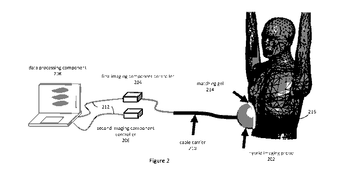
As shown in Figure 2, a hybrid medical imaging apparatus in accordance with an
embodiment of the present invention includes a hybrid imaging probe 202, first
and
second imaging component controllers 204, 206, and a data processing component
208.
In the described embodiments where the first imaging technology is an
ultrasound
imaging technology, the hybrid imaging probe includes an ultrasound imaging
probe
component and a microwave imaging probe component, and the first imaging
component controller 204 is an ultrasound imaging controller known to those
skilled in
the art. The second imaging component controller 206 is a microwave imaging
component controller, and in the described embodiments is in the form of a
vector
network analyser ("VNA") known to those skilled in the art.
Figure 3 is a block diagram of the data processing component 208 of the hybrid
medical
imaging apparatus, in accordance with the described embodiment of the present
invention. The data processing component 208 executes a hybrid medical imaging
process, as shown in Figure 4. As indicated in Figure 2, the data processing
component
CA 03131272 2021-08-24
WO 2020/181336
PCT/AU2020/050242
- 10 -
receives imaging data from the first imaging component controller 204 (being
an
ultrasound imaging component controller in the described embodiments) and
electromagnetic ("EM") scattering data from the second imaging component
controller,
with both imaging component controllers 204, 206 sending and receiving
corresponding
signals to and from the hybrid imaging probe 202.
Although the data processing component of the described embodiments is in the
form
of a computer with hybrid medical imaging processing components 302, 303
installed
therein, this need not be the case in other embodiments. As shown in Figure 3,
the data
processing component 208 of the described embodiments is based on a 64-bit
Intel
Architecture computer system, and the hybrid medical imaging process executed
by the
data processing component 208 is implemented as programming instructions of
software components 302, 303 stored on non-volatile (e.g., hard disk or solid-
state
drive) storage 304 associated with the computer system. However, it will be
apparent
that at least parts of the hybrid medical imaging process could alternatively
be
implemented, either in part or in its entirety, in one or more other forms,
such as
configuration data of a field-programmable gate array (FPGA), and/or as one or
more
dedicated hardware components, such as application-specific integrated
circuits
(ASICs), for example.
The data processing component 208 includes random access memory (RAM) 306, at
least one processor 308, and external interfaces 310, 312, 313, 314, all
interconnected
by a bus 316. The external interfaces include universal serial bus (USB)
interfaces 310,
at least one of which is connected to a keyboard 318 and a pointing device
such as a
mouse 319, and a display adapter 314, which is connected to a display device
such as
an LCD panel display 322. The first and second imaging component controllers
204, 206
are communicatively coupled to the data processing component 208 via the USB
interfaces 310, allowing these controllers 204, 206 to control their
respective probe sub-
components.
The software components 302, 303 include a first imaging component 302 that
receives
imaging signals or data from the first imaging component controller 204, and
generates
corresponding first images 305 of the subject's tissues. Those first images
305 are then
provided as a priori information to an EM processing component 303, which
estimates
dielectric properties of internal organs/tissues and optionally generates EM
images 307
CA 03131272 2021-08-24
WO 2020/181336
PCT/AU2020/050242
- 11 -
of those organs/tissues from the first images 303 and EM scattering data or
signals
received from the second (microwave) component controller 206, as described
below.
In use, the hybrid electromagnetic-ultrasound ("HEUS") imaging probe 202 is
used to
scan a region of interest (e.g., the head or torso) of the body of a
subject/patient. As
shown in Figure 5, in the described embodiments the probe 202 includes a
wideband
antenna or array of antennas 504 that is co-located with an ultrasonic
transducer or an
array of ultrasonic transducers 502.
Depending on the requirements of the imaging algorithm, the targeted organ to
be
imaged and the type of images, either an antenna or an array of wideband
antennas
(as shown in Figure 5) is used. The size of the antenna(s) and (if an array is
used) their
mutual coupling can be reduced in several ways, as described below.
For example, in some embodiments, the antenna size is dramatically reduced by
applying metamaterial loading in which the antenna is loaded with series
capacitance
and/or shunt inductance to create resonances that are independent of the size
of the
antenna, as described in S. Ahdi Rezaeieh, M. A. Antoniades and A. M. Abbosh,
"Miniaturization of Planar Yagi Antennas Using Mu-Negative Metamaterial-Loaded
Reflector," IEEE Transactions on Antennas and Propagation, vol. 65, no. 12,
pp. 6827-
6837, Dec. 2017.
In some embodiments, electromagnetic bandgap (EBG) structures are used to
reduce
mutual coupling by creating an electromagnetic bandgap that prevents the
radiation of
surface currents, as described in H. Nakano, K. Kikkawa, N. Kondo, Y. Iitsuka
and J.
Yamauchi, "Low-Profile Equiangular Spiral Antenna Backed by an EBG Reflector,"
IEEE
Transactions on Antennas and Propagation, vol. 57, no. 5, pp. 1309-1318, May
2009.
In some embodiments, the antennas include artificial magnetic surfaces (AMS)
such as
metasurfaces that are formed using arrays of periodic structures to generate
unidirectional radiation, as described in A. Rezaeieh, M. A. Antoniades and A.
M. Abbosh,
"Compact and Unidirectional Resonance-Based Reflector Antenna for Wideband
Electromagnetic Imaging," IEEE Transactions on Antennas and Propagation, vol.
66, no.
11, pp. 5773-5782, Nov. 2018. These surfaces generate zero reflection phase
which
CA 03131272 2021-08-24
WO 2020/181336 PCT/AU2020/050242
- 12 -
allows the antennas to be located at close proximity to one another and also
to the
reflecting surface of the reflector disposed behind each of the antennas.
Finally, in some embodiments, the hybrid probe 202 includes metamaterial
absorbers
that dissipate the energy of the received signal from certain angles to reduce
the leakage
of electromagnetic signals from the hybrid probe 202, as required by
hospitals.
In the described apparatus, the ultrasound probe component 502 and its
corresponding
controller 204 are used to provide the prior information regarding the
location of the
internal tissues or organ (e.g., the liver) of interest relative to the
patient's skin. For
example, to image the patient's liver, the antenna/antennas transmit microwave
signals
towards and into the patient's torso, and the reflected signals from each
path/tissue are
detected and data representing the detected signals sent by the microwave
component
controller 206 to the data processing component 208. A matching gel 214 can be
used
between the hybrid probe 202 and the patient's torso to facilitate the
penetration of the
signals into the patient's body and reduce surface reflections. The antenna
and
ultrasound signals are transmitted along respective cables by a common cable
loom to
the hybrid probe 202. The electromagnetic microwave signals are generated and
recorded by the portable vector network analyser (VNA) 206. Both the portable
VNA
206 and the US-controller 204 are communicatively coupled to the data
processing
component 208 using suitable data transfer interfaces, cables and protocols,
being USB
in the described embodiments. The data received from the ultrasound and
microwave
imaging component controllers 204, 206 are provided as inputs to the hybrid
medical
imaging process, as described below, and the electromagnetic permittivity and
optionally an image of the region of interest is then generated.
In the described embodiments, the scanning domain is modelled as a multilayer
dielectric slab which is illuminated by a plane wave normally incident from
the or each
antenna at z < 0, as shown in Figure 3. The i¨polarized incident electric
field can be
expressed as:
El (z) = 5'cE0e-Yinz (1)
where E9 is the wave amplitude and ym = jw\hieoem is the propagation constant
of the
matching medium with complex dielectric permittivity of km = e'm ¨jem. The
measured
distance between the skin and the region of interest, for example the
patient's liver d,
CA 03131272 2021-08-24
WO 2020/181336 PCT/AU2020/050242
- 13 -
is used to calculate the total electric field as a function of distance by the
sum of
traveling waves in each tissue region:
/
E0e-Yinz + EleYinz z < 0
Et(z) = E2e-Yaz + E3eYaz 0 < z < d (2)
E4e-Y1(z-d) z > d
Boundary conditions at the interfaces require the continuity of electric and
magnetic
fields Et(z) and Eotz(z) , which results in the following equations:
Eo + El = E2 + E3 (3)
E0¨E1 = i2 1(E2 21(E ,-2 ¨ -3/1
(4)
E2e-Yo + E3eYcid = E4 (5)
E2e Ycld ¨ E3end = 1132E4 (6)
where, fipq = 4'e is the complex refractive index, and ep = e'p ¨je"p is the
complex
Eq
dielectric permittivity of the p-th tissue layer. The solution for the
reflected wave is then
R32CYC/CI-FR2ie-Yda
(7)
R21R32e
E1 = yacce-yad Eo
where,
R21 = 1+11-21 (8)
1¨n21and
1+7-132 (9)
R32 = 1¨n32
Therefore, the S-parameter measured by the or each antenna is estimated by:
= Et(-M) = Eie Yrnm = Ki e-2yr,on = E32eYaci-FR24e-Yda e_2yinm
(10)
E'(-M) E0eYmm E0 R241232eYacce-Yda
In this equation, R32, which is a function of dielectric properties of the
liver (in this
example), is unknown. Knowing the thickness d and dielectric permittivity of
the outer
tissue layer kd, as well as the permittivity of the matching medium km, the
unknown
parameter R32 is estimated by minimizing the error between the measured and
calculated S-parameter, as follows:
R32 = argminISil ¨ g111 (11)
"el
Because the dielectric permittivity is a complex value, a multi-objective
optimization
technique (such as the one described in Kaisa Miettinen (1999), Nonlinear
Multiobjective
CA 03131272 2021-08-24
WO 2020/181336
PCT/AU2020/050242
- 14 -
Optimization, Springer, ISBN 978-0-7923-8278-2) can be used to find a non-
inferior
(trade-off) solution for (11) which simultaneously minimises the real and
imaginary
parts of the error. Therefore, the complex permittivity of the liver ksz is
estimated by:
ez = lR32-Fi) ed (12)
If the hybrid imaging probe 202 includes an array of antennas, the estimated 5-
parameters of each element from equation (10) are used to provide an
estimation
matrix that is used to find the effective permittivity of the liver via an
optimization
process. In the described embodiments, a distributed iterative optimization
algorithm
(such as those described in A. Fa!sone, K. Margellos and M. Prandini, "A
Distributed
Iterative Algorithm for Multi-Agent MILPs: Finite-Time Feasibility and
Performance
Characterization", IEEE Control Systems Letters, vol. 2, no. 4, pp. 563-568,
Oct. 2018
and J. Tsitsiklis, D. Bertsekas and M. Athans, "Distributed asynchronous
deterministic
and stochastic gradient optimization algorithms", in IEEE Transactions on
Automatic
Control, vol. 31, no. 9, pp. 803-812, September 1986) is used to minimise the
estimation error and converge to the global solution for equation (11). The
estimated
value is then used in equation (12) to find the effective permittivity ki of
the targeted
organ, such as the liver.
In embodiments with wideband or multi-frequency antenna(s), different
frequency
steps can be used to generate more accurate estimates. In that case, the Debye
function
is used to model the dielectric permittivity of the targeted tissue according
to:
ez(f) = eo, + 1E-Fs j6)E70 (13)
where, es is the permittivity at zero frequency, eo, is the permittivity at
infinite frequency,
and To is the relaxation time. By substituting equation (13) in the refraction
index
formula and solving the optimization problem of equation (11) for the three
constants
E, E00, and To, the dielectric properties of the organ, such as the liver, can
be estimated
as a function of frequency. In that regard, the signals should be sampled
evenly and
the number of frequency samples should be greater than six (twice the number
of
unknowns in the Debye function of equation (13)).
Knowing the values of the permittivity and conductivity of the healthy organ,
such as
the liver, across the used frequency band, the difference between the
estimated
permittivity of the scanned patient's organ, such as the liver, and the
healthy organ can
CA 03131272 2021-08-24
WO 2020/181336
PCT/AU2020/050242
- 15 -
be interpreted to assess the healthy or unhealthy status of the organ, such as
finding
the percentage of fat in the liver for the case of fatty liver disease, for
example.
In some embodiments, a horizontal cross-section of a patient's chest (torso)
is scanned
and virtually divided into two portions representing the "right side" and
"left side" of the
patient's torso so that the right side portion is mainly occupied by the
patient's liver,
whereas the left side portion of contains the patient's spleen, pancreas and
kidney
organs. In the microwave frequency band of 0.5-1 GHz, the dielectric
properties of the
organs on the left side have an average permittivity of 60, whereas the
average
permittivity of a healthy liver is about 48. Thus, there is about a 25%
difference between
the dielectric properties of the left and right-side organs in a healthy
patient.
Accordingly, the inventors have determined that, using the signal processing
techniques
described herein, the amplitude and phase of the back scattered microwave
signals that
are reflected or transmitted through these organs on the left and right side
portions of
the patient's torso can be used to determine the permittivity of the
investigated organ.
Then, these calculated values are used to define a threshold/range for healthy
subjects.
That is, if a person is healthy, then the reflected/transmitted signals from
left and right
sides exhibit a difference of around 25%. However, the average permittivity of
fatty
liver tissue is around 37, which increases the ratio of the signals for the
left and right
sides to about 62%, and there is more than 100% contrast between the
permittivity of
livers of healthy and unhealthy persons. Thus, these values can be used to
diagnose
and monitor fatty liver and similar diseases in the chest area.
Many modifications will be apparent to those skilled in the art without
departing from
the scope of the present invention.