Note: Descriptions are shown in the official language in which they were submitted.
CA 03132063 2021-08-30
WO 2020/236598 PCT/US2020/033152
MEDICAL IMAGING DEVICES AND SYSTEMS
RELATED APPLICATIONS
[0001] This application claims the benefit of priority under 35 U.S.C. 119
to U.S.
Provisional Application No. 62/849,311, titled "Devices to Access Peripheral
Regions of the
Lung for Direct Visualization with Tool Attachment", filed on May 17, 2019,
the entirety of
which is incorporated herein by reference.
[0002] This application claims the benefit of priority under 35 U.S.C. 119
to U.S.
Provisional Patent Application No. 62/849,649, titled "Apparatus to Provide an
Adjustable
Mechanism for Radial Ultrasound Port and Flush Port", filed on May 17, 2019,
the entirety of
which is incorporated herein by reference.
[0003] This application claims the benefit of priority under 35 U.S.C. 119
to U.S.
Provisional Patent Application No. 62/849,307, titled "Radial Ultrasound
Needle Biopsy
Devices", filed on May 17, 2019, the entirety of which is incorporated herein
by reference.
FIELD
[0004] The present disclosure relates generally to the field of medical
devices. In
particular, the present disclosure relates to devices, systems and methods
that utilize imaging,
and more particularly, devices, systems, and methods that integrate imaging,
such as ultrasound
imaging, and biopsy or other diagnostic and/or therapeutic capability in the
same device.
BACKGROUND
[0005] Generally, endoscopic imaging may be performed to determine the
internal
characteristics of one or more target anatomies. Oftentimes, imaging is used
for
positioning/locating purposes, such as during a diagnostic procedure. For
example, an ultrasound
imaging device may be inserted into a working channel of the endoscope to
image a target
anatomy in an effort to position a tool through an endoscope for a procedure,
such as to biopsy a
pulmonary nodule. In such examples, the ultrasound imaging device may be
removed from the
working channel once the endoscope is positioned and a needle may be inserted
into the working
channel to biopsy the pulmonary nodule if the endoscope is properly
positioned. Challenges with
such a procedure may include maintaining location of the nodule when the probe
is being
exchanged with the needle (e.g., when direct visualization with a bronchoscope
may not be
locatable at the nodule), controlling orientation of the needle with respect
to the nodule, and/or
bronchoscope, and having to actuate the biopsy needle into the nodule tissue
without the benefit
of real-time imaging.
1
CA 03132063 2021-08-30
WO 2020/236598 PCT/US2020/033152
[0006] Biopsies are a group of medical diagnostic tests used to determine
the structure
and composition of tissues or cells. In biopsy procedures, cells or tissues
are sampled from an
organ or other body part to permit their analysis, for example under
microscope. Generally, if an
abnormality is found through superficial examination such as palpation or
radiographic imaging,
a biopsy can be performed to determine the nature of the suspected
abnormality.
[0007] It is with these considerations in mind that a variety of
advantageous medical
outcomes may be realized by the devices, systems and methods of the present
disclosure.
SUMMARY
[0008] In one aspect, the present disclosure relates to a medical device,
comprising a
plunger assembly, a flush port assembly, and a handle body connecting the
plunger assembly to
the flush port assembly. The plunger assembly may be coupled to a first tool
and configured to
move the first tool in a distal and a proximal direction. The flush port
assembly may be coupled
to a second tool and configured to rotate, at least partially, about a
longitudinal axis of the
second tool. In some embodiments, the flush port assembly may be configured to
rotate at least
180 degrees about the longitudinal axis of the second tool. In various
embodiments, the first tool
may include a biopsy needle and the second tool may include a radial
ultrasound probe. In
several embodiments, the plunger assembly and the flush port assembly may be
parallel to one
another in the handle body. In many embodiments, the medical device may
include a bifurcation
joint in the handle body. In many such embodiments, the bifurcation joint may
connect the
plunger assembly to a first lumen of a dual-lumen catheter and the flush port
assembly to a
second lumen of the dual-lumen catheter. In various embodiments, the dual-
lumen catheter may
comprise a layer of braid and a layer of reflow. In some embodiments, the
second tool may
include an imaging transducer configured to communicatively couple with an
imaging controller
via a hub assembly. In some such embodiments, the imaging transducer is
coupled to the hub
assembly via a proximal drive cable with a first diameter and a distal drive
cable with a second
diameter, and the first diameter larger than the second diameter. In several
embodiments, an
impedance compensator connects the distal drive cable to the proximal drive
cable. In many
embodiments, the medical device may include a probe and a dual lumen catheter.
In many such
embodiments, the probe may include an imaging window and a marker, and the
dual lumen
catheter may connect the handle body to the probe, wherein the dual lumen
catheter comprises a
braided layer, wherein the braided layer is configured to axially rotate the
probe within a body
lumen in response to axial rotation of the handle body. In various
embodiments, the medical
device may include an imaging transducer extending through a first lumen of
the dual lumen
catheter and into the probe, the imaging transducer may be configured to
generate an image of a
2
CA 03132063 2021-08-30
WO 2020/236598 PCT/US2020/033152
body lumen via the imaging window, and the image of the body lumen may include
an
indication of the marker. In various such embodiments, the probe comprises a
side port and the
indication of the marker in the image of the body lumen indicates an
orientation of the side port
in the image of the body lumen. In some such embodiments, the side port and
the marker are
oriented 180 degrees apart on the probe. In several embodiments, the medical
device may
include a biopsy needle extending though a second lumen of the dual lumen
catheter and into the
probe, wherein the biopsy needle is configured to exit the probe via the side
port in response to
actuation of an actuation member included on the handlebody. In multiple
embodiments, the
medical device may include a biopsy needle extending though a second lumen of
the dual lumen
catheter and into the probe, wherein the biopsy needle is configured to exit
the probe via the side
port at an angle to the imaging window in response to actuation of an
actuation member included
in the handle assembly. In some embodiments, the medical device may include a
dual-lumen
catheter with first and second lumens. In some such embodiments, the first
tool is disposed in the
first lumen and the second tool is disposed in the second lumen. In various
embodiments, the
medical device may include a probe attached to the distal end of the dual-
lumen catheter,
wherein the probe includes a third lumen aligned with the first lumen of the
dual-lumen catheter
and a fourth lumen aligned with the second lumen of the dual-lumen catheter.
In various such
embodiments, the probe is attached to the distal end of the dual-lumen
catheter via a reflow
process. In one or more embodiments, the medical device may include an imaging
controller
coupled to the imaging transducer via a hub assembly and a coaxial cable. In
some
embodiments, the handle body comprises at least two ergonomic contours that
are mirrored.
[0009] In another aspect, the present disclosure relates to a system,
comprising a handle
assembly, a probe, and a dual lumen catheter. The handle assembly may include
a flush port and
the probe may include an imaging window and a marker. The dual lumen catheter
may connect
the handle assembly to the probe. Further, the dual lumen catheter may
comprise a braided layer
configured to axially rotate the probe within a body lumen in response to
axial rotation of the
handle assembly. In some embodiments, the system may include an imaging
transducer
extending through a first lumen of the dual lumen catheter and into the probe,
and the imaging
transducer configured to generate an image of a body lumen via the imaging
window, wherein
the image of the body lumen includes an indication of the marker. In many
embodiments, the
probe comprises a side port and the indication of the marker in the image of
the body lumen
indicates an orientation of the side port in the image of the body lumen. In
one or more
embodiments, the side port and the marker are oriented 180 degrees apart on
the probe. In many
embodiments, the system includes a biopsy needle extending though a second
lumen of the dual
3
CA 03132063 2021-08-30
WO 2020/236598 PCT/US2020/033152
lumen catheter and into the probe, wherein the biopsy needle is configured to
exit the probe via
the side port in response to actuation of an actuation member included in the
handle assembly.
[0010] In yet another aspect, the present disclosure relates to a system
comprising a
handle assembly, a probe, and a dual lumen catheter. The handle assembly may
include a
bifurcation joint and the probe may include an imaging window and a marker.
The dual lumen
catheter may connect the bifurcation joint to the probe, and be configured to
axially rotate the
probe within a body lumen in response to axial rotation of the handle
assembly.
[0011] In yet another aspect, the present disclosure relates to an
apparatus comprising a
processor and a memory comprising instructions that when executed by the
processor cause the
processor to perform one or more of the following. In some embodiments, the
memory may
include instructions to cause the processor to control one or more aspects of
an imaging
transducer, such as generating an image based on signals received from the
imaging transducer.
[0012] In yet another aspect, the present disclosure relates to a method.
The method may
include one or more of inserting a medical imaging device through a working
channel of a
bronchoscope, extending the medical imaging device past a distal end of the
bronchoscope,
generating an image with the medical imaging device, aligning the medical
imaging device to
take a biopsy of a nodule based on the image, and taking a biopsy of the
nodule based on the
image.
BRIEF DESCRIPTION OF THE DRAWINGS
[0013] Non-limiting embodiments of the present disclosure are described by
way of
example with reference to the accompanying figures, which are schematic and
not intended to be
drawn to scale. In the figures, each identical or nearly identical component
illustrated is typically
represented by a single numeral. For purposes of clarity, not every component
is labeled in every
figure, nor is every component of each embodiment shown where illustration is
not necessary to
allow those of ordinary skill in the art to understand the disclosure. In the
figures:
[0014] FIG. 1 illustrates an exemplary medical imaging device according to
one or more
embodiments described herein.
[0015] FIGS. 2A-2C illustrate various aspects of an exemplary medical
imaging device
according to one or more embodiments described herein.
[0016] FIG. 3A-3H illustrate various aspects of an exemplary probe for a
medical
imaging device according to one or more embodiments described herein.
[0017] FIGS. 4A-4C illustrate an exemplary handle assembly for a medical
imaging
device according to one or more embodiments described herein.
4
CA 03132063 2021-08-30
WO 2020/236598 PCT/US2020/033152
[0018] FIGS. 5A-5E illustrate various aspects of an exemplary handle
assembly
according to one or more embodiments described herein.
[0019] FIGS. 6A and 6B illustrate various internal components of an
exemplary handle
assembly according to one or more embodiments described herein.
[0020] FIGS. 7A-7D illustrate various aspects of an exemplary bifurcation
joint for a
handle assembly according to one or more embodiments described herein.
[0021] FIGS. 8A-8J illustrate various aspects of an exemplary flush port
assembly for a
handle assembly according to one or more embodiments described herein.
[0022] FIG. 9 illustrates various aspects of an exemplary imaging
controller according to
one or more embodiments described herein.
[0023] FIGS. 10A-10M illustrate various exemplary handle assemblies
according to one
or more embodiments described herein.
DETAILED DESCRIPTION
[0024] In various embodiments, the present disclosure relates generally to
medical
imaging devices, such as a real-time visualization and diagnostic and/or
therapeutic tool
assembly (e.g., an assembly with radial ultrasound imaging and biopsy needle
capability) which
may include an ergonomic handle and catheter configured for dual-function use
during a medical
procedure, such as a bronchoscopy. By way of non-limiting example, the medical
device may be
configured for use with a probe, such as one disposed at the distal end of the
catheter, and
delivered within a bronchoscope working channel to provide real-time
visualization (e.g., radial
ultrasound imaging) and manipulation (e.g., diagnostic biopsy sampling) of
pulmonary nodules
in peripheral regions of the lung. As disclosed herein, in various
embodiments, one or more
components of the medical imaging device may be configured to position a
catheter with a first
tool/instrument (e.g., a biopsy needle) within a peripheral region of the lung
while maintaining
real-time visualization of the pulmonary nodule (e.g., with a second
tool/instrument, such as a
radial ultrasound probe). In addition, or alternatively, the assembly may be
configured to allow a
medical professional to access, lock, and/or manipulate a tool/instrument
attached thereto using a
single hand.
[0025] Although embodiments of the present disclosure are described with
specific
reference to assemblies, systems and methods designed to provide dual-function
real-time
visualization and diagnostic sampling of pulmonary nodules within peripheral
regions of the
lung, it should be appreciated that such assemblies, systems and methods may
be used to
visualize and manipulate a variety of tissues within a variety of different
body lumens and/or
body passages for diagnostic and/or therapeutic purposes. In various
embodiments described
herein, direct visualization may refer to video imaging with an endoscope and
real-time
CA 03132063 2021-08-30
WO 2020/236598 PCT/US2020/033152
visualization may refer to imaging with an instrument (e.g., radial ultrasound
probe) inserted
through a working channel of the endoscope and past the distal end of the
endoscope.
Additionally, or alternatively, in one or more embodiments described herein,
direct visualization
may refer to imaging that utilizes the visible spectrum of light (video image)
and real-time
visualization may refer to imaging that does not utilize the visible spectrum
of light (e.g.,
ultrasound imaging or infrared imaging).
[0026] The present disclosure is not limited to the particular embodiments
described. The
terminology used herein is for the purpose of describing particular
embodiments only, and is not
intended to be limiting beyond the scope of the appended claims. Unless
otherwise defined, all
technical terms used herein have the same meaning as commonly understood by
one of ordinary
skill in the art to which the disclosure belongs.
[0027] As used herein, the singular forms "a," "an," and "the" are intended
to include the
plural forms as well, unless the context clearly indicates otherwise. It will
be further understood
that the terms "comprises" and/or "comprising," or "includes" and/or
"including" when used
herein, specify the presence of stated features, regions, steps, elements
and/or components, but
do not preclude the presence or addition of one or more other features,
regions, integers, steps,
operations, elements, components and/or groups thereof.
[0028] As used herein, the term "distal" refers to the end farthest away
from the medical
professional when introducing a device into a patient, while the term
"proximal" refers to the
end closest to the medical professional when introducing a device into a
patient.
[0029] With general reference to notations and nomenclature used herein,
one or more
portions of the detailed description which follows may be presented in terms
of program
procedures executed on a computer or network of computers. These procedural
descriptions and
representations are used by those skilled in the art to most effectively
convey the substances of
their work to others skilled in the art. A procedure is here, and generally,
conceived to be a self-
consistent sequence of operations leading to a desired result. These
operations are those
requiring physical manipulations of physical quantities. Usually, though not
necessarily, these
quantities take the form of electrical, magnetic, or optical signals capable
of being stored,
transferred, combined, compared, and otherwise manipulated. It proves
convenient at times,
principally for reasons of common usage, to refer to these signals as bits,
values, elements,
symbols, characters, terms, numbers, or the like. It should be noted, however,
that all of these
and similar terms are to be associated with the appropriate physical
quantities and are merely
convenient labels applied to those quantities.
[0030] Further, these manipulations are often referred to in terms, such as
adding or
comparing, which are commonly associated with mental operations performed by a
human
6
CA 03132063 2021-08-30
WO 2020/236598 PCT/US2020/033152
operator. However, no such capability of a human operator is necessary, or
desirable in most
cases, in any of the operations described herein that form part of one or more
embodiments.
Rather, these operations are machine operations. Useful machines for
performing operations of
various embodiments include general purpose digital computers as selectively
activated or
configured by a computer program stored within that is written in accordance
with the teachings
herein, and/or include apparatus specially constructed for the required
purpose. Various
embodiments also relate to apparatus or systems for performing these
operations. These
apparatuses may be specially constructed for the required purpose or may
include a general-
purpose computer. The required structure for a variety of these machines will
be apparent from
the description given.
[0031] Reference is now made to the drawings, wherein like reference
numerals are used
to refer to like elements throughout. In the following description, for
purpose of explanation,
numerous specific details are set forth in order to provide a thorough
understanding thereof. It
may be evident, however, that the novel embodiments can be practiced without
these specific
details. In other instances, well known structures and devices are shown in
block diagram form
to facilitate a description thereof. The intention is to cover all
modification, equivalents, and
alternatives within the scope of the claims.
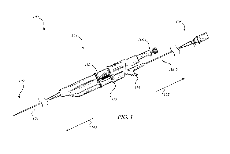
[0032] FIG. 1 illustrates a medical imaging device 100 according to one or
more
embodiments described herein. Generally, medical imaging device 100 may
include a probe 102,
a handle assembly 104, and a hub assembly 106. The probe 102 may be connected
to the handle
assembly 104 via a dual lumen catheter 108 that, among other features,
facilitates the efficient
and reliable use of first and second medical instruments/tools 116-1, 116-2 in
conjunction with
medical imaging device 100. For example, the first tool 116-1 may include a
biopsy needle and
the second tool 116-2 may include a radial ultrasound probe. The medical
imaging device 100
may include a distal end 145 at probe 102 and a proximal end at hub assembly
106. The handle
assembly 104 may include a tool lock 110, an actuation member 112, and a flush
port 114. The
actuation member 112 may operate the first tool 116-1 between multiple
positions when tool
lock 110 is unlocked. In one or more embodiments, the hub assembly 106 may
interface with
logic and/or control circuitry to operate at least the tool 116-2. For
example, tool 116-2 may
include one or more transducers for imaging that can be interfaced with a
controller (e.g.,
imaging control 990 of FIG. 9) via hub assembly 106. In many embodiments, one
or more
components illustrated in FIG. 1, or described with respect thereto, may be
the same or similar in
construction, function, and/or appearance as one or more other components
described herein.
Embodiments are not limited in this context.
7
CA 03132063 2021-08-30
WO 2020/236598 PCT/US2020/033152
[0033] In various embodiments, the probe 102 may be inserted into a body
lumen for
diagnostic and/or therapeutic purposes. For example, medical imaging device
100 may be
utilized to image and/or biopsy a nodule within a body lumen of a patient. In
some
embodiments, the medical imaging device 100 may be used as a stand-alone
device for insertion
into a body lumen. However, in additional, or alternative, embodiments the
medical imaging
device 100 may be configured to extend through the working channel of another
medical device
(e.g., a duodenoscope, endoscope, ureteroscope, bronchoscope, colonoscope,
arthroscope,
cystoscope, hysteroscope, etc.). For instance, medical imaging device 100 may
be inserted via a
bronchoscope to biopsy lung tissue.
[0034] In many embodiments, the medical imaging device 100 may be modular
(include
one or more modular assemblies), such as to facilitate efficient
manufacturing, selectable tools,
and/or reliable operation. In several embodiments, the first and second tools
116-1, 116-2 may
have a parallel configuration within the handle assembly 104. In several such
embodiments, the
parallel configuration may facilitate reliable and intuitive single-handed
operation with either
hand. For example, tool lock 110 may provide ambidextrous operation (see e.g.,
FIGS. 5C-5E).
[0035] The flush port may facilitate fluid to be provided proximate the
distal end 145,
such as via the lumen of tool 116-2. In several embodiments, a fluid, such as
saline, may be
introduced via the flush port 114. In some embodiments, a fluid that assists
with imaging may be
introduced via the flush port 114, such as a conductive medium that displaces
a another less
conductive medium. For example, saline may be introduced to the distal end of
medical device
100 via flush port 114 to enhance the propagation of sound waves from an
ultrasound transducer,
as tool 116-2 within probe 102, as compared to air. In some embodiments, flush
port may be
used to conduct other types of fluids for various other diagnostic or
therapeutic purposes.
[0036] The dual lumen catheter 108 and a proximal portion of tool 116-2
(e.g., between
flush port 114 and hub assembly 106) may have the same, or different,
diameters. In some
embodiments, a common diameter may be enabled by the fact that the proximal
portion of tool
116-2 has a larger diameter drive cable than the distal portion of tool 116-2
that extends through
dual lumen catheter 108.
[0037] FIGS. 2A-2C illustrate various aspects of medical imaging device 100
according
to one or more embodiments described herein. More specifically, FIG. 2A
illustrates axial
displacements and rotations of one or more of probe 102, handle assembly 104,
tool 116-1, and
tool 116-2. FIG. 2B illustrates axial displacements and rotations between
flush port 114 and
handle body 243. FIG. 2C illustrates axial rotations of tool 116-2 between the
distal and
proximal ends 145, 155 of medical imaging device 100. In embodiments described
herein,
components of the medical imaging device 100 may rotate and/or translate in
reliable, intuitive,
8
CA 03132063 2021-08-30
WO 2020/236598 PCT/US2020/033152
and unique and advantageous ways. For example, embodiments many include an
adjustable and
leak-proof flush port assembly configured for use with a bronchial radial
ultrasound system to
provide real-time imaging and targeting of difficult to access pulmonary
nodules. For instance,
the medical imaging device 100 may extend beyond the distal end of a
bronchoscope to access
narrower pulmonary passages than the distal end of the bronchoscope can
access. In such
instances, e.g., the medical imaging device may be configured to extend 15
centimeters or more
beyond the distal end of the bronchoscope. Further, the adjustable flush port
assembly may
include an adjustable ultrasound probe and/or flush port configured to allow a
physician to
proximally/distally (e.g., along a longitudinal axis), laterally (e.g., along
a radial axis), and/or
axially (e.g., about or around a longitudinal axis) position/reposition
components of the
bronchial radial ultrasound system (e.g., ultrasound probe, flush port, and/or
probe assembly)
within a peripheral region of the lung while maintaining a leak-proof seal to
simultaneously
flush fluid through a lumen of the radial ultrasound probe. In many
embodiments, one or more
components illustrated in FIGS. 2A-2C, or described with respect thereto, may
be the same or
similar in construction, function, and/or appearance as one or more other
components described
herein. Embodiments are not limited in this context.
[0038] Referring to FIG. 2A, the probe 102 may include imaging window 222,
side port
220, marker 224, and the handle assembly 104 may include actuation member 112.
Additionally,
lumens 218-1, 218-2 (or lumens 218) may extend approximately between the
distal end 245 of
probe 102 to the proximal end 255 of handle assembly 104. More specifically,
the first lumen
218-1 may include a distal opening at side port 220 and the second lumen 218-2
may include a
distal opening in or at the distal end 245. In some embodiments, one or more
of the lumens 218
may be capped or sealed at the distal end. For example, lumen 218-2 may be
capped by a
balloon at the distal end 245. In various embodiments, the first tool 116-1
may be disposed in the
first lumen 218-1 and the second tool 116-2 may be disposed in the second
lumen 218-1. In one
or more embodiments, the second tool 116-2 may include an imaging transducer.
In one or more
such embodiments, the first tool 116-1 may include a biopsy needle.
[0039] In the illustrated embodiment, the probe 102 includes an imaging
window 222
and a marker 224. In many embodiments, imaging window 222 may refer to one or
more
portions of the probe 102 that are substantially transparent to the imaging
energy wave lengths
while marker 224 may refer to one or more portions of the probe that are
relatively opaque to the
imaging energy wave lengths. Marker 244 may comprise any medium that absorbs
imaging
energy wavelengths (e.g., ultrasound waves). For example, metal or metal
alloys (e.g., stainless
steel or nitinol) may be used. In some embodiments, non-metals may be used,
such as air
pockets embedded in the wall of the imaging window. In various embodiments,
the marker 244
9
CA 03132063 2021-08-30
WO 2020/236598 PCT/US2020/033152
may be radiopaque, such as to show up on x-ray and/or fluoroscopic imaging
additionally, or
alternatively.
[0040] In such embodiments, marker 244 may be positioned to indicate in a
generated
image where the first tool 116-1 would be positioned when actuation member 112
is moved
distally along axial displacement 226-2 to cause axial displacement 226-1 in
tool 116-1,
resulting in tool 116-1 extending out of side port 220. To position the probe
102 based on
generated images, the handle assembly 104 may be rotated along axial rotation
228-1 to cause
probe 102 to rotate along axial rotation 228-2. For example, the handle
assembly 104 may be
rotated to align the side port 220 with a target nodule based on indications
of marker 224 in
generated images. In some such examples, once aligned, actuation member 112
may be moved
distally to cause the distal end of the first tool 116-1 to contact and/or
penetrate the target
nodule. In various embodiments, a marker may be embedded in a wall of a lumen,
such as the
wall of the second lumen 218-2. As will be appreciated, device rotation (e.g.,
orientation of the
marker and the needle radially) may enable more efficient biopsying of
eccentric nodules, e.g.,
when biopsying target tissue that has irregular margins, is of an asymmetric
shape, does not
extend around an entire circumference of the body lumen, and the like, where
control or
orientation and position of the needle may be more critical.
[0041] As an example, marker 224 may be oriented around the circumference
of the
imaging window at a known angle from side port 220. In such a case, e.g., when
targeting a
lung nodule for core biopsy, marker 224 may be oriented on the radial
ultrasound image at the
known angle from the intended biopsy site, so that a needle exiting side port
220 will be
correctly aligned with the biopsy site. In a further such example, the marker
224 may be oriented
on the radial ultrasound image 180 degrees across from the intended biopsy
site. In many
embodiments, the known angle from the intended biopsy site may be configured
such that
tolerances may be provided. For example, the marker 224 may be oriented on the
radial
ultrasound image 180 35 degrees from the intended biopsy site.
[0042] Referring to FIG. 2B, the handle assembly 104 may have a distal end
245 and a
proximal end 255 and include handle body 243 and flush port 114. In many
embodiments, flush
port 114 may have one or more of distal axial rotation 230, proximal axial
rotation 232, and
proximal/distal displacement 234. In several embodiments, the flush port 114
may rotate
approximately 270 degrees without contacting with the plunger assembly and/or
handle body. In
many embodiments, rotation of the flush port 114 may allow the flush port 114
to be positioned
such that the proximal drive cable does not interfere with usage, contributing
to ease of one-
handed control. In various embodiments, absent any interference structures,
the flush port 114
may rotate 360 degrees. Referring to FIG. 2C, the second tool 116-2 may extend
from the hub
CA 03132063 2021-08-30
WO 2020/236598 PCT/US2020/033152
assembly 106 through the flush port 114 to the distal end 245 of probe 102. In
various
embodiments, axial rotation 236-2 via hub assembly 106 may cause axial
rotation 236-1 in probe
102. In various such embodiments, axial rotation 236-1 may enable tool 116-2
to generate a
three hundred and sixty degree image and/or may allow tool 116-2 to be
oriented rotationally as
desired to align side port 220 and tool 116-1 with an intended target site. As
will be discussed in
more detail below, such as with respect to FIG. 9, hub assembly 106 may
connect to an imaging
controller that is able to rotate the tool 116-2 via the hub assembly 106.
[0043] FIGS. 3A-3H illustrate various aspects of an exemplary probe 302 for
a medical
imaging device according to one or more embodiments described herein. More
specifically, FIG.
3A illustrates a perspective view of a probe 302 and FIG. 3B illustrates a
cross-sectional view of
the probe 302. FIG. 3C illustrates a collar 342 in conjunction with a distal
juncture 346 of the
probe 302. FIG. 3D illustrates the collar 342 and distal juncture 346 in
conjunction with marker
344 and tubular member 354. FIG. 3E illustrates an endcap 340 of the probe
302. FIG. 3F
illustrates a front perspective view of probe 302. FIG. 3G illustrates a braid
360 utilized in the
dual lumen catheter 308. FIG. 3H illustrates an example image 362 generated by
the probe 302
(see e.g., FIG. 9). In embodiments described herein, components of the probe
302 may facilitate
specific tissues to be targeted in reliable, intuitive, and unique and
advantageous ways. For
example, embodiments many include an endcap 340 disposed on the distal end of
the dual lumen
catheter 308. Further, the endcap 340 may align with each lumen in catheter
308, enabling
needle 316-1 to exit side port 320 and ultrasound transducer 316-2 to utilize
imaging window
322 and marker 344. In many embodiments, marker 344 may provide indications
and/or
projections of the position of needle 316-1 as it exits side port 320. This,
among other features,
can provide techniques to improve the efficiency, accuracy, and/or reliability
with which
biopsies can be taken. For instance, the real-time images with marker 344 may
allow a user to
determine where and from what angle the biopsy needle will exit the side port
radially and/or
biopsy the tissue, prior to activation, which may be particularly useful in
biopsying eccentric
nodules. Embodiments are not limited in this context.
[0044] Biopsies can be performed on a number of organs, tissues, and body
sites, both
superficial and deep, and a variety of techniques may be utilized depending on
the tissue or body
part to be sampled, the location, size, shape and other characteristics of the
abnormality, the
number of abnormalities, and patient preference. FNA (fine needle aspiration)
is typically
performed to sample deep tissues such as the kidney using a fine gauge needle
(22 or 25 gauge)
inserted percutaneously or through an endoscope under ultrasound guidance (EUS-
FNA). By
contrast, surgical biopsy is generally performed as an open procedure and can
be either
excisional (removal of an entire lesion) or incisional (removal of a piece of
a lesion).
11
CA 03132063 2021-08-30
WO 2020/236598 PCT/US2020/033152
[0045] Surgical biopsies generally permit removal of more tissue than fine
needle
biopsies and, thus, are less prone to misdiagnosis. Open surgical procedures
may be significantly
more expensive than needle biopsies, may require more time for recuperation,
require sutures,
may leave a disfiguring scar, may require anesthesia, may carry a small risk
of mortality, and
may result in bleeding, infection and wound healing problems.
[0046] Fine needle biopsies, however, may carry risks of their own: the
relatively small
quantities of tissue sampled may not be representative of the region of
interest from which it is
taken, particularly when that region of interest is hard to image, or the
nodule is very small, very
hard, and/or eccentric. Additional difficulties may arise in the context of
ultrasound-guided fine
needle biopsies: fine-gauge biopsy needles are typically stiffer, and less
prone to deflection, than
the catheter-based endoscopic ultrasound transducers used to guide them in
some EUS-FNA
procedures. Thus, while it may be possible to guide the transducer to a site
of interest, it may not
be possible to accurately sample it if the needle is too stiff to navigate the
same path through the
tissue. In addition, current practice involves "blind" actuation of the biopsy
needle, which may
result in damage to non-target tissues or false negative results if healthy
tissue is sampled
inadvertently next to the intended suspect tissue site.
[0047] The difficulties of fine needle biopsies are magnified in the
context of pulmonary
nodule sampling, where breathing rhythm cause nodules, probes and needles to
move relative to
one another. It would be particularly desirable in this setting to be able to
visualize the nodule
and needle in real time during patient respiration to ensure accurate needle
tracking and
sampling. Accordingly, one or more embodiments and/or features herein may
resolve or
minimize these issues.
[0048] Referring to FIG. 3A, probe 302 may include endcap 340 coupled to
dual-lumen
catheter 308 (e.g., via overmolding and/or bonding by reflowing), needle 316-1
extending out of
side port 320, imaging window 322, marker 344, collar 342, and imaging
transducer 316-2. As
shown in the cross-sectional view in FIG. 3B, probe 302 may include first and
second lumens
318-1, 318-2, needle 316-1 with stylet 352 extending therethrough, imaging
transducer 316-2
with distal cable 356, side port 320 with ramp 348, imaging window 322, collar
342, marker
344, distal juncture 346, and strain relief 350.
[0049] Referring to FIG. 3C, the distal juncture 346 may comprise a portion
of the first
and second lumens 318. The distal juncture 346 may include side port 320 and
ramp 348. In
various embodiments, the angle of the ramp 348 may be, with respect to the
longitudinal axis in
the proximal direction, between 0 and 90 degrees. In many embodiments, higher
angles may
improve nodule (e.g., an eccentric lesion) targeting, but make needle
actuation more difficult.
For example, the higher the angle of the ramp 348, the larger the longitudinal
force required to
12
CA 03132063 2021-08-30
WO 2020/236598 PCT/US2020/033152
extend the needle up the ramp 348 and out of the side port 320 to actuate the
needle. Further,
higher angles may require the needle to extend further out of the side port
320 to enter the field
of view of the imaging transducer 316-2, limiting applicability to body lumens
with larger
diameters. Conversely, lower angles may require the probe 302 to be located
closer to a target
nodule, making it difficult to acquire biopsy samples from an eccentric nodule
and/or further
below the surface of the target nodule. Accordingly, the ramp angle may be
selected based on a
particular application. In one or more embodiments, the ramp angle may be
greater than or equal
to 3 degrees and less than or equal to 20 degrees. For instance, the angle of
ramp 348 may be 15
degrees. The distal juncture 346 and/or collar 342 may comprise a metal or
metal alloy (e.g.,
nickel-titanium). In some embodiments, the collar 342 is laser cut. In many
embodiments, the
collar 342 may provide one or more of rigidity, confinement, structure, and
flexibility. As shown
in FIG. 3D, a tubular member 354 may extend into the collar 342. In various
embodiments,
tubular member 354 may comprise a portion of the dual-lumen catheter. Marker
344 is also
illustrated in FIG. 3D. Marker 344 may comprise any medium that absorbs
imaging energy
wavelengths (e.g., ultrasound waves). For example, metal or metal alloys
(e.g., stainless steel or
nitinol) may be used. In some embodiments, non-metals may be used, such as
air. For example,
the marker 344 may comprise a pocket of air or a plurality of air bubbles
extruded into the wall
of the imaging window.
[0050] Referring to FIG. 3E, endcap 340 may receive the components
illustrated in FIG.
3D. Marker 344 may be disposed in marker pocket 357 and side port 320 may be
aligned with
port window 359. In some embodiments, marker pocket 357 may comprise a pocket
of air in a
wall of endcap 340. Additionally, endcap 340 include imaging window 322 and
strain relief 350.
In one or more embodiments, imaging window 322 may be the same or similar
material as the
other portions of endcap 340. In various embodiments, the strain relief 350
may limit bending
caused by marker 344 and/or distal juncture 346. In various embodiments, the
gap between
endcap 340 and the layers of braid 360 and reflow 361 remain to allow greater
flexibility.
[0051] As illustrated in FIG. 3F, dual-lumen catheter 308 may include a
tubular member
354 with two lumens, a layer of braid 360, and a layer of reflow 361 over the
braid 360. As
shown in FIG. 3G, the braid 360 may have an overlapping, woven, two wires per
band, two
over/two under and/or crisscross pattern. Other patterns, weave conditions,
materials, and the
like, may be contemplated based on a particular application of the medical
imaging device. In
various embodiments, the number of crossovers per inch of braid 360 may be
between 25 and
140. In some embodiments, different braid strands may be at between 60 and 120
degrees of
each other, such as 90 degrees. In many embodiments, the pattern of braid 360
may be selected
for a combination of flexibility and strength. In various embodiments, the
braid 360 may be
13
CA 03132063 2021-08-30
WO 2020/236598 PCT/US2020/033152
woven stainless steel or nitinol. In several embodiments, the braid 360 may
provide torsional
strength to the dual lumen catheter. In several such embodiments, the
torsional strength provided
by the braid 360 may enable rotation of the handle to translate into rotation
of the distal end. In
many embodiments, rotation of the handle may result in rotation of the distal
end (e.g., the
probe) with a known ratio. For example, rotation of the handle may result in
1:1, or substantially
1:1, rotation of the distal end. Further, the ability to rotate the handle and
cause rotation at the
distal end may facilitate targeting of nodules, such as eccentric nodules,
based on real-time
images with marker indications. In many embodiments, endcap 340 may be coupled
to the dual
lumen catheter 308 via a reflow and/or overmold process. In many embodiments,
the dual lumen
catheter 308 may additionally, or alternatively, include a braided layer, such
as braid 360. In
some embodiments, the braid 360 may utilize up to 356 different strands. For
example, some
embodiments may utilize 64 different strands.
[0052] FIG. 3H illustrates an example image 362 generated by the probe 302
(see e.g.,
FIG. 9). The image 362 may include marker 344, needle 316-1, and nodule 364.
As shown in
FIG. 3H, marker 344 may provide indications and/or projections of the position
of needle 316-1
as it exits side port 320 in generated images (e.g., ultrasound images). This,
among other
features, can provide techniques to improve the efficiency, accuracy, and/or
reliability with
which biopsies can be taken (e.g., the efficiency and reliability of
positioning of biopsy needle to
tip to optimize sampling). In the illustrated embodiment, needle 316-1 is
extended to
demonstrate the indication it may provide on the image compared to marker 344.
[0053] In various embodiments, marker 344 may be oriented around the
circumference
of the imaging window at a known angle from side port 320. In such a case,
e.g., when targeting
nodule 364 (e.g., an eccentric or concentric lung nodule) for core biopsy,
marker 344 may be
oriented on the image 362 (e.g., a radial ultrasound image) at the known angle
from the intended
biopsy site, so that a needle exiting side port 320 will be correctly aligned
with the biopsy site.
For example, in the illustrated embodiment, marker 344 may be oriented on the
image 362 at
180 35 degrees from the intended biopsy site (i.e., nodule 364).
[0054] FIGS. 4A-4C illustrate an exemplary handle assembly 404 for a
medical imaging
device according to one or more embodiments described herein. More
specifically, FIG. 4A
illustrates a side view of the handle assembly 404, FIG. 4B illustrates a
bottom view of the
handle assembly 404, and FIG. 4C illustrates a top view of the handle assembly
404. In
embodiments described herein, components of the handle assembly 404 may
facilitate intuitive,
ergonomic, and/or single-handed operations to target specific tissues in
reliable, intuitive, and
unique and advantageous ways. For example, handle assembly 404 may include one
or more
ergonomic contours, grip ribs, ergonomic reliefs, component positionings
and/or configurations
14
CA 03132063 2021-08-30
WO 2020/236598 PCT/US2020/033152
to provide convenient, comfortable, accurate, and fatigue minimizing
operation. For example,
tool lock 410 may provide ambidextrous operation and/or dual sided access for
single-hand use
(e.g., tool lock 410 is accessible by thumb while rotating the handle). In
another example, grip
ridge 444 and grip ribs 446, 447 may provide non slip surfaces on actuation
member 412. In
many embodiments, one or more components illustrated in FIGS. 4A-4C, or
described with
respect thereto, may be the same or similar in construction, function, and/or
appearance as one or
more other components described herein. Embodiments are not limited in this
context.
[0055] Referring to FIG. 4A, handle assembly 404 may have distal and
proximal ends
445, 455 and include handle body 443, actuation stop 442, actuation member 412
with grip ribs
446 and grip ridge 444, tool lock 410, ergonomic relief 448, strain reliefs
450-1, 450-2, plunger
assembly 440, and flush port assembly 452. Referring to FIG. 4B, handle
assembly 404 may
include one or more ergonomic contours 454-1, 454-2, 454-3, 454-4, tool lock
410, and grip rib
447. Referring to FIG. 4C, handle assembly 404 may include displacement gauge
457, Luer lock
458, and cap 460. In some embodiments, cap 460 may be a stylet cap and/or Luer
lock 458 may
be a syringe connector. In various embodiments, one or more of the ergonomic
contours 454,
grip ribs 446, 447, ergonomic reliefs 448, and/or grip ridges 444 may be
symmetrical,
complementary, and/or mirrored. Further, one or more surfaces may include
textures and/or
coatings to promote or fight friction.
[0056] Various handle assembly embodiments described herein may include one
or more
of a modular assembly, a bifurcated junction, linear needle orientation, a
manual slider (e.g.,
actuation member 112), a needle lock (e.g., tool lock 110), an integrated
flush port (e.g., flush
port assembly 452), a syringe attachment, and dual strain relief (e.g., strain
reliefs 450-1, 450-2).
The medical imaging device may include two independent modules: the needle
module and the
ultrasound module. These two modules may be assembled separately and joined
together inside
the handle body 443. In various embodiments, one or more of the modules may be
interchangeable. For instance, the needle module may be replaced with a module
with a different
tool, such as another diagnostic and/or therapeutic medical tool. The needle
line and the
ultrasound line may converge inside a bifurcated junction (e.g., bifurcation
junction 664) before
feeding into a dual-lumen catheter. The bifurcated junction may dictate the
bend radius of the
ultrasound line. The needle module (e.g., plunger assembly 640) may be axially
aligned with a
lumen (e.g., lumen 218-1), such as to reduce the force required to actuate the
needle. In other
words, the needle module may extend linearly into a first lumen of the dual-
lumen catheter.
[0057] In various embodiments, one or more features of the medical imaging
device may
provide for tactile registration. In some embodiments, the extended handle
profile and short
transition curve may provide more comfortable and substantial grip locations
and/or
CA 03132063 2021-08-30
WO 2020/236598 PCT/US2020/033152
accommodate a wider range of hand sizes. For example, handle assembly 404 may
accommodate adult hand sizes ranging from the 5th percentile of female hands
to the 95th
percentile of male hands. The displacement gauge 457 (e.g., corresponding to
graduated stroke
depths of the needle exiting the ramp and the side port) may be readily
readable from a variety of
viewing angles, such as by partially wrapping around the plunger. In some
embodiments, a soft-
touch finish on the actuation member 412 may create a contrasting feel to the
handle body 443
and/or match the distal handle finish. Some components may include rubberized
texture
overmolds and/or color accent, such as on the actuation member grip ridge 444.
In some
embodiments this and other features may provide an improved thumb grip and/or
visual travel
indication. The handle body may have a smooth/semi-gloss finish in some
embodiments.
Various embodiments may include a horizontal groove texture on the tool lock
410, such as for
an ergonomic detail and/or precision feel. Several embodiments include a
textured finish around
the tool lock 410 to create tactile contrast, such as for intuitive use. The
actuation stop 442 (or
hand hilt) and/or grip ridge 444 may provide 360 degree tactile registration.
In some
embodiments, the actuation stop 442 and/or grip ridge 444 may provide a
boundary for hand
position and/or hand protection during actuation. Further, the actuation stop
442 and/or grip
ridge 444 may provide a non-visual indicator of hand position. Various
embodiments may
include a soft touch finish and/or slight rubberized texture on a distal
portion of the handle body
443.
[0058] Several embodiments may include a solid color band that wraps around
the
handle body to indicate ultrasound zone is exposed when the actuation member
412 is moved
distally. In several such embodiments, the band may include a slight texture
change and/or an
ultrasound icon disposed proximately. In one or more embodiments, an
additional part break line
on a strain relief connection may allow for individual rotation.
[0059] FIGS. 5A-5E illustrate various aspects of an exemplary handle
assembly 504 for
a medical imaging device according to one or more embodiments described
herein. More
specifically, FIG. 5A illustrates the handle assembly 504 in an unactuated
configuration 500A
and FIG. 5B illustrates the handle assembly 504 in an actuated configuration
500B. In various
embodiments, actuation member 512 may be moved distally to cause a tool, such
as a biopsy
needle, to exit the side port of the probe as indicated in the image by the
marker. FIGS. 5C-5E
illustrate operation of a tool lock 510 of handle assembly 504. In embodiments
described herein,
components of the handle assembly 504 may facilitate intuitive, ergonomic,
and/or single-
handed operations to target specific tissues in reliable, intuitive, and
unique and advantageous
ways. For example, handle assembly 504 may utilize intuitive motions for
gripping, locking,
unlocking, and actuating to provide for convenient, comfortable, accurate, and
fatigue
16
CA 03132063 2021-08-30
WO 2020/236598 PCT/US2020/033152
minimizing operation. In many embodiments, one or more components illustrated
in FIGS. 5A-
5E, or described with respect thereto, may be the same or similar in
construction, function,
and/or appearance as one or more other components described herein.
Embodiments are not
limited in this context.
[0060] Referring to FIG. 5A, the handle assembly 504 is in unactuated
configuration
500A when the distal end of actuation member 512 is positioned proximal of the
tool lock 510.
In the unactuated configuration 500A, tool lock can be engaged or disengaged.
An unlock
movement 562-1 may be used to position the tool lock 510 in the middle such
that actuation
member can move distally over either side of the tool lock 510 in an actuation
movement 562-2
to place the handle assembly 504 in an actuated configuration 500B (see e.g.,
FIG. 5B). The
positioning of the tool lock will be discussed in more detail below with
reference to cross-
sectional line 565. In many embodiments, tool lock 510 may provide a visual
indicator of the
lock status of the plunger assembly. The ultrasound flush port valve (e.g.,
flush port assembly)
may be integrated into the handle assembly and/or handle body design. In
several embodiments,
the flush port is positioned and/or positionable away from a grip zone of a
user. In various
embodiments, the medical imaging device may utilize two syringes (e.g., one
for ultrasound
flushing and one for needle suction and/or aspiration). In many embodiments,
syringes may
attach to the medical imaging device with stopcocks Luer-lock fittings, and/or
check valves. For
example, a check valve may be positioned between the flush port and a syringe
connector (e.g.,
Luer-lock). The handle assembly may include integrated strain relief at both
the distal and
proximal ends of the handle body (see e.g., strain reliefs 450-1, 450-2). In
some embodiments,
the distal strain relief (e.g., strain relief 450-1 may be utilized as an
additional grip space).
[0061] Referring to FIGS. 5C-5E, handle assembly 504 may have positions
500C, 500D,
500E in addition to the unactuated configuration 500A and the actuated
configuration 500B. In
position 500C, the tool lock 510 may be in a first locked position with
actuation member 512
being blocked from moving distally (out of the page). Similarly, in position
500D, the tool lock
510 may be in a second locked position with actuation member 512 being blocked
from moving
distally (out of the page). In position 500E, tool lock 510 may be in an
unlocked position with
actuation member 512 not blocked from moving distally (out of the page). This
arrangement
may allow for actuation of the tool lock 510 from either side of the handle,
depending on which
side is more accessible to the fingers of the user. Further, as the distal
direction is out of the page
in FIGS. 5C-5E, it will be appreciated that although the flush port would be
visible, it is not
illustrated for simplicity.
[0062] FIGS. 6A and 6B illustrate various internal components of an
exemplary handle
assembly 604 for a medical imaging device according to one or more embodiments
described
17
CA 03132063 2021-08-30
WO 2020/236598 PCT/US2020/033152
herein. More specifically, FIG. 6A illustrates a first cross-sectional view of
the handle assembly
604 having a distal end 645 and a proximal end 655, and FIG. 6B illustrates a
second cross-
sectional view of the handle assembly 604. In the illustrated embodiments,
handle assembly 604
includes a handle body 659 comprising and/or coupling to a bifurcation joint
664, tool lock 610,
actuation member 612, plunger assembly 640, flush port assembly 652, and noise
compensators
670. In embodiments described herein, components of the handle assembly 604
may facilitate
intuitive, ergonomic, and/or single-handed operations to target specific
tissues in reliable,
intuitive, unique, and advantageous ways. For example, handle assembly 504 may
utilize
intuitive motions for gripping, locking, unlocking, and actuating to provide
for convenient,
comfortable, accurate, and fatigue minimizing operation. In various
embodiments, noise
compensators 670 may serve to reduce electrical noise in the distal and/or
proximal drive cables.
For example, noise compensators 670 may be an electronic choke, such as a
passive electric
component that suppresses high frequency noise in electronic circuits. In some
embodiments,
one or more of noise compensators 670 may utilize ferrite, such as a ferrite
ceramic. In many
embodiments, one or more components illustrated in FIGS. 6A and 6B, or
described with respect
thereto, may be the same or similar in construction, function, and/or
appearance as one or more
other components described herein. Embodiments are not limited in this
context.
[0063] FIGS. 7A-7D illustrate various aspects of an exemplary bifurcation
joint 764 for
a medical imaging device 700 according to one or more embodiments described
herein. More
specifically, FIG. 7A illustrates a cross-sectional view of the bifurcation
joint 764 in conjunction
with a handle body 743. FIG. 7B illustrates the bifurcation joint 764. FIGS.
7C and 7D illustrate
cross-sectional views of bifurcation joint 764. In various embodiments,
bifurcation joint 764
may connect the needle 716-1 to a first lumen of dual-lumen catheter 708 and a
conduit 744
carrying a portion of the distal drive cable 756 to a second lumen of dual-
lumen catheter 708. In
many embodiments, the bifurcation joint 764 may prevent fluid leaking within
the handle body
743 when fluids are passed into the dual-lumen catheter. In embodiments
described herein,
components of the bifurcation joint 764 may facilitate convenient, reliable,
efficient, and leak-
proof operation in unique and advantageous ways. For example, the bifurcation
joint 764 may
reduce or minimize the bend in conduit 744 to limit bend in the drive cable
(e.g., ultrasound
drive cable). In another example, bifurcation joint 764 includes a needle
support 742 to prevent
needle 716-1 from bending, such as when it is forced distally by the plunger
assembly. In many
embodiments, one or more components illustrated in FIGS. 7A-7D, or described
with respect
thereto, may be the same or similar in construction, function, and/or
appearance as one or more
other components described herein. Embodiments are not limited in this
context.
18
CA 03132063 2021-08-30
WO 2020/236598 PCT/US2020/033152
[0064] Referring to FIG. 7A, bifurcation joint 764 may be disposed in
handle body 743
and include needle support 742. More generally, bifurcation joint 764 may be
the component of
the medical imaging device that routes the first and second tools into the
first and second lumens
of the dual-lumen catheter. In many embodiments, bifurcation joint 764 may
align the first and
second tools for insertion into the dual-lumen catheter 708 while limiting the
amount of bend
required. For instance, bifurcation joint 764 may prevent either tool from
bending over 20
degrees. In another instance, bifurcation joint 764 may limit bending of a
drive cable for a
medical imaging device to less than 15 degrees. In many embodiments, the
minimum radius of
curvature for the drive cable may be 3 inches.
[0065] Medical imaging device 700 may also include strain relief 750-1.
Strain relief
750-1, or one or more other strain reliefs described herein, may limit bending
(e.g., bends over
25 degrees) of the dual-lumen catheter 708 or other portions along the length
of tool lumens
(e.g., conduit 744). In some embodiments, conduit 744 may comprise a polymer
tube, such as a
PEEK or Nylon tube. In several embodiments, bifurcation joint 764 supports
parallel alignment
of the plunger assembly and the flush port assembly in the handle assembly,
resulting in an
ergonomic and intuitive feel.
[0066] Referring to FIG. 7B, the dual-lumen catheter 708 may connect into
the
bifurcation join 764 at a catheter support 748, conduit 744 may connect into
the bifurcation joint
764 at conduit support 749, and needle 116-1 may connect into needle support
742. In many
embodiments, needle support 742 may prevent the needle from kinking,
paperclipping, bending,
and/or breaking. Bifurcation joint 764 may also include mounts 754-1, 754-2,
754-3 (or mounts
754) on both sides and/or indentations 753-1, 753-2 (or indentations 753). In
many
embodiments, the mounts 754 may be used to attach the bifurcation joint 764 to
handle body
743. As previously mentioned, in one or more embodiments, handle body 743 may
connect one
or more components of a medical imaging device, such as by serving as one or
more of a
mounting point, enclosure, structure, and the like. Additionally, or
alternatively, bifurcation joint
764 may include one or more view windows 752-1, 752-2 (or view windows 752).
In various
embodiments, the view windows 752 may allow visual verification the contents
of the lumens as
they pass through the bifurcation joint.
[0067] FIG. 7C includes a cross-sectional view of the bifurcation joint
764. As shown in
FIG. 7C, the layer of braid 360 and the tubular member (not labeled) begin on
the proximal side
of the indentations 752 and the layer of reflow 361 on the dual-lumen catheter
begins at the
distal end of the bifurcation joint 764. Additionally, or alternatively,
conduit 744 ends between
the view windows 752 and the conduit support 746. The needle support 742 ends
proximate to
the end of conduit 744. In several embodiments, immediately distal of the
needle support 742,
19
CA 03132063 2021-08-30
WO 2020/236598 PCT/US2020/033152
the first lumen of the bifurcation joint includes a junction taper 762-1. In
various embodiments,
immediately distal of the conduit 744, the second lumen of bifurcation joint
includes a junction
taper 762-2. In some embodiments, the junction tapers 762 facilitate one or
more of retaining the
conduit/needle support connected, limiting a bending radius, and improving
fluid flow (such as
preventing leaks or reducing turbulent flow). In medical imaging device 700,
needle 716-1 may
pass through the first lumen of bifurcation joint 764 and distal drive cable
756 may pass through
the second lumen of bifurcation joint 764. FIG. 7D illustrates handle body 743
with strain relief
750-1 removed, leaving strain relief pocket 751.
[0068] FIGS. 8A-8J illustrate various aspects of an exemplary flush port
assembly 852
for a medical imaging device according to one or more embodiments described
herein. More
specifically, FIG. 8A illustrates a cross-sectional view of the flush port
assembly 852. FIGS. 8B
and 8C illustrate axial displacement of flush port assembly 852 in distal and
proximal directions,
respectively. FIG. 8D illustrates axial rotation of flush port assembly 852.
FIG. 8E illustrates
various aspects of the flush port assembly 852. FIG. 8F illustrates various
components of flush
port assembly 852. FIG. 8G illustrates a flow path 861 from flush port 814
into conduit 844.
FIGS. 8H and 81 illustrate various aspects of the flush port assembly 852
including a stabilizer
823 and a bearing 868-2. FIG. 8J illustrates an impedance compensator 843
disposed between a
distal drive cable 856 and a proximal drive cable. In embodiments described
herein, components
of the flush port assembly 852 may facilitate convenient, reliable, efficient,
and leak-proof
operation in unique and advantageous ways. For example, the flush port
assembly 852 may
rotate independently of the proximal and distal drive cables 856, 858 while
maintaining a seal
with conduit 844 that facilitates introduction of fluid into the conduit 844
around the distal drive
cable 856. In another example, impedance compensator 843 matches impedance
between the
distal and proximal drive cables 856, 858. In many embodiments, one or more
components
illustrated in FIGS. 8A-8J, or described with respect thereto, may be the same
or similar in
construction, function, and/or appearance as one or more other components
described herein.
Embodiments are not limited in this context.
[0069] Referring to FIG. 8A, flush port assembly 852 may include bearings
868-1, 868-
2, seal 869, slide mount 823, proximal surface features 880, strain relief 850-
2, lip seal 866, port
member 872, flush port 814, port interface 882, flow junction 876, flow
chamber 874 with taper
825, distal surface features 878, a proximal portion of conduit 844, a portion
of the distal drive
cable 856 and a portion of the proximal drive cable 858. One or more
components described
herein (e.g., port interface 882) may be formed via a molding, extrusion,
and/or machining
procedure (e.g., overmolding, injection molding, vacuum molding, cold
extrusion, lathe, and the
like).
CA 03132063 2021-08-30
WO 2020/236598 PCT/US2020/033152
[0070] As previously mentioned, the flush port assembly 852 may be able to
be adjusted
in the proximal and distal directions. Accordingly, FIG. 8B illustrates the
port assembly 852
including slide mount 823, conduit 844, and port member 872 in the distal most
position and
FIG. 8C illustrates the port assembly 852 including slide mount 823, conduit
844, and port
member 872 in the proximal most position. In many embodiments, conduit 844 may
be
supported by a slide within port member 872 as it is proximally and distally
moved with respect
to the longitudinal axis of the device. The slide mount 823 may couple with a
peg or mounting
point on the handle body. In various embodiments, adjustment along slide mount
823 may be
utilized to calibrate the location of the imaging transducer with respect to
the imaging window,
such as during manufacture. In some embodiments, the end user may be able to
adjust along the
slide mount 823.
[0071] Referring to FIGS. 8D-8E, in one embodiment, a flush port assembly
852 of the
present disclosure may include a housing 810 defining a flow chamber 874. An
ultrasound port
812 (e.g., first port) may be formed within or otherwise extend through a
proximal portion of the
housing 810. In various embodiments, the ultrasound port 812 may be
coextensive (e.g.,
substantially aligned with, etc.) with the flow chamber 874. A flush port 814
(e.g., second port)
defining a fluid channel 115 therethrough may be disposed along (e.g.,
attached to, integrally
formed with, etc.) a middle portion of the housing 810. A fitting 816 may be
disposed around a
distal portion of the housing 810. In various embodiments, the housing 810 may
be configured to
rotate 360 (e.g., move axially) within the fitting 816 to alter a position of
the flush port 814
relative to a longitudinal axis of the flush port assembly 852 (e.g., move
proximally or distally)
and/or rotate (e.g., alter an axial position of) a radial ultrasound probe
extending through the
housing 810 (as discussed below). An outer surface of the distal portion of
the housing may
include distal surface features 818 configured to frictionally engage a
corresponding inner
surface of the fitting 816. By way of non-limiting example, the surface
feature may include a
rubber seal or 0-ring configured to maintain or lock an axial position of the
housing 810 relative
to the fitting 816 until a threshold level of rotational force is exerted on
the housing 810 (e.g., a
sufficient amount of force exerted by a physician's hand). In various
embodiments, an outer
surface of the housing 810 and/or flush port 814 may include a non-slip
surface (e.g., over-
molded or coated with rubber, etc.) to provide a physician with sufficient
grip to manipulate the
housing 810, e.g., when wearing wet gloves, etc. Slide mount 823 (e.g., an arm
or projection)
may extend from an outer surface of the fitting 816 to anchor or lock the
housing of the probe
assembly within a handle body (e.g., handle body 743) of a medical imaging
device with radial
ultrasound and needle biopsy capability.
21
CA 03132063 2021-08-30
WO 2020/236598 PCT/US2020/033152
[0072] In one embodiment, a first seal 869 (e.g., 0-ring, etc.) may be
disposed within a
distal portion of the flow chamber 874 (e.g., proximal to a distal opening of
the housing 810) and
a second seal 124 may be disposed within a proximal portion of the flow
chamber 874 (e.g.,
distal to a proximal opening of the housing 810). The first and second seals
869, 880-1 may be
configured to prevent fluid introduced (e.g., flushed) through the fluid
channel 815 of the flush
port 814 from exiting the flow chamber 874 (e.g., flowing/leaking distally
beyond the first seal
869 or proximally beyond the second seal 124). A bearing 126 may be disposed
within the
proximal portion of the flow chamber 874 proximal to the second seal 124. In
various
embodiments, the housing 810 and ultrasound port 812 may be configured to
receive a proximal
portion of a tool (e.g., a radial ultrasound probe) therethrough. The bearing
126 may be
configured to receive an outer surface of the radial ultrasound probe 130 to
support/facilitate
rotation of the radial ultrasound probe within the housing 810.
[0073] FIGS. 8F and 8G illustrate various components of the flush port
assembly 852,
such as those associated with fluid flow. The flush port assembly 852 of FIG.
8F includes
conduit 844, port member 872, proximal surface features 880, bearing 868-2,
stabilizer 823, lip
seal 866, flow port 857, flow channel 859, flow chamber 874, distal drive
cable 856, and conduit
844. FIG. 8G illustrates a flow path 861 of fluid introduced via the flush
port 814. Accordingly,
the flow path 861 may enter via the flush port 814, fill flow chamber 874,
follow flow channel
859 of port member 842, enter flow port 857 of port member 872, and proceed
into the conduit
844 around the distal drive cable 856. In various embodiments, the lumens
and/or flow
components described herein may be designed to handle at least 43 pounds per
square inch. Such
pressure rating may vary as dictated by design and/or performance
requirements.
[0074] FIG. 8H illustrates stabilizer 823, lip seal 866, and bearing 868-2
and FIG. 81
illustrates stabilizer 823 and bearing 868-2. In various embodiments, the
stabilizer 823 may
extend through one or more of the bearing 868-2 and lip seal 866. The drive
cable may extend
through the stabilizer 823. In many embodiments, the stabilizer may prevent
loss of stability
during rotation of the drive cable.
[0075] Referring to FIG. 8J, proximal of the stabilizer 823, an impedance
compensator
843 may connect distal drive cable 856 and proximal drive cable 858. In
several embodiments,
the proximal and distal drive cable along with the imaging transducer may
rotate at up to 2000 or
more revolutions per minute (rpm). For instance, the imaging transducer (and
drive cables) may
rotate at 1800 rpm. In various embodiments, impedance compensator 843 may
adapt for a
diameter change between the distal and proximal drive cables 856, 858. In many
embodiments,
the impedance compensator spins along with the distal and proximal drive
cables 856, 858. In
one or more embodiments, the impedance compensator 843 comprises a printed
circuit board
22
CA 03132063 2021-08-30
WO 2020/236598 PCT/US2020/033152
(PCB). In many embodiments, the lumen of the distal drive cable 856 is a
uniform size to
prevent kinking or winding up of the distal drive cable. In some embodiments,
the diameter
change between the proximal and distal drive cables prevents signal
degradation. For example, if
the proximal drive cable was as small as the distal drive cable, unacceptable
levels of signal
degradation may occur. In some embodiments, making the proximal drive cable
856 have a
larger diameter than the distal drive cable 858, reduced electromagnetic
emissions (e.g., noise)
can be achieved, such as via lower signal voltages. In several embodiments,
one or more of the
stabilizer 823, and the impedance compensator 843 may be filled with epoxy,
such as to prevent
fluid from leaking around the distal drive cable 856 and into the impedance
compensator 843 via
the stabilizer 823.
[0076] FIG. 9 illustrates various internal components of an exemplary
imaging
controller 990 according to one or more embodiments described herein.
Ultrasound controller
990 may include logic circuitry 992, memory 994, input/output (I/0) 996, and
user interface 998.
As previously mentioned, imaging controller 990 may couple with an imaging
transducer 916-2
via a proximal drive cable 958 connected between the hub assembly 906 and an
impedance
compensator 943, and a distal drive cable 956 connected between the impedance
compensator
943 and the imaging transducer 916-2. In embodiments described herein,
components of the
imaging control 990 may facilitate intuitive, accessible, dynamic monitoring
and control over
imaging transducer 916-2 in reliable, valuable, unique, and advantageous ways.
For example,
imaging controller 990 may control one or more of the calibration, frequency,
resolution,
translation, interpretation, integration, analysis, and/or display of images
generated by one or
more medical imaging devices described herein. In one or more embodiments,
imaging
controller 990 may utilize one or more of historical, contextual, user input,
and sensor data to
control aspects of the medical imaging device. For example, historical data
may include sensor
and/or imaging data from previous procedures. In some such embodiments, the
historical data
may be annotated based on user input. In many embodiments, one or more
components
illustrated in FIG. 9, or described with respect thereto, may be the same or
similar in
construction, function, and/or appearance as one or more other components
described herein.
Embodiments are not limited in this context.
[0077] In various embodiments, proximal and distal drive cable 958, 956 may
comprise
multiple conductors. In some embodiments, the drive cables may be coaxial
cables. In various
embodiment, the drive cables may provide one or more of power, torque,
communication
between the imaging transducer and the imaging controller.
[0078] One or more of the components, devices, and/or techniques described
herein may
be used as part of a system to facilitate the performance of medical
procedures (e.g., peripheral
23
CA 03132063 2021-08-30
WO 2020/236598
PCT/US2020/033152
lung nodule biopsy) in a safe, efficient, and reliable manner. In many
embodiments, the novel
system may include one or more medical devices capable of locating a patient-
specific anatomy,
positioning a flexible elongate member for access to the patient-specific
anatomy, and accessing
the patient-specific anatomy in a safe, accurate, and reliable manner. In
these and other ways,
components/techniques described here may improve patient care, increase user
experience,
decrease learning curve, improve success rates, and/or decrease adverse
outcomes via realization
of a more efficient and better functioning medical device with advantageous
features. In many
embodiments, one or more of the advantageous features may result in several
technical effects
and advantages over conventional devices and technology, including increased
capabilities and
improved adaptability. In various embodiments, one or more of the aspects,
techniques, and/or
components described herein may be implemented in a practical application via
one or more
computing devices, and thereby provide additional and useful functionality to
the one or more
computing devices, resulting in more capable, better functioning, and improved
computing
devices. Further, one or more of the aspects, techniques, and/or components
described herein
may be utilized to improve one or more technical fields including imaging,
endoscopy,
cannulation, diagnosis, treatment, imaging, robotics, embedded systems and/or
control systems.
[0079] In
several embodiments, components described herein may provide specific and
particular manners to render, interpret, transform, analyze, monitor, and/or
characterize images
generated by the medical imaging device, such as via imaging transducer 316-2
(see e.g., FIG.
3B). In several such embodiments, the specific and particular manners may
include, for instance,
controlling, monitoring, and/or interfacing with one or more of a transducer,
a joint, a working
channel, and a user interface to facilitate one or more endoscopy procedures.
In one example, the
specific and particular manner may simplify pulmonary procedures to facilitate
medical
professional to quickly learn to safely and reliably biopsy a target nodule.
[0080] In many
embodiments, one or more of the components described herein may be
implemented as a set of rules that improve computer-related technology by
allowing a function
not previously performable by a computer that facilitates an improved
technological result to be
achieved. In many embodiments, the function allowed is associated with medical
imaging
devices and/or procedures. For example, the function allowed may include
creating a combined
image comprising a characteristic of a wall of a body lumen and a
characteristic external to the
wall of the body lumen based on the first image generated via a first imaging
mode and a second
image generated via a second imaging mode. In some embodiments, the function
allowed may
include positioning a transducer within a focal region of another transducer
with one or more
joints, such as to facilitate image generation with the transducer. In various
embodiments, the
24
CA 03132063 2021-08-30
WO 2020/236598 PCT/US2020/033152
function allowed may include utilizing one or more joints to locate and/or
access objectives of a
cannulation procedure.
[0081] FIG. 10 illustrates an embodiment of an exemplary computing
architecture 1000
that may be suitable for implementing various embodiments as previously
described. In various
embodiments, the computing architecture 1000 may comprise or be implemented as
part of an
electronic device and/or medical device. In some embodiments, the computing
architecture 1000
may be representative, for example, of one or more components described
herein. In some
embodiments, computing architecture 1000 may be representative, for example,
of a computing
device that implements or utilizes one or more portions of components and/or
techniques
described herein, such as imaging controller 990, logic circuitry 992, memory,
994, 1/0 996,
and/or user interface 998. The embodiments are not limited in this context.
[0082] As used in various embodiments herein, the terms "system" and
"component" and
"module" can refer to a computer-related entity, either hardware, a
combination of hardware and
software, software, or software in execution, examples of which are provided
by the exemplary
computing architecture 1000. For example, a component can be, but is not
limited to being, a
process running on a processor, a processor, a hard disk drive, multiple
storage drives (of optical
and/or magnetic storage medium), an object, an executable, a thread of
execution, a program,
and/or a computer. By way of illustration, both an application running on a
controller 106 and
the controller 106 can be a component. One or more components can reside
within a process
and/or thread of execution, and a component can be localized on one computer
and/or distributed
between two or more computers. Further, components may be communicatively
coupled to each
other by various types of communications media to coordinate operations. The
coordination may
involve the uni-directional or bi-directional exchange of information. For
instance, the
components may communicate information in the form of signals communicated
over the
communications media. The information can be implemented as signals allocated
to various
signal lines. In such allocations, each message is a signal. Further
embodiments, however, may
alternatively employ data messages. Such data messages may be sent across
various connections.
Exemplary connections include parallel interfaces, serial interfaces, and bus
interfaces.
[0083] The computing architecture 1000 includes various common computing
elements,
such as one or more processors, multi-core processors, co-processors, memory
units, chipsets,
controllers, peripherals, interfaces, oscillators, timing devices, video
cards, audio cards,
multimedia input/output (1/0) components, power supplies, and so forth. The
embodiments,
however, are not limited to implementation by the computing architecture 1000.
[0084] As shown in FIG. 10, the computing architecture 1000 comprises a
processing
unit 1004, a system memory 1006 and a system bus 1008. The processing unit
1004 can be any
CA 03132063 2021-08-30
WO 2020/236598 PCT/US2020/033152
of various commercially available processors, including without limitation an
AMD Athlon ,
Duron and Opteron processors; ARM application, embedded and secure
processors; IBM
and Motorola DragonBall and PowerPC processors; IBM and Sony Cell
processors;
Intel Celeron , Core (2) Duo , Itanium , Pentium , Xeon , and XScale
processors; and
similar processors. Dual microprocessors, multi-core processors, and other
multi-processor
architectures may also be employed as the processing unit 1004.
[0085] The system bus 1008 provides an interface for system components
including, but
not limited to, the system memory 1006 to the processing unit 1004. The system
bus 1008 can be
any of several types of bus structure that may further interconnect to a
memory bus (with or
without a memory controller), a peripheral bus, and a local bus using any of a
variety of
commercially available bus architectures. Interface adapters may connect to
the system bus 1008
via a slot architecture. Example slot architectures may include without
limitation Accelerated
Graphics Port (AGP), Card Bus, (Extended) Industry Standard Architecture
((E)ISA), Micro
Channel Architecture (MCA), NuBus, Peripheral Component Interconnect
(Extended) (PCI(X)),
PCI Express, Personal Computer Memory Card International Association (PCMCIA),
and the
like.
[0086] The system memory 1006 may include various types of computer-
readable
storage media in the form of one or more higher speed memory units, such as
read-only memory
(ROM), random-access memory (RAM), dynamic RAM (DRAM), Double-Data-Rate DRAM
(DDRAM), synchronous DRAM (SDRAM), static RAM (SRAM), programmable ROM
(PROM), erasable programmable ROM (EPROM), electrically erasable programmable
ROM
(EEPROM), flash memory (e.g., one or more flash arrays), polymer memory such
as
ferroelectric polymer memory, ovonic memory, phase change or ferroelectric
memory, silicon-
oxide-nitride-oxide-silicon (SONOS) memory, magnetic or optical cards, an
array of devices
such as Redundant Array of Independent Disks (RAID) drives, solid state memory
devices (e.g.,
USB memory, solid state drives (S SD) and any other type of storage media
suitable for storing
information. In the illustrated embodiment shown in FIG. 10, the system memory
1006 can
include non-volatile memory 1010 and/or volatile memory 1012. In some
embodiments, system
memory 1006 may include main memory. A basic input/output system (BIOS) can be
stored in
the non-volatile memory 1010.
[0087] The computer 1002 may include various types of computer-readable
storage
media in the form of one or more lower speed memory units, including an
internal (or external)
hard disk drive (HDD) 1014, a magnetic floppy disk drive (FDD) 1016 to read
from or write to a
removable magnetic disk 1018, and an optical disk drive 1020 to read from or
write to a
removable optical disk 1022 (e.g., a CD-ROM or DVD). The HDD 1014, FDD 1016
and optical
26
CA 03132063 2021-08-30
WO 2020/236598 PCT/US2020/033152
disk drive 1020 can be connected to the system bus 1008 by an HDD interface
1024, an FDD
interface 1026 and an optical drive interface 1028, respectively. The HDD
interface 1024 for
external drive implementations can include at least one or both of Universal
Serial Bus (USB)
and Institute of Electrical and Electronics Engineers (IEEE) 994 interface
technologies. In
various embodiments, these types of memory may not be included in main memory
or system
memory.
[0088] The drives and associated computer-readable media provide volatile
and/or
nonvolatile storage of data, data structures, computer-executable
instructions, and so forth. For
example, a number of program modules can be stored in the drives and memory
units 1010,
1012, including an operating system 1030, one or more application programs
1032, other
program modules 1034, and program data 1036. In one embodiment, the one or
more application
programs 1032, other program modules 1034, and program data 1036 can include
or implement,
for example, the various techniques, applications, and/or components described
herein.
[0089] A user can enter commands and information into the computer 1002
through one
or more wire/wireless input devices, for example, a keyboard 1038 and a
pointing device, such
as a mouse 1040. Other input devices may include microphones, infra-red (IR)
remote controls,
radio-frequency (RF) remote controls, game pads, stylus pens, card readers,
dongles, finger print
readers, gloves, graphics tablets, joysticks, keyboards, retina readers, touch
screens (e.g.,
capacitive, resistive, etc.), trackballs, trackpads, sensors, styluses, and
the like. These and other
input devices are often connected to the processing unit 1004 through an input
device interface
1042 that is coupled to the system bus 1008 but can be connected by other
interfaces such as a
parallel port, IEEE 994 serial port, a game port, a USB port, an IR interface,
and so forth.
[0090] A monitor 1044 or other type of display device is also connected to
the system
bus 1008 via an interface, such as a video adaptor 1046. The monitor 1044 may
be internal or
external to the computer 1002. In addition to the monitor 1044, a computer
typically includes
other peripheral output devices, such as speakers, printers, and so forth.
[0091] The computer 1002 may operate in a networked environment using
logical
connections via wire and/or wireless communications to one or more remote
computers, such as
a remote computer 1048. In various embodiments, one or more interactions
described herein
may occur via the networked environment. The remote computer 1048 can be a
workstation, a
server computer, a router, a personal computer, portable computer,
microprocessor-based
entertainment appliance, a peer device or other common network node, and
typically includes
many or all of the elements described relative to the computer 1002, although,
for purposes of
brevity, only a memory/storage device 1050 is illustrated. The logical
connections depicted
include wire/wireless connectivity to a local area network (LAN) 1052 and/or
larger networks,
27
CA 03132063 2021-08-30
WO 2020/236598 PCT/US2020/033152
for example, a wide area network (WAN) 1054. Such LAN and WAN networking
environments
are commonplace in offices and companies, and facilitate enterprise-wide
computer networks,
such as intranets, all of which may connect to a global communications
network, for example,
the Internet.
[0092] When used in a LAN networking environment, the computer 1002 is
connected to
the LAN 1052 through a wire and/or wireless communication network interface or
adaptor 1056.
The adaptor 1056 can facilitate wire and/or wireless communications to the LAN
1052, which
may also include a wireless access point disposed thereon for communicating
with the wireless
functionality of the adaptor 1056.
[0093] When used in a WAN networking environment, the computer 1002 can
include a
modem 1058, or is connected to a communications server on the WAN 1054 or has
other means
for establishing communications over the WAN 1054, such as by way of the
Internet. The
modem 1058, which can be internal or external and a wire and/or wireless
device, connects to
the system bus 1008 via the input device interface 1042. In a networked
environment, program
modules depicted relative to the computer 1002, or portions thereof, can be
stored in the remote
memory/storage device 1050. It will be appreciated that the network
connections shown are
exemplary and other means of establishing a communications link between the
computers can be
used.
[0094] The computer 1002 is operable to communicate with wire and wireless
devices or
entities using the IEEE 802 family of standards, such as wireless devices
operatively disposed in
wireless communication (e.g., IEEE 802.16 over-the-air modulation techniques).
This includes
at least Wi-Fi (or Wireless Fidelity), WiMax, and BluetoothTM wireless
technologies, among
others. Thus, the communication can be a predefined structure as with a
conventional network or
simply an ad hoc communication between at least two devices. Wi-Fi networks
use radio
technologies called IEEE 802.11x (a, b, g, n, etc.) to provide secure,
reliable, fast wireless
connectivity. A Wi-Fi network can be used to connect computers to each other,
to the Internet,
and to wire networks (which use IEEE 802.3-related media and functions).
[0095] Various embodiments may be implemented using hardware elements,
software
elements, or a combination of both. Examples of hardware elements may include
processors,
microprocessors, circuits, circuit elements (e.g., transistors, resistors,
capacitors, inductors, and
so forth), integrated circuits, application specific integrated circuits
(ASIC), programmable logic
devices (PLD), digital signal processors (DSP), field programmable gate array
(FPGA), logic
gates, registers, semiconductor device, chips, microchips, chip sets, and so
forth. Examples of
software may include software components, programs, applications, computer
programs,
application programs, system programs, machine programs, operating system
software,
28
CA 03132063 2021-08-30
WO 2020/236598 PCT/US2020/033152
middleware, firmware, software modules, routines, subroutines, functions,
methods, procedures,
software interfaces, application program interfaces (API), instruction sets,
computing code,
computer code, code segments, computer code segments, words, values, symbols,
or any
combination thereof. Determining whether an embodiment is implemented using
hardware
elements and/or software elements may vary in accordance with any number of
factors, such as
desired computational rate, power levels, heat tolerances, processing cycle
budget, input data
rates, output data rates, memory resources, data bus speeds and other design
or performance
constraints.
[0096] One or more aspects of at least one embodiment may be implemented by
representative instructions stored on a machine-readable medium which
represents various logic
within the processor (e.g., logic circuitry), which when read by a machine
causes the machine to
fabricate logic to perform the techniques described herein. Such
representations, known as "IP
cores" may be stored on a tangible, machine readable medium and supplied to
various customers
or manufacturing facilities to load into the fabrication machines that
actually make the logic or
processor. Some embodiments may be implemented, for example, using a machine-
readable
medium or article which may store an instruction or a set of instructions
that, if executed by a
machine (e.g., logic circuitry), may cause the machine to perform a method
and/or operation in
accordance with the embodiments. Such a machine may include, for example, any
suitable
processing platform, computing platform, computing device, processing device,
computing
system, processing system, computer, processor, logic circuitry, or the like,
and may be
implemented using any suitable combination of hardware and/or software. The
machine-
readable medium or article may include, for example, any suitable type of
memory unit, memory
device, memory article, memory medium, storage device, storage article,
storage medium and/or
storage unit, for example, memory, removable or non-removable media, erasable
or non-erasable
media, writeable or re-writeable media, digital or analog media, hard disk,
floppy disk, Compact
Disk Read Only Memory (CD-ROM), Compact Disk Recordable (CD-R), Compact Disk
Rewriteable (CD-RW), optical disk, magnetic media, magneto-optical media,
removable
memory cards or disks, various types of Digital Versatile Disk (DVD), a tape,
a cassette, or the
like. The instructions may include any suitable type of code, such as source
code, compiled
code, interpreted code, executable code, static code, dynamic code, encrypted
code, and the like,
implemented using any suitable high-level, low-level, object-oriented, visual,
compiled and/or
interpreted programming language.
[0097] FIGS. 11A-11L illustrate various exemplary handle assemblies
according to one
or more embodiments described herein. More specifically, FIG. 11A illustrates
a first handle
assembly, FIG. 11B illustrates a second handle assembly, FIG. 11C illustrates
a third handle
29
CA 03132063 2021-08-30
WO 2020/236598 PCT/US2020/033152
assembly, FIG. 11D illustrates a fourth handle assembly, FIG. 11E illustrates
a fifth handle
assembly, FIG. 11F illustrates a sixth handle assembly, FIG. 11G illustrates a
seventh handle
assembly, FIG. 11H illustrates an eighth handle assembly, FIG. 111 illustrates
a ninth handle
assembly, FIG. 11J illustrates a tenth handle assembly, FIG. 11K illustrates
an eleventh handle
assembly, and FIG. 11L illustrates a twelfth handle assembly. Various aspects
and features of
one or more of the handle assemblies may be determined and/or combined based
on particular
application without departing from the scope of this disclosure. For example,
features of a
handle assembly may be selected based on hand dominance or size. Embodiments
are not
limited in this context.
[0098] As shown in FIG. 11B, in various embodiments, the distal strain
relief may flow
out of the main body while maintaining a clean bottom profile. Additionally,
or alternatively,
clean geometric intersections may be utilized to create part breaks and/or
visual hierarchy. In
some embodiments, the needle module (e.g., plunger assembly) may enter the
handle body in the
down position. In many embodiments, the sliding button (e.g., actuation
member) may include a
sculpted finger ridge. Further, a sliding button may be included on both sides
of the handle body.
In one or more embodiments, the grip zone may visually extend into the strain
relief. The tool
lock (slider below flush port) may include a graphical indicator. The grip
zone may be textured
and/or rubberized.
[0099] As shown in FIG. 11C, in various embodiments, the strain relief may
be
integrated into an overmolded side grip. Further, the tool lock (component on
face with small
circle in the middle) may be actuated front to back (e.g., similar to FIGS. 5C-
5E). Further, the
displacement gauge may be positioned for easy read out during actuation. In
many
embodiments, components, such as the displacement gauge, may be colored
differently to
increase visibility. For example, color changes may be used for visual
separation of functional
modules (e.g., plunger assembly and flush port assembly). As shown in FIG.
11D, in some
embodiments, the tool lock (knob on opposite side of flush port below grip
ribs) may be
locked/unlocked with a rotational movement. As shown in FIG. 11E, in various
embodiments, a
pronounced needle assembly may provide ready access to the stylet (e.g., via
the stylet cap).
Additionally, or alternatively, the actuation member may include a textured
(e.g., crosshatched
surface).
[0100] As shown in FIG. 11F, several embodiments may include a molded
slider
(actuation member) with a protruding center ridge, which may improve grip
and/or reachability.
Additionally, or alternatively, the distal end of the handle on the flush port
side may include a
rubberized, ergonomic grip zone with a sculpted hilt detail. In several
embodiments, the handle
body may taper towards the distal end, such as to provide a more natural grip
zone. As shown in
CA 03132063 2021-08-30
WO 2020/236598 PCT/US2020/033152
FIG. 11G, various embodiments may include a twist lock (e.g., rotatable disc
below flush port).
As shown in FIG. 11F, many embodiments may include tool lock that is recessed,
such as to
prevent inadvertent actuation. Additionally, or alternatively, embodiments may
include a faceted
grip zone (e.g., flush port assembly side).
[0101] One or more components described herein may be constructed from an
elastomer
and/or a polymer (e.g. polycarbonate, acrylonitrile butadiene styrene (ABS),
high-density
polyethylene (HDPE), Nylon, polyether ether ketone (PEEK), silicone,
thermoplastic, plastic, or
the like). Various components described herein may be constructed from a metal
(e.g., stainless
steel, titanium, aluminum, alloys, or the like). For example, port interface
882 may be
constructed from a polymer and housing 810 may be constructed from nitinol. In
another
example, endcap 340 may be constructed from a polymer while distal juncture
348 and collar
342 are constructed from stainless steel. In yet another example, the layer of
braid 360 may be
constructed from metal while the lay of reflow 361 comprises a polymer. Other
medical imaging
related techniques, features, and/or components that may be used herein are
disclosed in U.S.
Non-Provisional Patent Application titled "Devices to Access Peripheral
Regions of the Lung for
Direct Visualization with Tool Attachment", attorney docket number 8150.0581,
filed even date
herewith, the entirety of which is incorporated herein by reference, and/or
U.S. Non-Provisional
Patent Application titled "Apparatus to Provide an Adjustable Mechanism for
Radial Ultrasound
Port and Flush Port", attorney docket number 8150.0600, filed even date
herewith, the entirety
of which is incorporated herein by reference.
[0102] The medical devices of the present disclosure are not limited to
bronchoscopes,
and may include a variety of medical devices for accessing body passageways,
including, for
example, catheters, ureteroscopes, duodenoscopes, colonoscopes, arthroscopes,
cystoscopes,
hysteroscopes, and the like. Further, in some embodiments, reference to
endoscopy, endoscopic,
endoscope etc. may generally refer to any medical device inserted into a body
lumen. In one or
more embodiments, a body passageway may be accessed for a biopsy procedure.
For instance, a
bronchoscope may be inserted into a patient for a lung nodule biopsy procedure
(the location of
the lung nodule may have been previously determined, such as based on virtual
mapping and/or
radiology). Once the bronchoscope is positioned, the medical imaging device
may be inserted
through a working channel and out past the distal end of the bronchoscope
(e.g., 15 centimeters).
The imaging transducer may then be activated inside the airway to provide real-
time imaging of
the lung nodule. Based on real-time imaging of the lung nodule and the marker
indications, the
medical imaging device may be positioned to biopsy the lung nodule. Once
positioned, the
biopsy needle may be actuated one or more times to take one or more core
samples within the
hollow biopsy needle. Further, suction and aspiration through the needle may
be used to remove
31
CA 03132063 2021-08-30
WO 2020/236598 PCT/US2020/033152
the sample(s) from the hollow biopsy needle. Additionally, one or more steps
of this process
may be repeated as necessary in the same or other locations of the nodule,
and/or in other
locations of the same lung airway or of other airways of the lungs.
[0103] All of the devices and/or methods disclosed and claimed herein can
be made and
executed without undue experimentation in light of the present disclosure.
While the devices and
methods of this disclosure have been described in terms of preferred
embodiments, it may be
apparent to those of skill in the art that variations can be applied to the
devices and/or methods
and in the steps or in the sequence of steps of the method described herein
without departing
from the concept, spirit and scope of the disclosure. All such similar
substitutes and
modifications apparent to those skilled in the art are deemed to be within the
spirit, scope and
concept of the disclosure as defined by the appended claims.
32