Note: Descriptions are shown in the official language in which they were submitted.
CA 03132070 2021-08-30
WO 2020/180638 PCT/US2020/020262
APPARATUS AND METHODS FOR RESTORING TISSUE
Priority Claim
[0001] This application claims priority from U.S. Patent Application No.
16/290,363 filed March 1, 2019, which is hereby incorporated by reference in
its
entirety.
BACKGROUND
Technical Field
[0002] The present disclosure generally relates to apparatus and methods to
restore a tissue's function. More particularly, and without limitation, the
disclosed
embodiments relate to catheters, and catheter systems to create a natural
vessel
scaffolding.
Background Description
[0003] Balloon catheters are used in a number of surgical applications
including
occluding blood flow either distally or proximally of a treatment site. The
inflation of the
balloon must be controlled in order to avoid over expansion or rupture of the
balloon,
which may rupture or otherwise damage the vessel. Percutaneous Transluminal
Angioplasty (PTA), in which a balloon is used to open obstructed arteries, has
been
widely used to treat atherosclerotic lesions. However, this technique is
limited by the
vexing problems of re-occlusion and restenosis. Restenosis results from the
excessive
proliferation of smooth muscle cell (SMC), and the rate of restenosis is above
20%.
Thus, about one in five patients treated with PTA must be treated again within
several
months.
[0004] Additionally, stenting is a popular treatment, in which an affected
area of
the artery having been constricted as a result of progress of arteriosclerosis
is
mechanically expanded with the aid of a balloon catheter, followed by
placement of a
metallic stent within the vascular lumen to restore the flow of blood.
Constriction or
occlusion of the artery is problematic and can be itself, or cause, major
health
complications. Placement of a metallic stent has been found to result in 20%
to 30% of
patients requiring postoperative treatment. One cause of this high frequency
of required
postoperative treatment is vascular intimal hyperplasia within the vascular
lumen
resulting in lumen narrowing despite the stent being placed. In order to
decrease in-
stent restenosis, attempts have been made to design a stent of a type having a
surface
1
CA 03132070 2021-08-30
WO 2020/180638 PCT/US2020/020262
carrying a restenosis-inhibiting drug so that when the stent is placed in an
artery, the
drug is eluted in a controlled manner within the vascular lumen. Those
attempts have
led to commercialization of drug-eluting stents (hereinafter referred to as
DES) utilizing
sirolimus (immunosuppressor) and paclitaxel (cytotoxic antineoplastic drug).
However,
since those drugs have an effect of inhibiting the proliferation of vascular
cells
(endothelial cells and smooth muscle cells) by acting on the cell cycle
thereof, not only
can the vascular intimal hyperplasia resulting from an excessive proliferation
of the
smooth muscle cells be suppressed, but proliferation is also suppressed of
endothelial
cells once denuded during placement of the stent, resulting in the adverse
effect that
the repair or treatment of the intima of a blood vessel becomes reduced. In
view of the
fact that thrombosis tends to occur more easily at a site less covered with
endothelial
cells in the intima of a blood vessel, an antithrombotic drug must be
administrated for a
prolonged time, say, half a year or so and, even though the antithrombotic
drug is
administrated, the drug will run out and leading to a risk of late thrombosis
and
restenos is.
[0005] The technical problem underlying the present disclosure is therefore to
overcome these prior art difficulties by creating devices providing for
controlled delivery
and aspiration of therapeutic agents to the surrounding tissues, casting the
tissue to a
final shape, and activating the therapeutic agent in the tissue forming the
cast shape
and propping the vessel open. The solution to this technical problem is
provided by the
embodiments characterized in the claims.
SUMMARY
[0006] The embodiments of the present disclosure include catheters, catheter
systems, and methods of forming a tissue scaffolding using catheter systems.
Advantageously, the exemplary embodiments allow for controlled, uniform
delivery of
therapeutic agents to the surrounding tissues, casting the tissue to a final
shape, and
activating the therapeutic agent in the tissue forming the cast shape and
propping the
vessel open. The tissue may be a vessel wall of a vessel within the
cardiovascular
system.
[0007] An apparatus is provided according to an embodiment of the present
disclosure. The apparatus may include a catheter shaft extending from a
proximal end
to a distal tip, a first distal balloon positioned on a translucent distal
segment of the
catheter shaft proximal to the distal tip, the first distal balloon in fluid
communication
2
CA 03132070 2021-08-30
WO 2020/180638 PCT/US2020/020262
with a drug source via a first lumen. The first distal balloon may include: a
translucent
material, a first outer surface positioned at a first radial distance from a
center of the first
distal balloon, a second outer surface positioned at a second radial distance
from the
center of the first distal balloon, the second radial distance being larger
than the first
radial distance, and a patterned outer profile of first distal balloon formed
by the first
outer surface and the second outer surface. The apparatus may further include
a
second distal balloon positioned inside of and concentric with the first
distal balloon, the
second distal balloon in fluid communication with a second lumen separate from
the first
lumen. The apparatus may further include a first light fiber and a second
light fiber each
positioned in the catheter shaft and extending through the translucent distal
segment.
The drug source may provide at least one drug to the first distal balloon via
the first
lumen.
[0008] In some embodiments, the patterned outer profile formed by the second
outer surface includes longitudinal zones favoring less resistance than other
areas of
the first distal balloon, the longitudinal zones permitting the first distal
balloon to
selectively fold along these zones as the first distal balloon may be
compressed into a
smaller shape. The patterned outer profile may include longitudinal and
circumferential
surfaces that, with the first distal balloon expanded, engage and separate
plaque along
a wall of a vessel of a subject into smaller, less pressure resistant and
isolated sections.
[0009] In some embodiments, the longitudinal and circumferential surfaces may
be interconnected and define at least one confined volume on the outer surface
of the
first distal balloon, the confined volume defined by a difference in radial
distance from
the center of the first distal balloon between the first outer surface and the
second outer
surface. Accordingly, expanding the first distal balloon causes the confined
volume to
be sealed against the vessel wall, minimizing drug loss to smaller vessels,
side
branches or collaterals. Expanding the first distal balloon may create
isolated plaque
sections allowing the drug to penetrate a vessel wall through gaps between
each of the
isolated plaque sections.
[0010] In some embodiments, the first outer surface includes slitted apertures
that communicate the drug from the first distal balloon to a treatment area of
a subject.
In some embodiments, the second outer surface includes slitted apertures that
communicate the drug from the first distal balloon to a treatment area of a
subject.
[0011] In some embodiments, the first distal balloon may comprise a confined
volume in fluid communication with the proximal end of the catheter shaft. The
confined
3
CA 03132070 2021-08-30
WO 2020/180638 PCT/US2020/020262
volume may be in fluid communication with at least one slitted aperture
extending
through from an interior side of the first distal balloon to an exterior side
of the first distal
balloon providing fluid communication from the interior side of the first
distal balloon to
the exterior side of the first distal balloon. The at least one slitted
aperture may be in
fluid communication with the drug source, the drug source supplying a drug
into the
confined volume in the first distal balloon and through the slitted aperture
to the exterior
side of the first distal balloon.
[0012] In some embodiments, a volume pressure of the first distal balloon
increases and inflates the first distal balloon, the increased volume pressure
forces
edges of the slitted aperture to open apart thereby reducing the balloon
pressure. In
some embodiments, the second outer surface of the first distal balloon
contains a drug
secured to the surface permitting the simultaneous delivery of two distinct
and separate
drugs.
[0013] In some embodiments, expanding the second distal balloon
consequently expands the first distal balloon as an outer surface of the
second distal
balloon contacts an inner surface of the first distal balloon. The translucent
material of
the distal segment, the first distal balloon, and the second distal balloon is
transparent.
The first light fiber and the second light fiber provide light activation
through the distal
segment, the first distal balloon, and the second distal balloon.
[0014] According to another embodiment of the present disclosure a method of
tissue restoration in a blood vessel of a subject is provided. The method may
include
providing a catheter into the blood vessel, the catheter may include the
features of the
apparatus described herein. The catheter may include a catheter shaft
extending from a
proximal end to a distal tip, a first distal balloon positioned on a
translucent distal
segment of the catheter shaft proximal to the distal tip, the first distal
balloon in fluid
communication with a drug source via a first lumen, the first distal balloon
including: a
translucent material, a first outer surface positioned at a first radial
distance from a
center of the first distal balloon, a second outer surface positioned at a
second radial
distance from the center of the first distal balloon, the second radial
distance being
larger than the first radial distance; a patterned outer profile of first
distal balloon formed
by the first outer surface and the second outer surface. The catheter may
include a
second distal balloon positioned inside of and concentric with the first
distal balloon, the
second distal balloon in fluid communication with a second lumen separate from
the first
lumen; and a first light fiber and a second light fiber each positioned in the
catheter shaft
4
CA 03132070 2021-08-30
WO 2020/180638 PCT/US2020/020262
and extending through the translucent distal segment. The method may further
include
supplying a drug from the drug source to the first distal balloon; partially
expanding the
second distal balloon; expanding the first distal balloon into contact with
the blood
vessel in a treatment area; delivering the drug to the treatment area through
at least one
slitted aperture in the first distal balloon; fully expanding the second
distal balloon;
activating the first light fiber and the second light fiber thereby providing
light
transmission through the distal segment, the first distal balloon, and the
second distal
balloon to activate the drug in the treatment area.
[0015] In some embodiments, the method may include engaging and separating
plaque along a wall of a vessel of a subject into smaller, less pressure
resistant and
isolated sections using the longitudinal and circumferential surfaces of the
patterned
outer profile. The longitudinal and circumferential surfaces may be
interconnected and
define at least one confined volume on the outer surface of the first distal
balloon, the
confined volume defined by a difference in radial distance from the center of
the first
distal balloon between the first outer surface and the second outer surface.
The method
may include creating isolated plaque sections that allow the drug to penetrate
a vessel
wall through gaps between each of the isolated plaque sections. The method may
include expanding the second distal balloon thereby expanding the first distal
balloon as
an outer surface of the second distal balloon contacts an inner surface of the
first distal
balloon.
[0016] According to another embodiment, an apparatus is provided. The
apparatus may include a catheter shaft extending from a proximal end to a
distal tip, a
first distal balloon positioned on a translucent distal segment of the
catheter shaft
proximal to the distal tip, the first distal balloon in fluid communication
with a drug
source via a first lumen The first distal balloon may include: a translucent
material; a
first outer surface positioned at a first radial distance from a center of the
first distal
balloon; a second outer surface positioned at a second radial distance from
the center
of the first distal balloon, the second radial distance being larger than the
first radial
distance; a patterned outer profile of first distal balloon formed by the
first outer surface
and the second outer surface, the patterned outer profile including
interconnected
longitudinal and circumferential surfaces that define at least one confined
volume on the
outer surface of the first distal balloon, the confined volume defined by a
difference in
radial distance from the center of the first distal balloon between the first
outer surface
and the second outer surface. The apparatus may include a first light fiber
and a second
CA 03132070 2021-08-30
WO 2020/180638 PCT/US2020/020262
light fiber each positioned in the catheter shaft and extending through the
translucent
distal segment; wherein the drug source provides at least one drug to the
first distal
balloon via the first lumen the interconnected longitudinal and
circumferential surfaces ,
with the first distal balloon expanded, engage and separate plaque along a
wall of a
vessel of a subject into smaller, less pressure resistant and isolated
sections.
[0017] Additional features and advantages of the disclosed embodiments will be
set forth in part in the description that follows, and in part will be obvious
from the
description, or may be learned by practice of the disclosed embodiments. The
features
and advantages of the disclosed embodiments will be realized and attained by
the
elements and combinations particularly pointed out in the appended claims.
[0018] It is to be understood that both the foregoing general description and
the
following detailed description are examples and explanatory only and are not
restrictive
of the disclosed embodiments as claimed.
[0019] The accompanying drawings constitute a part of this specification. The
drawings illustrate several embodiments of the present disclosure and,
together with the
description, serve to explain the principles of the disclosed embodiments as
set forth in
the accompanying claims.
BRIEF DESCRIPTION OF THE DRAWINGS
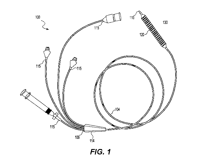
[0020] FIG. 1 is a side elevational view of an exemplary catheter, according
to
embodiments of the present disclosure.
[0021] FIG. 2A is a side elevational view of a distal portion of the catheter
of
FIG. 1.
[0022] FIG. 2B is a perspective partial section view of the exemplary catheter
of
FIG. 2A.
[0023] FIG. 3 is a perspective view of an exemplary catheter, according to
another exemplary embodiment of the present disclosure.
[0024] FIG. 4A is a cross-sectional view taken along line 4A-4A of FIG. 2A.
[0025] FIGS. 4B and 4C are cross-sectional views taken along line 4B-4B of
FIG. 2A, removing portions of the internal structure.
[0026] FIG. 5 is another view of the cross-sectional view taken along line 4A-
4A
of FIG. 2A, removing portions of the internal structure
[0027] FIG. 6 is a perspective view of the cross-sectional view of FIG. 5,
removing portions of the internal structure.
6
CA 03132070 2021-08-30
WO 2020/180638 PCT/US2020/020262
[0028] FIG. 7 shows a series of cross-sectional views showing an exemplary
catheter according to embodiments of the present disclosure.
[0029] FIG. 8 is a schematic plan view of a patterned outer surface according
to
an exemplary embodiment of the present disclosure.
[0030] FIG. 9 is a perspective view of a cross-sectional view removing
portions
of the internal structure, according to an exemplary embodiment of the present
disclosure.
[0031] FIGS. 10A to 10E are perspective views of exemplary catheters
according to embodiments of the present disclosure.
[0032] FIG. 11 is a perspective view of a cross-sectional view removing
portions
of the internal structure, according to an exemplary embodiment of the present
disclosure.
[0033] FIGS. 12A to 12E are perspective views of exemplary catheters
according to embodiments of the present disclosure.
[0034] FIG. 13 is a side elevational view of an exemplary catheter placed in a
vessel of a subject, according to an exemplary embodiment of the present
disclosure.
[0035] FIG. 14 is a side elevational view of an exemplary apparatus placed in
a
vessel of a subject, according to another exemplary embodiment of the present
disclosure.
DETAILED DESCRIPTION
[0036] Reference will now be made in detail to embodiments and aspects of the
present disclosure, examples of which are illustrated in the accompanying
drawings.
Where possible, the same reference numbers will be used throughout the
drawings to
refer to the same or like parts.
[0037] FIG. 1 illustrates an apparatus 100 in accordance with an embodiment of
this disclosure. The apparatus 100 having a catheter shaft 104 that extends
from a
proximal end 106 to a distal tip 110 of the apparatus 100. The apparatus 100
may be
configured for longitudinal movement and positioning within a vessel (e.g.
blood vessel)
of a subject. In some embodiments, the apparatus 100 may be configured for
occlusion
of the vessel and treatment of an area of the vessel. For example, the
apparatus 100
may be configured for occlusion of a blood vessel and delivery of a drug to
the occluded
area of the vessel and forming and casting a shape in the vessel, as will be
described in
more detail below.
7
CA 03132070 2021-08-30
WO 2020/180638 PCT/US2020/020262
[0038] The apparatus 100 may include a proximal end connector 114 positioned
at the proximal end of the apparatus 100, and the catheter shaft 104 may
extend in a
distal direction therefrom. The catheter shaft 104 may define a plurality of
lumens that
are accessible via a plurality of ports the proximal end connector 114. The
plurality of
ports 115 may be configured to engage with external sources desirable to
communicate
with the plurality of lumens. The ports may engage with external sources via a
variety of
connection mechanisms, including, but not limited to, syringes, over-molding,
quick-
disconnect connectors, latched connections, barbed connections, keyed
connections,
threaded connections, or any other suitable mechanism for connecting one of
the
plurality of ports to an external source. Non-limiting examples of external
sources may
include inflation sources (e.g. saline solutions), gaseous sources, treatment
sources
(e.g. medication, drugs, or any desirable treatment agents discussed further
below),
light sources, among others. In some embodiments, apparatus 100 can be used
with a
guide wire (not shown), via guide wire lumen 164 (see FIG. 4A), to assist in
guiding the
catheter shaft 104 to the target area of the vessel.
[0039] FIGS. 1, 2A, and 2B illustrate the apparatus 100 may include a first
distal
balloon 120 positioned over a distal segment 130 of the catheter shaft 104
proximal to
the distal tip 110. In some embodiments, the first distal balloon 120 may be
proximally
offset from the distal tip 110 a distance between 0 mm and 1 mm, 0 mm and 2
mm, 0
mm and 3 mm, 0 mm and 10 mm, or 0 and 50 mm. In some embodiments, the first
distal balloon 120 may inflate to 2 to 10 millimeters (mm) in diameter. In
other
embodiments, the first distal balloon 120 may inflate to 2 to 4 cm in
diameter. The first
distal balloon 120 may have a length of about 0.5 to 1 centimeters (cm), 1 to
2 cm, 1 to
3 cm, or 1 to 5 cm, or 1 to 10cm, or 1 to 15cm, or 1 to 20cm, or 1 to 25cm,
and may
take any shape suitable for supporting a wall of a blood vessel of the subject
when the
non-compliant or semi-compliant balloon is inflated. For example, the first
distal balloon
120 may expand into a cylindrical shape surrounding the distal segment 130 of
the
catheter shaft 104. The cylindrical shape may be gradually tapered inward at a
proximal
end and a distal end of the first distal balloon 120, thereby providing a
gradually tapered
proximal end and distal end of the first distal balloon 120 that taper into
contact with and
become flush with the catheter shaft 104. The first distal balloon 120 may
include a first
outer surface 124 and a second outer surface 126. The first outer surface 124
is
positioned a radial distance from the center of the first distal balloon 120.
The second
outer surface 126 is positioned more radially distant from the first outer
surface 124.
8
CA 03132070 2021-08-30
WO 2020/180638 PCT/US2020/020262
The radii of both surfaces 124, 126 may vary, but remain equidistant from one
another,
forming a nonuniform spheroidal shape of the first distal balloon 120.
[0040] Non-limiting examples of shapes the inflated first distal balloon 120
may
form include a cylindrical shape, football-shaped, spherical, ellipsoidal, or
may be
selectively deformable in symmetric or asymmetric shapes so as to limit the
potential
difference in the treated vessel shape and the untreated vessel shape reducing
edge
effects common between two surfaces of different stiffness as found in metal
stents.
The force exerted against a vessel interior by first distal balloon 120 may be
strong
enough to scaffold the vessel wall with the apparatus 100 held in a stationary
position
within the vessel or other hollow body structure. However, the force is not so
great as to
damage the interior surface of the vessel or other hollow body structure. The
first distal
balloon 120 may be substantially translucent
[0041] The apparatus 100 may further include a second distal balloon 122
positioned inside of and concentric with the first distal balloon 120. The
second distal
balloon 122 may have one continuous surface sealed at each end around the
catheter
shaft 104 forming an enclosed volume and in fluid communication through a port
with
the catheter shaft 104 through a distinct and separate lumen from the first
distal balloon
122 as will be described in more detail below. The second distal balloon 122
may be
substantially translucent. The second distal balloon 122 may be positioned
concentric
with and inside of the first distal balloon 120 such that inflating the second
distal balloon
122 may also expand the first distal balloon 120 as an outside surface of the
second
distal balloon 122 contacts an inside surface of the first distal balloon 120.
[0042] The apparatus 100 may include a plurality of connectors 115 positioned
proximally to the proximal end connector 114. For example, the first distal
balloon 120
may be terminated at the proximal end with a connector capable of receiving a
drug
source. In some embodiments, the connector may be a luer configuration. The
second
distal balloon 122 may be terminated at the proximal end with a separate and
distinct
connector capable of receiving a fluid for inflation, which may, in some
embodiments, be
a luer configuration. A center lumen (discussed in more detail below), may be
terminated at the proximal end with a connector capable of receiving a fluid
source for
clearing the lumen from the proximal termination to outside the distal tip,
and in some
embodiments may include a luer configuration. The center lumen may also
accommodate a guidewire for tracking the catheter apparatus to the desired
anatomical
location. As discussed in more detail below, the apparatus 100 may also
include light
9
CA 03132070 2021-08-30
WO 2020/180638 PCT/US2020/020262
fibers that may be terminated at the proximal end with an adaptor capable of
connecting
with a light source. Each light fiber may terminate with a separate and
distinct adaptor
or both light fibers may share an adaptor to a light source.
[0043] The materials of the apparatus 100 may be biocompatible. The catheter
shaft 104 may include material that is extrudable and capable of sustaining
lumen
integrity. The distal segment 130 of the catheter shaft 104 is substantially
translucent to
allow light transmission from light fibers. The catheter shaft 104 material is
rigid enough
to track over a guidewire and soft enough to be atraumatic. The catheter shaft
104 may
be made of materials including, but not limited to polymers, natural or
synthetic rubber,
metal and plastic or combinations thereof, nylon, polyether block amide
(PEBA),
nylon/PEBA blend, thermoplastic copolyester (TPC), a non-limiting example may
be
Hytrel , and polyethylene. The shaft materials can be selected so as to
maximize
column strength to the longitudinal length of the shaft. Further, the shaft
materials can
be braided, so as to provide sufficient column strength. The shaft materials
can also be
selected so as to allow the device to move smoothly along a guide wire. The
catheter
shaft 104 can also be provided with a lubricious coating as well as
antimicrobial and
antithrombogenic coatings. The shaft materials should be selected so as not to
interfere
with the efficacy of the agent to be delivered or collected. This interference
may take the
form of absorbing the agent, adhering to the agent or altering the agent in
any way. The
catheter shaft 104 of the present disclosure may be between about 2-16 French
units
("Fr." where one French equals 1/3 of a millimeter, or about 0.013 inches).
The catheter
shafts to be used in coronary arteries may be between about 3-5 Fr. in
diameter, and
more specifically may be 3 Fr. The catheter shafts to be used in peripheral
vessels may
be between about 5-8 Fr. in diameter, and more specifically 5 Fr. The catheter
shafts to
be used in the aorta may be between about 8-16 Fr. in diameter, and more
specifically
12 Fr.
[0044] The first distal balloon 120 and the second distal balloon 122 may be
substantially translucent permitting light from light fibers to be transmitted
substantially
beyond the inflated diameters of the first distal balloon 120. The compliance
of the first
distal balloon 120 and the first outer surface 124 and second surface 126 and
the
second distal balloon 122 may be comparable or dissimilar. For example, the
first distal
balloon 120 may be compliant such that the material conforms substantially to
a
vessel's morphology. The second distal balloon 122 material may be more rigid
and
CA 03132070 2021-08-30
WO 2020/180638 PCT/US2020/020262
noncompliant capable of higher internal pressures with minimal outward
expansion for
opening vessels more resistant to pressures.
[0045] FIG. 3 is a perspective view of another embodiment of the present
disclosure that may include a proximal balloon 118 in fluid communication with
an
additional lumen of the catheter shaft 104. The proximal balloon 118 may be
positioned
on the catheter shaft 104 proximal to the first distal balloon 120. In some
embodiments,
proximal balloon 118 may inflate to 2 to 10 millimeters (mm) in diameter. In
other
embodiments, the proximal balloon 118 may inflate to 3 to 5 centimeters (cm)
in
diameter. The proximal balloon 118 may have a length of about 1 to 2
centimeters (cm)
and may take any shape suitable for occluding and sealing a blood vessel of
the subject
when a compliant or semi-compliant balloon is inflated. Non-limiting examples
of shapes
the inflated balloon may form include oblong, football-shaped, spherical,
ellipsoidal, or
may be selectively deformable in symmetric or asymmetric shapes. The force
exerted
against a vessel interior by the proximal balloon 118 may be strong enough to
hold the
catheter assembly 100 in a stationary position within the vessel or other
hollow body
structure and provide an adequate seal to control blood or fluid flow.
However, the force
is not so great as to damage the interior surface of the vessel or other
hollow body
structure.
[0046] FIG. 4A is a cross-sectional view taken along line 4A-4A of FIG. 2A
showing a plurality of lumens within the assembly 100, according to an
embodiment of
this disclosure. The catheter shaft 104 may have an outside diameter and
outside
surface 130. The catheter shaft 104 may have an inside configuration of five
distinct and
separate lumens, extending from the proximal end 106 to the distal tip 110.
[0047] The first distal balloon 120 may be in fluid communication with a first
distal balloon inflation lumen 150. The second distal balloon 122 may be in
fluid
communication with a second distal balloon inflation lumen 154 that is
separate and
distinct from the first distal balloon inflation lumen 150. The first distal
balloon 120 may
be in fluid communication with an inflation source via the first distal
balloon inflation
lumen 150 separate from the second distal balloon inflation lumen 154. The
first distal
balloon inflation lumen 150 may extend through the catheter shaft 104 and have
an
input at one of the plurality of ports 115 of the proximal end connector 114.
Fluid
communication between the first distal balloon 120 and the inflation source
via the first
distal balloon inflation lumen 150 may cause the first distal balloon 120 to
selectively
inflate and deflate separately from and independently of the second distal
balloon 122.
11
CA 03132070 2021-08-30
WO 2020/180638 PCT/US2020/020262
Similarly, the second distal balloon 122 may be in fluid communication with an
inflation
source via the second distal balloon inflation lumen 154 separate from the
first distal
balloon inflation lumen 150. Fluid communication between the second distal
balloon 122
and the inflation source via the second distal balloon inflation lumen 154 may
cause the
second distal balloon 122 to selectively inflate and deflate separately from
and
independently of the first distal balloon 120.
[0048] A first light fiber lumen 158 and a second light fiber lumen 160 may be
positioned in the catheter shaft 104 to receive light fibers, and the first
light fiber lumen
158 and the second light fiber lumen 160 may extend from the proximal end 106
into the
distal segment 130, and may be positioned substantially symmetric,
longitudinally
opposed and parallel one to another within the catheter shaft 104. In another
exemplary
embodiment, the catheter shaft 104 may include a single light fiber lumen. In
still other
embodiments, the catheter shaft 104 may include a plurality of light fiber
lumens.
[0049] A guidewire lumen 164 may be concentric with the catheter shaft outside
diameter and may be arranged in the catheter shaft 104, from the proximal end
106
through the distal tip 110. The guidewire lumen 164 may accommodate a
guidewire to
aid the placement of the apparatus 100 to a desired anatomical position
communicating
with the proximal end and distal tip. The guidewire may be separate and
distinct from
the apparatus 100 and extend proximally beyond the proximal end and distally
beyond
the distal tip of the catheter shaft. The guidewire lumen 164 is located
concentric with
the catheter outer diameter; the catheter shaft is oriented concentrically
with the
guidewire permitting the catheter shaft 104 to follow the guidewire without
favoring one
side of the catheter shaft 104 or whipping from side to side. The guidewire
may remain
in the guidewire lumen 104 maintaining anatomical position during the
activation of the
light fibers.
[0050] FIGS. 4B and 4C illustrate cross-sectional views taken along line 4B-4B
of FIG. 2A. The apparatus 100 may further include a first light fiber 140 and
a second
light fiber 142 positioned in the catheter shaft 104 and extending through the
distal
segment 130. The light fibers 140, 142 may transmit light through the distal
segment
130, the second distal balloon 122, the first distal balloon 120. The light
fiber 140 may
be connected to the proximal end connector 114 and may have proximal ends that
connect to a light fiber activation source via at least one of the plurality
of ports 115. In
some embodiments, the light fibers 140, 142 may be configured to transmit
light at a
wavelength of 375 nanometers (nm) to 475 nm, and more specifically 450 nm that
12
CA 03132070 2021-08-30
WO 2020/180638 PCT/US2020/020262
transmits through the distal segment 130 and the first distal balloon 120. In
some
embodiments, the light first fiber 140 may be positioned in the first light
fiber lumen 158
and the second light fiber 142 may be positioned in the second light fiber
lumen 160.
[0051] In some embodiments, light from the light fibers 140, 142 may be unable
to penetrate through a guidewire 144 forming a shadow 145 opposite the light
and
beyond the guidewire 144. Accordingly, the light fibers 140, 142 may each
generate a
respective light transmission area 146. The light fiber lumens 158, 160 are
oriented
substantially opposite one another minimizing the shadow 145 formed by the
light
impenetrable guidewire 144, permitting the transmission of light to penetrate
the
circumference of the catheter shaft 104 from the first light fiber 140 or the
second light
fiber 142. In another embodiment, the catheter shaft 140 may include a single
light
fiber, and the guidewire may be removed for light penetration to the outer
tissue.
[0052] In some embodiments, the light fibers 140, 142 may be made from
plastic core and cladding. The refractive index of the core is high. The
refractive index
of the cladding is low. A non-limiting example of the core material may be
polymethyl
methacrylate (PMMA). A non-limiting example of the cladding may be a silicone
material.
[0053] FIG. 5 is another view of the cross-sectional view taken along line 4A-
4A
of FIG. 2A. As discussed above, the first distal balloon 120 may have two
distinct
surfaces. The first outer surface 124 may be positioned a radial distance (R1)
from the
center of the first distal balloon 120. The second outer surface 126 may be
positioned
more radially distant from the first outer surface 124. The radii of both
surfaces R1, R2
may vary, but remain equidistant from one another, forming a nonuniform
spheroidal
shape. The two surfaces 124, 126 may be two distinct components or materials.
[0054] As illustrated in FIG. 6, the second outer surface 126 may form a
pattern
representing circumferential directed sections 166 and longitudinal directed
sections
168 that may be interconnected, minimally contacting and sealing against the
first outer
surface 124. The interconnected circumferential sections 166 and the
longitudinal
sections 168 of the second outer surface pattern generate a confined volume
172 or
well. The confined volume 172 is formed from the wall connecting the
intersection points
174 and the sealing contact with the first outer surface 124. The pattern
generated by
the second outer surface 126 may not produce wells the same size or wells
positioned
symmetrically around the outer surface but may produce wells of a size and
position
13
CA 03132070 2021-08-30
WO 2020/180638 PCT/US2020/020262
suited to a function. In another embodiment, the first distal balloon 120 may
contain only
one distinct surface, forming no surface variation.
[0055] The pattern generated by the second outer surface 126 may have
longitudinal zones (e.g. longitudinal sections 168) favoring less resistance
than other
areas of the first distal balloon 120, permitting the first distal balloon 120
to selectively
fold along these longitudinal zones as the balloon may be compressed into a
smaller
shape.
[0056] FIG. 7 shows a series of cross-sectional views illustrating an
exemplary
catheter according to embodiments of the present disclosure. The pattern
generated by
the second outer surface 126 may contain longitudinal sections 168 and
circumferential
sections 166 capable of engaging and separating plaque 180 covering a portion
of a
vessel wall 182 into smaller, less pressure resistant and isolated sections.
The plaque
180 on the vessel wall 182 may be organized into a structure more rigid than
the vessel
wall 182 , reducing the ability of the vessel wall 182 to stretch and expand
circumferentially, accommodating the increased pressure of pulsatile blood
flow.
Inflating the first distal balloon 120, allows the pattern of the second outer
surface 126
to minimally contact the plaque 180, producing a focal point force from the
pressure of
the inflated first distal balloon 120. The focal point force may be larger
than the ultimate
strength of the plaque 180 at the location of the pattern contact (e.g. the
second outer
surface 126), causing the plaque 180 to separate into isolated areas mirroring
the
shape of the isolated volume 172. The differential rigidity between the plaque
180 and
the vessel wall 182, permits the separation of the plaque 180 with minimal
vessel
expansion. The plaque 180 may break before the vessel 182 expands. The second
outer surface pattern may be selected to optimize the desired plaque shape and
size.
Smaller patterns may impart less vessel expansion but may break off as debris.
Larger
patterns may impart more vessel expansion but may remain intact with the
vessel wall
and impede circumferential vessel expansion. In some embodiments, the pattern
shape
should balance these two competing needs.
[0057] FIG. 8 is a schematic plan view of illustrating the difference between
a
first expanded configuration of the patterned outer surface and a second
expanded
configuration of the patterned outer surface. The plaque 180 covering the
vessel wall
182 may impede a drug substance from penetrating the vessel wall 182.
Expanding the
first distal balloon 120 thereby creating isolated plaque sections, may permit
a drug
substance to penetrate the vessel wall 180 more easily through the gaps 190
between
14
CA 03132070 2021-08-30
WO 2020/180638 PCT/US2020/020262
each of the isolated plaque sections. The first distal balloon 120 may be
expanded into
contact with the plaque 180 in a first expanded configuration. The first
distal balloon 120
may be further expanded to a second expanded configuration. The second outer
surface 126 contacts the plaque 180 and may score, break, crack, separate, or
divide
the plaque 180 into smaller pieces from the point forces generated at the
second outer
surface 126, thereby creating the gaps 190 for improved drug penetration.
[0058] FIG. 9 illustrates the first outer surface 124 of the first distal
balloon 120
may have a thickness 194 forming an outside first surface and an inside first
surface
196. The inside first surface 196 forms a confined and isolated volume 170
(See also
FIG. 5) in fluid communication with the proximal end 106 of the catheter shaft
104 and a
plurality of slitted apertures 198. The first outer surface 124 and the second
outer
surface 126 are substantially translucent.
[0059] The confined volume 170 of the first distal balloon 120 may contain at
least one slitted aperture 198 penetrating through from one side to the other
side of the
first outer surface 124. The slitted apertures 198 may be any length within
the confined
volume 170, but not contact the wall of the second outer surface 126. The
slitted
apertures 198 may be oriented in any direction of the confined volume 170 on
the first
outer surface 124. The slitted apertures 198 may be in fluid communication
with a drug
source, the drug source supplying a drug into the fist distal balloon 120
volume and
through the slitted aperture 198.
[0060] The scoring ability of the first distal balloon 120 with the isolated
volumes
170 may permit breaking plaque for improved drug penetration. A reduction in
size of
the second outer surface 126 may reduce the amount of force required to
separate the
plaque 180, and an increased frequency of the second outer surface 126 in the
patterned outer profile may cause an increased number of cracks in the plaque
180 for
the drug penetration.
[0061] The edges of the slitted apertures 198 may remain together and closed
as the volume 170 of the first distal balloon 120 is filled with a drug
source, allowing the
first distal balloon 120 to nearly fill and inflate without loss of the drug
source. As the
first distal balloon volume fills and inflates, the volume pressure will
increase, forcing the
edges of the slitted aperture 198 to open apart and fill the well 172,
reducing the balloon
pressure. Similarly, as the volume and corresponding pressure of the first
distal balloon
120 is reduced from the filling the well 172, the edges of the slitted
aperture 198 may
close together and stop filling the well before the first distal balloon 120
deflates
CA 03132070 2021-08-30
WO 2020/180638 PCT/US2020/020262
substantially preventing surrounding debris from impeding the slitted aperture
198
function. In this manner of inflating and deflating the first distal balloon
120 may control
the delivery of the drug source as the wells 172 are filled and emptied.
[0062] In some embodiments, the apparatus 100 may be capable of delivery
two dugs simultaneously. For example, the outside of the first distal balloon
120 may be
coated with a first drug and a second drug may be delivered through the
slitted
apertures 198. Accordingly, the first drug and the second drug may be
different drugs.
In some embodiments, the first drug and the second drug may be the same drug.
In a
non-limiting example, the first distal balloon 120 may be coated with
Paclitaxel and
infusing Natural Vascular Scaffolding (NVS) through the slits and wells to the
vessel
wall.
[0063] As illustrated in FIGS. 9 through 10E, the slitted apertures 198 may
take
a variety of shapes and orientations. For example, the slitted apertures 198
may be a
single line through the well 172 which may be oriented vertically,
horizontally, or at any
appropriate angle therebetween. Additionally, the slitted aperture 198 may
include two
intersecting lines through the well 172. The lines may intersect at any
appropriate angle,
for example the lines may be perpendicular and meet at the mid-point of each
line. The
lines may not intersect or may intersect at any point along the lines. There
may also be
a plurality of lines arranged in any orientation.
[0064] FIG. 11 illustrates the circumferential sections 166 and longitudinal
sections 168 that may be interconnected sections of the second outer surface
126 of
the first distal balloon 120 may contain slitted apertures 200 penetrating
through from
one side to the other side of the second outer surface 126, fluidly
communicating from
the inside volume of the first distal balloon 120 to the outside surface of
the second
outer surface 126. Similarly, to the slitted apertures 198 of the first outer
surface 124 of
the first distal balloon 120, the slitted apertures 200 on the second outer
surface 126
may be any length and may be oriented in any direction. Additionally, there
may be
more than one slitted aperture 200 oriented within the second outer surface
126. The
slitted apertures 200 may function similarly under differential pressure.
[0065] As illustrated in FIGS. 11 through 12E, the slitted apertures 200 may
take a variety of shapes and orientations. For example, the slitted apertures
200 may be
a single line which may be oriented vertically, horizontally, or at any
appropriate angle
therebetween. Additionally, the slitted aperture 200 may include two
intersecting lines.
The lines may intersect at any appropriate angle, for example the lines may be
16
CA 03132070 2021-08-30
WO 2020/180638 PCT/US2020/020262
perpendicular and meet at the mid-point of each line. The lines may not
intersect or may
intersect at any point along the lines. There may also be a plurality of lines
arranged in
any orientation.
[0066] FIG. 13 is a side elevational view of an exemplary catheter placed in a
vessel of a subject, according to an exemplary embodiment of the present
disclosure.
The target area for a delivery of drug source may be a vessel of the
cardiovascular
system. The first distal balloon 120 may be inflated with a drug source and
expanded
toward the vessel wall. The second outer surface 126 of the first distal
balloon 120 may
engage and seal against the vessel wall 182. The vessel wall 182 may cover and
isolate the well 172, permitting the drug source to fill the well 172 when the
internal
pressure opens the edges of the slitted aperture (e.g. apertures 198 and/or
200),
exposing the vessel wall 182 to the drug. In the event a well 172 or series of
wells are
positioned in the proximity of a smaller vessel, side branch or collateral,
the drug from
those wells may be lost to the smaller vessels. However, all the remaining
wells 172
deliver drug to their adjacent vessel walls 182 such that drug is delivered
uniformly to
the vessel wall 182.
[0067] The second distal balloon 122 may be partially inflated before, during,
or
after the first distal balloon 120, reducing the volume of the first distal
balloon 120. This
complimentary operation of the first distal balloon 120 and second distal
balloon 122,
separately or simultaneously, permits reliable drug delivery to substantially
different
vessel anatomies. For example, the second distal balloon 122 may be inflated
first,
pushing the first distal balloon 120 towards the vessel wall 182. An aqueous
drug may
be used to inflate the first distal balloon 120 and deliver the drug while
maintaining
contact with the vessel wall 182 as the pressure in the first distal balloon
120 is reduced
from the loss of delivered drug. Similarly, an aqueous drug may fill the
volume of the
first distal balloon 120 until the edges of the slitted apertures 198 open.
The second
distal balloon 122 may be inflated gradually maintaining the pressure in the
first distal
balloon 120 continuing to deliver the drug. In some embodiments, expanding the
first
distal balloon 120 may seal the confined volume 170 against the vessel wall
182
thereby minimizing drug loss to smaller vessels, side branches or collaterals.
[0068] The second distal balloon 122 may be inflated, subsequent to drug
delivery by the first distal balloon 120, circumferentially supporting the
internal surface
of the vessel wall 182. While in this vessel supported position, a light
source may be
supplied to the light fibers 140, 142 in the catheter shaft 104 for
transmittance through
17
CA 03132070 2021-08-30
WO 2020/180638 PCT/US2020/020262
the catheter shaft 104, through the first distal balloon 120 and the second
distal balloon
122, and into the vessel wall 182 as previously described.
[0069] There are several combinations for the local delivery of the drug
source.
For example, a solid drug may be coated on the second surface of the first
distal
balloon 120 and an aqueous drug may be delivered through the slitted apertures
of the
first surface of the fist distal balloon. The drug may be the same, one solid
and one
aqueous, each penetrating the vessel wall differently. The drugs may be
complimentary
but different substances. The aqueous or solid drug may assist in the capacity
of an
excipient or activate its counterpart through a controlled reaction.
Similarly, the solid
drug may be coated on the first surface of the first distal balloon and an
aqueous drug
may be delivered through the slitted apertures on the second surface. The
drugs may
be dissimilar and non-complimentary affecting the vessel wall through
substantially
different methods of action.
[0070] Additionally, therapeutic agents useful with the device of the present
disclosure include any one of or a combination of several agents which are
gas, liquid,
suspensions, emulsions, or solids, which may be delivered or collected from
the vessel
for therapeutic or diagnostic purposes. Therapeutic agents may include
biologically
active substances, or substances capable of eliciting a biological response,
including,
but not limited to endogenous substances (growth factors or cytokines,
including, but
not limited to basic fibroblast growth factor, acidic fibroblast growth
factor, vascular
endothelial growth factor, angiogenic factors), viral vectors, DNA capable of
expressing
proteins, sustained release polymers, and unmodified or modified cells.
Therapeutic
agents may include angiogenic agents which induce the formation of new blood
vessels. Therapeutic agents may also include anti-stenosis or anti-restenosis
agents
which are used to treat the narrowing of blood vessel walls. Therapeutic
agents may
include light-activated agents such as light-activated anti-stenosis or light-
activated anti-
restenosis agents that may be used to treat the narrowing of blood vessel
walls.
[0071] FIG. 14 is a side elevational view of an exemplary apparatus placed in
a
vessel of a subject, according to another exemplary embodiment of the present
disclosure. The first distal balloon 120 may expand into contact with the
vessel 182 and
seal the confined volume 172 against the vessel wall 182, minimizing drug loss
to
smaller vessels, such as side branches or collaterals 220. In some
embodiments, the
longitudinal and circumferential surfaces (e.g. 168, 166 may contact the
vessel wall 182
and seal the confined volume 172. When the entire vessel is filled with a
drug, it will
18
CA 03132070 2021-08-30
WO 2020/180638 PCT/US2020/020262
escape through any exit, side branch or collateral. The first distal balloon
120 moves
the slitted apertures (e.g. 198) away from the vessel wall 182 and fills the
confined
volume 172. If the confined volume 172 is near an opening 222 (e.g. collateral
220), the
small amount of drug that is transmitted through that confined volume 172
would be lost
to the opening. If the confined volume 172 is not near an opening 222, the
drug may be
delivered to the vessel wall 182. The slitted apertures 198 may be distant
from the
vessel wall 182 inside the confined volume 172 and protected from potential
environment debris. The drug flow from the slitted apertures 198 is not
impeded by the
slitted apertures 198 contacting the vessel wall 182.
[0072] Another embodiment of this disclosure includes an exemplary method of
tissue restoration in a blood vessel of a subject. The method may include
providing a
catheter into the blood vessel. In some embodiments, the catheter may include
the
features of apparatus 100 described above. For example, the catheter may
include a
catheter shaft (e.g. catheter shaft 104) extending from a proximal end (e.g.
proximal end
106) to a distal tip (e.g. distal tip 110). A first distal balloon (e.g. first
distal balloon 120)
may be positioned on a translucent distal segment (e.g. distal segment 130) of
the
catheter shaft proximal to the distal tip, the first distal balloon in fluid
communication
with a drug source via a first lumen (e.g. first distal balloon inflation
lumen 150). The first
distal balloon may include a translucent material, a first outer surface (e.g.
first outer
surface 124) positioned at a first radial distance (e.g. R1) from a center of
the first distal
balloon, a second outer surface (e.g. second outer surface 126) positioned at
a second
radial distance (e.g. R2) from the center of the first distal balloon, the
second radial
distance being larger than the first radial distance. The first distal balloon
may have a
patterned outer profile formed by the first outer surface and the second outer
surface.
The catheter may further include a second distal balloon (e.g. second distal
balloon
122) positioned inside of and concentric with the first distal balloon, the
second distal
balloon in fluid communication with a second lumen (e.g. second distal balloon
inflation
lumen 154) separate from the first lumen. The catheter may further include a
first light
fiber (e.g. light fiber 140) and a second light fiber (e.g. light fiber 142)
each positioned in
the catheter shaft and extending through the translucent distal segment.
[0073] The method may further include partially expanding the second distal
balloon, supplying the drug from the drug source to the first distal balloon,
expanding
the first distal balloon into contact with the blood vessel (e.g. blood vessel
182) in a
treatment area; delivering the drug to the treatment area through at least one
slitted
19
CA 03132070 2021-08-30
WO 2020/180638 PCT/US2020/020262
aperture (e.g. slitted apertures 198, 200) in the first distal balloon; fully
expanding the
second distal balloon propping open the vessel, activating the first light
fiber and second
light fiber thereby providing light transmission through the distal segment,
the first distal
balloon, and the second distal balloon to activate the drug in the treatment
area. The
light transmission to the treatment area may activate the NVS, which may be
activated
by light. The expansion of the first distal balloon may shape the treatment
area (e.g.
vessel) as desired.
[0074] In some embodiments, the method may include engaging and separating
plaque (e.g. plaque 180) along a wall of a vessel of a subject into smaller,
less pressure
resistant and isolated sections using the longitudinal and circumferential
surfaces (e.g.
168, 166) of the patterned outer profile. The longitudinal and circumferential
surfaces
(e.g. 168, 166) may be interconnected and define at least one confined volume
(e.g.
volume or well 172) on the outer surface of the first distal balloon, the
confined volume
defined by a difference in radial distance (e.g. difference between R2 and R1)
from the
center of the first distal balloon between the first outer surface and the
second outer
surface. The method may further include creating isolated plaque sections that
allow the
drug to penetrate the vessel wall through gaps between each of the isolated
plaque
sections. The method may further include expanding the second distal balloon
thereby
expanding the first distal balloon as an outer surface of the second distal
balloon
contacts an inner surface of the first distal balloon.
[0075] Accordingly, the apparatus and methods described herein provide the
delivery of NVS to a treatment area (e.g. a vessel) and provide restoration to
that
treatment area using the apparatus or according to the methods described
above. The
apparatus and method described above provide concurrently scoring the vessel,
treating the vessel with one or more drugs (e.g. with Paclitaxel and NVS) with
minimal
loss to other vessels, scaffolding and casting the vessel, and light
activation of the one
or more drugs delivered to the treatment area. These advantages can be
accomplished
utilizing the apparatus and methods described herein.
[0076] The foregoing description has been presented for purposes of
illustration. It is not exhaustive and is not limited to precise forms or
embodiments
disclosed. Modifications and adaptations of the embodiments will be apparent
from
consideration of the specification and practice of the disclosed embodiments.
For
example, the described implementations include hardware and software, but
systems
and methods consistent with the present disclosure can be implemented as
hardware
CA 03132070 2021-08-30
WO 2020/180638 PCT/US2020/020262
alone. In addition, while certain components have been described as being
coupled to
one another, such components may be integrated with one another or distributed
in any
suitable fashion.
[0077] Moreover, while illustrative embodiments have been described herein,
the scope includes any and all embodiments having equivalent elements,
modifications,
omissions, combinations (e.g., of aspects across various embodiments),
adaptations
and/or alterations based on the present disclosure. The elements in the claims
are to be
interpreted broadly based on the language employed in the claims and not
limited to
examples described in the present specification or during the prosecution of
the
application, which examples are to be construed as nonexclusive. Further, the
steps of
the disclosed methods can be modified in any manner, including reordering
steps
and/or inserting or deleting steps.
[0078] The features and advantages of the disclosure are apparent from the
detailed specification, and thus, it is intended that the appended claims
cover all
systems and methods falling within the true spirit and scope of the
disclosure. As used
herein, the indefinite articles "a" and "an" mean one or more." Similarly, the
use of a
plural term does not necessarily denote a plurality unless it is unambiguous
in the given
context. Words such as "and" or "or" mean "and/or" unless specifically
directed
otherwise. Further, since numerous modifications and variations will readily
occur from
studying the present disclosure, it is not desired to limit the disclosure to
the exact
construction and operation illustrated and described, and accordingly, all
suitable
modifications and equivalents may be resorted to, falling within the scope of
the
disclosure.
[0079] Other embodiments will be apparent from consideration of the
specification and practice of the embodiments disclosed herein. It is intended
that the
specification and examples be considered as example only, with a true scope
and spirit
of the disclosed embodiments being indicated by the following claims.
21