Note: Descriptions are shown in the official language in which they were submitted.
CA 03133394 2021-09-13
WO 2020/210901 PCT/CA2020/050498
Nonviral Modification of T Cell Gene Expression
CROSS-REFERENCE TO RELATED APPLICATIONS
[0001] This application takes priority from United States Provisional
Applications
62/833,993 filed April 15, 2019; 62/861,220 filed June 13, 2019; and
62/923,525 filed
October 19, 2019.
BACKGROUND
(a) Field
[0001] The subject matter disclosed generally relates to delivery of
nucleic acid to
living cells, specifically living T lymphocytes (T cells), while maintaining
their viability.
(b) Related Prior Art
[0002] Altering gene expression for therapeutic purposes can be achieved
by
delivering nucleic acids in lipid nanoparticles (LNPs) to cells. Exogenous
mRNA has
promise as a means of generating in vivo protein expression, and when
delivered by
LNP rather than viral vectors, avoids the side effects and safety issues that
viral
delivery effects.
[0003] Chimeric Antigen Receptor T cell therapy (CAR) is a type of
targeted
immunotherapy now approved for human use (KymriahTM tisagenlecleucel and
YescartaTM axicabtagene ciloleucel). The process uses cells from the subject
being
treated, selects and enriches for T cells, and then engineers these cells
using a viral
vector to express a chimeric antigen receptor (CAR). The cells are returned to
the
subject, resulting in immunotherapy. 1
[0004] Despite the success of CAR treatment, there are issues: a) not all
the
treated T cells have CAR, b) there is variability in the amount of CARs
expressed on
the T cells that are transfected, c) patients undergoing CAR have often had
multiple
rounds of chemotherapy which means less healthy T cells which are harder to
enrich,
and 4) there is a high incidence (46% or more) in patients of Cytokine Release
Syndrome (CRS). 2'3 Patients with CRS require intensive care unit level care,
and
1
CA 03133394 2021-09-13
WO 2020/210901 PCT/CA2020/050498
treatment with powerful and expensive immunotherapies such as tocilizumab
(ActemraTm).
[0005] Viral based to T-cell transformation have been tried, but are labor
intensive, expensive and pose manufacturing and regulatory challenges. Vector
design and development takes time as suitable vectors determine the efficiency
of
transduction. Also, virus manufacturing methods are expensive because they are
highly regulated, need a lot of equipment, and labor intensive (one batch for
each
patient).
[0006] Viral based transfection also poses the risk that viral genome may
randomly insert into the human genome, and requires that the patient leave the
hospital to have T cells harvested and treated at a specialized viral
manufacturing
facility.
[0007] Another T cell transformation technology uses electroporation and
circular
DNA to revise T cell protein expression. Electroporated cells, however, can
take a
long time to proliferate, a sign indicating that health of the T cells have
been affected
by the process. A recent study showed that the viability of T Cells after
electroporation
was 31% as opposed to LNP mediated mRNA delivery.1 The "Sleeping Beauty CART
Therapy" is such an electroporation modality, but was put on hold in 2018.4,5
[0008] A nonviral approach that is less destructive than electroporation
would
advance T cell mediated immunotherapy treatments, while preserving T cell
viability
and subject health.
Summary of the Invention
[0009] According to an embodiment, there is provided a lipid mix
composition
including 35-55 Mol /0 ionizable lipid, 5-25 Mol /0 structural lipid, 25-40
Mol % sterol,
and 0.1-3 Mol % surfactant. According to another embodiment, the composition
is
mixed with a nucleic acid to form lipid particles. According to another
embodiment,
there is provided a lipid mix composition for use in transfecting nucleic acid
into target
cells. According to another embodiment, there is provided a lipid mix
composition in
which said transfecting takes place ex vivo.
2
CA 03133394 2021-09-13
WO 2020/210901 PCT/CA2020/050498
[0010] According to another embodiment, there is provided a lipid mix in
which
the structural lipid is DSPC. In another embodiment, the DSPC is present at 10-
20
Mol %. In yet another embodiment, the DSPC is present at 20 Mol %. In another
embodiment, there is provided a lipid mix in which the surfactant is
Polyoxyethylene
(10) stearyl ether. According to another embodiment, there is provided a lipid
mix in
which the surfactant is polysorbate 80. In another embodiment the surfactant
is
polyoxyethylene (40) stearate. In another embodiment, the surfactant is D-a-
Tocopherol polyethylene glycol 1000 succinate.
[0011] In embodiments of the invention, the ionizable lipid is any
ionizable lipid.
In some embodiments, the ionizable lipid is BOCHD-C3-DMA. In embodiments of
the
invention, the ionizable lipid is Dlin-MC3-DMA. In embodiments of the
invention, the
ionizable lipid is DODMA. In embodiments of the invention, the ionizable lipid
is KC2
(DLin-KC2-DMA). In other embodiments, the ionizable lipid is C12-200.
[0012] In embodiments of the invention, the ionizable lipid is from 40-50
Mol /0,
the structural lipid is from 10-20 Mol /0 DSPC, the sterol is from 37-39 Mol
/0, and the
surfactant is from 1-3 Mol /0.
[0013] In further embodiments of the invention, the ionizable lipid
comprises 50
Mol /0, the structural lipid comprises 10 Mol /0 DSPC, the sterol comprises
37.5 Mol /0
cholesterol, and the surfactant comprises 2.5 MolcY0 polyoxyethylene (10)
stearyl ether.
[0014] In other embodiments, the ionizable lipid comprises 40 Mol /0, the
structural lipid comprises 20 Mol /0 DSPC, the sterol comprises 37.5 Mol /0
cholesterol,
and the surfactant comprises 2.5 MolcY0 polyoxyethylene (10) stearyl ether.
[0015] In yet other embodiments of the invention, a lipid mix composition
is
disclosed wherein the ionizable lipid comprises 40 Mol /0, the structural
lipid comprises
20 Mol /0 DSPC, the sterol comprises 38.5 Mol /0 cholesterol, and the
surfactant
comprises 1.5 Mol /0 polysorbate 80. In other embodiments, the ionizable lipid
is 50
Mol /0, the structural lipid is 10 Mol /0 DSPC, the sterol is from 37-40 Mol
/0, and the
surfactant is about 0.5 MolcY0 to 2.5 MolcY0. In embodiments of the invention,
the
surfactant comprises about 2.5 MolcY0 polyoxyethylene (10) stearyl ether. In
3
CA 03133394 2021-09-13
WO 2020/210901 PCT/CA2020/050498
embodiments of the invention, the surfactant comprises about 1.5 MolcY0
polysorbate
80. In embodiments of the invention, the surfactant comprises about 0.5 MolcY0
polyoxyethylene (40) stearate. In embodiments of the invention, the surfactant
comprises about 0.5 Mol /0 D-a-Tocopherol polyethylene glycol 1000 succinate.
[0016] In embodiments of the invention, the lipid mix compositions of the
invention cells are especially suited for T cell transfection.
[0017] In embodiments of the invention there is provided a method of
treating T
cells in vitro comprising isolating T cells from a bodily fluid, and
contacting said cells
with a nucleic acid therapeutic encapsulated in a lipid mix composition
according to
embodiments of the invention.
[0018] In embodiments of the method of the invention, the T cells are
about to
begin, or are in the log phase of growth, when contact is made. In
embodiments,
contact is made from day 3 to day 7 of cell culture. In preferred embodiments,
contact
is made on day 3 of cell culture. In another embodiment, the contact is made
on day 7
of cell culture.
[0019] Features and advantages of the subject matter hereof will become
more
apparent in light of the following detailed description of selected
embodiments, as
illustrated in the accompanying figures. As will be realized, the subject
matter
disclosed and claimed is capable of modifications in various respects, all
without
departing from the scope of the claims. Accordingly, the drawings and the
description
are to be regarded as illustrative in nature, and not as restrictive and the
full scope of
the subject matter is set forth in the claims.
BRIEF DESCRIPTION OF THE DRAWINGS
[0020] Further features and advantages of the present disclosure will
become
apparent from the following detailed description, taken in combination with
the
appended drawings, in which:
4
CA 03133394 2021-09-13
WO 2020/210901 PCT/CA2020/050498
[0021] Fig. 1 is a linear plot of the growth (cell count) over time of
isolated T cells
following activation;
[0022] Fig. 2 is a bar graph showing relative OFF protein expression in
live
CD4+/CD8+ T cells treated 7 days post activation with 2pg of mRNA per 500,000
cells
in BOCHD-C3-DMA LNPs of six different lipid mix compositions exposed for 48h;
[0023] Fig. 3 is a bar graph showing relative OFF protein expression in
live
CD4+/CD8+ T cells treated 7 days post activation with 2pg of mRNA per 500,000
cells
in MC3 LNPs of five different lipid mix compositions exposed for 48h;
[0024] Fig. 4 is a bar graph showing total OFF expression in negatively
selected
T cells mediated by mRNA Lipid Nanoparticles (LNP) formulated with CT10, CT22
and
Lipid Mix A composition, and analyzed for gene expression by ELISA. The
ionizable
lipid was BOCHD-C3-DMA for all three compositions;
[0025] Fig. 5 is a distribution plot for OFF expression in mRNA-treated T-
cells
from different donors of both sexes aged 20-75 years. Different shape and/or
pattern
of the data point represents different donors, each of which cell population
was tested
with 5 different lipid mix compositions Lipid Mix A, CT7, S11, CT10, CT22;
[0026] Fig. 6 is a bar graph showing relative OFF expression in live T
cells
mediated by mRNA in BOCHD-C3-DMA LNPs at a dose of 2 pg mRNA per 500,000
cells and at a N/P ratio of 10. Primary human T cells from the same donor were
isolated from fresh whole blood using either negative selection or positive
selection
protocol and activated using a triple activator;
[0027] Fig. 7 is a histogram showing cell populations having certain
characteristics as measured by flow cytometry of live primary human T cells
treated 7
days post activation with mRNA LNPs of three different lipid mix compositions
for
48h. From top to bottom, the histograms represent OFF expression from cells
from
the CD8+ isolation (large polka dots), CD4+ isolation (horizontal pale
stripe), Pan T
isolation CD8+ cells only (smaller polka dots), Pan T isolation CD4+ cells
only
(horizontal dark stripe), all T cells from the Pan T isolation (dark grey),
and finally
untreated cells (light translucent grey). The ionizable lipid was BOCHD-C3-
DMA;
CA 03133394 2021-09-13
WO 2020/210901 PCT/CA2020/050498
[0028] Fig. 8 is a bar graph showing relative OFF protein expression
derived from
LNAP treated live CD4+/CD8+ T cells. The first bar labelled DOPE LNAP contains
structural lipid DOPE in place of DSPC, while the second bar labeled DSPC LNAP
(CT22) has DSPC as the structural lipid. Both lipid mix compositions
correspond to
Lipid Mix CT22 in terms of proportions of IL, structural lipids, cholesterol
and
polysorbate 80;
[0029] Fig. 9 is a bar graph showing the OFF expression in activated,
transfected
T cells by four different compositions with two different molar ratios of
DSPC;
[0030] Fig. 10 is two bar graphs, the first showing relative OFF protein
expression in live CD4+/CD8+ T cells treated 7 days post activation, with 2 pg
of
mRNA per 500,000 cells over 48h, and wherein the ionizable lipid was one of
BOCHD-
C3-DMA, DODMA, KC2, or MC3. The second is a graphical representation of LNP-
mediated transfection of isolated human T cells as measured by viability
(black bars)
and OFF expression (grey bars) using 500 ng LNAP per 125, 000 cells of CT10
composition with either BOCHD-C2-DMA or C12-200 as the ionizable lipid;
[0031] Fig. 11 is a series of bar graphs showing results for the lipid mix
composition comprising 40 Mol /0 ionizable lipid, 20 Mol /0 DSPC, 40-x Mol /0
cholesterol , and x Mol% stabilizer , where x = 0.5,1.5, or 2.5 Mol /0; Bar
graphs
labeled A(i) and (ii) are transfection efficiency, (i) and MFI (ii) of mRNA
LNPs
encoding eGFP in isolated primary human T cells with stabilizer Brij S10; bar
graphs
B(i) and (ii) are transfection efficiency (i) and MFI ii) with stabilizer Brij
S20; bar graphs
C(i) and (ii) are transfection efficiency (i) and MFI (ii) with stabilizer
Tween80; and bar
graphs D(i) and (ii) are transfection efficiency (i) and MFI (ii) with
stabilizer TPGS-1000
(D-a-Tocopherol polyethylene glycol 1000 succinate);
[0032] Fig. 12 is a bar graph showing viability of CD4+/CD8+ T cells
treated 7
days post activation with mRNA LNPs at N/P 10, comprised of the lipid mix
compositions referenced in the x axis, with live cells determined by flow
cytometry
using a live/dead stain FVS 570. The ionizable lipid was BOCHD-C3-DMA for all
three
compositions;
6
CA 03133394 2021-09-13
WO 2020/210901 PCT/CA2020/050498
[0033] Fig. 13 is a series of bar graphs showing three measurements, namely
%
OFF + live PAN T cells, OFF MFI, and T Cell viability, after T cells were
exposed to
CT10 LNAP either day 3 or day 7 after T cell expansion was initiated with
either
BOCHD-C3-DMA or MC3 as ionizable lipid;
[0034] Fig. 14 is a graphical representation of OFF % expression in viable
Pan T
cells exposed to LNAP on day 3 after activation from 15 different donors using
BOCHD-C3 as the ionizable lipid in CT10 composition;
[0035] Fig. 15 is a graphical representation of OFF % expression in viable
Pan T
cells from 6 different donors exposed to LNAP on day 7 using BOCHD-C3 (black
bars)
or MC3 (grey bars) as the ionizable lipid in CT10 composition;
[0036] Fig. 16 is two bar graphs illustrating transfection efficiency and
OFF
expression in isolated primary human T cells mediated by mRNA-LNPs containing
IL
with CT10 composition at N/P 8 under five conditions, fresh T cells, frozen T
cells
treated on day three, and frozen T cells treated on day four, frozen T cells
rested and
treated on day 3, and frozen T cells rested and treated on day 4;
[0037] Fig. 17 is a series of line graphs showing OFF expression in
isolated
primary human T cells transfected with mRNA-LNPs containing BOCHD-C3-DMA as
the IL, in a CT10 composition at N/P from 4-12. Transfection efficiency,
viability and
OFF MFI as measured by flow cytometry 48 hours after T cells were dosed with
mRNA-LNPs either 3 days or 7 days after activation, with 125 ng or 500 ng of
encapsulated mRNA per 125,000 cells;
[0038] Fig. 18 is a graphical representation of OFF expression in isolated
primary
human T cells mediated by varying doses of mRNA-LNPs containing lipid BOCHD
with
CT10 composition at N/P 8, 3 days after activation of T cells.
[0039] Fig 19 is a set of bar graphs showing OFF % and OFF MFI of viable T
cells measured by flow cytometry on days 2, 4, 7, or 14 post addition of CT10
LNAPs;
[0040] Fig. 20 is a bar graph showing total EPO expression in negatively
selected
T cells mediated by mRNA Lipid Nanoparticles (LNP) comprising CT10 lipid
compositions, analyzed after 48 hours of treatment. The T cells were harvested
and
7
CA 03133394 2021-09-13
WO 2020/210901 PCT/CA2020/050498
lysed for cytosolic EPO and media supernatant was sampled for secreted EPO.
The
ionizable lipid was BOCHD-C3-DMA or DLin-MC3-DMA for the test compositions,
and
controls were untreated T cells and serum controls provided by the
manufacturer of
ELISA kit (Quantikinee IVD Human Epo ELISA, and Quantikinee Human Serum
Controls) ;
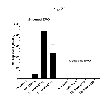
[0041] Fig. 21 is a bar graph showing total recombinant human
erythropoietin
(EPO) expression in negatively selected T cells mediated by mRNA LNP
comprising
CT10, CT22 and Lipid Mix A compositions, and analyzed after 48h of treatment.
The
T cells were harvested and lysed for cytosolic EPO and media supernatant was
sampled for secreted EPO. The ionizable lipid was BOCHD-C3-DMA for all three
compositions;
[0042] Fig. 22 is a graphical illustration of CD19 CAR expression in
isolated
primary human T cells mediated by mRNA-LNPs containing lipid BOCHD-C3-DMA
with CT10 composition at N/P 8 showing transfection efficiency and MFI
measured by
flow cytometry 12, 24, and 48 hours after LNP addition 3 days after triple
activation
with 125 ng of encapsulated mRNA per 125,000 cells;
[0002] Fig. 23 is a series of bar graphs showing CD19 CAR expression in
isolated primary human T cells mediated by mRNA-LNPs containing lipid BOCHD
with
CT10 or CT14 compositions at N/P 8. Transfection efficiency, MFIT cells were
dosed
with mRNA-LNPs 3 days after activation with 125 ng or 500 ng of encapsulated
mRNA
per 125, 000 cells.;
[0003] Fig 24 is the genetic structure of the custom CAR plasmid showing
the
pcDNA3.1 cloning vector containing the anti-CD19-h(BB)-eGFP-2nd generation CAR
(T7 Mut) gene cassette. The plasmid map was created using SnapGene Viewer
4.1.9. This plasmid is linearized for in vitro transcription and capped to
generate the
custom mRNA encoding the anti-CD19-h(BB)-eGFP-2nd generation chimeric antigen
receptor (CAR) expressed in human T cells.
DETAILED DESCRIPTION
8
CA 03133394 2021-09-13
WO 2020/210901 PCT/CA2020/050498
[0004] The present invention provides lipid mix compositions, their use in
generating lipid mix compositions of nucleic acid therapeutics and other
oligomers
such as peptides, and methods for using these lipid mixes and resulting lipid
mix
compositions to overcome transfection-resistant cell types.
[0005] In another aspect, the lipid mix compositions of the invention are
provided
for mixing with nucleic acid therapeutics to create a lipid nucleic acid
particle which
enhances delivery of the nucleic acid into target cells or tissues, with less
toxicity than
more traditional lipid mix compositions or lipid nucleic acid particles such
as those
made from commercially available lipid mixes such as LipofectamineTM or
TransfectamineTm transfecting agents.
[0006] In another aspect, the invention provides lipid mix compositions
including
ionizable lipid, one or more structural lipid(s), cholesterol, and a
particular surfactant.
[0007] In another aspect, the lipid mix compositions according the
invention are
provided for formulating nucleic acid and peptide therapeutics for the
treatment of
diseases of the central nervous system, or for cell reprogramming, or for ex
vivo
transformation of human T cells
[0008] "Lipid" refers to structurally diverse group of organic compounds
that are
fatty acid derivatives or sterols or could be lipid like materials as in
lipidoids (example
C12-200) and are characterized by being insoluble in water but soluble in many
organic solvents.
[0009] "Lipid Particles". The invention provides lipid particles
manufactured from
the lipid mix compositions described above. The lipid particle represents the
physical
organization of the lipid mix composition and a therapeutic agent. A lipid
nanoparticle
("LNP") is a small, semi-to-fully organized lipid particle. Lipid nucleic acid
particles or
LNAP are generally spherical assemblies of lipids, nucleic acid, cholesterol
and
stabilizing agents. Positive and negative charges, ratios, as well as
hydrophilicity and
hydrophobicity of the elements dictate the physical structure of the lipid
particles in
terms of orientation of components and LNAP dimensions. The structural
organization
9
CA 03133394 2021-09-13
WO 2020/210901 PCT/CA2020/050498
of lipid particles may lead to an aqueous interior with a minimum bilayer as
in
liposomes6 or it may have a solid interior as in solid nucleic acid lipid
nanoparticles.7
There may be phospholipid monolayers or bilayers in single or multiple forms.8
LNAP
is a subgroup of Lipid Particles or LNP, because the inclusion of nucleic acid
is
specified.
[0010] As used herein, "NIP" is the ratio of moles of the amine groups of
ionizable
lipids to those of the phosphate groups of nucleic acid. In embodiments of the
invention, N/P ratios are from 4 to 12, and most preferred ratios are from N/P
8-10. In
one embodiment the N/P ratio is 10. In a preferred embodiment, the N/P ratio
is 8.
[0011] "Lipid mix compositions" refers to the types of components, ratios
of
components, and the ratio of the total components to the nucleic acid
payloads. For
example, a lipid mix composition of 40 Mol /0 ionizable lipid, 20 Mol%
structural lipid,
17 Mol % sterol, and 2.5 Mol % surfactant would be one lipid mix composition.
The
nucleic acid component is associated with this lipid mix composition to form a
lipid
nucleic acid particle, or LNP, in a premeditated ratio such as ionizable lipid
amine (N)
to nucleic acid phosphate ratio (P) of N/P 4, N/P 6, N/P 8, N/P 10, N/P 12 or
another
relevant particular N/P ratio.
[0012] "Viability" when referring to cells in vitro, means the ability to
continue to
grow, divide, and continue to grow and divide, as is normal for the cell type
or tissue
culture strain. Cell viability is affected by harsh conditions or treatments.
Cell viability
is critical in ex vivo therapy or parenteral administration.
[0013] "Ionizable lipid." The lipid particles include an ionizable lipid.
As used
herein, the term "ionizable lipid" refers to a lipid that is cationic or
becomes ionizable
(protonated) as the pH is lowered below the pKa of the ionizable group of the
lipid,but
is more neutral at higher pH values. At pH values below the pKa, the lipid is
then able
to associate with negatively charged nucleic acids (e.g., oligonucleotides).
As used
herein, the term "ionizable lipid" includes zwitterionic lipids that assume a
positive
charge on pH decrease, and any of a number of lipid species that carry a net
positive
charge at a selective pH, such as physiological pH. Such lipids include, but
are not
CA 03133394 2021-09-13
WO 2020/210901 PCT/CA2020/050498
limited to, N,N-dioleyl-N,N-dimethylammonium chloride (DODAC); 1,2-dioleoy1-3-
dimethyaminopropane (DODAP), N-(2,3-dioleyloxy)propyI)-N,N,N-trimethylammonium
chloride (DOTMA); N,N-distearyl-N,N-dimethylammonium bromide (DDAB); N-(2,3-
dioleoyloxy)propyI)-N,N,N-trimethylammonium chloride (DOTAP); 3-(N¨(N',N'-
dimethylaminoethane)-carbamoyl) cholesterol (DC-Chol), and N-(1,2-
dimyristyloxyprop-3-yI)-N,N-dimethyl-N-hydroxyethyl ammonium bromide (DMRIE).
[0014] In other preferred embodiments, the ionizable lipid is an amino
lipid. In
preferred embodiments of the invention, the ionizable lipid is 1,17-bis(2-
octylcyclopropyl)heptadecan-9-y14-(dimethylamino) butanoate hydrochloride
("BOCHD-C3-DMA"). This compound is disclosed in United States Published
Application No. 2013323269. In other preferred embodiments, the ionizable
lipid is
heptatriaconta-6,9,28,31-tetraen-19-y14-(dimethylamino)butanoate (DLin-MC3-DMA
or
"MC3"). In other preferred embodiments, the ionizable lipid is 2,2-dilinoley1-
4-(2-
dimethylaminoethy1)41,3]-dioxolane (DLin-KC2-DMA or "KC2"). In other preferred
embodiments, the ionizable lipid is (1,1`-((2-(4-(2-((2-(bis(2-
hydroxydodecyl)amino)ethyl) (2-hydroxydodecyl) amino) ethyl) piperazin-1-y1)
ethyl)
azanediyl) bis(dodecan-2-oI)) or "C12-200".
[0015] In other embodiments, cationic lipids suitable for use in a lipid
nanoparticle
of the invention include, but are not limited to: DLenDMA; 98N12-5; reLNPs; KL
22 as
described in United States Patent Publication 20120295832 Al, H0T5001, also
called
CCBene; HOT 4003, HOT 5000, HOT 5001, H0T5002 all as disclosed by Ball, R et
al.
in PCT publication nos. W02020047061A1 and W02013/14910, and by Derosa,
Frank etal., in US10507183 BB; Lipidoids as mentioned in United States Patent
Pub.
No. 20180333366A1, ATX-002 as described by Payne et al. in United States
Patent
no. US10399937 BB; ATX-57, ATX-58, ATX-81, ATX-88 as described in United
States Patent No. 10,383,952 B2, 2-(1,2-di((9Z,12Z)-octadeca-9,12-dien-l-
yl)hydraziny1)-N,N-dimethylethan-1-amine), 4-(dimethylamino)-N',N'-di((9Z,12Z)-
octadeca-9,12-dien-l-ylbutanehydrazide, 2-(di((9Z,12,Z)-octadeca-9,12-dien-1 -
yl)amino)ethyl 4-(dimethylamino)butanoate, 2-(di((9Z,12Z)-octadeca-9,12-dien-1-
11
CA 03133394 2021-09-13
WO 2020/210901 PCT/CA2020/050498
yl)amino)ethyl, and 4-(4-methylpiperazin-1-yl)butanoate as described in United
States
Patent Pub. No. 2019292130 Al.
[0016] Other suitable amino lipids useful in the invention also include
those
described in PCT patent publication no. WO 2009/096558. Representative amino
lipids include 1,2-dilinoleyoxy-3-(dimethylamino)acetoxypropane (DLin-DAC),
1,2-
dilinoleyoxy-3-morpholinopropane (DLin-MA), 1,2-dilinoleoy1-3-
dimethylaminopropane
(DLinDAP), 1,2-dilinoleylthio-3-dimethylaminopropane (DLin-S-DMA), 1-linoleoy1-
2-
linoleyloxy-3-dimethylaminopropane (DLin-2-DMAP), 1,2-dilinoleyloxy-3-
trimethylaminopropane chloride salt (DLin-TMA.C1), 1,2-dilinoleoy1-3-
trimethylaminopropane chloride salt (DLin-TAP.C1), 1,2-dilinoleyloxy-3-(N-
methylpiperazino)propane (DLin-MPZ), 3-(N,N-dilinoleylamino)-1,2-propanediol
(DLinAP), 3-(N,N-dioleylamino)-1,2-propanedio (DOAP), 1,2-dilinoleyloxo-3-(2-
N,N-
dimethylamino)ethoxypropane (DLin-EG-DMA), 2,2-dilinoley1-4-
dimethylaminomethyl-
[1,3]-dioxolane (DLin-K-DMA), 1,2-dilinoleyloxy-N,N-dimethy1-3-aminopropane
(DLin-
DMA), and 1,2-dioleyloxy-3-dimethylaminopropane (DODMA).
[0017] In still other embodiments, ionizable lipids referred to in
U520180000953
by Almarsson, Orn And Lawlor, Ciaran Patrick such as 3-(didodecylamino)-
N1,N1,4-
tridodecyl-l-piperazineethanamine (KL10), 14,25-ditridecy1-15,18,21,24-
tetraaza-
octatriacontane (KL25), 2-(90xy)-N,N-dimethy1-3-[(9Z,2Z)-- octadeca-9,12-dien-
l-
yloxy]propan-l-amine (Octyl-CLin DMA), (2R)-2-(90xy)-N,N-dimethy1-3-[(9Z-
,12Z)-
octadeca-9,12-dien-l-y1 oxy]propan-l-amine (Octyl-CLinDMA (2R)), and (25)-2-
(90xy)-
N,N-dimethy1-3-[(9Z,1- 2Z)-octadeca-9,12-dien-1-y1 oxy]propan-l-amine (Octyl-
CLinDMA (2S)) are employed.
[0018] The ionizable lipid is present in embodiments of the composition and
lipid
particle of the invention preferably comprise an amount from about 35 to about
55
Mol /0, or more preferably 40 to about 50 Mo1%.
[0019] Structural lipids are also known as "helper lipids" or "neutral
lipids". The
composition and lipid particles of the invention include one or more
structural lipids at
about 10 to 20 Mol% of the composition. Suitable structural lipids are
believed to
12
CA 03133394 2021-09-13
WO 2020/210901 PCT/CA2020/050498
support the formation of particles. Structural lipids refer to any one of a
number of lipid
species that exist in either in an anionic, uncharged or neutral zwitterionic
form at
physiological pH. Representative structural lipids include
diacylphosphatidylcholines,
diacylphosphatidylethanolamines, diacylphosphatidylglycerols, ceramides,
sphingomyelins, dihydrosphingomyelins, cephalins, and cerebrosides.
[0020] Exemplary structural lipids include zwitterionic lipids, for
example,
distearoylphosphatidylcholine (DSPC), dioleoylphosphatidylcholine (DOPC),
dipalmitoylphosphatidylcholine (DPPC), dioleoyl-phosphatidylethanolamine
(DOPE),
palmitoyloleoylphosphatidylcholine (POPC), palmitoyloleoyl-
phosphatidylethanolamine
(POPE) and dioleoyl-phosphatidylethanolamine 4-(N-maleimidomethyl)-cyclohexane-
1-carboxylate (DOPE-mal), dipalmitoyl phosphatidyl ethanolamine (DPPE),
dimyristoylphosphoethanolamine (DMPE), distearoyl-phosphatidylethanolamine
(DSPE), 16-0-monomethyl PE, 16-0-dimethyl PE, 18-1-trans PE, 1-stearoy1-2-
oleoyl-
phosphatidyethanol amine (SOPE), and 1,2-dielaidoyl-sn-glycero-3-
phophoethanolamine (trans DOPE). In a preferred embodiment, the structural
lipid is
distearoylphosphatidylcholine (DSPC).
[0021] In another embodiment, the structural lipid is any lipid that is
negatively
charged at physiological pH. These lipids include phosphatidylglycerols such
as
dioleoylphosphatidylglycerol (DOPG), dipalmitoylphosphatidylglycerol (DPPG),
palmitoyloleyolphosphatidylglycerol (POPG), cardiolipin, phosphatidylinositol,
diacylphosphatidylserine, diacylphosphatidic acid, and other anionic modifying
groups
joined to neutral lipids. Other suitable structural lipids include glycolipids
(e.g.,
monosialoganglioside GM1).
[0022] Stabilizing agents are included in lipid mix compositions and lipid
nucleic
acid embodiments to ensure integrity of the mixture among other actions not
fully
understood. Stabilizing agents are a class of molecules which disrupt or help
form the
hydrophobic¨hydrophilic interactions among molecules. Examples of stabilizing
agents
include: Polysorbates (Tweens), and stabilizing lipid combinations including
polysorbate and maltoside, Alkyl polyglycosides, Sorbitan esters (Spans),
Polyoxyethylene alkyl esters, Polyoxyethylene alkyl ethers, Poloxamers, and
PEG-
13
CA 03133394 2021-09-13
WO 2020/210901 PCT/CA2020/050498
conjugated lipids. Preferred stabilizing lipids according to embodiments of
the
invention include:
[0023] BrijTM S10, also known as Polyoxyethylene (10) stearyl ether, BrijTM
S20,
also known as Polyoxyethylene (20) stearyl ether, BrijTM L23, also known as
Polyoxyethylene (23) lauryl ether, BrijTM 35, linear Formula: (C2H40)nC12H260,
CAS
Number: 9002-92-0; BrijTM L4, also known as Polyoxyethylene (4) lauryl ether
polysorbate 80 or Tweene 80 also known as polysorbate 80, and Myrj52, also
known
as polyoxyethylene (40) stearate.Suitable stabilizing agents include
polysorbate 80
(also known as Tween 80, IUPAC name 2-[243,4-bis(2-hydroxyethoxy)oxolan-2-y1]-
2-
(2-hydroxyethoxy)ethoxy]ethyl octadec-9-enoate), Myrj52 (Polyoxyethylene (40)
stearate, CAS Number: 9004-99-3), BrijTM S10 (Polyoxyethylene (10) stearyl
ether,
CAS Number: 9005-00-9), BrijTmS20, (Polyoxyethylene (20) stearyl ether, CAS
Number: 9005-00-9), BrijTm35 (Polyoxyethylene monolauryl ether, CAS [9002-92-
0]),
BrijTmL4 (Polyethylene glycol dodecyl ether, Polyoxyethylene (4) lauryl ether,
CAS
Number 9002-92-0), and TPGS-1000 ( D-a-Tocopherol polyethylene glycol 1000
succinate, CAS Number: 9002-96-4). The stabilizing agents may be used in
mixtures
and in combination.
[0024] In some embodiments, the surfactant comprises about 0.1 to 5 Mol% of
the overall lipid mixture.ln some embodiments, the surfactant comprises about
0.1 to 3
Mol% of the overall lipid mixture. In some embodiments, the surfactant
comprises
about 0.5 to 2.5 Mol% of the overall lipid mixture. In some embodiments, the
surfactant
is about 0.5, 0.6, 0.7, 0.8, 0.9, 1.0, 1.1, 1.2, 1.3, 1.4,1.5, 1.6, 1.7, 1.8,
1.9, 2.0, 2.1, 2.2,
2.3, 2.4, 2.5, and so forth.
[0025] Sterols are included in the preferred lipid mix compositions, and
lipid
particles made therefrom include sterols, such as cholesterol and phytosterol.
In the
lipid mixes of the invention, cholesterol is present at about 30 to 50 Mol% of
the final
lipid mix in some embodiments. Preferably, cholesterol is present at about 35
to 41
Mol% of the final lipid mix. Cholesterol is present as about 29.5, 39.5, 38.5,
and 37.5
Mol% in various preferred embodiments. Sterols include molecules structurally
related
to cholesterol family, analogues, natural or synthetic in origin. Modified and
naturally
14
CA 03133394 2021-09-13
WO 2020/210901 PCT/CA2020/050498
occurring plant sterols could be efficiently used instead of cholesterol.
Patel Sidharth
et.al. describes some naturally occurring sterols enhancing the mRNA delivery
in cell
line using a LNP. 6
[0026] Peptides. The lipid mix compositions and lipid particles of the
present
invention are useful for the systemic or local delivery of peptides. As used
herein, the
term "therapeutic peptide" is meant to include any amino acid chain whose
delivery
into a cell causes a desirable effect. A peptide is a short chain of amino
acids, two to
50 amino acids in length, as opposed to a protein which has a longer chain (50
amino
acids or more), often with tertiary and/or quaternary structure. The amino
acids in a
peptide are connected to one another in a sequence by bonds called peptide
bonds.
In some embodiments, the peptide or peptides are encapsulated with nucleic
acid(s).
[0027] Nucleic Acids. The lipid mix compositions and lipid particles of the
present
invention are useful for the systemic or local delivery of nucleic acids. As
used herein,
the term "nucleic acid therapeutic" (NAT) is meant to include any
oligonucleotide or
polynucleotide whose delivery into a cell causes a desirable effect. Fragments
containing up to 50 nucleotides are generally termed oligonucleotides, and
longer
fragments are called polynucleotides. In particular embodiments,
oligonucleotides of
the present invention are 8-50 nucleotides in length. In embodiments of the
invention,
oligonucleotides are 996 to 4500 nucleotides in length, as in the case of
messenger
RNA. In other embodiments of the invention, the messenger RNA is self-
amplifying
mRNA. Currently, NATs are being actively pursued in an increasing number of
pre-
clinical and clinical studies. These NATs include deoxyribonucleic acid,
complementary deoxyribonucleic acid, complete genes, ribonucleic acid,
oligonucleotides and ribozymes for gene therapies targeting a variety of
diseases,
such as cancer, infectious diseases, genetic disorders and neurodegenerative
diseases. NAT have shown clinical utility in OnpattroTM patisirin. Self -
amplifying
mRNa and other mRNAs (WT and base modified) are being evaluated as vaccines
for
infectious diseases (mRNA -1273 for COVID-19, mRNA 1944 for chikungunya), rare
diseases (mRNA -3704 for methylmalonic acidemia).
CA 03133394 2021-09-13
WO 2020/210901 PCT/CA2020/050498
[0028] As described herein, the nucleic acid therapeutic (NAT) is
incorporated
into the lipid particle during its formation. More than one nucleic acid
therapeutic may
be incorporated in this way. "LNAP" refers to the NAT in a lipid nanoparticle.
[0029] The nucleic acid that is present in a lipid particle according to
this
invention includes any form of nucleic acid that is known. The nucleic acids
used
herein can be single-stranded DNA or RNA, or double-stranded DNA or RNA, or
DNA-
RNA hybrids. Examples of double-stranded DNA include structural genes, genes
including control and termination regions, and self-replicating systems such
as viral or
plasmid DNA. Examples of double-stranded RNA include siRNA and other RNA
interference reagents. Single-stranded nucleic acids include antisense
oligonucleotides, guide RNA, including CRISPR-Cas9 gRNA, ribozymes, microRNA,
mRNA, and triplex-forming oligonucleotides. More than one nucleic acid may be
incorporated into the lipid particle, for example m RNA and guide RNA
together, or
different types of each.
[0030] Plasmid DNA is a preferred nucleic acid formulated in embodiments of
the
invention. A plasmid is a DNA molecule that is separate from chromosomal DNA
in a
cell, and can replicate independently. Plasmids range from less than 1000
nucleotides
to tens of thousands of nucleotides in size. The most common form is small
circular,
double-stranded DNA. Plasmids can be synthesized and delivered to mammalian
cells for therapeutic purposes. Synthetic plasmids are used as vectors in
molecular
cloning, serving to drive the replication of recombinant DNA sequences within
host
organisms. Plasmids may be introduced into cells via transformation using
physical
methods such as electroporation, or chemical means as in the present
invention, via
lipid particle-enhanced transfection. These lipid mix compositions of the
invention
have several advantages over physical techniques, including i) high
biocompatibility
and low toxicity in cell and tissue systems ii) relative ease of manufacture
iii) lipophilic
matrices are less susceptible to the erosion phenomena observed in polymeric
systems iv) an increased circulatory half-life in vivo due to their
invisibility from the
immune system.
16
CA 03133394 2021-09-13
WO 2020/210901 PCT/CA2020/050498
[0031] Thus, in one embodiment, the nucleic acid therapeutic (NAT) is a
plasmid
or circular nucleic acid construct or a linearized DNA. In one embodiment, the
NAT is
an mRNA or self-amplifying mRNA.
[0032] In some cases, a nucleic acid encodes a genetically engineered
receptor
that specifically binds to a ligand, such as a recombinant receptor, and a
molecule
involved in a metabolic pathway, or functional portion thereof. Alternately,
the
molecule involved in a metabolic pathway is a recombinant molecule, including
an
exogenous entity. A genetically engineered receptor and the molecule involved
in a
metabolic pathway may be encoded by one nucleic acid or two or more different
nucleic acids. In some examples, a first nucleic acid might encode a
genetically
engineered receptor that specifically binds to a ligand and a second nucleic
acid might
encode the molecule involved in a metabolic pathway.
[0033] The nucleic acid may be structured to co-express multiple, separate
peptide chains from the same promoter. The transcript may have the potential
to code
for more than one final product, such as two final products. At least one of
the nucleic
acids may have an internal ribosome binding site (IRES) separating the encoded
molecules such that the genetically engineered receptor and the molecule
involved in
a metabolic pathway are expressed under the control of the same promoter. An
"internal ribosome entry site" (IRES) is a nucleotide sequence that allows for
translation initiation in the middle of a messenger RNA (mRNA) sequence as
part of
protein synthesis. In some embodiments, the nucleic acid includes one or more
ribosomal skip sequences, such as picornavirus 2A ribosomal skip peptide, so
that the
two or more peptide chains or other products may be expressed in operable
linkage
with the same promoter, but produced as separate chains.
[0034] In some situations, a single promoter may direct expression of an
RNA
that contains, in a single open reading frame (ORF), two or three genes
separated
from one another by sequences encoding a self-cleavage peptide (e.g., 2A
sequences) or a protease recognition site (e.g., furin).
17
CA 03133394 2021-09-13
WO 2020/210901 PCT/CA2020/050498
[0035] In some embodiments, expression or activity of the genetically
engineered
or recombinant receptor and/or of the recombinant or engineered molecule
involved in
a metabolic pathway is constitutive; in some embodiments, one or more of such
expression or activity is engineered to be conditional, for example, induced
or
repressed by one or more natural or non-natural events or molecules.
[0036] In some embodiments, expression of the receptor and/or the molecule
is
under the control of a constitutive promoter, enhancer, or transactivator. In
some
embodiments, the expression is under the control of a conditional promoter,
enhancer
or transactivator.
[0037] In some examples, the expression of the molecule or receptor,
generally
the molecule, is conditional upon (e.g., is induced or repressed by, such as
via an
inducible promoter or other element) by one or more specific conditions,
events, or
molecules found or found at relatively higher levels in particular, regions of
the body,
disease, activation state, or tissues. For example, in some examples the
promoter can
be inducible or suppressible by hypoxia, glucose-poor or other nutrient-poor
conditions. See, e.g. Cao, et al. (2001) Gene Ther., 8: 1357-1362 and Dachs,
et al.
(2000) Eur. J. Cancer, 36:1649-1660, and Greco et al., (2002) Gene Ther.,
9:1403-
1411. In other expression control types, expression is regulated by activation
or
proliferative events. Exemplary inducible systems are those activatable by
NFkappaB,
NFAT or Nur77.
[0038] In some embodiments, expression of any of the peptides or nucleic
acids
described herein may be controlled by treating the cell with a modulating
factor, such
as doxycycline, tetracycline or analogues thereof.
[0039] Specific examples of transcription modulator domains that induce or
reduce expression in the presence of modulating factor include, but are not
limited to,
the transcription modulator domains found in the following transcription
modulators:
the Tet-On TM transcription modulator; the Tet-OffTm transcription modulator,
and the
Tet-On TM Advanced transcription modulator and the Tet-On TM 3G transcription
modulator; all of which are available from Clontech Laboratories, Mountain
View, Calif.
18
CA 03133394 2021-09-13
WO 2020/210901 PCT/CA2020/050498
[0040] In some embodiments, suitable promoters include, for example, CMV,
RNA polymerase (p01)111 promoters including, but not limited to, the (human
and
murine) U6 promoters, the (human and murine) H1 promoters, and the (human and
murine) 7SK promoters, including conditional variants thereof. In some
embodiments,
a hybrid promoter also can be prepared that contains elements derived from,
for
example, distinct types of RNA polymerase (p01)111 promoters. In some
embodiments,
the promoter sequence can be one that does not occur in nature, so long as it
functions in a eukaryotic cell, such as, for example, a mammalian cell.
[0041] The term "nucleic acids" also refers to ribonucleotides,
deoxynucleotides,
modified ribonucleotides, modified deoxyribonucleotides, modified phosphate-
sugar-
backbone oligonucleotides, other nucleotides, nucleotide analogs, and
combinations
thereof, and can be single stranded, double stranded, or contain portions of
both
double stranded and single stranded sequence, as appropriate. Messenger RNA
can
be modified or unmodified, base modified, and may include different type of
capping
structures, such as Cap1 .
[0042] As used herein, the terms "polynucleotide" and "oligonucleotide" are
used
interchangeably and mean single-stranded and double-stranded polymers of
nucleotide monomers, including 2'-deoxyribonucleotides (DNA) and
ribonucleotides
(RNA) linked by internucleotide phosphodiester bond linkages, e.g., 3'-5' and
2'-5',
inverted linkages, e.g., 3'-3' and 5'-5', branched structures, or
internucleotide analogs.
Polynucleotides have associated counter ions, such as H+, NH4+,
trialkylammonium,
Mg2+, Na+, and the like. A polynucleotide may be composed entirely of
deoxyribonucleotides, entirely of ribonucleotides, or chimeric mixtures
thereof.
Polynucleotides may be made up of internucleotide, nucleobase and/or sugar
analogs.
[0043] As used herein, "nucleic acid" is a nucleobase sequence-containing
polymer, or polymer segment, having a backbone formed from nucleotides, or
analogs
thereof.
[0044] The lipid particles according to some embodiments of the invention
can be
characterized by electron microscopy. The particles of the invention having a
i9
CA 03133394 2021-09-13
WO 2020/210901 PCT/CA2020/050498
substantially solid core have an electron dense core as seen by electron
microscopy.
One such structure is disclosed in United States Pat. No. 9,758,795 by Cullis
et al.
Electron dense is defined such that area-averaged electron density of the
interior 50%
of the projected area of a solid core particle (as seen in a 2-D cryo EM
image) is not
less than x% (x = 20%, 40%, 60%) of the maximum electron density at the
periphery of
the particle. Electron density is calculated as the absolute value of the
difference in
image intensity of the region of interest from the background intensity in a
region
containing no nanoparticle.
[0045] The lipid particles of the invention can be assessed for size using
devices
that size particles in solution, such as the Malvern TM ZetasizerTM. The
particles have a
mean particle diameter from about 15 to about 300 nm. In some embodiments, the
mean particle diameter is greater than 300 nm. In some embodiments, the lipid
particle has a diameter of about 300 nm or less, 250 nm or less, 200 nm or
less, 150
nm or less, 100 nm or less, or 50 nm or less. In one embodiment, the lipid
particle has
a diameter of from about 50 to about 150 nm. Smaller particles generally
exhibit
increased circulatory lifetime in vivo compared to larger particles. In one
embodiment,
the lipid particle has a diameter from about 15 to about 50 nm. Ex vivo
applications
do not require as small a particle as does in vivo applications.
[0046] Mixing. The lipid particles according to embodiments of the
invention can
be prepared by standard T-tube mixing techniques, turbulent mixing,
trituration mixing,
agitation promoting orders self-assembly, or passive mixing of all the
elements with
self-assembly of elements into nanoparticles. A variety of methods have been
developed to formulate lipid nanoparticles (LNP) containing genetic drugs
(LNAP).
Suitable methods are disclosed in US Patent No. 5,753,613 by Ansell, Mui and
Hope
and US Patent No. 6,734,171 by Saravolac et al., by way of example. These
methods
include mixing preformed lipid particles with nucleic acid therapeutic (NAT)
in the
presence of ethanol or mixing lipid dissolved in ethanol with an aqueous media
containing NAT.
[0047] Microfluidic two-phase droplet techniques have been applied to
produce
monodisperse polymeric microparticles for drug delivery or to produce large
vesicles
CA 03133394 2021-09-13
WO 2020/210901 PCT/CA2020/050498
for the encapsulation of cells, proteins, or other biomolecules. The use of
hydrodynamic flow focusing, a common microfluidic technique to provide rapid
mixing
of reagents, to create monodisperse liposomes of controlled size has been
demonstrated.
[0048] In general, parameters such as the relative lipid and NAT
concentrations
at the time of mixing, as well as the mixing rates are difficult to control
using current
formulation procedures, resulting in variability in the characteristics of NAT
produced,
both within and between preparations. Automatic micro-mixing instruments such
as the
NanoAssemblrTM instruments (Precision NanoSystems Inc, Vancouver, Canada)
enable the rapid and controlled manufacture of nanomedicines (liposomes, lipid
nanoparticles, and polymeric nanoparticles). NanoAssemblrTM instruments
accomplish
controlled molecular self-assembly of nanoparticles via microfluidic mixing
cartridges that allow millisecond mixing of nanoparticle components at the
nanoliter,
microlitre, or larger scale with customization or parallelization. Rapid
mixing on a small
scale allows reproducible control over particle synthesis and quality that is
not possible
in larger instruments.
[0049] Preferred methods incorporate instruments such as the microfluidic
mixing
devices like the NanoAssemblrTM SparkTM, lngniteTM or its predeccesor, the
BenchtopTM, and BlazeTM, in order to achieve nearly 100% of the nucleic acid
used in
the formation process is encapsulated in the particles in one step. In one
embodiment, the lipid particles are prepared by a process by which from about
90 to
about 100% of the nucleic acid used in the formation process is encapsulated
in the
particles.
[0050] United States Patent Nos. 9,758,795 and 9,943,846, by Cullis etal.
describe methods of using small volume mixing technology and novel
formulations
derived thereby. United States Patent No. 10,342,760 by Ramsay et al.
describes
more advanced methods of using small volume mixing technology and products to
formulate different materials. United States Patent No. 10,159,652 by Walsh,
et al.
discloses microfluidic mixers with different paths and wells to elements to be
mixed.
United States Patent Pub. No. 20180111830 AA by Wild, Leaver and Taylor
discloses
21
CA 03133394 2021-09-13
WO 2020/210901 PCT/CA2020/050498
microfluidic mixers with disposable sterile paths. United States Patent No.
10,076,730
by Wild, Leaver and Taylor discloses bifurcating toroidal microfluidic mixing
geometries
and their application to micromixing. United States Patent Pub. No. 2020023358
AA
by Chang, Klaassen, Leaver et al. discloses a programmable automated
micromixer
and mixing chips therefor. United States Design Nos. D771834, D771833,
D772427,
and D803416, by Wild and Weaver, and D800335, D800336 and D812242 by Chang
et al. disclose mixing cartridges having microchannels and mixing geometries
for mixer
instruments sold by Precision NanoSystems Inc.
[0051] In embodiments of the invention, devices for biological microfluidic
mixing
are used to prepare the lipid particles and therapeutic lipid mix compositions
of the
invention. The devices include a first and second stream of reagents, which
feed into
the microfluidic mixer, and lipid particles are collected from the outlet, or
in other
embodiments, emerge into a sterile environment.
[0052] The first stream includes a therapeutic agent in a first solvent.
Suitable
first solvents include solvents in which the therapeutic agents are soluble
and that are
miscible with the second solvent. Suitable first solvents include aqueous
buffers.
Representative first solvents include citrate and acetate buffers.
[0053] The second stream includes lipid mix materials in a second solvent.
Suitable second solvents include solvents in which the ionizable lipids are
soluble and
that are miscible with the first solvent. Suitable second solvents include 1,4-
dioxane,
tetrahydrofuran, acetone, acetonitrile, dimethyl sulfoxide, dimethylformamide,
acids,
and alcohols. Representative second solvents include aqueous ethanol 90%, or
anhydrous ethanol.
[0054] In one embodiment of the invention, a suitable device includes one
or
more microchannels (i.e., a channel having its greatest dimension less than 1
millimeter). In one example, the microchannel has a diameter from about 20 to
about
300pm. In examples, at least one region of the microchannel has a principal
flow
direction and one or more surfaces having at least one groove or protrusion
defined
therein, the groove or protrusion having an orientation that forms an angle
with the
22
CA 03133394 2021-09-13
WO 2020/210901 PCT/CA2020/050498
principal direction (e.g., a staggered herringbone mixer), as described in
United States
Patent Pub. No. 20040262223 AA, or a bifurcating toroidal flow as described in
United
States Patent Pub. No. 2018093232 AA. To achieve maximal mixing rates, it is
advantageous to avoid undue fluidic resistance prior to the mixing region.
Thus, one
example of a device has non-microfluidic channels having dimensions greater
than 1000 microns, to deliver the fluids to a single mixing channel.
[0055] Less automated microfluidic mixing methods and instruments such as
those disclosed in Zhang, S., et al.8 and Stroock A., et al.9 are also useful
in creating
lipid mix compositions of the invention. More primitive systems involving T-
tube mixing
are disclosed in Jeffs, LB et al.10 .
[0056] The lipid particles of the present invention may be used to deliver
a
therapeutic agent to a cell, in vitro or in vivo. In particular embodiments,
the
therapeutic agent is a nucleic acid, which is delivered to a cell using
nucleic acid-lipid
particles of the present invention. The nucleic acid can be an siRNA, miRNA,
an LNA,
a plasmid or replicon, an mRNA, or a single gene. In other embodiments, the
therapeutic agent is a peptide, which is delivered to a cell using peptide-
lipid particles
of the present invention. The methods and lipid mix compositions may be
readily
adapted for the delivery of any suitable therapeutic agent for the treatment
of any
disease or disorder that would benefit from such treatment.
[0057] In certain embodiments, the present invention provides methods for
introducing a nucleic acid into a cell (i.e.transfection). Transfection is a
technique
commonly used in molecular biology for the introduction of nucleic acid
therapeutics
(or NATs) from the extracellular to the intracellular space for the purpose of
transcription, translation and expression of the delivered gene(s).
Transfection
efficiency is commonly defined as either the i) percentage of cells in the
total treated
population showing positive expression of the delivered gene, as measured by
protein
quantification methods such as live cell imaging (for detection of fluorescent
protein),
flow cytometry or ELISA, or ii) the intensity or amount of protein expressed
by treated
cell(s). These methods may be carried out by contacting the particles or lipid
mix
23
CA 03133394 2021-09-13
WO 2020/210901 PCT/CA2020/050498
compositions of the present invention with the cells for a period of time
sufficient for
intracellular delivery to occur.
[0058] Typical applications include using well known procedures to provide
intracellular delivery of siRNA to knock down or silence specific cellular
targets.
Alternatively, applications include delivery of DNA or mRNA sequences that
code for
therapeutically useful polypeptides. In this manner, therapy is provided for
genetic
diseases by supplying deficient or absent gene products. Methods of the
present
invention may be practiced in vitro, ex vivo, or in vivo. For example, the
lipid mix
compositions of the present invention can also be used for delivery of nucleic
acids to
cells in vivo, using methods which are known to those of skill in the art. In
another
example, the lipid mix compositions of the invention can be used for delivery
of nucleic
acids to a sample of patient cells that are ex vivo, then are returned to the
patient.
[0059] The delivery of nucleic acid therapeutics by lipid compositions of
the
invention is described below.
[0060] For in vivo administration, the pharmaceutical compositions are
preferably
administered parenterally (e.g., intraarticularly, intravenously,
intraperitoneally,
subcutaneously, intrathecally, intradermally, intratracheally, intraosseous or
intramuscularly). In particular embodiments, the pharmaceutical compositions
are
administered intravenously, intrathecally, or intraperitoneally by a bolus
injection.
Other routes of administration include topical (skin, eyes, mucus membranes),
oral,
pulmonary, intranasal, sublingual, rectal, and vaginal.
[0061] For ex vivo applications, the pharmaceutical compositions are
preferably
administered to biological samples that have been removed from the organism,
then
the cells are washed and restored to the organism. The organism may be a
mammal,
and in particular may be human. This process is used for cell reprogramming,
genetic
restoration, immunotherapy, for example. The drug product is the modified
cell.
Examples of current cell products available commercially for immuno-oncology
applications are KymriahTM for B cell precursor acute lymphoblastic leukemia
and
YescartaTM for use in B cell lymphoma. This ex vivo therapy is also called as
CAR- T
24
CA 03133394 2021-09-13
WO 2020/210901 PCT/CA2020/050498
therapy wherein modified T cells with CD19-targeted chimeric antigen receptor
attacks
the CD19 presenting cancer cells of the patient. Leukemia is the leading cause
of
mortality in pediatric patients. Use of CAR-T therapy was transformative to
the
patient's cancer free recovery.
[0062] In one embodiment, the present invention provides a method of
modifying
human T cells with chimeric antigen receptor (CAR) encoded mRNA to produce CAR-
T cell product to be infused back into the patient, without any viral means of
delivery of
nucleic acid. Non-viral delivery can be a safer technology for modulating the
T cell
than a virus for programming the cells.
[0063] In related embodiments, the present invention provides a method of
modulating the T cell receptors to recognize and destroy neoantigens present
on the
surface of the tumor cells of the patient.
[0064] In one embodiment, the present invention provides a method of
modulating the expression of a target polynucleotide or polypeptide. These
methods
generally comprise contacting a cell with a lipid particle of the present
invention that is
associated with a nucleic acid capable of modulating the expression of a
target
polynucleotide or polypeptide. As used herein, the term "modulating" refers to
altering
the expression of a target polynucleotide or polypeptide. Modulating can mean
increasing or enhancing, or it can mean decreasing or reducing.
[0065] In related embodiments, the present invention provides a method of
treating a disease or disorder characterized by overexpression of a
polypeptide in a
subject, comprising providing to the subject a pharmaceutical composition of
the
present invention, wherein the therapeutic agent is selected from an siRNA, a
microRNA, an antisense oligonucleotide, and a plasmid capable of expressing an
siRNA, a microRNA, or an antisense oligonucleotide, and wherein the siRNA,
microRNA, or antisense RNA comprises a polynucleotide that specifically binds
to a
polynucleotide that encodes the polypeptide, or a complement thereof.
[0066] In related embodiments, the present invention provides a method of
treating a disease or disorder characterized by under-expression of a
polypeptide in a
CA 03133394 2021-09-13
WO 2020/210901 PCT/CA2020/050498
subject, comprising providing to the subject a pharmaceutical composition of
the
present invention, wherein the therapeutic agent is selected from an mRNA, a
self-
amplifying RNA (SAM), a self-replicating DNA, or a plasmid, comprises a
nucleic acid
therapeutic that specifically encodes or expresses the under-expressed
polypeptide, or
a complement thereof.
[0067] In embodiments, lipid mix compositions of the pharmaceutical
compositions described herein may be prepared by any method known or hereafter
developed according to the pharmacology principles. In general, such
preparatory
methods include the step of associating the active ingredient with an
excipient and/or
one or more other accessory ingredients.
[0068] A pharmaceutical composition in accordance with the present
disclosure
may be prepared, packaged, and/or sold in bulk, as a single unit dose, and/or
as a
plurality of single unit doses. As used herein, a "unit dose" refers to a
discrete amount
of the pharmaceutical composition comprising a predetermined amount of the
active
ingredient. The amount of the active ingredient may generally be equal to the
dosage
of the active ingredient which would be administered to a subject and/or a
convenient
fraction of such a dosage including, but not limited to, one-half or one-third
of such a
dosage.
[0069] In another embodiment, the composition is used to produce an
Advanced
Therapy Medicinal Product (ATMP) or cell and gene therapy products. The
compositions described herein can be considered as ancillary materials.
[0070] Relative amounts of the active ingredient, the pharmaceutically
acceptable
excipient, and/or any additional ingredients in a pharmaceutical composition
in
accordance with the present disclosure may vary, depending upon the identity,
size,
and/or condition of the subject being treated and further depending upon the
route by
which the composition is to be administered. For example, the composition may
comprise between 0.1 percent and 99 percent (w/w) of the active ingredient.
[0071] Pharmaceutical formulations may additionally comprise a
pharmaceutically acceptable excipient, which, as used herein, includes, but is
not
26
CA 03133394 2021-09-13
WO 2020/210901 PCT/CA2020/050498
limited to, any and all solvents, dispersion media, diluents, or other liquid
vehicles,
dispersion or suspension aids, surface active agents, isotonic agents,
thickening or
emulsifying agents, preservatives, and the like, as suited to the particular
dosage form
desired. Various excipients for formulating pharmaceutical compositions and
techniques for preparing the composition are known in the art (see Remington:
The
Science and Practice of Pharmacy, 21st Edition, A. R. Gennaro, Lippincott,
Williams
and Wilkins, Baltimore, MD, 2006).
[0072] In some embodiments, the particle size of the lipid particles may be
increased and/or decreased. The change in particle size may be able to help
counter
biological reactions such as, but not limited to, inflammation or may increase
the
biological effect of the NAT delivered to mammals by changing biodistribution.
Size
may also be used to determine target tissue, with larger particles being
cleared quickly
and smaller one reaching different organ systems.
[0073] Pharmaceutically acceptable excipients used in the manufacture of
pharmaceutical compositions include, but are not limited to, inert diluents,
surface
active agents and/or emulsifiers, preservatives, buffering agents, lubricating
agents,
and/or oils. Such excipients may optionally be included in the pharmaceutical
formulations of the invention.
[0074] In some embodiments, exemplary plasmid or other NAT encodes the
protein or enzyme selected from human growth hormone, erythropoietin, a 1 -
antitrypsin, acid alpha glucosidase, arylsulfatase A, carboxypeptidase N, a-
galactosidase A, alpha-L-iduronidase, iduronate-2- sulfatase, iduronate
sulfatase, N-
acetylglucosamine- 1 -phosphate transferase, N- acetylglucosaminidase, alpha-
glucosaminide acetyltransferase, N- acetylglucosamine 6-sulfatase, N-
acetylgalactosamine-4-sulfatase, beta- glucosidase, galactose-6-sulfate
sulfatase,
beta-galactosidase, beta-glucuronidase, glucocerebrosidase, heparan
sulfamidase,
heparin-N-sulfatase, lysosomal acid lipase, hyaluronidase,
galactocerebrosidase,
ornithine transcarbamylase (OTC), carbamoyl-phosphate synthetase 1 (CPS 1),
argininosuccinate synthetase (ASS 1), argininosuccinate lyase (ASL), arginase
1
(ARGI), cystic fibrosis transmembrane conductance regulator (CFTR), survival
motor
27
CA 03133394 2021-09-13
WO 2020/210901 PCT/CA2020/050498
neuron (SMN), Factor VIII, Factor IX, meganucleases like TALENS, Cas9 and self-
replicating RNA's, and low density lipoprotein receptors (LDLR).
[0075] Other plasmid or nucleic acids can be applied to cell-based system
using
this invention in the context of a research or screening platform. These
include the
introduction of genetic material for the purpose of inducing specific
physiological or
functional changes in cells, such as in the process of reprogramming for the
generation of induced pluripotent stem cells. In this case, specific genes
(known as
Yamanaka factors) are introduced to patient-derived somatic cells, which
trigger a
reversal of the cell to a stem cell-like state. These enable the cells to
divide indefinitely
and become pluripotent (able to differentiate to many other downstream cell
types)
which can be used for both research and clinical applications. These and
similar
genetic manipulation steps can be enhanced by the lipid particles of the
invention to
improve the efficiency of processes commonly used when working with induced
stem
cells.
[0076] In preferred embodiments, the nucleic acid is a plasmid composed of
double stranded deoxyribonucleic acid. A plasmid is a genetic structure that
resides in
a cell's cytoplasm (as opposed to the nucleic where the traditional cellular
genetics
reside) cell that can replicate independently of the chromosomes, typically a
small
circular DNA strand. This a synthetic mammalian genetic construct used as a
therapeutic option for manipulating the genetic function in a cell. Plasmids
can also be
used to create novel cellular or animal models for medical research. Plasmids
are an
important tool in molecular biology and as an emerging therapeutic due to
their i) ease
of manipulation and isolation ii) ability to self-replicate for scaled-up
manufacturing iii)
long term stability iv) functionality in a range of organisms and
applications. An
engineered plasmid will have, in addition to a replication origin (or not,
depending on
the intended use), restriction enzyme recognition sites to allow breaking the
circle to
introduce new genetic material, and a selective marker such as an antibiotic
resistance
gene. A plasmid may be from about 1000 base pairs (bp) to about 20 kilobase
pairs
(kbp).
28
CA 03133394 2021-09-13
WO 2020/210901 PCT/CA2020/050498
[0077] As used herein, the term "about" is defined as meaning 10% plus or
minus
the recited number. It is used to signify that the desired target
concentration might be,
for example, 40 Mol /0, but that through mixing inconsistencies, the actual
percentage
might differ by +/- 5 Mo1%.
[0078] As used herein, the term "substantially" is defined as being 5% plus
or
minus the recited number. It is used to signify that the desired target
concentration
might be, for example, 40 Mol /0, but that through measuring or mixing
inconsistencies,
the actual percentage might differ by +/- 5 Mol /0.
[0079] As used herein, the term "nucleic acid" is defined as a substance
intended
to have a direct effect in the diagnosis, cure, mitigation, treatment or
prevention of
disease, or to have direct effect in restoring, correcting or modifying
physiological
functions, or to act as a research reagent. In preferred embodiments, the
nucleic acid
is an oligonucleotide. In preferred embodiments, the therapeutic agent is a
nucleic
acid therapeutic, such as an RNA polynucleotide. In preferred embodiments, the
therapeutic agent is double stranded circular DNA (plasmid).
[0080] As used herein, the term " reagent" is defined by the fact that it
has a
direct influence on the biological effect of cells, tissues or organs.
Reagents include
but are not limited to polynucleotides, proteins, peptides, polysaccharides,
inorganic
ions and radionuclides. Examples of nucleic acid reagents include but are not
limited
to antisense oligonucleotides, ribozymes, micro RNA, mRNA, ribozyme, tRNA,
tracrRNA, sgRNA, snRNA, siRNA, shRNA, ncRNA, miRNA, mRNA, pre-condensed
DNA, pDNA or an aptamer. Nucleic acid reagents are used to silence genes (with
for
example siRNA), express genes (with for example mRNA), edit genomes (with for
example CRISPR/Cas9), and reprogram cells for return to the originating
organism (for
example ex vivo cell therapy to reprogram immune cells for cancer therapy).
Ancillary
material for ATMP (Advanced Therapy Medicinal Products) can be considered a
reagent.
[0081] In this disclosure, the word "comprising" is used in a non-limiting
sense to
mean that items following the word are included, but items not specifically
mentioned
29
CA 03133394 2021-09-13
WO 2020/210901 PCT/CA2020/050498
are not excluded. It will be understood that in embodiments which comprise or
may
comprise a specified feature or variable or parameter, alternative embodiments
may
consist, or consist essentially of such features, or variables or parameters.
A reference
to an element by the indefinite article "a" does not exclude the possibility
that more
than one of the elements is present, unless the context clearly requires that
there be
one and only one of the elements.
[0082] In this disclosure the recitation of numerical ranges by endpoints
includes
all numbers subsumed within that range including all whole numbers, all
integers and
all fractional intermediates (e.g., 1 to 5 includes 1, 1.5, 2, 2.75, 3, 3.80,
4, and 5 etc.).
In this disclosure the singular forms an "an", and "the" include plural
referents unless
the content clearly dictates otherwise. Thus, for example, reference to a
composition
containing "a compound" includes a mixture of two or more compounds. In this
disclosure term "or" is generally employed in its sense including "and/or"
unless the
content clearly dictates otherwise.
[0083] A T cell, or T lymphocyte, is a lymphocyte subtype that has the lead
role
in cell-mediated immunity. T cells can be distinguished from other white blood
cells,
(for example, B cells or natural killer cells), by the existence of a T cell
receptor on
the cell surface. The main categories of T cells include Helper (CD4+),
Cytotoxic
(CD8+), Memory and Regulatory T cells.
[0084] The log phase of growth with reference to T cell cultures means, for
example, the time that the cells undergo a rapid expansion, around day 5 or
day 6 post
activation. Log phase can be observed through a sudden increase in cell count,
this
rapid expansion can be used as a time point to begin preparing LNPs for T cell
treatment. In embodiments of the invention, T cells may be activated in
different ways.
The triple activation method using anti-CD3/CD28/CD2 antibodies is exemplified
below, but dual activation was also effective in our studies. Dual activation
is
performed using anti CD3/CD28 antibodies. Current clinically used protocols
employ
the dual activation protocol.
CA 03133394 2021-09-13
WO 2020/210901 PCT/CA2020/050498
[0085] T cells may in some cases be derived from differentiated from
induced
pluripotent stem cells (IPSC)11 or Embryonic Stem Cells (ESC).12
[0086] Preparation of T cells for transformation by methods of the
invention
includes one or more culture and/or preparation steps. The T cells are usually
isolated
from biological tissue (such as peripheral blood or arterial blood) derived
from a
mammalian subject. In some embodiments, the subject from which the cell is
isolated
has a disease or condition or in need of a cell therapy or to which cell
therapy will be
administered.
[0087] The cells in some embodiments are primary cells, such as primary
human
cells. The tissue sources include blood, tissue, lymph, and other tissue
sources taken
directly from the subject, and samples resulting from one or more processing
steps,
such as separation, centrifugation, washing, and/or incubation.
[0088] The tissue source from which the T cells are derived may be a blood
or a
blood-derived tissue source, or an apheresis or leukapheresis product.
Exemplary
tissue sources include whole blood, peripheral blood mononuclear cells
(PBMCs),
leukocytes, bone marrow, thymus, tissue biopsy, tumor, lymph node, spleen, or
other
lymphoid tissues. The cells in some embodiments are obtained from a different
species than the eventual subject needing therapy.
[0089] Isolation of the cells may include more preparation or non-affinity
based
cell separation. In some cases, cells are washed, centrifuged, and/or
incubated in the
presence of one or more reagents, for example, to remove or enrich for certain
components.
[0090] In some cases, cells from the circulating blood of a subject are
obtained
by apheresis or leukapheresis. The blood cells may be washed to remove the
plasma
fraction, and an appropriate buffer or media is used for subsequent processing
steps.
In some embodiments, the cells are washed with phosphate buffered saline
(PBS). In
some aspects, a washing step is performed by tangential flow filtration (TFF)
according
to the manufacturer's instructions (Spectrum Krosflo, GE Akta Flux, for
example). In
31
CA 03133394 2021-09-13
WO 2020/210901 PCT/CA2020/050498
some embodiments, the cells are resuspended in a variety of biocompatible
buffers
after washing, such as, for example, Ca/Mg ++ free PBS.
[0091] Separating the T cells from tissue sources may involve density-based
cell
separation methods, including the preparation of white blood cells from
peripheral
blood by lysing the red blood cells and centrifugation through a PercollTM or
FicollTM
gradient. Other methods include the separation of different cell types based
on the
expression or presence in the cell of one or more specific surface markers.
[0092] Specific subpopulations of T cells, such as cells positive or
expressing
high levels of one or more surface markers, e.g., CD28+, CD62L+, CCR7+, CD27+,
CD127+, CD4+, CD8+, CD45RA+, and/or CD45R0+ T cells, can be isolated by
positive
or negative selection techniques. As one example, CD3+, CD28+ T cells can be
positively selected using CD3/CD28 conjugated magnetic beads (e.g., DYNABEADS
M-450 CD3/CD28 T Cell Expander). A CD4+ or CD8+ selection step can be used to
separate CD4+ helper and CD8+ cytotoxic T cells. Memory T cells are present in
both
CD6212 and CD62L- subsets of CD8+ peripheral blood lymphocytes. Alternatively,
a
selection for CD4+ helper cells may be undertaken. In some cases, naive CD4+ T
lymphocytes are CD45R0-, CD45RA+, CD62L+, CD4+ T cells. In others, central
memory CD4+ cells are CD62L+ and CD45R0+. In still other cases, effector CD4+
cells
are CD62L- and CD45RO.
[0093] Cell populations can also be isolated using affinity magnetic
separation
techniques. The cells to be separated are incubated with magnetically
responsive
particles or microparticles, such as paramagnetic beads (e.g., DynabeadsTM
(Clontech) or MACSTM (Miltenyi) beads). The magnetically responsive material
is
attached to a binding partner that specifically binds to a surface marker,
present on the
cell, cells, or population of cells that it is desired to separate.
[0094] T cells may be isolated by positive or negative selection processes
from
tissue sources depending on preference. Kits for both are available, for
example,
from StemCell Technologies in Vancouver, Canada.
32
CA 03133394 2021-09-13
WO 2020/210901 PCT/CA2020/050498
[0095] For therapeutic purposes, isolation or separation is carried out
using an
apparatus that carries out one or more of the isolation, cell preparation,
separation,
processing, an incubation, required to transform the T cells. In some aspects,
the
system is used to carry out each of these steps in a closed or sterile
environment. In
one example, the system is a system as described in United States Patent Pub.
No.
20110003380 Al. Separation and/or other steps may be accomplished using the
CliniMACS system (Miltenyi Biotec). See, e.g., Klebanoff et al. (2012) J
Immunother. 35(9): 651-660, Terakura et al. (2012) Blood. 1:72-82, and Wang et
al.
(2012) J Immunother. 35(9):689-701. A desired cell population can be collected
and
enriched via flow cytometry, in which cells stained for multiple cell surface
markers are
carried in a fluid stream. Other methods include FACS or
microelectromechanical
systems (MEMS) chips in combination with a FACS-based detection system (see,
e.g.,
WO 2010/033140).
[0096] T cell incubation and treatment may be carried out in a culture
vessel,
such as a chamber, well, column, tube, tubing set, valve, vial, culture dish,
bag, tank or
other container for culture or cultivating cells. Stimulating conditions or
agents include
one or more agent, such as a ligand, capable of activating an intracellular
signaling
domain of a TCR complex. Incubation may be carried out as described in U.S.
Pat.
No. 6,040,177 to Riddell et al. T cell cultures can be expanded by adding non-
dividing
peripheral blood mononuclear cells (PBMC), (e.g., such that the resulting
population of
cells contains at least about 5, 10, 20, or 40 or more PBMC feeder cells for
each T
lymphocyte in the initial population to be expanded); and incubating the
culture.
[0097] T cell stimulating conditions include temperatures suitable for the
growth
of human T lymphocytes, for example, from 25 to 37 degrees Celsius.
Optionally, the
incubation may further include a supportive population of non-dividing EBV-
transformed lymphoblastoid cells (LCL) as feeder cells, at a ratio to initial
T cells of 10
to 1.
[0098] The present invention will be more readily understood by referring
to the
following examples which are given to illustrate the invention rather than to
limit its
scope.
33
CA 03133394 2021-09-13
WO 2020/210901 PCT/CA2020/050498
Methods
[0099] Isolation of Primary T Cells From Human Whole Blood And Expansion
[00100] Unless otherwise noted, all reagents were purchased from STEMCELL
Technologies, Vancouver, Canada. Also, unless otherwise noted, all biologicals
are
human derived or human-specific.
[00101] Lyophilized human IL-2 ("IL-2") (Peprotech Inc., Montreal, Canada)
was
reconstituted to a concentration of 0.1mg/m1 in sterile 1X PBS without calcium
or
magnesium in a biological safety cabinet. Adding 50p.1 of this IL-2 to 50mL of
lmmunoCultXFTM T Cell Expansion Medium generated the medium for T cells. 7-30
mL of Human Whole Peripheral Blood with ACDA anticoagulant was placed in a
sterile
50mL polypropylene conical tube in a biological safety cabinet.
[00102] T cells were isolated from blood samples using an EasySepTM Direct
Human T Cell Isolation Kit. First 50p.1/mL of Isolation CocktailTM and then
50p.1/mL of
the EasySepTM RapidSpheresTM were added to the tube of blood, which was mixed
gently and incubated at Room Temperature (RT) for 5 minutes. The tube was
placed
into an EasySepTM 50 MagnetTM apparatus and incubated at RT for 10 minutes.
The
enriched cell suspension was pipetted into a new sterile 50 mL polypropylene
tube,
and the RapidSpheresTM process repeated.
[00103] This doubly enriched cell suspension was pipetted into a new
sterile 50mL
polypropylene conical tube and centrifuged for 10 min at 300g at RT.
Supernatant was
removed and the cell pellet was resuspended in 10mL of PBS and respun at 300g
for
min to wash any remaining supernatant from the cells. The supernatant was
again
removed, and the cells resuspended in pre-warmed complete T cell media. A
sample
was drawn, and a Trypan blue exclusion test of cell viability was performed
(Thermo
Fisher).
[00104] Activation of T cells following Negative selection Protocol
[00105] Blood was drawn from healthy human donors and combined with ACDA,
an anticoagulant. A pan T cell negative selection kit, EasySepTM Direct human
T cell
34
CA 03133394 2021-09-13
WO 2020/210901 PCT/CA2020/050498
isolation kit was used to isolate both CD4+ and CD8+ T cells. The cells were
maintained in lmmunoCultXFTM T Cell Exp Medium supplemented with IL-2. On the
day of isolation, the cells were activated with a triple activator,
lmmunoCultTM Human
CD3/CD28/CD2 T Cell Activator.
[00106] Activation of T cells following Positive selection Protocol
[00107] Blood was drawn from healthy human donors and combined with ACDA,
an anticoagulant. A PBMC suspension was prepared using LymphoprepTM density
gradient centrifugation. T cells were then positively selected from the PBMC
suspension using EasySepTM Human CD3 Pos Selection Kit II. The cells expressed
IL-
2. On the day of isolation, the cells were activated with a triple activator,
lmmunoCultTM Human CD3/CD28/CD2 T Cell Activator.
[00108] Freezing and Thawing of T cells
[00109] Blood drawn from healthy human donors was combined with ACDA, an
anticoagulant. A pan T cell negative selection kit, EasySepTM Direct human T
cell
isolation kit was used to isolate both CD4+ and CD8+ T cells. Cells were
cryopreserved using CryoStor CS10 and stored in Liquid nitrogen. At the time
of
thaw, cells were maintained in lmmunoCultXFTM T Cell Exp Medium supplemented
with human recombinant IL2 (Peprotech). On the day of thaw, the cells were
activated
with either a double or a triple activator as indicated in the examples below.
[00110] Activation/expansion of T cells Detail
[00111] AT cell suspension was diluted in Complete T Cell media
(ThermoFisher)
to 106 cells/ml, and the cells activated by adding 25p.1 of either
lmmunoCultTM Human
CD3/CD28 (dual) T Cell ActivatorTM or lmmunocultTM Human CD3/CD28/CD2 (triple)
T
Cell ActivatorTM per mL of T cell media. Cell growth was monitored by a daily
cell
count under magnification. Cells were diluted with Complete T Cell media to
maintain
concentrations of about 106cells/mL. On about day 5, 6 or 7, the T cells
entered log
phase of growth, and a rapid expansion occurred. Figure 1 illustrates a T Cell
expansion response over 10 days.
CA 03133394 2021-09-13
WO 2020/210901
PCT/CA2020/050498
[00112] To confirm that the T cells are in log phase, CD25 expression was
measured and had to be greater than 80% as assessed by flow cytometry (BD
Biosciences), and the expansion of the cells can also be monitored by graphing
the
total number of T cells over time as in Fig. 1.
[00113] Microfluidic Mixing of Nucleic Acid Therapeutics (NAT) into Lipid
Nanoparticles (LNP) to form Lipid Nucleic Acid Particles (LNAP):
[00114] Lipid
Mix composition solutions were prepared in ethanol by combining
prescribed amounts of lipids (see Table 1) from individual lipid stocks in
ethanol. The
lipids were either purchased from Avanti Polar Lipids or Sigma, or contract
synthesized. Components of the Lipid Mixes were as follows:
[00115] 1,17-bis(2-octylcyclopropyl)heptadecan-9-y14-
(dimethylamino)butanoate
(with or without) hydrochloride (BOCHD-C3-DMA), neutral lipid DOPE,
cholesterol and
stabilizing agent Myrj52 (Polyoxyethylene (40) stearate) were components of
Lipid Mix
A. DODMA was used in place of BOCHD-C3-DMA for Lipid Mix A-DODMA, DLin-
Mc3-DMA for Lipid Mix A-MC3, and DLin-KC2-DMA for Lipid Mix A-KC2. The ratios
of
neutral lipid, cholesterol, and stabilizing agents in all compositions are
listed in Table
1, and in some cases, 0 - 0.1Mol /0 of DiD label was added to the composition
for post
preparation lipid particle characterization. This mixture is the lipid mix
solution referred
to below.
[00116] For ionizable lipids, the pH of the nanoparticle formulation buffer
is
typically below the pKa of the lipid. Once formulated, the nanoparticles can
be
suspended in any physiologically relevant buffer such as PBS, Dextrose etc.
[00117] Messenger RNA or plasmid nucleic acid therapeutic (NAT) as
described
below, was diluted using sodium acetate buffer to the required concentration.
Lipid
nucleic acid particle (LNAP) samples were then prepared by running both fluids
using
the NanoAssemblre Spark instrument. Briefly, 10-20p.g of nucleic acids in 100
mM
sodium acetate buffer in a total volume of 321it was mixed with 16pt of 37.5
mM lipid
mix solution as required by the N/P ratios (4, 6, 8, 10 or 12 in illustrated
examples).
The microfluidically mixed lipid nucleic acid particles (LNAP) made in the
instrument
36
CA 03133394 2021-09-13
WO 2020/210901 PCT/CA2020/050498
were immediately diluted down with 48 pt Ca++ and Mg++ free 1X PBS at pH 7.4
in
the aqueous output well. These LNAP were immediately collected into
microcentrifuge
tubes containing 961.tL of the same buffer at pH 7.4. Encapsulation efficiency
was
measured by a modified Ribogreen TM assay (Quanti-iT RiboGreen TM RNA assay
kit,
Fisher). This information was used to established the desired dosage.
[00118] The nucleic acid therapeutic model reagents used in the following
experiments were:
[00119] Trilink Cleancap eGFP mRNA: Cat. L-7601 (Trilink Biotechnologies,
San
Diego, CA); Trilink Cleancap EPO mRNA: Cat. L-7209 (Trilink Biotechnologies);
Millipore Sigma TagRFP Simplicon RNA Kit: Cat. SCR712 (contains both TagRFP
RNA & B18R RNA) (Millipore Sigma Canada, Oakville Ontario); CD19 CAR plasmid
with EGFP reporter was purchased from Creative Biolabs (Shirley, NY) and
contains a
T7 promoter (Mut)-signal peptide-scFv-CD8 hinge transmembrane-4-1BB-CD3zeta-
T2A-eGFP reporter gene CAR cassette (2353 bp) within the pcDNA. The total size
of
this custom CD19 CAR plasmid DNA template is around 7649 ¨ 7661 bp (see Figure
26).
[00120] An unmodified CAR messenger RNA (mRNA) transcript encoding the
CD19 scFv -h (BB -eGFP reporter gene cassette was synthesized by in vitro
transcription with wild-type bases and capped (Cap 1) using CleanCape AG
methodology by Trilink Biotechnologies Inc. This unmodified CAR mRNA
transcript
was enzymatically polyadenylated followed by a DNase and Phosphatase
treatment.
The final mRNA transcript product was silica membrane purified and packaged in
a
solution of 1 mM Sodium Citrate buffer (pH 6.4) at concentration of 1 mg/mL.
This
custom CD19 CAR plasmid vector and CD19 CAR encoding mRNA were purchased
from Creative Biolab and Trilink Biotechnologies Inc respectively.
[00121] "IL" is ionizable lipid. Where not specified, the ionizable lipid
is BOCHD-
C3-DMA. In other cases, as marked or noted in the figure descriptions, the
ionizable
lipid is DODMA, DLin-Mc3-DMA, or DLin- KC2-DMA as marked in the Examples and
Figures.
37
CA 03133394 2021-09-13
WO 2020/210901 PCT/CA2020/050498
Table 1: Components and Ratios of Lipid Mixes
Name Components. Units are Mole %,
Ionizable Lipid /structural lipid / cholesterol / surfactant
Lipid mix A 50 /10 DSPC /37.5 / 2.5 Myrj52 (Polyoxyethylene (40)
stearate)
Lipid mix S8 50 / 10 DSPC/37.5/2.5 BRIJTM 35 (Polyoxyethylene lauryl
ether)
Li id mix S9 50 /10 DSPC/37.5/2.5 BRIJTM S20 (Polyoxyethylene (20)
stearyl
ether)
Lipid mix S10 50 / 10 DSPC / 37.5 /2.5 TPGS1000
Lipid mix S11 50 / 10 DSPC / 37.5 /2.5 BRIJTmS10 (Polyoxyethylene (10)
stearyl
ether)
Lipid mix S12 50/ 10 DSPC/ 37.5 /2.5 BRIJTM L4 (Polyoxyethylene (4)
lauryl
ether)
Lipid mix LMO2 50 / 10 DSPC / 38.5 / 1.5 PEG-DMG-2K
Lipid mix CT7 50 / 10 DSPC /38.5 / 1.5 polysorbate 80
Lipid mix CT8 50 / 10 DSPC /39.5/0.5 Myrj52 (Polyoxyethylene (40)
stearate)
Lipid mix CT10 40/ 20 DSPC /37.5/2.5 BRIJTM S10 (Polyoxyethylene (10) stearyl
ether)
Lipid mix CT22 40/ 20 DSPC /38.5/1.5 polysorbate 80
Li id mix CT14 40 / 20 DSPC /39.5/ 0.5 TPGS 1000 (D-a-Tocopherol polyethylene
glycol 1000 succinate)
40 Mol /0 ionizable lipid, 20 Mol /0 DSPC, 39.5 Mol /0 cholesterol, 0.5
Lipid mix CT34 Mol /0 BrijTM 20
[00122] Lipid based formulations were also manufactured using the larger
NanoAssemblre Benchtop (later released as "Ignite" with advanced features but
similar volumes) for testing. Briefly, 350 pt of 1 mg/mL mRNA or pDNA was
diluted
using 100 mM sodium acetate buffer (pH 4) to the required concentration of
0.05 to 0.3
mg/mL depending on N/P ratio of 12, 10, 8, 6 or 4. Lipid nanoparticle samples
were
then prepared by running both fluids, namely, nucleic acids in aqueous solvent
and
Lipid Mix in ethanol at a flow ratio of 3:1 and at a total flow rate of 12
ml/minute.
Following mixing in the microfluidic device, the post cartridge lipid nucleic
acid particle
sample was diluted into RNAse free tubes containing three to 40 volumes of
phosphate buffered saline (PBS) buffer, pH 7.4. Ethanol was finally removed
using
Amicon TM centrifugal filters (Millipore, USA) at 3000 RPM, or using TFF
systems. Once
the required concentration was achieved, the lipid nucleic acid particles were
filter
sterilized using 200pm filters in aseptic conditions. Final encapsulation
efficiency was
38
CA 03133394 2021-09-13
WO 2020/210901 PCT/CA2020/050498
measured by a modified Ribogreen TM assay.
[00123] After the lipid particles were made as described in above, particle
size
(hydrodynamic diameter of the particles) was determined by Dynamic Light
Scattering
(DLS) using a ZetaSizer Nano ZSTM, Malvern Instruments, UK). He/Ne laser of
633
nm wavelength was used as the light source. Data were measured from the
scattered
intensity data conducted in backscattering detection mode (measurement angle =
173). Measurements were an average of 10 runs of two cycles each per sample. Z-
Average size was reported as the particle size, and defined as the harmonic
intensity
averaged particle diameter. Particle size measurements were also done using
Zetasizer Ultra (Malvern Instruments, UK) using multi angle dynamic light
scattering.
[00124] The results of the nucleic acid encapsulation for various Lipid
Mixes
described in the application are shown in Table 2. Observed particle
attributes were
generally in the range of 68 - 122 nm for mRNA or SARNA, and 73-153nm for
plasmid.
There was good encapsulation in all the formulations, with size variation or
polydispersity (PDI) under 0.3.
[00125] Table 2: Physicochemical Properties of Nucleic Acid LNPs
Manufactured
on the NanoAssemblre Spark, Benchtop, and Benchtop later model "IgniteTM"
[00126] [See next page]
39
CA 03133394 2021-09-13
WO 2020/210901
PCT/CA2020/050498
Table 2. Spark LNP
ID NG Size AVG PDI
EE%
Lipid Mix A mRNA 93.0 0.26 97.7
Lipid Mix S10 mRNA 110.4 0.12 89.8
Lipid Mix S11 mRNA 107.3 0.11 93.8
Lipid Mix CT10 mRNA 112.6 0.08 93.2
Lipid Mix CT7 mRNA 121.6 0.09 72.6
Lipid Mix CT22 mRNA 119.1 0.08 58.0
Lipid Mix A Plasmid 125.8 0.26 98.5
Lipid Mix S11 Plasmid 91.2 0.21 92.3
Lipid Mix CT10 Plasmid 72.7 0.26 70.4
Lipid Mix CT7 Plasmid 152.6 0.12 55.6
Table 2. Cont'd. NABT LNP
ID AVG AVG PDI EE%
Size
Lipid Mix A mRNA 68.0 0.07 84.7
Lipid Mix CT10 mRNA 71.7 0.09 90.6
Lipid Mix CT22 mRNA 89.1 0.08 74.3
Lipid Mix A Plasmid 101.0 0.12 95.0
Ignite LNP (later model of NABT)
ID kVG Size AVG PDI
EE%
Lipid Mix CT10 Plasmid 85.0 0.07 72.8
Lipid Mix A Simplicon TagRFP Self- Replicating RNA 85.4 0.11
91.8
Lipid Mix A B18R RNA 67.6 0.13 94.6
Lipid Mix CT10 Simplicon TagRFP Self-Replicating RNA 61.8 0.16
96.6
Lipid Mix CT10 B18R RNA 79.2 0.11 95.4
CA 03133394 2021-09-13
WO 2020/210901 PCT/CA2020/050498
Example 1
[00127] All reagents from StemCell Technologies unless otherwise stated. T
cells
were isolated from whole human peripheral blood using a negative selection
isolation
procedure (EasySepTM Human T Cell Isolation Kit). T cell activation and
expansion
was carried out using lmmunocultTM Human CD3/CD28/CD2 Activator in
lmmunoCultTM Human T Cell Expansion Media supplemented with recombinant
human IL-2 (Peprotech Inc., Rocky Hill USA). A representation of a typical T
cell
growth curve is provided in Fig. 1. T cells typically enter a logarithmic
phase of growth
48-96 hours after activation, which phase is characterized by a period of
rapid
proliferation and metabolic activity for 24-72 hours followed by a plateau in
the growth
curve as the cells start to return to a quiescent state. As depicted in Fig.
1, T cells may
be exposed to lipid nucleic acid before or during the log phase of growth (day
3), or
after the log phase of growth (day 7).
[00128] We tested the compositions of new Lipid Mixes against standard
Lipid Mix
A (all with BOCHD-C3-DMA as IL unless otherwise stated) using the triple T
cell
activation protocol (Pan T cells were activated with trio activator comprising
anti-
CD3/CD28/CD2 antibody) in LNP-mediated mRNA delivery and expression in vitro.
[00129] LNP formulated EGFP mRNA (Trilink Biotechnologies, San Diego,
CA) was added to 500,000 T cells in 1mL of complete T cell media, with 1 Rg/mL
of
Recombinant Human ApoE4 ("ApoE") (Peprotech Inc.).
[00130] The volume of LNAP required to achieve the desired dose of mRNA was
calculated based on the concentration of encapsulated mRNA as determined by
modified Ribogreen TM assay. T cells were counted through Trypan blue (Sigma)
exclusion and diluted to 500,000 cells/mL. Briefly, in a 12 well plate, 1mL
was
aliquoted into each well. ApoE was added to a final concentration of lug/mL in
each
well. Based upon the calculation in step 1, the required amount of mRNA LNP
was
added, in this case 2p.g, and the plate incubated for 48 hours.
[00131] Different lipid mix compositions were tested for their ability to
induce
transfection as measured by geometric mean fluorescence intensity of OFF
expressed
in T cells (as measured by flow cytometry). Fig. 2 shows the increased effect
of
41
CA 03133394 2021-09-13
WO 2020/210901 PCT/CA2020/050498
different LNP compositions S10, S11, CT10, CT7, and CT22 composition (details
in
Table 1) using the ionizable lipid BOCHD-C3-DMA at an N/P ratio 10 as compared
to
Lipid Mix A. Fig. 3 shows the effect on the %GFP positive live CD4+/CD8+ T
cells of
different LNP compositions CT7, S11, CT10, and CT22 using the ionizable lipid
MC3
at N/P ratio 10. Lipid mixes CT7, S11, CT10, and CT22 gave a higher level of
transfection than Lipid Mix A.
[00132] When the OFF expression was measured quantitatively, in picograms,
the
results are shown in Fig. 4. CT10 and CT22 perform much better than Lipid Mix
A.
[00133] The relative effect of Lipid Mix A, S11, CT7, CT10 and and CT22
formulations on T cells from a number of human donors aged 20-75 years of both
genders was compared to examine subject-to-subject variability. Fig. 5 is a
distribution plot for OFF expression in mRNA-treated T cells from different
donors.
Exposure to the lipid mix compositions occurred on day 7 after activation,
near the end
or just after the log phase of growth. Across the formulations tested,
inherent donor
variability appeared to influence formulation performance, however, donors
with low
performance in one Lipid Mix generally had lowered performance across all
Lipid
Mixes. All compositions, CT7, S11, CT10, and CT22 had better performance
compared to lipid mix A across all donors. Some formulations, for example CT10
or
CT22, appeared more robust in their ability to consistently achieve high
transfection
efficiency.
[00134] Table 3 below shows the geometric mean fluorescence intensity (MFI)
for
different lipid mix compositions. MFI is arguably a more precise measurement
than %
OFF expressing cells. The MFI shown below describes the level of eGFP produced
by
delivered mRNA. The triple activated T cells transfected on day 7 with Lipid
Mix A
LNAP and LMO2 LNAP gave a low MFI score, indicating poor success in
transfection
and expression. Those transfected with S10 showed an MFI score of 6. The best
lipid mix compositions were S11, CT7, CT10, and CT22, all with scores of 10.
The
data shows that LNAPs made using stabilizing agents such as TPGS1000, Brj S10,
and Tween 80 induced surprisingly higher eGFP protein than stabilizing agent
Myrj52,
or even industry standard PEG-DMG-2K.
42
CA 03133394 2021-09-13
WO 2020/210901 PCT/CA2020/050498
[00135] Table 3. Mean Fluorescent Intensity Achieved by Lipid Mix
Compositions
Formulated with mRNA using BOCHD-C3-DMA as the Ionizable Lipid.
Composition Surfactant used MFI Score (fold increase over Lipid
ID Mix A)
Lipid Mix A Myrj52 1
S10 TPGS1000 6
S11 Brij S10 10
LMO2 Lipid PEG-DMG 2
CT7 Tween80 10
CT10 Brij S10 10
CT22 Tween 80 10
[00136] The results for both MC3 and BOCHD-C3-DMA were comparable.
Results were consistent with those attained using the dual activation protocol
which
uses anti-CD3/CD28 antibodies.
Example 2
Effect of Negative and Positive Selection Protocol on T Cell Transfection
[00137] T cells were processed by either Negative Selection or Positive
Selection
protocols as described in the Methods above, and treated with CT10, CT22 and
S11
Lipid Mix Compositions formulating mRNA on day 7 at a dose of 2lig mRNA per
500,000 cells at N/P 10. T cells were analyzed for gene expression by flow
cytometry
48 hours after treatment. We found that LNAP transfection success is not
substantially
affected by the T cell isolation process, although we observed a slight
advantage in
using negative selection (Fig. 6).
Example 3
Downstream processing and analysis of treated T cells with flow cytometry
[00138] Three isolations of T cells were taken from a single donor and
divided into
three groups: Pan T cells (all T cells), CD4+ T cells alone, and CD8+ T cells
alone. At
48H following lipid particle mRNA exposure, the treated T cells were harvested
by
43
CA 03133394 2021-09-13
WO 2020/210901 PCT/CA2020/050498
transferring the cell suspensions to pre-labeled 1.5mL tubes and centrifuged
300 x g at
4 degrees Celsius for 10 minutes. Supernatant was removed and the pellet
resuspended in PBS. An amount of 0.5u1 of BD Horizon TM Fixable Viability
Stain
575VTM (BD Biosciences) was added, and the mixture incubated in the dark for
10
minutes at RT. The cells were centrifuged again as before, then washed twice
with
1mL of Stain buffer (BSA, BD Pharminigen), and the washed pellet placed in 100
p.I
BSA. The following antibodies were added to each tube of treated cells in 2
p.I
volumes: anit-CD25, anti-CD8, anti-CD4, (PerCP-Cy 5.5 Mouse Anti Human CD25,
BV786 Mouse Anti-Human CD8 Clone RPA-T8, and APC-CyTM7 Mouse Anti-Human
CD4 Clone 5K3, all from BD Pharmingen). For compensation purposes, in the OFF
only sample and viability control, no antibody was added, while in the single
stain
compensation tubes, only one antibody was added.
[00139] The tubes were incubated at 4 degrees C for 30 min, whereupon
400p.1 of
stain buffer (BSA) was added, and the cells were centrifuged again. Cells were
washed once with 1mL of stain buffer and spun down again as in step 1. Cell
pellets
were resuspended in 1mL of stain buffer and added to pre-labeled flow tubes
with cell
strainer caps (Corning Falcon).
[00140] Histogram Analysis of T Cell Populations was generated as follows:
Flow
cytometry was performed on the live primary human T cells. As illustrated in
Fig. 7,
from top to bottom, the histograms represent OFF expression from cells from
the
CD8+ isolation, CD4+ isolation, Pan T isolation CD8+ cells only, Pan T
isolation CD4+
cells only, all T cells from the Pan T isolation, and untreated cells. The
left lane shows
OFF expression using Lipid Mix A, the middle lane shows OFF expression using
Lipid
Mix CT7, and the far right lane shows OFF expression using Lipid Mix S11. All
LNP
compositions contained BOCHD-C3-DMA as ionizable lipid (IL). For the gating of
each population, cells were first gated by forward and side scatter followed
by
exclusion of doublets, and only live cells were considered through use of
Fixable
Viability Stain 570 (BD Biosciences). Cells were stained with CD4 and CD8
antibodies,
which allowed for gating of each subpopulation. Fig. 7 shows that the
untreated cells
44
CA 03133394 2021-09-13
WO 2020/210901 PCT/CA2020/050498
are neutral while the treated T cells various labels show a raised and
consistent GFP
expression.
Example 4
Activity Depends on Precise Lipid Mix Composition for T cells ¨ Structural
Lipid
[00141] Studies were undertaken to test lipid mix compositions of different
components. Generally, T cells were isolated from human peripheral blood cells
using
a negative selection procedure. On the day of isolation, the cells were
activated with a
triple activator. Fig. 8 is a bar graph showing relative GFP protein
expression in live
CD4+/CD8+ T cells treated 7 days post activation with eGFP mRNA in BOCHD-C3-
DMA (N/P 10) LNPs for 48 Hours at a dose of 2pg of mRNA per 500,000 cells.
Lipid
Mix CT22 ratios of components were used, but the structural lipid was either
DOPE or
DSPC.
[00142] Earlier studies in different cell types (such as neurons) showed a
preference for DOPE as the structural lipid. However, we found structural
lipid DSPC
was better than DOPE for T cell transfection. Table 4 lists the components and
ratios
of the lipid mix compositions whose transfection efficiency was illustrated in
Figure 8.
[00143] Table 4. DOPE and DSPC as structural lipids in two similar
formulations
Description: DOPE LNP Lipid Mix CT22
Figure Label: DOPE LNP DSPC LNP
IL 40 Mol % 40 Mol%
Structural Lipid 20 Mol % DOPE 20 Mole % DSPC
Cholesterol 37.5 Mol % 37.5 Mol %
Surfactant (Polysorbate 80) 2.5 Mol % 2.5 Mol %
Example 5
Activity Depends on Precise Lipid Mix Composition for T cells ¨ Ratios
[00144] GFP expression in transfected T Cells was assayed as in Example 4
above. Fig. 9 is a bar graph showing the GFP expression in activated,
transfected T
cells for four different Lipid Mix compositions with either 10 Mol % (S11,
CT7) or 20
CA 03133394 2021-09-13
WO 2020/210901 PCT/CA2020/050498
MOi % (CT10, CT22) of DSPC. Twenty Mol % of DSPC was significantly better than
the 10% ratio of DSPC in the tested compositions; from 20 to 30 percent
difference in
the amount of OFF expression was seen between the two ratios.
[00145] Another facet of the importance of the components selected is
illustrated
in Fig. 10. As illustrated in the upper bar graph, the identity of the
ionizable lipid was
shown not to have an effect on the activity of the lipid mix compositions. The
same
ratios and materials were combined while varying the identity of the ionizable
lipid
among MC-3, KC2, and BOCHD-C3-DMA. These ionizable lipids could be substituted
for each other without affecting the activity of the lipid mix composition to
transfect T
cells.
[00146] Indeed, as shown in the lower bar graph, the lipidoid C12-200 as
ionizable
lipid gave similar results to BOCHD-C3-DMA in terms of viability, % OFF
expressing T
cells, and OFF MFI, when administered in a CT10 lipid mix composition.
[00147] In conclusion, under these conditions, the choice of structural
lipid
affected the transfection efficiency ( /0GFP+), but the choice of ionizable
lipid did not
appear to. This shows the surprising influence of specific structural lipids
in LNP
composition as a major influencing factor on activity as opposed to the
ionizable lipid.
Example 6
Activity Depends on Precise Lipid Mix Composition for T cells ¨ Stabilizing
Lipid
[00148] Isolation of primary T cells from human whole blood and activation
/
expansion was performed as in general procedures above. Isolated T cells were
exposed to the formulated mRNA three days after activation; in the T cell
growth
curve, this time point corresponds to just before or at the log phase of
growth. A dose
of 125 ng of CleanCapTM EGFP (Trilink Biotechnologies, San Diego, CA) mRNA
encapsulated in LNP (see details below) was added to about 125,000 T cells in
0.25
mL of complete T cell media, with 1 ug/mL of Recombinant Human ApoE4 ("ApoE")
(Peprotech Inc., Montreal, Canada).
46
CA 03133394 2021-09-13
WO 2020/210901 PCT/CA2020/050498
[00149] The volume of LNP required for T cell treatment was calculated
based
upon Ribogreen TM assay results. T cells were counted through Trypan blue
(Sigma)
exclusion and diluted to 500,000 cells/mL. Briefly, in a 48 well plate, 0.25
mL was
aliquoted into each well. ApoE was added to a final concentration of 1 ug/mL
in each
well. Based upon the volume calculation, the required amount of mRNA LNP was
added, and the plate incubated for 48 hours.
[00150] Lipid mix compositions were tested for their ability to induce
transfection,
using flow cytometry to measure the geometric mean fluorescence intensity of
eGFP.
Shown in Fig. 11 A(i) to D(ii) are the transfection efficiencies (i) and mean
fluorescence
intensities (ii) of mRNA LNPs encoding eGFP in isolated primary human T cells
under
various conditions. The lipid mix composition is defined as ionizable lipid 40
Mol /0,
DSPC 20 Mol /0, cholesterol 40-x Mol /0, stabilizer x Mol /0, where x =
0.5,1.5, or 2.5
Mol /0.
[00151] The identity of the stabilizer as varied in Fig. 11, graphs A-D
were as
follows: Fig. A(i) and (ii) are data attained with stabilizer Brij S10, B(i)
and (ii) are data
attained with stabilizer Brij S20, C(i) and (ii) are data attained with
stabilizer Tween80,
and D(i) and (ii) are data attained with stabilizer TPGS-1000. The ionizable
lipid used
in all cases is BOCHD-C3-DMA. The T cells were isolated and activated using a
triple
activator on day 0, exposed to formulated mRNA on day 3, and harvested for
flow
cytometry on day 5.
[00152] It was found that exposing the cells to the mRNA LNPs three days
after
activation, corresponding to the very beginning of the log phase of growth,
resulted in
greater than 80% transfection efficiency for all compositions tested. It was
also found
that for each stabilizer used, the Mol% of stabilizer in the lipid mix
composition
influenced the total eGFP expression as indicated by the MFI. For each
stabilizer, the
Mol /0 which induced the maximal eGFP expression is indicated with the
following
names: Lipid Mix CT10, Lipid Mix CT34, Lipid Mix CT22, and Lipid Mix CT14.
47
CA 03133394 2021-09-13
WO 2020/210901 PCT/CA2020/050498
[00153] As measured by MFI, for Brij S10, 1.5 Mork was the best ratio; for
Tween80, 1.5 Mork was the best ratio. For Brij S20, 0.5 Mork was the best
ratio; and
for TPGS-1000, 0.5 Mork was the best ratio.
[00154] Testing of nonionic surfactants with differing chain lengths
indicates that
shorter polyoxyethylene chains are better for T cell delivery ex vivo.
Example 7
Lipid Compositions Effect on Cell Viability.
[00155] The effect on T cell viability exerted by treating T cells with
nucleic acid
containing Lipid composition Mixes during the sensitive Log Phase was
investigated.
T cells activated as in previous examples were treated during the Log Phase of
growth. T cell viability post treatment is shown in the bar graph in Fig. 12.
Lipid Mixes
A, S10, S11, CT10, CT7, and CT22 had no negative effect on T cell viability as
compared to a "no treatment" control. In a separate study not shown, we found
that
TransfectamineTm laboratory reagent was more toxic to these cells at similar
doses.
[00156] Thus, one can treat during T cell expansion and there is no loss in
proliferation.
Example 8
Treatment of activated T cells with OFF mRNA LNPS - Effect of T cell
Activation State
on Transfection
[00157] OFF expression was assayed in isolated primary human T cells as
prepared according to methods described above mediated by mRNA-LNPs containing
lipid BOCHD-C3-DMA or MC3 in a CT10 composition at N/P 10. Transfection
efficiency and geometric mean fluorescence intensity (MFI) were measured by
flow
cytometry 48 hours after LNAP addition. T cells were dosed with 125 or 500 ng
of
encapsulated mRNA LNPs per 125,000 cells either 3 or 7 days after activation,
and
results of the OFF assays are shown in Fig. 13. The assays demonstrate the
ability of
the CT10 composition LNAP to transfect T cells before, or after the activation
phase, at
48
CA 03133394 2021-09-13
WO 2020/210901 PCT/CA2020/050498
two dosages and with two different ionizable lipids (BOCHD-C3-DMA and MC3).
Percent OFF in live T cells and OFF MFI are shown and are slightly higher for
the day
3 LNP addition. Note that the viability of the T cells remains high despite
treatment in
the third and sixth bar graph (Viability).
Example 9
Activity maintained over different donors
[00158] T cells isolated from 15 different donors were able to express GFP
after treatment
with CT10 mediated eGFP mRNA. The results of this study, shown in Fig 14,
demonstrate
consistent success in transfecting many donors' T cells. In yet another study,
industry standard
MC3 was compared to BOCHD-C3-DMA in six different patients. As shown in Fig.
15, there
appeared to be no substantial difference between the two different ionizable
lipids in terms of
donor to donor variability. This means that consistent results would be
expected in human
patients.
Example 10
Effect of Cryopreservation on T Cell Transfectability with Compositions, and
Optimization of Method
[00159] OFF expression in isolated primary human T cells mediated by mRNA-
LNPs containing BOCHD-C3-DMA with CT10 composition at N/P 8 is shown in Fig.
16. Transfection efficiency, viability and OFF MFI measured by flow cytometry
48
hours after LNP addition. T cells were isolated from whole blood using
negative
isolation procedure (EasySepTM Human T Cell Isolation Kit, Stemcell
Technologies). A
portion of the isolated T cells were immediately placed in lmmunocult Human T
Cell
Expansion Media and activated using lmmunocultTM Human CD3/CD28/CD2 Activator
(Stemcell). For this portion of cells, 125 ng of mRNA encapsulated in LNPs was
added
3 days after activation to 125,000 cells per well. Meanwhile, the other
portion of
isolated T cells were cryopreserved in liquid nitrogen. Cryopreserved T cells
were
thawed and either activated immediately or allowed to rest on ImmunoCult T
Cell
Expansion Media for 24 hours prior to activation using lmmunoCultTM Human
49
CA 03133394 2021-09-13
WO 2020/210901 PCT/CA2020/050498
CD3/CD28/CD2 Activator. T cells were dosed with mRNA-LNPs either 3 or 4 days
after activation with 125 ng encapsulated mRNA per 125,000 cells. As shown in
Fig.
16, there is no substantial diminishment in the efficiency of T Cell
transfection post
cryopreservation. There is an improvement by treating on day 4 as opposed to
day 3
post activation in T cells that were previously cryopreserved.
Example 11
Effect of N/P Ratio
[00160] Transfection efficiency, viability and OFF MFI were measured in
isolated
primary human T cells mediated by mRNA-LNPs containing BOCHD with CT10
composition at N/P 4-12, by flow cytometry 48 hours after LNP addition.
Briefly;
primary human T cells were isolated from fresh whole blood using a negative
selection
protocol and activated using a triple activator. T cells were dosed with mRNA-
LNPs
either 3 days or 7 days after activation with 125 ng or 500 ng of encapsulated
mRNA
per 125,000 cells. Results of the testing are shown in Fig. 17. The MFI
increases in all
cases in which N/P is 8 and higher. Transfection efficiency also increased at
N/P 8
and higher.
Example 12
Dose Response and Duration of Expression
[00161] T Cells were isolated and activated using the triple activation
protocol
described in the Methods above. OFF expression mediated by varying doses of
mRNA-LNPs exposed to T cells 3 days after activation is shown in Fig. 18.
LNAPs
contained BOCHD-C3-DMA as the ionizable lipid with CT10 composition, and mRNA
was formulated at N/P 8. It was found that even the lowest dose of
encapsulated
mRNA tested, 62.5 ng mRNA per 500,000 cells, mediated efficient transfection
with
80% GFP+ cells. Increasing the dose slightly increases the transfection
efficiency, and
greatly increases the OFF MFI. These results indicate that LNP-mediated
transfection
occurs evenly across the entire T cell population, and expression levels are
easily
titratable with volumetric addition of LNAPs.
CA 03133394 2021-09-13
WO 2020/210901 PCT/CA2020/050498
[00162] In a similar experiment as above, T cells were dosed with mRNA-LNPs
and monitored for OFF expression for as long as 14 days after LNP addition. As
seen
in Figure 19, the percent of GFP+ live Pan T cells was over 90% on days 2 and
4 post
treatment. Even on day 14, there was some OFF being expressed.
Example 13
Erythropoietin mRNA Delivery and Expression
[00163] The Quantikinee IVD Human Epo ELISA double-antibody sandwich assay
was used to demonstrate mRNA delivery and activity in vitro. Reagents were
acquired
from Quantikine, Minneapolis, MN. The assay was performed as directed on the
Quantikinee IVD ELISA Human Erythropoietin Immunoassay protocol REF DEPOO
Package Insert. Briefly; primary human T cells were isolated from fresh whole
blood
using a negative selection protocol and activated using a triple activator. At
7 days
post-activation, the cells were treated with mRNA LNPs encoding EPO at 2lig
mRNA
per 500,000 cells and N/P 10. After 48 hours of treatment with mRNA LNPs, the
T
cells were harvested and lysed for cytosolic EPO and media supernatant was
sampled
for secreted EPO. Quantikinee Human Serum Controls were used. The results are
shown in Fig. 20 in mIU/mL.
Example 14
Comparative Data of Lipid Mix Compositions Showing Activity with EPO mRNA LNPs
in Primary Human T Cells
[00164] Frozen human T cells, previously isolated from fresh human whole
blood
using a negative selection protocol, were thawed and activated using a triple
activator
as previously described. At seven days post-activation, T cells were dosed
with CT10
formulated mRNA LNPs encoding recombinant human erythropoietin (EPO)
determined by ELISA (R&D Systems) at 2lig mRNA per 500,000 cells and N/P 10.
After 48 hours of treatment with mRNA LNPs the T cells were harvested and
lysed for
51
CA 03133394 2021-09-13
WO 2020/210901 PCT/CA2020/050498
cytosolic EPO and media supernatant was sampled for secreted EPO. Results are
shown in Fig. 21. LNPs made with CT10 and CT22 compositions outperform lipid
mix
A composition LNP in this application. It was also found that LNPs made with
BOCHD-
C3-DMA resulted in a higher level of secreted EPO than MC3 LNPs did.
Example 15
[00165] CD19 CAR expression in isolated primary human T cells mediated by
mRNA-LNPs containing lipid BOCHD-C3-DMA with CT10 composition at N/P 8 was
tested in vitro, with results shown in Fig. 22. Transfection efficiency and
MFI were
measured by flow cytometry 24 and 48 hours after LNP addition on day 3. T
cells were
isolated from whole blood using negative isolation procedure and T cell
activation and
expansion was carried out by triple activation in lmmunoCultTM Human T Cell
Expansion Media. T cells were treated with 125 ng of encapsulated mRNA per
125,000 cells. As seen in Fig. 18, CD19 CAR expression was maintained over 48
hours in transfected T cells in vitro.
Example 16
[00166] CD19 CAR expression in isolated primary human T cells mediated by
mRNA-LNPs containing lipid BOCHD with CT10 composition at N/P 8. The CAR
vector pcDNA3.1 anti-CD19 - h(BB Lambda) -EGFP-2nd-CAR (T7 Mut) 7661 bp was a
commercial product on sale from Creative BioLabs, NY, USA. Fig. 23.
[00167] Transfection efficiency, MFI, and viability measured by flow
cytometry 24
and 48 hours after LNP addition. T cells were isolated from whole blood using
negative
isolation and triple activator in lmmunoCultTM Human T Cell Expansion Media. T
cells
were dosed with mRNA-LNPs 3 days after activation with 125 ng or 500 ng of
encapsulated mRNA per 125,000 cells in 250uL Media. CT10 and CT14 compositions
were tested. Data shown in Fig. 23 is from one donor, but similar results were
seen in
another donor in a different experiment.
[00168] While preferred embodiments have been described above and
illustrated
in the accompanying drawings, it will be evident to those skilled in the art
that
52
CA 03133394 2021-09-13
WO 2020/210901 PCT/CA2020/050498
modifications may be made without departing from this disclosure. Such
modifications
are considered as possible variants comprised in the scope of the disclosure.
Citations
1. Vairy, S.; Garcia, J. L.; Teira, P.; Bittencourt, H., CTL019
(tisagenlecleucel): CAR-T
therapy for relapsed and refractory B-cell acute lymphoblastic leukemia. Drug
design,
development and therapy 2018, 12, 3885-3898.
2. Fitzgerald, J. C.; Weiss, S. L.; Maude, S. L.; Barrett, D. M.; Lacey, S.
F.; Melenhorst, J. J.;
Shaw, P.; Berg, R. A.; June, C. H.; Porter, D. L.; Frey, N. V.; Grupp, S. A.;
Teachey, D. T.,
Cytokine Release Syndrome After Chimeric Antigen Receptor T Cell Therapy for
Acute
Lymphoblastic Leukemia. Crit Care Med 2017, 45 (2), e124-e131.
3. Frey, N.; Porter, D., Cytokine Release Syndrome with Chimeric Antigen
Receptor T Cell
Therapy. Biol Blood Marrow Transplant 2018.
4. Porter, D. L.; Hwang, W. T.; Frey, N. V.; Lacey, S. F.; Shaw, P. A.;
Loren, A. W.; Bagg,
A.; Marcucci, K. T.; Shen, A.; Gonzalez, V.; Ambrose, D.; Grupp, S. A.; Chew,
A.; Zheng, Z.;
Milone, M. C.; Levine, B. L.; Melenhorst, J. J.; June, C. H., Chimeric antigen
receptor T cells
persist and induce sustained remissions in relapsed refractory chronic
lymphocytic leukemia. Sci
Trans' Med 2015, 7 (303), 303ra139.
5. Monjezi, R.; Miskey, C.; Gogishvili, T.; Schleef, M.; Schmeer, M.;
Einsele, H.; lvics, Z.;
Hudecek, M., Enhanced CAR T-cell engineering using non-viral Sleeping Beauty
transposition
from minicircle vectors. Leukemia 2017, 3/ (1), 186-194.
6. Patel, S.; Ashwanikumar, N.; Robinson, E.; Xia, Y.; Mihai, C.; Griffith,
J. P.; Hou, S.;
Esposito, A. A.; Ketova, T.; Welsher, K.; Joyal, J. L.; Almarsson, O.; Sahay,
G., Naturally-
occurring cholesterol analogues in lipid nanoparticles induce polymorphic
shape and enhance
intracellular delivery of mRNA. Nature Communications 2020, // (1), 983.
7. Garg, S.; Heuck, G.; 1p, S.; Ramsay, E., Microfluidics: a
transformational tool for
nanomedicine development and production. J Drug Target 2016, 24 (9), 821-835.
8. Zhang, S.-h.; Shen, S.-c.; Chen, Z.; Yun, J.-x.; Yao, K.-j.; Chen, B.-
b.; Chen, J.-z.,
Preparation of solid lipid nanoparticles in co-flowing microchannels. Chemical
Engineering
Journal 2008, 144 (2), 324-328.
9. Stroock, A. D.; Dertinger, S. K.; Ajdari, A.; Mezic, I.; Stone, H. A.;
Whitesides, G. M.,
Chaotic mixer for microchannels. Science 2002, 295 (5555), 647-51.
10. JEFFS, L. B., et al.õ A Scalable, Extrusion-Free Method for Efficient
Liposomal
Encapsulation of Plasmid DNA. Pharmaceutical Research 2005, 22 (3), 362-372.
11. Nianias, A.; Themeli, M., Induced Pluripotent Stem Cell (iPSC)-Derived
Lymphocytes for
Adoptive Cell lmmunotherapy: Recent Advances and Challenges. Current
hematologic
malignancy reports 2019, 14 (4), 261-268.
12. lriguchi, S.; Kaneko, S., Toward the development of true "off-the-
shelf" synthetic T-cell
immunotherapy. Cancer science 2019, 110(1), 16-22.
53