Note: Descriptions are shown in the official language in which they were submitted.
CA 03133452 2021-09-13
WO 2020/186251 PCT/US2020/022828
SIDE-DELIVERABLE TRANSCATHETER PROSTHETIC VALVES
AND METHODS FOR DELIVERING AND ANCHORING THE SAME
Cross-Reference to Related Applications
[0001] This application is a continuation of U.S. Patent Application Serial
No. 16/438,434,
filed June 11, 2019, entitled "Distal Subannular Anchoring Tab for Side-
Delivered Transcatheter
Mitral Valve Prosthesis," which claims priority to and the benefit of U.S.
Provisional Patent
Application Serial No. 62/818,108, filed March 14, 2019, entitled "Distal
Anchoring Tab for
Orthogonal Transcatheter Mitral Valve Prosthesis," and is a continuation of
U.S. Patent
Application Serial No. 16/442,504, filed June 16, 2019, entitled "A2 Clip for
Side-Delivered
Transcatheter Mitral Valve Prosthesis," which claims priority to and the
benefit of U.S. Provisional
Patent Application Serial No. 62/818,109, filed March 14, 2019, entitled "A2
Clip for Orthogonal
Transcatheter Mitral Valve Prosthesis," and is a continuation of U.S. Patent
Application Serial No.
16/445,210, filed June 19, 2019, entitled "Proximal, Distal, and Anterior
Anchoring Tabs for Side-
Delivered Transcatheter Mitral Valve Prosthesis," which claims priority to and
the benefit of U.S.
Provisional Patent Application Serial No. 62/818,688, filed March 14, 2019,
entitled "Proximal,
Distal, and Anterior Anchoring Tabs for Orthogonal Transcatheter Mitral Valve
Prosthesis," the
disclosure of each of which is incorporated herein by reference in its
entirety.
[0002] This application also claims priority to and the benefit of U.S.
Provisional Patent
Application Serial No. 62/818,742, filed March 14, 2019, entitled "Al-Pi
Targeting Guide Wire
Delivery Systems for Orthogonal Transcatheter Mitral Valve Prosthesis," the
disclosure of which
is incorporated herein by reference in its entirety.
Background
[0003] The embodiments described herein relate generally to transcatheter
prosthetic valves
and more particularly, to side-deliverable transcatheter prosthetic valves
having one or more
anchoring elements for securing the prosthetic valves in an annulus of a
native valve and methods
for delivering the same.
1
CA 03133452 2021-09-13
WO 2020/186251 PCT/US2020/022828
[0004] Prosthetic heart valves can pose challenges for delivery and
deployment within a heart,
particularly for delivery by catheters through the patient's vasculature
rather than through a
surgical approach. Delivery of traditional transcatheter prosthetic valves
generally includes
compressing the valve in a radial direction and loading the valve into a
delivery catheter such that
a central annular axis of the valve is parallel to a lengthwise axis of the
delivery catheter. The
valves are deployed from the end of the delivery catheter and expanded
outwardly in a radial
direction from the central annular axis. The expanded size (e.g., diameter) of
traditional valves,
however, can be limited by the internal diameter of the delivery catheter. The
competing interest
of minimizing delivery catheter size presents challenges to increasing the
expanded diameter of
traditional valves (e.g., trying to compress too much material and structure
into too little space).
Moreover, the orientation of the traditional valves during deployment can
create additional
challenges when trying to align the valves with the native valve annulus.
[0005] Some transcatheter prosthetic valves can be configured for side
and/or orthogonal
delivery, which can have an increased expanded diameter relative to
traditional valves. For
example, in side and/or orthogonal delivery, the valve and/or valve frame is
compressed and
loaded into a delivery catheter such that a central annular axis of the valve
and/or valve frame is
substantially orthogonal to the lengthwise axis of the delivery catheter,
which can allow the valve
to be compressed laterally and extended longitudinally (e.g., in a direction
parallel to the
lengthwise axis of the delivery catheter). In some such implementations, it is
further desirable to
provide an outer portion or valve frame that has a size and/or shape that
corresponds to a size
and/or shape of the annulus of the native valve (e.g., a mitral and/or a
tricuspid valve of a human
heart) while providing an inner flow control component that (i) is compatible
with the lateral
compression and/or longitudinal extension experienced during delivery and (ii)
has a substantially
cylindrical shape that allows for optimal function of the prosthetic valve
leaflets included therein.
With traditional and/or orthogonally delivered transcatheter prosthetic
valves, it is also desirable
to provide one or more ways of anchoring the valve in the native annuls
without substantially
increasing a compressed size of the valve.
[0006] Accordingly, a need exists for side-deliverable transcatheter
prosthetic valves having
one or more anchoring elements for securing the prosthetic valves in an
annulus of a native valve
and methods of delivering such prosthetic valves.
2
CA 03133452 2021-09-13
WO 2020/186251 PCT/US2020/022828
Summary
[0007] The embodiments described herein are directed to side-deliverable
transcatheter
prosthetic valves having one or more anchoring elements for securing the
prosthetic valves in an
annulus of a native valve and methods for delivering the same. In some
embodiments, a side-
deliverable prosthetic heart valve includes an outer frame having a perimeter
wall that
circumscribes a central channel extending along a central axis and a flow
control component
mounted within the central channel. The flow control component includes an
inner frame and a
set of leaflets coupled to the inner frame. The prosthetic valve is foldable
along a longitudinal axis
and compressible along the central axis to place the prosthetic valve in a
compressed configuration
for delivery via a delivery catheter. The longitudinal axis is substantially
parallel to a lengthwise
axis of the delivery catheter when the prosthetic valve is disposed therein.
The prosthetic valve is
configured to transition to an expanded configuration when the prosthetic
valve is released from
the delivery catheter. The prosthetic valve further includes a distal
anchoring element having a
first end portion coupled to a distal side of the perimeter wall of the outer
frame and a second end
portion opposite the first end portion. The second end portion is configured
to selectively engage
a guide wire to allow the distal anchoring element to be advanced along the
guide wire during
deployment of the prosthetic valve. The distal anchoring element is in an
extended configuration
during deployment to allow the distal anchoring element to capture at least
one of native leaflet or
chordae. In response to the guide wire being disengaged from the second end
portion, the distal
anchoring element transitions to a folded configuration in which at least one
of the native leaflet
or the chordae is secured between the distal anchoring element and the distal
side of the perimeter
wall.
Brief Description of the Drawings
[0008] FIGS. 1A and 1B are front view schematic illustrations of a side-
delivered transcatheter
prosthetic heart valve (also referred to herein as "prosthetic valve")
according to an embodiment,
and shown in an expanded configuration and a compressed configuration,
respectively.
[0009] FIGS. 1C and 1D are top view schematic illustrations of the
prosthetic valve of FIGS.
1A and 1B, and shown in the expanded configuration and the compressed
configuration,
respectively.
3
CA 03133452 2021-09-13
WO 2020/186251 PCT/US2020/022828
[0010] FIG. 1E is a schematic illustration of the prosthetic valve of FIGS.
1A-1D deployed
within an annulus of a native heart valve.
[0011] FIG. 2A is an illustration of a top view of a native mitral valve
showing approximate
locations of leaflet areas A1-A2-A3 and P1-P2-P3.
[0012] FIGS. 2B and 2C are illustrations of a side perspective view and an
exploded view,
respectively, of a side deliverable transcatheter prosthetic heart valve with
an extendable distal
anchoring element, according to an embodiment.
[0013] FIGS. 3A and 3B are illustrations of a side perspective view and an
exploded view,
respectively, of a side deliverable transcatheter prosthetic heart valve with
an extendable distal
anchoring element and an anterior anchoring element, according to an
embodiment.
[0014] FIGS. 4A and 4B are illustrations of a side perspective view and an
exploded view,
respectively, of a side deliverable transcatheter prosthetic heart valve with
an extendable distal
anchoring element and a proximal anchoring element, according to an
embodiment.
[0015] FIGS. 5A and 5B are illustrations of a side perspective view and an
exploded view,
respectively, of a side deliverable transcatheter prosthetic heart valve with
an extendable distal
anchoring element, an anterior anchoring element, and a proximal anchoring
element, according
to an embodiment.
[0016] FIGS. 6A and 6B are illustrations of a side perspective view and an
exploded view,
respectively, of a side deliverable transcatheter prosthetic heart valve with
an extendable distal
anchoring element and an anterior anchoring element, according to an
embodiment.
[0017] FIGS. 7A-7D are a series of illustrations showing a process of a
distal anchoring
element of a side deliverable transcatheter prosthetic heart valve capturing
native tissue, according
to an embodiment.
[0018] FIG. 7E is an illustration of a top view of the side deliverable
transcatheter prosthetic
heart valve of FIGS. 7A-7D showing the distal anchoring element wrapped around
the prosthetic
valve to capture native tissue, according to an embodiment.
4
CA 03133452 2021-09-13
WO 2020/186251 PCT/US2020/022828
[0019] FIG. 8 is an illustration of a top view of a side deliverable
transcatheter prosthetic heart
valve having a number of anchoring elements, according to an embodiment.
[0020] FIG. 9A is an illustration of a cross-sectional view of a human
heart showing the
relative locations of the mitral valve, the tricuspid valve, the aortic valve,
and the pulmonary valve.
[0021] FIG. 9B is an illustration of a cut-away side view of the human
heart having a trans-
septal (trans-femoral/inferior vena cava (IVC) or superior vena cava (SVC))
delivery catheter
crossing from the right atrium to the left atrium for access to the mitral
valve, according to an
embodiment.
[0022] FIGS. 10 and 11 are illustrations of a perspective view and a side
perspective view,
respectively, of a guide wire accessing a native valve annulus through the IVC
and wrapping under
or around a native A2 leaflet, according to an embodiment.
[0023] FIGS. 12-16 are various illustrations of a process of delivering and
deploying a side
deliverable transcatheter prosthetic heart valve in, for example, a native
mitral valve, according to
an embodiment.
[0024] FIGS. 17A and 17B are illustrations of a side perspective view of a
side deliverable
transcatheter prosthetic heart valve deployed in a native annulus (in dashed
line) with an anterior
anchoring element in an extended position and a retracted position,
respectively, according to an
embodiment.
[0025] FIGS. 18A and 18B are illustrations of a side perspective view of a
side deliverable
transcatheter prosthetic heart valve deployed in a native annulus (in dashed
line) with an anterior
anchoring element in an extended position and a retracted position,
respectively, according to an
embodiment.
[0026] FIGS. 19A and 19B are illustrations of a side perspective view of a
side deliverable
transcatheter prosthetic heart valve deployed in a native annulus (in dashed
line) with an anterior
anchoring element in an extended position and a retracted position,
respectively, according to an
embodiment.
CA 03133452 2021-09-13
WO 2020/186251 PCT/US2020/022828
[0027] FIGS. 20 and 21 are illustrations of a side view and a top view,
respectively, of a side
deliverable transcatheter prosthetic heart valve having a distal anchoring
element extending from
a valve body, according to an embodiment.
[0028] FIG. 22 is an illustration of the prosthetic valve of FIG. 20 in a
compressed
configuration and disposed within a delivery catheter.
[0029] FIG. 23 is an illustration of the prosthetic valve of FIG. 20 in a
partially expanded
configuration and partially released from the delivery catheter.
[0030] FIG. 24 is an illustration of a cut-away side view of the human
heart and showing a
side deliverable transcatheter prosthetic heart valve deployed in a native
valve, according to an
embodiment.
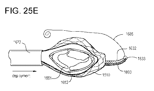
[0031] FIGS. 25A-25E illustrate various views of a process of placing a
side deliverable
transcatheter prosthetic heart valve in a compressed configuration, delivering
the compressed
prosthetic valve via a delivery catheter, and partially releasing the
prosthetic valve from the
delivery catheter for deployment in a native valve, according to an
embodiment.
[0032] FIGS. 26A-26C illustrate various views of a process of placing a
side deliverable
transcatheter prosthetic heart valve in a compressed configuration and loading
the compressed
prosthetic valve in a delivery catheter, according to an embodiment.
[0033] FIGS. 27A-27C illustrate various views of a process of placing a
side deliverable
transcatheter prosthetic heart valve in a compressed configuration and loading
the compressed
prosthetic valve in a delivery catheter, according to an embodiment.
[0034] FIGS. 28A-28C illustrate various views of a process of placing a
side deliverable
transcatheter prosthetic heart valve in a compressed configuration and loading
the compressed
prosthetic valve in a delivery catheter, according to an embodiment.
[0035] FIGS. 29A-29C illustrate various views of a process of placing a
side deliverable
transcatheter prosthetic heart valve in a compressed configuration and loading
the compressed
prosthetic valve in a delivery catheter, according to an embodiment.
6
CA 03133452 2021-09-13
WO 2020/186251 PCT/US2020/022828
[0036] FIG. 30 is an illustration of a top perspective view of an inner
frame of a flow control
component included in a prosthetic valve according to an embodiment.
[0037] FIGS. 31-33 illustrate of various views of the inner frame of FIG.
30 and shown in a
partially folded configuration, a folded configuration, and a folded and
compressed configuration,
respectively.
[0038] FIG. 34 is an illustration of a side view of an inner frame of a
flow control component
included in a prosthetic valve and shown as a linear wireframe sheet prior to
being formed into a
cylindrical configuration, according to an embodiment.
[0039] FIG. 35 is an illustration of a side perspective view of the inner
frame of FIG. 34 and
shown in the cylindrical configuration.
[0040] FIG. 36 is an illustration of a side view of a leaflet band of the
inner flow control
component having leaflet pockets sewn into a structural band of pericardial
tissue and shown in a
linear configuration.
[0041] FIG. 37 is an illustration of a bottom view of the leaflet band of
FIG. 36 and shown in
the linear configuration.
[0042] FIG. 38 is an illustration of a side perspective view of the leaflet
band of FIGS. 36 and
37, and shown in a cylindrical configuration suitable for coupling to the
inner frame of FIG. 35.
[0043] FIG. 39 is an illustration of a side perspective view of a portion
of the leaflet band of
FIG. 36 showing a single leaflet pocket sewn into the structural band.
[0044] FIG. 40 is an illustration of a bottom view of the leaflet band of
FIGS. 36-39 in the
cylindrical configuration and showing partial coaptation of the leaflets to
form a partially closed
fluid-seal.
[0045] FIGS. 41A-41D illustrate various views showing a process of
transitioning a side-
deliverable transcatheter prosthetic heart valve to a compressed configuration
for delivery,
according to an embodiment.
7
CA 03133452 2021-09-13
WO 2020/186251 PCT/US2020/022828
[0046] FIGS. 42A-42C illustrate various views showing a process of
transitioning a side-
deliverable transcatheter prosthetic heart valve to a compressed configuration
for delivery,
according to an embodiment.
[0047] FIGS. 43A-43C illustrate various views showing a process of
transitioning a side-
deliverable transcatheter prosthetic heart valve to a compressed configuration
for delivery,
according to an embodiment.
[0048] FIG. 44A is an illustration of a top view of a side deliverable
transcatheter prosthetic
heart valve in a compressed configured and disposed within a delivery
catheter, according to an
embodiment.
[0049] FIG. 44B is an illustration of a top perspective of the prosthetic
heart valve of FIG. 44A
partially released from the delivery catheter.
[0050] FIG. 45 is an illustration of a cut-away side view of a human heart
with a trans-septal
delivery catheter crossing from the right atrium to the left atrium for access
to the mitral valve,
according to an embodiment.
[0051] FIG. 45 is an illustration of a side view of a human heart having a
trans-septal (trans-
femoral/IVC or SVC) delivery catheter crossing from the right atrium to the
left atrium for access
to the mitral valve, according to an embodiment.
[0052] FIG. 46 is an illustration showing a process of using a distal
anchoring element of a
side deliverable transcatheter prosthetic heart valve to capture native
tissue, according to an
embodiment.
[0053] FIGS. 47-49 illustrate a side perspective views of a process of
deploying a side
deliverable transcatheter prosthetic heart valve, according to an embodiment.
[0054] FIGS. 50A-50D illustrate various views of an anterior anchor element
included in a
side deliverable transcatheter prosthetic heart valve according to an
embodiment, and shown in a
first configuration, a second configuration, a third configuration, and a
fourth configuration,
respectively.
8
CA 03133452 2021-09-13
WO 2020/186251 PCT/US2020/022828
[0055] FIGS. 51A-51H illustrate side perspective views of various anchors
for anchoring a
portion of a side deliverable transcatheter prosthetic heart valve to native
tissue, each according to
a different embodiment.
[0056] FIG. 52 is an illustration of a side perspective view of a side
deliverable transcatheter
prosthetic heart valve having multiple anterior anchor elements, according to
an embodiment.
[0057] FIG. 53 is an illustration of a side perspective view of a side
deliverable transcatheter
prosthetic heart valve having multiple anterior anchor elements, according to
an embodiment.
[0058] FIG. 54 is an illustration of a side perspective view of a side
deliverable transcatheter
prosthetic heart valve having a distal anchoring element with a graduated
stiffness, according to
an embodiment.
[0059] FIG. 55A is an illustration of a side perspective view of a side
deliverable transcatheter
prosthetic heart valve having a distal anchoring element and a proximal
anchoring element,
according to an embodiment.
[0060] FIG. 55B is an illustration of a side perspective view of the
prosthetic heart valve of
FIG. 55A deployed in an annulus of a native valve.
[0061] FIG. 56A is an illustration of a side perspective view of a side
deliverable transcatheter
prosthetic heart valve having a distal anchoring element and a proximal
anchoring element,
according to an embodiment.
[0062] FIG. 56B is an illustration of a side perspective view of the
prosthetic heart valve of
FIG. 56A deployed in an annulus of a native valve.
[0063] FIG. 57A is an illustration of a side perspective view of a side
deliverable transcatheter
prosthetic heart valve having a distal anchoring element and a proximal
anchoring element,
according to an embodiment.
[0064] FIG. 57B is an illustration of a side perspective view of the
prosthetic heart valve of
FIG. 57A deployed in an annulus of a native valve.
9
CA 03133452 2021-09-13
WO 2020/186251 PCT/US2020/022828
[0065] FIG. 58A is an illustration of a guide wire delivery catheter
providing access to, for
example, an Al -P1 target area of a native valve, according to an embodiment.
[0066] FIG. 58B is an illustration of an enlarged view of a portion of the
guide wire delivery
catheter of FIG. 58A.
[0067] FIG. 59 is an illustration of a circumferential balloon disposed
around an end portion
of a delivery catheter, according to an embodiment.
[0068] FIG. 60 is an illustration of a side view of a distal end portion of
a sheath included in a
delivery system and having a side port for delivering a guide wire to, for
example, an Al-P1 target
area of a native valve, according to an embodiment.
[0069] FIG. 61 is an illustration of a cross-sectional view of the sheath
of FIG. 60 engaging
native tissue and allowing the side port to deliver the guide wire.
[0070] FIG. 62 is an illustration of a side perspective view of a side
deliverable transcatheter
prosthetic heart valve having a septal tether configured to secure the
prosthetic heart valve in an
annulus of a native valve, according to an embodiment.
[0071] FIG. 63 is an illustration of a cross-sectional view of a side
deliverable transcatheter
prosthetic heart valve and secured in an annulus of a native valve, according
to an embodiment.
[0072] FIG. 64 is an illustration of a side view of a portion of a delivery
system having a
docking receptacle with a keyed feature and/or tissue grabbing features for
anchoring to a free wall
of native tissue, according to an embodiment.
[0073] FIG. 65 is an illustration of an enlarged view of at least a portion
of the tissue-grabbing
features of FIG. 64 shown anchored to the free wall.
[0074] FIG. 66 is a flowchart illustrating a method of deploying a side
deliverable transcatheter
prosthetic heart valve in an annulus of a native valve, according to an
embodiment.
Detailed Description
CA 03133452 2021-09-13
WO 2020/186251 PCT/US2020/022828
[0075] Disclosed embodiments are directed to transcatheter prosthetic heart
valves and/or
components thereof, and methods of manufacturing, loading, delivering, and/or
deploying the
transcatheter prosthetic valves and/or components thereof In some embodiments,
a side-
deliverable prosthetic heart valve includes an outer frame having a perimeter
wall that
circumscribes a central channel extending along a central axis and a flow
control component
mounted within the central channel. The flow control component includes an
inner frame and a
set of leaflets coupled to the inner frame. The prosthetic valve is foldable
along a longitudinal axis
and compressible along the central axis to place the prosthetic valve in a
compressed configuration
for delivery via a delivery catheter. The longitudinal axis is substantially
parallel to a lengthwise
axis of the delivery catheter when the prosthetic valve is disposed therein.
The prosthetic valve is
configured to transition to an expanded configuration when the prosthetic
valve is released from
the delivery catheter. The prosthetic valve further includes a distal
anchoring element having a
first end portion coupled to a distal side of the perimeter wall of the outer
frame and a second end
portion opposite the first end portion. The second end portion is configured
to selectively engage
a guide wire to allow the distal anchoring element to be advanced along the
guide wire during
deployment of the prosthetic valve. The distal anchoring element is in an
extended configuration
during deployment to allow the distal anchoring element to capture at least
one of native leaflet or
chordae. In response to the guide wire being disengaged from the second end
portion, the distal
anchoring element transitions to a folded configuration in which at least one
of the native leaflet
or the chordae is secured between the distal anchoring element and the distal
side of the perimeter
wall.
[0076] In some embodiments, a side-deliverable prosthetic heart valve
includes an outer frame
having a perimeter wall that circumscribes a central channel extending along a
central axis and a
flow control component mounted within the central channel. The flow control
component includes
an inner frame and a set of leaflets coupled to the inner frame. The
prosthetic valve is foldable
along a longitudinal axis and compressible along the central axis to place the
prosthetic valve in a
compressed configuration for delivery via a delivery catheter. The
longitudinal axis is
substantially parallel to a lengthwise axis of the delivery catheter when the
prosthetic valve is
disposed therein. The prosthetic valve is configured to transition to an
expanded configuration
when the prosthetic valve is released from the delivery catheter. The
prosthetic valve further
includes a distal anchoring element coupled to a distal side of the perimeter
wall of the outer frame
11
CA 03133452 2021-09-13
WO 2020/186251 PCT/US2020/022828
and an anterior anchoring element coupled to an anterior side of the perimeter
wall. The distal
anchoring element is releasably coupled to a guide wire and is configured to
be advanced along
the guide wire when in an extended configuration to capture at least one of a
distal native leaflet
or distal chordae. The distal anchoring element transitions to a folded
configuration when released
from the guide wire to secure the distal native leaflet or the distal chordae
between the distal
anchoring element and the distal side of the perimeter wall. The anterior
anchoring element
includes a sleeve and an anterior clip at least partially disposed in the
sleeve. The anterior clip can
be transitioned between a first configuration in which the anterior clip
extends from the sleeve in
the direction of the central axis to allow the anterior clip to capture at
least one of an anterior native
leaflet or anterior chordae, and a second configuration in which the anterior
clip is at least partially
retracted into the sleeve to secure the anterior native leaflet or the
anterior chordae between the
anterior clip and the anterior side of the perimeter wall.
[0077] Any of the prosthetic heart valves described herein can be a
relatively low profile, side-
deliverable implantable prosthetic heart valve. Any of the prosthetic heart
valves can be
transcatheter prosthetic heart valves configured to be delivered into a heart
via a delivery catheter.
The prosthetic heart valves can have at least an annular outer valve frame and
an inner flow control
component (e.g., a 2-leaflet or 3-leaflet valve, sleeve, and/or the like)
mounted in the valve frame.
In addition, the prosthetic heart valves can be include a single anchoring
element or multiple
anchoring elements configured to anchor the valve in the annulus of a native
valve.
[0078] Any of the prosthetic heart valves described herein can be
configured to transition
between an expanded configuration and a compressed configuration. For example,
any of the
embodiments described herein can be a balloon-inflated prosthetic heart valve,
a self-expanding
prosthetic heart valve, and/or the like.
[0079] Any of the prosthetic heart valves described herein can be
compressible ¨ into the
compressed configuration ¨ in a lengthwise or orthogonal direction relative to
the central axis of
the flow control component that can allow a large diameter valve (e.g., having
a height of about 5-
60 mm and a diameter of about 20-80 mm) to be delivered and deployed from the
inferior vena
cava directly into the annulus of a native mitral or tricuspid valve using,
for example, a 24-36Fr
12
CA 03133452 2021-09-13
WO 2020/186251 PCT/US2020/022828
delivery catheter and without delivery and deployment from the delivery
catheter at an acute angle
of approach.
[0080] Any of the prosthetic heart valves described herein can have a
central axis that is co-
axial or at least substantially parallel with blood flow direction through the
valve. In some
embodiments, the compressed configuration of the valve is orthogonal to the
blood flow direction.
In some embodiments, the compressed configuration of the valve is parallel to
or aligned with the
blood flow direction. In some embodiment, the valve can be compressed to the
compressed
configuration in two directions ¨ orthogonal to the blood flow direction
(e.g., laterally) and parallel
to the blood flow (e.g., axially). In some embodiments, a long-axis or
longitudinal axis is oriented
at an intersecting angle of between 45-135 degrees to the first direction when
in the compressed
configuration and/or the expanded configuration.
[0081] Any of the prosthetic heart valves described herein can include an
outer support frame
that includes a set of compressible wire cells having an orientation and cell
geometry substantially
orthogonal to the central axis to minimize wire cell strain when the outer
support frame is in a
compressed configuration, a rolled and compressed configuration, or a folded
and compressed
configuration.
[0082] In some embodiments, an outer support frame has a lower body portion
and an upper
collar portion. The lower body portion forms a shape such as a funnel,
cylinder, flat cone, or
circular hyperboloid when the outer support frame is in an expanded
configuration. In some
embodiments, the outer support frame is formed from a wire, a braided wire, or
a laser-cut wire
frame, and is covered with a biocompatible material. The biocompatible
material can be covered
such that an inner surface is covered with pericardial tissue, an outer
surface is covered with a
woven synthetic polyester material, and/or both the inner surface is covered
with pericardial tissue
and the outer surface is covered with a woven synthetic polyester material.
[0083] In some embodiments, an outer support frame has a side profile of a
flat cone shape
having an outer diameter R of 40-80 mm, an inner diameter r of 20-60 mm, and a
height of 5-60
mm. In some embodiments, an annular support frame has a side profile of an
hourglass shape
having a top diameter R1 of 40-80 mm, a bottom diameter R2 of 50-70 mm, an
internal diameter
r of 20-60 mm, and a height of 5-60 mm.
13
CA 03133452 2021-09-13
WO 2020/186251 PCT/US2020/022828
[0084] Any of the prosthetic heart valves described herein can include one
or more anchoring
element extending from a sidewall of a valve frame. For example, any of the
prosthetic heart
valves can include a distal anchoring element, which can be used, for example,
as a Right
Ventricular Outflow Tract ("RVOT") tab or a Left Ventricular Outflow Tract
("LVOT") tab. Any
of the valves described herein can also include an anchoring element extending
from a proximal
sided of the valve frame, which can be used, for example, to anchor the valve
to a proximal sub-
annular space. Any of the valves described herein can also include an anterior
or posterior
anchoring element extended from an anterior or posterior side of the valve
frame, respectively.
The anchoring elements can include and/or can be formed from a wire loop or
wire frame, an
integrated frame section, and/or a stent, extending from about 10-40 mm away
from the tubular
frame.
[0085] Any of the prosthetic heart valves described herein can include (i)
an upper anchoring
element attached to a distal upper edge of the tubular frame, the upper
anchoring element can
include or be formed from a wire loop or wire frame extending from about 2-20
mm away from
the tubular frame, and (ii) a lower anchoring element (e.g., used as a RVOT
tab) extending from a
distal side of the tubular frame, the lower anchoring element can include
and/or can be formed
from a wire loop or wire frame extending from about 10-40 mm away from the
tubular frame.
[0086] Any of the prosthetic heart valves described herein can include a
distal lower anchoring
element configured to be positioned into the RVOT of the right ventricle and a
proximal lower
anchoring element configured to be positioned into a sub-annular position in
contact with and/or
adjacent to sub-annular tissue of the right ventricle. The transcatheter
prosthetic heart valve can
also include a distal upper anchoring element configured to be positioned into
a supra-annular
position in contact with and/or adjacent to supra-annular tissue of the right
atrium. The distal
upper anchoring element can provide a supra-annular downward force in the
direction of the right
ventricle and the distal and proximal lower anchoring elements can provide a
sub-annular upward
force in the direction of the right atrium.
[0087] Any of the prosthetic heart valves described herein can include an
inner flow control
component that has a leaflet frame with 2-4 flexible leaflets mounted thereon.
The 2-4 leaflets are
configured to permit blood flow in a first direction through an inflow end of
the flow control
14
CA 03133452 2021-09-13
WO 2020/186251 PCT/US2020/022828
component and block blood flow in a second direction, opposite the first
direction, through an
outflow end of the flow control component. The leaflet frame can include two
or more panels of
diamond-shaped or eye-shaped wire cells made from heat-set shape memory alloy
material such
as, for example, Nitinol. The leaflet frame can be configured to be foldable
along a z-axis (e.g., a
longitudinal axis) from a rounded or cylindrical configuration to a flattened
cylinder configuration,
and compressible along a vertical y-axis (e.g., a central axis) to a
compressed configuration. In
some implementations, the leaflet frame can include a pair of hinge areas,
fold areas, connection
points, etc. that can allow the leaflet frame to be folded flat along the z-
axis prior to the leaflet
frame being compressed along the vertical y-axis. The inner frame can be, for
example, a single-
piece structure with two or more living hinges (e.g., stress concentration
riser and/or any suitable
structure configured to allow for elastic/nonpermanent deformation of the
inner frame). In other
implementations, the inner frame can be a two-piece structure where the hinge
areas are formed
using a secondary attachment method (e.g. sutures, fabrics, molded polymer
components, etc.)
[0088] In some embodiments, the inner flow control component in an expanded
configuration
forms a shape such as a funnel, cylinder, flat cone, or circular hyperboloid.
In some embodiments,
the inner flow control component has a leaflet frame with a side profile of a
flat cone shape having
an outer diameter R of 20-60 mm, an inner diameter r of 10-50 mm, where
diameter R is great
than diameter r, and a height of 5-60 mm. In some embodiments, the leaflet
frame is comprised
of a wire, a braided wire, or a laser-cut wire. In some embodiments, the
leaflet frame can have
one or more longitudinal supports integrated into or mounted thereon and
selected from rigid or
semi-rigid posts, rigid or semi-rigid ribs, rigid or semi-rigid batons, rigid
or semi-rigid panels, and
combinations thereof
[0089] Any of the prosthetic heart valves described herein and/or any
component, feature,
and/or aspect thereof can be similar to and/or substantially the same as the
prosthetic heart valves
(or components, features, and/or aspects thereof) described in International
Patent Application No.
PCT/US2019/051957, filed September 19, 2019, entitled "Transcatheter
Deliverable Prosthetic
Heart Valves and Method of Delivery" (referred to herein as "the '957 PCT"),
International Patent
Application No. PCT/U52019/067010, filed December 18, 2019, entitled
"Transcatheter
Deliverable Prosthetic Heart Valves and Methods of Delivery" (referred to
herein as "the '010
PCT"), and/or International Patent Application No. PCT/US2020/015231, filed
January 27, 2020,
CA 03133452 2021-09-13
WO 2020/186251 PCT/US2020/022828
"Collapsible Inner Flow Control Component for Side-Deliverable Transcatheter
Heart Valve
Prosthesis" (referred to herein as "the '231 PCT"), the disclosures of which
are incorporated herein
by reference in their entireties.
[0090] In some implementations, a prosthetic valve can be configured for
deployment, for
example, in an annulus of a native mitral valve. Use of a side-deliverable
transcatheter mitral
valve replacement allows a relatively large diameter valve to be delivered and
deployed from the
inferior vena cava trans-septally into the mitral valve without requiring an
oversized diameter
catheter and without requiring delivery and deployment from a catheter at an
acute angle of
approach.
[0091] In some embodiments, a prosthetic mitral valve can include one or
more anchoring
elements configured to secure the prosthetic valve in the native valve
annulus. For example, a
prosthetic mitral valve such as those described herein can include a distal
anchoring element, a
proximal anchoring element, and one or more anterior or posterior anchoring
elements (e.g., an
anterior Al, A2, or A3 anchoring element and/or a posterior Pl, P2, or P3
anchoring element).
[0092] In some embodiments, a prosthetic mitral valve includes a distal
anchoring tab that is
(i) extended around a posterior leaflet and/or chordae using a guide wire to
capture native mitral
leaflet and/or chordae tissue and (ii) allowed to contract when the guide wire
is withdrawn to pin
the native tissue against a sidewall of the valve. In some embodiments, the
valve further includes
a proximal anchoring element configured to anchor a proximal side of the valve
using a tab or loop
deployed to the A3-P3 (proximal) commissure area of the native mitral valve.
In some
embodiments, the valve further includes an A2 clip that is similarly
configured to extend or unfold
(e.g., via a guide wire or self-actuation) to capture native mitral leaflet
and/or chordae tissue and,
upon withdrawal of the guide wire and/or otherwise upon retracting or re-
folding the A2 clip, to
pin the native tissue against the sidewall of the valve. In some embodiments,
an A2 clip can be
formed of a braided polyethylene, treated pericardial tissue, ePTFE, or
Nitinol, and may have one
or more radio-opaque markers.
[0093] In some embodiments, a delivery system for delivering a side-
deliverable prosthetic
heart valve can include a catheter that can deliver a guide wire directly to
an A 1 -P1 commissure
of a native mitral valve. In some implementations, targeting the Al -P1
commissure area, can
16
CA 03133452 2021-09-13
WO 2020/186251 PCT/US2020/022828
allow a side deliverable valve such as those described herein to be directed
to the target area via a
delivery catheter for successful deployment of the valve.
[0094] Any of the delivery systems described herein can include a guide
wire catheter that can
be used independently of a valve delivery catheter. In some embodiments, the
guide wire catheter
has a custom shape, which articulates the distal tip of the guide wire
catheter to the Al/P1
commissure pointing behind the posterior native leaflets. In some embodiments,
the guide wire
catheter can be used prior to insertion of the valve delivery catheter or can
be threaded through the
valve delivery catheter prior to loading the valve and can exit straight
through the main lumen of
through a side port in communication with the main lumen. After the guide wire
is placed, the
guide wire catheter can be removed from the patient.
[0095] Any of the delivery systems described herein can include a delivery
catheter for a side-
deliverable prosthetic heart valve that includes an outer shaft having an
outer proximal end, an
outer distal end, and an outer shaft lumen, wherein the outer distal end is
closed with an atraumatic
ball mounted thereon. The outer shaft lumen has an inner diameter of 8-10mm
sized for passage
of a side delivered transcatheter prosthetic heart valve (e.g., a prosthetic
tricuspid valve and/or a
prosthetic mitral valve) therethrough.
[0096] Any method for manufacturing prosthetic heart valves described
herein can include
using additive or subtractive metal or metal-alloy manufacturing to produce a
self-expanding outer
support frame having a central channel and an outer perimeter wall
circumscribing a central
vertical axis. A collapsible flow control component is mounted within the
outer support frame and
configured to permit blood flow in a first direction through an inflow end of
the valve and block
blood flow in a second direction, opposite the first direction, through an
outflow end of the valve.
The flow control component has a leaflet frame with 2-4 flexible leaflets
mounted. The leaflet
frame can be formed using additive or subtractive metal or metal-alloy
manufacturing. The
additive metal or metal-alloy manufacturing can be 3D printing, direct metal
laser sintering
(powder melt), and/or the like. The subtractive metal or metal-alloy
manufacturing is
photolithography, laser sintering/cutting, CNC machining, electrical discharge
machining, and/or
the like. In some embodiments, a process of manufacturing can further include
mounting the flow
17
CA 03133452 2021-09-13
WO 2020/186251 PCT/US2020/022828
control component within the outer support frame, and covering an outer
surface of the outer
support frame with a pericardium material or similar biocompatible material.
[0097] Any method for delivering prosthetic heart valves described herein
can include at least
one of (i) compressing the valve along a central vertical axis to reduce a
vertical dimension of the
valve from top to bottom to place the valve in a compressed configuration,
(ii) unilaterally rolling
the valve into a compressed configuration from one side of the annular support
frame, (iii)
bilaterally rolling the valve into a compressed configuration from two
opposing sides of the
annular support frame, (iv) flattening the valve into two parallel panels that
are substantially
parallel to the long-axis, (v) flattening the valve into two parallel panels
that are substantially
parallel to the long-axis and then rolling the flattened valve into a
compressed configuration, or
(vi) flattening the valve into two parallel panels that are substantially
parallel to the long-axis and
then compressing the valve along a central vertical axis to reduce a vertical
dimension of the valve
from top to bottom to place the valve in a compressed configuration.
[0098] Any method for delivering prosthetic heart valves described herein
can include
orthogonal delivery of the prosthetic heart valve to a desired location in the
body that includes (i)
advancing a delivery catheter to the desired location in the body and (ii)
delivering the prosthetic
heart valve to the desired location in the body by releasing the valve from
the delivery catheter.
The valve is in a compressed configuration when in the delivery catheter and
transitions to an
expanded configuration when released from the delivery catheter.
[0099] Any method for delivering prosthetic heart valves described herein
can include
releasing the valve from the delivery catheter by (i) pulling the valve out of
the delivery catheter
using a pulling member (e.g., a wire or rod) that is releasably connected to a
sidewall, a drum or
collar, and/or an anchoring element (e.g., a distal anchoring element),
wherein advancing the
pulling member away from the delivery catheter pulls the compressed valve out
of the delivery
catheter, or (ii) pushing the valve out of the delivery catheter using a
pushing member (e.g., a wire,
rod, catheter, delivery member, etc.) that is releasably connected to a
sidewall, a drum or collar,
and/or an anchoring element (e.g., a distal anchoring element), wherein
advancing the pushing
member out of from the delivery catheter pushes the compressed valve out of
the delivery catheter.
18
CA 03133452 2021-09-13
WO 2020/186251 PCT/US2020/022828
[0100] In some embodiments, a method for deploying a side-deliverable
prosthetic heart valve
to a patient includes advancing a guide wire to an atrium, through a plane
defined by an annulus
of a native valve, and behind a native leaflet of the native valve. The
prosthetic valve is advanced
in an orthogonally compressed configuration through a lumen of a delivery
catheter and into the
atrium. The prosthetic valve includes a distal anchoring element releasably
coupled to the guide
wire such that the prosthetic valve is advanced along a portion of the guide
wire. The prosthetic
valve is released from the delivery catheter to allow at least a portion of
the prosthetic valve to
transition to an expanded configuration such that the distal anchoring element
is in an extended
configuration after the releasing. The prosthetic valve is advanced along the
guide wire to (i) place
the distal anchoring element in a position behind the native leaflet and (ii)
seat the prosthetic valve
in the annulus of the native valve. The guide wire is withdrawn to release the
distal anchoring
element to a folded position allowing the distal anchoring element to capture
at least one of native
leaflet or chordae and to secure the native leaflet or chordae between the
distal anchoring element
and a perimeter wall of the prosthetic valve.
[0101] Any method for delivering and/or deploying prosthetic heart valves
described herein
can include releasing the valve from a delivery catheter while increasing
blood flow during
deployment of the valve by (i) partially releasing the valve from the delivery
catheter to establish
blood flow around the partially released valve and blood flow through the flow
control component;
(ii) completely releasing the valve from the delivery catheter while
maintaining attachment to the
valve to transition to a state with increased blood flow through the flow
control component and
decreased blood flow around the valve; (iii) deploying the valve into a final
mounted position in a
native annulus to transition to a state with complete blood flow through the
flow control component
and minimal or no blood flow around the valve; and (iv) disconnecting and
withdrawing a
positioning catheter, pulling or pushing wire or rod, and/or the delivery
catheter.
[0102] Any method for delivering and/or deploying prosthetic heart valves
described herein
can include positioning the valve or a portion thereof in a desired position
relative to the native
tissue. For example, the method can include positioning a distal anchoring tab
of the heart valve
prosthesis into a ventricular outflow tract of the left or right ventricle. In
some embodiments, the
method can further include positioning an upper distal anchoring tab into a
supra-annular position,
where the upper distal anchoring tab provides a supra-annular downward force
in the direction of
19
CA 03133452 2021-09-13
WO 2020/186251 PCT/US2020/022828
the ventricle and the distal anchoring tab (e.g., the lower distal anchoring
tab) provides a sub-
annular upward force in the direction of the atrium. In some embodiments, the
method can include
rotating the heart valve prosthesis, using a steerable catheter, along an axis
parallel to the plane of
the valve annulus. In some embodiments, the method can include anchoring one
or more tissue
anchors attached to the valve into native tissue to secure the valve in a
desired position.
[0103] Any method for delivering and/or deploying prosthetic heart valves
described herein
can include orthogonal delivery of the prosthetic heart valve to a native
annulus of a human heart
that includes at least one of (i) advancing a delivery catheter to the
tricuspid valve or pulmonary
artery of the heart through the inferior vena cava (IVC) via the femoral vein,
(ii) advancing to the
tricuspid valve or pulmonary artery of the heart through the superior vena
cava (SVC) via the
jugular vein, or (iii) advancing to the mitral valve of the heart through a
trans-atrial approach (e.g.,
fossa ovalis or lower), via the IVC-femoral or the SVC-jugular approach; and
(iv) delivering and/or
deploying the prosthetic heart valve to the native annulus by releasing the
valve from the delivery
catheter.
[0104] In some embodiments, a method for delivering and/or deploying
prosthetic heart valves
described herein to a native mitral valve can include advancing a guide wire
trans-septally to the
left atrium, through an annular plane at the Al/PI commissure to a position
behind a native
posterior (e.g., P2) leaflet of a mitral valve of the patient. A delivery
catheter containing the valve
in an orthogonally compressed configuration is advanced to the left atrium of
the patient a delivery
catheter, wherein a distal anchoring tab of the valve is threaded onto the
guide wire. In some
embodiment, an A2 clip optionally may be threaded onto the guide wire. The
valve is released
from the delivery catheter, wherein the distal anchoring tab is in an open
configuration and tracks
over the guide wire during release. The valve is advanced over the guide wire
to move the distal
anchoring tab to the position behind the native posterior leaflet and to seat
the valve into the native
annulus. The guide wire is withdrawn to release the distal anchoring tab to
the folded position
allowing the tab to capture native leaflet and/or native chordae, and sandwich
the native leaflet
and/ or chordae between the folded tab and an outer perimeter wall of the
valve. In some
embodiments, the method may optionally include withdrawing the guide wire to
an A2 clip release
position to release the A2 clip to the open position allowing the A2 clip to
capture native leaflet
CA 03133452 2021-09-13
WO 2020/186251 PCT/US2020/022828
and/or native chordae, and sandwich the native leaflet and/or chordae between
the A2 clip and the
perimeter wall of the annular support frame.
[0105]
In some embodiments, the method can include delivering and/or deploying the
valve,
at least in part, via a single fixed curve catheter, a single deflectable
catheter, an outer fixed curve
catheter with an inner fixed curve catheter, an outer fixed curve catheter
with an inner deflectable
catheter, and an out deflectable catheter with an inner fixed curve catheter.
[0106]
Any method for delivering and/or deploying prosthetic heart valves described
herein
and/or any portion thereof can be similar to and/or substantially the same as
one or more methods
for delivering and/or deploying prosthetic heart valves (or portion(s)
thereof) described in the '957
PCT, the '010 PCT, and/or the '231 PCT.
[0107]
The terminology used herein is for the purpose of describing particular
embodiments
only and is not intended to limit the full scope of the claims. Unless defined
otherwise, all technical
and scientific terms used herein have the same meanings as commonly understood
by one of
ordinary skill in the art. Nothing in this disclosure is to be construed as an
admission that the
embodiments described in this disclosure are not entitled to antedate such
disclosure by virtue of
prior invention.
[0108]
As used herein, the singular forms "a", "an" and "the" are intended to include
the plural
forms as well, unless the context clearly indicates otherwise. With respect to
the use of
substantially any plural and/or singular terms herein, those having skill in
the art can translate from
the plural to the singular and/or from the singular to the plural as is
appropriate to the context
and/or application. The various singular/plural permutations may be expressly
set forth herein for
sake of clarity.
[0109]
In general, terms used herein, and especially in the appended claims (e.g.,
bodies of the
appended claims) are generally intended as "open" terms (e.g., the term
"including" should be
interpreted as "including but not limited to," the term "having" should be
interpreted as "having at
least," etc.).
Similarly, the terms "comprises" and/or "comprising," when used in this
specification, specify the presence of stated features, integers (or fractions
thereof), steps,
operations, elements, and/or components, but do not preclude the presence or
addition of one or
21
CA 03133452 2021-09-13
WO 2020/186251 PCT/US2020/022828
more other features, integers (or fractions thereof), steps, operations,
elements, components, and/or
groups thereof. As used in this document, the term "comprising" means
"including, but not limited
to."
[0110] As used herein the term "and/or" includes any and all combinations
of one or more of
the associated listed items. It should be understood that virtually any
disjunctive word and/or
phrase presenting two or more alternative terms, whether in the description,
claims, or drawings,
should be understood to contemplate the possibilities of including one of the
terms, either of the
terms, or both terms. For example, the phrase "A or B" will be understood to
include the
possibilities of "A" or "B" or "A and B."
[0111] All ranges disclosed herein also encompass any and all possible
subranges and
combinations of subranges thereof unless expressly stated otherwise. Any
listed range should be
recognized as sufficiently describing and enabling the same range being broken
down into at least
equal subparts unless expressly stated otherwise. As will be understood by one
skilled in the art,
a range includes each individual member.
[0112] The term "valve prosthesis," "prosthetic heart valve," and/or
"prosthetic valve" can
refer to a combination of a frame and a leaflet or flow control structure or
component, and can
encompass both complete replacement of an anatomical part (e.g., a new
mechanical valve
replaces a native valve), as well as medical devices that take the place of
and/or assist, repair, or
improve existing anatomical parts (e.g., the native valve is left in place).
[0113] The disclosed valves include a member (e.g., a frame) that can be
seated within a native
valve annulus and can be used as a mounting element for a leaflet structure, a
flow control
component, or a flexible reciprocating sleeve or sleeve-valve. It may or may
not include such a
leaflet structure or flow control component, depending on the embodiment. Such
members can be
referred to herein as an "annular support frame," "tubular frame," "wire
frame," "valve frame,"
"flange," "collar," and/or any other similar terms.
[0114] The term "flow control component" can refer in a non-limiting sense
to a leaflet
structure having 2-, 3-, 4-leaflets of flexible biocompatible material such a
treated or untreated
pericardium that is sewn or joined to a annular support frame, to function as
a prosthetic heart
22
CA 03133452 2021-09-13
WO 2020/186251 PCT/US2020/022828
valve. Such a valve can be a heart valve, such as a tricuspid, mitral, aortic,
or pulmonary, that is
open to blood flowing during diastole from atrium to ventricle, and that
closes from systolic
ventricular pressure applied to the outer surface. Repeated opening and
closing in sequence can
be described as "reciprocating." The flow control component is contemplated to
include a wide
variety of (bio)prosthetic artificial heart valves. Bioprosthetic pericardial
valves can include
bioprosthetic aortic valves, bioprosthetic mitral valves, bioprosthetic
tricuspid valves, and
bioprosthetic pulmonary valves.
[0115] Any of the disclosed valve embodiments may be delivered by a
transcatheter approach.
The term "transcatheter" is used to define the process of accessing,
controlling, and/or delivering
a medical device or instrument within the lumen of a catheter that is deployed
into a heart chamber
(or other desired location in the body), as well as an item that has been
delivered or controlled by
such as process. Transcatheter access is known to include cardiac access via
the lumen of the
femoral artery and/or vein, via the lumen of the brachial artery and/or vein,
via lumen of the carotid
artery, via the lumen of the jugular vein, via the intercostal (rib) and/or
sub-xiphoid space, and/or
the like. Moreover, transcatheter cardiac access can be via the inferior vena
cava (IVC), superior
vena cava (SVC), and/or via a trans-atrial (e.g., fossa ovalis or lower).
Transcatheter can be
synonymous with transluminal and is functionally related to the term
"percutaneous" as it relates
to delivery of heart valves. As used herein, the term "lumen" can refer to the
inside of a cylinder
or tube. The term "bore" can refer to the inner diameter of the lumen.
[0116] The mode of cardiac access can be based at least in part on "body
channel" may be
used to define a blood conduit or vessel within the body, the particular
application of the disclosed
embodiments of prosthetic valves determines the body channel at issue. An
aortic valve
replacement, for example, would be implanted in, or adjacent to, the aortic
annulus. Likewise, a
tricuspid or mitral valve replacement would be implanted at the tricuspid or
mitral annulus. Certain
features are particularly advantageous for one implantation site or the other.
However, unless the
combination is structurally impossible, or excluded by claim language, any of
the valve
embodiments described herein could be implanted in any body channel.
[0117] The term "expandable" as used herein may refer to a component of the
heart valve
capable of expanding from a first, delivery diameter to a second, implantation
diameter. An
23
CA 03133452 2021-09-13
WO 2020/186251 PCT/US2020/022828
expandable structure, therefore, does not mean one that might undergo slight
expansion from a
rise in temperature, or other such incidental cause. Conversely, "non-
expandable" should not be
interpreted to mean completely rigid or a dimensionally stable, as some slight
expansion of
conventional "non-expandable" heart valves, for example, may be observed.
[0118] Any of the disclosed valve embodiments may be delivered via
traditional transcatheter
delivery techniques or via orthogonal delivery techniques. For example,
traditional delivery of
prosthetic valves can be such that a central cylinder axis of the valve is
substantially parallel to a
length-wise axis of the delivery catheter. Typically, the valves are
compressed in a radial direction
relative to the central cylinder axis and advanced through the lumen of the
delivery catheter. The
valves are deployed from the end of the delivery catheter and expanded
outwardly in a radial
direction from the central cylinder axis.
[0119] As used herein the terms "side-delivered," "side-delivery,"
"orthogonal delivery,"
"orthogonally delivered," and/or so forth can be used interchangeably to
describe such a delivery
method and/or a valve delivered using such a method. Orthogonal delivery of
prosthetic valves
can be such that the central cylinder axis of the valve is substantially
orthogonal to the length-wise
axis of the delivery catheter. With orthogonal delivery, the valves are
compressed (or otherwise
reduced in size) in a direction substantially parallel to the central cylinder
axis and/or in a lateral
direction relative to the central cylinder axis. As such, a length-wise axis
(e.g., a longitudinal axis)
of an orthogonally delivered valve is substantially parallel to the length-
wise axis of the delivery
catheter. In other words, an orthogonally delivered prosthetic valve is
compressed and/or delivered
at a roughly 90 degree angle compared to traditional processes of compressing
and delivering
transcatheter prosthetic valves. Moreover, prosthetic valves configured to be
orthogonally
delivered and the processes of delivering such valves are described in detail
in the '957 PCT and/or
the '010 PCT incorporated by reference hereinabove.
[0120] Mathematically, the term "orthogonal" refers to an intersecting
angle of 90 degrees
between two lines or planes. As used herein, the term "substantially
orthogonal" refers to an
intersecting angle of 90 degrees plus or minus a suitable tolerance. For
example, "substantially
orthogonal" can refer to an intersecting angle ranging from 75 to 105 degrees.
24
CA 03133452 2021-09-13
WO 2020/186251 PCT/US2020/022828
[0121] As used herein, the term "tissue anchor" generally refers to a
fastening device that
connects a portion of an outer frame of a prosthetic to native annular tissue,
usually at or near a
periphery of a collar of the prosthetic valve. The tissue anchor may be
positioned to avoid piercing
tissue and just rely on the compressive force of the two plate-like collars on
the captured tissue, or
a tissue anchor (with or without an integrated securement wire) may pierce
through native tissue
to provide anchoring, or a combination of both. The tissue anchor may have a
securement
mechanism, such as a pointed tip, a groove, a flanged shoulder, a lock, one or
more apertures,
and/or the like. In some embodiments, a securement mechanism can be attached
or anchored to a
portion of an outer frame by any attachment or anchoring mechanisms, including
a knot, a suture,
a wire crimp, a wire lock, a cam mechanism, or combinations.
[0122] Any of the prosthetic valves and/or components thereof may be
fabricated from any
suitable biocompatible material or combination of materials. For example, an
outer valve frame,
an inner valve frame (e.g., of an inner flow control component), and/or
components thereof may
be fabricated from biocompatible metals, metal alloys, polymer coated metals,
and/or the like.
Suitable biocompatible metals and/or metal alloys can include stainless steel
(e.g., 316 L stainless
steel), cobalt chromium (Co-Cr) alloys, nickel-titanium alloys (e.g.,
Nitinolg), and/or the like.
Moreover, any of the outer or inner frames described herein can be formed from
superelastic or
shape-memory alloys such as nickel-titanium alloys (e.g., Nitinolg). Suitable
polymer coatings
can include polyethylene vinyl acetate (PEVA), poly-butyl methacrylate (PBMA),
translute
Styrene Isoprene Butadiene (SIBS) copolymer, polylactic acid, polyester,
polylactide, D-lactic
polylactic acid (DLPLA), polylactic-co-glycolic acid (PLGA), and/or the like.
Some such polymer
coatings may form a suitable carrier matrix for drugs such as, for example,
Sirolimus, Zotarolimus,
Biolimus, Novolimus, Tacrolimus, Paclitaxel, Probucol, and/or the like.
[0123] Some biocompatible synthetic material(s) can include, for example,
polyesters,
polyurethanes, polytetrafluoroethylene (PTFE) (e.g., Teflon), and/or the like.
Where a thin,
durable synthetic material is contemplated (e.g., for a covering), synthetic
polymer materials such
expanded PTFE or polyester may optionally be used. Other suitable materials
may optionally
include elastomers, thermoplastics, polyurethanes, thermoplastic polycarbonate
urethane,
polyether urethane, segmented polyether urethane, silicone polyether urethane,
polyetheretherketone (PEEK), silicone-polycarbonate urethane, polypropylene,
polyethylene,
CA 03133452 2021-09-13
WO 2020/186251 PCT/US2020/022828
low-density polyethylene (LDPE), high-density polyethylene (HDPE), ultra-high
density
polyethylene (UHDPE), polyolefins, polyethylene-glycols, polyethersulphones,
polysulphones,
polyvinylpyrrolidones, polyvinylchlorides, other fluoropolymers, polyesters,
polyethylene-
terephthalate (PET) (e.g., Dacron), Poly-L-lactic acids (PLLA), polyglycolic
acid (PGA), poly(D,
L-lactide/glycolide) copolymer (PDLA), silicone polyesters, polyamides
(Nylon), PTFE,
elongated PTFE, expanded PTFE, siloxane polymers and/or oligomers, and/or
polylactones, and
block co-polymers using the same.
[0124] Any of the outer valve frames, inner valve frames (e.g., of the flow
control
components), and/or portions or components thereof can be internally or
externally covered,
partially or completely, with a biocompatible material such as pericardium. A
valve frame may
also be optionally externally covered, partially or completely, with a second
biocompatible
material such as polyester or Dacron . Disclosed embodiments may use tissue,
such as a
biological tissue that is a chemically stabilized pericardial tissue of an
animal, such as a cow
(bovine pericardium), sheep (ovine pericardium), pig (porcine pericardium), or
horse (equine
pericardium). Preferably, the tissue is bovine pericardial tissue. Examples of
suitable tissue
include that used in the products Duraguard , Peni-Guard , and Vascu-Guard ,
all products
currently used in surgical procedures, and which are marketed as being
harvested generally from
cattle less than 30 months old.
[0125] The embodiments herein, and/or the various features or advantageous
details thereof,
are explained more fully with reference to the non-limiting embodiments that
are illustrated in the
accompanying drawings and detailed in the following description. Descriptions
of well-known
components and processing techniques are omitted so as to not unnecessarily
obscure the
embodiments herein. The examples used herein are intended merely to facilitate
an understanding
of ways in which the embodiments herein may be practiced and to further enable
those of skill in
the art to practice the embodiments herein. Accordingly, the examples should
not be construed as
limiting the scope of the embodiments herein. Rather, these embodiments are
provided so that this
disclosure will be thorough and complete, and will fully convey the scope of
the inventive concepts
to those skilled in the art. Like numbers refer to like elements throughout.
26
CA 03133452 2021-09-13
WO 2020/186251 PCT/US2020/022828
[0126] FIGS. 1A-1E are various schematic illustrations of a transcatheter
prosthetic valve 102
according to an embodiment. The transcatheter prosthetic valve 102 is
configured to be deployed
in a desired location within a body (e.g., of a human patient) and to permit
blood flow in a first
direction through an inflow end of the transcatheter prosthetic valve 102 and
to block blood flow
in a second direction, opposite the first direction, through an outflow end of
the transcatheter
prosthetic valve 102. For example, the transcatheter prosthetic valve 102 can
be a transcatheter
prosthetic heart valve configured to be deployed within the annulus of a
native tricuspid valve or
native mitral valve of a human heart to supplement and/or replace the
functioning of the native
valve.
[0127] The transcatheter prosthetic valve 102 (also referred to herein as
"prosthetic valve" or
simply "valve") is compressible and expandable in at least one direction
relative to a long-axis 111
of the valve 102 (also referred to herein as "horizontal axis," "longitudinal
axis," or "lengthwise
axis"). The valve 102 is compressible and expandable between an expanded
configuration (FIGS.
1A, 1C, and 1E) for implanting at a desired location in a body (e.g., a human
heart) and a
compressed configuration (FIGS. 1B and 1D) for introduction into the body
using a delivery
catheter 172.
[0128] In some embodiments, the valve 102 can be centric, or radially
symmetrical. In other
embodiments, the valve 102 can be eccentric, or radially asymmetrical relative
to the y-axis or a
central axis 113. In some eccentric embodiments, the valve 102 (or an outer
frame thereof) may
have a D-shape (viewed from the top) so the flat portion can be matched to the
anatomy in which
the valve 102 will be deployed. For example, in some instances, the valve 102
may be deployed
in the tricuspid annulus and may have a complex shape determined by the
anatomical structures
where the valve 102 is being mounted. In the tricuspid annulus, the
circumference of the tricuspid
valve may be a rounded ellipse, the septal wall is known to be substantially
vertical, and the
tricuspid is known to enlarge in disease states along the anterior-posterior
line. In other instances,
the valve 102 may be deployed in the mitral annulus (e.g., near the anterior
leaflet) and may have
a complex shape determined by the anatomical structures where the valve 102 is
being mounted.
For example, in the mitral annulus, the circumference of the mitral valve may
be a rounded ellipse,
the septal wall is known to be substantially vertical, and the mitral is known
to enlarge in disease
states.
27
CA 03133452 2021-09-13
WO 2020/186251 PCT/US2020/022828
[0129] In some embodiments, the valve 102 (and/or at least a portion
thereof) may start in a
roughly tubular configuration, and may be heat-shaped and/or otherwise formed
into any desired
shape. In some embodiments, the valve 102 can include an upper atrial cuff or
flange for atrial
sealing and a lower transannular section (e.g., a body section, a tubular
section, a cylindrical
section, etc.) having an hourglass cross-section for about 60-80% of the
circumference to conform
to the native annulus along the posterior and anterior annular segments while
remaining
substantially vertically flat along 20-40% of the annular circumference to
conform to the septal
annular segment. While the valve 102 is shown in FIGS. 1A-1E as having a given
shape, it should
be understood that the size and/or shape of the valve 102 (and/or at least a
portion thereof) can be
based on a size and/or shape of the anatomical structures of the native
tissue.
[0130] As shown, the valve 102 generally includes an annular support frame
110 and a flow
control component 150. In addition, the valve 102 and/or at least the annular
support frame 110
of the valve 102 optionally can include one or more anchoring element. For
example, in the
embodiment shown in FIGS. 1A-1E, the annular support frame 110 includes at
least a distal
anchoring element 132. In other embodiments, the annular support frame 110 can
optionally
include a proximal anchoring element 134, an anterior anchoring element 135,
and/or any other
suitable anchoring element (not shown in FIGS. 1A-1E). In some
implementations, the distal
anchoring element 132, the proximal anchoring element 134, and the anterior
anchoring elements
135 can be lower anchoring elements and the valve 102 and/or the annular
support frame 110 can
include one or more upper anchoring elements (e.g., a distal upper anchoring
element, a proximal
upper anchoring element, and/or the like (not shown)). In some
implementations, the valve 102
and/or aspects or portions thereof can be similar to and/or substantially the
same as the valves
(and/or the corresponding aspects or portions thereof) described in detail in
the '957 PCT, the '010
PCT, and/or the '231 PCT incorporated by reference hereinabove. Accordingly,
certain aspects,
portions, and/or details of the valve 102 may not be described in further
detail herein.
[0131] The annular support frame 110 (also referred to herein as "tubular
frame," "valve
frame," "wire frame," "outer frame," or "frame") can have or can define an
aperture or central
channel 114 that extends along the central axis 113 (e.g., the y-axis). The
central channel 114
(e.g., a central axial lumen or channel) can be sized and configured to
receive the flow control
component 150 across a portion of a diameter of the central channel 114. The
frame 110 may have
28
CA 03133452 2021-09-13
WO 2020/186251 PCT/US2020/022828
an outer circumferential surface for engaging native annular tissue that may
be tensioned against
an inner aspect of the native annulus to provide structural patency to a
weakened native annular
ring.
[0132] The frame 110 includes a cuff or collar (not shown) and a
transannular, body, and/or
tubular section (not shown). The cuff or collar (referred to herein as
"collar") can be attached to
and/or can form an upper edge of the frame 110. When the valve 102 is deployed
within a human
heart, the collar can be an atrial collar. The collar can be shaped to conform
to the native
deployment location. In a mitral valve replacement, for example, the collar
can have varying
portions to conform to the native valve and/or a portion of the atrial floor
surrounding the mitral
valve. In some embodiments, the collar can have a distal and proximal upper
collar portion. The
distal collar portion can be larger than the proximal upper collar portion to
account for annular
geometries, supra-annular geometries, and/or subannular geometries. Examples
of collars are
described below with reference to specific embodiments.
[0133] The frame 110 may optionally have a separate atrial collar attached
to the upper (atrial)
edge of the frame 110, for deploying on the atrial floor that is used to
direct blood from the atrium
into the flow control component 150 and to seal against blood leakage
(perivalvular leakage)
around the frame 110. The frame 110 may also optionally have a separate
ventricular collar
attached to the lower (ventricular) edge of the frame 110, for deploying in
the ventricle
immediately below the native annulus that is used to prevent regurgitant
leakage during systole, to
prevent dislodging of the valve 102 during systole, to sandwich or compress
the native annulus or
adjacent tissue against the atrial cuff or collar, and/or optionally to attach
to and support the flow
control component 150. Some embodiments may have both an atrial collar and a
ventricular collar,
whereas other embodiments either include a single atrial collar, a single
ventricular collar, or have
no additional collar structure. In some embodiments, an atrial collar and/or
ventricular collar can
be formed separately from the transannular or body section of the frame 110
and can be coupled
to the transannular section via any suitable coupling method (e.g., sewn,
bound, welded, etc.). In
other embodiments, an atrial collar and/or a ventricular collar can be
unitarily and/or
monolithically formed with the transannular or body section of the frame 110.
29
CA 03133452 2021-09-13
WO 2020/186251 PCT/US2020/022828
[0134] The frame 110 and/or at least the transannular or body section
thereof can be a ring, a
cylindrical tube, a conical tube, D-shaped tube, and/or any other suitable
annular shape. In some
embodiments, the frame 110 and/or at least the transannular or body section
thereof may have a
side profile of a flat-cone shape, an inverted flat-cone shape (narrower at
top, wider at bottom), a
concave cylinder (walls bent in), a convex cylinder (walls bulging out), an
angular hourglass, a
curved, graduated hourglass, a ring or cylinder having a flared top, flared
bottom, or both. The
frame 110 may have a height in the range of about 5-60 mm, may have an outer
diameter
dimension, R, in the range of about 20-80 mm, and may have an inner diameter
dimension in the
range of about 21-79 mm, accounting for the thickness of the frame 110 (e.g.,
a wire material
forming the frame 110).
[0135] The frame 110 is compressible for delivery and when released it is
configured to return
to its original (uncompressed) shape. The frame 110 may be constructed as a
wire, a braided wire,
or a laser cut wire frame. In some embodiments, the frame 110 can include
and/or can form a set
of compressible wire cells having an orientation and cell geometry
substantially orthogonal to the
central vertical axis 113 to minimize wire cell strain when the frame 110 is
in a vertical compressed
configuration, a rolled and compressed configuration, or a folded and
compressed configuration.
In some implementations, the frame 110 can include and/or can be formed of a
shape-memory
element allowing the frame 110 to be self-expanding. In some instances,
suitable shape-memory
materials can include metals and/or plastics that are durable and
biocompatible. For example, the
frame 110 can be made from superelastic metal wire, such as a Nitinol wire or
other similarly
functioning material. In some embodiments, the frame 110 can be formed from
stainless steel,
cobalt-chromium, titanium, and/or other functionally equivalent metals and/or
alloys. In other
embodiments, the frame 110 can be formed from any suitable material and can be
expandable from
the compressed configuration using a transcatheter expansion balloon and/or
the like.
[0136] The frame 110 may also have and/or form additional functional
elements (e.g., loops,
anchors, etc.) for attaching accessory components such as biocompatible
covers, tissue anchors,
releasable deployment and retrieval control guides, knobs, attachments,
rigging, and so forth. The
frame 110 may be optionally internally or externally covered, partially or
completely, with a
biocompatible material such as pericardium, polyester, Dacron , and/or the
like. In some
implementations, the frame 110 (or aspects and/or portions thereof) can be
structurally and/or
CA 03133452 2021-09-13
WO 2020/186251 PCT/US2020/022828
functionally similar to the frames (or corresponding aspects and/or portions
thereof) described in
detail in the '957 PCT, the '010 PCT, and/or the '231 PCT.
[0137] As described above, the frame 110 and/or the valve 102 can include
at least a distal
anchoring element 132. In some embodiments, the frame 110 and/or the valve 102
can include
the distal anchoring element 132 as well as the proximal anchoring element 134
and/or the anterior
anchoring element 135. The anchoring elements of the valve 102 and/or the
frame 110 can be any
suitable shape, size, and/or configuration such as any of those described in
detail in the '957 PCT,
the '010 PCT, the '231 PCT, and/or any of those described herein with respect
to specific
embodiments. For example, the distal, proximal, and anterior anchoring
elements 132, 134, and
135 can be lower anchoring elements (e.g., coupled to and/or included in a
lower portion of the
frame 110). In some embodiments, the frame 110 and/or the valve 102 can also
optionally include
one or more of a distal upper anchoring element, one or more proximal upper
anchoring element,
and/or any other suitable anchoring element(s). The anchoring elements of the
frame 110 can
include and/or can be formed from a wire loop or wire frame, an integrated
frame section, and/or
a stent, extending about 10-40 mm away from the frame 110.
[0138] In some embodiments, the frame 110 can optionally include a guide
wire coupler 133
configured to selectively engage and/or receive a portion of a guide wire or a
portion of a guide
wire assembly. In certain embodiments, the distal lower anchoring element 132
can form and/or
can include a feature that forms the guide wire coupler 133. In other
implementations, the guide
wire coupler 133 can be attached to any suitable portion of the frame 110, to
the proximal
anchoring element 134, to the anterior anchoring element 135, and/or to any
other anchoring
elements and/or features of the frame 110 (e.g., a distal or proximal upper
anchoring element). In
some embodiments, the guide wire coupler 133 is configured to allow a portion
of the guide wire
to extend through an aperture of the guide wire coupler 133, thereby allowing
the valve 102 to be
advanced over or along the guide wire. In some embodiments, the guide wire
coupler 133 can
selectively allow the guide wire to be advanced therethrough while blocking or
preventing other
elements and/or components such as a pusher or the like.
[0139] In some embodiments, the distal anchoring element 132 can include
the guide wire
coupler 133 and can be configured to transition between one or more states
and/or configurations
31
CA 03133452 2021-09-13
WO 2020/186251 PCT/US2020/022828
based at least in part on an interaction with the guide wire. For example, in
some embodiments,
the distal anchoring element 132 can have and/or can be placed in an extended
configuration when
the guide wire is coupled to and/or otherwise extends through the guide wire
coupler 133, and can
have and/or can be placed in an contracted or folded configuration when the
guide wire is released
from the guide wire coupler 133.
[0140] The anchoring elements 132, 134, and 135 of the valve 102 can be
configured to engage
a desired portion of the native tissue to mount the frame 110 to the annulus
of the native valve in
which the valve 102 is deployed. For example, in some implementations, the
distal anchoring
element 132 can extend from a lower distal side of the frame 110 and into a
RVOT or a LVOT.
In such implementations, the distal anchoring element 132 can be shaped and/or
biased such that
the distal anchoring element 132 exerts a force on the subannular tissue
operable to at least partially
secure the distal end portion of the valve 102 in the native annulus. In some
implementations, the
proximal anchoring element 134 can be, for example, a proximal lower anchoring
element and can
be configured to engage subannular tissue on a proximal side of the native
annulus to aid in the
securement of the valve 102 in the annulus. Likewise, the anterior anchoring
element 134 can be
an anterior lower anchoring element that can engage subannular tissue on an
anterior side of the
native annulus to aid in the securement of the valve 102 in the annulus.
[0141] In some implementations, the distal anchoring element 132, the
proximal anchoring
element 134, and/or the anterior anchoring element 135 can be configured to
transition, move,
and/or otherwise reconfigure between a first configuration in which the
anchoring elements 132,
134, and/or 135, respectively, extend from the frame 110 a first amount or
distance and a second
configuration in which the anchoring elements 132, 134, and/or 135,
respectively, extend from the
frame 110 a second amount or distance. For example, in some embodiments, the
anchoring
elements 132, 134, and/or 135 can have a first configuration in which the
anchoring elements 132,
134, and/or 135 are in a compressed, undeployed, folded, and/or restrained
state (e.g., a position
that is near, adjacent to, and/or in contact with the transannular section of
the frame 110, and a
second configuration in which the anchoring elements 132, 134, and/or 135 are
in an expanded,
extended, deployed, unfolded, and/or unrestrained state (e.g., extending away
from the
transannular section of the frame 110). As described in further detail herein,
any of the anchoring
elements 132, 134, and/or 135 can be actuated and/or otherwise transitioned
between the first
32
CA 03133452 2021-09-13
WO 2020/186251 PCT/US2020/022828
configuration and the second configuration during deployment to selectively
engage native tissue,
chordae, and/or any other anatomic structures to aid in the securement of the
valve 102 in the
native annulus.
[0142] The flow control component 150 can refer in a non-limiting sense to
a device for
controlling fluid flow therethrough. In some embodiments, the flow control
component 150 can
be a leaflet structure having 2-leaflets, 3-leaflets, 4-leaflets, or more,
made of flexible
biocompatible material such a treated or untreated pericardium. The leaflets
can be sewn or joined
to a support structure such as an inner frame, which in turn, can be sewn or
joined to the outer
frame 110. For example, in some embodiments, the flow control component 150
can be coupled
to the frame 110 (e.g., to a drum, collar portion, transannular section,
and/or the like) via tissue, a
biocompatible mesh, one or more woven or knitted fabrics, one or more
superelastic or shape-
memory alloy structures, which is sewn, sutured, and/or otherwise secured to a
portion of the
frame. In some embodiments, the flow control component 150 (or portions and/or
aspects thereof)
can be similar to, for example, any of the flow control components described
in the '231 PCT.
[0143] In some embodiments, the flow control component 150 and/or the inner
frame thereof
can have a substantially cylindrical or tubular shape when the valve 102 is in
the expanded
configuration (see e.g., FIG. 1C) and can be configured to elastically deform
when the valve 102
is placed in the compressed configuration (see e.g., FIGS. 1B and 1D). The
inner frame and/or
portions or aspects thereof can be similar in at least form and/or function to
the outer frame 110
and/or portions or aspects thereof For example, the inner frame can be
compressible for delivery
and when released it is configured to return to its original (uncompressed)
shape. The inner frame
can be formed of a shape-memory element allowing the inner frame to be self-
expanding. In some
instances, suitable shape-memory materials can include metals and/or plastics
that are durable and
biocompatible such as, for example, Nitinol.
[0144] The inner frame of the flow control component 150 can be similar in
at least form
and/or function the inner frame of the flow control components 150 described
in the '231 PCT.
For example, the inner frame may be constructed as a wire, a braided wire, or
a laser cut wire
frame. In some embodiments, the inner frame can include and/or can form a set
of compressible
wire cells having an orientation and cell geometry substantially orthogonal to
an axis of the flow
33
CA 03133452 2021-09-13
WO 2020/186251 PCT/US2020/022828
control component 150 to minimize wire cell strain when the inner frame is in
a compressed
configuration. In some embodiments, the inner frame can have and/or can form
any suitable
number of elastically deformable diamond-shaped or eye-shaped wire cells,
and/or the like.
Although not shown in FIGS. 1A-1E, in some embodiments, the inner frame can
include and/or
can be formed with two halves that can be coupled together to allow the inner
frame to elastically
deform in response to lateral compression or folding along or in a direction
of a lateral axis 115,
as described in further detail herein.
[0145] The flow control component 150 can be mounted within the frame 110
and configured
to permit blood flow in a first direction through an inflow end of the valve
and block blood flow
in a second direction, opposite the first direction, through an outflow end of
the valve. For
example, the flow control component 150 can be configured such that the valve
102 functions, for
example, as a heart valve, such as a tricuspid valve, mitral valve, aortic
valve, or pulmonary valve,
that can open to blood flowing during diastole from atrium to ventricle, and
that can close from
systolic ventricular pressure applied to the outer surface. Repeated opening
and closing in
sequence can be described as "reciprocating."
[0146] As shown in FIGS. 1A-1D, the flow control component 150 is mounted
within the
central channel 114 of the frame 110. More specifically, the flow control
component 150 can be
mounted within the central channel 114 such that the axis of the flow control
component 150 that
extends in the direction of blood flow through the flow control component 150
is substantially
parallel to the central axis 113 of the frame 110. In some embodiments, the
flow control
component 150 can be centered within the central channel 114 of the frame 110.
In other
embodiments, the flow control component 150 can be disposed in an off-center
position within the
central channel 114. In some embodiments, the central channel 114 can have a
diameter and/or
perimeter that is larger than a diameter and/or perimeter of the flow control
component 150.
Although not shown in FIGS. 1A-1E, in some embodiments, the valve 102 can
include a spacer or
the like that can be disposed within the central channel 114 adjacent to the
flow control component
150. In other embodiments, a spacer can be a cover or the like coupled to a
portion of the frame
110 and configured to cover a portion of the central channel 114. In some
instances, the spacer
can be used to facilitate the coupling of the flow control component 150 to
the frame 110.
34
CA 03133452 2021-09-13
WO 2020/186251 PCT/US2020/022828
[0147] As described above, the valve 102 is compressible and expandable
between the
expanded configuration and the compressed configuration. The valve 102 can
have a first height
or size along the central axis 113 when in the expanded configuration and can
have a second height
or size, less than the first height or size, along the central axis 113 when
in the compressed
configuration. The valve 102 can also be compressed in additional directions.
For example, the
valve 102 can be compressed along the lateral axis 115 that is perpendicular
to both the
longitudinal axis 111 and the central axis 113 (see e.g., FIG. 1D).
[0148] The valve 102 is compressed during delivery of the valve 102 and is
configured to
expand once released from the delivery catheter. More specifically, the valve
102 is configured
for transcatheter orthogonal delivery to the desired location in the body
(e.g., the annulus of a
native valve), in which the valve 102 is compressed in an orthogonal or
lateral direction relative
to the dimensions of the valve 102 in the expanded configuration (e.g., along
the central axis 113
and/or the lateral axis 115). During delivery, the longitudinal axis 111 of
the valve 102 is
substantially parallel to a longitudinal axis of the delivery catheter. In
orthogonal delivery, the
longitudinal axis 111 is oriented at an intersecting angle between 45 and 135
degrees relative to
the central axis 113 (e.g., perpendicular or at about 90 degrees) and is in a
substantially parallel
orientation relative to a lengthwise cylindrical axis of the delivery
catheter.
[0149] The valve 102 is in the expanded configuration prior to being loaded
into the delivery
catheter and/or after being released from the delivery catheter and deployed
or implanted (or ready
to be deployed or implanted) at the desired location in the body. The shape of
the expanded valve
102 can be that of a large diameter shortened cylinder with an extended collar
(e.g., the collar).
When in the expanded configuration shown in FIGS. 1A, 1C, and 1E, the valve
102 has an extent
in any direction orthogonal or lateral to the longitudinal axis 111 (e.g.,
along the central axis 113
and/or the lateral axis 115) that is larger than a diameter of the lumen of
the delivery catheter used
to deliver the valve 102. For example, in some embodiments, the valve 102 can
have an expanded
height (e.g., along the central axis 113) of 5-60 mm. In certain embodiments,
the valve 102 can
have an expanded height including, for example, 5 mm, 10 mm, 15 mm, 20 mm, 25
mm, 30 mm,
35 mm, 40 mm, 45 mm, 50 mm, 55 mm, and 60 mm, and/or any size or fraction of a
size
therebetween. In some embodiments, the valve 102 can have an expanded diameter
length (e.g.,
along the longitudinal axis 111) and width (e.g., along the lateral axis 115)
of about 20-80 mm, or
CA 03133452 2021-09-13
WO 2020/186251 PCT/US2020/022828
about 40-80 mm. In certain embodiments, the valve 102 can have an expanded
length and/or width
including, for example, 20 mm, 25 mm, 30 mm, 35 mm, 40 mm, 45 mm, 50 mm, 55
mm, 60 mm,
65 mm, 70 mm, 75 mm, and 80 mm, and/or any size or fraction of a size
therebetween.
[0150] When in the compressed configuration shown in FIGS. 1B and 1D, the
valve 102 has
an extent in any direction orthogonal or lateral to the longitudinal axis 111
(e.g., along the central
axis 113 and/or the lateral axis 115) that is smaller than the diameter of the
lumen of the delivery
catheter, allowing the valve 102 to be delivered therethrough. For example, in
some embodiments,
the valve 102 can have a compressed height (e.g., along the central axis 113)
and a compressed
width (e.g., along the lateral axis 115) of about 6-15 mm, about 8-12 mm, or
about 9-10 mm. In
certain embodiments, the valve 102 can have a compressed height and/or width
including, for
example, 6 mm, 7 mm, 8 mm, 9 mm, 10 mm, 11 mm, 12 mm, 13 mm, 14 mm, and 15 mm,
and/or
any size or faction of a size therebetween. The valve 102 can be compressed by
compressing,
rolling, folding, and/or any other suitable manner, or combinations thereof,
as described in detail
in the '957 PCT, the '010 PCT, and/or the '231 PCT. It is contemplated in some
embodiments
that the length of the valve 102 (e.g., along the longitudinal axis 111) is
not compressed for
delivery. Rather, in some embodiments, the length of the 102 can be increased
in response to
compression of the valve 102 along the central axis 113 and/or the lateral
axis 115.
[0151] Although not shown in FIGS. 1A-1E, in some implementations, a
delivery system can
include one or more features or components configured to deliver the valve 102
to a desired
location in the body (e.g., the annulus of a native valve). For example, a
delivery system can
include the delivery catheter, a positioning tool, and the guide wire. The
delivery system can be
configured to orthogonally deliver the compressed valve 102 and/or portions of
the valve 102 (e.g.,
the compressed frame 110 or the compressed flow control component 150) to a
desired location in
the body such as, for example, the annulus of a native tricuspid valve and/or
the annulus of a native
mitral valve of the human heart. For example, the delivery catheter can be 12-
34 Fr, with any
suitable corresponding internal lumen diameter and/or an internal lumen
diameter sufficient to
receive the prosthetic valve 102 in the compressed configuration. In some
implementations, the
delivery system and/or aspects or portions thereof can be substantially
similar in at least form,
function, and/or operation as those described in detail in the '957 PCT, the
'010 PCT, and/or the
'231 PCT, and thus, is not described in further detail herein.
36
CA 03133452 2021-09-13
WO 2020/186251 PCT/US2020/022828
[0152] As shown in FIG. 1E, the valve 102 can be delivered, for example, to
an atrium of the
human heart and disposed within an annulus of a native valve such as, for
example, the pulmonary
valve (PV), the mitral valve (MV), the aortic valve (AV), and/or the tricuspid
valve (TV). As
described above, the valve 102 can be in the compressed configuration and
delivered to the annulus
via the delivery system and can be released from the delivery system and
allowed to expand to the
expanded configuration. For example, the valve 102 can be delivered to the
atrium of the human
heart and released from the delivery catheter (not shown) via any of the
delivery systems, devices,
and/or methods described in detail in the '957 PCT, the '010 PCT, and/or the
'231 PCT.
[0153] In some implementations, the delivery of the valve 102 can include
advancing a guide
wire into the atrium of the human heart, through the native valve, and to a
desired position within
the ventricle (e.g., the RVOT or the LVOT). After positioning the guide wire,
the delivery catheter
can be advanced along and/or over the guide wire and into the atrium (e.g.,
via the IVC, the SVC,
and/or a trans-septal access). In some embodiments, the guide wire coupler 136
can be coupled to
a proximal end portion of the guide wire and the valve 102 can be placed in
the compressed
configuration, allowing the valve 102 to be advanced along the guide wire and
through a lumen of
the delivery catheter, and into the atrium.
[0154] The deployment of the valve 102 can include placing the distal
anchoring element 132
(e.g., the distal lower anchoring element 132) in the ventricle (RV, LV) below
the annulus while
the remaining portions of the valve 102 are in the atrium (RA, LA). In some
instances, the distal
anchoring element 132 can be advanced over and/or along the guide wire to a
desired position
within the ventricle such as, for example, an outflow tract of the ventricle.
For example, in some
implementations, the valve 102 can be delivered to the annulus of the native
tricuspid valve (TV)
and at least a portion of the distal anchoring element 132 can be positioned
in the RVOT. In other
implementations, the valve 102 can be delivered to the annulus of the native
mitral valve (MV)
and at least a portion of the distal anchoring element 132 can be positioned
in the LVOT.
[0155] In some implementations, the distal anchoring element 132 can be
placed and/or
transitioned from the first or folded configuration to the second or extended
configuration during
delivery and/or deployment prior to the valve 102 being completely seated in
the native annulus.
In some embodiments, for example, the distal anchoring element 132 can be in
the second or
37
CA 03133452 2021-09-13
WO 2020/186251 PCT/US2020/022828
extended configuration by virtue of the guide wire coupler 136 being coupled
to and/or otherwise
engaging the guide wire. The distal anchoring element 132, therefore, can be
in the second or
extended configuration when inserted through the native annulus and into the
ventricle. In some
instances, the distal anchoring element 132 can extend around and/or through
one or more portions
of native tissue, chordae, and/or the like, which can allow the distal
anchoring element 132 to
capture and/or engage the native tissue, chordae, etc. when the distal
anchoring element 132 is
transitioned and/or returned to the first configuration, as described in
further detail here.
[0156] In some implementations, the prosthetic valve 102 can be temporarily
maintained in a
partially deployed state. For example, the valve 102 can be partially inserted
into the annulus and
held at an angle relative to the annulus to allow blood to flow from the
atrium to the ventricle
partially through the native valve annulus around the valve 102, and partially
through the valve
102, which can allow for assessment of the valve function.
[0157] The valve 102 can be placed or seated in the annulus (PVA, MVA, AVA,
and/or TVA)
of the native valve (PV, MV, AV, and/or TV) such that the transannular section
of the valve frame
110 extends through the annulus and into the ventricle while the collar (e.g.,
atrial collar) remains
in the atrium (for a tricuspid or mitral valve, or aorta for an aortic valve,
or pulmonary artery for a
pulmonary valve) in a supra-annular position. For example, in some
embodiments, a positioning
tool and/or pusher (not shown) can be used to push at least the proximal end
portion of the valve
102 into the annulus. In some implementations, the proximal anchoring element
134 can be
maintained in its first configuration as the valve 102 is seated in the
annulus. For example, as
described above, the proximal anchoring element 134 can be in contact with,
adjacent to, and/or
near the transannular section of the frame 110 while in the first
configuration, which in turn, can
limit an overall circumference of a lower portion of the frame 110, thereby
allowing the
transannular section of the frame 110 to be inserted through the annulus.
[0158] Once seated, the proximal anchoring element 134 can be transitioned
from its first
configuration to its second configuration, as described in detail in the '010
PCT. Accordingly,
once the valve 102 is seated in the annulus, the proximal anchoring element
134 can be placed in
its second configuration in which the proximal anchoring element 134 contacts,
engages, and/or is
otherwise disposed adjacent to subannular tissue. In some implementations, the
proximal
38
CA 03133452 2021-09-13
WO 2020/186251 PCT/US2020/022828
anchoring element 134 can be configured to engage and/or capture native
tissue, chordae, and/or
the like when the proximal anchoring element 134 is disposed in the ventricle.
For example, in
some implementations, after seating the valve 102 in the annulus, the proximal
anchoring element
134 can be transitioned from the first (compressed) configuration to the
second (extended)
configuration such that the proximal anchoring element 134 extends around
and/or through one or
more portions of native tissue, chordae, etc. The proximal anchoring element
134 can then be
returned to the first configuration to capture and/or secure the one or more
portions of native tissue,
chordae, etc. between the proximal anchoring element 134 and, for example, the
transannular
section of the outer frame 110. In other implementations, the proximal
anchoring element 134 can
be maintained in the second (extended) configuration after the valve 102 is
seated in the native
annulus. In such implementations, the proximal anchoring element 134, for
example, can contact
and/or engage subannular tissue on a proximal side of the annulus such that
the proximal anchoring
element and a proximal portion of the atrial collar exert a compressive force
on a proximal portion
of the annular tissue.
[0159] In embodiments in which the valve 102 includes the optional anterior
anchoring
element 135, the anterior anchoring element 135 can be in the first
(compressed or retracted)
configuration prior to the valve 102 being completed seated in the native
annulus, as described
above with reference to the proximal anchoring element 134. As such, a
perimeter of transannular
section can be sufficiently small to allow the transannular section to the
inserted through the
annulus of the native valve. Once seated, the anterior anchoring element 135
can be transitioned
from its first configuration to its second (extended) configuration. For
example, in some
embodiments, the valve 102 and/or the frame 110 can include a sleeve, conduit,
tube, channel, etc.
in which a first portion of the anterior anchoring element 135 is disposed
when in the first
configuration and when transitioned to the second configuration, a second
portion of the anterior
anchoring element 135 less than the first portion is disposed within the
sleeve, conduit, tube,
channel, etc. Said another way, the anterior anchoring element 135 can extend
from the sleeve,
conduit, tube, channel, etc. when in the second configuration.
[0160] In some embodiments, the anterior anchoring element 135 can form a
hook or clip
when in the second configuration. For example, the anterior anchoring element
135 can be formed
from a shape-memory alloy or the like that is biased or heat set into the hook
or clip shape or
39
CA 03133452 2021-09-13
WO 2020/186251 PCT/US2020/022828
configuration. In some implementations, such an arrangement can allow the
anterior anchoring
element 135 to extend around and/or through a portion of native tissue,
chordae, and/or the like.
The anterior anchoring element 135 can then be actuated, transitioned, moved,
etc. from the second
(extended) configuration back toward the first (compressed or folded)
configuration. For example,
the anterior anchoring element 135 can be retracted or at least partially
retracted into the sleeve,
conduit, tube, channel, etc. of the valve 102. With the anterior anchoring
element 135 positioned
around, positioned through, and/or otherwise engaged with the native tissue,
chordae, etc., the
transitioning of the anterior anchoring element 135 from the second
configuration to the first
configuration can result in the anterior anchoring element 135 capturing
and/or securing at least a
portion of the native tissue, chordae, etc. between the anterior anchoring
element 135 and, for
example, the transannular section of the frame 110.
[0161] In this manner, the distal anchoring element 132 can be configured
to engage native
tissue on a distal side of the annulus, the proximal anchoring element 134 can
be configured to
engage native tissue on a proximal side of the annulus, and the anterior
anchoring element 135 can
be configured to engage native tissue on the anterior side of the annulus,
thereby securely seating
the valve 102 in the native annulus, as shown in FIG. 1E.
[0162] While not shown in FIGS. 1A-1E, in some implementations, the valve
102 and/or the
delivery system can include one or more tissue anchors that can be used to
anchor one or more
portions of the valve 102 to the annular tissue, as described in detail in the
'957 PCT. In some
embodiments, the tissue anchors can be configured to puncture, pierce, and/or
otherwise secure
the anchoring elements 132, 134, and/or 135, and/or the atrial collar to the
annular tissue. In other
embodiments, the tissue anchors can be, for example, a traumatic anchors
configured to secure the
anchoring elements 132, 134, and/or 135, and/or the atrial collar to the
annular tissue without
puncturing, piercing, and/or otherwise causing trauma to the native tissue.
[0163] Provided below is a discussion of certain aspects or embodiments of
side deliverable
transcatheter prosthetic valves (e.g., prosthetic heart valves). The
transcatheter prosthetic valves
(or aspects or portions thereof) described below with respect to specific
embodiments can be
substantially similar in at least form and/or function to the valve 102 and/or
corresponding aspects
or portions of the valve 102 described above with reference to FIGS. 1A-1E.
Similarly, the valves
CA 03133452 2021-09-13
WO 2020/186251 PCT/US2020/022828
described below (or aspects or portions thereof) can be similar in at least
form and/or function to
the valves described in detail in the '957 PCT, the '010 PCT, and/or the '231
PCT. Thus, certain
aspects and/or portions of the specific embodiments may not described in
further detail herein.
[0164] Any of the prosthetic valves described herein can be used to replace
a native valve of
a human heart including, for example, a mitral valve, a tricuspid valve, an
aortic valve, and/or a
pulmonary valve. While examples of specific valves are described herein, it
should be understood
that they have been presented by way of example only and not limitation. Thus,
while some
prosthetic valves are described herein as being configured to replace a native
mitral valve or a
native tricuspid valve, it should be understood that such a prosthetic valve
can be used to replace
any native valve unless expressly stated otherwise or unless one skilled in
the art would clearly
recognize that one or more components and/or features would otherwise make the
prosthetic valve
incompatible for such use.
[0165] In some embodiments, a side deliverable transcatheter prosthetic
heart valve can be
configured to replace, for example, a native mitral valve of the human heart.
FIG. 2A is an
illustration of a top view of a native mitral valve showing approximate
locations of native leaflet
anterior (A) areas A1-A2-A3 and native leaflet posterior (P) areas P1-P2-P3.
[0166] FIGS. 2B and 2C are illustrations of a side perspective view and an
exploded view,
respectively, of a side-deliverable (orthogonally deliverable) transcatheter
prosthetic heart valve
202 (also referred to herein as "prosthetic valve" or "valve"), according to
an embodiment. In
some implementations, the valve 202 can be deployed in, for example, an
annulus of a native mitral
valve. The valve 202 is configured to permit blood flow in a first direction
through an inflow end
of the valve 202 and to block blood flow in a second direction, opposite the
first direction, through
an outflow end of the valve 202. For example, the prosthetic valve 202 can be
a side deliverable
transcatheter prosthetic heart valve configured to be deployed within the
annulus of a native
tricuspid valve or native mitral valve of a human heart to supplement and/or
replace the functioning
of the native valve.
[0167] The valve 202 is compressible and expandable in at least one
direction relative to an x-
axis of the valve 202 (also referred to herein as "horizontal axis,"
"longitudinal axis," "long axis,"
and/or "lengthwise axis"). The valve 202 is compressible and expandable
between an expanded
41
CA 03133452 2021-09-13
WO 2020/186251 PCT/US2020/022828
configuration for implanting at a desired location in a body (e.g., a human
heart) and a compressed
configuration for introduction into the body using a delivery catheter (not
shown in FIGS. 2B and
2C). In some embodiments, the horizontal x-axis of the valve 202 is orthogonal
to (90 degrees),
or substantially orthogonal to (75-105 degrees), or substantially oblique to
(45-135 degrees) to a
central (vertical) y-axis when in the expanded and/or compressed
configuration. Moreover, the
horizontal x-axis of the valve 202 in the compressed configuration is
substantially parallel to a
lengthwise cylindrical axis of the delivery catheter in which the valve 202 is
disposed.
[0168] In some embodiments, the valve 202 has an expanded or deployed
height of about 5-
60 mm, about 5-30 mm, about 5-20 mm, about 8-12 mm, or about 8-10 mm, and an
expanded or
deployed diameter (e.g., length and/or width) of about 25-80 mm, or about 40-
80 mm. In certain
embodiments, the expanded or deployed diameter (e.g., length and/or width) can
be about 25 mm,
30 mm, 35 mm, 40 mm, 45 mm, 50 mm, 55 mm, 60 mm, 65 mm, 70 mm, 75 mm, and 80
mm (or
any value or fraction of a value therebetween). In some embodiments, the valve
202 has a
compressed height (y-axis) and width (z-axis) of about 6-15 mm, about 8-12 mm,
or about 9-10
mm. It is contemplated in preferred embodiments that the length of the valve
202 (e.g., along the
x-axis) is not compressed or otherwise reduced since it can extend along the
length of the central
cylindrical axis of the delivery catheter.
[0169] In certain preferred embodiments, the valve 202 is centric, or
radially symmetrical. In
other preferred embodiments, the valve 202 is eccentric, or radially
asymmetrical (e.g., along or
relative to the y-axis). In some eccentric embodiments, the frame 210 may have
a D-shape in
cross-section, with a flat portion or surface configured to substantially
match an annulus of a native
mitral valve at or near the anterior leaflet.
[0170] The valve 202 includes an annular outer support frame 210 and a
collapsible flow
control component 250 mounted within the annular outer support frame 210. The
annular outer
support frame 210 (also referred to herein as "outer frame") is made from a
shape-memory material
such as Nickel-Titanium alloy, for example Nitinol, and is therefore a self-
expanding structure
from a compressed configuration to an expanded configuration. The outer frame
210 has a
transannular and/or body section 212 that circumscribes, forms, and/or defines
a central (interior)
channel 214 about and/or along the vertical or central axis (y-axis), and has
an atrial collar 220
42
CA 03133452 2021-09-13
WO 2020/186251 PCT/US2020/022828
attached circumferentially at a top edge of the transannular and/or body
section 212 of the outer
frame 210. The atrial collar 220 is shaped to conform to the native deployment
location. In a
tricuspid replacement, for example, the atrial collar 220 can have a tall back
wall portion to
conform to the septal area of the native valve, and can have a distal and
proximal upper collar
portion. The distal upper collar portion can be larger than the proximal upper
collar portion to
account for the larger flat space above (atrial) the ventricular outflow tract
(VOT) subannular area.
In a mitral replacement, for example, the annular collar 220 and/or outer
frame 210 may be D-
shaped or shaped like a hyperbolic paraboloid to mimic the native structure.
[0171] The outer frame 210 further has a proximal side 219 and a distal
side 222. A distal
anchoring element 232 (e.g., a superelastic wire loop distal tab) is coupled
to and/or extends from
the distal side 222. In some embodiments, the distal anchoring element 232 is
an integrated tab
that is unitarily constructed with the body section 212 of the outer frame
210. The distal anchoring
element 232 may vary in size and shape. For example, in some embodiments, the
distal anchoring
element 232 (e.g., a right VOT tab) may be sufficiently long to reach into the
entry of the
pulmonary artery (in the case of a tricuspid replacement).
[0172] In other embodiments, the shape of the distal anchoring element 232
is configured to
conform to the Al commissural area of the mitral valve. For example, in some
embodiments, the
distal anchoring element 232 can be about 10-40 mm in length. Moreover, the
distal anchoring
element 232 can be a reconfigurable distal anchoring element 232 configured to
transition
between, for example, a compressed, contracted, and/or folded configuration
(e.g., a first
configuration) and an extended or unfolded configuration (e.g., a second
configuration).
[0173] For example, in some implementations, the distal anchoring element
232 is configured
to track on a guide wire (not shown) inserted near the Al leaflet/commissure
of the mitral valve.
In some implementations, the guide wire is pre-positioned around the native
mitral leaflets and/or
chordae, especially the mitral A2 leaflet, to facilitate the over-wire
placement of the distal
anchoring element 232 around the "back side" of the A2 leaflet to clamp the
native A2 leaflet
against the frame 210. In some implementations, the distal anchoring element
232 can be
configured in the extended configuration to reach around the P2 and/or P3
leaflets of a native
mitral valve and/or chordae associated therewith and can be transitioned to
the compressed
43
CA 03133452 2021-09-13
WO 2020/186251 PCT/US2020/022828
configuration to capture and/or pin native tissue, chordae, etc., between the
distal anchoring
element 232 and the transannular section 212 of the outer frame 210.
[0174] As shown in FIG. 2C, at least the outer support frame 210 of the
valve 202 is covered,
wrapped, and/or surrounded by a biocompatible cover 240. The biocompatible
cover 240 can be
a mesh material, a pericardial tissue, a woven synthetic polyester material,
and/or any other
suitable biocompatible material such as those described above.
[0175] The collapsible (inner) flow control component 250 is mounted within
the outer frame
210. The flow control component 250 has a foldable and compressible inner wire
frame 252 (also
referred to as "inner leaflet frame" or "inner frame") with two or more fold
areas, hinge areas,
coupling areas, elastically deformable regions, etc. A set of 2-4 flexible
leaflets 261 are mounted
in or on the inner frame 252. In some embodiments, the flow control component
250 has three
leaflet 261 cusps or pockets mounted within the inner frame 252.
[0176] The inner flow control component 250, like the outer frame 210, is
foldable and
compressible. For example, the inner frame 252 is foldable along or in the
direction of a z-axis
(e.g., foldable at the fold areas or the like) from a cylindrical
configuration to a flattened cylinder
configuration (or a two-layer band), where the fold areas are located on a
distal side and on a
proximal side of the inner frame 252. The flow control component 250, like the
outer frame 210,
is also vertically (y-axis) compressible to a shortened or compressed
configuration. By folding
(compressing) in the direction of the z-axis and vertically compressing in the
y-axis, the valve 202
is permitted to maintain a relatively large dimension along the horizontal (x-
axis). In some
implementations, the outer frame 210 and the flow control component 250 are
reduced along z-
axis until the side walls are in contact or nearly so. This also allows the
outer frame 210 and the
flow control component 250 to maintain the radius along the horizontal axis (x-
axis), to minimize
the number of wire cells, which make up the outer and the inner frames, that
can be damaged by
forces applied during folding and/or compression necessary for loading into
the delivery catheter.
[0177] The flow control component 250 has a diameter and/or perimeter that
is smaller than a
diameter and/or perimeter of the central channel 214 of the outer frame 210.
The flow control
component 250 is mounted to or within the outer frame 210 such that a central
or vertical axis (y-
axis) of the inner frame 252 is parallel to the central or vertical axis (y-
axis) of the outer frame
44
CA 03133452 2021-09-13
WO 2020/186251 PCT/US2020/022828
210. In some embodiments, the y-axis defined by the inner frame 252 is
parallel to but offset from
the y-axis defined by the outer frame 210 (FIG. 2B). In some implementations,
a spacer element
230 is disposed within the central channel 214 and can facilitate the mounting
of a portion of the
flow control component 250 (e.g., an otherwise unsupported portion) to the
outer support frame
210. In some embodiments, the spacer element 230 can be a cylindrical tube or
frame configured
to support a portion of the flow control component 250. In other embodiments,
the spacer element
230 can be any suitable shape, size, and/or configuration. The spacer element
230 can be a covered
or uncovered wire loop or the like that can be coupled to and/or integrated
with a drum or collar
of the frame 210.
[0178] In some embodiments, the spacer element 230 can also provide for
controlled
regurgitation of the valve 202. For example, in some embodiments, the spacer
230 can be
uncovered or covered with a fluid permeable mesh, cloth, and/or biocompatible
material. In some
embodiments, the uncovered spacer 230 can be later plugged with an inserted
stent, cover, plug,
and/or the like (e.g., once regurgitation is no longer desirable for the
proper functioning of the
heart of the patient). In some embodiments, the spacer element 230 can be used
for pace-maker
wiring, or for punching a hole for planned partial regurgitation. In an
embodiment where planned
partial regurgitation is warranted, the used of an uncovered spacer 230 in
place of the spacer
element 230 provides for controlled regurgitation of the valve. The uncovered
spacer 230 can be
later plugged with a later-inserted stent or cover or plug, once regurgitation
is no longer needed by
the patient.
[0179] In some embodiments, the spacer element 230 can be similar to or
substantially the
same as the inner frame 252 of the flow control component 250 without having
leaflets mounted
therein. In other embodiments, the spacer element 230 can include leaflets
mounted therein (e.g.,
similar in form and/or configuration as the leaflets 261 or different in form
and/or configuration
from the leaflets 261). Similarly stated, the valve 202 can include two flow
control components
250 with each flow control component 250 acting as a spacer with respect to
the other flow control
component 250.
[0180] In certain embodiments, the inner frame 252 can have a diameter of
about 25-30 mm,
the outer frame 210 can have a diameter of about 50-80 mm, and the atrial
collar 220 can extend
CA 03133452 2021-09-13
WO 2020/186251 PCT/US2020/022828
beyond the top edge of the outer frame by about 20-30 mm to provide a seal on
the atrial floor
against perivalvular leaks (PVLs). The flow control component 250 and the
outer frame 210 can
be foldable (e.g., in the direction of the z-axis) and/or compressible (e.g.,
in the direction of the y-
axis) to reduce a side of the entire valve 202 to fit within the inner
diameter of a 24-36 Fr (8-12
mm inner diameter) delivery catheter (not shown in this FIGS. 2B and 2C).
[0181] FIGS. 3A and 3B are illustrations of a side perspective view and an
exploded view,
respectively, of a side-deliverable (orthogonally deliverable) transcatheter
prosthetic heart valve
302 (also referred to herein as "prosthetic valve" or "valve"), according to
an embodiment. In
some implementations, the valve 302 can be deployed in, for example, an
annulus of a native mitral
valve. The valve 302 is configured to permit blood flow in a first direction
through an inflow end
of the valve 302 and to block blood flow in a second direction, opposite the
first direction, through
an outflow end of the valve 302. The valve 302 is compressible and expandable
between an
expanded configuration for implanting in an annulus of a target valve (e.g.,
in a human heart) and
a compressed configuration for introduction into the body using a delivery
catheter (not shown in
FIGS. 3A and 3B).
[0182] The valve 302 includes an annular outer support frame 310 and a
collapsible flow
control component 350 mounted within the annular outer support frame 310. The
annular outer
support frame 310 (also referred to herein as "outer frame") is made from a
shape-memory material
such as Nickel-Titanium alloy, for example Nitinol, and is therefore a self-
expanding structure
from a compressed configuration to an expanded configuration. At least a
portion of the outer
frame 310 is covered, wrapped, and/or surrounded by a biocompatible cover 340
such as those
described above.
[0183] The outer frame 310 has a transannular and/or body section 312 that
circumscribes,
forms, and/or defines a central (interior) channel 314 about and/or along the
vertical or central axis
(y-axis), and has an atrial collar 320 attached circumferentially at a top
edge of the transannular
and/or body section 312 of the outer frame 310. The outer frame 310 has a
proximal side 319 and
a distal side 322.
[0184] The collapsible (inner) flow control component 350 is mounted within
the outer frame
310 adjacent to a covered or uncovered spacer 330. The flow control component
350 has an inner
46
CA 03133452 2021-09-13
WO 2020/186251 PCT/US2020/022828
frame 352 with two or more fold areas, hinge areas, coupling areas,
elastically deformable regions,
etc. A set of 2-4 flexible leaflets 361 are mounted in or on the inner frame
352. The inner flow
control component 350, like the outer frame 310, is foldable and compressible.
The flow control
component 350 is mounted to or within the outer frame 310 such that a central
or vertical axis (y-
axis) of the inner frame 352 is coaxial with and/or at least parallel to
(e.g., parallel to but offset
from) the central axis (y-axis) of the outer frame 310.
[0185] FIGS. 3A and 3B further show the valve 302 includes a distal
anchoring element 332
and a proximal anchoring element 334. The distal anchoring element 332 (e.g.,
a superelastic wire
loop distal tab) is coupled to and/or extends from the distal side 322 of the
outer frame 310 and
the proximal anchoring element 334 (e.g., a superelastic wire loop proximal
tab) is coupled to
and/or extends from the proximal side 319 of the outer support frame 310. In
some embodiments,
the distal anchoring element 332 and the proximal anchoring element 334 can be
integrated tabs
that are unitarily constructed with the body section 312 of the outer frame
310. The anchoring
elements 332 and 334 may vary in size and shape. For example, in some
embodiments, the shape
of the distal anchoring element 332 is configured to conform to the Al
commissural area of the
mitral valve. In some embodiments, the shape of the proximal anchoring element
334 is
configured to conform to the A3 commissural area of the mitral valve.
[0186] In some embodiments, at least the distal anchoring element 332 can
be transitioned
between, for example, a compressed, contracted, and/or folded configuration
(e.g., a first
configuration) and an extended or unfolded configuration (e.g., a second
configuration). In the
extended configuration, the distal anchoring element 332 can reach around the
P2 and/or P3
leaflets of a native mitral valve and/or chordae associated therewith and when
transitioned to the
compressed configuration, the distal anchoring element 332 can capture and/or
pin native tissue,
chordae, etc., between the distal anchoring element 332 and the transannular
section 312 of the
outer frame 310.
[0187] FIGS. 4A and 4B are illustrations of a side perspective view and an
exploded view,
respectively, of a side-deliverable (orthogonally deliverable) transcatheter
prosthetic heart valve
402 (also referred to herein as "prosthetic valve" or "valve"), according to
an embodiment. In
some implementations, the valve 402 can be deployed in, for example, an
annulus of a native mitral
47
CA 03133452 2021-09-13
WO 2020/186251 PCT/US2020/022828
valve. The valve 402 is configured to permit blood flow in a first direction
through an inflow end
of the valve 402 and to block blood flow in a second direction, opposite the
first direction, through
an outflow end of the valve 402. The valve 402 is compressible and expandable
between an
expanded configuration for implanting in an annulus of a target valve (e.g.,
in a human heart) and
a compressed configuration for introduction into the body using a delivery
catheter (not shown in
FIGS. 4A and 4B).
[0188] The valve 402 includes an annular outer support frame 410 and a
collapsible flow
control component 450 mounted within the annular outer support frame 410. The
annular outer
support frame 410 (also referred to herein as "outer frame") is made from a
shape-memory material
such as Nickel-Titanium alloy, for example Nitinol, and is therefore a self-
expanding structure
from a compressed configuration to an expanded configuration. At least a
portion of the outer
frame 410 is covered, wrapped, and/or surrounded by a biocompatible cover 440
such as those
described above.
[0189] The outer frame 410 has a transannular and/or body section 412 that
circumscribes,
forms, and/or defines a central (interior) channel 414 about and/or along the
vertical or central axis
(y-axis), and has an atrial collar 420 attached circumferentially at a top
edge of the transannular
and/or body section 412 of the outer frame 410. The outer frame 410 has a
proximal side 419 and
a distal side 422.
[0190] The collapsible (inner) flow control component 450 is mounted within
the outer frame
410 adjacent to a covered or uncovered spacer 430. The flow control component
450 has an inner
frame 452 with two or more fold areas, hinge areas, coupling areas,
elastically deformable regions,
etc. A set of 2-4 flexible leaflets 461 are mounted in or on the inner frame
452. The inner flow
control component 450, like the outer frame 410, is foldable and compressible.
The flow control
component 450 is mounted to or within the outer frame 410 such that a central
or vertical axis (y-
axis) of the inner frame 452 is coaxial with and/or at least parallel to
(e.g., parallel to but offset
from) the central axis (y-axis) of the outer frame 410.
[0191] FIGS. 4A and 4B further show the valve 402 includes a distal
anchoring element 432,
an anterior anchoring element 435, and a sleeve 436. The distal anchoring
element 432 (e.g., a
superelastic wire loop distal tab) is coupled to and/or extends from the
distal side 422. In some
48
CA 03133452 2021-09-13
WO 2020/186251 PCT/US2020/022828
embodiments, the distal anchoring element 432 is an integrated tab that is
unitarily constructed
with the body section 412 of the outer frame 410. In some embodiments, the
shape of the distal
anchoring element 432 is configured to conform to the Al commissural areas of
the mitral valve
and can be transitioned between, for example, a compressed, contracted, and/or
folded
configuration (e.g., a first configuration) and an extended or unfolded
configuration (e.g., a second
configuration). In some implementations, the distal anchoring element 432 can
be configured in
the extended configuration to reach around the P2 and/or P3 leaflets of a
native mitral valve and/or
chordae associated therewith and can be transitioned to the compressed
configuration to capture
and/or pin native tissue, chordae, etc., between the distal anchoring element
432 and the
transannular section 412 of the outer frame 410.
[0192] The anterior anchoring element 435 and the sleeve 436 are mounted on
an anterior side
of the transannular section 412 of the outer frame 410. At least a portion of
the anterior anchoring
element 435 is disposed in the sleeve 436. The anterior anchoring element 435
is reconfigurable
between a first configuration (e.g., a retracted) and a second configuration
(e.g., extended). In
some implementations, the anterior anchoring element 435 can be extended
(e.g., to the second
configuration) subannularly during deployment of the valve 402 (e.g., after at
least partially seating
the valve 402 in the annulus) such that a portion of the anterior anchoring
element 435 extends
from the sleeve 436 to engage and/or capture native leaflet tissue (e.g., the
A2 leaflet, tissue,
chordae, etc.). The anterior anchoring element 435 can be retracted (e.g., to
the first configuration)
to capture and secure the tissue between the anterior anchoring element 435
and the transannular
section 412 of the outer frame 410.
[0193] In some embodiments, the anterior anchoring element 435 can be
actuated and/or
transitioned using a steerable catheter and/or a guide wire. In some
embodiments, the anterior
anchoring element 435 and/or the sleeve 436 can include imaging markers or the
like that can help
guide the steerable catheter or guide wire to the anterior anchoring element
435. While the anterior
anchoring element 435 is described as being moved, actuated, and/or
transitioned relative to the
sleeve 436, in some embodiments, the sleeve 436 can be retracted or moved
relative to the anterior
anchoring element 435 to expose a larger portion thereof allowing the anterior
anchoring element
435 to engage native tissue. In some embodiments, both the anterior anchoring
element 435 and
the sleeve 436 can be actuated, transitioned, moved, and/or reconfigured.
49
CA 03133452 2021-09-13
WO 2020/186251 PCT/US2020/022828
[0194] In some implementations, the use of an anterior anchoring element
435 on one side
(A2) and a wrap-around distal anchoring element 432 on an opposite side (P2)
can provide
oppositional anchoring and securement and can reduce micro-motion and
encourage in-growth
success of the valve.
[0195] FIGS. 5A and 5B are illustrations of a side perspective view and an
exploded view,
respectively, of a side-deliverable (orthogonally deliverable) transcatheter
prosthetic heart valve
502 (also referred to herein as "prosthetic valve" or "valve"), according to
an embodiment. In
some implementations, the valve 502 can be deployed in, for example, an
annulus of a native mitral
valve. The valve 502 is configured to permit blood flow in a first direction
through an inflow end
of the valve 502 and to block blood flow in a second direction, opposite the
first direction, through
an outflow end of the valve 502. The valve 502 is compressible and expandable
between an
expanded configuration for implanting in an annulus of a target valve (e.g.,
in a human heart) and
a compressed configuration for introduction into the body using a delivery
catheter (not shown in
FIGS. 5A and 5B).
[0196] The valve 502 includes an annular outer support frame 510 and a
collapsible flow
control component 550 mounted within the annular outer support frame 510. The
annular outer
support frame 510 (also referred to herein as "outer frame") is made from a
shape-memory material
such as Nickel-Titanium alloy, for example Nitinol, and is therefore a self-
expanding structure
from a compressed configuration to an expanded configuration. At least a
portion of the outer
frame 510 is covered, wrapped, and/or surrounded by a biocompatible cover 540
such as those
described above.
[0197] The outer frame 510 has a transannular and/or body section 512 that
circumscribes,
forms, and/or defines a central (interior) channel 514 about and/or along the
vertical or central axis
(y-axis), and has an atrial collar 520 attached circumferentially at a top
edge of the transannular
and/or body section 512 of the outer frame 510. The outer frame 510 has a
proximal side 519 and
a distal side 522.
[0198] The collapsible (inner) flow control component 550 is mounted within
the outer frame
510 adjacent to a covered or uncovered spacer 530. The flow control component
550 has an inner
frame 552 with two or more fold areas, hinge areas, coupling areas,
elastically deformable regions,
CA 03133452 2021-09-13
WO 2020/186251 PCT/US2020/022828
etc. A set of 2-4 flexible leaflets 561 are mounted in or on the inner frame
552. The inner flow
control component 550, like the outer frame 510, is foldable and compressible.
The flow control
component 550 is mounted to or within the outer frame 510 such that a central
or vertical axis (y-
axis) of the inner frame 552 is coaxial with and/or at least parallel to
(e.g., parallel to but offset
from) the central axis (y-axis) of the outer frame 510.
[0199] FIGS. 5A and 5B further show the valve 502 includes a distal
anchoring element 532,
a proximal anchoring element 534, an anterior anchoring element 535, and a
sleeve 536. The distal
anchoring element 532 (e.g., a superelastic wire loop distal tab) is coupled
to and/or extends from
the distal side 522 of the outer frame 510 and the proximal anchoring element
534 (e.g., a
superelastic wire loop proximal tab) is coupled to and/or extends from the
proximal side 519 of
the outer support frame 510. In some embodiments, the distal anchoring element
532 and the
proximal anchoring element 534 can be integrated tabs that are unitarily
constructed with the body
section 512 of the outer frame 510. The anterior anchoring element 535 and the
sleeve 536 are
mounted on an anterior side of the transannular section 512 of the outer frame
510. At least a
portion of the anterior anchoring element 535 is disposed in the sleeve 536.
The anchoring
elements 532, 534, and/or 535 may vary in size and shape. For example, in some
embodiments,
the shape of the distal anchoring element 532 is configured to conform to the
Al commissural area
of the mitral valve; the shape of the proximal anchoring element 534 is
configured to conform to
the A3 commissural area of the mitral valve; and the shape of the anterior
proximal anchoring
element 535 is configured to conform to the A2 commissural area of the mitral
valve.
[0200] In some embodiments, the distal anchoring element 532 can be
transitioned between,
for example, a compressed, contracted, and/or folded configuration (e.g., a
first configuration) and
an extended or unfolded configuration (e.g., a second configuration). In the
extended
configuration, the distal anchoring element 532 can reach around the P2 and/or
P3 leaflets of a
native mitral valve and/or chordae associated therewith and when transitioned
to the compressed
configuration, the distal anchoring element 532 can capture and/or pin native
tissue, chordae, etc.,
between the distal anchoring element 532 and the transannular section 512 of
the outer frame 510.
[0201] In some embodiments, the anterior anchoring element 535 is
reconfigurable between a
first configuration (e.g., a retracted) and a second configuration (e.g.,
extended). In some
51
CA 03133452 2021-09-13
WO 2020/186251 PCT/US2020/022828
implementations, the anterior anchoring element 535 can be extended (e.g., to
the second
configuration) subannularly during deployment of the valve 502 (e.g., after at
least partially seating
the valve 502 in the annulus) such that a portion of the anterior anchoring
element 535 extends
from the sleeve 536 to engage and/or capture native leaflet tissue (e.g., the
A2 leaflet, tissue,
chordae, etc.). The anterior anchoring element 535 can be retracted (e.g., to
the first configuration)
to capture and secure the tissue between the anterior anchoring element 535
and the transannular
section 512 of the outer frame 510. In some embodiments, the distal anchoring
element 532 and/or
the anterior anchoring element 535 can be actuated and/or transitioned using a
steerable catheter
and/or the guide wire. In some implementations, retracting the guide wire can
allow the distal
anchoring element 532 and/or the anterior anchoring element 535 to transition
from, for example,
the extended configurations to the retracted or folded configurations.
[0202] FIGS. 6A and 6B are illustrations of a side perspective view and an
exploded view,
respectively, of a side-deliverable (orthogonally deliverable) transcatheter
prosthetic heart valve
602 (also referred to herein as "prosthetic valve" or "valve"), according to
an embodiment. In
some implementations, the valve 602 can be deployed in, for example, an
annulus of a native mitral
valve. The valve 602 is configured to permit blood flow in a first direction
through an inflow end
of the valve 602 and to block blood flow in a second direction, opposite the
first direction, through
an outflow end of the valve 602. The valve 602 is compressible and expandable
between an
expanded configuration for implanting in an annulus of a target valve (e.g.,
in a human heart) and
a compressed configuration for introduction into the body using a delivery
catheter (not shown in
FIGS. 6A and 6B).
[0203] The valve 602 includes an annular outer support frame 610 and a
collapsible flow
control component 650 mounted within the annular outer support frame 610. The
annular outer
support frame 610 (also referred to herein as "outer frame") is made from a
shape-memory material
such as Nickel-Titanium alloy, for example Nitinol, and is therefore a self-
expanding structure
from a compressed configuration to an expanded configuration. At least a
portion of the outer
frame 610 is covered, wrapped, and/or surrounded by a biocompatible cover 640
such as those
described above.
52
CA 03133452 2021-09-13
WO 2020/186251 PCT/US2020/022828
[0204] The outer frame 610 has a transannular and/or body section 612 that
circumscribes,
forms, and/or defines a central (interior) channel 614 about and/or along the
vertical or central axis
(y-axis), and has an atrial collar 620 attached circumferentially at a top
edge of the transannular
and/or body section 612 of the outer frame 610. The outer frame 610 has a
proximal side 619 and
a distal side 622.
[0205] The collapsible (inner) flow control component 650 is mounted within
the outer frame
610 adjacent to a covered or uncovered spacer 630. The flow control component
650 has an inner
frame 652 with two or more fold areas, hinge areas, coupling areas,
elastically deformable regions,
etc. A set of 2-4 flexible leaflets 661 are mounted in or on the inner frame
652. The inner flow
control component 650, like the outer frame 610, is foldable and compressible.
The flow control
component 650 is mounted to or within the outer frame 610 such that a central
or vertical axis (y-
axis) of the inner frame 652 is coaxial with and/or at least parallel to
(e.g., parallel to but offset
from) the central axis (y-axis) of the outer frame 610.
[0206] FIGS. 6A and 6B further show the valve 602 includes a distal
anchoring element 632,
a proximal anchoring element 634, and an anterior anchoring element 635. The
distal anchoring
element 632 (e.g., a superelastic wire loop distal tab) is coupled to and/or
extends from the distal
side 622 of the outer frame 610 and the proximal anchoring element 634 (e.g.,
a superelastic wire
loop proximal tab) is coupled to and/or extends from the proximal side 619 of
the outer support
frame 610. In some embodiments, the distal anchoring element 632 and the
proximal anchoring
element 634 can be integrated tabs that are unitarily constructed with the
body section 612 of the
outer frame 610. In the embodiment shown in FIGS. 6A and 6B, the anterior
anchoring element
635 is not disposed in a sleeve, as described above with reference to the
anchoring element 535.
Rather, the anterior anchoring element 635 is mounted on an anterior side of
the transannular
section 612 of the outer frame 610 via one or more attachment points 638.
[0207] The anchoring elements 632, 634, and/or 635 may vary in size and
shape. For example,
in some embodiments, the shape of the distal anchoring element 632 is
configured to conform to
the Al commissural area of the mitral valve; the shape of the proximal
anchoring element 634 is
configured to conform to the A3 commissural area of the mitral valve; and the
shape of the anterior
53
CA 03133452 2021-09-13
WO 2020/186251 PCT/US2020/022828
proximal anchoring element 635 is configured to conform to the A2 commissural
area of the mitral
valve.
[0208] In some embodiments, the distal anchoring element 632 can be
transitioned between,
for example, a compressed, contracted, and/or folded configuration (e.g., a
first configuration) and
an extended or unfolded configuration (e.g., a second configuration). In the
extended
configuration, the distal anchoring element 632 can reach around the P2 and/or
P3 leaflets of a
native mitral valve and/or chordae associated therewith and when transitioned
to the compressed
configuration, the distal anchoring element 632 can capture and/or pin native
tissue, chordae, etc.,
between the distal anchoring element 632 and the transannular section 612 of
the outer frame 610.
[0209] In some embodiments, the anterior anchoring element 635 is
reconfigurable between a
first configuration (e.g., a retracted) and a second configuration (e.g.,
extended). In some
implementations, the anterior anchoring element 635 can be extended (e.g., to
the second
configuration) subannularly during deployment of the valve 602 to engage
and/or capture native
leaflet tissue (e.g., the A2 leaflet, tissue, chordae, etc.). The anterior
anchoring element 635 can
be retracted (e.g., to the first configuration) to capture and secure the
tissue between the anterior
anchoring element 635 and the transannular section 612 of the outer frame 610.
In some
embodiments, the distal anchoring element 632 and/or the anterior anchoring
element 635 can be
actuated and/or transitioned using a steerable catheter and/or the guide wire.
In some
implementations, retracting the guide wire can allow the distal anchoring
element 632 and/or the
anterior anchoring element 635 to transition from, for example, the extended
configurations to the
retracted or folded configurations.
[0210] FIGS. 7A-7E are a series of illustrations of a prosthetic valve 702
showing capture of
native tissue P1, P2 by a distal anchoring element 732. FIG. 7A shows the
distal anchoring element
732 tracking over a guide wire 785 to a desired position relative to the
native tissue P1, P2. FIG.
7B shows the distal anchoring element 732 in the desired position and a
withdrawal of the guide
wire 785. The distal anchoring element 732 is actuated and/or contracted when
the guide wire 785
is withdrawn (e.g., the distal anchoring element 732 can be a shape-memory
device or the like).
FIG. 7C shows the distal anchoring element 732 pulling the native tissue P1,
P2 against a distal
wall of an outer frame 710 of the valve 702. FIG. 7D shows a completed capture
of the native
54
CA 03133452 2021-09-13
WO 2020/186251 PCT/US2020/022828
tissue P1, P2 and anchoring of the valve 702 with the distal anchoring element
732 pressing the
native tissue against the outer frame 710 to facilitate in-growth and reduce
micro-motion of the
valve. FIG. 7E shows that, in some implementations, the distal anchoring
element 732 can be
configured to wrap around substantially all or a relatively large portion of
the valve 702 to capture
P1/A1, P2, and P3/A3 native tissue.
[0211] FIG. 8 is an illustration of a top view of side deliverable
transcatheter prosthetic valve
802, according to an embodiment. The prosthetic valve 802 has an outer frame
810 and a flow
control component 850 mounted therein. The outer frame 810 is eccentric having
a D-shape that
can conform to a native annulus (e.g., an annulus of a native mitral valve).
The outer frame 810
can also be slightly over-sized (e.g., by about 10-15% for a particular
patient anatomy, as
determined by pre-operative planning and inter-operative or pre-operative
imaging).
[0212] The outer frame 810 includes and/or is coupled to a distal anchoring
element 832 that
can track over a guide wire 885. In addition, the prosthetic valve 802 and/or
outer frame 810
includes or is coupled to a proximal anchoring element 834, an anterior
anchoring element 835,
and two posterior anchoring elements 837. FIG. 8 also shows a positioning tool
890 (e.g., a
catheter such as, for example, a guide wire catheter) that can engage and/or
transition any of the
anchoring elements 832, 834, 835, and/or 837 to capture native leaflet and or
chordae. In some
implementations, the positioning tool 890 can initially advance the valve 802
out of a delivery
catheter (not shown), and secondarily can steer and/or position the valve 802
within the annulus
or one or more of the anchoring elements 832, 834, 835, and/or 837 relative to
the annulus. In
some embodiments, one or more of the posterior anchoring elements 837 can be
configured to
engage and/or contact a portion of the distal anchoring element 832 that wraps
around the valve
802 to the posterior side. In such embodiments, the one or more posterior
anchoring element 837
can secure the distal anchoring element 832 in a desired (e.g., wrapped)
configuration.
[0213] FIG. 8 further shows that, in some embodiments, the outer frame 810
can include a
distal stabilizing element 832A that is adjacent to the distal anchoring
element 832. In some
implementations, the distal stabilizing element 832A can contact subannular
tissue to stabilize,
reduce, and/or minimize undesirable rotation or twisting of the valve 802
relative to the annulus.
CA 03133452 2021-09-13
WO 2020/186251 PCT/US2020/022828
[0214] FIG. 9A is an illustration of a cross-sectional view of a human
heart showing the
relative locations of the mitral valve (MV), the tricuspid valve (TV), the
aortic valve (AV), and
the pulmonary valve (PV), according to an embodiment.
[0215] FIG. 9B is an illustration of a side view of a human heart having a
trans-septal (trans-
femoral/IVC or SVC) delivery catheter 972 crossing from the right atrium to
the left atrium for
access to the mitral valve, according to an embodiment. The delivery catheter
972 can be used,
for example, to orthogonally or side deliver a transcatheter prosthetic mitral
valve such as any of
those described herein.
[0216] FIGS. 10 and 11 are illustrations of a side perspective view and a
side view,
respectively, of a delivery catheter 1072 (or guide wire 1085) accessing a
native valve via the IVC
and wrapping under and/or around a native A2 leaflet to access, for example, a
P2 location of the
native valve, according to an embodiment.
[0217] FIGS. 12-16 are various illustrations of a process of delivering and
deploying a side
deliverable transcatheter prosthetic heart valve 1102 in, for example, a
native mitral valve,
according to an embodiment. FIG. 12 shows a guide wire 1185 directing the
prosthetic valve 1102
to an Al leaflet with the valve 1102 in a compressed configuration within a
delivery catheter 1172,
according to an embodiment. The prosthetic valve 1102 includes a distal
anchoring element 1132
is disposed or threaded over the guide wire 1185 to guide the distal anchoring
element 1132 (and
thus, the valve 1102) into a desired deployment location.
[0218] FIG. 13 shows the distal anchoring element 1132 being deployed in an
Al location of
the native mitral valve. The valve 1102 is shown in a partial deployment stage
being partially
expelled from delivery catheter 1172.
[0219] FIG. 14 shows an outer frame 1110, an atrial collar 1120, a flow
control component
1150, and a spacer 130 of the prosthetic valve 1102. The valve 1102 is shown
fully expelled from
delivery catheter 1172 and positioned temporarily at an upwards angle with the
distal anchoring
element 132 in the Al location the atrial collar about the annulus of the
mitral valve. This angled
positioning avoids a pop-off effect and allows the prosthetic valve 1102 to
engage the blood flow
while the native mitral valve continues to operate. A proximal anchoring
element 1134 is shown
56
CA 03133452 2021-09-13
WO 2020/186251 PCT/US2020/022828
above the annulus prior to a proximal side of the valve 1102 being shoe-horned
into place
anchoring the proximal side of the valve 1102.
[0220] FIG. 15 is a top view showing the prosthetic valve 1102 deployed in
the native annulus
(visible in dashed line). FIG. 16 is a side perspective view of the prosthetic
valve 1102 deployed
in the native annulus (visible in dashed line). The flow control component
1150 is shown offset
within the outer frame 1110 and disposed distally within the outer frame 1110
relative to a spacer
1130. The distal anchoring element 1132 is shown wrapping around a portion of
the native tissue
to secure the native tissue against a transannular section of the outer frame
1110.
[0221] FIGS. 17A and 17B are side perspective views of a side deliverable
transcatheter
prosthetic heart valve 1202 deployed in an annulus of a native valve such as,
for example, a native
mitral valve (visible in dashed line), according to an embodiment. The
prosthetic valve 1202
includes an outer frame 1210 with a flow control component 1250 mounted within
a central
channel of the outer frame 1210 and a spacer 1230 mounted adjacent to the flow
control component
1250. The valve 1202 further includes a distal anchoring element 1232 coupled
to and/or
extending from a distal side of the outer frame 1210 and an anterior anchoring
element 1235
coupled to and/or extending from an anterior side of the outer frame 1210.
[0222] FIG. 17A shows the valve 1202 deployed in the annulus with the
distal anchoring
element 1232 wrapping around a portion of the native tissue to secure the
native tissue against a
transannular section of the outer frame 1210. The anterior anchoring element
1235 is shown in an
extended configuration to engage native tissue such as, for example, an A2
leaflet, chordae, and/or
the like. FIG. 17B shows the anterior anchoring element 1235 in a retracted or
compressed
configuration in which the anterior anchoring element 1235 secures at least a
portion of the A2
leaflet, chordae, and/or anterior native tissue against the transannular
section of the outer frame
1210.
[0223] FIGS. 18A and 18B are side perspective views of a side deliverable
transcatheter
prosthetic heart valve 1302 deployed in an annulus of a native valve such as,
for example, a native
mitral valve (visible in dashed line), according to an embodiment. The
prosthetic valve 1302
includes an outer frame 1310 with a flow control component 1350 mounted within
a central
channel of the outer frame 1310 and a spacer 1330 mounted adjacent to the flow
control component
57
CA 03133452 2021-09-13
WO 2020/186251 PCT/US2020/022828
1350. The valve 1302 further includes a distal anchoring element 1332 coupled
to and/or
extending from a distal side of the outer frame 1310, a proximal anchoring
element 1334 coupled
to and/or extending from a proximal side of the outer frame 1310, and an
anterior anchoring
element 1335 coupled to and/or extending from an anterior side of the outer
frame 1310.
[0224] FIG. 18A shows the valve 1302 deployed in the annulus with the
distal anchoring
element 1332 extending from the outer frame 1310 and engaging native tissue on
a distal side of
the annulus, and the proximal anchoring element 1334 extending from the outer
frame 1310 and
engaging native tissue on a proximal side of the annulus. The anterior
anchoring element 1335 is
shown in an extended configuration to engage native tissue such as, for
example, an A2 leaflet,
chordae, and/or the like. FIG. 18B shows the anterior anchoring element 1335
in a retracted or
compressed configuration in which the anterior anchoring element 1335 secures
at least a portion
of the A2 leaflet, chordae, and/or anterior native tissue against the
transannular section of the outer
frame 1310.
[0225] FIGS. 19A and 19B are side perspective views of a side deliverable
transcatheter
prosthetic heart valve 1402 deployed in an annulus of a native valve such as,
for example, a native
mitral valve (visible in dashed line), according to an embodiment. The
prosthetic valve 1402
includes an outer frame 1410 with a flow control component 1450 mounted within
a central
channel of the outer frame 1410 and a spacer 1430 mounted adjacent to the flow
control component
1450. The valve 1402 further includes a distal anchoring element 1432 coupled
to and/or
extending from a distal side of the outer frame 1410, a proximal anchoring
element 1434 coupled
to and/or extending from a proximal side of the outer frame 1410, and an
anterior anchoring
element 1435 coupled to and/or extending from an anterior side of the outer
frame 1410.
[0226] FIG. 19A shows the valve 1402 deployed in the annulus with the
distal anchoring
element 1432 wrapping around a portion of the native tissue to secure the
native tissue against a
transannular section of the outer frame 1410. The proximal anchoring element
1334 is shown
extending from the outer frame 1310 and engaging native tissue on a proximal
side of the annulus.
The anterior anchoring element 1435 is shown in an extended configuration to
engage native tissue
such as, for example, an A2 leaflet, chordae, and/or the like. FIG. 19B shows
the anterior
anchoring element 1435 in a retracted or compressed configuration in which the
anterior anchoring
58
CA 03133452 2021-09-13
WO 2020/186251 PCT/US2020/022828
element 1435 secures at least a portion of the A2 leaflet, chordae, and/or
anterior native tissue
against the transannular section of the outer frame 1410.
[0227] FIGS. 20-24 are various illustrations of a process of delivering and
deploying a side
deliverable transcatheter prosthetic heart valve 1502 in, for example, a
native mitral valve,
according to an embodiment. FIG. 20 is a side view of the prosthetic valve
1502 in an expanded
configuration and having a distal anchoring element 1532 extending from an
outer frame 1510 of
the prosthetic valve 1502. The outer frame 1510 is also shown including and/or
being coupled to
an atrial collar 1520.
[0228] FIG. 21 is a top view of the prosthetic valve 1502 in the expanded
configuration and
showing the distal anchoring element 1532 extending from the outer frame 1510,
the atrial collar
1520 coupled to and/or included in the outer frame 1510, and a flow control
component 1550
mounted within a central channel of the outer frame 1510.
[0229] FIG. 22 is a side view of the prosthetic valve 1502 in a compressed
configured and
disposed within a delivery catheter 1572 from delivery into an atrium of a
heart. The valve 1502
is in an orthogonally folded and/or compressed configuration that allows the
valve 1502 to be
advanced through a lumen of the delivery catheter 1572.
[0230] FIG. 23 is a side view of the prosthetic valve 1502 partially
released from the delivery
catheter 1572 for deployment. The valve 1502 is configured to transition from
the compressed
configuration to the expanded configuration as the valve 1502 is released from
the delivery catheter
1572. Moreover, the distal anchoring element 1532 is shown extending from the
valve 1502 in a
distal direction.
[0231] FIG. 24 is a side view of the delivery catheter 1572 at least
partially disposed in the
atrium of the heart and shown with the valve 1502 partially released from the
delivery catheter
1572. The distal anchoring element 1532 is shown tracking over and/or along a
guide wire 1585
in a process of capturing native tissue (e.g., native leaflet(s) and/or
chordae).
[0232] FIGS. 25A-25E illustrate a side-delivered transcatheter prosthetic
heart valve 1602
according to an embodiment, and shown being transitioned to a compressed
configuration, loaded
59
CA 03133452 2021-09-13
WO 2020/186251 PCT/US2020/022828
into a delivery catheter 1672 for transcatheter delivery to a native annulus
of a heart, and partially
released from the deliver catheter 1672 for deployment into the native
annulus.
[0233] FIG. 25A shows the valve 1602 in a folded configuration along the z-
axis (front to back
when viewed from the broader side). FIG. 25A shows an outer frame 1610 with a
flow control
component 1650 and a spacer 1630 disposed within a central channel of the
outer frame 1610. A
distal anchoring element 1632 is shown extending from a distal side of the
outer frame 1610. A
collar 1620 of the outer frame 1610 is shown folded/flattened at proximal and
distal hinge points
or fold areas 1619 and 1622. The flow control component 1650 is shown
including leaflets 1661
that are mounted within a folded/flattened inner frame 1652.
[0234] FIG. 25B shows the valve 1602 in a vertically compressed
configuration. For example,
the valve 1602 is laterally folded (e.g., in the direction of the z-axis, at
the hinge points and/or fold
areas 1619 and 1622 of the outer frame 1610) and compressed vertically (e.g.,
in the direction of
the y-axis). The flow control component 1650 and the spacer 1630 are also
folded and compressed.
FIG. 25B also shows a guide wire 1685, which can be threaded through a guide
wire coupler 1633
of the distal anchoring element 1632.
[0235] FIG. 25C shows the valve 1602 partially loaded into the delivery
catheter 1672. The
outer frame 1610, the folded collar 1620, the spacer 1630, and the flow
control component 1650
having the leaflets 1661 and the inner frame 1652 are in and/or are being
transitioned into a folded
and compressed configuration.
[0236] FIG. 25D is an end view of the delivery catheter 1672 that shows the
loaded valve 1602
in the folded and compressed configuration.
[0237] FIG. 25E shows the folded and compressed valve 1602 being released
from the delivery
catheter 1672, and beginning to transition from the folded and compressed
configuration to an
expanded configuration for deployment into the native annulus. The guide wire
coupler 1633 of
the distal anchoring element 1632 is shown disposed or threaded over the guide
wire 1685.
[0238] FIGS. 26A-26C illustrate a side-delivered transcatheter prosthetic
heart valve 1702
according to an embodiment, and shown being transitioned to a compressed
configuration and
loaded into a delivery catheter 1772 for transcatheter delivery to a native
annulus of a heart.
CA 03133452 2021-09-13
WO 2020/186251 PCT/US2020/022828
[0239] FIG. 26A shows the valve 1702 in a folded configuration along the z-
axis (front to back
when viewed from the broader side). FIG. 26A shows an outer frame 1710 with a
flow control
component 1750 and a spacer 1730 disposed within a central channel of the
outer frame 1710. A
distal anchoring element 1732 is shown extending from a distal side of the
outer frame 1710. An
anterior anchoring element 1735 is shown mounted to an anterior side of the
outer frame 1710.
The anterior anchoring element 1735 is in a non-extended or unactuated
configuration. A collar
1720 of the outer frame 1710 is shown folded/flattened at proximal and distal
hinge points or fold
areas 1719 and 1722. The flow control component 1750 is shown including
leaflets 1761 that are
mounted within a folded/flattened inner frame 1752.
[0240] FIG. 26B shows the valve 1702 in a vertically compressed
configuration. For example,
the valve 1702 is laterally folded (e.g., in the direction of the z-axis, at
the hinge points and/or fold
areas 1719 and 1722 of the outer frame 1710) and compressed vertically (e.g.,
in the direction of
the y-axis). The flow control component 1750 and the spacer 1730 are also
folded and compressed.
The anterior anchoring element 1735 is shown vertically compressed in response
to the vertical
compression of the outer frame 1710. FIG. 26B also shows a guide wire 1785,
which can be
threaded through a guide wire coupler 1733 of the distal anchoring element
1732.
[0241] FIG. 26C shows the valve 1702 partially loaded into the delivery
catheter 1772. The
outer frame 1710 having the anterior anchoring element 1735, the folded collar
1720, the spacer
1730, and the flow control component 1750 having the leaflets 1761 and the
inner frame 1752 are
in and/or are being transitioned into a folded and compressed configuration.
[0242] FIGS. 27A-27C illustrate a side-delivered transcatheter prosthetic
heart valve 1802
according to an embodiment, and shown being transitioned to a compressed
configuration and
loaded into a delivery catheter 1872 for transcatheter delivery to a native
annulus of a heart.
[0243] FIG. 27A shows the valve 1802 in a folded configuration along the z-
axis (front to back
when viewed from the broader side). FIG. 27A shows an outer frame 1810 with a
flow control
component 1850 and a spacer 1830 disposed within a central channel of the
outer frame 1810. A
distal anchoring element 1832 is shown extending from a distal side of the
outer frame 1810 and
a proximal anchoring element 1834 is shown extending from a proximal side of
the outer frame
1810. A collar 1820 of the outer frame 1810 is shown folded/flattened at
proximal and distal hinge
61
CA 03133452 2021-09-13
WO 2020/186251 PCT/US2020/022828
points or fold areas 1819 and 1822. The flow control component 1850 is shown
including leaflets
1861 that are mounted within a folded/flattened inner frame 1852.
[0244] FIG. 27B shows the valve 1802 in a vertically compressed
configuration. For example,
the valve 1802 is laterally folded (e.g., in the direction of the z-axis, at
the hinge points and/or fold
areas 1819 and 1822 of the outer frame 1810) and compressed vertically (e.g.,
in the direction of
the y-axis). The flow control component 1850 and the spacer 1830 are also
folded and compressed.
The anterior anchoring element 1835 is shown vertically compressed in response
to the vertical
compression of the outer frame 1810. FIG. 27B also shows a guide wire 1885,
which can be
threaded through a guide wire coupler 1833 of the distal anchoring element
1832.
[0245] FIG. 27C shows the valve 1802 partially loaded into the delivery
catheter 1872. The
outer frame 1810, the folded collar 1820, the spacer 1830, and the flow
control component 1850
having the leaflets 1861 and the inner frame 1852 are in and/or are being
transitioned into a folded
and compressed configuration.
[0246] FIGS. 28A-28C illustrate a side-delivered transcatheter prosthetic
heart valve 1902
according to an embodiment, and shown being transitioned to a compressed
configuration and
loaded into a delivery catheter 1972 for transcatheter delivery to a native
annulus of a heart.
[0247] FIG. 28A shows the valve 1902 in a folded configuration along the z-
axis (front to back
when viewed from the broader side). FIG. 28A shows an outer frame 1910 with a
flow control
component 1950 and a spacer 1930 disposed within a central channel of the
outer frame 1910. A
distal anchoring element 1932 is shown extending from a distal side of the
outer frame 1910 and
a proximal anchoring element 1934 is shown extending from a proximal side of
the outer frame
1910. An anterior anchoring element 1935 is shown mounted to an anterior side
of the outer frame
1910. The anterior anchoring element 1935 is in a non-extended or unactuated
configuration. A
collar 1920 of the outer frame 1910 is shown folded/flattened at proximal and
distal hinge points
or fold areas 1919 and 1922. The flow control component 1950 is shown
including leaflets 1961
that are mounted within a folded/flattened inner frame 1952.
[0248] FIG. 28B shows the valve 1902 in a vertically compressed
configuration. For example,
the valve 1902 is laterally folded (e.g., in the direction of the z-axis, at
the hinge points and/or fold
62
CA 03133452 2021-09-13
WO 2020/186251 PCT/US2020/022828
areas 1919 and 1922 of the outer frame 1910) and compressed vertically (e.g.,
in the direction of
the y-axis). The flow control component 1950 and the spacer 1930 are also
folded and compressed.
The anterior anchoring element 1935 is shown vertically compressed in response
to the vertical
compression of the outer frame 1910. FIG. 28B also shows a guide wire 1985,
which can be
threaded through a guide wire coupler 1933 of the distal anchoring element
1932.
[0249] FIG. 28C shows the valve 1902 partially loaded into the delivery
catheter 1972. The
outer frame 1910 having the anterior anchoring element 1935, the folded collar
1920, the spacer
1930, and the flow control component 1950 having the leaflets 1961 and the
inner frame 1952 are
in and/or are being transitioned into a folded and compressed configuration.
[0250] FIGS. 29A-29C illustrate a side-delivered transcatheter prosthetic
heart valve 2002
according to an embodiment, and shown being transitioned to a compressed
configuration and
loaded into a delivery catheter 2072 for transcatheter delivery to a native
annulus of a heart.
[0251] FIG. 29A shows the valve 2002 in a folded configuration along the z-
axis (front to back
when viewed from the broader side). FIG. 29A shows an outer frame 2010 with a
flow control
component 2050 and a spacer 2030 disposed within a central channel of the
outer frame 2010. A
distal anchoring element 2032 is shown extending from a distal side of the
outer frame 2010 and
a proximal anchoring element 2034 is shown extending from a proximal side of
the outer frame
2010. An anterior anchoring element 2035 is shown mounted to an anterior side
of the outer frame
2010. The anterior anchoring element 2035 is in a non-extended or unactuated
configuration. A
collar 2020 of the outer frame 2010 is shown folded/flattened at proximal and
distal hinge points
or fold areas 2019 and 2022. The flow control component 2050 is shown
including leaflets 2061
that are mounted within a folded/flattened inner frame 2052.
[0252] FIG. 29B shows the valve 2002 in a vertically compressed
configuration. For example,
the valve 2002 is laterally folded (e.g., in the direction of the z-axis, at
the hinge points and/or fold
areas 2019 and 2022 of the outer frame 2010) and compressed vertically (e.g.,
in the direction of
the y-axis). The flow control component 2050 and the spacer 2030 are also
folded and compressed.
The anterior anchoring element 2035 is shown vertically compressed in response
to the vertical
compression of the outer frame 2010. FIG. 29B also shows a guide wire 2085,
which can be
threaded through a guide wire coupler 2033 of the distal anchoring element
2032.
63
CA 03133452 2021-09-13
WO 2020/186251 PCT/US2020/022828
[0253] FIG. 29C shows the valve 2002 partially loaded into the delivery
catheter 2072. The
outer frame 2010 having the anterior anchoring element 2035, the folded collar
2020, the spacer
2030, and the flow control component 2050 having the leaflets 2061 and the
inner frame 2052 are
in and/or are being transitioned into a folded and compressed configuration.
[0254] FIGS. 30-33 illustrate an inner leaflet frame 2152 of a flow control
component
according to an embodiment. FIG. 30 is an illustration of a top perspective
view of the inner leaflet
frame 2152. In some embodiments, the inner leaflet frame 2152 is formed of two
separate
wireframe sheets or members that are coupled at lateral connection points 2165
and 2166 (e.g.,
fold areas, elastically deformable regions, coupled edged portions, etc.). The
inner leaflet frame
2152 is shown in an expanded or cylindrical configuration (e.g., prior to
being folded and/or
compressed).
[0255] FIG. 31 shows the inner leaflet frame 2152 in a partially folded
configuration. The
inner leaflet frame 2152 is shown with wireframe sidewalls that allow for
rotating or hinging at
least at the lateral connection points 2165 and 2166. The inner leaflet frame
2152 can be
configured to fold as shown in response to the valve being folded and/or
compressed for delivery.
FIG. 32 shows the inner leaflet frame 2152 in a completely folded
configuration. The wireframe
sidewalls have been rotated, hinged, and/or folded at their lateral connection
points 2165 and 2166.
[0256] FIG. 33 shows the inner leaflet frame 2152 in a folded and
vertically compressed into
a compressed configuration. The wireframe sidewalls can form cells (e.g.,
diamond-shaped cells
or the like) that can oriented in a direction of compression to allow for
elastic compression of the
inner frame 2152. In some embodiments, the inner frame 2152 can be vertically
compressed into
a pleated or accordion (compressed) configuration.
[0257] FIGS. 34-40 illustrate one or more portions of an inner flow control
component 2250
according to an embodiment. FIG. 34 is an illustration of a side view of an
inner leaflet frame
2252 of the flow control component. The inner leaflet frame 2252 is configured
as and/or
otherwise forms a linear wireframe sheet prior to being further assembled into
a cylinder structure.
FIG. 35 shows the inner leaflet frame 2252 in the cylinder structure or
configuration (or a conical
structure or configuration) with edge portions of the linear wireframe sheet
being connected or
coupled at lateral connection points 2265 and 2266 (e.g., hinge areas, fold
areas, etc.). Moreover,
64
CA 03133452 2021-09-13
WO 2020/186251 PCT/US2020/022828
the inner leaflet frame 2252 can be expanded (e.g., driven, formed, bent,
etc.) from the linear sheet
configuration into the cylinder structure or configuration.
[0258] FIGS. 36 and 37 are side view and a bottom view, respectively,
illustrating a structural
band 2264 of pericardial tissue with leaflet pockets 2261 sewn into the
structural band 2264, before
assembly into a cylindrical leaflet component and before mounting on and/or
into the inner frame
2252 to form the collapsible (foldable, compressible) flow control component
2250.
[0259] FIG. 38 is an illustration of a side perspective view of the
structural band 2264 formed
of pericardial tissue with the leaflet pockets 2261 sewn into the structural
band 2264, after
assembly into the cylindrical leaflet configuration, the leaflet pockets 2261
being disposed on an
inner surface of the structural band 2264.
[0260] FIG. 39 is an illustration of a side perspective view of part of the
structural band 2264
of pericardial tissue showing a single leaflet pocket 2261 sewn into the
structural band 2264. The
leaflet pocket 2261 is shown with partial coaptation of the leaflet pocket
2261 to the structural
band 2264 such that an open edge 2263 extends outward and a sewn edge 2262
forms a closed top
parabolic edge providing attachment.
[0261] FIG. 40 is an illustration of a bottom view of the flow control
component 2250. The
cylindrical structural band 2264 and leaflet components 2261 are shown with
partial coaptation
towards forming a closed fluid-seal.
[0262] FIGS. 41A-41D illustrate various views showing a process of
transitioning a side-
deliverable transcatheter prosthetic heart valve and/or an outer frame thereof
to a compressed
configuration for delivery, according to an embodiment.
[0263] FIG. 41A is an illustration of a top perspective view of an outer
frame 2310 of the valve
2302 in a cylinder configuration, shown at the beginning of a process of
folding and compression
of the outer frame 2310. Although not shown in FIG. 41A, in some
implementations, the outer
frame 2310 can receive a flow control component 2350 within a central channel
of the outer frame
2310 prior to the folding and compression (e.g., the outer frame 2310 and the
flow control
component are folded, compressed, and delivered together. In other
implementations, the outer
frame 2310 can be delivered independent of the flow control component. In such
implementations,
CA 03133452 2021-09-13
WO 2020/186251 PCT/US2020/022828
the flow control component can undergo a similar process of folding and
compression and can be
mounted in the outer frame 2310 after delivery (e.g., in the atrium of the
heart).
[0264] FIG. 41B is a top perspective view of the outer frame 2310 in a
partially folded
configuration with sidewalls of the outer frame 2310 rotating or hinging at
lateral connection
points or hinge points 2319 and 2322. FIG. 41C is a side view of the outer
frame 2310 in a
completely folded flat configuration with the frame sidewalls rotated or
hinged at their lateral
connection points or hinge portion 2319 and 2322. FIG. 41D is a side view of
the outer frame
2310 in a folded and vertically compressed configuration with the frame
sidewalls vertically
compressed in a pleated or accordion configuration. In some implementations,
the outer frame
2310 in the folded and compressed configuration can have a size that allows
the outer frame 2310
to be delivered via a delivery catheter.
[0265] FIGS. 42A-42C illustrate various views showing a process of
transitioning a side-
deliverable transcatheter prosthetic heart valve and/or an outer frame thereof
to a compressed
configuration for delivery, according to an embodiment. FIG. 42A is a top
perspective view of an
outer frame 2410 of a valve in a partially folded configuration with sidewalls
of the outer frame
2410 rotating or hinging at lateral connection points or hinge points 2419 and
2422. The outer
frame 2410 includes at least an anterior anchoring element 2435 that can be
configured to flex or
bend as the outer frame 2410 is transitioned to the folded and compressed
configuration. FIG. 42B
is a side view of the outer frame 2410 in a completely folded flat
configuration with the frame
sidewalls rotated or hinged at their lateral connection points or hinge
portion 2419 and 2422. FIG.
42C is a side view of the outer frame 2410 in a folded and vertically
compressed configuration
with the frame sidewalls vertically compressed in a pleated or accordion
configuration. The
anterior anchoring element 2435 is similarly vertically compressed when the
outer frame 2410 is
compressed. In some implementations, the outer frame 2410 in the folded and
compressed
configuration can have a size that allows the outer frame 2410 to be delivered
via a delivery
catheter.
[0266] FIGS. 43A-43C illustrate various views showing a process of
transitioning a side-
deliverable transcatheter prosthetic heart valve and/or an outer frame thereof
to a compressed
configuration for delivery, according to an embodiment. FIG. 43A is a top
perspective view of an
66
CA 03133452 2021-09-13
WO 2020/186251 PCT/US2020/022828
outer frame 2510 of a valve in a partially folded configuration with sidewalls
of the outer frame
2510 rotating or hinging at lateral connection points or hinge points 2519 and
2522. The outer
frame 2510 includes at least a distal anchoring element 2532 and a proximal
anchoring element
2534. The distal anchoring element 2532 includes a guide wire coupler 2533
that can receive
and/or that can be disposed about a portion of a guide wire (not shown) to
allow the outer frame
2510 to be advanced to a desired location in the body. FIG. 43B is a side view
of the outer frame
2510 in a completely folded flat configuration with the frame sidewalls
rotated or hinged at their
lateral connection points or hinge portion 2519 and 2522. FIG. 43C is a side
view of the outer
frame 2510 in a folded and vertically compressed configuration with the frame
sidewalls vertically
compressed in a pleated or accordion configuration. In some implementations,
the outer frame
2510 in the folded and compressed configuration can have a size that allows
the outer frame 2510
to be delivered via a delivery catheter. The arrangement of the distal
anchoring element 2532 and
the proximal anchoring element 2534 can be such that the anchoring elements
2532 and 2534
extend substantially in a longitudinal direction (e.g., along the x-axis) and
thus, can remain
unfolded and/or uncompressed during delivery.
[0267] FIGS. 44A and 44B illustrate a valve 2602 according to an
embodiment. FIG. 44A is
an illustration of a top view of the valve 2602 shown in a compressed
configuration and disposed
(e.g., orthogonally loaded) within a delivery catheter 2672. The valve 2602
includes an outer
frame 2610 having a distal anchoring element 2632 extending forward along an x-
axis and a
proximal anchoring element 2634 extending backwards or trailing along the x-
axis. A flow control
component 2650 is shown disposed within the outer frame 2610. FIG. 44B is an
illustration of a
top view of the valve 2602 partially released from the delivery catheter 2672.
The distal anchoring
element 2632 is shown leading the valve 2602 (along a guide wire 2685 to a
deployment location.
The flow control component 2650 is shown beginning to open and showing two of
three leaflets
2661 opening from a folded, lie-flat configuration with the third leaflet
opening from a folded
configuration where it is folded back on itself when in the delivery catheter
2672.
[0268] FIGS. 45 and 46 are illustrations showing a process of using a
distal anchoring element
of a side deliverable transcatheter prosthetic valve 2702 to capture native
tissue, according to an
embodiment. In some instances, the process can include the steps of (1)
providing the foldable,
compressible orthogonal prosthetic mitral valve 2702 (FIG. 26), (2) loading
the valve 2702
67
CA 03133452 2021-09-13
WO 2020/186251 PCT/US2020/022828
sideways into a delivery catheter 2772, (3) and advancing the valve 2702 to
the heart via the IVC
or SVC over a pre-placed guide wire 2785 that is threaded onto a distal
anchoring element 2732.
The process then continues with (4) partially expelling a straightened end
portion of the distal
anchoring element 2732 of the valve 2702 from the delivery catheter 2772, (5)
capturing, for
example, a P2 leaflet and/or chordae by partially withdrawing the guide wire
2785 to contract or
to allow the distal anchoring element 2732 to contract into a pre-curved,
biased, or original
configuration (FIG. 46), (6) partially expelling the valve 2702 to allow a set
of prosthetic leaflets
to begin functioning and check for perivalvular leaks (PVLs), (7) positioning
valve 2702 in the
annulus of the native valve, and (8) completing deployment of the valve 2702
into the native
annulus.
[0269] FIGS. 47-49 are side perspective views of a side deliverable
transcatheter prosthetic
heart valve 2802, according to an embodiment, and illustrating a process of
deployment. FIG. 47
shows the valve 2802 having an anterior anchoring element 2835 in a folded,
compressed, and/or
unactuated position. The anterior anchoring element 2835 is mounted to an
anterior side of the
valve 2802 (or an outer frame thereof) via any number of attachment points
2838. A positioning
tool 2890 (e.g., a steerable catheter/guidewire) is shown deploying an
engagement portion 2939 of
the anterior anchoring element 2835.
[0270] FIG. 48 shows the positioning tool 2890 placing the engagement
portion 2839 of the
anterior anchoring element 2835 into an extended position to engage and/or
capture leaflet tissue.
The attachment points 2838 secure a portion of the anterior anchoring element
2835 so that when
the engagement portion 2839 is extended, it generates a spring-back force to
capture the tissue
caught between the engagement portion 2839 and a portion of an outer frame of
the valve 2802.
FIG. 49 shows the anterior anchoring element 2835 returned to the folded
and/or compressed
configuration with a portion of anterior leaflet tissue and/or chordae
disposed between the
engagement portion 2839 and the outer frame.
[0271] FIGS. 50A-50D illustrate various views of an anterior anchor element
2935 included
in a side deliverable transcatheter prosthetic heart valve according to an
embodiment, and shown
in a first configuration, a second configuration, a third configuration, and a
fourth configuration,
respectively. FIG. 50A shows the anterior anchoring element 2935 having an
engagement portion
68
CA 03133452 2021-09-13
WO 2020/186251 PCT/US2020/022828
2939, and being at least partially stowed in a sleeve 2936 before deployment.
Attachment points
2938 are shown adjacent the engagement portion 2939 and are configured to
mount the anterior
anchoring element 2935 to an outer frame of the valve and/or to the sleeve
2936, which in turn is
mounted to the outer frame. FIG. 50B shows the anterior anchoring element 2935
extended in a
ventricular direction along the central (y) axis. The attachment points 2938
are positioned above
the engagement portion 2939 of the anterior anchoring element 2935. The
engagement portion
2939 is shown folded and/or non-extended in a subannular position. FIG. 50C
shows the anterior
anchoring element 2935 completely unfolded for capturing anterior tissue. The
attachment point
2938 are positioned above the engagement portion 2939 of the anterior
anchoring element 2935.
The engagement portion 2939 is shown unfolded and extended in a subannular
position to capture
native tissue. FIG. 50D shows the anterior anchoring element 2935 in a folded
and/or retracted
position after capture of tissue where the tissue (not shown) is pinned
against a perimeter wall of
the outer frame. In the retracted position, the attachment points 2938 are
positioned relatively
adjacent the engagement portion 2939 of the anterior anchoring element 2935.
The engagement
portion 2939 is shown in a partially unfolded and/or partially extended
configuration (e.g., a
capturing configuration).
[0272] FIGS. 51A-51G illustrate side perspective views of various anchors
and/or anchor loop
configurations for anchoring a portion of a side deliverable transcatheter
prosthetic heart valve to
native tissue, each according to a different embodiment. The anchors and/or
anchor loop
configurations can be included in, for example, an anterior anchoring element
and/or any other
suitable anchoring element of a prosthetic valve. For example, FIG. 51A shows
a post-type hook
3039; FIG. 51B shows a loop-type hook 3139; FIG. 51C shows a paddle-type hook
3239; FIG.
51D shows a double loop-type hook 3339; FIG. 51E shows a footer-type hook
3439; FIG. 51F
shows a bent loop-type hook 3539 with an optional locking nut 3531; and FIG.
51G shows a bent
loop-type hook 3639 with an optional locking nut 3651.
[0273] FIGS. 52 and 53 are side perspective views of a side deliverable
transcatheter prosthetic
heart valve 3702 having a distal anchoring element 3732, a proximal anchoring
element 3732, and
multiple anterior anchor elements 3735A and 3735B, according to an embodiment.
The valve
3702 has an outer frame 3710 with an atrial collar 3720 and a flow control
component 3750
mounted within a central channel of the outer frame 3710. The distal anchoring
element 3732
69
CA 03133452 2021-09-13
WO 2020/186251 PCT/US2020/022828
extends from a distal side of the outer frame 3710 and includes a guide wire
coupler 3733 that can
receive and/or can be threaded on a guide wire for delivering the valve 3702
to a desired location.
The distal anchoring element 3732 can provide subannular anchoring on a distal
side of the annulus
and can, in some implementations, wrap around the posterior aspect or portion
of the native valve.
The proximal anchoring element 3734 extends from a proximal side of the outer
frame 3710 and
provides subannular anchoring on a proximal side of the annulus. The valve
3702 includes two
anterior anchoring elements 3735A and 3735B mounted to an anterior side of the
outer frame
3710. FIG. 52 shows the two anterior anchoring element 3735A and 3735B in a
folded, non-
extended, and/or non-actuated configuration. FIG. 53 shows the two anterior
anchoring elements
3735A and 3735B in an extended configuration to engage and/or capture anterior
native tissue
and/or chordae. The anterior anchoring elements 3735A and 3735B can be
retracted from the
extended configuration to secure and/or pin the native tissue and/or chordae
against the outer frame
3710.
[0274] FIG. 54 is a side view of a side deliverable transcatheter
prosthetic heart valve 3802,
according to an embodiment. The valve 3802 includes, for example, a graduated
stiffness distal
anchoring element 3832 having a softer rigidity in a position or section near
or adjacent an outer
frame 3810 of the valve 3802, and a stiffer rigidity in a portion or section
away from the outer
frame 3810. The valve 3802 is shown with an offset flow control component
3850. While the
valve 3802 is shown with a distal anchoring element 3832 having the graduated
stiffness, in other
embodiments, a valve 3802 can include the distal anchoring element 3832 having
the graduated
stiffness and/or a proximal anchoring element having a similar or different
graduated stiffness.
[0275] FIG. 55A is a side view of a side deliverable transcatheter
prosthetic heart valve 3802,
according to an embodiment. The valve 3902 includes an outer frame 3910 having
a distal
anchoring element 3932 extending from a distal side of the outer frame 3910
and a proximal
anchoring element 3934 extending from a proximal side of the outer frame 3910.
A flow control
component 3950 is shown mounted in an offset position within a central channel
of the outer frame
3910. The anchoring elements 3932 and 3934 are, for example, a single-piece
structure that wraps
around the outer frame 3910 of the valve 3902. FIG. 55B is a cut-away side
view of a heart
showing the valve 3902 deployed in an annulus of a native valve. The anchoring
elements 3932
CA 03133452 2021-09-13
WO 2020/186251 PCT/US2020/022828
and 3934 act in concert to provide, for example, a anchoring force on the
valve 3902 in a downward
direction.
[0276] FIG. 56A is a side view of a side deliverable transcatheter
prosthetic heart valve 3802,
according to an embodiment. The valve 4002 includes an outer frame 4010 having
a distal
anchoring element 4032 extending from a distal side of the outer frame 4010
and a proximal
anchoring element 4034 extending from a proximal side of the outer frame 4010.
A flow control
component 4050 is shown mounted in an offset position within a central channel
of the outer frame
4010. The anchoring elements 4032 and 4034 are, for example, independent
elements that each
wrap around a portion of the outer frame 4010 and include portions or fingers
that can engage
native tissue. FIG. 56B is a cut-away side view of a heart showing the valve
4002 deployed in an
annulus of a native valve. The anchoring elements 4032 and 4034 are shown
having portions or
fingers that wrap around native tissue such as, for example, the chordae. In
some implementations,
the anchoring elements 4032 and 4034 can become entangled in the native
chordae to promote in-
growth and secure anchoring.
[0277] FIG. 57A is a side view of a side deliverable transcatheter
prosthetic heart valve 3802,
according to an embodiment. The valve 4102 includes an outer frame 4110 having
a distal
anchoring element 4132 extending from a distal side of the outer frame 4110
and a proximal
anchoring element 4134 extending from a proximal side of the outer frame 4110.
A flow control
component 4150 is shown mounted in an offset position within a central channel
of the outer frame
4110. The anchoring elements 4132 and 4134 are, for example, independent
elements that each
wrap around a portion of the outer frame 4110 and include curved-loop portions
and/or ends that
can engage native tissue. FIG. 57B is a cut-away side view of a heart showing
the valve 4102
deployed in an annulus of a native valve. The anchoring elements 4132 and 4134
are shown having
the curved-loop portions or ends wrap around native tissue such as, for
example, the chordae to
promote in-growth and secure anchoring.
[0278] FIG. 58A is an illustration of a guide wire delivery catheter 4287
providing access to,
for example, an Al -P1 target area of a native valve, according to an
embodiment. A guide wire
4285 can extend out of a side port of the guide wire delivery catheter 4287
and can provide a path
71
CA 03133452 2021-09-13
WO 2020/186251 PCT/US2020/022828
for positioning a valve in a desired location (e.g., the Al-Pi target
location). FIG. 58B is an
illustration of an enlarged view of a portion of the guide wire delivery
catheter 4287.
[0279] FIG. 59 is an illustration of a delivery catheter 4372 providing
access to, for example,
an atrium of a heart, according to an embodiment. The delivery catheter 4372
can be, for example,
a 28 Fr delivery catheter with an end portion (e.g., an atrially exposed end
portion) disposed in the
atrium. A circumferential balloon 4373 is shown inflated around the atrial
exposed end portion of
the delivery catheter 4372 to temporarily secure the delivery catheter 4372 to
the atrial wall.
[0280] FIG. 60 is a cut-away side view of a portion of a heart showing a
guide wire delivery
catheter 4487 extending into a volume of the heart, according to an
embodiment. The guide wire
delivery catheter 4487 is shown extending through the native annulus and into
the left ventricle.
A guide wire 4485 is shown extending from the guide wire delivery catheter
4487 to a target Al -
P1 area. FIG. 61 is an enlarged cross-sectional view of the guide wire
delivery catheter 4487. The
guide wire delivery catheter 4487 is shown with an atraumatic closed end 448
that defines and/or
includes a side port 4489 to allow the guide wire 4485 to extend out of a
distal end portion of the
guide wire delivery catheter 4487 without causing trauma to the native tissue.
[0281] FIG. 62 is a perspective side view of a side deliverable
transcatheter prosthetic heart
valve 4502, according to an embodiment. The valve 4502 is shown having a
septal tether that
includes a relatively rigid elongate member 4592 attached at an end portion
thereof to an anchor
4593 (e.g., a paddle-type anchor or the like). The septal tether can be used
to maintain a position
of the deployed valve 4502 in, for example, an annulus of a native mitral
valve by placing the
anchor 4593 in a trans-septal puncture used for trans-septal delivery from the
IVC to the left
atrium. FIG. 63 is cut-away view of a heart showing the location of the
deployed valve 4502 in
the annulus of the native mitral valve.
[0282] FIG. 64 is cut-away view of a heart showing a guide wire delivery
catheter 4687 (or
positioning tool) inserted into a left atrium of a heart, according to an
embodiment. A guide wire
4685 is shown extending from the guide wire delivery catheter 4687. A distal
end portion of the
guide wire 4685 is shown with a docking receptacle 4694 with a keyed-tissue
grabbing feature
4695 for anchoring to the free wall of the left ventricle. FIG. 65 is an
enlarged cut-away view of
the heart showing the docking receptacle 4694 anchored to the free wall of the
left ventricle.
72
CA 03133452 2021-09-13
WO 2020/186251 PCT/US2020/022828
[0283] FIG. 66 is a flowchart illustrating a method 10 of deploying a side
deliverable
transcatheter prosthetic heart valve in an annulus of a native valve,
according to an embodiment.
The prosthetic valve can be any of the valves disclosed herein. For example,
the valve can have
(i) an outer frame with one or more of a distal anchoring element, a proximal
anchoring element,
and/or an anterior anchoring element(s) and (ii) a flow control component
mounted within the
outer frame configured to permit blood flow in a single direction through an
inflow end of the
valve and to block blood flow in an opposite direction through an outflow end
of the valve. The
valve can be delivered via, for example, side or orthogonal delivery. For
example, the valve can
be delivered via any of the processes and/or methods described in detail
herein and/or in the '957
PCT.
[0284] The method 10 includes advancing a guide wire to an atrium, through
a plane defined
by the annulus of the native valve, and behind a native leaflet of the native
valve, at 11. In some
implementations, the native valve can be a native tricuspid valve or a native
mitral valve. The
prosthetic valve is advanced in an orthogonally compressed configuration
through a lumen of a
delivery catheter and along the guide wire and into the atrium, at 12. For
example, in some
embodiments, the prosthetic valve can include a distal anchoring element that
has, for example, a
guide wire coupler that can engage and/or can be disposed on or about the
guide wire. In some
embodiments, the guide wire coupler can be an atraumatic ball disposed at an
end of the distal
anchoring element that defines an opening configured to receive the guide
wire.
[0285] The prosthetic valve is released from the delivery catheter to allow
at least a portion of
the prosthetic valve to transition to an expanded configuration with the
distal anchoring element
of the prosthetic valve in an extended configuration, at 13. In some
embodiments, for example,
the distal anchoring element can be a reconfigurable anchoring element that
can be in an extended
configuration during delivery and/or deployment and configured to transition
to a compressed or
folded configuration to secure the prosthetic valve in the annulus of the
native valve.
[0286] The prosthetic valve is advanced along the guide wire to place the
distal anchoring
element in a position behind the native leaflet and to seat the prosthetic
valve in the annulus of the
native valve, at 14. For example, the native valve can be a native tricuspid
valve and the native
leaflet can be a posterior (e.g., P2) leaflet. The guide wire is withdrawn to
release the distal
73
CA 03133452 2021-09-13
WO 2020/186251 PCT/US2020/022828
anchoring element from the extended position to the folded position allowing
the distal anchoring
element to capture at least one of native leaflet or chordae and to secure the
native leaflet or chordae
between the distal anchoring element and a perimeter wall of the prosthetic
valve, at 15. For
example, in some embodiments, the distal anchoring element can be a biased,
self-folding, and/or
self-contracting anchoring element that can return to the folded position when
the guide wire is
withdrawn. In some embodiments, the distal anchoring element can have a length
that is sufficient
to capture a desired amount of native tissue and/or chordae, thereby securing
at least a distal end
portion of the prosthetic valve in the native annulus.
[0287] Many modifications and variations can be made without departing from
its spirit and
scope, as will be apparent to those skilled in the art. Functionally
equivalent methods and
apparatuses within the scope of the disclosure, in addition to those
enumerated herein, will be
apparent to those skilled in the art from the foregoing descriptions. Such
modifications and
variations are intended to fall within the scope of the appended claims. The
present disclosure is
to be limited only by the terms of the appended claims, along with the full
scope of equivalents to
which such claims are entitled. It is to be understood that this disclosure is
not limited to particular
methods, reagents, compounds, compositions or biological systems, which can,
of course, vary. It
is also to be understood that the terminology used herein is for the purpose
of describing particular
embodiments only, and is not intended to be limiting.
[0288] While various embodiments have been described above, it should be
understood that
they have been presented by way of example only, and not limitation. Where
methods described
above indicate certain events occurring in certain order, the ordering of
certain events may be
modified. Additionally, certain of the events may be performed concurrently in
a parallel process
when possible, as well as performed sequentially as described above.
[0289] Where schematics and/or embodiments described above indicate certain
components
arranged in certain orientations or positions, the arrangement of components
may be modified.
While the embodiments have been particularly shown and described, it will be
understood that
various changes in form and details may be made. Any portion of the apparatus
and/or methods
described herein may be combined in any combination, except mutually exclusive
combinations.
74
CA 03133452 2021-09-13
WO 2020/186251 PCT/US2020/022828
[0290] The embodiments described herein can include various combinations
and/or sub-
combinations of the functions, components, and/or features of the different
embodiments
described. Various of the above-disclosed and other features and functions, or
alternatives thereof,
may be combined into many other different systems or applications. Various
presently unforeseen
or unanticipated alternatives, modifications, variations, or improvements
therein may be
subsequently made by those skilled in the art, each of which is also intended
to be encompassed
by the disclosed embodiments.